Embed Size (px)
Citation preview
PepFect 14, a novel cell-penetrating peptidefor oligonucleotide delivery in solution andas solid formulationKariem Ezzat1,*, Samir EL Andaloussi2, Eman M. Zaghloul2, Taavi Lehto3,Staffan Lindberg1, Pedro M. D. Moreno2, Joana R. Viola2, Tarek Magdy1,Rania Abdo1, Peter Guterstam1, Rannar Sillard1, Suzan M. Hammond4,Matthew J. A. Wood4, Andrey A. Arzumanov5, Michael J. Gait5,C. I. Edvard Smith2,*, Mattias Hallbrink1,6 and Ulo Langel1,3,*
1Department of Neurochemistry, Stockholm University, SE-106 91 Stockholm, 2Department of LaboratoryMedicine, Karolinska Institutet, SE-141 86 Huddinge, Sweden, 3Tartu University, Institute of Technology,504 11 Tartu, Estonia, 4Department of Physiology, Anatomy and Genetics, University of Oxford, South ParksRoad, Oxford, OX1 3QX, 5Medical Research Council, Laboratory of Molecular Biology, Cambridge, CB2 0QH,UK and 6Cepep 2, SE-104 30, Stockholm, Sweden
Received August 28, 2010; Revised January 24, 2011; Accepted January 27, 2011
ABSTRACT
Numerous human genetic diseases are causedby mutations that give rise to aberrant alterna-tive splicing. Recently, several of these debilitatingdisorders have been shown to be amenable forsplice-correcting oligonucleotides (SCOs) thatmodify splicing patterns and restore the pheno-type in experimental models. However, transla-tional approaches are required to transform SCOsinto usable drug products. In this study, we presenta new cell-penetrating peptide, PepFect14 (PF14),which efficiently delivers SCOs to different cellmodels including HeLa pLuc705 and mdx mousemyotubes; a cell culture model of Duchenne’smuscular dystrophy (DMD). Non-covalent PF14-SCO nanocomplexes induce splice-correction atrates higher than the commercially availablelipid-based vector LipofectamineTM 2000 (LF2000)and remain active in the presence of serum.Furthermore, we demonstrate the feasibility of in-corporating this delivery system into solid formula-tions that could be suitable for several therapeuticapplications. Solid dispersion technique is utilizedand the formed solid formulations are as active
as the freshly prepared nanocomplexes in solutioneven when stored at an elevated temperatures forseveral weeks. In contrast, LF2000 drastically losesactivity after being subjected to same procedure.This shows that using PF14 is a very promisingtranslational approach for the delivery of SCOs indifferent pharmaceutical forms.
INTRODUCTION
Despite the tremendous success of basic biomedicalresearch in the last decades, new-drug output frompharmaceutical companies has been constant over thelast 60 years and mostly based on small molecules (1).This gap between discoveries and therapeutics hasrecently led to intense interest in translational research;to transform biomedical discoveries into commercializabledrug products (2). One group of gene modulating agentsshowing great potential as therapeutic drug products issplice-correcting oligonucleotides (SCOs). Recent studiesusing high-throughput sequencing indicate that 95–100%of human pre-mRNAs have alternative splice forms (3).Mutations that affect alternative pre-mRNA splicing havebeen linked to a variety of cancers and genetic diseases,and SCOs can be used to silence mutations that causeaberrant splicing, thus restoring correct splicing and
*To whom correspondence should be addressed. Tel: +468164264; Fax: +468161371; Email: [email protected] may also be addressed to C. I. Edvard Smith. Tel: +46858583651; Fax: +46858583650; Email: [email protected] may also be addressed to Ulo Langel. Tel: +468161793; Fax: +46816137; Email: [email protected] address:Peter Guterstam, GE Healthcare Bio-Sciences, SE-751 84 Uppsala, Sweden.
Nucleic Acids Research, 2011, 1–15doi:10.1093/nar/gkr072
! The Author(s) 2011. Published by Oxford University Press.This is an Open Access article distributed under the terms of the Creative Commons Attribution Non-Commercial License (http://creativecommons.org/licenses/by-nc/2.5), which permits unrestricted non-commercial use, distribution, and reproduction in any medium, provided the original work is properly cited.
Nucleic Acids Research Advance Access published February 23, 2011 at Karolinska Institutet on M
arch 1, 2011nar.oxfordjournals.org
Dow
nloaded from
function of the defective gene (4,5). One example isDuchenne’s muscular dystrophy (DMD), a geneticdisease that affects 1 in 3500 young boys worldwide (6).DMD is a neuromuscular disorder caused mainly bynonsense or frame-shift mutations in the dystrophingene. SCOs are used to induce targeted ‘exon skipping’and to correct the reading frame of mutated dystrophinmRNA such that shorter, partially-functional dystrophinforms are produced (7). SCOs targeting exon 51 are cur-rently in human clinical trials in various parts of Europeto treat DMD (8,9). However, translating the promisingresults of SCOs into bed-side drug products requires op-timization of many parameters ranging from enhancementof cellular uptake and biodistribution to pharmaceuticalformulation and long-term stability.SCOs are antisense oligonucleotides (ONs) ranging
from 15 to 25 bases in length. In contrast to the normalantisense approach, SCOs must not activate RNase H,which would destroy the pre-mRNA target before itcould be spliced (4,5). That is why these ONs are of dif-ferent chemical nature than DNA and RNA. 20-O-methylRNA (20-OMe) and phosphorothioate (PS) RNA areamong the chemistries successfully utilized to inducesplice-correction (4). Such chemical modifications impartbetter annealing with RNA and enhance the serum stabil-ity of the ONs (10). For these reasons PS-20-OMe SCOsare used in this study. From a pharmaceutical perspective,PS-20-OMe SCOs are to be considered class III drugs inthe biopharmaceutical classification system (11), with highsolubility and low permeability owing to their high mo-lecular weight and dense charge distribution (12). That iswhy several methods have been devised to enhance theiruptake into cells. Cationic liposomes have been routinelyused as delivery vectors for ONs; however, toxicityremains a significant problem, according to severalin vivo findings (13).One class of vectors that appears potent for this purpose
without the associated toxicity of cationic liposomes iscell-penetrating peptides (CPPs). CPPs are polybasicand/or amphipathic peptides, usually less than 30 aminoacids in length, that possess the ability to translocateacross cellular plasma membranes. They have gainedmuch attention in recent years due to their ability to effi-ciently and safely deliver an array of therapeutic cargos,from small molecules to nanopaticles both in vitro andin vivo (14,15). For splice-correction, CPPs covalentlyconjugated to different SCOs have been reported to suc-cessfully induce splice-correction in different models(7,16). However, despite being less toxic, the transfectionefficiencies reported have generally been less than thoseachieved utilizing lipid-based vectors such as (LF2000)(17). Interestingly, some CPPs have been successfullyexploited for ON delivery using a non-covalently complex-ation strategy (18). Having net positive charge, such CPPshave been shown to form nano-sized complexes with nega-tively charged ONs, which are efficiently internalized bycells presumably via an endocytosis-dependant mechanism(15,18,18,19). The non-covalent complexation strategy hasthe advantage of avoiding laborious chemical conjugationof the CPP with its cargo. Moreover, it was found thatlower concentrations of ONs are generally required to
achieve a biological response utilizing the non-covalentcomplexation (17,19). However, one limitation of thisstrategy is that much of the delivered cargo becomesentrapped in endosomes following endocytosis.Therefore, several chemical modifications have been intro-duced into CPPs to enhance endosomal escape (19).N-terminal stearic acid modification was shown toenhance both the complexation capacity and endosomalescape properties of certain CPPs (19,20). It has beenshown previously by our group that the stearic acid-modified CPPs, stearyl-TP10 (21) and stearyl-(RxR)4(17), are able to transfect cells with PS-20-OMe SCOs .However, a drawback with the above-mentioned peptidesis that they are generally less efficient than commerciallipid-based vectors such as LF2000, and relatively ineffi-cient at transfecting cells in the presence of serum proteins.
Although CPPs have been extensively studied as drugdelivery vectors, formulation of CPP-based therapeuticsinto different pharmaceutical forms has never been giventhe same attention. Pharmaceutical formulation hasalways a major influence on the efficiency and stabilityof active pharmaceutical ingredients. In addition, eachtherapeutic application requires its own pharmaceuticalform. Therefore, developing different formulations forCPP-based therapeutics could have great implications ontheir activity and stability, besides, broadening the rangeof therapeutic applications that CPPs could be utilized in.Particularly, solid formulation is the most widely usedpharmaceutical form. It is used in tablets, capsules,sustained-release formulations, powders for inhalationand powders for injection. In addition, solid formulationsare the most stable forms upon storage and transport.Thus, from a translational perspective, it would be ofgreat importance to be able to formulate CPP-basedtherapeutics as solid form.
Here, we describe a new stearylated CPP that we desig-nate PepFect 14 (PF14). It is a modified version of ourpreviously reported peptide, stearyl-transportan10(stearyl-TP10) (21), where we utilized ornithines andleucines instead of lysines and isoleucines. PF14 demon-strates remarkable splice-correction activity in two differ-ent cell models including mdx mouse myotubes, a cellculture model of DMD, even in the presence of serum.Furthermore, the feasibility of formulating these nano-complexes into solid formulation was investigated andthe efficiency and stability of such formulations wereassessed. Remarkably, the solid formulations obtainedupon drying the nanocomplexes over soluble excipientswere as active as the freshly prepared counterpart evenwhen stored at elevated temperatures for several weeks.
MATERIALS AND METHODS
Oligonucleotide and peptide synthesis
PS-20-O-methyl RNA ON (Table 1) was synthesized onan AKTATM oligopilotTM plus 10 synthesizer withOligosyntTM 15 (GE Healthcare, Sweden) pre-packed syn-thesis columns as previously described (22). For50-labeling, a molar excess of 10 equivalents Cy5-amidite(GE Healthcare, UK) at 0.1 M was used and Cy5-amidite
2 Nucleic Acids Research, 2011
at Karolinska Institutet on March 1, 2011
nar.oxfordjournals.orgD
ownloaded from
coupling proceeded for 10min. The crude ON was purifiedby anion-exchange chromatography and desalting fol-lowed by anion-exchange high-performance liquid chro-matography (HPLC) and mass analysis as previouslydescribed (22). The molarity of ONs was determined bymeasurements of optical density at 260 nm. The PS-20-O-Me RNA ON for mdx mouse myotubes (Table 1) withsodium counterions was obtained from RiboTask (23).PF14 (Table 1) was synthesized (SYRO multiple peptidesynthesizer, MultiSynTech, Germany) on Fmoc-Rink-amide-4-methylbenzhydrylamine resin (0.67mmol/g,IRIS Biotech, Germany) using standard Fmoc solid-phasepeptide synthesis (24). Stearic acid (Sigma-Aldrich)was coupled to the N-terminus by using TBTU/HOBt[2-(1H-benzotriazole-1-yl)-1,1,3,3-tetramethyluroniumtetrafluoroborate/1-hydroxybenzotriazole] (IRIS Biotech,Germany) activation in dimethylformamide/dichloro-methane. The peptide was cleaved using 95% TFA/2.5%water/2.5% triisopropylsilane for 3 h and precipitated indiethylether. The obtained crude peptide was dried invacuum overnight. The peptide was purified by HPLCon a Discovery" C-18 Supelco" column (Sigma-Aldrich,Sweden) using a gradient of acetonitrile/ water containing0.1% TFA. The identity of the purified product wasverified by analytical HPLC and by Perkin ElmerprOTOFTM 2000 matrix-assisted laser desorption ioniza-tion time-of-flight mass spectrometer (Perkin Elmer,Sweden). The mass-spectrum was acquired in positiveion reflector mode using a-cyano-4-hydroxycinnamicacid as a matrix (Sigma-Aldrich) (10mg/ml, 7:3 acetoni-trile: water, 0.1% TFA). The molarity of the peptide wasdetermined based on dilutions of accurately weighedsubstances.
Cell culture
HeLa pLuc705 cells, kindly provided by Prof. R. Kole,and U2OS cells were grown at 37!C, 5% CO2 inDulbecco’s Modified Eagle’s Medium (DMEM) withglutamax supplemented with 0.1mM non-essentialamino acids, 10% fetal bovine serum (FBS), 200U/mlpenicillin and 200 mg/ml streptomycin (Invitrogen,Sweden). Mdx mouse myotubes were obtained from con-fluent H2K mdx cells seeded in gelatin-coated 24-wellplates following 2 days of serum deprivation (DMEMwith 5% horse serum) (25). More information regardingcell culture and myotubes development can be found inSupplementary Figure S1.
Splice-correction assay
HeLa pLuc705 cells (5" 104) were seeded 24 h prior to ex-periments into 24-well plates. PS-20-OMe SCOs weremixed with PF14 at different molar ratios (MRs) inMQ-water in 10% of the final treatment volume (i.e.50 ml). Complexes were formed for 1 h at room tempera-ture and meanwhile the cell medium was replaced in thewells with fresh serum-free or serum-containing DMEMmedia (450 ml). Thereafter, complexes were addedto each well. When using LF2000, Oligofectamine,LipofectamineTM RNAiMAX (Invitrogen, Sweden) andjetPEI (Polyplus transfection, In Vitro, Sweden), thecomplexes were prepared according to the manufacturer’sprotocol. Cells were treated with PF14-SCO nano-complexes at different MRs (3:1, 5:1, 7:1, 10:1, 20:1) for4 h in serum-free medium (or in serum-containing media)followed by addition of serum to a final concentration of10% (only to cells grown in serum-free medium) andincubated for additional 20 h. Thereafter, the cells werelysed using 100 ml 0.1% Triton X-100 inHEPES-buffered Krebs Ringer (HKR) buffer for 30minat room temperature. Luciferase activity was measuredusing Promega’s luciferase assay system onGLOMAXTM 96 microplate luminometer (Promega,Sweden) and normalized to protein content determinedusing the Lowry method (BioRad, USA). In experimentswith chloroquine, after complex formation and prior totreatment of cells, chloroquine (final concentration100 mM) was added to the transfection mixture in orderto promote endosomal escape. Four hours after additionof the nanocomplexes and chloroquine to cells, cellmedium was replaced with fresh medium in order toavoid the toxic effects of chloroquine. In experiments as-sessing the impact of temperature on splice correction,cells were kept for 30min at 37 or 4!C prior to additionof PF14-SCO complexes. Cells were treated for 1 h,washed twice with PBS and cultivated back at 37!C foradditionally 20 h. For experiments with mdx mousemyotubes, PF14 nanocomplexes at different MRs wereprepared as mentioned above and incubated withmyotubes for 4 h in 0.5ml OptiMEM or in DMEM/10%FBS. ONs complexed with Lipofectamine 2000 inOptiMEM according to the manufacturer’s protocol,were also incubated with myotubes in OptiMEM as acontrol. The media in all cases was replaced by 1ml ofDMEM/5% horse serum media for further incubation.
Reverse-transcription PCR
For the HeLa pLuc705 cell-line, cells were trypsinized andtotal RNA was isolated from the cell pellets using theRNeasy plus kit (QIAGEN, Sweden). The quality ofRNA was verified by agarose gel electrophoresis. Threenanogram of RNA were used in each reverse-transcriptionPCR (RT–PCR) reaction in which the total volume was20 ml using the ONE STEP RT–PCR kit (QIAGEN). Theprimers had the following sequences: Fwd-50-TTGATATGTGGATTTCGAGTCGTC-30; Rev-50-TGTCAATCAGAGTGCTTTTGGCG-30. The program for the RT–PCRwas as follows: 55!C, 35min, then 95!C, 15min, forreverse transcription step directly followed by the PCR
Table 1. Sequences of PF14 and PS-20-O-methyl oligoribonucleotides
Name Sequence
PF14 Stearyl- AGYLLGKLLOOLAAAALOOLL-NH2PF14 A AGYLLGKLLOOLAAAALOOLL-NH2SCO for HeLa
pLuc705 cells50-CCU CUU ACC UCA GUU ACA
SCO for mdxmouse myotubes
50-GGCCAAACCUCGGCUUACCU
Nucleic Acids Research, 2011 3
at Karolinska Institutet on March 1, 2011
nar.oxfordjournals.orgD
ownloaded from
(94!C, 30 s, then 55!C, 30 s, then 72!C, 30 s) for 30 cyclesand finally 72!C, 10min, for final extension. The PCRproducts were analyzed in a 2% agarose gel in 1"TBEbuffer and visualized by SYBR Gold (Invitrogen,Molecular Probes) staining. Gels were documented usingthe Fluor-S system with a cooled CCD camera (BioRad)and analyzed with the Quantity One software (BioRad).For the mdx mouse myotubes, 24 h after transfec-tion myotubes were washed twice with PBS and totalRNA was extracted with 0.5ml of TRI Reagent (Sigma).RNA preparations were treated with RNasefree DNase (2U) and Proteinase K (20 mg) prior toRT–PCR analysis. RT–PCR was carried out in 12.5mlwith 0.5mg RNA template using SuperScript IIIOne-Step RT–PCR System with Platinum Taq DNA poly-merase (Invitrogen) primed by forward primer 50-CAGAAT TCT GCC AAT TGC TGAG-30 and reverseprimer 50-TTC TTC AGC TTG TGT CAT CC-30. Theinitial cDNA synthesis was performed at 55!C for30min followed by 30 cycles of 95!C for 30 s, 55!C for1min and 68!C for 80 s. RT PCR product (1 ml) was thenused as the template for secondary PCR performed in25 ml with 0.5U Super TAQ polymerase (HTBiotechnologies) and primed by forward primer 50-CCCAGT CTA CCA CCC TAT CAG AGC-30 and reverseprimer 50-CCT GCC TTT AAG GCT TCC TT-30. Thecycling conditions were 95!C for 1min, 57!C for 1minand 72!C for 80 s for 25 cycles. Products were examinedby 2% agarose gel and after scanning using Gene ToolsAnalysis Software (SynGene), the relative amount of exon23 skipping was expressed as a percentage at a given con-centration of conjugates averaged over duplicates of threeexperiments.
Fluorometry and live-cell confocal imaging
HeLa pLuc705 cells (5" 104) were seeded 24 h before theexperiment in 24-well plates. PF14-Cy5-labeled SCOnanocomplexes (MR5, 200 nM SCO) were added to thecells in 500 ml serum-free medium for 4 h followed by theaddition of serum to a final concentration of 10% andincubated for additional 20 h. Cells were washed twice inHKR buffer and trypsinized for 10min with 0.25%trypsin (Invitrogen, Sweden). Cells were then centrifugedat 1000g for 5min at 4!C, and cell pellets were lysed in250ml 0.1% Triton X-100 in HKR buffer for 1 h, afterwhich 200ml lysate was transferred to a black 96-wellplate. Fluorescence was measured at 650/670 nm onFlexStation II fluorescence reader (Molecular devices,USA) and the amount of internalized compound wasnormalized to the amount of protein was determinedusing the Lowry method (BioRad, USA). In experimentswhere splice-correction was measured simultaneously,luciferase activity was measured as mentioned above forthe same cell lysate. For live cell confocal imaging micros-copy studies, a number of 3.6" 104 U2OS cells wereseeded in an 8-well IBItreat m-Slide (IBIDI; LRI instru-ments AB; Stockholm, Sweden), the day before the experi-ment. PF14-Cy5-labeled SCO complexes (MR5, 100nMSCO) and 5 mM of 10 KDa A488-Dextran (MolecularProbes, Invitrogen) were added to the cell medium and
co-incubated for 3 h. Cells were then washed extensivelywith 1"HBSS and finally 300 ml of OptiMEM withoutphenol red (Invitrogen) was added. Live cell imagingwas then performed using a Zeiss LSM 510 confocalmicroscope equipped with a Plan-Apochromat 63" /1.4oil DIC objective with a 0.8 aperture. Images wereanalyzed and prepared with Zeiss LSM image browseror ImageJ (when imageJ was used the function despecklewas utilized to reduce noise in the image)
Cell proliferation Wst-1
Cell proliferation was assessed by the Roche Wst-1 prolif-eration assay according to the manufacturer’s instruc-tions. Briefly, 1" 104 cells were seeded 1 day prior to theexperiment on a 96-well plate. Cells were treated withPF14-SCO nanocomplexes at different MRs for 4 h inserum-free medium followed by the addition of serum toa final concentration of 10% and incubated for additional20 h. LF2000 was used according to the manufacturer’sprotocol in serum-free conditions utilizing the same SCOdose. Wst-1 was added according to the manufacturer’sprotocol (Roche Diagnostics Scandinavia AB, Sweden).Wst-1 measures the activity of the mitochondrial dehydro-genases to convert tetrazolium salts to formazan andcell proliferation is directly correlated to the amount offormazan dye that is formed. Absorbance was measuredat 450 nm on Digiscan absorbance reader (Labvision viaAH Diagnostics AB, Sweden). The percentage of viablecells was determined by normalizing the values obtainedfor treated cells with untreated cells.
Solid dispersion
Excipient solutions of lactose monohydrate (MerckChemicals, Germany), mannitol (Duchefa Biochemie,Holland) and PEG 6000 (Sigma-Aldrich, Sweden) wereprepared at final concentration of 100mg/ml in MQwater. PF14-SCO nanocomplexes (MR5, 200 nM) wereprepared in 5% of the final treatment volume (i.e. 25 ml).After complexation, different proportions of the excipientsolutions were added to the final concentrations of 1.67,3.33 and 5% solubilized excipient in the reaction mixture.The mixture was thereafter dried in Savant DNAspeed-vac (model DNA 120–230) for 2 h, during whichthe temperature ranged between 55 and 60!C. Beforetransfection, the dried powder was reconstituted in 50 mlof MQ water and added to cells grown in 450ml of serum-containing medium and incubated for 24 h. LF2000complexes were prepared according to manufacturer’sprotocol. Subsequently, the same proportions of the ex-cipient solutions utilized for PF14 formulation wereadded, and then the mixture was dried and reconstitutedsimilarly. Luciferase activity was measured as statedearlier.
Dynamic light scattering and zeta potential studies
Hydrodynamic mean diameter of the nanocomplexes wasdetermined by dynamic light scatter (DLS) studies using aZetasizer Nano ZS apparatus (Malvern Instruments, UK).PF14-SCO nanocomplexes, freshly prepared in solution orin solid dispersion (SD) formulations, were prepared
4 Nucleic Acids Research, 2011
at Karolinska Institutet on March 1, 2011
nar.oxfordjournals.orgD
ownloaded from
according to the protocols for transfection and diluted inOpti-MEM" with 10% serum into a final volume of500 ml. Samples were assessed in disposable low-volumecuvettes. All data was converted to ‘relative intensity’plots from where the mean hydrodynamic diameter wasderived.
Zeta potential wasmeasured inOptiMEM supplementedwith 10% FCS or in 0.01mM KCl. Measurements wereperformed utilizing the abovementioned instrument, setto automode and a number of five runs.
Stability study
Lactose solid dispersion formulations at a concentrationof 3.33% were prepared according to the protocol fortransfection and stored either at room temperature or inovens of adjusted temperatures (40 and 60!C). Sampleswere monitored for 8 weeks and tested at 0, 2, 4 and8 weeks time points.
Statistics
Values in all experiments are represented as mean±SEMof at least three independent experiments done in dupli-cate. Differences were considered significant at *P< 0.05using two-tailed Student’s t-test.
RESULTS
PF14 efficiently induces SCO-mediated splice-correctionin serum-free and serum-containing media in HeLapLuc705 cell model
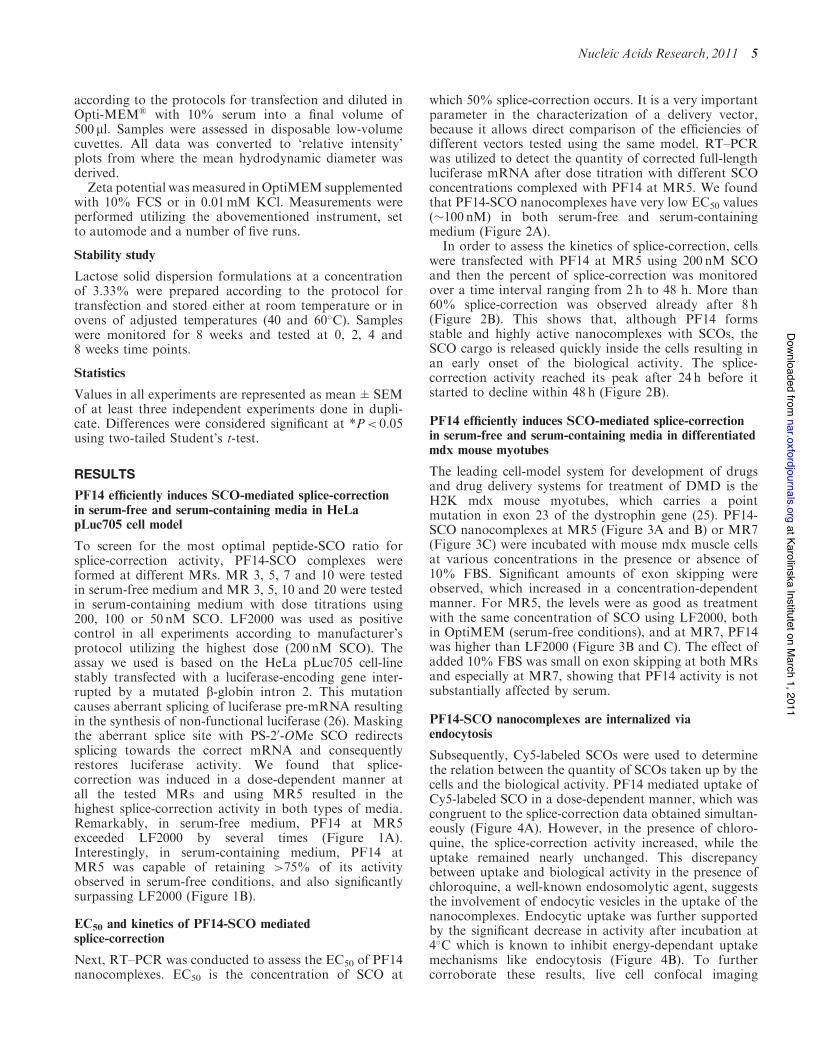
To screen for the most optimal peptide-SCO ratio forsplice-correction activity, PF14-SCO complexes wereformed at different MRs. MR 3, 5, 7 and 10 were testedin serum-free medium and MR 3, 5, 10 and 20 were testedin serum-containing medium with dose titrations using200, 100 or 50 nM SCO. LF2000 was used as positivecontrol in all experiments according to manufacturer’sprotocol utilizing the highest dose (200 nM SCO). Theassay we used is based on the HeLa pLuc705 cell-linestably transfected with a luciferase-encoding gene inter-rupted by a mutated b-globin intron 2. This mutationcauses aberrant splicing of luciferase pre-mRNA resultingin the synthesis of non-functional luciferase (26). Maskingthe aberrant splice site with PS-20-OMe SCO redirectssplicing towards the correct mRNA and consequentlyrestores luciferase activity. We found that splice-correction was induced in a dose-dependent manner atall the tested MRs and using MR5 resulted in thehighest splice-correction activity in both types of media.Remarkably, in serum-free medium, PF14 at MR5exceeded LF2000 by several times (Figure 1A).Interestingly, in serum-containing medium, PF14 atMR5 was capable of retaining >75% of its activityobserved in serum-free conditions, and also significantlysurpassing LF2000 (Figure 1B).
EC50 and kinetics of PF14-SCO mediatedsplice-correction
Next, RT–PCR was conducted to assess the EC50 of PF14nanocomplexes. EC50 is the concentration of SCO at
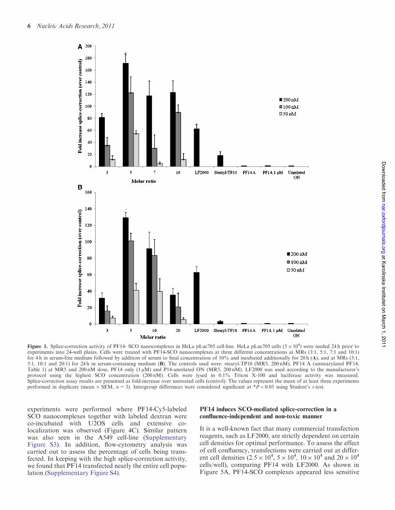
which 50% splice-correction occurs. It is a very importantparameter in the characterization of a delivery vector,because it allows direct comparison of the efficiencies ofdifferent vectors tested using the same model. RT–PCRwas utilized to detect the quantity of corrected full-lengthluciferase mRNA after dose titration with different SCOconcentrations complexed with PF14 at MR5. We foundthat PF14-SCO nanocomplexes have very low EC50 values(#100 nM) in both serum-free and serum-containingmedium (Figure 2A).In order to assess the kinetics of splice-correction, cells
were transfected with PF14 at MR5 using 200 nM SCOand then the percent of splice-correction was monitoredover a time interval ranging from 2h to 48 h. More than60% splice-correction was observed already after 8 h(Figure 2B). This shows that, although PF14 formsstable and highly active nanocomplexes with SCOs, theSCO cargo is released quickly inside the cells resulting inan early onset of the biological activity. The splice-correction activity reached its peak after 24 h before itstarted to decline within 48 h (Figure 2B).
PF14 efficiently induces SCO-mediated splice-correctionin serum-free and serum-containing media in differentiatedmdx mouse myotubes
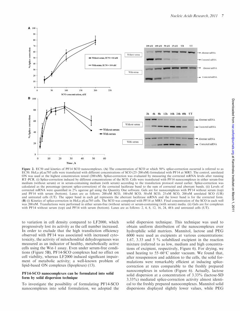
The leading cell-model system for development of drugsand drug delivery systems for treatment of DMD is theH2K mdx mouse myotubes, which carries a pointmutation in exon 23 of the dystrophin gene (25). PF14-SCO nanocomplexes at MR5 (Figure 3A and B) or MR7(Figure 3C) were incubated with mouse mdx muscle cellsat various concentrations in the presence or absence of10% FBS. Significant amounts of exon skipping wereobserved, which increased in a concentration-dependentmanner. For MR5, the levels were as good as treatmentwith the same concentration of SCO using LF2000, bothin OptiMEM (serum-free conditions), and at MR7, PF14was higher than LF2000 (Figure 3B and C). The effect ofadded 10% FBS was small on exon skipping at both MRsand especially at MR7, showing that PF14 activity is notsubstantially affected by serum.
PF14-SCO nanocomplexes are internalized viaendocytosis
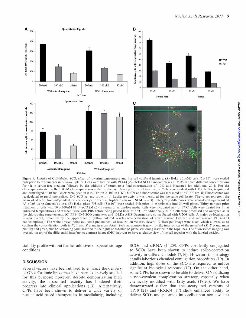
Subsequently, Cy5-labeled SCOs were used to determinethe relation between the quantity of SCOs taken up by thecells and the biological activity. PF14 mediated uptake ofCy5-labeled SCO in a dose-dependent manner, which wascongruent to the splice-correction data obtained simultan-eously (Figure 4A). However, in the presence of chloro-quine, the splice-correction activity increased, while theuptake remained nearly unchanged. This discrepancybetween uptake and biological activity in the presence ofchloroquine, a well-known endosomolytic agent, suggeststhe involvement of endocytic vesicles in the uptake of thenanocomplexes. Endocytic uptake was further supportedby the significant decrease in activity after incubation at4!C which is known to inhibit energy-dependant uptakemechanisms like endocytosis (Figure 4B). To furthercorroborate these results, live cell confocal imaging
Nucleic Acids Research, 2011 5
at Karolinska Institutet on March 1, 2011
nar.oxfordjournals.orgD
ownloaded from
experiments were performed where PF14-Cy5-labeledSCO nanocomplexes together with labeled dextran wereco-incubated with U2OS cells and extensive co-localization was observed (Figure 4C). Similar patternwas also seen in the A549 cell-line (SupplementaryFigure S3). In addition, flow-cytometry analysis wascarried out to assess the percentage of cells being trans-fected. In keeping with the high splice-correction activity,we found that PF14 transfected nearly the entire cell popu-lation (Supplementary Figure S4).
PF14 induces SCO-mediated splice-correction in aconfluence-independent and non-toxic manner
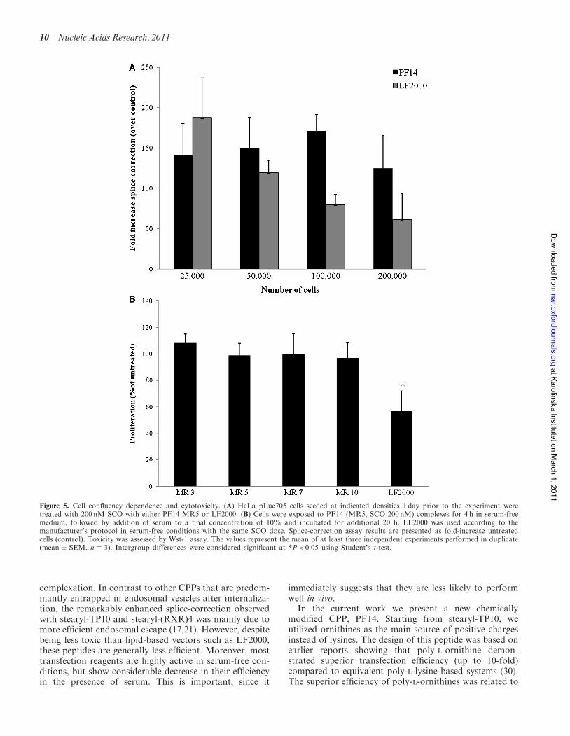
It is a well-known fact that many commercial transfectionreagents, such as LF2000, are strictly dependent on certaincell densities for optimal performance. To assess the effectof cell confluency, transfections were carried out at differ-ent cell densities (2.5" 104, 5" 104, 10" 104 and 20" 104
cells/well), comparing PF14 with LF2000. As shown inFigure 5A, PF14-SCO complexes appeared less sensitive
Figure 1. Splice-correction activity of PF14- SCO nanocomplexes in HeLa pLuc705 cell-line. HeLa pLuc705 cells (5" 104) were seeded 24 h prior toexperiments into 24-well plates. Cells were treated with PF14-SCO nanocomplexes at three different concentrations at MRs (3:1, 5:1, 7:1 and 10:1)for 4 h in serum-free medium followed by addition of serum to final concentration of 10% and incubated additionally for 20 h (A), and at MRs (3:1,5:1, 10:1 and 20:1) for 24 h in serum-containing medium (B). The controls used were: stearyl-TP10 (MR5, 200 nM), PF14 A (unstearylated PF14,Table 1) at MR5 and 200 nM dose, PF14 only (1mM) and P14-unrelated ON (MR5, 200 nM). LF2000 was used according to the manufacturer’sprotocol using the highest SCO concentration (200 nM). Cells were lysed in 0.1% Triton X-100 and luciferase activity was measured.Splice-correction assay results are presented as fold-increase over untreated cells (control). The values represent the mean of at least three experimentsperformed in duplicate (mean±SEM, n=3). Intergroup differences were considered significant at *P< 0.05 using Student’s t-test.
6 Nucleic Acids Research, 2011
at Karolinska Institutet on March 1, 2011
nar.oxfordjournals.orgD
ownloaded from
to variation in cell density compared to LF2000, whichprogressively lost its activity as the cell number increased.In order to exclude that the high transfection efficiencyobserved with PF14 was associated with increased cyto-toxicity, the activity of mitochondrial dehydrogenases wasmeasured as an indicator of healthy, metabolically activecells using the Wst-1 assay. Even under serum-free condi-tions (Figure 5B), PF14-SCO complexes had no effect oncell viability, whereas LF2000 induced significant impair-ment of metabolic activity; a well-known problem oflipid-based ON complexes (lipoplexes) (13).
PF14-SCO nanocomplexes can be formulated into solidform by solid dispersion technique
To investigate the possibility of formulating PF14-SCOnanocomplexes into solid formulation, we adopted the
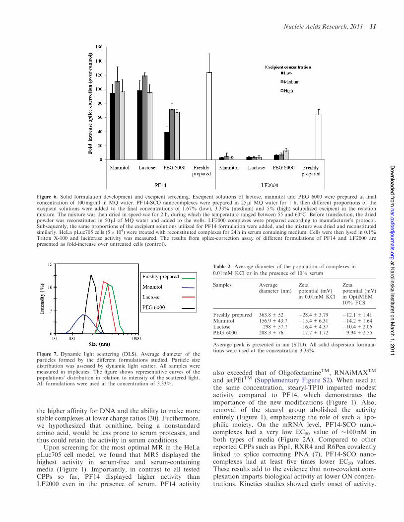
solid dispersion technique. This technique was used toobtain uniform distribution of the nanocomplexes overhydrophilic solid matrices. Mannitol, lactose and PEG6000 were used as excipients at various concentrations:1.67, 3.33 and 5 % solubilized excipient in the reactionmixture (referred to as low, medium and high concentra-tions of excipient, respectively, Figure 6). For drying, weused heating to 55–60!C under vacuum. We found that,after resuspension and addition to the cells, the solid for-mulations were remarkably efficient at inducing splice-correction at rates comparable to the freshly preparednanocomplexes in solution (Figure 6). Actually, lactosesolid dispersion at a concentration of 3.33% (lactose-SD3.33%) mediated splice-correction activity almost identi-cal to the freshly prepared nanocomplexes. Mannitol soliddispersions displayed slightly lower values, while PEG
Figure 2. EC50 and kinetics of PF14 SCO nanocomplexes. (A) The concentration of SCO at which 50% splice-correction occurred is referred to asEC50. HeLa pLuc705 cells were transfected with different concentrations of SCO (25–200 nM) formulated with PF14 at MR5. The control, unrelatedON was used at the highest concentration tested (200 nM). Splice-correction was evaluated by measuring the corrected mRNA levels after runningRT–PCR. (i) Splice-correction induced by different concentrations of the SCO. Cells were transfected with PF14 nanocomplexes in either serum-freemedium (without serum) or in serum-containing medium (with serum) according to the transfection protocol stated earlier. Splice-correction wascalculated as the percentage (percent splice-correction) of the corrected luciferase band to the sum of corrected and aberrant bands. (ii) Levels ofcorrected mRNA were quantified in 2% agarose gel using the Quantity One software. Gels are for nanocomplexes with PF14 without serum (top)and PF14 with serum (bottom). Lanes are as follows: 200 nM SCO, 100 nM SCO, 50 nM SCO, 25 nM SCO, 200 nM unrelated SCO (UR)and untreated cells (UT). The upper band in each gel represents the aberrant luciferase mRNA and the lower band is for the corrected form.(B) (i) Kinetics of splice-correction in HeLa pLuc705 cells. The SCO was complexed with PF14 at MR5. Final concentration of the SCO in each wellwas 200 nM. Transfections were performed in either serum-free (without serum) or serum-containing (with serum) media. (ii) Gels are for complexeswith PF14 without serum (top) and PF14 with serum (bottom). Lanes are as follows: 2, 4, 8, 12, 16, 24, 48 h and untreated cells (UT).
Nucleic Acids Research, 2011 7
at Karolinska Institutet on March 1, 2011
nar.oxfordjournals.orgD
ownloaded from
6000 solid dispersions demonstrated the highest deviationfrom the values obtained using the freshly prepared nano-complexes (Figure 6). In contrast, applying the same pro-cedure on LF2000 nearly eliminated any residual activity.On the contrary, the high residual activity of PF14 formu-lations suggests that the PF14 nanocomplexes remainintact after drying and dispersion under these conditions.
DLS analysis and zeta potential measurements ofPF14-SCO nanocomplexes
To confirm the presence of intact nanocomplexes in theformulations, DLS studies and zeta potential measure-ments were performed. These assessments were conducted
in the presence of serum to allow possible correlationsbetween such parameters and transfection activity atphysiological conditions. Serum contains a number ofproteins and protein aggregates that will scatter light re-sulting in a population of particles independent of thePF14-SCO nanocomplexes and can also affect the zetapotential by surface adsorption of some serum proteins.However, we believe that although the presence of serumwill affect such measurements, these conditions are morerelevant to the physiological ones. For DLS, we havecompared the populations detected in serum-containingmedium alone and after addition of PF14-SCO nano-complexes. It was possible to distinguish the constantprotein aggregates (inherent to the serum, and >100 nm)and a completely independent population associated tothe PF14 nanocomplexes (data not shown). Due to thepresence of serum proteins, the polydispersity index(PdI) of the solution containing the nanocomplexes doesnot describe the homogeneity of the PF14 nanocomplexesalone. For this reason, we refer to the shape of the curvesthat characterize the different populations while eval-uating the homogeneity of the particles. For zeta poten-tial, measurements were carried out either in 0.01mMKClor in the presence of 10% serum. After resuspension of thesolid formulations, DLS measurements showed that nano-complexes are present in all the tested formulations.However, the uniformity of particles size distributionand the extent of similarity in relation to the freshlyprepared nanocomplexes differed with different excipients.Lactose-SD 3.33% formulations were most homoge-neous and most similar to the distribution pattern of thefreshly prepared nanocomplexes in solution (Figure 7 andTable 2). These results go in accordance with the transfec-tion results where lactose-SD 3.33% displayed the highestsplice-correction activity. Moreover, this analysis demon-strates that different excipients affect the physicochemicalcharacterizes of the nanocomplexes differently in termsof particle size and uniformity, and this effect is reflectedon the biological activity. We studied the surface charge(zeta potential) of the PF14 nanocomplexes, both freshlyprepared and after drying at 50–60!C in the solid disper-sion procedure. Interestingly, independent of formula-tion type and media (presence or absence of serum),the nanocomplexes displayed negative surface charges(Table 2).
Solid dispersion formulations are stable for several weeksat elevated temperatures
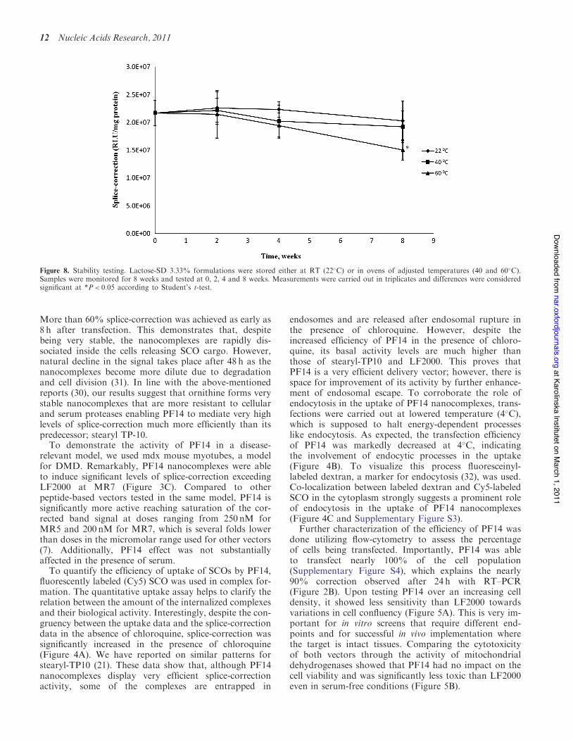
Finally, we tested the stability of the best-performing for-mulation, lactose-SD3.33%, when stored for 2 months atan elevated temperatures. The formulations were stored atthree different temperatures (RT, 40 and 60!C) andsampled over an 8 weeks period. We found that thetested formulations were remarkably stable. There wasno statistically significant loss in transfection efficienciesat any time-point except for the 60!C 8-weeks point wherethe transfection efficiency decreased to 70% of the initialvalue (Figure 8). Compared to lyophilized lipoplexes(27,28), PF14 formulations demonstrated an excellent
C
in OptiMEM in DMEM/10%FBS LF2000
0
10
20
30
40
50
60B
in OptiMEM in DMEM/10%FBS LF2000
Am
ount
of e
xon
23 s
kipp
ed, %
250 nM
125 nM
62.5 nM
0
10
20
30
40
50
Am
ount
of e
xon
23 s
kipp
ed, %
200 nM
100 nM
50 nM
A
Figure 3. Splice-correction activity of PF14-SCO nanocomplexes in mdxmouse myotubes. (A) RT–PCR analysis to measure exon skipping activityof PF14-SCO nanocomplexes in mdx mouse myotubes at three differentconcentrations, at MR5 in serum-free OptiMEM and DMEM/10% FBSin comparison with control LF2000 transfection in OptiMEM. Exonskipping activity was measured as the amount of RT–PCR productafter treatment of cells with PF14-SCO nanocomplexes at MR5 (B) orMR7 (C) at three different concentrations. The values represent the meanof at least three independent experiments performed in duplicate(mean±SEM, n=3).
8 Nucleic Acids Research, 2011
at Karolinska Institutet on March 1, 2011
nar.oxfordjournals.orgD
ownloaded from
stability profile without further additives or special storageconditions.
DISCUSSION
Several vectors have been utilized to enhance the deliveryof ONs. Cationic liposomes have been extensively studiedfor this purpose; however, despite demonstrating highactivity, the associated toxicity has hindered theirprogress into clinical applications (13). Alternatively,CPPs have been shown to deliver a wide variety ofnucleic acid-based therapeutics intracellularly, including
SCOs and siRNA (14,29). CPPs covalently conjugatedto SCOs have been shown to induce splice-correctionactivity in different models (7,16). However, this strategyentails laborious chemical conjugation procedures (19). Inaddition, high doses of the SCO are required to inducesignificant biological response (17). On the other hand,some CPPs have shown to be able to deliver ONs utilizinga non-covalent complexation strategy, especially whenchemically modified with fatty acids (18,20). We havedemonstrated earlier that the stearylated versions ofTP10 (21) and (RXR)4 (17) show enhanced ability todeliver SCOs and plasmids into cells upon non-covalent
Figure 4. Uptake of Cy5-labeled SCO, effect of lowering temperature and live cell confocal imaging. (A) HeLa pLuc705 cells (5" 104) were seeded24 h prior to experiments into 24-well plates. Cells were treated with PF14-Cy5-lebeled SCO nanocomplexes at MR5 at three different concentrationsfor 4 h in serum-free medium followed by the addition of serum to a final concentration of 10% and incubated for additional 20 h. For thechloroquine-treated wells, 100mM chloroquine was added to the complexes prior to cell treatments. Cells were washed with HKR buffer, trypsinizedand centrifuged at 1000g. Pellets were lysed in 0.1% Triton X-100 in HKR buffer and fluorescence was measured at 650/670 nm. (i) Fluorescence wasrecalculated to pmol internalized Cy5 SCO per mg protein. (ii) Luciferase activity was measured for the same cell lysate. The values represent themean of at least two independent experiments performed in triplicate (mean±SEM, n=3). Intergroup differences were considered significant at*P< 0.05 using Student’s t-test. (B) HeLa pLuc 705 cells (5" 104) were seeded 24 h prior to experiments into 24-well plates. Thirty minutes priortreatment of cells with 50 or100 nM PF14-SCO (MR5) in serum or serum-free media, cells were incubated at 4 or 37!C. Cells were treated for 1 h atindicated temperatures and washed twice with PBS before being placed back at 37!C for additionally 20 h. Cells were processed and analyzed as inthe chloroquine experiements. (C) PF14-Cy5-SCO complexes and 10 kDa A488-Dextran were co-incubated with U2OS cells. A major co-localizationis seen overall, presented by the appearance of yellow colored vesicles (co-localization of green marked Dextran and red marked PF14-SCOnanocomplexes). The white arrows point out some pre-eminent co-localization vesicles. Several Z-slices per image were taken which allowed us toconfirm the co-localization both in X, Y and Z plane in more detail. Such an example is given by the intersection of the green/red (X, Y plane; mainpicture) and green/blue (Z sectioning panel inserted to the right) or red/blue (Z plane sectioning inserted in the top) lines. The fluorescence imaging wasoverlaid on top of the differential interference contrast image (DIC) in order to have a relative view of the cell together with the labeled vesicles.
Nucleic Acids Research, 2011 9
at Karolinska Institutet on March 1, 2011
nar.oxfordjournals.orgD
ownloaded from
complexation. In contrast to other CPPs that are predom-inantly entrapped in endosomal vesicles after internaliza-tion, the remarkably enhanced splice-correction observedwith stearyl-TP10 and stearyl-(RXR)4 was mainly due tomore efficient endosomal escape (17,21). However, despitebeing less toxic than lipid-based vectors such as LF2000,these peptides are generally less efficient. Moreover, mosttransfection reagents are highly active in serum-free con-ditions, but show considerable decrease in their efficiencyin the presence of serum. This is important, since it
immediately suggests that they are less likely to performwell in vivo.
In the current work we present a new chemicallymodified CPP, PF14. Starting from stearyl-TP10, weutilized ornithines as the main source of positive chargesinstead of lysines. The design of this peptide was based onearlier reports showing that poly-L-ornithine demon-strated superior transfection efficiency (up to 10-fold)compared to equivalent poly-L-lysine-based systems (30).The superior efficiency of poly-L-ornithines was related to
Figure 5. Cell confluency dependence and cytotoxicity. (A) HeLa pLuc705 cells seeded at indicated densities 1 day prior to the experiment weretreated with 200 nM SCO with either PF14 MR5 or LF2000. (B) Cells were exposed to PF14 (MR5, SCO 200 nM) complexes for 4 h in serum-freemedium, followed by addition of serum to a final concentration of 10% and incubated for additional 20 h. LF2000 was used according to themanufacturer’s protocol in serum-free conditions with the same SCO dose. Splice-correction assay results are presented as fold-increase untreatedcells (control). Toxicity was assessed by Wst-1 assay. The values represent the mean of at least three independent experiments performed in duplicate(mean±SEM, n=3). Intergroup differences were considered significant at *P< 0.05 using Student’s t-test.
10 Nucleic Acids Research, 2011
at Karolinska Institutet on March 1, 2011
nar.oxfordjournals.orgD
ownloaded from
the higher affinity for DNA and the ability to make morestable complexes at lower charge ratios (30). Furthermore,we hypothesized that ornithine, being a nonstandardamino acid, would be less prone to serum proteases, andthus could retain the activity in serum conditions.
Upon screening for the most optimal MR in the HeLapLuc705 cell model, we found that MR5 displayed thehighest activity in serum-free and serum-containingmedia (Figure 1). Importantly, in contrast to all testedCPPs so far, PF14 displayed higher activity thanLF2000 even in the presence of serum. PF14 activity
also exceeded that of OligofectamineTM, RNAiMAXTM
and jetPEITM (Supplementary Figure S2). When used atthe same concentration, stearyl-TP10 imparted modestactivity compared to PF14, which demonstrates theimportance of the new modifications (Figure 1). Also,removal of the stearyl group abolished the activityentirely (Figure 1), emphasizing the role of such a lipo-philic moiety. On the mRNA level, PF14-SCO nano-complexes had a very low EC50 value of #100 nM inboth types of media (Figure 2A). Compared to otherreported CPPs such as Pip1, RXR4 and R6Pen covalentlylinked to splice correcting PNA (7), PF14-SCO nano-complexes had at least five times lower EC50 values.These results add to the evidence that non-covalent com-plexation imparts biological activity at lower ON concen-trations. Kinetics studies showed early onset of activity.
Figure 6. Solid formulation development and excipient screening. Excipient solutions of lactose, mannitol and PEG 6000 were prepared at finalconcentration of 100mg/ml in MQ water. PF14-SCO nanocomplexes were prepared in 25 ml MQ water for 1 h, then different proportions of theexcipient solutions were added to the final concentrations of 1.67% (low), 3.33% (medium) and 5% (high) solubilized excipient in the reactionmixture. The mixture was then dried in speed-vac for 2 h, during which the temperature ranged between 55 and 60!C. Before transfection, the driedpowder was reconstituted in 50 ml of MQ water and added to the wells. LF2000 complexes were prepared according to manufacturer’s protocol.Subsequently, the same proportions of the excipient solutions utilized for PF14 formulation were added, and the mixture was dried and reconstitutedsimilarly. HeLa pLuc705 cells (5" 104) were treated with reconstituted complexes for 24 h in serum containing medium. Cells were then lysed in 0.1%Triton X-100 and luciferase activity was measured. The results from splice-correction assay of different formulations of PF14 and LF2000 arepresented as fold-increase over untreated cells (control).
Figure 7. Dynamic light scattering (DLS). Average diameter of theparticles formed by the different formulations studied. Particle sizedistribution was assessed by dynamic light scatter. All samples weremeasured in triplicates. The figure shows representative curves of thepopulations’ distribution in relation to intensity of the scattered light.All formulations were used at the concentration of 3.33%.
Table 2. Average diameter of the population of complexes in0.01mM KCl or in the presence of 10% serum
Samples Averagediameter (nm)
Zetapotential (mV)in 0.01mM KCl
Zetapotential (mV)in OptiMEM10% FCS
Freshly prepared 363.8±52 $28.4±3.79 $12.1±1.41Mannitol 156.9±43.7 $15.4±6.31 $14.2±1.64Lactose 298±57.7 $16.4±4.37 $10.4±2.06PEG 6000 208.3±76 $17.7±1.72 $9.94±2.55
Average peak is presented in nm (STD). All solid dispersion formula-tions were used at the concentration 3.33%.
Nucleic Acids Research, 2011 11
at Karolinska Institutet on March 1, 2011
nar.oxfordjournals.orgD
ownloaded from
More than 60% splice-correction was achieved as early as8 h after transfection. This demonstrates that, despitebeing very stable, the nanocomplexes are rapidly dis-sociated inside the cells releasing SCO cargo. However,natural decline in the signal takes place after 48 h as thenanocomplexes become more dilute due to degradationand cell division (31). In line with the above-mentionedreports (30), our results suggest that ornithine forms verystable nanocomplexes that are more resistant to cellularand serum proteases enabling PF14 to mediate very highlevels of splice-correction much more efficiently than itspredecessor; stearyl TP-10.To demonstrate the activity of PF14 in a disease-
relevant model, we used mdx mouse myotubes, a modelfor DMD. Remarkably, PF14 nanocomplexes were ableto induce significant levels of splice-correction exceedingLF2000 at MR7 (Figure 3C). Compared to otherpeptide-based vectors tested in the same model, PF14 issignificantly more active reaching saturation of the cor-rected band signal at doses ranging from 250 nM forMR5 and 200 nM for MR7, which is several folds lowerthan doses in the micromolar range used for other vectors(7). Additionally, PF14 effect was not substantiallyaffected in the presence of serum.To quantify the efficiency of uptake of SCOs by PF14,
fluorescently labeled (Cy5) SCO was used in complex for-mation. The quantitative uptake assay helps to clarify therelation between the amount of the internalized complexesand their biological activity. Interestingly, despite the con-gruency between the uptake data and the splice-correctiondata in the absence of chloroquine, splice-correction wassignificantly increased in the presence of chloroquine(Figure 4A). We have reported on similar patterns forstearyl-TP10 (21). These data show that, although PF14nanocomplexes display very efficient splice-correctionactivity, some of the complexes are entrapped in
endosomes and are released after endosomal rupture inthe presence of chloroquine. However, despite theincreased efficiency of PF14 in the presence of chloro-quine, its basal activity levels are much higher thanthose of stearyl-TP10 and LF2000. This proves thatPF14 is a very efficient delivery vector; however, there isspace for improvement of its activity by further enhance-ment of endosomal escape. To corroborate the role ofendocytosis in the uptake of PF14 nanocomplexes, trans-fections were carried out at lowered temperature (4!C),which is supposed to halt energy-dependent processeslike endocytosis. As expected, the transfection efficiencyof PF14 was markedly decreased at 4!C, indicatingthe involvement of endocytic processes in the uptake(Figure 4B). To visualize this process fluoresceinyl-labeled dextran, a marker for endocytosis (32), was used.Co-localization between labeled dextran and Cy5-labeledSCO in the cytoplasm strongly suggests a prominent roleof endocytosis in the uptake of PF14 nanocomplexes(Figure 4C and Supplementary Figure S3).
Further characterization of the efficiency of PF14 wasdone utilizing flow-cytometry to assess the percentageof cells being transfected. Importantly, PF14 was ableto transfect nearly 100% of the cell population(Supplementary Figure S4), which explains the nearly90% correction observed after 24 h with RT–PCR(Figure 2B). Upon testing PF14 over an increasing celldensity, it showed less sensitivity than LF2000 towardsvariations in cell confluency (Figure 5A). This is very im-portant for in vitro screens that require different end-points and for successful in vivo implementation wherethe target is intact tissues. Comparing the cytotoxicityof both vectors through the activity of mitochondrialdehydrogenases showed that PF14 had no impact on thecell viability and was significantly less toxic than LF2000even in serum-free conditions (Figure 5B).
Figure 8. Stability testing. Lactose-SD 3.33% formulations were stored either at RT (22!C) or in ovens of adjusted temperatures (40 and 60!C).Samples were monitored for 8 weeks and tested at 0, 2, 4 and 8 weeks. Measurements were carried out in triplicates and differences were consideredsignificant at *P< 0.05 according to Student’s t-test.
12 Nucleic Acids Research, 2011
at Karolinska Institutet on March 1, 2011
nar.oxfordjournals.orgD
ownloaded from
Finally, we wanted to take our delivery system a stepfurther by developing a suitable formulation for adminis-tration. The formulation process is an indispensable stepfor the development of any therapeutic product. It hasgreat effect on enhancing the efficiency and stability ofactive pharmaceutical ingredients. Additionally, specificformulations have to be tailored for different therapeuticapproaches. Among the different pharmaceutical formspresent, solid formulations remain the most widely used.It is the form used in tablets, capsules, powders for inhal-ation and even powders for injection. This is because theyare easy to handle and very stable upon storage and trans-portation. Although CPPs have been extensively exploitedin recent years for the delivery of various therapeutics(15), to our knowledge, formulation of CPPs into differentpharmaceutical forms has never been thoroughly studied.Therefore, to expand the range of therapeutic applicationsof CPP-based therapeutics, there is a need for studiesexploring the possibility of incorporating CPPs in differ-ent pharmaceutical forms; especially the solid form. Totest the feasibility of formulating PF14-SCO complexesinto solid formulation, solid dispersion technique wasutilized. Although initially invented to increase the solu-bility of poorly water-soluble drugs by dispersing themover hydrophilic solid matrices (33), it is not used forthe same purpose in the present work. This is becausePF14-SCO complexes are already water-soluble.However, the technique was adopted to enable us obtainuniform distribution of the nanocomplexes over water-soluble excipients by solvent evaporation. For solventevaporation (in this case water), we chose speed dryingunder elevated temperature (55–60!C) and reduced pres-sure. Despite being relatively stressful drying method, it ismore relevant to the widely used, cost-effective, heat-based pharmaceutical drying techniques. As shown inFigure 6, we found that that lactose-SD 3.33% was themost optimal formulation, achieving splice-correctionactivity similar to that of the freshly prepared complexesin solution. Mannitol solid dispersions mediated loweractivity, while PEG formulations were the least active.It was clear that different excipients and concentrationsthereof had a huge impact on the activity of the formula-tion. This suggests that excipients affect the nano-complexes in different ways. The nanocomplexes areformed by non-covalent electrostatic interactions withthe ON. Therefore, different excipients varying in theirsurface charge distribution could interfere with these inter-actions affecting the physicochemical properties of thenanocomplexes and eventually the biological activity.However, an excipient concentration of 3.33% is shownto be relatively superior in all the excipients tested, whichsuggests that there is an optimal ratio between the concen-tration of nanocomplexes in solution and the amount ofsolubilized solid material. On the contrary, LF2000 almostlost its entire activity upon application of this drying anddispersion procedure (Figure 6). The success of PF14-SCOcomplexes to withstand harsh drying and dispersion pro-cedure at which the widely used commercial lipid-basedvector failed suggests that PF14-SCO nanocomplexes arephysicochemically more stable than the lipoplexes, yetbiologically more active. Additionally, we characterized
the effect of formulation on the kinetics of splice correc-tion obtained by the best formulation (lactose-SD 3.33%).We found that it demonstrated similar kinetic profile tothe freshly prepared nanocomplexes (SupplementaryFigure S5).In order to assess that the presence of intact nano-
complexes after the formulation procedure, DLS studieswere performed comparing the freshly prepared nano-complexes with the solid formulations obtained afterdrying at 50–60!C . We found that the particles size andparticle-size distribution is highly affected by the type ofexcipient used (Figure 7 and Table 2). Lactose-SD 3.33%formulation, which mediated the highest splice-correctionactivity, also had DLS profile most similar to the freshlyprepared nanocomplexes. This proves that the choice ofexcipient for formulation of such nanocomplexes is anextremely important step that affects both their physico-chemical properties and biological activity. Yet, anotherphysicochemical property that could affect the biologicalactivity is the zeta potential. Particles of different surfacecharge are expected to have distinct interactions withthe plasma membrane, which should affect the uptakemechanism and efficiency of the delivery vector. Weexpected that a cationic CPP-based nanocomplex shouldpossess net positive surface charge. Surprisingly however,we found that all the nanocomplexes, either freshlyprepared or in solid formulation, with or without serum,possessed a net negative charge. The efficiency of suchnanocomplexes can be explained in light of earlierresearch on lipoplexes showing that cationic liposomeand plasmid DNA complexes are negatively chargedunder optimal transfection conditions (34) and that thisholds in vivo as well (35). In further research studying theuptake mechanisms of CPPs it will be interesting to eluci-date how this net negative charge affects the interactionwith the plasma membrane and the biological activity.Next, we subjected the best performing formulation,
lactose-SD 3.33%, to stressful stability testing conditionsat elevated temperatures for 2 months. Storage at hightemperatures increases the rate of degradation reactionsthat could take years to occur (36). For this reason thismethod is routinely used to assess the long-term stabilityof formulations (27,28,37–39). Lactose-SD 3.33% formu-lation demonstrated high stability over the tested timeperiod with no significant loss in transfection efficiencyat most of the time points (Figure 8). The only significantdecrease in activity was the 60!C 8-weeks time point,where it was #70% of the initial activity. However, thisis a remarkably good stability profile for a plain formula-tion without any type of preservatives, coating or specialpackaging. In addition, these results further corroboratethe high stability of PF14 nanocomplexes. In comparisonwith solid formulation of lipoplexes, PF14 nanocomplexesdemonstrate very good activity and stability profiles.Earlier reports showing that lipoplexes could be driedover solid matrices mainly utilized the freeze-drying tech-nique in the presence of lyoprotectant sugars like trehal-ose, mannitol or sucrose (38–40). However, despite usingsufficient levels of lyoprotectant sugars, significantdamage was still evident when dilute lipoplex preparationswere subjected to freeze-drying (28). This damage occurs
Nucleic Acids Research, 2011 13
at Karolinska Institutet on March 1, 2011
nar.oxfordjournals.orgD
ownloaded from
during the freezing step, and sugars have a limitedcapacity to protect against freezing-induced damage (28).Furthermore, industrially, the freeze-drying (lyophiliza-tion) process is known to be much more cumbersomeand costly than the conventional heat-based pharmaceut-ical drying procedure. Additionally, it was shown thatthe lyophilized lipoplexes suffered progressive loss intransfecetion efficiency when subjected to stabilitysettings similar to what we have used in the presentstudy (27). The degradation was mainly due to the gener-ation of reactive-oxygen species and was increased in thepresence of metal contaminants (27,37). Consequently,chelators were used to enhance the stability profile;however, the effects of these additives on vector stabilityduring lyophilization are difficult to predict (37). To ourknowledge, PF14 is the first vector reported to retain nearly100% of its transfection efficiency utilizing a heat-baseddrying and dispersion procedure. Furthermore, PF14showed a very good stability profile in the absence of add-itional formulation requirements.In conclusion, we have shown that the chemically
modified CPP, PF14, is able to efficiently deliver SCOsto different cell-lines including a disease-relevant hard-to-transfect cell-line, H2K. Furthermore, we have success-fully been able to formulate PF14-SCO nanocomplexesinto solid formulation utilizing the solid dispersion tech-nique using a convenient heat-based drying method. Thesolid formulations demonstrated splice-correction activityalmost identical to the freshly prepared nanocomplexes;however, this efficiency was sensitive to the type of excipi-ent used and its concentration. The nanocomplexesremained intact after formulation and the formulationremained active even when subjected to stressful stabilityconditions for 2 months. Importantly, being able to for-mulate PF14 nanocomplexes into an active and stablesolid formulation is an important proof-of-principlethat CPP-based therapeutics could be incorporated insolid pharmaceutical forms. This could significantlyextend the horizon of therapeutic applications of CPPs.Collectively, our results suggest that PF14 is a promisingtranslational approach for delivering SCOs in differentpharmaceutical forms and opens new opportunities todevelop more pharmaceutically relevant assays for CPPs.
SUPPLEMENTARY DATA
Supplementary Data are available at NAR Online.
ACKNOWLEDGEMENTS
We would like to thank Johanna Unger for her assistancein the preparation of the initial experiments. The confocalimaging study was performed at the Live Cell Imagingunit, Department of Biosciences and Nutrition,Karolinska Institutet, Huddinge, Sweden.
FUNDING
Swedish Research Council (VR-NT); VINNOVA-SAMBIO joint project; SSF (Sweden-Japan); Center for
Biomembrane Research, Stockholm; Knut and AliceWallenberg’s Foundation; the EU trough the EuropeanRegional Development Fund through the Center ofExcellence in Chemical Biology, Estonia; the targetedfinancing SF0180027s08 from the Estonian Government;the DoRa Program of The European Social Fund;Archimedes Foundation; the Torsten and RagnarSoderberg Foundation; KI faculty funds for funding ofpostgraduated students (to J.R.V.); Egyptian Ministry ofHigher Education (to E.M.Z.); Swedish Society ofMedical Research (SSMF to S.E.A.); Work in the Gaitlaboratory is supported by the Medical ResearchCouncil (Unit program U105178803). Funding for openaccess charge: Swedish Research Council (VR-NT).
Conflict of interest statement. None declared.
REFERENCES
1. Munos,B. (2009) Lessons from 60 years of pharmaceuticalinnovation. Nat. Rev. Drug Discov., 8, 959–968.
2. Butler,D. (2008) Translational research: crossing the valley ofdeath. Nature, 453, 840–842.
3. Nilsen,T.W. and Graveley,B.R. (2010) Expansion of theeukaryotic proteome by alternative splicing. Nature, 463, 457–463.
4. Sazani,P. and Kole,R. (2003) Therapeutic potential ofantisense oligonucleotides as modulators of alternative splicing.J. Clin. Invest., 112, 481–486.
5. Bauman,J., Jearawiriyapaisarn,N. and Kole,R. (2009) Therapeuticpotential of splice-switching oligonucleotides. Oligonucleotides, 19,1–13.
6. Wood,M.J. (2010) Toward an oligonucleotide therapy forduchenne muscular dystrophy: a complex development challengeSci. Transl. Med., 2, 25ps15.
7. Ivanova,G.D., Arzumanov,A., Abes,R., Yin,H., Wood,M.J.,Lebleu,B. and Gait,M.J. (2008) Improved cell-penetratingpeptide-PNA conjugates for splicing redirection in HeLa cells andexon skipping in mdx mouse muscle. Nucleic Acids Res., 36,6418–6428.
8. van Deutekom,J.C., Janson,A.A., Ginjaar,I.B., Frankhuizen,W.S.,Aartsma-Rus,A., Bremmer-Bout,M., den Dunnen,J.T., Koop,K.,van der Kooi,A.J., Goemans,N.M. et al. (2007) Local dystrophinrestoration with antisense oligonucleotide PRO051. N. Engl. J.Med., 357, 2677–2686.
9. Kinali,M., Arechavala-Gomeza,V., Feng,L., Cirak,S., Hunt,D.,Adkin,C., Guglieri,M., Ashton,E., Abbs,S., Nihoyannopoulos,P.et al. (2009) Local restoration of dystrophin expression with themorpholino oligomer AVI-4658 in duchenne muscular dystrophy:a single-blind, placebo-controlled, dose-escalation,proof-of-concept study. Lancet Neurol., 8, 918–928.
10. Bell,N.M. and Micklefield,J. (2009) Chemical modification ofoligonucleotides for therapeutic, bioanalytical and otherapplications. Chembiochem, 10, 2691–2703.
11. Amidon,G.L., Lennernas,H., Shah,V.P. and Crison,J.R. (1995) Atheoretical basis for a biopharmaceutic drug classification: thecorrelation of in vitro drug product dissolution and in vivobioavailability. Pharm. Res., 12, 413–420.
12. Lipinski,C.A., Lombardo,F., Dominy,B.W. and Feeney,P.J. (2001)Experimental and computational approaches to estimate solubilityand permeability in drug discovery and development settings.Adv. Drug Deliv. Rev., 46, 3–26.
13. Dass,C.R. (2004) Lipoplex-mediated delivery of nucleic acids:factors affecting in vivo transfection. J. Mol. Med., 82, 579–591.
14. Ezzat,K., El Andaloussi,S., Abdo,R. and Langel,U. (2010)Peptide-based matrices as drug delivery vehicles.Curr. Pharm. Des., 16, 1167–1178.
15. Heitz,F., Morris,M.C. and Divita,G. (2009) Twenty years ofcell-penetrating peptides: from molecular mechanisms totherapeutics. Br. J. Pharmacol., 157, 195–206.
14 Nucleic Acids Research, 2011
at Karolinska Institutet on March 1, 2011
nar.oxfordjournals.orgD
ownloaded from
16. Wood,M.J., Gait,M.J. and Yin,H. (2010) RNA-targetedsplice-correction therapy for neuromuscular disease. Brain, 133,957–972.
17. Lehto,T., Abes,R., Oskolkov,N., Suhorutsenko,J.,Copolovici,D.M., Mager,I., Viola,J.R., Simonson,O.E., Ezzat,K.,Guterstam,P. et al. (2010) Delivery of nucleic acids with astearylated (RxR)4 peptide using a non-covalent co-incubationstrategy. J. Control. Release, 141, 42–51.
18. Deshayes,S., Morris,M., Heitz,F. and Divita,G. (2008) Delivery ofproteins and nucleic acids using a non-covalent peptide-basedstrategy. Adv. Drug Deliv. Rev., 60, 537–547.
19. Mae,M., Andaloussi,S.E., Lehto,T. and Langel,U. (2009)Chemically modified cell-penetrating peptides for the delivery ofnucleic acids. Expert Opin. Drug Deliv., 6, 1195–1205.
20. Futaki,S., Ohashi,W., Suzuki,T., Niwa,M., Tanaka,S., Ueda,K.,Harashima,H. and Sugiura,Y. (2001) Stearylated arginine-richpeptides: a new class of transfection systems. Bioconjug. Chem.,12, 1005–1011.
21. Mae,M., El Andaloussi,S., Lundin,P., Oskolkov,N.,Johansson,H.J., Guterstam,P. and Langel,U. (2009) A stearylatedCPP for delivery of splice correcting oligonucleotides using anon-covalent co-incubation strategy. J. Control. Release, 134,221–227.
22. Guterstam,P., Lindgren,M., Johansson,H., Tedebark,U.,Wengel,J., El Andaloussi,S. and Langel,U. (2008) Splice-switchingefficiency and specificity for oligonucleotides with locked nucleicacid monomers. Biochem. J., 412, 307–313.
23. Lu,Q.L., Rabinowitz,A., Chen,Y.C., Yokota,T., Yin,H., Alter,J.,Jadoon,A., Bou-Gharios,G. and Partridge,T. (2005) Systemicdelivery of antisense oligoribonucleotide restores dystrophinexpression in body-wide skeletal muscles. Proc. Natl Acad. Sci.USA, 102, 198–203.
24. Coin,I., Beyermann,M. and Bienert,M. (2007) Solid-phase peptidesynthesis: from standard procedures to the synthesis of difficultsequences. Nat. Protoc., 2, 3247–3256.
25. Morgan,J.E., Beauchamp,J.R., Pagel,C.N., Peckham,M.,Ataliotis,P., Jat,P.S., Noble,M.D., Farmer,K. and Partridge,T.A.(1994) Myogenic cell lines derived from transgenic mice carryinga thermolabile T antigen: a model system for the derivation oftissue-specific and mutation-specific cell lines. Dev. Biol., 162,486–498.
26. Kang,S.H., Cho,M.J. and Kole,R. (1998) Up-regulation ofluciferase gene expression with antisense oligonucleotides:implications and applications in functional assay development.Biochemistry, 37, 6235–6239.
27. Molina,M.D. and Anchordoquy,T.J. (2008) Degradation oflyophilized lipid/DNA complexes during storage: the role of lipidand reactive oxygen species. Biochim. Biophys. Acta, 1778,2119–2126.
28. Yu,J. and Anchordoquy,T.J. (2009) Synergistic effects ofsurfactants and sugars on lipoplex stability during freeze-dryingand rehydration. J. Pharm. Sci., 98, 3319–3328.
29. El Andaloussi,S., Lehto,T., Mager,I., Rosenthal-Aizman,K.,Oprea,J., Simonson,O.E., Sork,H., Ezzat,K., Copolovici,D.M.,Kurrikoff,K. et al. (2011) Design of a peptide-based vector,PepFect6, for efficient delivery of siRNA in cell culture andsystemically in vivo. Nucleic Acids Res., doi:10.1093/nar/gkq1299[Epub ahead of print].
30. Ramsay,E. and Gumbleton,M. (2002) Polylysine andpolyornithine gene transfer complexes: a study of complexstability and cellular uptake as a basis for their differentialin-vitro transfection efficiency. J. Drug Target., 10, 1–9.
31. Hallbrink,M., Oehlke,J., Papsdorf,G. and Bienert,M. (2004)Uptake of cell-penetrating peptides is dependent on peptide-to-cellratio rather than on peptide concentration. Biochim. Biophys.Acta, 1667, 222–228.
32. Alam,M.R., Dixit,V., Kang,H., Li,Z.B., Chen,X., Trejo,J.,Fisher,M. and Juliano,R.L. (2008) Intracellular delivery of ananionic antisense oligonucleotide via receptor-mediatedendocytosis. Nucleic Acids Res., 36, 2764–2776.
33. Vasconcelos,T., Sarmento,B. and Costa,P. (2007) Solid dispersionsas strategy to improve oral bioavailability of poor water solubledrugs. Drug Discov. Today, 12, 1068–1075.
34. Son,K.K., Patel,D.H., Tkach,D. and Park,A. (2000) Cationicliposome and plasmid DNA complexes formed in serum-freemedium under optimum transfection condition are negativelycharged. Biochim. Biophys. Acta, 1466, 11–15.
35. Son,K.K., Tkach,D. and Hall,K.J. (2000) Efficient in vivogene delivery by the negatively charged complexes ofcationic liposomes and plasmid DNA. Biochim. Biophys. Acta,1468, 6–10.
36. Ahuja,S. and Scypinski,S. (2001) Handbook of ModernPharmaceutical Analysis. Academic Press, San Diego.
37. Molina,M.C. and Anchordoquy,T.J. (2007) Metal contaminantspromote degradation of lipid/DNA complexes duringlyophilization. Biochim. Biophys. Acta, 1768, 669–677.
38. Armstrong,T.K. and Anchordoquy,T.J. (2004) Immobilizationof nonviral vectors during the freezing step of lyophilization.J. Pharm. Sci., 93, 2698–2709.
39. Molina,M.C., Armstrong,T.K., Zhang,Y., Patel,M.M., Lentz,Y.K.and Anchordoquy,T.J. (2004) The stability of lyophilized lipid/DNA complexes during prolonged storage. J. Pharm. Sci., 93,2259–2273.
40. Brus,C., Kleemann,E., Aigner,A., Czubayko,F. and Kissel,T.(2004) Stabilization of oligonucleotide-polyethylenimine complexesby freeze-drying: physicochemical and biological characterization.J. Control. Release, 95, 119–131.
Nucleic Acids Research, 2011 15
at Karolinska Institutet on March 1, 2011
nar.oxfordjournals.orgD
ownloaded from
Supplementary Data
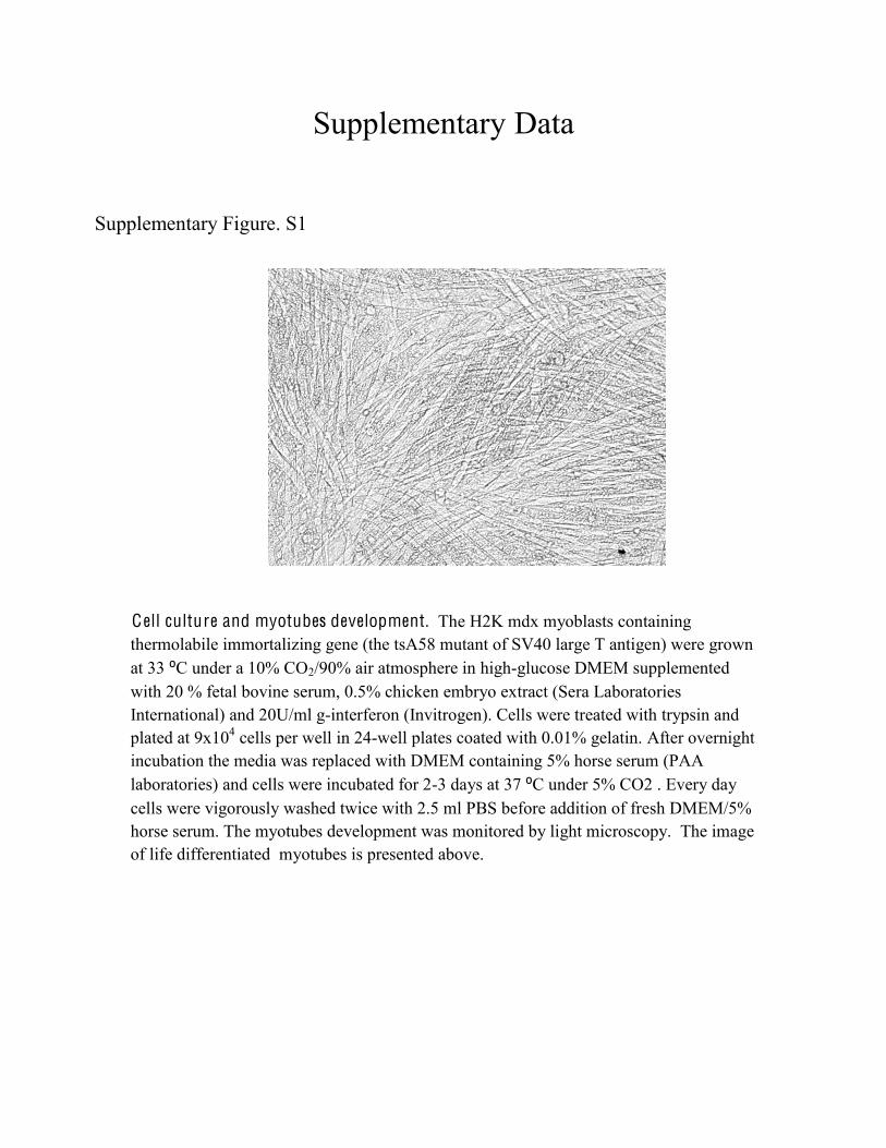
Cell culture and myotubes development. The H2K mdx myoblasts containing thermolabile immortalizing gene (the tsA58 mutant of SV40 large T antigen) were grown at 33 C under a 10% CO2/90% air atmosphere in high-glucose DMEM supplemented with 20 % fetal bovine serum, 0.5% chicken embryo extract (Sera Laboratories International) and 20U/ml g-interferon (Invitrogen). Cells were treated with trypsin and plated at 9x10⁴ cells per well in 24-well plates coated with 0.01% gelatin. After overnight incubation the media was replaced with DMEM containing 5% horse serum (PAA laboratories) and cells were incubated for 2-3 days at 37 C under 5% CO2 . Every day cells were vigorously washed twice with 2.5 ml PBS before addition of fresh DMEM/5% horse serum. The myotubes development was monitored by light microscopy. The image of life differentiated myotubes is presented above.
Supplementary Figure. S1
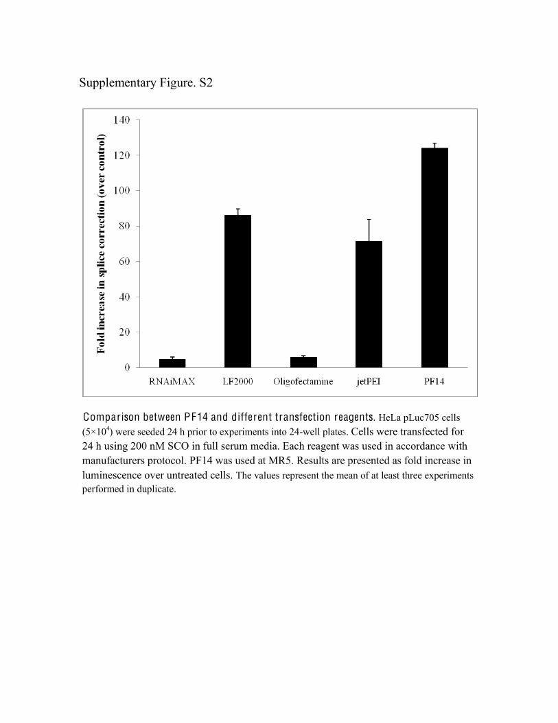
Comparison between PF14 and different transfection reagents. HeLa pLuc705 cells (5×10⁴) were seeded 24 h prior to experiments into 24-well plates. Cells were transfected for 24 h using 200 nM SCO in full serum media. Each reagent was used in accordance with manufacturers protocol. PF14 was used at MR5. Results are presented as fold increase in luminescence over untreated cells. The values represent the mean of at least three experiments performed in duplicate.
Supplementary Figure. S2
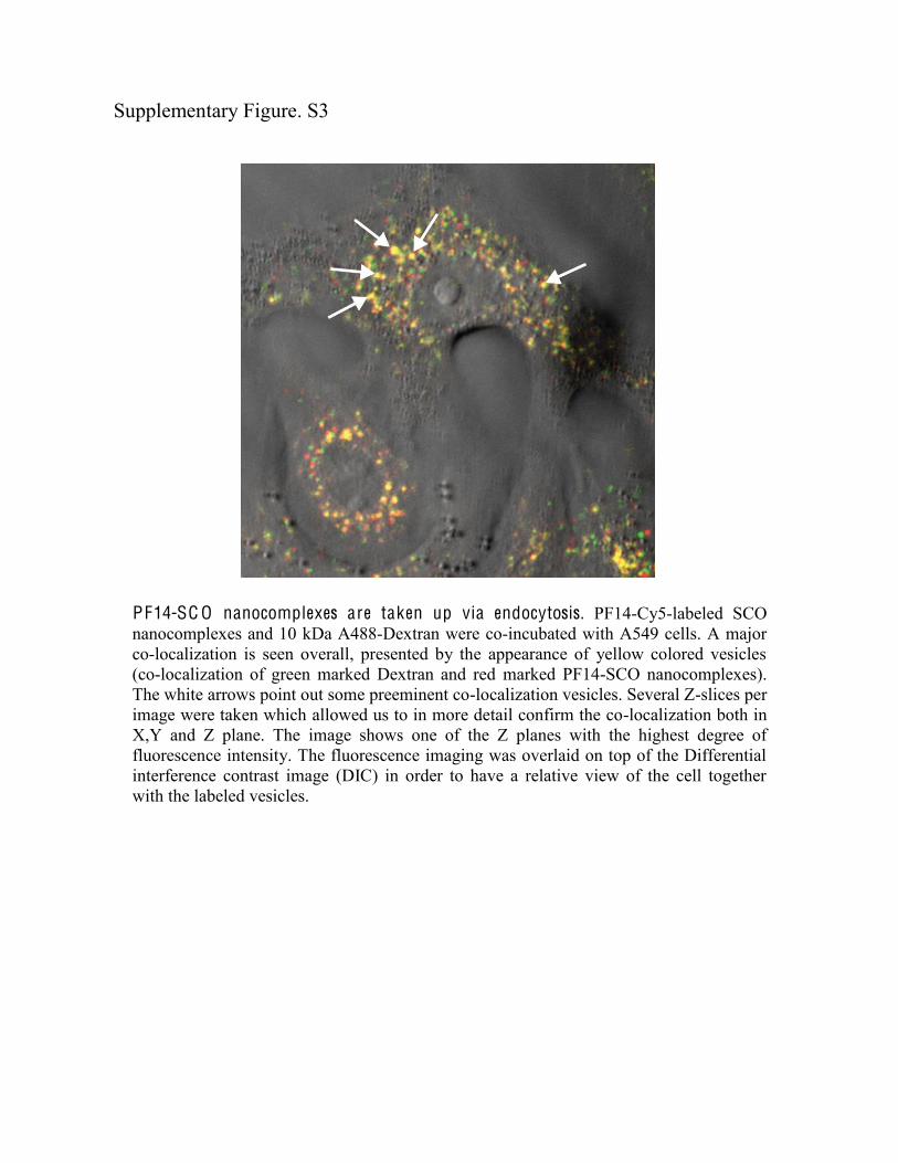
PF14-SC O nanocomplexes are taken up via endocytosis. PF14-Cy5-labeled SCO nanocomplexes and 10 kDa A488-Dextran were co-incubated with A549 cells. A major co-localization is seen overall, presented by the appearance of yellow colored vesicles (co-localization of green marked Dextran and red marked PF14-SCO nanocomplexes). The white arrows point out some preeminent co-localization vesicles. Several Z-slices per image were taken which allowed us to in more detail confirm the co-localization both in X,Y and Z plane. The image shows one of the Z planes with the highest degree of fluorescence intensity. The fluorescence imaging was overlaid on top of the Differential interference contrast image (DIC) in order to have a relative view of the cell together with the labeled vesicles.
Supplementary Figure. S3
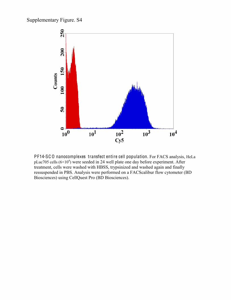
PF14-SC O nanocomplexes transfect entire cell population. For FACS analysis, HeLa pLuc705 cells (6×10⁴) were seeded in 24 well plate one day before experiment. After treatment, cells were washed with HBSS, trypsinized and washed again and finally ressuspended in PBS. Analysis were performed on a FACScalibur flow cytometer (BD Biosciences) using CellQuest Pro (BD Biosciences).
Supplementary Figure. S4
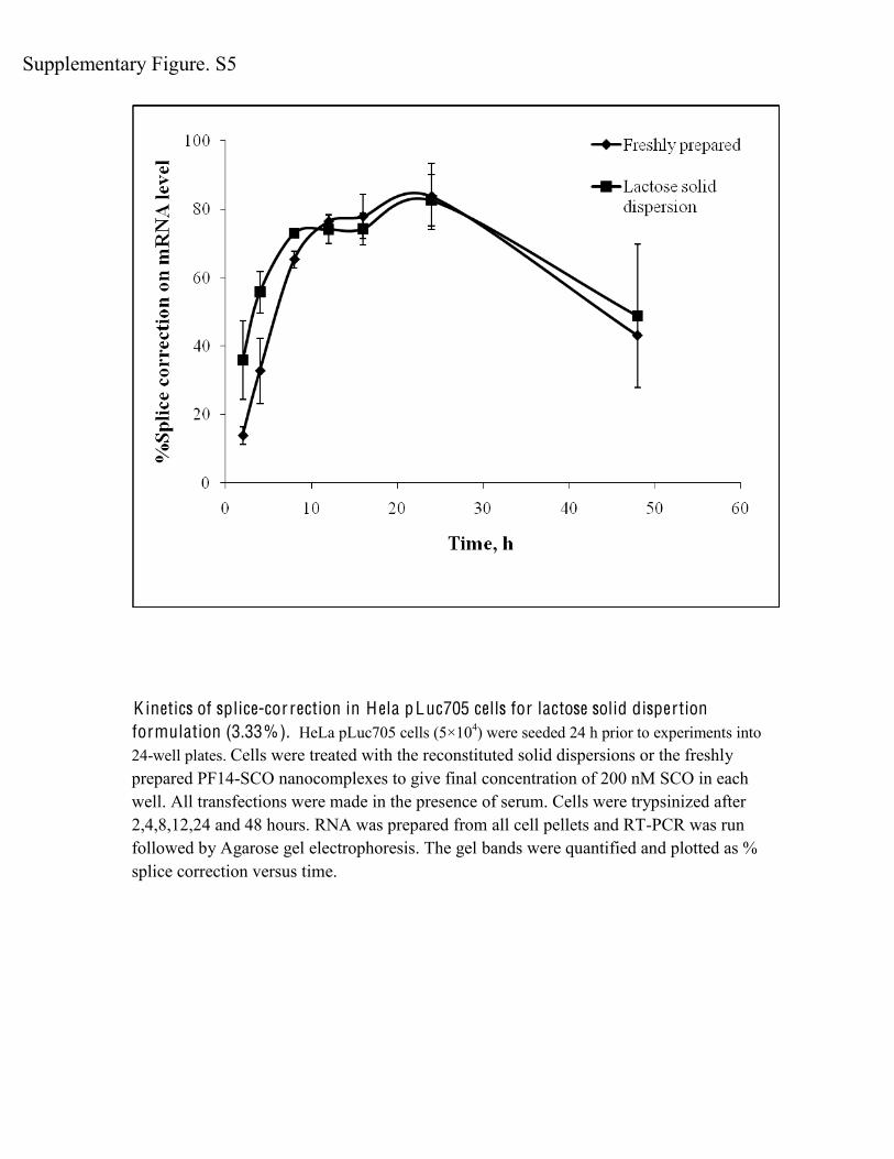
K inetics of splice-cor rection in H ela pLuc705 cells for lactose solid dispertion formulation (3.33%). HeLa pLuc705 cells (5×10⁴) were seeded 24 h prior to experiments into 24-well plates. Cells were treated with the reconstituted solid dispersions or the freshly prepared PF14-SCO nanocomplexes to give final concentration of 200 nM SCO in each well. All transfections were made in the presence of serum. Cells were trypsinized after 2,4,8,12,24 and 48 hours. RNA was prepared from all cell pellets and RT-PCR was run followed by Agarose gel electrophoresis. The gel bands were quantified and plotted as % splice correction versus time.
Supplementary Figure. S5


