Embed Size (px)
Citation preview

Peptide pheromone-induced transfer of plasmid pCF10 in Enterococcusfaecalis: probing the genetic and molecular basis for specificity of the
pheromone response
Gary M. Dunny*, Michelle H. Antiporta, Helmut HirtDepartment of Microbiology, University of Minnesota Medical School, 1460 Mayo Bldg, 420 Delaware St., SE, Minneapolis, MN 55455-0312, USA
Received 28 December 2000; accepted 29 March 2001
Abstract
The tetracycline resistance plasmid pCF10 represents a class of unique mobile genetic elements of the bacterial genus Enterococcus,whose conjugative transfer functions are inducible by peptide sex pheromones excreted by potential recipient cells. These plasmids play asignificant role in the dissemination of virulence and antibiotic resistance genes among the enterococci, which have become majornosocomial pathogens. Pheromone response by plasmid-carrying donor cells involves specific import of the peptide signal molecule, andsubsequent interaction of the signal with one or more intracellular regulatory gene products. The pheromones are chromosomally encodedhydrophobic octa- or hepta-peptides, and different families of homologous plasmids encode the ability to respond to each pheromone.Among the four pheromone-responsive plasmids that have been characterized in some detail, there is considerable conservation in the genesencoding pheromone sensing and regulatory functions, and the peptides themselves show considerable similarity. In spite of this, there isextremely high specificity of response to each peptide, with virtually no “cross-induction” of transfer of non-cognate pheromone plasmidsby the pheromones. This communication reviews the evidence for this specificity and discusses current molecular and genetic approachesto defining the basis for specificity. © 2001 Elsevier Science Inc. All rights reserved.
1. Introduction
The conjugative tetracycline-resistance plasmid pCF10was identified about 20 years ago in a strain of Enterococ-cus faecalis associated with a nosocomial infection [16].Since that time, pCF10 has served as a model system foranalysis of conjugative plasmid transfer in enterococci[3,18,19]. Recently, analysis of vancomycin-resistant E.faecium strains from the same hospital [24,25] was carriedout. The results suggest that pCF10-derived conjugativeelements have circulated among the endemic enterococcalstrains in a single hospital for over two decades, acquiringdifferent transposable resistance determinants as the selec-tive pressures from antimicrobial chemotherapy havechanged over time. Thus, the study of the pCF10 family ofplasmids has yielded insights into both the molecular mech-anisms of plasmid transfer, and into the role that these
plasmids play in the dissemination of resistance genes in aclinical setting.
One of the most interesting aspects of the biology of thisplasmid, is that, in natural environments, a donor cell car-rying pCF10 only expresses its conjugative transfer func-tions when potential recipient cells are in close proximity.The plasmid-encoded ability of the donor cell to senserecipients is dependent on the production by the recipient ofthe heptapeptide mating pheromone cCF10 (sequence:LVTLVFV) [37]. The pCF10 plasmid is representative of agroup of enterococcal pheromone responsive plasmids [11,54]. These novel genetic elements all seem to encode asimilar set of regulatory geneproducts which allow the hostcell to sense exogenous pheromone, to activate expressionof transfer functions in response to this signal, and to avoidself-induction by endogenous pheromone encoded by itsown chromosome.
In this communication, we shall use results obtainedfrom the analysis of pCF10 to illustrate some of the mainfeatures of pheromone-inducible conjugation. Many of thesteps in this process are similar for other pheromone plas-mids, such as pAD1, pPD1, and pAM373 (see [11] for a
* Corresponding author. Tel.: �1-612-625-9930; fax: �1-612-626-0623.
E-mail address: [email protected] (G.M. Dunny).
Peptides 22 (2001) 1529–1539
0196-9781/01/$ – see front matter © 2001 Elsevier Science Inc. All rights reserved.PII: S0196-9781(01)00489-2

recent review of these plasmid systems). Studies of theseelements by several groups have also contributed signifi-cantly to our understanding of the pCF10 system. This formof conjugation is a complex bacterial behavior which iscontrolled by a sophisticated cell-cell communication sys-tem. In this system, the signal molecule is a small peptide.The major focus of the following paragraphs will be on thegenetic and molecular basis for the exquisite sensitivity andspecificity of the peptide sensing machinery and of thedownstream events in the pheromone response pathway.
2. An overview of pheromone-induced conjugation
As illustrated in Fig. 1, pheromone-dependent conjuga-tive transfer of pCF10 is a complex process involving sev-eral distinct stages. Signaling occurs via specific binding of
the cCF10 peptide by an extracellular pCF10-encoded li-poprotein (PrgZ) and the subsequent import of the peptideinto the cytoplasm of the responder cell via the chromo-somal oligopeptide permease (Opp) system [35]. Thus im-port of the signal, rather than transduction across the mem-brane is essential to initiate conjugation. The importedpeptide interacts with one or more intracellular targets, mostnotably the cytoplasmic, pCF10-encoded negative regulatorPrgX. Recent data [4] indicate that this interaction abolishesa transcriptional or post-transcriptional repressor function ofPrgX, allowing transcription to read through positive con-trol elements and into the genes encoding the structuralapparatus for conjugative transfer. The positive controlmechanism seems to involve transcription anti-termination,mRNA stabilization, and translational enhancement, but it isnot yet clear which of these activities is directly stimulatedby pheromone [5,6]. One of the first obvious signs of in-
Fig. 1. Summary of the major steps in pheromone-induced conjugative transfer of pCCF10. A. The pCF10-containing donor cell can sense the presence ofpotential recipient cells by detecting the cCF10 peptide excreted into the culture medium. B. The cCF10 binding protein PrgZ binds the free peptide and usesthe chromosomally encoded Opp (Oligopeptide permease) system to import the pheromone. C. Binding of internalized cCF10 by an intracellular effectormolecule, probably PrgX, releases a negative control mechanism that allows for expression of pCF10-encoded positive control elements, and subsequentexpression of plasmid transfer proteins, including Asc10. D. Following Asc10-mediated donor-recipient binding and formation of a mating channel, thepCF10 DNA processing machinery nicks the plasmid at a specific sequence in the oriT region. The relaxase remains bound to the 5� end of the nicked DNA,a single strand is unwound, and transferred to the recipient via the channel. Once the newly plasmid has established in the transconjugant, negative controlfunctions described in the text and in Fig. 2 prevent self-induction by endogenous cCF10.
1530 G.M. Dunny et al. / Peptides 22 (2001) 1529–1539

duction is expression of the cell-surface adhesin Asc10(Aggregation Substance; encoded by the pCF10 prgB gene).This protein mediates formation of mating aggregates be-tween donor and recipient cells, which promotes highlyefficient transfer, even in liquid cultures [45]. Once aggre-gation has occurred, a mating channel is presumably formedbetween the mating partners; although very little is knownabout this channel. Asc10 probably is not an integral com-ponent [45], and it is possible that many conjugative plas-mids of Gram-positive bacteria utilize a channel of similarstructure. Pure cultures of pheromone-induced donor cellswill also self-aggregate, a phenotype that can be used as asimple biological assay for pheromone activity. Once ag-gregation and mating channel formation have taken place,conjugative DNA processing is required to effect transfer ofthe plasmid to the recipient cell. By analogy to other con-jugation systems, it is believed that a nicking of one strandof the plasmid at a specific residue within the oriT (origin oftransfer) region of the plasmid by the conjugative relaxaseenzyme initiates a rolling circle form of unwinding. Next, asingle strand of the DNA is transferred to the recipient,recircularized, and ultimately used as a template for synthe-sis of the complementary strand. Recent studies have iden-tified an oriT sequence for pCF10 as well as the putativerelaxase gene encoding the enzyme that produces a se-quence-specific and strand-specific nick at the oriT to ini-tiate the conjugative DNA transfer process (J. Staddon andG. Dunny, unpublished).
Once transfer occurs, the plasmid must encode negativecontrol functions to block the activity of cCF10 producedendogenously. This type of control is necessary to block selfinduction by endogenous pheromone. Both the extracellulargrowth medium and the cell wall of recipients containpheromone. The extracellular pheromone is neutralized bythe inhibitor peptide iCF10, and the cell wall-associatedcCF10 is inhibited, degraded or sequestered by the mem-brane protein PrgY [9]. The activities of these endogenousnegative regulators are just sufficient to control endogenouscCF10, while allowing the cell to be exquisitely sensitive toexogenous cCF10 [37].
As noted previously, pCF10 is only one representative ofa family of pheromone-responsive plasmids. Each familyencodes a response to a different peptide. For examplepCF10 encodes a response to cCF10, pAD1 encodes aresponse to cAD1, and pPD1 encodes a response to cPD1.About a half dozen different enterococcal plasmids or plas-
mid families have been shown to encode a response to aspecific peptide of known sequence, and there are almostcertainly additional combinations to be elucidated. Compre-hensive listings of the known pheromone plasmid systemsand the cognate peptides have been presented in severalrecent reviews [11,18,54]. To the extent that various plas-mids have been analyzed, there is considerable similarity inboth the size and in the molecular nature of the peptides, andthe regulatory genes and mechanisms of induction em-ployed by these elements. In addition, clinical isolates of E.faecalis or E. faecium commonly contain several differentpheromone plasmids in the same strain. In such strains, itwould not be unexpected to see a certain degree of “crossinduction” between different pheromone-responsive conju-gation systems in the same cell. However, the results de-scribed below show that the induction process is highlyspecific. In subsequent sections we will present evidence forthis specificity and discuss its implications for the varioussteps involved in the process of inducible conjugation.
Because enterococci are opportunistic pathogens in hu-mans, it is not always easy to produce experimental infec-tions under controlled conditions that resemble the mostcommon nosocomial infections seen with these organisms.It is also likely that in different circumstances, distinct geneproducts contribute to enterococcal virulence. Several linesof investigation have indicated that the gene products en-coded by the pheromone plasmids, such as the AggregationSubstance proteins of pAD1 and pCF10 [10,33,44,48,51],and the cytolysin encoded by plasmids such as pAD1 [10,28,30] can increase the virulence of enterococcal hoststrains in experimental animal and tissue culture modelsystems. In addition, several of the enterococcal pheromoneand inhibitor peptides have been shown to be chemotacticfor neutrophils [22,50], but the significance of these findingshas not been examined in experimental infection models.The molecular and genetic basis of enterococcal virulencehas recently been reviewed [29,36], and interested readersare urged to consult these sources for more detailed infor-mation on this subject.
3. Specificity of pheromone induction
The results from the simple experiment summarized inTable 1 illustrate the selective inducing activity of a peptidepheromone for a specific plasmid conjugation system. A
Table 1Specificity of the plasmid transfer of plasmid pCF10
induction cCF10 cCF10 � cPD1 cPD1 no peptide
T/D 2.2 � 10�2 � 1.8 � 10�2 1.4 � 10�2 � 9.7 �10�3 2.0 � 10�7 � 6.5 � 10�8 2.1 � 10�7 � 7.9 � 10�8
The E. faecalis strain OG1RF containing the two plasmids pCF10 and pPD1 (OG1RF:pCF10:pPD1) was induced with cCF10, cPD1 or both respectively.Cells were allowed to grow for two hours and then inoculated 1:10 in a culture of the recipient strain OG1SSp. The mating proceeded for 10 min after whichthe donor cell count and the resulting transconjugants were enumerated. Transconjugants per donor levels (T/D) for the transfer of plasmid pCF10 are given.The results are the mean of three independent experiments.
1531G.M. Dunny et al. / Peptides 22 (2001) 1529–1539
donor cell containing plasmids pCF10 and pPD1, whenexposed to both cCF10 and cPD1 peptides showed high-frequency transfer of the pCF10 plasmid. The frequency oftransfer is comparable to that seen when exposed to cCF10alone. However, when exposed to cPD1 alone, the high-frequency of pCF10 transfer was not detected (in results notshown a similar induction of pPD1 transfer was observed inresponse to cPD1). These results clearly illustrate that thedonor cell is able to selectively respond to cCF10. Moreimportantly, they rule out the possibility of “cross-induc-tion” of non-cognate plasmids by cCF10 or cPD1. Interest-ingly, Ehrenfeld et al. obtained very similar results a num-ber of years ago, in experiments using donor cells carryingpAD1 and pPD1 [21]. Given the complexity of the induc-tion process as illustrated in Fig. 1, it is clear that there areseveral stages at which specificity must be extremely high toavoid cross-induction. These include the specific bindingand import of the pheromone, the interaction of the phero-mone with its intracellular target, the increase in expressionof transfer functions mediated by the inducible positivecontrol system, and the DNA processing associated withtransfer of the plasmid. In addition to the specificity ofinduction in response to exogenous pheromone, it is alsolikely that the negative control systems that prevent self-induction by endogenous pheromone function in a highlyspecific fashion. The sections below will address each ofthese components of the pCF10 system in more detail.
4. Pheromone structure and biosynthesis
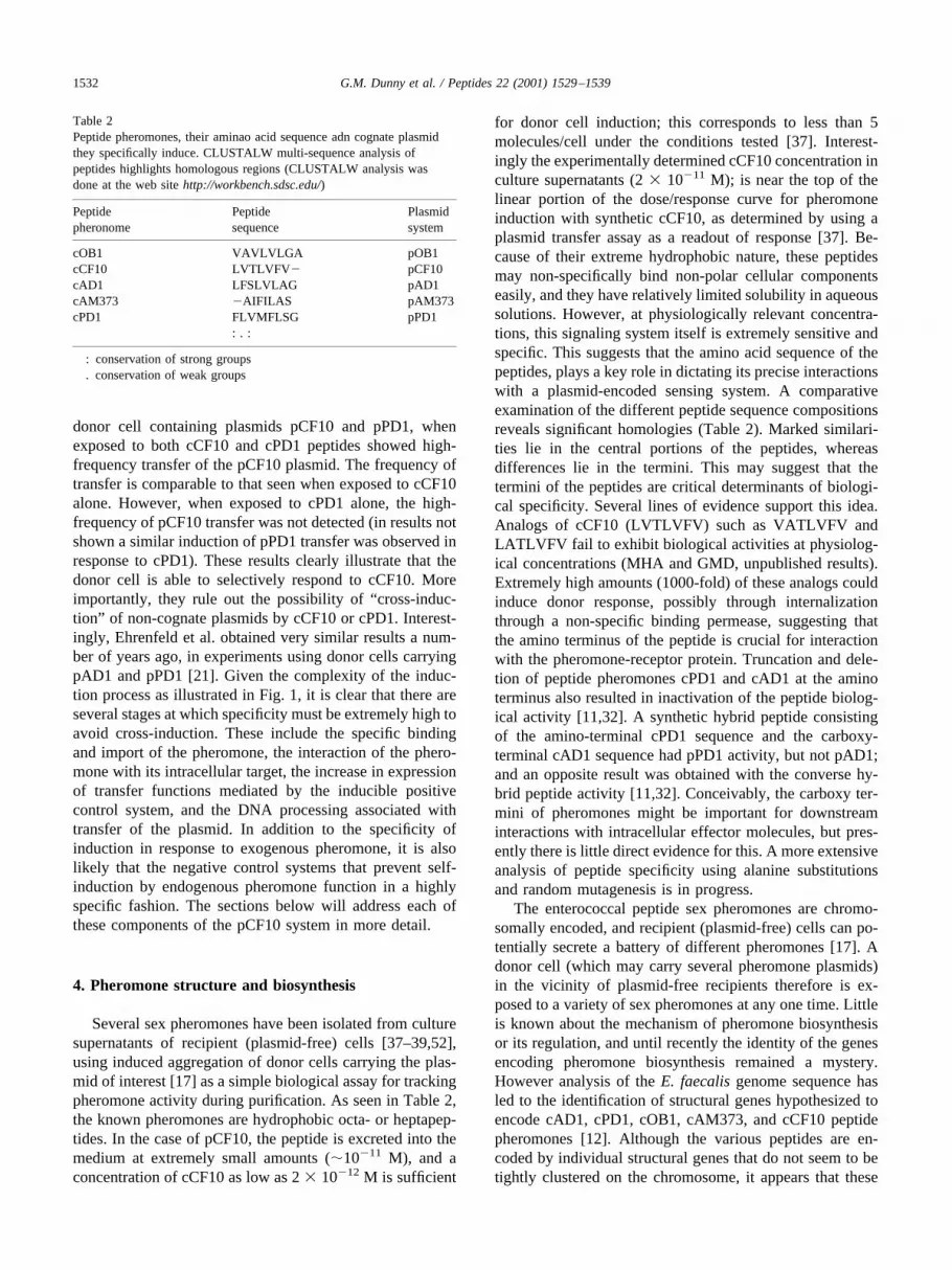
Several sex pheromones have been isolated from culturesupernatants of recipient (plasmid-free) cells [37–39,52],using induced aggregation of donor cells carrying the plas-mid of interest [17] as a simple biological assay for trackingpheromone activity during purification. As seen in Table 2,the known pheromones are hydrophobic octa- or heptapep-tides. In the case of pCF10, the peptide is excreted into themedium at extremely small amounts (�10�11 M), and aconcentration of cCF10 as low as 2 � 10�12 M is sufficient
for donor cell induction; this corresponds to less than 5molecules/cell under the conditions tested [37]. Interest-ingly the experimentally determined cCF10 concentration inculture supernatants (2 � 10�11 M); is near the top of thelinear portion of the dose/response curve for pheromoneinduction with synthetic cCF10, as determined by using aplasmid transfer assay as a readout of response [37]. Be-cause of their extreme hydrophobic nature, these peptidesmay non-specifically bind non-polar cellular componentseasily, and they have relatively limited solubility in aqueoussolutions. However, at physiologically relevant concentra-tions, this signaling system itself is extremely sensitive andspecific. This suggests that the amino acid sequence of thepeptides, plays a key role in dictating its precise interactionswith a plasmid-encoded sensing system. A comparativeexamination of the different peptide sequence compositionsreveals significant homologies (Table 2). Marked similari-ties lie in the central portions of the peptides, whereasdifferences lie in the termini. This may suggest that thetermini of the peptides are critical determinants of biologi-cal specificity. Several lines of evidence support this idea.Analogs of cCF10 (LVTLVFV) such as VATLVFV andLATLVFV fail to exhibit biological activities at physiolog-ical concentrations (MHA and GMD, unpublished results).Extremely high amounts (1000-fold) of these analogs couldinduce donor response, possibly through internalizationthrough a non-specific binding permease, suggesting thatthe amino terminus of the peptide is crucial for interactionwith the pheromone-receptor protein. Truncation and dele-tion of peptide pheromones cPD1 and cAD1 at the aminoterminus also resulted in inactivation of the peptide biolog-ical activity [11,32]. A synthetic hybrid peptide consistingof the amino-terminal cPD1 sequence and the carboxy-terminal cAD1 sequence had pPD1 activity, but not pAD1;and an opposite result was obtained with the converse hy-brid peptide activity [11,32]. Conceivably, the carboxy ter-mini of pheromones might be important for downstreaminteractions with intracellular effector molecules, but pres-ently there is little direct evidence for this. A more extensiveanalysis of peptide specificity using alanine substitutionsand random mutagenesis is in progress.
The enterococcal peptide sex pheromones are chromo-somally encoded, and recipient (plasmid-free) cells can po-tentially secrete a battery of different pheromones [17]. Adonor cell (which may carry several pheromone plasmids)in the vicinity of plasmid-free recipients therefore is ex-posed to a variety of sex pheromones at any one time. Littleis known about the mechanism of pheromone biosynthesisor its regulation, and until recently the identity of the genesencoding pheromone biosynthesis remained a mystery.However analysis of the E. faecalis genome sequence hasled to the identification of structural genes hypothesized toencode cAD1, cPD1, cOB1, cAM373, and cCF10 peptidepheromones [12]. Although the various peptides are en-coded by individual structural genes that do not seem to betightly clustered on the chromosome, it appears that these
Table 2Peptide pheromones, their aminao acid sequence adn cognate plasmidthey specifically induce. CLUSTALW multi-sequence analysis ofpeptides highlights homologous regions (CLUSTALW analysis wasdone at the web site http://workbench.sdsc.edu/)
Peptidepheronome
Peptidesequence
Plasmidsystem
cOB1 VAVLVLGA pOB1cCF10 LVTLVFV� pCF10cAD1 LFSLVLAG pAD1cAM373 �AIFILAS pAM373cPD1 FLVMFLSG pPD1
: . :
: conservation of strong groups. conservation of weak groups
1532 G.M. Dunny et al. / Peptides 22 (2001) 1529–1539

peptides are all produced from the signal peptides of li-poprotein precursors. Biosynthesis of the mature phero-mone, therefore, would involve some type of proteolyticdegradation of the signal peptides (pro-pheromones). TheEep protein, which is predicted to encode a membraneprotease [8] is involved in maturation of several phero-
mones, as shown by An et al. [2]. Fig. 2A illustrates thecurrent model for the production of cCF10 in recipient cellsvia Eep-mediated proteolysis of the cleaved CcfA signalpeptide as CcfA lipoprotein is secreted across the cytoplas-mic membrane. Nothing is known at present about thefunctions of the lipoproteins themselves.
Fig. 2. The current model for synthesis of cCF10 by recipients and control of endogenous cCF10 by donors. A. In recipient cells, the ccfA gene encodes asecreted lipoprotein whose function is not currently known. As the lipoprotein is secreted across the cytoplasmic membrane and anchored to the cell wall(presumably via a Signal Peptidase II mechanism), the cleaved signal peptide is subject to further proteolytic processing by the Eep protein, and the maturecCF10 molecule is exported across the membrane. Some of this material is released into the culture fluid while a considerable amount remains associatedwith the cell wall. B. In a donor cell, the secretion of cCF10 across the membrane occurs in a manner essentially identical to the process in recipient cells.The pCF10-encoded inhibitor peptide iCF10 neutralizes the cCF10 released into the medium, while the PrgY protein degrades or inhibits the activity of thecell-wall associated cCF10. This process could either occur by degradation or sequestration of the pheromone, such that it cannot interact with PrgZ. In eithercase, PrgZ remains accessible to any cCF10 from the extracellular medium that is not competitively inhibited by iCF10.
1533G.M. Dunny et al. / Peptides 22 (2001) 1529–1539

5. Pheromone binding and import
The PrgZ pheromone binding protein is a homolog of thechromosomally encoded OppA proteins found in many bac-terial spp [53]; the chromosome of E. faecalis encodes anOppA protein, and the TraC proteins encoded by the pAD1and pPD1 plasmids are similar proteins that have the samefunction as PrgZ. Initial evidence for the role of PrgZ inpheromone binding came from experiments showing thatcells containing pCF10 could reduce the pheromone titer ofsolutions containing cCF10 (during a 15 min incubation),and that an isogenic strain carrying the prgZ gene cloned ona multicopy plasmid removed more pheromone under thesame conditions [49]. This activity correlated with the levelsof PrgZ protein in the cell as determined by Western blot-ting. The reduction in titer associated with PrgZ expressionlikely represents a combination of cCF10 binding and im-port. It has also been shown that the concerted action ofPrgZ and the chromosomal Opp machinery is required toimport pheromone to elicit a mating response in donor cells[35]. At very high cCF10 concentrations, the chromosomalOppA protein can substitute for PrgZ, but at physiologicallyrelevant levels of pheromone, only PrgZ has sufficient af-finity to allow import of the signal molecule. It is generallybelieved that the inhibitor peptides act as competitive in-hibitors for initial interaction with the binding proteins. Thebinding affinities of the inhibitors seem to be 10–100 foldlower than those of the corresponding pheromones, basedon functional assays [13,41,43].
The amino-terminal segments of all of the enterococcalproteins contain signal sequences characteristic of secretedlipoproteins, which are attached to the outer surface of thecytoplasmic membrane via covalent linkage of a lipid moi-ety to the N-terminal cysteine residue of the mature protein(following secretion and signal peptide cleavage. Studies ofE. coli and Salmonella typhimurium OppAs indicate thatunmodified peptides in the range of 2–5 amino acid residuesin length (of essentially any sequence) can insert into the acleft between two major domains of the protein pocket(amino-terminus first). This causes a conformational changewhich causes the two domains to snap together in a type“Venus flytrap” mechanism. Molecular modeling and X-raycrystallographic studies of Salmonella and E. coli OppAsbound to various substrate peptides suggest that there issufficient space in the pocket to accommodate various sidechains of amino acids of the peptide. Key OppA residueslining the pocket can form salt bridges with one anotherwhen hydrophobic peptides are bound, and form similarinteractions with substrate side groups in the case of hydro-philic substrates [53]. This gives the binding proteins greatflexibility in substrate interactions, as long as the totallength of the peptide is appropriate to fill the binding cleft.
The OppA proteins of at least some Gram-positive bac-teria show significant differences in their substrate specific-ity. Recent analysis of the kinetics of peptide binding toLactococcus lactis OppA [47] suggests that this protein can
accommodate much longer peptides (�18 amino acids)with the carboxy terminal region proposed to extend outfrom the cleft and lie along the outer surface of the closed“flytrap.” The sequence of the peptide affects the bindingkinetics in the lactococcal system, but the OppA protein canstill accommodate a wide range of substrates. This groupalso found that the release of the bound peptide to the othercomponents of the Opp ABC transport system may be acritical step in determining the rate of peptide transport.
The pheromone binding proteins are remarkable in thatthey show significant similarity to several OppA proteins,and they are very similar to one another (�50% amino acididentity). However, they are each highly specific for thebinding of their cognate pheromone. An alignment of theenterococcal pheromone binding protein sequences (minusthe signal peptides) encoded by pCF10, pAD1, and pPD1 ispresented in Fig. 3, along with Salmonella OppA. The first300 residues of all three enterococcal proteins are virtuallyidentical to one another, while last �220 residues showmore variability. Interestingly, there are several regionswhere two of the proteins are almost identical and the thirdis unique. Some of these residues line the binding cleft andmay contribute to specificity. An aspartate residue (D432 inPrgZ) conserved in many OppAs, is believed to be at theinnermost portion of the binding cleft.
A genetic analysis of the binding specificity of PrgZ iscurrently underway in our laboratory. Random mutagenesisand the construction of PrgZ/TraC chimeric proteins are themajor tools being used in this analysis. The function of thesemutant proteins will be determined using the whole cellbinding assays described above, as well as analysis of theactivities of purified proteins. In addition to the initial in-teraction of cCF10 with PrgZ, the release of the peptide intothe trans-membrane channel believed to be formed by theOppB and OppC proteins likely plays a crucial role insignaling. The molecular details of the interaction betweenthe binding protein and channel components of all Oppsystems is an area which is still a complete mystery. How-ever, it seems obvious that the interaction between PrgZ andthe Opp channel that follows the initial binding of cCF10 toPrgZ must involve a substantial alteration in PrgZ confor-mation to allow for release of the peptide. In view of thesubstantial conservation in primary amino acid sequence ofall of the peptide binding proteins shown in Fig. 3, it will beextremely interesting to learn the basis for the remarkabledifferences in substrate specificity.
6. Control of endogenous pheromone by donor cells
Due to the chromosomal origin of the pheromones, donor(plasmid-containing) cells can also potentially producepheromones. The system has evolved two independentmechanisms to prevent donor-cell self induction by endog-enously produced pheromone. One of these is the produc-tion of plasmid-encoded pheromone inhibitors. The inhibi-
1534 G.M. Dunny et al. / Peptides 22 (2001) 1529–1539

Fig. 3. Multi-sequence alignment analysis of the various enterococcal pheromone-binding proteins against OppA from Salmonella typhimurium. The shadedboxes indicate the exact residue match amongst the enterococcal binding proteins. A “* ” denotes an exact residue match, a “: ” denotes conservation of stronggroups, and a “. ” denotes conservation of weak groups. CLUSTALW was done at the website http://workbench.sdsc.edu. Signal sequences have been deletedprior to analysis.
1535G.M. Dunny et al. / Peptides 22 (2001) 1529–1539

tors are also hydrophobic hepta-and octapeptides formedupon proteolytic cleavage of 22–23 amino acid precursors[13,40,41]. Comparative analysis of the inhibitor peptidesshows significant homologies (Table 2). Not surprisingly,they also share significant homology with the pheromones.cCF10 shows significant similarity to iAD1 and iPD1, how-ever it shows no significant inhibitory activity against cellscarrying pAD1 or pPD1. This provides another illustrationof how the exact amino acid sequences of these peptides arecrucial determinants of their biological activity. As might beexpected, there is considerable homology between the pher-omones and their cognate inhibitors, but the inhibitor doesnot mimic the inducing activity of the pheromone. Instead,inhibitor peptides are believed to act as antagonists bycompetitively binding to the pheromone-receptor protein[40,41].
In the pCF10 system, there is a remarkable coordinationbetween the levels of iCF10 and cCF10 released into theculture fluid by donor cells. It has been determined that theculture fluid of donor cells contains a cCF10 concentrationessentially identical to that of a plasmid-free recipient cul-ture grown under the same conditions [41]. However, thedonor cultures also contain iCF10 in a large excess (10–100fold, depending on how the inhibitor activity is assayed),which is just sufficient to neutralize the cCF10 released bythe same cells. As discussed further below, it is likely thatiCF10 expression is coordinately regulated with that ofconjugative transfer functions in order to prevent inappro-priate synthesis of conjugation machinery while allowingthe cell to efficiently detect the slight increase of cCF10resulting from growth of recipients in close proximity.
In addition to controlling cCF10 of endogenous origin inthe culture medium, donor cells must also prevent self-induction by cell-associated cCF10. In a recent analysis ofpheromone production and inhibition [9], we found thatcCF10 producing enterococci contain a substantial pool ofcell-associated cCF10, mostly in the cell wall fraction. Theaverage recipient cell appears to have about twice as muchcCF10 in its wall, as it releases into the culture fluid. Theavailable data suggests that iCF10 neutralizes the extracel-lular pheromone, but has little effect on the wall-associatedactivity. The plasmid-encoded membrane protein PrgYfunctions to control endogenous wall-associated cCF10without noticeably affecting the level of cCF10 actuallyreleased by the cell [9]. Thus, as diagrammed in Fig. 2B,there are two essential and independent negative controlfunctions required to control endogenous pheromone,iCF10 and PrgY. The exact mechanism of PrgY action isnot clear. It could involve pheromone sequestration or deg-radation, it might interfere with direct binding of wall-associated cCF10 with PrgZ (while still allowing for freeinteraction of PrgZ with pheromone from the culture medi-um), or it could interfere in some other way with the coop-erative re-internalization of endogenous cCF10 in the cellwall by PrgZ and the Opp proteins. Computer analysis ofPrgY predicts 3 or 4 membrane spanning regions, in addi-
tion to cytoplasmic and extracellular loops. This wouldcertainly allow PrgY to carry out any of the hypotheticalinhibitory processes listed above. Interestingly, the TraBproteins of pAD1 and pPD1 share considerable homologywith PrgY [26], and the pPD1 traB gene will complement aprgY mutation in pCF10, but the pAD1 gene will not (E.Bryan and G. Dunny, unpublished results). In contrast to thepCF10 system, both of the TraB proteins have been impli-cated as “pheromone shut-down” proteins, believed to re-duce the levels of endogenous pheromone excreted into themedium by donor cells [1,43]. While it is possible that PrgYoperates by a unique mechanism, it seems more likely thatall three proteins function similarly, but that the peptidesthemselves might interact somewhat differently with thehost cell envelope during secretion. For example, cCF10 ismore hydrophobic than either cAD1 or cPD1, so it mighthave an increased tendency to stick to cell envelope com-ponents (such as lipoteichoic acids or hydrophobic domainsof cell wall proteins), following their cleavage from theprecursor peptide. Further genetic and molecular analysis ofPrgY function, including construction of TraB/PrgY chime-ras, and studies of the interactions of PrgY with otherproteins promises to shed more light on this question.
7. The intracellular phase of pheromone induction
In order to induce a mating response in a donor cell,cCF10 must be internalized by the donor cell, as describedabove. The ultimate result of this process is the expressionof the prgB—encoded Aggregation Substance (Asc10) pro-tein, along with a number of other transfer gene products,and conjugative transfer of the plasmid to any recipient cellsin the vicinity [45]. The activation process has been ana-lyzed extensively, and has been shown to be a complexprocess involving several forms of post transcriptional reg-ulation involving novel RNA effector molecules. The dataavailable currently indicates that all of the inducible transfergene products may be expressed from a single promoter atthe 5� end of the prgQ region. This region encodes a smallopen reading frame encoding a 22 aa peptide, which isprocessed to iCF10 [41]. The predominant transcript de-tected in pCF10-containing cells from the prgQ promoter isabout 430 nt (QS) and contains untranslated sequencesessential for activation of transfer gene expression by pher-omone. QS RNA is produced at high levels in the presenceor absence of exogenous cCF10, although recent results (T.Bae and G.M. Dunny, in preparation) suggest that phero-mone addition may increase this transcript moderately. Ininduced cells another relatively stable RNA (QL), extendedat the 3� end by about 100 nt, is detected, and significanttranscription readthrough to prgB and beyond occurs. RNAsequences in the prgQ region and in the downstream prgSregion appear to contribute to positive control of prgBexpression by transcription readthrough, mRNA stabiliza-tion, and translational up-regulation [5–7]. It is not yet clear
1536 G.M. Dunny et al. / Peptides 22 (2001) 1529–1539

whether these changes in gene expression at the “down-stream end” of the induction process result from severalindependent steps, or from a single event. In either case, itappears that the pheromone plays a direct role in the intra-cellular activation phase beyond competing with icF10 for amolecular target, since mutants in the iCF10 coding se-quence are still inducible [6].
The complexity of the pheromone-induced gene activa-tion process is apparent from the brief description presentedabove. However, the previously completed experiments de-fining the biochemical differences between induced anduninduced cells [5,7] did not directly address the nature ofthe molecular “switch” which is responsible for the conver-sion from one state to the other, and which is presumablythe direct target for binding by the internalized pheromone.Our recent experiments suggest that the pCF10-encodedPrgX protein plays a key role in the pheromone-dependentswitch to the induced state, and may be the intracellularreceptor for internalized cCF10. The prgX gene had beenidentified as an essential negative regulator of transfer geneexpression a number of years ago [26,31], but technicalproblems with lethal effects of some prgX mutations, cou-pled with an apparent cis-acting mode of action hinderedour progress in elucidating the role of the PrgX protein.Several of these problems have now been solved, and re-cently completed experiments show that PrgX, as well as aregulatory RNA (Qa) processed from the 5� end of thefunctional prgX mRNA [3] are the key intracellular effec-tors of negative control in the pCF10 system.
In our current model [3,4], negative control of transfergene expression in the donor cell cytoplasm is mediated bythe combined actions of PrgX and Qa. The latter componentfunctions as an antisense RNA to a region of prgQ mRNA.We propose that pairing of these two RNAs precludes thefolding of QS into a factor-independent terminator, allow-ing for readthrough of transcription beyond the 3� end ofQS. In the absence of Qa the 5� stem region of this putativeterminator may be sequestered via pairing with an upstreamcomplimentary sequence. In this model, Qa functions as aneffector in a transcription attenuation mechanism; negativecontrol by Qa is not responsive to pheromone. In the pAD1system, a similar antisense transcript mD has been identi-fied, and has been proposed to increase termination, but bya somewhat different mechanism. Although the 5� portionsof the two antisense RNAs are nearly identical, the sequencediverges significantly in the 3� regions; this may increasethe specificity of each RNA for its cognate sense message.PrgX appears to function by a complimentary, but indepen-dent mechanism. Our genetic data suggests that PrgX mod-ulates either the initiation of prgQ transcription or the sta-bility of QS transcripts. The net result of PrgX action is aslight reduction in QS levels such that iCF10 can be ex-pressed at sufficient levels, but essentially all of the QS inthe cell is paired with Qa, resulting in termination of prgQtranscription before it extends into the QL region. We hy-pothesize that pheromone binding to PrgX abolishes its
negative control function, implicating PrgX as the majorpheromone responsive component of the switch. Recentresults suggest that PrgX exists as a dimer in uninducedcells, and that pheromone treatment of cells alters thePrgX oligomerization state [4]. In support of this model,we have considerable genetic evidence for cCF10 bind-ing to PrgX. However direct binding assays are still inprogress, along with analyses of PrgX binding to RNAand DNA sequences in the regulatory region. In thepAD1 and pPD1 systems there is published evidence forDNA binding and pheromone binding to the PrgX ho-mologs, termed TraA proteins [23,42]. However, the lev-els of sequence similarity among these three proteins arethe lowest of all of the protein regulators of these plas-mids, suggesting that they may not all function the sameway. In addition, certain aspects the biological signifi-cance of the binding interactions identified in vitro aredifficult to reconcile with a unifying model for induction.Nonetheless, it is reasonable to assume that the cCF10/PrgX interaction represents another stage where a highdegree of specificity is operative.
8. Downstream effects of induction on conjugation
As noted previously, numerous plasmid-encoded geneproducts actually comprise the functional conjugation ma-chinery. Results of sequence analysis of transfer genes sug-gest that the gene products encoding the Aggregation Sub-stance proteins and the components of the mating channelmay be highly conserved [15,27,55]. Therefore, these geneproducts might not function in a highly specific fashion.However, the DNA processing machinery, comprised of therelaxase and accessory proteins, likely shows high specific-ity for its conjugative plasmid. Preliminary studies of thepCF10 system (J. Staddon and G.M. Dunny, unpublishedresults) suggest that this plasmid encodes a relaxase func-tion, specific for the pCF10 oriT, and similar to that ofseveral other conjugative elements from Gram-negative andGram-positive bacteria. Interestingly, the analysis of thecomplete sequences of pAM373 [14] and pAD1 (D.Clewell, personal communication) did not identify any con-jugative DNA processing protein homologs. Thus, differentpheromone plasmids may employ different processingmechanisms. While there is no reason to believe that cCF10interacts directly with the DNA processing machinery, it islikely that the evolution of the specificity of these compo-nents for their target DNA sequence occurred coordinatelywith that of the peptide signal for its targets.
9. Conclusion
Pheromone-induced plasmid transfer in enterococci thefirst example of a cell-cell signaling system in prokaryotes,where a specific peptide signal molecule was identified. In
1537G.M. Dunny et al. / Peptides 22 (2001) 1529–1539

the last 10 years many more cell communication systemshave been discovered and a great deal of analysis of previ-ously identified systems have occurred [20]. Peptides seemto be the most commonly employed signal molecules for theGram-positive bacteria, while acyl homoserine lactones aremore commonly utilized for this purpose in Gram-negativemicrobes. The enterococcal pheromone systems have sev-eral unique features not associated with most of the otherpeptide signaling systems. One of these is that the signalmolecule is imported specifically, rather than transducing asignal across the cell envelope. Although the CSF (alsoknown as PhrC) peptide of B. subtilis is also imported[34,46], most other peptide signaling systems utilize a two-component signal transduction system to respond to thesignal [18]. A second distinguishing feature of this system isthat the levels of induction achieved with physiologicallyrelevant pheromone concentrations are actually very mod-est. Indeed, it almost seems that the system is designed tomake the very minimal amount of transfer gene productsrequired to disseminate the plasmid, while retaining anextremely high sensitivity to the presence of exogenouspheromone. While the pheromone system shares manycommon features of the density-dependent “QuorumSensing” systems described in other bacteria, there areadditional layers of evolutionary complexity in the en-terococcal systems. Rather than serving as a monitor forthe density of a single cell type, as in the case of thequorum sensing systems controlling such traits as lumi-nescence and virulence, the enterococcal pheromonesserve to communicate the density of one cell type (recip-ients) to another (donors). Finally, the enterococcal sys-tems represent a situation where a plasmid has evolved asystem to sense a molecule whose production is encodedby the chromosome of the host organism. These plasmidsthus represent a very evolutionarily sophisticated ver-sions of selfish DNA.
Acknowledgments
The authors thank all of the members of the Dunnylaboratory for providing much of the data on which thispaper is based, and Don Clewell for sharing unpublisheddata from the pAD1 system. This research is supported byNIH grants GM49530 and HL51987.
References
[1] An FY, Clewell DB. Characterization of the determinant (traB) en-coding sex pheromone shutdown by the hemolysin/bacteriocin plas-mid, pAD1 in Enterococcus faecalis. Plasmid 1994;31:215–21.
[2] An FY, Sulavik MC, Clewell DB. Identification and characterizationof a determinant (eep) on the Enterococcus faecalis chromosome thatis involved in production of the peptide sex pheromone cAD1. JBacteriol 1999;181:5915–21.
[3] Bae T, Clerc-Bardin S, Dunny GM. Analysis of expression of prgX,a key negative regulator of the transfer of the Enterococcus faecalispheromone-inducible plasmid pCF10. J Mol Biol 2000;297:861–75.
[4] Bae T, Dunny GM. Dominant negative mutants of prgX: evidence fora role of PrgX dimerization in negative regulation of pheromone-inducible conjugation. Mol Microbiol 2001;39:1307–20.
[5] Bensing BA, Dunny GM. Pheromone-inducible expression of anaggregation protein in Enterococcus faecalis requires interaction of aplasmid-encoded RNA with components of the ribosome. Mol Mi-crobiol 1997;24:285–94.
[6] Bensing BA, Manias DA, Dunny GM. Pheromone cCF10 and plas-mid pCF10-encoded regulatory molecules act post-transcriptionallyto activate expression of downstream conjugation functions. MolMicrobiol 1997;24:295–308.
[7] Bensing BA, Meyer BJ, Dunny GM. Sensitive detection of bacterialtranscription initiation sites and differentiation from RNA processingsites in the pheromone-induced plasmid transfer system of Entero-coccus faecalis. Proc Natl Acad Sci USA 1996;93:7794–9.
[8] Brown MS, Ye J, Rawson RB, Goldstein JL. Regulated intramem-brane proteolysis: a control mechanism conserved from bacteria tohumans. Cell 2000;100:391–8.
[9] Buttaro(Leonard) BA, Antiporta MH, Dunny GM. Cell-associatedpheromone peptide (cCF10) production and pheromone inhibition inEnterococcus faecalis. J Bacteriol 2000;182:4926–33.
[10] Chow JW, Thal LA, Perri MB, Vazquez JA, Donabedian SM, ClewellDB, Zervos MJ. Plasmid-associated hemolysin and aggregation sub-stance production contribute to virulence in experimental enterococ-cal endocarditis. Antimicrobial Agents and Chemotherapy 1993;37:2474–7.
[11] Clewell DB. Sex pheromone systems in enterococci. In: Dunny GM,Winans SC, editors. Cell-cell signaling in bacteria. Washington, DC:ASM Press, 1999. p. 10–20.
[12] Clewell DB, An FY, Flannagan SE, Antiporta MH, Dunny GM.Enterococcal sex pheromone precursors are part of signal sequencesfor surface lipoproteins. Mol Microbiol 2000;35:246–8.
[13] Clewell DB, Pontius LT, An FY, Ike Y, Suzuki A, Nakayama J. Nu-cleotide sequence of the sex pheromone inhibitor (iAD1) determinantof Enterococcus faecalis conjugative plasmid pAD1. Plasmid 1900;24:156–61.
[14] De Boever EH, Clewell DB, Fraser CM. Enterococcus faecalis con-jugative plasmid pAM373: complete nucleotide sequence and geneticanalysis of sex pheromone response. Mol Microbiol 2000;37:1327–41.
[15] Devriese LA, Collins MD, Wirth R. The genus Enterococcus. In:Balows A, et al, editors. “The Procaryotes.” New York: Springer-Verlag, 1992. p. 1465–81.
[16] Dunny GM, Funk C, Adsit JC. Direct stimulation of the transfer ofantibiotic resistance by sex pheromones in Streptococcus faecalis.Plasmid 1981;6:270–8.
[17] Dunny GM, Craig RA, Carron RL, Clewell DB. Plasmid transfer inStreptococcus faecalis: production of multiple pheromones by recip-ients. Plasmid 1979;2:454–65.
[18] Dunny GM, Leonard BAB. Cell-cell communication in gram-positivebacteria. Annu Rev Microbiol 1997;51:527–64.
[19] Dunny GM, Leonard BAB, Hedberg PJ. Pheromone-inducible con-jugation in Enterococcus faecalis: interbacterial and host-parasitechemical communication. J Bacteriol 1995;177:871–6.
[20] Dunny GM, Winans SC, editors. Cell-cell signaling in bacteria.Washington, DC: American Society for Microbiology Press, 1999.
[21] Ehrenfeld EE, Kessler RE, Clewell DB. Identification of pheromone-induced surface proteins in Streptococcus faecalis and evidence of arole for lipoteichoic acid in the formation of mating aggregates. JBacteriol 1986;168:6–12.
[22] Ember JA, Hugli TE. Characterization of the human neutrophil re-sponse to sex pheromones from Streptococcus faecalis. Am J Pathol1989;134:797–805.
1538 G.M. Dunny et al. / Peptides 22 (2001) 1529–1539
[23] Fujimoto S, Clewell DB. Regulation of the pAD1 sex pheromoneresponse of Enterococcus faecalis by direct interaction between thecAD1 peptide mating signal and the negatively regulating, DNA-binding TraA protein. Proc Natl Acad Sci USA 1998;95:6430–5.
[24] Heaton MP, Discotto LF, Pucci MJ, Handwerger S. Mobilization ofvancomycin resistance by transposon-mediated fusion of a VanAplasmid with an Enterococcus faecium sex pheromone-response plas-mid. Gene 1996;171:9–17.
[25] Heaton MP, Handwerger S. Conjugative mobilization of a vancomy-cin resistance plasmid by a putative Enterococcus faecium sex pher-omone response plasmid. Microb Drug Resist 1995;1:177–83.
[26] Hedberg PJ, Leonard BAB, Ruhfel RE, Dunny GM. Identificationand characterization of the genes of Enterococcus faecalis plasmidpCF10 involved in replication and in negative control of pheromone-inducible conjugation. Plasmid 1996;35:46–57.
[27] Hirt H, Wirth R, Muscholl A. Comparative analysis of 18 sex pher-omone plasmids from Enterococcus faecalis: detection of a newinsertion element on pPD1 and hypotheses on the evolution of thisplasmid family. Mol Gen Genet 1996;252:640–7.
[28] Ike Y, Hashimoto H, Clewell DB. Hemolysin of Streptococcus fae-calis subspecies zymogenes contributes to virulence in mice. InfectImmun 1984;45:528–30.
[29] Jett BD, Huycke MM, Gilmore MS. Virulence of Enterococci. ClMicrobiol Rev 1994;7:462–78.
[30] Jett BD, Jensen HG, Nordquist RE, Gilmore MS. Contribution of thepAD1-encoded cytolysin to the severity of experimental Enterococ-cus faecalis endophthalmitis. Infect Immun 1992;60:2445–52.
[31] Kao S-M, Olmsted SB, Viksnins AS, Gallo JC, Dunny GM. Molec-ular and genetic analysis of a region of plasmid pCF10 containingpositive control genes and structural genes encoding surface proteinsinvolved in pheromone-inducible conjugation in Enterococcus faeca-lis. J Bacteriol 1991;173:7650–64.
[32] Kitada C, Fujino M, Mori M, Sakagami Y, Isogai A, Suzuki A,Clewell D, Craig R. Synthesis and structure/activity relationships ofStreptococcus faecalis sex pheromones cPD1 and cAD1. In: IzumiyaN, editor. Peptide chemistry 1984. Osaka: Protein Research Founda-tion, 1985. p. 43–8.
[33] Kreft B, Marre R, Schramm U, Wirth R. Aggregation substance ofEnterococcus faecalis mediates adhesion to cultured renal tubularcells. Infect Immun 1992;60:25–30.
[34] Lazazzera BA, Solomon JM, Grossman AD. An exported peptidefunctions intracellularly to contribute to cell density signaling inBacillus subtilis. Cell 1997;89:917–25.
[35] Leonard BAB, Podbielski A, Hedberg PJ, Dunny GM. Enterococcusfaecalis pheromone binding protein, PrgZ, recruits a chromosomaloligopeptide permease system to import sex pheromone cCF10 forinduction of conjugation. Proc Natl Acad Sci USA 1996;93:260–4.
[36] McCormick JK, Hirt H, Dunny GM, Schlievert PM. Pathogenicmechanisms of enterococcal endocarditis. Current Infectious DiseaseReports 2000;2:315–21.
[37] Mori M, Sakagami Y, Ishii Y, Isogai A, Kitada C, Fujino M, AdsitJC, Dunny GM, Suzuki A. Structure of cCF10, a peptide sex phero-mone which induces conjugative transfer of the Streptococcus faeca-lis tetracycline resistance plasmid, pCF10. J Biol Chem 1988;263:14574–8.
[38] Mori M, Sakagami Y, Narita M, Isogai A, Fujino M, Kitada C, CraigRA, Clewell DB, Suzuki A. Isolation and structure of the bacterial sexpheromone, cAD1, that induces plasmid transfer in Streptococcusfaecalis. FEBS Lett 1984;178:97–100.
[39] Mori M, Tanaka H, Sakagami Y, Isogai A, Fujino M, Kitada C, WhiteBA, An FY, Clewell DB, Suzuki A. Isolation and structure of theStreptococcus faecalis sex pheromone, cAM373. FEBS Lett 1986;206:69–72.
[40] Nakayama J, Ono Y, Suzuki A. Isolation and structure of the sexpheromone inhibitor, iAM373, of Enterococcus faecalis. Biosci Bio-tech Biochem 1995;59:1358–9.
[41] Nakayama J, Ruhfel RE, Dunny GM, Isogai A, Suzuki A. The prgQgene of the Enterococcus faecalis tetracycline resistance plasmid,pCF10, encodes a peptide inhibitor, iCF10. J Bacteriol 1994;176:2003–4.
[42] Nakayama J, Takanami Y, Horii T, Sakuda S, Suzuki A. Molecularmechanism of peptide-specific pheromone signaling in Enterococcusfaecalis: functions of pheromone receptor TraA and pheromone-binding protein TraC encoded by plasmid pPD1. J Bacteriol 1998;180:449–56.
[43] Nakayama J, Yoshida K, Kobayashi H, Isogai A, Clewell DB, SuzukiA. Cloning and characterization of a region of Enterococcus faecalisplasmid pPD1 encoding pheromone inhibitor (ipd), pheromone sen-sitivity (traC), and pheromone shutdown (traB) genes. J Bacteriol1995;177:5567–73.
[44] Olmsted SB, Dunny GM, Erlandsen SL, Wells CL. A plasmid-encoded surface protein on Enterococcus faecalis augments its inter-nalization by cultured intestinal epithelial cells. J Infect Dis 1994;170:1549–56.
[45] Olmsted SB, Kao S-M, van Putte LJ, Gallo JC, Dunny GM. Role ofthe pheromone-inducible surface protein Asc10 in mating aggregateformation and conjugal transfer of the Enterococcus faecalis plasmidpCF10. J Bacteriol 1991;173:7665–72.
[46] Perego M, Hoch JA. Cell-cell communication regulates the effects ofprotein aspartate phosphatases on the phosphorelay controlling de-velopment in Bacillus subtilis. Proc Natl Acad Sci USA 1996;93:1549–53.
[47] Picon A, Kunji ERS, Lanfermeijer FC, Konings WN, Poolman B.Specificity mutants of the binding protein of the oligopeptide trans-port system of Lactococcus lactis. J Bacteriol 2000;182:1600–8.
[48] Rakita RM, Vanek NN, Jacques-Palaz K, Mee M, Mariscalco MM,Dunny GM, Snuggs M, van Winkle WB, Simon SI. Enterococcusfaecalis bearing aggregation substance is resistant to killing by humanneutrophils despite phagocytosis and neutrophil activation. InfectImmun 1999;67:6067–75.
[49] Ruhfel RE, Manias DA, Dunny GM. Cloning and characterization ofa region of the Enterococcus faecalis conjugative plasmid, pCF10,encoding a sex pheromone binding function. J Bacteriol 1993;175:5253–9.
[50] Sannomiya P, Craig RA, Clewell DB, Suzuki A, Fujino M, Till GO,Marasco WA. Characterization of a class of nonformylated Entero-coccus faecalis-derived neutrophil chemotactic peptides: the sexpheromones. Proc Natl Acad Sci USA 1990;87:66–70.
[51] Schlievert PM, Gahr PJ, Assimacopoulos AP, Dinges MM, StoehrJA, Harmala JW, Hirt H, Dunny GM. Aggregation and bindingsubstances enhance pathogenicity in rabbit models of Enterococcusfaecalis endocardtitis. Infect Immun 1998;66:218–23.
[52] Suzuki A, Mori M, Sakagami Y, Isogai A, Fujino M, Kitada C, CraigRA, Clewell DB. Isolation and structure of bacterial sex pheromone,cPD1. Science 1984;226:849–50.
[53] Tame JRH, Murshudov GN, Dodson EJ, Neil TK, Dodson GG,Higgins CF, Wilkinson AJ. The structural basis of sequence-indepen-dent peptide binding by OppA protein. Science 1994;264:1578–81.
[54] Wirth R. The sex pheromone system of Enterococcus faecalis: morethan just a plasmid-collection mechanism? Eur J Biochem 1994;222:235–46.
[55] Wirth R, Olmsted SB, Galli D, Dunny GM. Comparative analysis ofcAD1 and cCF10 induced aggregation substances of Enterococcusfaecalis. In: Dunny GM, et al, editors. Genetics and molecular biol-ogy of streptococci, lactococci, and enterococci. Washington, DC:American Society for Microbiology, 1991. p. 34–8.
1539G.M. Dunny et al. / Peptides 22 (2001) 1529–1539


