Embed Size (px)
Citation preview
Peritubular Dentin Lacks Piezoelectricity
S. Habelitz1,*, B.J. Rodriguez2, S.J. Marshall1, G.W. Marshall1, S.V. Kalinin2, and A.Gruverman3
1 Department of Preventive and Restorative Dental Sciences, University of California, San Francisco, Box0758, 707 Parnassus Ave., San Francisco, CA 94143-0758, USA
2 Condensed Matter Sciences Division, Oak Ridge National Laboratory, Oak Ridge, TN 37831, USA
3 Departments of Materials Science and Engineering and Physics, North Carolina State University, Raleigh,NC 27695, USA
AbstractDentin is a mesenchymal tissue, and, as such, is based on a collagenous matrix that is reinforced byapatite mineral. Collagen fibrils show piezoelectricity, a phenomenon that is used by piezoresponseforce microscopy (PFM) to obtain high-resolution images. We applied PFM to image human dentinwith 10-nm resolution, and to test the hypothesis that zones of piezoactivity, indicating the presenceof collagen fibrils, can be distinguished in dentin. Piezoelectricity was observed by PFM in the dentinintertubular matrix, while the peritubular dentin remained without response. High-resolution imagingof chemically treated intertubular dentin attributed the piezoelectric effect to individual collagenfibrils that differed in the signal strength, depending on the fibril orientation. This study supports thehypothesis that peritubular dentin is a non-collagenous tissue and is thus an exception amongmineralized tissues that derive from the mesenchyme.
Keywordsatomic force microscopy; piezoelectricity; dentin; collagen
INTRODUCTIONIn vertebrate animals, hard tissues such as bone, dentin, and cementum are generated bymesenchymal cells that express a matrix of fibrillar collagen type I, which subsequentlymineralizes through the introduction of apatite nanocrystals into the fibrils and into theextrafibrillar space among fibrils. Peritubular dentin is considered an exception amongmineralized tissues, since analysis of recent data has indicated that it forms from anextracellular matrix rich in glutamic acid (Weiner et al., 1999; Gotliv et al., 2006). Thedifficulty of achieving a defined biochemical analysis of this tissue derives from its small size.Peritubular dentin is defined as the tissue of increased mineral content that surrounds dentintubules. It extends about 1 to 2 μm. Thus far, it has been observed only in the teeth of largermammals, and appears to be absent in rodents (Boyde, 1984; Magne et al., 2002; Gotliv etal., 2006). The nanomechanical properties suggest that at least 60 vol% of apatite mineral ispresent in peritubular dentin (E-modulus, 36.1 GPa), while the bulk intertubular dentin (E-modulus, 20.6 GPa) contains only about 45 vol% (Marshall et al., 2001a). The function ofperitubular dentin is unclear, but it may be mechanical, since the increased stiffness ofperitubular dentin can increase stresses around tubules and thus affect crack propagation.
*corresponding author, E-mail: [email protected].
NIH Public AccessAuthor ManuscriptJ Dent Res. Author manuscript; available in PMC 2009 June 20.
Published in final edited form as:J Dent Res. 2007 September ; 86(9): 908–911.
NIH
-PA Author Manuscript
NIH
-PA Author Manuscript
NIH
-PA Author Manuscript
There are two contrary models that describe the development of peritubular dentin. It has beensuggested that the increased mineral content around the tubules is the effect of continuousmineral deposition from the pulpal fluid, based on the observation that the collagen matrix ofthe intertubular dentin continues into the peritubular region (Dai et al., 1991). In contrast,Gotliv et al. (2006) provided a detailed analysis of the tubule area in bovine dentin, using time-of-flight secondary ion mass spectrometry, revealing that proteins rich in glumatic acid arepredominant in peritubular dentin, while the quantity of amino acids characteristic of collagen,e.g., proline and hydroxyproline, was minor. This led to the conclusion that collagen is eitherabsent or present in only low quantities in peritubular dentin; hence, a specialized non-collagenous matrix must be secreted by the odontoblasts to mineralize the tubule wall to ahigher degree than the intertubular dentin. Earlier, Takuma and Eda (1966) showed stainingdifferences between intertubular and peritubular dentin. Ultrastructural differences wereobserved by Goldberg et al. (1978), using electron microscopy. Thomas (1984) reported onthe presence of a lamina limitans, referring to tubular structures that appeared on dentinspecimens when demineralized and treated with collagenase. These structures were rich inglycosaminoglycans and were not susceptible to digestion with collagenase, and therefore mostlikely represented peritubular dentin. Developing dentin was studied by Jones and Boyde(1984), who described structural and biochemical differences between peritubular andintertubular dentin after papain digestion of predentin.
Piezoresponse force microscopy (PFM), a recent advancement of atomic force microscopy(AFM), facilitates the visualization of piezoelectric domains in materials, and can quantify thelocal piezoelectric constant with nanometer resolution (Gruverman et al., 1998; Rodriguez etal., 2006). Application of a modulating voltage to a conductive AFM tip results in an alternatingfield at the tip that induces local contraction and dilation of the substrate if it is piezoelectric.An image is created when the induced mechanical vibration of the material is translated intoa two-dimensional map of piezoelectric constants in picometer/volt units. Mineralized tissuesare known to exhibit piezoelectricity (Fukada, 1995). Initially, hydroxyapatite mineral wasassumed to be the component that causes this phenomenon; however, it is now known that thenon-centrosymmetric structure of the collagen molecule is responsible for the piezoelectricbehavior of collagen fibrils (ElMessiery et al., 1979). This study tested whether individualcollagen fibrils are the source of piezoelectricity in dentin, and whether PFM is a useful toolfor clarifying if collagen is indeed absent in the peritubular region.
MATERIALS & METHODSHuman third molars with documented history were extracted after informed patient consentwas obtained according to protocols approved by the University of California, San FranciscoCommittee on Human Research. Patients were between 22 and 34 yrs old. Teeth were sterilizedby γ-radiation and stored in deionized water at 4°C until prepared. We prepared sagittal mid-coronal sections of 9 teeth (thickness < 1 mm) by polishing through a series of SiO2 papersand with water-based diamond paste to 0.25 μm (Buehler, Lake Bluff, IL, USA). Ultrasonictreatment in water for 10 sec was used to clean the surface. Two groups (n = 3 each) ofspecimens were investigated: (1) polished only; and (2) polished and etched with10 vol% citricacid for 15 sec and subsequently treated with an aqueous solution of 1 vol% sodiumhypochlorite (NaOClaq) for up to 150 sec. The NaOClaq treatment was applied for the non-specific removal of layers of non-collagenous and collagenous proteins. As reported earlier(Habelitz et al., 2002), this procedure removes non-collagenous proteins initially and facilitatesthe visualization of individual collagen fibrils by atomic force microscopy (AFM).
Dentin sections were glued to metal disks and imaged by AFM (Autoprobe M5, Park ScientificInstruments, Sunnyvale, CA, USA) operating in the PFM mode with conductive cantilevers.The PFM technique is based on the detection of electromechanical responses of a piezoelectric
Habelitz et al. Page 2
J Dent Res. Author manuscript; available in PMC 2009 June 20.
NIH
-PA Author Manuscript
NIH
-PA Author Manuscript
NIH
-PA Author Manuscript
sample to an electrical modulation applied to the conductive AFM tip in contact with the samplesurface, as has been explained in detail (Alexe and Gruverman, 2004). While in contact, an acmodulation voltage, Vtip = Vdc + Vaccosωt, is applied between a conductive probing tip incontact with the sample surface and a conductive substrate, Vtip = resulting voltage at the tip,Vdc = direct current voltage applied for topographic imaging, and Vac = voltage from alternatecurrent applied to the conductive tip. The applied modulation voltage induces a localmechanical vibration, if the sample is piezoelectric, by inducing the converse piezoelectriceffect. The voltage-induced contraction and dilation of the substrate correspond to theamplitude of the signal, which becomes a function of the voltage Vac applied to the tip, allowingfor the calculation of the effective piezoelectric constant at any tip-substrate contact point. Theresulting image is a plot of piezoelectric constants in pm/V units over a defined area of thesubstrate. The unique strength of PFM is that both vertical and lateral components of surfacedisplacement can be measured (Alexe and Gruverman, 2004), providing information on bothnormal (vertical) and in-plane (lateral) components of the electromechanical response. Thisallows for the generation of images based on either vertical or lateral deformation of thesubstrate, referred to as VPFM and LPFM, respectively. While vertical PFM (VPFM) imagesare based on the electromechanical response at 90 degrees from the surface, lateral PFM(LPFM) images are constructed based on the lateral component of substrate deformation andthe electromechanical response in the horizontal plane.
The size of the tip-sample contact area is enlarged, due to the conductive coating of the tip.Hence, the lateral resolution that can be achieved in PFM is reduced after tip coating and isusually on the order of 5 to 10 nm. We obtained images by applying an AC modulation voltage(2.0 V, 15 kHz) to the Pt-coated Si tips (MikroMasch, Tallinn, Estonia; NSC 14, resonantfrequency ~ 150 kHz, spring constant k ~ 4.5 N/m). All samples were scanned under dryconditions in contact mode at scan sizes between 3 and 25 μm. Four image types weresimultaneously derived from the topographic, error, vertical PFM, and lateral PFM modes.
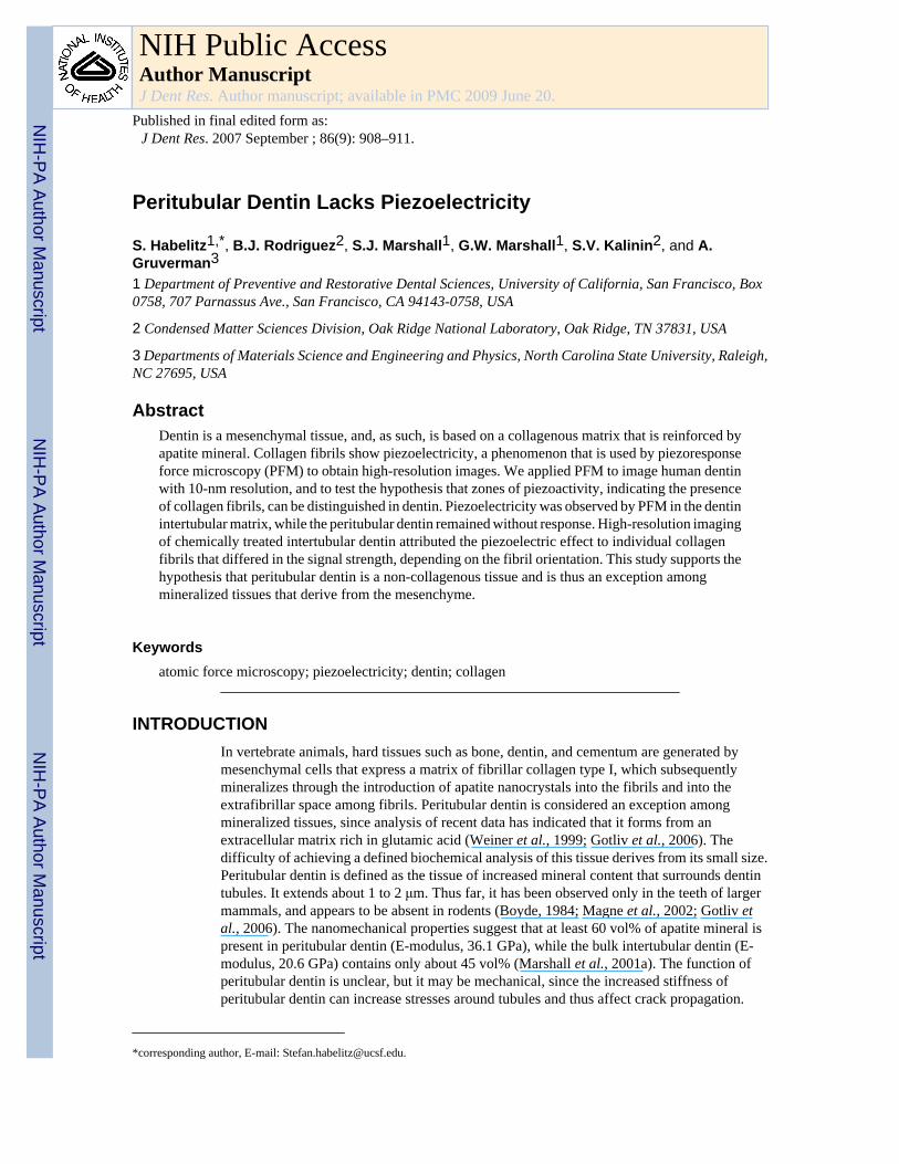
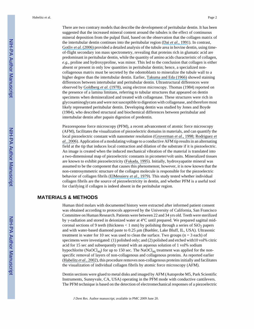
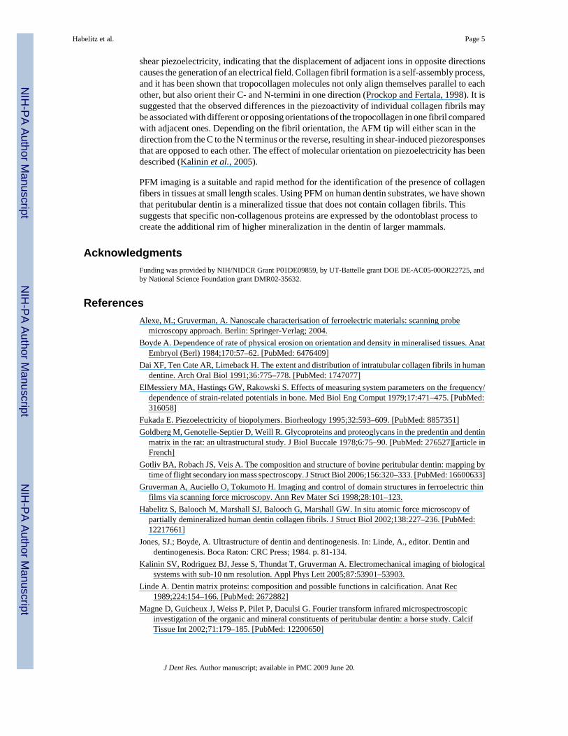
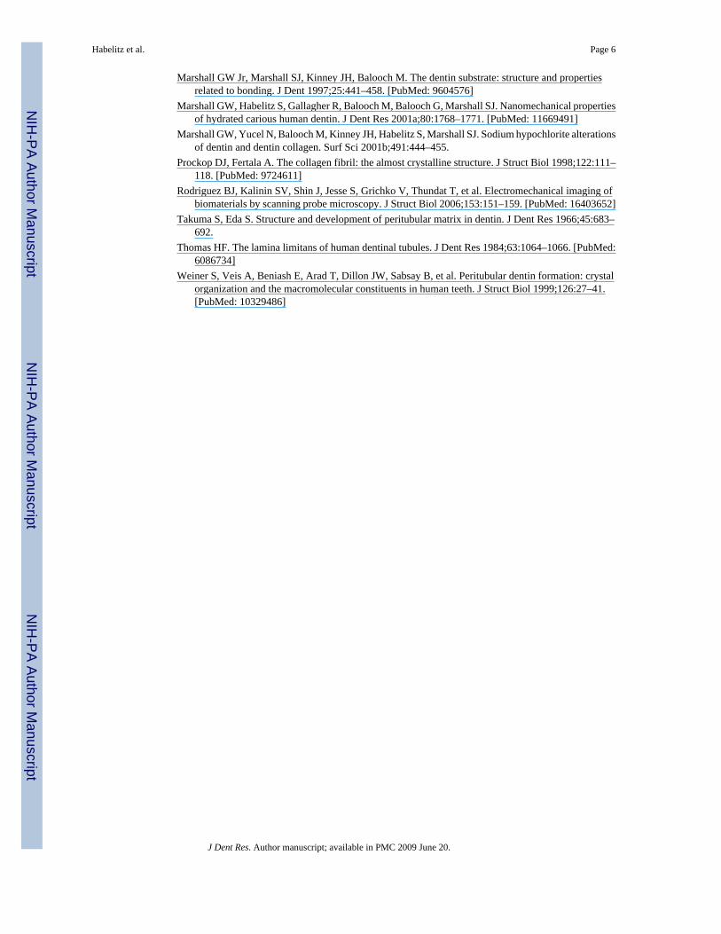
RESULTSAtomic force microscopy showed a characteristic dentin surface after polishing with 0.25-μmdiamond paste (Fig. 1). Intertubular dentin constituted the bulk of the specimen, intersected bytubules of about 1 μm diameter. (Figs. 1a, 1b, and 1c were obtained from the topographic,VPFM, and LPFM data, respectively.) In the topographic image, a thin ring (~ 1 μm wide)with decreased surface roughness surrounding each tubule was recognizable and could beattributed to peritubular dentin. Both PFM modes showed a moderate piezoelectric signal fromthe intertubular dentin, with an effective piezoelectric coefficient between 0.15 and 0.25 pm/V. No detectable piezoresponse was obtained from a zone of about 1 μm around the tubuleattributed to peritubular dentin (dark area). The tubule itself showed strong piezoresponse inthe VPFM mode, which was due to an artifact generated by the sudden change in height at thetubule wall. The LPFM mode was not susceptible to this artifact and showed no piezoelectricityin the tubule. AFM images of dentin from another tooth in topographic and PFM modes werecompared at higher resolution (Fig. 2). While peritubular dentin could barely be distinguishedfrom intertubular dentin by topography (Fig. 2a), PFM showed a clear demarcation betweenthe two (Fig. 2b). A thin rim of about 1 μm, which was not piezoactive, surrounded each tubule.
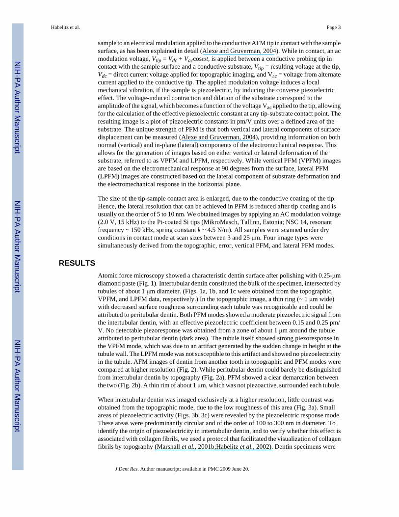
When intertubular dentin was imaged exclusively at a higher resolution, little contrast wasobtained from the topographic mode, due to the low roughness of this area (Fig. 3a). Smallareas of piezoelectric activity (Figs. 3b, 3c) were revealed by the piezoelectric response mode.These areas were predominantly circular and of the order of 100 to 300 nm in diameter. Toidentify the origin of piezoelectricity in intertubular dentin, and to verify whether this effect isassociated with collagen fibrils, we used a protocol that facilitated the visualization of collagenfibrils by topography (Marshall et al., 2001b;Habelitz et al., 2002). Dentin specimens were
Habelitz et al. Page 3
J Dent Res. Author manuscript; available in PMC 2009 June 20.
NIH
-PA Author Manuscript
NIH
-PA Author Manuscript
NIH
-PA Author Manuscript
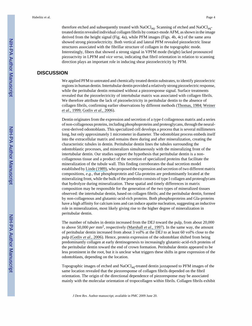
therefore etched and subsequently treated with NaOClaq. Scanning of etched and NaOClaq-treated dentin revealed individual collagen fibrils by contact-mode AFM, as shown in the imagederived from the height signal (Fig. 4a), while PFM images (Figs. 4b, 4c) of the same areashowed strong piezoelectricity. Both vertical and lateral PFM revealed piezoelectric linearstructures associated with the fibrillar structure of collagen in the topographic mode.Interestingly, fibers that showed a strong signal in VPFM mode (bright) lacked pronouncedpiezoactvity in LPFM and vice versa, indicating that fibril orientation in relation to scanningdirection plays an important role in inducing shear piezoelectricity by PFM.
DISCUSSIONWe applied PFM to untreated and chemically treated dentin substrates, to identify piezoelectricregions in human dentin. Intertubular dentin provided a relatively strong piezoelectric response,while the peritubular dentin remained without a piezoresponse signal. Surface treatmentsrevealed that the piezoelectricity of intertubular matrix was associated with collagen fibrils.We therefore attribute the lack of piezoelectricity in peritubular dentin to the absence ofcollagen fibrils, confirming earlier observations by different methods (Thomas, 1984; Weineret al., 1999; Gotliv et al., 2006).
Dentin originates from the expression and secretion of a type-I collagenous matrix and a seriesof non-collagenous proteins, including phosphoproteins and proteoglycans, through the neural-crest-derived odontoblasts. This specialized cell develops a process that is several millimeterslong, but only approximately 1 micrometer in diameter. The odontoblast process embeds itselfinto the extracellular matrix and remains there during and after mineralization, creating thecharacteristic tubules in dentin. Peritubular dentin lines the tubules surrounding theodontoblastic processes, and mineralizes simultaneously with the mineralizing front of theintertubular dentin. Our studies support the hypothesis that peritubular dentin is a non-collagenous tissue and a product of the secretion of specialized proteins that facilitate themineralization of the tubule wall. This finding corroborates the dual secretion modelestablished by Linde (1989), who proposed the expression and secretion of two different matrixcompositions, e.g., that phosphoprotein and Gla-proteins are predominantly located at themineralizing front, while the bulk of the predentin consists of type I collagen and proteoglycansthat hydrolyze during mineralization. These spatial and timely differences in matrixcomposition may be responsible for the generation of the two types of mineralized tissuesobserved: the intertubular dentin, based on collagen fibrils; and the peritubular dentin, formedby non-collagenous and glutamic-acid-rich proteins. Both phosphoproteins and Gla-proteinshave a high affinity for calcium ions and can induce apatite nucleation, suggesting an inductiverole in mineralization, most likely giving rise to the higher degree of mineralization inperitubular dentin.
The number of tubules in dentin increased from the DEJ toward the pulp, from about 20,000to above 50,000 per mm2, respectively (Marshall et al., 1997). In the same way, the amountof peritubular dentin increased from about 3 vol% at the DEJ to at least 60 vol% close to thepulp (Gotliv et al., 2006). Hence, protein expression of the odontoblast shifted from beingpredominantly collagen at early dentinogenesis to increasingly glutamic-acid-rich proteins ofthe peritubular dentin toward the end of crown formation. Peritubular dentin appeared to beless prominent in the root, but it is unclear what triggers these shifts in gene expression of theodontoblasts, depending on the location.
Topographic images of etched and NaOClaq-treated dentin juxtaposed to PFM images of thesame location revealed that the piezoresponse of collagen fibrils depended on the fibrilorientation. The origin of the directional dependence of piezoresponse may be associatedmainly with the molecular orientation of tropocollagen within fibrils. Collagen fibrils exhibit
Habelitz et al. Page 4
J Dent Res. Author manuscript; available in PMC 2009 June 20.
NIH
-PA Author Manuscript
NIH
-PA Author Manuscript
NIH
-PA Author Manuscript
shear piezoelectricity, indicating that the displacement of adjacent ions in opposite directionscauses the generation of an electrical field. Collagen fibril formation is a self-assembly process,and it has been shown that tropocollagen molecules not only align themselves parallel to eachother, but also orient their C- and N-termini in one direction (Prockop and Fertala, 1998). It issuggested that the observed differences in the piezoactivity of individual collagen fibrils maybe associated with different or opposing orientations of the tropocollagen in one fibril comparedwith adjacent ones. Depending on the fibril orientation, the AFM tip will either scan in thedirection from the C to the N terminus or the reverse, resulting in shear-induced piezoresponsesthat are opposed to each other. The effect of molecular orientation on piezoelectricity has beendescribed (Kalinin et al., 2005).
PFM imaging is a suitable and rapid method for the identification of the presence of collagenfibers in tissues at small length scales. Using PFM on human dentin substrates, we have shownthat peritubular dentin is a mineralized tissue that does not contain collagen fibrils. Thissuggests that specific non-collagenous proteins are expressed by the odontoblast process tocreate the additional rim of higher mineralization in the dentin of larger mammals.
AcknowledgmentsFunding was provided by NIH/NIDCR Grant P01DE09859, by UT-Battelle grant DOE DE-AC05-00OR22725, andby National Science Foundation grant DMR02-35632.
ReferencesAlexe, M.; Gruverman, A. Nanoscale characterisation of ferroelectric materials: scanning probe
microscopy approach. Berlin: Springer-Verlag; 2004.Boyde A. Dependence of rate of physical erosion on orientation and density in mineralised tissues. Anat
Embryol (Berl) 1984;170:57–62. [PubMed: 6476409]Dai XF, Ten Cate AR, Limeback H. The extent and distribution of intratubular collagen fibrils in human
dentine. Arch Oral Biol 1991;36:775–778. [PubMed: 1747077]ElMessiery MA, Hastings GW, Rakowski S. Effects of measuring system parameters on the frequency/
dependence of strain-related potentials in bone. Med Biol Eng Comput 1979;17:471–475. [PubMed:316058]
Fukada E. Piezoelectricity of biopolymers. Biorheology 1995;32:593–609. [PubMed: 8857351]Goldberg M, Genotelle-Septier D, Weill R. Glycoproteins and proteoglycans in the predentin and dentin
matrix in the rat: an ultrastructural study. J Biol Buccale 1978;6:75–90. [PubMed: 276527][article inFrench]
Gotliv BA, Robach JS, Veis A. The composition and structure of bovine peritubular dentin: mapping bytime of flight secondary ion mass spectroscopy. J Struct Biol 2006;156:320–333. [PubMed: 16600633]
Gruverman A, Auciello O, Tokumoto H. Imaging and control of domain structures in ferroelectric thinfilms via scanning force microscopy. Ann Rev Mater Sci 1998;28:101–123.
Habelitz S, Balooch M, Marshall SJ, Balooch G, Marshall GW. In situ atomic force microscopy ofpartially demineralized human dentin collagen fibrils. J Struct Biol 2002;138:227–236. [PubMed:12217661]
Jones, SJ.; Boyde, A. Ultrastructure of dentin and dentinogenesis. In: Linde, A., editor. Dentin anddentinogenesis. Boca Raton: CRC Press; 1984. p. 81-134.
Kalinin SV, Rodriguez BJ, Jesse S, Thundat T, Gruverman A. Electromechanical imaging of biologicalsystems with sub-10 nm resolution. Appl Phys Lett 2005;87:53901–53903.
Linde A. Dentin matrix proteins: composition and possible functions in calcification. Anat Rec1989;224:154–166. [PubMed: 2672882]
Magne D, Guicheux J, Weiss P, Pilet P, Daculsi G. Fourier transform infrared microspectroscopicinvestigation of the organic and mineral constituents of peritubular dentin: a horse study. CalcifTissue Int 2002;71:179–185. [PubMed: 12200650]
Habelitz et al. Page 5
J Dent Res. Author manuscript; available in PMC 2009 June 20.
NIH
-PA Author Manuscript
NIH
-PA Author Manuscript
NIH
-PA Author Manuscript
Marshall GW Jr, Marshall SJ, Kinney JH, Balooch M. The dentin substrate: structure and propertiesrelated to bonding. J Dent 1997;25:441–458. [PubMed: 9604576]
Marshall GW, Habelitz S, Gallagher R, Balooch M, Balooch G, Marshall SJ. Nanomechanical propertiesof hydrated carious human dentin. J Dent Res 2001a;80:1768–1771. [PubMed: 11669491]
Marshall GW, Yucel N, Balooch M, Kinney JH, Habelitz S, Marshall SJ. Sodium hypochlorite alterationsof dentin and dentin collagen. Surf Sci 2001b;491:444–455.
Prockop DJ, Fertala A. The collagen fibril: the almost crystalline structure. J Struct Biol 1998;122:111–118. [PubMed: 9724611]
Rodriguez BJ, Kalinin SV, Shin J, Jesse S, Grichko V, Thundat T, et al. Electromechanical imaging ofbiomaterials by scanning probe microscopy. J Struct Biol 2006;153:151–159. [PubMed: 16403652]
Takuma S, Eda S. Structure and development of peritubular matrix in dentin. J Dent Res 1966;45:683–692.
Thomas HF. The lamina limitans of human dentinal tubules. J Dent Res 1984;63:1064–1066. [PubMed:6086734]
Weiner S, Veis A, Beniash E, Arad T, Dillon JW, Sabsay B, et al. Peritubular dentin formation: crystalorganization and the macromolecular constituents in human teeth. J Struct Biol 1999;126:27–41.[PubMed: 10329486]
Habelitz et al. Page 6
J Dent Res. Author manuscript; available in PMC 2009 June 20.
NIH
-PA Author Manuscript
NIH
-PA Author Manuscript
NIH
-PA Author Manuscript
Figure 1.AFM images of human dentin obtained with conductive tips with modulated voltage applied.(a) Topographic image; (b) vertical piezoresponse force mode (VPFM); and (c) lateralpiezoresponse force mode (LPFM). Piezoelectric constant reached values of up to 0.25 pm/V(bright areas) and is absent around tubules in the peritubular dentin (dark area).
Habelitz et al. Page 7
J Dent Res. Author manuscript; available in PMC 2009 June 20.
NIH
-PA Author Manuscript
NIH
-PA Author Manuscript
NIH
-PA Author Manuscript
Figure 2.AFM images of human dentin obtained with conductive tips with modulated voltage applied.(a) Topographic image shows 2 dentin tubules (T) surrounded by a brighter rim, attributed tothe peritubular dentin (PTD) and the intertubular dentin matrix (ITD). (b) Piezoresponse forceimage of the same location, showing piezoelectricity of the intertubular dentin, while tubulesand the peritubular region do not exhibit a piezoelectric signal (dark).
Habelitz et al. Page 8
J Dent Res. Author manuscript; available in PMC 2009 June 20.
NIH
-PA Author Manuscript
NIH
-PA Author Manuscript
NIH
-PA Author Manuscript
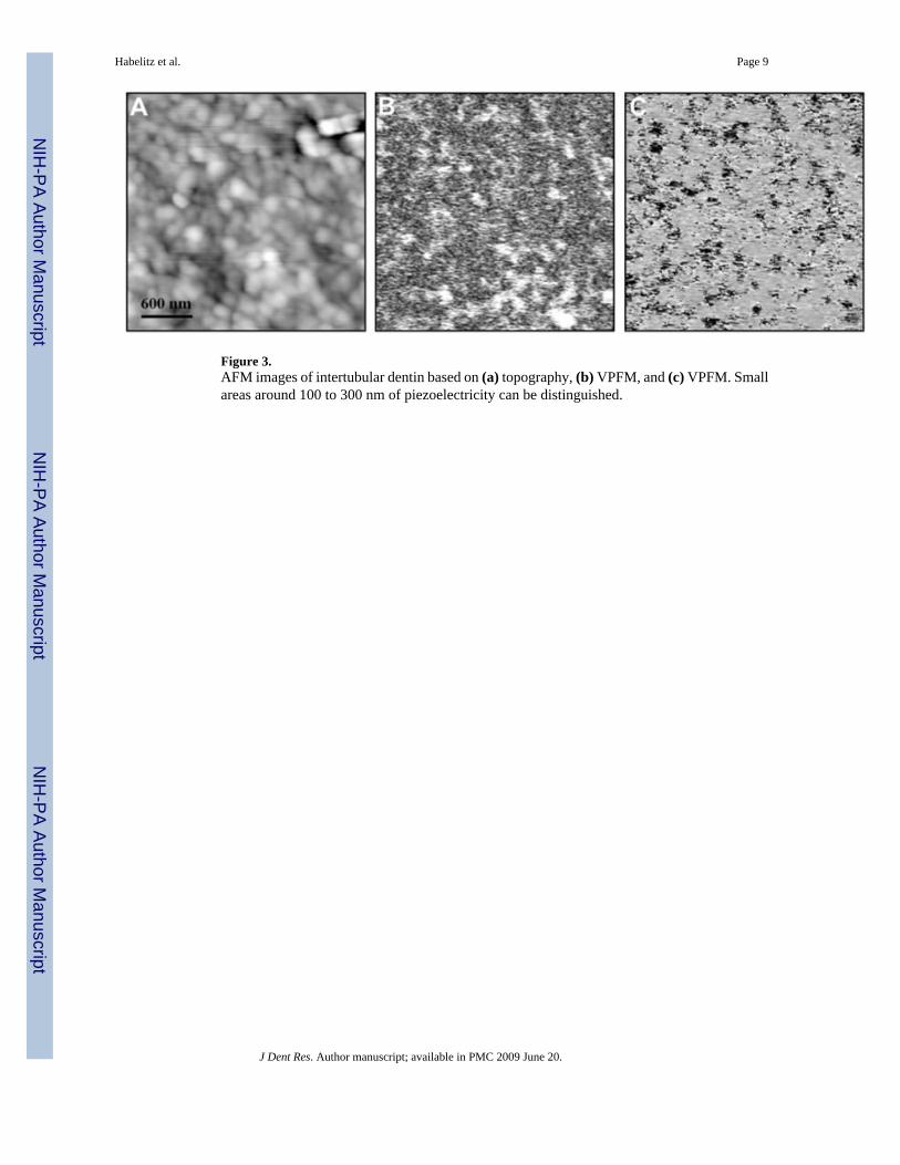
Figure 3.AFM images of intertubular dentin based on (a) topography, (b) VPFM, and (c) VPFM. Smallareas around 100 to 300 nm of piezoelectricity can be distinguished.
Habelitz et al. Page 9
J Dent Res. Author manuscript; available in PMC 2009 June 20.
NIH
-PA Author Manuscript
NIH
-PA Author Manuscript
NIH
-PA Author Manuscript
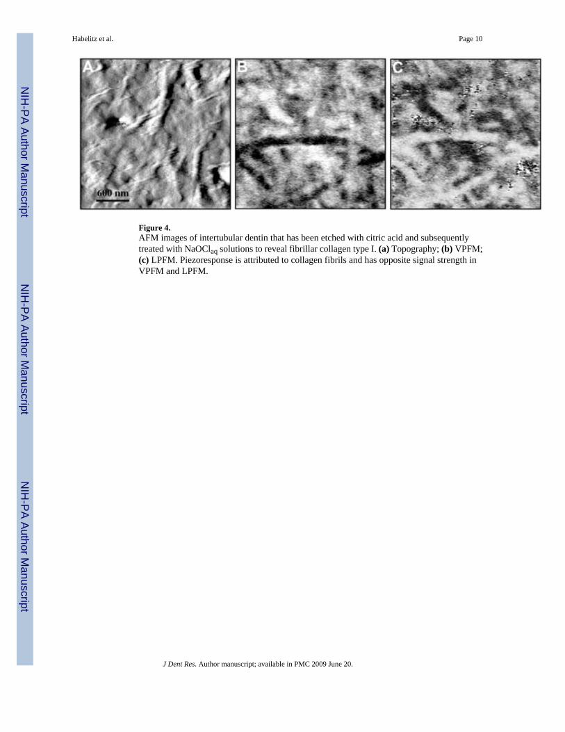
Figure 4.AFM images of intertubular dentin that has been etched with citric acid and subsequentlytreated with NaOClaq solutions to reveal fibrillar collagen type I. (a) Topography; (b) VPFM;(c) LPFM. Piezoresponse is attributed to collagen fibrils and has opposite signal strength inVPFM and LPFM.
Habelitz et al. Page 10
J Dent Res. Author manuscript; available in PMC 2009 June 20.
NIH
-PA Author Manuscript
NIH
-PA Author Manuscript
NIH
-PA Author Manuscript


