Embed Size (px)
Citation preview
Bulletin of Faculty of Pharmacy, Cairo University (2012) 50, 17–39
Cairo University
Bulletin of Faculty of Pharmacy, Cairo University
www.elsevier.com/locate/bfopcuwww.sciencedirect.com
ORIGINAL ARTICLE
Pharmacognostical study of Chorisia insignis HBK. grown
in Egypt
Taha EI Alfy a, Salma El Sawi b, Sahar Abd El Tawab c, Doaa Moawad b,*
a Pharmacognosy Department, Faculty of Pharmacy, Cairo University, Cairo, Egyptb Pharmacognosy Department, National Research Centre, Dokki 12622, Cairo, Egyptc Botany Department, Faculty of Women, Ain Shams University, Cairo, Egypt
Received 16 November 2011; accepted 8 January 2012Available online 2 May 2012
*
N
Eg
01
E
M
11
an
Pe
U
ht
KEYWORDS
Botany;
Chorisia insignis;
DNA fingerprint;
Pharmacopoeial constants;
Protein electrophoresis
Corresponding author. Add
o. 12, National Research Cen
ypt. Tel.: +20 02 3852120
116006263.
-mail addresses: drdoda@
oawad).
10-0931 ª 2012 Faculty of P
d hosting by Elsevier B.V. A
er review under responsibi
niversity.
tp://dx.doi.org/10.1016/j.bfop
Production and h
ress: Pha
tre, El T
3/02 333
gmail.co
harmacy,
ll rights
lity of F
cu.2012.
osting by E
Abstract Chorisia insignisHBK. is a perennial deciduous plant native to South America. This study
presents the macro and micromorphological features of the plant and the determination of its phar-
macopoeial constants (Moisture = 12.80%, Total ash = 11.72%,Water soluble ash = 9.24%, Acid
insoluble ash = 0.16% and Crude fibre = 20.33%). Furthermore, the DNA of the plant was
extracted from leaf samples and analyzed by polymerase chain reaction (PCR) using 12 decamer prim-
ers. The DNA fingerprint showed a total of 95 fragments. Protein electrophoresis was carried out
using SDS–PAGE (sodium dodecyl sulphate polyacrylamide gel electrophoresis) technique to moni-
tor the seed storage protein expressed by the active genes of the whole genomic DNA of C. insignis
HBK. The data obtained revealed the presence of 20 sharp protein bands having a wide range of
molecular weights. The percentage of protein in the seeds was found to be 26.83%as determined using
rmacognosy Department, Lab
ahrir St., Dokki 12622, Cairo,
02441, mobile: 01006268896/
m, [email protected] (D.
Cairo University. Production
reserved.
aculty of Pharmacy, Cairo
01.001
lsevier
18 T. El Alfy et al.
micro-Kjeldahl method. Analysis of the total amino acids content of the seeds revealed the identifica-
tion of 15 amino acids, mainly glutamic acid (26.71%), aspartic acid (11.98%) and glycine (11.00%).
ª 2012 Faculty of Pharmacy, Cairo University. Production and hosting by Elsevier B.V. All rights
reserved.
1. Introduction
Chorisia insignis HBK. is an ornamental plant known as thewhite floss silk tree, belongs to family Bombacaceae. It is na-
tive to South America, Peru, Brazil and Argentina.1–3 Chorisiawas named in honor of the botanical artist and traveler Lud-wig I. Choris (1795–1828, 19th century).1
Chorisia insignis HBK. is mainly cultivated for its ornamen-tal brilliant flowers and for the silky white fibre (or floss) that isobtained from the ripened fruits. This floss has been used tostuff cushions, pillows and vests which explain the common
name of this tree, ‘‘floss silk tree’’.1,2
The flavonoidal content and the biological activity of theleaves of the plant were previously investigated.4
The present work includes the botanical study of the differ-ent organs of C. insignis HBK., determination of certain phar-macopoeial constants of the leaves, DNA fingerprinting as well
as investigation of the protein and amino acids contents of theseeds. This study was carried out to confirm the identity andpurity of C. insignis HBK. cultivated in Egypt.
Table 1 Primers used for the RAPD analysis and their
sequences.
No. Name of the
primers
Sequence of the
primers (50 fi 30)
1 OPA-16 AGCCAGCGAA
2 OPA-09 GGGTAACGCC
3 OPA-03 AGTCAGCCAC
4 OPA-18 AGGTGACCGT
5 OPB-10 CTGCTGGGAC
6 OPM-04 GGCGGTTGTC
7 OPQ-04 AGTGCGCTGA
8 OPH-07 CAGCCCAGAG
9 OPQ-17 GAAGCCCTTG
10 OPB-07 GGTGACGCAG
11 OPB-03 CATCCCCCTG
12 OPB-08 GTCCACACGG
2. Material and methods
2.1. Plant material
Samples of different organs of C. insignis HBK. were collected
from National Research Centre (NRC) garden, Dokki, Egyptin June 2006, and were kindly authenticated by Dr. MohamedGibali, senior botanist and by Agr. Eng. Tereez Labib, consul-
tant of plant taxonomy at the Ministry of Agriculture and ex.director of Orman Botanical Garden, Giza, Egypt.
Specimens for morphological studies were dried according
to the standard herbarium techniques and a voucher specimen(No. 23569) is kept at NRC Herbarium. Photographs were ta-ken using Sony Cyber-shot digital camera.
Anatomical investigations were performed on cross sections
of the stem of the young branches, stem bark, spines, leaves,flowers, fruit and seeds which were preserved in 70% ethylalcohol containing 5% glycerin. The photographs were taken
using a Fuji FinePix A500 digital camera.Samples of the plant under investigation were separately
air-dried, turned to coarse powder (1700/355) and kept in
tightly closed amber coloured glass containers for furtherinvestigation.
2.2. Determination of certain pharmacopoeial constants of theleaves
Certain pharmacopoeial constants (viz. moisture, total ash,
water soluble ash, acid insoluble ash and crude fibre contents)were carried out adopting the Egyptian Pharmacopoeia.5
2.3. DNA fingerprinting
The procedure was performed on ground freeze dried wholeleaves as stated in the literature.6–8 Primers used to amplifythe DNA with their sequences are listed in Table 1.
2.4. Protein banding analysis
The process was carried out according to Ornstein9 and
Laemmli.10
2.5. Determination of the total protein and amino acids contents
The total protein content was carried out by micro-Kjeldahlmethod.11 Acid hydrolysis was done according to the methodof Block et al.12
3. Results
3.1. Macromorphology
Chorisia insignis HBK. is a perennial deciduous plant andflowers from September to December. The trunk is notablefor its knob-like projections which develop as the tree ages.
The tree is about 15 m in height with a trunk enlarged orswollen (bottle shaped) at the base having a circumference ofabout 200 cm covered with short spines (Fig. 1A–C).
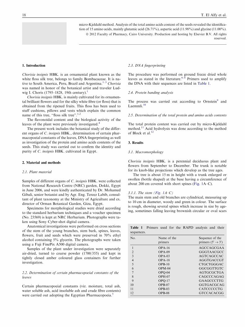
3.1.1. The stem (Fig. 1A–C)The main stem and old branches are cylindrical, measuring up
to 10 cm in diameter, woody and green in colour. The surfaceis rough, showing several spines which increase in size by age-ing, sometimes falling leaving brownish circular or oval scars
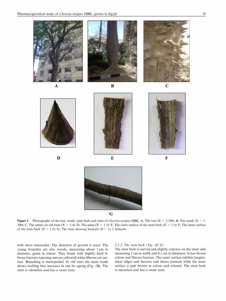
Figure 1 Photographs of the tree, trunk, stem bark and stem of Chorisia insignis HBK. A, The tree (X = 3/100); B, The trunk (X = 1/
200); C, The spines on old stem (X = 1/4); D, The spine (X = 1.5); E, The outer surface of the stem bark (X = 1.5); F, The inner surface
of the stem bark (X = 1.5); G, The stem showing lenticels (X= 1); l, lenticels.
Pharmacognostical study of Chorisia insignis HBK. grown in Egypt 19
with short internodes. The direction of growth is erect. Theyoung branches are also woody, measuring about 1 cm indiameter, green in colour. They break with slightly hard fi-brous fracture exposing uneven yellowish white fibrous cut sur-
face. Branching is monopodial. In old trees the main trunkshows swelling that increases in size by ageing (Fig. 1B). Thestem is odourless and has a sweet taste.
3.1.2. The stem bark (Fig. 1E–G)The stem bark is curved and slightly concave on the inner sidemeasuring 1 cm in width and 0.1 cm in thickness. It has brown
colour and fibrous fracture. The outer surface exhibits longitu-dinal ridges and furrows and shows lenticels while the innersurface is pale brown in colour and striated. The stem bark
is odourless and has a sweet taste.
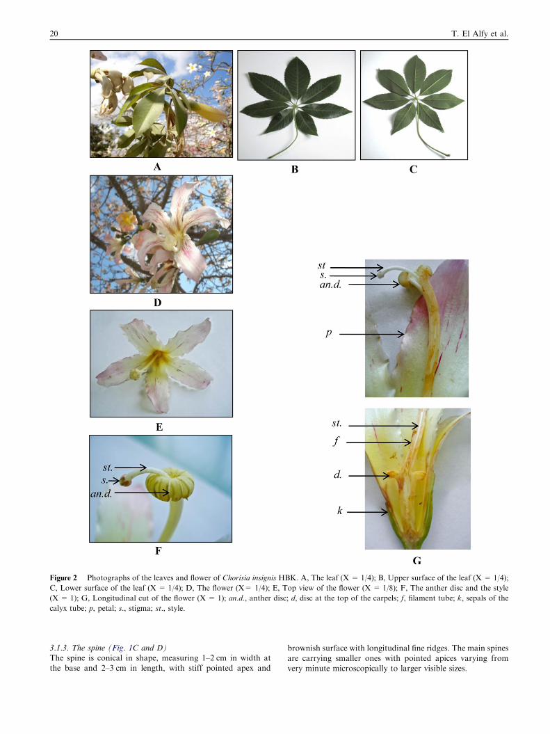
Figure 2 Photographs of the leaves and flower of Chorisia insignis HBK. A, The leaf (X = 1/4); B, Upper surface of the leaf (X = 1/4);
C, Lower surface of the leaf (X = 1/4); D, The flower (X= 1/4); E, Top view of the flower (X = 1/8); F, The anther disc and the style
(X = 1); G, Longitudinal cut of the flower (X = 1); an.d., anther disc; d, disc at the top of the carpels; f, filament tube; k, sepals of the
calyx tube; p, petal; s., stigma; st., style.
20 T. El Alfy et al.
3.1.3. The spine (Fig. 1C and D)The spine is conical in shape, measuring 1–2 cm in width atthe base and 2–3 cm in length, with stiff pointed apex and
brownish surface with longitudinal fine ridges. The main spinesare carrying smaller ones with pointed apices varying fromvery minute microscopically to larger visible sizes.
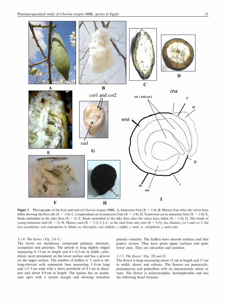
Figure 3 Photographs of the fruit and seed of Chorisia insignis HBK. A, Immature fruit (X = 1/4); B, Mature fruit after the valves have
fallen showing the floss silk (X = 1/4); C, Longitudinal cut in immature fruit (X = 1/4); D, Transverse cut in immature fruit (X = 1/4); E,
Seeds embedded in the silky floss (X = 1); F, Seeds embedded in the silky floss after the valves have fallen (X = 1/8); G, The inside of
young immature seed (X = 5); H, Mature seed (X = 2.5); I, L.C. in the seed from side view (X = 8.5); cha, chalaza; cot 1 and cot 2, the
two cotyledons; end, endosperm; h, hilum; m, micropyle; rad, radicle; r, raphe; s, seed; st., strophiole; t, seed coat.
Pharmacognostical study of Chorisia insignis HBK. grown in Egypt 21
3.1.4. The leaves (Fig. 2A–C)The leaves are deciduous, compound palmate, alternate,exstipulate and petiolate. The petiole is long slightly ridged
measuring 8–13 cm in length and 0.1–0.2 cm in width, cylin-drical, more prominent on the lower surface and has a grooveon the upper surface. The number of leaflets is 7; each is ob-
long-obovate with symmetric base measuring 5–8 cm longand 1.5–3 cm wide with a short petioleole of 0.1 cm in diam-eter and about 0.9 cm in length. The lamina has an acumi-nate apex with a serrate margin and showing reticulate
pinnate venation. The leaflets have smooth surfaces and thinpapery texture. They have green upper surfaces and palerlower ones. They are odourless and tasteless.
3.1.5. The flower (Fig. 2D and E)The flower is large measuring about 13 cm in length and 17 cmin width, showy and solitary. The flowers are pentacyclic,
pentamerous and pedicellate with no characteristic odour ortaste. The flower is actinomorphic, hermaphrodite and hasthe following floral formula:
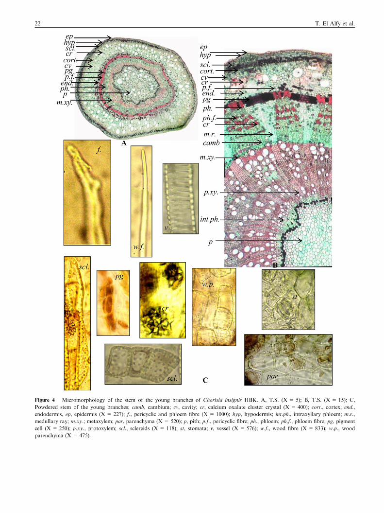
Figure 4 Micromorphology of the stem of the young branches of Chorisia insignis HBK. A, T.S. (X = 5); B, T.S. (X = 15); C,
Powdered stem of the young branches; camb, cambium; cv, cavity; cr, calcium oxalate cluster crystal (X = 400); cort., cortex; end.,
endodermis, ep, epidermis (X = 227); f., pericyclic and phloem fibre (X = 1000); hyp, hypodermis; int.ph., intraxyllary phloem; m.r.,
medullary ray; m.xy.; metaxylem; par, parenchyma (X = 520); p, pith; p.f., pericyclic fibre; ph., phloem; ph.f., phloem fibre; pg, pigment
cell (X = 250); p.xy., protoxylem; scl., sclereids (X = 118); st, stomata; v, vessel (X = 576); w.f., wood fibre (X = 833); w.p., wood
parenchyma (X = 475).
22 T. El Alfy et al.
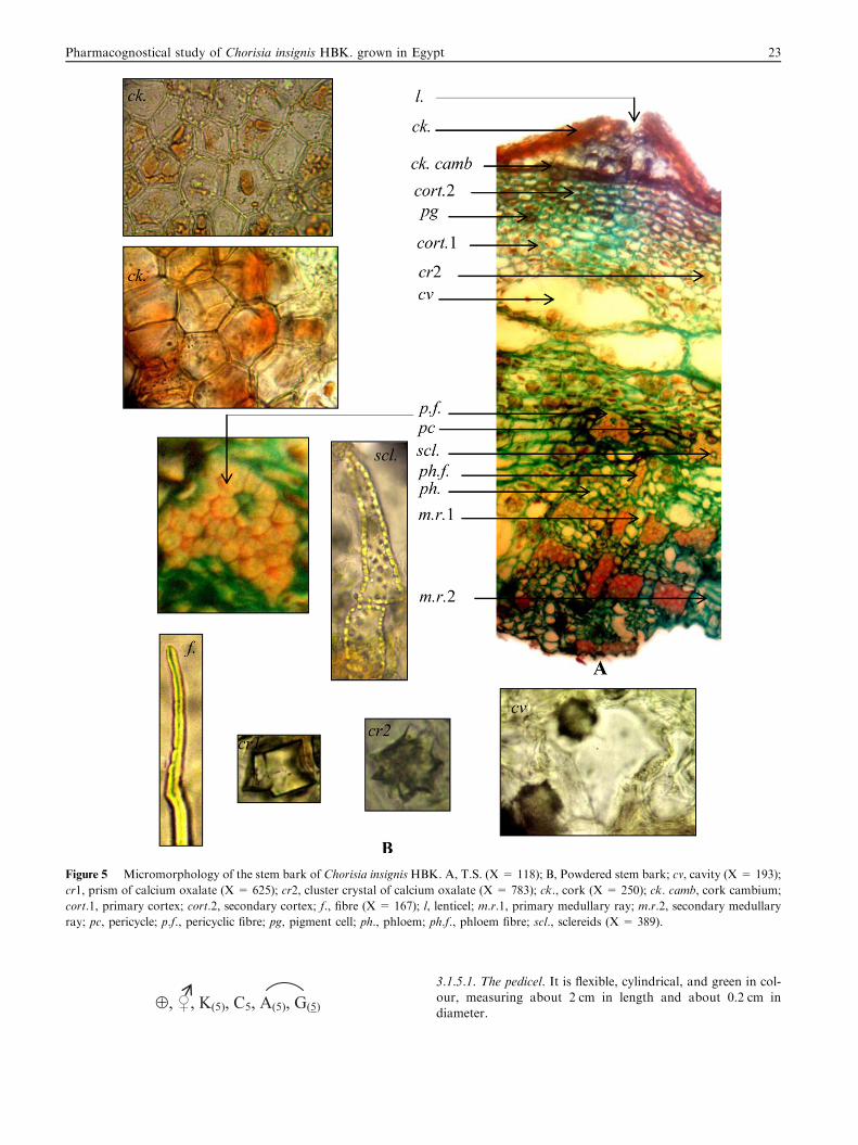
Figure 5 Micromorphology of the stem bark of Chorisia insignis HBK. A, T.S. (X = 118); B, Powdered stem bark; cv, cavity (X = 193);
cr1, prism of calcium oxalate (X = 625); cr2, cluster crystal of calcium oxalate (X = 783); ck., cork (X = 250); ck. camb, cork cambium;
cort.1, primary cortex; cort.2, secondary cortex; f., fibre (X = 167); l, lenticel; m.r.1, primary medullary ray; m.r.2, secondary medullary
ray; pc, pericycle; p.f., pericyclic fibre; pg, pigment cell; ph., phloem; ph.f., phloem fibre; scl., sclereids (X = 389).
Pharmacognostical study of Chorisia insignis HBK. grown in Egypt 23
3.1.5.1. The pedicel. It is flexible, cylindrical, and green in col-our, measuring about 2 cm in length and about 0.2 cm indiameter.
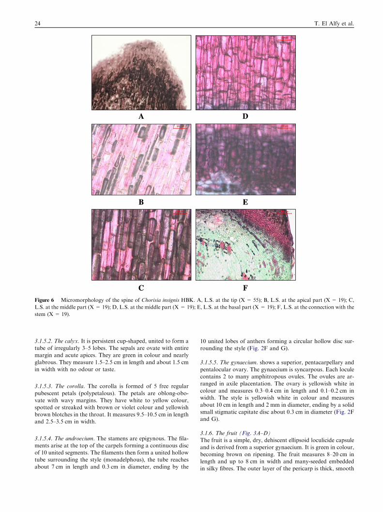
Figure 6 Micromorphology of the spine of Chorisia insignis HBK. A, L.S. at the tip (X = 55); B, L.S. at the apical part (X = 19); C,
L.S. at the middle part (X = 19); D, L.S. at the middle part (X = 19); E, L.S. at the basal part (X = 19); F, L.S. at the connection with the
stem (X = 19).
24 T. El Alfy et al.
3.1.5.2. The calyx. It is persistent cup-shaped, united to form atube of irregularly 3–5 lobes. The sepals are ovate with entiremargin and acute apices. They are green in colour and nearly
glabrous. They measure 1.5–2.5 cm in length and about 1.5 cmin width with no odour or taste.
3.1.5.3. The corolla. The corolla is formed of 5 free regular
pubescent petals (polypetalous). The petals are oblong-obo-vate with wavy margins. They have white to yellow colour,spotted or streaked with brown or violet colour and yellowish
brown blotches in the throat. It measures 9.5–10.5 cm in lengthand 2.5–3.5 cm in width.
3.1.5.4. The androecium. The stamens are epigynous. The fila-ments arise at the top of the carpels forming a continuous discof 10 united segments. The filaments then form a united hollowtube surrounding the style (monadelphous), the tube reaches
about 7 cm in length and 0.3 cm in diameter, ending by the
10 united lobes of anthers forming a circular hollow disc sur-rounding the style (Fig. 2F and G).
3.1.5.5. The gynaecium. shows a superior, pentacarpellary and
pentalocular ovary. The gynaecium is syncarpous. Each loculecontains 2 to many amphitropous ovules. The ovules are ar-ranged in axile placentation. The ovary is yellowish white in
colour and measures 0.3–0.4 cm in length and 0.1–0.2 cm inwidth. The style is yellowish white in colour and measuresabout 10 cm in length and 2 mm in diameter, ending by a solid
small stigmatic capitate disc about 0.3 cm in diameter (Fig. 2Fand G).
3.1.6. The fruit (Fig. 3A–D)The fruit is a simple, dry, dehiscent ellipsoid loculicide capsuleand is derived from a superior gynaecium. It is green in colour,
becoming brown on ripening. The fruit measures 8–20 cm inlength and up to 8 cm in width and many-seeded embeddedin silky fibres. The outer layer of the pericarp is thick, smooth
Pharmacognostical study of Chorisia insignis HBK. grown in Egypt 25
and glabrous. It has a short fracture, measuring about 0.5 cm
in thickness with no odour or taste.
3.1.7. The seed (Fig. 3E–I)The seed is small, globular, dark brown in colour, measuring0.3–0.7 cm in diameter and is derived from amphitropousovule. The seed is almost rounded with a local enlargement
over the position of the raphe (strophiole), embedded in silkyfloss having no odour or taste.
3.2. Micromorphology
3.2.1. The stem of the young branches (Fig. 4)A transverse section in the green stem (young branches)(Fig. 4A and B) is more or less circular in outline. It is formedof an epidermis consisting of polygonal, isodiametric cells with
thin cellulosic straight anticlinal walls and anomocytic sto-mata. The cortex is formed of an outer region consisting ofhypodermis of 3–4 layers of thin-walled parenchymatous cellsfollowed by 3–4 layers of sclerenchymatous cells. The sclereids
are polygonal isodiametric, sometimes elongated, lignified,
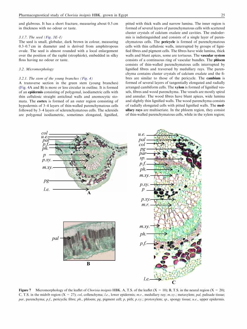
Figure 7 Micromorphology of the leaflet of Chorisia insignis HBK. A
C, T.S. in the midrib region (X = 27); col, collenchyma; l.e., lower epid
par, parenchyma; p.f., pericyclic fibre; ph., phloem; pg, pigment cell; p
pitted with thick walls and narrow lumina. The inner region is
formed of several layers of parenchymatous cells with scatteredcluster crystals of calcium oxalate and cavities. The endoder-mis is indistinguished and consists of a single layer of paren-chymatous cells. The pericycle is formed of parenchymatous
cells with thin cellulosic walls, interrupted by groups of ligni-fied fibres and pigment cells. The fibres have wide lumina, thickwalls and blunt apices, some are tortuous. The vascular system
consists of a continuous ring of vascular bundles. The phloem
consists of thin-walled parenchymatous cells interrupted bylignified fibres and traversed by medullary rays. The paren-
chyma contains cluster crystals of calcium oxalate and the fi-bres are similar to those of the pericycle. The cambium isformed of several layers of tangentially elongated and radially
arranged cambiform cells. The xylem is formed of lignified ves-sels, fibres and wood parenchyma. The vessels are mostly spiraland annular. The wood fibres have blunt apices, wide luminaand slightly thin lignified walls. The wood parenchyma consists
of radially elongated cells with pitted lignified walls. The med-
ullary rays are multiseriate. In the phloem region, they consistof thin-walled parenchymatous cells, while in the xylem region;
, T.S. of the leaflet (X = 10); B, T.S. in the neural region (X = 20);
ermis; m.r., medullary ray; m.xy.; metaxylem; pal, palisade tissue;
, pith; p.xy.; protoxylem; sp., spongy tissue; u.e., upper epidermis.
26 T. El Alfy et al.
they consist of radially elongated cells with lignified walls. The
intraxyllary (perimedullary) phloem is formed of patches of cel-lulosic phloem parenchyma at the periphery of the pith just be-neath the xylem vessels. The pith consists of thin-walledparenchymatous cells showing numerous cluster crystals of
calcium oxalate and cavities. The cells in the periphery of thepith, in the neighbourhood of the primary xylem, are muchsmaller, closely packed to the perimedullary zone. The perime-
dullary zone, showing the perimedullary phloem, is lined fromthe inner side by a continuous layer of parenchymatous cellscontaining pigments.
3.2.2. Powder of the stem of the young branches (Fig. 4C)Powdered stem of the young branches is green in colour,
odourless and has a sweet taste. It is characterized microscop-ically by the presence of the following elements:
1. Fragments of polygonal epidermal cells with straight anti-clinal walls and covered with thin smooth cuticle showinganomocytic stomata.
2. Fragments of thin-walled parenchymatous cells of the cor-
tex and pith, some of them with brown contents.3. Numerous cluster crystals of calcium oxalate.4. Numerous slightly radially elongated pigment cells.
5. Fragments of lignified vessels with spiral and annularthickenings.
6. Fragments of slightly thickened lignified wood parenchyma
having pitted lignified walls.7. Numerous elongated tortuous pericyclic and phloem fibres
with blunt apices, narrow lumina and thick lignified walls.
8. Numerous elongated wood fibres with blunt apices, widelumina and slightly thin lignified walls.
9. Fragments of polygonal isodiametric (sometimes elon-gated) pitted lignified sclereids with thick walls and narrow
lumina.
3.2.3. The stem bark (Fig. 5)A transverse section in the stem bark (Fig. 5A) is formed of thecork consisting of 3–4 layers of polygonal, slightly tangentially
elongated suberised thick-walled cells. The cells are equallythickened and lacking intercellular spaces except lenticels.The lenticels consist of a mass of loose unsuberised thin-walled
cells. The cork is followed by the cork cambium appears as onelayer. The secondary cortex is formed of 5–7 layers of collen-chymatous cells. The cells are polygonal, few are filled with
brown contents, in addition to numerous clusters and fewprisms of calcium oxalate. The primary cortex is formed ofan outer zone consisting of several layers of rounded thin-walled parenchymatous cells with brown contents, numerous
cluster crystals and few prisms of calcium oxalate. The middlezone shows 2 layers of large empty cavities. The inner zoneconsists of 3–4 layers of small slightly thick-walled cellulosic
parenchymatous cells. The pericycle is parenchymatous inter-rupted by groups of lignified pericyclic fibres. They are thick-walled having narrow lumina and blunt tips. Some of the
groups of fibres are accompanied by sclereids. The sclereidsare polygonal isodiametric, sometimes elongated, lignified, pit-ted with thick walls and narrow lumina. The phloem consists ofprimary and secondary phloems traversed by medullary rays.
The primary phloem is collapsed. The secondary phloem isparenchymatous traversed by groups of phloem fibres. The
fibres are similar to those of the pericycle. The parenchyma
contains cluster crystals of calcium oxalate and is interruptedby groups of sclereids. The medullary rays consist of secondaryand primary medullary rays, formed of rectangular, radiallyelongated and rounded parenchymatous cells. The secondary
medullary rays are biseriate to triseriate while the primarymedullary rays are uniseriate.
3.2.4. Powder of the stem bark (Fig. 5B)Powdered stem bark is brown in colour, odourless and has asweet taste. It is characterized microscopically by the presence
of the following elements:
1. Fragments of thick-walled cork cells.
2. Large empty cavities from the middle zone of the primarycortex.
3. Numerous elongated thick-walled lignified pericyclic and
phloem fibres with narrow lumina and blunt tips.4. Numerous clusters and few prisms of calcium oxalate.5. Fragments of polygonal isodiametric sclereids, sometimes
elongated, lignified, pitted with thick walls and slightly nar-
row lamina.
3.2.5. The spine (Fig. 6A–F)A longitudinal section in the spine shows a mass of axiallyelongated parenchymatous cells filled with brown contents
which increase at the base and the top.
3.2.6. The leaflet
3.2.6.1. The lamina (Fig. 7). A transverse section in the leaflet(Fig. 7A–C) shows upper and lower epidermises formed of
polygonal isodiametric cells with thin cellulosic slightly wavyanticlinal walls. They are covered with thin striated cuticleand show anomocytic stomata. Stomata are more frequenton the lower one. The mesophyll is dorsiventral, differentiated
into discontinuous upper palisade and spongy tissue. The pal-isade (Fig. 7B) is formed of 2 layers of compact short andcolumnar cells containing chloroplast, some of them are filled
with red brown contents. It is interrupted in the midrib by col-ourless cortical tissue. The spongy tissue is formed of 3–4 lay-ers of chlorenchymatous cells which are thin-walled rounded
or slightly irregular in shape and showing moderately wideintercellular spaces. It shows scattered cluster crystals andprisms of calcium oxalate and pigment cells. The cortical tissue
of the midrib consists of 5–7 layers of collenchyma, followedby a zone of parenchymatous cells, which are polygonal toround with thin cellulosic walls and showing intercellularspaces. The endodermis is indistinguishable. The pericycle is
formed of lignified fibres forming an arc below the vascularbundle and a patch above it. The fibres have tapering apices,wide lumina and slightly thick lignified walls; some of them
are irregular, forked or tortuous and bent. The vascular bundleconsists of one big arcuate abaxial vascular bundle and an-other smaller inverted adaxial one surrounding the pith. The
phloem consists of thin-walled parenchymatous cells. The xy-
lem is formed of lignified vessels, fibres and wood parenchyma.The vessels are mostly spiral and annular. The fibres haveblunt apices, wide lumina and slightly thick lignified walls.
The wood parenchyma consists of radially elongated cells withpitted lignified walls. The medullary rays form radiating linestraversing the xylem. The pith is small arcuate, consists of
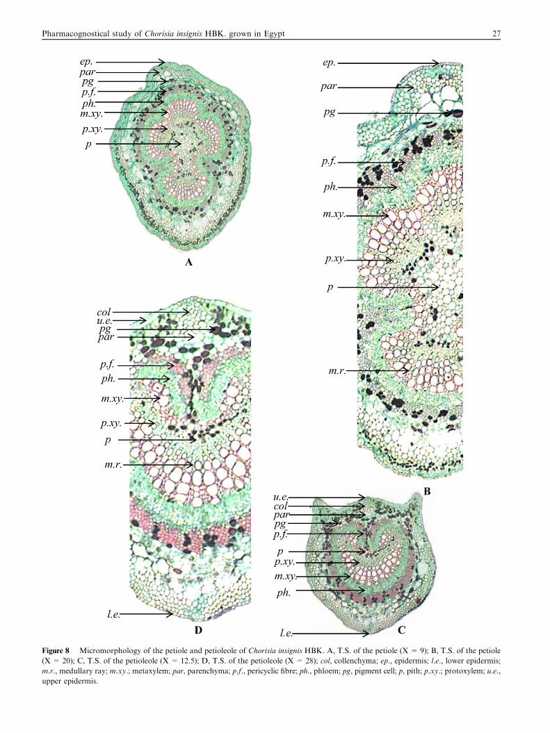
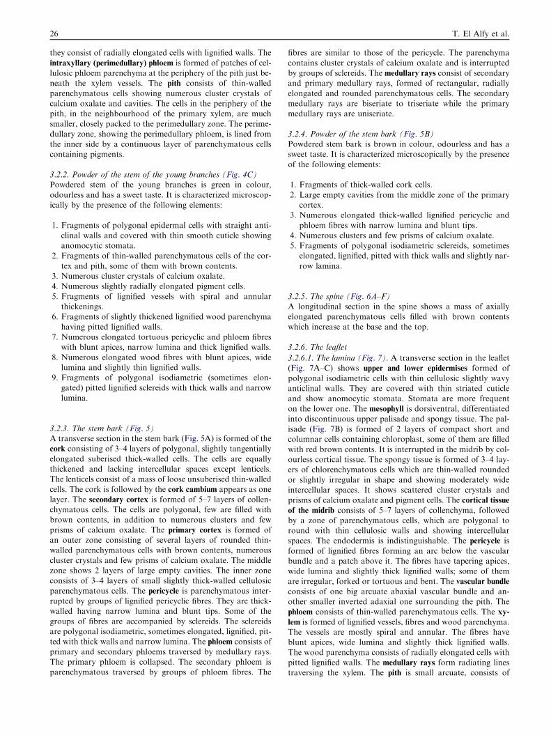
Figure 8 Micromorphology of the petiole and petioleole of Chorisia insignis HBK. A, T.S. of the petiole (X = 9); B, T.S. of the petiole
(X = 20); C, T.S. of the petioleole (X = 12.5); D, T.S. of the petioleole (X = 28); col, collenchyma; ep., epidermis; l.e., lower epidermis;
m.r., medullary ray; m.xy.; metaxylem; par, parenchyma; p.f., pericyclic fibre; ph., phloem; pg, pigment cell; p, pith; p.xy.; protoxylem; u.e.,
upper epidermis.
Pharmacognostical study of Chorisia insignis HBK. grown in Egypt 27
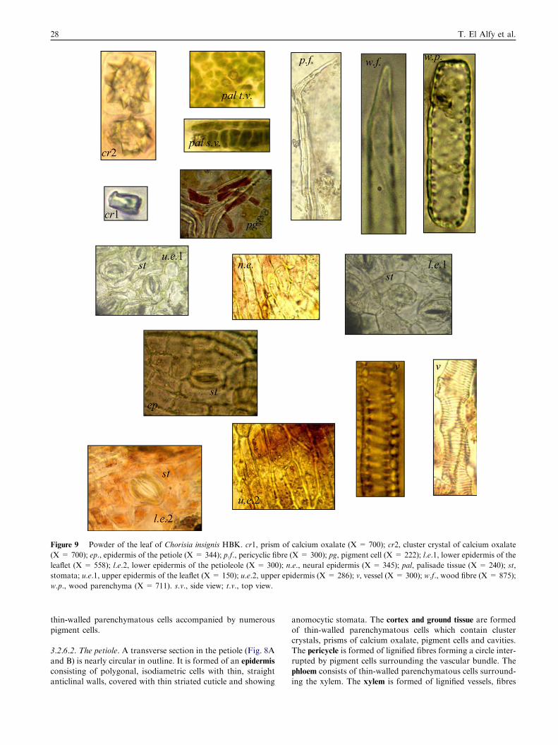
Figure 9 Powder of the leaf of Chorisia insignis HBK. cr1, prism of calcium oxalate (X = 700); cr2, cluster crystal of calcium oxalate
(X = 700); ep., epidermis of the petiole (X = 344); p.f., pericyclic fibre (X = 300); pg, pigment cell (X = 222); l.e.1, lower epidermis of the
leaflet (X = 558); l.e.2, lower epidermis of the petioleole (X = 300); n.e., neural epidermis (X = 345); pal, palisade tissue (X = 240); st,
stomata; u.e.1, upper epidermis of the leaflet (X = 150); u.e.2, upper epidermis (X = 286); v, vessel (X = 300); w.f., wood fibre (X = 875);
w.p., wood parenchyma (X = 711). s.v., side view; t.v., top view.
28 T. El Alfy et al.
thin-walled parenchymatous cells accompanied by numerouspigment cells.
3.2.6.2. The petiole. A transverse section in the petiole (Fig. 8A
and B) is nearly circular in outline. It is formed of an epidermis
consisting of polygonal, isodiametric cells with thin, straightanticlinal walls, covered with thin striated cuticle and showing
anomocytic stomata. The cortex and ground tissue are formedof thin-walled parenchymatous cells which contain clustercrystals, prisms of calcium oxalate, pigment cells and cavities.The pericycle is formed of lignified fibres forming a circle inter-
rupted by pigment cells surrounding the vascular bundle. Thephloem consists of thin-walled parenchymatous cells surround-ing the xylem. The xylem is formed of lignified vessels, fibres
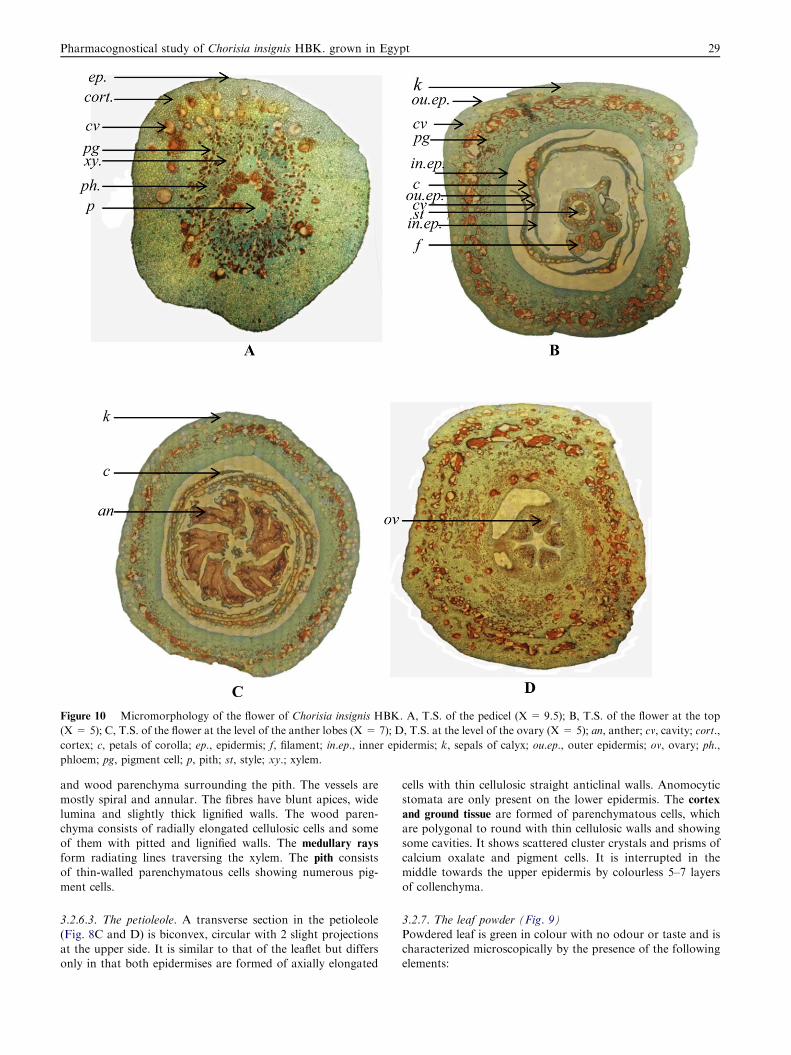
Figure 10 Micromorphology of the flower of Chorisia insignis HBK. A, T.S. of the pedicel (X = 9.5); B, T.S. of the flower at the top
(X = 5); C, T.S. of the flower at the level of the anther lobes (X = 7); D, T.S. at the level of the ovary (X = 5); an, anther; cv, cavity; cort.,
cortex; c, petals of corolla; ep., epidermis; f, filament; in.ep., inner epidermis; k, sepals of calyx; ou.ep., outer epidermis; ov, ovary; ph.,
phloem; pg, pigment cell; p, pith; st, style; xy.; xylem.
Pharmacognostical study of Chorisia insignis HBK. grown in Egypt 29
and wood parenchyma surrounding the pith. The vessels aremostly spiral and annular. The fibres have blunt apices, widelumina and slightly thick lignified walls. The wood paren-chyma consists of radially elongated cellulosic cells and some
of them with pitted and lignified walls. The medullary rays
form radiating lines traversing the xylem. The pith consistsof thin-walled parenchymatous cells showing numerous pig-
ment cells.
3.2.6.3. The petioleole. A transverse section in the petioleole
(Fig. 8C and D) is biconvex, circular with 2 slight projectionsat the upper side. It is similar to that of the leaflet but differsonly in that both epidermises are formed of axially elongated
cells with thin cellulosic straight anticlinal walls. Anomocyticstomata are only present on the lower epidermis. The cortex
and ground tissue are formed of parenchymatous cells, whichare polygonal to round with thin cellulosic walls and showing
some cavities. It shows scattered cluster crystals and prisms ofcalcium oxalate and pigment cells. It is interrupted in themiddle towards the upper epidermis by colourless 5–7 layers
of collenchyma.
3.2.7. The leaf powder (Fig. 9)Powdered leaf is green in colour with no odour or taste and ischaracterized microscopically by the presence of the followingelements:
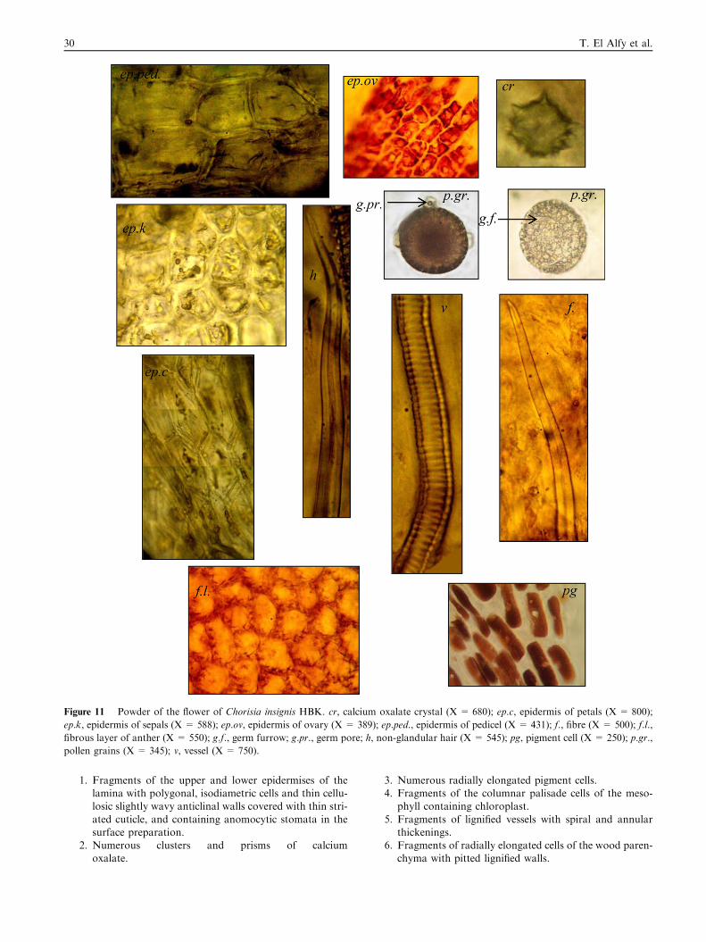
Figure 11 Powder of the flower of Chorisia insignis HBK. cr, calcium oxalate crystal (X = 680); ep.c, epidermis of petals (X = 800);
ep.k, epidermis of sepals (X = 588); ep.ov, epidermis of ovary (X = 389); ep.ped., epidermis of pedicel (X = 431); f., fibre (X = 500); f.l.,
fibrous layer of anther (X = 550); g.f., germ furrow; g.pr., germ pore; h, non-glandular hair (X = 545); pg, pigment cell (X = 250); p.gr.,
pollen grains (X = 345); v, vessel (X = 750).
30 T. El Alfy et al.
1. Fragments of the upper and lower epidermises of thelamina with polygonal, isodiametric cells and thin cellu-losic slightly wavy anticlinal walls covered with thin stri-
ated cuticle, and containing anomocytic stomata in thesurface preparation.
2. Numerous clusters and prisms of calciumoxalate.
3. Numerous radially elongated pigment cells.4. Fragments of the columnar palisade cells of the meso-
phyll containing chloroplast.
5. Fragments of lignified vessels with spiral and annularthickenings.
6. Fragments of radially elongated cells of the wood paren-chyma with pitted lignified walls.
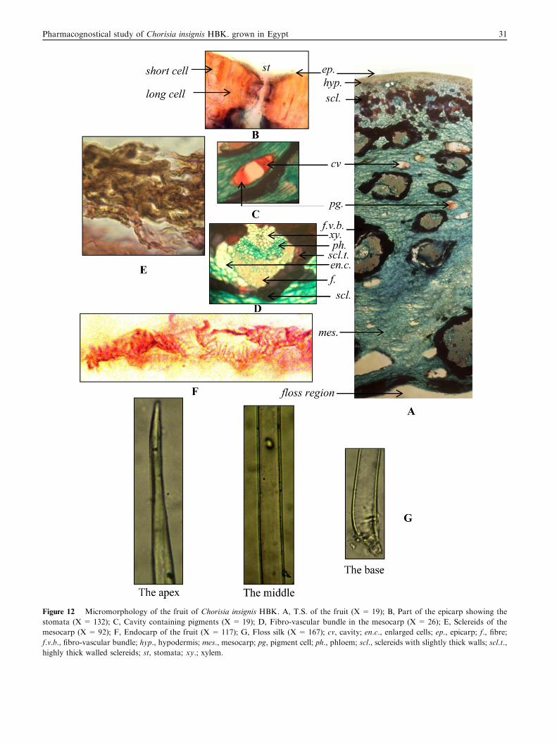
Figure 12 Micromorphology of the fruit of Chorisia insignis HBK. A, T.S. of the fruit (X = 19); B, Part of the epicarp showing the
stomata (X = 132); C, Cavity containing pigments (X = 19); D, Fibro-vascular bundle in the mesocarp (X = 26); E, Sclereids of the
mesocarp (X = 92); F, Endocarp of the fruit (X = 117); G, Floss silk (X = 167); cv, cavity; en.c., enlarged cells; ep., epicarp; f., fibre;
f.v.b., fibro-vascular bundle; hyp., hypodermis; mes., mesocarp; pg, pigment cell; ph., phloem; scl., sclereids with slightly thick walls; scl.t.,
highly thick walled sclereids; st, stomata; xy.; xylem.
Pharmacognostical study of Chorisia insignis HBK. grown in Egypt 31
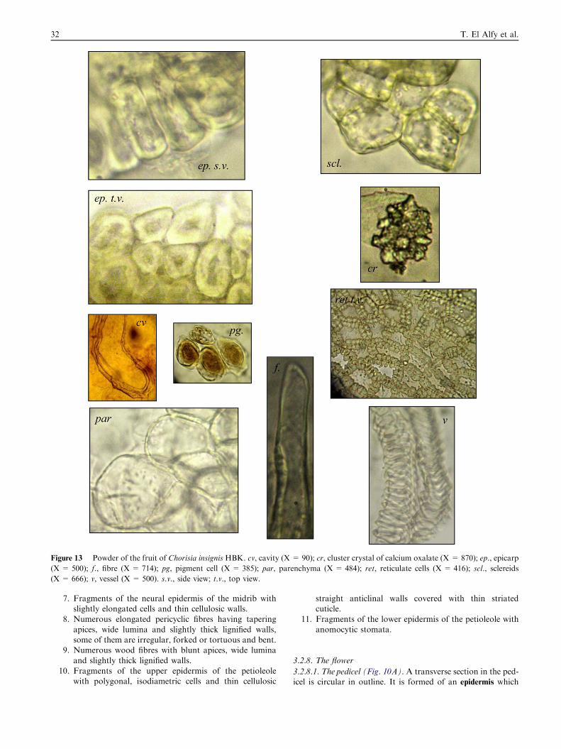
Figure 13 Powder of the fruit of Chorisia insignis HBK. cv, cavity (X = 90); cr, cluster crystal of calcium oxalate (X = 870); ep., epicarp
(X = 500); f., fibre (X = 714); pg, pigment cell (X = 385); par, parenchyma (X = 484); ret, reticulate cells (X = 416); scl., sclereids
(X = 666); v, vessel (X = 500). s.v., side view; t.v., top view.
32 T. El Alfy et al.
7. Fragments of the neural epidermis of the midrib withslightly elongated cells and thin cellulosic walls.
8. Numerous elongated pericyclic fibres having tapering
apices, wide lumina and slightly thick lignified walls,some of them are irregular, forked or tortuous and bent.
9. Numerous wood fibres with blunt apices, wide luminaand slightly thick lignified walls.
10. Fragments of the upper epidermis of the petioleolewith polygonal, isodiametric cells and thin cellulosic
straight anticlinal walls covered with thin striatedcuticle.
11. Fragments of the lower epidermis of the petioleole with
anomocytic stomata.
3.2.8. The flower
3.2.8.1. The pedicel (Fig. 10A). A transverse section in the ped-icel is circular in outline. It is formed of an epidermis which
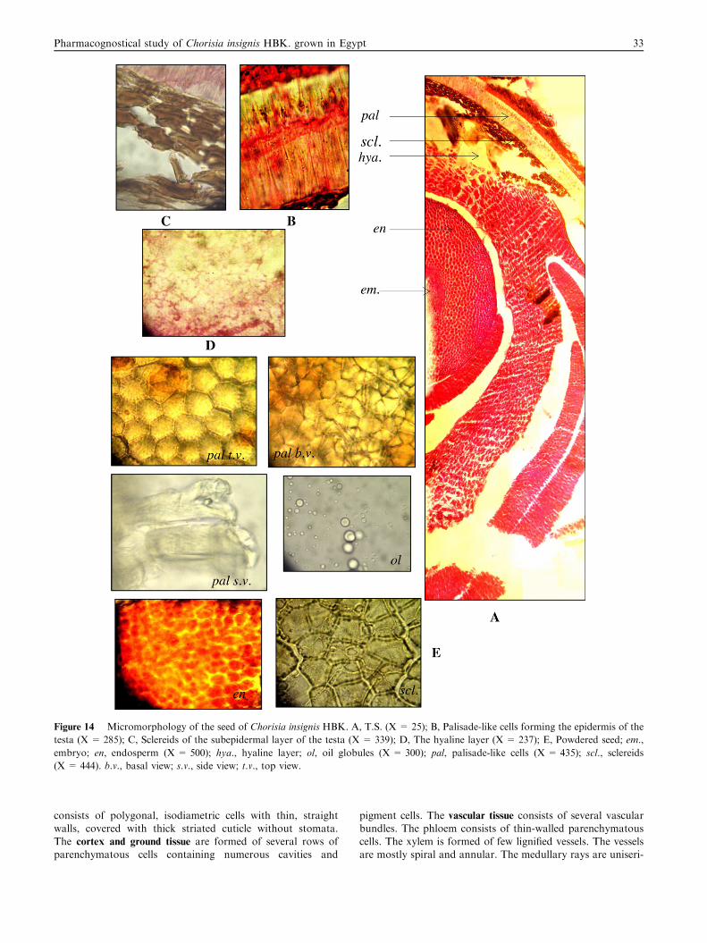
Figure 14 Micromorphology of the seed of Chorisia insignis HBK. A, T.S. (X = 25); B, Palisade-like cells forming the epidermis of the
testa (X = 285); C, Sclereids of the subepidermal layer of the testa (X = 339); D, The hyaline layer (X = 237); E, Powdered seed; em.,
embryo; en, endosperm (X = 500); hya., hyaline layer; ol, oil globules (X = 300); pal, palisade-like cells (X = 435); scl., sclereids
(X = 444). b.v., basal view; s.v., side view; t.v., top view.
Pharmacognostical study of Chorisia insignis HBK. grown in Egypt 33
consists of polygonal, isodiametric cells with thin, straightwalls, covered with thick striated cuticle without stomata.The cortex and ground tissue are formed of several rows ofparenchymatous cells containing numerous cavities and
pigment cells. The vascular tissue consists of several vascularbundles. The phloem consists of thin-walled parenchymatouscells. The xylem is formed of few lignified vessels. The vesselsare mostly spiral and annular. The medullary rays are uniseri-
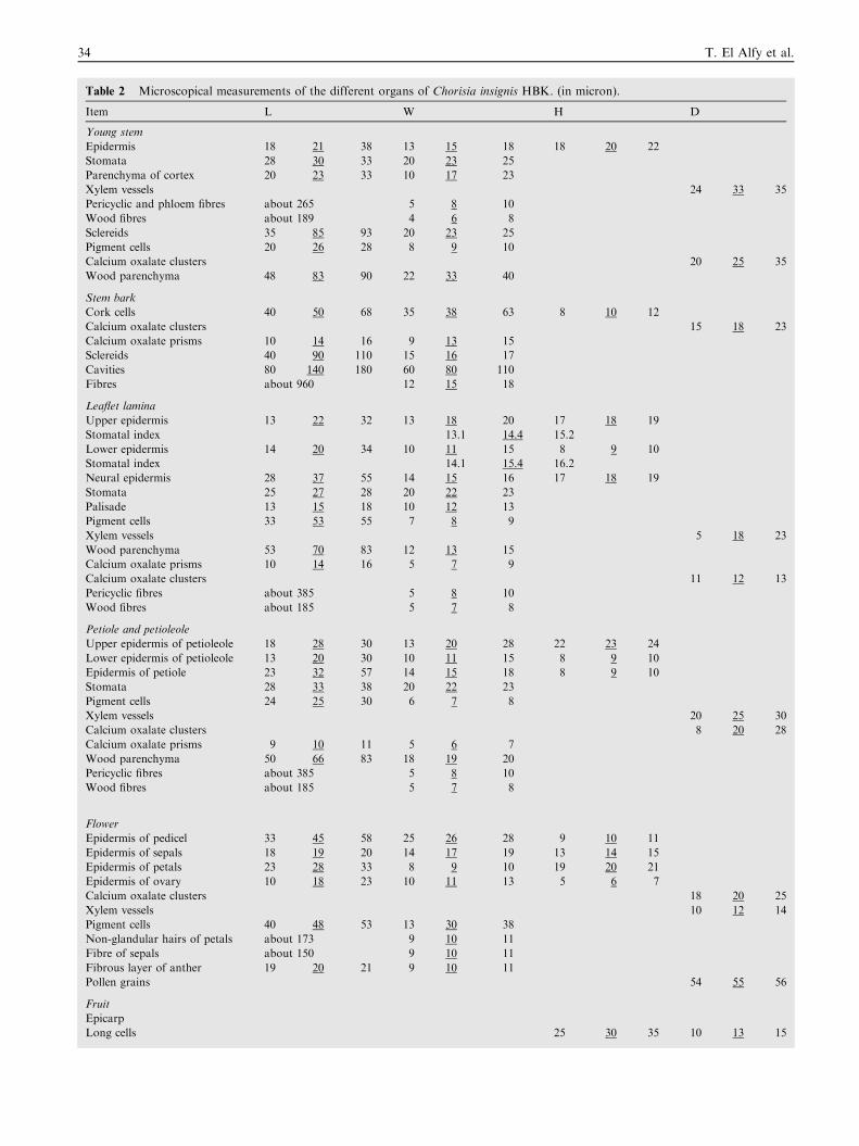
Table 2 Microscopical measurements of the different organs of Chorisia insignis HBK. (in micron).
Item L W H D
Young stem
Epidermis 18 21 38 13 15 18 18 20 22
Stomata 28 30 33 20 23 25
Parenchyma of cortex 20 23 33 10 17 23
Xylem vessels 24 33 35
Pericyclic and phloem fibres about 265 5 8 10
Wood fibres about 189 4 6 8
Sclereids 35 85 93 20 23 25
Pigment cells 20 26 28 8 9 10
Calcium oxalate clusters 20 25 35
Wood parenchyma 48 83 90 22 33 40
Stem bark
Cork cells 40 50 68 35 38 63 8 10 12
Calcium oxalate clusters 15 18 23
Calcium oxalate prisms 10 14 16 9 13 15
Sclereids 40 90 110 15 16 17
Cavities 80 140 180 60 80 110
Fibres about 960 12 15 18
Leaflet lamina
Upper epidermis 13 22 32 13 18 20 17 18 19
Stomatal index 13.1 14.4 15.2
Lower epidermis 14 20 34 10 11 15 8 9 10
Stomatal index 14.1 15.4 16.2
Neural epidermis 28 37 55 14 15 16 17 18 19
Stomata 25 27 28 20 22 23
Palisade 13 15 18 10 12 13
Pigment cells 33 53 55 7 8 9
Xylem vessels 5 18 23
Wood parenchyma 53 70 83 12 13 15
Calcium oxalate prisms 10 14 16 5 7 9
Calcium oxalate clusters 11 12 13
Pericyclic fibres about 385 5 8 10
Wood fibres about 185 5 7 8
Petiole and petioleole
Upper epidermis of petioleole 18 28 30 13 20 28 22 23 24
Lower epidermis of petioleole 13 20 30 10 11 15 8 9 10
Epidermis of petiole 23 32 57 14 15 18 8 9 10
Stomata 28 33 38 20 22 23
Pigment cells 24 25 30 6 7 8
Xylem vessels 20 25 30
Calcium oxalate clusters 8 20 28
Calcium oxalate prisms 9 10 11 5 6 7
Wood parenchyma 50 66 83 18 19 20
Pericyclic fibres about 385 5 8 10
Wood fibres about 185 5 7 8
Flower
Epidermis of pedicel 33 45 58 25 26 28 9 10 11
Epidermis of sepals 18 19 20 14 17 19 13 14 15
Epidermis of petals 23 28 33 8 9 10 19 20 21
Epidermis of ovary 10 18 23 10 11 13 5 6 7
Calcium oxalate clusters 18 20 25
Xylem vessels 10 12 14
Pigment cells 40 48 53 13 30 38
Non-glandular hairs of petals about 173 9 10 11
Fibre of sepals about 150 9 10 11
Fibrous layer of anther 19 20 21 9 10 11
Pollen grains 54 55 56
Fruit
Epicarp
Long cells 25 30 35 10 13 15
34 T. El Alfy et al.
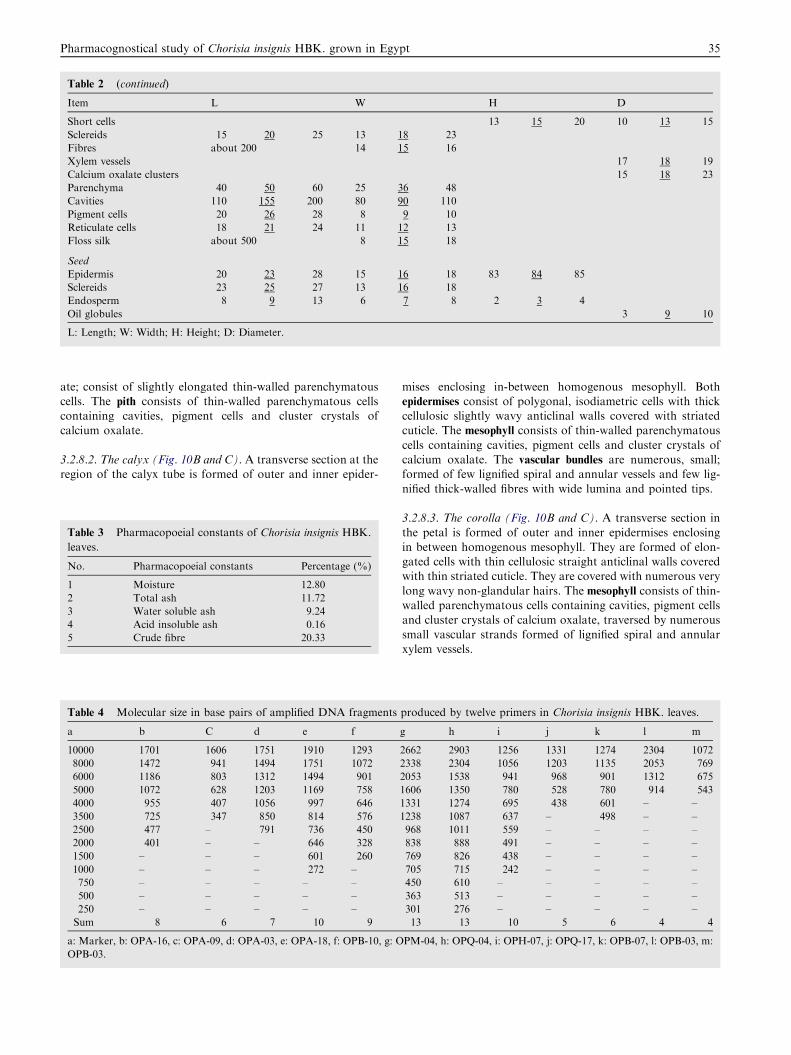
Table 2 (continued)
Item L W H D
Short cells 13 15 20 10 13 15
Sclereids 15 20 25 13 18 23
Fibres about 200 14 15 16
Xylem vessels 17 18 19
Calcium oxalate clusters 15 18 23
Parenchyma 40 50 60 25 36 48
Cavities 110 155 200 80 90 110
Pigment cells 20 26 28 8 9 10
Reticulate cells 18 21 24 11 12 13
Floss silk about 500 8 15 18
Seed
Epidermis 20 23 28 15 16 18 83 84 85
Sclereids 23 25 27 13 16 18
Endosperm 8 9 13 6 7 8 2 3 4
Oil globules 3 9 10
L: Length; W: Width; H: Height; D: Diameter.
Pharmacognostical study of Chorisia insignis HBK. grown in Egypt 35
ate; consist of slightly elongated thin-walled parenchymatouscells. The pith consists of thin-walled parenchymatous cellscontaining cavities, pigment cells and cluster crystals ofcalcium oxalate.
3.2.8.2. The calyx (Fig. 10B and C). A transverse section at theregion of the calyx tube is formed of outer and inner epider-
Table 3 Pharmacopoeial constants of Chorisia insignis HBK.
leaves.
No. Pharmacopoeial constants Percentage (%)
1 Moisture 12.80
2 Total ash 11.72
3 Water soluble ash 9.24
4 Acid insoluble ash 0.16
5 Crude fibre 20.33
Table 4 Molecular size in base pairs of amplified DNA fragments
a b C d e f
10000 1701 1606 1751 1910 1293
8000 1472 941 1494 1751 1072
6000 1186 803 1312 1494 901
5000 1072 628 1203 1169 758
4000 955 407 1056 997 646
3500 725 347 850 814 576
2500 477 – 791 736 450
2000 401 – – 646 328
1500 – – – 601 260
1000 – – – 272 –
750 – – – – –
500 – – – – –
250 – – – – –
Sum 8 6 7 10 9
a: Marker, b: OPA-16, c: OPA-09, d: OPA-03, e: OPA-18, f: OPB-10, g: O
OPB-03.
mises enclosing in-between homogenous mesophyll. Bothepidermises consist of polygonal, isodiametric cells with thickcellulosic slightly wavy anticlinal walls covered with striatedcuticle. The mesophyll consists of thin-walled parenchymatous
cells containing cavities, pigment cells and cluster crystals ofcalcium oxalate. The vascular bundles are numerous, small;formed of few lignified spiral and annular vessels and few lig-
nified thick-walled fibres with wide lumina and pointed tips.
3.2.8.3. The corolla (Fig. 10B and C). A transverse section in
the petal is formed of outer and inner epidermises enclosingin between homogenous mesophyll. They are formed of elon-gated cells with thin cellulosic straight anticlinal walls coveredwith thin striated cuticle. They are covered with numerous very
long wavy non-glandular hairs. The mesophyll consists of thin-walled parenchymatous cells containing cavities, pigment cellsand cluster crystals of calcium oxalate, traversed by numerous
small vascular strands formed of lignified spiral and annularxylem vessels.
produced by twelve primers in Chorisia insignis HBK. leaves.
g h i j k l m
2662 2903 1256 1331 1274 2304 1072
2338 2304 1056 1203 1135 2053 769
2053 1538 941 968 901 1312 675
1606 1350 780 528 780 914 543
1331 1274 695 438 601 – –
1238 1087 637 – 498 – –
968 1011 559 – – – –
838 888 491 – – – –
769 826 438 – – – –
705 715 242 – – – –
450 610 – – – – –
363 513 – – – – –
301 276 – – – – –
13 13 10 5 6 4 4
PM-04, h: OPQ-04, i: OPH-07, j: OPQ-17, k: OPB-07, l: OPB-03, m:
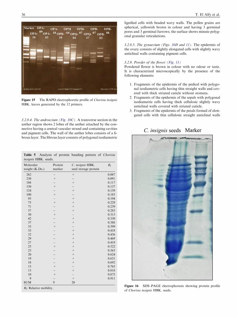
Figure 15 The RAPD electrophoretic profile of Chorisia insignis
HBK. leaves generated by the 12 primers.
36 T. El Alfy et al.
3.2.8.4. The androecium (Fig. 10C). A transverse section in theanther region shows 2 lobes of the anther attached by the con-nective having a central vascular strand and containing cavities
and pigment cells. The wall of the anther lobes consists of a fi-brous layer. The fibrous layer consists of polygonal isodiametric
Table 5 Analysis of protein banding pattern of Chorisia
insignis HBK. seeds.
Molecular
weight (K.Da.)
Protein
marker
C. insignis HBK.
seed storage protein
Rf
262 � + 0.087
250 + � 0.091
188 � + 0.117
150 + � 0.137
124 � + 0.159
100 + � 0.185
95 � + 0.194
75 + + 0.229
71 � + 0.239
57 � + 0.283
50 + � 0.313
42 � + 0.330
37 � + 0.388
35 + � 0.399
33 � + 0.418
32 � + 0.436
29 � + 0.469
27 � + 0.419
25 + + 0.522
23 � + 0.563
20 � + 0.624
19 � + 0.631
18 � + 0.692
15 + � 0.765
13 � + 0.818
10 + � 0.875
9 � + 0.911
SUM 9 20
Rf: Relative mobility.
lignified cells with beaded wavy walls. The pollen grains are
spherical, yellowish brown in colour and having 3 germinalpores and 3 germinal furrows; the surface shows minute polyg-onal granular reticulations.
3.2.8.5. The gynaecium (Figs. 10D and 11). The epidermis ofthe ovary consists of slightly elongated cells with slightly wavyanticlinal walls containing pigment cells.
3.2.9. Powder of the flower (Fig. 11)Powdered flower is brown in colour with no odour or taste.
It is characterized microscopically by the presence of thefollowing elements:
1. Fragments of the epidermis of the pedicel with polygo-nal isodiametric cells having thin straight walls and cov-ered with thick striated cuticle without stomata.
2. Fragments of the epidermis of the sepals with polygonalisodiametric cells having thick cellulosic slightly wavyanticlinal walls covered with striated cuticle.
3. Fragments of the epidermis of the petals formed of elon-
gated cells with thin cellulosic straight anticlinal walls
Figure 16 SDS–PAGE electrophoresis showing protein profile
of Chorisia insignis HBK. seeds.
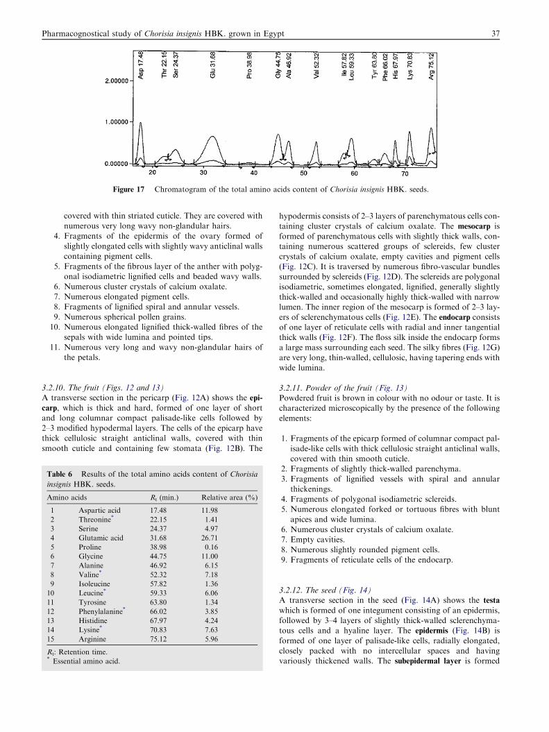
Figure 17 Chromatogram of the total amino acids content of Chorisia insignis HBK. seeds.
Pharmacognostical study of Chorisia insignis HBK. grown in Egypt 37
covered with thin striated cuticle. They are covered withnumerous very long wavy non-glandular hairs.
4. Fragments of the epidermis of the ovary formed of
slightly elongated cells with slightly wavy anticlinal wallscontaining pigment cells.
5. Fragments of the fibrous layer of the anther with polyg-onal isodiametric lignified cells and beaded wavy walls.
6. Numerous cluster crystals of calcium oxalate.7. Numerous elongated pigment cells.8. Fragments of lignified spiral and annular vessels.
9. Numerous spherical pollen grains.10. Numerous elongated lignified thick-walled fibres of the
sepals with wide lumina and pointed tips.
11. Numerous very long and wavy non-glandular hairs ofthe petals.
3.2.10. The fruit (Figs. 12 and 13)A transverse section in the pericarp (Fig. 12A) shows the epi-
carp, which is thick and hard, formed of one layer of shortand long columnar compact palisade-like cells followed by2–3 modified hypodermal layers. The cells of the epicarp havethick cellulosic straight anticlinal walls, covered with thin
smooth cuticle and containing few stomata (Fig. 12B). The
Table 6 Results of the total amino acids content of Chorisia
insignis HBK. seeds.
Amino acids Rt (min.) Relative area (%)
1 Aspartic acid 17.48 11.98
2 Threonine* 22.15 1.41
3 Serine 24.37 4.97
4 Glutamic acid 31.68 26.71
5 Proline 38.98 0.16
6 Glycine 44.75 11.00
7 Alanine 46.92 6.15
8 Valine* 52.32 7.18
9 Isoleucine 57.82 1.36
10 Leucine* 59.33 6.06
11 Tyrosine 63.80 1.34
12 Phenylalanine* 66.02 3.85
13 Histidine 67.97 4.24
14 Lysine* 70.83 7.63
15 Arginine 75.12 5.96
Rt: Retention time.* Essential amino acid.
hypodermis consists of 2–3 layers of parenchymatous cells con-taining cluster crystals of calcium oxalate. The mesocarp isformed of parenchymatous cells with slightly thick walls, con-taining numerous scattered groups of sclereids, few cluster
crystals of calcium oxalate, empty cavities and pigment cells(Fig. 12C). It is traversed by numerous fibro-vascular bundlessurrounded by sclereids (Fig. 12D). The sclereids are polygonal
isodiametric, sometimes elongated, lignified, generally slightlythick-walled and occasionally highly thick-walled with narrowlumen. The inner region of the mesocarp is formed of 2–3 lay-
ers of sclerenchymatous cells (Fig. 12E). The endocarp consistsof one layer of reticulate cells with radial and inner tangentialthick walls (Fig. 12F). The floss silk inside the endocarp formsa large mass surrounding each seed. The silky fibres (Fig. 12G)
are very long, thin-walled, cellulosic, having tapering ends withwide lumina.
3.2.11. Powder of the fruit (Fig. 13)Powdered fruit is brown in colour with no odour or taste. It ischaracterized microscopically by the presence of the following
elements:
1. Fragments of the epicarp formed of columnar compact pal-
isade-like cells with thick cellulosic straight anticlinal walls,covered with thin smooth cuticle.
2. Fragments of slightly thick-walled parenchyma.
3. Fragments of lignified vessels with spiral and annularthickenings.
4. Fragments of polygonal isodiametric sclereids.5. Numerous elongated forked or tortuous fibres with blunt
apices and wide lumina.6. Numerous cluster crystals of calcium oxalate.7. Empty cavities.
8. Numerous slightly rounded pigment cells.9. Fragments of reticulate cells of the endocarp.
3.2.12. The seed (Fig. 14)A transverse section in the seed (Fig. 14A) shows the testa
which is formed of one integument consisting of an epidermis,followed by 3–4 layers of slightly thick-walled sclerenchyma-tous cells and a hyaline layer. The epidermis (Fig. 14B) is
formed of one layer of palisade-like cells, radially elongated,closely packed with no intercellular spaces and havingvariously thickened walls. The subepidermal layer is formed
38 T. El Alfy et al.
of 3–4 layers of sclerenchymatous cells (Fig. 14C). The sclere-
ids are polygonal isodiametric, sometimes elongated havinglignified thick walls. The hyaline layer (Fig. 14D) is formedof collapsed tangentially elongated parenchymatous cells withthin cellulosic walls. The endosperm is mucilaginous, formed of
polygonal cells with thin cellulosic walls containing oil glob-ules. The embryo is formed of polygonal cells with thin cellu-losic walls. It is curved and consists of two cotyledons.
3.2.13. Powder of the seed (Fig. 14E)Powdered seed is brown in colour with no odour or taste. It is
characterized microscopically by the presence of the followingelements:
1. Fragments of radially elongated palisade-like cells, closelypacked with no intercellular spaces, having variously thick-ened walls.
2. Fragments of polygonal cells of the endosperm with thincellulosic walls.
3. Sclereids with regular thickening.4. Numerous oil globules.
Microscopical measurements of the different organs of C.insignis HBK. are listed in Table 2.
3.3. Determination of certain pharmacopoeial constants of the
leaves
The results are compiled in Table 3. These constants could beused as criteria for the identity and purity of C. insignis HBK.
leaves.
3.4. DNA fingerprinting
Polymerase chain reaction (PCR) with 12 decamer primersproduced a total of 95 fragments as shown in Table 4 andFig. 15. The molecular size of the produced fragments revealed
the presence of a wide range of sequences. The maximum sizewas 2903 base pairs after using primer OPQ-04 which pro-duced 13 rapid amplified polymorphic DNA (RAPD) frag-
ments. However, the minimum molecular size was 242 basepairs when OPH-07 was used.
3.5. Protein banding analysis
Protein electrophoresis was carried out using SDS–PAGE (so-dium dodecyl sulphate polyacrylamide gel electrophoresis)
technique to monitor the seed storage protein expressed bythe active genes of the whole genomic DNA of C. insignisHBK.
The protein banding profile of the seed storage protein isdictated in Table 5. The data analyzed by gel documentationsystem revealed the presence of 20 sharp protein bands as pic-
tured in Fig. 16. These observed bands had a wide range ofmolecular weight ranging from 262 to 9 k Dalton. The relativemobilities of these bands (Rf) ranged from 0.087 to 0.911.
3.6. Determination of the total protein content
The percentage of the total protein in C. insignis HBK. seeds
was found to be 26.83%, which explains the possible use ofthe plant as a protein source for animals.
3.7. Determination of the amino acids content
The amino acids content of C. insignis HBK. seeds is repre-sented in Fig. 17 and illustrated in Table 6. Analysis of the
amino acids content of C. insignis HBK. seeds revealed theidentification of 15 amino acids consisting of 6 essential aminoacids and 9 non-essential amino acids. The major essential
amino acids were lysine (7.63%) and valine (7.18%) followedby leucine (6.06%) and phenylalanine (3.85%) then threonine(1.41%) and isoleucine (1.36%), while the main non-essentialamino acid was glutamic acid (26.71%) followed by aspartic
acid (11.98%) and glycine (11.00%) then alanine (6.15%),arginine (5.96%), serine (4.97%), histidine (4.24%), tyrosine(1.34%) and finally proline (0.16%).
4. Discussion and conclusion
In the present study, macro and micromorphological featuresof C. insignis HBK. were investigated. This study showedbotanical characteristics that can differentiate the plant from
other Bombacaceae plants.Certain pharmacopoeial constants were carried out on the
powdered leaves which could be used as criteria for the iden-
tity and purity of the plant. The genomic DNA of the planthas been subjected to random amplified analysis. Twelvedecamer oligonucleotide primers had induced successiveamplifications with a wide range of molecular sizes. The anal-
ysis of the amplified fragments generated by RAPD reactionsrevealed that the biotypes of C. insignis HBK. produced dif-ferent molecular patterns. However, the primers OPM-04 and
OPQ-04 can be used for the identification of C. insignis HBK.at the genetic level since they generate fragments with widemolecular size.
In addition, qualitative estimation of protein profiles ofC. insignis HBK. seed storage protein was analyzed usingSDS-gel electrophoresis. The electrophoretic protein bandsof an organism are the mirror pattern of the transcriptional
events during the life cycle of that organism. Protein profileis used as an efficient tool for identification as well asdifferentiation between species as previously reported by
Higazy.13 The present finding revealed the presence of highmolecular weight long chain polypeptides. However, thelow molecular weight proteins represented by five intense
protein bands are believed to be classified as inhibitoryenzymes as reported by Farr and Cohen-Fix14 andTinker-Kulberg and Morgan.15
Moreover, the percentage of the total protein in C. insignisHBK. seeds was found to be 26.83% as determined by micro-Kjeldahl method. Analysis of the amino acid content of C.insignis HBK. seeds revealed the identification of 15 amino
acids.In conclusion, these results can be used for the identifica-
tion of C. insignis HBK. and its differentiation from other re-
lated species.
References
1. Huxley A. Dictionary of Gardening. The New Royal Horticultural
Society; 1992; p. 609.
2. Bailey LH. Hortus 3rd: A Concise Dictionary of Plants Cultivated
in the United States and Canada. Staff of the L.H. Bailey
Hortorium. Cornell University; 1976; p. 266.
Pharmacognostical study of Chorisia insignis HBK. grown in Egypt 39
3. Barwick M. A World Encyclopedia Guide. Tropical and Subtrop-
ical Trees; 2004; p. 110.
4. El Alfy TS, El Sawi SA, Sleem A, Moawad DM. Investigation of
flavonoidal content and biological activities of Chorisia insignis
HBK. leaves. Aust J B Appl Sci 2010;4(6):1334–48.
5. Egyptian Pharmacopoeia. Cairo: General Organization for Gov-
ernment Printing Affairs, 2005.
6. Vasudevan H. DNA fingerprinting in the standardization of herbs
and nutraceuticals; 2004 Aug. Available from: <http://
www.scq.ubc.ca/?p=286>.
7. Giannino DF, Manfioletti G, Schneider C. The CTAB-DNA
precipitation method: a common many scale preparation of
template DNA from phagmede, phages or plasmids suitable for
squenching. Res Rep 1989;7:514–9.
8. Williams JGK, Kubelik AR, Livak KJ, Rafalski JA, Tingey SV.
DNA polymorphisms amplified by arbitrary primers are useful as
genetic markers. Nucleic Acids Res 1990;18:6531–5.
9. Ornstein I. Discelectrophoresis background and theory. Ann N Y
Acad Sci 1984;121:321–49.
10. Laemmli UK. Cleavage of structural proteins during the assem-
bly of the head of bacteriophage T4. Nature 1970;227(259):
680–5.
11. Pearson D. The Chemical Analysis of Food. 6th ed. Lon-
don: Churchill Ltd.; 1970.
12. Block RJ, Durrum EL, Zweig G. Annual of paper chromatogra-
phy and paper electrophoresis. 2nd ed. Academic; 1958.
13. Higazy HY. Cytogenetic and molecular reevaluation of the
relationships of species belonging to the two genera, Brassica
and Sinapsis. Cytologia 2005;70:447–54.
14. Farr KA, Cohen-Fix O. The metaphase to anaphase transition. J
Biochem 1999;263:1–14.
15. Tinker-Kulberg RL, Morgan DO. Pd1 and Esp1 control both
anaphase and mitotic exit in normal cells after DNA damage.
Genes Dev 1999;13(15):1936–49.


