Embed Size (px)
Citation preview
© 2014 Tsukamoto et al. This work is published by Dove Medical Press Limited, and licensed under Creative Commons Attribution – Non Commercial (unported, v3.0) License. The full terms of the License are available at http://creativecommons.org/licenses/by-nc/3.0/. Non-commercial uses of the work are permitted without any further
permission from Dove Medical Press Limited, provided the work is properly attributed. Permissions beyond the scope of the License are administered by Dove Medical Press Limited. Information on how to request permission may be found at: http://www.dovepress.com/permissions.php
Drug Design, Development and Therapy 2014:8 1955–1964
Drug Design, Development and Therapy Dovepress
submit your manuscript | www.dovepress.com
Dovepress 1955
O r i g i n a l r e s e a r c h
open access to scientific and medical research
Open access Full Text article
http://dx.doi.org/10.2147/DDDT.S60247
intestinal absorption, organ distribution, and urinary excretion of the rare sugar D-psicose
ikuko Tsukamoto1,*akram hossain2,3,*Fuminori Yamaguchi2
Yuko hirata2
Youyi Dong2
Kazuyo Kamitori2
li sui2
Machiko nonaka2
Masaki Ueno4
Kazuyuki nishimoto5
hirofumi suda5
Kenji Morimoto6
Tsuyoshi shimonishi7,†
Madoka saito8
Tao song9
ryoji Konishi1
Masaaki Tokuda2
1Department of Pharmaco-Bio-informatics, Faculty of Medicine, Kagawa University, Miki, Kagawa, Japan; 2Department of cell Physiology, Faculty of Medicine, Kagawa University, Kagawa, Japan; 3Matsutani chemical industry co, ltd, itami, Japan; 4Department of inflammation Pathology, Faculty of Medicine, Kagawa University, Kagawa, Japan; 5Division of radioisotope research, life science research center, Kagawa University, Kagawa, Japan; 6rare sugar research center, Kagawa University, Kagawa, Japan; 7iZUMOring llc, Miki, Kita, Kagawa, Japan; 8Department of Pharmacy, Okayama University hospital, Okayama, Japan; 9The First affiliated hospital, china Medical University, shenyang, People’s republic of china
*These authors contributed equally to this work
†Tsuyoshi shimonishi has passed away
Background: The purpose of this study was to evaluate intestinal absorption, organ distribution,
and urinary elimination of the rare sugar D-psicose, a 3-carbon stereoisomer of D-fructose
that is currently being investigated and which has been found to be strongly effective against
hyperglycemia and hyperlipidemia.
Methods: This study was performed using radioactive D-psicose, which was synthesized
enzymatically from radioactive D-allose. Concentrations in whole blood, urine, and organs were
measured at different time points until 2 hours after both oral and intravenous administrations and
7 days after a single oral administration (100 mg/kg body weight) to Wistar rats. Autoradiography
was also performed by injecting 100 mg/kg body weight of 14C-labeled D-psicose or glucose
intravenously to C3H mice.
Results: Following oral administration, D-psicose easily moved to blood. The maximum blood
concentration (48.5±15.6 µg/g) was observed at 1 hour. Excretion to urine was 20% within
1 hour and 33% within 2 hours. Accumulation to organs was detected only in the liver. Following
intravenous administration, blood concentration was decreased with the half-life=57 minutes,
and the excretion to urine was up to almost 50% within 1 hour. Similarly to the results obtained
with oral administration, accumulation to organs was detected only in the liver. Seven days after
the single-dose oral administration, the remaining amounts in the whole body were less than 1%.
Autoradiography of mice showed results similar to those in rats. High signals of 14C-labeled
D-psicose were observed in liver, kidney, and bladder. Interestingly, no accumulation of D-psicose
was observed in the brain.
Conclusion: D-psicose was absorbed well after oral administration and eliminated rapidly
after both oral and intravenous administrations, with short duration of action. The study pro-
vides valuable pharmacokinetic data for further drug development of D-psicose. Because the
findings were mainly based on animal study, it is necessary to implement human trials to study
the metabolism pathway, which would give an important guide for human intake and food
application of D-psicose.
Keywords: 14C-labeled D-psicose, organ accumulation, pharmacokinetics, autoradiography
IntroductionRare sugar D-psicose (also called D-allulose), whose unique effect against hyperglyce-
mia and obesity which recently excited the people, has received intense attention from
researchers as well as food companies as they aim to produce a variety of D-psicose-
containing, low-calorie foodstuffs for those with diabetic or obesity tendency, as a
means of reducing the present high prevalence of these conditions. Rare sugars were
defined as monosaccharides and their derivatives at the first international symposium
of the International Society of Rare Sugars (ISRS), 2002, held in Takamatsu, Kagawa,
correspondence: akram hossain Department of cell Physiology, Faculty of Medicine, Kagawa University, 1750-1 ikenobe, Miki, Kita, Kagawa, 761-0793 Japan Tel +81 87 891 2095 Fax +81 87 891 2096 email [email protected]
Drug Design, Development and Therapy 2014:8submit your manuscript | www.dovepress.com
Dovepress
Dovepress
1956
Tsukamoto et al
Japan.1 Rare sugars are present in small quantities in com-
mercial mixtures of D-glucose and D-fructose obtained from
hydrolysis of sucrose or isomerization of D-glucose.2
D-psicose, an epimer of D-fructose isomerized at the
C-3 position, is a rare ketohexose that can be derived in
small amounts from wheat,3 itea plants,4 processed cane, and
beet molasses. A trace amount of D-psicose is also detected
in human urine5 and on the surface of human skin.6 Due to
its rarity, the biological functions of D-psicose have not yet
been sufficiently explored, but unique, innovative methods
of production through Izumoring7,8 has enabled a number of
investigations.9–18 It has been found experimentally to have
dramatic effects against obesity and type 2 diabetes mellitus
(T2DM) in normal11,14 as well as T2DM16–18 rat models,
and clinically against hyperglycemia and hyperlipidemia
in healthy13 and borderline diabetic humans,15 hence its
use as a sugar substitute for obese and diabetic patients.
As the mechanism of controlling high glucose levels, the
potency of absorption of D-psicose over D-glucose from
the intestine,19 as well as inhibition of activities of enzymes
for the digestion of polysaccharides, such as glucoamylase
and maltase, have been mentioned as possible reasons.14
D-psicose has also been shown to inhibit hepatic fatty
acid synthase20 by the mechanism of controlling adipose
tissue deposition, followed by decreased body weight gain
in comparison to D-glucose. In addition to the antihyper-
glycemic and antilipidemic effects of D-psicose, its zero-
calorie credit and 70% relative sweetness have led food
companies to prepare various foodstuffs using D-psicose
as a substitute for sugar. Mainly to substitute sucrose by
D-psicose for food production, due to D-psicose’s high
solubility,21 smooth texture22 and low glycemic response23
in comparison to sucrose,21 given that consumption of
sugar-sweetened beverages has been increasingly associ-
ated with negative health outcomes such as overweight,
obesity, T2DM, and metabolic syndrome.24 Based largely
on these associations, many researchers and health care
practitioners have proposed that non-caloric, high-intensity
sweeteners provide a beneficial alternative to sugar in foods
and beverages.25 D-psicose was approved as “Generally
Recognized As Safe” (GRAS) by the US Food and Drug
Administration (FDA) in August 2011 (GRN No 400), and
it is allowed to be used as an ingredient in a range of foods
and dietary supplements.26
In almost all the above-mentioned physiological stud-
ies, different doses of D-psicose were used to decide a
nonhazardous therapeutic dose, since the pharmacokinetics
are yet to be fully known, although they have been described
to a small extent in the literature by Whistler et al.27 Moreover,
for approval for food production in the industry as a safe food
additive, the Japanese Ministry of Health, Labor and Welfare
requires assessments of the degradability of D-psicose by
fermentation and digestion/absorption studies. Whistler
et al27 reported the metabolism of D-psicose in rats using
D-[U-14C] psicose and measured the contents in urine, feces,
glycogen, exhaled carbon dioxide, and carcasses 6–72 hours
after oral and intravenous administrations. Research studies
and human trials11,13,15,17,18 have shown high safety and efficacy
of D-psicose against obesity and T2DM, but in addition to
oral administration, injection is also needed for emergency
purposes, therefore an oral formulation with therapeutic
efficacy comparable with that of injection is important. As
such, a study of the pharmacokinetic parameters of D-psicose
is needed.
In this study, the pharmacokinetic parameters of D-psicose
was investigated after oral and intravenous administrations
in rats. Additionally, data on human studies will be accepted
more widely and rapidly to the researchers. However, data
from this animal study, as well as the ongoing double-blind
human study, which is currently under strict supervision, will
highlight the significance of D-psicose use.
Materials and methodsMaterialsD-[1-14C] allose (37.2 mM 3% ethanol solution,
1.96 GBq/mmol) and D-[1-14C] glucose (0.654 mM 0.3%
EtOH solution, 11.3 GBq/mmol) were obtained from
Amersham Biosciences UK Ltd (Little Chalfont, Bucking-
hamshire, UK). Cold D-psicose, D-allose, and L-rhamnose
isomerase immobilized beads were supplied by the Rare
Sugar Research Center of Kagawa University (Miki, Kagawa,
Japan). Carbo-Sorb® E and Permafluor® E+ were from
PerkinElmer, Inc., (Waltham, MA, USA).
animalsThis study was approved by the Animal Care and Use
Committee of Kagawa University (permission number:
2013–45) and the experiment was conducted in accor-
dance with the Institutional Guidelines and Rules for
Care and Use of Laboratory Animals. Male Wistar rats
(n=30), weighing 280–320 g, and male C3H mice (n=10),
weighing 28–32 g, were purchased from Charles River
Laboratories Japan, INC. (Yokohama, Japan) and housed
for at least 7 days in a clean room in the animal facility
of the Faculty of Medicine, Kagawa University. The rats
were given free access to commercial chow and water,
and were maintained according to the Kagawa University
animal guidelines.
Drug Design, Development and Therapy 2014:8 submit your manuscript | www.dovepress.com
Dovepress
Dovepress
1957
Pharmacokinetics of rare sugar D-psicose
Biodistribution experimentAll the rats used for both oral and intravenous D-psicose
administration were fasted for 24 hours before surgery;
on the experiment day, they were shifted to the radio isotope
facility because of the use of radioactive D-allose and D-glu-
cose. For intravenous administration, rats were anesthetized
with an intraperitoneal injection of pentobarbital sodium
(1%), and a polyethylene catheter (Intramedic™ PE-50; Clay
Adams, Parsippany, NJ, USA) was inserted into the femoral
vein for D-psicose or vehicle administration. Anesthesia
was continued until the end of the 120-minute experimental
period, with further administration of small doses of diluted
pentobarbital when needed. Oral administration was per-
formed in awake rats by gavage using a metallic tube directly
into the stomach. Approximately 0.6 mL psicose solution
(30 mg, 120 kBq) was administered (dose: 100 mg/kg) in
both oral and intravenous administrations. Urine was col-
lected from the urinary bladder by penis ligation.
The rats were sacrificed 10, 30, 60, and 120 minutes after
administration. The weights of whole brain, thymus, lung,
liver, heart, spleen, and kidney were measured. The amount
of total urine in the urinary bladder was also measured. The
contents in gastrointestinal (GI) organs (stomach, small
intestine, large intestine, and cecum) were collected and
homogenized.
sample oxidizer analysisFrom the collected specimens, a small quantity (0.1–0.2 g)
of blood, skin, and muscle was weighed and then burned in a
sample oxidizer (model 307; PerkinElmer). 14C was recovered
as CO2 with a CO
2 absorber (Carbo-Sorb E). The radioactivity
in the absorber was measured by a liquid scintillation coun-
ter (Aloca LSC-5100; Hitachi Aloka Medical, Ltd., Tokyo,
Japan) with a scintillator (Permafluor E+). The recovery by the
sample oxidizer, estimated by comparing the count of burned
and unburned samples, was 97.3%±2.8%. In oral administra-
tion, the remaining amounts of administered dose 7 days after
a single dose (100 mg/kg) were also measured.
autoradiographyAfter fasting for 24 hours, mice were injected via tail vein
with 20 kBq (100 mg/kg body weight) of 14C-labeled psi-
cose or 14C-labeled glucose dissolved in saline (0.2 mL).
Thirty minutes after the injection, mice were anesthetized
with diethyl ether and perfused transcardially with 0.01 M
phosphate-buffered saline after cutting of the right auricle.
The whole bodies of the animals were immersed in dry ice/
acetone and kept in a deep freezer at −50°C. Whole-body
frozen sections (40 µm) from the sagittal plane immersed in
8% carboxymethylcellulose compound were prepared with
an autocryotome (NA-500F; Nakagawa Seisakusho, Tokyo,
Japan), dried at −20°C, then exposed to an imaging plate
(BAS-IP MS2040; FUJIFILM Holdings Corporation, Tokyo,
Japan) for 8 weeks.
statisticsAll experiments were performed at least three times. Mean
values and standard error of the means were shown. Statistical
differences between groups were analyzed by Student’s t-test.
A P-value less than 0.05 was considered to be statistically
significant.
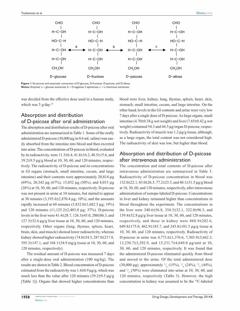
Resultssynthesis of 14c-labeled D-psicosePrior to synthesis of the radioactive compound, the
experimental condition was evaluated using cold material.
Hot D-allose solution was diluted with the same amount
of water (18.6 mM in 1.5% EtOH solution), and 400 µL
of the solution was incubated with L-rhamnose isomerase
immobilized beads (1 mm in diameter) at 37°C for
72 hours. L-rhamnose isomerase catalyzes the isomer-
ization between D-allose and D-psicose (enzyme “c” in
Figure 1). The properties of enzyme immobilized beads
have been described previously.28 Under these conditions,
we confirmed that more than 50% of D-allose was converted
to D-psicose. Synthesized D-psicose was separated by
high-performance liquid chromatography (HPLC) with a
Hitachi GL-C611, Gel Pack column with GL-C600 guard
column operated by an L-6200 Intelligent Pump, L-2350
Column Oven, L-2490 radioisotope detector, and D-2500
Chromato-integrator (all manufactured by Hitachi Aloka
Medical, Ltd., Tokyo, Japan) (sample volume 400 µL;
mobile phase, distilled water; flow rate 1.0 mL/min; column
temperature 60°C). The eluent around the retention time of
D-psicose (from 27 to 35 minutes) was collected, concen-
trated, and separated again with HPLC. Repeating these
processes three times, we succeeded in removing the con-
tamination of D-allose, and confirmed the purity to be over
99% by HPLC. Since 10.9 MBq of D-psicose was obtained,
the yield=20% from the starting material of 54.1 MBq of
D- allose. The specific radioactivity of 14C-labeled D- psicose
was assumed to be the same as that of the starting material
of 14C-labeled D-allose, ie, 1.96 GBq/mmol.
For the animal experiment, we prepared a D-psicose
solution (containing 50 mg and 200 kBq/mL saline for rats,
15 mg and 100 kBq/mL for mice) by mixing the synthesized
hot and cold compounds. The solution was stored at 5°C until
use. The doses for rats and mice were both 100 mg/kg, which
Drug Design, Development and Therapy 2014:8submit your manuscript | www.dovepress.com
Dovepress
Dovepress
1958
Tsukamoto et al
was decided from the effective dose used in a human study,
which was 5 g/day.14
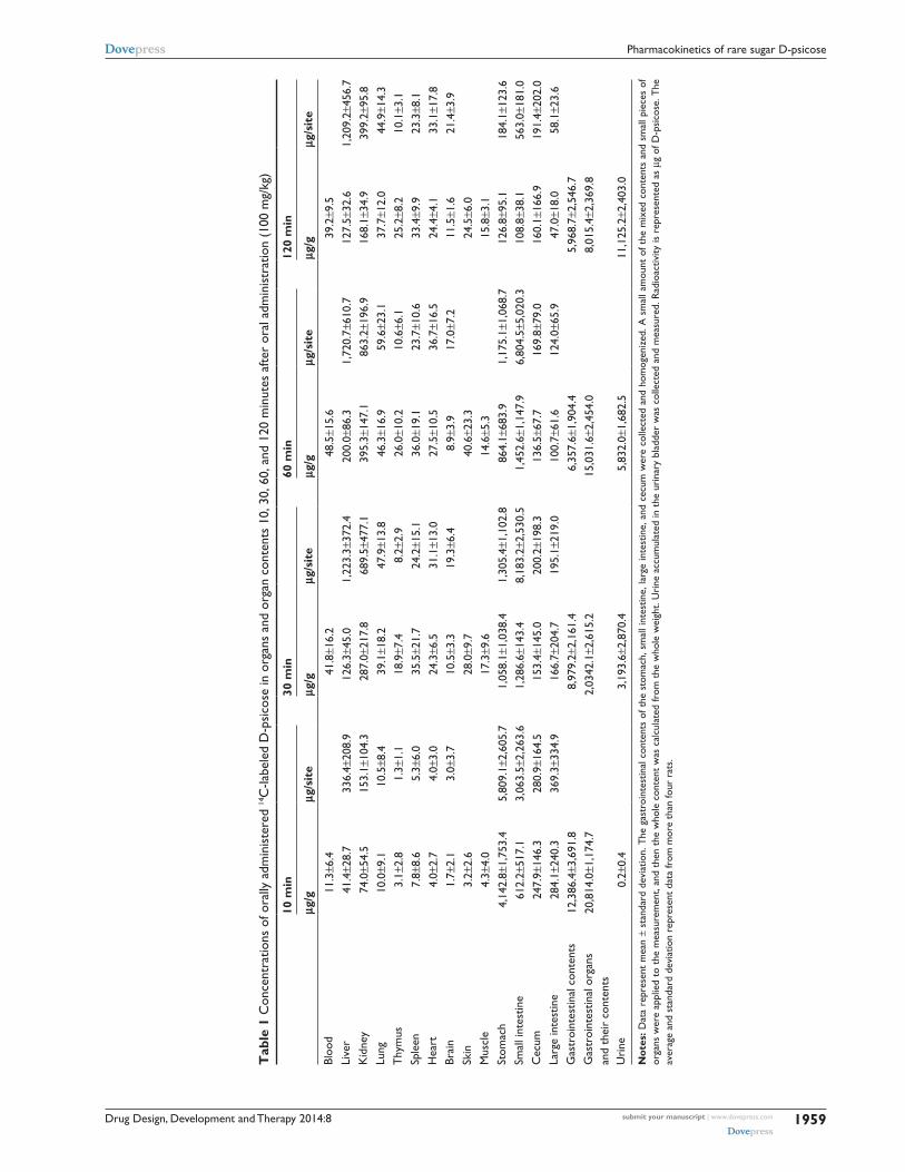
absorption and distribution of D-psicose after oral administrationThe absorption and distribution results of D-psicose after oral
administration are summarized in Table 1. Some of the orally
administered D-psicose (30,000 µg in 0.6 mL saline) was eas-
ily absorbed from the intestine into blood and then excreted
into urine. The concentrations of D-psicose in blood, evaluated
by its radioactivity, were 11.3±6.4, 41.8±16.2, 48.5±15.6, and
39.2±9.5 µg/g blood at 10, 30, 60, and 120 minutes, respec-
tively. The radioactivity of D-psicose and its concentrations
in GI organs (stomach, small intestine, cecum, and large
intestine) and their contents were approximately 20,814 µg
(69%), 20,342 µg (67%), 15,032 µg (50%), and 8,015 µg
(26%) at 10, 30, 60, and 120 minutes, respectively. D-psicose
was not present in urine at 10 minutes, but started to appear
at 30 minutes (3,193.6±2,870.4 µg; 10%), and the amounts
rapidly increased at 60 minutes (5,832.0±1,682.5 µg; 19%)
and 120 minutes (11,125.2±2,403.0 µg; 37%). D-psicose
levels in the liver were 41.4±28.7, 126.3±45.0, 200±86.3, and
127.5±32.6 µg/g liver tissue at 10, 30, 60, and 120 minutes,
respectively. Other organs (lung, thymus, spleen, heart,
brain, skin, and muscle) showed lower radioactivity, whereas
kidney showed higher radioactivity (74.0±54.5, 287.0±217.8,
395.3±147.1, and 168.1±34.9 mg/g tissue at 10, 30, 60, and
120 minutes, respectively).
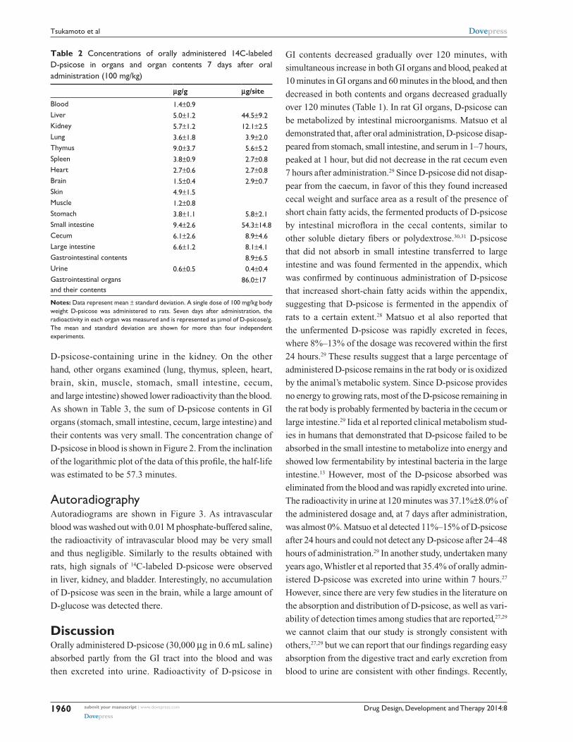
The residual amount of D-psicose was measured 7 days
after a single-dose oral administration (100 mg/kg). The
results are shown in Table 2. Blood concentration of D- psicose
estimated from the radioactivity was 1.4±0.9 µg/g, which was
much less than the value after 120 minutes (39.2±9.5 µg/g
[Table 1]). Organs that showed higher concentrations than
blood were liver, kidney, lung, thymus, spleen, heart, skin,
stomach, small intestine, cecum, and large intestine. On the
other hand, levels in the GI contents and urine were very low
7 days after a single dose of D-psicose. As large organs, small
intestine (4.70±0.38 g wet weight) and liver (7.65±0.42 g wet
weight) contained 54.3 and 44.5 µg/organ D-psicose, respec-
tively. Radioactivity of muscle was 1.2 µg/g tissue, although,
as a large organ, the total content was not considered high.
The radioactivity of skin was low, but higher than blood.
absorption and distribution of D-psicose after intravenous administrationThe concentration and total contents of D-psicose after
intravenous administration are summarized in Table 3.
Radioactivity of D-psicose concentration in blood was
132.0±22.1, 83.0±26.5, 77.2±23.5, and 40.1±31.5 µg/g blood
at 10, 30, 60, and 120 minutes, respectively, after intravenous
administration of isotope-labeled D-psicose. Concentrations
in liver and kidney remained higher than concentrations in
blood throughout the experiment. The concentrations in
the liver were 348.6±56.8, 354.3±32.1, 322.0±96.3, and
139.4±52.8 µg/g liver tissue at 10, 30, 60, and 120 minutes,
respectively, and those in kidney were 868.9±202.6,
649.8±175.0, 462.9±183.7, and 243.8±101.5 µg/g tissue at
10, 30, 60, and 120 minutes, respectively. Radioactivity of
D-psicose in urine was 4,773.6±1,376.6, 7,303.9±3,662.3,
13,230.7±3,292.9, and 15,231.7±4,649.0 µg/unit at 10,
30, 60, and 120 minutes, respectively. It was found that
the administered D-psicose eliminated quickly from blood
and moved to the urine. Of the total administered dose
(30,000 µg), approximately 1/6 (15%), 1/
4 (24%), 2/
5 (44%),
and 1/2 (50%) were eliminated into urine at 10, 30, 60, and
120 minutes, respectively (Table 3). However, the high
concentration in kidney was assumed to be the 14C-labeled
a
CHO
H−C−OH
HO−C−H
H−C−OH
H−C−OH
CH2OH
D−glucose
CHO
H−C−OH
HO−C−H
H−C−OH
H−C−OH
CH2OH
D−fructose
CHO
H−C−OH
HO−C−H
H−C−OH
H−C−OH
CH2OH
D−psicose
CHO
H−C−OH
HO−C−H
H−C−OH
H−C−OH
CH2OH
D−allose
b c
Figure 1 structures and enzymatic conversion of D-glucose, D-fructose, D-psicose, and D-allose.Notes: enzymes: a = glucose isomerase; b = D-tagatose 3 epimerase; c = l-rhamnose isomerase.
Drug Design, Development and Therapy 2014:8 submit your manuscript | www.dovepress.com
Dovepress
Dovepress
1959
Pharmacokinetics of rare sugar D-psicose
Tab
le 1
con
cent
ratio
ns o
f ora
lly a
dmin
iste
red
14c
-labe
led
D-p
sico
se in
org
ans
and
orga
n co
nten
ts 1
0, 3
0, 6
0, a
nd 1
20 m
inut
es a
fter
oral
adm
inis
trat
ion
(100
mg/
kg)
10 m
in30
min
60 m
in12
0 m
in
μg/g
μg/s
ite
μg/g
μg/s
ite
μg/g
μg/s
ite
μg/g
μg/s
ite
Bloo
d11
.3±6
.441
.8±1
6.2
48.5
±15.
639
.2±9
.5li
ver
41.4
±28.
733
6.4±
208.
912
6.3±
45.0
1,22
3.3±
372.
420
0.0±
86.3
1,72
0.7±
610.
712
7.5±
32.6
1,20
9.2±
456.
7K
idne
y74
.0±5
4.5
153.
1±10
4.3
287.
0±21
7.8
689.
5±47
7.1
395.
3±14
7.1
863.
2±19
6.9
168.
1±34
.939
9.2±
95.8
lung
10.0
±9.1
10.5
±8.4
39.1
±18.
247
.9±1
3.8
46.3
±16.
959
.6±2
3.1
37.7
±12.
044
.9±1
4.3
Thy
mus
3.1±
2.8
1.3±
1.1
18.9
±7.4
8.2±
2.9
26.0
±10.
210
.6±6
.125
.2±8
.210
.1±3
.1sp
leen
7.8±
8.6
5.3±
6.0
35.5
±21.
724
.2±1
5.1
36.0
±19.
123
.7±1
0.6
33.4
±9.9
23.3
±8.1
hea
rt4.
0±2.
74.
0±3.
024
.3±6
.531
.1±1
3.0
27.5
±10.
536
.7±1
6.5
24.4
±4.1
33.1
±17.
8Br
ain
1.7±
2.1
3.0±
3.7
10.5
±3.3
19.3
±6.4
8.9±
3.9
17.0
±7.2
11.5
±1.6
21.4
±3.9
skin
3.2±
2.6
28.0
±9.7
40.6
±23.
324
.5±6
.0M
uscl
e4.
3±4.
017
.3±9
.614
.6±5
.315
.8±3
.1st
omac
h4,
142.
8±1,
753.
45,
809.
1±2,
605.
71,
058.
1±1,
038.
41,
305.
4±1,
102.
886
4.1±
683.
91,
175.
1±1,
068.
712
6.8±
95.1
184.
1±12
3.6
smal
l int
estin
e61
2.2±
517.
13,
063.
5±2,
263.
61,
286.
6±14
3.4
8,18
3.2±
2,53
0.5
1,45
2.6±
1,14
7.9
6,80
4.5±
5,02
0.3
108.
8±38
.156
3.0±
181.
0c
ecum
247.
9±14
6.3
280.
9±16
4.5
153.
4±14
5.0
200.
2±19
8.3
136.
5±67
.716
9.8±
79.0
160.
1±16
6.9
191.
4±20
2.0
larg
e in
test
ine
284.
1±24
0.3
369.
3±33
4.9
166.
7±20
4.7
195.
1±21
9.0
100.
7±61
.612
4.0±
65.9
47.0
±18.
058
.1±2
3.6
gas
troi
ntes
tinal
con
tent
s12
,386
.4±3
,691
.88,
979.
2±2,
161.
46,
357.
6±1,
904.
45,
968.
7±2,
546.
7g
astr
oint
estin
al o
rgan
s
and
thei
r co
nten
ts20
,814
.0±1
,174
.72,
0342
.1±2
,615
.215
,031
.6±2
,454
.08,
015.
4±2,
369.
8
Uri
ne0.
2±0.
43,
193.
6±2,
870.
45,
832.
0±1,
682.
511
,125
.2±2
,403
.0
Not
es: D
ata
repr
esen
t m
ean
± st
anda
rd d
evia
tion.
The
gas
troi
ntes
tinal
con
tent
s of
the
sto
mac
h, s
mal
l int
estin
e, la
rge
inte
stin
e, a
nd c
ecum
wer
e co
llect
ed a
nd h
omog
eniz
ed. a
sm
all a
mou
nt o
f the
mix
ed c
onte
nts
and
smal
l pie
ces
of
orga
ns w
ere
appl
ied
to t
he m
easu
rem
ent,
and
then
the
who
le c
onte
nt w
as c
alcu
late
d fr
om t
he w
hole
wei
ght.
Uri
ne a
ccum
ulat
ed in
the
uri
nary
bla
dder
was
col
lect
ed a
nd m
easu
red.
rad
ioac
tivity
is r
epre
sent
ed a
s µg
of D
-psi
cose
. The
av
erag
e an
d st
anda
rd d
evia
tion
repr
esen
t da
ta fr
om m
ore
than
four
rat
s.
Drug Design, Development and Therapy 2014:8submit your manuscript | www.dovepress.com
Dovepress
Dovepress
1960
Tsukamoto et al
GI contents decreased gradually over 120 minutes, with
simultaneous increase in both GI organs and blood, peaked at
10 minutes in GI organs and 60 minutes in the blood, and then
decreased in both contents and organs decreased gradually
over 120 minutes (Table 1). In rat GI organs, D-psicose can
be metabolized by intestinal microorganisms. Matsuo et al
demonstrated that, after oral administration, D-psicose disap-
peared from stomach, small intestine, and serum in 1–7 hours,
peaked at 1 hour, but did not decrease in the rat cecum even
7 hours after administration.29 Since D-psicose did not disap-
pear from the caecum, in favor of this they found increased
cecal weight and surface area as a result of the presence of
short chain fatty acids, the fermented products of D-psicose
by intestinal microflora in the cecal contents, similar to
other soluble dietary fibers or polydextrose.30,31 D-psicose
that did not absorb in small intestine transferred to large
intestine and was found fermented in the appendix, which
was confirmed by continuous administration of D-psicose
that increased short-chain fatty acids within the appendix,
suggesting that D-psicose is fermented in the appendix of
rats to a certain extent.28 Matsuo et al also reported that
the unfermented D-psicose was rapidly excreted in feces,
where 8%–13% of the dosage was recovered within the first
24 hours.29 These results suggest that a large percentage of
administered D-psicose remains in the rat body or is oxidized
by the animal’s metabolic system. Since D-psicose provides
no energy to growing rats, most of the D-psicose remaining in
the rat body is probably fermented by bacteria in the cecum or
large intestine.29 Iida et al reported clinical metabolism stud-
ies in humans that demonstrated that D-psicose failed to be
absorbed in the small intestine to metabolize into energy and
showed low fermentability by intestinal bacteria in the large
intestine.13 However, most of the D-psicose absorbed was
eliminated from the blood and was rapidly excreted into urine.
The radioactivity in urine at 120 minutes was 37.1%±8.0% of
the administered dosage and, at 7 days after administration,
was almost 0%. Matsuo et al detected 11%–15% of D-psicose
after 24 hours and could not detect any D-psicose after 24–48
hours of administration.29 In another study, undertaken many
years ago, Whistler et al reported that 35.4% of orally admin-
istered D-psicose was excreted into urine within 7 hours.27
However, since there are very few studies in the literature on
the absorption and distribution of D-psicose, as well as vari-
ability of detection times among studies that are reported,27,29
we cannot claim that our study is strongly consistent with
others,27,29 but we can report that our findings regarding easy
absorption from the digestive tract and early excretion from
blood to urine are consistent with other findings. Recently,
Table 2 concentrations of orally administered 14c-labeled D-psicose in organs and organ contents 7 days after oral administration (100 mg/kg)
μg/g μg/site
Blood 1.4±0.9liver 5.0±1.2 44.5±9.2Kidney 5.7±1.2 12.1±2.5lung 3.6±1.8 3.9±2.0Thymus 9.0±3.7 5.6±5.2spleen 3.8±0.9 2.7±0.8heart 2.7±0.6 2.7±0.8Brain 1.5±0.4 2.9±0.7skin 4.9±1.5Muscle 1.2±0.8stomach 3.8±1.1 5.8±2.1small intestine 9.4±2.6 54.3±14.8cecum 6.1±2.6 8.9±4.6large intestine 6.6±1.2 8.1±4.1gastrointestinal contents 8.9±6.5Urine 0.6±0.5 0.4±0.4gastrointestinal organs and their contents
86.0±17
Notes: Data represent mean ± standard deviation. a single dose of 100 mg/kg body weight D-psicose was administered to rats. seven days after administration, the radioactivity in each organ was measured and is represented as µmol of D-psicose/g. The mean and standard deviation are shown for more than four independent experiments.
D-psicose-containing urine in the kidney. On the other
hand, other organs examined (lung, thymus, spleen, heart,
brain, skin, muscle, stomach, small intestine, cecum,
and large intestine) showed lower radioactivity than the blood.
As shown in Table 3, the sum of D-psicose contents in GI
organs (stomach, small intestine, cecum, large intestine) and
their contents was very small. The concentration change of
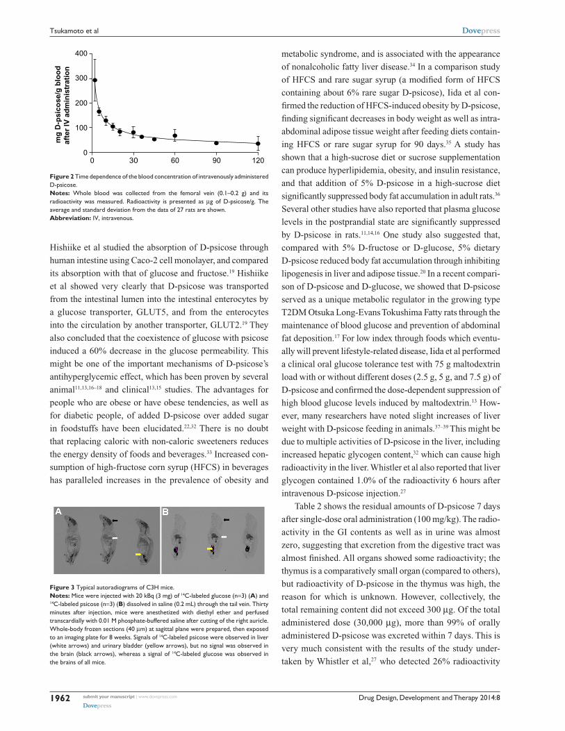
D-psicose in blood is shown in Figure 2. From the inclination
of the logarithmic plot of the data of this profile, the half-life
was estimated to be 57.3 minutes.
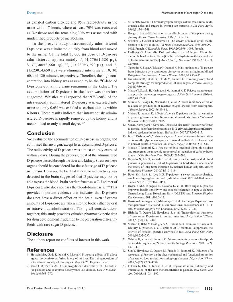
autoradiographyAutoradiograms are shown in Figure 3. As intravascular
blood was washed out with 0.01 M phosphate-buffered saline,
the radioactivity of intravascular blood may be very small
and thus negligible. Similarly to the results obtained with
rats, high signals of 14C-labeled D-psicose were observed
in liver, kidney, and bladder. Interestingly, no accumulation
of D-psicose was seen in the brain, while a large amount of
D-glucose was detected there.
DiscussionOrally administered D-psicose (30,000 µg in 0.6 mL saline)
absorbed partly from the GI tract into the blood and was
then excreted into urine. Radioactivity of D-psicose in
Drug Design, Development and Therapy 2014:8 submit your manuscript | www.dovepress.com
Dovepress
Dovepress
1961
Pharmacokinetics of rare sugar D-psicose
Tab
le 3
con
cent
ratio
ns a
nd c
onte
nts
of 14
c-la
bele
d D
-psic
ose
10, 3
0, 6
0, a
nd 1
20 m
inut
es a
fter
intr
aven
ous
adm
inist
ratio
n (1
00 m
g/kg
)
10 m
in30
min
60 m
in12
0 m
in
μg/g
μg/s
ite
μg/g
μg/s
ite
μg/g
μg/s
ite
μg/g
μg/s
ite
Bloo
d13
2.0±
22.1
83.0
±26.
577
.2±2
3.5
40.1
±31.
5li
ver
348.
6±56
.83,
015.
4±43
4.9
354.
3±32
.13,
188.
1±75
2.4
322.
0±96
.32,
653.
0±64
1.5
139.
4±52
.81,
299.
2±25
4.7
Kid
ney
868.
9±20
2.6
1,80
7.7±
389.
064
9.8±
175.
01,
321.
0±38
1.1
462.
4±18
3.7
945.
8±32
1.9
243.
8±10
1.5
493.
3±19
9.1
lung
118.
2±16
.514
7.3±
31.3
71.4
±19.
182
.5±2
6.8
67.6
±22.
477
.1±1
4.3
42.5
±19.
949
.1±1
8.1
Thy
mus
56.1
±22.
320
.6±5
.445
.8±2
3.7
16.6
±7.3
45.2
±19.
817
.3±2
.930
.3±9
.614
.3±3
.4sp
leen
72.8
±13.
852
.8±1
5.8
58.8
±22.
342
.7±1
8.0
60.7
±21.
841
.3±1
5.7
37.2
±16.
726
.9±1
6.0
hea
rt60
.5±8
.352
.5±5
.038
.2±1
1.1
37.2
±14.
236
.1±1
2.8
31.8
±9.6
28.7
±9.3
27.6
±8.7
Brai
n9.
1±3.
216
.9±6
.78.
6±5.
115
.7±9
.29.
9±2.
618
.3±4
.611
.2±6
.520
.8±1
3.4
skin
91.4
±21.
672
.6±2
7.6
55.6
±10.
424
.7±1
7.6
Mus
cle
31.7
±13.
323
.5±1
1.7
23.5
±3.6
16.6
±6.4
stom
ach
72.4
±14.
210
9.3±
28.0
52.6
±19.
975
.1±2
9.3
34.4
±11.
347
.8±1
0.1
28.6
±11.
640
.3±1
5.5
smal
l int
estin
e12
8.3±
22.7
689.
0±17
2.6
81.7
±35.
945
4.8±
187.
573
.0±2
5.7
381.
1±92
.837
.5±1
9.0
205.
2±86
.9c
ecum
90.6
±32.
610
3.5±
45.3
52.1
±18.
981
.7±2
0.7
50.7
±16.
665
.1±2
5.9
34.4
±19.
845
.9±1
9.5
larg
e in
test
ine
110.
5±35
.614
0.4±
47.3
81.0
±46.
886
.6±3
9.0
50.4
±18.
255
.4±1
5.7
36.9
±15.
045
.3±1
6.0
gas
troi
ntes
tinal
con
tent
s68
.7±5
0.5
61.6
±42.
176
.8±4
2.2
72.8
±64.
7g
astr
oint
estin
al o
gans
an
d th
eir
cont
ents
1,16
6.6±
229.
989
5.1±
140.
256
2.5±
85.9
437.
4±52
.7
Uri
ne4,
773.
6±1,
376.
67,
303.
9±3,
662.
313
,230
.7±3
,292
.915
,231
.7±4
649.
3
Not
es: D
ata
repr
esen
t m
ean
± st
anda
rd d
evia
tion.
The
gas
troi
ntes
tinal
con
tent
s of
the
sto
mac
h, s
mal
l int
estin
e, la
rge
inte
stin
e, a
nd c
ecum
wer
e co
llect
ed a
nd h
omog
eniz
ed. a
sm
all a
mou
nt o
f the
mix
ed c
onte
nts
from
gas
troi
ntes
tinal
or
gans
and
sm
all p
iece
s of
org
ans
wer
e ap
plie
d to
the
mea
sure
men
t, an
d th
en th
e w
hole
con
tent
was
cal
cula
ted
from
the
who
le w
eigh
t. U
rine
acc
umul
ated
in th
e ur
inar
y bl
adde
r w
as c
olle
cted
and
mea
sure
d. r
adio
activ
ity w
as r
epre
sent
ed
as µ
mol
of D
-psi
cose
. The
mea
n an
d st
anda
rd d
evia
tion
repr
esen
t da
ta fr
om m
ore
than
four
rat
s.
Drug Design, Development and Therapy 2014:8submit your manuscript | www.dovepress.com
Dovepress
Dovepress
1962
Tsukamoto et al
Hishiike et al studied the absorption of D-psicose through
human intestine using Caco-2 cell monolayer, and compared
its absorption with that of glucose and fructose.19 Hishiike
et al showed very clearly that D-psicose was transported
from the intestinal lumen into the intestinal enterocytes by
a glucose transporter, GLUT5, and from the enterocytes
into the circulation by another transporter, GLUT2.19 They
also concluded that the coexistence of glucose with psicose
induced a 60% decrease in the glucose permeability. This
might be one of the important mechanisms of D-psicose’s
antihyperglycemic effect, which has been proven by several
animal11,13,16–18 and clinical13,15 studies. The advantages for
people who are obese or have obese tendencies, as well as
for diabetic people, of added D-psicose over added sugar
in foodstuffs have been elucidated.22,32 There is no doubt
that replacing caloric with non-caloric sweeteners reduces
the energy density of foods and beverages.33 Increased con-
sumption of high-fructose corn syrup (HFCS) in beverages
has paralleled increases in the prevalence of obesity and
metabolic syndrome, and is associated with the appearance
of nonalcoholic fatty liver disease.34 In a comparison study
of HFCS and rare sugar syrup (a modified form of HFCS
containing about 6% rare sugar D-psicose), Iida et al con-
firmed the reduction of HFCS-induced obesity by D-psicose,
finding significant decreases in body weight as well as intra-
abdominal adipose tissue weight after feeding diets contain-
ing HFCS or rare sugar syrup for 90 days.35 A study has
shown that a high-sucrose diet or sucrose supplementation
can produce hyperlipidemia, obesity, and insulin resistance,
and that addition of 5% D-psicose in a high-sucrose diet
significantly suppressed body fat accumulation in adult rats.36
Several other studies have also reported that plasma glucose
levels in the postprandial state are significantly suppressed
by D-psicose in rats.11,14,16 One study also suggested that,
compared with 5% D-fructose or D-glucose, 5% dietary
D-psicose reduced body fat accumulation through inhibiting
lipogenesis in liver and adipose tissue.20 In a recent compari-
son of D-psicose and D-glucose, we showed that D-psicose
served as a unique metabolic regulator in the growing type
T2DM Otsuka Long-Evans Tokushima Fatty rats through the
maintenance of blood glucose and prevention of abdominal
fat deposition.17 For low index through foods which eventu-
ally will prevent lifestyle-related disease, Iida et al performed
a clinical oral glucose tolerance test with 75 g maltodextrin
load with or without different doses (2.5 g, 5 g, and 7.5 g) of
D-psicose and confirmed the dose-dependent suppression of
high blood glucose levels induced by maltodextrin.13 How-
ever, many researchers have noted slight increases of liver
weight with D-psicose feeding in animals.37–39 This might be
due to multiple activities of D-psicose in the liver, including
increased hepatic glycogen content,32 which can cause high
radioactivity in the liver. Whistler et al also reported that liver
glycogen contained 1.0% of the radioactivity 6 hours after
intravenous D-psicose injection.27
Table 2 shows the residual amounts of D-psicose 7 days
after single-dose oral administration (100 mg/kg). The radio-
activity in the GI contents as well as in urine was almost
zero, suggesting that excretion from the digestive tract was
almost finished. All organs showed some radioactivity; the
thymus is a comparatively small organ (compared to others),
but radioactivity of D-psicose in the thymus was high, the
reason for which is unknown. However, collectively, the
total remaining content did not exceed 300 µg. Of the total
administered dose (30,000 µg), more than 99% of orally
administered D-psicose was excreted within 7 days. This is
very much consistent with the results of the study under-
taken by Whistler et al,27 who detected 26% radioactivity
400
300
mg
D-p
sico
se/g
blo
od
afte
r IV
ad
min
istr
atio
n
200
100
00 30 60 90 120
Figure 2 Time dependence of the blood concentration of intravenously administered D-psicose.Notes: Whole blood was collected from the femoral vein (0.1–0.2 g) and its radioactivity was measured. radioactivity is presented as µg of D-psicose/g. The average and standard deviation from the data of 27 rats are shown.Abbreviation: iV, intravenous.
Figure 3 Typical autoradiograms of c3h mice.Notes: Mice were injected with 20 kBq (3 mg) of 14c-labeled glucose (n=3) (A) and 14c-labeled psicose (n=3) (B) dissolved in saline (0.2 ml) through the tail vein. Thirty minutes after injection, mice were anesthetized with diethyl ether and perfused transcardially with 0.01 M phosphate-buffered saline after cutting of the right auricle. Whole-body frozen sections (40 µm) at sagittal plane were prepared, then exposed to an imaging plate for 8 weeks. signals of 14c-labeled psicose were observed in liver (white arrows) and urinary bladder (yellow arrows), but no signal was observed in the brain (black arrows), whereas a signal of 14c-labeled glucose was observed in the brains of all mice.
Drug Design, Development and Therapy 2014:8 submit your manuscript | www.dovepress.com
Dovepress
Dovepress
1963
Pharmacokinetics of rare sugar D-psicose
as exhaled carbon dioxide and 95% radioactivity in the
urine within 7 hours, where at least 70% was recovered
as D-psicose and the remaining 30% was associated with
unidentified products of metabolism.
In the present study, intravenously administered
D- psicose was eliminated quickly from blood and moved
to the urine. Of the total 30,000 µg dose of D-psicose
administered, approximately 1/6 (4,770±1,380 µg),
1/4 (7,300±3,660 µg), 2/
5 (13,230±3,290 µg), and 1/
2
(15,230±4,650 µg) were eliminated into urine at 10, 30,
60, and 120 minutes, respectively. Therefore, the high con-
centration into kidney was assumed to be the 14C-labeled
D-psicose-containing urine remaining in the kidney. The
accumulation of D-psicose in the liver was therefore
suggested. Whistler et al reported that 97%–98% of the
intravenously administered D-psicose was excreted into
urine and only 0.6% was exhaled as carbon dioxide within
6 hours. These results indicate that intravenously admin-
istered D-psicose is rapidly removed by the kidney and is
metabolized to only a small degree.27
ConclusionWe evaluated the accumulation of D-psicose in organs, and
confirmed that no organ, except liver, accumulated D-psicose.
The radioactivity of D-psicose was almost entirely excreted
within 7 days. During the process, most of the administered
D-psicose passed through the liver and kidney. Stress on these
organs should be considered for the safe usage of D-psicose
in humans. However, the fact that almost no radioactivity was
detected in the brain suggested that D-psicose may not be
able to pass the blood–brain barrier; fructose, a C-3 epimer of
D-psicose, also does not pass the blood–brain barrier.40 This
provides important evidence that indicates that D-psicose
does not have a direct effect on the brain, even if excess
amounts of D-psicose are taken into the body, either by oral
or intravenous administration. Taking all considerations
together, this study provides valuable pharmacokinetic data
for drug development in addition to the preparation of healthy
foods with rare sugar D-psicose.
DisclosureThe authors report no conflicts of interest in this work.
References1. Hossain MA, Goda F, Izuishi K, Maeta H. Protective effects of D-allose
agianst ischemia-reperfusion injury of rat liver. The 1st symposium of international society of rare sugars. May 23–27, Kagawa, Japan.
2. Cree GM, Perlin AS. O-isopropylidene derivatives of D-allulose (D-psicose) and D-erythro-hexopyranos-2,3-diulose. Can J Biochem. 1968;46:765–770.
3. Miller BS, Swain T. Chromatographic analysis of the free amino-acids, organic acids and sugars in wheat plant extracts. J Sci Food Agric. 1960;11:344–348.
4. Hough L, Stacey BE. Variation in the allitol content of Itea plants during photosynthesis. Phytochemistry. 1966;5:171–175.
5. Strecker G, Goubet B, Montreuil J. The ketoses of human urine. Identi-fication of D (+)-allulose. C R Hebd Seances Acad Sci. 1965;260:999–1002. French. C R Acad Sc Paris. 1965;260:999–1003. French.
6. Padberg G. Über die Kohlenhydrate im wäßrigen Eluat der menschlichen Hautoberfläche [On the carbohydrates in the water eluate of the human skin surface]. Arch Klin Exp Dermatol. 1967;229:33–39. German.
7. Takeshita K, Suga A, Takada G, Izumori K. Mass production of D- psicose from d-fructose by a continuous bioreactor system using immobilized D-tagatose 3-epimerase. J Biosci Bioeng. 2000;90:453–455.
8. Granström TB, Takata G, Tokuda M, Izumori K. Izumoring: a novel and complete strategy for bioproduction of rare sugars. J Biosci Bioeng. 2004;97:89–94.
9. Matsuo T, Suzuki H, Hashiguchi M, Izumori K. D-Psicose is a rare sugar that provides no energy to growing rats. J Nutr Sci Vitaminol (Tokyo). 2002;48:77–80.
10. Murata A, Sekiya K, Watanabe Y, et al. A novel inhibitory effect of D-allose on production of reactive oxygen species from neutrophils. J Biosci Bioeng. 2003;96:89–91.
11. Matsuo T, Izumori K. Effects of dietary D-psicose on diurnal variation in plasma glucose and insulin concentrations of rats. Biosci Biotechnol Biochem. 2006;70:2081–2085.
12. Suna S, Yamaguchi F, Kimura S, Tokuda M, Jitsunari F. Preventive effect of D-psicose, one of rare ketohexoses, on di-(2-ethylhexyl) phthalate (DEHP)-induced testicular injury in rat. Toxicol Lett. 2007;173:107–117.
13. Iida T, Kishimoto Y, Yoshikawa Y, et al. Acute D-psicose administration decreases the glycemic responses to an oral maltodextrin tolerance test in normal adults. J Nutr Sci Vitaminol (Tokyo). 2008;54: 511–514.
14. Matsuo T, Izumori K. d-Psicose inhibits intestinal alpha-glucosidase and suppresses the glycemic response after ingestion of carbohydrates in rats. J Clin Biochem Nutr. 2009;45:202–206.
15. Hayashi N, Iida T, Yamada T, et al. Study on the postprandial blood glucose suppression effect of D-psicose in borderline diabetes and the safety of long-term ingestion by normal human subjects. Biosci Biotechnol Biochem. 2010;74:510–519.
16. Baek SH, Park SJ, Lee HG. D-psicose, a sweet monosaccharide, ameliorate hyperglycemia, and dyslipidemia in C57BL/6J db/db mice. J Food Sci. 2010;75:H49–H53.
17. Hossain MA, Kitagaki S, Nakano D, et al. Rare sugar D-psicose improves insulin sensitivity and glucose tolerance in type 2 diabetes Otsuka Long-Evans Tokushima Fatty (OLETF) rats. Biochem Biophys Res Commun. 2011;405:7–12.
18. Hossain A, Yamaguchi F, Matsunaga T, et al. Rare sugar D-psicose pro-tects pancreas β-islets and thus improves insulin resistance in OLETF rats. Biochem Biophys Res Commun. 2012;425:717–723.
19. Hishiike T, Ogawa M, Hayakawa S, et al. Transepithelial transports of rare sugar D-psicose in human intestine. J Agric Food Chem. 2013;61(30):7381–386.
20. Matsuo T, Baba Y, Hashiguchi M, Takeshita K, Izumori K, Suzuki H. Dietary D-psicose, a C-3 epimer of D-fructose, suppresses the activity of hepatic lipogenic enzymes in rats. Asia Pac J Clin Nutr. 2001;10:233–237.
21. Oshima H, Kimura I, Izumori K. Psicose contents in various food prod-ucts and its origin. Food Science and Technology Research. 2006;12(2): 137–143.
22. Sun Y, Hayakawa S, Ogawa M, Fukada K, Izumori K. Influence of a rare sugar, d-Psicose, on the physicochemical and functional properties of an aerated food system containing egg albumen. J Agric Food Chem. 2008;56(12):4789–4796.
23. Fukada K, Ishii T, Tanaka K, et al. Crystal structure, solubility, and mutarotation of the rare monosaccharide D-psicose. Bull Chem Soc Jpn. 2010;83:1193–1197.

Drug Design, Development and Therapy
Publish your work in this journal
Submit your manuscript here: http://www.dovepress.com/drug-design-development-and-therapy-journal
Drug Design, Development and Therapy is an international, peer-reviewed open-access journal that spans the spectrum of drug design and development through to clinical applications. Clinical outcomes, patient safety, and programs for the development and effective, safe, and sustained use of medicines are a feature of the journal, which
has also been accepted for indexing on PubMed Central. The manu-script management system is completely online and includes a very quick and fair peer-review system, which is all easy to use. Visit http://www.dovepress.com/testimonials.php to read real quotes from published authors.
Drug Design, Development and Therapy 2014:8submit your manuscript | www.dovepress.com
Dovepress
Dovepress
Dovepress
1964
Tsukamoto et al
24. Malik VS, Popkin BM, Bray GA, Després JP, Willett WC, Hu FB. Sugar-sweetened beverages and risk of metabolic syndrome and type 2 diabetes: a meta-analysis. Diabetes Care. 2010;33(11):2477–2483.
25. Gardner C, Wylie-Rosett J, Gidding SS, et al; American Heart Association Nutrition Committee of the Council on Nutrition, Physical Activity and Metabolism, Council on Arteriosclerosis, Thrombosis and Vascular Biology, Council on Cardiovascular Disease in the Young, and the American D. Nonnutritive sweeteners: current use and health perspectives: a scientific statement from the American Heart Association and the American Diabetes Association. Circulation. 2012;126(4):509–519.
26. Mu W, Zhang W, Feng Y, Jiang B, Zhou L. Recent advances on applications and biotechnological production of D-psicose. Appl Microbiol Biotechnol. 2012;94:1461–1467.
27. Whistler RL, Singh PP, Lake WC. D-Psicose metabolism in the rat. Carbohydr Res. 1974;34:200–202.
28. Morimoto K, Park CS, Ozaki M, et al. Large scale production of D-allose from D-psicose using continuous bioreactor and separation system. Enzyme Microb Technol. 2006;38:855–859.
29. Matsuo T, Tanaka T, Hashiguchi M, Izumori K, Suzuki H. Metabolic effects of D-psicose in rats: studies on faecal and urinary excretion and caecal fermentation. Asia Pac J Clin Nutr. 2003;12(2):225–231.
30. Figdor SK, Rennhard HH. Caloric utilization and disposition of [14C]polydextrose in the rat. J Agric Food Chem. 1981;29:1181–1189.
31. Topping DL, Clifton PM. Short-chain fatty acids and human colonic function: roles of resistant starch and nonstarch polysaccharides. Physiol Rev. 2001;81:1031–1064.
32. Sun Y, Hayakawa S, Izumori K. Modification of ovalbumin with a rare ketohexose through the Maillard reaction: effect on protein structure and gel properties. J Agric Food Chem. 2004;52:1293–1299.
33. Swithers SE. Artificial sweeteners produce the counterintuitive effect of inducing metabolic derangements. Trends Endocrinol Metab. 2013;24(9):431–441.
34. Dekker MJ, Su Q, Baker C, Rutledge AC, Adeli K. Fructose: a lighly lipogenic nutrient implicated insulin resistance, hepatic steatosis and the metabolic syndrome. Am J Physiol Endocrinol Metab. 2010;299:E685–E694.
35. Iida T, Yamada T, Hayashi N, et al. Reduction of abdominal fat accu-mulation in rats by 8-week ingestion of a newly developed sweetener made from high fructose corn syrup. Food Chem. 2013;138:781–785.
36. Ochiai M, Onishi K, Yamada T, Iida T, Matsuo T. D-psicose increases energy expenditure and decreases body fat accumulation in rats fed a high-sucrose diet. Int J Food Sci Nutr. 2014;65(2):245–250.
37. Yagi K, Matsuo T. The study on long-term toxicity of D-psicose in rats. J Clin Biochem Nutr. 2009;45:271–277.
38. Matsuo T, Reika Ishii, Tetsuo Iida, Takako Yamada, Satoshi Tamamine and Yoko Shirai. Ninety-day oral toxicity study of rare sugar syrup in male Wistar rats. Current topics in toxicology 2011;7:41–50.
39. Matsuo T, Ishii R, Shirai Y. The 90-day oral toxicity of D-psicose in male Wistar rats. J Clin Biochem Nutr. 2012;50:158–161.
40. Oldendorf WH. Brain uptake of radiolabeled amino acids, amines, and hexoses after arterial injection. Am J Physiol. 1971;221:1629–1639.