Embed Size (px)
Citation preview

Pharmacological Validation of Trypanosoma bruceiPhosphodiesterases B1 and B2 as Druggable Targets for AfricanSleeping Sickness
Nicholas D. Bland2, Cuihua Wang1, Craig Tallman1, Alden E. Gustafson2, Zhouxi Wang1,Trent D. Ashton4, Stefan O. Ochiana1, Gregory McAllister5, Kristina Cotter2, Anna P. Fang2,Lara Gechijian2, Norman Garceau3, Rajiv Gangurde3, Ron Ortenberg3, Mary JoOndrechen1, Robert K. Campbell2, and Michael P. Pollastri1,*
1Northeastern University Department of Chemistry and Chemical Biology, Hurtig 102, 360Huntington Avenue, Boston, MA 02115.2Marine Biological Laboratory, Josephine Bay Paul Center for Comparative Molecular Biologyand Evolution, 7 MBL Street, Woods Hole, MA 02543.3Blue Sky Biotech, 60 Prescott Street, Worcester, MA 01605-2661.4Boston University Department of Chemistry, 590 Commonwealth Avenue, Boston, MA 02215.5Novartis Institutes for Biomedical Research, 250 Massachusetts Avenue, Cambridge, MA 02139.
AbstractNeglected tropical disease drug discovery requires application of pragmatic and efficient methodsfor development of new therapeutic agents. In this report we describe our target repurposingefforts for the essential phosphodiesterase (PDE) enzymes TbrPDEB1 and TbrPDEB2 ofTrypanosoma brucei, the causative agent for human African trypanosomiasis (HAT). We describeprotein expression and purification, assay development, and benchmark screening of a collectionof 20 established human PDE inhibitors. We disclose that the human PDE4 inhibitor piclamilast,and some of its analogs, show modest inhibition of TbrPDEB1 and B2, and quickly kill thebloodstream form of the subspecies T. brucei brucei. We also report the development of ahomology model of TbrPDEB1 that is useful for understanding the compound-enzymeinteractions and for comparing the parasitic and human enzymes. Our profiling and earlymedicinal chemistry results strongly suggest that human PDE4 chemotypes represent a betterstarting point for optimization of TbrPDEB inhibitors than those that target any other humanPDEs.
IntroductionHuman African trypanosomiasis (HAT), or African sleeping sickness, is a neglected tropicaldisease that affects tens of thousands of patients annually.1 The disease is fatal unless treatedand current treatments are limited in both efficacy and safety.2 Financial incentives for drugdiscovery against HAT are quite limited due to the economically disadvantaged regions
*To whom correspondence should be addressed: [email protected]; Northeastern University Department of Chemistry andChemical Biology, Hurtig 102, 360 Huntington Avenue, Boston, MA 02115. 617-373-2703..Supporting Information. A tabulation of all the benchmarked human PDE inhibitors, with their references and screening data is in theSupporting Information, and is publically available as a shared data set at www.collaborativedrugdiscovery.com. The SupportingInformation also includes protein sequences of TbrPDEB1 and B2, and alignment between TbrPDEB1, B2, LmjPDEB1, andhPDE4D. This material is available free of charge via the Internet at http://pubs.acs.org.
NIH Public AccessAuthor ManuscriptJ Med Chem. Author manuscript; available in PMC 2012 December 8.
Published in final edited form as:J Med Chem. 2011 December 8; 54(23): 8188–8194. doi:10.1021/jm201148s.
NIH
-PA Author Manuscript
NIH
-PA Author Manuscript
NIH
-PA Author Manuscript

where this disease is endemic. As a strategy to overcome this disincentive for drugdiscovery, we hypothesized that medicinal chemistry knowledge against human drug targetscould be repurposed to facilitate rapid and cost-effective drug discovery against parasitedrug targets.3 In this approach, existing drugs and drug-like compounds are used topharmacologically validate parasite targets, and to serve as early hits or leads from which tooptimize parasite-specific therapeutics.
Nucleotide phosphodiesterases (PDEs) are vital regulators of signalling pathways,hydrolyzing the secondary messengers cyclic adenosine monophosphate (cAMP) and cyclicguanosine monophosphate (cGMP). Humans have 11 families of PDEs, most of which havenumerous isoforms. PDEs are involved in a variety of biological processes and have beenimplicated in a wide range of therapeutic indications.4-7 Despite the size of the PDE familyand active site sequence identity between isoforms ranging from 25-52%, highly selectiveinhibitors have been developed, leading to clinical candidates and drugs for chronicobstructive pulmonary disorder (PDE4), erectile dysfunction (PDE5) and schizophrenia(PDE10).
By contrast, Trypanosoma brucei has only five PDEs, all of which are specific for cAMP.8The Trypanosoma brucei PDEB (TbrPDEB) family is encoded by two tandemly arrangedgenes and its members have distinct sub-cellular localizations.9 TbrPDEB1 and TbrPDEB2are not individually essential for parasite survival but when the enzymes are knocked downby RNAi simultaneously, the parasites are incapable of proper cell division and ultimatelydie.9,10
In this report we describe the selection of TbrPDEB1 and TbrPDEB2 from a list ofprioritized drug targets from the T. brucei genome and our initial inhibitor chemotypeexplorations. In confirmation of the previous RNAi experiments described above, thesetargets are validated pharmacologically in both in vitro biochemical assays and in parasitecultures. An initial survey of chemical space shows tolerance of structural modifications ofthe lead chemotype in regions that can potentially be exploited to optimize selectivity andpotency.
ResultsThe Trypanosoma brucei brucei genome was examined for the presence of homologs ofknown, chemically validated drug targets that have compounds that have successfullypassed Phase II clinical trials. Since the pathogens infiltrate the central nervous system(CNS), we further restricted our focus to parasite homologs of known targets withprecedents for clinical compounds capable of crossing the blood brain barrier, from whichwe selected the T. brucei brucei cyclic AMP phosphodiesterases TbrPDEB1(Tb09.160.3590) and TbrPDEB2 (Tb09.160.3630). The catalytic domains of thetrypanosomal PDEB enzymes have 30-35% identity to the successful PDE targets fromhumans, and others have demonstrated that certain established human PDE inhibitors areactive against partially purified preparations of TbrPDEB1and B2.11,12 We thereforehypothesized that a broader exploration of human PDE inhibitor chemotypes could identifygood starting points for chemical optimization. A similar approach was recently reported,describing the profiling of human PDE5 inhibitors as a starting point for antimalarialagents.13
The His-tagged catalytic domains of TbrPDEB1 (Ser580-Arg930) and TbrPDEB2 (Ser580-Ser925) were expressed using the Sf21/baculovirus system and purified by nickel-sepharosechromatography, yielding milligram quantities of active protein. The kinetic parameters ofthe recombinant catalytic domains were determined, and the KM of cAMP with the
Bland et al. Page 2
J Med Chem. Author manuscript; available in PMC 2012 December 8.
NIH
-PA Author Manuscript
NIH
-PA Author Manuscript
NIH
-PA Author Manuscript

TbrPDEB1 was measured to be 40 μM (±3.4), and with the TbrPDEB2 catalytic domain theKM was 11.5 μM (±4.4). Specific activities of 0.35 nmol/min/μg (±0.01) and 0.12 nmol/min/μg (±0.01) were observed for TbrPDEB1 and TbrPDEB2 respectively. Thus, while bothproteins behaved similarly biochemically, TbrPDEB2 had a slightly lower KM and specificactivity compared with TbrPDEB1. The KM of the catalytic domain of TbrPDEB2 is lowerthan that reported previously (KM ≥ 44 IM) and was more similar to the full length proteinwith the cAMP binding GAF domains intact (4.5 IM).14 The inverse is seen with TbrPDEB1where the previously described KM of 8 IM is significantly lower than that observed withour TbrPDEB1.12 However, in previous reports, TbrPDEB1 and TbrPDEB2 were expressedas full-length enzymes, in different expression systems (yeast and human cells respectively),and neither were purified; these differences may explain the disparity in observed KMvalues, and we therefore judged these differences acceptable and our proteins suitable forsupporting inhibitor development.
Extensive sets of human PDE inhibitors exist, and these compounds represent potentialstarting points for new inhibitor discovery against these trypanosomal PDEs. Our profilingset (annotated in the Supporting Information) includes a representative sampling of PDEinhibitor chemotypes that have been disclosed to date. Consistent with our preference tobuild on scaffolds with evidence for safety and tolerability, wherever possible we includedcompounds that have been reported in advanced stages of preclinical development or whichhave been approved for use. While the T. brucei PDEBs are known to be cAMP-selective,both approved drugs and development candidates against cGMP-specific PDEs (such asPDE5) were included, as were development candidates against those that hydrolyze bothcAMP and cGMP (such as PDE10). Compounds were selected and synthesized usingpublished methods, donated, or purchased, and were tested at a single concentration.
We sought to identify compounds capable of inhibiting both TbrPDEB1 and TbrPDEB2since RNAi only kills trypanosomes when both enzymes are disrupted. The feasibility ofachieving dual inhibition is supported by analysis of the protein sequences. Overall proteinsequence identity between TbrPDEB1 and TbrPDEB2 is 75% using a ClustalW2alignment,15 and the sequence identity within the catalytic domains of these two proteins is88%. The 3D structure alignment of TbrPDEB1, TbrPDEB2, human PDE4 and L. majorPDEB1 is shown in the Supporting Information in tabular form; residues involved in bindingare highlighted. The high similarity between the active sites of TbrPDEB1 and TbrPDEB2suggests that identification of a compound that inhibits both TbrPDEB1 and B2 should be anachievable goal. We elected to perform primary screening against TbrPDEB1, followed bysecondary screening against TbrPDEB2 with inhibitors that show better than 20 μMinhibitory potency (IC50) against TbrPDEB1.
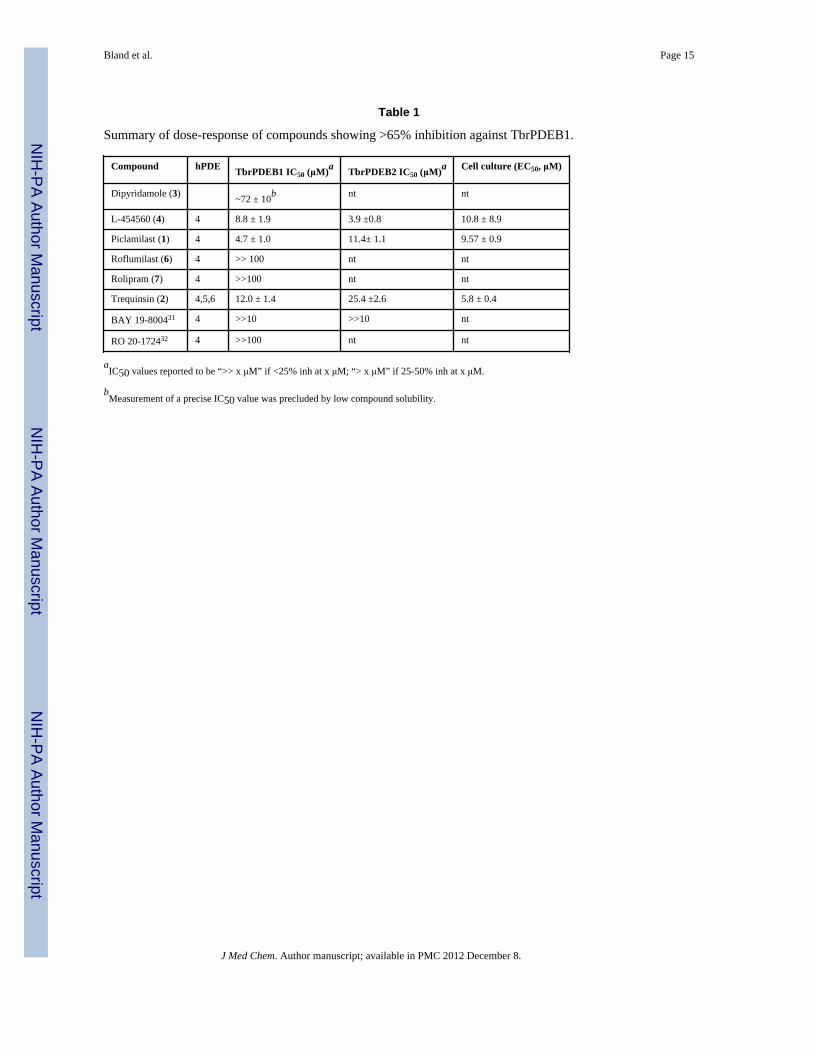
The scatter plot in Figure 1A shows the percent inhibition data as a function of human PDEinhibitor class, and Table 1 contains dose-response assessments of those inhibitors showinggreater than 65% inhibition at the single dose concentration of benchmarked inhibitors.Initial tests of the PDE4 inhibitors piclamilast (1) and trequinsin (2), and the PDE4/6inhibitor dipyridamole (3) (the latter two tested at 100 μM) showed over 50% inhibition.Subsequent tests of additional PDE4 inhibitors (L-454560 (4) and GSK-256066, (5) alsoinhibited TbrPDEB1 by at least 50% at 10 μM. Assessment of 1 in dose-response studiesrevealed IC50 values of 4.7 μM against TbrPDEB1, and 11.4 μM versus TbrPDEB2 (Figure2). While 1 demonstrated the greatest potency against TbrPDEB1 among the compoundstested, the closely-related PDE4 inhibitor roflumilast (6) was essentially inactive, as wasrolipram (7), a compound sharing a substantial substructure with piclamilast. Compound 8, arecently-disclosed human PDE10 inhibitor,16 displayed 55% inhibition at 100 μM.Recognizing the extensive precedence in human PDE4 inhibitors,17-19 including a number
Bland et al. Page 3
J Med Chem. Author manuscript; available in PMC 2012 December 8.
NIH
-PA Author Manuscript
NIH
-PA Author Manuscript
NIH
-PA Author Manuscript

of compounds that have entered clinical trials, we made this family of inhibitors our primaryfocus.
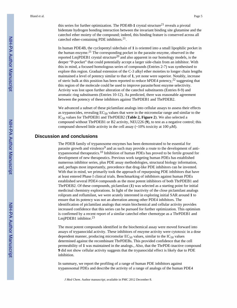
To help elucidate binding site features that could drive potency differences between theparasite and human enzymes, and to facilitate compound binding hypotheses to drivemedicinal chemistry optimization, homology models of TbrPDEB1 and B2 wereconstructed. Multiple template structures were tried, but the Leishmania major PDEB1(LmjPDEB1) crystal structure20 yielded the best-scoring model structures and is mostclosely related to our target (Supplementary Material). Figure 3A shows the X-ray crystalstructure previously reported for piclamilast bound to hPDE4 (PDB ID 1XM4),21 and thepredicted pose for 1 in the model structure of TbrPDEB1 (Figure 3B). A superposition ofthe TbrPDEB1 model structure (pink) with the human PDE4 crystal structure (gray) isshown in Figure 3C, highlighting the modest amino acid differences in the proximity of thedrug binding site that are likely implicated in the differential binding potency between thehuman and parasite enzymes.
Comparison of the active sites in the homology model structures for the two TbrPDEBsconfirms that their pairwise conservation is high (see Supporting Information), and that theypossess regions previously observed in other PDEs, including a metal binding pocket (Mpocket); a solvent-filled side pocket (S pocket); and a pocket containing the conservedpurine-binding glutamine residue and hydrophobic clamp (Q pocket), with two hydrophobicpockets (Q1 and Q2) on either side.21 Besides confirming the high similarity in the activesite region between TbrPDEB1 and B2, homology modeling also indicates that both arelikely to have an extended binding site cleft, termed the “parasite-“ or “P-pocket,” that wasfirst observed in LmjPDEB1.20 This extended pocket provides a tunnel from the binding siteleading to the exterior of the protein (Figure 3D), consistent with the P-pocket observed inLmjPDEB1. This feature is absent from every human PDE, and may therefore be exploitablefor development of selective inhibitors of parasite PDEs.
While TbrPDEB1 and human PDE4B show overall similarity in the active site, comparisonof the homology model with human PDE4-inhibitor crystal structures illustrates multipledifferences. These differences may explain the lower inhibitory potency we observe in thetrypanosome enzymes, compared to those reported against human PDE4B and PDE4D. Forexample, the cyclopentyl ring of 1 (and that of rolipram (7) and the cyclopropylmethyl ofroflumilast (6)) is buried in the lipophilic Q2 pocket in human PDE4, whereas it is predictedto incompletely fill the P-pocket in TbrPDEB1 and to therefore have more energeticallyunfavorable exposure to solvent. The Q1 pocket, which accepts the methoxy groups of 1 and7, and the difluoromethyl group of 6, shows subtle differences in polarity. There are a totalof five residues in the Q pocket that are not conserved between human PDE4B andTbrPDE1 that represent significant differences in shape, polarity, hydrogen-bondingcapability, hydrophobicity/hydrophilicity, and/or polarizability: P396, Y403, T407, M411,and S442 in human PDE4B are substituted by V826, S833, A837, T841, and G873,respectively, in TbrPDE1 (Figure 3C). These changes are likely to affect the bindingproperties of the Q-pocket region. Finally, the metal binding pocket is slightly more closedin the TbrPDEB1 model compared to hPDE4B and the observed amino acid difference(Ser786 to Ala348) may alter the interactions between the metal (via intervening watermolecules) and the dichloropyridine nitrogen atom of roflumilast and piclamilast or thepyrrolidone headgroup of rolipram. Cumulatively, these relatively small changes mayaccount for the observed differences in potencies between these similar compounds.
In light of the tight structure-activity relationships between piclamilast and its structuralcongeners roflumilast and rolipram, we wished to further evaluate the impact of minorchanges to the piclamilast chemotype. This would allow us to understand the suitability of
Bland et al. Page 4
J Med Chem. Author manuscript; available in PMC 2012 December 8.
NIH
-PA Author Manuscript
NIH
-PA Author Manuscript
NIH
-PA Author Manuscript
this series for further optimization. The PDE4B-1 crystal structure21 reveals a pivotalbidentate hydrogen bonding interaction between the invariant binding site glutamine and thecatechol ether moiety of the compound; indeed, this binding feature is conserved across allcatechol ether-containing PDE inhibitors.21.
In human PDE4B, the cyclopentyl sidechain of 1 is oriented into a small lipophilic pocket inthe human enzyme.21 The corresponding pocket in the parasite enzyme, observed in thereported LmjPDEB1 crystal structure20 and also apparent in our homology models, is thedeeper “P-pocket” that could potentially accept a larger side-chain from an inhibitor. Withthis in mind, a focused homologous series of compounds (Entries 2-7) was synthesised toexplore this region. Gradual extension of the C-3 alkyl ether moieties to longer chain lengthsmaintained a level of potency similar to that of 1, yet none were superior. Notably, increaseof steric bulk at this position has been reported to reduce hPDE4 potency,22 suggesting thatthis region of the molecule could be used to improve parasite/host enzyme selectivity.Activity was lost upon further alteration of the catechol substituents (Entries 8-9) andaromatic ring substituents (Entries 10-12). As predicted, there was reasonable agreementbetween the potency of these inhibitors against TbrPDEB1 and TbrPDEB2.
We advanced a subset of these piclamilast analogs into cellular assays to assess their effectsas trypanocides, revealing EC50 values that were in the micromolar range and similar to theIC50 values for TbrPDEB1 and TbrPDEB2 (Table 2, Figure 2). We also selected acompound without TbrPDEB1 or B2 activity, NEU226 (9), to test as a negative control; thiscompound showed little activity in the cell assay (~10% toxicity at 100 μM).
Discussion and conclusionsThe PDEB family of trypanosome enzymes has been demonstrated to be essential forparasite growth and virulence9 and as such may provide a route to the development of anti-trypanosomal therapeutics.10 Inhibition of human PDEs has proved to be fertile ground fordevelopment of new therapeutics. Previous work targeting human PDEs has establishednumerous inhibitor series, plus PDE assay methodologies, structural biology information,and, perhaps most importantly, precedence that drug-like PDE inhibitors can be invented.With that in mind, we primarily took the approach of repurposing PDE inhibitors that haveat least entered Phase I clinical trials. Benchmarking of inhibitors against human PDEsestablished several PDE4 compounds as the most potent inhibitors of both TbrPDEB1 andTbrPDEB2. Of these compounds, piclamilast (1) was selected as a starting point for initialmedicinal chemistry explorations. In light of the inactivity of the close piclamilast analogsrolipram and roflumilast, we were acutely interested in exploring initial SAR around 1 toensure that its potency was not an aberration among other PDE4 inhibitors. Theidentification of piclamilast analogs that retain biochemical and cellular activity providesincreased confidence that this series can be pursued for further optimization. This optimismis confirmed by a recent report of a similar catechol ether chemotype as a TbrPDEB1 andLmjPDEB1 inhibitor.23
The most potent compounds identified in the biochemical assay were moved forward intoassays of trypanocidal activity. These inhibitors of enzyme activity were cytotoxic in a dosedependent manner, producing micromolar EC50 values, similar to the IC50 valuesdetermined against the recombinant TbrPDEBs. This provided confidence that the cellpermeability of 1 was maintained in the analogs., Also, that the TbrPDE-inactive compound9 did not show cellular activity suggests that the trypanocidal effect is likely due to PDEinhibition.
In summary, we report the profiling of a range of human PDE inhibitors againsttrypanosomal PDEs and describe the activity of a range of analogs of the human PDE4
Bland et al. Page 5
J Med Chem. Author manuscript; available in PMC 2012 December 8.
NIH
-PA Author Manuscript
NIH
-PA Author Manuscript
NIH
-PA Author Manuscript

inhibitor piclamilast (1). This compound and some of its analogs are trypanocidal in a dose-responsive manner. In the context of a target repurposing approach, since the prototypicalPDE4 inhibitor rolipram (7) was found to be inactive in our hands and by others, we alsoshow the importance of testing as many known human inhibitor compounds against thepathogen homologs as feasible. In doing so, we have uncovered a potentially optimizablechemotype for inhibition of trypanosomal PDEs. Work is ongoing to improve the potency ofthese inhibitors and to understand the structural basis of enzyme binding
Experimental SectionCompound procurement
EHNA, milrinone, trequinsin, etazolate, rolipram, Ro 20-1724, dipyridamole, and zaprinastwere obtained from Fisher Scientific. Roflumilast was obtained from Carbomer, Inc. BAY19-8004 was obtained from Axon Medchem, and IBMX was obtained from Sigma-Aldrich.Sildenafil was generously provided by Pfizer, Inc. and was also prepared as previouslyreported.24 All other compounds were prepared according to their published reports (seeSupporting Information for compound structures and reference information).
Vector ConstructionDNA synthesis was employed to create open reading frames encoding the catalytic domainsof T. brucei brucei PDEB1 (Tb09.160.3590, Ser580-Arg930) and PDEB2 (Tb09.160.3630,Ser580-Ser925) fused to an N-terminal tandem affinity tag composed of 8X polyhistidine,human Propionyl-CoA Carboxylase (Ser635-Leu702) and a TEV protease cleavage site. TheORFs were codon-optimized for expression in Spodoptera frugiperda Sf21 cells, cloned intopFastBac1 using 5' EcoR1 and 3' XhoI, and sequenced to confirm the integrity of the DNA.Recombinant baculovirus stocks were generated using the Bac-to-Bac BaculovirusExpression System (Invitrogen) and expression of proteins with the predicted size wereidentified by Western blot with anti-His antibody.
Protein expression and purificationProtein was expressed in the Sf21 cell line (Invitrogen). The cells were harvested bycentrifugation, lysed by sonication in buffer A (500 mM NaCl, 5 mM Imidazole, 20 mMTris, 1.25 mM Brij 35, 10 mM Triton X-100, 5 mM Tween 20, pH 7.5), and the lysate wasclarified by centrifugation at 40,000 g for 1 hour. The clarified lysate was loaded onto Ni-Sepharose resin (GE Healthcare) at 4°C and washed with twenty column volumes (CV) ofbuffer A followed by 20 CV buffer A containing 50 mM imidazole. Bound protein waseluted stepwise in Buffer A containing 100 mM, 200 mM, 300 mM, and 500 mM imidazole.Fractions containing enriched protein were pooled, dialyzed against buffer B (20 mM Tris,pH 7.4, 150 mM NaCl), adjusted to 20% glycerol, and snap-frozen in liquid nitrogen prior tostorage at -80°C. The proteins were estimated to be 90% pure by SDS-PAGE.
TbrPDE assay conditionsRecombinant TbrPDEB1 (0.25Ig) and TbrPDEB2 (0.5Ig) were assayed at 37°C in 10mMTris pH 7.4, bovine serum albumin (0.2 mg/ml), 10mM MgCl2, 25IM cAMP (EnzoLifesciences) and an excess of 5` nucleotidase (>1000U), as determined by titration (EnzoLifesciences). Reactions were terminated by the addition of BioMol green (EnzoLifesciences) which was also used to detect changes in the level of phosphate. This wasmeasured by absorbance at 620nm using a Tecan Sunrise plate reader. Inhibitors weredissolved in DMSO and preincubated with the assay mixture at a final DMSO concentrationof 2%, 5% or 10% (v/v) for 10 minutes prior to the addition of substrate. All IC50 valueswere determined at a final concentration of 10% DMSO. Inhibition was determined by thechange in the initial velocity relative to a vehicle only control. The KM values were
Bland et al. Page 6
J Med Chem. Author manuscript; available in PMC 2012 December 8.
NIH
-PA Author Manuscript
NIH
-PA Author Manuscript
NIH
-PA Author Manuscript
determined as described above with the exception that substrate concentrations varied in therange of 7.8 to 250 IM. The specific activity was determined at 2%, 5% and 10% DMSO bycomparison with standards. Standard curves were generated by the reaction of 5`nucleotidase with 0.05 to 3 nmol AMP (Enzo Lifesciences), under conditions identical tothose described above, with the exception that PDE was omitted from the reaction. Allvalues are the mean of three or more independent experiments. Data were analyzed usingGraphPad Prism 5.0.
Trypanosome cell culture assaysBloodstream forms of Trypanosoma brucei brucei strain 427 were grown at 37 °C in a 5%CO2 atmosphere in HMI-11 medium25 supplemented with 10% fetal bovine serum (FBS,Sigma). Cells in the mid-logarithmic stage of growth were diluted to a density of 104 cells/ml and were incubated with a range of concentrations of inhibitor in DMSO or DMSOalone. The final concentration of DMSO was 1%. Cell densities were determined after 48 husing Alamar blue (Invitrogen) per the manufacturer's instructions. All values are the meanof three or more independent experiments.
Homology modelingThe protein sequences of TbrPDEB1 and TbrPDEB2 (see Supporting Information) wereobtained from TriTrypDB (http://tritrypdb.org/) using the gene identification numbersTbrPDEB1 (Tb09.160.3590) and TbrPDEB2 (Tb09.160.3630). The protein sequences ofboth proteins were searched against the PDB database (http://www.pdb.org/) using PSI-BLAST.26 Only the catalytic domains of these proteins were used for comparative modelingusing the homology feature in the YASARA suite of programs.27 Multiple TbrPDEB1 andTbrPDEB2 models were built using human PDE4 and/or LmjPDEB120 as templates. Thebest ranked models for both TbrPDEB1 and TbrPDB2 were based on LmjPDEB1 as thetemplate. Multiple modeling evaluation tools were used to confirm the quality of the modelstructures, including PROCHECK,28 MolProbity,29 and Verify3D,30 described in detail inthe Supporting Information.
Piclamilast dockingThe model TbPDEB1 structures were further processed using the Maestro 9.1 proteinpreparation wizard (Schrodinger, LLC, 2010, New York, NY). A restrained minimization ofthe protein structure was performed using the default constraint of 0.3 Å RMSD and OPLS2001 force field. The 3D coordinates of piclamilast (1) were then generated using the ligpreputility in Maestro 9.0. The docking parameters were first examined by replication of thecrystal structures of the PDE4D/roflumilast complex (PDB ID 1XOQ) and the PDE4B/rolipram complex (PDB ID 1XMY).21 Docking was performed with Glide version 3.5 instandard precision (SP) mode. The docking experiments were conducted with the constraintthat at least one H-bond must be formed between the ligand and conserved Gln in the Pclamp.
Chemical synthesisUnless otherwise noted, reagents were obtained from Sigma-Aldrich, Inc. (St. Louis, MO),and used as received. Reaction solvents were purified by passage through alumina columnson a purification system manufactured by Innovative Technology (Newburyport, MA).NMR spectra were obtained Varian NMR systems, operating at 400 or 500 MHz for 1Hacquisitions as noted. LCMS analysis was performed using a Waters Alliance reverse-phaseHPLC, with single-wavelength UV-visible detector and LCT Premier time-of-flight massspectrometer (electrospray ionization). All newly synthesized compounds were deemed>95% pure by LCMS analysis prior to submission for biological testing.
Bland et al. Page 7
J Med Chem. Author manuscript; available in PMC 2012 December 8.
NIH
-PA Author Manuscript
NIH
-PA Author Manuscript
NIH
-PA Author Manuscript

General procedure—Various substituted benzoic acids (0.2 mmol) were obtained in pre-weighed quantities in 8 mL screw cap vials. To the acid (1 equiv.) was added 3 mL ofthionyl chloride and the mixture was agitated on a heated shaker plate at 90 °C for 3 hours.The crude mixture was concentrated using a Genevac evaporator, and residual thionylchloride was azeotropically removed with 2-3 sequential additions and evaporations oftoluene. In a separate, flame-dried flask, 3,5-dichloropyridin-4-amine (0.75 equiv.) wasdissolved in dry THF and then added dropwise to sodium hydride (2 equiv.) in dry THF (0.1M final concentration) under an inert atmosphere at 0 °C. The mixture was allowed to warmto room temperature and was stirred for 2 hours and cooled once again to 0 °C. The acidchloride prepared above was dissolved in THF (0.1 M final concentration) and addeddropwise to the aminopyridine suspension, and the reaction mixture was stirred for 24 hoursat room temperature. The solvent was removed by evaporation, and the residue was taken upin EtOAc. The organic layer was washed with 1 M HCl (1x), then NaHCO3 (3x) and driedover Na2SO4. The desired products were isolated following purification via silica gelchromatography (EtOAc/hexanes gradient).
N-(3,5-dichloropyridin-4-yl)-3,4,5-trimethoxybenzamide (9). Yield: 54%. 1H NMR (400MHz, CDCl3) δ 8.60 (s, 2H), 7.78 (s, 1H), 7.22 (s, 2H), 3.08 (s, 9H). LCMS found 357.01[M+H]+.
N-(3,5-dichloropyridin-4-yl)-3-ethoxy-4-methoxybenzamide (11). Yield: 12%. 1H NMR(500 MHz, CDCl3) δ 8.56 (s, 2H), 7.64 (br, 1H), 7.52 (s, 1H), 7.5 (m, 1H), 6.95 (d, J = 8.0Hz, 1H), 4.21 (q, 2H), 3.96 (s, 3H), 1.50 (t, J = 7.0 Hz, 3H). LCMS found 341.01 [M+H]+.
N-(3,5-dichloropyridin-4-yl)-4-methoxy-3-propoxybenzamide (12). Yield: 25%. 1H NMR(400 MHz, d6-DMSO) δ 10.41 (s, 1H), 8.73 (s, 2H), 7.65 (d, J = 8.8 Hz, 1H), 7.54 (d, J = 2.0Hz, 1H), 7.10 (d, J = 8.8 Hz, 1H), 3.97 (t, J = 6.6 Hz, 2H), 3.84 (s, 3H), 1.75 (m, 2H), 0.97(t, J = 7.6 Hz, 3H). . LCMS found 355.01 [M+H]+.
N-(3,5-dichloropyridin-4-yl)-3-isopropoxy-4-methoxybenzamide (13). Yield: 37%. 1HNMR (400 MHz, CDCl3) δ 8.54 (s, 2H), 7.71 (s, 1H), 7.53 (m, 1H), 7.50 (d, J = 2.0 Hz, 1H),6.94 (d, J = 8.8 Hz, 1H), 4.64 (m, 1H), 3.93 (s, 1H), 1.40 (d, J = 6.0 Hz, 1H). LCMS found355.01 [M+H]+.
3-(benzyloxy)-N-(3,5-dichloropyridin-4-yl)-4-methoxybenzamide (15). Yield: 22%. 1HNMR (400 MHz, CDCl3) δ 8.54 (s, 2H), 7.59 (s, 1H), 7.53 (m, 2H), 7.46 (d, J = 7.2 Hz, 2H),7.37 (t, J = 7.2 Hz, 2H), 7.32 (d, J = 7.2 Hz, 1H), 6.97 (d, J = 8 Hz, 1H), 5.21 (s, 2H), 3.95(s, 3H). LCMS found 403.01 [M+H]+.
N-(3,5-dichloropyridin-4-yl)-3,4-diethoxybenzamide (16). Yield: 50%. 1H NMR (400 MHz,CDCl3) δ 8.54 (s, 2H), 7.71 (s, 1H), 7.51-7.47 (m, 2H), 6.92 (d, J = 8.0 Hz, 1H), 4.16 (m,4H), 1.48 (t, 6H). LCMS found 355.01 [M+H]+.
4-((3,5-dichloropyridin-4-yl)carbamoyl)-2-methoxyphenyl acetate (17). Yield: 45%. 1HNMR (400 MHz, CDCl3) δ 8.57 (s, 2H), 7.71 (s, 1H), 7.60 (m, 1H), 7.50 (d, J = 8.0 Hz, 1H),7.18 (d, J = 8.0 Hz, 1H), 3.93 (s, 3H), 2.35 (s, 3H). LCMS found 355.01 [M+H]+.
2-chloro-N-(3,5-dichloropyridin-4-yl)-3,4-dimethoxybenzamide (18). Yield: 49%. 1H NMR(400 MHz, CDCl3) δ 8.55 (s, 2H), 8.25 (s, 1H), 7.71 (d, J = 8.8 Hz, 1H), 6.93 (d, J = 8.8 Hz,1H), 3.94 (s, 3H), 3.89 (s, 3H). LCMS found 361.01 [M+H]+.
2-chloro-N-(3,5-dichloropyridin-4-yl)-4,5-dimethoxybenzamide (19). Yield: 51%. 1H NMR(500 MHz, d6-DMSO) δ 10.65 (s, 1H), 8.73 (s, 2H), 7.41 (s, 1H), 7.13 (d, J = 7.5 Hz, 1H),3.82 (s, 3H), 3.80 (s, 3H). LCMS found 361.01 [M+H]+.
Bland et al. Page 8
J Med Chem. Author manuscript; available in PMC 2012 December 8.
NIH
-PA Author Manuscript
NIH
-PA Author Manuscript
NIH
-PA Author Manuscript

Supplementary MaterialRefer to Web version on PubMed Central for supplementary material.
AcknowledgmentsFunding from the National Institutes of Health (R01AI082577, including an ARRA summer supplement), theNational Science Foundation (MCB-0843603), Northeastern University, Boston University, and gift of sildenafilfrom Pfizer, Inc. are gratefully acknowledged.
Abbreviations
HAT human African trypanosomiasis
BF bloodstream form T. brucei
cAMP cyclic adenosine monophosphate
cGMP cyclic guanosine monophosphate
PDB Protein Data Bank
PDE Phosphodiesterase
TbrPDEB1 PDEB1 of Trypanosoma brucei
TbrPDEB2 PDEB2 of Trypanosoma brucei
CNS central nervous system
NTD neglected tropical disease
EHNA erythro-9-(2-hydroxy-3-nonly)adenine
References1. Hotez PJ, Molyneux DH, Fenwick A, Kumaresan J, Sachs SE, Sachs JD, Savioli L. Control of
neglected tropical diseases. N. Engl. J. Med. 2007; 357:1018–1027. [PubMed: 17804846]2. Croft SL, Barrett MP, Urbina JA. Chemotherapy of trypanosomiases and leishmaniasis. Trends in
Parasitology. 2005; 21:508–512. [PubMed: 16150644]3. Pollastri MP, Campbell RK. Target repurposing for neglected diseases. Future Med. Chem. 2011;
3:1307–1315. [PubMed: 21859304]4. Gupta R, Kumar G, Kumar RS. An update on cyclic nucleotide phosphodiesterase (PDE) inhibitors:
phosphodiesterases and drug selectivity. Methods Find. Exp. Clin. Pharmacol. 2005; 27:101–118.[PubMed: 15834463]
5. Lugnier C. Cyclic nucleotide phosphodiesterase (PDE) superfamily: A new target for thedevelopment of specific therapeutic agents. Pharmacol. Therap. 2006; 109:366–398. [PubMed:16102838]
6. Martin H, Christopher K. Prof. Dr. Hugo Kubinyi DGM. Chemogenomics in Drug Discovery.2005:243–288.
7. Menniti FS, Faraci WS, Schmidt CJ. Phosphodiesterases in the CNS: targets for drug development.Nat. Rev. Drug. Discov. 2006; 5:660–670. [PubMed: 16883304]
8. Gould MK, de Koning HP. Cyclic-nucleotide signalling in protozoa. FEMS Microbiol. Rev. 20119. Oberholzer M, Marti G, Baresic M, Kunz S, Hemphill A, Seebeck T. The Trypanosoma brucei
cAMP phosphodiesterases TbrPDEB1 and TbrPDEB2: flagellar enzymes that are essential forparasite virulence. FASEB J. 2007; 21:720–731. [PubMed: 17167070]
10. Shakur Y, de Koning HP, Ke H, Kambayashi J, Seebeck T. Therapeutic potential ofphosphodiesterase inhibitors in parasitic diseases. Handb. Exp. Pharmacol. 2011:487–510.[PubMed: 21695653]
Bland et al. Page 9
J Med Chem. Author manuscript; available in PMC 2012 December 8.
NIH
-PA Author Manuscript
NIH
-PA Author Manuscript
NIH
-PA Author Manuscript

11. Rascon A, Soderling SH, Schaefer JB, Beavo JA. Cloning and characterization of a cAMP-specificphosphodiesterase (TbPDE2B) from Trypanosoma brucei. Proc. Natl. Acad. Sci. USA. 2002;99:4714–4719. [PubMed: 11930017]
12. Zoraghi R, Seebeck T. The cAMP-specific phosphodiesterase TbPDE2C is an essential enzyme inbloodstream form Trypanosoma brucei. Proc. Natl. Acad. Sci. USA. 2002; 99:4343–4348.[PubMed: 11930001]
13. Beghyn TB, Charton J, Leroux F, Laconde G, Bourin A, Cos P, Maes L, Deprez B. Drug toGenome to Drug: Discovery of New Antiplasmodial Compounds. J. Med. Chem. 2011
14. Laxman S, Rascon A, Beavo JA. Trypanosome cyclic nucleotide phosphodiesterase 2B bindscAMP through its GAF-A domain. J. Biol. Chem. 2005; 280:3771–3779. [PubMed: 15563461]
15. Thompson JD, Higgins DG, Gibson TJ. CLUSTAL W: improving the sensitivity of progressivemultiple sequence alignment through sequence weighting, position-specific gap penalties andweight matrix choice. Nucleic Acids Res. 1994; 22:4673–4680. [PubMed: 7984417]
16. Chappie TA, Humphrey JM, Allen MP, Estep KG, Fox CB, Lebel LA, Liras S, Marr ES, MennitiFS, Pandit J, Schmidt CJ, Tu M, Williams RD, Yang FV. Discovery of a series of 6,7-dimethoxy-4-pyrrolidylquinazoline PDE10A inhibitors. J. Med. Chem. 2007; 50:182–185.[PubMed: 17228859]
17. Dyke HJ, Montana JG. Update on the therapeutic potential of PDE4 inhibitors. Expert Opin.Invest. Drugs. 2002; 11:1–13.
18. Montana JG, Dyke HJ. Phosphodiesterase 4 inhibitors. Annu. Rep. Med. Chem. 2001; 36:41–56.19. Pages L, Gavalda A, Lehner MD. PDE4 inhibitors: a review of current developments (2005 -
2009). Expert Opin. Ther. Pat. 2009; 19:1501–1519. [PubMed: 19832118]20. Wang H, Yan Z, Geng J, Kunz S, Seebeck T, Ke H. Crystal structure of the Leishmania major
phosphodiesterase LmjPDEB1 and insight into the design of the parasite-selective inhibitors. Mol.Microbiol. 2007; 66:1029–1038. [PubMed: 17944832]
21. Card GL, England BP, Suzuki Y, Fong D, Powell B, Lee B, Luu C, Tabrizizad M, Gillette S,Ibrahim PN, Artis DR, Bollag G, Milburn MV, Kim S-H, Schlessinger J, Zhang KYJ. StructuralBasis for the Activity of Drugs that Inhibit Phosphodiesterases. Structure. 2004; 12:2233–2247.[PubMed: 15576036]
22. Ashton MJ, Cook DC, Fenton G, Karlsson J-A, Palfreyman MN, Raeburn D, Ratcliffe AJ, SounessJE, Thurairatnam S, Vicker N. Selective Type IV Phosphodiesterase Inhibitors as AntiasthmaticAgents. The Syntheses and Biological Activities of 3-(Cyclopentyloxy)-4-methoxybenzamidesand Analogs. J. Med. Chem. 1994; 37:1696–1703. [PubMed: 8201604]
23. Seebeck T, Sterk GJ, Ke H. Phosphodiesterase inhibitors as a new generation of antiprotozoandrugs: exploiting the benefit of enzymes that are highly conserved between host and parasite.Future Med. Chem. 2011; 3:1289–1306. [PubMed: 21859303]
24. Terrett NK, Bell AS, Brown D, Ellis P. Sildenafil (VIAGRA), a potent and selective inhibitor oftype 5 cGMP phosphodiesterase with utility for the treatment of male erectile dysfunction. Bioorg.Med. Chem. Lett. 1996; 6:1819–1824.
25. Hirumi H, Martin S, Hirumi K, Inoue N, Kanbara H, Saito A, Suzuki N. Cultivation ofbloodstream forms of Trypanosoma brucei and T. evansi in a serum-free medium. Trop. Med. &Int. Health. 1997; 2:240–244. [PubMed: 9491102]
26. Altschul SF, Madden TL, Schaffer AA, Zhang J, Zhang Z, Miller W, Lipman DJ. Gapped BLASTand PSI-BLAST: a new generation of protein database search programs. Nucleic Acids Res. 1997;25:3389–3402. [PubMed: 9254694]
27. Krieger E, Joo K, Lee J, Raman S, Thompson J, Tyka M, Baker D, Karplus K. Improving physicalrealism, stereochemistry, and side-chain accuracy in homology modeling: Four approaches thatperformed well in CASP8. Proteins. 2009; 77(Suppl 9):114–122. [PubMed: 19768677]
28. Laskowski RA, Macarthur MW, Moss DS, Thornton JM. PROCHECK - A program to check thestereochemical quality of protein structures. J. Appl. Crystallog. 1993; 26:283–291.
29. Chen VB, Arendall WB, Headd JJ, Keedy DA, Immormino RM, Kapral GJ, Murray LW,Richardson JS, Richardson DC. MolProbity: all-atom structure validation for macromolecularcrystallography. Acta Crystallogr., Sect. D: Biol. Crystallogr. 2010; 66:12–21. [PubMed:20057044]
Bland et al. Page 10
J Med Chem. Author manuscript; available in PMC 2012 December 8.
NIH
-PA Author Manuscript
NIH
-PA Author Manuscript
NIH
-PA Author Manuscript

30. Eisenberg D, Luthy R, Bowie JU. VERIFY3D: Assessment of protein models with three-dimensional profiles. Macromol. Crystallog. Pt. B. 1997; 277:396–404.
31. Braeunlich, G.; Fischer, R.; Es-sayed, M.; Henning, R.; Sperzel, M.; Schlemmer, K-H.; Nielsch,U.; Tudhope, S.; Sturton, G. Preparation of N-(3-benzofuranyl)ureas as antiinflammatory agents..1996. et, a.EP731099A1
32. Gruenman, V.; Hoffer, M. 4-Benzyl-2-imidazolidinones from N-[(1-cyano-2-phenyl)ethyl]carbamates.. 1975. US3923833A
Bland et al. Page 11
J Med Chem. Author manuscript; available in PMC 2012 December 8.
NIH
-PA Author Manuscript
NIH
-PA Author Manuscript
NIH
-PA Author Manuscript

Figure 1.(A) Benchmark screening data of human PDE inhibitors against TbrPDEB1 at 100 μM (red)and 10 μM (blue). Tabulation of the experimental data is contained in the SupportingInformation; (B) Representative PDE inhibitors tested in Figure 1A.
Bland et al. Page 12
J Med Chem. Author manuscript; available in PMC 2012 December 8.
NIH
-PA Author Manuscript
NIH
-PA Author Manuscript
NIH
-PA Author Manuscript

Figure 2.Dose-response curves for piclamilast against TbrPDEB1 (circles), TbrPDEB2 (squares) andbloodstream form T. brucei brucei (triangles).
Bland et al. Page 13
J Med Chem. Author manuscript; available in PMC 2012 December 8.
NIH
-PA Author Manuscript
NIH
-PA Author Manuscript
NIH
-PA Author Manuscript
Figure 3.(A) The crystal structure of human PDE4B complexes with piclamilast (PDB ID: 1XM4)21
compared to (B) The predicted pose for 1 in the comparative model of TbrPDEB1; (C) TheTbrPDEB1 homology model (pink) superimposed with hPDE4 (gray). Non-conservedbinding site residues are shown as sticks (residues of TbrPDEB1 colored magenta). HumanPDE4 numbering is as reported by Card et al.21 (D) View of the P-pocket in the TbrPDEB1homology model structure, viewed from the opposite face.
Bland et al. Page 14
J Med Chem. Author manuscript; available in PMC 2012 December 8.
NIH
-PA Author Manuscript
NIH
-PA Author Manuscript
NIH
-PA Author Manuscript
NIH
-PA Author Manuscript
NIH
-PA Author Manuscript
NIH
-PA Author Manuscript
Bland et al. Page 15
Table 1
Summary of dose-response of compounds showing >65% inhibition against TbrPDEB1.
Compound hPDE TbrPDEB1 IC50 (μM)a TbrPDEB2 IC50 (μM)a Cell culture (EC50, μM)
Dipyridamole (3) ~72 ± 10b nt nt
L-454560 (4) 4 8.8 ± 1.9 3.9 ±0.8 10.8 ± 8.9
Piclamilast (1) 4 4.7 ± 1.0 11.4± 1.1 9.57 ± 0.9
Roflumilast (6) 4 >> 100 nt nt
Rolipram (7) 4 >>100 nt nt
Trequinsin (2) 4,5,6 12.0 ± 1.4 25.4 ±2.6 5.8 ± 0.4
BAY 19-800431 4 >>10 >>10 nt
RO 20-172432 4 >>100 nt nt
aIC50 values reported to be “>> x μM” if <25% inh at x μM; “> x μM” if 25-50% inh at x μM.
bMeasurement of a precise IC50 value was precluded by low compound solubility.
J Med Chem. Author manuscript; available in PMC 2012 December 8.

NIH
-PA Author Manuscript
NIH
-PA Author Manuscript
NIH
-PA Author Manuscript
Bland et al. Page 16
Tabl
e 2
SAR
stud
ies o
f pic
lam
ilast
ana
logs
.
Ent
ryC
mpd
R1
R2
R3
R4
R5
Tbr
PDE
B1
IC50
(μM
)aT
brPD
EB
2 IC
50 (μ
M)a
T. b
ruce
i EC
50 (μ
M)
11
HO
Me
HH
4.7
± 1.
011
.4±
1.1
9.6
± 0.
9
210
22H
OM
eO
Me
HH
>100
>100
311
HO
EtO
Me
HH
16.5
±3.4
34.0
±0.9
412
HO
PrO
Me
HH
13.6
± 4.
49.
4±2.
413
.0±2
.4
513
HO
iPr
OM
eH
H>3
0b>3
0b27
.6±7
.1
614
22H
OB
uO
Me
HH
7.7
± 3.
914
.3±3
.717
.7±3
.1
715
HO
Bn
OM
eH
H12
.5±5
.311
.2±1
.110
.1±1
.2
816
HO
EtO
EtH
H>1
00nd
917
HO
Me
OA
cH
H>1
00nd
109
HO
Me
OM
eO
Me
H>>
100
nd>>
100
1118
Cl
OM
eO
Me
HH
>>10
0nd
1219
HO
Me
OM
eH
Cl
>>10
0nd
J Med Chem. Author manuscript; available in PMC 2012 December 8.

NIH
-PA Author Manuscript
NIH
-PA Author Manuscript
NIH
-PA Author Manuscript
Bland et al. Page 17a IC
50 v
alue
s rep
orte
d to
be
“>>
x μM
” if
<25%
inh
at x
μM
; “>
x μM
” if
25-5
0% in
h at
x μ
M.
b Mea
sure
men
t of a
pre
cise
IC50
val
ue w
as p
recl
uded
by
low
com
poun
d so
lubi
lity.
J Med Chem. Author manuscript; available in PMC 2012 December 8.


![PGE1 stimulation of HEK293 cells generates multiple contiguous domains with different [cAMP]: role of compartmentalized phosphodiesterases](https://img.pdfslide.net/doc/110x75/634b1fb19fa0ab3f6b085188/pge1-stimulation-of-hek293-cells-generates-multiple-contiguous-domains-with-different.jpg)


