Embed Size (px)
Citation preview

Research Article
Phosphatidylinositol 3-kinase-dependent transcriptionalsilencing of the translational repressor 4E-BP1
R. Azar, S. Najib, H. Lahlou+, C. Susini and S. Pyronnet*
INSERM U858, Institut de M�decine Mol�culaire de Rangueil, D�partement Cancer, BP 84225, 31432Toulouse Cedex 4 (France), Fax: +33-561322403, e-mail: [email protected]
Received 17 July 2008; received after revision 6 August 2008; accepted 18 August 2008Online First 22 September 2008
Abstract. The suppressor of translation initiation 4E-BP1 functions as a key regulator in cellular growth,differentiation, apoptosis and survival. While thecontrol of 4E-BP1 activity via phosphorylation hasbeen widely studied, the molecular mechanisms andthe signaling pathways that govern 4E-BP1 geneexpression are largely unknown. Here we show thatinactivation of phosphatidylinositol 3-kinase (PI3K)consequent to stable expression of the antiprolifer-ative somatostatin receptor 2 (sst2) in pancreaticcancer cells leads to transcriptional accumulation ofthe hypophosphorylated forms of 4E-BP1 protein. In
cancer cells, while 4E-BP1 gene promoter is main-tained repressed in a PI3K-dependent mechanism,sst2-dependent inactivation of the PI3K/Akt pathwayreleases 4E-BP1 gene transcription. Furthermore, theuse of a pharmacological inhibitor and dominant-negative or -positive mutants of PI3K all affect 4E-BP1 protein expression and promoter activity indifferent cell lines. These data show that, in additionto inactivation of 4E-BP1 via hyperphosphorylation,signaling through the PI3K pathway silences 4E-BP1gene transcription.
Keywords. Translation initiation, 4E-BP1, PI3K, Egr-1, pancreatic cancer.
Introduction
The phosphatidylinositol 3-kinase (PI3K) family playsa critical role in many cellular functions includingmembrane trafficking, proliferation and metabolism.Among the three classes of PI3K, class I is the mostdocumented. It is a heterodimer comprised of a p85regulatory subunit and a p110 catalytic subunit. Inresponse to extracellular stimuli, p85 is specificallyassociated with tyrosine-phosphorylated activatedgrowth factor receptors or their substrates. Recruit-
ment of the PI3K complex to the cell membranesactivates p110, which in turn catalyzes the phosphor-ylation of phosphatidylinositols (PtIns). PtIns thenaffect both transcription and translation via activationof different effectors including protein kinase B(PKB/Akt), protein kinase C (PKC), serum andglucocorticoid-inducible kinase (SGK), small GTP-binding proteins like RAC1 and CDC42-dependentpathways [1].Signaling through PI3K-Akt regulates several tran-scription factors implicated in cell survival andapoptosis, such as the forkhead transcription factor(FOXO). Akt phosphorylates FOXO, leading to itsnuclear export and its proteasomal degradation. Aktalso facilitates p53 degradation through phosphoryla-tion of murine double minute 2 (MDM2). p53
+ Present address: Molecular Oncology Group, McGill Univer-sity, Royal Victoria Hospital, 687 Pine Av. West, Room H5.21,Montreal, QC, H3A 1A1 (Canada)
* Corresponding author.
Cell. Mol. Life Sci. 65 (2008) 3110 – 31171420-682X/08/193110-8DOI 10.1007/s00018-008-8418-2� Birkh�user Verlag, Basel, 2008
Cellular and Molecular Life Sciences

inactivation amplifies the PI3K-Akt signals by de-creasing the transcription of phosphatase and tensinhomologue (PTEN), which negatively regulates PI3K.This positive auto-feedback loop of PI3K-Akt path-way is enhanced by the finding that the nuclear factorof kB (NF-kB) can repress PTEN promoter activity.Thus, Akt induces nuclear translocation of NF-kB byactivating IKK, a kinase that allows degradation ofIkB (NF-kB inhibitor). Cyclic-AMP response ele-ment-binding protein (CREB), c-MYC, b-catenin,hypoxia inducible factor 1a (HIF1a) are also targetsof PI3K-Akt signaling [2]. Moreover, the other PI3K-dependent pathways feed this Akt-dependent tran-scription regulation.While PI3K activates eIF2B by inhibition of glycogensynthase kinase 3 (GSK3), mammalian target ofrapamycin (mTOR) remains the prominent moleculethat links PI3K to protein synthesis. PI3K-Akt signalrepresses tuberous sclerosis complex 2 (TSC2), atumor suppressor and a GAP for the small GTPaseRheb [3]. Since Rheb-GTP interacts with mTOR-GbL-Raptor complex (mTORC1) leading to itsactivation, increased Rheb-GTP level promotes pro-tein synthesis by phosphorylation of p70 S6K (S6K)and the translation initiation factor 4E-binding pro-tein (4E-BP1), which is a crucial regulator of trans-lation initiation [4].In the absence of nutrients or growth factors, 4E-BP1is hypophosphorylated, a form that can interact withthe eukaryotic initiation factor 4E (eIF4E). Thisinteraction prevents the association of eIF4E toeIF4G and the formation of eIF4F complex, a bridgebetween capped mRNA and the 43S pre-initiationcomplex. In response to PI3K-Akt signaling,mTORC1 phosphorylates 4E-BP1 on Thr37 andThr46 either directly (by interaction of 4E-BP1 TOSmotif with raptor) or via other kinases associated tomTOR [5]. These phosphorylated sites are a primingevent that facilitates the phosphorylation of serum-induced sites on Thr70 and Ser65, thereby abrogatingthe association of 4E-BP1 to eIF4E and releasingtranslation repression [6]. Three other sites (Ser84,Ser101 and Ser112) on 4E-BP1 have been shown to bephosphorylated, but their impacts on eIF4E interac-tion remain controversial [7, 8].Here we show that the role of PI3K signaling pathwayin the regulation of 4E-BP1 function is not limited toinhibition of 4E-BP1 activity via protein phosphor-ylation. Signaling through the PI3K pathway alsoimpinges upon 4E-BP1 expression via Egr-1-depend-ent transcriptional silencing.
Materials and methods
Cell culture and drug treatments. BxPC-3 wild-type(BxPC-3), BxPC-3 mock-transfected (BxC) or stablytransfected with human sst2 cDNA (Bx2), HEK-293and BON cell lines were used in this study. Dulbecco�smodified Eagle�s medium (DMEM) with 1 or 4.5 g/lglucose was purchased from LONZA, other culturereagents were from GIBCO except Plasmocin andgeneticin (InvivoGen). PI3K activity was inhibitedusing 25 mM LY294002 (Calbiochem) for 24 h.
Analysis of 4E-BP1-eIF4E interaction. BxC and Bx2cells were harvested in lysis buffer [25 mM Tris-HClpH 7.4, 50 mM KCL, 5 % glycerol, 0.5% Nonidet P-40and 1 mM DTT supplemented with protease inhibitormixtures (Roche)]), and clarified by centrifugation at12 000 rpm for 10 min at 48C. The protein content wasdetermined in the supernatant using the Bradfordmethod (Bio-Rad). After preclearing with 10 mlProtein G-agarose-conjugate beads for 1 h at 48C,cell lysates were subjected to immunoprecipitation aspreviously described [9], except that protein extractswere incubated with 10 ml eIF4E antibody-agaroseconjugate (Santa Cruz Biotechnology) overnight at48C.
Western blotting. Cells were plated in 100-mm-diameter culture dishes (106 cells/dish) and grownfor 24 h. After a rapid wash with ice-cold PBS, cellswere harvested as described above, and Westernblotting was performed as previously described [10].Briefly, equal amounts of protein were boiled in 4�sample buffer [250 mM Tris-HCl, pH 6.8, 8 % sodiumdodecyl sulfate (SDS), 20 % 2-mercaptoethanol, 50 %glycerol, bromophenol blue]. Samples were separatedby SDS-polyacrylamide gel electrophoresis (SDS-PAGE) followed by electrophoretic transfer ontonitrocellulose membranes (Pall Life Sciences). Mem-branes were incubated with: mouse monoclonal anti-bodies to b-tubulin (Sigma), phospho-specific p38 andphospho-specific ERK1/2 (cell signaling); rabbit pol-yclonal antibody against eIF4G-I was kindly providedby N. Sonenberg (Department of Biochemistry andMcGill Cancer Center, McGill University), ERK2,p38 and egr-1 (Santa Cruz Biotechnology), phospho-JNK (Thr183/Tyr185), JNK, phospho-Akt (ser473),Akt, eIF4E, total and phospho-specific 4E-BP1 (cellsignaling). Membranes were then subjected to immu-noblotting using goat horseradish peroxidase-conju-gated secondary antibodies to mouse or rabbit IgG(Pierce). Peroxidase activity was revealed using theenhanced chemiluminescence (ECL) system (Pierce).Quantitative analyses were carried out by usingPhoretix 1D software (Samba technologies).
Cell. Mol. Life Sci. Vol. 65, 2008 Research Article 3111

In vivo labeling, protein synthesis and ornithinedecarboxylase assay. Cells were plated in 60-mm-diameter culture dishes (4�105 cells/dish) and grownfor 2 days. Cells were starved for methionine for30 min at 378C in methionine-free DMEM andsupplemented with 7.5 % FCS and 2 mM glutamine.The medium was then replaced with fresh methionine-free medium containing [35S]methionine (10 mCi/ml)
from Amersham Biosciences. The radioactive medi-um was removed after a 30-min pulse, and cells wererinsed twice with ice-cold PBS. Cells were then lysedat 48C in lysis buffer as described above, and proteincontent was quantified. Equal amounts were eitherimmunoprecipitated using polyclonal antibodies di-rected against 4E-BP1 (Cell signaling) or directlydissolved in 4� sample buffer as described [11].
Table 1. Primers for plasmid constructions and chromatin immunoprecipitation (ChIP) assay.
Forward primer Reverse primerConstructionPlasmid name
-960/+64 5� ggggtaccgcctcaaacccctgggct 3� 5� tccgctcgaggtctcctgtgcgctgcac 3�
-628/+64 5� ggggtaccaacgcccttccccaccac 3� 5� tccgctcgaggtctcctgtgcgctgcac 3�
-278/+64 5� ggggtaccattaatttaggcgagcta 3� 5� tccgctcgaggtctcctgtgcgctgcac 3�
-160/+64 5� ggggtaccagcccgtgagcagacggg 3� 5� tccgctcgaggtctcctgtgcgctgcac 3�
-278/-1 5� ggggtaccattaatttaggcgagcta 3� 5� accgctcgagctcgccccgtcccgcccc 3�
-160/-1 5� ggggtaccagcccgtgagcagacggg 3� 5� accgctcgagctcgccccgtcccgcccc 3�
-219/-123 5� ggggtaccacggaggggcagtcgctg 3� 5� accgctcgagggatttgtagtccgcgcc 3�
-278/-142 5� ggggtaccattaatttaggcgagcta 3� 5� accgctcgagcccgtctgctcacgggct 3�
-960/-260 5� ggggtaccgcctcaaacccctgggct 3� 5� accgctcgagtagctcgcctaaattaat 3�
-628/-260 5� ggggtaccaacgcccttccccaccac 3� 5� accgctcgagtagctcgcctaaattaat 3�
-960/-610 5� ggggtaccgcctcaaacccctgggct 3� 5� accgctcgaggtggtggggaagggcgtt 3�
ChIP assay
Sequence
-160/+64 5� agcccgtgagcagacgggagt 3� 5� ggtctcctgtgcgctgcaccc 3�
-960/-610 5� gcctcaaacccctgggctcaa 3� 5� gtggtggggaagggcgttgga 3�
Figure 1. Inhibition of cap-dependent translation in somatostatin receptor 2 (sst2)-expressing cells. (A) Ornithine decarboxylase (ODC)activity was measured in growing cells (top). Immunoblotting of equal amounts of proteins using specific antibodies was performed asindicated (middle). Histograms represent the amount of 4E-BP1 normalized to b-tubulin. They are the means � SEM of three separateexperiments, and are relative to the value obtained for BxC cells and which was set at 1 (bottom). (B) Immunoblotting of equal amounts ofproteins using specific antibodies was performed as indicated (top). Histograms represent the proportions of 4E-BP1 that arephosphorylated at each site, as indicated (top). Results are representative of three separate experiments (bottom). (C) Immunoblotting ofproteins following eukaryotic initiation factor 4E (eIF4E) immunoprecipitation using specific antibodies was performed as indicated (top).Histograms represent the ratio of eIF4G or 4E-BP1 to eIF4E amounts and are normalized to the ratio obtained with 4E-BP1 and eIF4E inBxC cells, which was set at 1. They are the means � SEM of three separate experiments (bottom).
3112 R. Azar et al. Transcriptional regulation of 4E-BP1

Ornithine decarboxylase (ODC) activity was deter-mined as described previously [12]. Briefly, cells werescraped in lysis buffer (0.25 M Tris-HCl pH 7.4, 1 mMEDTA, 1 mM DTT), and subjected to two freeze-thaw cycles. The lysate was centrifuged at 14 000 rpmfor 10 min to remove cellular debris. Equal amounts oftotal protein (50 mg) were incubated with 2.5 mCi
[14C]ornithine (Amersham) and 50 mM pyridoxal 5-phosphate for 1 h at 378C. Incubations were per-formed in 96-well microtiter plates. Liberated 14CO2
was trapped in a covering 3MM paper saturated with asolution of barium hydroxide. The dried 3MM paperwas exposed to an X-ray film.
Figure 2. 4E-BP1 induction by sst2 is transcriptional. (A) Following pulse-labeling of cells (left), [35S]methionine incorporation into totalprotein (top) or into immunoprecipitated 4E-BP1 protein (bottom) was visualized by autoradiography, as described in Methods.Histograms represent the amount of 4E-BP1 normalized to [35S]methionine incorporation into total protein and are the means� SEM ofthree separate experiments expressed relative to the value obtained for BxC cells, which was set at 1 (right). (B) Total RNAwas subjected toNorthern blotting as described in Methods (top), and quantified, normalized to 28S RNA and expressed as described in (A) (bottom). (C)Luciferase (LUC) activity was assayed 36 h following BxC or Bx2 cells transfection. Transfection efficiency was normalized to theconcurrent transfection of a CMV-Renilla luciferase reporter plasmid. Luciferase activities are represented as the means� SEM of threeindependent experiments performed in triplicates. They are relative to the luciferase activity obtained for pGL2B (devoid of promotersequence) and which was set at 1. Sequences inserted upstream from luciferase (open rectangles) were as follows. Thin line: 4E-BP1 genesequence upstream form the +1 transcription start site; thick line: 4E-BP1 gene sequence located between the +1 transcription start siteand the AUG initiator codon (5� UTR); filled small circles: computer-predicted Egr-1 responsive elements (numbered 1–6); grayrectangle: SV40 promoter. (D) Luciferase activity was assayed 36 h following transfection. Transfection efficiency was normalized to theconcurrent transfection of a CMV-Renilla luciferase reporter plasmid. Histograms are representative of three independent experimentsperformed in triplicates. They represent the ratio between luciferase activities measured in Bx2 cells and luciferase activity measured inBxC cells, and are relative to the ratio obtained for pGL2B.
Cell. Mol. Life Sci. Vol. 65, 2008 Research Article 3113
RNA isolation and Northern blotting. Total RNA wasisolated using RNeasy Kit (Qiagen) according to themanufacturer�s instructions. From the total RNA,10 mg was denaturated in RNA sample buffer (39 mMMOPS pH 7, 58.5 % deionized formamide, 10.8%formaldehyde, 3 % ethidium bromide) for 15 min at658C, separated by electrophoresis on agarose form-aldehyde gels, and transferred to a nylon membrane(HybondTM-N+; Amersham Biosciences) by capillarytransfer in a 10� SSC buffer (Invitrogen). After UV-cross linking (Cross-linker; Stratagene) and prehy-bridization for 2 h at 688C with QuikHyb� (Strata-gene), filters were hybridized for 3 h with 32P-labeledprobes made from the agarose gel-purified RT-PCRproducts of each gene using the RadPrime DNALabeling System (Invitrogen) and 10 mg salmon spermDNA (Stratagen). After washing, hybridized mem-branes were exposed to a PhosphorImager (MolecularDynamics). Equal loading of RNA was confirmed bystaining of the ribosomal RNA with ethidium bro-mide. Signals were quantified using the ImageQuantsoftware (Amersham).
Luciferase reporter gene analysis. To determine 4E-BP1 promoter activity, the dual-luciferase reporterassay system (Promega) was used. In brief, cells(105 cells/well) were plated in six-well plates, transi-ently transfected with pGL2-control vector (pGL2C),pGL2-Basic vector (pGL2C) or human 4E-BP1promoter sequences-firefly luciferase constructs andpCMV-Renilla luciferase plasmid (Promega) usingExgen 500 (Euromedex). pGL2-B, full-length 4E-BP1
promoter construct and pGL2-C were kindly providedby M. Rolli-Derkinderen. Fragments of 4E-BP1promoter were PCR-amplified using primers extend-ed by KpnI (forward primers) and by XhoI (reverseprimers) restriction sites (see primers listed inTable 1), digested by KpnI/XhoI restriction endonu-cleases and inserted into KpnI/XhoI-linearizedpGL2B. The pCMV-Renilla luciferase plasmid wasused to evaluate transfection efficiency. Transfectedcells were incubated in normal culture medium for36 h, and harvested in Passive Lysis Buffer (Promega).Extracts were assayed for firefly and Renilla lucifer-ase activities and detected with Centro LB 960(Berthold Technologies). The relative luciferase ac-tivity was calculated by normalizing firefly luciferaseactivity to that of Renilla luciferase activity.
Chromatin immunoprecipitation (ChIP) analysis. Ex-periments were performed using EZ ChIPTM assay kit(Upstate Biotechnology) according to the manufac-turer�s instructions. Immunoprecipitation was proc-essed using mouse monoclonal antibody against egr-1(Santa Cruz) and PCR amplification was performedusing the primers listed in Table 1.
Results and discussion
We have previously shown that the regulation oftranslation initiation plays a critical role in theinhibition of pancreatic cancer cell (BxPC3) prolif-eration by stable expression of the somatostatinreceptor 2 (sst2) [11]. sst2 induces IRES-dependenttranslation initiation of connexin 26 and 43 mRNAs,and connexins assemble into functional intercellulargap junctions that restore density-dependent inhib-ition of cell proliferation. This paper also suggestedthat changes in cap- or IRES-dependent translationinitiation could result from modifications of thetranslational repressor 4E-BP1. However, the natureof 4E-BP1 modifications and the molecular mecha-nisms responsible for 4E-BP1 changes were notelucidated. To clarify how sst2 can control 4E-BP1activity, we first searched whether 4E-BP1 phos-phorylation and/or expression, and therefore cap-dependent translation, could be altered in sst2-expressing cells. One protein whose expression isconsidered as sensitive to changes in 4E-BP1 activityin pancreatic cancer cells is ODC [13]. As expected,ODC activity (which generally reflects ODC proteinamount) was much lower in cells that express sst2(Fig. 1A, top). This was consistent with the observa-tion that 4E-BP1 amount was in contrast higher insst2-expressing cells, while the amounts of eIF4E andeIF4G and that of b-tubulin (which was used as a
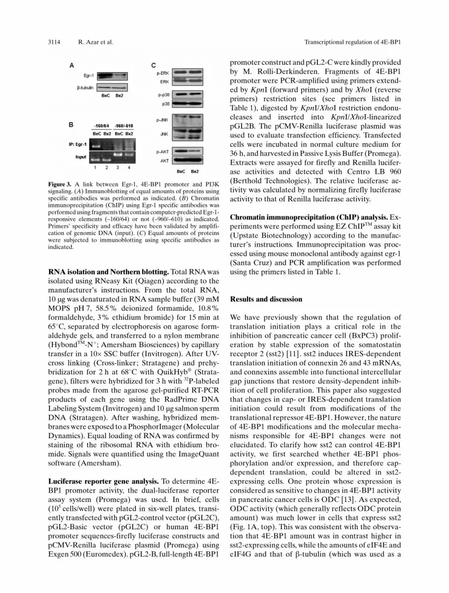
Figure 3. A link between Egr-1, 4E-BP1 promoter and PI3Ksignaling. (A) Immunoblotting of equal amounts of proteins usingspecific antibodies was performed as indicated. (B) Chromatinimmunoprecipitation (ChIP) using Egr-1 specific antibodies wasperformed using fragments that contain computer-predicted Egr-1-responsive elements (–160/64) or not (–960/–610) as indicated.Primers� specificity and efficacy have been validated by amplifi-cation of genomic DNA (input). (C) Equal amounts of proteinswere subjected to immunoblotting using specific antibodies asindicated.
3114 R. Azar et al. Transcriptional regulation of 4E-BP1

loading control) were similar in both cell lines(Fig. 1A, bottom). Furthermore, the use of 4E-BP1phospho-specific antibodies showed that Ser65 (andto a lesser extent Thr70) were hypophosphorylated insst2-expressing cells, while the signal detected with p-Thr37/46 antibodies perfectly reflected that of total4E-BP1, indicating that phosphorylations at Thr37 orThr46 were not regulated by sst2 (Fig. 1B). Asexpected, increased levels of 4E-BP1 and hypophos-phorylations of Ser65 and Thr70 resulted in increasedbinding of 4E-BP1 to eIF4E but were detrimental toeIF4G association with eIF4E in sst2-expressing cells(Fig. 1C). These data indicate that sst2 is capable ofinhibiting eIF4F assembly (and therefore cap-de-
pendent translation) via hypophosphorylation andaccumulation of 4E-BP1 protein.We have previously shown that, in BxPC3 cells stablyexpressing sst2, PI3K is inhibited as a consequence ofdirect binding of the regulatory p85 subunit to thereceptor [14], and it is well established that 4E-BP1phosphorylation is under the control of the PI3K-Akt/mTOR-signaling pathway. We therefore decided notto continue our investigations on the regulation of 4E-BP1 phosphorylation but instead to focus our atten-tion on the intriguing observation that 4E-BP1expression was regulated by sst2. We first determinedthe molecular step at which 4E-BP1 expression couldbe controlled by sst2. While treatment of cells with
Figure 4. PI3K controls 4E-BP1 promoter. (A) Equal amounts of proteins from cells untreated or treated with LY294002 for 24 h weresubjected to immunoblotting using specific antibodies as indicated. (B) Equal amounts of proteins from cells untreated or treated withLY294002 were subjected to immunoblotting using specific antibodies as indicated (top). ChIP using Egr-1 specific antibodies wasperformed as described in Fig. 3B (bottom). (C) Cells were untreated or treated with LY294002 36 h following transfection, and luciferaseactivity was assayed as described in Methods. Transfection efficiency was normalized to the concurrent transfection of a CMV-Renillaluciferase reporter plasmid. Histograms represent the means� SEM of three independent experiments performed in triplicates. They areexpressed as the ratio between luciferase activities measured in the presence of LY294002 and luciferase activities measured in the absenceof LY294002 (which were set at 1 for each cell line). (D) Luciferase activity was assayed 36 h following co-transfection of HEK cells with–960/+64 4E-BP1 promoter and pSG5 (mock) or p85 (wild-type p85), p-85-SH2 (an inactive form of p85), p110 (wild-type p110a) or p110-CAAX (a constitutively active form of p110a) plasmids, as indicated. Transfection efficiency was normalized to the concurrent transfectionof a CMV-Renilla luciferase reporter plasmid. Histograms represent the means � SEM of three independent experiments performed intriplicates. They are relative to the value obtained for mock (psG5) transfected cells and which was set at 1. t-test: * p<0.05; ** p<0.01; ***p<0.005.)
Cell. Mol. Life Sci. Vol. 65, 2008 Research Article 3115

cycloheximide showed that sst2 had no effect on 4E-BP1 protein half-life (data not shown), a pulse-labeling of cells revealed that 4E-BP1 de novo syn-thesis was, however, up-regulated in sst2-expressingcells (Fig. 2A). Similarly, treatment with actinomycin-D showed that sst2 had no effect on 4E-BP1 mRNAhalf-life (data not shown), while a Northern blotanalysis revealed that 4E-BP1 mRNA level was higherin sst2-expressing cells (Fig. 2B). To ensure that suchhigher amount of 4E-BP1 mRNA was actually aconsequence of increased transcription rate of 4E-BP1 gene, the promoter of 4E-BP1 was tested in aluciferase reporter assay. As compared to a vectordevoid of promoter sequence (pGL2B), a vectorcarrying a –960/+64 4E-BP1 promoter fragmentpermitted efficient luciferase expression in pancreaticBxPC3 cells, although it was weaker than the SV40promoter, which was used as a positive control(Fig. 2C). These data also revealed that 4E-BP1promoter was much more active in sst2-expressingcells and that sst2 specifically targeted 4E-BP1promoter as SV40 promoter activity was independentof sst2 expression (Fig. 2C).To go further in the delineation of the promoterfragment, and hence in the identification of thetranscription factor, which could be involved in sst2-dependent regulation of 4E-BP1 expression, a seriesof 4E-BP1 promoter deletion mutants has beengenerated, and tested in a luciferase reporter assay.Intriguingly, it was not possible to identify a uniquepromoter fragment that contained all the require-
ments for regulation by sst2. However, all 4E-BP1promoter segments possessing at least one GC-richEgr-1-responsive element were sensitive to sst2 ex-pression (Fig. 2D). None of the six GC-rich Egr-1putative elements (numbered 1–6 in Fig. 2D) wasprominent as they all contributed to sst2 effect on 4E-BP1 promoter. Conversely, the unique fragmentlacking Egr-1-responsive element (–960/–610) didnot respond to sst2. These data demonstrate that 4E-BP1 promoter activity is enhanced in sst2-expressingpancreatic cancer cells, and that the transcriptionfactor Egr-1 is a probable candidate involved in suchregulation.Egr-1 is an ”early growth response gene” encoding atranscription factor that has been shown to silence 4E-BP1 promoter activity following activation of theMAPK signaling pathway in hematopoietic cell lines[15]. Consistently, Egr-1 is overexpressed in pancre-atic cells that do not express sst2 (Fig. 3A). Further-more, a ChIP assay revealed that a fragment ofendogenous 4E-BP1 promoter that contains severalEgr-1-responsive elements can be immunoprecipitat-ed by anti-Egr-1-specific antibodies much more effi-ciently in cells lacking sst2 (Fig. 3B, compare lane 1 tolane 2), while a fragment lacking Egr-1-responsiveelement cannot (lanes 3 and 4). The regulation of Egr-1 expression in pancreatic cancer cells could not beattributed to changes in MAPK activity, as sst2 had noeffect on Erk1/2, p38 or JNK phosphorylation(Fig. 3C). However, the fact that Akt phosphorylationwas inhibited in sst2-expressing cells (Fig. 3C), andthat we have shown that PI3K is inhibited in BxPC3cells as a consequence of direct binding of theregulatory p85 subunit to sst2 [14], suggested that4E-BP1 expression could be regulated by the PI3Kpathway. This suggestion was further supported byearlier papers reporting that Egr-1 expression can betargeted by PI3K signaling following activation ofother G protein-coupled receptors [16, 17].We therefore anticipated that 4E-BP1 transcriptioncould be maintained repressed by the PI3K signalingpathway in pancreatic cancer cells, and that sst2 couldrelease 4E-BP1 expression as a consequence of PI3Kinhibition. As expected, pharmacological inhibition ofPI3K activity by LY294002, attested by inhibition ofAkt phosphorylation, increased 4E-BP1 protein level(Fig. 4A, compare lane 1 to lane 2). PI3K inhibitionalso provoked a decrease in Egr-1 expression whichwas accompanied by a corresponding decrease in theamount of Egr-1 bound to endogenous 4E-BP1promoter (Fig. 4B), and a corresponding increase in4E-BP1 promoter activity (Fig. 4C, BxPC3 cells).Furthermore, the implication of PI3K signaling inthe regulation of 4E-BP1 transcriptional expressionwas not limited to exocrine pancreatic cells as treat-
Figure 5. PI3K controls cap-dependent translation through 4E-BP1 phosphorylation and transcription. Following PI3K activationby extracellular stimuli, 4E-BP1 gene (eif4ebp1) transcription isrepressed (possibly through Egr-1) and induction of the Akt/mTOR pathway leads to 4E-BP1 phosphorylation. Consequently,sequestered eIF4E (4E) is released and can serve for cap-depend-ent translation initiation.
3116 R. Azar et al. Transcriptional regulation of 4E-BP1

ment with LY294002 enhanced 4E-BP1 protein ex-pression in cells originating from human pancreaticendocrine tumors (Fig. 4A, BON cells), and enhanced4E-BP1 promoter activity in BON cells (Fig. 4C, BONcells), and in cells originating from human embryonickidney (Fig. 4C, HEK cells).Finally, 4E-BP1 promoter activity could be eitherenhanced by the use of a dominant negative mutant ofthe p85a regulatory subunit (p85-SH2) or inhibited bythe use of a constitutively active mutant of thecatalytic p110a subunit (p110-CAAX) of PI3K(Fig. 4D), thus indicating that PI3Ka subunits areinvolved in 4E-BP1 regulation. These tests were alsoperformed to ensure that the effects obtained withLY294002 were not due to nonspecific inactivation ofother kinases such as PI4K.Taken together, these data demonstrate that, inaddition to 4E-BP1 inactivation by phosphorylation,signaling through PI3K pathway also impinges upon4E-BP1 amount via transcriptional silencing (Fig. 5).Egr-1 appears as a good candidate involved in PI3K-dependent repression of 4E-BP1 gene transcription.Because 4E-BP1 expression has been shown to bedown-regulated in many high-grade tumors and sincePI3K activity is often enhanced in cancer cells, itwould be interesting to determine whether 4E-BP1loss in tumors is due to higher PI3K activity. Finally, itis probable that PI3K can regulate 4E-BP1 tran-scription through other transcription factors. Thisspeculation can be deduced from two reports showingthat in Drosophila 4E-BP1 transcription is under thecontrol of FOXO [18], a forkhead transcription factorwhich is itself controlled by PI3K signaling [19].
Acknowledgements. We thank M. Rolli-Derkinderen for 4E-BP1promoter sequence, B. Vanhaesebroeck for p85 and p110 con-structs and N. Sonenberg for eIF4GI antibody. This work wasfunded by grants from INSERM, from La Ligue Contre le Cancer,from Canc�rop�le Grand-Sud-Ouest and from Association pour laRecherche sur le Cancer (ARC). R. Azar was a recipient offellowships from ARC, from Fondation pour la RechercheM�dicale (FRM), from Agence Universitaire de la Francophonie(AUF).
1 Hennessy, B. T., Smith, D. L., Ram, P. T., Lu, Y. and Mills, G. B.(2005) Exploiting the PI3K/AKT pathway for cancer drugdiscovery. Nat. Rev. Drug Discov. 4, 988–1004
2 Bader, A. G., Kang, S., Zhao, L. and Vogt, P. K. (2005)Oncogenic PI3K deregulates transcription and translation.Nat. Rev. Cancer 5, 921–929
3 Stocker, H., Radimerski, T., Schindelholz, B., Wittwer, F.,Belwat, P., Daram, P., Breuer, S., Thomas, G. and Hafen, E.(2003) Rheb is an essential regulator of S6K in controlling cellgrowth in Drosophila. Nat. Cell Biol. 5, 559–565
4 Long, X., Lin, Y., Ortiz-Vega, S., Yonezawa, K. and Avruch, J.(2005) Rheb binds and regulates the mTOR kinase. Curr.Biol. 15, 702–713
5 Wang, X., Beugnet, A., Murakami, M., Yamanaka, S. andProud, C. G. (2005) Distinct signaling events downstream ofmTOR cooperate to mediate the effects of amino acids andinsulin on initiation factor 4E-binding proteins. Mol. Cell.Biol. 25, 2558–2572
6 Gingras, A. C., Raught, B., Gygi, S. P., Niedzwiecka, A., Miron,M., Burley, S. K., Polakiewicz, R. D., Wyslouch-Cieszynska,A., Aebersold, R. and Sonenberg, N. (2001) Hierarchicalphosphorylation of the translation inhibitor 4E-BP1. GenesDev. 15, 2852–2864
7 Ferguson, G., Mothe-Satney, I. and Lawrence. J. C. (2003) Ser-64 and Ser-111 in PHAS-I are dispensable for insulin-stimu-lated dissociation from eIF4E. J. Biol. Chem. 278, 47459–47465
8 Wang, X., Li, W., Parra, J. L., Beugnet, A. and Proud, C. G.(2003) The C terminus of initiation factor 4E-binding protein 1contains multiple regulatory features that influence its functionand phosphorylation. Mol. Cell. Biol. 23, 1546–1557
9 Pyronnet, S., Dostie, J. and Sonenberg, N. (2001) Suppressionof cap-dependent translation in mitosis. Genes Dev. 15, 2083–2093
10 Pyronnet, S., Imataka, H., Gingras, A. C., Fukunaga, R.,Hunter, T. and Sonenberg, N. (1999) Human eukaryotictranslation initiation factor 4G (eIF4G) recruits mnk1 tophosphorylate eIF4E. EMBO J. 18, 270–279
11 Lahlou, H., Fanjul, M., Pradayrol, L., Susini, C. and Pyronnet,S. (2005) Restoration of functional gap junctions throughinternal ribosome entry site-dependent synthesis of endoge-nous connexins in density-inhibited cancer cells. Mol. Cell.Biol. 25, 4034–4045
12 Pyronnet, S., Pradayrol, L. and Sonenberg, N. (2000) A cellcycle-dependent internal ribosome entry site. Mol. Cell. 5,607–616
13 Pyronnet, S., Gingras, A. C., Bouisson, M., Kowalski-Chauvel,A., Seva, C., Vaysse, N., Sonenberg, N. and Pradayrol, L. (1998)Gastrin induces phosphorylation of eIF4E binding protein 1and translation initiation of ornithine decarboxylase mRNA.Oncogene 16, 2219–2227
14 Bousquet, C., Guillermet-Guibert, J., Saint-Laurent, N.,Archer-Lahlou, E., Lopez, F., Fanjul, M., Ferrand, A., Fourmy,D., Pichereaux, C., Monsarrat, B., Pradayrol, L., Est�ve, J. P.and Susini, C. (2006) Direct binding of p85 to sst2 somatostatinreceptor reveals a novel mechanism for inhibiting PI3Kpathway. EMBO J. 25: 3943–3954
15 Rolli-Derkinderen, M., Machavoine, F., Baraban, J. M.,Grolleau, A., Beretta, L. and Dy, M. (2003) ERK and p38inhibit the expression of 4E-BP1 repressor of translationthrough induction of Egr-1. J. Biol. Chem. 278, 18859–18867
16 Guillemot, L., Levy, A., Raymondjean, M. and Rothhut, B.(2001) Angiotensin II-induced transcriptional activation of thecyclin D1 gene is mediated by Egr-1 in CHO-AT(1A) cells. J.Biol. Chem. 276, 39394–39403
17 Fujino, H., Xu, W. and Regan, J. W. (2003) Prostaglandin E2induced functional expression of early growth response factor-1 by EP4, but not EP2, prostanoid receptors via the phospha-tidylinositol 3-kinase and extracellular signal-regulated kin-ases. J. Biol. Chem. 278, 12151–12156
18 Tettweiler, G., Miron, M., Jenkins, M., Sonenberg, N. andLasko, P. F. (2005) Starvation and oxidative stress resistance inDrosophila are mediated through the eIF4E-binding protein,d4E-BP. Genes Dev. 19, 1840–1843
19 Puig, O., Marr, M. T., Ruhf, M. L. and Tjian, R. (2003) Controlof cell number by Drosophila FOXO: Downstream andfeedback regulation of the insulin receptor pathway. GenesDev. 17, 2006–2020
Cell. Mol. Life Sci. Vol. 65, 2008 Research Article 3117


