Embed Size (px)
Citation preview

Phosphatidylserine externalization and membrane blebbing areinvolved in the nonclassical export of FGF1
Aleksandr Kirov1, Huda Al-Hashimi1, Phil Solomon1, Courtney Mazur1, Philip E. Thorpe2,Peter J. Sims3, Francesca Tarantini4, Thallapuranam K. Suresh Kumar5, and IgorPrudovsky1,*
1Maine Medical Center Research Institute, Scarborough, Maine Medical Center, ME040742Department of Pharmacology and the Harold Simmons Cancer Center, UT Southwestern,Dallas, TX753903Departmentof Pathology & Laboratory Medicine, University of Rochester School of Medicine andDentistry, Rochester, NY146424Department of Critical Care Medicine and Surgery, Geriatric Medicine Unit, University ofFlorence, Florence50139, Italy5Department of Chemistry & Biochemistry, University of Arkansas, Fayetteville, AR 72701
AbstractThe mechanisms of nonclassical export of signal peptide-less proteins remain insufficientlyunderstood. Here we demonstrate that stress-induced unconventional export of FGF1, a potent andubiquitously expressed mitogenic and proangiogenic protein, is associated with and dependent onthe formation of membrane blebs and localized cell surface exposure of phosphatidylserine. Inaddition, we found that the differentiation of promonocytic cells results in massive FGF1 release,which also correlates with membrane blebbing and exposure of phosphatidylserine. These findingsindicate that the externalization of acidic phospholipids could be used as a pharmacological targetto regulate the availability of FGF1 in the organism.
KeywordsFGF1; nonclassical secretion; phosphatidylserine exposure; blebbing; PLSCR1
IntroductionMany extracellular proteins do not have a signal peptide in their primary structure, and thuscannot be secreted through the classical export pathway involving the endoplasmicreticulum (ER) and Golgi apparatus. Instead, they use unconventional export mechanismsthat are not fully understood (Grieve and Rabouille, 2011; Nickel and Seedorf, 2008;Prudovsky et al., 2008). Among the nonclassically released proteins are the mostubiquitously expressed members of the FGF family, FGF1 and FGF2 that play importantroles in tissue repair, angiogenesis, inflammation, and tumorigenesis (Dorey and Amaya;Korc and Friesel, 2009; Ornitz and Itoh, 2001). FGF1 is released from fibroblastic cellsunder various conditions of cellular stress (Ananyeva et al., 1997; Jackson et al., 1992;Mouta Carreira et al., 2001; Shin et al., 1996)as a part of a copper-dependent (Landriscina et
*To whom correspondence should be addressed: Igor Prudovsky, Center for Molecular Medicine, Maine Medical Center ResearchInstitute, 81 Research Dr., Scarborough, ME 04074. Tel: (207)-396-8146. Fax: (207)-396-8179., [email protected].
NIH Public AccessAuthor ManuscriptJ Cell Biochem. Author manuscript; available in PMC 2013 March 1.
Published in final edited form as:J Cell Biochem. 2012 March ; 113(3): 956–966. doi:10.1002/jcb.23425.
NIH
-PA Author Manuscript
NIH
-PA Author Manuscript
NIH
-PA Author Manuscript

al., 2001a)multiprotein complex that also includes sphingosine kinase 1 (Soldi et al., 2007),the 40 kDa form of synaptotagmin 1 (LaVallee et al., 1998) and the small calcium-bindingprotein S100A13 (Landriscina et al., 2001b). Previously, we found that under conditions ofcellular stress, FGF1 migrates from the internal areas of cytoplasm to the vicinity of the cellmembrane (Prudovsky et al., 2002). However, it remained unclear whether FGF1 exportproceeds through the entire cell surface or occurs in limited membrane domains.
In the present study, we demonstrate that under stress conditions in fibroblastic cells, FGF1is exported through cell membrane domains characterized by the formation of protrusionsand externalization of phosphatidylserine (PS), an acidic phospholipid that under normalconditions is confined to the inner leaflet of the plasma membrane. Moreover, smallmolecular compounds that inhibit externalization of PS also repressed FGF1 export. Anartificial increase in interactions between the cell membrane and the underlying peripheralactin cytoskeleton significantly suppressed FGF1 secretion, thereby confirming a role formembrane blebbing in FGF1 export. In addition, we found that FGF1 export fromdifferentiated U937 promonocytic leukemia cells also correlates with membrane blebbingand PS externalization. Chelation of intracellular Ca2+ resulted in the inhibition of both PSexposure and FGF1 export in differentiated U937 cells. Although U937 differentiation isaccompanied by a dramatic increase in the expression of phospholipid scramblase 1(PLSCR1), a protein originally proposed to mediate Ca2+ -activated transbilayer movementof phospholipids resulting in cell surface exposure of PS(Sims and Wiedmer, 2001), weshow that the expression of PLSCR1 is unrelated to FGF1 export under these conditions.
Materials and MethodsCell Culture
Murine NIH 3T3 cells (ATCC, Manassas, VA) were maintained in DMEM (HyClone,Logan, UT) supplemented with 10% bovine calf serum (HyClone). U937 cells (ATCC) weregrown in RPMI medium (HyClone) supplemented with 10% fetal calf serum. U937 cellsstably transfected with FGF1 (Mandinova et al., 2003)or FGF1:GFP were grown in mediumsupplemented with 400 μg/ml geneticin (Invitrogen, Carlsbad, CA).
Genetic constructsFGF1 cloned in the pMEXneo expression vector (Jackson et al., 1992)was recloned into theSalI and EcoRI restriction sites of the pcDNA3/HA vector (Clontech, Mountain View, CA)thus forming FGF1:HA. Then, FGF1:HA was cloned into the multiple cloning site (MCS) ofthe shuttle vector pAdlox (generous gift of Lisa Phipps, Somatix Therapy Corporation, CA).
Mouse PLSCR1:GFP was expressed in the pMiG retroviral vector (Nanjundan et al., 2003).
FGF1:GFP cloned in the pcDNA 3.1 vector was a kind gift of Andrew Baird (HumanBioMolecular Research Institute, San Diego, CA).
Constitutively active (CA) mutant T567D and wild type (WT) ezrin, both with the 3′attachment of the VSVG tag (Algrain et al., 1993) were excised using Hind III and XbaIenzymes from the pC6 vector. They were then cloned into the MCS of the TRE2Hygroexpression vector (Clontech), in which Hind III and XbaI restriction sites were introducedby PCR mutagenesis.
CMVt-rtTA construct was a kind gift of John Hiscott (McGill University, Montreal).
Human PLSCR1 shRNA construct and control shRNA cloned in the pGFP-V-RS retroviralvector were obtained from Origene (Rockville, MD). The following target sequence
Kirov et al. Page 2
J Cell Biochem. Author manuscript; available in PMC 2013 March 1.
NIH
-PA Author Manuscript
NIH
-PA Author Manuscript
NIH
-PA Author Manuscript
inPLSCR1 mRNA was used: 5′-TGAAAGTCTCCTCAGGAAATCTGAAGTCT-3′ (Zhaoet al., 2004).
Production of viruses and viral transfectionRecombinant FGF1:HA adenovirus was produced, purified, and titered as described (Duarteet al., 2008). Briefly, CRE8 cells were transfected with SfiI-digested pAdlox-derivedconstructs, and infected with the ψ5 virus. The lysates were prepared 2 days after infection.The virus was passed twice through CRE8 cells, and purified from the second passage usinga cesium density gradient. The virus was quantified by optical density at 260 nm, and thebioactivity was determined by a plaque-forming unit assay. Adenoviral transduction wasperformed in serum-free DMEM with approximately 103 viral particles/cell in the presenceof poly-D-Lysine hydrobromide (Sigma) (5×10³ molecules/viral particle) for 2 h at 37°C.Then the adenovirus-containing medium was removed and replaced with serum-containingmedium. The cells were plated for experiments 24–48 hours after transduction.
PLSCR1:GFP and control GFP retroviruses were produced in the Recombinant Viral VectorCore of MMCRI. The packaging cell line Bosc was transfected with the PLSCR1:GFP orGFP coding retroviral constructs using polybrene. Conditioned medium from the 2 dayculture of the producer cell line was collected and filtered to remove cell debris. Activelygrowing recipient cells were incubated for 2 h with retrovirus-containing conditionedmedium in the presence of 5μg/ml hexadimethrine bromide. The medium was replaced withfresh growth medium after 2 h.
Generation of stable cell transfectantsTo achieve inducible expression of ezrin, the NIH 3T3 cells were cotransfected with CMVt-rtTAand WT ezrin/pTRE2Hygro, CA ezrin/PTRE2Hygro or empty pTRE2Hygro vectorusing the Fugene reagent (Roche, Nutley, NJ) according to the manufacturer’s protocol.Transfected cell clones were selected in the medium containing 2 μg/ml puromycin (Sigma,St. Louis, MO) and 50 mg/ml hygromycin (Roche). To assess the inducibility of ezrinexpression, cells of individual clones were plated on glass coverslips and incubated for 48 hin medium containing 0 or 10 μg/ml doxycycline (Sigma). The cells were formalin fixed andthe doxycycline-inducible expression of WT or CA ezrin was verified byimmunofluorescence using anti-VSVG antibodies (Sigma) followed by secondary FITC-conjugated antibodies (Vector Laboratories).
U937 cells expressing FGF1 (Mandinova et al., 2003) were retrovirally transfected withPLSCR1 shRNA and control shRNA, and selected in medium containing 2 μg/mlpuromycin (Sigma, St Louis, MO). After three weeks of selection, GFP fluorescence wasobserved in 10–20 % of the surviving cells. GFP-positive cells were then selected by flowcytometry (FACSVantage, BD) in the Flow Cytometry Core of MMCRI.
U937 cells expressing FGF1 (Mandinova et al., 2003) were retrovirally transduced withPLSCR1:GFP or GFP. Three days after transduction, GFP fluorescence was observed in 5%of the cells. The GFP-positive cells were selected using flow cytometry and furtherpropagated. Cell populations with at least 90% of GFP-positive cells were used forexperiments.
U937 cells were transfected with FGF1:GFP using the Nucleofector II system (Koeln,Germany) and special Amaxa transfection buffer C. After three weeks of selection in themedium with 800 μg/ml G418, GFP fluorescence was observed in 5–10% of the survivingcells. GFP-positive cells were then selected using flow cytometry.
Kirov et al. Page 3
J Cell Biochem. Author manuscript; available in PMC 2013 March 1.
NIH
-PA Author Manuscript
NIH
-PA Author Manuscript
NIH
-PA Author Manuscript

Fluorescence microscopyTo simultaneously assess the externalization of FGF1 and PS, NIH 3T3, cells transducedwith FGF1:HA were incubated for 20 min at 4°C with anti-HA monoclonal antibodies(Covance, Princeton, NJ) and FITC-conjugated Annexin V (Invitrogen) in buffer composedof 10 mM HEPES (pH 7.5), 140 mM NaCl and 2.5 mM CaCl2. Next, the cells were fixed in10% neutral formalin, and externalized FGF1 was detected by using the CY3-conjugatedanti-mouse IgG antibody (Sigma).
Alternatively, the cells were incubated for 20 min at 4°C with anti-HA monoclonalantibodies (Covance) together with the PS-targeting chimeric antibody, bavituximab(Peregrine Pharmaceuticals Inc., Tustin, CA), in PBS supplemented with 1% bovine serumalbumin. Bavituximab binds to PS in a β2-glycoprotein I-dependent manner (Luster et al.,2006). Next, the cells were fixed in 10% neutral formalin, externalized FGF1 was detectedusing the CY3-conjugated anti-mouse IgG antibody (Sigma), and externalized PS wasdetected by using FITC-conjugated anti- human IgG antibody (Invitrogen).
To visualize external and internal cell membranes, the cells were fixed for 10 min in 10%neutral formalin, incubated 30 min in blocking buffer (PBS with 5% bovine serum albuminand 0.1% Triton X100), and then stained with 5,5′-Phe2-DilC18(3) or Vybrant (both fromInvitrogen) in the blocking buffer. Nuclei were stained with TO-PRO3 (Invitrogen). Tovisualize the actin cytoskeleton, fixed and permeabilized cells were stained with Alexa488-conjugated phalloidin (Invitrogen).
To simultaneously visualize externalized and intracellular FGF1:HA, the transduced NIH3T3 cells were incubated for 20 min at 4°C with FITC-conjugated anti-HA antibodies(Sigma) in buffer composed of 10mM Hepes (pH 7.5), 140 mM NaCl, and 2.5 mM CaCl2,then fixed with 10% neutral formalin, permeabilized with 0.1% Triton X100, incubated withanti-HA rabbit monoclonal antibodies (Covance), and then with the CY3-conjugated anti-rabbit IgG antibodies (Sigma).
To visualize the externalized PS in U937 cells, we incubated live cells with phycoerythrin-conjugated Annexin V (Invitrogen). The visualization of externalized FGF1:GFP in stablytransfected U937 cells was achieved by incubation of live cells with E6 monoclonal anti-GFP antibodies (Invitrogen) followed by formalin fixation and staining with CY3-conjugated anti-mouse IgG antibodies.
Fluorescent preparations were embedded in Vectashield (Vector Laboratories, Burlingame,CA), and the images were taken using the Leica SP1 confocal microscope at the MMCRIconfocal microscopy facility.
Analysis of PLSCR1 expressionRT-PCR analysis of PLSCR1 expression was performed with total RNA isolated from U937cells using the RNeasy kit (Qiagen, Valencia, CA). Total RNA (1 μg) was used as atemplate for the PCR reaction performed with the Platinum Tap One Step RT-PCR Kit(Invitrogen). The following PCR primers were utilized: forward 5′-GGTGCCTGTTTCCTCATTGAC-3′ and reverse 5′-GTCCTTTTTCTCAAATTGAC-3′. β-actin expression served as a control for RNA loading. The amplification products werevisualized by 1.5% agarose gel electrophoresis and ethidium bromide staining.
Analysis of FGF1 releaseThe NIH 3T3 cells were used to study FGF1 release 48 h after adenoviral transduction withFGF1. The heat shock-induced FGF1 release assay was performed by incubation of the cells
Kirov et al. Page 4
J Cell Biochem. Author manuscript; available in PMC 2013 March 1.
NIH
-PA Author Manuscript
NIH
-PA Author Manuscript
NIH
-PA Author Manuscript

at 42°C for 110 minutes in serum-free DMEM containing 5 U/ml of heparin (Sigma), aspreviously described (Jackson et al., 1992). Control cultures were incubated at 37°C for thesame period. Conditioned media were collected, briefly centrifuged at 1000 g to removedetached cells and FGF1 was isolated for immunoblot analysis using heparin-Sepharosechromatography as described. A similar method was used to study the export of stablytransfected FGF1 from adherent differentiated U937 cells. To assess the FGF1 release fromnonadherent U937 cells, the cells were collected by centrifugation and then resuspended inserum-free DMEM containing 5 U/ml of heparin.
Cell viability was assessed by measuring lactate dehydrogenase (LDH) activity in themedium using the Promega CytoTox kit (Promega, Madison, WI).
To analyze the association of released FGF1 with microparticles and exosomes, theconditioned media were pre-centrifuged for 10 min at 1000 g to remove detached cells andthen supernatants were centrifuged for 1 h at 100,000 g. The supernatants were removed andpellets were resuspended in 1 ml PBS containing 0.1% Triton X 100 to lyse the precipitatedvesicles. FGF1 was isolated from the supernatants and pellet lysates using heparinchromatography.
Results1. Stress-induced export of FGF1 occurs in the limited domains of the cell surfacecharacterized by membrane protrusions
To investigate the topologic distribution of FGF1 that is exported to the cell surface, NIH3T3 cells were adenovirally transduced with FGF1:HA and incubated for 110 min inDMEM at 42°C (heat shock) or 37°C (control). The cells were incubated at 4°C for anadditional 20 min in isotonic HEPES buffer with Ca2+ and FITC-conjugated mousemonoclonal anti-HA antibodies, and then fixed. Following the fixation, the cells werepermeabilized and stained for intracellular FGF1 (using rabbit anti-HA antibodies followedby CY3-conjugated secondary antibodies) or membranes (using 5,5′-Phe2-DilC18(3) orVybrant lipid stains). As expected, non-stressed cells were largely negative for externalizedFGF1. The stressed cells exhibited distinct 0.5–8 μm round or oval domains that werepositive for externalized FGF1 (Figure 1A). Typically, these domains were alsocharacterized by an increased concentration of intracellular FGF1 (Figure 1B). Membranestaining and confocal studies at high magnification demonstrated that portals of FGF1release were usually associated with membrane blebs (Figure 1C). The expression of FGF1at the cell surface was stress-dependent and transient. Indeed, 2 h after the cells werereturned to37°C, the FGF1-HA positivity of cell surface was mostly lost (data not shown).The presence of externalized FGF1 on cell membrane protrusions could indicate that FGF1is released into the medium as a component of the detached blebs. Indeed, bleb-derivedmicroparticles are involved in the export of multiple biologically active proteins (Al-Nedawiet al., 2009; Hendrix et al.; Sabatier et al., 2009). To examine this possibility, membraneparticles were collected by ultracentrifugation from media conditioned by heat shockedFGF1-expressing NIH 3T3 cells as described for FGF2 by Nickel et al. (Seelenmeyer et al.,2008). The resulting pellet and supernatant were examined for the presence of FGF1.Almost all of the released FGF1 was found in the supernatant (Figure 1D). Based on theseresults, we conclude that cell surface protrusions represent the portals but not the vehiclesfor stress-induced FGF1 export.
2. FGF1 export domains contain externalized PSFGF1 and other members of FGF1 release complex bind PS with high affinity (Prudovsky etal., 2008; Tarantini et al., 1995). We have shown that FGF1 specifically destabilizes bilayers
Kirov et al. Page 5
J Cell Biochem. Author manuscript; available in PMC 2013 March 1.
NIH
-PA Author Manuscript
NIH
-PA Author Manuscript
NIH
-PA Author Manuscript

composed of acidic phospholipids, such as PS, phoshatidylglycerol and phosphatidylinositol(Graziani et al., 2006). Thus, we hypothesized that stress conditions, resulting in themovement of PS from inner to outer plasma membrane leaflet might provide a mechanismfor transport of FGF1 from the cytosol into the external medium (Prudovsky et al., 2008).To assess this hypothesis for nonclassical export of FGF1, we incubated stressed and controlcells with a mixture of anti-HA antibodies and FITC-labeled Annexin V, which binds toexternalized PS. After fixation, the cells were incubated with CY3-conjugated secondaryantibodies. Confocal microscopy revealed that the FGF1 export domains were PS positive(Figure 1E,F). These results were confirmed with bavituximab (Marconescu and Thorpe,2008) that binds and stabilizes complexes of PS and β2-glycoprotein I (Luster et al., 2006).Bleb-like structures on the surface of heat shocked cells were coincidently stained with bothanti-HA antibodies and bavituximab (Figure 1G). These results suggest that FGF1 exportoccurred in conjunction with the externalization of PS. The stress conditions we used werestrictly controlled and did not induce cell death as assessed by the absence of LDH releaseinduction in all supernatants and by DAPI staining used to detect the formation of apoptoticnuclear fragments (data not shown).
3. Chemical compounds inhibiting PS externalization repress stress-induced FGF1 exportOur next aim was to determine if there is a causal relationship between cell surface PSexposure and stress-induced FGF1 export. The asymmetric distribution of PS between innerand outer plasma membrane leaflets is believed to be regulated by multiple membrane-associated enzymes (Lenoir et al., 2007). The most extensively studied are: (1)aminophospholipid translocase (Seigneuret and Devaux, 1984), a Type IV ATPase thatserves to sequester PS to the inner surface of the plasma membrane by catalyzing itsmovement from outer to inner leaflet; and (2) phosholipid scramblase 1 (PLSCR1) (Simsand Wiedmer, 2001), a membrane-associated protein that was isolated and characterizedbased upon its ability to mediate collapse of transbilayer phospholipid asymmetry throughCa2+-activated bidirectional transbilayer movement of PS and other lipids in reconstitutedproteoliposomes, and was proposed to serve this same “phospholipid scramblase” functionin the plasma membrane. However, no chemical compound has been Identified that targets aspecific PS translocating enzyme and thus induce or inhibit PS exposure.
At the same time, several chemicals were reported to suppress PS externalization. Toexplore the functional relationship between movement of PS to the cell surface and export ofFGF1 into the cell medium, we undertook to identify inhibitors of both processes. Taurine isa non-essential amino acid that is a membrane-stabilizing and cytoprotective (Anderheggenet al., 2006) agent, which is present in high concentrations in animal organisms and used asan anti- inflammatory nutrient supplement (Yamori et al., 2010). Using in vivo staining withphycoerythrin- conjugated annexin V, we found that taurine caused an inhibition of heatshock-induced PS externalization in NIH 3T3 cells (Figure 2A). Using heparinchromatography followed by immunoblotting, we found that taurine also induced aconcentration-dependent inhibition of FGF1 export from NIH 3T3 cells (Figure 2B).
In addition, the β-adrenergic receptor agonist, isoproterenol, has been reported to inhibit cellsurface PS exposure (Lang et al., 2005). We found that similar to taurine, this compoundinhibited PS exposure and FGF1 export (Figure 2C,D).
Thus, two unrelated molecules were demonstrated to suppress both PS externalization andFGF1 export.
Kirov et al. Page 6
J Cell Biochem. Author manuscript; available in PMC 2013 March 1.
NIH
-PA Author Manuscript
NIH
-PA Author Manuscript
NIH
-PA Author Manuscript

4. Strengthening the binding between plasma membrane and the submembranecytoskeleton inhibitsFGF1 export
Bleb formation relies on the local weakening of the connection between the plasmamembrane and submembrane actin (Charras et al., 2009; Fackler and Grosse, 2008). The cellmembrane is bound to actin by the proteins of ERM family, one of which is ezrinphosphorylated at threonine 567(Arpin et al., 2011). The expression of the constitutivelyactive (CA) T567D mutant of ezrin results in the stable binding of actin cytoskeleton to thecell membrane (Fievet et al., 2007). We generated NIH 3T3 cells that inducibly express WTor CA ezrin after doxycycline stimulation (Figure 3A). We compared FGF1 release from thedoxycycline-stimulated NIH 3T3 cells cotransfected with rtTA and either WT ezrin, CAezrin or empty vector (Figure 3C). Doxycycline caused considerable FGF1 export from allthese cells at normal temperature, which may be due to its stressful effect on mitochondria(van den Bogert et al., 1986). However, heat shock significantly increased FGF1 secretion indoxycycline-treated cells. We found that in the cells expressing CA ezrin the export of FGF1was suppressed both at 37 and 42°C (Figure 3C). Cells expressing CA ezrin also exhibited adecreased externalization of PS after heat shock (Figure 3B). These data support theimportance of bleb formation (which is dependent on the decrease of cytoskeleton-membrane interaction) for the export of FGF1.
5. Bleb formation and externalization of PS correlate with FGF1 export in differentiatedpromonocytic leukemia cells
The correlation between FGF1 export and blebbing in fibroblastic cells prompted us toexplore whether a similar connection can be observed in a promonocyte culture, U937.Therefore, we used U937 cells stably transfected with FGF1. We assessed FGF1 export at37 and 42°C in undifferentiated U937 cells and in cells induced to undergo a macrophage-like differentiation by treatment with 150 nM PMA for 48 h (Figure 4A). Althoughundifferentiated cells growing in suspension exhibited a barely detectable FGF1 export,adherent differentiated cells were characterized by a massive FGF1 release (Figure 4A).Interestingly, heat shock only marginally induced FGF1 secretion from differentiated cells(Figure 4A), and lactate dehydrogenase release assays did not reveal cell damage undereither PMA treatment or heat shock (results not shown). Undifferentiated U937 cells had asmooth surface both at normal temperature and at the heat shock (Figure 4B). In contrast,adherent differentiated U937 cells formed numerous membrane blebs (Figure 4B). Inaddition, PMA-treated U937 cells exhibited localized externalization of PS (Figure 4C). Tovisualize the release of FGF1 to the cell surface, we used U937 cells stably transfected withFGF1:GFP. In this experiment, externalized FGF1 was detected using monoclonal anti-GFPantibodies followed by CY3-conjugated secondary antibodies, while internal FGF1 wasdetected by GFP fluorescence (Figure 4D,E). As expected, undifferentiated U937 cells didnot show significant externalization of FGF1, while pronounced FGF1 externalization wasobserved in differentiated cells. Unlike heat shocked NIH 3T3, we did not observe distinctdomains enriched in externalized FGF1 in differentiated U937 cells. This is apparently dueto the diffusion of externalized FGF1 across the cell surface during the prolongeddifferentiation period, which is characterized by high mobility of cell membrane. In contrast,the localized distribution of externalized PS could be explained by its relatively shortlifetime on cell surface.
In conclusion, these experiments demonstrate that macrophage-like differentiation results instrong FGF1 export, which correlates with membrane blebbing and PS externalization.
Kirov et al. Page 7
J Cell Biochem. Author manuscript; available in PMC 2013 March 1.
NIH
-PA Author Manuscript
NIH
-PA Author Manuscript
NIH
-PA Author Manuscript

6. Intracellular calcium, but not PLSCR1 is required for FGF1 export from differentiatedU937 cells
Because differentiation of U937 cells is accompanied by increased cytosolic calciumconcentration (Klein et al., 1990), which can stimulate PS externalization (Vance andSteenbergen, 2005), we assessed the effect of the cell-permeable calcium chelator, BAPTA-AM, on FGF1 export from PMA-treated U937 cells. We found that BAPTA-AM inhibitedboth FGF1 export (Figure 5A) and PS exposure (Figure 5B) in these cells. Interestingly,copper chelator tetrathiomolybdate (TM), which inhibits FGF1 export from stressedfibroblasts (Landriscina et al., 2001a), failed to suppress FGF1 secretion from differentiatedU937 macrophages (Supplementary Figure 1).
PLSCR1 is an endofacial plasma membrane protein proposed to mediate intracellular Ca2+-activated transbilayer movement of plasma membrane phospholipids, resulting in PSexposure at the cell surface (Sims and Wiedmer, 2001). Chen et al. showed thatundifferentiated U937had low expression of PLSCR1, which was dramatically enhancedupon the induction of cell differentiation (Huang et al., 2006). We confirmed these resultsusing RT-PCR with specific PLSCR1 primers (Figure 5C). The marked contrast ofundifferentiated U937 that do export very low amounts of FGF1 versus PMA-differentiatedU937 that extensively release FGF1 suggested that this up-regulation of FGF1 export uponcell differentiation might be directly related to the concomitant up-regulation of PLSCR1expression. To examine this possibility, U937 cells stably overexpressing PLSCR1:GFPwere generated. We found that the over-expression of PLSCR1 failed to stimulate FGF1release from heat shocked undifferentiated U937 cells, and did not enhance FGF1 secretionstimulated by PMA (Figure 5D). Furthermore, we produced U937 cells in which PLSCR1had been stably knocked down (Figure 5E). We found that PLSCR1 knockdown had noeffect on FGF1 secretion in PMA-treated cells (Figure 5F).
DiscussionUnlike spontaneous FGF2 export that occurs through submicron-sized non-raft membranepatches evenly distributed through the cell surface (Engling et al., 2002), stress-inducedFGF1 secretion proceeds through large membrane domains. The co-localization ofexternalized FGF1 and PS in these domains corroborates our earlier hypothesis that stress-induced translocation of acidic phospholipids could be a driving force of stress-dependentFGF1 export (Prudovsky et al., 2008). We demonstrated that FGF1 not only selectivelybinds PS (Tarantini et al., 1995)but also efficiently destabilizes artificial membranescomposed of acidic phospholipids, such as PS, phosphatidylglycerol andphosphatidylinositol, but not zwitterionic phosphatidylcholine (Graziani et al., 2006). The40 kDa form of synaptotagmin 1, a critical member of the FGF1 export complex, whichefficiently binds PIP2 (Sutton et al., 1995), permeabilizes artificial bilayers composed ofphosphatidylinositol but not of phosphatidylcholine. Moreover, mutations of FGF1 and 40kDa synaptotagmin 1, which abolish the destabilization of acidic phospholipid liposomes,strongly inhibit the stress-induced release of these proteins. In addition, another member ofFGF1 export pathway, sphingosine kinase 1 (Stahelin et al., 2005) binds PS with highaffinity. The transmembrane translocation of PS occurs both in the plasma membrane undercell stress and in the membranes of the trans-Golgi network under normal conditions (Fairnet al., 2011). The mechanisms underlying these processes are not well understood. Schroit etal. (Mirnikjoo et al., 2009) demonstrated that PS externalization in apoptotic cells is a resultof lysosome fusion with the plasma membrane. However, we found that thelysosomotrophic amine, chloroquine, as well as a caspase I inhibitor failed to suppress PSexposure and FGF1 export in heat shocked NIH 3T3 cells (data not shown). Interestingly,the co-localization with exposed PS has been observed for another non-classically exportedprotein, epimorphin (Hirai et al., 2007).
Kirov et al. Page 8
J Cell Biochem. Author manuscript; available in PMC 2013 March 1.
NIH
-PA Author Manuscript
NIH
-PA Author Manuscript
NIH
-PA Author Manuscript

We found that two chemical compounds, the nonessential amino acid taurine and the agonistof β-adrenergic receptors isoproterenol, efficiently suppressed both the stress-inducedexternalization of PS and FGF1 export. Interestingly, taurine exhibits anti-inflammatoryactivity (Yamori et al., 2010)and inhibits the growth of certain tumor types (Neary et al.,2010). Although the molecular mechanisms underlying the biological effects of taurine areunknown, it has been used as a beneficial nutritional supplement(Yamori et al., 2010). Basedon our results, we suggest that the inhibition of nonclassical export of mitogenic and pro-inflammatory proteins at least partially underlies the biological effects of taurine.
The localization of externalized FGF1 in the protrusions of the cell membrane could indicatethat FGF1 export proceeds as a result of the detachment of cytoplasmic blebs. However, ourexperiments in which microparticles were removed from conditioned medium byultracentrifugation showed that FGF1 is not associated with detached blebs or releasedexosomes, and is present in the supernatant. Interestingly, Nickel et al. demonstrated similarresults using fractionation of conditioned medium to study the spontaneous export of FGF2(Seelenmeyer et al., 2008). Based on these experiments, we conclude that FGF1 is exportedfrom the surface of membrane protrusions in stressed cells, and FGF1 subsequentlypartitions as a soluble protein into the medium. It is possible that the high curvature of thecell membrane in protrusions facilitates the transmembrane movement of PS along with itsassociated FGF1 to the external surface of the bleb. Bleb formation requires the dissociationbetween the plasma membrane and underlying subplasmalemmal actin cytoskeleton(Charras et al., 2009; Fackler and Grosse, 2008). The retraction of cell protrusions relies onthe local increase of ezrin, a protein responsible for the attachment of cell membrane tofilamentous actin (Arpin et al., 2011). Our experiments with inducible expression of CAezrin demonstrate the importance of blebbing for stress-induced export of FGF1.
The experiments with U937 cells provided additional support for the role of blebbing and PSexternalization in FGF1 export. Indeed, although heat shock, which does not influence U937cell shape, failed to induce the release of FGF1 from these cells, phorbol ester-induceddifferentiation caused both the formation of cell protrusions and massive FGF1 export fromU937 cells. In addition, the differentiation of U937 cells was accompanied by PSexternalization. Both FGF1 release and PS exposure in differentiated U937 cells dependedon intracellular calcium.
PLSCR1 has been widely assumed to mediate the cell surface exposure of PS through itscapacity to mediate Ca2+ activated transbilayer movement of phospholipids in reconstitutedmembrane vesicles, thereby promoting collapse of transbilayer lipid asymmetry (Sims andWiedmer, 2001). Interestingly, PLSCR1, being a signal peptide-less protein (like FGF1), issecreted through a lipid raft-dependent nonclassical pathway (Merregaert et al., 2010). Weperformed a series of experiments to assess the potential role of PLSCR1 in the stress-induced export of FGF1 from differentiated U937 cells, which express this protein at higherlevels than do undifferentiated cells. These results indicate that PLSCR1 is not required forFGF1 secretion from U937 cells. Although a strong correlation exists between FGF1 exportand PLSCR1 expression in U937 cells, PLSCR1 has been shown to be neither sufficient nornecessary for PS externalization in live cells (Zhou et al., 2002), and it has other functionsbesides phospholipid translocation (Ben-Efraim et al., 2004; Chen et al., 2010). Thus, theacidic phospholipid transporter(s) involved in the nonclassical secretion of FGF1 have yet tobe elucidated. In addition, the mechanistic similarity between stress- induced FGF1 exportfrom fibroblast and differentiation-related FGF1 release from macrophages is also a subjectof further studies. Both processes exhibit correlation between FGF1 secretion and PSexposure. However, unlike stressed fibroblasts (Landriscina et al., 2001a), FGF1 exportfrom differentiated macrophages was resistant to copper chelation by tetrathiomolybdate.
Kirov et al. Page 9
J Cell Biochem. Author manuscript; available in PMC 2013 March 1.
NIH
-PA Author Manuscript
NIH
-PA Author Manuscript
NIH
-PA Author Manuscript

In conclusion, bleb formation and PS externalization appear to be causally related to stress-induced FGF1 export. Although the exact molecular mechanism of stress-induced FGF1translocation has yet to be understood, the finding that pharmacological inhibition of PSexternalization also inhibits FGF1 release suggests that the two processes are intertwined.Potentially, inhibitors of PS export, or PS-targeting antibodies such as bavituximab, couldimpede FGF1 release and angiogenesis in tumors. Interestingly, the recent study by Riedl etal. (Riedl et al., 2011) demonstrates a strong correlation between the non-apoptoticexternalization of PS and malignancy of melanomas.
Supplementary MaterialRefer to Web version on PubMed Central for supplementary material.
AcknowledgmentsContract grant sponsor 1: NIH; Contract grant numbers: RR15555 (Project 4), R01 HL35627 Contract grantsponsor 2: Maine Cancer Foundation
We are very grateful to Monique Arpin (Institut Curie, Paris, France) for providing the ezrin constructs. We thankPeregrine Pharmaceutical, Tustin, CA for providing bavituximab. We are deeply thankful to Alan Schroit(University of Texas Southwestern Medical Center, Dallas) for the constructive discussion of our results. The studyhas been supported by a Maine Cancer Foundation grant to IP and NIH grants P20 RR15555to Robert Friesel(Project 4 IP) and HL35627 to IP. PJS was supported by NIH grants HL036946 and HL063819. In this work, weused the services of the Flow Cytometry Core supported by NIH grant P20 RR181789 (Don Wojchowski, PI), andProtein, Nucleic Acid and Cell Imaging Core, and Recombinant Viral Vector Core supported by grant P20RR15555 (Robert Friesel, PI).
Abbreviations
CA constitutively active
FGF1 fibroblast growth factor 1
PLSCR1 phospholipid scramblase 1
PS phosphatidylserine
ReferencesAl-Nedawi K, Meehan B, Rak J. Microvesicles: messengers and mediators of tumor progression. Cell
Cycle. 2009; 8(13):2014–2018. [PubMed: 19535896]Algrain M, Turunen O, Vaheri A, Louvard D, Arpin M. Ezrin contains cytoskeleton and membrane
binding domains accounting for its proposed role as a membrane-cytoskeletal linker. J Cell Biol.1993; 120(1):129–139. [PubMed: 8416983]
Ananyeva NM, Tijurmin AV, Berliner JA, Chisolm GM, Liau G, Winkles JA, Haudenschild CC.Oxidized LDL mediates the release of fibroblast growth factor-1. ArteriosclerThrombVascBiol.1997; 17:445–453.
Anderheggen B, Jassoy C, Waldmann-Laue M, Forster T, Wadle A, Doering T. Taurine improvesepidermal barrier properties stressed by surfactants-a role for osmolytes in barrier homeostasis. JCosmet Sci. 2006; 57(1):1–10. [PubMed: 16676119]
Arpin M, Chirivino D, Naba A, Zwaenepoel I. Emerging role for ERM proteins in cell adhesion andmigration. Cell Adh Migr. 2011; 5(2):199–206. [PubMed: 21343695]
Ben-Efraim I, Zhou Q, Wiedmer T, Gerace L, Sims PJ. Phospholipid scramblase 1 is imported into thenucleus by a receptor-mediated pathway and interacts with DNA. Biochemistry. 2004; 43(12):3518–3526. [PubMed: 15035622]
Charras GT, Mitchison TJ, Mahadevan L. Animal cell hydraulics. J Cell Sci. 2009; 122(Pt 18):3233–3241. [PubMed: 19690051]
Kirov et al. Page 10
J Cell Biochem. Author manuscript; available in PMC 2013 March 1.
NIH
-PA Author Manuscript
NIH
-PA Author Manuscript
NIH
-PA Author Manuscript

Chen CW, Sowden M, Zhao Q, Wiedmer T, Sims PJ. Nuclear phospholipid scramblase 1 prolongs themitotic expansion of granulocyte precursors during G-CSF-induced granulopoiesis. J Leukoc Biol.2010; 90(2):221–233. [PubMed: 21447647]
Dorey K, Amaya E. FGF signalling: diverse roles during early vertebrate embryogenesis.Development. 2010; 137(22):3731–3742. [PubMed: 20978071]
Duarte M, Kolev V, Kacer D, Mouta-Bellum C, Soldi R, Graziani I, Kirov A, Friesel R, Liaw L, SmallD, Verdi J, Maciag T, Prudovsky I. Novel cross-talk between three cardiovascular regulators:thrombin cleavage fragment of Jagged1 induces fibroblast growth factor 1 expression and release.Mol Biol Cell. 2008; 19(11):4863–4874. [PubMed: 18784255]
Engling A, Backhaus R, Stegmayer C, Zehe C, Seelenmeyer C, Kehlenbach A, Schwappach B,Wegehingel S, Nickel W. Biosynthetic FGF-2 is targeted to non-lipid raft microdomains followingtranslocation to the extracellular surface of CHO cells. J Cell Sci. 2002; 115(Pt 18):3619–3631.[PubMed: 12186948]
Fackler OT, Grosse R. Cell motility through plasma membrane blebbing. J Cell Biol. 2008; 181(6):879–884. [PubMed: 18541702]
Fairn GD, Schieber NL, Ariotti N, Murphy S, Kuerschner L, Webb RI, Grinstein S, Parton RG. High-resolution mapping reveals topologically distinct cellular pools of phosphatidylserine. J Cell Biol.2011; 194(2):257–275. [PubMed: 21788369]
Fievet B, Louvard D, Arpin M. ERM proteins in epithelial cell organization and functions. BiochimBiophys Acta. 2007; 1773(5):653–660. [PubMed: 16904765]
Graziani I, Bagala C, Duarte M, Soldi R, Kolev V, Tarantini F, Kumar TK, Doyle A, Neivandt D, YuC, Maciag T, Prudovsky I. Release of FGF1 and p40 synaptotagmin 1 correlates with theirmembrane destabilizing ability. Biochem Biophys Res Commun. 2006
Grieve AG, Rabouille C. Golgi bypass: skirting around the heart of classical secretion. Cold SpringHarb Perspect Biol. 2011; 3(4)
Hendrix A, Westbroek W, Bracke M, De Wever O. An ex(o)citing machinery for invasive tumorgrowth. Cancer Res. 2010; 70(23):9533–9537. [PubMed: 21098711]
Hirai Y, Nelson CM, Yamazaki K, Takebe K, Przybylo J, Madden B, Radisky DC. Non-classicalexport of epimorphin and its adhesion to {alpha}v-integrin in regulation of epithelialmorphogenesis. J Cell Sci. 2007; 120(Pt 12):2032–2043. [PubMed: 17535848]
Huang Y, Zhao Q, Zhou CX, Gu ZM, Li D, Xu HZ, Sims PJ, Zhao KW, Chen GQ. Antileukemic rolesof human phospholipid scramblase 1 gene, evidence from inducible PLSCR1-expressing leukemiccells. Oncogene. 2006; 25(50):6618–6627. [PubMed: 16702944]
Jackson A, Friedman S, Zhan X, Engleka KA, Forough R, Maciag T. Heat shock induces the release offibroblast growth factor 1 from NIH 3T3 cells. Proc Natl Acad Sci U S A. 1992; 89(22):10691–10695. [PubMed: 1279690]
Klein JB, Schepers TM, Dean WL, Sonnenfeld G, McLeish KR. Role of intracellular calciumconcentration and protein kinase C activation in IFN-gamma stimulation of U937 cells. JImmunol. 1990; 144(11):4305–4311. [PubMed: 2140394]
Korc M, Friesel RE. The role of fibroblast growth factors in tumor growth. Curr Cancer Drug Targets.2009; 9(5):639–651. [PubMed: 19508171]
Landriscina M, Bagala C, Mandinova A, Soldi R, Micucci I, Bellum S, Prudovsky I, Maciag T.Copper induces the assembly of a multiprotein aggregate implicated in the release of fibroblastgrowth factor 1 in response to stress. J Biol Chem. 2001a; 276(27):25549–25557. [PubMed:11432880]
Landriscina M, Soldi R, Bagala C, Micucci I, Bellum S, Tarantini F, Prudovsky I, Maciag T. S100A13participates in the release of fibroblast growth factor 1 in response to heat shock in vitro. J BiolChem. 2001b; 276(25):22544–22552. [PubMed: 11410600]
Lang PA, Kempe DS, Akel A, Klarl BA, Eisele K, Podolski M, Hermle T, Niemoeller OM, AttanasioP, Huber SM, Wieder T, Lang F, Duranton C. Inhibition of erythrocyte “apoptosis” bycatecholamines. Naunyn Schmiedebergs Arch Pharmacol. 2005; 372(3):228–235. [PubMed:16247607]
Kirov et al. Page 11
J Cell Biochem. Author manuscript; available in PMC 2013 March 1.
NIH
-PA Author Manuscript
NIH
-PA Author Manuscript
NIH
-PA Author Manuscript
LaVallee TM, Tarantini F, Gamble S, Carreira CM, Jackson A, Maciag T. Synaptotagmin-1 is requiredfor fibroblast growth factor-1 release. J Biol Chem. 1998; 273(35):22217–22223. [PubMed:9712835]
Lenoir G, Williamson P, Holthuis JC. On the origin of lipid asymmetry: the flip side of ion transport.Curr Opin Chem Biol. 2007; 11(6):654–661. [PubMed: 17981493]
Luster TA, He J, Huang X, Maiti SN, Schroit AJ, de Groot PG, Thorpe PE. Plasma protein beta-2-glycoprotein 1 mediates interaction between the anti-tumor monoclonal antibody 3G4 and anionicphospholipids on endothelial cells. J Biol Chem. 2006; 281(40):29863–29871. [PubMed:16905548]
Mandinova A, Soldi R, Graziani I, Bagala C, Bellum S, Landriscina M, Tarantini F, Prudovsky I,Maciag T. S100A13 mediates the copper-dependent stress-induced release of IL-1{alpha} fromboth human U937 and murine NIH 3T3 cells. J Cell Sci. 2003; 116(Pt 13):2687–2696. [PubMed:12746488]
Marconescu A, Thorpe PE. Coincident exposure of phosphatidylethanolamine and anionicphospholipids on the surface of irradiated cells. Biochim Biophys Acta. 2008; 1778(10):2217–2224. [PubMed: 18570887]
Merregaert J, Van Langen J, Hansen U, Ponsaerts P, El Ghalbzouri A, Steenackers E, Van Ostade X,Sercu S. Phospholipid scramblase 1 is secreted by a lipid raft-dependent pathway and interactswith the extracellular matrix protein 1 in the dermal epidermal junction zone of human skin. J BiolChem. 2010; 285(48):37823–37837. [PubMed: 20870722]
Mirnikjoo B, Balasubramanian K, Schroit AJ. Suicidal membrane repair regulates phosphatidylserineexternalization during apoptosis. J Biol Chem. 2009; 284(34):22512–22516. [PubMed: 19561081]
Mouta Carreira C, Landriscina M, Bellum S, Prudovsky I, Maciag T. The comparative release of FGF1by hypoxia and temperature stress. Growth Factors. 2001; 18(4):277–285. [PubMed: 11519826]
Nanjundan M, Sun J, Zhao J, Zhou Q, Sims PJ, Wiedmer T. Plasma membrane phospholipidscramblase 1 promotes EGF-dependent activation of c-Src through the epidermal growth factorreceptor. J Biol Chem. 2003; 278(39):37413–37418. [PubMed: 12871937]
Neary PM, Hallihan P, Wang JH, Pfirrmann RW, Bouchier-Hayes DJ, Redmond HP. The evolvingrole of taurolidine in cancer therapy. Ann Surg Oncol. 2010; 17(4):1135–1143. [PubMed:20039217]
Nickel W, Seedorf M. Unconventional mechanisms of protein transport to the cell surface ofeukaryotic cells. Annu Rev Cell Dev Biol. 2008; 24:287–308. [PubMed: 18590485]
Ornitz DM, Itoh N. Fibroblast growth factors. Genome Biol. 2001; 2(3)Prudovsky I, Bagala C, Tarantini F, Mandinova A, Soldi R, Bellum S, Maciag T. The intracellular
translocation of the components of the fibroblast growth factor 1 release complex precedes theirassembly prior to export. J Cell Biol. 2002; 158(2):201–208. [PubMed: 12135982]
Prudovsky I, Tarantini F, Landriscina M, Neivandt D, Soldi R, Kirov A, Small D, Kathir KM,Rajalingam D, Kumar TK. Secretion without Golgi. J Cell Biochem. 2008; 103(5):1327–1343.[PubMed: 17786931]
Riedl S, Rinner B, Asslaber M, Schaider H, Walzer S, Novak A, Lohner K, Zweytick D. In search of anovel target -Phosphatidylserine exposed by non-apoptotic tumor cells and metastases ofmalignancies with poor treatment efficacy. Biochim Biophys Acta. 2011; 1808(11):2638–2645.[PubMed: 21810406]
Sabatier F, Camoin-Jau L, Anfosso F, Sampol J, Dignat-George F. Circulating endothelial cells,microparticles and progenitors: key players towards the definition of vascular competence. J CellMol Med. 2009; 13(3):454–471. [PubMed: 19379144]
Seelenmeyer C, Stegmayer C, Nickel W. Unconventional secretion of fibroblast growth factor 2 andgalectin-1 does not require shedding of plasma membrane-derived vesicles. FEBS Lett. 2008;582(9):1362–1368. [PubMed: 18371311]
Seigneuret M, Devaux PF. ATP-dependent asymmetric distribution of spin-labeled phospholipids inthe erythrocyte membrane: relation to shape changes. Proc Natl Acad Sci U S A. 1984; 81(12):3751–3755. [PubMed: 6587389]
Kirov et al. Page 12
J Cell Biochem. Author manuscript; available in PMC 2013 March 1.
NIH
-PA Author Manuscript
NIH
-PA Author Manuscript
NIH
-PA Author Manuscript

Shin JT, Opalenik SR, Wehby JN, Mahesh VK, Jackson A, Tarantini F, Maciag T, Thompson JA.Serum-starvation induces the extracellular appearance of FGF-1. Biochim Biophys Acta. 1996;1312(1):27–38. [PubMed: 8679713]
Sims PJ, Wiedmer T. Unraveling the mysteries of phospholipid scrambling. Thromb Haemost. 2001;86(1):266–275. [PubMed: 11487015]
Soldi R, Mandinova A, Venkataraman K, Hla T, Vadas M, Pitson S, Duarte M, Graziani I, Kolev V,Kacer D, Kirov A, Maciag T, Prudovsky I. Sphingosine kinase 1 is a critical component of thecopper-dependent FGF1 export pathway. Exp Cell Res. 2007; 313(15):3308–3318. [PubMed:17643421]
Stahelin RV, Hwang JH, Kim JH, Park ZY, Johnson KR, Obeid LM, Cho W. The mechanism ofmembrane targeting of human sphingosine kinase 1. J Biol Chem. 2005; 280(52):43030–43038.[PubMed: 16243846]
Sutton RB, Davletov BA, Berghuis AM, Sudhof TC, Sprang SR. Structure of the first C2 domain ofsynaptotagmin I: a novel Ca2+/phospholipid-binding fold. Cell. 1995; 80(6):929–938. [PubMed:7697723]
Tarantini F, Gamble S, Jackson A, Maciag T. The cysteine residue responsible for the release offibroblast growth factor-1 residues in a domain independent of the domain for phosphatidylserinebinding. J Biol Chem. 1995; 270(49):29039–29042. [PubMed: 7493920]
van den Bogert C, Dontje BH, Holtrop M, Melis TE, Romijn JC, van Dongen JW, Kroon AM. Arrestof the proliferation of renal and prostate carcinomas of human origin by inhibition ofmitochondrial protein synthesis. Cancer Res. 1986; 46(7):3283–3289. [PubMed: 3011245]
Vance JE, Steenbergen R. Metabolism and functions of phosphatidylserine. Prog Lipid Res. 2005;44(4):207–234. [PubMed: 15979148]
Yamori Y, Taguchi T, Hamada A, Kunimasa K, Mori H, Mori M. Taurine in health and diseases:consistent evidence from experimental and epidemiological studies. J Biomed Sci. 2010; 17(Suppl1):S6. [PubMed: 20804626]
Zhao KW, Li X, Zhao Q, Huang Y, Li D, Peng ZG, Shen WZ, Zhao J, Zhou Q, Chen Z, Sims PJ,Wiedmer T, Chen GQ. Protein kinase Cdelta mediates retinoic acid and phorbol myristate acetate-induced phospholipid scramblase 1 gene expression: its role in leukemic cell differentiation.Blood. 2004; 104(12):3731–3738. [PubMed: 15308560]
Zhou Q, Zhao J, Wiedmer T, Sims PJ. Normal hemostasis but defective hematopoietic response togrowth factors in mice deficient in phospholipid scramblase 1. Blood. 2002; 99(11):4030–4038.[PubMed: 12010804]
Kirov et al. Page 13
J Cell Biochem. Author manuscript; available in PMC 2013 March 1.
NIH
-PA Author Manuscript
NIH
-PA Author Manuscript
NIH
-PA Author Manuscript
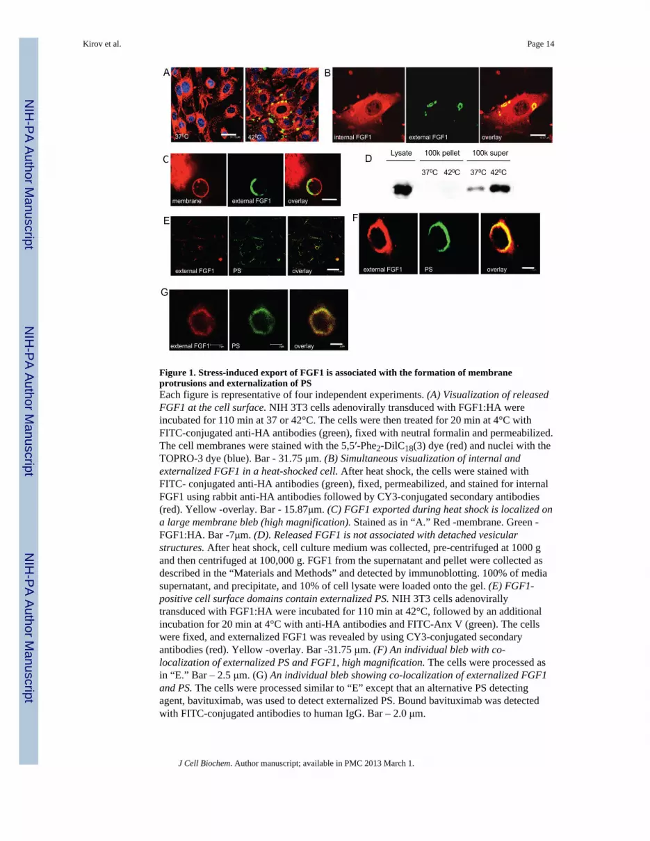
Figure 1. Stress-induced export of FGF1 is associated with the formation of membraneprotrusions and externalization of PSEach figure is representative of four independent experiments. (A) Visualization of releasedFGF1 at the cell surface. NIH 3T3 cells adenovirally transduced with FGF1:HA wereincubated for 110 min at 37 or 42°C. The cells were then treated for 20 min at 4°C withFITC-conjugated anti-HA antibodies (green), fixed with neutral formalin and permeabilized.The cell membranes were stained with the 5,5′-Phe2-DilC18(3) dye (red) and nuclei with theTOPRO-3 dye (blue). Bar - 31.75 μm. (B) Simultaneous visualization of internal andexternalized FGF1 in a heat-shocked cell. After heat shock, the cells were stained withFITC- conjugated anti-HA antibodies (green), fixed, permeabilized, and stained for internalFGF1 using rabbit anti-HA antibodies followed by CY3-conjugated secondary antibodies(red). Yellow -overlay. Bar - 15.87μm. (C) FGF1 exported during heat shock is localized ona large membrane bleb (high magnification). Stained as in “A.” Red -membrane. Green -FGF1:HA. Bar -7μm. (D). Released FGF1 is not associated with detached vesicularstructures. After heat shock, cell culture medium was collected, pre-centrifuged at 1000 gand then centrifuged at 100,000 g. FGF1 from the supernatant and pellet were collected asdescribed in the “Materials and Methods” and detected by immunoblotting. 100% of mediasupernatant, and precipitate, and 10% of cell lysate were loaded onto the gel. (E) FGF1-positive cell surface domains contain externalized PS. NIH 3T3 cells adenovirallytransduced with FGF1:HA were incubated for 110 min at 42°C, followed by an additionalincubation for 20 min at 4°C with anti-HA antibodies and FITC-Anx V (green). The cellswere fixed, and externalized FGF1 was revealed by using CY3-conjugated secondaryantibodies (red). Yellow -overlay. Bar -31.75 μm. (F) An individual bleb with co-localization of externalized PS and FGF1, high magnification. The cells were processed asin “E.” Bar – 2.5 μm. (G) An individual bleb showing co-localization of externalized FGF1and PS. The cells were processed similar to “E” except that an alternative PS detectingagent, bavituximab, was used to detect externalized PS. Bound bavituximab was detectedwith FITC-conjugated antibodies to human IgG. Bar – 2.0 μm.
Kirov et al. Page 14
J Cell Biochem. Author manuscript; available in PMC 2013 March 1.
NIH
-PA Author Manuscript
NIH
-PA Author Manuscript
NIH
-PA Author Manuscript

Figure 2. Chemical agents repressing PS exposure inhibit stress-induced FGF1 exportEach figure is representative of three independent experiments. (A). Taurine inhibits PSexternalization. NIH 3T3 cells were incubated with or without 40 mM taurine (Sigma) for2h at 37°C in DMEM plus 10 % serum, followed by an additional 110 min 42°C in DMEMwithout serum. Next, the cells were incubated for 20 min at 4°C with annexin V conjugatedto phycoerythrin. The cells were examined under a confocal microscope; the combinedphase contrast and phycoerythrin fluorescence (red) images are presented. PS externalizationis marked with arrows. Bar -20 μm. (B). Taurine inhibits heat shock-induced FGF1 exportin a concentration-dependent manner. NIH 3T3 cells adenovirally transduced with FGF1were incubated with or without 8, 20, or 40 μM taurine (Sigma) for 2 h at 37°C in DMEMplus 10 % fetal calf serum, followed by an additional incubation for 110 min at 37°C or42°C in DMEM without serum. FGF1 was isolated from conditioned media and detected byimmunoblotting. (C) Isoproterenol inhibits PS externalization. NIH 3T3 cells wereincubated with or without 20 μM isoproterenol (Sigma) for 2 h at 37°C in DMEM plus 10 %serum and then for additional 110 min at 42°C in DMEM without serum. The cells werethen incubated for 20 min at 4°C with annexin V:phycoerythrin (red), formalin fixed,permeabilized, stained with Alexa 488- conjugated phalloidin (green), and examined using aconfocal microscope. PS externalization is marked with arrows. Bar - 15.87 μm. (D).Isoproterenol inhibits heat shock-induced FGF1 export. NIH 3T3 cells adenovirallytransduced with FGF1 were incubated with or without 20 μM isoproterenol for 2 h at 37°Cin DMEM plus 10 % fetal calf serum, followed by an additional incubation for 110 min at37°C or 42°C in DMEM without serum. FGF1 was isolated from conditioned media anddetected using immunoblotting.
Kirov et al. Page 15
J Cell Biochem. Author manuscript; available in PMC 2013 March 1.
NIH
-PA Author Manuscript
NIH
-PA Author Manuscript
NIH
-PA Author Manuscript

Figure 3. Expression of constitutively active ezrin decreases stress-induced PS externalizationand attenuates FGF1 exportEach figure is representative of three independent experiments. (A). Peripheral distributionof CA ezrin in stably transfected cells. NIH 3T3 cells stably cotransfected with rtTA and CAezrin under control of the Tet promoter were stimulated for 48 h with 10 μg/ml doxycycline,formalin fixed, and stained for CA ezrin (anti-VSVG antibodies followed by FITC-conjugated secondary antibodies, green) and DNA (TO-PRO3, red). Confocal image.Peripheral ezrin is marked with arrows. Bar -15.87μm. (B). Decrease of PS externalizationin cells expressing CA ezrin. NIH 3T3 cells stably transfected with rtTA, and either theempty pTRE2Hygro vector or the CA ezrin vector were stimulated for 48 h withdoxycycline, and incubated for 110 min at 42°C. The live cells were then stained withannexin V:phycoerythrin (red), formalin fixed, stained with Alexa-488 conjugatedphalloidin (green), and examined by confocal microscopy. PS externalization is marked witharrows. Bar -15.87μm (C). CA ezrin decreases FGF1 export. NIH 3T3 cells cotransfectedwith rtTA and either the empty pTRE2Hygro vector, WT ezrin, or CA ezrin wereadenovirally transduced with FGF1, stimulated for 48 h with doxycycline, and thenincubated for 110 min at 37°C or 42°C in DMEM without serum. FGF1 was isolated fromconditioned media and detected by immunoblotting.
Kirov et al. Page 16
J Cell Biochem. Author manuscript; available in PMC 2013 March 1.
NIH
-PA Author Manuscript
NIH
-PA Author Manuscript
NIH
-PA Author Manuscript

Figure 4. Phorbol ester-induced differentiation of promonocytic leukemia cells is accompaniedby FGF1 export, membrane blebbing, and PS externalizationEach figure is representative of four independent experiments. (A). PMA treatment inducesFGF1 export from U937 cells. U937 cells stably transfected with FGF1 were treated or nottreated with 150 nM PMA for 48 h, and then incubated for 110 min at 37°C or 42°C. FGF1was isolated from conditioned media and detected using immunoblotting. 100% mediumand 10% cell lysate were loaded per lane. (B). PMA treatment induces bleb formation inU937 cells. Control and PMA-treated U937 cells were formalin fixed and stained with theVybrant membrane stain (red). The nuclei were stained with TOPRO3 (blue). Confocalimages. Bar – 20 μm. (C). PMA-induced differentiation of U937 cells is accompanied by theexternalization of PS. PMA-treated and untreated U937 cells transfected with FGF1:GFP(green) were incubated for 20 min with phycoerythrin-conjugated Annexin V (red), formalinfixed, and examined by confocal microscopy. PS externalization is marked with arrows. Bar- 15.87μm (D,E). Externalization of FGF1 in PMA-treated U937 cells. PMA-treated anduntreated U937 cells transfected with FGF1:GFP (green) were incubated for 20min withmonoclonal anti-GFP antibodies (red), formalin fixed, incubated with CY3-conjugatedsecondary antibodies, and examined by confocal microscopy. FGF1 externalization ismarked with arrows. Bar – 31.75μm. E: PMA-treated U937 transfected with FGF1:GFP,higher magnification. Processed as in “D.” Additional staining of nuclei with TOPRO-3(blue). Bar -19μm.
Kirov et al. Page 17
J Cell Biochem. Author manuscript; available in PMC 2013 March 1.
NIH
-PA Author Manuscript
NIH
-PA Author Manuscript
NIH
-PA Author Manuscript

Figure 5. Intracellular calcium but not PLSCR1 is required for FGF1 export from U937 cellsEach figure is representative of three independent experiments. (A). BAPTA-AM inhibitsFGF1 export from U937 cells in a concentration-dependent manner. U937 cells stablytransfected with FGF1 were treated with 150 nM PMA for 48 h, and then incubated for 110min at 37°C in presence or absence of BAPTA-AM. FGF1 was isolated from conditionedmedia and detected using immunoblotting. 100% medium and 2% cell lysate were loadedper lane. (B). BAPTA-AM inhibits PS externalization in differentiated U937 cells. PMA-treated U937 cells were preincubated for 2 h with or without 50 μM BAPTA-AM and thenincubated for 20 min with phycoerythrin-conjugated Annexin V (red), formalin fixed,permeabilized, stained with Alexa 488-phalloidin (green) and studied using confocalmicroscopy. PS externalization is marked with arrows. Bar – 31.75μm. (C). UndifferentiatedU937 cells have low PLSCR1 expression, which is enhanced with PMA treatment. RNA wasisolated from undifferentiated U937 cells and U937 cells treated for 48 h with 150 nMPMA. RT-PCR was performed using PLSCR1 and β-actin (control) primers. (D). Over-expression of PLSCR1 does not enhance FGF1 export from U937 cells. U937 cells stablytransfected with PLSCR1:GFP or with GFP (control) were treated or not with 150 nM PMAfor 48 h, and incubated for 110 min at 37°C or 42°C. FGF1 was isolated from conditionedmedia and detected by immunoblotting. 100% medium and 10% cell lysate were loaded perlane. (E). Specific shRNA decreases PLSCR1 expression in U937 cells. RNA was isolatedfrom PMA-treated U937 cells stably transfected with PLSCR1 shRNA or control shRNA.RT PCR was performed using PLSCR1 and β-actin (control) primers. (F). shRNAknockdown of PLSCR1 does not affect FGF1 export from U937 cells. U937 cells stablytransfected with FGF1 and cotransfected with PLSCR1 shRNA or control shRNA weretreated or not with 150 nM PMA for 48 h, and then incubated for 110 min at 37°C or 42°C.FGF1 was isolated from conditioned media and detected using immunoblotting.
Kirov et al. Page 18
J Cell Biochem. Author manuscript; available in PMC 2013 March 1.
NIH
-PA Author Manuscript
NIH
-PA Author Manuscript
NIH
-PA Author Manuscript


