Embed Size (px)
Citation preview
�����������������
Citation: Härdrich, M.; Haase-Fielitz,
A.; Fielitz, J.; Boschmann, M.;
Pivovarova-Ramich, O.; Pfeiffer,
A.F.H.; Rudovich, N.; Weylandt, K.H.;
Butter, C. Physical Performance and
Non-Esterified Fatty Acids in Men
and Women after Transcatheter
Aortic Valve Implantation (TAVI).
Nutrients 2022, 14, 203. https://
doi.org/10.3390/nu14010203
Academic Editor: Asim K. Duttaroy
Received: 23 November 2021
Accepted: 30 December 2021
Published: 2 January 2022
Publisher’s Note: MDPI stays neutral
with regard to jurisdictional claims in
published maps and institutional affil-
iations.
Copyright: © 2022 by the authors.
Licensee MDPI, Basel, Switzerland.
This article is an open access article
distributed under the terms and
conditions of the Creative Commons
Attribution (CC BY) license (https://
creativecommons.org/licenses/by/
4.0/).
nutrients
Article
Physical Performance and Non-Esterified Fatty Acids in Menand Women after Transcatheter Aortic ValveImplantation (TAVI)Michaela Härdrich 1,†, Anja Haase-Fielitz 1,2,*,† , Jens Fielitz 3,4,5 , Michael Boschmann 5,Olga Pivovarova-Ramich 6,7,8 , Andreas F. H. Pfeiffer 7,8, Natalia Rudovich 9,10, Karsten H. Weylandt 11
and Christian Butter 1
1 Department of Cardiology, Heart Centre Brandenburg Bernau, Faculty of Health Sciences Brandenburg,Brandenburg Medical School (MHB) Theodor Fontane, 16321 Bernau, Germany;[email protected] (M.H.); [email protected] (C.B.)
2 Institute of Social Medicine & Health Care Systems Research, Otto von Guericke University Magdeburg,39120 Magdeburg, Germany
3 DZHK (German Centre for Cardiovascular Research), Partner Site Greifswald, 17489 Greifswald, Germany;[email protected]
4 Department of Internal Medicine B, Cardiology, University Medicine Greifswald, 17489 Greifswald, Germany5 Experimental & Clinical Research Centre (ECRC), a Joint Cooperation between Charité—University Medicine
Berlin and Max Delbrück Centre (MDC) for Molecular Medicine in the Helmholtz Association,13125 Berlin, Germany; [email protected]
6 Research Group Molecular Nutritional Medicine, Department of Molecular Toxicology, German Institute ofHuman Nutrition Potsdam-Rehbruecke, 14558 Nuthetal, Germany; [email protected]
7 Department Endocrinology and Metabolism, Charité—Universitätsmedizin Berlin, 10117 Berlin, Germany;[email protected]
8 German Center for Diabetes Research (Deutsches Zentrum Für Diabetesforschung e.V.), 85764 Neuherberg,Germany
9 Department of Internal Medicine, Spital STS AG, University of Zurich, 8006 Zurich, Switzerland;[email protected]
10 Department of Internal Medicine, Spital Bülach, 8180 Bülach, Switzerland11 Medical Department, Divisions of Hepatology, Gastroenterology, Oncology, Haematology, Palliative Care,
Endocrinology and Diabetes, Ruppiner Kliniken, Brandenburg Medical School, 16816 Neuruppin, Germany;[email protected]
* Correspondence: [email protected]; Tel.: +49-3338-694-649; Fax: +49-3338-694-644† These authors contributed equally to this work.
Abstract: Background: Men and women with valvular heart disease have different risk profiles forclinical endpoints. Non-esterified fatty acids (NEFA) are possibly involved in cardio-metabolic disease.However, it is unclear whether NEFA concentrations are associated with physical performance inpatients undergoing transcatheter aortic valve implantation (TAVI) and whether there are sex-specificeffects. Methods: To test the hypothesis that NEFA concentration is associated with sex-specificphysical performance, we prospectively analysed data from one hundred adult patients undergoingTAVI. NEFA concentrations, physical performance and anthropometric parameters were measuredbefore and 6 and 12 months after TAVI. Physical performance was determined by a six-minute walkingtest (6-MWT) and self-reported weekly bicycle riding time. Results: Before TAVI, NEFA concentrationswere higher in patients (44 women, 56 men) compared to the normal population. Median NEFAconcentrations at 6 and 12 months after TAVI were within the reference range reported in the normalpopulation in men but not women. Men but not women presented with an increased performancein the 6-MWT over time (p = 0.026, p = 0.142, respectively). Additionally, men showed an increasedability to ride a bicycle after TAVI compared to before TAVI (p = 0.034). NEFA concentrations beforeTAVI correlated with the 6-MWT before TAVI in women (Spearman’s rho −0.552; p = 0.001) but notin men (Spearman’s rho −0.007; p = 0.964). No association was found between NEFA concentrationsand physical performance 6 and 12 months after TAVI. Conclusions: NEFA concentrations improvedinto the reference range in men but not women after TAVI. Men but not women have an increased
Nutrients 2022, 14, 203. https://doi.org/10.3390/nu14010203 https://www.mdpi.com/journal/nutrients
Nutrients 2022, 14, 203 2 of 14
physical performance after TAVI. No association between NEFA and physical performance wasobserved in men and women after TAVI.
Keywords: heart failure; body composition; six-minute walking test (6-MWT); coronary heart disease;diabetes mellitus; chronic kidney disease; sex-specific differences
1. Introduction
Transcatheter aortic valve implantation (TAVI) is a standard treatment for aortic steno-sis in patients with moderate to high surgical risk. Aortic valve stenosis is characterized byincreased inflammation, fibrosis, and calcification of the aortic valve leaflets [1,2]. Obesityand impaired lipid metabolism have emerged as risk factors for aortic stenosis [3–5]. Inpatients with aortic stenosis, sex-related differences in clinical presentation, metabolismand pathophysiology of valvular calcification have been described [6,7]. Women seem tohave less valvular calcification, but more fibrosis compared with men [8]. Additionally,recent studies have suggested sex-related differences in cardiac remodelling and reverseremodelling after TAVI [9].
Non-esterified fatty acids (NEFA) are formed during the lipolysis of adipose tissueand serve as energy suppliers predominantly metabolized through β-oxidation [10]. NEFAhave proinflammatory effects and lead to insulin resistance and the inhibition of glycolysisduring ischemia reperfusion [11]. In addition to metabolic effects, neuro-humoral activationhas been observed in patients with heart failure and increased NEFA concentrations [12].
A reactive hyperadrenergic state and stimulation of lipolysis by catecholamines as wellas elevated natriuretic peptides are contributing factors [10,12,13]. Additionally, increasedNEFA concentrations impair nitric oxide-dependent and -independent vasodilatation andcontribute to the formation of oxygen radicals and endothelial dysfunction [14]. Possibleconsequences are increased blood pressure and the formation of atherosclerotic plaques dueto NEFA accumulation in blood vessels [15,16]. Female patients with aortic stenosis tend towalk for shorter time periods on a treadmill and achieve lower metabolic equivalents [17].
In sum, men and women with valvular heart disease have different risk profiles forclinical endpoints. As NEFA have been shown to be involved in cardio-metabolic diseaseand might increase cardiometabolic risk, they might play a role as biomarkers in thiscontext. However, whether NEFA concentrations differ according to sex and if their levelsare associated with physical performance after TAVI has not yet been reported.
Accordingly, we evaluated sex-specific concentrations of NEFA before and after TAVIand analysed if there was a sex-specific relationship of NEFA with physical performance asmeasured by a six-minute walking test (6-MWT) and weekly bicycle riding time.
2. Materials and Methods2.1. Study Design and Patients
This prospective single-centre cohort study was conducted at Brandenburg HeartCentre during January 2017 and July 2019. Within this time, 1264 TAVI procedures wereperformed in our department. The study was approved by the local ethics committee AS87(bB)/2015. Written informed consent to participate in the study and to publish the resultswas obtained from all individual patients included in the study.
Inclusion criteria comprised symptomatic heart failure, severe aortic stenosis withplanned TAVI and written informed consent. Exclusion criteria were lack of legal capacity,life expectancy below 1 year and missing baseline blood draw.
2.2. Anthropometric Analysis
Body weight was measured in light clothing and without shoes. Waist size wasmeasured at the shortest point below the lower rib margin and the iliac crest. Body massindex (BMI) was calculated as weight (kilograms) divided by height squared (meters).
Nutrients 2022, 14, 203 3 of 14
Measurement of skinfold thickness was used to indirectly determine total body fat,adjusting for age and sex. Skinfold thickness was measured at three different sites (triceps,back, hips) using a calliper, i.e., fat-measuring forceps. On the triceps, a skinfold was liftedparallel to the long axis of the upper arm and measured on the dorsal side of the upperarm over the triceps’ brachii muscle midway between the acromion and olecranon. Withthe arm hanging, a skinfold was then determined on the dorsum just below the inferior tipof the scapula. The skinfold was oblique to the long axis of the body. Last, a measurementwas taken on the lateral axillary line just above the iliac crest. The skinfold was orientedparallel to the long axis of the body. The three results were added together and expressed inmillimetres. Measurements were carried out by the same operator; fat mass was collectedand analysed by specific formulas provided by the manufacturer.
2.3. Physical Performance
Six-minute walking test (6-MWT) before and after TAVI was used as sub-maximalexercise test to assess aerobic capacity and endurance. Patients were asked to walk asfar as possible within 6 min. The distance was measured with a distance measuringwheel. After completion of the 6 min test or after the patient had stopped, the Borg scalewas assessed. Patients were asked to rate their exertion on the scale during the activity,combining all sensations and feelings of physical stress and fatigue. The scale ranges from6—corresponding to an activity that is not strenuous at all—to 20, which is perceived bythe patient as maximally strenuous. In addition, patients were asked for physical activityrecords measured as time spent bicycle riding weekly before and after TAVI.
2.4. Sample Collection
Blood samples were taken one day after hospital admission from the fasting patient forsubsequent biomarker determination. Serum and Ethylenediaminetetraacetic acid (EDTA)plasma was prepared by centrifugation at 1452 g and 5 ◦C for 10 min and were frozen at−80 ◦C till analyses. NEFA were assessed in serum samples with Pentra C400 benchtopanalyser (Horiba Medical, Grabels, France) using NEFA-HR (2) Assay (FUJIFILM WakoChemicals Europe GmbH, Neuss, Germany) according to the manufacturer’s instructions.
In addition, on the day of hospital admission, the following laboratory parameterswere routinely determined in all patients: blood count, electrolytes, renal retention pa-rameters (estimated glomerular filtration rate, eGFR according to Chronic Kidney Disease,CKD-EPI formula, serum creatinine), lipid status (total cholesterol, LDL and HDL choles-terol, triglycerides), C-reactive protein (CRP), NT-Pro-BNP, blood glucose and HbA1c. Forroutine blood measurements the autoanalyzer COBAS Integra 800 (Roche Diagnostics,Basel, Switzerland) was used.
2.5. Data Collection
A 12-lead Electrocardiogram (ECG) was recorded prior to TAVI and post TAVI. De-mographic variables, comorbidities, medications, New-York Heart-association (NYHA)classification and logistic EuroSCORE (European System for Cardiac Operative Risk Evalu-ation) were collected.
Post TAVI, type and size of valve were recorded as well as any complications occurringperi or post TAVI. Relevant complications were bleeding, occurrence of vascular complica-tions after femoral puncture, respiratory insufficiency, the development of delirium or acutekidney injury, stroke, occurrence of a new left bundle branch block and a necessary deviceimplantation (pacemaker, ICD, or CRT). Length of stay in hospital or the intensive care unitas well as patient’s discharge status (discharge to home, direct rehabilitation, transfer toinpatient care at another hospital) were recorded. Finally, mortality during hospitalizationand after 6 and 12 months, as well as possible rehospitalization within 30 days of hospitaldischarge, were recorded.
Nutrients 2022, 14, 203 4 of 14
2.6. Transthoracic Echocardiography
All patients underwent transthoracic echocardiography preoperatively to quantifyaortic valve stenosis. Both mean and maximum pressure gradients across the aortic valvewere determined by continuous-wave Doppler. When the mean pressure gradient exceeded40 mmHg, aortic valve stenosis was considered high-gradient aortic valve stenosis. Whenthe mean pressure gradient was less than 40 mmHg and the valve orifice area determinedby the continuity equation was <1.0 cm2, a diagnosis of low-flow low-gradient aortic valvestenosis with preserved LVEF was made if the LVEF was greater than 50% and the strokevolume index was <35 mL/m2. If these criteria were present at an LVEF less than 50%, itwas classified as high-grade aortic valve stenosis with reduced ejection fraction. In unclearcases, transoesophageal echocardiography was performed to confirm the diagnosis byplanimetric determination of the valve orifice area. LVEF was determined via biplaneaccording to Simpson’s biplane method in 4-chamber and 2-chamber views; in the fewcases where the acoustic conditions were insufficient, it had to be estimated visually. Theclassification of normal, low-grade, moderate-grade and high-grade LVEF followed theManual for the Indication and Performance of Echocardiography—Update 2020 of theGerman Society of Cardiology.
2.7. TAVI Procedure
Transcatheter aortic valve implantation has been performed at the Brandenburg HeartCentre since 2008 and serves as an alternative, minimally invasive procedure to conven-tional aortic valve replacement in patients with aortic valve stenosis and high surgical risk.The diagnosis is confirmed by transthoracic echocardiography and, if necessary, by transoe-sophageal echocardiography in case of ambiguity. If the procedure has not already beenperformed by the referring hospital, each patient receives a left heart catheter examinationto determine the current coronary status and, if necessary, therapy for stenosis requiringtreatment. A computer tomography (CT)-scan of the heart and the pelvic–leg vasculaturealso provides important information for surgical planning. At the Brandenburg Heart Cen-tre, the transfemoral approach is used in most cases. The pelvic–leg vessels are measured,and the thoracic aorta is assessed. Annulus as well as the distance to the coronaries aredetermined in order to select the most suitable valve type preoperatively. When all thesedata are available, the heart team, consisting of cardiologists, cardiac surgeons, and anesthe-siologists, will make the indication for further therapy. TAVI is predominantly performedunder general anaesthesia. In some cases, especially when a high risk of delirium and nopossible peri-interventional complications are anticipated, TAVI is also performed underanalgo-sedation. Depending on valve anatomy and calcification, a predilatation may berequired. In the majority a direct implantation is performed. Under angiographic control,the valve prosthesis is positioned and finally released under tachycardic pacing, which isinduced by a passaged pacemaker that has been washed in beforehand. Finally, the femoralplaced sheaths are removed, and the patient is transferred to cardiac intermediate care viathe recovery room with the femoral pressure dressing in place.
Follow-up care was performed by the heart failure outpatient clinic of the BrandenburgHeart Centre after 6 and 12 months and comprised transthoracic echocardiography, labora-tory tests, 12-lead ECG, and a physical examination as well as a questionnaire includinginformation regarding weekly duration of exercise.
2.8. Statistics
Statistical analysis plan was approved by the authors before analyses began. As thiswas a pilot study, we used a convenient sample size for testing the reliability and validityof the data collected.
Categorical data were reported as percentages with a 95% confidence interval (CI) ofthe mean percentage and compared using the Fisher exact test. After testing for normaldistribution, continuous data were reported as the median with 25th to 75th percentiles, andnonparametric data were compared using the Mann–Whitney U test. The Friedmann test
Nutrients 2022, 14, 203 5 of 14
was used to evaluate changes over time within one group. We used nonparametric bivariatecorrelation and report Spearman correlation coefficients. We used list-wise deletion whengenerating boxplots, including several endpoint measurements over time and pair-wisedeletion for baseline values presented in the tables.
The ability of NEFA to predict (i) improvement in NYHA class, (ii) improvement in thedistance of the 6-MWT and (iii) rehospitalisation within 30 days was assessed by plottingreceiver operating characteristic (ROC) curves and further reported as area under the curve(AUC) with 95% CIs. An AUC-ROC value of >0.7 was taken to indicate a reasonableperformance [18]. Information on missing data is provided in the tables’ footnotes.
Statistical significance is denoted by 2-sided p-values < 0.05. Statistical analysis wasperformed using SPSS 26.0 (SPSS Inc., Chicago, IL, USA).
3. Results3.1. Patient Characteristics
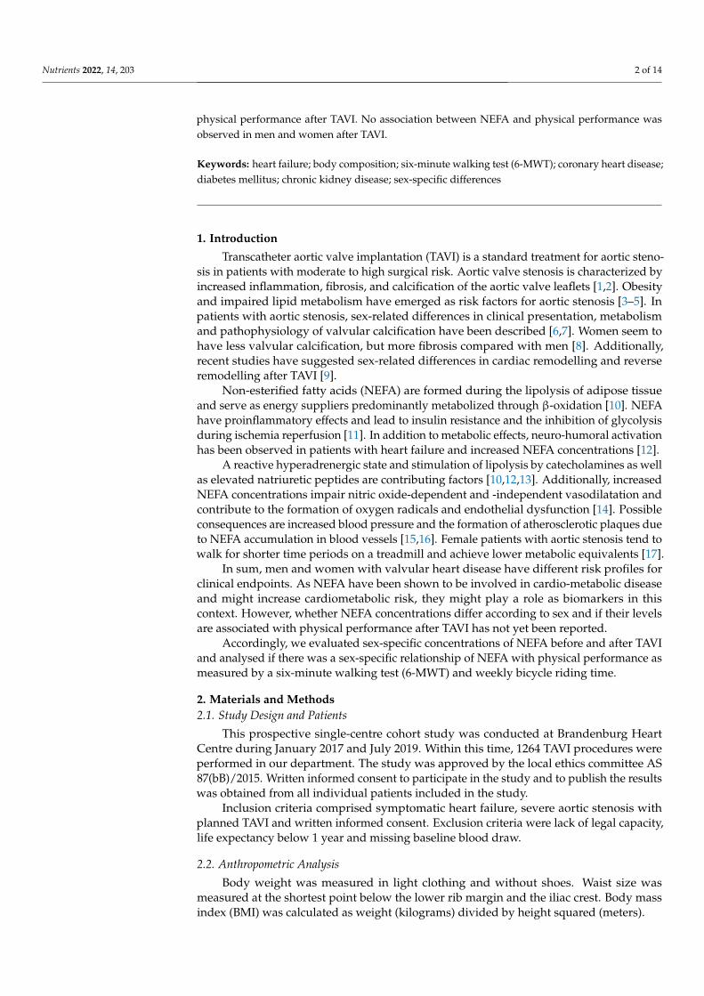
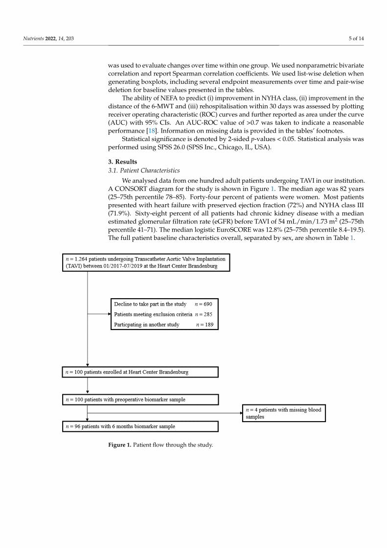
We analysed data from one hundred adult patients undergoing TAVI in our institution.A CONSORT diagram for the study is shown in Figure 1. The median age was 82 years(25–75th percentile 78–85). Forty-four percent of patients were women. Most patientspresented with heart failure with preserved ejection fraction (72%) and NYHA class III(71.9%). Sixty-eight percent of all patients had chronic kidney disease with a medianestimated glomerular filtration rate (eGFR) before TAVI of 54 mL/min/1.73 m2 (25–75thpercentile 41–71). The median logistic EuroSCORE was 12.8% (25–75th percentile 8.4–19.5).The full patient baseline characteristics overall, separated by sex, are shown in Table 1.
Nutrients 2022, 14, x FOR PEER REVIEW 6 of 16
Figure 1. Patient flow through the study. Figure 1. Patient flow through the study.
Nutrients 2022, 14, 203 6 of 14
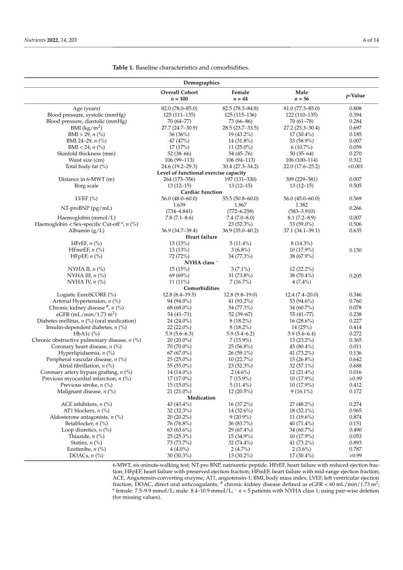
Table 1. Baseline characteristics and comorbidities.
Demographics
Overall Cohortn = 100
Femalen = 44
Malen = 56 p-Value
Age (years) 82.0 (78.0–85.0) 82.5 (78.3–84.8) 81.0 (77.3–85.0) 0.808Blood pressure, systolic (mmHg) 125 (111–135) 125 (115–136) 122 (110–135) 0.394Blood pressure, diastolic (mmHg) 70 (64–77) 73 (66–86) 70 (61–78) 0.284
BMI (kg/m2) 27.7 (24.7–30.9) 28.5 (23.7–33.5) 27.2 (25.3–30.4) 0.697BMI > 29, n (%) 36 (36%) 19 (43.2%) 17 (30.4%) 0.185
BMI 24–29, n (%) 47 (47%) 14 (31.8%) 33 (58.9%) 0.007BMI < 24, n (%) 17 (17%) 11 (25.0%) 6 (10.7%) 0.059
Skinfold thickness (mm) 52 (38–66) 54 (45–76) 50 (35–64) 0.270Waist size (cm) 106 (99–113) 106 (94–113) 106 (100–114) 0.312
Total body fat (%) 24.6 (19.2–29.3) 30.4 (27.3–34.2) 22.0 (17.6–25.2) <0.001Level of functional exercise capacity
Distance in 6-MWT (m) 264 (173–356) 197 (131–330) 309 (229–381) 0.007Borg scale 13 (12–15) 13 (12–15) 13 (12–15) 0.505
Cardiac functionLVEF (%) 56.0 (48.0–60.0) 55.5 (50.8–60.0) 56.0 (45.0–60.0) 0.569
NT-proBNP (pg/mL) 1.639(734–4.841)
1.867(772–6.258)
1.382(583–3.910) 0.266
Haemoglobin (mmol/L) 7.8 (7.1–8.6) 7.4 (7.0–8.0) 8.1 (7.2–8.9) 0.007Haemoglobin < Sex-specific Cut-off *, n (%) 23 (52.3%) 33 (59.0%) 0.506
Albumin (g/L) 36.9 (34.7–39.4) 36.9 (35.0–40.2) 37.1 (34.1–39.1) 0.635Heart failure
HFrEF, n (%) 13 (13%) 5 (11.4%) 8 (14.3%)0.150HFmrEF, n (%) 13 (13%) 3 (6.8%) 10 (17.9%)
HFpEF, n (%) 72 (72%) 34 (77.3%) 38 (67.9%)NYHA class ~
NYHA II, n (%) 15 (15%) 3 (7.1%) 12 (22.2%)0.205NYHA III, n (%) 69 (69%) 31 (73.8%) 38 (70.4%)
NYHA IV, n (%) 11 (11%) 7 (16.7%) 4 (7.4%)Comorbidities
Logistic EuroSCORE (%) 12.8 (8.4–19.5) 12.8 (9.8–19.0) 12.4 (7.4–20.0) 0.346Arterial Hypertension, n (%) 94 (94.0%) 41 (93.2%) 53 (94.6%) 0.760
Chronic kidney disease #, n (%) 68 (68.0%) 34 (77.3%) 34 (60.7%) 0.078eGFR (mL/min/1.73 m2) 54 (41–71) 52 (39–67) 55 (41–77) 0.238
Diabetes mellitus, n (%) (oral medication) 24 (24.0%) 8 (18.2%) 16 (28.6%) 0.227Insulin-dependent diabetes, n (%) 22 (22.0%) 8 (18.2%) 14 (25%) 0.414
HbA1c (%) 5.9 (5.6–6.3) 5.9 (5.4–6.2) 5.9 (5.6–6.4) 0.272Chronic obstructive pulmonary disease, n (%) 20 (20.0%) 7 (15.9%) 13 (23.2%) 0.365
Coronary heart disease, n (%) 70 (70.0%) 25 (56.8%) 45 (80.4%) 0.011Hyperlipidaemia, n (%) 67 (67.0%) 26 (59.1%) 41 (73.2%) 0.136
Peripheral vascular disease, n (%) 25 (25.0%) 10 (22.7%) 15 (26.8%) 0.642Atrial fibrillation, n (%) 55 (55.0%) 23 (52.3%) 32 (57.1%) 0.688
Coronary artery bypass grafting, n (%) 14 (14.0%) 2 (4.6%) 12 (21.4%) 0.016Previous myocardial infarction, n (%) 17 (17.0%) 7 (15.9%) 10 (17.9%) >0.99
Previous stroke, n (%) 15 (15.0%) 5 (11.4%) 10 (17.9%) 0.412Malignant disease, n (%) 21 (21.0%) 12 (20.5%) 9 (16.1%) 0.172
MedicationACE inhibitors, n (%) 43 (43.4%) 16 (37.2%) 27 (48.2%) 0.274AT1 blockers, n (%) 32 (32.3%) 14 (32.6%) 18 (32.1%) 0.965
Aldosterone antagonists, n (%) 20 (20.2%) 9 (20.9%) 11 (19.6%) 0.874Betablocker, n (%) 76 (76.8%) 36 (83.7%) 40 (71.4%) 0.151
Loop diuretics, n (%) 63 (63.6%) 29 (67.4%) 34 (60.7%) 0.490Thiazide, n (%) 25 (25.3%) 15 (34.9%) 10 (17.9%) 0.053Statins, n (%) 73 (73.7%) 32 (74.4%) 41 (73.2%) 0.893
Ezetimibe, n (%) 4 (4.0%) 2 (4.7%) 2 (3.6%) 0.787DOACs, n (%) 30 (30.3%) 13 (30.2%) 17 (30.4%) >0.99
6-MWT, six-minute-walking test; NT-pro BNP, natriuretic peptide, HFrEF, heart failure with reduced ejection frac-tion; HFpEF, heart failure with preserved ejection fraction; HFmEF, heart failure with mid-range ejection fraction;ACE, Angiotensin-converting enzyme; AT1, angiotensin-1; BMI, body mass index; LVEF, left ventricular ejectionfraction; DOAC, direct oral anticoagulants; # chronic kidney disease defined as eGFR < 60 mL/min/1.73 m2;* female: 7.5–9.9 mmol/L; male: 8.4–10.9 mmol/L; ~ n = 5 patients with NYHA class 1; using pair-wise deletion(for missing values).
Nutrients 2022, 14, 203 7 of 14
A higher proportion of women presented with a BMI outside the age-adjusted refer-ence range compared to men (68.2% vs. 41.1%, p = 0.007). Total body fat before TAVI washigher in women vs. men (median 30.4%, 25–75th percentile 27.3–34.3 vs. 22.0%, 25–75thpercentile 17.6–25.2, p < 0.001). The logistic EuroSCORE, medication at hospital admissionand comorbidities did not differ between women and men, except for the prevalence ofcoronary heart disease, which was higher in men vs. women (80.4% vs. 56.8%, p = 0.011)(Table 1).
3.2. NEFA Concentrations before and after TAVI
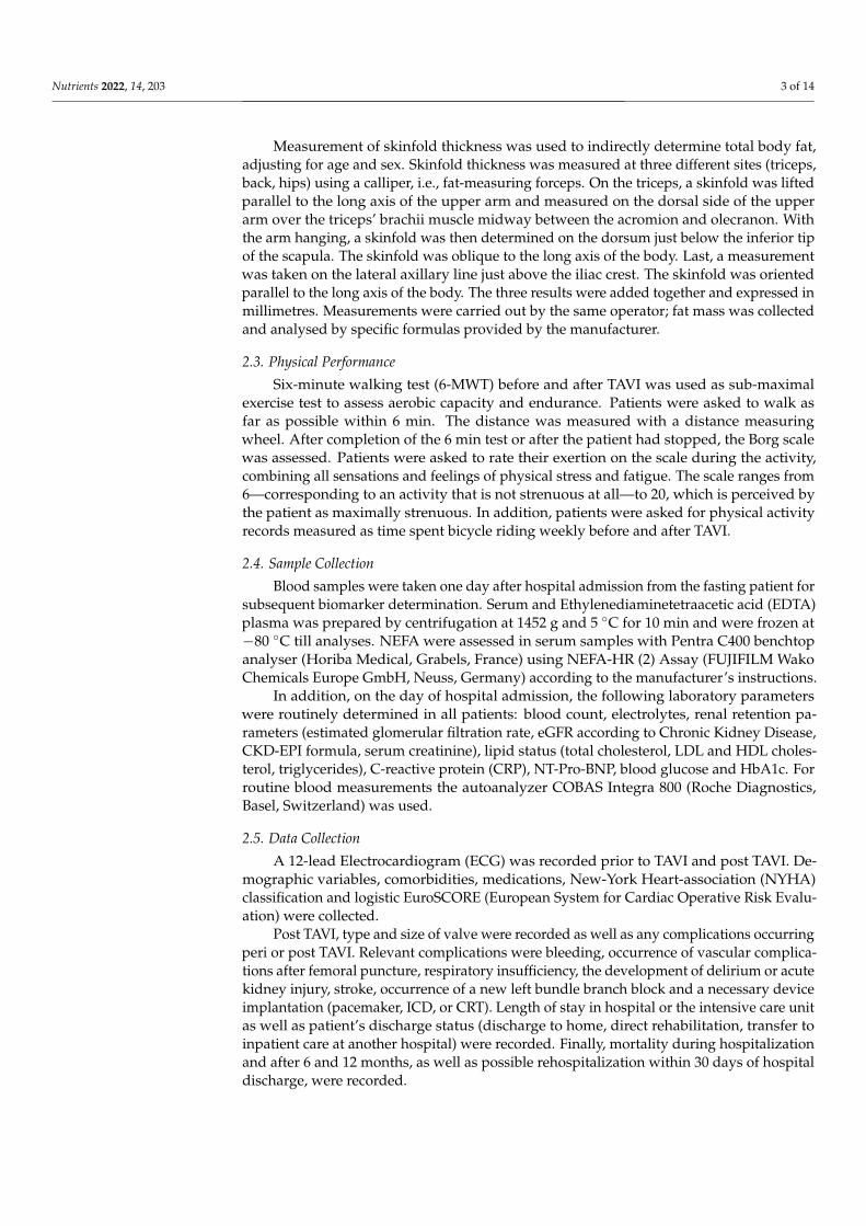
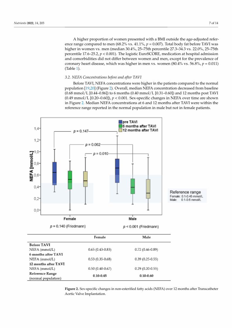
Before TAVI, NEFA concentrations were higher in the patients compared to the normalpopulation [19,20] (Figure 2). Overall, median NEFA concentration decreased from baseline(0.68 mmol/L [0.44–0.86]) to 6 months (0.40 mmol/L [0.31–0.60]) and 12 months post TAVI(0.49 mmol/L [0.20–0.60]), p < 0.001. Sex-specific changes in NEFA over time are shownin Figure 2. Median NEFA concentrations at 6 and 12 months after TAVI were within thereference range reported in the normal population in male but not in female patients.
Nutrients 2022, 14, x FOR PEER REVIEW 9 of 16
Figure 2. Sex-specific changes in non-esterified fatty acids (NEFA) over 12 months after Transcath-
eter Aortic Valve Implantation.
Figure 2. Sex-specific changes in non-esterified fatty acids (NEFA) over 12 months after TranscatheterAortic Valve Implantation.
Nutrients 2022, 14, 203 8 of 14
3.3. Clinical Outcomes
A total of 55.7% of all patients showed an improvement in the 6-MWT. The NYHAclass improved in 66.2% of patients over time (Table 2). Overall, the median length ofstay in hospital was 13 days (9–17). Perioperative vascular complication occurred in fourpatients (all male). The rehospitalisation rate within 30 days was 10.2%, and the 12-monthmortality was 6%. Hospital outcome and mortality did not differ between men and women(Table 2).
Table 2. Outcome data.
Outcome
Overall Cohortn = 100
Femalen = 44
Malen = 56 p-Value
∆ Body mass index (kg/m2) 6 months afterTAVI
−0.31 (−0.72–0.69) −0.32(−0.75–0.42) −0.30 (−0.63–0.84) 0.383
NT-proBNP (pg/mL) 6 months after TAVI 1.053 (379–1.935) 1.320 (518–2.367) 878 (324–1.33) 0.185Improvement in 6-MWT >10%, n (%) 34 (55.7%) 14 (60.9%) 20 (52.6%) 0.530Improvement in NYHA class, n (%) 47 (66.2%) 17 (63.0%) 30 (68.2%) 0.652
Improvement in EF > 10%, n (%) 24 (29.7%) 9 (29.0%) 15 (30.0%) 0.926Postoperative complication
Device implantation due to AV block III, n (%) 15 (15.0%) 3 (7.1%) 12 (21.4%) 0.051Left bundle branch block, n (%) 15 (15.0%) 5 (11.6%) 10 (18.2%) 0.572
Paravalvular aortic regurgitationgrade I–IIgrade IIgrade III
10 (10.0%)2 (2.0%)
-
4 (9.1%)0 (0%)
-
6 (10.7%)2 (3.6%)
-
0.7880.311
Acute kidney injury, n (%) 11 (11.0%) 5 (11.6%) 6 (10.7%) 0.886Bleeding, n (%) 5 (5.0%) 2 (4.8%) 3 (5.5%) >0.99
Delir, n (%) 4 (4.0%) 1 (2.3%) 3 (5.5%) 0.631Stroke, n (%) 2 (2.0%) 1 (2.3%) 1 (1.8%) >0.99
Length of stay in Intensive care unit (days) 2.0 (1.5–14.0) 2.0 (2.0–20.0) 1.0 (5.0–6.0) 0.554Length of stay in hospital (days) 13.0 (9.0–17.0) 13.0 (11.0–17.0) 11.5 (9.0–17.0) 0.180
Rehospitalization within 30 days, n (%) 10 (10.2%) 3 (7.1%) 7 (12.5%) 0.509Worsening eGFR > 10% within 12 months 32 (42.1%) 15 (51.7%) 17 (36.2%) 0.182
MortalityIn-hospital mortality, n (%) 5 (5.0%) 3 (7.0%) 2 (3.6%) 0.650
6-month mortality, n (%) 5 (5.0%) 3 (7.0%) 2 (3.6%) 0.65012-month mortality, n (%) 6 (6.0%) 3 (7.0%) 3 (5.4%) >0.99
NEFA concentrations before TAVI did not predict improvement in NYHA class (AUC-ROC 0.460 [95% CI 0.316–0.605], p = 0.586), improvement in the distance of the 6-MWT(AUC-ROC 0.571 [95% CI 0.423–0.718], p = 0.349) or rehospitalisation within 30 days(AUC-ROC 0.633 [95% CI 0.461–0.805], p = 0.191) with no sex-specific effect observed (allAUC-ROC < 0.68, all p > 0.15).
3.4. Physical Performance before and after TAVI
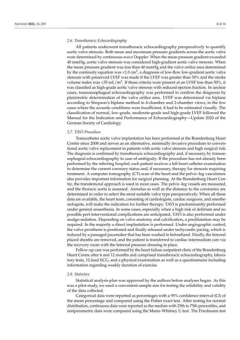
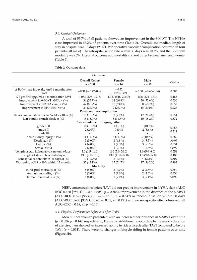
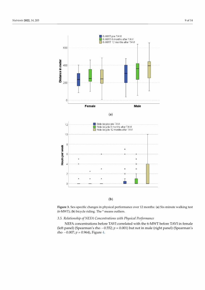
Men but not women presented with an increased performance in 6-MWT over time(p = 0.026, p = 0.142, respectively), Figure 3a. Additionally, according to the weekly durationof exercise, men showed an increased ability to ride a bicycle after TAVI compared to beforeTAVI (p = 0.034). There were no changes in bicycle riding in female patients over time(Figure 3b).
Nutrients 2022, 14, 203 9 of 14
Nutrients 2022, 14, x FOR PEER REVIEW 11 of 16
before TAVI (p = 0.034). There were no changes in bicycle riding in female patients over
time (Figure 3b).
(a)
(b)
Figure 3. Sex-specific changes in physical performance over 12 months: (a) Six-minute walking test
(6-MWT); (b) bicycle riding. The * means outliers.
3.5. Relationship of NEFA Concentrations with Physical Performance
NEFA concentrations before TAVI correlated with the 6-MWT before TAVI in female
(left panel) (Spearman’s rho −0.552; p = 0.001) but not in male (right panel) (Spearman’s
rho −0.007; p = 0.964), Figure 4.
Figure 3. Sex-specific changes in physical performance over 12 months: (a) Six-minute walking test(6-MWT); (b) bicycle riding. The * means outliers.
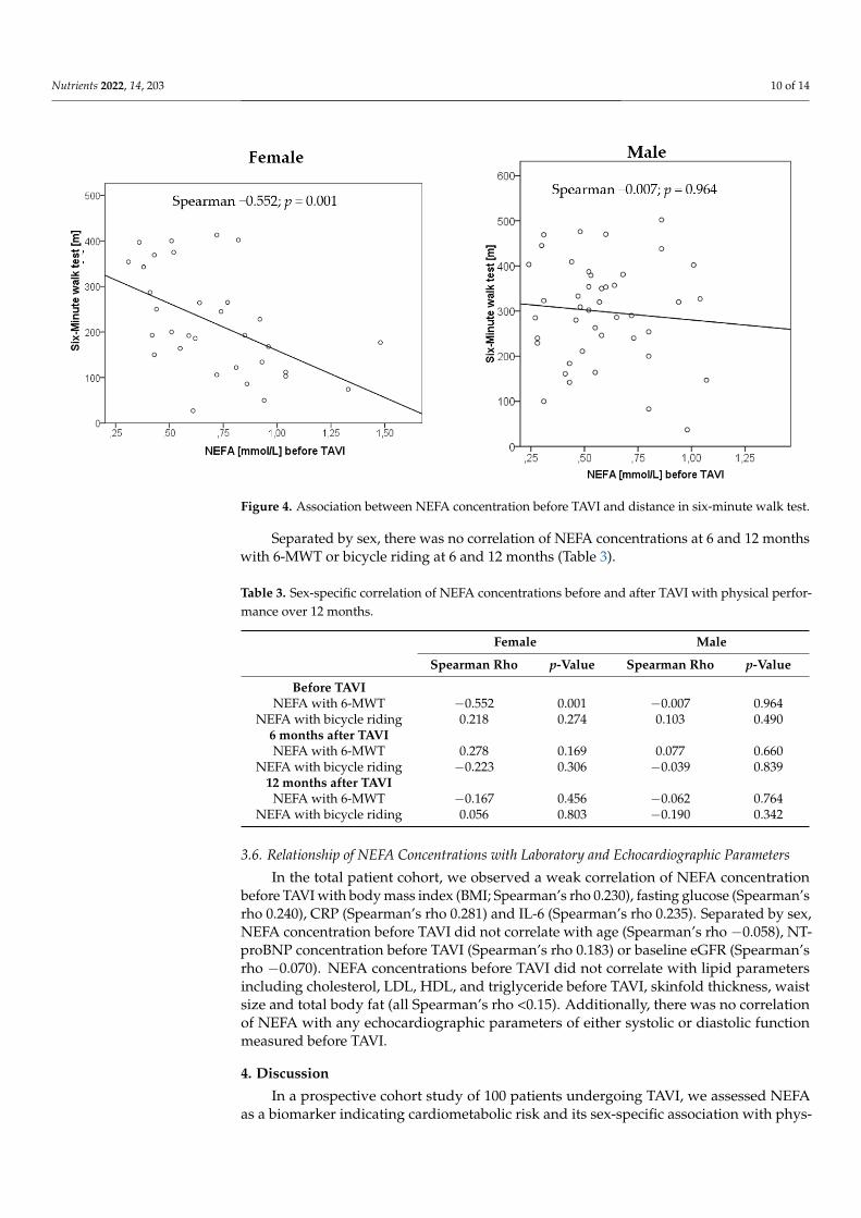
3.5. Relationship of NEFA Concentrations with Physical Performance
NEFA concentrations before TAVI correlated with the 6-MWT before TAVI in female(left panel) (Spearman’s rho −0.552; p = 0.001) but not in male (right panel) (Spearman’srho −0.007; p = 0.964), Figure 4.
Nutrients 2022, 14, 203 10 of 14Nutrients 2022, 14, x FOR PEER REVIEW 12 of 16
Figure 4. Association between NEFA concentration before TAVI and distance in six-minute walk
test.
Separated by sex, there was no correlation of NEFA concentrations at 6 and 12
months with 6-MWT or bicycle riding at 6 and 12 months (Table 3).
Table 3. Sex-specific correlation of NEFA concentrations before and after TAVI with physical per-
formance over 12 months.
Female Male
Spearman Rho p-Value Spearman Rho p-Value
Before TAVI
NEFA with 6-MWT −0.552 0.001 −0.007 0.964
NEFA with bicycle riding 0.218 0.274 0.103 0.490
6 months after TAVI
NEFA with 6-MWT 0.278 0.169 0.077 0.660
NEFA with bicycle riding −0.223 0.306 −0.039 0.839
12 months after TAVI
NEFA with 6-MWT −0.167 0.456 −0.062 0.764
NEFA with bicycle riding 0.056 0.803 −0.190 0.342
3.6. Relationship of NEFA Concentrations with Laboratory and Echocardiographic Parameters
In the total patient cohort, we observed a weak correlation of NEFA concentration
before TAVI with body mass index (BMI; Spearman’s rho 0.230), fasting glucose (Spear-
man’s rho 0.240), CRP (Spearman’s rho 0.281) and IL-6 (Spearman’s rho 0.235). Separated
by sex, NEFA concentration before TAVI did not correlate with age (Spearman’s rho
−0.058), NT-proBNP concentration before TAVI (Spearman’s rho 0.183) or baseline eGFR
(Spearman’s rho −0.070). NEFA concentrations before TAVI did not correlate with lipid
parameters including cholesterol, LDL, HDL, and triglyceride before TAVI, skinfold
thickness, waist size and total body fat (all Spearman’s rho <0.15). Additionally, there was
no correlation of NEFA with any echocardiographic parameters of either systolic or dias-
tolic function measured before TAVI.
Figure 4. Association between NEFA concentration before TAVI and distance in six-minute walk test.
Separated by sex, there was no correlation of NEFA concentrations at 6 and 12 monthswith 6-MWT or bicycle riding at 6 and 12 months (Table 3).
Table 3. Sex-specific correlation of NEFA concentrations before and after TAVI with physical perfor-mance over 12 months.
Female Male
Spearman Rho p-Value Spearman Rho p-Value
Before TAVINEFA with 6-MWT −0.552 0.001 −0.007 0.964
NEFA with bicycle riding 0.218 0.274 0.103 0.4906 months after TAVINEFA with 6-MWT 0.278 0.169 0.077 0.660
NEFA with bicycle riding −0.223 0.306 −0.039 0.83912 months after TAVI
NEFA with 6-MWT −0.167 0.456 −0.062 0.764NEFA with bicycle riding 0.056 0.803 −0.190 0.342
3.6. Relationship of NEFA Concentrations with Laboratory and Echocardiographic Parameters
In the total patient cohort, we observed a weak correlation of NEFA concentrationbefore TAVI with body mass index (BMI; Spearman’s rho 0.230), fasting glucose (Spearman’srho 0.240), CRP (Spearman’s rho 0.281) and IL-6 (Spearman’s rho 0.235). Separated by sex,NEFA concentration before TAVI did not correlate with age (Spearman’s rho −0.058), NT-proBNP concentration before TAVI (Spearman’s rho 0.183) or baseline eGFR (Spearman’srho −0.070). NEFA concentrations before TAVI did not correlate with lipid parametersincluding cholesterol, LDL, HDL, and triglyceride before TAVI, skinfold thickness, waistsize and total body fat (all Spearman’s rho <0.15). Additionally, there was no correlationof NEFA with any echocardiographic parameters of either systolic or diastolic functionmeasured before TAVI.
4. Discussion
In a prospective cohort study of 100 patients undergoing TAVI, we assessed NEFAas a biomarker indicating cardiometabolic risk and its sex-specific association with phys-
Nutrients 2022, 14, 203 11 of 14
ical performance before and 6 and 12 months after intervention. The study participantsrepresented a typical patient cohort with aortic stenosis undergoing TAVI.
Here, we report that in both men and women, NEFA concentrations before TAVIwere above those of elderly cardiac, non-TAVI patients. Importantly, NEFA concentrationsimproved into the normal range in men but not women after TAVI. Before TAVI, NEFAconcentrations inversely correlated with distance in the 6-MWT in women but not in men.We also found sex-specific differences in the physical performance of patients after TAVIand showed a significant improvement over 12 months only in men. Finally, we observed adecrease in NT-proBNP after TAVI in men and women.
Under normal conditions, the healthy heart derives two-thirds of its energy fromfree fatty acids [21]. Elevated concentrations of NEFA may lead to increased myocardialuptake, increased triglyceride synthesis and fat storage within cardiomyocytes, resultingin lipotoxicity, apoptosis and left ventricular dysfunction [12–14,22]. The CardiovascularHealth Study found that NEFA concentrations were associated with a higher risk of heartfailure in older adults [23]. Additionally, elevated NEFA concentrations were associatedwith increased 3-month mortality in patients with acute heart failure; however, sex-specificeffects have not been reported [13].
Interestingly, in human studies, ANP has stimulated lipid mobilization, modulatedinsulin secretion, and inhibited lipolysis [24,25]. Circulating levels of natriuretic peptidesincrease in accordance with the severity of the heart failure [26]. Thus, elevated NEFAconcentrations in heart failure circumstances are not only depended on the activation of thesympathoadrenergic system but also activate the natriuretic peptide system. Sex differencesin visceral fat lipolysis are well documented [27]. It is known that, especially in visceral fat,lipolysis is more active in men and is also more rapidly downregulated by the decrease inlipolytic factors such as BNP [28]. In this regard, lipolysis induced by agents acting at theadenylate cyclase and protein kinase A levels were almost enhanced two-fold in men [29].
There may be several potential explanations for the study findings. The missingcorrelation of NEFA with NT-proBNP levels, which were not significant before or after TAVIand lack of sex-specific changes after TAVI suggest that sex-specific differences in NEFAmay not be explained by a direct link to natriuretic peptides in the present study, a linkwhich has been established for men in chronic heart failure and after ANP infusion [30,31].
In the present study, NEFA concentrations over time appear to reflect an increasedmetabolism stimulated by physical performance in men but not in women. According tothe Randle cycle [32], decreased NEFA plasma levels result in an improved glucose uptakeinto skeletal muscle, which might explain the observed improved physical performancein men six months after TAVI. The observed inverse correlation between NEFA and the6-MWT distance at baseline in women but not men might be related to a higher BMI, aslightly higher age and more severe heart failure with higher proportion of NYHA classIV in women compared to men. In the present study, men had a lower total body fatcompared with women. They were able to walk a farther distance in 6-MWT before TAVI.We speculate that with different fat distribution and higher NEFA levels, men had betterexercise status before TAVI.
Physical performance can be trained in cardiac insufficiency and leads to better oxygenextraction despite similar cardiopulmonary function. The observed sex-specific differencesin changes in NEFA concentration over time could be due to higher motivation in mento demonstrate an improved physical performance after TAVI. Contrarily, female sex wasrecently reported to be an independent factor for reduced exercise capacity improvementafter TAVI measured by 6-MWT [33].
NEFA concentrations reported in more than 10,000 partly elderly patients with differ-ent cardiovascular diseases [34–36] ranged within the upper limit of the reference rangereported for normal population [19,20]. However, for patients undergoing TAVI with aorticvalvular stenosis in the present study, we found NEFA concentrations above the upperlimit of the reference range of the normal population. Reasons for this observation might
Nutrients 2022, 14, 203 12 of 14
be an increased cardiometabolic risk profile along with increased age, increased BMI or thedecreased heart function of patients undergoing TAVI.
Our study has several strengths and limitations. We investigated a relatively largepatient cohort followed over a relatively long time reporting metabolic and anthropometricparameters and physical performance indices. However, the study was performed at onecentre, limiting the generalizability of the study findings. Physical performance was inpart self-reported; however, the findings of bicycle riding were confirmed by the 6-MWT.We did not measure ANP plasma levels in this study but provided NT-proBNP plasmalevels. Additionally, we did not obtain data on frailty as a potentially critical determinantof functional change after TAVI.
We did not assess preexisting orthopedic or psychiatric diseases such as depression inthe study; both comorbidities may be more prevalent in elderly women and could affectperformance of the 6-MWT. Furthermore, given that NEFAs are composed of a complex setof different fatty acids, a limitation of our study is that we only measured the total contentof NEFA, and not individual fatty acid species contained in this lipid fraction.
Our findings are novel regarding the kinetics of NEFA during 12 months after TAVIand their sex-specific changes. The findings may imply that metabolic profile and physicalperformance are different in men and women. Such observations may be considered in riskassessment and post TAVI rehabilitation programs if study findings can be confirmed.
Future studies systematically implementing exercise capacity assessment pre- and postTAVI might help to improve patient risk stratification and evaluate the potential sex-specificrole of NEFA.
5. Conclusions
In this hypothesis-generating study, NEFA concentrations appear to improve in menbut not women into the reference range after TAVI. This was associated with an increasedphysical performance in men compared to women. Corresponding mechanisms uncoveringpotential sex-related cardiometabolic differences need to be explored in further studies.
Author Contributions: Conceptualization, C.B.; methodology, A.H.-F. and J.F.; formal analysis, M.H.and A.H.-F.; investigation, M.B., O.P.-R., A.H.-F., M.H. and N.R.; resources, A.F.H.P.; writing—originaldraft preparation, A.H.-F.; writing—review and editing, C.B., M.B., J.F., M.H., K.H.W., A.F.H.P., N.R.and O.P.-R.; visualization, A.H.-F.; supervision, C.B.; project administration, C.B.; funding acquisition,C.B. All authors have read and agreed to the published version of the manuscript.
Funding: This research was partially funded by the Brandenburg Ministry of Sciences, Research andCultural Affairs.
Institutional Review Board Statement: The study was approved by the local ethics committee, theLandesärztekammer Brandenburg (AS 87(bB)/2015).
Informed Consent Statement: Informed consent was obtained from all subjects involved in the study.
Data Availability Statement: Not applicable.
Acknowledgments: We thank Anne-Katrin Hübner (RN) for assistance throughout all aspects ofour study.
Conflicts of Interest: The authors declare no conflict of interest.
References1. Bäck, M.; Gasser, T.C.; Michel, J.-B.; Caligiuri, G. Biomechanical factors in the biology of aortic wall and aortic valve diseases.
Cardiovasc. Res. 2013, 99, 232–241. [CrossRef] [PubMed]2. Lindman, B.R.; Clavel, M.A.; Mathieu, P.; Iung, B.; Lancellotti, P.; Otto, C.M.; Pibarot, P. Calcific aortic stenosis. Nat. Rev. Dis.
Primers 2016, 2, 16006. [CrossRef] [PubMed]3. Plunde, O.; Bäck, M. Fatty acids and aortic valve stenosis. Kardiol. Polska 2021, 79, 614–621. [CrossRef] [PubMed]4. Artiach, G.; Bäck, M. Omega-3 Polyunsaturated Fatty Acids and the Resolution of Inflammation: Novel Therapeutic Opportu-
nities for Aortic Valve Stenosis? Front. Cell Dev. Biol. 2020, 8, 584128. [CrossRef]
Nutrients 2022, 14, 203 13 of 14
5. Van Driel, B.O.; Schuldt, M.; Algül, S.; Levin, E.; Güclü, A.; Germans, T.; Rossum, A.C.V.; Pei, J.; Harakalova, M.; Baas, A.; et al.Metabolomics in Severe Aortic Stenosis Reveals Intermediates of Nitric Oxide Synthesis as Most Distinctive Markers. Int. J. Mol.Sci. 2021, 22, 3569. [CrossRef] [PubMed]
6. Smith, J.G.; Luk, K.; Schulz, C.A.; Engert, J.C.; Do, R.; Hindy, G.; Rukh, G.; Dufresne, L.; Almgren, P.; Owens, D.S. Association oflow-density lipoprotein cholesterol-related genetic variants with aortic valve calcium and incident aortic stenosis. JAMA 2014,312, 1764–1771. [CrossRef]
7. Denegri, A.; Romano, M.; Petronio, A.S.; Angelillis, M.; Giannini, C.; Fiorina, C.; Branca, L.; Barbanti, M.; Costa, G.; Brambilla, N.;et al. Gender Differences after Transcatheter Aortic Valve Replacement (TAVR): Insights from the Italian Clinical Service Project. J.Cardiovasc. Dev. Dis. 2021, 8, 114. [CrossRef]
8. Simard, L.; Cote, N.; Dagenais, F.; Mathieu, P.; Couture, C.; Trahan, S.; Bosse, Y.; Mohammadi, S.; Page, S.; Joubert, P.; et al.Sex-Related Discordance Between Aortic Valve Calci-fication and Hemodynamic Severity of Aortic Stenosis: Is Valvular Fibrosisthe Explanation? Circ. Res. 2017, 120, 681–691. [CrossRef] [PubMed]
9. Ninomiya, R.; Orii, M.; Fujiwara, J.; Yoshizawa, M.; Nakajima, Y.; Ishikawa, Y.; Kumagai, A.; Fusazaki, T.; Tashiro, A.; Kin, H.;et al. Sex-Related Differences in Cardiac Remodeling and Reverse Remodeling After Transcatheter Aortic Valve Implantation inPatients with Severe Aortic Stenosis in a Japanese Population. Int. Hear. J. 2020, 61, 961–969. [CrossRef]
10. Ebbert, J.O.; Jensen, M.D. Fat Depots, Free Fatty Acids, and Dyslipidemia. Nutrients 2013, 5, 498–508. [CrossRef]11. Boden, G. Obesity, insulin resistance and free fatty acids. Curr. Opin. Endocrinol. Diabetes Obes. 2011, 18, 139–143. [CrossRef]12. Zhu, Z.; Jiang, W.; Wang, Y.; Wu, Y.; Chen, H.; Zhao, X. Plasma levels of free fatty acid differ in patients with left ventricular
pre-served, mid-range, and reduced ejection fraction. BMC Cardiovasc. Disord. 2018, 18, 104. [CrossRef]13. Degoricija, V.; Trbušic, M.; Potocnjak, I.; Radulovic, B.; Pregartner, G.; Berghold, A.; Scharnagl, H.; Stojakovic, T.; Tiran, B.; Frank,
S. Serum concentrations of free fatty acids are associated with 3-month mortality in acute heart failure patients. Clin. Chem. Lab.Med. 2019, 57, 1799–1804. [CrossRef] [PubMed]
14. De Kreutzenberg, S.V.; Puato, M.; Kiwanuka, E.; Del Prato, S.; Pauletto, P.; Pasini, L.; Tiengo, A.; Avogaro, A. Elevated non-esterified fatty acids impair nitric oxide independent vasodilation, in humans: Evidence for a role of inwardly rectifying potassiumchannels. Atherosclerosis 2003, 169, 147–153. [CrossRef]
15. Tripathy, D.; Mohanty, P.; Dhindsa, S.; Syed, T.; Ghanim, H.; Aljada, A.; Dandona, P. Elevation of Free Fatty Acids InducesInflammation and Impairs Vascular Reactivity in Healthy Subjects. Diabetes 2003, 52, 2882–2887. [CrossRef] [PubMed]
16. Sobczak, A.; Blindauer, C.; Stewart, A. Changes in Plasma Free Fatty Acids Associated with Type-2 Diabetes. Nutrients 2019, 11,2022. [CrossRef]
17. Saeed, S.; Dweck, M.R.; Chambers, J. Sex differences in aortic stenosis: From pathophysiology to treatment. Expert Rev. Cardiovasc.Ther. 2020, 18, 65–76. [CrossRef]
18. Swets, J.A. Measuring the accuracy of diagnostic systems. Science 1988, 240, 1285–1293. [CrossRef] [PubMed]19. Aufenanger, J.; Kattermann, R. Klinisch-chemische Meßgröße: Freie Fettsäuren (FFS), S. 319–320 in Greiling/Greßner: Lehrbuch der
Klinischen Chemie und Pathobiochemie, 3rd ed.; Schattauer: Stuttgart, Germany, 1995.20. Magkos, F.; Patterson, B.W.; Mohammed, B.S.; Klein, S.; Mittendorfer, B. Women Produce Fewer but Triglyceride-Richer Very
Low-Density Lipoproteins than Men. J. Clin. Endocrinol. Metab. 2007, 92, 1311–1318. [CrossRef]21. Lopaschuk, G.D.; Ussher, J.R.; Folmes, C.D.L.; Jaswal, J.S.; Stanley, W.C. Myocardial fatty acid metabolism in health and disease.
Physiol. Rev. 2010, 90, 207–258. [CrossRef]22. Karpe, F.; Dickmann, J.R.; Frayn, K.N. Fatty acids, obesity, and insulin resistance: Time for a reevaluation. Diabetes 2011, 60,
2441–2449. [CrossRef] [PubMed]23. Djoussé, L.; Benkeser, D.; Arnold, A.; Kizer, J.R.; Zieman, S.J.; Lemaitre, R.N.; Tracy, R.P.; Gottdiener, J.S.; Mozaffarian, D.;
Siscovick, D.S.; et al. Plasma free fatty acids and risk of heart failure: The Cardiovascular Health Study. Circ. Heart Fail. 2013, 6,964–969. [CrossRef]
24. Birkenfeld, A.L.; Boschmann, M.; Moro, C.; Adams, F.; Heusser, K.; Franke, G.; Berlan, M.; Luft, F.C.; Lafontan, M.; Jordan, J.Lipid mobilization with physiological atrial na-triuretic peptide concentrations in humans. J. Clin. Endocrinol. Metab. 2005, 90,3622–3628. [CrossRef] [PubMed]
25. Uehlinger, D.E.; Weidmann, P.; Gnädinger, M.P.; Hasler, L.; Bachmann, C.; Shaw, S.; Hellmüller, B.; Lang, R.E. Increase inCirculating Insulin Induced by Atrial Natriuretic Peptide in Normal Humans. J. Cardiovasc. Pharmacol. 1986, 8, 1122–1129.[CrossRef]
26. Nicholls, D.P.; Onuoha, G.N.; McDowell, G.; Elborn, J.; Riley, M.S.; Nugent, A.M.; Steele, I.C.; Shaw, C.; Buchanan, K.D.Neuroendocrine changes in chronic cardiac failure. Basic Res. Cardiol. 1996, 91, 13–20.
27. Blaak, E. Gender differences in fat metabolism. Curr. Opin. Clin. Nutr. Metab. Care 2001, 4, 499–502. [CrossRef]28. Henderson, G.C.; Fattor, J.A.; Horning, M.A.; Faghihnia, N.; Johnson, M.; Mau, T.L.; Luke-Zeitoun, M.; Brooks, G.A. Lipolysis
and fatty acid metabolism in men and women during the postexercise recovery period. J. Physiol. 2007, 584, 963–981. [CrossRef][PubMed]
29. Lönnqvist, F.; Thörne, A.; Large, V.; Arner, P. Sex differences in visceral fat lipolysis and metabolic complications of obesity. Arter.Thromb. Vasc. Biol. 1997, 17, 1472–1480. [CrossRef]
Nutrients 2022, 14, 203 14 of 14
30. Birkenfeld, A.L.; Boschmann, M.; Moro, C.; Adams, F.; Heusser, K.; Tank, J.; Diedrich, A.; Schroeder, C.; Franke, G.; Berlan, M.;et al. ß-Adrenergic and Atrial Natriuretic Peptide Inter-actions on Human Cardiovascular and Metabolic Regulation. J. Clin.Endocrinol. Metab. 2006, 91, 5069–5075. [CrossRef]
31. Szabó, T.; Postrach, E.; Mähler, A.; Kung, T.; Turhan, G.; Von Haehling, S.; Anker, S.D.; Boschmann, M.; Doehner, W. Increasedcatabolic activity in adipose tissue of patients with chronic heart failure. Eur. J. Hear. Fail. 2013, 15, 1131–1137. [CrossRef]
32. Delarue, J.; Magnan, C. Free fatty acids and insulin resistance. Curr. Opin Clin. Nutr. Metab. Care 2007, 10, 142–148. [CrossRef][PubMed]
33. Abdul-Jawad Altisent, O.; Puri, R.; Regueiro, A.; Chamandi, C.; Rodriguez-Gabella, T.; Del Trigo, M.; Campelo-Parada, F.;Couture, T.; Marsal, J.R.; Côté, M.; et al. Predictors and Association with Clinical Outcomes of the Changes in Exercise CapacityAfter Transcatheter Aortic Valve Replacement. Circulation 2017, 136, 632–643. [CrossRef] [PubMed]
34. Miedema, M.D.; Maziarz, M.; Biggs, M.L.; Zieman, S.J.; Kizer, J.R.; Ix, J.H.; Mozaffarian, D.; Tracy, R.P.; Psaty, B.M.; Siscovick, D.S.;et al. Plasma-Free Fatty Acids, Fatty Acid–Binding Protein 4, and Mortality in Older Adults (from the Cardiovascular HealthStudy). Am. J. Cardiol. 2014, 114, 843–848. [CrossRef] [PubMed]
35. Pilz, S.; Scharnagl, H.; Tiran, B.; Wellnitz, B.; Seelhorst, U.; Boehm, B.O.; März, W. Elevated plasma free fatty acids predict suddencardiac death: A 6.85-year follow-up of 3315 patients after coronary angiography. Eur. Hear. J. 2007, 28, 2763–2769. [CrossRef]
36. Jin, J.-L.; Cao, Y.-X.; Liu, H.-H.; Zhang, H.-W.; Guo, Y.-L.; Wu, N.-Q.; Zhu, C.-G.; Xu, R.-X.; Gao, Y.; Sun, J.; et al. Impact of freefatty acids on prognosis in coronary artery disease patients under different glucose metabolism status. Cardiovasc. Diabetol. 2019,18, 1–9. [CrossRef] [PubMed]


