Embed Size (px)
Citation preview
1163 Editorial
7th Tannin Conference
1164 Lectures
1165 Short Lectures
58th International Congress and Annual Meeting ofthe Society for Medicinal Plant and NaturalProduct Research
1167 Lectures
1169 WS I: Workshops for Young Researchers
1169 Cellular and molecular mechanisms of action of naturalproducts and medicinal plants
1171 WS II: Workshops for Young Researchers
1171 Lead finding from Nature – Pitfalls and challenges ofclassical, computational and hyphenated approaches
1173 WS III: Permanent Committee on Regulatory Affairs ofHerbal Medicinal Products
1173 The importance of a risk-benefit analysis for themarketing authorization and/or registration of (tradi-tional) herbal medicinal products (HMPs)
1174 WS IV: Permanent Committee on Biological andPharmacological Activities of Natural Products
1174 Use of polyphenols against cardiovascular diseases
1175 WS V: Permanent Committee on Manufacturing andQuality Control of Herbal Remedies
1175 The use of hyphenated techniques in the quality controlof herbal medicinal products
1176 WS VI: Permanent Committee on Breeding andCultivation of Medicinal Plants
1176 Biotechnology in breeding and cultivation of medicinalplants
1177 Special Session: Opportunities and challenges in theexploitation of biodiversity – Complying with theprinciples of the convention on biological diversity
1178 Short Lectures
1193 Posters
1193 Aphrodisiaca from plants
1193 Authentication of plants and drugs/DNA-Barcoding/PCR profiling
1197 Biodiversity
1208 Biopiracy and bioprospecting
1208 Enzyme inhibitors from plants
1214 Fertility management by natural products
1214 Indigenous knowledge of traditional medicine andevidence based herbal medicine
1230 Miscellaneous
1292 Natural products for the treatment of infectious diseases
1323 New analytical methods
1337 New Targets for herbal medicines
1350 Pharmacology
1362 Authors’ Index
1374 Masthead
Planta Medicawww.thieme-connect.de/ejournals | www.thieme.de/fz/plantamedica
August 2010 · Page 1163 – 1374 · Volume 76 12 · 2010
" Cover picture: Pixtal
Planta Med 2010; 76: 1163–1374 Georg Thieme Verlag KG Stuttgart · New York · ISSN 0032-0943
1163Abstracts
7th Tannin Conference (Presymposium) and58th International Congress and Annual Meeting of theSociety for Medicinal Plant and Natural Product Research
Date/Location: 29th August – 2nd September 2010, Berlin, Germany
Chairman: Professor Dr. Matthias F. MelzigProfessor Dr. Herbert Kolodziej
The 58th International Congress and Annual Meeting of the Society for Medicinal Plant and Natural Product Research as well asthe 7th Tannin Conference offered as presymposium will be held this year in Berlin, Germany. The congress venue is going to bethe Henry Ford Building of the Freie Universit�t Berlin, which is well equipped to host such an important scientific event.
The objective of the 7th Tannin Conference (presymposium) is to promote further collaborations between chemists, biologistsand human health related disciplines and to focus on expanded possibilities of polyphenols for their application in humanhealth, nutrition, and the food industry. This meeting provides an opportunity for the members of the “tannin family" to discussideas with experts on herbal medicines and natural product chemistry and an exciting venue for GA participants to exchangescientific information.
The specific objectives of the 58th International Congress and Annual Meeting of the Society for Medicinal Plant and NaturalProduct Research will be to promote dialogue and the exchange of medical practices and resources of modern and traditionalnations. Alexander von Humboldt – born in Berlin and the foremost natural scientist of the early 19th century – was one of thepioneers of international scientific exchange. In 1828, he organised the first international scientific conference in Berlin,Germany, attended by about 600 participants from all over the world. This unique meeting was a model for many similarreunions in various countries in the following years. The focus was on the scientific exchange across borders – also addressed inthe 58th International Congress and Annual Meeting of the Society for Medicinal Plant and Natural Product Research.
Some selected main topics of the congress are:– New analytical methods– Authentication of plants and drugs/DNA-Barcoding/PCR profiling– New targets for herbal medicines– Enzyme inhibitors from plants– Natural products for the treatment of infectious diseases– Indigenous knowledge of traditional medicine and evidence based herbal medicine– Biopiracy and bioprospecting
The programme of the Congress is offering nine invited lectures to be delivered by distinguished scientists, 60 short lectureswhich will be in parallel sessions and more than 650 poster presentations. In addition, seven workshops will be held on specifictopics. As Presidents of the Congress, we are very happy that the scientific programme attracted so many scientists fromapproximately 70 different countries. Many thanks are most sincerely extended to Georg Thieme Verlag KG for the properprocessing of the huge number of abstracts and to the agency CTW – Congress Organisation Thomas Wiese GmbH fororganizing this Congress. We also express our gratitude to all members of the Scientific Committee who acted as reviewersand contributed to the good level of scientific quality of this abstract volume.
We hope that everybody will enjoy their stay in Berlin, an attractive venue with international atmosphere and culture.
Prof. Dr. Herbert KolodziejCo-ordinator of the 7th Tannin Conference andPresident of the 58th International Congress and Annual Meeting of the Society for Medicinal Plant and Natural Product Research
Prof. Dr. Matthias F. MelzigPresident of the 58th International Congress and Annual Meetingof the Society for Medicinal Plant and Natural Product Research
7th Tannin Conference
Lectures
TC-1Stereochemical structure determination ofcomplexes of tea catechins and caffeineIshizu TFukuyama University, Faculty of Pharmacy andPharmaceutical Sciences, Sanzo, 1 Gakuen-cho,7290292 Fukuyama, Japan
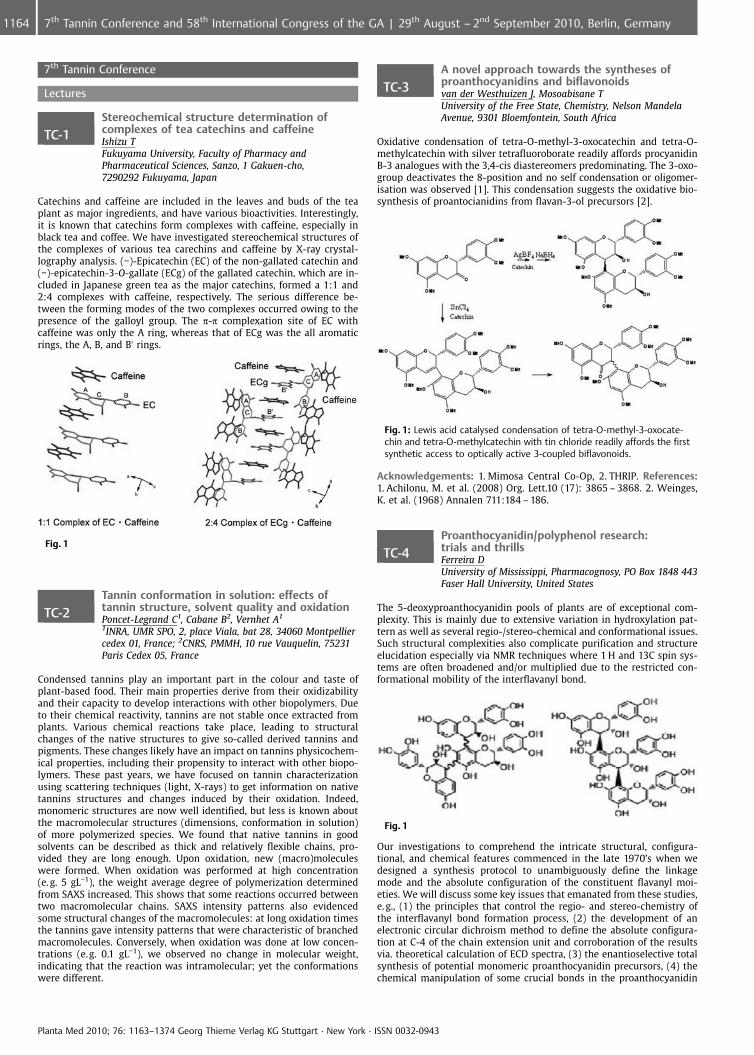
Catechins and caffeine are included in the leaves and buds of the teaplant as major ingredients, and have various bioactivities. Interestingly,it is known that catechins form complexes with caffeine, especially inblack tea and coffee. We have investigated stereochemical structures ofthe complexes of various tea carechins and caffeine by X-ray crystal-lography analysis. (�)-Epicatechin (EC) of the non-gallated catechin and(�)-epicatechin-3-O-gallate (ECg) of the gallated catechin, which are in-cluded in Japanese green tea as the major catechins, formed a 1:1 and2:4 complexes with caffeine, respectively. The serious difference be-tween the forming modes of the two complexes occurred owing to thepresence of the galloyl group. The p-p complexation site of EC withcaffeine was only the A ring, whereas that of ECg was the all aromaticrings, the A, B, and B’ rings.
Fig. 1
TC-2Tannin conformation in solution: effects oftannin structure, solvent quality and oxidationPoncet-Legrand C1, Cabane B2, Vernhet A1
1INRA, UMR SPO, 2, place Viala, bat 28, 34060 Montpelliercedex 01, France; 2CNRS, PMMH, 10 rue Vauquelin, 75231Paris Cedex 05, France
Condensed tannins play an important part in the colour and taste ofplant-based food. Their main properties derive from their oxidizabilityand their capacity to develop interactions with other biopolymers. Dueto their chemical reactivity, tannins are not stable once extracted fromplants. Various chemical reactions take place, leading to structuralchanges of the native structures to give so-called derived tannins andpigments. These changes likely have an impact on tannins physicochem-ical properties, including their propensity to interact with other biopo-lymers. These past years, we have focused on tannin characterizationusing scattering techniques (light, X-rays) to get information on nativetannins structures and changes induced by their oxidation. Indeed,monomeric structures are now well identified, but less is known aboutthe macromolecular structures (dimensions, conformation in solution)of more polymerized species. We found that native tannins in goodsolvents can be described as thick and relatively flexible chains, pro-vided they are long enough. Upon oxidation, new (macro)moleculeswere formed. When oxidation was performed at high concentration(e. g. 5 gL�1), the weight average degree of polymerization determinedfrom SAXS increased. This shows that some reactions occurred betweentwo macromolecular chains. SAXS intensity patterns also evidencedsome structural changes of the macromolecules: at long oxidation timesthe tannins gave intensity patterns that were characteristic of branchedmacromolecules. Conversely, when oxidation was done at low concen-trations (e.g. 0.1 gL�1), we observed no change in molecular weight,indicating that the reaction was intramolecular; yet the conformationswere different.
TC-3A novel approach towards the syntheses ofproanthocyanidins and biflavonoidsvan der Westhuizen J, Mosoabisane TUniversity of the Free State, Chemistry, Nelson MandelaAvenue, 9301 Bloemfontein, South Africa
Oxidative condensation of tetra-O-methyl-3-oxocatechin and tetra-O-methylcatechin with silver tetrafluoroborate readily affords procyanidinB-3 analogues with the 3,4-cis diastereomers predominating. The 3-oxo-group deactivates the 8-position and no self condensation or oligomer-isation was observed [1]. This condensation suggests the oxidative bio-synthesis of proantocianidins from flavan-3-ol precursors [2].
Fig. 1: Lewis acid catalysed condensation of tetra-O-methyl-3-oxocate-chin and tetra-O-methylcatechin with tin chloride readily affords the firstsynthetic access to optically active 3-coupled biflavonoids.
Acknowledgements: 1. Mimosa Central Co-Op, 2. THRIP. References:1. Achilonu, M. et al. (2008) Org. Lett.10 (17): 3865 – 3868. 2. Weinges,K. et al. (1968) Annalen 711:184 – 186.
TC-4Proanthocyanidin/polyphenol research:trials and thrillsFerreira DUniversity of Mississippi, Pharmacognosy, PO Box 1848 443Faser Hall University, United States
The 5-deoxyproanthocyanidin pools of plants are of exceptional com-plexity. This is mainly due to extensive variation in hydroxylation pat-tern as well as several regio-/stereo-chemical and conformational issues.Such structural complexities also complicate purification and structureelucidation especially via NMR techniques where 1 H and 13C spin sys-tems are often broadened and/or multiplied due to the restricted con-formational mobility of the interflavanyl bond.
Fig. 1
Our investigations to comprehend the intricate structural, configura-tional, and chemical features commenced in the late 1970’s when wedesigned a synthesis protocol to unambiguously define the linkagemode and the absolute configuration of the constituent flavanyl moi-eties. We will discuss some key issues that emanated from these studies,e. g., (1) the principles that control the regio- and stereo-chemistry ofthe interflavanyl bond formation process, (2) the development of anelectronic circular dichroism method to define the absolute configura-tion at C-4 of the chain extension unit and corroboration of the resultsvia. theoretical calculation of ECD spectra, (3) the enantioselective totalsynthesis of potential monomeric proanthocyanidin precursors, (4) thechemical manipulation of some crucial bonds in the proanthocyanidin
Planta Med 2010; 76: 1163–1374 Georg Thieme Verlag KG Stuttgart · New York · ISSN 0032-0943
1164 7th Tannin Conference and 58th International Congress of the GA | 29th August – 2nd September 2010, Berlin, Germany
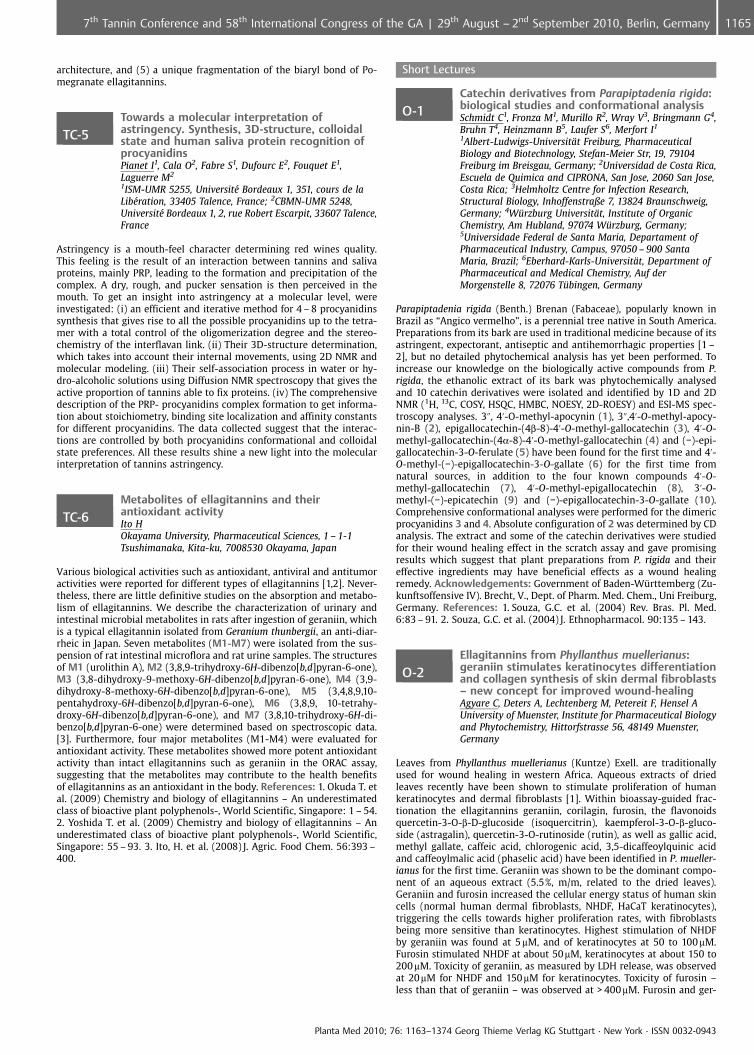
architecture, and (5) a unique fragmentation of the biaryl bond of Po-megranate ellagitannins.
TC-5Towards a molecular interpretation ofastringency. Synthesis, 3D-structure, colloidalstate and human saliva protein recognition ofprocyanidinsPianet I1, Cala O2, Fabre S1, Dufourc E2, Fouquet E1,Laguerre M2
1ISM-UMR 5255, Universit� Bordeaux 1, 351, cours de laLib�ration, 33405 Talence, France; 2CBMN-UMR 5248,Universit� Bordeaux 1, 2, rue Robert Escarpit, 33607 Talence,France
Astringency is a mouth-feel character determining red wines quality.This feeling is the result of an interaction between tannins and salivaproteins, mainly PRP, leading to the formation and precipitation of thecomplex. A dry, rough, and pucker sensation is then perceived in themouth. To get an insight into astringency at a molecular level, wereinvestigated: (i) an efficient and iterative method for 4 – 8 procyanidinssynthesis that gives rise to all the possible procyanidins up to the tetra-mer with a total control of the oligomerization degree and the stereo-chemistry of the interflavan link. (ii) Their 3D-structure determination,which takes into account their internal movements, using 2D NMR andmolecular modeling. (iii) Their self-association process in water or hy-dro-alcoholic solutions using Diffusion NMR spectroscopy that gives theactive proportion of tannins able to fix proteins. (iv) The comprehensivedescription of the PRP- procyanidins complex formation to get informa-tion about stoichiometry, binding site localization and affinity constantsfor different procyanidins. The data collected suggest that the interac-tions are controlled by both procyanidins conformational and colloidalstate preferences. All these results shine a new light into the molecularinterpretation of tannins astringency.
TC-6Metabolites of ellagitannins and theirantioxidant activityIto HOkayama University, Pharmaceutical Sciences, 1 – 1-1Tsushimanaka, Kita-ku, 7008530 Okayama, Japan
Various biological activities such as antioxidant, antiviral and antitumoractivities were reported for different types of ellagitannins [1,2]. Never-theless, there are little definitive studies on the absorption and metabo-lism of ellagitannins. We describe the characterization of urinary andintestinal microbial metabolites in rats after ingestion of geraniin, whichis a typical ellagitannin isolated from Geranium thunbergii, an anti-diar-rheic in Japan. Seven metabolites (M1-M7) were isolated from the sus-pension of rat intestinal microflora and rat urine samples. The structuresof M1 (urolithin A), M2 (3,8,9-trihydroxy-6H-dibenzo[b,d]pyran-6-one),M3 (3,8-dihydroxy-9-methoxy-6H-dibenzo[b,d]pyran-6-one), M4 (3,9-dihydroxy-8-methoxy-6H-dibenzo[b,d]pyran-6-one), M5 (3,4,8,9,10-pentahydroxy-6H-dibenzo[b,d]pyran-6-one), M6 (3,8,9, 10-tetrahy-droxy-6H-dibenzo[b,d]pyran-6-one), and M7 (3,8,10-trihydroxy-6H-di-benzo[b,d]pyran-6-one) were determined based on spectroscopic data.[3]. Furthermore, four major metabolites (M1-M4) were evaluated forantioxidant activity. These metabolites showed more potent antioxidantactivity than intact ellagitannins such as geraniin in the ORAC assay,suggesting that the metabolites may contribute to the health benefitsof ellagitannins as an antioxidant in the body. References: 1. Okuda T. etal. (2009) Chemistry and biology of ellagitannins – An underestimatedclass of bioactive plant polyphenols-, World Scientific, Singapore: 1 – 54.2. Yoshida T. et al. (2009) Chemistry and biology of ellagitannins – Anunderestimated class of bioactive plant polyphenols-, World Scientific,Singapore: 55 – 93. 3. Ito, H. et al. (2008) J. Agric. Food Chem. 56:393 –400.
Short Lectures
O-1Catechin derivatives from Parapiptadenia rigida:biological studies and conformational analysisSchmidt C1, Fronza M1, Murillo R2, Wray V3, Bringmann G4,Bruhn T4, Heinzmann B5, Laufer S6, Merfort I1
1Albert-Ludwigs-Universit�t Freiburg, PharmaceuticalBiology and Biotechnology, Stefan-Meier Str, 19, 79104Freiburg im Breisgau, Germany; 2Universidad de Costa Rica,Escuela de Quimica and CIPRONA, San Jose, 2060 San Jose,Costa Rica; 3Helmholtz Centre for Infection Research,Structural Biology, Inhoffenstraße 7, 13824 Braunschweig,Germany; 4W�rzburg Universit�t, Institute of OrganicChemistry, Am Hubland, 97074 W�rzburg, Germany;5Universidade Federal de Santa Maria, Departament ofPharmaceutical Industry, Campus, 97050 – 900 SantaMaria, Brazil; 6Eberhard-Karls-Universit�t, Department ofPharmaceutical and Medical Chemistry, Auf derMorgenstelle 8, 72076 T�bingen, Germany
Parapiptadenia rigida (Benth.) Brenan (Fabaceae), popularly known inBrazil as “Angico vermelho”, is a perennial tree native in South America.Preparations from its bark are used in traditional medicine because of itsastringent, expectorant, antiseptic and antihemorrhagic properties [1 –2], but no detailed phytochemical analysis has yet been performed. Toincrease our knowledge on the biologically active compounds from P.rigida, the ethanolic extract of its bark was phytochemically analysedand 10 catechin derivatives were isolated and identified by 1D and 2DNMR (1H, 13C, COSY, HSQC, HMBC, NOESY, 2D-ROESY) and ESI-MS spec-troscopy analyses. 3@, 4’-O-methyl-apocynin (1), 3@,4’-O-methyl-apocy-nin-B (2), epigallocatechin-(4b-8)-4’-O-methyl-gallocatechin (3), 4’-O-methyl-gallocatechin-(4a-8)-4’-O-methyl-gallocatechin (4) and (�)-epi-gallocatechin-3-O-ferulate (5) have been found for the first time and 4’-O-methyl-(�)-epigallocatechin-3-O-gallate (6) for the first time fromnatural sources, in addition to the four known compounds 4’-O-methyl-gallocatechin (7), 4’-O-methyl-epigallocatechin (8), 3’-O-methyl-(�)-epicatechin (9) and (�)-epigallocatechin-3-O-gallate (10).Comprehensive conformational analyses were performed for the dimericprocyanidins 3 and 4. Absolute configuration of 2 was determined by CDanalysis. The extract and some of the catechin derivatives were studiedfor their wound healing effect in the scratch assay and gave promisingresults which suggest that plant preparations from P. rigida and theireffective ingredients may have beneficial effects as a wound healingremedy. Acknowledgements: Government of Baden-W�rttemberg (Zu-kunftsoffensive IV). Brecht, V., Dept. of Pharm. Med. Chem., Uni Freiburg,Germany. References: 1. Souza, G.C. et al. (2004) Rev. Bras. Pl. Med.6:83 – 91. 2. Souza, G.C. et al. (2004) J. Ethnopharmacol. 90:135 – 143.
O-2Ellagitannins from Phyllanthus muellerianus:geraniin stimulates keratinocytes differentiationand collagen synthesis of skin dermal fibroblasts– new concept for improved wound-healingAgyare C, Deters A, Lechtenberg M, Petereit F, Hensel AUniversity of Muenster, Institute for Pharmaceutical Biologyand Phytochemistry, Hittorfstrasse 56, 48149 Muenster,Germany
Leaves from Phyllanthus muellerianus (Kuntze) Exell. are traditionallyused for wound healing in western Africa. Aqueous extracts of driedleaves recently have been shown to stimulate proliferation of humankeratinocytes and dermal fibroblasts [1]. Within bioassay-guided frac-tionation the ellagitannins geraniin, corilagin, furosin, the flavonoidsquercetin-3-O-b-D-glucoside (isoquercitrin), kaempferol-3-O-b-gluco-side (astragalin), quercetin-3-O-rutinoside (rutin), as well as gallic acid,methyl gallate, caffeic acid, chlorogenic acid, 3,5-dicaffeoylquinic acidand caffeoylmalic acid (phaselic acid) have been identified in P. mueller-ianus for the first time. Geraniin was shown to be the dominant compo-nent of an aqueous extract (5.5%, m/m, related to the dried leaves).Geraniin and furosin increased the cellular energy status of human skincells (normal human dermal fibroblasts, NHDF, HaCaT keratinocytes),triggering the cells towards higher proliferation rates, with fibroblastsbeing more sensitive than keratinocytes. Highest stimulation of NHDFby geraniin was found at 5 mM, and of keratinocytes at 50 to 100 mM.Furosin stimulated NHDF at about 50mM, keratinocytes at about 150 to200 mM. Toxicity of geraniin, as measured by LDH release, was observedat 20mM for NHDF and 150 mM for keratinocytes. Toxicity of furosin –less than that of geraniin – was observed at > 400mM. Furosin and ger-
Planta Med 2010; 76: 1163–1374 Georg Thieme Verlag KG Stuttgart · New York · ISSN 0032-0943
11657th Tannin Conference and 58th International Congress of the GA | 29th August – 2nd September 2010, Berlin, Germany
aniin stimulated the biosynthesis of collagen from NHDF at 50 mM and5 – 10mM respectively. Geraniin at 105 mM significantly stimulated thedifferentiation in NHEK while furosin had a minor influence on theexpression of involucrin and cytokeratins K1 and K10. References:1. Agyare et al., (2009). J. Ethnopharmacol. 125:393 – 403.
O-3Thiolytic screening method for exploringcondensed tannin variation in a unique sainfoingermplasm bankStringano E1, Hayot C2, Smith L2, Theodoridou K3,Aufrere J3, Brown R1, Hayes W4, Cramer R4, Mueller-Harvey I5
1University of Reading, Agriculture, 1 Earley Gate, P O Box236, RG6 6AT Reading RG6 6AT, United Kingdom; 2NIAB,Huntingdon Road, CB3 0LE Cambridge, United Kingdom;3INRA UR1213 Herbivores, Centre de Clermont-Ferrand –Theix, Theix, 63122 Saint-Genes-Champanelle, France;4University of Reading, Department of Chemistry,Whiteknights, RG6 6AD Reading, United Kingdom;5University of Reading, ASRG Chemistry & Biochemistry Lab,Agriculture Department, 1 Earley Gate, P O Box 236 ReadingRG6 6AT, United Kingdom
This EU funded ‘HealthyHay’ project established a sainfoin (Onobrychisviciifolia) germplasm bank at NIAB, Cambridge, with 360 accessionsfrom around the world. A screening method was developed to charac-terise tannins by thiolytic degradation [1] directly in green plants for thefirst time. The method was validated by separate analysis of unextract-able, extractable and purified tannins using thiolysis, HPLC-GPC andMALDI-TOF MS. Most tannins (58 to 73% of the total) could be recoveredafter Toyopearl HW50 fractionation with water, aqueous methanol andacetone. The greatest losses during purification occurred amongst largermolecular weight tannins with mean degree of polymerisation (mDP)> 18. The composition of water-, aqueous methanol- and acetone-solubletannins differed considerably in their mDP and trans/cis ratios, but notin their prodelphinidin/procyanidin (PD/PC) ratios. Direct thiolysis re-vealed that the condensed tannin contents in this germplasm collectionranged from 0.6 to 2.8 g/100 dry weight; mDP ranged from 12 to 84; PD/PC ratios from 53/47 to 95/5; and trans/cis ratios from 12/88 to 34/66.Detailed analysis of leaves and stems of the Perly variety, which wasgrown at INRA near Clermont-Ferrand (France) and harvested at threephenological stages between 2 June to 16 July, demonstrated a 2-foldhigher tannin content and a 3-fold lower mDP in the less mature com-pared to mature plants. There was little change in PD/PC or trans/cisratios. Leaf and stem tannins differed particularly in their mDP and PD/PC ratios. This screening method will underpin future breeding pro-grammes to improve the nutritional and veterinary properties of sain-foin for animals and to reduce greenhouse gas emissions (NOx and CH4)from ruminants. Acknowledgements: EU Marie Curie Research Train-ing Network (MRTN_CT-2006 – 035805). References: 1. Guyot, S. et al.(2001) In: ‘Flavonoids and Other Polyphenols’. Methods in Enzymology335, 57 – 70.
O-4Proanthocyanidins and ellagitannins: newinsights into the use for antiadhesive prophylaxisagainst viral and microbial pathogens and as skinactive compoundsHensel A, Gescher K, L�hr G, K�hn J, Agyare CUniversity of M�nster, Institute of Pharmaceutical Biologyand Phytochemistry, Hittorfstr. 56, 48149 M�nster, Germany
The study deals with molecular investigations on potential targets forproanthocyanidins and ellagitannins used as antiadhesive compoundsfor antiviral, antimicrobial and wound-healing activity. Antibacterialand antiviral effects of defined proanthocyanidins and ellagitanninswere investigated against Herpes simplex virus I (HSV-1) and Porphyr-omonas gingivalis, the major pathogen for periodontitis. Geraniin, pro-cyanidin B2 and 3,3’-digalloylated B2 exhibited strong antiviral activity.Galloylation strongly increased the activity. Activity was due to an in-hibition of viral adhesion to host cells; penetration and replication werenot influenced. The digalloylated dimer interacts with the major viralsurface gD-adhesin, which was oligomerized, forming a rigid structure,not able to initiate further adsorption process. Antiadhesive effects ofoligomeric proanthocyanidins against P. gingivalis were due to inhibi-tion of the major bacterial adhesins (Lys- and Arg-gingipaine). Addition-ally the expression of bacterial virulence factors is inhibited signifi-
cantly, leading to a diminished pathogenic activity. The role of procya-nidin-enriched extracts within clinical development products is dis-cussed. Positive effects of tannins on skin are traditionally described,while the mode of action is unknown. Therefore the influence on ger-aniin on cell physiology of skin cells was investigated. Geraniin stimu-lated cell differentiation of keratinocytes (involucrin, cytokreatin 1, 10)and proteins responsible for formation of extracellular matrix (collagen).Potential pathways how tannins act on skin physiology are discussed.Summarizing it is shown that tannins act quite specifically on definedtargets. Effects on target proteins are not as unspecific as often claimed.Therefore the medical use of tannins has to be investigated in moredetail.
O-5In vitro transport studies of Hawthornprocyanidins by Caco-2 monolayers: transportand effluxZumdick S, Deters A, Hensel AInstitute for Pharmaceutical Biology and Phytochemistry,University of M�nster, Hittorfstr. 56, 48149 M�nster,Germany
Extracts from Hawthorn (Crataegus sp.) are considered as a rationalbased phytomedicine for declining cardiac performance, with flavonoidsand procyanidins as active compounds. Especially oligomeric procyani-dins (OPC) are assessed to have the most marked pharmacodynamiceffects. Detailed investigations on the bioavailability of procyanidinsafter oral ingestion are not available. The current study was designedto investigate the absorption of OPC from different fractions/extracts ofCrataegi folium cum flore using validated monolayers of the humanCaco-2 cell line grown in Transwells. Respective concentrations of dis-tinct OPC clusters were determined in the basolateral and apical com-partments and in cell lysates of Caco-2 cells. Quantitation was per-formed by HPLC on diol stationary phase with fluorescence detection.No significant absorption of OPCs with degree of polymerization (DP) ‡2into the basolateral compartments was observed after apical applicationof 125 – 250 mg/mL, but OPC with DP 6 to 9 were detected in the respec-tive cell lysates. Interestingly, transport of procyanidin B2 in the baso-lateral ? apical direction was higher than that in the apical ? basolat-eral direction, indicating efflux transporters carrying out OPCs after in-itial absorption to the apical side. This hypothesis was clearly proven byuse of verapamil, a P-glycoprotein inhibitor in the absorption assay to-gether with OPC as shown with B2: data from these experiments provedan increased apical ? basolateral absorption of B2. These data clearlyprove the suitability of the Caco-2 system for transport studies of poly-phenols and indicate that intestinal OPC absorption is subjected to anefflux competition.
O-6Chemical characterisation and sensoryevaluation of Bordeaux wines. Correlation withwine ageChira K, Teissedre PInstitut des Sciences de la Vigne et du Vin, 210 chemin deLeysotte CS 50008, 33882 Villenave d’Ornon Cedex, France
Quality evaluation of a red wine is based on wine-tasting. Chemicalanalyses are performed to understand what compounds influence sen-sory properties and how they affect them. Quantitative determination ofcertain chemical compounds is a criterion of wine valuation origin [1]and authenticity [2]. Our study concerning wine quality was carried outwith 24 vintages of Cabernet-Sauvignon (CS) and with 7 vintages ofMerlot (M) produced by Bordeaux wine-growing areas. Proanthocyani-din monomers and oligomers were identified and quantified by HPLC-UV-Fluo. Galloylation (%G) and prodelphinidins percentage (%P), meandegree of polymerization (mDP) were determined [3]. Total phenoliccompounds, total anthocyanins, total tannins, hue, IC’ (color intensity),total acidity, ethanol level and pH were evaluated. Sensory analysisconcerning astringency and bitterness intensity was also performed.Total phenolic compounds, anthocyanins, tannins, tannin monomers,hue, IC’,% G,% P, mDP and astringency intensity differentiate both winesaccording to vintage. Correlation between wine age and: mDP, hue,astringency, phenolic compounds, tannin monomers, total tannin levelsare obtained. The qualitative wine tannin characterization is establishedbetween astringency and mDP (R 2 = 0.509, p = 0.051, CS; R2 = 0.780,p = 0.000 M). mDP is a vintage marker (R 2 = 0.796, p = 0.000; CS andR2 = 0.946, p = 0.000; M). Scale patterns between wine mDP and bothageing and tannin perception are proposed. M wines are characterized
Planta Med 2010; 76: 1163–1374 Georg Thieme Verlag KG Stuttgart · New York · ISSN 0032-0943
1166 7th Tannin Conference and 58th International Congress of the GA | 29th August – 2nd September 2010, Berlin, Germany
by lower levels of total phenolic compounds, tannins, anthocyanins,mDP,% G,% P% levels as well as by lower astringency intensity than CSwines. References: 1. Forina, M. et al. (1986) Vitis 25: 189 – 201. 2. Ar-vanitoyannis, I. S. et al. (1999) Trends Food Sci. Technol. 10: 321 – 336. 3.Drinkine, J. et al.(2007) J. Agric. Food Chem. 55: 6292 – 6299.
O-7Fluorescence lifetime imaging microscopy (FLIM)to demonstrate the nuclear binding of flavanolsand (�-epigallocatechin gallateMueller-Harvey I1, Botchway S2, Feucht W3, Polster J3,Burgos P2, Parker A2
1University of Reading, Agriculture, 1 Earley Gate, P O Box236, RG6 6AT Reading, United Kingdom; 2RutherfordAppleton Laboratory, Central Laser Facility, HSIC, OX11 0QXDidcot, United Kingdom; 3Technical University of Munich,Wissenschaftszentrum Weihenstephan, Freising, 85354Freising, Germany
The use of light microscopy and DMACA staining strongly suggested thatplant and animal cell nuclei act as sinks for flavanols [1, 2]. Detailed uv-vis spectroscopic titration experiments indicated that histone proteinsare the likely binding sites in the nucleus [2]. Here we report the devel-opment of a multi-photon excitation microscopy technique combinedwith fluorescent lifetime measurements of flavanols. Using this techni-que, (+) catechin, (�) epicatechin and (�) epigallocatechin gallate (EGCG)showed strikingly different excited state lifetimes in solution. Interac-tion of histone proteins with flavanols was indicated by the appearanceof a significant t2-component of 1.7 to 4.0 ns. Tryptophan interferencecould be circumvented in the in vivo fluorescence lifetime imaging mi-croscopy (FLIM) experiments with 2-photon excitation at 630 nm. Thisenabled visualisation and semi-quantitative measurements that demon-strated unequivocally the absorption of (+)catechin, (�)epicatechin andEGCG by nuclei of onion cells. 3D FLIM revealed for the first time thatexternally added EGCG penetrated the whole nucleus in onion cells. Therelative proportions of EGCG in cytoplasm: nucleus: nucleoli were ca.1:10:100. FLIM experiments may therefore facilitate probing the healtheffects of EGCG, which is the major constituent of green tea. Acknowl-edgements: The Science and Technology Facilities Council providedfacility access time References: 1. Feucht W. et al. (2004) Plant CellRep 22: 430 – 436. 2. Polster J. et al (2003) Biol. Chem. 384: 997 – 1006.
O-8Fractionation and characterization of highmolecular weight proanthocyanidin frompersimmon fruit by thiolysis-HPLC, size-exclusionchromatography, MALDI-TOF/MS, and NMRLi C1, Hagerman A2
1Huazhong Agricultural University, College of Food Scienceand Technology, College of Food Science and Technology,Wuhan 430070, China; 2Miami University (Ohio), Chemistry& Biochemistry, Department of Chemistry & Biochemistry,45056 Oxford, United States
High molecular weight proanthocyanidin (condensed tannin) from per-simmon pulp was fractionated on Toyopearl TSK-HW-50-F. The crudetannin and the three fractions were characterized by thiolysis-HPLC-ESI-MS, GPC, MALDI-TOF-MS and 13C-NMR. Thiolysis-HPLC-ESI-MS showedthat the proanthocyanidin terminal units were catechin and epigalloca-techin gallate, and extender units were epicatechin, epigallocatechin,(epi)gallocatechin-3-O-gallate, and (epi)catechin-3-O-gallate. The crudetannin had a very high prodelphinidin content (65 – 80%) and a highdegree of 3-O-galloylation (72%). The composition of the fractions andthe unfractionated tannin was similar, but the fractions were distin-guished by degree of polymerization. Thiolysis suggested that the per-simmon tannin was comprised of polymers ranging from 13kD to 20kD(degree of polymerization 30 – 50), but sizes estimated by GPC weremuch smaller. MALDI-TOF-MS revealed the presence of a heteropolyfla-vanol series including (epi)catechin and (epi)gallocatechin repeatingunits, and suggested that the persimmon proanthocyanidin containedsome A-type interflavan linkages. The crude material was gently chemi-cally degraded with acid to yield products that were amenable to NMRanalysis, which was used to confirm the A-type linkages. Acknowledge-ments: Financial support was provided by the National Natural ScienceFoundation of China (No.30972398), and the Key Project of ChineseMinistry of Education (No.109115) to Huazhong Agricultural University,and by Agricultural Research Services Specific Cooperative AgreementNumber 58 – 1932 – 6-634 with Miami University.
58th International Congress and Annual Meeting of theSociety for Medicinal Plant and Natural Product Research
Lectures
L-1Sustainable drugs and global health careCordell GNatural Products Inc, 9447 Hamlin Avenue Evanston, UnitedStates
The global population has now reached 7 billion, and forests and otherresources around the world are being irreversibly depleted for energy,food, shelter, material goods, and drugs to accommodate populationneeds. For the developed world, efforts have been initiated to make drugproduction “greener”, with milder synthetic reagents, shorter reactiontimes, and more efficient processing, thereby using less energy, becom-ing more atom efficient, and generating fewer by-products. However, formost of the world’s population, plants, based on many well-establishedsystems of medicine, in either crude or extract form, represent thefoundation of primary health care for the foreseeable future. Contem-porary harvesting methods for medicinal plants are severely depletingthese critical indigenous resources. However, maintaining and enhan-cing the availability of quality medicinal agents on a sustainable basis isan unappreciated public health care concept. To accomplish these goalsfor future health care, and restore the health of the Earth, a profoundparadigm shift is necessary: all medicinal agents should be regarded as asustainable commodity, irrespective of their source. Several approachesto enhancing the availability of safe and efficacious plant-based medic-inal agents will be presented including integrated strategies to manifestthe four pillars (information, botany, chemistry, and biology) for medic-inal plant quality control. These integrated initiatives involve informa-tion systems, metabolomics, biotechnology, nanotechnology, in-fieldanalysis of medicinal plants, and the application of new detection tech-niques for the development of medicinal plants with enhanced levels ofsafe and reproducible biological agents.
L-2Plant molecular systematics: prospects foridentifying species and for analysing bioactivecompound evolutionBorsch TFreie Universit�t Berlin, Botanischer Garten und BotanischesMuseum Berlin-Dahlem, Direktor der Zentraleinrichtung,K�nigin-Luise-Straße 6 – 8, 14195 Berlin, Germany
DNA-based approaches to unravel plant evolutionary relationships, togain insights into patterns and processes of speciation, and to unravelthe diversity of genotypes within species have revolutionized plant biol-ogy. Phylogenetic hypotheses now include all major lineages of flower-ing plants and the inclusion of more and more genera and species intophylogenetic trees is on their way. This offers a unique opportunity toreconstruct the evolution of secondary compounds and their biosyn-thetic pathways (macroevolution). For example, in the genus Hypericum(St. John’s Wort) the evolution of bioactive compounds such as hypericinand hyperforin correlates with certain phylogenetic lineages. If there isan up to date taxonomic information source that connects genetic andphenotypic data for organisms to the respective taxon names, sequencedata can be used to identify even small fragments of plant materials.Although this is a promising application, it should be noted that therequired comprehensive monographic treatments so far exist for only asmall fraction of flowering plant genera.
L-3Infectious diseases and herbal medicinesPieters LDepartment of Pharmaceutical Sciences, University ofAntwerp, Universiteitsplein 1, 2610 Antwerp, Belgium
For many years the Laboratory of Pharmacognosy and PharmaceuticalAnalysis has been involved in collaborative projects with research insti-tutes in developing countries, where traditional medicine still plays animportant role in local health care systems, for economic as well ascultural reasons. The aim of our work is twofold: Firstly, to provide ascientific basis for the therapeutic use of medicinal plants in thesecountries (or to discourage their use if not), and to sustain the develop-ment of standardised herbal medicinal products, the quality of whichcan be controlled, with proven safety and efficacy; secondly, to charac-terise lead compounds that can be used to develop new therapeutic
Planta Med 2010; 76: 1163–1374 Georg Thieme Verlag KG Stuttgart · New York · ISSN 0032-0943
11677th Tannin Conference and 58th International Congress of the GA | 29th August – 2nd September 2010, Berlin, Germany
agents. During the past years our attention has mainly been focused,from a geographical point of view, on Tanzania, DR Congo and Guinea-Conakry; and from a medicinal point of view, on malaria. A biologicalscreening programme of plants used in Tanzania against infectious dis-eases led to the selection of Elaeodendron schlechteranum (Celastraceae)and Ormocarmpum kirkii (Papilionaceae) for further investiagtion, basedon their antibacterial and antiplasmodial properties, respectively. FromE. schlechteranum a quinone-methide triterpene, 22b-hydroxytingenoneor tingenin B, with a pronounced activity against a range of microorgan-isms was isolated, although a high cytotoxicity was observed as well.From O. kirkii a series of (3 – 3’)-biflavonoids was obtained, includingseveral new ones, showing antiplasmodial activity to a various degree,which allowed to establish some structure-activity relationships. In ascreening programme of plants traditionally used in DR Congo againstmalaria, Nauclea pobeguinii (Rubiaceae) was selected for further inves-tigation. The main constituent of N. pobeguinii was the alkaloid stricto-samide, and an HPLC method was developed and validated for the quan-tification of this compound in an 80% EtOH stem bark extract. The anti-malarial activity of this standardised extract was established in vivo, andclinical studies in DR Congo have been initiated in order to provide aherbal medicinal product with proven safety and efficacy for non-severemalaria for the local market.
L-4Validating the antimicrobial activity of Africantraditional medicines – Reflecting on a decade ofresearchvan Vuuren SUniversity of Witwatersrand, Department of Pharmacy andPharmacology, Pharmaceutical Microbiology SeniorLecturer, 7 York Rd, Parktown Johannesburg, South Africa
Unique and diverse botanical resources make traditional healing an in-tegral part of the African cultural heritage. Plants have been used forcenturies as anti-infective agents and the need to validate the traditionaluse in the last decade is addressed with respect to past challenges, latestdevelopments and future recommendations. Microbiological methods(disc diffusion, minimum inhibitory concentration, time-kill and inter-active assays) will be reviewed with practical examples given from someof the most widely used indigenous African medicinal plants. An exten-sive review of Artemisia afra will be given, encompassing antimicrobialscreening, geographical variation, major compound analysis, comparisonwith commercial essential oils and the use in combination with otherplants. Furthermore, the potential use in formulations is demonstratedwhere preservative efficacies within a cream formulation indicated bac-tericidal activity against Staphylococcus aureus, Pseudomonas aeruginosaand Escherichia coli. Pathogen specific studies will be presented wheresome of the most commonly used plants to treat sexually transmittedinfections demonstrate activities as low as 0.2 mg/ml (Hypericum aethio-picum and Polygala fruticosa, tested against Gardnerella vaginalis). Also,the five most active plants tested against anti-diarrhoeal pathogens inthe remote area of northern Maputaland include Psiduim guajava andGarcinia livingstonei (MIC values of 0.01 and 0.08 mg/ml respectivelyagainst Bacillus cereus), Gymnosporia senegalensis and Syzygium corda-tum (0.13 mg/ml against Enterococcus faecalis) and Sclerocarya birea(0.13 mg/ml against Proteus vulgaris, Salmonella typhimurium, Shigellaflexneri and Staphylococcus aureus). These antimicrobial studies play animportant role in the understanding of traditional healing and advan-cing the phytotherapeutic application of medicinal plants.
L-5Herbal medicine research in TaiwanYang NAgricultural Biotechnology Research Center, 128 AcademiaRoad, Section 2, Nankang, Taipei 115, Taiwan Taipei, Taiwan
In a collective effort to upgrade and integrate Traditional Chinese Med-icine (TCM) research, development and application, various nationalresearch centers and program projects were set up in Taiwan and rea-sonable successes have been achieved. A brief introduction and snapshots of these programs will be presented. As an example of TCM R&Din Taiwan, the research program at Academia Sinica will be examined inmore detail. Cross-talk, collaborating research laboratories have sinceestablished theme projects and defined experimental systems for anti-inflammation and immuno-modulation studies. These include investi-gations on T-cells, dendritic cells, tumor cell-related immunomodula-tory, and anti-inflammatory bioactivities in response to phytocom-pounds/botanical substances extracted from Chinese or Western medic-
inal plants including Anoectochilus, Echinacea, Bidens and Wedeliaplants. Potential chemoprevention and anti-tumor activities of thesephytoextracts/phytochemicals (e.g., shikonin, [BF(S+L)Ep], cytopiloyne,Wedelia Chinensis) have been investigated in breast and prostate tumorsystems obtaining encouraging results. Functional genomics, proteomicsand metabolomics studies have also yielded significant and interestingfindings. Experimental approaches using clinically-relevant in vivo andex vivo study systems are being evaluated for translation of researchfindings into medical and biotechnological applications. With TCM andmedicinal plant research infrastructure outlined above, our highestpriority for future R&D in Taiwan is to initiate, establish and optimizeinternational research collaboration: research foci of such interest willbe contemplated. Acknowledgements: Agriculture Biotechnology Re-search Center, Academia Sinica, Taipei, Taiwan. National Science Council,Taiwan.
L-6South Africa’s medicinal flora – abundantopportunities and daunting challengesViljoen ATshwane University of Technology, Department ofPharmaceutical Sciences, Private Bag X680 Pretoria, SouthAfrica
South Africa is considered to be one of the most biodiverse areas in theworld and harbours close to 10% of the world’s flora. This diversity hasprovided abundant opportunities for the development of traditionalhealing practices and it is estimated that 60% of South Africa’s popula-tion still rely on herbal medicines, often as a source for primary health-care. Ironically, despite this access to unlimited botanical resources andthe longstanding use of medicinal plants, very few medicinal plantshave been developed into commercial products. Basic research under-pins any commercial development, yet for many important ethnomedic-inal plants aspects relating to quality, efficacy and safety remain poorlyexplored. The paper will provide a succinct overview on the uses, chem-istry and biological properties for some of the most important medicinalspecies indigenous to South Africa including; Aloe ferox (Cape aloes),Pelargonium sidoides, Sutherlandia frutescens (cancer bush), Hoodiagordonii, Agathosma betulina (buchu), Mesembryanthemum tortuosum,Aspalathus linearis (rooibos tea), Lippia javanica, Siphonochilus aethio-picus (African ginger) etc. The challenges encountered when researchingSouth Africa’s medicinal flora such as chemical variation, the need forquality control protocols, indigenous knowledge systems and benefitsharing will also be highlighted.
L-7Medicinal plants and natural products from LatinAmerica – Subjects of international scientificexchangesGupta MCentro de Investigaciones Farmacognosticas de la FloraPaname�
Latin American countries possess part of the world’ s biodiversity. Of the17 megadiverse countries in the world six are in the neotropics. Thepercentage of Amerindian groups is high in this region. There is wide-spread use of medicinal plants among these groups. The number ofmonographs on Latin American plants in different pharmacopoeias var-ies from 5% in ESCOP, 12% in WHO and up to 15% in European Pharma-copoeia. Results of some collaborative programs such as the Iberoamer-ican Program of Science and Technology for Development (CYTED) andOrganization of American States (OAS), which have fostered scientificexchanges in this region, will be presented. Case studies of research onselected medicinal plants used in traditional medicine will be high-lighted. An overview of research on Panamanian medicinal plants asource of bioactive compounds will also be presented. Acknowledge-ments: OAS and CYTED Program.
L-8Traditional uses and scientific evidence forselected native medicinal plants from Jordan:a critical evaluationAfifi-Yazar F, Abu Dahab R, Kasabri VFaculty of Pharmacy, University of Jordan, PharmaceuticalSciences, Queen Rania Al-Abdullah Street, 11942 Amman,Jordan
Traditional medicine is part of the Jordanian culture. Both rural andurban Jordanian societies depend on traditional medicine; using plants
Planta Med 2010; 76: 1163–1374 Georg Thieme Verlag KG Stuttgart · New York · ISSN 0032-0943
1168 7th Tannin Conference and 58th International Congress of the GA | 29th August – 2nd September 2010, Berlin, Germany
that are mainly locally grown. The occurrence of about 2500 plant spe-cies is recorded. A fifth of these are classified as medicinal plants and areused for the treatment of common mild diseases in addition to thetreatment of chronic and/or incurable diseases. Although the practiceof traditional medicine is based on years of belief and observation,scientific knowledge for most is very limited. To contribute to the exist-ing, although limited, pool of data, our prime interests are native plantswith antidiabetic and anticancer activities. Phase one of our investiga-tion involved screening crude extracts of plants with claimed hypogly-cemic activity in in vitro and in vivo experiments [1]. In parallel, thesystemically collected medicinal plants were screened for their antipro-liferative activity using different carcinoma cell lines [2]. Once efficacyand safety were established, in phase two, biologically active and safeplants were further evaluated in in vitro mechanistic assays [3, 4]. Re-sults were compared to clinically used drugs. In phase three, the activeplants were phytochemically investigated. Findings of our investigationsare aiming, where possible, to link the traditional use with scientificevidence. References: 1. Hamdan, I. I., Afifi, F. U. (2004) J Ethnopharma-col 93: 117 – 21. 2. Abu-Dahab, R., Afifi, F. (2007) Sci Pharm 75:121 – 36.3. Raghavan, G. et al. (2007) Planta Med 73: 427 – 32. 4. Wang, C. C. C. etal. (2008) Basic Clin Pharmacol Toxicol 102: 491 – 7.
L-9Genomic mining – a concept for the discovery ofnew bioactive natural productsGross HUniversity of Bonn, Institute for Pharmaceutical Biology,Nussallee 6, 53115 Bonn, Germany
Natural products continue to represent an important source for leadstructures for drug discovery, but with discovery rates of novel structur-al classes in decline, the need to explore alternate sources of chemicaldiversity is evident. Genomic mining represents hereby a complemen-tary strategy to address these issues and to access a tremendous sourceof new, biologically active metabolites. The base for this claim is pro-vided by the outcomes of the recent genome sequencing projects whichrevealed the presence of numerous biosynthetic gene clusters for whichthe corresponding metabolites are currently unknown [1]. Importantly,it has been observed that the numbers of such orphan biosynthetic locifar outnumbers the quantity of the gene clusters directing the synthesisof known compounds of most of the considered organisms [2]. Appar-ently, only a fraction of natural products has been analyzed and themajority await discovery. Considering this discrepancy and that severalhundreds of sequencing programs are ongoing, the huge potential of thegenomic mining approach for natural product discovery becomes appar-ent. The potential of this untapped resource can be expanded evenfurther by the exploitation of not only genomic, but also metagenomicDNA. After outlining the rationale and methods [1,3] of genomic mining,the success [2], but also the limits of this fascinating and interdisciplin-ary strategy will be illustrated by selected examples from plant andmicrobial genomes. References: 1. Gross (2007) Appl. Microbiol. Bio-technol. 75:267 – 277. 2. Gross (2009) Curr. Opin. Drug Discov. Devel.12:207 – 219. 3. Gross et al. (2007) Chem. Biol. 14:53 – 63.
WS I: Workshops for Young Researchers
Cellular and molecular mechanisms of action of naturalproducts and medicinal plants.Chairs: Th. Efferth, D. Tasdemir, A. Hensel
WS I ILImpulse Lecture: Elucidation of the molecularbasis of anti-inflammatory natural compoundsfrom traditional medicinal plantsWerz OUniversity of Tuebingen, Pharmaceutical Institute, Auf derMorgenstelle 8, 72076 Tuebingen, Germany
Although many traditional medicinal plants are used in the therapy ofinflammatory disorders, the bioactive ingredients and/or the molecularbasis underlying the anti-inflammatory action are often unclear. Numer-ous plant-derived natural products with anti-inflammatory propertieshave previously been reported to reduce the biosynthesis of eicosanoids,i. e., prostaglandins (PGs) and leukotrienes (LTs) and this has essentiallybeen attributed to an interference with the respective key enzymescyclooxygenase (COX) and 5-lipoxygenase (5-LO). The dual inhibitionof 5-LO and of microsomal PGE2 synthase-1 (mPGES-1), which formspro-inflammatory PGE2 from COX-2 derived PGH2, is a novel and pro-mising strategy for the therapy of inflammation being superior oversingle inhibition in terms of higher anti-inflammatory efficacy but alsomay cause fewer side effects. We have investigated selected naturalproducts for which either anti-inflammatory efficacy in vivo or inhibi-tion of PG biosynthesis was described. Here we report that many ofthese compounds (e. g., hyperforin, epigallocatechin gallate, garcinol,curcumin, myrtucommulone, arzanol) are poor inhibitors of COX en-zymes but instead efficiently inhibit mPGES-1. Hence, the previouslyobserved reduced PGE2 levels are rather due to inhibition of mPGES-1than COX-1/2. Of interest, the mPGES-1 inhibitiory effect is often asso-ciated with suppression of 5-LO. Finally, analysis of selected compoundsin experimental models of inflammation confirm inhibition of PGE2 andLT biosynthesis in vivo and suggest a valuable therapeutic potential forintervention with inflammatory diseases.
WS I-1Eruca sativa Mill.: Phytochemical profile andantimicrobial properties of rocket leafy saladsFilocamo A1, Paterniti Mastrazzo G1, Maimone P1,Buongiorno L2, Catania S3, De Pasquale R1, Bisignano G1,Melchini A2
1Pharmaco-Biological Department, School of Pharmacy,Univesity of Messina, Vill. Annunziata, 98168 Messina, Italy;2Foundation “Prof. Antonio Imbesi”, P.zza Pugliatti 1, 98122Messina, Italy; 3Interdepartmental Centre of Experimental,Environmental and Occupational Toxicology (CITSAL), ViaConsolare Valeria, 98122 Messina, Italy
In the last decade salad species consumption is becoming increasinglyimportant worldwide, encouraged from the positive link between eatingfresh raw materials and absorption of health-promoting phytochemicals[1]. Rocket salads are well-known in the traditional medicine for theirtherapeutic properties as astringent, diuretic, digestive, emollient, tonic,depurative, laxative, rubefacient and stimulant [2]. However, the antimi-crobial activity of rocket salad species has been poorly investigated. Ourstudy was designed to characterize the phytochemical profile of Erucasativa Mill. and to evaluate additionally their in vitro antimicrobial activ-ity upon a representative range of pathogenic bacteria and fungi. Differ-ent plant extracts were prepared and tested in order to compare theactivity of individual groups of phytochemicals. LC-MS and HPLC/DADanalyses led to the identification of glucosinolates and flavonoids, respec-tively. Minimum inhibitory concentration (MIC) and minimum bacterici-dal concentration (MBC) of all the extracts were determined using brothdilution methods in 96 wells micro plates. The plant extract showed verysignificant activity against Gram +ve bacteria with MIC and MBC valuesranging from 0.125 – 1 mg/ml and 1 – 4 mg/ml, respectively. A lower ac-tivity was observed against Gram -ve and fungi. However, the plant ex-tract containing GLSs had no effect on any of the Gram +ve and Gram -vebacteria at any of the doses used. These results suggest that the greaterantimicrobial of Eruca sativa leaf extract is not related to the GLS contentbut to other phytochemicals, and might be useful in controlling humanpathogens through the diet. Acknowledgements: This work was sup-ported by Foundation “Prof. Antonio Imbesi” (Messina, Italy). Refer-ences: 1. Vermeulen, M. et al. (2006) J Agric Food Chem 54:5350 – 5358.
Planta Med 2010; 76: 1163–1374 Georg Thieme Verlag KG Stuttgart · New York · ISSN 0032-0943
11697th Tannin Conference and 58th International Congress of the GA | 29th August – 2nd September 2010, Berlin, Germany
2. Perry, L. M. a. M., J., Medicinal plants of SE Asia. Attributed propertiesand uses. The MIT Press: Cambridge, Massachusetts and London.
WS I-2Glucosinolates and their respective enzymatichydrolysis products are not involved in the invitro antioxidant properties of rocket saladspeciesMaimone P1, Taviano M1, Paterniti Mastrazzo G1,Buongiorno L2, Melchini A2, Trovato A1, Miceli N1
1Pharmaco-Biological Department, School of Pharmacy,University of Messina, Vill. Annunziata, 98168 Messina,Italy; 2Foundation “Prof. Antonio Imbesi”, P.zza Pugliatti 1,98122 Messina, Italy
Epidemiological and experimental studies provide some evidence thatchronic diseases could be prevented by high consumption of certainvegetables [1]. Oxidative stress is involved in the pathogenesis of thesediseases, and may be reduced by improving physiological antioxidantdefences through dietary interventions [2]. Dietary patterns rich in plantfoods are the most likely to protect against oxidative stress by absorp-tion of a wide range of naturally occurring antioxidants, that may act assynergists to reduce reactive oxygen species levels [3]. Cruciferous ve-getables are widely studied for their health benefits due partly to thehigh content of antioxidants, like vitamin E and C, carotenoids, polyphe-nols, as well as characteristic phytochemicals, known as glucosinolates(GLSs). The role of GLSs and their enzymatic hydrolysis products,namely isothiocyanates (ITCs), as natural antioxidants is debated [4 –6]. In this study, the antioxidant properties of bioactive compoundsobtained from Eruca sativa Mill. were investigated using in vitro sys-tems. The primary antioxidant properties by DPPH test and reducingpower assay, and the secondary antioxidant ability by ferrous ion(Fe2+) chelating activity were evaluated. The plant extract showed asignificant primary antioxidant activity in both DPPH test and reducingpower assay, and a strong Fe2+ chelating ability. However, neither GLSfraction nor respective pure ITCs had antioxidant effects using the sameexperimental methods at all doses tested. The results suggest that thesephytochemicals are unlikely to account for the direct antioxidant effectsof Eruca sativa extract. References: 1. Pomerlau, J. et al. (2005) J Nutr135:2486 – 2495. 2. Rahman, K. (2007) Clin Interv Aging 2:219 – 236. 3.Podsedek, A. (2007) Food Sci Technol 40:1 – 11. 4. Mart�nez-S�nchez, A.et al. (2008) J Agric Food Chem 56:2330 – 2340. 5. Plumb, G.W. et al.(1996) Free Rad Res 25:75 – 86. 6. Valgimigli, L., Iori, R. (2009) EnvironMol Mutagen 50:222 – 37.
WS I-3Bioactive fatty acids and cerebrosides from theTCM drug Arisaema sp.Rozema E1, Fakhrudin N1, Atanasov A1, Schuster D2,Heiss E1, Sonderegger H3, Krieg C3, Gruber C1, Huck C3,Dirsch V1, Bonn G3, Kopp B1
1University of Vienna, Department of Pharmacognosy,Althanstrasse 14, 1090 Vienna, Austria; 2University ofInnsbruck, Department of Pharmaceutical Chemistry,Innrain 52c, 6020 Innsbruck, Austria; 3University ofInnsbruck, Institute of Analytical Chemistry andRadiochemistry, Innrain 52a, 6020 Innsbruck, Austria
In this study active compounds from the TCM drug Arisaema sp. [1] werecharacterized by bioassay-guided isolation. Extracts and fractions of Aris-aema sp. were tested for agonistic activity towards peroxisome prolifera-tor-activated receptor-a and -g (PPAR) and for activation of the AMP-activated protein kinase (AMPK). These proteins are therapeutical targetsin treatment of metabolic disorders [2,3]. An apolar fraction stronglyactivated PPAR-a and -g and had positive effects on AMPK activity invitro. Among the main compounds identified by GC-MS were n-hexade-canoic acid, 9,12-octadecadienoic acid, 9-octadecenoic acid, octadecanoicacid, 13-phenyltridecanoic acid and pentadecanoic acid. Since cerebro-sides from Arisaema with antihepatotoxic activity reported by Jung et al[4], were found to bind PPAR-a and -g in silico, isolation and activitystudies on these glycosphingolipids were continued. From a polar frac-tion, with moderate agonistic effect on PPAR-a and -g in vitro, cerebro-sides I-VI were isolated. Their structures were elucidated by NMR, ESI-MS-MS and matrix free LDI-TOF-MS-MS. In conclusion, in the presentactivity and analytical studies chemical constituents of Arisaema sp. thatshowed in vitro activity on important anti-diabetic targets were revealed.These findings affirm the great value and rich source of Chinese herbaldrugs for natural product research. Acknowledgements: Sino-Austria
Project, supported by the Austrian Federal Ministry of Science and Researchand Federal Ministry of Health, Women and Youth. This project was alsosupported in part by the Austrian Science Fund [NFN S 10704-B037] and theAustrian Federal Ministry for Science and Research [ACM-2009 – 01206].References: 1. Bensky D. et al (2004) Chinese Herbal Medicine MateriaMedica. Eastland Press. Seattle. 2. Kersten, S. et al (2000) Nature405:421 – 424. 3. Winder W.W. et al (1999) Am. J. Physiol. Endocrinol.Metab. 277:1 – 10. 4. Jung, J.H. et al. (1996) J. Nat. Prod. 59:319 – 322.
WS I-4Isolation of differencially expressed genes fromPsoralea corylifolia by DD-PCRShaukat I1, Muhammad S3, Shaukat R2, Jamil A3
1Chalmers University of Technology, Department ofChemical and Biological Engineering, 412 96 G�teborg,Sweden; 2University of Agriculture, Faisalabad, NIFSAT,38040 Faisalabad, Pakistan; 3University of AgricultureFaisalabad, Molecular Biochemistry Lab, Department ofChemistry and Biochemistry, 38040 Faisalabad, Pakistan
According to the world health organization (WHO) 80% of the world’spopulation uses medicinal plants for the treatment of diseases. In recentyears medicinal plants are primary health source for pharmaceutical in-dustry. Psoralea corylifolia (Psoralea seed) is used in treatment of manyskin diseases in Pakistan and India. Development of resistance in fungi tocommonly used antifungal drugs diverted the attention of researchestowards medicinal plants. In the present study we focused our researchtowards isolation of differenzially expressed genes from seedlings of Psor-alea corylifolia after induction with a fungus, Fusarium solani. RNA wasisolated from control (non-induced) and fungal induced seedlings. Quan-tity and integrity of RNA was checked by agarose gel electrophoresis andspectroscopy. cDNA was formed from RNA by reverse transcription usingoligo-dT primers. All PCR reactions contained the same T11MN primer,and an arbitrary primer. Different arbitrary primers (HAP 25 – 32) weretried for each cDNA. The amplified products were resolved on 6% dena-turing polyacrylamide gel electrophoresis and detection was carried outwith silver staining. Differenzially expressed genes were isolated from theinduced samples after comparing with the controls after sequencing.
WS I-5Natural coumarins and furanocoumarins aspositive gabaergic modulatorsSinghuber J1, Baburin I2, Zehl M1, Hering S2, Kopp B1
1Department of Pharmacognosy, University of Vienna,Althanstraße 14, 1090 Vienna, Austria; 2Department ofPharmacology and Toxicology, University of Vienna,Althanstraße 14, 1090 Vienna, Austria
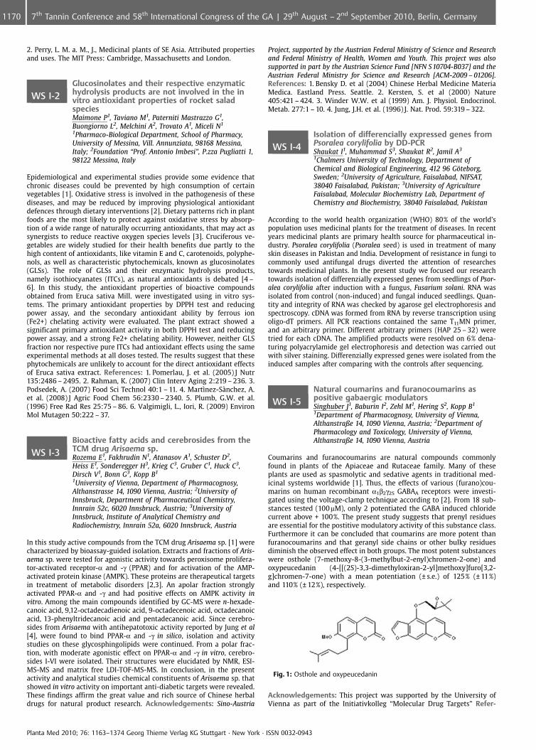
Coumarins and furanocoumarins are natural compounds commonlyfound in plants of the Apiaceae and Rutaceae family. Many of theseplants are used as spasmolytic and sedative agents in traditional med-icinal systems worldwide [1]. Thus, the effects of various (furano)cou-marins on human recombinant a1b2g2S GABAA receptors were investi-gated using the voltage-clamp technique according to [2]. From 18 sub-stances tested (100mM), only 2 potentiated the GABA induced chloridecurrent above + 100%. The present study suggests that prenyl residuesare essential for the postitive modulatory activity of this substance class.Furthermore it can be concluded that coumarins are more potent thanfuranocoumarins and that geranyl side chains or other bulky residuesdiminish the observed effect in both groups. The most potent substanceswere osthole (7-methoxy-8-(3-methylbut-2-enyl)chromen-2-one) andoxypeucedanin (4-[[(2S)-3,3-dimethyloxiran-2-yl]methoxy]furo[3,2-g]chromen-7-one) with a mean potentiation (€ s.e.) of 125% (€ 11%)and 110% (€ 12%), respectively.
Fig. 1: Osthole and oxypeucedanin
Acknowledgements: This project was supported by the University ofVienna as part of the Initiativkolleg “Molecular Drug Targets” Refer-
Planta Med 2010; 76: 1163–1374 Georg Thieme Verlag KG Stuttgart · New York · ISSN 0032-0943
1170 7th Tannin Conference and 58th International Congress of the GA | 29th August – 2nd September 2010, Berlin, Germany
ences: 1. Murray et al. (1982). The natural coumarins: occurrence,chemistry and biochemistry. Wiley. London. 2. Baburin I. et al. (2006).Pflugers Arch. 453: 117 – 23.
WS I-6Anti-inflammatory potency of the traditionallyused antimalarial plant Fagraea fragransJonville M1, Baghdikian B2, Ollivier E2, Angenot L1,Fr�d�rich M1, Legault J3
1Universit� de Li�ge, Laboratoire de Pharmacognosie,Pharmacy, Avenue de l’H�pital, 1 (B36), 4000 Li�ge, Belgium;2Universit� de la M�diterran�e, Laboratoire dePharmacognosie, Pharmacy, Boulevard Jean Moulin, 27,13005 Marseille, France; 3Universit� du Qu�bec Chicoutimi, Laboratoire LASEVE, Sciences fondamentales,Boulevard de l’Universit�, 555, G7H2B1 Chicoutimi, Canada
Fagraea fragrans Roxb. (Gentianaceae) was selected following an ethno-pharmacological survey in Cambodia to treat fever or malaria. Littlework has been done so far on this plant; only the presence of secoiri-doids (gentiopicroside [1], [2], fagraldehyde, sweroside and swertiamar-in [3]) has been described. Methanol and dichloromethane extracts havebeen prepared from bark and leaves to assess in vitro anti-plasmodialassays. The bioguided chromatographic fractionation against Plasmo-dium falciparum led to a spread of activity in different fractions. Multipleminor compounds acting in synergy could explain the activity providedin the crude CH2Cl2 extracts of the bark and the leaves. On the otherhand, fever symptom is not only displayed during malaria disease butalso in various illnesses including inflammatory reaction. Thus, the dif-ferent fractions of F. fragrans have been tested on inhibition of NitricOxide (NO) overproduction on LPS-stimulated murine macrophages. Thebest anti-inflammatory potency (10 mg/ml = 80% inhibition) was pro-vided by a fraction coming from the leaf apolar extract. Further investi-gations will be carried out in our laboratory to identify the active com-pounds inhibiting NO. Acknowledgements: the Belgian National Fundfor Scientific Research (FNRS) (grant N� 3452005) and the Samuel deChamplain convention between France and Quebec (n� 62 103) aregreatly acknowledged. References: 1. Wan, A.S.C., Chow, Y.L. (1964)J.Pharma.Pharmacol. 16: 484 – 486. 2. Natarajan, P.N. et al. (1974) PlantaMed. 25: 258 – 260. 3. Jonville, M.C. et al. (2008) J. Nat. Prod. 71: 2038 –2040.
WS II: Workshops for Young Researchers
Lead finding from Nature – Pitfalls and challenges of classical,computational and hyphenated approaches.Chairs: J. M. Rollinger, A. R. Bilia, J-L. Wolfender
WS II ILImpulse Lecture: The potential of naturalproducts in drug discovery – What is thechallenge in academia?Gertsch JUniversity of Bern, Institute of Biochemistry and MolecularMedicine, 3012 Bern, Switzerland
For more than a century bioactive natural products have served as mo-lecular tools for biochemists and as inspiration for pharmacologists andmedicinal chemists. Many prescribed drugs have directly or indirectlybeen discovered in natural product research1. However, with the rise ofhigh-throughput screening technologies and the demand for huge syn-thetic compound libraries, as well as the more recent indroduction ofbiologics (e. g. therapeutic monoclonal antibodies), the interest in natur-al product research has declined in industry2. At the same time, thenumber of new chemical entities has declined too. While there are stillsome potentially interesting bioactive plant and animal natural productsthe chemical diversity of microorganisms (both terrestrial and marine)remains largely unknown. In the last decade, new academic initiativeshave been realized to study natural products as potential lead structuresor e. g. also to validate traditional herbal medicines3. Thus, academia isincreasingly taking over natural product research. Academic researchwith natural products has led to thousands of scientific papers, describ-ing actual or potential therapeutic uses. Unfortunately, only very fewhave triggered the development of innovative biochemical tool com-pounds or new drugs. The reasons for this are manyfold. In order to besuccessful, natural product research in academia should take advantageof the new developments in industry (high-content screening, promis-ing targets, chemoinformatics, molecular library design, etc.) and com-
bine the strength of “academic freedom” with the scrutinity of industrialselection. References: 1. D.J. Newman & G.M. Cragg, Natural products assources of new drugs over the last 25 years. J. Nat. Prod,. 2007, 70: 461 –77. 2. J.V. Li & JW Vederas, Drug discovery and natural products: end ofan era or an endless frontier? Science, 2009, 325: 161 – 5. 3. J. Gertsch.How scientific is the science in ethnopharmacology? Historical perspec-tives and epistemological problems. J. Ethnopharmacol. 2009, 122:177 –83.
WS II-1Bioactivity-guided isolation of potentialanti-inflammatory constituents from BetonicaofficinalisPicker P1, Mihaly-Bison J2, Vogl S1, Zehl M1, Urban E3,Reznicek G1, Saukel J1, Wawrosch C1, Binder B2, Kopp B1
1Department of Pharmacognosy, University of Vienna,Althanstrasse 14, 1090 Vienna, Austria; 2Department ofVascular Biology and Thrombosis Research, MedicalUniversity of Vienna, Schwarzspanierstrasse 17, 1090Vienna, Austria; 3Department of Medicinal Chemistry,University of Vienna, Althanstrasse 14, 1090 Vienna, Austria
Betonica officinalis (Lamiaceae) has been used in Austrian traditionalmedicine since ancient times against inflammatory disorders. The aimof this study was to investigate the anti-inflammatory properties ofextracts, derived fractions, and isolated pure compounds of this plantby assessment of their effect on genes (E-selectin, IL-8) that are inducedby inflammatory stimuli (TNF-a or LPS) in endothelial cells [1,2]. Theplant material (herb) was extracted with dichloromethane (DCM) usingan accelerated solvent extractor. Chlorophyll was separated by liquid-liquid-partition between DCM and a mixture of MeOH-H2O 1:1, in orderto increase the concentration of the active compounds. Since the pur-ified DCM extract showed strong activity in the mentioned assay, abioactivity-guided fractionation was carried out. Subfractions were ob-tained by solid-phase extraction using C 18 cartridges eluted with 30%,70%, and 100% MeOH. The 30% and the 70% subfractions, which showedhighest activity, were further fractionated by HPLC in order to identifyand investigate their active constituents, whose structures were eluci-dated by HPLC-MS, 1D, and 2D NMR spectroscopy. Besides of someknown polymethylated flavonoids (e. g. salvigenin), particularly the iri-doid 8-O-acetylharpagide and two new diterpenoids were found to in-hibit between 46% and 99% the LPS-stimulated induction of E-selectinat the concentration of 10 mg/ml, evidencing a considerable potential asnew anti-inflammatory agents. Acknowledgements: This work isfunded by the Austrian Science Fund, NFN: S10704-B037. References:1. Chang et al. (2005) Exp Cell Res. 309(1):121 – 36. 2. Kadl et al. (2002)Vascul Pharmacol. 38(4):219 – 27.
WS II-2Characterization of anti-inflammatory triterpeneacids from rose hip powder (Rosa canina L.)Saaby L, J�ger A, Moesby L, Hansen E, Christensen SFaculty of Pharmaceutical Sciences, University ofCopenhagen, Department of Medicinal Chemistry,Universitetsparken 2, 2100 Copenhagen O, Denmark
The standardized rose hip powder LitoMove� (Rosa canina L.) is a widelyused herbal remedy. Clinical trials have revealed that consumption ofrose hip powder can reduce pain in patients suffering from osteoarthritis[1,2]. Synovial inflammation mainly mediated by macrophages has beenreported to be involved in the pathology of osteoarthritis [3,4]. There-fore, the anti-inflammatory activity of crude extracts of standardizedrose hip powder (LitoMove) was investigated and active principles iso-lated using the human monocytic cell line Mono Mac 6 as a model forinflammation. Incubation of Mono Mac 6 cells with a crude dichloro-methane extract of rose hip powder significantly inhibited the lipopoly-saccharide (LPS) induced interleukin-6 (IL-6) release in a concentrationdependent manner. Through bioassay-guided fractionation this anti-in-flammatory effect was correlated to a mixture of three triterpene acids;oleanolic, betulinic and ursolic acid (IC50 21 € 6mM). Investigation of theanti-inflammatory activity of each of the three triterpene acids revealedthat oleanolic and ursolic acid was able to inhibit the LPS induced re-lease of IL-6, in contrast to betulinic acid. Interestingly, combination ofeither oleanolic or ursolic acid with betulinic acid enhanced the anti-inflammatory effect of both oleanolic and ursolic acid. Acknowledge-ments: Hyben Vital International ApS is thanked for financial support.References: 1. Chrubasik, C. et al. (2006) Phytother. Res. 20:1.3. 2. Chris-tensen, R. et al. (2008) Osteoarhritis Cartilage 16:965 – 972. 3. Farahat,
Planta Med 2010; 76: 1163–1374 Georg Thieme Verlag KG Stuttgart · New York · ISSN 0032-0943
11717th Tannin Conference and 58th International Congress of the GA | 29th August – 2nd September 2010, Berlin, Germany
M. et al (1993) Ann. Rheum. Dis. 52:870 – 875. 4. Bondeson, J. et al.(2006) Arthritis Res. Ther. 8:R187.
WS II-3Research of antifungal compounds from theAmazonian biomass by a bio-inspired approachBasset C1, Stien D2, Salmen Espindola L3
1UMR ECOFOG – UAG, L3MA, BP 792, 97337 Cayenne,French Guiana; 2UMR ECOFOG – CNRS, L3MA, BP 792, 97337Cayenne, French Guiana; 3UnB/Laboratorio defamacognosia, Faculdade de CiÞncias da Safflde,Universidade de Brasilia, DF 70910 – 900 Brasilia, Brazil
Our research aims at understanding the chemical resistance mechan-isms of durable woods against fungi. Our ultimate goal is to isolate andidentify antifungal compounds from these woods that could be used forthe treatment of human fungal diseases. We therefore screened highlydurable Amazonian wood selected from technical databases [1] anddemonstrated that bioactive secondary metabolites responsible of thenatural durability of the woods [2] can also be used to treat mycoses.This screening has given a very high percentage of positive hits (30%) for70 extracts tested, therefore validating the bio-inspiration hypothesis.
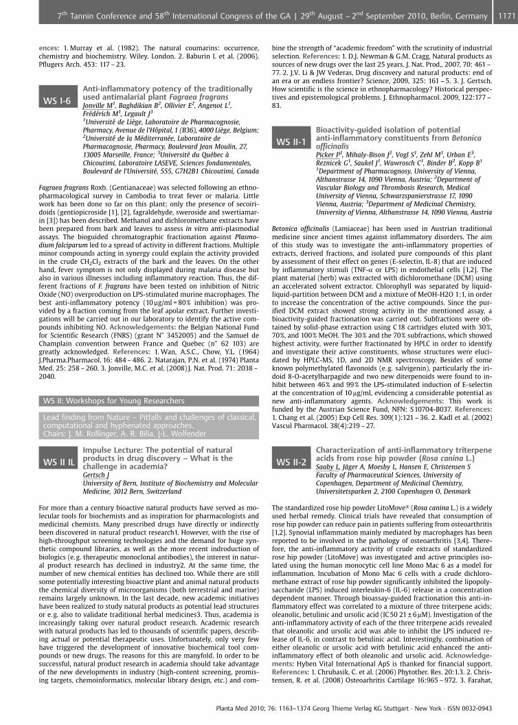
Fig. 1: Structure of piceatanol (1), isolated from S. longifolia
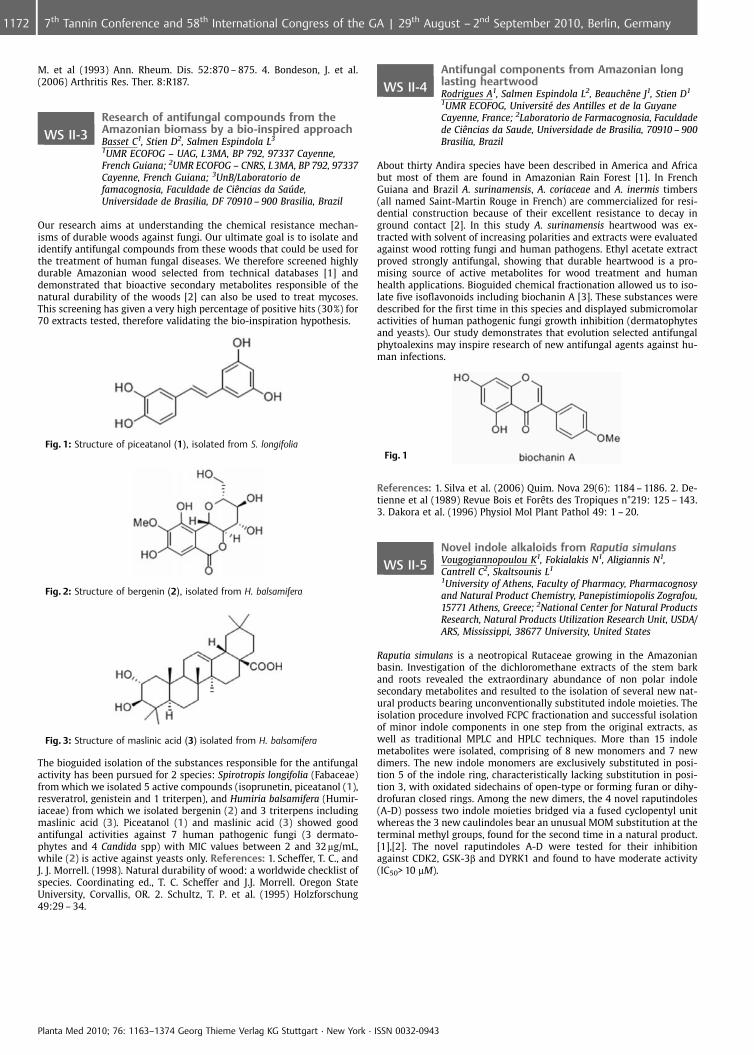
Fig. 2: Structure of bergenin (2), isolated from H. balsamifera
Fig. 3: Structure of maslinic acid (3) isolated from H. balsamifera
The bioguided isolation of the substances responsible for the antifungalactivity has been pursued for 2 species: Spirotropis longifolia (Fabaceae)from which we isolated 5 active compounds (isoprunetin, piceatanol (1),resveratrol, genistein and 1 triterpen), and Humiria balsamifera (Humir-iaceae) from which we isolated bergenin (2) and 3 triterpens includingmaslinic acid (3). Piceatanol (1) and maslinic acid (3) showed goodantifungal activities against 7 human pathogenic fungi (3 dermato-phytes and 4 Candida spp) with MIC values between 2 and 32 mg/mL,while (2) is active against yeasts only. References: 1. Scheffer, T. C., andJ. J. Morrell. (1998). Natural durability of wood: a worldwide checklist ofspecies. Coordinating ed., T. C. Scheffer and J.J. Morrell. Oregon StateUniversity, Corvallis, OR. 2. Schultz, T. P. et al. (1995) Holzforschung49:29 – 34.
WS II-4Antifungal components from Amazonian longlasting heartwoodRodrigues A1, Salmen Espindola L2, BeauchÞne J1, Stien D1
1UMR ECOFOG, Universit� des Antilles et de la GuyaneCayenne, France; 2Laboratorio de Farmacognosia, Faculdadede CiÞncias da Saude, Universidade de Brasilia, 70910 – 900Brasilia, Brazil
About thirty Andira species have been described in America and Africabut most of them are found in Amazonian Rain Forest [1]. In FrenchGuiana and Brazil A. surinamensis, A. coriaceae and A. inermis timbers(all named Saint-Martin Rouge in French) are commercialized for resi-dential construction because of their excellent resistance to decay inground contact [2]. In this study A. surinamensis heartwood was ex-tracted with solvent of increasing polarities and extracts were evaluatedagainst wood rotting fungi and human pathogens. Ethyl acetate extractproved strongly antifungal, showing that durable heartwood is a pro-mising source of active metabolites for wood treatment and humanhealth applications. Bioguided chemical fractionation allowed us to iso-late five isoflavonoids including biochanin A [3]. These substances weredescribed for the first time in this species and displayed submicromolaractivities of human pathogenic fungi growth inhibition (dermatophytesand yeasts). Our study demonstrates that evolution selected antifungalphytoalexins may inspire research of new antifungal agents against hu-man infections.
Fig. 1
References: 1. Silva et al. (2006) Quim. Nova 29(6): 1184 – 1186. 2. De-tienne et al (1989) Revue Bois et ForÞts des Tropiques n�219: 125 – 143.3. Dakora et al. (1996) Physiol Mol Plant Pathol 49: 1 – 20.
WS II-5Novel indole alkaloids from Raputia simulansVougogiannopoulou K1, Fokialakis N1, Aligiannis N1,Cantrell C2, Skaltsounis L1
1University of Athens, Faculty of Pharmacy, Pharmacognosyand Natural Product Chemistry, Panepistimiopolis Zografou,15771 Athens, Greece; 2National Center for Natural ProductsResearch, Natural Products Utilization Research Unit, USDA/ARS, Mississippi, 38677 University, United States
Raputia simulans is a neotropical Rutaceae growing in the Amazonianbasin. Investigation of the dichloromethane extracts of the stem barkand roots revealed the extraordinary abundance of non polar indolesecondary metabolites and resulted to the isolation of several new nat-ural products bearing unconventionally substituted indole moieties. Theisolation procedure involved FCPC fractionation and successful isolationof minor indole components in one step from the original extracts, aswell as traditional MPLC and HPLC techniques. More than 15 indolemetabolites were isolated, comprising of 8 new monomers and 7 newdimers. The new indole monomers are exclusively substituted in posi-tion 5 of the indole ring, characteristically lacking substitution in posi-tion 3, with oxidated sidechains of open-type or forming furan or dihy-drofuran closed rings. Among the new dimers, the 4 novel raputindoles(A-D) possess two indole moieties bridged via a fused cyclopentyl unitwhereas the 3 new caulindoles bear an unusual MOM substitution at theterminal methyl groups, found for the second time in a natural product.[1],[2]. The novel raputindoles A-D were tested for their inhibitionagainst CDK2, GSK-3b and DYRK1 and found to have moderate activity(IC50> 10 mM).
Planta Med 2010; 76: 1163–1374 Georg Thieme Verlag KG Stuttgart · New York · ISSN 0032-0943
1172 7th Tannin Conference and 58th International Congress of the GA | 29th August – 2nd September 2010, Berlin, Germany
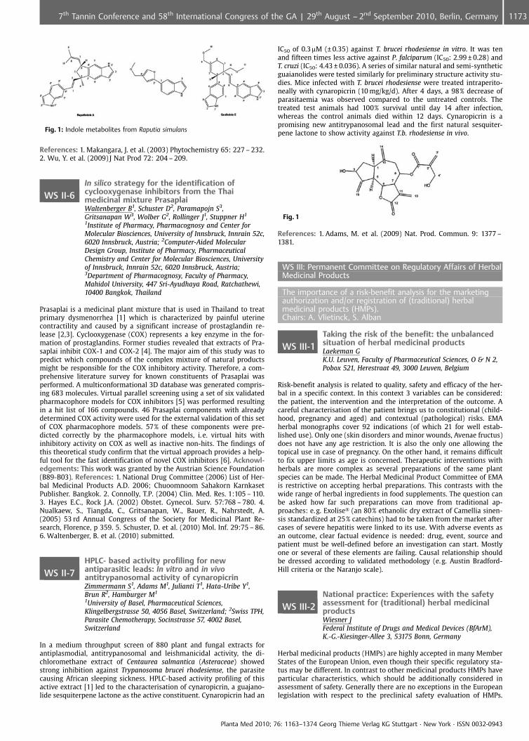
Fig. 1: Indole metabolites from Raputia simulans
References: 1. Makangara, J. et al. (2003) Phytochemistry 65: 227 – 232.2. Wu, Y. et al. (2009) J Nat Prod 72: 204 – 209.
WS II-6In silico strategy for the identification ofcyclooxygenase inhibitors from the Thaimedicinal mixture PrasaplaiWaltenberger B1, Schuster D2, Paramapojn S3,Gritsanapan W3, Wolber G2, Rollinger J1, Stuppner H1
1Institute of Pharmacy, Pharmacognosy and Center forMolecular Biosciences, University of Innsbruck, Innrain 52c,6020 Innsbruck, Austria; 2Computer-Aided MolecularDesign Group, Institute of Pharmacy, PharmaceuticalChemistry and Center for Molecular Biosciences, Universityof Innsbruck, Innrain 52c, 6020 Innsbruck, Austria;3Department of Pharmacognosy, Faculty of Pharmacy,Mahidol University, 447 Sri-Ayudhaya Road, Ratchathewi,10400 Bangkok, Thailand
Prasaplai is a medicinal plant mixture that is used in Thailand to treatprimary dysmenorrhea [1] which is characterized by painful uterinecontractility and caused by a significant increase of prostaglandin re-lease [2,3]. Cyclooxygenase (COX) represents a key enzyme in the for-mation of prostaglandins. Former studies revealed that extracts of Pra-saplai inhibit COX-1 and COX-2 [4]. The major aim of this study was topredict which compounds of the complex mixture of natural productsmight be responsible for the COX inhibitory activity. Therefore, a com-prehensive literature survey for known constituents of Prasaplai wasperformed. A multiconformational 3D database was generated compris-ing 683 molecules. Virtual parallel screening using a set of six validatedpharmacophore models for COX inhibitors [5] was performed resultingin a hit list of 166 compounds. 46 Prasaplai components with alreadydetermined COX activity were used for the external validation of this setof COX pharmacophore models. 57% of these components were pre-dicted correctly by the pharmacophore models, i. e. virtual hits withinhibitory activity on COX as well as inactive non-hits. The findings ofthis theoretical study confirm that the virtual approach provides a help-ful tool for the fast identification of novel COX inhibitors [6]. Acknowl-edgements: This work was granted by the Austrian Science Foundation(B89-B03). References: 1. National Drug Committee (2006) List of Her-bal Medicinal Products A.D. 2006; Chuoomnoom Sahakorn KarnkasetPublisher. Bangkok. 2. Connolly, T.P. (2004) Clin. Med. Res. 1:105 – 110.3. Hayes E.C., Rock J.A. (2002) Obstet. Gynecol. Surv. 57:768 – 780. 4.Nualkaew, S., Tiangda, C., Gritsanapan, W., Bauer, R., Nahrstedt, A.(2005) 53 rd Annual Congress of the Society for Medicinal Plant Re-search, Florence, p 359. 5. Schuster, D. et al. (2010) Mol. Inf. 29:75 – 86.6. Waltenberger, B. et al. (2010) submitted.
WS II-7HPLC- based activity profiling for newantiparasitic leads: In vitro and in vivoantitrypanosomal activity of cynaropicrinZimmermann S1, Adams M1, Julianti T1, Hata-Uribe Y1,Brun R2, Hamburger M1
1University of Basel, Pharmaceutical Sciences,Klingelbergstrasse 50, 4056 Basel, Switzerland; 2Swiss TPH,Parasite Chemotherapy, Socinstrasse 57, 4002 Basel,Switzerland
In a medium throughput screen of 880 plant and fungal extracts forantiplasmodial, antitrypanosomal and leishmanicidal activity, the di-chloromethane extract of Centaurea salmantica (Asteraceae) showedstrong inhibition against Trypanosoma brucei rhodesiense, the parasitecausing African sleeping sickness. HPLC-based activity profiling of thisactive extract [1] led to the characterisation of cynaropicrin, a guajano-lide sesquiterpene lactone as the active constituent. Cynaropicrin had an
IC50 of 0.3mM (€ 0.35) against T. brucei rhodesiense in vitro. It was tenand fifteen times less active against P. falciparum (IC50: 2.99 € 0.28) andT. cruzi (IC50: 4.43 € 0.036). A series of similar natural and semi-syntheticguaianolides were tested similarly for preliminary structure activity stu-dies. Mice infected with T. brucei rhodesiense were treated intraperito-neally with cynaropicrin (10 mg/kg/d). After 4 days, a 98% decrease ofparasitaemia was observed compared to the untreated controls. Thetreated test animals had 100% survival until day 14 after infection,whereas the control animals died within 12 days. Cynaropicrin is apromising new antitrypanosomal lead and the first natural sesquiter-pene lactone to show activity against T.b. rhodesiense in vivo.
Fig. 1
References: 1. Adams, M. et al. (2009) Nat. Prod. Commun. 9: 1377 –1381.
WS III: Permanent Committee on Regulatory Affairs of HerbalMedicinal Products
The importance of a risk-benefit analysis for the marketingauthorization and/or registration of (traditional) herbalmedicinal products (HMPs).Chairs: A. Vlietinck, S. Alban
WS III-1Taking the risk of the benefit: the unbalancedsituation of herbal medicinal productsLaekeman GK.U. Leuven, Faculty of Pharmaceutical Sciences, O & N 2,Pobox 521, Herestraat 49, 3000 Leuven, Belgium
Risk-benefit analysis is related to quality, safety and efficacy of the her-bal in a specific context. In this context 3 variables can be considered:the patient, the intervention and the interpretation of the outcome. Acareful characterisation of the patient brings us to constitutional (child-hood, pregnancy and aged) and contextual (pathological) risks. EMAherbal monographs cover 92 indications (of which 21 for well estab-lished use). Only one (skin disorders and minor wounds, Avenae fructus)does not have any age restriction. It is also the only one allowing thetopical use in case of pregnancy. On the other hand, it remains difficultto fix upper limits as age is concerned. Therapeutic interventions withherbals are more complex as several preparations of the same plantspecies can be made. The Herbal Medicinal Product Committee of EMAis restrictive on accepting herbal preparations. This contrasts with thewide range of herbal ingredients in food supplements. The question canbe asked how far such preparations can move from traditional ap-proaches: e. g. Exolise� (an 80% ethanolic dry extract of Camellia sinen-sis standardized at 25% catechins) had to be taken from the market aftercases of severe hepatitis were linked to its use. With adverse events asan outcome, clear factual evidence is needed: drug, event, source andpatient must be well-defined before an investigation can start. Mostlyone or several of these elements are failing. Causal relationship shouldbe dressed according to validated methodology (e. g. Austin Bradford-Hill criteria or the Naranjo scale).
WS III-2National practice: Experiences with the safetyassessment for (traditional) herbal medicinalproductsWiesner JFederal Institute of Drugs and Medical Devices (BfArM),K.-G.-Kiesinger-Allee 3, 53175 Bonn, Germany
Herbal medicinal products (HMPs) are highly accepted in many MemberStates of the European Union, even though their specific regulatory sta-tus may be different. In contrast to other medicinal products HMPs haveparticular characteristics, which should be additionally considered inassessment of safety. Generally there are no exceptions in the Europeanlegislation with respect to the preclinical safety evaluation of HMPs.
Planta Med 2010; 76: 1163–1374 Georg Thieme Verlag KG Stuttgart · New York · ISSN 0032-0943
11737th Tannin Conference and 58th International Congress of the GA | 29th August – 2nd September 2010, Berlin, Germany
Nevertheless, when applying the legislation a reasonable adaptation tocover the particulars of HMPs (e.g. testing strategies) is inevitable. TheCommittee on Herbal Medicinal Products (HMPC) was established at theEuropean Medicines Agency (EMA) to generate a harmonised view an allissues concerning HMPs. Within this work the HMPC adopted a series ofguidance documents referring to the safety of HMPs (e. g. general gui-dance on safety assessment, papers on specific issues like toxicity ofdefined natural products etc.). With respect to the traditional use ofherbal medicinal products there is an option to reduce the set of teststo be performed in the context of preclinical testing. Nevertheless, ex-periences of the German national competent authority considering morethan 300 applications in the field of traditional herbal medicinal pro-ducts showed that consideration of the particulars of herbal medicinalproducts does not only open the chance to reduce the preclinical testingadequately but also provokes some additional questions which shouldbe addressed case by case.
WS III-3European practice: experiences with arisk-benefit analysis of HMPs for the elaborationof ESCOP monographsSteinhoff BCo-Chairperson of the ESCOP Scientific Committee, Auf demForst 53 Bonn, Germany
ESCOP, the European Scientific Cooperative on Phytotherapy, wasfounded in 1989 as a European umbrella organisation of national asso-ciations of phytotherapy. During the past 20 years, ESCOP has beenworking on more than 100 monographs on efficacy and safety in orderto contribute to the scientific harmonisation process. A monograph com-prises therapeutic indications, dosage recommendations, information onpotential risks and pharmacological properties. During the preparationof monographs, benefit and risk of preparations of the respective herbaldrug and its preparations are evaluated on the basis of pharmacologicaland clinical data as well as of toxicological data and clinical safety. Theassessment results in a decision about which therapeutic indicationsand dosage recommendations are justified and which information onpotential risks is required. In case the risks outweigh the benefit, or incase a therapeutic use cannot be proven, a monograph will not be pre-pared. Thus the issue of a benefit-risk analysis has to be addressedduring the elaboration of each monograph. After publication of the2nd Edition with 80 monographs in 2003, a Supplement containing35 revised and new monographs was published in 2009. It can be or-dered from the book trade or from the ESCOP Secretariat (www.escop.-com).
WS III-4European practice: Well-established use ofherbal medicinal products and clinical researchMeng GDr. Wilmar Schwabe GmbH & Co KG, Willmar-Schwabe-Str.4, 76227 Karlsruhe, Germany
When a clinical trial is about specific efficacy of an active substance therandomised placebo-controlled trial (RCT) is the gold-standard. This isthe background of the well-known hierarchy of evidence of EvidenceBased Medicine (EBM) also. Well-established use (WEU) of a drug asdefined by European law requires proven efficacy by definition. But,when the modalities of the WEU of a specific product are assessed at acertain point during the product’s lifetime many other questions con-tribute to the total body of evidence relevant for this assessment. Thetalk outlines the background of this situation and shows in which dif-ferent ways clinical research may contribute. In this broader view theRCT design is only one option among others depending on the researchquestion. The assessment of WEU needs a broader perspective, beyondof what an RCT can deliver.
WS IV: Permanent Committee on Biological andPharmacological Activities of Natural Products
Use of polyphenols against cardiovascular diseases.Chair: V. Butterweck
WS IV-1Challenges for research on dietary polyphenols inthe prevention of cardiovascular diseases – Newpropositions for a structure-function analysisScalbert A, Manach C, Rothwell JINRA, Centre de Recherche de Clermont-Ferrand/Theix,63122 Saint-Genes-Champanelle, France
Much evidence supports the role of dietary polyphenols in the preven-tion of cardiovascular diseases. However polyphenols are not all equal.Their bioavailability and biological properties differ widely according totheir fine chemical structures, and the exact nature of the most protec-tive compounds is still largely unknown. New tools are clearly needed toexplore the links between intakes of individual polyphenols and the riskof cardiovascular disease. We have built a new comprehensive databaseon polyphenol contents in foods called Phenol-Explorer (http://www.phenol-explorer.eu/) that includes more than 36,000 compositiondata for 502 polyphenols in 452 foods. This database is being furtherdeveloped to include data on polyphenol metabolites and their concen-trations in plasma and urine. The data is being used in cohort studies toestimate intake of the 502 polyphenols and the resulting concentrationsof polyphenol metabolites. This will allow exploration of the links be-tween polyphenols and disease risk with an unparalleled precision.
WS IV-2Flavonoids and cardiovascular disease riskfactors – a meta-analysis and recent findingsfrom prospective cohort studiesCassidy AUniversity of East Anglia, School of Medicine, Norwich,United Kingdom
Our recent meta-analysis of randomized clinical trials provides a snap-shot of the current state-of-the-art in relation to the relative effective-ness of the different flavonoid subclasses in modifying biomarkers ofcardiovascular (CVD) risk and highlights areas in which limited dataexists. The available data suggests there may be clinically relevant ef-fects of some flavonoid subclasses on cardiovascular risk factors, and todate the effects of flavonoids from soy and cocoa have been the mainfocus of attention. Chocolate/cocoa increased FMD acutely (3.99%; 95%CI: 2.86, 5.12; six studies) and chronically (1.45%; 0.62, 2.28; two stu-dies) and reduced both systolic blood pressure (�5.88 mm Hg; �9.55,�2,21; five studies) and diastolic blood pressure (�3.30 mm Hg; �5.77,�0.83; four studies) following chronic intake. Only soy protein isolate,but not whole soy or soy extracts, reduced diastolic blood pressure(�1.99 mm Hg; �2.86, �1.12; nine studies) and LDL cholesterol (�0.19mmol/L; �0.24, �0.14; thirty-nine studies). For many subclasses (antho-cyanins and flavanones) there was insufficient evidence to draw conclu-sions about efficacy. We are currently updating the review, including allstudies conducted through until Jan 2010 with a view to also examinepotential structure-activity relationships. Recent data from ongoing col-laborations with the Harvard cohorts (Nurses Health Study and HelathProfessionals Follow-up Study) on flavonoid sub-class intake and cardi-ovascular health endpoints will also be discussed.
WS IV-3Vascular protection of tea and grape-derivedpolyphenols: in vitro and in vivo evidenceSchini-Kerth V, Auger C, Etienne-Selloum N, Chataigneau TFaculty of Pharmacy, CNRS UMR 7213, 74, route du rhin,67401 Illkirch, France
Epidemiological studies have indicated that regular intake of polyphenolrich sources such as vegetables, fruit, red wine and tea is associated witha decreased risk of coronary diseases. The protective effect of polyphe-nols on the vascular system has been attributable in part to their directaction on endothelial cells resulting in an enhanced formation of nitricoxide (NO) and endothelium-derived hyperpolarizing factor (EDHF), twofactors playing a major role in the control of vascular homeostasis. In-deed, tea and grape-derived polyphenols cause pronounced endothe-lium-dependent relaxations of isolated arteries, which are mediated byNO and, also in some arteries, by EDHF. The increased formation of NO isinitiated by a pro-oxidant response in endothelial cells, which triggers
Planta Med 2010; 76: 1163–1374 Georg Thieme Verlag KG Stuttgart · New York · ISSN 0032-0943
1174 7th Tannin Conference and 58th International Congress of the GA | 29th August – 2nd September 2010, Berlin, Germany
activation of Src kinase with the subsequent activation of the PI3-kinase/Akt pathway resulting in the activation of endothelial NO synthase fol-lowing its phosphorylation on Ser1177. Intake of red wine polyphenolsin the drinking water is also able to prevent the development of anendothelial dysfunction and hypertension induced by infusion of angio-tensin II to rats, and also of an endothelial dysfunction in ageing. Theprotective effect of polyphenols involves their ability to prevent theexcessive oxidative stress, the arachidonic acid-derived formation ofendothelium-dependent contracting factors, and to regulate the localangiotensin II system in the pathologic artery. Thus, polyphenols inducea great variety of effects on both endothelial cells and vascular smoothmuscle cells to protect blood vessels.
WS IV-4Crataegus extract WS�1442 in patients withcongestive heart failure class NYHA II-III: TheSPICE trialHolubarsch CMedian Clinics Bad Krozingen, Herbert-Hellmann-Allee 38,79189 Bad Krozingen, Germany
WS�1442 is registered for treatment of early stages of congestive heartfailure (CHF) in several countries, e.g. Germany. As there was no studyavailable regarding efficacy and safety of WS�1442 in more severely im-paired patients already treated with optimal therapy, SPICE (Survival andPrognosis: Investigation on Crataegus Extract WS�1442) was performed.SPICE is a double-blind mortality trial conducted in 156 centres in 13 Eur-opean countries. Patients were eligible if their left ventricular functionwas markedly impaired (WMI£1.2). These patients already receiving op-timal pharmacological therapy (83% ACE-inhibitors, 64% b-blockers, 56%glycosides, 39% spironolactone, 85% diuretics) were randomised toWS�1442 or placebo. Treatment duration was 2 years; primary endpointwas the time until first cardiac event (sudden cardiac death (SCD), deathdue to progressive heart failure, myocardial infarction, hospitalization;FCE). 2681 patients (mean age 60 yrs, 84% males, 44% NYHAIII, meanLVEF 24%) were randomised and evaluated. At all times, WS�1442 wassuperior to placebo regarding FCE, but without reaching statistical sig-nificance. For the first 18 months of WS�1442 treatment, deaths due tocardiac cause were reduced significantly (�20%; p = 0.046) and WS�1442postponed cardiac deaths by four months. This effect was even morepronounced in patients with LVEF‡25% (�33% reduction; p = 0.044) andalso holds true for total mortality and SCD up to 24 months. In conclu-sion, the study confirms the safety of WS�1442 in CHF patients with amean LVEF of 24% and being treated with optimal pharmacological ther-apy. More important, WS�1442 may postpone deaths due to cardiaccause in this patient population.
WS V: Permanent Committee on Manufacturing and QualityControl of Herbal Remedies
The use of hyphenated techniques in the quality control ofherbal medicinal products.Chair: C. Erdelmeier
WS V-1LC-NMR/MS and LC-SPE-NMR/MS: technicalrealization, possibilities and limitationsGodejohann MBruker BioSpin GmbH, Silberstreifen 4, 78287 Rheinstetten,Germany
The hyphenation between chromatographic and spectroscopic techni-ques is well established and approved in various fields, e. g. pharmaceu-tical or natural products analysis. It combines commercially availableanalytical HPLC equipment with an NMR spectrometer and a mass spec-trometer using specially developed interfaces according to the type ofapplication. LC-NMR/MS directly transfers the separated peak into anNMR flow probe and via split into the ion source of a mass spectrometer.Chromatographic separation is done using mixtures of protonated organ-ic and deuterated aqueous mobile phase, resulting in strong backgroundresonances from the organic solvent which needs to be suppressed inorder to detect small signals originating from the analytes. Chromato-graphic peaks can be send either directly to the NMR or stored in loopsfor subsequent transfer into the NMR probe. This direct coupling is espe-cially used for polar and/or labile compounds present in high to moder-ate concentrations. Post column solid phase extraction was first intro-duced 1998 by Griffiths and Horton: peaks eluting from a reversed phasecolumn are diluted by aqueous phase and trapped on small SPE car-
tridges packed with reversed phase material. Elution into the NMR flowprobe or tube can be done with deuterated organic solvent preferablyafter drying of the cartridge with nitrogen gas in order to remove residualprotonated solvent. This indirect coupling can be used for medium polarto unpolar compounds down to very low concentrations due to the pos-sibility of multiple peak trapping. In combination with cryogenicallycooled NMR probes highest NMR sensitivity can be achieved.
WS V-2HPLC-SPE-NMR in studies of medicinal plants andherbal productsJaroszewski JUniversity of Copenhagen, Faculty of PharmaceuticalSciences, Universitetsparken 2, 2100 Copenhagen, Denmark
Hyphenated NMR spectroscopy is a state-of-the-art method for rapid andrigorous determination of natural product structures in plant extractswithout isolation of the individual components in the classical sense.Among hyphenated NMR techniques, HPLC-SPE-NMR is the most conve-nient and superior in terms of quality of data, the most serious limitationbeing problems with solid-phase extraction of highly polar but nonionicspecies. The technique is particularly important in projects aimed at dis-covery of new chemical entities in medicinal plants, e. g., in connectionwith natural product discovery of novel pharmacologically active com-pounds. Moreover, studies related to standardization and uniformity ofherbal products, performed either using metabolomic techniques or tra-ditional methods, can benefit from support by hyphenated NMR experi-ments. HPLC-SPE-NMR work can be performed either using a flow NMRprobe or a microtubes, and will benefit from increased sensitivity ofcryogenically cooled probes. Examples of use of HPLC-SPE-NMR in ana-lysis of medicinal plants and herbal products will be shown.
WS V-3Hyphenated analytical techniques andphytoindustrial reality: do we need Ferraris topull trailers?Lang FDr. Willmar Schwabe Arzneimittel, Analytische Entwicklung,Dr.-Willmar-Schwabe-Str. 4, 76227 Karlsruhe, Germany
In recent years multi-hyphenated methods coupling NMR spectroscopy(as the most powerful method for structure elucidation of natural com-pounds) with HPLC or SPE (solid phase extraction) have been describedand used by several high performance working groups. In principalthose methods allow a rapid identification of a multitude of plant con-stituents avoiding the classical and complicated isolation and character-isation procedure. Within the presentation it is discussed if multihyphe-nated analytical techniques might be used during analytical develop-ment and routine analysis of herbal preparations and herbal medicinalproducts. In most cases the methods seem to be oversized for the ana-lysis of traditional or well established HMPs, extracts and plants. How-ever multihyphenated techniques may play a role during the basic re-search on medicinal plants, the development of new plant extracts, thescreening on active or analytical markers, and for a holistic characterisa-tion of plant extracts and bioanalytical profiling. As the techniques arebased on special equipment and dedicated personal a broad industrialuse seems unlikely. Corresponding problems should be solved withincooperations of the industry and the specialised labs.
WS V-4Choice of analytical assay methods from apharmacopoeial point of viewBald MEuropean Pharmacopoeia Department, EDQM/Council ofEurope, 7 all�e Kastner, CS 30026, 67081 Strasbourg, France
The purpose of the European Pharmacopoeia is to promote public healthby the provision of recognised common standards for use by healthcareprofessionals and others concerned with the quality of medicines. Suchstandards are to be appropriate as a basis for the safe use of medicinesby patients and consumers. Their existence:– facilitates the free movement of medicinal products in Europe;– ensures the quality of medicinal products and their components
imported into or exported from Europe.European Pharmacopoeia monographs and other texts are designed tobe appropriate to the needs of:– regulatory authorities;
Planta Med 2010; 76: 1163–1374 Georg Thieme Verlag KG Stuttgart · New York · ISSN 0032-0943
11757th Tannin Conference and 58th International Congress of the GA | 29th August – 2nd September 2010, Berlin, Germany
– those engaged in the control of quality of medicinal products andtheir consitutents;
– manufacturers of starting materials and medicinal products.In this context, the author will provide background information onwhich grounds the choice of assay methods is made by the groups ofexperts and the European Pharmacopeia Commission. In the particularhe will address the advantages and difficulties linked to the introductionof hyphenated assay techniques to monographs and the potential im-plications for the users of the monographs.
WS VI: Permanent Committee on Breeding and Cultivation ofMedicinal Plants
Biotechnology in breeding and cultivation of medicinal plants.Chair: Ch. Franz
WS VI-1Metabolic engineering for improvedheterologous terpenoid biosynthesisRyd�n A1, Melillo E1, Czepnik M1, Kayser O2
1University of Groningen, Pharmaceutical Biology, AntoniusDeusinglaan 1, 9713 AV Groningen, Netherlands; 2TechnicalUniversity Dortmund, Technical Biochemistry, Emil-Figge-Strasse 66, 44227 Dortmund, Germany
Terpenoids belong to the largest class of natural compounds and areproduced in all living organisms. The isoprenoid skeleton is based onassembling of C 5 building blocks, but the biosynthesis of a great varietyof terpenoids ranging from monoterpenoids to polyterpenoids is notfully understood today. Terpenoids play a fundamental role in humannutrition, cosmetics, and medicine. In the past 20 years, many metabolicengineering efforts have been undertaken in plants but also in micro-organisms to improve the production of various terpenoids like artemi-sinin and paclitaxel. Recently, inverse metabolic engineering and com-binatorial biosynthesis as main strategies in synthetic biology have beenapplied to produce high-cost natural products like artemisinin and pa-clitaxel in heterologous microorganisms. Artemisinin is an importantantimalarial drug and its demand can hardly been covered by plantcultivation and harvesting herbal material for extraction. Therefore ad-ditional biotechnological approaches have been applied to solve theproblem of sufficient demand and cultivation independent of ecologicalconditions. Today most of the combinatorial biosynthesis studies havebeen carried out in E. coli or S. cerevisiae. This presentation describes therecent progresses made in metabolic engineering of the terpenoid path-way and the early artemsinin pathway in Xanthophyllomyces dendror-hous as new industrial host. X. dendrorhous has been developed as ter-pene factory because of its ability to biosynthesise high quantities ofdifferent terpenoid classes (e. g. mono-, di-, sesquiterpenoids). Earlygenes like amorphadiene synthase (ADS), Cyp450 71AV1 and DHAA Re-ductase (RED 1) have been assembled and expressed successfully. Withparticular focus on fundamental aspects as knock out strategies, vectordesign, and metabolic profiling assembly of the early artemisinin bio-synthesis towards dihydroartemisinic acid will be discussed. Refer-ences: 1. Tsuruta H, Paddon CJ, Eng D, Lenihan JR, Horning T, AnthonyLC, Regentin R, Keasling JD, Renninger NS, Newman JD (2009) 4: e4489.2. Muntendam R, Melillo E, Ryd�n A, Kayser O (2009) Appl MicrobiolBiotechnol. 84:1003 – 19. 3. Ryd�n A, Ruyter-Spira C, Quax W, Osada H,Muranaka T, Kayser O, Bouwmeester H (2010) Planta Medica, in press.
WS VI-2Generation of novel medicinal plants bymetabolic engineeringWarzecha HTechnische Universit�t Darmstadt, Biologie,Schnittspahnstrasse 3 – 5, 64287 Darmstadt, Germany
Plants produce a plethora of valuable natural products, many of whichare used by humans as nutrients, colorants, flavors, fragrances, andpharmaceutics. For numerous natural products the dependency on nat-ural occurring resources strongly limits their availability due to theiroccurance in eventually endangered species, due to the minute amountsof metabolites present in plant tissue, and due to the reluctance of manymedicinal plants to propagation and cultivation in sufficient quantities.During the last decades an enormous amount of knowledge has beengenerated regarding the biochemistry and genetics of numerous com-plex natural product biosynthesis pathways in plants, like those leadingto morphinans, terpenoid indole alkaloids, or lignans. This steadily in-creasing knowledge know opens up opportunities to engineer these
pathways in heterologous hosts but also for metabolic engineering ofthose pathways to significantly enhance the yield of the desired pro-ducts in planta. Additionally, novel catalysts like the human cytochromeP450 monooxygenases can be engineered into plants, presenting uniqueoptions to manipulate plant metabolism (1,2). Current achievements aswell as future perspectives of metabolic engineering of medicinal plantswill be evaluated in this presentation. References: 1. Warzecha, H. et al.(2007) Plant Biotech.J. 5: 185 – 191. 2. Warzecha, H. et al. (2010) J. Biosc.Bioeng. 109: 288 – 290.
WS VI-3Plant breeding strategy for plants used for thepurification of natural productsHagels H, Wolf TBoehringer Ingelheim Pharma GmbH & Co. KG, Launch &Strategic Products, Phyto Center, Binger Straße 173, 55216Ingelheim/Rhein, Germany
Meanwhile the pharmaceutical industry has largely moved away fromplant-derived natural products because of the advent of a huge varietyof organo-chemical and bio-technological methodologies. These meth-odologies enable a shorter timeframe for the scale up of a substancefrom early development to launch and ensure a more robust supply.Herbal raw materials contain between about 0.01 up to some per centsof the natural product needed. This content is the basic default for theamounts needed for supply. An increase of this content has a directinput in the efficiency of the extraction process. Next to the screeningof its distribution in the different plant parts the processability of theextraction from these plant parts has to be evaluated. After the decisionwhich plant part should be used, the development of the extractionprocess should lead to the evaluation of possible “impurities” whichcause a laborious separation. Next to an increase of the natural productthe reduction of these substances is the focus of the breeding program.Quality management tools like e.g. “Quality function deployment”(QFD) should be installed to ensure a systematic approach. Further moremeasures to carry out the botanical rating should be specified in clearand reproducible dimensions. Project plans with clear defined milestones and decision trees used to decide how to continue if an experi-ment fails are even valuable tools to ensure the success of each project.A systematic and strict management opens up the possibility to letnatural products come back to the forefront.
WS VI-4DNA-Barcoding: the master key to authenticatemedicinal plants?Novak JInstitute for Applied Botany and Pharmacognosy, Universityof Veterinary Medicine, Veterinaerplatz 1, 1200 Vienna,Austria
DNA-barcoding is an initiative to define a standard DNA-fragment cap-able to assign a specimen to a known species. But so far no single DNA-fragment was identified working equally well for all plant species.Therefore, a combination of DNA-fragments is now the proposed assess-ment to identify a wide range of plant species [1]. The approach toidentify medicinal plant specimens by a DNA-barcode seems convincing.However, some peculiarities of routine should be taken into account:– Time and costs of routine analysis– Resolution of difficult genera (‘barcoding gap’): sometimes the evo-
lutionary differences between related species (‘barcoding gap’) arerather small and need careful and intensive method development.
– Hybridisations: for DNA-barcoding mostly uniparentally inheritedchloroplast markers are in use, hybridisations may therefore go un-detected. Biparentally inherited nuclear markers should be preferredin cases of known species hybridisations.
– DNA degradation in processed drug material: Some processing stepswill lead to DNA degradation. Therefore the identification methodsneed to be adapted to processed materials.
Molecular methods are already a good complementation meeting thedemands of routine analysis in being reliable, fast and affordable. For thepresence, it would be a great advantage to use all available systemscomplementarily, because they balance in many cases the weaknessesof individual approaches. Molecular approaches, however, have the po-tential to become the single authentication method in the future fuelledby an enormously fast progress in technology development. References:1. Chase, M.W., Fay, M.F. (2009) Science 325:682 – 683.
Planta Med 2010; 76: 1163–1374 Georg Thieme Verlag KG Stuttgart · New York · ISSN 0032-0943
1176 7th Tannin Conference and 58th International Congress of the GA | 29th August – 2nd September 2010, Berlin, Germany
Special Session: Opportunities and challenges in theexploitation of biodiversity – Complying with the principlesof the convention on biological diversity.Moderation: M. Simmonds (UK)
S-1Impact of the convention on biodiversity onbioprospecting in phytomedicinePhillips LDenzil Phillips International, 25 Stanmore Gardens,Richmond, United Kingdom
The Convention on Biological Diversity (CBD) provides the legal frame-work for biodiversity conservation worldwide. The CBD has three mainobjectives: (1) the conservation of biodiversity; (2) the sustainable useof biodiversity; (3) the sharing of benefits from the use of genetic re-sources Despite being in force more than 17 years the number of suc-cessful access and benefit sharing schemes (ABS) based on CBD princi-ples are limited. Based on a review of 22 studies of ABS globally weanalysed positive and negative impacts of the CBD on the global phy-to-medicines industry and common problems facing implementation?Just what is the resource in question? A plant, an idea, a DNA fingerprint– plant science and genetic engineering has rapidly progressed since1993 when the CBD was drafted. Who really owns what? In cultureswhere communal ownership, traditional knowledge and oral historypredominate and where national boundaries have limited meaning iswesternjurisprudence appropriate to answer this question? Prior In-formed Consent. Who represents “the people” in such negotiations?The proliferation of organisations claiming to “represent” owners ofbioresources makes it sometimes difficult to identify who is consentingto what. Equitable benefit sharing arrangements. There are no interna-tional norms concerning how big, in what form and how long CBDbenefit sharing arrangements should exist. Who is the arbitrator of fair-ness in this arena? Growth or equity? ABS negotiations often spend toomuch time debating potential profit sharing arrangements instead ofensuring that projects succeed and prosper; yet success rates in biopros-pecting projects is low. References: 1. Denzil Phillips International (Edi-tor), Plants, People and Nature (2009) AAMPS Publishing, Mauritius.
S-2The implications of South Africa’s bioprospectinglegislation (NEMBA, Act 10 of 2004): local lessonsfor global benefitCrouch N1, Smith G2
1South African National Biodiversity Institute, EthnobotanyUnit, PO Box 52099 Berea Road, 4007 Durban, South Africa;2South African National Biodiversity Institute,Biosystematics Research & Biodiversity Collections, P BagX101, 0001 Pretoria, South Africa
South Africa, as a megadiverse country has, since 1 April 2008, regulatedbioprospecting, access and benefit-sharing activities in accordance withits obligations as a ratifying Party to the CBD. The context and process ofkey legislation developments in South Africa are discussed, prior topresenting a critique[1] which emphasises the practical impacts, espe-cially on drug discovery, arising from the newly introduced systems. Thesubsequent effects on existing bioresource-based industries withinSouth Africa, together with current as well as future bioprospectingactivities are assessed. It is clear that various practicalities of biopros-pecting methodology have been poorly accommodated, resulting in thedevelopment of impracticable and unnecessarily restrictive regulations,albeit well-intentioned. In particular, it is difficult for bioprospectors toestablish broad-scale screening programmes given their user insecurity,legal uncertainty, and cost-inefficiency. Existing bioresource-based in-dustries within South Africa also face potential closure in view of oner-ous bioprospecting permit application requirements. Further, the regu-lations have impacted negatively on basic biological research underwayboth in-country and internationally. Consideration is made of time andfinancial cost implications, as well as unworkable bureaucracy, whichmay discourage international partners from valuable and often essentialcollaborations in this sphere. An alternative, practical, CBD-compliantmodel on which to base urgently required legislative reforms is pre-sented. The South African experience is contextualized in relation toother biodiverse countries of the developing world that that have soughtto service their CBD obligations through the introduction of nationallegislation. We find that as a case study South Africa provides locallessons for global benefit. References: 1. Crouch, N. et al. (2008) S.A. J.Sci. 104:355 – 366.
S-3Access and benefit sharing and the ethicalsourcing of biodiversityOliva MUnion for Ethical BioTrade, 32, Rue de Berne, 1201 Geneva,Switzerland
The fair and equitable sharing of benefits is a central pillar of the con-servation and sustainable use of biodiversity, as recognized by the Con-vention on Biological Diversity (CBD). Yet access and benefit sharing(ABS) remains little known in many of the sectors researching and work-ing with biodiversity-based ingredients, including cosmetics, food andbeverages. Lack of clarity on the scope of ABS and limited implementa-tion in national legislation means companies are not aware of how ABSis relevant or how it could be put in practice – even as they often doengage in sustainable development initiatives with local partners. Asawareness of biodiversity and calls for ethical practices increase, how-ever, equitable benefit sharing is becoming more and more important. Anew international regime on ABS will likely be finalized in 2010. This iswhere the work of the Union for Ethical BioTrade (UEBT), which pro-motes the “Sourcing with Respect” of biodiversity, plays an importantrole. Indeed, the fair and equitable sharing of benefits derived from theuse of biodiversity is at the core of Ethical BioTrade. ABS principles areincluded in the Ethical BioTrade standard and the UEBT third-party ver-ification system, through its assessment of company policies and theirimplementation, can determine gaps and issues to be addressed in re-gards to ABS. In addition, UEBT provides technical advice and support onABS issues, including through practical tools and workshops. By addres-sing ABS in its outreach activities, UEBT is also helping to raise aware-ness of ABS within industry.
S-4ProBenefit – Conclusions and lessons from anABS project in EcuadorPloetz CVDI Technologiezentrum GmbH, VDI-Platz 1, 40468D�sseldorf, Germany
The UN Convention on Biological Diversity (CBD) gives all countries thesovereign rights over their genetic resources. Many countries now de-velop regulations for access and benefit sharing (ABS), among themEcuador. However, so far no well-documented model procedures forABS exist that respect both the needs of local and indigenous commu-nities and of small and medium-sized pharmaceutical companies thatseek access to medicinal plants. The goal of ProBenefit was to develop asuitable procedure for equitable benefit-sharing for the use of biologicalresources and the associated indigenous knowledge in line with theprinciples of the CBD. To this end the project partners, together withthe Ecuadorian government, the local Indian organisations and otherrelevant groups in society, as well as interested non-governmental orga-nisations, explored new models for sustainable use of biodiversity in theEcuadorian Amazon region. A participatory approach was chosen toanalyze stakeholders’ needs and perceptions and to create a consulta-tion process for indigenous communities in the project region. An agree-ment on collaboration between the project and the major regional in-digenous organization FONAKIN was signed. A training course on biolo-gical, pharmaceutical, economic, legal and political aspects of ABS wasorganized for indigenous representatives in the project region. In theend, no access contract was agreed upon within the framework of theproject. Acknowledgements: The reasons were analyzed within theresearch project and conclusions for further work on an ABS policyregime were drawn. Legal and socio-political analyses, scenario devel-opment, participatory approaches, ethnobotanical studies and pharma-ceutical testing made up the methodological set-up of the interdisciplin-ary project. ProBenefit started in June 2003 and had a duration of fiveyears. It was funded by the German Federal Ministry of Education andResearch (BMBF) as part of the program BioTeam.
Planta Med 2010; 76: 1163–1374 Georg Thieme Verlag KG Stuttgart · New York · ISSN 0032-0943
11777th Tannin Conference and 58th International Congress of the GA | 29th August – 2nd September 2010, Berlin, Germany
Short Lectures
SL-1Phylogeny as selection tool for exploringCNS-activity in the AmaryllidaceaeRønsted N1, Bay-Smidt M1, Krydsfelt K1, van Staden J2,Stafford G2, J�ger A3
1University of Copenhagen, Medicinal Chemistry,Universitetsparken 2, 2100 Copenhagen, Denmark;2University of KwaZulu-Natal, P/bag X01, 3209 Scottsville-Pietermaritzburg, South Africa; 3University of Copenhagen,Medicinal Chemistry, Universitetsparken 2, 2100Copenhagen, Denmark
To discover whether DNA-based phylogenies can be used predictively toidentify species for bioprospecting, we analysed 37 taxa of the tribeHaemantheae (Amaryllidaceae). DNA sequences from the nuclear ribo-somal internal transcribed spacer (ITS) and the plastid trnL-F regionswere used. Maximum parsimony analyses divided the Haemantheaeinto two main clades, A corresponding to the genera Clivia and Cryptos-tephanus, and B corresponding to the genera Apodolirion-Gethyllis-Hae-manthus-Scadoxus. Acetylcholinesterase inhibitory activity was de-tected in all investigated clades except the Apodolirion-Gethyllis clade.No alkaloids were detected by GC-MS in extracts of the Gethyllis species,which could explain the lack of acetylcholinesterase activity. Within theHaemantheae, dose-dependent affinity to the serotonin transporter wasrestricted to the genus Haemanthus. The GC-MS profiles indicated thatHaemanthus species contain alkaloids of a different type than alkaloidsin other members of the tribe. In conclusion, the phylogeny produced inthis study can be used to predict in which clades compounds with CNSactivity can, or cannot, be found – and thus where to search for candi-date species with potential for drug discovery.
SL-2
1H NMR-based metabolic profiling for the qualitycontrol of Thymus vulgaris raw plant materialPieri V1, Sturm S1, Seger C1, Franz C2, Stuppner H1
1Institute of Pharmacy/Pharmacognosy, CMBI, University ofInnsbruck, Innrain 52c, 6020 Innsbruck, Austria; 2Institutefor Applied Botany and Pharmacognosy, University ofVeterinary Medicine, Veterinaerplatz 1, 1210 Vienna, Austria
Conventional chromatographic methods for the characterization of Thy-mus vulgaris raw plant material comprise a combination of GC and LC,respectively employed for the analysis of monoterpenoids and phenolicconstituents. Herein we demonstrate the suitability of 1H NMR-basedmetabolic profiling for the quality control of T. vulgaris aerial parts ofcommercial and field culture origin. Extracts of 146 different T. vulgarisaerial parts samples were obtained by direct extraction with DMSO-d6
and were subsequently analyzed by 1H NMR spectroscopy. The prelimin-ary analysis of the acquired 600 MHz 1H NMR spectra was performed byPrincipal Component Analysis (PCA). A PCA model explaining 95.2% ofthe total variance using 5 principal components showed clear discrimi-nation of thymol, carvacrol, and linalool chemotypes and enabled quickidentification of interesting samples among the large number of samplesanalyzed. The use of anthracene as internal standard allowed quantifi-cation of five monoterpenoids (thymol, carvacrol, linalool, p-cymene,and g-terpinene) and rosmarinic acid in the DMSO-d6 crude extracts,employing the same spectral dataset. The NMR method was validatedin terms of linearity, precision, accuracy and robustness. The quantita-tive results were compared to those obtained with conventional chro-matographic methods such as GC and HPLC and were found to be inreasonable agreement. The simultaneous analysis of monoterpenoidsand rosmarinic acid, difficult to achieve with one single chromato-graphic technique, makes 1H NMR an attractive method for the qualitycontrol of T. vulgaris raw plant material. Acknowledgements: This workwas financially supported by Bionorica research GmbH, 6020 Innsbruck,Austria.
SL-3Potential of UHPLC for crude plant extractanalysis: profiling, dereplication andmetabolomicsWolfender J, Eugster P, Guillarme D, Kratou H, Glauser G,Martel S, Rudaz S, Carrupt PSchool of Pharmaceutical Sciences, University of Geneva,University of Lausanne, School of Pharmaceutical Sciences,Quai E Ansermet 30, 1211 Geneva, Switzerland
Ultra High Pressure Liquid Chromatography (UHPLC) systems operatingat very high pressures and using sub-2 mm packing columns have al-lowed a remarkable decrease in analysis time and increase in peak ca-pacity to HPLC [1]. UHPLC has rapidly been accepted and is graduallyapplied to various fields of plant analysis such as quality control, profil-ing and fingerprinting, dereplication and metabolomics. The shorteranalysis time and the sharper peaks produced by UHPLC require detec-tors with sufficiently high acquisition rates. In this respect, UV/DAD,evaporative light scattering detector (ELSD) and time-of-flight massanalyzers (TOF-MS) are particularly well adapted [2]. Examples of fastfingerprinting (high throughput) and precise profiling of crude plantextracts (high resolution) will be presented, and compared with classicalHPLC methods. Gradient transfer methods and applications of high tem-perature will be discussed as well. UHPLC coupled to MS represents apowerful platform for the rapid on-line identification of natural pro-ducts. Through the high mass accuracy of the TOF-MS measurements,molecular formula provides a universal way to characterize natural pro-ducts (NPs). However, for an efficient cross search in databases, an in-strument-independent retention parameter is needed. In this respect,attempts to standardize the chromatographic dimension of the LC-MSdatasets have been made. A protocol to extract online the log P para-meter in specific profiling conditions [3] from the retention behavior ofNPs, as standards or in extracts, will be presented. References: 1. Guil-larme, D. et al. (2007) J. Chromatogr. A. 2007, 1149, 20 – 29. 2. Wolfender,JL. et al. (2009) Planta Med. 75, 719 – 734. 3. Henchoz, Y. et al. (2008) J.Med. Chem., 51, 396 – 99.
SL-4Metabolomic analysis of Ranunculus spp. aspotential agents involved in the aetiology ofEquine Grass SicknesMichl J, Modarai M, Edwards S, Heinrich MThe School of Pharmacy, University of London, Centre forPharmacongosy and Phytotherapy, 29 – 39 Brunswick SqLondon, United Kingdom
Introduction: Equine grass sickness (EGS) is a polyneuropathy affectingthe central, peripheral and enteric nervous systems of grazing horses(esp. in Great Britain). It is very likely that EGS has a multifactorialaetiology, including intoxication with Clostridium botulinum type C, butthe causes of the disease are still unknown. Our recent research hasshown that a range of edaphic and botanical factors are strongly asso-ciated with EGS outbreaks including the regular occurrence of Ranuncu-lus spp. on EGS-sites. Aim: To determine if the metabolomic variabilityof Ranunculus spp. could be linked to increased risk of EGS outbreaks.Methods: Ranunculus samples from twelve farms with EGS outbreaksand nine controls were extracted with methanol in triplicate. Metabolicprofiling using 1 H-NMR spectroscopy and multivariate data analysis incombination with principal component analysis (PCA), partial leastsquares discriminant analysis (PLS-DA) and partial least squares (PLS)was used. Results: Metabolomic differences were found between differ-ent Ranunculus spp. PCA on R. repens methanol extracts from all EquineGrass Sickness and control sites showed a cluster of control samples,whereas there was a higher variation among the EGS samples. Seasonalvariation and correlations with elevated metal levels from soil samplesfrom the same sites were also found. Conclusion: The metabolomiccomposition of Ranunculus spp. on EGS and non-EGS sites differ. Highlevels of iron, nitrate or chromium may have an impact on the variationin the extracts’ metabolomic profile. A toxic metabolite – protoanemo-nin (a hydrolisation product of ranunculin) may provoke lesions in thehorses’ gastrointestinal tract.
Planta Med 2010; 76: 1163–1374 Georg Thieme Verlag KG Stuttgart · New York · ISSN 0032-0943
1178 7th Tannin Conference and 58th International Congress of the GA | 29th August – 2nd September 2010, Berlin, Germany
SL-5Genetic diversity revealed by ISSR molecularmarker in different species of OcimumShinde K1, Shinde V2, Kurane J2, Harsulkar A2, Mahadik K2
1The School of Pharmacy, Dept. of Pharmaceutical andBiological Chemistry, University of London, 29 – 39Brunswick Square, London, 11 London, United Kingdom;2Poona College of Pharmacy, Bharati Vidyapeeth University,Department of Pharmacognosy and Phytochemistry,Erandwane, Pune, 411038 Pune, India
Genomic DNA assays of different Ocimum species were performed withinter simple sequence repeats primers (ISSR). The genetic diversityamong four Ocimum species namely O. basillicum var. basillicum, O.americanum, O. basillicum var. thysiflora, and six different accessions ofO. tenuiflorum collected from various region of Maharashtra were stu-died. Ocimum species are well-known for their medicinal benefits andare commonly used in indigenous system of medicine. The results ob-tained from ISSR study showed that Ocimum species and O. tenuiflorumaccession shows high genetic difference. For this study DNA was isolatedusing modified CTAB protocol. The collected Ocimum species with someexception shows 100% polymorphism while collected O. tenuiflorumaccessions showed genetic diversity that vary from as low 50% to ashigh as 89%. The collected Ocimum accessions with some exceptionswere grouped according to geographical location and also according toaltitude at which they grow. The phylogenetic map shows genetic diver-sity among variety where Jaccard’s coefficient varies from 0.22 to 0.80and it varies from 0.55 to 0.86 among accessions. Jaccard’s coefficientwas used as the measure of similarity or distance. The results obtainedfrom this study will help in conservation of genetic diversity in Ocimumgenotype and also for future crossing of Ocimum for their high yieldingmarker compound. This project also revealed the suitability of ISSR-PCRfor assessing genetic relationship among Ocimum genotype. References:1. Zietkiewicz, E., Rafalski, A., Labuda, D. (1994) Genomics 20:176 – 183.2. Yao, H., Zhao, Y., Chen, DF et al., (2008) Biologia Plantarum 52(1):117 – 120.
SL-6Pharmacophore-guided elucidation of the activeprinciple from natural compoundsSchuster D1, Wolber G2
1University of Innsbruck, Institute of PharmacyPharmaceutical Chemistry, Computer-Aided MolecularDesign Group, Innrain 52c, 6020 Innsbruck, Austria; 2FreieUniversit�t Berlin, Institute of Pharmacy/PharmaceuticalChemistry, K�nigin-Luise-Str. 2+4 Raum 275 Vorderhaus,14195 Berlin, Germany
Pharmacophore-based virtual screening is broadly applied to predictbioactivities of small molecules. [1] One pharmacophore model repre-sents chemical features of a compound that are required for a specificbiological response, for example a specific arrangement of hydrophobicareas and hydrogen bonds. When a large number of such models iscombined, the pharmacological profile of a compound can be predicted,including potential desired and adverse effects. This so-called pharma-cophoric profile can guide the identification of biological effects of nat-ural products. [2] The approach especially aims to elucidate the activeprinciple of traditionally used herbal and fungal remedies, thereby ac-celerating the identification of their molecular mode-of-action. This pre-sentation will introduce the concept of pharmacophoric profiling, de-scribe the setup and data analysis of a profiling run, and show successfulapplication examples to natural products. Acknowledgements: Thiswork was supported by the Austian Science Fund (FWF, NFN-projectDNTI, No. S 107020-B03, a Young Talents Grant, and the Erika CremerHabilitation Program, both from the University of Innsbruck). Refer-ences: 1. Langer T., Hofmann R. (2007) Pharmacophores and Pharmaco-phore Searches. Wiley-VCH, Weinheim, Germany. 2. Rollinger JM. (2009)Phytochem. Lett. 2: 53 – 58.
SL-7Targeted toxins and saponins – a powerfulcooperationWeng A1, Bachran D2, Jenett-Siems K1, Fuchs H2, Melzig M1
1Freie Universit�t Berlin, Institut f�r Pharmazie, K�nigin-Luise-Str. 2+4, 14195 Berlin, Germany; 2Central Institute ofLaboratory Medicine and Pathobiochemistry, Charit�Universit�tsmedizin Berlin, Campus Benjamin Franklin,Hindenburgdamm 30, 12200 Berlin, Germany
Saponins from Gypsophila species are known to enhance the cytotoxi-city of targeted toxins. In general targeted toxins consist of two compo-nents: a toxin moiety with enzymatic activity and a ligand moiety,which mediates specific binding to cancer cells. It was shown that sa-ponins from Gypsophila paniculata L. enhanced the toxicity of a targetedtoxin, composed of the N-glycosidase saporin from Saponaria officinalisL. and human epidermal growth factor up to 2,500 000-fold in primarybreast cancer cells. Meanwhile the efficiency of the combined applica-tion of saponins and targeted toxins was shown in a mouse tumourmodel. Compared to the single application of the targeted toxin(SA2E), the combination with saponins lead to a tremendous reduction(at least 50-fold) of the SA2E dosage. By this dose reduction side effectswere only moderate. This represents an enormous step forward in thetargeted anti tumour therapy with recombinant fusion toxins. However,in all these studies a mixture of saponins was used. For the furthersuccessful development of a combinatorial anti cancer therapy withtargeted toxins and saponins pure saponins are mandatory. This studywas intended to isolate pure saponins from Gypsophila paniculata L. inorder to scruntinize the toxicity enhancing properties of isolated Gypso-phila saponins on targeted toxins. One of the four isolated saponinsshowed strong, one moderate and two no cytotoxicity enhancing prop-erties. On the basis of these results we determined the structural pre-requisites for the saponin-mediated toxicity enhancement of targetedtoxins.
SL-8Discovery of novel neuraminidase inhibitingscaffolds from Alpinia katsumadaiGrienke U1, Schmidtke M2, Kirchmair J3, Stuppner H1,Liedl K3, Rollinger J1
1Institute of Pharmacy/Pharmacognosy and Center forMolecular Biosciences – University of Innsbruck, Innrain52c, 6020 Innsbruck, Austria; 2Institute of Virology andAntiviral Therapy – Friedrich Schiller University Jena, Hans-Kn�ll-Straße 2, 07745 Jena, Germany; 3Institute ofTheoretical Chemistry and Center for Molecular Biosciences– University of Innsbruck, Innrain 52a, 6020 Innsbruck,Austria
Current antivirals for the treatment of pandemic influenza are mainlydirected against neuraminidase (NA) [1]. In the search for new antiviraldrug leads from natural sources, the seed extract of the TraditionalChinese Medicinal plant Alpinia katsumadai Hayata has been phyto-chemically investigated and afforded six constituents. Four out of fiveisolated diarylheptanoids inhibited the NA of human influenza virus A/PR/8/34 of subtype H1N1 in low micromolar ranges. Katsumadain A,which showed an IC50 of 1.05 € 0.42 mM, also inhibited the NA of fourH1N1 swine influenza viruses with IC50 values between 0.9 and 1.64mM,and demonstrated antiviral effects in plaque reduction assays [2]. Tostudy the assumed binding interactions in the flexible loop region ofthe investigated enzyme, extensive molecular dynamics (MD) simula-tions were performed. The most potent NA-inhibitor from A. katsumadaiseeds, Katsumadain A, was studied by computational docking and re-vealed well-established interactions between the protein and the core ofthis novel NA-inhibiting natural scaffold with excellent surface comple-mentarities. The virtual predictions were in accordance with experimen-tally-derived SAR data of the investigated diarylheptanoids [2], and havealready provided a valuable tool for the identification of further novelnatural leads to combat flu. References: 1. Krug, R.M. et al. (2009)Trends Pharmacol. Sci. 30:269 – 277. 2. Grienke, U. et al. (2010) J. Med.Chem. 53:778 – 786.
Planta Med 2010; 76: 1163–1374 Georg Thieme Verlag KG Stuttgart · New York · ISSN 0032-0943
11797th Tannin Conference and 58th International Congress of the GA | 29th August – 2nd September 2010, Berlin, Germany
SL-9Natural products inhibiting the liver stage of themalaria parasiteLauinger I1, Tarun A2, Herman R2, Kappe S2, Tasdemir D1
1Centre for Pharmacognosy and Phytotherapy, School ofPharmacy, University of London, 29 – 39 Brunswick Square,wc1n 1ax London, United Kingdom; 2Seattle BiomedicalResearch Institute, 307 Westlake Avenue N, WA 98109 –5219 Seattle, United States
Malaria parasite has a complex life cycle involving both human andmosquito as hosts. In human, the sporozoites inoculated by the Ano-pheles mosquito first invade the hepatocytes and develop the liver stage(LS) parasites from which the blood stage (BS) parasites originate. Anti-malarial drug discovery is primarily directed against the asexual BSparasites that cause the clinical symptoms of the disease. The LS thathas a longer duration, lower parasite loads and causal malaria prophy-laxis effect, has not been truly exploited for drug purposes due to tech-nical challenges. Primaquine is the only licensed drug used against LSparasites, but is highly toxic. Only two natural products with low selec-tivity and unknown targets were reported to kill LS parasites [1]. Weinvestigated an in-house natural product library for LS activity by flowcytometry and immunofluorescence, both based on in vitro infections ofPlasmodium yoelii in the hepatoma cells (HepG2:CD 81). Recent studies[2,3] indicate that LS parasites exhibit an absolute requirement for denovo type II fatty acid biosynthesis (FAS-II) making this pathway anexcellent target for LS drug discovery. Hence the compounds were alsotested against recombinant FAS-II enzymes. Several lichen metabolitesappeared to kill LS parasites, without toxicity on primary mammalian orhepatoma cells, and inhibited multiple crucial plasmodial FAS-II en-zymes. Hence, these are the first natural products active against Plas-modium LS with a potential target. This presentation will cover ourefforts to find natural products targeting the LS malaria parasite. Ac-knowledgements: The School of Pharmacy is gratefully acknowledgedfor funding. References: 1. Carraz, M. et al. (2006) PLoS Med. 3:e513. 2.Vaughan, AM. et al. (2008) Cell. Microbiol. 11:506 – 520. 3. Yu, M. et al.(2008) Cell Host Microbe 4:567 – 578.
SL-10In vitro and in vivo activity of cynaropicrin againstTrypanosoma brucei rhodesienseAdams M1, Zimmermann S1, K�enzi P1, Julianti T1, Hata Y1,Brun R2, Hamburger M1
1Institute of Pharmaceutical Biology, Department ofPharmaceutical Sciences, Klingelbergstrasse 50, 4056 Basel,Switzerland; 2Swiss Tropical and Public Health Institute,Socinstrasse 57, 4002 Basel, Switzerland
As an outcome of a recent screen for new antiparasitic leads, cynaropi-crine was identified as a potent inhibitor of Trypanosoma brucei rhode-siense [1]. This guajanolide sesquiterpene lactone found in Centaureaand Cynara species was more active against T. b. rhodesiense in vitro(IC50: 0.3mM) than against Plasmodium falciparum (IC50: 3.0mM) andTrypanosoma cruzi (IC50: 4.4 mM). In vivo it decreased T. b. rhodesienseparasitaemia by 98% after 4 days when 10 mg/kg/d when administeredintraperitoneally. The mice had a 100% survival rate after 14 days (con-trol: 0%). Preliminary structure activity studies with natural and semi-synthetic derivatives showed the necessity of the 2-(hydroxymethyl), 2-propenoic acid side chain, for the preferential toxicity towards T. b.rhodesiense. The interaction of cynaropicrine with trypanothione -a try-panosomatid specific glutathione spermidine conjugate essential forparasite redox metabolism- was studied. Under physiological conditionstrypanothione spontaneously forms stable bisadducts with cynaripicrinvia a Michael addition of the thiols to the exocyclic double bonds at C-13and C-3’, as was shown in NMR experiments. This adduct formation wasalso studied in STIB 900 strain and in NY-at1 (TbMRPA – efflux pump oftrypanothione conjugates overexpressing) T. b. rodesiense strains. Theantitrypanosomal activity of cynaropicrin shown in vivo and in vitromay be due to interaction with trypanothione metabolism. References:1. Adams, M. et al. (2009) Nat. Prod. Commun. 9: 1377 – 1381.
SL-11Eupatorium perfoliatum L.: Antiinflammatory andantiplasmodial activity of the dichloromethaneextract and novel sesquiterpene lactonesMaas M1, da Costa F2, Deters A1, Schmidt T1, Hensel A1
1University of M�nster, Institute of Pharmaceutical Biologyund Phytochemistry, Hittorfstraße 56, 48149 M�nster,Germany; 2Universidade de S¼o Paulo, Faculdade deCiÞncias FarmacÞuticas de Ribeir¼o Preto, Av. Do Caf� s/n,14040 – 903 Ribeir¼o Preto, Brazil
Eupatorium perfoliatum L., Asteraceae, is a medicinal herb from NorthAmerica with a well documented use for the treatment of fever, flu, andmalaria. At present, it is widely used as homeopathic remedy with im-munomodulatory activity (e. g. in Contramutan�). In order to elucidatethe active compounds and the mechanisms of action, the aerial parts ofthe plant were extensively studied. From the methanolic-aqueous (7/3V/V) extract three uncommon glucaric acid derivatives have been iso-lated [1]. Soxhlet extraction with dichloromethane followed by columnchromatography yielded eight sesquiterpene lactones, two germacrano-lides (euperfolitin [2] and a new heliangolide) and six new guaianolides.Five guaianolides are members of a homologous group with 8-O-tiglicacid, 11-methyl, and 2-oxo-3,4-en functionality. They mainly differ inthe degree of oxidation of C-14, ranging from a methyl group up to afree carboxylic acid function. Furthermore, an unusual dimeric guaiano-lide with a novel mode of linkage between C-14 and C-4 was identified.In vitro tests with the macrophage cell line RAW 264.7 indicated thatpotential immunomodulatory effects of the dichloromethane extract arenot due to enhanced phagocytosis but are rather mediated by antiin-flammatory activity. This was shown by concentration dependent (1 mg/mL up to 100 mg/mL) inhibition of NO-production from LPS-stimulatedmacrophages. This effect correlated with a significantly reduced activa-tion of iNOS (Western Blot). Microarray investigation revealed new in-sights into potential modes of actions. Fractions of the dichloromethaneextract also exhibited a moderate inhibitory activity against Plasmodiumfalciparum, which is in accordance with the reported traditional antima-larial use. References: 1. Maas, M. et al. (2009) Molecules 14:36 – 45. 2.Herz, W. et al. (1977) J. Org. Chem. 42:2264 – 2271.
SL-12Antimicrobial activity of Kielmeyera variabilisCoqueiro A1, Gibbons S2, Bolzani V3
1Instituto de Qu�mica – UNESP, Araraquara, FranciscoDegni, s/n, 14800 – 900 Araraquara, Brazil; 2The School ofPharmacy-University of London, Centre for Pharmacognosyand Phytotherapy, 29 – 39 Brunswick Square, WC 1N 1AXLondon, United Kingdom; 3Instituto de Qu�mica – UNESP,Araraquara, Qu�mica Org�nica, Francisco Degni, s/n,14800900 Araraquara, Brazil
Kielmeyera variabilis Mart. (Clusiaceae), a tree commonly known in Bra-zil as “malva-do-campo”, is used in Brazilian folk medicine to treatseveral tropical diseases such as schistosomiasis, leishmaniasis, malaria,fungal and bacterial infections [1]. Multidrug-resistant Staphylococcusaureus (MRSA) have been a major threat to public health in hospitalsand the community in the past decade [2]. As part of our on-goingproject to isolated bioactive compounds from Brazilian species, theanti-staphylococcal properties of the K. variabilis was investigated. TheMICs were obtained using the methodology described by Gibbons andUdo [3]. The EtOH extracts from leaves (EFK) and stems (EGK) weretested against S. aureus strain (SA 1199B) with the MIC values of 128and 32 mg/mL, respectively. The EGK was partitioned between hexane(EHGK), EtOAc (EAGK) and n-BuOH (EBGK). The EHGK showed strongactivity against SA 1199B (MIC = 16 mg/mL, compared to the control Nor-floxacin MIC = 32 mg/mL). The EHGK was bioassay-guided fractionatedusing chromatographic techniques (Sephadex� LH-20, silica gel column,SPE and PTLC) leading to the isolation of the bioactive compound 1. Thecompound 1 (a phloroglucinol derivate) was tested against six S. aureusstrains and showed a strong activity with MIC values of 0.25 – 2 mg/mL(Table 1).
Table 1: MICs of compound 1 and standard antibiotic in mg/mL
SA 1199B RN 4220 EMRSA 15 EMRSA 16 Xu 212 ATCC 25943
Compound 1 2 0.25 1 0.5 0.25 2Norfloxacin 32 0.5 0.5 128 8 0.5
Acknowledgements: 1 FAPESP 2 School of Pharmacy, University ofLondon, London, UK. References: 1. Alves, T. M. A. et al. (2000) Men.Inst. Oswaldo Cruz. 95:367 – 373. 2. White, C. (2004) BMJ 329:131. 3.Gibbons, S., Udo, E. E. (2000) Phytother. Res. 14:139 – 140.
Planta Med 2010; 76: 1163–1374 Georg Thieme Verlag KG Stuttgart · New York · ISSN 0032-0943
1180 7th Tannin Conference and 58th International Congress of the GA | 29th August – 2nd September 2010, Berlin, Germany
SL-13Dual antibiofilm and antimetalloproteinase-9activities of panduratin A in experimentalperiodontal disease models: A potent naturalperiodontotherapeuticYanti Y1, Hwang J2
1Atma Jaya Catholic University, Biotechnology, JalanJenderal Sudirman 51, 12930 Jakarta, Indonesia; 2YonseiUniversity, Biotechnology, 134 Shinchon-dong, Seodaemun-gu, 120 – 749 Seoul, Korea, Republic Of
We examined the efficacy of panduratin A, a chalcone compound iso-lated from Kaempferia pandurata Roxb., on inhibition of oral biofilmsand MMP expression by conducting experimental periodontal diseasemodels in vitro and in vivo. Oral biofilms were formed by multi-speciesoral colonizers, including Streptococcus mutans, S. sanguis, and Actino-myces viscosus. Human gingival fibroblasts and oral epithelial cells in-duced by Porphyromonas gingivalis were employed as the in vitro oralculture models for the expression of MMPs and their signaling path-ways. The effect of panduratin A on attenuating the expression ofMMP-dependent gingival inflammation was also evaluated by conduct-ing in vivo test using lipopolysaccharide (LPS)-induced rat gingiva. Ourfindings suggest that panduratin A possesses anti-biofilm activity byeliminating oral bacterial colonization during early dental plaque forma-tion. Panduratin A may control periodontal inflammation involvingMMP-2 and MMP-9 induction in human gingival fibroblasts and oralepithelial cells in vitro. In oral epithelial cells, panduratin A decreasesMMP-9 gene expression by suppressing P. gingivalis supernatant-stimu-lated AP-1 activation which may be mediated by blocking ERK1/2 andJNK phosphorylation. Meanwhile, in gingival fibroblasts, panduratin Ainhibits P. gingivalis supernatant-stimulated MMP-2 gene expressionthrough down-regulating CREB signaling that may be facilitated by at-tenuation of p38 phosphorylation. Panduratin A also attenuates the ex-pression of MMP-9 secretion, protein, and mRNA in LPS-induced ratgingival inflammation in vivo, thus subsequently resulting in the im-provement of the gingival tissue morphology. With dual potent activitiesas antibiofilm and anti-MMP-9 agent, panduratin A could be applied as apromising candidate in periodontotherapy. References: 1. Yanti et al.(2009). J Oral Sci 51(1), 87 – 95. 2. Yanti et al. (2009). Biol Pharm Bull31(1), 110 – 115. 3. Yanti et al. (2009). Biol Pharm Bull 31(10), 1770 –1775.
SL-14In silico access for the discovery of 11b-HSD 1inhibiting triterpenes from Eriobotrya japonicaRollinger J1, Kratschmar D2, Schuster D1, Pfisterer P1,Brandst�tter S1, Stuppner H1, Wolber G1, Odermatt A2
1University of Innsbruck, Institute of Pharmacy/Pharmacognosy, Innrain 52c, 6020 Innsbruck, Austria;2University of Basel, Department of PharmaceuticalSciences, Klingelbergstr. 50, 4056 Basel, Switzerland
An elevated activity of 11b-hydroxysteroid dehydrogenase type 1 (11b-HSD 1) has been associated with metabolic disorders including obesityand type 2 diabetes, with recent findings providing evidence for bene-ficial effects of 11b-HSD 1 inhibitors, making this enzyme a promisingtherapeutic target [1]. Recently we investigated different extracts oftraditionally used anti-diabetic medicinal plants for potential anti-glu-cocorticoid effects. Among them, the extracts of E. japonica (Thunb.)Lindl. showed a dose-dependent inhibition of 11b-HSD 1, with a prefer-ential inhibition of 11b-HSD 1 versus 11b-HSD2 measured in cell lysates.These results were confirmed in intact stably transfected HEK-293 cells[2]. In this study the loquat leaves were phytochemically investigatedfollowing predictions of a pharmacophore-based virtual screening. De-termination of the 11b-HSD 1 and 11b-HSD2 inhibitory activities in celllysates of virtually predicted hits in combination with a bioactivity-guided approach revealed triterpenes from the ursane type as selective,low mmolar inhibitors of 11b-HSD1: corosolic acid (IC 50 0.81mM), 3-epicorosolic acid methyl ester (IC 50 5.2 mM), 2-a hydroxy-3-oxo urs-12-en-28-oic acid (IC 50 17mM), tormentic acid methyl ester (IC509.4 mM), and ursolic acid (IC 50 1.9mM). Intriguingly, a mixture of loquatconstituents with moderate activities displayed a pronounced additiveeffect. By means of molecular modeling studies and the identification ofthe 11b-HSD 1 inhibiting 11-keto-ursolic acid (IC 50 2.1mM) and 3-acet-yl-11-keto-ursolic acid (IC 50 1.3 mM) a structure-activity relationshipwas deduced for this group of pentacyclic triterpenes [3]. The mechan-ism elucidated in this study together with previously determined phar-macological activities provides this class of compounds from loquatwith an astonishing multi-targeted anti-diabetic profile.
Acknowledgements: This work was granted by the Austrian FederalMinistry of Science and Research and the Austrian Federal Ministry ofHealth within the Project “Treatment of Metabolic Syndrome by Tradi-tional Chinese Medicine”, and the Swiss National Science Foundation,N�31003A-124912. References: 1. Odermatt A; Nashev LG (2010) J Ster-oid Biochem Mol Biol 119: 1 – 13. 2. Gumy C; et al. (2009) Fitoterapia 80:200 – 5. 3. Rollinger JM; et al. (2010) Bioorg Med Chem 18: 1507 – 15.
SL-15Lupeolic acid derivatives from Boswellia speciesas inhibitors of the cytosolic phospholipase A2aVerhoff M1, Seitz S2, Jauch J2, Werz O1
1Universit�t T�bingen, Pharmazeutische Analytik, Auf derMorgenstelle 8, 72076 T�bingen, Germany; 2Universit�t desSaarlandes, Geb. C 4.2, 66123 Saarbr�cken, Germany
Resins derived from Boswellia species are traditionally used in chronicinflammatory diseases like rheumatoid arthritis, inflammatory boweldiseases or psoriasis. Recent data disclose a higher risk for patientssuffering from these diseases to be affected by occlusive vascular eventswhich partly can be attributed to an elevated activation state of bloodplatelets [1, 2, 3]. Here, we analyzed the impact of different triterpenicacids isolated from Boswellia carterii on the activity of cytosolic phos-pholipase (cPL)A2a. This enzyme plays a central role in inflammation bycatalyzing the release of arachidonic acid (AA) from the sn-2 position ofphospholipids in cellular membranes [4]. Using a cell-free assay of(cPL)A2a activity we found that two thus far unidentified lupeolic acidderivatives, that is 3a,28-dihydroxylup-20(29)-en-4b-oic acid (OH-LA)and 3a-acetoxy-28-hydroxylup-20(29)-en-4b-oic acid (Ac-OH-LA) actas potent (cPL)A2a inhibitors (IC50= 5 and 12mM, respectively). Theirinhibitory potential on AA release could also be observed in cell-basedassays using A23187-stimulated blood cells. In stimulated human plate-lets the lupeolic acid derivatives also inhibited the formation of meta-bolites of AA which was restored by supplementation of exogenous AA,supporting selective impact of the compounds on PLA2 activity. More-over, OH-LA and Ac-OH-LA inhibited platelet aggregation induced bycollagen but not after stimulation with U46619, a thromboxane receptoragonist. Together, we identified two novel hydroxy-derivatives of lupeo-lic acid from Boswellia carterii which act as inhibitors of (cPL)A2a. Thesedata suggest a potential of Boswellia extracts as anti-inflammatory re-medy associated with possible cardiovascular protective functions inchronic inflammatory diseases. References: 1. von Hundelshausen P.et al. (2007) Circ Res. 100(1):27 – 40. 2. Collins CE. (1997) Aliment Phar-macol Ther. 11(2):237 – 47. 3. Mallbris L. et al (2004) Eur J Epidemiol.19(3):225 – 30. 4. Diez E. Et al. (1992) J Biol Chem. 267(26):18342 – 8.
SL-16Detection of trypsin inhibition and antioxidanteffects on TLCHoughton P, Simmonds M, Larssen SRoyal Botanic Gardens Kew, Sustainable Uses Group, JodrellLaboratory RICHMOND, United Kingdom
Activities of plant extracts in cosmetic preparations include trypsin in-hibition and antioxidation. Trypsin inhibition detection was adaptedfrom a HPLC procedure [4]. TLC plates sprayed with 4 mg/mL trypsinin TRIS buffer pH 8.2, left at 25¤ for 10 min, then sprayed with 1 mg/mLNA-benzoyl-L-arginine 4 nitroanilide (L-BAPA) in TRIS buffer pH 8.2gave white zones against yellow background for inhibitors after 30 minat 25¤; minimum detectale amount (MLA) of positive control hexam-idine diisethionate 1 mg. In situ TLC detection of oxidation of unsaturatedlipids through malondialdehyde formation using thiobarbituric acid(TBA) was used [2]. Plates were sprayed with 2.5% v/v linseed oil indichloromethane, dried for 5 min, then sprayed with 15% aq. perchloricacid, heated at 37¤ for 10 min and sprayed with 1% TBA in 50mM aq.NaOH. Antioxidants appear as pale zones against a pink backgroundafter 10 min. MDA of positive control propyl gallate 1 mg. A modificationof the Griess reaction was used to detect NO scavengers [3]. The devel-oped plate was sprayed with 20mM aq. sodium nitroprusside, left in fulldaylight for 10 min at 25¤ then sprayed with 1% w/v sulfanilimide in2% aq. phosphoric acid, followed immediately by 0.1% N-(1-naphthy-l)ethylene diamine in 2% aq. phosphoric acid. NO scavengers appearafter 20 min as pale zones against a purple background; MLA of positivecontrol carboxy-PTIO potassium salt 100 ng. References: 1. Dickson, R.et al. (2006) Phytother. Res. 20:41 – 45. 2. Ozgen, U. et al. (2006) Pharm.Biol. 44: 107 – 112. 3. Fox, J.B (1979) Analyt. Chem. 51:1493 – 1502. 4.Schl�ter, H. et al. (2008) Anal. Bioanal. Chem. 392: 783 – 789.
Planta Med 2010; 76: 1163–1374 Georg Thieme Verlag KG Stuttgart · New York · ISSN 0032-0943
11817th Tannin Conference and 58th International Congress of the GA | 29th August – 2nd September 2010, Berlin, Germany
SL-17Search for a-glucosidase and a-amylaseinhibitors from Indonesian medicinal plantsGunawan-Puteri M, Kato E, Kawabata JHokkaido University, Graduate School of AgricultureLaboratory of Food Biochemistry, Kita-ku, Kita 9 Nishi 9,0608589 Sapporo, Japan
One effective way to treat diabetes is to suppress carbohydrate digestiondue to utilization of a-amylase and/or a-glucosidase inhibitors. The ob-jective of our research was to explore the potential of Indonesian med-icinal herbs for putative antidiabetic activity via inhibition of a-amylaseand a-glucosidase. In vitro activity was determined via inhibition ofsucrose and/or maltose hydrolysis by rat intestinal a-glucosidase, andstarch azure hydrolysis by porcine pancreatic a-amylase. Among 57 sam-ples that were found to have strong inhibitory activity against a-amylaseand/or a-glucosidase, 17 species are here reported for the first time to bepotential anti-diabetic herbs. Among these, Macaranga tanarius leavesinhibited both a-glucosidase and a-amylase. Five ellagitannins showinginhibitory activity against rat intestinal maltase were successfully iso-lated and identified. Of the isolated compounds, two were new and werenamed macatannins A (IC50 = 0.80 mM) and B (IC50 = 0.55 mM), whilethe other compounds were identified as mallotinic acid (IC 50 > 5.00mM), corilagin (IC50 = 2.63 mM), and chebulagic acid (IC 50 = 1.00 mM).
SL-18Quinone reductase inducing polyacetylenes ofthe Araliaceae: Effects of structure andconfiguration on activityKonkin D1,2, Page J1,2
1Department of Biology, University of Saskatchewan, 112Science Place, S7N 5E2, Saskatoon, Canada 2PlantBiotechnology Institute, National Research Council ofCanada, 110 Gymnasium Place, S 7N 0W9, Saskatoon,Canada
Consumption of “indirect antioxidants” – compounds that increase theproduction of antioxidant proteins in vivo – is a promising means to pre-vent or delay diseases in which oxidative stress plays an underlying role.Indeed, indirect antioxidants show potent protective action in animalmodels of cancer, cardiovascular disease, Parkinson’s disease and ischemia.Indirect antioxidants act by increasing the activity of Nrf2, a transcriptionfactor responsible for the constitutive and inducible expression of antiox-idant and detoxifying proteins such as quinone reductase (QR), heme oxy-genase-1 and superoxide dismutase. In order to identify novel activators ofNrf2, we used the activity of the representative enzyme, QR, as a guide toisolate the active components from the methanolic extracts of two mem-bers of the Araliaceae from Canada, Oplopanax horridus and Aralia nudi-caulis. Several related polyacetylenes with potent activity were isolatedand a number of structure activity-relationships were observed. Impor-tantly, the stereochemical configuration of these compounds directly im-pacted their relative potencies as QR inducing agents.
SL-19Relationship between chemical structure andantioxidant activity of luteolin and its glycosidesisolated from Thymus sipyleus subsp. sipyleus var.sipyleus zgen U1, Mavi A2, Terzi Z3, Kazaz C4, As�i A5, Kaya Y6,Se�en H4
1Atat�rk University, Faculty of Pharmacy, Department ofPharmacognosy, 25240 Erzurum, Turkey; 2Atat�rkUniversity, Kazim Karabekir Education Faculty, Departmentof Chemistry, 25240 Erzurum, Turkey; 3Zeynep Pharmacy,Osman Tan Avenue, Vakfikebir, 61400 Trabzon, Turkey;4Atat�rk University, Faculty of Sciences, Department ofChemistry, 25240 Erzurum, Turkey; 5Atat�rk University,Faculty of Pharmacy, Department of PharmaceuticalToxicology, 25240 Erzurum, Turkey; 6Atat�rk University,Faculty of Sciences, Department of Biology, 25240 Erzurum,Turkey
The genus Thymus (Lamiaceae) is represented by about 200 speciesworldwide [1]. There are 39 (64 taxa) Thymus species in Turkey, 27 taxaof which are endemic [2 – 4]. Thymus sipyleus Boiss. subsp. sipyleus var.sipyleus is an endemic species which grows widely in Turkey [2] and isused as a spice in Turkey [5]. One triterpenic acid (ursolic acid), onephenolic acid (rosmarinic acid), and four flavonoids (luteolin, luteolin7-O-(6@-feruloyl)-b-glucopyranoside, luteolin 5-O-b-glucopyranoside,
and luteolin 7-O-b-glucuronide) were isolated from the aerial parts ofT. sipyleus subsp. sipyleus var. sipyleus and identified. Afterwards, in vitrolipid peroxidation inhibition effects of the compounds were determinedusing TBA test methods in a bovine brain liposome system. All com-pounds inhibited lipid peroxidation in various degrees except for ursolicacid. The order of the lipid peroxidation activities of luteolin and itsglycosides were: Luteolin 7-O-b-glucuronide > luteolin 5-O-b-glucopyr-anoside > luteolin 7-O-(6@-feruloyl)-b-glucopyranoside > rosmarinic acid> luteolin. However, the activity order of the compounds was completelydifferent in DPPH radical-scavenging activity. None of the compoundsshow Fe chelating activity. References: 1. Evans W.C. (1989). Trease andEvans’ Pharmacognosy, 13th ed. Bailliere Tindall: London, Great Britain.2. Jalas J. (1982). Thymus L., in Flora of Turkey and the East AegeanIslands, Vol. 7, pp. 349 – 382, edited by P.H. Davis, University Press,Edinburgh, UK. 3. Davis P.H., Mill R.R., Kit T. (1988). Flora of Turkeyand the East Aegean Islands, Vol. 10, pp. 209 – 504, University Press,Edinburgh, UK. 4. G�ner A., �zhatay N, Ekim T., Baser K.H.C. (2000).Flora of Turkey and the East Aegean Islands, Vol. 11, p. 209, UniversityPress, Edinburgh, UK. 5. �zgen U. et al. (2004). Econ. Bot. 58:691 – 696.
SL-20Anti-inflammatory potential of flavonoid fractionof Tamarindus indica Linn (seeds)Sakat S, Tupe P, Hule A, Juvekar AInstitute of Chemical Technology, Department ofPharmaceutical Science and Technology, N.P. Marg,Matunga, 400019 Mumbai, India
The present study was designed to investigate anti-inflammatory poten-tial of flavonoid fraction of Tamarindus indica Linn (seeds) in rodents.The flavonoid fraction was prepared from methanol extract (successive)of T. indica seeds (Family: Caesalpiniaceae) by the method of Shinoda[1]. Anti-inflammatory activity was studied by carrageenan, histamineand serotonin induced rat paw edema [2], prostaglandin inhibitory ac-tivity [3], acetic acid induced capillary permeability [4], sodium CMCinduced leukocytes emigration [5] and cotton pellets induced granulo-ma test [6] at the dose levels of 12.5, 25 and 50 mg/kg. Indomethacin,aspirin, prednisolone and dexamethasone were used as standard drugsfor the respective model. The results were analyzed by One-way analysisof variance (ANOVA) followed by Dunnet’s t-test. Oral administration offlavonoid fraction of T. indica showed, dose dependent inhibition ofedema in acute phase of inflammation. It also significantly (p < 0.01)inhibited castor oil induced diarrhoea in mice. Acetic acid induced ca-pillary permeability and total leukocyte count was significantly(p < 0.01) inhibited by the flavonoid fraction in a dose dependent man-ner. The weight of the granuloma formation was significantly (p < 0.01)decreased, indicating its effectiveness in the proliferative phase of in-flammation. The results obtained suggest the anti-inflammatory effectof flavonoid fraction of T. indica seeds in acute and chronic phase ofinflammation. References: 1. Shinoda, J. (1928) Pharma Soc Jpn. 48(3): 214 – 220. 2. Winter, C et al. (1962) Proceedings of the Society forExpt. Biol. Med., (111): 544 – 547. 3. Awouters, F. et al. (1978) J PharmPharmacol. 30 (1): 41 – 45. 4. Whittle, B. (1964) British J Pharmacol. 22:246 – 253. 5. Qin C et al. (2001) Chinese Pharmacol. Bull. 17: 715 – 716. 6.Winter, A. (1957) J Ameri. Pharmace. Asso. Sci. Ecol. 46: 515 – 519.
SL-21Potent immunostimulatory and antioxidantactivities of three – (�) catechin-O-rhamnosidesfrom Eastern Nigeria mistletoeOsadebe P1, Kawamura A2, Procksh P3, Amal H3,Abdessamad D3, Nwodo J1, Ajali U1, Esimone C4, Nworu S1,Okoye T1, Omeje E1
1University of Nigeria, Nsukka, Department ofPharmaceutical and Medicinal Chemistry, Department ofPharmaceutical and Medicinal Chemistry, Faculty ofPharmceutical Sciences, Nsukka, 410001 Enugu, Nigeria;2City University of the New York, Department of Chemistry,Hunter College of CUNY, 10065 New York, United States;3Heinrich-Hein University, D�sseldorf, Gebaude, Germany,Department of Pharmaceutical Biology, 40225 D�sseldorf,Germany; 4Nnamdi Azikiwe University, Awka, AnambraState, Department of Pharmaceutical Microbiology andBiotechnology, Faculty of Pharmaceutical Sciences, PMB6025, 420281 Awka, Nigeria
Mistletoes of Eastern Nigeria origin, Loranthus micranthus, harvestedfrom our local Kola tree, Kola acuminata, was recently reported to be
Planta Med 2010; 76: 1163–1374 Georg Thieme Verlag KG Stuttgart · New York · ISSN 0032-0943
1182 7th Tannin Conference and 58th International Congress of the GA | 29th August – 2nd September 2010, Berlin, Germany
the most potent specie in terms of antidiabetic [1], immunostimulatoryactivities [2, 3]. Furthermore, an unpublished data showed that thismistletoe possesses pronounced in vitro antioxidant activities whencompared to ascorbic acid. In bid to study the chemical composition ofthis specie of mistletoe, elaborate bioassay-guided fractionation andpurification led to the isolation of three rhamnoside based glycosidesnamely; – (�) catechin-7-O-rhamnoside, – (�) catechin-3-O- rhamnosideand a 4’-methoxy derivative of the 7-O-rhamnoside. Characterizationand unequivocal structural assignments of these compounds wereachieved by a combination of UV/visible, IR, NMR, MS, COSY experi-ments including, HMBC and HQSC. These flavonoid glycosides exhibitedvery potent immunostimulatory activity on cell line (C 57Bl/6 spleno-cytes) against Lipopolysaccharide and Concanavalin A standards. Theirmeasured antioxidant potentials in terms of effective concentration at50% reduction in diphenyl picrazyl hydrazyl radical activity (EC 50) wereremarkably high (< or = to 55. 42 € 0.99 mg/ml) when compared to as-corbic acid (17.6 € 1.78 mg/ml) We are herein reporting, for the firsttime, the presence and some biological activities of three catechin-basedrhamnoside glycoside in mistletoe harvested from Eastern Nigeria Ac-knowledgements: The authors specially acknowledge Mr. Alfred Ozio-ko of BDCP, Nsukka for collection and identification of the plant materi-al. References: 1. Osadebe PO, et al (2004) J Ethnopharmacology, 95:133 – 138. 2. Osadebe PO, et al (2009) J Ethnopharmacology, 125:287 –293. 3. Osadebe et al (2008), RPMP Vol 27: 473 – 485.
SL-22Cytotoxic abietane diterpenes from Peltodonlongipes and their mode of actionFronza M1, G�nther S2, Murillo R3, Lamy E4, Heinzmann B5,Laufer S6, Merfort I1
1Albert-Ludwigs-University Freiburg, Department ofPharmaceutical Biology and Biotechnology, Stefan-Meier-Str. 19 (VF), 79104 Freiburg in BR, Germany; 2Albert-Ludwigs-Universit�t Freiburg, PharmazeutischeBioinformatik, Hermann-Herder-Strasse 9, 79104 Freiburgi. Br., Germany; 3Universidad de Costa Rica, S�o Jose, 2060San Jos�, Costa Rica; 4Albert-Ludwigs University Freiburg,Department of Environmental Health Sciences, BreisacherStraße 115b, 79106 Freiburg im Br, Germany; 5UniversidadeFederal de Santa Maria, Departamento de Farm�ciaIndustrial, Avenida Roraima, n�¤ 1000, 97105900 SantaMaria, Brazil; 6University of T�bingen, Dept. Pharm./Medicinal Chemistry, Morgenstelle 8, 72076 T�bingen,Germany
Peltodon longipes (Labiatae), also known as “baicuru amarelo” in southof Brazil, has been used in folk medicine as an anti-inflammatory andantiseptic remedy (1). The effective compounds responsible for theseeffects are still unknown. Here we report the isolation and structureelucidation of 12 diterpenes from the abietane type. 12,14-dihydroxy-11-methoxyabieta-8,11,13-trien-7-one was isolated for the first time,whereas the other diterpenes have already been known. Structure elu-cidation was based on 1D-(1H and 13C) and 2D NMR experiments (COSY,HMQC, HMBC and NOESY) as well as EIMS. Cytotoxic activity of thesecompounds was evaluated against two different cancer cell lines, MIAPaCa-2 (Human Pancreatic Carcinoma Cell Line) and MV-3 (Human Mel-anoma Cell Line). 7a acetoxyroyleanone, the major compound, showedthe highest cytotoxic activity with an IC50 value of 4.7 and 7.5mM, re-spectively. Interestingly, some diterpenes preferentially inhibited topoi-somerase-I in the relaxation assay and gave lower IC50 values (2.8 and4.7 mM) than the known classical topoisomerase-I inhibitor camptothe-cin (IC50 28mM). Docking experiments revealed that these diterpenesmay target topoisomerase-I differenzially to camptothecin. While camp-tothecin stabilizes type I topoisomerase-DNA complexes, there is evi-dence that the diterpenes compete with DNA for topoisomerase bindingthrough direct interaction. A new binding side is suggested which allowscorrelation with the experimental data. Additionally studies are in pro-gress to give further insights in the mechanism of action. Acknowl-edgements: Financial support from the government Baden-W�rttem-berg (Zukunftsoffensive IV) is gratefully acknowledged. References:1. Mentz, L.A. et al. (1997) Caderno de Farm�cia, 13: 25 – 48.
SL-23Treatment of equine sarcoid with the mistletoeextract ISCADOR� P (viscum album austriacus) – adouble-blind placebo controlled studyChristen-Clottu O, Klocke PResearch Institute of Organic Agriculture, Animal Health,Ackerstrasse, 5070 Frick, Switzerland
Equine Sarcoid (ES) is the most common skin tumour in horses withhigh recurrence rate. No single universal treatment is effective. Viscumalbum extracts (VAE) are used as adjunct in the treatment of humancancer. We hypothesized that therapy with VAE (Iscador� P) is alsoeffective in Equine Sarcoids. Of 53 horses with ES, 42 were treated solelywith VAE or placebo as monotherapy and 11 were treated after selectiveexcision of ES. Horses were randomly assigned to the VAE (n = 32) orplacebo group (n = 21). Horses received increasing concentrations of VAEfrom 0.1 mg/ml to 20 mg/ml or physiological NaCl solution 3 times aweek over 105 days s.c. Number, localization, size and type of the ESwere documented over 12 months. Cure or improvement rate (decreaseof tumour size by 50% or more in at least half of the tumours) wereassessed after one year. Sarcoid level assessment included analysis of1 to 7 clinically relevant sarcoids per horse. In the VAE group, 13 horses(41%) showed improvement. Of these, 9 patients showed complete re-mission (28%). In the control group only 3 cases (14%), all showingcomplete remission, were classified as improved. In the VAE group27 ES (38%) showed complete remission and 48 (67%) improvementafter one year compared to 9 ES with complete remission (13%; n. s.)and 17 ES (40%; p < 0.01) with improvement in the placebo group. VAE(Iscador� P) represent a safe and effective treatment for ES, particularlyin cases with multiple sarcoids.
SL-24Natural phenolics and nuclear receptors (ER &PPAR): Isolation, structure determination andbiological evaluationHalabalaki M1, Djiogue S1, Tchoumtchoua J1,Arapogianni N1, Paschali A1, Alexi X2, Kretzschmar G3,Alexis M2, Vollmer G3, Skaltsounis L1
1Department of Pharmacognosy and Natural ProductsChemistry, University of Athens, School of Pharmacy,Department of Pharmacognosy and Natural ProductsChemistry, Panepistimioupolis Zografou, 15771 Athens,Greece; 2Institute of Biological Research and Biotechnology,National Hellenic Research Foundation, National HellenicResearch Foundation, 11635 Athens, Greece; 3MolekulareZellphysiologie & Endokrinologie, Institut fur Zoologie,Technische Universitaet Dresden, 01062 Dresden, Germany
Plants originated mainly from Mediterranean region as well as fromother higly deverse hot-spots all over the world have been phytochemi-cally explored in the context of our continuing study towards the dis-covery of novel, active natural products presenting high affinity to nu-clear reseptors. In particular estrogen receptor (ER) and peroxisome pro-liferator-activated receptor (PPAR) were the target-reseptors under in-vestigation. Our study is mainly focused to the identification of second-ary metabolites which present the capability to activate both isoforms ofestrogen receptor (ERa & ERb) [1] as well as the two isoforms of theperoxisome proliferator-activated receptor (PPARg & PPARd) [2]. Numer-ous of secondary metabolites belonging mostly to simple phenolics, toflavonoids and specifically to flavones, flavonols and methylether deri-vatives thereof and isoflavonoids mainly isoflavones, prenylated isofla-vones and 2-arylbenzofurans have been identified using a bioguidedapproach or LC-MS-based methodologies. The structure elucidation ofthe isolated compounds was performed via HRMSn and 1 & 2D NMR [3],[4], [5], [6]. The biological evaluation was focused on the determinationof the relative binding activity (RBA values) of the purified compoundsto ERa and ERb and/or PPARg and PPARd as well as on the exploration ofthe biological michanism of action using e-screen, reporter gene andanalysis of gene expression assays. Determination of their proliferativeand/or cytotoxic properties was also performed. A series of novel naturalcompounds belonging mostly to 2-arylbenzofurans and prenylated iso-flavones were identified whereas a numerous thereof found to presentsignificant estrogenic, antiestrogenic, SERM and/or antidiabetic activity[7], [8], [9]. References: 1. Miksicek, RJ, (1993) Mol. Pharmac. 44: 37 –43. 2. Knouff, C. and Auwerx, J. (2004) Endoc. Rev. 25: 899 – 918. 3.Halabalaki, M et al., (2000) J. Nat. Prod. 63 (8): 1672 – 1674. 4. Halaba-laki, M et al., (2006) Planta Medica 72, (6) 488 – 493. 5. Halabalaki, M etal., (2008) J. Nat. Prod. 71 (11): 1934 – 1937. 6.Djiogue, S. et al. (2009) J.Nat. Prod. 72 (9): 1603 – 1607. 7. Katsanou, SE (2007) J. Ster. Biochem.
Planta Med 2010; 76: 1163–1374 Georg Thieme Verlag KG Stuttgart · New York · ISSN 0032-0943
11837th Tannin Conference and 58th International Congress of the GA | 29th August – 2nd September 2010, Berlin, Germany
Mol. Biol. 104 (3 – 5): 228 – 36. 8. Dontas I, (2006) Maturitas 53 (2):234 – 242. 9. Arapogiani N-E (2007), 45th Int. Meeting & Ann. Meetingof GA, Graz, Austria.
SL-25High-throughput natural products chemistry: Isit possible?O’Neil-Johnson M1, Eldridge G1, Norcross J2, Peck T2,Milling C2
1Sequoia Sciences, Inc., 1912 Innerbelt Business Center Dr.,63114 Saint Louis, United States; 2Protasis/MRM, 101Tomaras Ave., 61874 Savoy, United States
Sequoia Sciences identifies novel chemistry from its library of structu-rally diverse small molecules isolated from plants. The proprietary de-sign of this library allows for the screening of these compounds at opti-mal HTS concentrations without non-drug-like interferences. Sequoiabuilt this analytical process such that rapid isolation and structure elu-cidation of active compounds could be accomplished. Using the extre-mely sensitive CapNMR probe, structure elucidation of active com-pounds is completed on samples of limited mass. Relative to syntheticlibrary HTS, the question remains, is it really high-throughput naturalproducts chemistry? Can the rate limiting step, structure elucidationprocess, be a higher throughput process? The scientific strategy thatSequoia employs in order to rapidly uncover the chemical diversity con-tained in plant natural products will be outlined. This presentation willexpand upon the ground breaking CapNMR probe by describing theMultiSampleTM CapNMR probe. This advanced capillary NMR probeacquires complete NMR data sets on two samples simultaneously. TheProtasis Dual SampleTM Probe (DSP) has now extended the high-throughput process to include NMR data acquisition. This talk outlinessample loading and data acquisition ease of the DSP probe, presentingdata on biologically active compounds isolated from preparative HPLCfractions from Sequoia’s library. By essentially achieving the same per-formance for each sample as achieved using a single sample CapNMRprobe, the DSP probe provides a doubling of throughput in 1D proton aswell as all gradient 2D experiments. Sequoia’s inclusion of the DSP probecompliments its current platform technologies for high-throughput nat-ural products research.
SL-26Challenges in the discovery and development ofnovel antibiotics – will there be a new drugderived from natural sources?Henkel TInterMed Discovery GmbH, Otto-Hahn-Str. 15, 44227Dortmund, Germany
Bacterial infections are an ongoing threat to the individual humanhealth status as well as for the societies. The main challenge is resis-tance to multiple antibiotics which is spreading throughout the worldinduced through intensive use of existing antibiotics, seriously challen-ging our ability to treat bacterial infections successfully. Consequently,there is an urgent need for novel (class) antibacterial (AB) compoundswithout cross-resistance to antibiotics already in use. Global sales ofantibiotics are substantial at $33,919 m with significant growth (2005)of 8%. However, in the last two decades only very few new antibioticsreached the market; the market growth is thus dominated by the onlytwo new AB compounds classes launched within the last two decades,Linezolide (Pfizer), and the natural product Daptomycin (Cubist). Thislack in success as well as market potential resulted in a decline in R&Defforts – many big Pharma companies abandoned their AB internal pro-grams. What are the technical obstacles for AB discovery? What are theMarket and regulatory obstacles? This presentation attempts to sum-marise challenges in the identification and development of new ABcompounds, incl. those derived from natural sources as well as to reflectcurrently applied strategies to overcome them, incl. successful exam-ples. References: 1. J.A. Leeds, E.K. Schmitt, P. Krastel (2006), ExpertOpin. Investig. Drugs 15(3). 2. K.M. Overbye, J.F. Barrett (2005), DDT10: 45. 3. D.J. Payne, M.N. Gwynn, D.J. Holmes, D.L. Pompliano (2007),Nature Rev. Drug. Disc. 6: 29.
SL-27Silexan, an orally administered Lavandula oilpreparation, is effective in the treatment ofanxiety disorders and related conditionsKasper SMedical University of Vienna, Department of Psychiatry andPsychotherapy, W�hringer G�rtel 18 – 20, 1090 Vienna,Austria
Four clinical trials investigating the efficacy of Silexan in anxiety andrelated conditions have been completed: (1) In a 10 week, double-blind,randomized study with 221 adults suffering from subsyndromal anxiety80 mg/d silexan was superior to placebo in reducing the total scores ofthe Hamilton Anxiety Scale and the Pittsburgh Sleep Quality Index(HAMA, PSQI; p < 0.01), and significantly more patients receiving silexanwere responders or in remission; (p < 0.01) [1]. (2) In 77 patients withgeneralized anxiety disorder (GAD) 80 mg/d silexan and 0.5 mg/d lora-zepam administered double-blind for 6 weeks were comparably effica-cious in reducing the HAMA total score and in improving various othermeasures of anxiety, global impairment and sleep [2]. (3) Another ran-domized, double-blind, 10-week trial compared 80 mg/d silexan andplacebo in 170 patients with anxiety accompanied by restlessness anddisturbed sleep. Silexan was superior regarding HAMA total score reduc-tion (p < 0.05) and in improving disturbed sleep (PSQI total score:p = 0.09), and the patients treated with the herbal drug showed higherresponder (p < 0.05) and remission rates (p = 0.10). (4) In an open-labelstudy the effect of 80 mg/d silexan on neurasthenia and post-traumaticstress disorders (PTSD) was investigated (n = 50). During 10 weeks oftreatment the patients showed comparable, significant improvementsfor anxiety, depressed mood, and for waking up frequency and duration(p < 0.01) [3]. In all trials silexan was very well tolerated and showed nosedative effects. The results support the efficacy of silexan in anxiety,and show perspectives for further investigations in related indications.References: 1. Kasper S., et al. (submitted for publication). 2. Woelk H.,Schl�fke S. (2010) Phytomedicine 17: 94 – 99. 3. Stange R., et al. (2007)Focus on Alternative and Complementary Therapies 12 (Suppl. 1): 46.
SL-28Sinupret� – an updated pharmacological view ona success storyGessner AUniversity of Erlangen-N�rnberg, Microbiological Institute,Wasserturmstr. 3, 91054 Erlangen, Germany
Rhinosinusitis is a common disease in countries with a western lifestyle;its incidence has increased considerably in the past few years. Thepathophysiology of rhinosinusitis is dominated by inflammatory pro-cesses in the upper airways induced by viral or bacterial infection re-sulting in obstruction of drainage and ventilation in the nasal cavity andparanasal sinuses. Based on these pathological challenges, the aim of aneffective therapy is the restoration of drainage and ventilation. Sinu-pret�, developed by Bionorica SE, is a uniquely composed herbal med-icinal product containing five different herbal components (Gentianaeradix, Primulae flos cum calycibus, Rumicis herba, Sambuci flos andVerbenae herba) as active principle. For Sinupret� multiple pharmaco-dynamic properties were evaluated and will be summarized related tothe main indication – acute and chronic rhinosinusitis. Strong antimi-crobial activities were determined by in vitro bioassays using microdilu-tion-methods. The minimal bactericidal concentration (MBC) was calcu-lated for gram-positive and -negative bacterial strains, relevant forupper airway infections. A substantial antiviral activity of non-toxicconcentrations of Sinupret� against a broad panel of human pathogenicviruses causing infections of the upper respiratory tract was shown aftertreatment of the infected cells using plaque-reduction assays, analysesof cytopathogenic effects and ELISAs for viral proteins. Sinupret� inhi-bits expression and activity of pro-inflammatory cytokines in vitro andmediates significant anti-inflammatory therapeutic activity in in vivoinflammation models. Secretolytic effects of Sinupret� lead to activationof the secrete-producing cells and to increased bronchial secretionwhich was demonstrated in different in vivo models.
Planta Med 2010; 76: 1163–1374 Georg Thieme Verlag KG Stuttgart · New York · ISSN 0032-0943
1184 7th Tannin Conference and 58th International Congress of the GA | 29th August – 2nd September 2010, Berlin, Germany
SL-29Safety and tolerability of EPs� 7630(Umckaloabo�)Matthys H1, K�hler S2
1Medical Director emeritus, Department of Pneumology,University Hospital Freiburg, Hochr�ttestr. 17, 79117Freiburg, Germany; 2Qualified Person forPharmacovigilance, Dr. Willmar Schwabe GmbH & Co. KG,Klinische Forschung, Willmar-Schwabe-Str. 4, 76227Karlsruhe, Germany
Herbal medicines play an increasingly important role in the perceptionof physicians and patients looking for equally effective, albeit safer ap-proaches to conventional management of respiratory tract infections(RTIs). This review systematically presents the available evidence onthe safety and tolerability profile of EPs� 7630. EPs� 7630 is a herbaldrug preparation from the roots of Pelargonium sidoides (1:8 – 10), ex-traction solvent: ethanol 11% (w/w), and is widely used in several coun-tries for RTI treatment. Based upon the study reports of all 25 clinicaltrials as well as post-marketing surveillance studies with EPs� 7630completed by 2006, this review includes data from 9218 patients withacute or chronic RTIs such as tonsillopharyngitis, sinusitis or bronchitisand from 31 healthy subjects. EPs� 7630 was well tolerated and itssafety is underlined by the fact that no serious adverse drug reactionswere reported in this large patient population. Comparing EPs� 7630and placebo groups, adverse events were similar with regard to qualityand quantity throughout almost all organ systems and symptoms. Theonly differences were a slightly higher incidence of gastrointestinal dis-orders, namely epigastric pain, nausea and diarrhoea, and of hypersen-sitivity reactions (mostly skin reactions), as well as gingival bleedingand epistaxis associated with EPs� 7630 treatment compared to placebo.Data on treatment-emergent changes in liver enzymes from placebo-controlled trials gave no indication of an unfavourable influence ofEPs� 7630. EPs� 7630 therefore is a well-tolerated herbal medicine inthe management of RTIs in children from 1 year of age and adults alike.
SL-30Silibinin in the treatment of chronic hepatitis C –A review of the latest preclinical and clinicalfindingsPohl RMadaus GmbH, Research & Development, Colonia Allee 15,51067 Cologne, Germany
Aims: Silymarin, the extract from milk thistle fruits (Silybum maria-num) has been used as a hepatoprotectant since ancient times. Thepotential benefit of silymarin in the treatment of viral induced liverdiseases continues to be discussed controversially. Silibinin, a majorcomponent of silymarin, is a well known potent scavenger of severaloxidizing species. It has been shown in the past that oxidative stressmight mediate HCV-induced activation of STAT-3 and at the same timeto impair IFN-alpha signalling, causing resistance to the antiviral actionof IFN-alpha in chronically HCV infected patients. Consequently, thehypothesis has been developed that antioxidative properties of silymar-in or its constituent silibinin may theoretically improve the response innonresponder hepatitis C patients to the standard of care (SOC) antiviraltherapy with pegylated interferon and ribavirin (PegIFN/RBV). Clinicalfindings: An investigator intiated a multicenter, open-label clinical trial(Prof. Ferenci, University Hospital of Vienna) titled “NonresponderStudy: Multicenter Study to Evaluate the Efficacy of Silymarin in Addi-tion to Combination Therapy with Pegylated Interferon Alfa 2A andRibavirin in Patients with Chronic Hepatitis C”. The objective of the trialwas initially to use silibinin as an antioxidant in hepatitis C patients notresponding to SOC. Unexpectedly, silibinin administered parenterally,showed a powerful antiviral activity. A recently done interims analysisshowed at week 25 that 57% of the patients (still under PegIFN and RBVcombination therapy) were HCV RNA negative. Another therapeutic ap-proach of intravenous administration of silibinin in patients non re-sponding to SOC is the so called “rescue treatment”. After completingat least 24 weeks of SOC therapy non-responding patients received ad-ditionally 20 mg/kg/d silibinin intravenously for 15 days. Thereafter Pe-gIFN/RBV was continued. It could be shown that silibinin is effective ontreatment of nonresponders to full dose of PegIFN/RBV combinationtherapy. A slightly different approach has been followed by Biermer &Berg in Germany who used silibinin for the successful treatment of asevere CHC-patient in the context of ribavirin. Pre-clinical findings:Meanwhile, the ability of silibinin to inhibit HCV enzymatic functionsand replication has been tested in HCV RNA-dependent RNA polymerase(RdRp) and NS 3/4A protease enzyme assays. Its ability to inhibit repli-
cation of an HCV genotype 1b replicon model and the JFH1 infectiousHCV model in cell culture was also studied. It could be shown thatsilibinin inhibited RdRp function and also inhibited both HCV genotype1b replicon replication and HCV genotype 2a strain JFH1 replication incell culture. Summary: Intravenous silibinin has been shown to be apotent antiviral agent in patients with chronic HCV not responding tostandard therapy with pegylated interferon plus ribavirin. Moreover,silibinin has been found to be a direct inhibitor of hepatitis C virusRNA-dependent RNA polymerase in vitro.
SL-31Approaches for the integration ofphytochemicals into classical cancerchemotherapy by modulation of multi-factorialdrug resistanceEfferth TInstitute of Pharmacy and Biochemistry, University ofMainz, Department of Pharmaceutical Biology, StaudingerWeg 5, 55128 Mainz, Germany
Drug resistance has dogged oncology since decades. Overcoming drugresistance is urgently required to improve chemotherapy. The multipli-city of drug resistance implies that gene expression profiles predict drugresistance with higher accuracy than single gene expression studies. Weanalyzed different tumor types by microarray-based mRNA and immu-nohistochemistry-based protein analyses. Data were subjected to hier-archical cluster analyses. Expression profiles predicted drug responseand survival times of patients. This may facilitate the development ofindividualized therapy options in the future. Strategies are to use eithernon-cross-resistant phytochemicals or natural products modulating re-sistance to standard anticancer drugs. We present three examples hownatural products and their derivatives modulate resistance to estab-lished cancer drugs. (1) The anti-malarial artemisinin from Artemisiaannua L. and its semi-synthetic derivatives inhibit the ATP-binding cas-sette (ABC) transporters P-glycoprotein/MDR1 and MRP1, which contri-bute to multidrug resistance phenotype of tumors and to the blood brainbarrier. (2) Artesunate exerts synergistic activity in combination withErlotinib, a small molecule tyrosine kinase inhibitor of the epidermalgrowth factor receptor (EGFR) in transfected human glioblastoma multi-forme cells. The inhibition of specific kinases in the EGFR downstreamsignaling pathways is responsible for this synergism. (3) Artesunatesynergistically interacts with Rituximab, a monoclonal antibody againstCD 20 to treat Non-Hodgkin lymphoma. Rituximab affects the transcrip-tion factors NF-kappa B, Sp1, and YY1, which leads to altered expressionprofiles of anti-oxidant stress response or apoptosis-regulating genes,thereby increasing the activity of artesunate towards Non-Hodgkin lym-phoma cells.
SL-32Deoxyelephantopin suppresses lung metastasisof B16 melanoma in miceChao W1, Cheng Y1, Lee S1, Shyur L2
1Academia Sinica, No 128, Sec 2, Academia Road, Nankang,115 Taipei, Taiwan; 2Academia Sinica, AgriculturalBiotechnology Research Center, No 128, Sec 2, AcademiaRoad, Nankang, 115 Taipei, Taiwan
Elephantopus scaber L. (Asteraceae) is a traditional herbal medicineclaimed for anti-cancer effects. We evaluated the in vitro and in vivoefficacy of a major sesquiterpene lactone constituent of E. scaber, deox-yelephantopin (DET), for its anti-B16 melanoma cell activity and theunderlying molecular mechanism. Our data show that DET could inducecell cycle arrest at G2/M phase at 5 mM, with the decrease of cyclins A,B1, and D 1 protein expression. When increased DET concentration to8 mM, apoptotic hallmarks PARP and caspase 3 were activated in atime-dependent manner. DET also inhibited B16 cell migration accom-panied with inhibiting of MMP-9 activity. A non-invasive real-time invivo imaging system to monitor the melanoma cell growth and metas-tasis in mice is created in this study. The stable B16 melanoma cell clonecarrying COX-2 promoter driven-luciferase gene was established andused to comparative study of the efficacy of DET and a chemotherapeu-tic drug cisplatin in C 57BL/6 J mice. We observed that Pre-DET10(10 mg/kg BW), and cisplatin (CP, 2 mg/kg BW) have similar profoundeffect on inhibiting lung metastasis of B16 melanoma and increase ofmedian survival rate in tested mice (tumor control: 33 days, pre-DET10:43 days, CP2: 43 days). Notably, Pre-DET10 treatment has little or noside-effects compared to cisplatin treatment in mice. This report pre-
Planta Med 2010; 76: 1163–1374 Georg Thieme Verlag KG Stuttgart · New York · ISSN 0032-0943
11857th Tannin Conference and 58th International Congress of the GA | 29th August – 2nd September 2010, Berlin, Germany
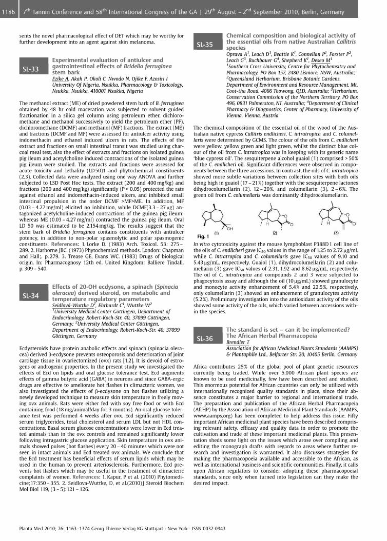
sents the novel pharmacological effect of DET which may be worthy forfurther development into an agent against skin melanoma.
SL-33Experimental evaluation of antiulcer andgastrointestinal effects of Bridelia ferrugineastem barkEzike A, Akah P, Okoli C, Nwodo N, Ojike F, Azosiri IUniversity Of Nigeria, Nsukka, Pharmacology & Toxicology,Nsukka, Nsukka, 410001 Nsukka, Nigeria
The methanol extract (ME) of dried powdered stem bark of B. ferrugineaobtained by 48 hr cold maceration was subjected to solvent guidedfractionation in a silica gel column using petroleum ether, dichloro-methane and methanol successively to yield the petroleum ether (PF),dichloromethane (DCMF) and methanol (MF) fractions. The extract (ME)and fractions (DCMF and MF) were assessed for antiulcer activity usingindomethacin and ethanol induced ulcers in rats. The effects of theextract and fractions on small intestinal transit was studied using char-coal meal test, also the effect of extracts and fractions on isolated guineapig ileum and acetylcholine induced contractions of the isolated guineapig ileum were studied. The extracts and fractions were assessed foracute toxicity and lethality (LD 50)1 and phytochemical constituents(2,3). Collected data were analyzed using one way ANOVA and furthersubjected to LSD Post Hoc tests. The extract (200 and 400 mg/kg) andfractions (200 and 400 mg/kg) significantly (P < 0.05) protected the ratsagainst ethanol and indomethacin-induced ulcers, and inhibited smallintestinal propulsion in the order DCMF >MF>ME. In addition, MF(0.03 – 4.27 mg/ml) elicited no inhibition, while DCMF(3.3 – 27 mg) an-tagonized acetylcholine-induced contractions of the guinea pig ileum;whereas ME (0.03 – 4.27 mg/ml) contracted the guinea pig ileum. OralLD 50 was estimated to be 2,154 mg/kg. The results suggest that thestem bark of Bridelia ferruginea contains constituents with antiulcerpotency, in addition to non-polar spasmolytic and polar spasmogenicconstituents. References: 1. Lorke D. (1983) Arch. Toxicol. 53: 275 –289. 2. Harborne JBC. (1973) Phytochemical methods. London: Chapmanand Hall;. p. 279. 3. Trease GE, Evans WC. (1983) Drugs of biologicalorigin. In: Pharmacognosy 12th ed. United Kingdom: Balliere Tindall.p. 309 – 540.
SL-34Effects of 20-OH ecdysone, a spinach (Spinaciaoleracea) derived steroid, on metabolic andtemperature regulatory parametersSeidlov�-Wuttke D1, Ehrhardt C1, Wuttke W2
1University Medical Center G�ttingen, Department ofEndocrinology, Robert-Koch-Str. 40, 37099 G�ttingen,Germany; 2University Medical Center G�ttingen,Department of Endocrinology, Robert-Koch-Str. 40, 37099G�ttingen, Germany
Ecdysteroids have protein anabolic effects and spinach (spinacia olera-cea) derived b-ecdysone prevents osteoporosis and deterioration of jointcartilage tissue in ovariectomized (ovx) rats [1,2]. It is devoid of estro-gens or androgenic properties. In the present study we investigated theeffects of Ecd on lipids and oral glucose tolerance test. Ecd augmentseffects of gamma butyric acid (GABA) in neurons and since GABA-ergicdrugs are effective to ameliorate hot flashes in climacteric women, wealso investigated the effects of b-ecdysone on hot flashes utilizing anewly developed technique to measure skin temperature in freely mov-ing ovx animals. Rats were either fed with soy free food or with Ecdcontaining food (18 mg/animal/day for 3 months). An oral glucose toler-ance test was performed 4 weeks after ovx. Ecd significantly reducedserum triglycerides, total cholesterol and serum LDL but not HDL con-centrations. Basal serum glucose concentrations were lower in Ecd trea-ted animals than in the ovx controls and remained significantly lowerfollowing intragastric glucose application. Skin temperature in ovx ani-mals showed pulses (hot flashes) every 20 – 40 minutes which were notseen in intact animals and Ecd treated ovx animals. We conclude thatthe Ecd treatment has beneficial effects of serum lipids which may beused in the human to prevent arteriosclerosis. Furthermore, Ecd pre-vents hot flashes which may be useful in the treatment of climactericcomplaints of women. References: 1. Kapur, P et al. (2010) Phytomedi-cine;17:350 – 355. 2. Seidlova-Wuttke, D, et al.(2010) J Steroid BiochemMol Biol 119, (3 – 5):121 – 126.
SL-35Chemical composition and biological activity ofthe essential oils from native Australian CallitrisspeciesOprava A1, Leach D1, Beattie K1, Connellan P1, Forster P2,Leach G3, Buchbauer G4, Shepherd K1, Deseo M1
1Southern Cross University, Centre for Phytochemsitry andPharmacology, PO Box 157, 2480 Lismore, NSW, Australia;2Queensland Herbarium, Brisbane Botanic Gardens,Department of Environment and Resource Management, Mt.Coot-tha Road, 4066 Toowong, QLD, Australia; 3Herbarium,Conservation Commission of the Northern Territory, PO Box496, 0831 Palmerston, NT, Australia; 4Department of ClinicalPharmacy & Diagnostics, Center of Pharmacy, University ofVienna, Vienna, Austria
The chemical composition of the essential oil of the wood of the Aus-tralian native cypress Callitris endlicheri, C. intratropica and C. columel-laris were determined by GCMS. The colour of the oils from C. endlicheriwere yellow, yellow green and light green, whilst the distinct blue col-our of the oil from C. intratropica was in keeping with its generic name‘blue cypress oil’. The sesquiterpene alcohol guaiol (1) comprised > 50%of the C. endlicheri oil. Significant differences were observed in compo-nents between the three accessions. In contrast, the oils of C. intratropicashowed more subtle variations between collection sites with both oilsbeing high in guaiol (17 – 21%) together with the sesquiterpene lactonesdihydrocolumellarin (2), 12 – 20%, and columellarin (3), 2 – 6%. Thegreen oil from C. columellaris was dominantly dihydrocolumellarin.
Fig. 1
In vitro cytotoxicity against the mouse lymphoblast P388D 1 cell line ofthe oils of C. endlicheri gave IC50 values in the range of 1.25 to 2.72 mg/mLwhile C. intratropica and C. columellaris gave IC50 values of 9.10 and5.43 mg/mL, respectively. Guaiol (1), dihydrocolumellarin (2) and colu-mellarin (3) gave IC50 values of 2.31, 1.92 and 8.62 mg/mL, respectively.The oil of C. intratropica and compounds 2 and 3 were subjected tophagocytosis assay and although the oil (10 mg/mL) showed granulocyteand monocyte activity enhancement of 5.4% and 22.5%, respectively,only columellarin (3) showed an enhancement of granulocytes activity(5.2%). Preliminary investigation into the antioxidant activity of the oilsshowed some activity of the oils, which varied between accessions with-in the species.
SL-36The standard is set – can it be implemented?The African Herbal PharmacopeiaBrendler TAssociation for African Medicinal Plants Standards (AAMPS)& Plantaphile Ltd., Belforter Str. 20, 10405 Berlin, Germany
Africa contributes 25% of the global pool of plant genetic resourcescurrently being traded. While over 5.000 African plant species areknown to be used medicinally, few have been described and studied.This enormous potential for African countries can only be utilized withinternationally recognized quality standards in place, since their ab-sence constitutes a major barrier to regional and international trade.The preparation and publication of the African Herbal Pharmacopeia(AfrHP) by the Association of African Medicinal Plant Standards (AAMPS,www.aamps.org) has been completed to help address this issue. Fiftyimportant African medicinal plant species have been described compris-ing relevant safety, efficacy and quality data in order to promote thecultivation and trade of these important medicinal plants. This presen-tation sheds some light on the issues which arose over compiling andediting the monograph drafts with regards to areas where further re-search and investigation is warranted. It also discusses strategies formaking the pharmacopoeia available and accessible to the African, aswell as international business and scientific communities. Finally, it callsupon African regulators to consider adopting these pharmacopoeialstandards, since only when turned into legislation can they make thedesired impact.
Planta Med 2010; 76: 1163–1374 Georg Thieme Verlag KG Stuttgart · New York · ISSN 0032-0943
1186 7th Tannin Conference and 58th International Congress of the GA | 29th August – 2nd September 2010, Berlin, Germany
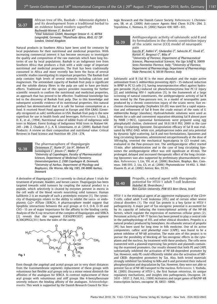
SL-37African tree of life, Baobab – Adansonia digitata Land its development from a traditional herbal toan evidence based modern superfruitBuchwald-Werner S1, Beckett K2
1Vital Solutions GmbH, Hausinger Strasse 4 – 6, 40764Langenfeld, Germany; 2PhytoTrade Africa, R&D, E2 7JPLondon, United Kingdom
Natural products in Southern Africa have been used for centuries bylocal populations for their nutritional and medicinal properties. Withincreasing commercial interest it has become necessary to investigatethe quality and composition of the raw materials which have long his-tories of use by local populations. Baobab is an indigenous tree fromSouthern Africa that produces a fruit with a wide range of importantnutritional and medicinal properties. The fruit has been traditionallyconsumed in Africa and more recently has been the focus of severalscientific studies investigating its important properties. The Baobab fruitpulp contains high levels of several minerals including calcium andmagnesium. The antioxidant capacity of Baobab fruit pulp is significantand the soluble dietary fibres in the pulp are said to have pre-bioticeffects. Traditional use of this species provides reasoning for furtherscientific research to confirm the nutritional and medicinal properties,an approach that has proved to be effective. Traditional knowledge hasled to the discovery of Baobab fruit pulp. Due to this knowledge andsubsequent scientific evidence of its nutritional properties, this naturalproduct has demonstrated that it is safe for human consumption as afood. It received Novel Food approval in Europe and FDA GRAS (Gener-ally recognized as safe) notification in USA. Baobab fruit pulp is the newsuperfruit for use in health foods and beverages. References: 1. Saka, J.D. K., et al., (1994). Nutritional value of edible fruits of indigenous wildtrees in Malawi. Forest Ecology of Indigenous Wild trees in Malawi 64:245 – 248. 2. Chadare, F. J., A. R. Linnemann, et al. (2009). Baobab FoodProducts: A review on their composition and nutritional value. CriticalReviews in Food Science and Nutrition 49: 254 – 274.
SL-38The pharmacophore of thapsigarginChristensen S1, Skytte D1, Liu H1, Nielsen H1,Svenningsen L1, Jensen C1, Møller J2
1University of Copenhagen, Faculty of PharmaceuticalSciences, Department of Medicinal Chemistry,Universitetsparken 2, 2100 Copehagen Ø, Denmark;2University of Aarhus, Department of Physilogy andBiophysics, Ole Worms Alle blg 1185, DK-8000 Aarhus,Denmark
A derivative of thapsigargin (1) is currently in clinical phase 1 trials fortreatment of prostate, bladder, and breast cancer. Thapsigargin has beentargeted towards solid tumours by coupling the natural product to apeptide, which selectively is cleaved by enzymes present in excess inthe cell walls of the blood vessels nourishing solid tumours. Humankallikrein 2 is an example of such a proteolytic enzyme [1]. The cytoxi-city of thapsigargin relates to the ability to inhibit the sarco- or endo-plasmic Ca2+ ATPase (SERCA). A pharmacophore model suggest thatlipophilic interactions between the acyl groups at O-3, O-8, O-10 andCH3 – 15 are of major importance for the affinity to the binding cavity.Analysis of the X-ray structure of the complex of thapsigargin and SERCA[2] reveals that the segment F(834)FRY(837) andthe segmentA(306)IPEGL(311) form the sides of the cavity.
Fig. 1
Even though the angeloyl and acetyl groups are in very short distancesfrom the transmembrane segments replacement of these groups withvoluminous but flexible acyl groups only to a minor extent diminish theaffinities of the analogues for SERCA. In contrast replacement of theseacyl groups with voluminous and stiff groups like 4-benzylbenzoateseverely reduces the binding affinity of the analogues. Acknowledge-ments: This work is supported by the Danish Research Council for Stra-
tegic Research and the Danish Cancer Society. References: 1. Christen-sen, SB. et al. (2009) Anti-cancer Agents Med Chem 9:276 – 294. 2.Toyoshima, C, Nomura, H. (2002) Nature 418: 605 – 611.
SL-39Antihyperalgesic activity of salvianolic acid B andits formulations in the chronic constriction injuryof the sciatic nerve (CCI) model of neuropaticpainIsacchi B1, Fabbri V1, Ghelardini C2, Salvicchi A2, Vivoli E2,Karioti A1, Bergonzi C1, Bilia A1
1University of Florence, Department of PharmaceuticalSciences, Pharmaceutical Sciences, Via Ugo Schiff 6, 50019Sesto Fiorentino Florence, Italy; 2University of Florence,Department of Pharmacology, Department of Pharmacology,Viale Pieraccini, 6, 50139 Florence, Italy
Salvianolic acid B (Sal B) is the most abundant and the major activeantioxidant from S. miltiorrhiza preventing Ab25 – 35-induced reductionin BPRP in PC12 cells [1], having neuronprotective effect against hydro-gen peroxide (H2O2)-induced rat pheochromocytoma line PC12 injury[2] and inhibiting HIV-1 replication [3]. In the framework of a largescreening of natural constituents, the antihyperalgesic activity of Sal Bwas tested by the Paw-pressure test, in a model of neuropathic painproduced by a chronic constriction injury of the sciatic nerve. Size ex-clusion chromatography (Sephadex LH-20) was used for a rapid separa-tion and refinement of Sal B from the crude extract of the roots of S.miltiorrizha Bunge (danshen). Hydroalchoholic solutions were used aseluents for a safe and convenient separation obtaining Sal B almost pureby NMR (> 96%). Liposomial formulations were prepared using eggphosphatydil choline, cholesterol and 18:0/18:0 PEG 2000 in the caseof long circulationg liposome. Encapsulation efficacy of Sal B was eval-uated by HPLC-DAD, while size, polydispersion index and zeta potentialby dynamic light scattering. Sal B and two formulations, liposomes andlong circulating liposomes, administered intraperitoneally at the dose of100 mg/kg, reverted the mechanical hyperalgesia in CCI treated rats,evaluated in the Paw-pressure test. The antihyperalgesic effect started15 min. after administration and in the case of long circulating lipo-somes the antihyperalgesic effect was still significant at 45 min. Theprolonged, delayed and increased therapeutic effect of the long circulat-ing liposomes was also supported by preliminary pharmacokinetic stu-dies. References: 1. Lin, YH. et al. (2006) Biochem. Biophys. Res. Com-mun. 348:593. 2. Liu, CS. et al. (2006) Phytomedicine 14:492. 3. Abd-Elazem IS. et al. (2002) Antivir. Res. 55:91.
SL-40Propolis, a natural agent with therapeuticpotential for HTLV-1 adult T-cell leukemiaHuleihel M, Shvarzbeyn JBen-Gurion University, POB 653 Beer-Sheva, Israel
HTLV-1 is the etiological agent of an aggressive malignancy of the CD4+T-cells, called adult T-cell leukemia (ATL) and of certain other severeclinical disorders (1). The viral Tax protein is a key factor in HTLV-1pathogenicity. A major part of Tax oncogenic potential is accounted forby its capacity of inducing the transcriptional activity of the NF-kBfactors, which regulate the expression of numerous cellular genes (2).Persistent activity of NF-?? factors has been proved to play a central rolein the pathophysiology of ATL and other clinical disorders. Propolis is anatural product produced by honeybees. An ethanolic extract of propolis(PE) has been used for long time in folk medicine. One of its activecomponents, caffeic acid phenethyl ester (CAPE), was found to be apotent inhibitor of NF-kB activation. The main aim of this project is topursue the possibility of blocking all Tax oncogenic effects in the cyto-plasm and the nucleus, by treatment with these products. The cells weretransected with a plasmid expressing Tax protein and plasmids contain-ing the examined promoters. Our results showed that both PE and CAPEsubstantially inhibited the activation of NF-kB-dependent promoter byTax. However, only PE could efficiently inhibit also the activation of SRF-and CREB- dependent promoters by Tax. Also, both tested materialsstrongly inhibited Tax binding to IkBa and b and prevented their inducedphosphorylation and degradation by Tax. However, they were not able toprevent Tax or of NF-kB transport to the nucleus. References: 1. Yoshida,M. (2005) Discovery of HTLV-1, the first human retrovirus, its uniqueregulatory mechanisms, and insights into pathogenesis, Oncogene, 24:5931 – 5937. 2. Pahl, H.L. (1999) Activators and target genes of Rel/NF-kBtranscription factors, oncogene 18, 6853 – 6866.
Planta Med 2010; 76: 1163–1374 Georg Thieme Verlag KG Stuttgart · New York · ISSN 0032-0943
11877th Tannin Conference and 58th International Congress of the GA | 29th August – 2nd September 2010, Berlin, Germany
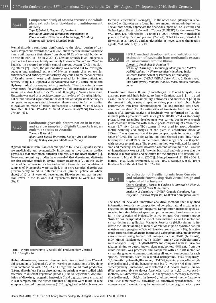
SL-41Comparative study of Mentha arvensis Linn wholeplant extracts for antioxidant and antidepressantactivityTupe P, Sakat S, Nagmoti D, Juvekar AInsitute of Chemical Technology, Department ofPharmaceutical Sciences and Technology, N.P. Marg,Matunga (E), 400019 Mumbai, India
Mental disorders contribute significantly to the global burden of dis-eases. Projections towards the year 2020 show that the neuropsychiatricillness will increase their share from about 10.5% of the total burden ofdisease in 1990 to 15% in 2020 [1]. Mentha arvensis is an indigenousplant of the Lamiaceae family commonly known as ‘Pudina’ and ‘Mint’ inEnglish. It is reported to exhibit central nervous system (CNS) modulat-ing effects [2]. Hence, the present study was carried out to investigateaqueous and methanol extracts of Mentha arvensis Linn extracts forantioxidant and antidepressant activity. Aqueous and methanol extractsof Mentha arvensis were preliminary studied for in vitro antioxidantactivity using 1, 1-Diphenyl-2-Picrylhydrazyl (DPPH), Nitric oxide andHydroxyl radical scavenging activity methods. Then the extracts wereinvestigated for antidepressant activity by Tail suspension and Forcedswim test at dose level of 125, 250 and 500 mg/kg in Swiss albino mice.Fluoxetin was used as a positive control at the dose of 10 mg/kg. Metha-nol extract showed significant antioxidant and antidepressant activity ascompared to aqueous extract. However, there is need for further studiesto evaluate its mode of action. References: 1. Kastrup M, et al (2007)Dan. Med. Bull 54: 42 – 432. 2. Pia M. Vuorela et al.(2006) Fitoterapia77:429 – 434.
SL-42Cardiotonic glycoside determination in in vitroand ex vitro samples of Digitalis lamarckii Ivan, anendemic species to AnatoliaYucesan B, Gurel EAbant Izzet Baysal University, Biology, Art and Sciencefaculty, Golkoy campus, 14280 Bolu, Turkey
Digitalis lamarckii Ivan is an endemic species to Turkey. Digitalis speciesare medicinally and economically important as they contain cardiacglycosides that strengthen diffusion and regulate the heart rhythm [1].Moreover, preliminary studies have revealed that digoxin and digitoxinare also effective agents in several cancer treatments [2]. In this studycardenolide patterns in in vitro and ex vitro (from natural populations)samples of D. lamarckii were studied. In in vitro samples, digoxin waspredominantly found in different tissues (lamina, petiole or wholeshoot) of 12 or 18-week old regenerants. Digoxin content was, in gen-eral, lower in the 18-week old regenerants than 12-week old ones(Fig.1).
Fig. 1: In vitro regenerant (12 weeks old) produced from 2.0 mg/lBA+0.5 mg/l NAA
Highest digoxin was, however, observed in lamina excised from 12-weekold regenerants (130 mg/kg). When varying concentrations of BA aloneor in combination with NAA, BA at 1.0 mg/l was found most productive(6.9 mg digoxin/kg). For ex vitro, natural populations were studied withreference to different vegetation periods (June to September). Accumu-lation of digoxin, gitoxigenin, lanatoside C and digitoxin were estimatedin leaf samples, and the higher amounts of digoxin were found in Junesamples extracted from mid-leaves (1010 mg/kg) and reddish leaves col-
lected in September (1062 mg/kg). On the other hand, gitoxigenin, lana-toside C or digitoxin were found in trace amount. Acknowledgements:The authors deeply appreciate the financial support of The Scientific andTechnological Research Council of Turkey (TUBITAK) for the project TO-VAG-106O470. References: 1. Baytop T (1999). Therapy with medicinalplants in Turkey: Past and present. (2nd ed). Nobel kitabevi, Istanbul. 2.Newman et al. (2008). Cardiac glycosides as novel cancer therapeuticagents. Mol. Intv. 8(1): 36 – 49.
SL-43HPTLC method development and validation forestimation of isovitexin from methanolic extractof Enicostemma littorale BlumeSawant L, Prabhakar B, Pandita NSchool of Pharmacy & Technology Management, SVKMSNMIMS University, Pharmaceutical chemistry, SeniorResearch fellow, School of Pharmacy & TechnologyManagement, SVKMS NMIMS University, V. L. Mehta road,Vile parle (West), Mumbai 400056, India, 400056 Mumbai,India
Enicostemma littorale Blume (Chota-Kirayat or Chota-Chirayata) is aglabrous perennial herb belongs to family Gentianaceae [1]. It is usedas anti-diabetic, anti-inflammatory, anticancer and antioxidant [2, 3]. Inthe present study, a new, simple, sensitive, precise and robust high-performance thin layer chromatographic (HPTLC) method was devel-oped and validated for the estimation of isovitexin from methanolicextract of E. littorale. Analysis of isovitexin was performed on TLC alu-minium plates pre-coated with silica gel 60 RP-18 F-254 as stationaryphase. Linear ascending development was carried out in twin troughglass chamber saturated with mobile phase consisting of acetonitrile-water (3:7, v/v). Camag TLC scanner III was used for spectrodensito-metric scanning and analysis of the plate in absorbance mode at235 nm. The system was found to give compact spots for isovitexin (Rfvalue of 0.40). The data for calibration plots showed good linear rela-tionship with r2 = 0.99853 in the concentration range of 50 – 400 spot-1with respect to peak area. The present method was validated for preci-sion and recovery. The total isovitexin content was found to be 0.4% (w/w) in methanolic extract of E. littorale. Statistical analysis proves that themethod is reproducible and selective for the analysis of isovitexin. Re-ferences: 1. Murali, B. et al. (2002) J. Ethnopharmacol. 81:199 – 204. 2.Maroo, J. et al. (2003) Phytomed. 10:196 – 199. 3. Sadique, J. et al. (1987)Biochem Med Metabol Biol. 37:167 – 176.
SL-44Dereplication of Brazilian plants from Cerradoand Atlantic Forest using NMR virtual design andhyphenated techniquesCastro-Gamboa I, Burgos R, Cardoso P, Carnevale F, Pilon A,Nasser Lopes M, Silva D, Bolzani VInstitute of Chemistry – UNESP, Organic Chemistry, RuaProfessor Francisco Degni s/n, 14800900 Araraquara, Brazil
The need for new and innovative analytical methods that may shedinformation towards the composition of complex natural mixtures is akeystone on bioprospection programs. Dereplication methodologies as-sociated to state-of-the-art spectroscopic techniques, have been success-ful in the selection of biologically active extracts. Our research group“NuBBE” has incorporated the use of those methods as well as molecularvirtual design using Nuclear Magnetic Resonance (NMR) aiming to in-crease the understanding of molecular relationships on dynamic naturalmatrixes and synergism effects of bioactive crude extracts. Highly activecrude extracts, from Abarema lusoria and Calea pinnatifida, previously invitro screened using human cell lineages such as HL-60 (Leukemia),MDA-MB435 (Melanoma), HCT-8 (Colon) and SF-295 (Glioblastoma),were analyzed using HPLC/DAD-HRMS and compared with in silico da-tabases aiming to detect known plant metabolites. NMR data from thecrude extracts was processed and compared with an NMR molecularvirtual designed environment containing all known compounds for eachspecies. Flavonoids, such as 8-methyl-naringenine, 4’,5,7-trihydroxy-3’,6-dimethoxy-8-methylflavanone, 3’,4’,5,6,7-pentahydroxy-8-methyl-dihydroflavonol and the benzophenone bis (2-hydroxy-4,6-dimethoxy-3-methylphenyl) metanone, were detected in A. lusoria. From C. pinna-tifida we were able to detect flavonoids, such as 4’,5,7-trihydroxy-3-methoxy-6,8-dimethylflavanone, 4’,7-dihydroxy-5-methoxy-6-methyl-dihydroflavonol, 5,6,7-trihydroxy-3’,4’-dimethoxy-8-methylflavanoneand, 3’,4’-dimethoxy-5,7-dihydroxy-6,8-dimethyldihydroflavonol. Theoccurrence of flavonoids may be associated to the original activity re-
Planta Med 2010; 76: 1163–1374 Georg Thieme Verlag KG Stuttgart · New York · ISSN 0032-0943
1188 7th Tannin Conference and 58th International Congress of the GA | 29th August – 2nd September 2010, Berlin, Germany
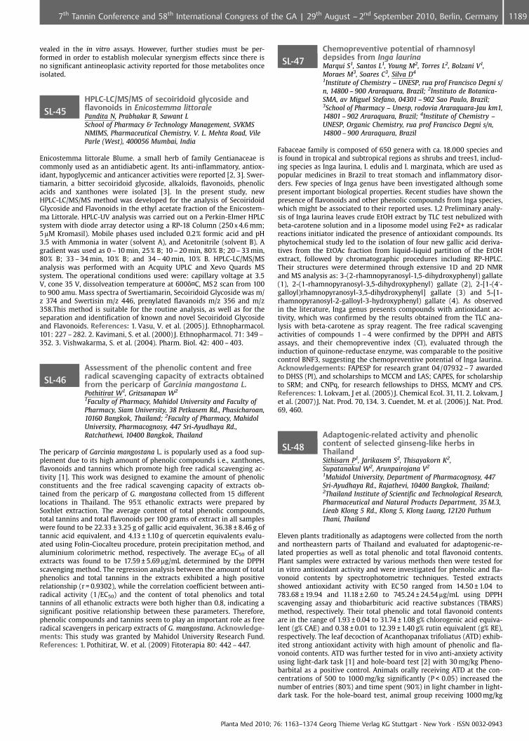
vealed in the in vitro assays. However, further studies must be per-formed in order to establish molecular synergism effects since there isno significant antineoplasic activity reported for those metabolites onceisolated.
SL-45HPLC-LC/MS/MS of secoiridoid glycoside andflavonoids in Enicostemma littoralePandita N, Prabhakar B, Sawant LSchool of Pharmacy & Technology Management, SVKMSNMIMS, Pharmaceutical Chemistry, V. L. Mehta Road, VileParle (West), 400056 Mumbai, India
Enicostemma littorale Blume. a small herb of family Gentianaceae iscommonly used as an antidiabetic agent. Its anti-inflammatory, antiox-idant, hypoglycemic and anticancer activities were reported [2, 3]. Swer-tiamarin, a bitter secoiridoid glycoside, alkaloids, flavonoids, phenolicacids and xanthones were isolated [3]. In the present study, newHPLC-LC/MS/MS method was developed for the analysis of SecoiridoidGlycoside and Flavonoids in the ethyl acetate fraction of the Enicostem-ma Littorale. HPLC-UV analysis was carried out on a Perkin-Elmer HPLCsystem with diode array detector using a RP-18 Column (250 x 4.6 mm;5mM Kromasil). Mobile phases used included 0.2% formic acid and pH3.5 with Ammonia in water (solvent A), and Acetonitrile (solvent B). Agradient was used as 0 – 10 min, 25% B; 10 – 20 min, 80% B; 20 – 33 min,80% B; 33 – 34 min, 10% B; and 34 – 40 min, 10% B. HPLC-LC/MS/MSanalysis was performed with an Acquity UPLC and Xevo Quards MSsystem. The operational conditions used were: capillary voltage at 3.5V, cone 35 V, dissolveation temperature at 600¤C, MS 2 scan from 100to 900 amu. Mass spectra of Swertiamarin, Secoiridoid Glycoside was m/z 374 and Swertisin m/z 446, prenylated flavanoids m/z 356 and m/z358.This method is suitable for the routine analysis, as well as for theseparation and identification of known and novel Secoiridoid Glycosideand Flavonoids. References: 1. Vasu, V. et al. (2005) J. Ethnopharmacol.101: 227 – 282. 2. Kavimani, S. et al. (2000) J. Ethnopharmacol. 71: 349 –352. 3. Vishwakarma, S. et al. (2004). Pharm. Biol. 42: 400 – 403.
SL-46Assessment of the phenolic content and freeradical scavenging capacity of extracts obtainedfrom the pericarp of Garcinia mangostana L.Pothitirat W1, Gritsanapan W2
1Faculty of Pharmacy, Mahidol University and Faculty ofPharmacy, Siam University, 38 Petkasem Rd., Phasicharoan,10160 Bangkok, Thailand; 2Faculty of Pharmacy, MahidolUniversity, Pharmacognosy, 447 Sri-Ayudhaya Rd.,Ratchathewi, 10400 Bangkok, Thailand
The pericarp of Garcinia mangostana L. is popularly used as a food sup-plement due to its high amount of phenolic compounds i. e., xanthones,flavonoids and tannins which promote high free radical scavenging ac-tivity [1]. This work was designed to examine the amount of phenolicconstituents and the free radical scavenging capacity of extracts ob-tained from the pericarp of G. mangostana collected from 15 differentlocations in Thailand. The 95% ethanolic extracts were prepared bySoxhlet extraction. The average content of total phenolic compounds,total tannins and total flavonoids per 100 grams of extract in all sampleswere found to be 22.33 € 3.25 g of gallic acid equivalent, 36.38 € 8.46 g oftannic acid equivalent, and 4.13 € 1.10 g of quercetin equivalents evalu-ated using Folin-Ciocalteu procedure, protein precipitation method, andaluminium colorimetric method, respectively. The average EC50 of allextracts was found to be 17.59 € 5.69 mg/mL determined by the DPPHscavenging method. The regression analysis between the amount of totalphenolics and total tannins in the extracts exhibited a high positiverelationship (r = 0.9302), while the correlation coefficient between anti-radical activity (1/EC50) and the content of total phenolics and totaltannins of all ethanolic extracts were both higher than 0.8, indicating asignificant positive relationship between these parameters. Therefore,phenolic compounds and tannins seem to play an important role as freeradical scavengers in pericarp extracts of G. mangostana. Acknowledge-ments: This study was granted by Mahidol University Research Fund.References: 1. Pothitirat, W. et al. (2009) Fitoterapia 80: 442 – 447.
SL-47Chemopreventive potential of rhamnosyldepsides from Inga laurinaMarqui S1, Santos L1, Young M2, Torres L2, Bolzani V1,Moraes M3, Soares C3, Silva D4
1Institute of Chemistry – UNESP, rua prof Francisco Degni s/n, 14800 – 900 Araraquara, Brazil; 2Instituto de Botanica-SMA, av Miguel Stefano, 04301 – 902 Sao Paulo, Brazil;3School of Pharmacy – Unesp, rodovia Araraquara-Jau km1,14801 – 902 Araraquara, Brazil; 4Institute of Chemistry –UNESP, Organic Chemistry, rua prof Francisco Degni s/n,14800 – 900 Araraquara, Brazil
Fabaceae family is composed of 650 genera with ca. 18.000 species andis found in tropical and subtropical regions as shrubs and trees1, includ-ing species as Inga laurina, I. edulis and I. marginata, which are used aspopular medicines in Brazil to treat stomach and inflammatory disor-ders. Few species of Inga genus have been investigated although somepresent important biological properties. Recent studies have shown thepresence of flavonoids and other phenolic compounds from Inga species,which might be associated to their reported uses. 1,2 Preliminary analy-sis of Inga laurina leaves crude EtOH extract by TLC test nebulized withbeta-carotene solution and in a liposome model using Fe2+ as radicalarreactions initiator indicated the presence of antioxidant compounds. Itsphytochemical study led to the isolation of four new gallic acid deriva-tives from the EtOAc fraction from liquid-liquid partition of the EtOHextract, followed by chromatographic procedures including RP-HPLC.Their structures were determined through extensive 1D and 2D NMRand MS analysis as: 3-(2-rhamnopyranosyl-1,5-dihydroxyphenyl) gallate(1), 2-(1-rhamnopyranosyl-3,5-dihydroxyphenyl) gallate (2), 2-[1-(4’-galloyl)rhamnopyranosyl-3,5-dihydroxyphenyl] gallate (3) and 5-[1-rhamnopyranosyl-2-galloyl-3-hydroxyphenyl) gallate (4). As observedin the literature, Inga genus presents compounds with antioxidant ac-tivity, which was confirmed by the results obtained from the TLC ana-lysis with beta-carotene as spray reagent. The free radical scavengingactivities of compounds 1 – 4 were confirmed by the DPPH and ABTSassays, and their chemopreventive index (CI), evaluated through theinduction of quinone-reductase enzyme, was comparable to the positivecontrol BNF3, suggesting the chemopreventive potential of Inga laurina.Acknowledgements: FAPESP for research grant 04/07932 – 7 awardedto DHSS (PI), and scholarships to MCCM and LAS; CAPES, for scholarshipto SRM; and CNPq, for research fellowships to DHSS, MCMY and CPS.References: 1. Lokvam, J et al. (2005) J. Chemical Ecol. 31, 11. 2. Lokvam, Jet al. (2007) J. Nat. Prod. 70, 134. 3. Cuendet, M. et al. (2006) J. Nat. Prod.69, 460.
SL-48Adaptogenic-related activity and phenoliccontent of selected ginseng-like herbs inThailandSithisarn P1, Jarikasem S2, Thisayakorn K2,Supatanakul W2, Arunpairojana V2
1Mahidol University, Department of Pharmacognosy, 447Sri-Ayudhaya Rd., Rajathevi, 10400 Bangkok, Thailand;2Thailand Institute of Scientific and Technological Research,Pharmaceutical and Natural Products Department, 35 M.3,Lieab Klong 5 Rd., Klong 5, Klong Luang, 12120 PathumThani, Thailand
Eleven plants traditionally as adaptogens were collected from the northand northeastern parts of Thailand and evaluated for adaptogenic-re-lated properties as well as total phenolic and total flavonoid contents.Plant samples were extracted by various methods then were tested forin vitro antioxidant activity and were investigated for phenolic and fla-vonoid contents by spectrophotometric techniques. Tested extractsshowed antioxidant activity with EC 50 ranged from 14.50 € 1.04 to783.68 € 19.94 and 11.18 € 2.60 to 745.24 € 24.54 mg/mL using DPPHscavenging assay and thiobarbituric acid reactive substances (TBARS)method, respectively. Their total phenolic and total flavonoid contentsare in the range of 1.93 € 0.04 to 31.74 € 1.08 g% chlorogenic acid equiva-lent (g% CAE) and 0.38 € 0.01 to 12.39 € 1.40 g% rutin equivalent (g% RE),respectively. The leaf decoction of Acanthopanax trifoliatus (ATD) exhib-ited strong antioxidant activity with high amount of phenolic and fla-vonoid contents. ATD was further tested for in vivo anti-anxiety activityusing light-dark task [1] and hole-board test [2] with 30 mg/kg Pheno-barbital as a positive control. Animals orally receiving ATD at the con-centrations of 500 to 1000 mg/kg significantly (P < 0.05) increased thenumber of entries (80%) and time spent (90%) in light chamber in light-dark task. For the hole-board test, animal group receiving 1000 mg/kg
Planta Med 2010; 76: 1163–1374 Georg Thieme Verlag KG Stuttgart · New York · ISSN 0032-0943
11897th Tannin Conference and 58th International Congress of the GA | 29th August – 2nd September 2010, Berlin, Germany
ATD significantly increased the number of head-dip (37%). The resultsindicated that plant samples, especially the leaves of Acanthopanax tri-foliatus possess antioxidant and anti-anxiety activities supports theirethnomedical uses as adaptogenic agents. Acknowledgements: TheGraduate Program Development under the Collaboration between Thai-land Institute of Scientific and Technological Research and Universities.References: 1. Crawley, J. et al. (1980) Pharmacol. Biochem. Behav.13:167 – 170. 2. Treit, D. et al. (1981) Pharmacol. Biochem. Behav.15(4):619 – 626.
SL-49The effectiveness of a standardised Echinaceapreparation in preventing colds, flus and otherrespiratory disorders for air-travellersTiralongo E, Lea R, Wee S, Hanna M, Griffiths LGriffith University, School of Pharmacy, Gold Coast Campus,4222 Gold Coast, Australia
Air travel, especially on intercontinental flights can be stressful. Studieshave shown that passengers’ well-being is influenced by the cabin en-vironment such as quality of cabin air, oxygen pressure in the cabin,motion or vibration and oil additives used in aircraft engines [1, 2].Various studies have reported and investigated the occurrence of nasaldryness [3] and the increased risk of developing upper respiratory dis-orders such as allergic rhinitis [4] and attracting virus or bacteria in-duced respiratory infections [5, 6]. Studies have shown that Echinacea,one of the most widely used herbal medicines worldwide, can decreasethe severity of cold symptoms and improve quality of life if used as earlytreatment [7 – 9]. Some studies also suggest positive effects for the pre-vention of the common cold when using Echinacea [8]. Here we reporton a randomised, double-blind placebo controlled clinical trial with 183participants who were travelling return from Australia to America, Eur-ope or Africa for a period of 1 – 5 weeks on non-stop commercial flightsvia economy class. Participants were using coated Echinacea (standar-dised to 4.42 mg alkylamides) or placebo tablets, and trial dosing con-sisted of three protocols (priming, travel and sick), depending on thephase of travel of the participants and their health status. Outcomeswere assessed using a survey which included questions about upperrespiratory symptoms (based on WURSS-44), quality of life (based onSF-36) and the occurrence of additional air travel related symptoms.Each participant completed this survey before travel (baseline), aftertravel (return) and at 4 weeks after return from travel (follow-up). Pre-liminary results so far indicate that at follow-up a significantly lowernumber of participants reported respiratory illness in the Echinaceatreated group compared to placebo (P = 0.02) and a significantly highernumber of participants treated with Echinacea reported a “good” qualityof life compared to placebo (P = 0.005). Moreover, a significantly highernumber of participants in the placebo group compared to the Echinaceagroup reported respiratory illness at follow-up compared to baseline(P = 0.044) suggesting that the placebo group had not returned to thepre-travel level in terms of respiratory illness. References: 1. Nicholson,A.N., et al. Travel Med Infect Dis, 2003. 1(2): p. 94 – 102. 2. Hinninghofen,H. et al., Auton Neurosci, 2006. 129(1 – 2): p. 80 – 5. 3. Norback, D., et al.,Scand J Work Environ Health, 2006. 32(2): p.138 – 44. 4. Ohrui, N., et al.,Ann Allergy Asthma Immunol, 2005. 95(4): p. 350 – 3. 5. Vogt, T.M., etal., J Travel Med, 2006. 13(5): p. 268 – 72. 6. Evans, A., et al., Aviat SpaceEnviron Med, 2006. 77(9): p. 974 – 6. 7. Gillespie, E.L. et al., Conn Med,2006. 70(2): p. 93 – 7. 8. Woelkart, K. et al. Planta Med, 2008. 74(6):p. 633 – 7. 9. Linde, K., et al., Cochrane Database Syst Rev, 2006(1):p. CD 000530.
SL-50Mechanisms of the anti-proliferative andpro-apoptotic effect of the herbal fixedcombination STW 5 on colon adenocarcinomacells in vitroBonaterra G1, Kelber O2, Weiser D2, Okpanyi S2, Z�gel S3,Hildebrand W3, Kinscherf R3
1Medical Faculty Mannheim, University of Heidelberg,Section Macroscopic Anatomy, TRIDOMUS C, Ludolf-Krehl-Str. 13 – 17, 68167 Mannheim, Germany; 2ScientificDepartment, Steigerwald Arzneimittelwerk GmbH, Havelstr.5, 64295 Darmstadt, Germany; 3Department of Medical CellBiology, University Marburg, Robert-Koch-Straße 8, 35037Marburg, Germany
The herbal preparation STW 5 is widely used in the treatment of func-tional dyspepsia and other motility-related gastrointestinal disorders.
Our objective was to determine the molecular mechanisms of the anti-proliferative and pro-apoptotic effects on colon adenocarcinoma cells(HT-29). Cells were treated with diclofenac (Diclo), aspirin (ASA), STW5 or its components STW 6 (Iberis amara totalis), STW 5-K II (pepper-mint), STW 5-K VII (milk thistle) or STW 5-K VIII (lemon balm). Anti-proliferative effects were measured with sulforhodamine. Apoptosis wasidentified by YO-PRO-1� staining. Treatment with Diclo (0.1 mM), ASA(2.5 mM), STW 5 (100m g/ml) or its components STW 6 (12.5m g/ml),STW 5-K II (50m g/ml), STW K VII (100m g/ml) or STW 5-K VIII (25m g/ml) inhibited proliferation by ca. 50 – 60% (ASA or Diclofenac 45 – 50%).STW 5 (as well as ASA or Diclo) induced apoptosis 3 to 4- fold. 100 mg/mlSTW 5 showed a 20% or 30% induction of Caspase-3 or BAX expression,whereas ASA or Diclo revealed inhibitory effects. 100 mg/ml STW 5 inhib-ited the Bcl2 mRNA expression. STW 25 – 100 mg/ml up-regulated theexpression of NFkB p65 subunit. Our data suggest that STW 5 and someof its components show antiproliferative and pro-apoptotic effects onHT-29 cells in vitro, possibly due to an activation of the caspase cascadeand the NFkB pathway. Active concentrations of STW 5 are, in relation totherapeutic doses, comparable to those of ASA and Diclo, suggesting asimilar favourable effect on colon carcinoma risk.
SL-51Does willow bark extract have a sildenafil-likeaction? Effects of different fractions on thesynthesis of cGMP and NO in human umbilicalvein endothelial cells (HUVEC)Fiebich B1, M�ller J2, Kelber O2, Okpanyi S2, Weiser D2,Appel K1
1VivaCell Biotechnology GmbH, Ferdinand-Porsche-Str. 5,79211 Denzlingen, Germany; 2Scientific Department,Steigerwald Arzneimittelwerk GmbH, Havelstr. 5, 64295Darmstadt, Germany
Objectives: In the past years several methods have been implementedfor testing substances which may exhibit sildenafil like properties. Wil-low bark extract, used in therapy of painful musculoskeletal diseases,has been supposed also to exhibit effects on endothelial cells and thusinduce NO and might then indirectly increase cGMP and act slidenafil-like. We therefore investigated different fractions with a HUVEC cellmodel. Method: We measured intracellular concentrations of cGMPand the release of NO after treatment of HUVEC cells with 5 differentfractions (A-E) of the standardized willow bark extract STW 33-I (ex-traction medium water, DER 16 – 23:1), covering the whole spectrum ofcomponents from lipophilic to hydrophilic compounds. Results: FractionE showed a significant cGMP enhancement in concentrations from 10 –100 mg/ml. In investigating the validity of the assay this effect was com-parable to a positive control (NO donor and PDE5 inhibitor (TO156)).Fractions A and E increased NO release whereas fraction C led to inhibi-tion of NO release. Conclusions: Fractions of willow bark extract in-creasing NO release and cGMP level, such as fraction E, might be inter-esting candidates for natural aphrodisiac products with a sildenafil-likemode of action. Fractions exhibiting the property to reduce NO releaselike fraction C might be further tested for their anti-inflammatory re-sponse as previous research findings from the study of inflammatorydiseases showed the importance of free radicals like NO in enhancingthe disease outcome. Taken together, this study shows that willow barkextract has several exciting modes of action possibly interesting forfurther indications.
SL-52Effects of ethanolic extract of Pedalium murexLinn. on sexual behavior and reproductiveparameter of male ratsSharma V, Thakur M, Chauhan N, Dixit VDepartment of Pharmaceutical Sciences Dr. H.S. GourUniversity, Dr. H.S. Gour University, 470003 Sagar, India
Throughout the entire ages, men and women have incessantly pursuedall means to enhance, maintain and bring back their sexual ability, or tostimulate the sexual desire of the opposite sex [1].The vajikaran rasaya-na is special class of Ayurvedia that deals with herbs associated with animprovement of male sexual potency and thereby ensure a supraja, orbetter progeny. The main aim of Vajikaran was achievement of success-ful copulation for healthy reproduction, along with an improvement insexual pleasure as an additional benefit [2]. Dried fruit of Pedaliummurex known as bada gokhru is one amongst the traditionally usedherbs to enhance sexual dynamics. Administration of 50, 100, 150 mg/kg b.w. ethanolic extract of P. murex improved overall sexual behavior
Planta Med 2010; 76: 1163–1374 Georg Thieme Verlag KG Stuttgart · New York · ISSN 0032-0943
1190 7th Tannin Conference and 58th International Congress of the GA | 29th August – 2nd September 2010, Berlin, Germany
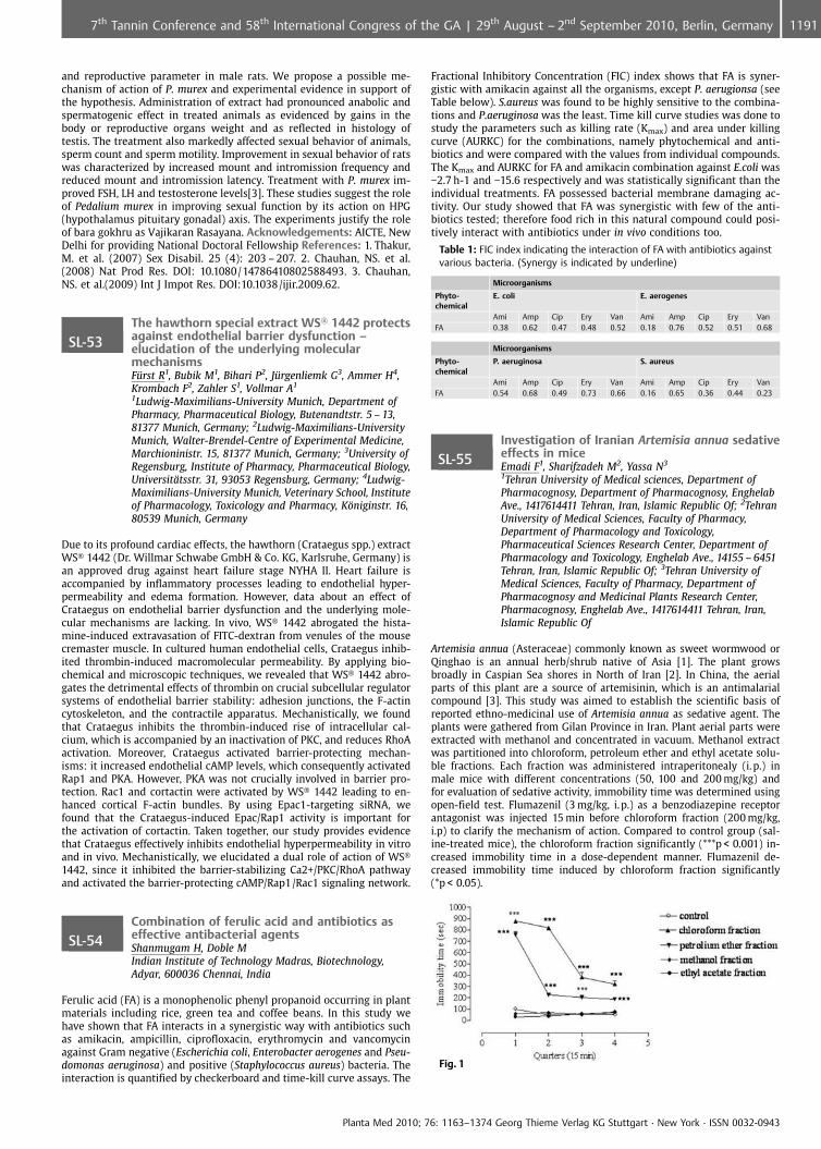
and reproductive parameter in male rats. We propose a possible me-chanism of action of P. murex and experimental evidence in support ofthe hypothesis. Administration of extract had pronounced anabolic andspermatogenic effect in treated animals as evidenced by gains in thebody or reproductive organs weight and as reflected in histology oftestis. The treatment also markedly affected sexual behavior of animals,sperm count and sperm motility. Improvement in sexual behavior of ratswas characterized by increased mount and intromission frequency andreduced mount and intromission latency. Treatment with P. murex im-proved FSH, LH and testosterone levels[3]. These studies suggest the roleof Pedalium murex in improving sexual function by its action on HPG(hypothalamus pituitary gonadal) axis. The experiments justify the roleof bara gokhru as Vajikaran Rasayana. Acknowledgements: AICTE, NewDelhi for providing National Doctoral Fellowship References: 1. Thakur,M. et al. (2007) Sex Disabil. 25 (4): 203 – 207. 2. Chauhan, NS. et al.(2008) Nat Prod Res. DOI: 10.1080/14786410802588493. 3. Chauhan,NS. et al.(2009) Int J Impot Res. DOI:10.1038/ijir.2009.62.
SL-53The hawthorn special extract WS� 1442 protectsagainst endothelial barrier dysfunction –elucidation of the underlying molecularmechanismsF�rst R1, Bubik M1, Bihari P2, J�rgenliemk G3, Ammer H4,Krombach F2, Zahler S1, Vollmar A1
1Ludwig-Maximilians-University Munich, Department ofPharmacy, Pharmaceutical Biology, Butenandtstr. 5 – 13,81377 Munich, Germany; 2Ludwig-Maximilians-UniversityMunich, Walter-Brendel-Centre of Experimental Medicine,Marchioninistr. 15, 81377 Munich, Germany; 3University ofRegensburg, Institute of Pharmacy, Pharmaceutical Biology,Universit�tsstr. 31, 93053 Regensburg, Germany; 4Ludwig-Maximilians-University Munich, Veterinary School, Instituteof Pharmacology, Toxicology and Pharmacy, K�niginstr. 16,80539 Munich, Germany
Due to its profound cardiac effects, the hawthorn (Crataegus spp.) extractWS� 1442 (Dr. Willmar Schwabe GmbH & Co. KG, Karlsruhe, Germany) isan approved drug against heart failure stage NYHA II. Heart failure isaccompanied by inflammatory processes leading to endothelial hyper-permeability and edema formation. However, data about an effect ofCrataegus on endothelial barrier dysfunction and the underlying mole-cular mechanisms are lacking. In vivo, WS� 1442 abrogated the hista-mine-induced extravasation of FITC-dextran from venules of the mousecremaster muscle. In cultured human endothelial cells, Crataegus inhib-ited thrombin-induced macromolecular permeability. By applying bio-chemical and microscopic techniques, we revealed that WS� 1442 abro-gates the detrimental effects of thrombin on crucial subcellular regulatorsystems of endothelial barrier stability: adhesion junctions, the F-actincytoskeleton, and the contractile apparatus. Mechanistically, we foundthat Crataegus inhibits the thrombin-induced rise of intracellular cal-cium, which is accompanied by an inactivation of PKC, and reduces RhoAactivation. Moreover, Crataegus activated barrier-protecting mechan-isms: it increased endothelial cAMP levels, which consequently activatedRap1 and PKA. However, PKA was not crucially involved in barrier pro-tection. Rac1 and cortactin were activated by WS� 1442 leading to en-hanced cortical F-actin bundles. By using Epac1-targeting siRNA, wefound that the Crataegus-induced Epac/Rap1 activity is important forthe activation of cortactin. Taken together, our study provides evidencethat Crataegus effectively inhibits endothelial hyperpermeability in vitroand in vivo. Mechanistically, we elucidated a dual role of action of WS�1442, since it inhibited the barrier-stabilizing Ca2+/PKC/RhoA pathwayand activated the barrier-protecting cAMP/Rap1/Rac1 signaling network.
SL-54Combination of ferulic acid and antibiotics aseffective antibacterial agentsShanmugam H, Doble MIndian Institute of Technology Madras, Biotechnology,Adyar, 600036 Chennai, India
Ferulic acid (FA) is a monophenolic phenyl propanoid occurring in plantmaterials including rice, green tea and coffee beans. In this study wehave shown that FA interacts in a synergistic way with antibiotics suchas amikacin, ampicillin, ciprofloxacin, erythromycin and vancomycinagainst Gram negative (Escherichia coli, Enterobacter aerogenes and Pseu-domonas aeruginosa) and positive (Staphylococcus aureus) bacteria. Theinteraction is quantified by checkerboard and time-kill curve assays. The
Fractional Inhibitory Concentration (FIC) index shows that FA is syner-gistic with amikacin against all the organisms, except P. aerugionsa (seeTable below). S.aureus was found to be highly sensitive to the combina-tions and P.aeruginosa was the least. Time kill curve studies was done tostudy the parameters such as killing rate (Kmax) and area under killingcurve (AURKC) for the combinations, namely phytochemical and anti-biotics and were compared with the values from individual compounds.The Kmax and AURKC for FA and amikacin combination against E.coli was�2.7 h-1 and �15.6 respectively and was statistically significant than theindividual treatments. FA possessed bacterial membrane damaging ac-tivity. Our study showed that FA was synergistic with few of the anti-biotics tested; therefore food rich in this natural compound could posi-tively interact with antibiotics under in vivo conditions too.
Table 1: FIC index indicating the interaction of FA with antibiotics againstvarious bacteria. (Synergy is indicated by underline)
Microorganisms
Phyto-chemical
E. coli E. aerogenes
Ami Amp Cip Ery Van Ami Amp Cip Ery VanFA 0.38 0.62 0.47 0.48 0.52 0.18 0.76 0.52 0.51 0.68
Microorganisms
Phyto-chemical
P. aeruginosa S. aureus
Ami Amp Cip Ery Van Ami Amp Cip Ery VanFA 0.54 0.68 0.49 0.73 0.66 0.16 0.65 0.36 0.44 0.23
SL-55Investigation of Iranian Artemisia annua sedativeeffects in miceEmadi F1, Sharifzadeh M2, Yassa N3
1Tehran University of Medical sciences, Department ofPharmacognosy, Department of Pharmacognosy, EnghelabAve., 1417614411 Tehran, Iran, Islamic Republic Of; 2TehranUniversity of Medical Sciences, Faculty of Pharmacy,Department of Pharmacology and Toxicology,Pharmaceutical Sciences Research Center, Department ofPharmacology and Toxicology, Enghelab Ave., 14155 – 6451Tehran, Iran, Islamic Republic Of; 3Tehran University ofMedical Sciences, Faculty of Pharmacy, Department ofPharmacognosy and Medicinal Plants Research Center,Pharmacognosy, Enghelab Ave., 1417614411 Tehran, Iran,Islamic Republic Of
Artemisia annua (Asteraceae) commonly known as sweet wormwood orQinghao is an annual herb/shrub native of Asia [1]. The plant growsbroadly in Caspian Sea shores in North of Iran [2]. In China, the aerialparts of this plant are a source of artemisinin, which is an antimalarialcompound [3]. This study was aimed to establish the scientific basis ofreported ethno-medicinal use of Artemisia annua as sedative agent. Theplants were gathered from Gilan Province in Iran. Plant aerial parts wereextracted with methanol and concentrated in vacuum. Methanol extractwas partitioned into chloroform, petroleum ether and ethyl acetate solu-ble fractions. Each fraction was administered intraperitonealy (i.p.) inmale mice with different concentrations (50, 100 and 200 mg/kg) andfor evaluation of sedative activity, immobility time was determined usingopen-field test. Flumazenil (3 mg/kg, i.p.) as a benzodiazepine receptorantagonist was injected 15 min before chloroform fraction (200 mg/kg,i.p) to clarify the mechanism of action. Compared to control group (sal-ine-treated mice), the chloroform fraction significantly (***p < 0.001) in-creased immobility time in a dose-dependent manner. Flumazenil de-creased immobility time induced by chloroform fraction significantly(*p < 0.05).
Fig. 1
Planta Med 2010; 76: 1163–1374 Georg Thieme Verlag KG Stuttgart · New York · ISSN 0032-0943
11917th Tannin Conference and 58th International Congress of the GA | 29th August – 2nd September 2010, Berlin, Germany
The results of the present study suggest that A. annua growing in Iranhas sedative effects which are probably mediated via benzodiazepinereceptors pathways Acknowledgements: The authors would like tothank the authorities of Pharmacy Faculty, Tehran University of Medic-inal sciences for its financial support. References: 1. Wright, C.W.(2002) Artemisia, Taylor and Francis, London, 107 – 117. 2. Zargari A.(1997) Medicinal Plants, Tehran University Publications, Tehran, Iran,3:65 – 66. 3. Gupta, S. et al. (2002) Antimicrob Agents Chemother.46(5): 1510 – 1515.
SL-56Cyclobutane dimer iridoids from Tabebuiaargentea Britt. (Bignoniaceae)Bruzual De Abreu M1, Dal Piaz F2, Temraz A3, DeTommasi N2, Braca A1
1Universit di Pisa, Dipartimento di Scienze Farmaceutiche,Via Bonanno 33, 56126 Pisa, Italy; 2Universit di Salerno,Dipartimento di Scienze Farmaceutiche, Via ponte DonMelillo, 84084 Fisciano (SA), Italy; 3Al Azhar University,Nasr-City, 11371 Cairo, Egypt
Tabebuia argentea Britt. (Bignoniaceae) is a tree native of South Americaand it is used in folk medicine as anti-inflammatory and against influ-enza [1]. Previous studies of T. argentea apolar extracts reported theisolation of beta-sitosterol, methyl cinnamate, lapachol, ethyl p-hydro-xycinnamate, betulinic acid, 3,4’,5-trihydroxy-7-methoxyflavone, vera-tric acid, and p-anisic acid [2] while phytochemical studies of the genusTabebuia led to the characterization of iridoids [3], and flavonoids [4]. Asa part of an investigation on plants acclimatized at the El Zoharia Re-search Garden of Cairo, we carried out a phytochemical study of polarextract of the plant. Two new cyclobutane dimer iridoids, one new pi-noresinol type lignan, together with many known iridoids and flavo-noids, were isolated and characterized from the aerial parts methanolextract. Their structures were elucidated by 1D and 2D NMR spectro-scopy, as well as ESI mass spectrometry. In the framework of a screeningto search for natural compounds as HSP90 inhibitors, all T. argenteaisolated compounds were tested through Surface Plasmon Resonance,a technique that allowed to study the protein/ligand interaction [5].Some iridoids bound the protein with a good affinity constant. Refer-ences: 1. Agra, M.F. (1996) Plantas de Medicina popular dos Cariris Vel-hos, Paraiba Brasil: esp�cies mais comuns. Editora Uni¼o, Jo¼o Pessoa,Brasil. 2. Barbosa-Filho, J.M. et al (2004) Phyton: 221 – 228. 3. von Poser,G.L. et al (2000) Biochem. Syst. Ecol. 28: 351 – 366. 4. Prakash E.O., RaoJ.T. (1999) Fitoterapia 70: 287 – 289. 5. Dal Piaz, F. et al. (2009) J. Med.Chem. 52: 3814 – 3828.
SL-57Effects of kaerophyllin against hepatic fibrosis inratsLee T1, Lin Y2, Huang Y1
1National Yang-Ming University, Institute of TraditionalMedicine, 155, Li-Nong Street, Section 2, 112 Taipei, Taiwan;2National Research Institute of Chinese Medicine, 155 – 1, Li-Nong Street, Section 2, 112 Taipei, Taiwan
Engulfment of apoptotic bodies (ABs) of hepatocytes is associated withactivation of hepatic stellate cells (HSCs) and liver fibrosis in vivo. Thepresent study is to investigate the in vitro and in vivo antifibrotic effectsof kaerophyllin [a-(trans-3,4-dimethoxybenzylidene)-b-(3,4-methylene-dioxybenzyl)-g-butyrolactone] from Bupleurum scorzonerifolium. Liverfibrosis was induced by thioacetamide (TAA; 200 mg/kg, i. p.) injectiontwice weekly for 6 weeks. Two groups of rats received either high or lowdoses (15 mg/kg and 30 mg/kg, twice daily) of kaerophyllin by gavage inrats. LX 2, a human hepatic stellate cell line, incubated in the presence ofUV-induced HepG2 ABs was used to investigate the engulfment of ABsby HSCs and the role of kaerophyllin in the inhibition of HSCs activation.Phagocytosis of ABs by LX 2 induced activation, with production ofcollagen I and asmooth muscle actin (aSMA), increased motility andNF kB activity. Kaerophyllin inhibited LX2 phagocytosis of ABs and acti-vation. TAA administration induced liver fibrosis, which was attenuatedby kaerophyllin treatment, with reduction of GOT and GPT levels andaSMA protein expression. Kaerophyllin reduced TAA-induced liver fibro-sis and inhibited HSC activation and engulfment of ABs.
SL-58Novel phenalenone derivativesMoustafa M, Elsebai MF, Kehraus S, K�nig GBonn University, Institute for Pharmaceutical Biology,Nussallee 6, 53115 Bonn, Germany
Marine-derived fungi are an important source of pharmacologically ac-tive natural products. Investigation of the crude extract of the fungusConiothyrium cereale provided a new series of phenalenone derivatives,obtained by HPLC separations. The structures of the compounds wereestablished on the basis of extensive spectroscopic studies (1 H; 13C;HSQC, COSY, NOESY and HMBC), mass spectral analysis (LC/MS andHRESIMS), UV and IR. The absolute stereochemistry is based on thespecific optical rotation and CD spectra which were compared withpublished data. Structurally most unusual and unprecedented is thelactame-containing compound 7. The compounds are subjected to cyto-toxicity and antimicrobial evaluation and also towards the the enzymeshuman leukocyte elastase (HLE), trypsin, chymotrypsin, acetylcholines-terase, papain, cholesterolesterase and thrombin. Compounds 6 and7 showed good in vitro cytotoxicity of IC 50 = 41 and 27mM respectively.Compounds 6 and 8 showed antimicrobial activity of MIC = 7.8 and15.6 mg/mL respectively. Compounds 1, 2 and 7 showed potent inhibitionof HLE with IC 50 values of 7.16, 10.9 and 13.3 mM, respectively.
Fig. 1: Structures of metabolites isolated from Coniothyrium cereale
Acknowledgements: Financial support from the Egyptian Governmentand from BMBF (project No. 03F0415A)
SL-59Hepatoprotective effect of Averrhoa bilimbi Linn.methanol extract on carbon tetrachlorideinduced liver damage in albino ratsTupe P, Nagmoti D, Sakat S, Juvekar AInsitute of Chemical Technology, Department ofPharmaceutical Sciences and Technology, N.P. Marg,Matunga (E), 400019 Mumbai, India
Averrhoa bilimbi is an indigenous plant of the Averrhoaceae family,which was used traditionally for liver disorders [1]. However, there wereno scientific reports available on its hepatoprotective activity. Hence, thepresent study was carried out to investigate the hepatoprotective effectof Averrhoa bilimbi Linn. methanol extract on carbon tetrachloride (CCl4)induced liver damage in albino rats. Liver toxicity was induced by in-traperitoneal injection of CCl4 at the dose of 1 ml/kg with a gap of 72 hrsfor 10 days. Blood serum marker enzymes like glutamate oxaloacetatetransaminase (GOT), glutamate pyruvate transaminase (GPT), alkalinephosphatase (ALP), total protein and bilirubin were estimated [2]. Ad-ministrations of Averrhoa bilimbi methanol extract at the doses of 250and 500 mg/kg, p. o. showed significant (p < 0.01) decrease in serum GPT,GOT, ALP, and bilirubin levels as compared to negative control. The totalprotein level was decreased due to hepatic damage induced by CCl4 andit was found to be increased in methanol extract of Averrhoa bilimbitreated group. Treatment of rats with CCl4 led to a marked increase inlipid peroxidation as measured by malondialdehyde (MDA), which wasassociated with a significant reduction of the hepatic antioxidant systemlike reduced glutathione (GSH) [3]. These biochemical alterations result-ing from CCl4 administration were significantly (p < 0.01) inhibited bytreatment with Averrhoa bilimbi methanol extract. The results werecomparable with silymarin at the dose of 100 mg/kg, p. o. The data sug-gest that Averrhoa bilimbi methanol extract may act as a hepatoprotec-tive and antioxidant agent. References: 1. Kirtikar K, Basu B (1981) In-dian medicinal plants. 2nd edition, International Book Distributors, 443.2. Shahjahan, M. et al. (2004) Indian J Med Res 120: 194 – 198. 3. Shenoy,A. et al (2002) Ind J Pharmacol.46 (2): 167 – 174.
Planta Med 2010; 76: 1163–1374 Georg Thieme Verlag KG Stuttgart · New York · ISSN 0032-0943
1192 7th Tannin Conference and 58th International Congress of the GA | 29th August – 2nd September 2010, Berlin, Germany
SL-60Inhibition of histamine-induced inflammatoryreactions in intestinal mucosa by STW 5Merkel K1, Klein K1, Jandaghi D1, Vinson B2, Kelber O2,Laufer S3, Heinle H1
1Institut f�r Physiologie, Universt�t T�bingen, Gmelinstr. 5,72076 T�bingen, Germany; 2Wissenschaftliche Abteilung,Steigerwald Arzneimittelwerk GmbH, Havelstr. 5, 64295Darmstadt, Germany; 3Pharmazeutisches Institut,Universit�t T�bingen, Pharmazeutische Chemie, Auf derMorgenstelle 8, 72076 T�bingen, Germany
Herbal medicine is a therapeutic option in the therapy of irritable bowelsyndrome (IBS), a disease, for which an inflammatory etiology is dis-cussed. In such inflammatory reactions in the gastrointestinal tract, his-tamine seems to be an important trigger. So the question raised,whether STW 5, a herbal medicine with clinically proven efficacy inthe therapy of IBS (2), and consisting of a fixed combination of 9 herbalextracts, has anti-inflammatory and antioxidant effects (3). For the mea-surements of these effects a pharmacological model involving mucosapreparations from mouse ileum was developed. As a marker of inflam-matory reactions, free radical production was measured via luminol-enhanced chemiluminescence. Histamine (5 to 100mmol/L) strongly in-creased this parameter. Similar to the antioxidant trolox, STW 5 had asignificant inhibitory effect even in dilutions down to 0.1 ml/ml). Fromthe extracts contained in STW 5, those from peppermint and chamomileshowed highest effects, that of greater celandine herb was least active. Itcan be concluded, that ileal mucosa preparations stimulated by hista-mine are a model with significant relevance in studies on diseases in-volving intestinal inflammation. The inhibitory effects exerted by STW5 and its constituents in that reaction might be involved in its therapeu-tic effects. References: 1. Breunig et al. 2007, J Physiol 583:731. 2. Ma-disch et al. 2004, Aliment Pharmacol Ther 19, 271 – 279; Germann et al.2006, Phytomedicine, 13, SV, 45 – 50.
Posters
Aphrodisiaca from plants
P001Plants used for aphrodisiacs purposes in centralSouth America: southwestern Mato Grosso state,BrazilRieder AUniversidade do Estado de Mato Grosso (UNEMAT); eEMPAER-MT, Campus de C�ceres, Av. S¼o Jo¼o s/n, BairroCavalhada, 78200000 C�ceres, Brazil
We studied plants used as aphrodisiacs in central South America, regiondividing the watershed of the La Plata River (Pantanal-North) and Ama-zon (High-Grapor� Juruena), Mato Grosso-Brazil. Informants (63) of thecollected data (2004 – 2005), were healers (people reference-popularherbal medicine) residents in the cities studied. The species most usedfor aphrodisiacs are: I-Catuaba [Anemopaegma arvense (Vell.) Stellfeld &J.F. Souza, (Bignoniaceae)]; II-N -de-Cachorro [Heteropterys aphrodisiacaMachado, (Malpighiaceae)]; III-Tiririca [Cyperus rotundus L., (Cypera-ceae)]. Besides these, less frequently, are also mentioned as an aphrodi-siac and/or sexual problems to solve: IV-Ginseng-brasileiro [Pfaffia glo-merata (Spreng.) Pedersen, (Amaranthaceae]; V- Melancia [Citrullus la-natus (Thunb.) Matsum. & Nakai, (Cucurbitaceae)]; VI-Carobinha [Jacar-anda semiserrata Cham., (Bignoniaceae)]; VII-Salsaparrilha [Herreria sal-saparilha Mart., (Asparagaceae)] e VIII-Velame [Macrosiphonia velame(A. St.-Hil.) M�ll. Arg., (Apocynaceae)]. The “prepared aphrodisiacs”most used are obtained from the roots of I and II extracted in wine orrum. The species I, II and VIII have occurred in the native grassland andsavanna land of central Brazil, and the species VI and VII are also nativeplace where there is or was sparse forest, but the species III, IV and V areexotic occurrence of spontaneous or cultivated. There is increased de-mand for these plants in the region studied. The people believe in theeffective aphrodisiac activity of some species, especially for I and II.Phytochemicals studies using extracts of the roots of the species “I”and “II” showed a significant effect on the seminiferous tubele of adultrats Wistar[1], which may be promising. Acknowledgements: Institu-tional support- UNEMAT, FAPEMAT and EMPAER-MT; Suport providedfor all PLAMED team-project: Bonila MGO, Carniello MA et al. Refer-ences: 1. Chieregatto, L.C. (2005). Effect of the extracts of Heteropterysaphrodisiaca O. Mach. and Anemopaegma arvense (Vell.) Stellf. on Semi-niferous Tubule of Adul Rats Wistar. UFV, Vi�osa (BR).
P002Damiana (Turnera diffusa Willd.) – a traditionallyused aphrodisiac as modern PDE-5 inhibitorFeistel B1, Walbroel B1, Benedek B2
1Finzelberg GmbH & Co KG, Koblenzer Str. 48 – 56, 56626Andernach, Germany; 2PhytoLab GmbH & Co. KG,Dutendorfer Str. 5 – 7, 91487 Vestenmbergsgreuth, Germany
Selective phosphodiesterase-5 (PDE-5) inhibitors like Sildenafil, Tarda-nafil or Vardenafil are commonly used for the treatment of ErectileDysfunction (ED). PDE-5 inhibitors are generally not considered as aph-rodisiacs because they do not have any direct effect on the libido. How-ever, increased ability to attain an erection may be interpreted as in-creased sexual arousal by users of these drugs. An aphrodisiac is a sub-stance that increases sexual desire. One link between PDE-5 inhibitorsand aphrodisiacs might be the herbal drug Folia Damiana. The leaves ofDamiana (Turnera aphrodisiaca) have a long history of traditional use asaphrodisiac in Mexico [1]. Nowadays Damiana products claimed to in-crease libido and sexual performance are widely marketed, whereasscientific data are scarce: Damiana extracts were shown to stimulatesexual behaviour in rats [2,3]. Our previous investigations showed forthe first time, that Damiana leaf preparations influenced PDE-5 activityin vitro [4]. In the present study a crude 60% w/w ethanolic Damianaextract was investigated for its inhibitory activity on PDE-5 in vitro andwas further optimised using different purification techniques. In conclu-sion, PDE-5 inhibition might be a possible mechanism of action of Dami-ana. An human study on the effects of FB9389 on mild Erectile Dysfunc-tion in male subjects is currently in progress. References: 1. Mills S.,Bone K (2005) The Essential Guide to Herbal Safety, 358 – 359, Elsevier.2. Arletti, R. et al. (1999) Psychopharmacology 143:15 – 19. 3. Estrada-Reyes, R. et al. (2009) J Ethnopharmacol 123:423 – 429. 4. Patent Appli-cation WO2008071684.
Authentication of plants and drugs/DNA-Barcoding/PCRprofiling
P003Metabolotaxonomy of Tibetan medicinal plantHalenia elliptica with HPLCWang Q1, Wang M1, Liu X1, Zhang G1, Xue C2
1Yunnan University, School of Chemical Science andTechnology, Cuihu northern 2#, 650091 kunming, China;2Kunming Institute of Botany, Chinese Academy of Sciences,Laboratory of Plant Biodiversity and Biogeography, Lanheiroad 132#, Kunming, Yunnan China, 650204 Kunming,China
Halenia elliptica (Gentianaceae) has an extensive distribution and was atraditional medicines use in China [1]. It possess the ability to reducefever, detoxify and act as choleretic and liver tonics, it has been mainlyused for the treatment of hepatic and choleric and inflammatory dis-eases, such as hepatitis, cholecystitis. Some other Gentianaceae species,in particular, Swertia erythrosticta, S. franchetiana and S. tetraptera areoften marketed as H. elliptica, and therapeutic effects of H. elliptica arenot achieved. In this study, a simple, reliable and reproducible method,base on high performance liquid chromatography (HPLC), for developingchromatographic fingerprints to discriminate among these species isdescribed. The data of fingerprints of H. elliptica and its adulterantsestablished by HPLC were all processed with two kinds of mathematicmethods including correlation coefficient and cosine value of vectorialangle to validate their similarities. The chromatographic profiles includ-ing retention time and peak area are different between H. elliptica andSwertia species. The similarity coefficients of H. elliptica and three Swer-tia species (S. erythrosticta, S. franchetiana and S. tetraptera) were of0.325, 0.436 and 0.774, respectively. H. elliptica was closely related toS. tetraptera, with similarity coefficients of 0.774. This conclusion agreesfully with results from molecular and morphology studies S. tetraptera isthe closest living relative of H. elliptica. This method provides effectiveand accurate identification of H. elliptica. Acknowledgements: This re-search was supported by the Natural Science Foundation of China (NSFC30770153 to CY Xue). References: 1. Yang YC. Tibetan Medicines. Qin-ghai People Press, Qinghai. (in Chinese); 1991. p.111.
Planta Med 2010; 76: 1163–1374 Georg Thieme Verlag KG Stuttgart · New York · ISSN 0032-0943
11937th Tannin Conference and 58th International Congress of the GA | 29th August – 2nd September 2010, Berlin, Germany
P004Authentication and identification of Chineseplant materials Periploca sepium Bunge,Acanthopanax gracilistylus W. W. Smith andAcanthopanax senticosus (Rupr. & Maxim.)Harms.Wagner S1, Ruzicka J2, Novak J2, Boechzelt H3, Bauer R1
1University of Graz – Institute of Pharmaceutical Sciences,Department of Pharmacognosy, Universit�tsplatz 4/I, 8010Graz, Austria; 2University of Veterinary Medicine Vienna,Institute for Applied Botany and Pharmacognosy,Veterin�rplatz 1, 1210 Vienna, Austria; 3Joanneum ResearchForschungsgesellschaftmbH, Institute of SustainableTechniques and Systems, Elisabethstraße 16/I, 8010 Graz,Austria
The increasing number of plant species used in herbal medicinal pro-ducts leads to misidentification, incorrect plant substitutions and adul-terations. Nevertheless authenticated raw material is the basic startingpoint for the development of a botanical product. One example is thechallenging differentiation between Periplocae Cortex (Periploca sepiumBunge, family Asclepiadaceae), Acanthopanacis Cortex (Acanthopanaxgracilistylus W. W. Smith, family Araliaceae) and Acanthopanacis senti-cosi Radix et Caulis (Acanthopanax senticosus (Rupr. & Maxim.) Harms.or Eleutherococcus senticosus Rupr. & Maxim.) Maxim., family Aralia-ceae). In fact, some Acanthopanacis Cortex plant material used in herbalproducts has been identified as its common substitute Periplocae Cor-tex. This is possibly due to confusion because of the Chinese names ofthe plants: “Ciwujia” refers to Acanthopanacis senticosi Radix et Caulisand “Wujiapi” to Acanthopanacis Cortex. The pinyin name of PeriplocaeCortex is “Xiangjiapi” or northern “Wujiapi” to be distinguishable fromsouthern “Wujiapi” (Acanthopanacis Cortex). Nevertheless 80 percent ofAcanthopanacis Cortex in the Chinese domestic market is estimated tobe Periplocae Cortex [1,2]. For the bioassay-guided fractionation of anti-cancer activities of both plants authentication of plant material andplant extracts from different origin was conducted by microscopic andchromatographic methods such as TLC, GC and HPLC. The odour com-pounds 2-hydroxy-4-methoxybenzaldehyde, vanillin, borneol, thymoland linalool and the eleutherosides E, B and E1 were used as chemicalmarkers. As the results were not distinct enough, they were ensured byDNA sequence analysis of the nuclear ribosomal internal transcribedspacer (ITS) of the raw materials. ITS sequence analysis finally alloweda clear discrimination of the two genera Periploca and Acanthopanax [3].References: 1. Bensky, D., Clavey, S., St�ger, E. (2004) Chinese HerbalMedicine – Materia Medica. 3 rd edition. Eastland press Inc. Seattle,USA. 2. Chinese Pharmacopoeia Commission (2005) Pharmacopoeia ofthe People’s Republic of China. Volume I. English version. People’s Med-ical Publishing House. Beijing, China. 3. Maruyama, T., et al. (2008)Authentication of the Traditional Medicinal Plant Eleutherococcus senti-cosus by DNA and Chemical Analyses. Planta Medica 74. 787 – 789.
P005Authentication of the traditional Tibetanmedicinal plant Lygodium japonicum usingMALDI-TOF spectrometryXue H1, Wang Q2
1Bruker Daltonics Incorporation, Beijing Deputy Office,Evenbright International Trust Mansion, Suite 3102,11 Zhong Guan Cun South Avenue, 100081 Beijing, China;2Yunnan University, Department of Chemistry, Departmentof Chemistry, Yunnan University, Kunming, Yunnan, 650091Kunming, China
Botanical supplements for health enhancement are being increasingused all over the world [1]. The integrity of herbal supplements is ofgreat importance because adulteration or contamination of a botanicalproduct can have a direct effect on the efficacy or even safety of theproduct by introducing unknown phytochemicals [2]. Matrix associatedlaser desorption ionization time of flight (MALDI TOF) MS has beenshown to be a rapid and sensitive method for characterization of organ-isms [3]. Here we provide an innovative, based on MALDI TOF massspectrometry method for the quick molecular identification of Lygodiumjaponicum. This species is a perennial fern belonging to the family Lygo-diaceae that has a long history of widespread use in traditional Tibetanfolk medicine. Highly specific mass spectrometric profiles from 8 differ-ent Lygodium species were obtained. Signals generated from proteinswith molecular weights of about 11 kDa have been selected as specificbiomarkers for unambiguous discrimination. Several biomarkers for Ly-godium japonicum (m/z 11522.47, 1129.65, 11676.32) were defined.
Structural characterization by mass spectrometry of these proteins gen-erating biomarker signals allowed us to identify them as Lygodium ja-ponicum. Acknowledgements: This research was supported by the Nat-ural Science Foundation of China (NSFC 30770153). References: 1. Zhao,Z et al.(2006) Planta Med 72:865 – 874. 2. Lum, MR et al. (2005) PlantaMed 71:841 – 846. 3. Alispahic, M et al (2010) J Medical Microbiology 59:295 – 301.
P006Application of HPLC-Tandem-MS forauthentication of the traditional Tibetanmedicinal plant Rhodiola roseaXue H1, Wang Q2
1Bruker Daltonics Incorporation, Beijing Deputy Office,Evenbright International Trust Mansion, Suite 3102,11 Zhong Guan Cun South Avenue, 100081 Beijing, China;2Yunnan University, Department of Chemistry, Departmentof Chemistry, Yunnan University, Kunming, Yunnan, 650091Kunming, China
Rhodiola rosea, a high-altitude plant, is extensively being used in tradi-tional folk medicine in Tibet to treat fatigue, asthma, haemorrhage, de-pression, anaemia, impotence, gastrointenstinal ailments, infections,and nervous system disorders [1]. Some other Rhodiola species, in par-ticular, R. alterna, R. brevipetiolata, R. crenulata, R. kirilowii, and R. sacha-linensis are often marketed as R. rosea, and thus, the therapeutic effectsof R. rosea are not achieved. In an attempt to develop a mass spectro-metry method for discriminating among these species, peptides fromthe six Rhodiola species were analyzed and isolated by high-perfor-mance liquid chromatography electrospray ionization tandem massspectrometry (HPLC-Tandem-MS). On the basis of the mass spectralfingerprints, 27 Rhodiola samples were successfully differentiated byprincipal component analysis (PCA) of the mass spectral raw data. ThePCA results were also validated with cluster analysis and supervised PCAanalysis. Using these fingerprints, some R. rosea-specific peptides weredetected. This method provides effective and accurate identification ofRhodiola rosea. Acknowledgements: This research was supported bythe Natural Science Foundation of China (NSFC 30770153). References:1. Brown RP et al. (2010) Herbal Gram 56: 40 – 52.
P007Molecular survey of the Thai herbal drug “Reonoi” based on internal transcribed spacer (ITS 1)nucleotide sequence analysisRinthong P, Paktipat W, Caichampoo WFaculty of Pharmacy, Maha Sarakham University,Pharmaceutical Chemistry and Natural Product ResearchUnit, Faculty of Pharmacy, Maha Sarakham University,44150 Kantarawichai, Mahasarakham, Thailand
“Reo noi” has been commonly used as Thai folk medicine to treat dys-pepsia and gastric diseases. It is derived from the ripe fruits of someAmomum plants (Zingiberaceae), such as A. uliginosum, A. villosum andA. xanthioides [1]. The identification of “Reo noi” commercial herbaldrugs is generally based on their morphological characteristic. In thisstudy, we collected ten “Reo noi” herbal drugs from the main traditionalherbal drug supplier throughout Thailand. Their macroscopic characterswere observed and internal transcribed space1 (ITS1) nucleotide se-quences of nuclear DNA were analysed. Our results revealed that “Reonoi” herbal drugs all had similar morphological characters whereas themolecular data revealed three different nucleotide sequences. Six sam-ples of “Reo noi” herbal drugs had the same nucleotide sequence asauthentic A. uliginosum. This analysis revealed that the majority of“Reo noi” herbal drugs were obtained from A. uliginosum and the otherspecies should be considered as either substitutes or adulterants.Acknowledgements: This work was financial supported from Facultyof Pharmacy, Maha SaraKham University and Department of MedicalSciences, Ministry of Health, Thailand. References: 1. Farnsworth, N. R.,Bunyapraphatsara, N. (1992) Thai medicinal plants. Prachachon. Bang-kok.
Planta Med 2010; 76: 1163–1374 Georg Thieme Verlag KG Stuttgart · New York · ISSN 0032-0943
1194 7th Tannin Conference and 58th International Congress of the GA | 29th August – 2nd September 2010, Berlin, Germany
P008Medicinal plant identification: molecularidentification of different Taxus species by DNAfingerprinting (TAXUS-DNA-ID)Bonardi C1, Gualdi V2, Iguera R1, Losini I2, Piffanelli P2
1Indena s.p. a., Botanic QC/QA, Via Don Minzoni, 6, 20090Settala, Milan, Italy; 2Parco Tecnologico Padano, GenomicsPlatform, Via Einstein, Localit Cascina Codazza, 26900Lodi, Italy
Taxus represents an important genus since the remote antiquity. Overthe last fifty years specific attention was devoted by the pharmaceuticalresearch to the characterization and purification of Taxus-derived che-mical constituents as tricyclic diterpenes with a taxane nucleus as po-tent anticancer agents. In particular, Taxus x media Rehder is of interestfor the taxol content and Taxus baccata L. for the accumulation of 10-desacetylbaccatin III [1]. Species identification and classification of thecommercial specimens is very important but at the same time contro-versial, because morphological characteristics for species diagnosis arefew. The objective of the present study was to develop molecular mar-kers that can be used for the identification and differentiation of theaerial part of these species of high relevance for the pharma sector. Inthe present work, the innovative technology of SNP (Single NucleotidePolymorphism) genotyping was used to identify species-specific DNAmarkers for unequivocal identification of the following species and cul-tivars: T. baccata, T. brevifolia, T. canadensis, T. cuspidata, T. globosa, T.fuana, T. floridana, T. x media and cultivars (Browni, Densiformis, DGS,Hicksii, Wardi, Runyan, Tautoni), T. wallichiana and cultivars. A molecularDNA fingerprint method for the authentication of Taxus species wasdeveloped and validated for its applicability to processed specimens.The DNA fingerprint method TAXUS-DNA-ID enables the rapid and reli-able identification of species and cultivars of Taxus. The use of thismethod opens the route to precise and timely quality controls for originand purity of Taxus-derived raw materials. References: 1. BrunetonJ.,1999 Pharmacognosy. Lavoiser publishing. Cond�-sur-Noireau(France).
P009Phytoequivalence in the global marketplace forbotanical products (III): using yeast functionalgenomics to characterize Equisetum arvenseextracts from America, Asia and EuropeCook R1, Hennell J1, Lee S1, Carles M1, Khoo C1,Govindaraghavan S2, Higgins V3, Sucher N1
1Centre for Complementary Medicine Research, University ofWestern Sydney, Locked Bag 1797, Penrith South DC, 1797Sydney, Australia; 2LIPA Pharmaceuticals Ltd., 21 ReaghsFarm Road, Minto, 2566 Sydney, Australia; 3School ofBiomedical and Health Sciences, College of Health andScience, University of Western Sydney, Locked Bag 1797,Penrith South DC, 1797 Sydney, Australia
Using phytochemical profiling by HPTLC, HPLC-PDA-MS/MS, we pre-viously reported that extracts of E. arvense originating from America,Asia and Europe exhibited qualitative and quantitative differences inflavonoid glycosides and phenyl carboxylic acid content [1]. Extractsare considered equivalent and interchangeable by manufacturers of her-bal medicines if certified to have originated from the same species andprepared using similar extraction solvents and procedures. The assump-tion underlying current industry practice is that phytochemical variabil-ity between extracts does not impact on their pharmacological activity.Unfortunately, this assumption cannot be easily tested, as human clin-ical studies would be prohibitively expensive and take years to com-plete. Therefore, there is a need for simpler, laboratory-based modelsystems for the characterization of the pharmacological activity of com-plex herbal extracts. We explored the use of Saccharomyces cerevisiae,the best-studied eukaryotic organism, for this purpose. Specifically, weinvestigated the effect of E. arvense extracts on S. cerevisiae gene expres-sion using Affymetrix Yeast 2.0 genechip microarrays. Linear modellingand principal component analysis was used to identify differenzial geneexpression elicited by the extracts. Changes were found in expression ofgenes involved in mRNA translation, drug transport and phospholipidsmetabolic pathways. Comparison of the observed patterns of gene ex-pression with the phytochemical composition of the extracts revealedthat the phytochemical variation was reflected in their effect on yeastgene expression. Together, the data show that functional genomics in S.cerevisiae may be developed as a sensitive bioassay for the quality con-trol of herbal extracts. References: 1. Lee, S. et al. (2008) Planta Med74(9): 921 – 922.
P010Using 6-gingerol content and gene mapping toidentify two types of Gingers used in Thaitraditional medicineTappayuthpijarn P, Sakpakdeejaroen I, Seelanan T,Itharat AThammasart University, Faculty of Medicine, 95 mu8Pahonyothin Street., Pathumthani. 12120, Thailand, 12120Pathumthani, Thailand
Two species of ginger are used in Thai traditional medicine. Zingiberofficinale Roscoe are commonly known as Khing or Khing Haeng whichis widely used as food, carminative, stimulant and mixing in polyherbsremedies for balancing patient conditions. Another ginger is Zingiberligulatum Roxb. [1], sometimes called Khing Klang or Khing Haeng whichis only used as carmiative. The later ginger has less punget smell butoften misunderstood and misused as Khing Haeng. Fresh rhizomes ofgingers from 12 sources in 4 parts of Thailand were collected and studiedfor microscopic characters and 6-gingerol content. Also, the fresh leaveswere studied for DNA profiles using amplified fragment length poly-morphism (AFLP) methodology [2]. HPLC analyes revealed that 6-ginger-ol was found only in Z. officinale, not in Z. ligulatum. In addition, the DNAmapping was different in the two ginger species investigated, whereasthe microscopic examination of dried rhizomes showed no differences.These results lead to the question whether commercial dried gingersfrom various sources were Z. officinale or not. Dried ginger rhizomes werepurchased from 14 sources, represented 4 parts of Thailand, and wasstudied for standardization according to Thai Herbal Pharmacopoeia.The 6-gingerol was found in 12 samples in range of 0.72 – 8.24% w/w.The other 2 gingers were 6-gingerol-free were recollected as fresh speci-mens for growing, and then confirmed by AFLP as Z. ligulatum. Acknowl-edgements: Thammasart University Research Fund for financial support.References: 1. Hooker JD (1894) Flora of British India vol.1: 245. 2. ThaiHerbal Pharmacopoeia (2000) vol. 2. 3. Vos P. et al (1995). Nucleic AcidsResearch 23: 4407 – 4414. 4. Yildirim F, Akkaya MS (2006). Genetic Re-sources and Crop Evolution 55: 1033 – 1042.
P011Rapid molecular authentication of the medicinalplant Taraxacum mongolium from its adulterantsby ribosomal DNA internal transcribed spacer(ITS)-primed polymerase chain reactionChao J1, Chen M5, Chen H2, Lin M5, Chang W5, Chen K3,Lee M4, Lee M5
1National Defence Medical Center, No.161, Section 6, Min-Chuan East Road, Taipei 114, Taiwan, 114 Taipei, Taiwan;2Mingchi University of Technology, Department of Safety,Health and Environmental Engineering, 84 Gungjuan Rd.,Taishan, Taipei 24301, Taiwan, 24301 Taipei, Taiwan; 3ChinaMedical University Hospital, Department of Anesthesiology,91, Hsueh-Shih Road, Taichung, Taiwan, ROC, 404 Taichung,Taiwan; 4Tungs Taichung MetroHarbor Hospital,Department of Medical Research, No.699, Sec.1, ChungchiRd., Wuchi Township, Taichung County 435, Taiwan, 435Taichung, Taiwan; 5China Medical University, School ofChinese Medicine Resources, 91, Hsueh-Shih Road, Taichung,Taiwan, ROC Taichung, Taiwan
Taraxacum mongolium (TM) is a traditional Chinese medicine (TCM) thatused as an important component in healthy drink in Taiwan. Due to itssimilarity on morphological features between TM and Taraxacum offici-nale, Ixeridium laevigatum, Youngia japonica, Ixeris chinensis, Emilia sonch-ifolia var. javanica, is very common misused and becomes its adulterant ofTaracum mongolium. In this study, the internal transcribed spacer 1 (ITS 1)nuclear ribosomal DNA (nrDNA) served as DNA barcode and allele-speci-fic sequence-primed polymerase chain reaction were exploited for theirapplication in the differentiation of TM from its related adulterants. Usingextracted genomic DNA from TM and others Taraxacum plants leaves astemplate, the PCR reaction was performed with a set of specifically de-signed primers. The results showed that highly specific 250 bp PCR pro-duct of TM was successfully amplified; however, not any fragment wasamplified from other Taraxacum plants and species. This indicated thatour specific primers can be used to discriminate TM form other Taraxa-cum species. Applying this method to detect DNA barcode, it might be analternative way to rapidly authenticate plants used in TCM and to developa specific TM “identification kit” in the future. References: 1. Loopmediated isothermal amplification (LAMP): a new generation of innova-tive gene amplification technique; perspectives in clinical diagnosis ofinfectious diseases. (2008) Rev Med Virol, 18: 407 – 421.
Planta Med 2010; 76: 1163–1374 Georg Thieme Verlag KG Stuttgart · New York · ISSN 0032-0943
11957th Tannin Conference and 58th International Congress of the GA | 29th August – 2nd September 2010, Berlin, Germany
P012Diospyros villosa root botanical identificationCirera J, da Silva G, Gomes E, Serrano R, Silva OiMed. UL, Faculty of Pharmacy, University of Lisbon,Farmacognosia, Avenida Prof. Gama Pinto, 1649 – 019Lisbon, Portugal
Diospyros villosa L. (de Winter) var. villosa (Ebenaceae) is an Africanshrub, which naturally occurs in Mozambique. Roots of D. villosa areused in the local traditional medicine as cathartic remedy for stomachand intestinal complaints [1]. The present work aims to establish bota-nical criteria to determine most relevant morphological roots features.Methodology includes microscopic and macroscopic analysis of trans-versal and long-cross section of entire, fragmentized and powderedplant material by light and scanning electron microscopy techniques.Quantitative microscopy studies were also performed. The most usefulmicroscopic characters for roots identification purposes as herbal drugare: periderm composed of cork with 5 to 7 layers of tangentially elon-gated rectangular cells and few layers of phellogen and phelloderm;cortex with 6 to 8 layers of parenchymatous cells with frequent groupsof 8 to 10 sclereids, oval to elongate in shape, crystalliferous brachyscler-eids with a short roughly isodiametric form and prismatic calcium ox-alate crystals to go around; tanniferous cells (detected by histochemicalreaction with potassium dichromate) and some cells with irregular cav-ities filled with quinones (detected by histochemical reaction with B�rn-traeger reactive) present in the cortical parenchyma; phloem crossed byuniseriate medullar rays and prominent xylem with bordered pittedvessels; numerous single and composed starch grains occur on the cor-tical parenchyma, inside the medullar ray cells and into the medullarparenchyma. Fragments of the above mentioned structures have alsobeen observed on the powdered material. Obtained morphological char-acters allowed the D. villosa root identification and should be includedin a future pharmacopeia monograph. References: 1. Hutchings A et al.(1996) Zulu Medicianl Plants. University of Natal Press. Pietermaritz-burg.
P013The application of micrographic parameters inthe quality control of Moringa oleifera leafTani�a M1, da Silva G1, Lanzana F2, Agostinho A3, Gomes E1,Serrano R1, Silva O1
1iMed. UL, Faculty of Pharmacy, University of Lisbon,Pharmacognosy, Avenida Prof. Gama Pinto, 1649 – 019Lisbon, Portugal; 2Centro de Terapias Naturais Integradas eErvan�rio, Avenida da Marginal 4272 Maputo,Mozambique; 3Minist�rio da CiÞncia e Tecnologia, AvenidaPatrice Lumumba 770 Maputo, Mozambique
Moringa oleifera Lam. (Moringaceae), commonly known as “drumsticktree” or “horse radish tree” is native to north India but is now foundthroughout the tropics. Leaves of this high medicinal value plant havebeen reported to have antihypercholesterolemic action [1] and thosewith other risk factor, such as hypertension [2] or diabetes mellitus[3]. Reports have also described the plant to be highly potent anti-in-flammatory agent, antitumour activity and hepatoprotective againstantitubercular drugs such as isoniazid and rifampicin [4]. Many usesfor leaves include: biogas, green manure, domestic cleaning agent, bio-pesticide and it is particularly useful as a human food [5]. Our workaims to set M. oleifera leaf botanical identification parameters to thewhole, fragmentized and powdered plant material by means of lightand scanning electron microscopy. The most useful micrographic para-meters observed on the leaf are: A bilateral organization with 1 – 2 celllayers of palisade tissue on the upper epidermis and spongy parenchymawith a small intercellular space volume on the lower epidermis; a sur-face showing a slightly sinuous cuticle in both epidermises; anomocyticstomata with an irregular distribution, surrounded by 4 to 6 subsidiarycells; calcium oxalate cluster crystals on the palisade parenchyma; glandcanals in the parenchyma central veins; unicellular non-glandular tri-chomes scarce. The powdered material is characterized by the presenceof fragments containing the above-named structures. Results of thisstudy will be very useful in the identification of this medicinal andedible plant as raw material for use by the pharmaceutical and alimen-tary industry. References: 1. Ghasi, S. et al. (2000) J. Ethnopharmacol.69:21 – 25. 2. Faizi, S., et al. (1998) Planta Med. 64 (3):225 – 228. 3. Kar,A., et al. (2003) J. Ethnopharmacol. 84 (1):105 – 108. 4. Fakurazi, S. et al.(2008) Food Chem. Toxicol. 46:2611 – 2615. 5. Fahey, JW (2005) T. F. L.Journal 1: 5.
P014Morphological characters for the identificationof Lonicera caerulea var. altaica dried fruitsGlos K1, da Silva G2, Baj T1, Glowniak K1, Serrano R2,Silva O2
1Medical University of Lublin, Department ofPharmacognosy with Medicinal Plant Unit, 1 Chodzki St.,20 – 093 Lublin, Poland; 2iMed. UL, Faculty of Pharmacy,University of Lisbon, Pharmacognosy, Avenida Prof. GamaPinto, 1649 – 019 Lisbon, Portugal
Dried fruits of Lonicera caerulea var. altaica (Caprifoliaceae) are used intraditional Mongolian medicine to treat liver, stomach and cardiac dis-eases [1]. Although some previous comparative studies were reportedon the Lonicera genus [2], there aren’t any studies on the morphoanato-mical characterization of the fruits of L. caerulea var. altaica (syn. L.altaica). The aim of this work was to provide botanical markers usefulfor the identification of this edible dried fruit, as an eventual raw mate-rial for the pharmaceutical and alimentary industry. Used methodologyincludes the macroscopic and microscopic analysis of the whole, slicedand powdered dried fruits by light and scanning electron microscopy.Quantitative microscopy studies were also performed. Macroscopically,the dried fruit is a dark blue berry that presents a conspicuouslywrinkled outer surface. The micro-morphological characters that havediagnostic value to characterize the fruit, are as follows: pericarp com-posed of epicarp with one layer of cells with superficial polygonal con-tour with anomocytic stomata, red coloured parenchymatic mesocarpand endocarp with sclereids and calcium oxalate crystals druses; a col-lenchymatic sheath on the peduncle pericycle and vascular bundles;seeds with a spindle-shaped naked without wing; browning exotestaand endotesta layers and the embryo is surrounded by several layersof endosperm cells with droplets of oil and few calcium oxalate crystalsdruses type on the cotyledons. The powder fruit is characterized by thepresence of fragments of the above-named structures. Obtained mor-phological characters can be included in a future quality control mono-graph of L. caerulea var. altaica fruit. References: 1. Ariunaa Z. & KhaidavT. (2008) Book of Abstracts. Medical University of Lublin, Faculty ofPharmacy. Lublin, pps 68. 2. Jacobs B. et al (2009) Annals of Botany104: 253 – 276.
P015Modern approaches to characterise the quality ofPropolisKnapp K1, Graikou K2, Chinou I2, Kn�ss W3
1Universit�t Bonn, Pharmazeutische Biologie, Nussallee 6,53115 Bonn, Germany; 2School of Pharmacy Univ. of Athens,Lab of Pharmacognosy – Chemistry of Natural Products,Univ. Campus of Zografou, 157 71 Athens, Greece; 3FederalInstitute for Drugs and Medical Devices, Kurt-Georg-Kiesinger Allee 3, 53175 Bonn, Germany
Propolis is a natural resinous material. It is used in folk medicine, incosmetology and in food industry for health foods and nutrition supple-ments and claimed to improve human health and prevent diseases. InGermany there are also authorized traditional medicinal products. Formany parts of Europe Populus species are the main sources of propolis.Our research project addresses characterisation of the quality of propolisand products derived thereof by recent analytical and molecular biolo-gical methods in order to analyse and compare samples of differentorigin. Characterisation of quality is based on HPLC, GC and MS techni-ques [1] as well as metabolomic approaches by NMR-fingerprint incombination with Principal Component Analysis. An alternative charac-terisation is performed by sequencing of ITS-regions of DNA isolatedfrom propolis samples to identify the plant species from which samplesare originating. PCR-related techniques are also a promising tool to re-trace the origin of propolis products. In selected samples from GermanyDNA from Populus and Betula species were identified whereas in a com-mercial sample of green Brazilian propolis DNA from Baccharis dracun-culifolia was identified [2]. Chemical profiling and NMR-fingerprinting ofstudied samples of different origins displayed significant differences,e. g. quality and quantity of aromatic and aliphatic acids and esters,flavonoids etc. The application of such modern analytical techniques isnecessary to characterise complex products like propolis and to guaran-tee a reproducible quality for a safe and adequate use of propolis pro-ducts. The antimicrobial activities of all studied samples will be evalu-ated as well. References: 1. Popova et al.(2009) Phytochem. 70 (10),1262 – 1271. 2. De Moura SA et al. (2009) eCAM 10: 1 – 9.
Planta Med 2010; 76: 1163–1374 Georg Thieme Verlag KG Stuttgart · New York · ISSN 0032-0943
1196 7th Tannin Conference and 58th International Congress of the GA | 29th August – 2nd September 2010, Berlin, Germany
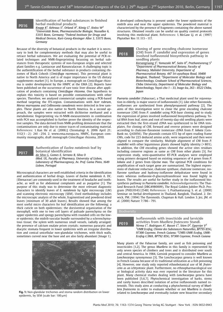
P016Identification of herbal substances in finishedherbal medicinal productsKnapp K1, Kehraus S1, Orland A2, K�nig G1, Kn�ss W2
1Universit�t Bonn, Pharmazeutische Biologie, Nussallee 6,53115 Bonn, Germany; 2Federal Institute for Drugs andMedical Devices, Kurt-Georg-Kiesinger Allee 3, 53175 Bonn,Germany
Because of the diversity of botanical products in the market it is neces-sary to look for complementary methods that may also be useful toretrace herbal substances. We are evaluating the potential of PCR-re-lated techniques and NMR-fingerprinting focussing on herbal sub-stances from therapeutic systems of non-European origin and selectedplant families e.g. Lamiaceae and Ranunculaceae. A challenging topic isthe authentification of the herbal substance in products containing rhi-zomes of Black Cohosh (Cimicifuga racemosa). This perennial plant isnative to North America and is of major importance in the US dietarysupplements market [1]. In Europe, a monograph on Cimicifugae rhizo-ma is under development by the HMPC at the EMA [2]. Reports havebeen published on the occurrence of rare toxic liver disease after appli-cation of products containing Cimicifugae rhizoma. One hypothesis toexplain this toxicity is based on possible contaminations with otherspecies. Therefore, we analysed commercial samples using a PCR-basedmethod targeting the ITS-region. Contaminations with Acer rubrum,Morus murrayana and Collinsonia canadensis were detected in few sam-ples. These plants are also native to North America. In a second ap-proach, the samples were extracted with dichlorine methanol and ametabolomic fingerprinting via H-NMR-measurements in combinationwith PCA was accomplished to further prove the identity of the respec-tive samples. The data derived from NMR-profiling demonstrate that thecontaminations are only due to minor traces of contaminating material.References: 1. Kan He et al. (2006) J Chromatogr A. 2006 April 21;1112(1 – 2): 241 – 254. 2. www.ema.europa.eu, HMPC, European com-munity monographs, draft monograph Cimicifugae rhizoma, 2010.
P017Authentication of Euclea natalensis leaf bybotanical identificationda Silva G, Gomes E, Serrano R, Silva OiMed. UL, Faculty of Pharmacy, University of Lisbon,Laboratory of Pharmacognosy, Av. Prof. Gama Pinto, 1649Lisbon, Portugal
Microscopical characters are well established criteria in the identificationand authentication of herbal drugs. Leaves of Euclea natalensis A. DC.(Ebenaceae) are commonly used in the treatment of headache and tooth-ache, as well as for abdominal complaints and as purgative [1]. Thepurpose of this study was to determine the most relevant diagnosticcharacters to identify leaves of E. natalensis by light microscopy (LM)and scanning electronic microscopy (SEM). Used methodology includesthe microscopic analysis of the whole, fragmentised and powdered driedleaves (minimum of 30 adult leaves). Results showed that among themost useful micro characters for leaf identification are the following: athick cuticle on both epidermises; the dorsiventral organization of themesophyll, with one to two cell layers of palisade parenchyma on theupper epidermis and spongy parenchyma with rounded cells on the low-er epidermis; the midrib vascular bundle surrounded by a sclerenchyma-tous tissue; the xylem with numerous small vessels, radially arranged;the presence of calcium oxalate prism crystals; numerous paracytic anddiacytic stomata frequent in lower epidermis with an irregular distribu-tion and conical unicellular non-glandular trichomes, with thick walls,sometimes curved near the base and are also fairly abundant (Image 1).
Fig. 1: Non-glandular trichomes and stoma random distribution on lowerepidermis, by SEM (scale bar: 100 mm)
A developed collenchyma is present under the lower epidermis of themidrib area and near the upper epidermis. The powdered material ischaracterized by the presence of fragments containing the above namedstructures. Obtained results can be useful on quality control protocolsinvolving this medicinal plant. References: 1. McGaw LJ. et al. (1997)Phytother Res 11:113 – 117.
P018Cloning of gene encoding chalcone isomerase(CHI) from P. candollei and expression of genesinvolved isoflavonoid biosynthesis pathway inseedling plantsKorsangruang S1, Yamazaki M2, Saito K2, Prathanturarug S1
1Department of Pharmaceutical Botany, Faculty ofPharmacy, Mahidol University, Department ofPharmaceutical Botany, 447 Sri-ayuthaya Road, 10400Bangkok, Thailand; 2Department of Molecular Biology andBiotechnology, Graduate School of Pharmaceutical Sciences,Chiba University, Department of Molecular Biology andBiotechnology, Yayoi-cho 1 – 33, Inage-ku, 263 – 8522 Chiba,Japan
Pueraria candollei (Fabaceae), a Thai medicinal plant used for rejuvena-tion in elderly, is major source of isoflavonoids [1]. Like other flavonoids,isoflavones are synthesized from phenylpropanoid pathway [2]. Theaims of this investigation were to clone the gene encoding chalconeisomerase (CHI) from P. candollei using PCR-base cloning and to studythe expression of genes involved isoflavonoid biosynthesis pathway. To-tal RNA from leaf, stem and root of twenty-day-old seedling plants wereextracted then the first-strand cDNA was synthesized to be DNA tem-plates. For cloning of CHI, forward and reverse primers were designedaccording to chalcone-flavanone isomerase cDNA from P. lobata (Gen-Bank no. Q 43056). The plasmids contain 672 bp of open reading frameCHIs, code for 223 deduced amino acids, were sequenced and then werealigned to compare the similarity. Multiple alignment of CHI from P.candollei with other leguminous plants showed highly identity (> 80%).In addition, the CHI encoding genes showed the active sites residuesincluding conserve regions as same as CHI from other plants [3]. Forexpression study, semi-quantitative RT-PCR analyses were employedusing primers designed based on existing sequences of 4 genes from P.lobata and 2 genes from Glycine max. The optimal PCR conditions foramplification of each target gene are summarized. The highest expres-sions of chalcone reductase, chalcone synthase, chalcone isomerase, iso-flavone synthase and hydroxy-isoflavone dehydratase were found inroots whereas isoflavone-O-glucosyltransferase was found highly inleaves. The results are useful for further study in the isoflavonoid bio-synthesis pathway in Pueraria candollei. Acknowledgements: The Thai-land Research Fund (DBG4980009), The Royal Golden Jubilee Ph.D. Pro-gram (PHD/0143/2548) References: 1. Prathanturarug S. et al. (2000)Seminar on herbal development in Thailand, Bangkok, Thailand. 2. De-wick, PM. (1994) The flavonoids. Chapman & Hall. London 3. Jez, JM. etal. (2000) Nature 7:786 – 791.
Biodiversity
P019Isoflavonoids with insecticide and larvicideactivities from Muellera frutescens Standl.Nirma C1, Rodrigues A1, Basset C1, Stien D2, Eparvier V2
1UMR Ecofog, Chimie des Substances Naturelles, BP795 IESG,97300 Cayenne, French Guiana; 2CNRS-UMR Ecofog, UMR-Ecofog-L3MA, BP792 IESG, 97300 Cayenne, French Guiana
Many plants of the Fabaceae family, are used as fish poisoning andinsecticides [1,2]. The genus Muellera in this family is represented byonly seven species of climbers and trees and is distributed over southand central America. In 1984, Geesink proposed to consider Muellera asLonchocarpus synonymous [3]. The Lonchocarpus genera is well knownin French Guiana because of its traditional utilization as a fish poisoning[4]. However, one study only reported ethnobotanical use of M. frutes-cens as an ichtyotoxic plant. Furthermore, no ethnopharmacological useor biological activity data was ever reported in the literature for thisplant. Many chemical studies dealing with Lonchocarpus genera havebeen published [5,6,7]. Phytochemical investigation of barks, stemsand rarely leaves described isolation of active isoflavonoids named ro-tenoids. This study aims at conducting a phytochemical survey of Muel-lera frutescens in order to evaluate whether or not Muellera is closelyrelated to Lonchocarpus and eventually isolate new bioactive secondary
Planta Med 2010; 76: 1163–1374 Georg Thieme Verlag KG Stuttgart · New York · ISSN 0032-0943
11977th Tannin Conference and 58th International Congress of the GA | 29th August – 2nd September 2010, Berlin, Germany
metabolites. Three different extracts (hexane, ethyl acetate and metha-nol) of bark, roots and leaves were prepared and tested on various bio-logical assays. We discovered insecticide and larvicide activities for allextracts, and none of them exhibited cytotoxicity on human cells. Thebioguided fractionation of the most active extract (bark hexane extract)allowed us to isolate eight isoflavonoids, the structures of which wereelucidated by spectroscopic methods. It was found that Muellera is in-deed closely related to Lonchocarpus, therefore corroborating Geesink’sproposal. References: 1. Thasana, N., et al. (2001.) Heterocycles 6, 1121 –1125. 2. Kumar, P. el al. (1989) Phytochem 28, 916 – 918. 3. Geesink, R.(1984) Taxon 33, 742 – 743. 4. Moretti, C., Grenand, P. (1982). J. ethno-pharmacol. 139 – 160. 5. Blatt, C.T.T et al. (2002). Phytotherapy Res. 16,320 – 325. 6. Fang, N et al. (1999) J. Agric. Food Chem. 47, 2130 – 2136. 7.Cabizza, M. et al. (2004) J. Agric. Food Chem. 52, 288 – 293.
P020The FairWild Standard – A fair and sustainabledeal for wild-collected ingredients throughoutthe supply chainDuerbeck K, Staubli F, Ottens BFairWild Foundation, Weststrasse 51, 8570 Weinfelden,Switzerland
The increasing demand for natural products in the sectors of food, cos-metics, wellness and medicinal ingredients poses major ecological andsocial challenges. The high pressure on potentially vulnerable plants canendanger local ecosystems and livelihoods of collectors who often be-long to the poorest social groups in the countries of origin. In order todeal with these challenges, a standard and certification system for sus-tainable wild-collection of plants was developed by different stake-holders and interest groups, leading to the establishment of the FairWildFoundation in 2008. The Foundation is responsible for the maintenanceand implementation of the FairWild standard that consists of soundecological as well as social and fair-trade principles and criteria. Fair-Wild Foundation aims to provide a worldwide framework for imple-menting a sustainable, fair and value-added management and tradingsystem for wild-collected natural ingredients and products thereof. Fair-Wild Foundation informs, advises and assists customers with projects insustainable wild-collection and implementation of the standard. In ad-dition, FairWild Foundation advises on the inclusion of sustainable andfair management principles in conservation strategies, trade policy andother regulations. While encouraging sustainable and fair business prac-tices, the FairWild Standard also focuses on influencing consumerchoice. The FairWild Standard is based on existing traditional knowledgeand appropriate resource management, as well as on Fair Trade princi-ples and ILO Standards. The Standard is geared to a worldwide approach.
P021The effects of salt stress on yield and somecomponents in Chamomile genotypes duringgrowth stageGhanavati M, Kazemi K, Ghaderian RPayame Noor University, (PNU), 19395 – 4679 Tehran, Iran,Islamic Republic Of
One possible management option for growers, dealing with decreases inchamomile production caused by salinity. Our objectives were to inves-tigate the effects of salinity levels on yield and some yield componentsin growth stage of two chamomile spices. Eight genotypes of Matricariarecutita and Matricaria aurea were used in this study. The treatmentsincluded salt levels of control, 6, 12, and 18 dSm-1 in sand culture withnutrient solutions. The salinity stress exerted at 2 durations: The firstfrom seedlings stage and second duration of stress exertion began atstem elongation to harvest. The traits measured plant height, rootlength, the number of leaf per plant, node numbers, and stem freshand dry weight, roots fresh and dry weight. The salt application trialsindicated that dry matter yield was decreased with increasing NaCldoses. The dry matter yields were higher in control than that of the18 dSm-1 NaCl levels. All the criteria investigated suggest therefore thatM. aurea were superior to M. recutita genotypes. Positive and highlysignificant relationships existed between DM yield with PH (0.36**), RL(0.31**), IN (0.14*), LN (0.18**), SFW (0.51**), RFW (0.42**), RDW (0.33**),and RRW (0.23**). Path analysis showed that plant height (36.7%), Rootfresh weight (27.0%), and stem fresh weight (16.5%), had strong positivedirect effect, in that order internode number (15.2%), stem relative water(23.6%) and root dry weight (14.3%) had strong negative direct effect.There was a significant difference between genotypes studied for alltraits except for root relative water content.
P022Chemodiversity of Polynesian liverwortsLudwiczuk A1, Raharivelomanana P2, Komala I3, Pham A2,Bianchini J2, Asakawa Y3
1Medical University Of Lublin, Pharmacognosy WithMedicinal Plants Unit, 1 Chodzki Str., 20 – 093 Lublin,Poland; 2University Of French Polynesia, Bpn� 6570 Faaa/Tahiti, 98702 Faaa, French Polynesia; 3Tokushima BunriUniversity, Faculty Of Pharmaceutical Sciences, 180Yamashiro-Cho, 770 – 8514 Tokushima, Japan
Liverworts are known as rich sources of secondary metabolites, whichvolatile constituents such as terpenoids belong to a wide variety ofcarbon skeletons. These metabolites can be used as chemosystematicsmarkers and are very helpful for taxonomic significance because liver-worts are very small plants which morphological classification is extre-mely difficult. Aiming to assess the diversity of Polynesian liverworts,chemical analysis (GC-MS, 1D and 2D 1H and 13C NMR) of volatile con-stituents of six species (Trichocolea pluma, Chandonanthus hirtellus, Mas-tigophora diclados, Jungermania sp., Plagiochila sp. and Cyathodium foe-tidissimum) collected in French Polynesia had been performed. All theinvestigated species are chemically different, each species biosynthe-sized own peculiar compounds and some of them are biomarkers ofthe liverworts species. T. pluma biosynthesized characteristic isoprenylphenyl ethers, herbetane-type sesquiterpenoids for M. diclados, cem-brane-type diterpenoid are peculiar for C. hirtellus and fusiccocane-typefor Plagiochila sp. Interesting chemical constituents had been also inthese Polynesian liverworts such as: vanillic acid methyl ester firstlyreported to occur from the Marchiantophyta (T. pluma), skatol producedby C. foetidissimum, (E)-ectocarpene and dictyotene previously found inmarine algae and now detected in C. hirtellus. These relevant findingsexpress part of the biodiversity specificity of Polynesian bryophytes [1].Acknowledgements: This work was supported in part by Grant-in-aidfor Open Research (A.L.) from the Ministry of Education, Culture, Sports,Science and Technology, Japan. References: 1. Ludwiczuk, A. et al(2009), Nat. Prod. Com.10: 1387 – 1392.
P023Chemical composition – biological activities ofselected samples of propolis from South GreeceGraikou K1, Chinou I1, Gortzi O2, Lalas S2
1Unversity of Athens, School of Pharmacy, Lab ofPharmacognosy and Chemistry of Natural Products,University campus of Zografou, 15771 Athens, Greece;2Technological institute of Larisa (Annex karditsa), Dept ofFood Technology, Karditsa, 43100 Karditsa, Greece
Propolis (bee glue) is a well known natural product with healing proper-ties. The chemical composition of propolis is highly variable and de-pends mainly on the local flora. Greece is characterized by high biodi-versity flora, assuming different propolis composition. In the frameworkof our continuing work on propolis samples from Greece and all aroundthe world, four propolis samples from Peloponnese and one from theisland of Kos were analyzed by GC/MS after silylation. In these samplesthe diterpenes: totarol, totarolone, abietic acid, imbricataloic acid, iso-agatholal, agathadiol, communic acid, junicedric acid, 13-epi-cupressicacid and isocupressic acid have been identified as major constituentswhile the characteristic propolis flavonoids were either in smallamounts or even absent. These findings confirm the presence of conifertrees as propolis plant sources in Greek samples. Diterpenes have beenpreviously found in propolis samples from North-West Greece [2], Creteand Italy (Sicily) [3] while propolis from the temperate zone havingPopulus spp. as plant sources [1] have large amounts of esters of phe-nolic acids and flavonoids. All the samples showed significant antibac-terial activity against nine Gram-negative and -positive human patho-genic bacteria and three fungi, probably due to the large amounts ofditerpenes, while the antioxidative activity of all samples has been as-sayed as well thorough Rancimat method. References: 1. Bankova, V., etal. (2000) Apidologie 31:3 – 15. 2. Melliou, E., Chinou, I. (2004) PlantaMedica 70:1 – 5. 3. Popova M. et al. (2010) J. Agric. Food Chem. 58:3167 – 3176.
Planta Med 2010; 76: 1163–1374 Georg Thieme Verlag KG Stuttgart · New York · ISSN 0032-0943
1198 7th Tannin Conference and 58th International Congress of the GA | 29th August – 2nd September 2010, Berlin, Germany
P024Expression of 3b-HSD and P5bR, genes coding forD5 – 3b-hydroxysteroid dehydrogenase (3b-HSD)and progesterone 5b-reductase (P5bR), in leavesand cell cultures of Digitalis lanata EHRH.Ernst M1, Herl V2, Schr�der S1, M�ller-Uri F1, Kreis W1
1Pharmazeutische Biologie, Biologie, Staudtstraße 5, 91058Erlangen, Germany; 2Tier�rtzliches Institut, Burckardtweg 2,37077 G�ttingen, Germany
Plants of the genus Digitalis produce cardiac glycosides that are used inthe therapy of cardiac insufficiency in humans [1]. 3b-HSD and P5bR areboth supposed to be important enzymes in the biosynthesis of theseproducts [2, 3, 4]. Enzyme activity and expression of the respectivegenes encoding 3b-HSD and P5bR were demonstrated in cardenolide-accumulating leaves of Digitalis lanata but also in cardenolide-free per-manent cell suspension cultures (K3OHD) initiated from D. lanataleaves. P5bR activity was 3.7 times higher in D. lanata leaves (7.8mkat/kg protein) than in K3OHD cells (2.1 mkat/kg protein) (analysed by GC-MS). 3b-HSD activity in D. lanata leaves and K3OHD cells was detectedby HPLC and it was found that the activity is about in the same range inleaves (13.9mkat/kg protein) and in suspension-cultured cells (11.3mkat/kg protein). Expression of the respective genes, namely AY585867.1(P5bR gene) and DQ 466890.1 (3b-HSD gene), was demonstrated byreal-time qPCR analysis. The expression of the 3b-HSD gene in leaves(8.7 mRNA level) is nearly twice as high as in K3OHD cells (4.9 mRNAlevel). Analyses revealed that P5bR is most strongly expressed in D.lanata leaves (42 mRNA level). The relative mRNA level was about 3.2times higher than in suspension cells (13.3 mRNA level) which nicelyfits to the activity data. Since a new, inducible progesterone 5b-reduc-tase was identified recently [5] the expression of the respective gene,P5bR2, in K3OHD cells and D. lanata leaves is now under examinationand the new results will be presented and discussed. References:1. Hoppe, UC et al. (2005) Z Kardiol 94:488 – 509. 2. Kreis, W. et al.(1998) Planta Med 64:491 – 499. 3. Kreis W, M�ller-Uri F (2009) AnnRev Plant 40:304 – 363. 4. G�rtner, DE. Et al. (1990) FEBS Lett 271:239 –242. 5. Pedro Per�z-Bermffldez et al. (2010) New Phytol 185:687 – 700.
P025Evolution and function of progesterone5b-reductase genes in angiospermsMunkert J, Bauer P, Brydziun M, M�ller-Uri F, Kreis WPharmazeutische Biologie, Biologie, Staudtstraße 5, 91058Erlangen, Germany
About 60 genera of the angiosperms have been described to contain 5b-cardenolides [1, 2, 3]. They occur in monocots, basal eudicots, rosids andasterids. The unpredictable occurrence of 5b-cardenolides in the angios-perms raises the question whether this trait has evolved only once orseveral times during evolution. The analysis of genes suggested to beinvolved in cardenolide biosynthesis may help to address this question.Progesterone 5b-reductases (P5bR, P5bR2) are thought to catalyse a stepin the 5b-cardenolide biosynthesis, namely the conversion of progester-one to 5b-pregnane-3,20-dione. Therefore we isolated 11 new P5bRsorthologues from several 5b-cardenolide-free and 5b-cardenolide-pro-ducing plant species. All sequences were analysed in silico and wereshown to be highly conserved. They contain certain motifs that qualifythem as members of a class of stereo-selective enone reductases. Phy-logenetic analysis shows that in the angiosperms P5bRs form two sepa-rate clusters. The cladogramme generated from protein sequencesshowed that one cluster (Cluster II) of P5bRs correlated nicely with theassumed phylogenetic relationship of the species in the cluster. Thisimplied that these genes have evolved from a common ancestral gene.A second cluster contained p5br-genes of Populus, Vitis and D. purpurea.All three genera also contained paralogues in cluster II. In this grouponly the D. purpurea P5bR2 was recently described to encode a func-tional P5bR that can be induced by stress [4]. The occurrence of theseclosely related functional P5bRs in 5b-cardenolide-free and 5b-cardeno-lide-containig plants let us presume that cluster II P5bRs are involved inmore than just 5b-cardenolide biosynthesis. References: 1. Singh, B.,Rastogi, R.P. (1970), Phytochem. 9: 315 – 331. 2. Meleto, C.P., Medarde,M., San Feliciano, A. (2000), Molecules 5: 51 – 81. 3. Kreis, W., M�ller-Uri,F. (2010), Biochemistry of sterols, cardiac glycosides, brassinosteroids,phytoecdysteroids and steroid saponins. In: Wink M. (Ed.) Biochemistryof Plant Secondary Metabolism. Volume 40, 2nd Ed. 304 – 363, CRC Press,Sheffield. 4. Perez-Bermudez, P. et al. (2010), New Phytol. 185: 687 – 700.
P026Variation in the essential oil content andcomposition of a Finnish oregano collectionHolm Y1, Galambosi B2, Hiltunen R1
1Faculty of Pharmacy, Division of Pharmaceutical Biology,P.O. Box 56, 00014 University of Helsinki, Finland; 2MTTAgrifood Research Finland, Ecological Production,L�nnrotinkatu 5, 50100 Mikkeli, Finland
Fifteen oregano samples (Origanum vulgare L.) from eleven differentlocations in southern Finland plus one commercial Greek oregano (Or-iganum viride ssp. viride) were cultivated in Mikkeli, south-eastern Fin-land in 2008 and 2009. The plants were harvested at flowering time inAugust 2009, dried and stored in paper bags. The plant material wassubmitted to hydrodistillation in a Karlsruher-Stahl apparatus for twohours in order to isolate the oil and determine the oil content. The oilcontent varied from 0.2 to 0.8% in the Finnish oreganos and was 3.7% inthe Greek oregano. The oils were analysed by GC-MS using an unpolarRTX-1 and a polar Stabilwax column (30 m x 0.20 mm i.d. Restek Cor-poration, CA, USA). The identification was based on retention indicesand comparison with library spectra. Altogether 47 components wereidentified and the identified constituents represented 64.9 – 96.9% ofthe total oil. The dominant constituents were sabinene (up to 17%), g-terpinene (up to 12%), b-caryophyllene (up to 20%), germacrene-D (up to17%), caryophyllene oxide (up to 23%) and carvacrol (up to 85%). Acluster analysis of the essential oil compositions classsifed the oils intofive groups: 1) b-caryophyllene/germacrene-D, 2) caryophyllene oxide,3) carvacrol, 4) b-caryophyllene and 5) sabinene/germacrene-D type.Only the Greek oregano was a carvacrol type. Great variations in oreganooils of Mediterranean origin have been reported (1, 2), but not in orega-no of Finnish origin. References: 1. Figueredo, G et al. (2006) Flav Fragr J21: 134 – 139. 2. De Martino, L. et al. (2009) Molecules 14: 2735 – 2746.
P027Changes in distribution and structure of wildOriganum vulgare L. (Lamiaceae) populationsduring the last decade in Armenia andimplications for conservationAbrahamyan A1, Crockett S2
1Armenian State Agrarian University, Horticultural, AraratRegion, Surenavan village, Street Shahumyan, House 54, 009Ararat, Armenia; 2Karl-Franzens-University Graz,Pharmacognosy, Street Universitaetsplatz, house 4, 8010Graz, Austria
Armenia has a rich flora of ca. 3600 plant species (ca. 50% of the entireCaucasian flora), distributed across (semi)desert, steppe, forest and al-pine landscapes. Native plant biodiversity and conservation status ofthese species, particularly those with economic value, needs furtherassessment [1]. Anthropogenic threats to biodiversity (overpopulation,deforestation and urbanization) have simultaneously hindered researchand increased the need for it. Of the ca. 500 species with medicinal/economic use records, ca. 50 are used in folk medicine, including bothwild-collected (e.g. Crataegus sp., Hypericum perforatum) and culti-vated (e. g. Chamomilla recutita, Mentha piperita) species [2]. Only lim-ited information, however, on genetic biodiversity, population structure,and conservation status of these species is available. From 2006 – 2009,field studies were conducted to re-locate wild Origanum vulgare L. po-pulations based on historical records, and discover new populations. Thegrowth, phenological and habitat characteristics, population size andlocation (GPS mapping), were assessed. Historical records indicated thatthis species occurred widely in the central/northern regions, but nearlyhalf the populations had vanished. Remaining populations diminished insize, plant number and experienced fragmentation during the studyperiod. 3 new populations were located in the south/south-eastern re-gions, indicating that the abundance and distributional range is expand-ing here. Anthropogenic threats included: poor land management, in-creasing population pressure, and excessive collection of plants. Thisresearch provided baseline data for the development of ex situ and invitro strategies to conserve unique genotypes, and assess the sustain-ability of wild populations according to IUCN Red Book Criteria, of thisimportant species in Armenia. References: 1. IUCN, WHO, WWF (1993).Guidelines on the Conservation of Medicinal Plants, IUCN, Gland, Swit-zerland, 50 p. 2. Fayvush, G., Danielyan T., Nalbandyan A. (2004) Arme-nia as a producer of medicinal plants: possibilities and perspectives.Available online (accessed 12 April 2010): http://www.nature ic.am/NCSA/Publication/Medical_Plants_eng.pdf.
Planta Med 2010; 76: 1163–1374 Georg Thieme Verlag KG Stuttgart · New York · ISSN 0032-0943
11997th Tannin Conference and 58th International Congress of the GA | 29th August – 2nd September 2010, Berlin, Germany
P028The essential oil composition of some Lavandulaspecies grown in RomaniaMartinou E1, Hancianu M2, Robu S2, Tzakou O1
1School of Pharmacy, University of Athens, Department ofPharmacognosy and Chemistry of Natural Products,Panepistimioupoli Zographou, 15771 Athens, Greece; 2Gr. T.Popa University of Medicine and Pharmacy, Pharmacognosy,Faculty of Pharmacy, Universitatii No.16, 700115 Iasi,Romania
The genus Lavandula belongs to the family of Lamiaceae and consists of30 species [1]. They are aromatic evergreen shrubs widely distributed.Parts used are the flowering tops from which the essential oil is ob-tained [2,3]. The most well known is L. angustifolia (true lavender)which preparations are traditionally used to treat symptoms of neuro-tonic disorders [3]. Lavender oil has been used empirically since ancienttimes for its antiseptic properties and today is used in pharmaceuticalsand cosmetic products [1,2]. Five different Lavandula species, subspe-cies, cultivars or hybrids harvested from the Botanical Gardens fromGalati, Romania, were investigated for their essential oil composition.The species were: Lavandula angustifolia Mill., L. angustifolia subsp.angustifolia, L. angustifolia subsp. pyrenaica (DC.) Guinea, L. angustifoliacv. Munstead, L. hybrida Rev. (L. angustifolia Mill. x L. latifolia Medik.).Essential oils were obtained from dried inflorescences by hydrodistilla-tion and analysed by means of GC/FID and GC/MS. The identification ofthe volatile compounds was based on comparison of their retentionindices (RI), and mass spectra with those obtained from authentic sam-ples and/or NIST/NBS, Wiley libraries and literature. Linalool (20.0 –36.0%) and linalyl acetate (12.9 – 22.5%) were the main components inall analysed samples. References: 1. Mabberley, D.J. 1997. The Plant-Book. Cambridge University Press, Cambridge. 2. Leung A.Y., Foster S.1996. Encyclopaedia of Common Natural Ingredients Used in Food,Drugs, and Cosmetics, 2nd ed., John Wiley & Sons Inc, New York. 3.Blumenthal M. 2000. Herbal Medicine. Expanded Commission E Mono-graphs. American Botanical Council, Austin.
P029In vitro shoot cultures of endemic Cyclopiagenistoides (Honeybush) as a source of valuablepolyphenolic compoundsLuczkiewicz M1, Kokotkiewicz A1, Hering A2, Gorynski K3,Bucinski A3, Ochocka R1
1Department of Pharmacognosy, Medical University ofGdansk, Pharmacognosy with Medicinal Plant Garden, Al.gen. J. Hallera107, 80 – 416 Gdansk, Poland; 2Department ofBiology and Pharmaceutical Botany, Medical University ofGdansk, Biology and Pharmaceutical Botany, Al. gen. J.Hallera 107, 80 – 416 Gdansk, Poland; 3Department ofBiopharmacy, Nicolaus Copernicus University in Torun,Collegium Medicum in Bydgoszcz, Biopharmacy,Sklodowskiej-Curie st 9, 85 – 095 Bydgoszcz, Poland
The plants from Cyclopia genus (Honeybush) are endemic, South-Africanshrubs characteristic for the fynbos plant formation of the Cape FloristicRegion. Aerial parts of several Cyclopia species, including C. genistoides,are used to manufacture the honeybush herbal tea, recognized by itsdinstictive sweet, honey-like aroma, low level of tannins and lack ofcaffeine [1,2]. Extracts obtained from Cyclopia plants are rich in poly-phenols, including xanthones (mangiferin and isomangiferin), flava-nones (hesperidin), flavones and isoflavones, and as such exhibit sub-stantial antioxidative and antimutagenic activity [1,2,3]. In vitro propa-gation protocol for C. genistoides has been established. Solid Schenk-Hildebrandt (SH) medium supplemented with 2.0 mg l-1 6-(g,g-dimethy-lallylamino)purine (2iP) and 0.22 mg l-1 thidiazuron (TDZ) have beenshown to be the best for shoot culture initiation, as well as for thedevelopment of high number of microshoots. Shoot elongation proce-dure involved explant cultivation on the full strength SH medium sup-plemented with 1.0 mg l-1 indole-3-butyric acid (IBA), followed by 30-day long growth on SH medium with reduced sucrose level, supplemen-ted with the same amount of IBA. As a result, elongated shoots repre-senting intact plant morphology were obtained. Root induction wasachieved on solid SH medium with reduced sucrose and nitrate concen-trations, supplemented with 5.0 mg l-1 indole-3-acetic acid (IAA) and50.0 mg l-1 citric acid. The regenerated plants were acclimatized in theglasshouse, with the use of peat/gravel/perlite subtrate (1:1:1). Bothregenerated plants and in vitro microshoots of C. genistoides were eval-uated for the production of selected polyphenolic compounds (LC-ESI-MS and LC-DAD analysis). Acknowledgements: the study was sup-
ported by grant no N N302 041936 from Polish Ministry of Educationand Science. References: 1. Joubert E. et al. (2008) J. Ethnopharmacol.19:376 – 412. 2. Kokotkiewicz A., Luczkiewicz M. (2009) Fitoterapia80:3 – 11. 3. Joubert E. et al. (2008) J. Agric. Food Chem. 56:954 – 963.
P030Genetic analyses of the endangered medicinalplant Podophyllum emodiYu Y1, Yan A1, Xue H2
1Beijing Institute of Radiation Medicine, Beijing Institute ofRadiation Medicine, 27 Taiping Road, 100850 Beijing, China,100850 Beijing, China; 2Bruker Daltonics Incorporation,Beijing Deputy Office, Evenbright International TrustMansion, Suite 3102, 11 Zhong Guan Cun South Avenue,100081 Beijing, China
The medicinal use of Podophyllum emodi Wall (Himalayan Mayapple;family: Berberideceae), a high-altitude plant species native to the alpineand sub alpine areas of Himalayas, dates back to ancient times [1]. Theplant has been used in traditional Chinese System of Medicine for treat-ment of a number of ailments. To determine the population structureand outcrossing rate across the range of the species, we conducted AFLPanalysis using four primer combinations for 31 populations. The geneticdiversity of this species was high based on the level of polymorphic loci(155 of 197 loci; 92.31%) and Nei’s gene diversity (ranging from 0.14532to 0.2435; overall 0.2446). There was significant population genetic dif-ferentiation (G(ST)= 0.287; circle minus (II)= 0.221 from the Bayesianf = 0 model). Results from the AMOVA analysis suggest that a majorityof the genetic variance is attributed to variation within populations(69.43%), which is also evident from the PCoA. An estimate of the out-crossing rate based on genotypes of progenies from seven of the 31 po-pulations using the multilocus method from the program MLTR rangedfrom 0.695 to 0.903, suggesting that the species is predominantly out-crossing. These results are encouraging for conservation, signifying thatpopulations may persist due to continued genetic exchange sustained bythe outcrossing mating system of the species. Acknowledgements: Thisresearch was supported by the Natural Science Foundation of China(NSFC 30770153). References: 1. Chawla R et al. (2005) Mol Cell Bio-chem 273: 193 – 208.
P031Searching for plant-derived natural products byin vitro cultivationSchumann A, Claus D, Gerth ABioPlanta GmbH, Deutscher Platz 5, 04103 Leipzig, Germany
Modern cancer therapy uses more and more natural plant compoundsfor the development of new therapeutics during the last years. Some ofthem are rather approachable by the use of bioreactor and/or cell cul-ture techniques than by green house cultivation or wild collection. Morethan 200 different species of herbs and rare medicinal plants with tradi-tional use were taken into in vitro culture. In bioreactors with fullyautomated nutrient and gas exchange root, shoot and cell cultures werecultivated under different growth conditions. About 500 extracts wereanalysed by Analyticon’s LC/MS-based Chemodiversity Profiling Plat-form. Combining bioreactor technology with high throughput screeningmethods high productive plants were selected according to their bio-mass production and content of active compounds. First results havebeen shown that in vitro cell and organ cultures can produce quantita-tively more active compounds than plants of wild collection or greenhouse culture. Compared to this, 20% of the bioreactor material showeda new chemical pattern of natural compounds. In addition, spectrum ofsecondary metabolites varies at different in vitro culture types. The mostpromising in vitro cultures were selected for scale-up in 5 to 10 l bior-eactors to optimise the production of preferable compounds. Fractiona-tion of selected extracts and structure elucidation demonstrate that invitro cultures are able to produce secondary metabolites with the samestructural diversity as traditionally grown plants. Therefore, in vitro cul-tivated plants combined with the screening platform technology are anattractive source for identification of novel drugs and providing naturalplant compounds as pharmaceutical products.
Planta Med 2010; 76: 1163–1374 Georg Thieme Verlag KG Stuttgart · New York · ISSN 0032-0943
1200 7th Tannin Conference and 58th International Congress of the GA | 29th August – 2nd September 2010, Berlin, Germany
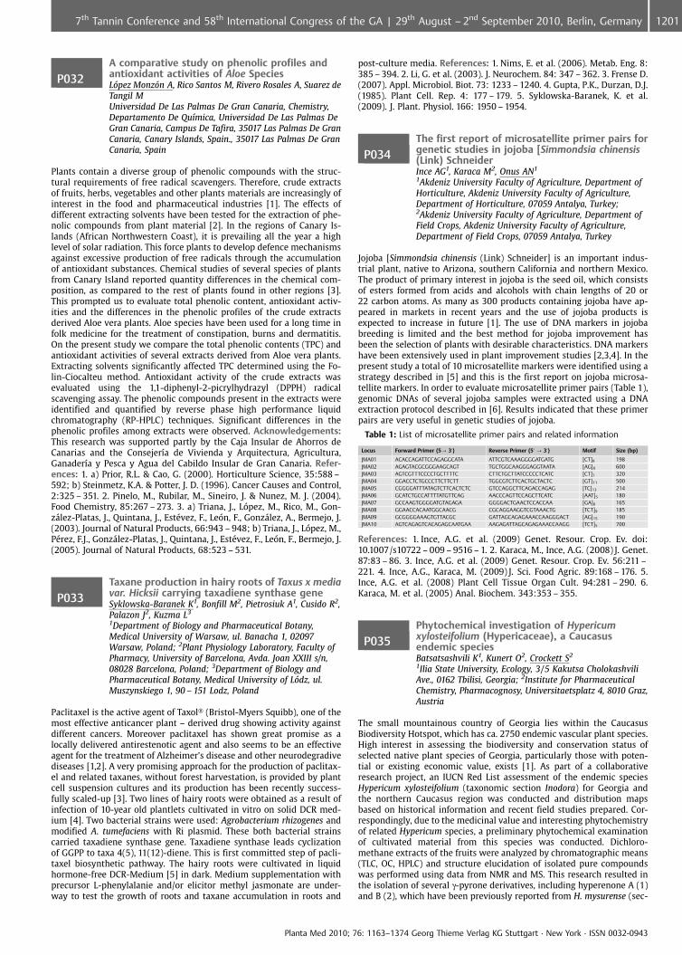
P032A comparative study on phenolic profiles andantioxidant activities of Aloe SpeciesL�pez Monz�n A, Rico Santos M, Rivero Rosales A, Suarez deTangil MUniversidad De Las Palmas De Gran Canaria, Chemistry,Departamento De Qu�mica, Universidad De Las Palmas DeGran Canaria, Campus De Tafira, 35017 Las Palmas De GranCanaria, Canary Islands, Spain., 35017 Las Palmas De GranCanaria, Spain
Plants contain a diverse group of phenolic compounds with the struc-tural requirements of free radical scavengers. Therefore, crude extractsof fruits, herbs, vegetables and other plants materials are increasingly ofinterest in the food and pharmaceutical industries [1]. The effects ofdifferent extracting solvents have been tested for the extraction of phe-nolic compounds from plant material [2]. In the regions of Canary Is-lands (African Northwestern Coast), it is prevailing all the year a highlevel of solar radiation. This force plants to develop defence mechanismsagainst excessive production of free radicals through the accumulationof antioxidant substances. Chemical studies of several species of plantsfrom Canary Island reported quantity differences in the chemical com-position, as compared to the rest of plants found in other regions [3].This prompted us to evaluate total phenolic content, antioxidant activ-ities and the differences in the phenolic profiles of the crude extractsderived Aloe vera plants. Aloe species have been used for a long time infolk medicine for the treatment of constipation, burns and dermatitis.On the present study we compare the total phenolic contents (TPC) andantioxidant activities of several extracts derived from Aloe vera plants.Extracting solvents significantly affected TPC determined using the Fo-lin-Ciocalteu method. Antioxidant activity of the crude extracts wasevaluated using the 1,1-diphenyl-2-picrylhydrazyl (DPPH) radicalscavenging assay. The phenolic compounds present in the extracts wereidentified and quantified by reverse phase high performance liquidchromatography (RP-HPLC) techniques. Significant differences in thephenolic profiles among extracts were observed. Acknowledgements:This research was supported partly by the Caja Insular de Ahorros deCanarias and the Consejer�a de Vivienda y Arquitectura, Agricultura,Ganader�a y Pesca y Agua del Cabildo Insular de Gran Canaria. Refer-ences: 1. a) Prior, R.L. & Cao, G. (2000). Horticulture Science, 35:588 –592; b) Steinmetz, K.A. & Potter, J. D. (1996). Cancer Causes and Control,2:325 – 351. 2. Pinelo, M., Rubilar, M., Sineiro, J. & Nunez, M. J. (2004).Food Chemistry, 85:267 – 273. 3. a) Triana, J., L pez, M., Rico, M., Gon-z�lez-Platas, J., Quintana, J., Est�vez, F., Le n, F., Gonz�lez, A., Bermejo, J.(2003). Journal of Natural Products, 66:943 – 948; b) Triana, J., L pez, M.,P�rez, F.J., Gonz�lez-Platas, J., Quintana, J., Est�vez, F., Le n, F., Bermejo, J.(2005). Journal of Natural Products, 68:523 – 531.
P033Taxane production in hairy roots of Taxus x mediavar. Hicksii carrying taxadiene synthase geneSyklowska-Baranek K1, Bonfill M2, Pietrosiuk A1, Cusido R2,Palazon J2, Kuzma L3
1Department of Biology and Pharmaceutical Botany,Medical University of Warsaw, ul. Banacha 1, 02097Warsaw, Poland; 2Plant Physiology Laboratory, Faculty ofPharmacy, University of Barcelona, Avda. Joan XXIII s/n,08028 Barcelona, Poland; 3Department of Biology andPharmaceutical Botany, Medical University of L�dz, ul.Muszynskiego 1, 90 – 151 Lodz, Poland
Paclitaxel is the active agent of Taxol� (Bristol-Myers Squibb), one of themost effective anticancer plant – derived drug showing activity againstdifferent cancers. Moreover paclitaxel has shown great promise as alocally delivered antirestenotic agent and also seems to be an effectiveagent for the treatment of Alzheimer’s disease and other neurodegradivediseases [1,2]. A very promising approach for the production of paclitax-el and related taxanes, without forest harvestation, is provided by plantcell suspension cultures and its production has been recently success-fully scaled-up [3]. Two lines of hairy roots were obtained as a result ofinfection of 10-year old plantlets cultivated in vitro on solid DCR med-ium [4]. Two bacterial strains were used: Agrobacterium rhizogenes andmodified A. tumefaciens with Ri plasmid. These both bacterial strainscarried taxadiene synthase gene. Taxadiene synthase leads cyclizationof GGPP to taxa 4(5), 11(12)-diene. This is first committed step of pacli-taxel biosynthetic pathway. The hairy roots were cultivated in liquidhormone-free DCR-Medium [5] in dark. Medium supplementation withprecursor L-phenylalanie and/or elicitor methyl jasmonate are under-way to test the growth of roots and taxane accumulation in roots and
post-culture media. References: 1. Nims, E. et al. (2006). Metab. Eng. 8:385 – 394. 2. Li, G. et al. (2003). J. Neurochem. 84: 347 – 362. 3. Frense D.(2007). Appl. Microbiol. Biot. 73: 1233 – 1240. 4. Gupta, P.K., Durzan, D.J.(1985). Plant Cell. Rep. 4: 177 – 179. 5. Syklowska-Baranek, K. et al.(2009). J. Plant. Physiol. 166: 1950 – 1954.
P034The first report of microsatellite primer pairs forgenetic studies in jojoba [Simmondsia chinensis(Link) SchneiderInce AG1, Karaca M2, Onus AN1
1Akdeniz University Faculty of Agriculture, Department ofHorticulture, Akdeniz University Faculty of Agriculture,Department of Horticulture, 07059 Antalya, Turkey;2Akdeniz University Faculty of Agriculture, Department ofField Crops, Akdeniz University Faculty of Agriculture,Department of Field Crops, 07059 Antalya, Turkey
Jojoba [Simmondsia chinensis (Link) Schneider] is an important indus-trial plant, native to Arizona, southern California and northern Mexico.The product of primary interest in jojoba is the seed oil, which consistsof esters formed from acids and alcohols with chain lengths of 20 or22 carbon atoms. As many as 300 products containing jojoba have ap-peared in markets in recent years and the use of jojoba products isexpected to increase in future [1]. The use of DNA markers in jojobabreeding is limited and the best method for jojoba improvement hasbeen the selection of plants with desirable characteristics. DNA markershave been extensively used in plant improvement studies [2,3,4]. In thepresent study a total of 10 microsatellite markers were identified using astrategy described in [5] and this is the first report on jojoba microsa-tellite markers. In order to evaluate microsatellite primer pairs (Table 1),genomic DNAs of several jojoba samples were extracted using a DNAextraction protocol described in [6]. Results indicated that these primerpairs are very useful in genetic studies of jojoba.
Table 1: List of microsatellite primer pairs and related information
Locus Forward Primer (5? 3’) Reverse Primer (5’ ? 3’) Motif Size (bp)
JMA01 ACACCAGATTCCAGAGGCATA ATTCGTCAAAGGGGATGATG [CT]8 198JMA02 AGAGTACGCGGGAAGCAGT TGCTGGCAAGGGAGGTAATA [AG]8 600JMA03 AGTCGTTTCCCCTGCTTTTC CTTCTGCTTATCCCCCTCATC [CT]7 320JMA04 GGACCTCTGCCCTTCTTCTT TGGCGTCTTCACTGCTACTC [GT]11 500JMA05 CGGGGATTTATAGTCTTCACTCTC GTCCAGGCTTCAGACCAGAG [TC]13 214JMA06 GCATCTGCCATTTTATGTTCAG AACCCAGTTCCAGCTTCATC [AAT]5 180JMA07 GCCAAGTGGGGATGTAGAGA GGGGACTGAACTCCACCAA [GA]8 165JMA08 GGAACCACAATGGCAACG CGCAGGAAGGTCGTAAACTG [TCT]9 185JMA09 GCGGGGAAAGTGTTACGC GATTAGCAGAGAAACCAAGGGACT [AG]15 190JMA10 AGTCAGAGTCACAGAGCAATGAA AAGAGATTAGCAGAGAAACCAAGG [TCT]5 700
References: 1. Ince, A.G. et al. (2009) Genet. Resour. Crop. Ev. doi:10.1007/s10722 – 009 – 9516 – 1. 2. Karaca, M., Ince, A.G. (2008) J. Genet.87:83 – 86. 3. Ince, A.G. et al. (2009) Genet. Resour. Crop. Ev. 56:211 –221. 4. Ince, A.G., Karaca, M. (2009) J. Sci. Food Agric. 89:168 – 176. 5.Ince, A.G. et al. (2008) Plant Cell Tissue Organ Cult. 94:281 – 290. 6.Karaca, M. et al. (2005) Anal. Biochem. 343:353 – 355.
P035Phytochemical investigation of Hypericumxylosteifolium (Hypericaceae), a Caucasusendemic speciesBatsatsashvili K1, Kunert O2, Crockett S2
1Ilia State University, Ecology, 3/5 Kakutsa CholokashviliAve., 0162 Tbilisi, Georgia; 2Institute for PharmaceuticalChemistry, Pharmacognosy, Universitaetsplatz 4, 8010 Graz,Austria
The small mountainous country of Georgia lies within the CaucasusBiodiversity Hotspot, which has ca. 2750 endemic vascular plant species.High interest in assessing the biodiversity and conservation status ofselected native plant species of Georgia, particularly those with poten-tial or existing economic value, exists [1]. As part of a collaborativeresearch project, an IUCN Red List assessment of the endemic speciesHypericum xylosteifolium (taxonomic section Inodora) for Georgia andthe northern Caucasus region was conducted and distribution mapsbased on historical information and recent field studies prepared. Cor-respondingly, due to the medicinal value and interesting phytochemistryof related Hypericum species, a preliminary phytochemical examinationof cultivated material from this species was conducted. Dichloro-methane extracts of the fruits were analyzed by chromatographic means(TLC, OC, HPLC) and structure elucidation of isolated pure compoundswas performed using data from NMR and MS. This research resulted inthe isolation of several g-pyrone derivatives, including hyperenone A (1)and B (2), which have been previously reported from H. mysurense (sec-
Planta Med 2010; 76: 1163–1374 Georg Thieme Verlag KG Stuttgart · New York · ISSN 0032-0943
12017th Tannin Conference and 58th International Congress of the GA | 29th August – 2nd September 2010, Berlin, Germany
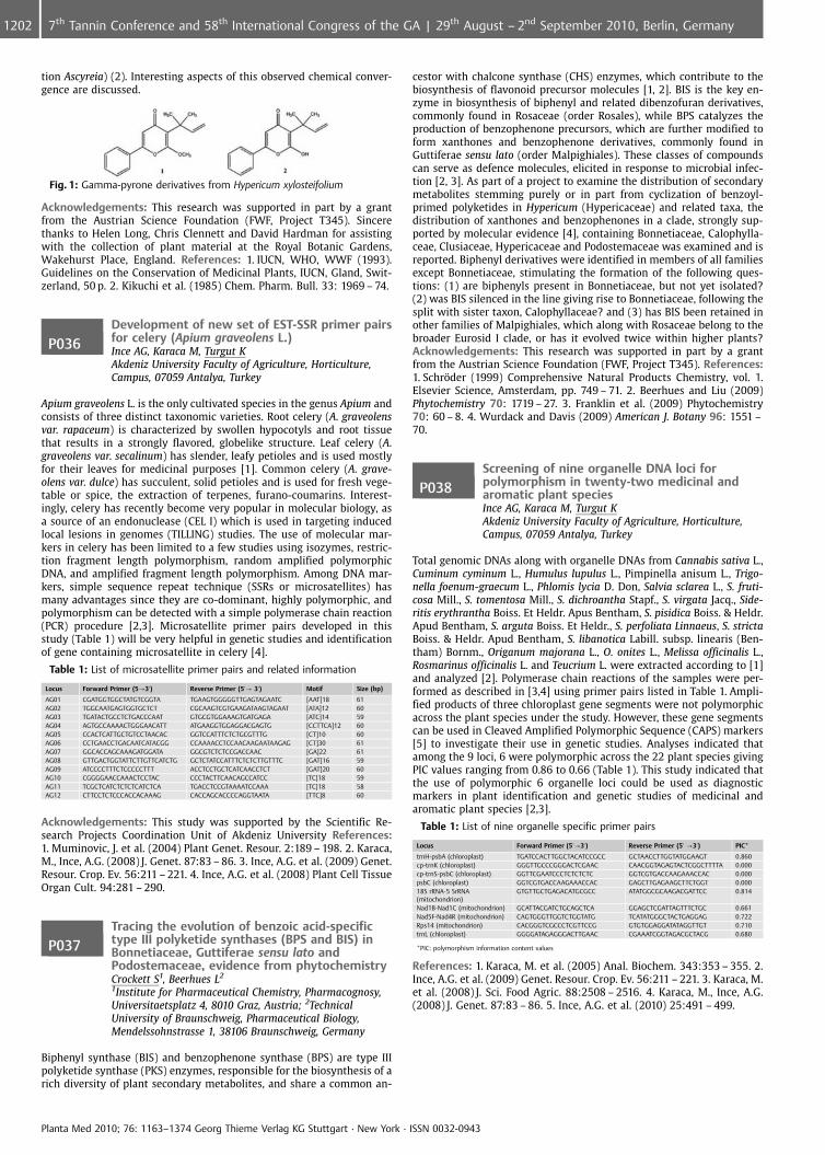
tion Ascyreia) (2). Interesting aspects of this observed chemical conver-gence are discussed.
Fig. 1: Gamma-pyrone derivatives from Hypericum xylosteifolium
Acknowledgements: This research was supported in part by a grantfrom the Austrian Science Foundation (FWF, Project T345). Sincerethanks to Helen Long, Chris Clennett and David Hardman for assistingwith the collection of plant material at the Royal Botanic Gardens,Wakehurst Place, England. References: 1. IUCN, WHO, WWF (1993).Guidelines on the Conservation of Medicinal Plants, IUCN, Gland, Swit-zerland, 50 p. 2. Kikuchi et al. (1985) Chem. Pharm. Bull. 33: 1969 – 74.
P036Development of new set of EST-SSR primer pairsfor celery (Apium graveolens L.)Ince AG, Karaca M, Turgut KAkdeniz University Faculty of Agriculture, Horticulture,Campus, 07059 Antalya, Turkey
Apium graveolens L. is the only cultivated species in the genus Apium andconsists of three distinct taxonomic varieties. Root celery (A. graveolensvar. rapaceum) is characterized by swollen hypocotyls and root tissuethat results in a strongly flavored, globelike structure. Leaf celery (A.graveolens var. secalinum) has slender, leafy petioles and is used mostlyfor their leaves for medicinal purposes [1]. Common celery (A. grave-olens var. dulce) has succulent, solid petioles and is used for fresh vege-table or spice, the extraction of terpenes, furano-coumarins. Interest-ingly, celery has recently become very popular in molecular biology, asa source of an endonuclease (CEL I) which is used in targeting inducedlocal lesions in genomes (TILLING) studies. The use of molecular mar-kers in celery has been limited to a few studies using isozymes, restric-tion fragment length polymorphism, random amplified polymorphicDNA, and amplified fragment length polymorphism. Among DNA mar-kers, simple sequence repeat technique (SSRs or microsatellites) hasmany advantages since they are co-dominant, highly polymorphic, andpolymorphism can be detected with a simple polymerase chain reaction(PCR) procedure [2,3]. Microsatellite primer pairs developed in thisstudy (Table 1) will be very helpful in genetic studies and identificationof gene containing microsatellite in celery [4].
Table 1: List of microsatellite primer pairs and related information
Locus Forward Primer (5?3’) Reverse Primer (5’? 3’) Motif Size (bp)
AG01 CGATGGTGGCTATGTCGGTA TGAAGTGGGGGTTGAGTAGAATC [AAT]18 61AG02 TGGCAATGAGTGGTGCTCT CGCAAGTCGTGAAGATAAGTAGAAT [ATA]12 60AG03 TGATACTGCCTCTGACCCAAT GTGCGTGGAAAGTGATGAGA [ATC]14 59AG04 AGTGCCAAAACTGGGAACATT ATGAAGGTGGAGGACGAGTG [CCTTCA]12 60AG05 CCACTCATTGCTGTCCTAACAC GGTCCATTTCTCTGCGTTTG [CT]10 60AG06 CCTGAACCTGACAATCATACGG CCAAAACCTCCAACAAGAATAAGAG [CT]30 61AG07 GGCACCAGCAAAGATGGATA GGCGTCTCTCCGACCAAC [GA]22 61AG08 GTTGACTGGTATTCTTGTTCATCTG GCTCTATCCATTTCTCTCTTGTTTC [GAT]16 59AG09 ATCCCCTTTCTCCCCCTTT ACCTCCTGCTCATCAACCTCT [GAT]20 60AG10 CGGGGAACCAAACTCCTAC CCCTACTTCAACAGCCATCC [TC]18 59AG11 TCGCTCATCTCTCTCATCTCA TGACCTCCGTAAAATCCAAA [TC]18 58AG12 CTTCCTCTCCCACCACAAAG CACCAGCACCCCAGGTAATA [TTC]8 60
Acknowledgements: This study was supported by the Scientific Re-search Projects Coordination Unit of Akdeniz University References:1. Muminovic, J. et al. (2004) Plant Genet. Resour. 2:189 – 198. 2. Karaca,M., Ince, A.G. (2008) J. Genet. 87:83 – 86. 3. Ince, A.G. et al. (2009) Genet.Resour. Crop. Ev. 56:211 – 221. 4. Ince, A.G. et al. (2008) Plant Cell TissueOrgan Cult. 94:281 – 290.
P037Tracing the evolution of benzoic acid-specifictype III polyketide synthases (BPS and BIS) inBonnetiaceae, Guttiferae sensu lato andPodostemaceae, evidence from phytochemistryCrockett S1, Beerhues L2
1Institute for Pharmaceutical Chemistry, Pharmacognosy,Universitaetsplatz 4, 8010 Graz, Austria; 2TechnicalUniversity of Braunschweig, Pharmaceutical Biology,Mendelssohnstrasse 1, 38106 Braunschweig, Germany
Biphenyl synthase (BIS) and benzophenone synthase (BPS) are type IIIpolyketide synthase (PKS) enzymes, responsible for the biosynthesis of arich diversity of plant secondary metabolites, and share a common an-
cestor with chalcone synthase (CHS) enzymes, which contribute to thebiosynthesis of flavonoid precursor molecules [1, 2]. BIS is the key en-zyme in biosynthesis of biphenyl and related dibenzofuran derivatives,commonly found in Rosaceae (order Rosales), while BPS catalyzes theproduction of benzophenone precursors, which are further modified toform xanthones and benzophenone derivatives, commonly found inGuttiferae sensu lato (order Malpighiales). These classes of compoundscan serve as defence molecules, elicited in response to microbial infec-tion [2, 3]. As part of a project to examine the distribution of secondarymetabolites stemming purely or in part from cyclization of benzoyl-primed polyketides in Hypericum (Hypericaceae) and related taxa, thedistribution of xanthones and benzophenones in a clade, strongly sup-ported by molecular evidence [4], containing Bonnetiaceae, Calophylla-ceae, Clusiaceae, Hypericaceae and Podostemaceae was examined and isreported. Biphenyl derivatives were identified in members of all familiesexcept Bonnetiaceae, stimulating the formation of the following ques-tions: (1) are biphenyls present in Bonnetiaceae, but not yet isolated?(2) was BIS silenced in the line giving rise to Bonnetiaceae, following thesplit with sister taxon, Calophyllaceae? and (3) has BIS been retained inother families of Malpighiales, which along with Rosaceae belong to thebroader Eurosid I clade, or has it evolved twice within higher plants?Acknowledgements: This research was supported in part by a grantfrom the Austrian Science Foundation (FWF, Project T345). References:1. Schr�der (1999) Comprehensive Natural Products Chemistry, vol. 1.Elsevier Science, Amsterdam, pp. 749 – 71. 2. Beerhues and Liu (2009)Phytochemistry 70: 1719 – 27. 3. Franklin et al. (2009) Phytochemistry70: 60 – 8. 4. Wurdack and Davis (2009) American J. Botany 96: 1551 –70.
P038Screening of nine organelle DNA loci forpolymorphism in twenty-two medicinal andaromatic plant speciesInce AG, Karaca M, Turgut KAkdeniz University Faculty of Agriculture, Horticulture,Campus, 07059 Antalya, Turkey
Total genomic DNAs along with organelle DNAs from Cannabis sativa L.,Cuminum cyminum L., Humulus lupulus L., Pimpinella anisum L., Trigo-nella foenum-graecum L., Phlomis lycia D. Don, Salvia sclarea L., S. fruti-cosa Mill., S. tomentosa Mill., S. dichroantha Stapf., S. virgata Jacq., Side-ritis erythrantha Boiss. Et Heldr. Apus Bentham, S. pisidica Boiss. & Heldr.Apud Bentham, S. arguta Boiss. Et Heldr., S. perfoliata Linnaeus, S. strictaBoiss. & Heldr. Apud Bentham, S. libanotica Labill. subsp. linearis (Ben-tham) Bornm., Origanum majorana L., O. onites L., Melissa officinalis L.,Rosmarinus officinalis L. and Teucrium L. were extracted according to [1]and analyzed [2]. Polymerase chain reactions of the samples were per-formed as described in [3,4] using primer pairs listed in Table 1. Ampli-fied products of three chloroplast gene segments were not polymorphicacross the plant species under the study. However, these gene segmentscan be used in Cleaved Amplified Polymorphic Sequence (CAPS) markers[5] to investigate their use in genetic studies. Analyses indicated thatamong the 9 loci, 6 were polymorphic across the 22 plant species givingPIC values ranging from 0.86 to 0.66 (Table 1). This study indicated thatthe use of polymorphic 6 organelle loci could be used as diagnosticmarkers in plant identification and genetic studies of medicinal andaromatic plant species [2,3].
Table 1: List of nine organelle specific primer pairs
Locus Forward Primer (5’?3’) Reverse Primer (5’ ?3’) PIC*
trnH-psbA (chloroplast) TGATCCACTTGGCTACATCCGCC GCTAACCTTGGTATGGAAGT 0.860cp-trnK (chloroplast) GGGTTGCCCGGGACTCGAAC CAACGGTAGAGTACTCGGCTTTTA 0.000cp-trnS-psbC (chloroplast) GGTTCGAATCCCTCTCTCTC GGTCGTGACCAAGAAACCAC 0.000psbC (chloroplast) GGTCGTGACCAAGAAACCAC GAGCTTGAGAAGCTTCTGGT 0.00018S rRNA-5 SrRNA(mitochondrion)
GTGTTGCTGAGACATGCGCC ATATGGCGCAAGACGATTCC 0.814
Nad1B-Nad1C (mitochondrion) GCATTACGATCTGCAGCTCA GGAGCTCGATTAGTTTCTGC 0.661Nad5F-Nad4R (mitochondrion) CAGTGGGTTGGTCTGGTATG TCATATGGGCTACTGAGGAG 0.722Rps14 (mitochondrion) CACGGGTCGCCCTCGTTCCG GTGTGGAGGATATAGGTTGT 0.710trnL (chloroplast) GGGGATAGAGGGACTTGAAC CGAAATCGGTAGACGCTACG 0.680
*PIC: polymorphism information content values
References: 1. Karaca, M. et al. (2005) Anal. Biochem. 343:353 – 355. 2.Ince, A.G. et al. (2009) Genet. Resour. Crop. Ev. 56:211 – 221. 3. Karaca, M.et al. (2008) J. Sci. Food Agric. 88:2508 – 2516. 4. Karaca, M., Ince, A.G.(2008) J. Genet. 87:83 – 86. 5. Ince, A.G. et al. (2010) 25:491 – 499.
Planta Med 2010; 76: 1163–1374 Georg Thieme Verlag KG Stuttgart · New York · ISSN 0032-0943
1202 7th Tannin Conference and 58th International Congress of the GA | 29th August – 2nd September 2010, Berlin, Germany
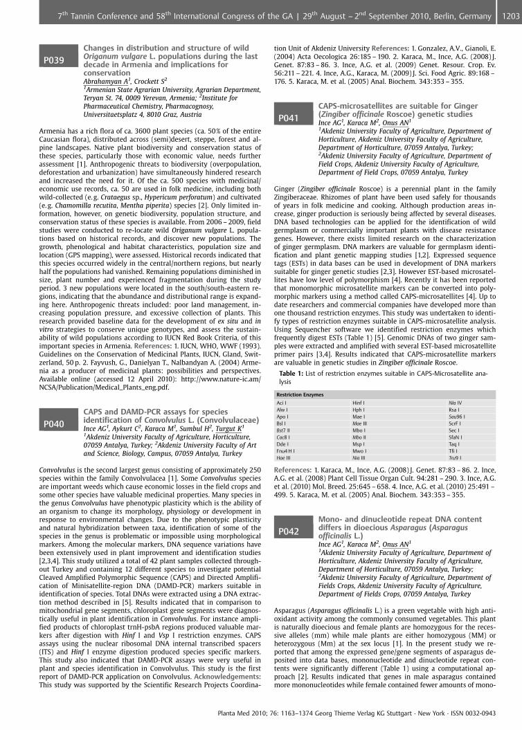
P039Changes in distribution and structure of wildOriganum vulgare L. populations during the lastdecade in Armenia and implications forconservationAbrahamyan A1, Crockett S2
1Armenian State Agrarian University, Agrarian Department,Teryan St. 74, 0009 Yerevan, Armenia; 2Institute forPharmaceutical Chemistry, Pharmacognosy,Universitaetsplatz 4, 8010 Graz, Austria
Armenia has a rich flora of ca. 3600 plant species (ca. 50% of the entireCaucasian flora), distributed across (semi)desert, steppe, forest and al-pine landscapes. Native plant biodiversity and conservation status ofthese species, particularly those with economic value, needs furtherassessment [1]. Anthropogenic threats to biodiversity (overpopulation,deforestation and urbanization) have simultaneously hindered researchand increased the need for it. Of the ca. 500 species with medicinal/economic use records, ca. 50 are used in folk medicine, including bothwild-collected (e.g. Crataegus sp., Hypericum perforatum) and cultivated(e. g. Chamomilla recutita, Mentha piperita) species [2]. Only limited in-formation, however, on genetic biodiversity, population structure, andconservation status of these species is available. From 2006 – 2009, fieldstudies were conducted to re-locate wild Origanum vulgare L. popula-tions based on historical records, and discover new populations. Thegrowth, phenological and habitat characteristics, population size andlocation (GPS mapping), were assessed. Historical records indicated thatthis species occurred widely in the central/northern regions, but nearlyhalf the populations had vanished. Remaining populations diminished insize, plant number and experienced fragmentation during the studyperiod. 3 new populations were located in the south/south-eastern re-gions, indicating that the abundance and distributional range is expand-ing here. Anthropogenic threats included: poor land management, in-creasing population pressure, and excessive collection of plants. Thisresearch provided baseline data for the development of ex situ and invitro strategies to conserve unique genotypes, and assess the sustain-ability of wild populations according to IUCN Red Book Criteria, of thisimportant species in Armenia. References: 1. IUCN, WHO, WWF (1993).Guidelines on the Conservation of Medicinal Plants, IUCN, Gland, Swit-zerland, 50 p. 2. Fayvush, G., Danielyan T., Nalbandyan A. (2004) Arme-nia as a producer of medicinal plants: possibilities and perspectives.Available online (accessed 12 April 2010): http://www.nature-ic.am/NCSA/Publication/Medical_Plants_eng.pdf.
P040CAPS and DAMD-PCR assays for speciesidentification of Convolvulus L. (Convolvulaceae)Ince AG1, Aykurt C2, Karaca M1, Sumbul H2, Turgut K1
1Akdeniz University Faculty of Agriculture, Horticulture,07059 Antalya, Turkey; 2Akdeniz University Faculty of Artand Science, Biology, Campus, 07059 Antalya, Turkey
Convolvulus is the second largest genus consisting of approximately 250species within the family Convolvulacea [1]. Some Convolvulus speciesare important weeds which cause economic losses in the field crops andsome other species have valuable medicinal properties. Many species inthe genus Convolvulus have phenotypic plasticity which is the ability ofan organism to change its morphology, physiology or development inresponse to environmental changes. Due to the phenotypic plasticityand natural hybridization between taxa, identification of some of thespecies in the genus is problematic or impossible using morphologicalmarkers. Among the molecular markers, DNA sequence variations havebeen extensively used in plant improvement and identification studies[2,3,4]. This study utilized a total of 42 plant samples collected through-out Turkey and containing 12 different species to investigate potentialCleaved Amplified Polymorphic Sequence (CAPS) and Directed Amplifi-cation of Minisatellite-region DNA (DAMD-PCR) markers suitable inidentification of species. Total DNAs were extracted using a DNA extrac-tion method described in [5]. Results indicated that in comparison tomitochondrial gene segments, chloroplast gene segments were diagnos-tically useful in plant identification in Convolvulus. For instance ampli-fied products of chloroplast trnH-psbA regions produced valuable mar-kers after digestion with Hinf I and Vsp I restriction enzymes. CAPSassays using the nuclear ribosomal DNA internal transcribed spacers(ITS) and Hinf I enzyme digestion produced species specific markers.This study also indicated that DAMD-PCR assays were very useful inplant and species identification in Convolvulus. This study is the firstreport of DAMD-PCR application on Convolvulus. Acknowledgements:This study was supported by the Scientific Research Projects Coordina-
tion Unit of Akdeniz University References: 1. Gonzalez, A.V., Gianoli, E.(2004) Acta Oecologica 26:185 – 190. 2. Karaca, M., Ince, A.G. (2008) J.Genet. 87:83 – 86. 3. Ince, A.G. et al. (2009) Genet. Resour. Crop. Ev.56:211 – 221. 4. Ince, A.G., Karaca, M. (2009) J. Sci. Food Agric. 89:168 –176. 5. Karaca, M. et al. (2005) Anal. Biochem. 343:353 – 355.
P041CAPS-microsatellites are suitable for Ginger(Zingiber officinale Roscoe) genetic studiesInce AG1, Karaca M2, Onus AN1
1Akdeniz University Faculty of Agriculture, Department ofHorticulture, Akdeniz University Faculty of Agriculture,Department of Horticulture, 07059 Antalya, Turkey;2Akdeniz University Faculty of Agriculture, Department ofField Crops, Akdeniz University Faculty of Agriculture,Department of Field Crops, 07059 Antalya, Turkey
Ginger (Zingiber officinale Roscoe) is a perennial plant in the familyZingiberaceae. Rhizomes of plant have been used safely for thousandsof years in folk medicine and cooking. Although production areas in-crease, ginger production is seriously being affected by several diseases.DNA based technologies can be applied for the identification of wildgermplasm or commercially important plants with disease resistancegenes. However, there exists limited research on the characterizationof ginger germplasm. DNA markers are valuable for germplasm identi-fication and plant genetic mapping studies [1,2]. Expressed sequencetags (ESTs) in data bases can be used in development of DNA markerssuitable for ginger genetic studies [2,3]. However EST-based microsatel-lites have low level of polymorphism [4]. Recently it has been reportedthat monomorphic microsatellite markers can be converted into poly-morphic markers using a method called CAPS-microsatellites [4]. Up todate researchers and commercial companies have developed more thanone thousand restriction enzymes. This study was undertaken to identi-fy types of restriction enzymes suitable in CAPS-microsatellite analysis.Using Sequencher software we identified restriction enzymes whichfrequently digest ESTs (Table 1) [5]. Genomic DNAs of two ginger sam-ples were extracted and amplified with several EST-based microsatelliteprimer pairs [3,4]. Results indicated that CAPS-microsatellite markersare valuable in genetic studies in Zingiber officinale Roscoe.
Table 1: List of restriction enzymes suitable in CAPS-Microsatellite ana-lysis
Restriction Enzymes
Aci I Hinf I NIa IVAlw I Hph I Rsa IApo I Mae I Sau96 IBsl I Mae III ScrF IBst7 II Mbo I Sec ICac8 I Mbo II SfaN IDde I Msp I Taq IFnu4 H I Mwo I Tfi IHae III Nia III Tru9 I
References: 1. Karaca, M., Ince, A.G. (2008) J. Genet. 87:83 – 86. 2. Ince,A.G. et al. (2008) Plant Cell Tissue Organ Cult. 94:281 – 290. 3. Ince, A.G.et al. (2010) Mol. Breed. 25:645 – 658. 4. Ince, A.G. et al. (2010) 25:491 –499. 5. Karaca, M. et al. (2005) Anal. Biochem. 343:353 – 355.
P042Mono- and dinucleotide repeat DNA contentdiffers in dioecious Asparagus (Asparagusofficinalis L.)Ince AG1, Karaca M2, Onus AN1
1Akdeniz University Faculty of Agriculture, Department ofHorticulture, Akdeniz University Faculty of Agriculture,Department of Horticulture, 07059 Antalya, Turkey;2Akdeniz University Faculty of Agriculture, Department ofFields Crops, Akdeniz University Faculty of Agriculture,Department of Fields Crops, 07059 Antalya, Turkey
Asparagus (Asparagus officinalis L.) is a green vegetable with high anti-oxidant activity among the commonly consumed vegetables. This plantis naturally dioecious and female plants are homozygous for the reces-sive alleles (mm) while male plants are either homozygous (MM) orheterozygous (Mm) at the sex locus [1]. In the present study we re-ported that among the expressed gene/gene segments of asparagus de-posited into data bases, mononucleotide and dinucleotide repeat con-tents were significantly different (Table 1) using a computational ap-proach [2]. Results indicated that genes in male asparagus containedmore mononucleotides while female contained fewer amounts of mono-
Planta Med 2010; 76: 1163–1374 Georg Thieme Verlag KG Stuttgart · New York · ISSN 0032-0943
12037th Tannin Conference and 58th International Congress of the GA | 29th August – 2nd September 2010, Berlin, Germany
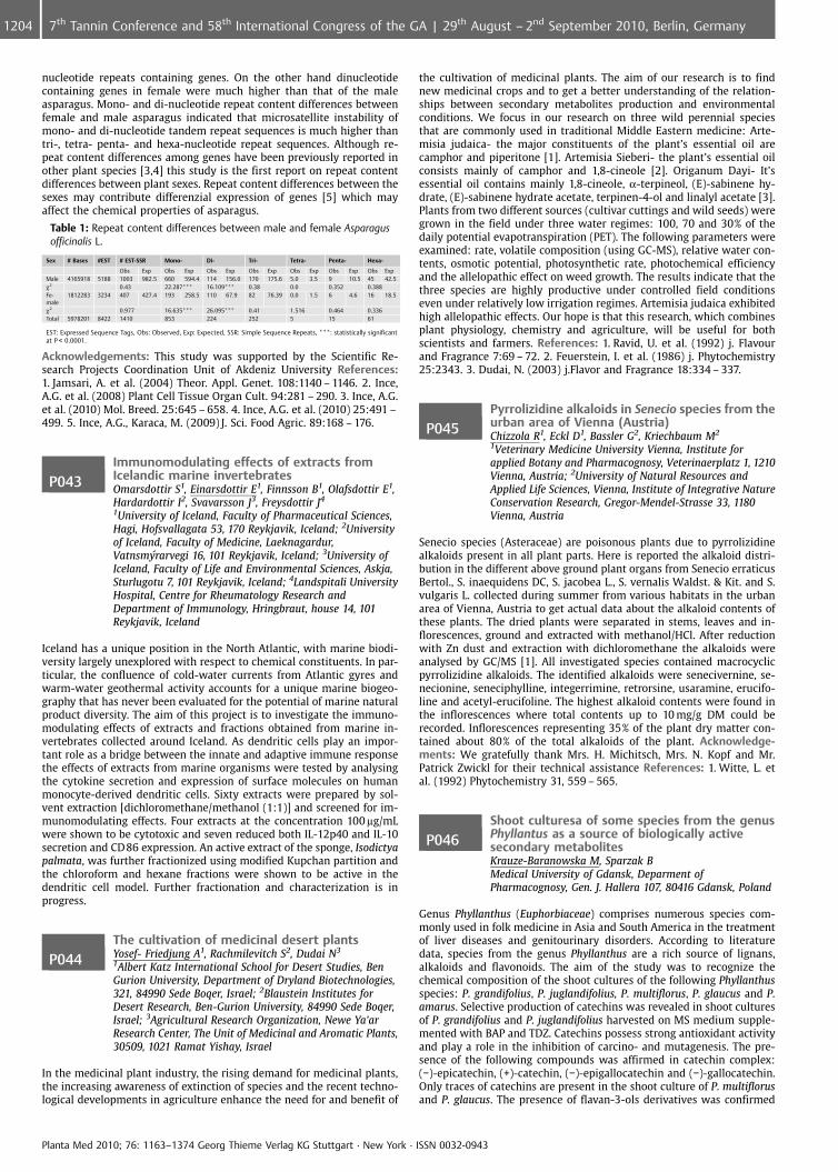
nucleotide repeats containing genes. On the other hand dinucleotidecontaining genes in female were much higher than that of the maleasparagus. Mono- and di-nucleotide repeat content differences betweenfemale and male asparagus indicated that microsatellite instability ofmono- and di-nucleotide tandem repeat sequences is much higher thantri-, tetra- penta- and hexa-nucleotide repeat sequences. Although re-peat content differences among genes have been previously reported inother plant species [3,4] this study is the first report on repeat contentdifferences between plant sexes. Repeat content differences between thesexes may contribute differenzial expression of genes [5] which mayaffect the chemical properties of asparagus.
Table 1: Repeat content differences between male and female Asparagusofficinalis L.
Sex # Bases #EST # EST-SSR Mono- Di- Tri- Tetra- Penta- Hexa-
Obs Exp Obs Exp Obs Exp Obs Exp Obs Exp Obs Exp Obs ExpMale 4165918 5188 1003 982.5 660 594.4 114 156.0 170 175.6 5.0 3.5 9 10.5 45 42.5c2 0.43 22.287*** 16.109*** 0.38 0.0 0.352 0.388Fe-male
1812283 3234 407 427.4 193 258.5 110 67.9 82 76.39 0.0 1.5 6 4.6 16 18.5
c2 0.977 16.635*** 26.095*** 0.41 1.516 0.464 0.336Total 5978201 8422 1410 853 224 252 5 15 61
EST: Expressed Sequence Tags, Obs: Observed, Exp: Expected, SSR: Simple Sequence Repeats, ***: statistically significantat P < 0.0001.
Acknowledgements: This study was supported by the Scientific Re-search Projects Coordination Unit of Akdeniz University References:1. Jamsari, A. et al. (2004) Theor. Appl. Genet. 108:1140 – 1146. 2. Ince,A.G. et al. (2008) Plant Cell Tissue Organ Cult. 94:281 – 290. 3. Ince, A.G.et al. (2010) Mol. Breed. 25:645 – 658. 4. Ince, A.G. et al. (2010) 25:491 –499. 5. Ince, A.G., Karaca, M. (2009) J. Sci. Food Agric. 89:168 – 176.
P043Immunomodulating effects of extracts fromIcelandic marine invertebratesOmarsdottir S1, Einarsdottir E1, Finnsson B1, Olafsdottir E1,Hardardottir I2, Svavarsson J3, Freysdottir J4
1University of Iceland, Faculty of Pharmaceutical Sciences,Hagi, Hofsvallagata 53, 170 Reykjavik, Iceland; 2Universityof Iceland, Faculty of Medicine, Laeknagardur,Vatnsmyrarvegi 16, 101 Reykjavik, Iceland; 3University ofIceland, Faculty of Life and Environmental Sciences, Askja,Sturlugotu 7, 101 Reykjavik, Iceland; 4Landspitali UniversityHospital, Centre for Rheumatology Research andDepartment of Immunology, Hringbraut, house 14, 101Reykjavik, Iceland
Iceland has a unique position in the North Atlantic, with marine biodi-versity largely unexplored with respect to chemical constituents. In par-ticular, the confluence of cold-water currents from Atlantic gyres andwarm-water geothermal activity accounts for a unique marine biogeo-graphy that has never been evaluated for the potential of marine naturalproduct diversity. The aim of this project is to investigate the immuno-modulating effects of extracts and fractions obtained from marine in-vertebrates collected around Iceland. As dendritic cells play an impor-tant role as a bridge between the innate and adaptive immune responsethe effects of extracts from marine organisms were tested by analysingthe cytokine secretion and expression of surface molecules on humanmonocyte-derived dendritic cells. Sixty extracts were prepared by sol-vent extraction [dichloromethane/methanol (1:1)] and screened for im-munomodulating effects. Four extracts at the concentration 100 mg/mLwere shown to be cytotoxic and seven reduced both IL-12p40 and IL-10secretion and CD86 expression. An active extract of the sponge, Isodictyapalmata, was further fractionized using modified Kupchan partition andthe chloroform and hexane fractions were shown to be active in thedendritic cell model. Further fractionation and characterization is inprogress.
P044The cultivation of medicinal desert plantsYosef- Friedjung A1, Rachmilevitch S2, Dudai N3
1Albert Katz International School for Desert Studies, BenGurion University, Department of Dryland Biotechnologies,321, 84990 Sede Boqer, Israel; 2Blaustein Institutes forDesert Research, Ben-Gurion University, 84990 Sede Boqer,Israel; 3Agricultural Research Organization, Newe Ya’arResearch Center, The Unit of Medicinal and Aromatic Plants,30509, 1021 Ramat Yishay, Israel
In the medicinal plant industry, the rising demand for medicinal plants,the increasing awareness of extinction of species and the recent techno-logical developments in agriculture enhance the need for and benefit of
the cultivation of medicinal plants. The aim of our research is to findnew medicinal crops and to get a better understanding of the relation-ships between secondary metabolites production and environmentalconditions. We focus in our research on three wild perennial speciesthat are commonly used in traditional Middle Eastern medicine: Arte-misia judaica- the major constituents of the plant’s essential oil arecamphor and piperitone [1]. Artemisia Sieberi- the plant’s essential oilconsists mainly of camphor and 1,8-cineole [2]. Origanum Dayi- It’sessential oil contains mainly 1,8-cineole, a-terpineol, (E)-sabinene hy-drate, (E)-sabinene hydrate acetate, terpinen-4-ol and linalyl acetate [3].Plants from two different sources (cultivar cuttings and wild seeds) weregrown in the field under three water regimes: 100, 70 and 30% of thedaily potential evapotranspiration (PET). The following parameters wereexamined: rate, volatile composition (using GC-MS), relative water con-tents, osmotic potential, photosynthetic rate, photochemical efficiencyand the allelopathic effect on weed growth. The results indicate that thethree species are highly productive under controlled field conditionseven under relatively low irrigation regimes. Artemisia judaica exhibitedhigh allelopathic effects. Our hope is that this research, which combinesplant physiology, chemistry and agriculture, will be useful for bothscientists and farmers. References: 1. Ravid, U. et al. (1992) j. Flavourand Fragrance 7:69 – 72. 2. Feuerstein, I. et al. (1986) j. Phytochemistry25:2343. 3. Dudai, N. (2003) j.Flavor and Fragrance 18:334 – 337.
P045Pyrrolizidine alkaloids in Senecio species from theurban area of Vienna (Austria)Chizzola R1, Eckl D1, Bassler G2, Kriechbaum M2
1Veterinary Medicine University Vienna, Institute forapplied Botany and Pharmacognosy, Veterinaerplatz 1, 1210Vienna, Austria; 2University of Natural Resources andApplied Life Sciences, Vienna, Institute of Integrative NatureConservation Research, Gregor-Mendel-Strasse 33, 1180Vienna, Austria
Senecio species (Asteraceae) are poisonous plants due to pyrrolizidinealkaloids present in all plant parts. Here is reported the alkaloid distri-bution in the different above ground plant organs from Senecio erraticusBertol., S. inaequidens DC, S. jacobea L., S. vernalis Waldst. & Kit. and S.vulgaris L. collected during summer from various habitats in the urbanarea of Vienna, Austria to get actual data about the alkaloid contents ofthese plants. The dried plants were separated in stems, leaves and in-florescences, ground and extracted with methanol/HCl. After reductionwith Zn dust and extraction with dichloromethane the alkaloids wereanalysed by GC/MS [1]. All investigated species contained macrocyclicpyrrolizidine alkaloids. The identified alkaloids were senecivernine, se-necionine, seneciphylline, integerrimine, retrorsine, usaramine, erucifo-line and acetyl-erucifoline. The highest alkaloid contents were found inthe inflorescences where total contents up to 10 mg/g DM could berecorded. Inflorescences representing 35% of the plant dry matter con-tained about 80% of the total alkaloids of the plant. Acknowledge-ments: We gratefully thank Mrs. H. Michitsch, Mrs. N. Kopf and Mr.Patrick Zwickl for their technical assistance References: 1. Witte, L. etal. (1992) Phytochemistry 31, 559 – 565.
P046Shoot culturesa of some species from the genusPhyllantus as a source of biologically activesecondary metabolitesKrauze-Baranowska M, Sparzak BMedical University of Gdansk, Deparment ofPharmacognosy, Gen. J. Hallera 107, 80416 Gdansk, Poland
Genus Phyllanthus (Euphorbiaceae) comprises numerous species com-monly used in folk medicine in Asia and South America in the treatmentof liver diseases and genitourinary disorders. According to literaturedata, species from the genus Phyllanthus are a rich source of lignans,alkaloids and flavonoids. The aim of the study was to recognize thechemical composition of the shoot cultures of the following Phyllanthusspecies: P. grandifolius, P. juglandifolius, P. multiflorus, P. glaucus and P.amarus. Selective production of catechins was revealed in shoot culturesof P. grandifolius and P. juglandifolius harvested on MS medium supple-mented with BAP and TDZ. Catechins possess strong antioxidant activityand play a role in the inhibition of carcino- and mutagenesis. The pre-sence of the following compounds was affirmed in catechin complex:(�)-epicatechin, (+)-catechin, (�)-epigallocatechin and (�)-gallocatechin.Only traces of catechins are present in the shoot culture of P. multiflorusand P. glaucus. The presence of flavan-3-ols derivatives was confirmed
Planta Med 2010; 76: 1163–1374 Georg Thieme Verlag KG Stuttgart · New York · ISSN 0032-0943
1204 7th Tannin Conference and 58th International Congress of the GA | 29th August – 2nd September 2010, Berlin, Germany
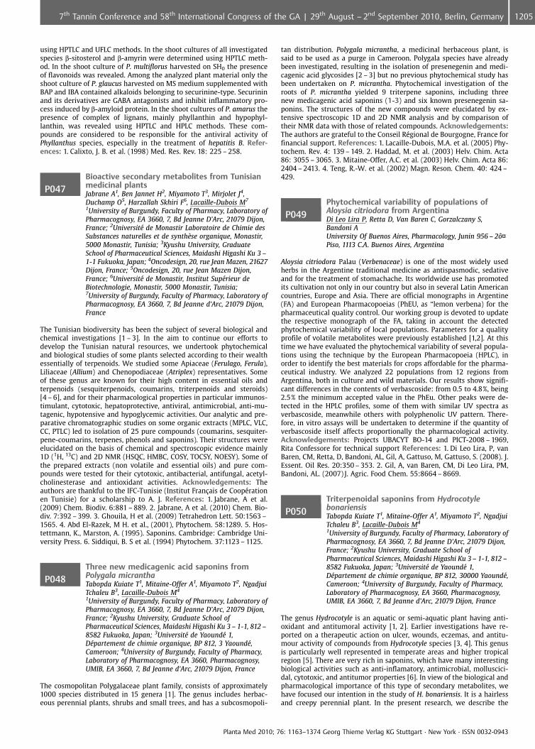
using HPTLC and UFLC methods. In the shoot cultures of all investigatedspecies b-sitosterol and b-amyrin were determined using HPTLC meth-od. In the shoot culture of P. multiflorus harvested on SH0 the presenceof flavonoids was revealed. Among the analyzed plant material only theshoot culture of P. glaucus harvested on MS medium supplemented withBAP and IBA contained alkaloids belonging to securinine-type. Securininand its derivatives are GABA antagonists and inhibit inflammatory pro-cess induced by b-amyloid protein. In the shoot cultures of P. amarus thepresence of complex of lignans, mainly phyllanthin and hypophyl-lanthin, was revealed using HPTLC and HPLC methods. These com-pounds are considered to be responsible for the antiviral activity ofPhyllanthus species, especially in the treatment of hepatitis B. Refer-ences: 1. Calixto, J. B. et al. (1998) Med. Res. Rev. 18: 225 – 258.
P047Bioactive secondary metabolites from Tunisianmedicinal plantsJabrane A1, Ben Jannet H2, Miyamoto T3, Mirjolet J4,Duchamp O5, Harzallah Skhiri F6, Lacaille-Dubois M7
1University of Burgundy, Faculty of Pharmacy, Laboratory ofPharmacognosy, EA 3660, 7, Bd Jeanne D’Arc, 21079 Dijon,France; 2Universit� de Monastir Laboratoire de Chimie desSubstances naturelles et de synth�se organique, Monastir,5000 Monastir, Tunisia; 3Kyushu University, GraduateSchool of Pharmaceutical Sciences, Maidashi Higashi Ku 3 –1-1 Fukuoka, Japan; 4Oncodesign, 20, rue Jean Mazen, 21627Dijon, France; 5Oncodesign, 20, rue Jean Mazen Dijon,France; 6Universit� de Monastir, Institut Sup�rieur deBiotechnologie, Monastir, 5000 Monastir, Tunisia;7University of Burgundy, Faculty of Pharmacy, Laboratory ofPharmacognosy, EA 3660, 7, Bd Jeanne d’Arc, 21079 Dijon,France
The Tunisian biodiversity has been the subject of several biological andchemical investigations [1 – 3]. In the aim to continue our efforts todevelop the Tunisian natural resources, we undertook phytochemicaland biological studies of some plants selected according to their wealthessentially of terpenoids. We studied some Apiaceae (Ferulago, Ferula),Liliaceae (Allium) and Chenopodiaceae (Atriplex) representatives. Someof these genus are known for their high content in essential oils andterpenoids (sesquiterpenoids, coumarins, triterpenoids and steroids)[4 – 6], and for their pharmacological properties in particular immunos-timulant, cytotoxic, hepatoprotective, antiviral, antimicrobial, anti-mu-tagenic, hypotensive and hypoglycemic activities. Our analytic and pre-parative chromatographic studies on some organic extracts (MPLC, VLC,CC, PTLC) led to isolation of 25 pure compounds (coumarins, sesquiter-pene-coumarins, terpenes, phenols and saponins). Their structures wereelucidated on the basis of chemical and spectroscopic evidence mainly1D (1H, 13C) and 2D NMR (HSQC, HMBC, COSY, TOCSY, NOESY). Some ofthe prepared extracts (non volatile and essential oils) and pure com-pounds were tested for their cytotoxic, antibacterial, antifungal, acetyl-cholinesterase and antioxidant activities. Acknowledgements: Theauthors are thankful to the IFC-Tunisie (Institut Fran�ais de Coop�rationen Tunisie) for a scholarship to A. J. References: 1. Jabrane, A et al.(2009) Chem. Biodiv. 6:881 – 889. 2. Jabrane, A et al. (2010) Chem. Bio-div. 7:392 – 399. 3. Ghouila, H et al. (2009) Tetrahedron Lett. 50:1563 –1565. 4. Abd El-Razek, M H. et al., (2001), Phytochem. 58:1289. 5. Hos-tettmann, K., Marston, A. (1995). Saponins. Cambridge: Cambridge Uni-versity Press. 6. Siddiqui, B. S et al. (1994) Phytochem. 37:1123 – 1125.
P048Three new medicagenic acid saponins fromPolygala micranthaTabopda Kuiate T1, Mitaine-Offer A1, Miyamoto T2, NgadjuiTchaleu B3, Lacaille-Dubois M4
1University of Burgundy, Faculty of Pharmacy, Laboratory ofPharmacognosy, EA 3660, 7, Bd Jeanne D’Arc, 21079 Dijon,France; 2Kyushu University, Graduate School ofPharmaceutical Sciences, Maidashi Higashi Ku 3 – 1-1, 812 –8582 Fukuoka, Japan; 3Universit� de Yaound� 1,D�partement de chimie organique, BP 812, 3 Yaound�,Cameroon; 4University of Burgundy, Faculty of Pharmacy,Laboratory of Pharmacognosy, EA 3660, Pharmacognosy,UMIB, EA 3660, 7, Bd Jeanne d’Arc, 21079 Dijon, France
The cosmopolitan Polygalaceae plant family, consists of approximately1000 species distributed in 15 genera [1]. The genus includes herbac-eous perennial plants, shrubs and small trees, and has a subcosmopoli-
tan distribution. Polygala micrantha, a medicinal herbaceous plant, issaid to be used as a purge in Cameroon. Polygala species have alreadybeen investigated, resulting in the isolation of presenegenin and medi-cagenic acid glycosides [2 – 3] but no previous phytochemical study hasbeen undertaken on P. micrantha. Phytochemical investigation of theroots of P. micrantha yielded 9 triterpene saponins, including threenew medicagenic acid saponins (1-3) and six known presenegenin sa-ponins. The structures of the new compounds were elucidated by ex-tensive spectroscopic 1D and 2D NMR analysis and by comparison oftheir NMR data with those of related compounds. Acknowledgements:The authors are grateful to the Conseil R�gional de Bourgogne, France forfinancial support. References: 1. Lacaille-Dubois, M.A. et al. (2005) Phy-tochem. Rev. 4: 139 – 149. 2. Haddad, M. et al. (2003) Helv. Chim. Acta86: 3055 – 3065. 3. Mitaine-Offer, A.C. et al. (2003) Helv. Chim. Acta 86:2404 – 2413. 4. Teng, R.-W. et al. (2002) Magn. Reson. Chem. 40: 424 –429.
P049Phytochemical variability of populations ofAloysia citriodora from ArgentinaDi Leo Lira P, Retta D, Van Baren C, Gorzalczany S,Bandoni AUniversity Of Buenos Aires, Pharmacology, Junin 956 – 2�¤Piso, 1113 C.A. Buenos Aires, Argentina
Aloysia citriodora Palau (Verbenaceae) is one of the most widely usedherbs in the Argentine traditional medicine as antispasmodic, sedativeand for the treatment of stomachache. Its worldwide use has promotedits cultivation not only in our country but also in several Latin Americancountries, Europe and Asia. There are official monographs in Argentine(FA) and European Pharmacopoeias (PhEU, as “lemon verbena) for thepharmaceutical quality control. Our working group is devoted to updatethe respective monograph of the FA, taking in account the detectedphytochemical variability of local populations. Parameters for a qualityprofile of volatile metabolites were previously established [1,2]. At thistime we have evaluated the phytochemical variability of several popula-tions using the technique by the European Pharmacopoeia (HPLC), inorder to identify the best materials for crops affordable for the pharma-ceutical industry. We analyzed 22 populations from 12 regions fromArgentina, both in culture and wild materials. Our results show signifi-cant differences in the contents of verbascoside: from 0.5 to 4.8%, being2.5% the minimum accepted value in the PhEu. Other peaks were de-tected in the HPLC profiles, some of them with similar UV spectra asverbascoside, meanwhile others with polyphenolic UV pattern. There-fore, in vitro assays will be undertaken to determine if the quantity ofverbascoside itself affects proportionally the pharmacological activity.Acknowledgements: Projects UBACYT BO-14 and PICT-2008 – 1969,Rita Confessore for technical support References: 1. Di Leo Lira, P, vanBaren, CM, Retta, D, Bandoni, AL, Gil, A, Gattuso, M, Gattuso, S. (2008). J.Essent. Oil Res. 20:350 – 353. 2. Gil, A, van Baren, CM, Di Leo Lira, PM,Bandoni, AL. (2007) J. Agric. Food Chem. 55:8664 – 8669.
P050Triterpenoidal saponins from HydrocotylebonariensisTabopda Kuiate T1, Mitaine-Offer A1, Miyamoto T2, NgadjuiTchaleu B3, Lacaille-Dubois M4
1University of Burgundy, Faculty of Pharmacy, Laboratory ofPharmacognosy, EA 3660, 7, Bd Jeanne D’Arc, 21079 Dijon,France; 2Kyushu University, Graduate School ofPharmaceutical Sciences, Maidashi Higashi Ku 3 – 1-1, 812 –8582 Fukuoka, Japan; 3Universit� de Yaound� 1,D�partement de chimie organique, BP 812, 30000 Yaound�,Cameroon; 4University of Burgundy, Faculty of Pharmacy,Laboratory of Pharmacognosy, EA 3660, Pharmacognosy,UMIB, EA 3660, 7, Bd Jeanne d’Arc, 21079 Dijon, France
The genus Hydrocotyle is an aquatic or semi-aquatic plant having anti-oxidant and antitumoral activity [1, 2]. Earlier investigations have re-ported on a therapeutic action on ulcer, wounds, eczemas, and antitu-mour activity of compounds from Hydrocotyle species [3, 4]. This genusis particularly well represented in temperate areas and higher tropicalregion [5]. There are very rich in saponins, which have many interestingbiological activities such as anti-inflamatory, antimicrobial, molluscici-dal, cytotoxic, and antitumor properties [6]. In view of the biological andpharmacological importance of this type of secondary metabolites, wehave focused our intention in the study of H. bonariensis. It is a hairlessand creepy perennial plant. In the present research, we describe the
Planta Med 2010; 76: 1163–1374 Georg Thieme Verlag KG Stuttgart · New York · ISSN 0032-0943
12057th Tannin Conference and 58th International Congress of the GA | 29th August – 2nd September 2010, Berlin, Germany
isolation and structure elucidation of six new oleanane-type triterpenoi-dal saponins, Bonarienosides A-F (1 - 6) from the 70% aqueous MeOHextract of Hydrocotyle bonariensis roots. Their structures were estab-lished on the basis of NMR spectroscopic data (13C, 1H, COSY, HMBC,TOCSY, and NOESY) and mass spectrometry. Acknowledgements: Theauthors are grateful to the Conseil R�gional de Bourgogne, France forfinancial support. References: 1. Yu, F. et al. (2007) Phytomed. 14: 166 –171. 2. Jayashree, G. et al. (2003) Fitoterapia 74: 431 – 434. 3. Malhotra,C.L. et al. (1961) Ind. J. Pharm. 23: 106. 4. Miyata, S. et al. (1981) Antic-ancer Crude Drugs and their Prescription 185. 5. Stevens, C. et al. (2000)Missouri Botanical Garden Press. 110 – 115. 6. Lacaille-Dubois, M.A., Mi-taine-Offer A.C. (2005) Phytochem. Rev. 4: 139 – 149.
P051Diversity of medicinal plants used by thepopulation of the state of Mato Grosso, BrazilRieder AUniversidade do Estado de Mato Grosso (UNEMAT); eEMPAER-MT, Campus de C�ceres, Av. S¼o Jo¼o s/n, BairroCavalhada, 78200000 C�ceres, Brazil
The population of Mato Grosso at the present consists in a greater pro-portion of families who migrated to this State of Brazil after the 1960 s.These were mixed and coexist well with the native population region-ally. This miscegenation this culturally enriched region of central SouthAmerica where there are at least three biomes (Amazon Rainforest,Pantanal, Cerrado), with great diversity of species. This along ethnobo-tanical studies reveals the use of expressive diversity of medicinal spe-cies. We mention below the results of some studies conducted in theState of Mato Grosso (MT), Brazil. In 1994 a study with 12 healers in thecapital of Mato Grosso (Cuiab�) found that they were attending publicoffering 225 species considered medicinal. In 2001, involving the entirestate of MT, 648 teachers listed 67 species as the most important med-icinal plants used by them. In 2002, another ethnobotanical study foundthat 339 residents of C�ceres (MT) listed 86 species of medicinal herbsgrown in their backyards for their own use. In 2005, a study involving21 municipalities and 63 informants Southwestern MT noted that themain medicinal plants used formed a list of 503 species. A more recentstudy (2009) dedicated to knowing which plants use to control diabetesin MT noted that the available literature provides a list of 133 species isformed. It is believed that the diversity of species listed is mainly relatedto floristic richness of biodiversity and cultural center in this Brazilianregion of South America. Acknowledgements: UNEMAT-institutionalsupport, and FAPEMAT EMPAER-MT; support afforded by the wholeproject team PLAMED: Bonila MGO, Carniello M, Ramos A PR et al.
P052Screening of Satureja khuzestanica Jamzad andS. rechingeri Jamzad collections for high yieldinggenotypesHadian J1, Najafi F1, Salehnia A2, Ehteshamnia A2,Ganjipoor P2
1Medicinal plants and drug research institute shahidBeheshti University, G. C., Deratment of Agriculture, Evin,Tehran, 19835389 Tehran, Iran, Islamic Republic Of;2Department of Research and Development (R&D),Khorraman Co, Khoramabad Industrial city, 68135 – 579Khoramabad, Iran, Islamic Republic Of
Satureja khuzestanica Jamzad and S. rechingeri Jamzad are endemic spe-cies of Lamiaceae growing wild in Iran. The essential oils of both speciesare rich in Carvacrol, a phenolic compound widely used in food, feed andpharmaceutical industries. At the beginning of a breeding and domes-tication program, a collection of S. khuzestanica and S. rechingeri includ-ing 12 populations and 1200 genotypes were investigated for morpho-logical traits, herbal drug yield, oil and carvarcol content. Variation washigher within rather than between populations. Essential oil contentvaried between 0.3 to 5.4% for S. khuzestanica and 1.8 – 9.45% for S.rechingeri. Carvarol was major component of all analyzed samples of S.khuzestanica (83.1 – 96.5%) and S. rechingeri (85.2 – 97.2%). Studied gen-otypes were also highly variable based on morphological traits and her-bal drug yield. Elite genotypes were selected for future breeding pro-gram. References: 1. Jamzad, Z. (1996) Ann. Natu. Mus. Wien 98: 75 –77. 2. Jamzad Z. (1994) Iran J. Bot. 6: 215 – 218. 3. Pank, F. et al. (2004).Zeit. Arz.Gew. 9:72 – 79.
P053Determination of the best selection criteria forgenetic improvement of seed and oil yield inspring safflower cultivarsGolparvar A1, Ghasemi Pirbalouti A2
1Islamic Azad University, Khorasgan Branch, Agronomy andPlant Breeding, Isfahan, Iran, 88148 Isfahan, Iran, IslamicRepublic Of; 2Islamic Azad University, Shahrekord Branch,Agronomy and Plant Breeding, Shahrekord, Iran, 88146Shahrekord, Iran, Islamic Republic Of
Identification of the important seed and oil yield components in saf-flower as an important medicinal plant in Iran is very efficient in geneticimprovement of these traits via indirect selection. For this reason, tenspring safflower cultivars were sown in a randomized complete blockdesign with three replications in research field of Islamic Azad University,Khorasgan branch. Results of correlation, regression and path analysisrevealed that traits 1000-seed weight and seed number plant-1 has con-siderable positive and direct effects on plant seed yield and accounted forthe largest amount of variation exist in this trait. On the other hand,traits 1000-seed weight, days to physiological maturity and seed numberplant-1 were the most important plant oil yield components and suggestfor breeding of this trait. Over all, results revealed that selection for high-er amounts of traits 1000-seed weight and seed number plant-1 canimprove indirectly plant seed and oil yield in spring safflower cultivarsespecially in early breeding generations. Key words: Spring safflower,indirect selection, correlation analysis, step-wise regression and pathanalysis. References: 1. Abolhasani, Kh. and Saeidi, G. (2006). Evaluationof drought tolerance of safflower lines based on tolerance and sensitivityindices to water stress. J. Sci. Technol. Agric. Natur. Resour. 10 (3): 419 –422. 2. Arslan, B. (2007). Assessing of heritability and variance compo-nents of yield and some agronomic traits of different safflower cultivars.Asian. J. Pl. Sci. 6 (3): 554 – 557. 3. Ashkani, J., Pakniyat, H. and Ghotbi, V.(2007). Genetic evaluation of several physiological traits for screening ofsuitable spring safflower genotypes under stress and non-stress irriga-tion regimes. Pak. J. Bio. Sci. 10 (14): 2320 – 2326.
P054Chemical composition of the essential oil ofLilium ledebourii from IranSheikhjabbari A1, Nejad Ebrahimi S2, Sheikhjabbari H3
1Students Research Center, Pharmacy, Tabriz University ofMedical Science, 51664 Tabriz, Iran, Islamic Republic Of;2Medicinal plants and Drugs, Department ofPhytochemistry, Shahid Beheshti University Tehran, Iran,Islamic Republic Of; 3Ardabil Department of Environmental,Karshenasan, 53618 Ardabil, Iran, Islamic Republic Of
Lilium ledebourii (Liliaceae), locally named “Susan-e-Chelcheragh” is anendemic and rare species growing on the highlands of Damash region inGilan Province and Fandeghlo forests in Ardabil Province north part ofIran. The habitat of this species is under IUCN protection, Category III(Natural Monument) by the Iranian Department of Environment since1976. This plant is a unique species of Lilium genus that is native ofnorth Iran [1]. The aim of the present study was the investigation ofthe chemical composition of the essential oil of L. ledebourii (Apiaceae)from Iran by GC/FID and GC/MS methods. The plant material was col-lected during its flowering stage in June, 2008, on the Ardabil Mountainsof the North-West of Iran. The essential oil was obtained by hydrodis-tillation of the dried herb using a Clevenger type apparatus. The oil yieldcalculated on moisture free basis was 0.1%. Twenty-six compounds werecharacterized in the essential oil of Lilium ledebourii, representing 99.1%of the oil, of which linalool (74.1%), E-6-tetradecanyl acetate (11.7%),benzyl salycilate (2.7%) and hexadecanal were found to be the majorcomponents. Analysis of oil revealed that oil was rich in linalool. Ac-knowledgements: This work has been supported by Shahid BeheshtiUniversity Research Council. References: 1. Saeidifard, M., Hosseini,S.M., Padasht Dehkaei, M.N., (2008) Rostaniha 9 (2 (32)):137.
Planta Med 2010; 76: 1163–1374 Georg Thieme Verlag KG Stuttgart · New York · ISSN 0032-0943
1206 7th Tannin Conference and 58th International Congress of the GA | 29th August – 2nd September 2010, Berlin, Germany
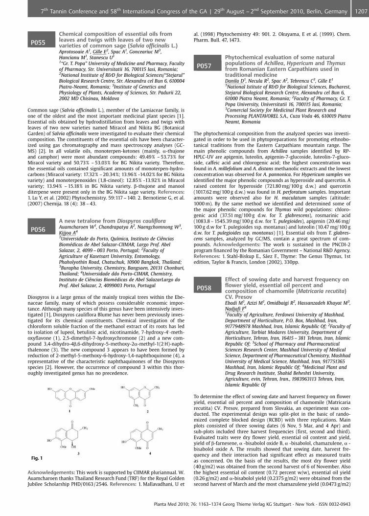
P055Chemical composition of essential oils fromleaves and twigs with leaves of two newvarieties of common sage (Salvia officinalis L.)Aprotosoaie A1, Gille E2, Spac A1, Gonceariuc M3,Hancianu M1, Stanescu U1
1“Gr. T. Popa” University of Medicine and Pharmacy, Facultyof Pharmacy, Str. Universitatii 16, 700115 Iasi, Romania;2National Institute of R&D for Biological Sciences/“Stejarul”Biological Research Centre, Str. Alexandru cel Bun 6, 610004Piatra-Neamt, Romania; 3Institute of Genetics andPhysiology of Plants, Academy of Sciences, Str. Padurii 22,2002 MD Chisinau, Moldova
Common sage (Salvia officinalis L.), member of the Lamiaceae family, isone of the oldest and the most important medicinal plant species [1].Essential oils obtained by hydrodistillation from leaves and twigs withleaves of two new varieties named Miracol and Nikita BG (BotanicalGarden) of Salvia officinalis were investigated to evaluate their chemicalcomposition. The constituents of the essential oils have been character-ized using gas chromatography and mass spectroscopy analyses (GC-MS) [2]. In all volatile oils, monoterpen-ketones (mainly, a-thujoneand camphor) were most abundant compounds: 49.49% – 53.73% forMiracol variety and 50.73% – 53.03% for BG Nikita variety. Therefore,the essential oils contained significant amounts of monoterpen-hydro-carbons (Miracol variety: 17.32% – 20.34%; 13.96% -14.02% for BG Nikitavariety) and monoterpenoxides (1,8-cineol): 12.85% -13.92% in Miracolvariety; 13.94% – 15.18% in BG Nikita variety. b-thujone and manoolditerpene were present only in the BG Nikita sage variety. References:1. Lu Y, et al. (2002) Phytochemistry. 59:117 – 140. 2. Bernotiene G, et al.(2007) Chemija. 18 (4): 38 – 43.
P056A new tetralone from Diospyros caulifloraAuamcharoen W1, Chandrapatya A2, Naengchomnong W3,Kijjoa A4
1Universidade do Porto, Qu�mica, Instituto de CiÞnciasBiom�dicas de Abel Salazar-CIIMAR, Largo Prof. AbelSalazar, 2, 4099 – 003 Porto, Portugal; 2Faculty ofAgriculture of Kasetsart University, Entomology,Phaholyothin Road, Chatuchuk, 10900 Bangkok, Thailand;3Burapha University, Chemistry, Bangsaen, 20131 Chonburi,Thailand; 4Universidade ddo Porto-CIMAR, Chemistry,Instituto de CiÞncias Biom�dicas de Abel SalazarLargo doProf. Abel Salazar, 2, 4099003 Porto, Portugal
Diospyros is a large genus of the mainly tropical trees within the Ebe-naceae family, many of which possess considerable economic impor-tance. Although many species of this genus have been intensively inves-tigated [1], Diospyros cauliflora Blume has never been previously inves-tigated for its chemical constituents. Chemical investigation of thechloroform soluble fraction of the methanol extract of its roots has ledto isolation of lupeol, betulinic acid, nicotinamide, 7-hydroxy-4’-meth-oxyflavone (1), 2,5-dimethyl-7-hydroxychromone (2) and a new com-pound 3,4-dihydro-4b,6-dihydroxy-5-methoxy-2a-methyl-1(2 H)-naph-thalenone (3). The new compound 3 appears to have been formed byreduction of 2-methyl-5-methoxy-6-hydroxy-1,4-naphthoquinone (4), arepresentative of the characteristic naphthaquinones of the Diospyrosspecies [2]. However, the occurrence of compound 3 within this thor-oughly investigated genus has no precedence.
Fig. 1
Acknowledgements: This work is supported by CIIMAR pluriannual. W.Auamcharoen thanks Thailand Research Fund (TRF) for the Royal GoldenJubilee Scholarship PHD/0163/2546. References: 1. Mallavadhani, U et
al. (1998) Phytochemistry 49: 901. 2. Okuyama, E et al. (1999). Chem.Pharm. Bull. 47, 1473.
P057Phytochemical evaluation of some naturalpopulations of Achillea, Hypericum and Thymusfrom Romanian Eastern Carpathians used intraditional medicineDanila D1, Necula R2, Spac A2, Tebrencu C3, Gille E1
1National Istitute of R&D for Biological Sciences, Bucharest,Stejarul Biological Research Centre, Alexandru cel Bun 6,61000 Piatra Neamt, Romania; 2Faculty of Pharmacy, Gr. T.Popa University, Universitatii 16, 700115 Iasi, Romania;3Comercial Society for Medicinal Plant Research andProcessing PLANTAVOREL S.A., Cuza Voda 46, 610019 PiatraNeamt, Romania
The phytochemical composition from the analyzed species was investi-gated in order to be used in phytopreparations for promoting ethnobo-tanical traditions from the Eastern Carpathians mountain range. Themain phenolic compounds from Achillea samples identified by RP-HPLC-UV are apigenin, luteolin, apigenin-7-glucoside, luteolin-7-gluco-side, caffeic acid and chlorogenic acid; the highest concentration wasfound in A. millefolium and A. distans methanolic extracts and the lowestconcentration was observed for A. pannonica. For Hypericum samples weidentified the major phenolic compounds as hyperoside and quercetin. Araised content for hyperoside (721.80 mg/100 g d.w.) and quercetin(1017.62 mg/100 g d.w.) was found in H. perforatum samples. Importantamounts were observed also for H. maculatum samples (altitude:1000 m). By the same method we identified and determined some ofthe major phenolic compounds for Thymus wild populations: chloro-genic acid (37.51 mg/100 g d.w. for T. glabrescens), rosmarinic acid(1083.8 – 1545.39 mg/100 g d.w. for T. pulegioides), apigenin (20.46 mg/100 g d.w for T. pulegioides ssp. montanus) and luteolin (10.47 mg/100 gd.w. for T. pulegioides ssp. montanus) [1]. Essential oils from T. glabres-cens samples, analyzed by GC/MS, contain a great spectrum of com-pounds. Acknowledgements: The work is sustained in the PNCDI-2program financed by the Romanian Government – National R&D Agency.References: 1. Stahl-Biskup E., S�ez F., Thyme: The Genus Thymus, 1stedition, Taylor & Francis, London (2002), 330pp.
P058Effect of sowing date and harvest frequency onflower yield, essential oil percent andcomposition of chamomile (Matricaria recutita)CV. PresovEbadi M1, Azizi M1, Omidbaigi R2, Hassanzadeh Khayat M3,Nadjafi F4
1Faculty of Agriculture, Ferdowsi University of Mashhad,Department of Horticulture, P.O. Box, Mashhad, Iran,9177948978 Mashhad, Iran, Islamic Republic Of; 2Faculty ofAgriculture, Tarbiat Modares University, Department ofHorticulture, Tehran, Iran, 16415 – 381 Tehran, Iran, IslamicRepublic Of; 3School of Pharmacy and PharmaceuticalSciences Research Center, Mashhad University of MedicalScience, Department of Pharmaceutical Chemistry, MashhadUniversity of Medical Science, Mashhad, Iran, 917751365Mashhad, Iran, Islamic Republic Of; 4Medicinal Plant andDrug Research Institute, Shahid Beheshti University,Agriculture, evin, Tehran, Iran., 1983963113 Tehran, Iran,Islamic Republic Of
To determine the effect of sowing date and harvest frequency on floweryield, essential oil percent and composition of chamomile (Matricariarecutita) CV. Presov, prepared from Slovakia, an experiment was con-ducted. The experimental design was split-plot in the basic of rando-mized complete blocked design (RCBD) with three replications. Mainplots consisted of three sowing dates (6 Nov, 5 Mar, and 4 Apr) andsub-plots included three harvest frequencies (first, second and third).Evaluated traits were dry flower yield, essential oil content and yield,yield of b-farnesene, a -bisabolol oxide B, a -bisabolol, chamazulene, a -bisabolol oxide A. The results showed that sowing date, harvest fre-quency and their interaction had significant effect as measured traitsas concerned. On the basis of the results, the most dry flower yield(40 g/m2) was obtained from the second harvest of 6 of November. Alsothe highest essential oil content (0.72 percent w/w), essential oil yield(0.26 g/m2) and a-bisabolol yield (0.2375 g/m2) were obtained from thesecond harvest of March and the most chamazulene yield (0.0473 g/m2)
Planta Med 2010; 76: 1163–1374 Georg Thieme Verlag KG Stuttgart · New York · ISSN 0032-0943
12077th Tannin Conference and 58th International Congress of the GA | 29th August – 2nd September 2010, Berlin, Germany
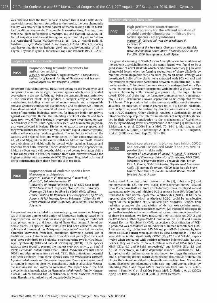
was obtained from the third harvest of March that it had a little differ-ence with second harvest. According to the results, the best chamomilequality was attained in second harvest of March sowing date in Mash-had condition. Keywords: Chamomile, Harvesting time, Planting time,Medicinal plant References: 1. Marcum, D.B and Hanson, B.R.2006. Ef-fect of irrigation and harvest timing on peppermint oil yield in Califor-nia. Agricultural Water Management.82:118 – 128. 2. Naghdi Badi, H.,Yazdani, D., Sajed Mohammad, A and Nazari, F.2004. Effects of spacingand harvesting time on herbage yield and quality/quantity of oil inThyme, Thymus vulgaris L. Industrial Crops and Products,19:231 – 236.
Biopiracy and bioprospecting
P059Bioprospecting Icelandic liverworts foranticancer activityJensen S, Omarsdottir S, Ogmundsdottir H, Olafsdottir EUniversity of Iceland, Faculty of Pharmaceutical Sciences,Hofsvallagata 53, 107 Reykjavik, Iceland
Liverworts (Marchantiophyta, Hepaticae) belong to the bryophytes andcomprise of about six to eight thousand species which are distributedeverywhere in the world. Although being the structurally most primitiveof the terrestrial plants they produce a complex array of secondarymetabolites, including a number of mono- sesqui- and diterpenoidsand also aromatic compounds like bibenzyls and bis (bibenzyls). Studieshave demonstrated that several of these compounds possess a widerange of interesting biological activities, for instance inhibitory activityagainst cancer cells. Herein, the inhibiting effects of extracts and frac-tions from two different Icelandic liverworts were investigated on can-cer cell-lines in vitro. Chiloscyphus pallescens and Marchantia polymor-pha were collected in Iceland. Diethyl ether extracts were prepared andthey were further fractionated on VLC (Vacuum Liquid Chromatography)with a n-hexane:ethyl acetate gradient. The inhibitory effects of theextracts and selected fractions were tested in four concentrations 80,40, 20 and 10 mg/ml, on the cancer cell-lines MCF-7 and T47D. Resultswere obtained as% viable cells by crystal violet staining. Extracts andfractions from both liverwort species demonstrated dose-dependent in-hibitory effects onto cell growth, viability and colony formation abilityof the cancer cells in vitro. One fraction from each liverwort showed thehighest activity with approximate IC 50 20 mg/ml. Bioguided isolation ofactive constituents from these fractions is in progress.
P060Bioprospection of endemic species fromMarquesas archipelagoIngert N1, Lindauer T2, Bombarda I1, Bianchini J1,Moretti C3, Raharivelomanana P4
1University Of French Polynesia, Bp N� 6570 Faaa Tahiti,98702 Faaa, French Polynesia; 2Louis Pasteur University,Pharmacy, 74 Route Du Rhin Bp 60024, 67401 Illkirch,France; 3Institut De Recherche Et Developpement, Bp No 529Papeete, 98713 Papeete, French Polynesia; 4University OfFrench Polynesia, Bpno 6570 Faaa/Tahiti, 98702 Faaa, FrenchPolynesia
The present work is included in a research program focused on Marque-sas archipelago aiming valorization of Marquesas heritage based on abioprospection. We focussed our investigation on a study of traditionaluses, phytochemistry and bioactivity of Nuku Hiva taxons. A list of en-demic plants (53 species) from Nuku Hiva had been collected. An eth-nobotanical framework on “Marquesas biodiversity” was held to gathernaturalist knowledge from local population showing a partial loss oftraditional uses. Extracts obtained from different parts (leaf, bark androot) were submitted to a biological screening using two in vitro bioas-says: cytotoxicity (KB) and radical scavenging (DPPH). Three speciesextracts were found to present the highest cytotoxic activity at 1 mg/mllevel: Hernandia nukuhivensis (root: 100%), Maytenus crenatus (bark:100%) and Myrsine nukuhivensis (leaf: 72%). High antioxidant potentialhad been evaluated from three species extracts: Wikstroemia coriacea,Myrsine nukuhivensis and Waltheria tomentosa. Two species were foundto contain interesting bioactive constituents such as alkaloids: Hernan-dia nukuhivensis and Waltheria tomentosa. We report herein the firstphytochemical investigation on Hernandia nukuhivensis (family Hernan-diaceae) which allowed the identification of three bioactive constitu-ents: litseglutin A, reticulin and deoxypodophyllotoxin.
Enzyme inhibitors from plants
P061High-performance countercurrentchromatography for the efficient isolation ofalkaloid acetylcholinesterase inhibitors fromNerine species (Amaryllidaceae)Marston A1, Cawood M1, van der Westhuizen J1,Zietsman P2
1University of the Free State, Chemistry, Nelson MandelaDrive Bloemfontein, South Africa; 2National Museum, POBox 266, 9300 Bloemfontein, South Africa
In a general screening of South African Amaryllidaceae for inhibitors ofthe enzyme acetylcholinesterase, the genus Nerine was found to be agood source of novel alkaloids which exhibit this bioactivity [1]. In viewof material losses when performing separations of these alkaloids bymultiple chromatographic steps on silica gel, an all-liquid strategy wasinvestigated. Bulbs of the plants were extracted with 90% ethanol andthe resulting extracts were partitioned between chloroform and 1% am-monia solution. Chloroform fractions were chromatographed on a Dy-namic Extractions Spectrum instrument with suitable 2-phase solventsystems, chosen by a TLC screening approach [2]. The high rotationspeed (1500 rpm) of the high-performance countercurrent chromatogra-phy (HPCCC) instrument allowed separations to be completed within2 – 3 hours. This procedure led to the one-step purification of numerousalkaloids, on injection of sample charges up to 2 g. Certain alkaloids,such as lycorine, could be isolated by direct crystallisation of the rele-vant HPCCC fractions. Other alkaloids required a Sephadex LH-20 gelfiltration clean-up step. The interest in inhibitors of acetylcholinesteraselies in their possible contribution to the management of Alzheimer’sdisease by modifying levels of acetylcholine in the brain [3]. References:1. Marston, A. et al. (2009) Planta Med. 75: 944. 2. Marston, A. andHostettmann, K. (2006) J. Chromatogr. A 1112: 181 – 194. 3. Houghton,P. et al. (2006) Nat. Prod. Rep. 23: 181 – 199.
P062Vanda coerulea stem’s bio-markers inhibit COX-2and prevent UV-induced MMP-9 and pro MMP-1production in skin cellsSimmler C1, Leplanquais V2, Andr� P2, Bont� F3, Lobstein A1
1Faculty of Pharmacy University of Strasbourg, UMR 7200,laboratory of pharmacognosy, 74 route du rhin, 67400Illkirch, France; 2LVMH recherche, Departement InnovationActifs, 185 avenue de verdun, 45800 Saint Jean de Braye,France; 3Guerlain, 125 rue du Pr�sident Wilson, 92290Levallois Perret, France
Background: According to our previous results [1], imbricatin (1) andmethoxycoleonin (2), the two major dihydrophenanthrenes isolatedfrom V. coerulea Griff ex. Lindl (Orchidaceae) stems, displayed radicalscavenging activities and inhibited PGE-2 release from UVB (60mJ/cm2)irradiated human normal epidermal keratinocytes (NHEK). It has beenreported that COX-2, which synthetised PGE-2, could be an effectivetarget for the regulation of UV-induced skin disorders. Besides, UVBradiation promotes the degradation of dermal extracellular matrix(ECM) by matrix metalloproteinases (MMPs) [2]. Principal findings: Toget further insights in the anti-inflammatory and skin protection effectsof these bio-markers, we have measured their activities on COX-2 andon UV-induced MMP-9/pro-MMP-1 production on NHEK and HumanNormal Dermal Fibroblast (HNDF), respectively. Quantification of PGE-2 produced was considered as an indicator of human recombinant COX-2 enzyme activity. UV induced MMP-9 and pro-MMP-1 released by irra-diated NHEK and HNDF were quantified by Elisa. Compounds (1) and (2)were able to inhibit significantly COX-2 (IC50 12.0 and 5.8 mM, respec-tively) when compared with positive reference indomethacin (1.6mM).Besides, they were able to prevent cellular release of UV-induced proMMP-1(IC50 6.7 and 9.4mM, respectively) and MMP-9 (IC50 2.8 and2.6 mM, respectively) in a dose dependant manner. Conclusions: PGE-2, implied in skin inflammation, is also known to trigger the release ofMMPs, promoting dermal matrix damages but also cellular proliferation[3]. So, the antioxidant dihydro-phenanthrenes isolated from V. coeruleastems displayed complementary skin care and anti ageing effects byinhibiting PGE-2 and major MMPs production from skin cells. Refer-ences: 1. Simmler C et al (2009) Planta Med. 2. Ritti� L et al. (2002)Aging Res Rev 3. Tripp CS et al (2003) J Invest Dermatol.
Planta Med 2010; 76: 1163–1374 Georg Thieme Verlag KG Stuttgart · New York · ISSN 0032-0943
1208 7th Tannin Conference and 58th International Congress of the GA | 29th August – 2nd September 2010, Berlin, Germany
P063Inhibitory activity of the Chinese herbal formulaHuang Lian Jie Du Tang and its singlecomponents on Leukotriene and ProstaglandinsynthesisWidowitz U1, Lu A2, Bauer R1
1Karl-Franzens-Universit�t Graz, IPW/Pharmacognosy,Universit�tsplatz 4, 8010 Graz, Austria; 2China Academy ofChinese Medical Sciences, Institute of Basic Research inClinical Medicine, Dongzhimen, 100700 Beijing, China
The Chinese herbal formula Huang Lian Jie Du Tang, consisting of Rhi-zoma Coptidis, Radix Scutellariae, Cortex Phellodendri, and Fructus Gar-deniae, is used in China as a decoction to clear heat and to relievetoxicity. According to Western medicine it is used in the treatment ofgastritis, hypertension, cerebrovascular diseases, liver diseases, againsteczema, chronic inflammations of the intestine, as well as for psychiatricdisorders like schizophrenia and depression. Previous investigations de-monstrated antioxidant and anti-cancer activities of the mixture, as wellas its abilities to inhibit the progression of arterios progression of arter-iosclerosis and to lower plasma triglycerides [1,2,3,4,5,6]. clerosis and tolower plasma triglycerides [1,2,3,4,5,6]. The aim of our study was tocompare the effects of the mixture and of the single herbs concerningtheir ability to inhibit leukotriene biosynthesis in human granulocytesand prostaglandin formation by COX-1 and COX-2. Decoctions of themixture and of the single herbs were lyophilized and tested. Moreover,decoctions were fractionated using liquid-liquid chromatography withn-heptane, dichlormethane, ethylacetate and n-butanol. Leukotrieneand prostaglandin biosynthesis inhibition assays were performed aspreviously described [7,8]. The investigation showed that Huang LianJie Du Tang and decoctions of the single herbs are inhibiting the forma-tion of LTB4 in human granulocytes, as recently described [9], but havehardly any inhibitory effect on cyclooxygenases (Fig.1)
Fig. 1: Anti-inflammatory effects of decoctions of Huang Lian Jie Du Tangand of its single herbal components
Acknowledgements: The investigations were financially supported bythe Austrian Federal Ministries of Health and of Science and Researchwithin the research project “TCM and Age Related Diseases”. Refer-ences: 1. Sekiya, N. et al. (2003) Phytother. Res. 17: 147 – 151. 2. Ma, Z.et al. (2005) Blood 105: 3312 – 3318. 3. Otha, Y. et al. (2004) Journal ofEthnopharmacology 94: 323 – 329. 4. Sekiya, N. et al. (2005) Biol. Pharm.Bull. 28: 294 – 298. 5. Otha, Y. et al. (1999) Journal of Ethnopharmacol-ogy 67: 377 – 384. 6. Sekiya, N. et al. (2002) Phytomedicine 9: 455 – 460.7. Fiebich, BL et al. (2005) Planta Med. 71: 12 – 19. 8. Adams, M. et al.(2004) Planta Med. 70: 904 – 908. 9. Zheng, H. (2009) J. Pharm. Pharma-col. 61:1699 – 1707.
P0644(1 H)-Quinolones as inhibitors of LTB4 formationin vitroWube A, Widowitz U, Blunder M, H�fner A, Bauer R,Bucar FInstitute of Pharmaceutical Sciences, Karl-Franzens-University Graz, Department of Pharmacognosy,Universit�tsplatz 4, 8010 Graz, Austria
Different 2-alkyl-4(1 H)-quinolones from the fruits of Euodia rutaecarpaHook. f. & Thomson (Rutaceae) inhibited leukotriene biosynthesis inprevious studies [1]. It was concluded that the characteristic lipophilicside chain might be essential for the observed activities. In order tosubstantiate this assumption, derivatives of 1-methyl-4(1 H)-quinoloneswith variations of the side chain at C-2 differing in length and unsatura-tion were synthesised and tested for inhibition of leukotriene B4 forma-tion in an in vitro assay using activated neutrophile granulocytes [2]. Asaturated side chain as well as a trans double bond at C-1’ lead only tomoderate inhibition at 50 mM (15.1 – 28.3% inhibition of LTB4 formation)whereas compounds with a cis double bond in combination with chainlengths between C-10 and C-14 showed the highest activities (42.1 –93.0% inhibition at 50 mM). Increasing the chain length to C-20 signifi-
cantly lowered the activity (9.8% inhibition; positive control zileuton78.5% inhibition at 10mM). The most active compound revealed no cy-totoxicity at 300 mM even after 48 h incubation time in MRC-5 cells (XTTassay) [3]. It can be concluded that the length and saturation of thelipophilic side chain at C-2 in 1-methyl-4(1 H)-quinolones exert a sig-nificant influence on leukotriene biosynthesis in neutrophile granulo-cytes. Acknowledgements: Financial support by FWF (project no.P21152-B18 and NFN DNTI S 10705-B03) is gratefully acknowledged.References: 1. Adams, M. et al. (2004) Planta Med 70:904 – 908. 2. Blun-der, M. et al. (2010) Bioorg Med Chem. 18:2809 – 15. 3. Konkimalla, V.B.et al. (2010) Biochem Pharmacol 79:1573 – 1580.
P065Terminalia macroptera, an African medicinal plantPham A1, Wangensteen H1, Paulsen B1, Diallo D2,Malterud K1
1University of Oslo, School of Pharmacy, Department ofPharmaceutical Chemistry, Section Pharmacognosy, P.O. Box1068 Blindern, 0316 Oslo, Norway; 2University of Bamako,Department of Traditional Medicine, Pb 1746 Bamako, Mali
We have identified constituents of the leaves of the West African med-icinal plant Terminalia macroptera Guill. & Perr. (Combretaceae) andstudied their activity as antioxidants and enzyme inhibitors. The non-polar extract was inactive as a scavenger of the diphenylpicrylhydrazyl(DPPH) radical. The polar extract (methanol) had high activity in thisassay (IC50 6.2 mg/ml) and constituted a large portion, 35%, of the weightof the dry leaves. In another assay for antioxidant activity, inhibition ofxanthine oxidase (which catalyzes the formation of superoxide radical),the methanol extract also showed activity (IC50 52 mg/ml). Cis-polyiso-prene (1) was the major non-polar constituent. Eight constituents wereisolated from the methanol extract and characterized by spectroscopy:chebulic acid trimethyl ester (2), methyl gallate (3), corilagin (4), shiki-mic acid (5), chebulagic acid (6), chebulinic acid (7), rutin (8) and nar-cissin (9). The novel compound 2 (which may be an artifact formed fromchebulic acid), showed high radical scavenging activity (IC50 4.7 mg/ml),but was inactive as xanthine oxidase inhibitor. Compounds 1 – 5 havenot been described previously for this plant. 8 and 9 have previouslybeen found in the flowers of T. macroptera, but not in the leaves. Themain substance corilagin, 4, a good radical scavenger (IC50 2.7 mg/ml)and a moderate xanthin oxidase inhibitor (IC50 ca 105 mg/ml) has pre-viously been reported to be antiinflammatory and antiviral. The antiin-flammatory activity may be correlated to the antioxidant/radical scaven-ging effect. This may to some extent explain the medical use of thisplant in West Africa.
P066Cholinesterase inhibitors of Rauvolfia serpentinaalkaloidsTappayuthpijarn P1, Jansom C1, Jansom V1, Ingkaninun K2
1Thammasart University, Faculty of Medicine, 95 mu8Pahonyothin Street., Pathumthani. 12120, Thailand, 12120Pathumthani, Thailand; 2Faculty of Pharmaceutical Science,Naresuan University, Faculty of Pharmaceutical Science,Naresuan University, Meuang, – Pisanulok, Thailand
At present, cholinesterase inhibitors are used in the treatment of mem-ory impairment diseases. Rauvolfia serpentina (L.) Benth. ex. Kurz (RS)has been used as additional herb in some Thai traditional antirejuvenat-ing and neurotonic remedies to potentiate hypnotic and tranquil effectswithin limited dose of dried root 50 mg t.i.d. or 150 mg/day. Dried root ofRS (20 gm) was macerated in ethanol (48 hr x2 times) and the filtratewas freeze-dried to yield crude extract (RsC) 500 mg. Another RS (200gm) was extracted for alkaloids by partition basic extract fraction withchloroform to obtain crude alkaloids, A (RsA) 345 mg and B (RsB)197 mg.Determination by TLC then detection under UV 365 and Dragendoff’sreagent revealed that RsA was composed of 4 weak-base alkaloidswhereas RsB was 2 alkaloids. Enzyme inhibitory activity of both acet-ylcholinesterase (AChE) and human butyrylcholinesterase (BuChE) wereperformed on RsC, RsA, RsB by the method of Elman (1,2,3). The resultsshowed that the ethanolic extract of RsC, RsA, RsB at dose of 0.1 mg/mlhad inhibitory effect more than 70% on both enzymes. The IC 50 valueson BuChE were lower than on AChE. Moreover, RsB, RsA had more in-hibitory activity on both enzyme than RsC. These results revealed thebenefit effect of Rs in increasing neurotransmitter when given in limiteddose.
Planta Med 2010; 76: 1163–1374 Georg Thieme Verlag KG Stuttgart · New York · ISSN 0032-0943
12097th Tannin Conference and 58th International Congress of the GA | 29th August – 2nd September 2010, Berlin, Germany
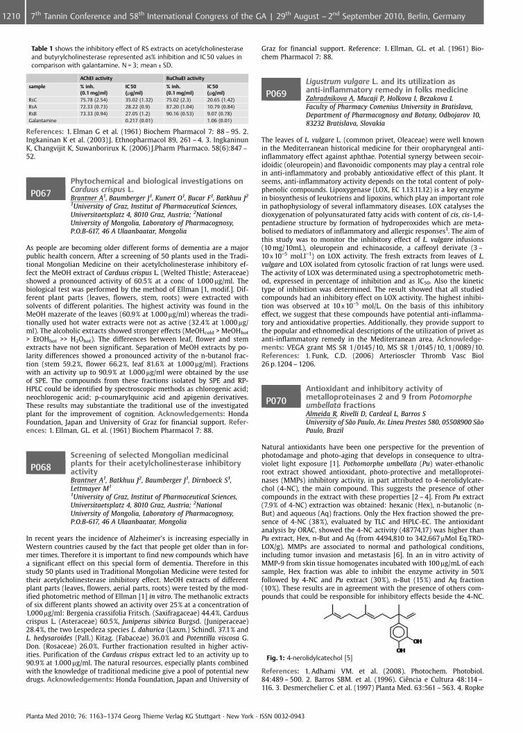
Table 1 shows the inhibitory effect of RS extracts on acetylcholinesteraseand butyrylcholinesterase represented as% inhibition and IC 50 values incomparison with galantamine. N = 3; mean € SD.
AChEI activity BuChuEI activity
sample % inh.(0.1 mg/ml)
IC 50(mg/ml)
% inh.(0.1 mg/ml)
IC 50(mg/ml)
RsC 75.78 (2.54) 35.02 (1.32) 75.02 (2.3) 20.65 (1.42)RsA 72.33 (0.73) 28.22 (0.9) 87.20 (1.04) 10.79 (0.84)RsB 73.33 (0.94) 27.05 (1.2) 90.16 (0.53) 9.07 (0.78)Galantamine 0.217 (0.01) 1.06 (0.01)
References: 1. Elman G et al. (1961) Biochem Pharmacol 7: 88 – 95. 2.Ingkaninan K et al. (2003) J. Ethnopharmacol 89, 261 – 4. 3. IngkaninunK, Changvijit K, Suwanborirux K. (2006) J.Pharm Pharmaco. 58(6):847 –52.
P067Phytochemical and biological investigations onCarduus crispus L.Brantner A1, Baumberger J1, Kunert O1, Bucar F1, Batkhuu J2
1University of Graz, Institut of Pharmaceutical Sciences,Universitaetsplatz 4, 8010 Graz, Austria; 2NationalUniversity of Mongolia, Laboratory of Pharmacognosy,P.O.B-617, 46 A Ulaanbaatar, Mongolia
As people are becoming older different forms of dementia are a majorpublic health concern. After a screening of 50 plants used in the Tradi-tional Mongolian Medicine on their acetylcholinesterase inhibitory ef-fect the MeOH extract of Carduus crispus L. (Welted Thistle; Asteraceae)showed a pronounced activity of 60.5% at a conc of 1.000 mg/ml. Thebiological test was performed by the method of Ellman [1, modif.]. Dif-ferent plant parts (leaves, flowers, stem, roots) were extracted withsolvents of different polarities. The highest activity was found in theMeOH mazerate of the leaves (60.9% at 1.000 mg/ml) whereas the tradi-tionally used hot water extracts were not as active (32.4% at 1.000 mg/ml). The alcoholic extracts showed stronger effects (MeOHcold > MeOHhot
> EtOHhot >> H2Ohot). The differences between leaf, flower and stemextracts have not been significant. Separation of MeOH extracts by po-larity differences showed a pronounced activity of the n-butanol frac-tion (stem 59.2%, flower 66.2%, leaf 81.6% at 1.000 mg/ml). Fractionswith an activity up to 90.9% at 1.000 mg/ml were obtained by the useof SPE. The compounds from these fractions isolated by SPE and RP-HPLC could be identified by spectroscopic methods as chlorogenic acid;neochlorogenic acid; p-coumarylquinic acid and apigenin derivatives.These results may substantiate the traditional use of the investigatedplant for the improvement of cognition. Acknowledgements: HondaFoundation, Japan and University of Graz for financial support. Refer-ences: 1. Ellman, GL. et al. (1961) Biochem Pharmacol 7: 88.
P068Screening of selected Mongolian medicinalplants for their acetylcholinesterase inhibitoryactivityBrantner A1, Batkhuu J2, Baumberger J1, Dirnboeck S1,Lettmayer M1
1University of Graz, Institut of Pharmaceutical Sciences,Universitaetsplatz 4, 8010 Graz, Austria; 2NationalUniversity of Mongolia, Laboratory of Pharmacognosy,P.O.B-617, 46 A Ulaanbaatar, Mongolia
In recent years the incidence of Alzheimer’s is increasing especially inWestern countries caused by the fact that people get older than in for-mer times. Therefore it is important to find new compounds which havea significant effect on this special form of dementia. Therefore in thisstudy 50 plants used in Traditional Mongolian Medicine were tested fortheir acetylcholinesterase inhibitory effect. MeOH extracts of differentplant parts (leaves, flowers, aerial parts, roots) were tested by the mod-ified photometric method of Ellman [1] in vitro. The methanolic extractsof six different plants showed an activity over 25% at a concentration of1,000 mg/ml: Bergenia crassifolia Fritsch. (Saxifragaceae) 44.4%, Carduuscrispus L. (Asteraceae) 60.5%, Juniperus sibirica Burgsd. (Juniperaceae)28.4%, the two Lespedeza species L. dahurica (Laxm.) Schindl. 37.1% andL. hedysaroides (Pall.) Kitag. (Fabaceae) 36.0% and Potentilla viscosa G.Don. (Rosaceae) 26.0%. Further fractionation resulted in higher activ-ities. Purification of the Carduus crispus extract led to an activity up to90.9% at 1.000 mg/ml. The natural resources, especially plants combinedwith the knowledge of traditional medicine give a pool of potential newdrugs. Acknowledgements: Honda Foundation, Japan and University of
Graz for financial support. Reference: 1. Ellman, GL. et al. (1961) Bio-chem Pharmacol 7: 88.
P069Ligustrum vulgare L. and its utilization asanti-inflammatory remedy in folks medicineZahradnikova A, Mucaji P, Holkova I, Bezakova LFaculty of Pharmacy Comenius University in Bratislava,Department of Pharmacognosy and Botany, Odbojarov 10,83232 Bratislava, Slovakia
The leaves of L. vulgare L. (common privet, Oleaceae) were well knownin the Mediterranean historical medicine for their oropharyngeal anti-inflammatory effect against aphthae. Potential synergy between secoir-idoidic (oleuropein) and flavonoidic components may play a central rolein anti-inflammatory and probably antioxidative effect of this plant. Itseems, anti-inflammatory activity depends on the total content of poly-phenolic compounds. Lipoxygenase (LOX, EC 1.13.11.12) is a key enzymein biosynthesis of leukotriens and lipoxins, which play an important rolein pathophysiology of several inflammatory diseases. LOX catalyses thedioxygenation of polyunsaturated fatty acids with content of cis, cis-1,4-pentadiene structure by formation of hydroperoxides which are meta-bolised to mediators of inflammatory and allergic responses1. The aim ofthis study was to monitor the inhibitory effect of L. vulgare infusions(10 mg/10mL), oleuropein and echinacoside, a caffeoyl derivate (3 –10 x 10�5 mol.l�1) on LOX activity. The fresh extracts from leaves of L.vulgare and LOX isolated from cytosolic fraction of rat lungs were used.The activity of LOX was determinated using a spectrophotometric meth-od, expressed in percentage of inhibition and as IC50. Also the kinetictype of inhibition was determined. The result showed that all studiedcompounds had an inhibitory effect on LOX activity. The highest inhibi-tion was observed at 10 x 10�5 mol/L. On the basis of this inhibitoryeffect, we suggest that these compounds have potential anti-inflamma-tory and antioxidative properties. Additionally, they provide support tothe popular and ethnomedical descriptions of the utilization of privet asanti-inflammatory remedy in the Mediterranean area. Acknowledge-ments: VEGA grant MS SR 1/0145/10, MS SR 1/0145/10, 1/0089/10.References: 1. Funk, C.D. (2006) Arterioscler Thromb Vasc Biol26 p.1204 – 1206.
P070Antioxidant and inhibitory activity ofmetalloproteinases 2 and 9 from Potomorpheumbellata fractionsAlmeida R, Rivelli D, Cardeal L, Barros SUniversity of S¼o Paulo, Av. Lineu Prestes 580, 05508900 S¼oPaulo, Brazil
Natural antioxidants have been one perspective for the prevention ofphotodamage and photo-aging that develops in consequence to ultra-violet light exposure [1]. Pothomorphe umbellata (Pu) water-ethanolicroot extract showed antioxidant, photo-protective and metalloprotei-nases (MMPs) inhibitory activity, in part attributed to 4-nerolidylcate-chol (4-NC), the main compound. This suggests the presence of othercompounds in the extract with these properties [2 – 4]. From Pu extract(7.9% of 4-NC) extraction was obtained: hexanic (Hex), n-butanolic (n-But) and aqueous (Aq) fractions. Only the Hex fraction showed the pre-sence of 4-NC (38%), evaluated by TLC and HPLC-EC. The antioxidantanalysis by ORAC, showed the 4-NC activity (48774,17) was higher thanPu extract, Hex, n-But and Aq (from 4494,810 to 342,667mMol Eq.TRO-LOX/g). MMPs are associated to normal and pathological conditions,including tumor invasion and metastasis [6]. In an in vitro activity ofMMP-9 from skin tissue homogenates incubated with 100 mg/mL of eachsample, Hex fraction was able to inhibit the enzyme activity in 50%followed by 4-NC and Pu extract (30%), n-But (15%) and Aq fraction(10%). These results are in agreement with the presence of others com-pounds that could be responsible for inhibitory effects beside the 4-NC.
Fig. 1: 4-nerolidylcatechol [5]
References: 1. Adhami VM. et al. (2008). Photochem. Photobiol.84:489 – 500. 2. Barros SBM. et al. (1996). CiÞncia e Cultura 48:114 –116. 3. Desmerchelier C. et al. (1997) Planta Med. 63:561 – 563. 4. Ropke
Planta Med 2010; 76: 1163–1374 Georg Thieme Verlag KG Stuttgart · New York · ISSN 0032-0943
1210 7th Tannin Conference and 58th International Congress of the GA | 29th August – 2nd September 2010, Berlin, Germany
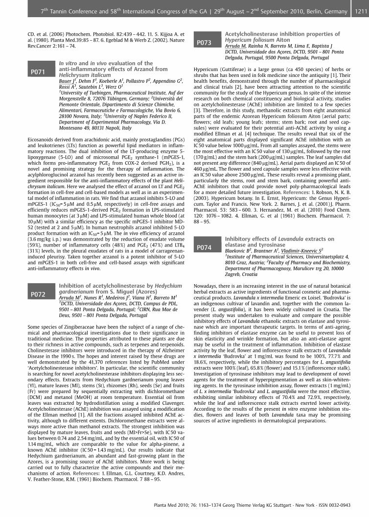
CD. et al. (2006) Photochem. Photobiol. 82:439 – 442. 11. 5. Kijjoa A. etal. (1980). Planta Med.39:85 – 87. 6. Egeblad M & Werb Z. (2002). NatureRev.Cancer 2:161 – 74.
P071In vitro and in vivo evaluation of theanti-inflammatory effects of Arzanol fromHelichrysum italicumBauer J1, Dehm F1, Koeberle A1, Pollastro F2, Appendino G2,Rossi A3, Sautebin L3, Werz O1
1University of Tuebingen, Pharmaceutical Institute, Auf derMorgenstelle 8, 72076 T�bingen, Germany; 2Universit delPiemonte Orientale, Dipartimento di Scienze Chimiche,Alimentari, Farmaceutiche e Farmacologiche, Via Bovio 6,28100 Novara, Italy; 3University of Naples Federico II,Department of Experimental Pharmacology, Via D.Montesano 49, 80131 Napoli, Italy
Eicosanoids derived from arachidonic acid, mainly prostaglandins (PGs)and leukotrienes (LTs) function as powerful lipid mediators in inflam-matory reactions. The dual inhibition of the LT-producing enzyme 5-lipoxygenase (5-LO) and of microsomal PGE2 synthase-1 (mPGES-1,which forms pro-inflammatory PGE2 from COX-2 derived PGH2), is anovel and promising strategy for the therapy of inflammation. Theacylphloroglucinol arzanol has recently been suggested as an active in-gredient responsible for the anti-inflammatory effects of the plant Heli-chrysum italicum. Here we analysed the effect of arzanol on LT and PGE2
formation in cell-free and cell-based models as well as in an experimen-tal model of inflammation in rats. We find that arzanol inhibits 5-LO andmPGES-1 (IC50= 5mM and 0.5mM, respectively) in cell-free assays andefficiently reduces mPGES-1-derived PGE2 formation in LPS-stimulatedhuman monocytes (at 3mM) and LPS-stimulated human whole blood (at10 mM) with a similar efficiency as the specific mPGES-1 inhibitor MD-52 (tested at 2 and 5mM). In human neutrophils arzanol inhibited 5-LOproduct formation with an IC50= 5mM. The in vivo efficiency of arzanol(3.6 mg/kg i. p.) was demonstrated by the reduction of exudate volume(59%), number of inflammatory cells (48%) and PGE2 (47%) and LTB4
(31%) levels, in the pleural exudates of rats in a model of carrageenan-induced pleurisy. Taken together arzanol is a potent inhibitor of 5-LOand mPGES-1 in both cell-free and cell-based assays with significantanti-inflammatory effects in vivo.
P072Inhibition of acetylcholinesterase by Hedychiumgardnerianum from S. Miguel (Azores)Arruda M1, Nunes R1, Medeiros J2, Viana H1, Barreto M1
1DCTD, Universidade dos A�ores, DCTD, Campus de PDL,9501 – 801 Ponta Delgada, Portugal; 2CIRN, Rua Mae deDeus, 9501 – 801 Ponta Delgada, Portugal
Some species of Zingiberaceae have been the subject of a range of che-mical and pharmacological investigations due to their significance intraditional medicine. The properties attributed to these plants are dueto their richness in active compounds, such as terpenes and terpenoids.Cholinesterase inhibitors were introduced in the therapy of AlzheimerDisease in the 1990 s. The hopes and interest raised by these drugs arewell demonstrated by the 41,370 references listed by PubMed under‘Acetylcholinesterase inhibitors’. In particular, the scientific communityis searching for novel acetylcholinesterase inhibitors displaying less sec-ondary effects. Extracts from Hedychium gardnerianum young leaves(Yl), mature leaves (Ml), stems (St), rhizomes (Rh), seeds (Se) and fruits(Fr) were prepared by sequentially extracting with dichloromethane(DCM) and metanol (MeOH) at room temperature. Essential oil fromleaves was extracted by hydrodistillation using a modified Clavenger.Acetylcholinesterase (AChE) inhibition was assayed using a modificationof the Ellman method [1]. All the fractions assayed inhibited AChE ac-tivity, although to different extents. Dichloromethane extracts were al-ways more active than methanol extracts. The strongest inhibition wasdisplayed by mature leaves, fruits and seeds (Ml>Fr>Se), with IC 50 va-lues between 0.74 and 2.54 mg/mL, and by the essential oil, with IC50 of1.14 mg/mL, which are comparable to the value for alpha-pinene, aknown AChE inhibitor (IC 50 = 1.43 mg/mL). Our results indicate thatHedychium gardnerianum, an abundant and fast-growing plant in theAzores, is a promising source of AChE inhibitors. More work is beingcarried out to fully characterize the active compounds and their me-chanisms of action. References: 1. Ellman, G.L. Courtney, K.D. Andres,V. Feather-Stone, R.M. (1961) Biochem. Pharmacol. 7 88 – 95.
P073Acetylcholinesterase inhibition properties ofHypericum foliosum AitonArruda M, Rainha N, Barreto M, Lima E, Baptista JDCTD, Universidade dos A�ores, DCTD, 9501 – 801 PontaDelgada, Portugal, 9500 Ponta Delgada, Portugal
Hypericum (Guttiferae) is a large genus (ca 450 species) of herbs orshrubs that has been used in folk medicine since the antiquity [1]. Theirhealth benefits, demonstrated through the number of pharmacologicaland clinical trials [2], have been attracting attention to the scientificcommunity for the study of the Hypericum genus. In spite of the intenseresearch on both chemical constituency and biological activity, studieson acetylcholinesterase (AChE) inhibition are limited to a few species[3]. Therefore, in this study, methanolic extracts from eight anatomicalparts of the endemic Azorean Hypericum foliosum Aiton (aerial parts;flowers; old leafs; young leafs; stems; stem bark; root and seed cap-sules) were evaluated for their potential anti-AChE activity by using amodified Ellman et al. [4] technique. The results reveal that six of theeight anatomical parts displayed significant AChE inhibition with anIC 50 value below 1000 mg/mL. From all samples assayed, the stems werethe most effective with an IC50 value of 130 mg/mL, followed by the root(170 g/mL) and the stem bark (200 mg/mL) samples. The leaf samples didnot present any difference (840 mg/mL). Aerial parts displayed an IC 50 of460 mg/mL. The flower and seed capsule samples were less effective withan IC 50 value above 2500 mg/mL. These results reveal a promising plant,particularly the stems, root and stem bark, containing powerful anti-AChE inhibitors that could provide novel poly-pharmacological leadsfor a more detailed future investigation. References: 1. Robson, N. K. B.(2003). Hypericum botany. In E. Ernst, Hypericum: the Genus Hyperi-cum. Taylor and Francis. New York. 2. Barnes, J. et al. (2001) J. Pharm.Pharmacol. 53: 583 – 600. 3. Hernandez, M. et al. (2010) Food Chem.120: 1076 – 1082. 4. Ellman, G. et al (1961) Biochem. Pharmacol. 7:88 – 95.
P074Inhibitory effects of Lavandula extracts onelastase and tyrosinaseBlaekovic B2, Brantner A1, Vladimir-Kneevic S2
1Institute of Pharmaceutical Sciences, Universitaetsplatz 4,8010 Graz, Austria; 2Faculty of Pharmacy and Biochemistry,Department of Pharmacognosy, Marulicev trg 20, 10000Zagreb, Croatia
Nowadays, there is an increasing interest in the use of natural botanicalherbal extracts as active ingredients of functional cosmetic and pharma-ceutical products. Lavandula x intermedia Emeric ex Loisel. ‘Budrovka’ isan indigenous cultivar of lavandin and, together with the common la-vender (L. angustifolia), it has been widely cultivated in Croatia. Thepresent study was undertaken to evaluate and compare the possibleinhibitory effects of Lavandula ethanolic extracts on elastase and tyrosi-nase which are important therapeutic targets. In terms of anti-ageing,finding inhibitors of elastase enzyme can be useful to prevent loss ofskin elasticity and wrinkle formation, but also an anti-elastase agentmay be useful in the treatment of inflammation. Inhibition of elastaseactivity by the leaf, flower and inflorescence stalk extracts of Lavandulax intermedia ‘Budrovka’ at 1 mg/mL was found to be 100%, 77.7% and18.6%, respectively, while the inhibitory percentages for L. angustifoliaextracts were 100% (leaf), 65.8% (flower) and 15.1% (inflorescence stalk).Investigation of tyrosinase inhibitors may lead to development of novelagents for the treatment of hyperpigmentation as well as skin-whiten-ing agents. In the tyrosinase inhibition assay, flower extracts (1 mg/mL)of L. x intermedia ‘Budrovka’ and L. angustifolia were the most effective,exhibiting similar inhibitory effects of 70.4% and 72.9%, respectively,while the leaf and inflorescence stalk extracts exerted lower activity.According to the results of the present in vitro enzyme inhibition stu-dies, flowers and leaves of both Lavandula taxa may be promisingsources of active ingredients in dermatological preparations.
Planta Med 2010; 76: 1163–1374 Georg Thieme Verlag KG Stuttgart · New York · ISSN 0032-0943
12117th Tannin Conference and 58th International Congress of the GA | 29th August – 2nd September 2010, Berlin, Germany
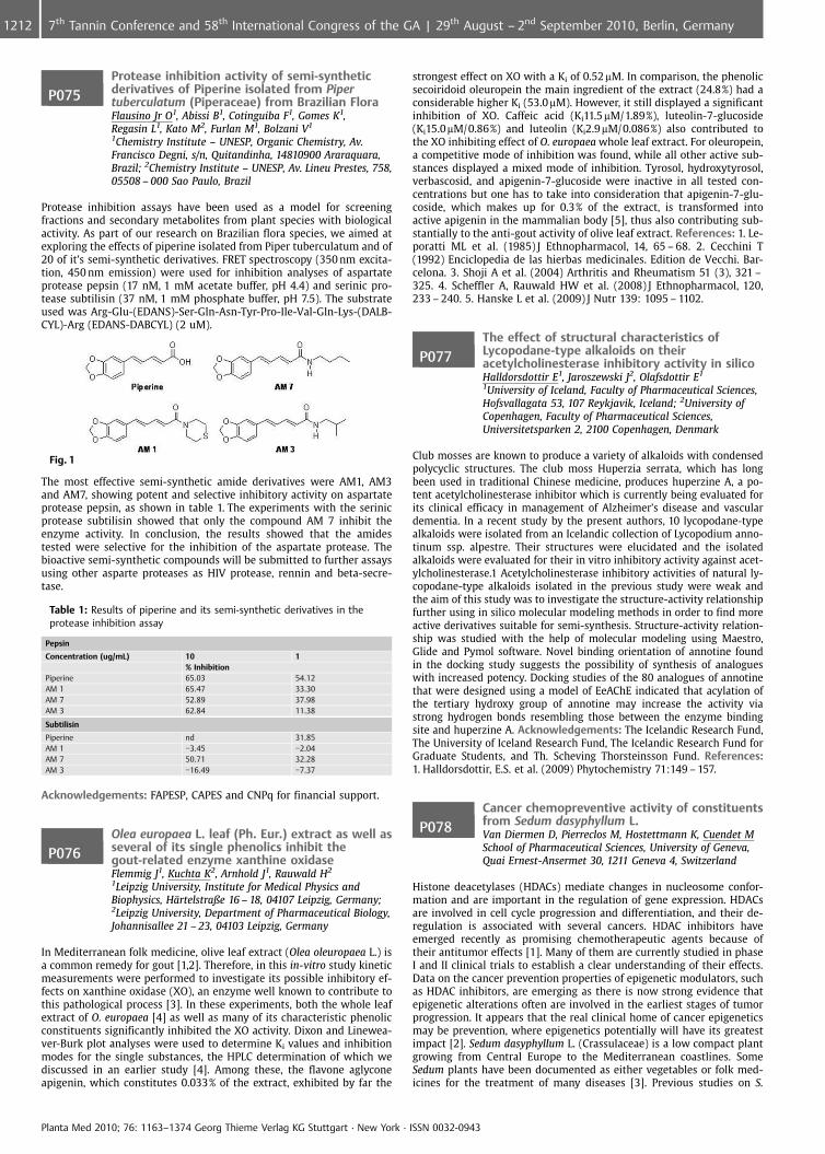
P075Protease inhibition activity of semi-syntheticderivatives of Piperine isolated from Pipertuberculatum (Piperaceae) from Brazilian FloraFlausino Jr O1, Abissi B1, Cotinguiba F1, Gomes K1,Regasin L1, Kato M2, Furlan M1, Bolzani V1
1Chemistry Institute – UNESP, Organic Chemistry, Av.Francisco Degni, s/n, Quitandinha, 14810900 Araraquara,Brazil; 2Chemistry Institute – UNESP, Av. Lineu Prestes, 758,05508 – 000 Sao Paulo, Brazil
Protease inhibition assays have been used as a model for screeningfractions and secondary metabolites from plant species with biologicalactivity. As part of our research on Brazilian flora species, we aimed atexploring the effects of piperine isolated from Piper tuberculatum and of20 of it’s semi-synthetic derivatives. FRET spectroscopy (350 nm excita-tion, 450 nm emission) were used for inhibition analyses of aspartateprotease pepsin (17 nM, 1 mM acetate buffer, pH 4.4) and serinic pro-tease subtilisin (37 nM, 1 mM phosphate buffer, pH 7.5). The substrateused was Arg-Glu-(EDANS)-Ser-Gln-Asn-Tyr-Pro-Ile-Val-Gln-Lys-(DALB-CYL)-Arg (EDANS-DABCYL) (2 uM).
Fig. 1
The most effective semi-synthetic amide derivatives were AM1, AM3and AM7, showing potent and selective inhibitory activity on aspartateprotease pepsin, as shown in table 1. The experiments with the serinicprotease subtilisin showed that only the compound AM 7 inhibit theenzyme activity. In conclusion, the results showed that the amidestested were selective for the inhibition of the aspartate protease. Thebioactive semi-synthetic compounds will be submitted to further assaysusing other asparte proteases as HIV protease, rennin and beta-secre-tase.
Table 1: Results of piperine and its semi-synthetic derivatives in theprotease inhibition assay
Pepsin
Concentration (ug/mL) 10 1% Inhibition
Piperine 65.03 54.12AM 1 65.47 33.30AM 7 52.89 37.98AM 3 62.84 11.38
Subtilisin
Piperine nd 31.85AM 1 �3.45 �2.04AM 7 50.71 32.28AM 3 �16.49 �7.37
Acknowledgements: FAPESP, CAPES and CNPq for financial support.
P076Olea europaea L. leaf (Ph. Eur.) extract as well asseveral of its single phenolics inhibit thegout-related enzyme xanthine oxidaseFlemmig J1, Kuchta K2, Arnhold J1, Rauwald H2
1Leipzig University, Institute for Medical Physics andBiophysics, H�rtelstraße 16 – 18, 04107 Leipzig, Germany;2Leipzig University, Department of Pharmaceutical Biology,Johannisallee 21 – 23, 04103 Leipzig, Germany
In Mediterranean folk medicine, olive leaf extract (Olea oleuropaea L.) isa common remedy for gout [1,2]. Therefore, in this in-vitro study kineticmeasurements were performed to investigate its possible inhibitory ef-fects on xanthine oxidase (XO), an enzyme well known to contribute tothis pathological process [3]. In these experiments, both the whole leafextract of O. europaea [4] as well as many of its characteristic phenolicconstituents significantly inhibited the XO activity. Dixon and Linewea-ver-Burk plot analyses were used to determine Ki values and inhibitionmodes for the single substances, the HPLC determination of which wediscussed in an earlier study [4]. Among these, the flavone aglyconeapigenin, which constitutes 0.033% of the extract, exhibited by far the
strongest effect on XO with a Ki of 0.52mM. In comparison, the phenolicsecoiridoid oleuropein the main ingredient of the extract (24.8%) had aconsiderable higher Ki (53.0 mM). However, it still displayed a significantinhibition of XO. Caffeic acid (Ki11.5 mM/1.89%), luteolin-7-glucoside(Ki15.0mM/0.86%) and luteolin (Ki2.9mM/0.086%) also contributed tothe XO inhibiting effect of O. europaea whole leaf extract. For oleuropein,a competitive mode of inhibition was found, while all other active sub-stances displayed a mixed mode of inhibition. Tyrosol, hydroxytyrosol,verbascosid, and apigenin-7-glucoside were inactive in all tested con-centrations but one has to take into consideration that apigenin-7-glu-coside, which makes up for 0.3% of the extract, is transformed intoactive apigenin in the mammalian body [5], thus also contributing sub-stantially to the anti-gout activity of olive leaf extract. References: 1. Le-poratti ML et al. (1985) J Ethnopharmacol, 14, 65 – 68. 2. Cecchini T(1992) Enciclopedia de las hierbas medicinales. Edition de Vecchi. Bar-celona. 3. Shoji A et al. (2004) Arthritis and Rheumatism 51 (3), 321 –325. 4. Scheffler A, Rauwald HW et al. (2008) J Ethnopharmacol, 120,233 – 240. 5. Hanske L et al. (2009) J Nutr 139: 1095 – 1102.
P077The effect of structural characteristics ofLycopodane-type alkaloids on theiracetylcholinesterase inhibitory activity in silicoHalldorsdottir E1, Jaroszewski J2, Olafsdottir E1
1University of Iceland, Faculty of Pharmaceutical Sciences,Hofsvallagata 53, 107 Reykjavik, Iceland; 2University ofCopenhagen, Faculty of Pharmaceutical Sciences,Universitetsparken 2, 2100 Copenhagen, Denmark
Club mosses are known to produce a variety of alkaloids with condensedpolycyclic structures. The club moss Huperzia serrata, which has longbeen used in traditional Chinese medicine, produces huperzine A, a po-tent acetylcholinesterase inhibitor which is currently being evaluated forits clinical efficacy in management of Alzheimer’s disease and vasculardementia. In a recent study by the present authors, 10 lycopodane-typealkaloids were isolated from an Icelandic collection of Lycopodium anno-tinum ssp. alpestre. Their structures were elucidated and the isolatedalkaloids were evaluated for their in vitro inhibitory activity against acet-ylcholinesterase.1 Acetylcholinesterase inhibitory activities of natural ly-copodane-type alkaloids isolated in the previous study were weak andthe aim of this study was to investigate the structure-activity relationshipfurther using in silico molecular modeling methods in order to find moreactive derivatives suitable for semi-synthesis. Structure-activity relation-ship was studied with the help of molecular modeling using Maestro,Glide and Pymol software. Novel binding orientation of annotine foundin the docking study suggests the possibility of synthesis of analogueswith increased potency. Docking studies of the 80 analogues of annotinethat were designed using a model of EeAChE indicated that acylation ofthe tertiary hydroxy group of annotine may increase the activity viastrong hydrogen bonds resembling those between the enzyme bindingsite and huperzine A. Acknowledgements: The Icelandic Research Fund,The University of Iceland Research Fund, The Icelandic Research Fund forGraduate Students, and Th. Scheving Thorsteinsson Fund. References:1. Halldorsdottir, E.S. et al. (2009) Phytochemistry 71:149 – 157.
P078Cancer chemopreventive activity of constituentsfrom Sedum dasyphyllum L.Van Diermen D, Pierreclos M, Hostettmann K, Cuendet MSchool of Pharmaceutical Sciences, University of Geneva,Quai Ernest-Ansermet 30, 1211 Geneva 4, Switzerland
Histone deacetylases (HDACs) mediate changes in nucleosome confor-mation and are important in the regulation of gene expression. HDACsare involved in cell cycle progression and differentiation, and their de-regulation is associated with several cancers. HDAC inhibitors haveemerged recently as promising chemotherapeutic agents because oftheir antitumor effects [1]. Many of them are currently studied in phaseI and II clinical trials to establish a clear understanding of their effects.Data on the cancer prevention properties of epigenetic modulators, suchas HDAC inhibitors, are emerging as there is now strong evidence thatepigenetic alterations often are involved in the earliest stages of tumorprogression. It appears that the real clinical home of cancer epigeneticsmay be prevention, where epigenetics potentially will have its greatestimpact [2]. Sedum dasyphyllum L. (Crassulaceae) is a low compact plantgrowing from Central Europe to the Mediterranean coastlines. SomeSedum plants have been documented as either vegetables or folk med-icines for the treatment of many diseases [3]. Previous studies on S.
Planta Med 2010; 76: 1163–1374 Georg Thieme Verlag KG Stuttgart · New York · ISSN 0032-0943
1212 7th Tannin Conference and 58th International Congress of the GA | 29th August – 2nd September 2010, Berlin, Germany
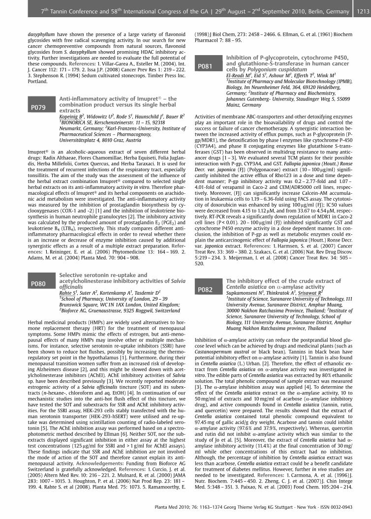
dasyphyllum have shown the presence of a large variety of flavonoidglycosides with free radical scavenging activity. In our search for newcancer chemopreventive compounds from natural sources, flavonoidglycosides from S. dasyphyllum showed promising HDAC inhibitory ac-tivity. Further investigations are needed to evaluate the full potential ofthese compounds. References: 1. Villar-Garea A., Esteller M. (2004). Int.J. Cancer 112: 171 – 179. 2. Issa J.P. (2008) Cancer Prev Res 1: 219 – 222.3. Stephenson R. (1994) Sedum cultivated stonecrops. Timber Press Inc.Portland.
P079Anti-inflammatory activity of Imupret� – thecombination product versus its single herbalextractsKopeinig B2, Widowitz U2, Rode S1, Haunschild J1, Bauer R2
1BIONORICA SE, Kerschensteinerstr. 11 – 15, 92318Neumarkt, Germany; 2Karl-Franzens-University, Institute ofPharmaceutical Sciences – Pharmacognosy,Universit�tsplatz 4, 8010 Graz, Austria
Imupret� is an alcoholic-aqueous extract of seven different herbaldrugs: Radix Althaeae, Flores Chamomillae, Herba Equiseti, Folia Juglan-dis, Herba Millefolii, Cortex Quercus, and Herba Taraxaci. It is used forthe treatment of recurrent infections of the respiratory tract, especiallytonsillitis. The aim of the study was the assessment of the influence ofthe herbal extract combination Imupret� compared to selected singleherbal extracts on its anti-inflammatory activity in vitro. Therefore phar-macological effects of Imupret� and its herbal components on arachido-nic acid metabolism were investigated. The anti-inflammatory activitywas measured by the inhibition of prostaglandin biosynthesis by cy-clooxygenases (COX-1 and -2) [1] and the inhibition of leukotriene bio-synthesis in human neutrophile granulocytes [2]. The inhibitory activitywas calculated by the produced amount of prostaglandin E2 (PGE2) andleukotriene B4 (LTB4), respectively. This study compares different anti-inflammatory pharmacological effects in order to reveal whether thereis an increase or decrease of enzyme inhibition caused by additionalsynergistic effects as a result of a multiple extract preparation. Refer-ences: 1. Reininger, E. et al. (2006) Phytomedicine 13: 164 – 169. 2.Adams, M. et al. (2004) Planta Med. 70: 904 – 908.
P080Selective serotonin re-uptake andacetylcholinesterase inhibitory activities of SalviaofficinalisRahte S1, Suter A2, Kortenkamp A1, Tasdemir D1
1School of Pharmacy, University of London, 29 – 39Brunswick Square, WC 1N 1AX London, United Kingdom;2Bioforce AG, Gruenaustrasse, 9325 Roggwil, Switzerland
Herbal medicinal products (HMPs) are widely used alternatives to hor-mone replacement therapy (HRT) for the treatment of menopausalsymptoms. Some HMPs mimic the effects of estrogen, but anti-meno-pausal effects of many HMPs may involve other or multiple mechan-isms. For instance, selective serotonin re-uptake inhibitors (SSRI) havebeen shown to reduce hot flushes, possibly by increasing the thermo-regulatory set point in the hypothalamus [1]. Furthermore, during theirmenopausal transition women suffer from an increased risk of develop-ing Alzheimers disease [2], and this might be slowed down with acet-ylcholinesterase inhibitors (AChEI). AChE inhibitory activities of Salviasp. have been described previously [3]. We recently reported moderateestrogenic activity of a Salvia officinalis tincture (SOT) and its subex-tracts (n-hexane-, chloroform and aq. EtOH) [4]. In continuation of ourmechanistic studies into the anti-hot flush effect of this tincture, wehave tested the SOT and subextracts for SSR and AChE inhibitory activ-ities. For the SSRI assay, HEK-293 cells stably transfected with the hu-man serotonin transporter (HEK-293-hSERT) were utilised and re-up-take was determined using scintillation counting of radio-labeled sero-tonin [5]. The AChE inhibition assay was performed based on a spectro-photometric method described by Ellman [6]. Neither SOT, nor the sub-extracts displayed significant inhibition in either assay at the highesttest concentrations (125 mg/ml for SSRI and > 1 g/ml for AChEI assays).These findings indicate that SSR and AChE inhibition are not involvedthe mode of action of the SOT and therefore cannot explain its anti-menopausal activity. Acknowledgements: Funding from Bioforce AGSwitzerland is gratefully acknowledged. References: 1. Curcio, J. et al.(2005) Altern Med Rev. 10: 216 – 221. 2. Mulnard, R. et al. (2000) JAMA283: 1007 – 1015. 3. Houghton, P. et al. (2006) Nat Prod Rep. 23: 181 –199. 4. Rahte S. et al (2008). Planta Med. 75: 1073. 5. Ramamoorthy, E.
(1998) J Biol Chem, 273: 2458 – 2466. 6. Ellman, G. et al. (1961) BiochemPharmacol 7: 88 – 95.
P081Inhibition of P-glycoprotein, cytochrome P450,and glutathione-S-transferase in human cancercells by Polygonium cuspidatumEl-Readi M1, Eid S1, Ashour M1, Efferth T2, Wink M1
1Institute of Pharmacy and Molecular Biotechnology (IPMB),Biology, Im Neuenheimer Feld, 364, 69120 Heidelberg,Germany; 2Institute of Pharmacy and Biochemistry,Johannes Gutenberg- University, Staudinger Weg 5, 55099Mainz, Germany
Activities of membrane ABC-transporters and other detoxifying enzymesplay an important role in the bioavailability of drugs and control thesuccess or failure of cancer chemotherapy. A synergistic interaction be-tween the increased activity of efflux pumps, such as P-glycoprotein (P-gp/MDR1), the detoxification by phase I enzymes like cytochrome P-450(CYP3A4), and phase II conjugating enzymes like glutathione S-trans-ferases (GST) has been observed in multidrug resistance to many antic-ancer drugs [1 – 3]. We evaluated several TCM plants for their possibleinteraction with P-gp, CYP3A4, and GST. Fallopia japonica (Houtt.) RonseDecr. var. japonica (FJ) (Polygonaceae) extract (10 – 100 mg/ml) signifi-cantly inhibited the active efflux of Rho123 in a dose and time depen-dent manner; P-gp inhibitory activity was 0.2 – 2.77-fold and 1.07 –4.01-fold of verapamil in Caco-2 and CEM/ADR5000 cell lines, respec-tively. Moreover, (FJ) can significantly increase Calcein-AM accumula-tion in leukaemia cells to 1.19 – 6.36-fold using FACS assay. The cytotoxi-city of doxorubicin was enhanced by using 100 mg/ml (FJ); IC50 valueswere decreased from 4.15 to 1.12mM, and from 33.67 to 4.54mM, respec-tively. RT-PCR reveals a significantly down regulation of MDR1 in Caco-2cell lines (P < 0.01). 20 – 100 mg/ml (FJ) inhibited significantly GST andcytochrome P450 enzyme activity in a dose dependent manner. In con-clusion, the inhibition of P-gp as well as metabolic enzymes could ex-plain the anticarcinogenic effect of Fallopia japonica (Houtt.) Ronse Decr.var. japonica extract. References: 1. Harmsen, S. et al. (2007) CancerTreat Rev. 33: 369 – 380. 2. Szakacs, G. et al. (2006) Nat. Rev Drug Discov.5:219 – 234. 3. Meijerman, I. et al. (2008) Cancer Treat Rev. 34: 505 –520.
P082The inhibitory effect of the crude extract ofCentella asiatica on a-amylase activitySupkamonseni N1, Thinkratok A1, Srisawat R2
1Institute of Science, Suranaree University of Technology, 111University Avenue, Suranaree District, Amphur Muang,30000 Nakhon Ratchasima Province, Thailand; 2Institute ofScience, Suranaree University of Technology, School ofBiology, 111 University Avenue, Suranaree District, AmphurMuang Nakhon Ratchasima province, Thailand
Inhibition of a-amylase activity can reduce the postprandial blood glu-cose level which can be achieved by drugs and medicinal plants (such asCastanospermum austral or black bean). Tannins in black bean havepotential inhibitory effect on a-amylase activity [1]. Tannin is also foundin Centella asiatica (L.) Urban. [2]. Therefore, the effect of ethanolic ex-tract from Centella asiatica on a-amylase activity was investigated invitro. The edible parts of Centella asiatica was extracted by 80% ethanolicsolution. The total phenolic compound of sample extract was measured[3]. The a-amylase inhibition assay was applied [4]. To determine theeffect of the Centella asiatica extract on the a-amylase activity, 10 to50 mg/ml of extracts and 10 mg/ml of acarbose (a-amylase inhibitorydrug), and active compounds found in Centella asiatica (tannin, rutin,and quercetin) were prepared. The results showed that the extract ofCentella asiatica contained total phenolic compound equivalent to97.45 mg of gallic acid/g dry weight. Acarbose and tannin could inhibita-amylase activity (97.6% and 37.9%, respectively). Whereas, quercetinand rutin did not inhibit a-amylase activity which was similar to thestudy of Jo et al. [5]. Moreover, the extract of Centella asiatica had a-amylase inhibitory activity (11.4%) at the final concentration of 30 mg/ml while other concentrations of this extract had no inhibition.Although, the percentage of inhibition by Centella asiatica extract wasless than acarbose, Centella asiatica extract could be a benefit candidatefor treatment of diabetes mellitus. However, further in vivo studies areneeded to be investigated. References: 1. Carmona, A. et al. (1996) J.Nutr. Biochem. 7:445 – 450. 2. Zheng, C. J. et al. (2007) J. Chin IntegeMed. 5:348 – 351. 3. Paixao, N. et al. (2003) Food Chem. 105:204 – 214.
Planta Med 2010; 76: 1163–1374 Georg Thieme Verlag KG Stuttgart · New York · ISSN 0032-0943
12137th Tannin Conference and 58th International Congress of the GA | 29th August – 2nd September 2010, Berlin, Germany
4. Nickavar, B. et al. (2008) Iran. J. Pharm. Res. 7:297 – 303. 5. Jo, S-H. etal. (2010) Int. J. Applied Res. Natural Products 2:52 – 60.
P083Screening of some medicinal plants forantidiabetic activityNickavar B, Mojab FSchool of Pharmacy, Shahid Beheshti University of MedicalSciences, Department of Pharmacognosy, P.O. Box: 14155 –6153 Tehran, Iran, Islamic Republic Of
Diabetes mellitus is one of the most serious chronic diseases character-ized by chronic hyperglycemia. Traditional herbal remedies are still inuse by diabetic patients especially in 3rd World Countries. The presentwork was carried out to investigate the potential antidiabetic effects ofsome Iranian medicinal plants using an in vitro a-amylase inhibitionassay. The ethanol extracts obtained from ten plants (Trigonella foenu-graecum, Camellia sinensis, Urtica dioica, Vaccinium arctostaphylos, Urticapilulifera, Calendula officinalis, Juglans regia, Olea europaea, Salvia offici-nalis, Arctium lappa) were tested on inhibitory activity of a-amylase,expressed as IC50 values, calculated from Log concentration-responsecurves. As positive control acarbose was used. Among the tested sam-ples, Camellia sinensis (Theaceae) leaf (IC50= 1.54 mg/ml), Trigonella foe-num-graecum (Fabacae) seed (IC50= 1.87 mg/ml) and leaf (IC50= 1.92 mg/ml), and Urtica dioica (Urticaceae) leaf (IC50= 1.89 mg/ml) revealed ap-preciable a-amylase inhibitory activities in a concentration-dependentmanner.
P084An antidiabetic compound from Vacciniumarctostaphylos berriesNickavar B1, Amin G2
1School of Pharmacy, Shahid Beheshti University of MedicalSciences, Department of Pharmacognosy, P.O. Box: 14155 –6153 Tehran, Iran, Islamic Republic Of; 2Faculty ofPharmacy, Tehran University of Medical Sciences,Department of Pharmacognosy, P.O. Box: 14155 – 6451,Tehran, Iran, Islamic Republic Of
Vaccinium arctostaphylos L. (Ericaceae) is a compact shrub up to 3 m inheight which grows wildly in the northern forests of Iran. Its berries areedible cherry and are used as an antidiabetic agent in Traditional IranianMedicine. The present study deals with the bioassay guided fractiona-tion of V. arctostaphylos berry extract and in vitro a-amylase enzymeinhibition assay of the extract and fractions for their antidiabetic activ-ities. The crude extract showed a suitable dose-dependent inhibitoryeffect against a-amylase activity [IC50= 1.91 (1.89 – 1.94) mg/mL]. Theactivity guided fractionation of the extract led to the isolation and pur-ification of an anthocyanin from it. The compound was identified asmalvidin-3-O-b-glucoside by determination of its hydrolytic and spec-tral data. The compound demonstrated a suitable dose-dependent en-zyme inhibitory activity [IC50= 0.16 (0.16 – 0.17) mg/mL].
Fertility management by natural products
P085Study the abortificant effects of pennyroyaldecoction in pregnant miceZeinali Nasrabadi F1, Dashti-R M2, Hosseini Biuki S3,Anvari M2
1Medical student, Shaheed Sadoughi medical UniversityYazd, Iran, Islamic Republic Of; 2Assistant Professor, ShaheedSadoughi medical University of Yazd yazd, Iran, IslamicRepublic Of; 3pharmacology student, Shaheed Sadoughimedical University of Yazd yazd, Iran, Islamic Republic Of
Objective: Pennyroyal (Mentha pulegium L.) is a widely available herbthat has long been used as an abortifacient. In this study, the effects of aPennyroyal decoction on foetuses in pregnant Mice was assessed. Meth-ods: 20 female BALB/c mice, weighting 25 – 30 grams, were breed in theanimal house of the medical college. The first day of pregnancy was theday on which the vaginal plaque was observed. The pregnant mice weredivided into 2 subgroups. Each pregnant animal was placed in a separatecage throughout the gestational period. Animals in the control groupreceived tap water, animals of the test groups received an aqueous pen-nyroyal decoction in the 2nd trimester of the gestational period. In 18thday of pregnancy, animals were anesthetized and their foetuses wereextracted through a cesarean section. The placenta was excised,
weighed, and the number and placement of implantation sites, live,dead and resorbed foetuses were recorded. All foetuses were stereomicroscopically examined for any morphological abnormalities. Result:There was no significant difference between the mean number of re-sorbed and dead fetuses of the test and control group. Conclusion:Although the number of included animals is very low, our data indicatethat Pennyroyal has no negative effect on pregnancy.
P086Effect of ethanolic extract Anacyclus pyrethrumDC on reproductive parameter of male ratsSharma V, Thakur M, Chauhan N, Dixit VDepartment of Pharmaceutical Sciences Dr. H.S.GourUniversity, Dr. H.S. Gour University, 470003 Sagar, India
Vajikaran is a speciality in Ayurvedic system of medicine in India dealingwith herbs possessing rejuvenative and revitalizing properties for im-proving sexual dynamics and reproduction capacity. Anacyclus pyre-thrum root is used traditionally used to enhance sexual power. An in-sight in the scientific literature appraised insufficient data to validatethe rejuvinative property of Anacyclus pyrethrum. The study was there-fore performed to evaluate the effectiveness of ethanolic extract of Ana-cyclus pyrethrum at a dose equivalent to 50, 100 and 150 mg/kg b.w.respectively in improving overall sexual behavior and reproductive para-meters in male rats. Following parameters were evaluated the effect ofextract on body and organ weights, change in histoarchitecture of testis,testosterone, FSH, LH level in blood, fructose level in seminal vesicles,sperm parameters and sexual behavior was studied. Administration ofethanolic extract had pronounced anabolic and spermatogenic effect intreated animals as evidenced by weight gains in the body and reproduc-tive organs. Improvement in sexual behavior of animals as reflected inreduction of mount and intromission latency, an increase in mount andintromission frequency and enhanced attractability towards female. Theincreased spermatogenesis in treated group was confirmed by change inhistoarchitecture and increase sperm count. The level of testosteroneFSH, LH and fructose content in seminal vesicles was significantly in-creases in treated groups. These studies suggest the possible role ofalkylamides of Anacyclus pyrethrum in improving sexual function by itsaction on HPG axis. These findings support the folk use of this plant asVajikaran. Acknowledgements: AICTE, New Delhi for providing Na-tional Doctoral Fellowship. References: 1. Thakur M, Dixit VK,(2007)Sex Disabil. 25 (4): 203 – 207. 2. Chauhan NS, Sharma V, Dixit VK,(2008) Nat Prod Res. DOI: 10.1080/14786410802588493. 3. ChauhanNS, Dixit VK,(2009) Int J Impot Res. DOI:10.1038/ijir.2009.62.
Indigenous knowledge of traditional medicine and evidencebased herbal medicine
P087Nephroprotective and nephrocurative potentialof Momordica dioica Roxb in streptozotocininduced diabetic nephropathyJain A, Lodhi S, Jain N, Singhai ADr H S Gour University, Dept of Pharmaceutical Sciences,Sagar, 470003 Sagar, India
Momordica dioica Roxb (MD) is a climbing creeper and known as Kakorain Hindi. This plant possesses hypoglycemic gastroprotective and ulcerhealing activities. Fruit is reported for hepatoprotective, nephroprotec-tive and antidiabetic potential [1,2]. On the basis of traditional claim MDfruit was taken up for the study. Dried and powdered MD fruit wasdefatted and extracted with 95% ethanol. For screening of the activity,Wistar albino rats of either sex were divided into normal control, dia-betic control and treated groups. Diabetes was induced with single doseof streptozotocin (55 mg/kg i. p.). In protective regimen, 4 – 30 days ex-perimental period, ethanolic extract of MD fruit was administered asorally at 200 and 400 mg/kg/day, whereas curative regimen IT was ad-ministered onward 18 days upto 30 days. The final serum glucose, urea,creatinine and urine albumin levels were measured as functional profileof kidney function which was found to be significantly lower in treatedgroups than the diabetic control group. Malondialdehyde (MDA) wassignificantly (p < 0.001) lowered in treated groups compare to diabeticcontrol group. Treated groups have shown significant increased(p < 0.001) in reduced glutathione (GSH) level which was reduced indiabetic control group compare to control groups. Change in bodyweight was found to be non significant. Results were supported byhistopathological studies. Acknowledgments: Indian Council of MedicalResearch, New Delhi, India for financial support. References:1. Jain A,
Planta Med 2010; 76: 1163–1374 Georg Thieme Verlag KG Stuttgart · New York · ISSN 0032-0943
1214 7th Tannin Conference and 58th International Congress of the GA | 29th August – 2nd September 2010, Berlin, Germany
Singahi AK (2008) Nat Prod Res (DOI: 10.1080/14786410802267569). 2.Reddy, GT, Kumar, BR, Mohan, GK, Mullangi, R, (2006) Asian J of Phar-macody Pharmacokin 6(4):327 – 329.
P088Preventive and curative effects of Phyllanthusacidus (L.) Skeels extract against thioacetamideinduced acute liver damage in Wistar ratsJain N, Jain A, Lodhi S, Singhai ADr. H. S. Gour University, Department of PharmaceuticalSciences, Department of Pharmaceutical Sciences,Department of Pharmaceutical Sciences, Sagar (M.P.),470003 Sagar, India
Phyllanthus acidus (L.) Skeels has long been used in folk medicine totreat chronic liver diseases [1, 2]. In present study, protection of 70%ethanolic extracts of Phyllanthus acidus aerial parts (PAE) was evaluatedin male Wistar albino rats against thioacetamide (TAA)-induced hepaticdamage in preventive and curative models. Experimental rats received asingle dose of TAA (100 mg/kg, s. c.) as hepatotoxin [3]. In preventivemodel, PAE was given orally at a dose of 125 and 250 mg/kg, 48, 24 and2 h prior to TAA administration, meanwhile in curative model; rats weretreated with PAE at a dose of 125 and 250 mg/kg, 2, 24 and 48 h afterTAA intoxication. The protective effect was compared with Silymarin(100 mg/kg, p. o.). Rats pre-treated with PAE and Silymarin remarkablyprevented the elevation of serum aspartate transaminase (AST), alaninetransaminase (ALT), lactate dehydrogenase (LDH) and malondialdehyde(MDA) in TAA-treated rats. Rats treated with PAE and Silymarin after theestablishment of TAA liver injury showed significant (p £0.05) protec-tion of liver as evidenced from normal AST, ALT, LDH and MDA levelscompared with toxin control rats. Hepatic glutathione (GSH) and otherantioxidant enzymatic levels of superoxide dismutase (SOD), catalase(CAT) and glutathione peroxidase (GPx) were significantly (p £0.05)increased by extract treatment in both experimental groups. Histo-pathological observation of rat livers also indicated hepatoprotectionagainst TAA liver damage. Phytochemical studies revealed the presenceof saponins, triterpenes and phenolic compounds in PAE which could beresponsible for the possible hepatoprotective action. References: 1. Kir-tikar, K.R., Basu, B.D. (1987) Indian Medicinal Plants. Probasi Press. Cal-cutta. 2. Unander, D.W. et al. (1995) J. Ethno. 45: 1 – 18. 3. Kumar, G. et al.(2004) J. Ethno. 92:37 – 40.
P089Application of Leonurus cardiaca L. oil extract fortreatment of psycho neurological disorders inclinicShikov A1, Pozharitskaya O1, Demchenko D1, Makarov V1,Shikh E2
1St-Petersburg Institute of Pharmacy, 47/5, Piskarevskyprospect, 195067 St-Petersburg, Russian Federation;2Institute of Clinical Pharmacology, Moscow MedicalAcademy, 11, Yauzskaya str., 109240 Moscow, RussianFederation
Leonurus cardiaca L. has a long history of application in Russian tradi-tional medicine and now it is described in a number of pharmacopoeiasaround the world. L. cardiaca is used in form of tinctures and tabletswith dry extract and has been demonstrated to have sedative, hypoten-sive and cardiotonic effects. Recently an oil extract of L. cardiaca (OEL)was prepared by roto-pulsed extraction [1]. Its effects on animals werestudied [2], but clinical application was not described. Fifty patientswith stage 1 and stage 2 arterial hypertension accompanied by anxietyand sleep disorders were treated 28 days with 1200 mg (4 capsule) OELper day. OEL was standardized in iridoids (0.15 mg/capsule) [3]. Subjec-tive complaints of patients with stage 1 hypertension improved after14 days, with stage 2 hypertension after 21 days of treatment and weresignificantly different from baseline after 28 days of treatment. Anxietywas reduced by 61% and 62%, emotional liability – by 53% and 20%,headache – by 41% and 34% and sleep disorders by 47% and 42% re-spectively. A statistically significant decrease and normalization in bloodpressure was noted at 21 days and 28 days of treatment respectively.According to CGI scale large improvement and no side effects was ob-served for 32%, moderate effect for 48%, and weak effect for 8% of thepatients. Only 12% of the patients did not respond to therapy. Thus thissmall uncontrolled pilot-study shows a positive effect of OEL on psycho-emotional status and arterial blood pressure of patients with psychoneurological disorders. References: 1. Shikov, A.N. (2006) Pharmaceuti-cal Chemistry Journal 40(7):385 – 388. 2. Makarov, V.G. et al. (2006)
Eksp Klin Farmakol. 69: 23 – 25. 3. Kosman, V.M. et al. (2002) Pharma-ceutical Chemistry Journal 36:96 – 99.
P090Gastroprotective effect of methanol extract ofOxalis corniculata Linn (whole plant)experimental animalsSakat S, Tupe P, Juvekar AInstitute of Chemical Technology, Department ofPharmaceutical Science and Technology, N.P. Marg,Matunga, 400019 Mumbai, India
In Indian traditional medicine, whole plant of Oxalis corniculata Linn(family: Oxalidaceae) is used to treat gastric and related disorders [1].However, there are no scientific evidences available on its anti-ulceractivity. Hence, present study was carried out to investigate anti-ulcereffect of O. corniculata (whole plant) methanol extract using pyloricligation and indomethacin induced gastric ulceration in the Sparque-Dawely rats (180 – 200 g) at the dose levels of 125, 250 and 500 mg/kg.In pylorus ligation induced ulceration model, various parameters werestudied such as gastric secretions, its pH, total acidity, free acidity, andulcer index, where as in indomethacin induced ulceration model, ulcerindex and percentage inhibition of ulceration were calculated. Raniti-dine, a standard anti-ulcer/antisecretory agent was used as positive con-trol at the dose 100 mg/kg. Results were analyzed by One-way analysisof variance (ANOVA) followed by Dunnet’s t-test. Oral treatment of O.corniculata (whole plant) showed, significant (p < 0.01) decrease in thegastric secretions, total acidity and free acidity. However, pH of thegastric content was significantly (p < 0.05) increased only at higher dose,500 mg/kg. It showed also significant (p < 0.01) decrease in number ofulcers and ulcer score index in pyloric ligation and indomethacin in-duced ulceration models. Overall, results of the study showed that, themethanol extract of O. corniculata possess significant anti-ulcer proper-ties in a dose dependent manner. Preliminary phytochemical evaluationof the extract showed the presence of flavonoids, which are among thecytoprotective materials for which anti-ulcerogenic efficacy has beenconfirmed. Hence, the present anti-ulcer activity of O. corniculata extractcould be attributed to the presence of flavonoids. References: 1. Kiriti-kar K., Basu B., (1984) Indian medicinal plants, Lalit Mohan Basu. Deh-radun. 437.
P091Lupeol derivatives from the Eastern Nigeriamistletoe, Loranthus micranthus Linn.(Loranthaceae) with enhanced cell proliferativepotentialsOsadebe P1, Omeje E1, Kawamura A2, Okoye F1
1University of Nigeria, Nsukka, Pharmaceutical andMedicinal Chemsitry, Faculty of Pharmaceutical Sciences,University of Nigeria Nsukka, 41001 Enugu, Nigeria; 2CityUniversity of the New York, Department of Chemistry,Hunter College of CUNY, The City University of New York,U.S.A, Department of Chemistry, Hunter College of CUNY,The City University of New York, U.S.A, 10065 New York,United States
Bioassay-guided fractionation of the crude extract of Eastern Nigerianmistletoe Loranthus micranthus Linn. (Loranthaceae) showed that thehexane fraction was very active in terms of immunostimulation withenhanced macrophage activation and cell proliferative abilities [1, 2].Further fractionation and purification of the hexane fraction led to theisolation of a known triterpenoid, lupeol and some derivatives whichwere esters of either a C 14 or C 16 fatty acid. Characterization and un-equivocal structural assignments were achieved by a combination of UV/visible, IR, NMR, MS, COSY experiments including, HMBC, HQSC, NOEs,NOESY. The characterized compounds exhibited mild immunostimula-tory activity on cell line (C 57Bl/6 splenocytes) and flow cytometry tech-niques against Lipopolysaccharide and Concanavalin A standards. This isthe first report of the presence and immune stimulation potentials ofthe triterpenoid, Lupeol and derivatives isolated from our local mistle-toe. Acknowledgements: The authors specially acknowledge Dr. C. SNworu for the Pharmacological tests. References: 1. Osadebe PO, et al(2009) J Ethnopharmacology, 125:287 – 293. 2. Osadebe et al (2008),RPMP Vol 27: 473 – 485.
Planta Med 2010; 76: 1163–1374 Georg Thieme Verlag KG Stuttgart · New York · ISSN 0032-0943
12157th Tannin Conference and 58th International Congress of the GA | 29th August – 2nd September 2010, Berlin, Germany
P092Investigation of Thai traditional way fordetoxification of aloeGritsanapan W1, Tangyuenyongwatana P2
1Faculty of Pharmacy, Mahidol University, Department ofPharmacognosy, 447 Sri-Ayudthaya Road, RatchatewiBangkok, Thailand; 2Faculty of Oriental Medicine, RangsitUniversity, 52/347 Muang Aek, 12000 Pathumtani, Thailand
In Thai traditional medicine, dried juice extract (DJE) of Aloe vera (L.)Burm has been used in many formulations for a long time [1]. The Thaitraditional way recommends detoxifying the aloe DJE before combiningwith other herbal constituents. The reason for detoxification is to de-crease the gastrointestinal tract irritation of aloe. The traditional methodto detoxify aloe is adding small amount of water into the DJE powderand heating in clay pot until the dried and crispy mixture is formed. Theobjective of this study is to verify the chemical conversion of majoringredients in aloe DJE using HPTLC analysis (Silica gel 60 GF254, sol-vent system; ethyl acetate: methanol: water 100: 13.5: 10, detection at420 nm). Our study found that the amount of a major compound aloin(Rf 0.43) in the DJE was decreased after heating while the amount of itsaglycone aloe-emodin (Rf 0.80) was increased from 50 mg/mL to473.2 mg/mL. This result explains that hydrolysis and oxidation reactionscan occur and some aloin is conversed to aloe-emodin resulted in de-creasing anthraquinone glycoside intensity, which can cause irritation ofthe gastrointestinal tract and increase the chance for gripping action [2].Further study of other glycosides and aglycones in the DJE of aloe is inprogress. References: 1. Poomchusri, NT. (1973) Ayurvedic study. 2ndedn, Bangkok; Promjakkanpimp. 2. Robber, J E.; Speedie, M K.; Tyler, VE. (1996) Pharmacognosy and Pharmacobiotechnology. Pennsylvania;Williams & Wilkins, p 52.
P093Hypericum species in Estonian folk traditions andin local scientific studiesRaal A1, Soukand R2, Nagel K1
1University of Tartu, Department of Pharmacy, Nooruse 1,50411 Tartu, Estonia; 2Estonian Literary Museum,Vanemuise 42, 51003 Tartu, Estonia
Both Hypericum perforatum and H. maculatum are common speciesthroughout Estonia and neighbouring countries. The study used folkloretexts from the HERBA (1) database collected between 1868 – 1994 onmedicinal plants in Estonia. 117 texts describing 137 usage cases ofplants bearing vernacular names related to naistepuna [Hypericum inEstonian, direct translation – women’s red] were selected and analyzedusing textological methods. Almost 30% of the usage cases were relatedto gynecological problems (as the name implies), followed by 14% forstomach problems, 10% for bladder and kidney problems and 5% eachfor stomachache caused by overexertion, skin diseases and pain.Although altogether 8 species belonging to 7 genera or even familiesbore such vernacular names in different places, the most frequentlyused plants were two species of genus Hypericum. Those two externallyvery similar species were not distinguished by ordinary people and wereused in folk medicine as one. The content of hypericin (2) and totalflavonoids before (3) and after (4) hydrolysis of glucosides was deter-mined spectrophotometrically in both Hypericum species. The standarddeviations of these methods did not exceed 0.5%, 8.7% and 5.4%, respec-tively (n = 4). The flowering tops (15 cm) and flowers in three differentblooming stages (at the beginning of blooming, in full bloom and at theend of blooming) of H. perforatum and H. maculatum, collected in 2008from South-Estonia (Antsla parish), were used as plant material. H. ma-culatum contained about a 2.5 times more hypericin (141 – 228 mg%)than H. perforatum (75 – 81 mg%). The amount of hypericin detectedwas higher in the flowers and lower in the flowering tops of both ana-lyzed Hypericum species, but the comparative analysis of H. perforatumja H. maculatum showed no similar tendencies for flavonoid content.Both species contained practically the same amount of total flavonoids(5,6 – 6,2% and 4,6 – 6,0%, respectively), but H. perforatum containedsomewhat more flavonoid aglycones (1,1 – 2,0%) than H. maculatum(0,7 – 1,5%). It is necessary to continue this work. References: 1. S�u-kand, R., Kalle, R. (2008) HERBA: Historical Estonian Folkmedical HerbalDatabase – http://herba.folklore.ee. 2. Ceskoslovensky l�kopis (1987),vol. 4. Avicenum, Praha. 3. U.S.S.R. Pharmacopoeia (1987), vol. 11. Med-itsina, Moscow [in Russian]. 4. Georgijevski, V.P. et al. (1990) Hayka,Novosibirsk [in Russian].
P094Structure-based design of new analogs ofWithaferin A as antitumor agentsJimenez C1, LLanos G2, Araujo L3, Moujir L3, Rodriguez J1,Jim�nez I2, Bazzocchi I2
1Universidade da Coru�2Universidad de La Laguna,Instituto Universitario Bio-Org�nica Antonio Gonz�lez, Av.Astrof�sico Francisco S�nchez 2, 38206 La Laguna, Tenerife,Spain; 3Universidad de La Laguna, Departamento deMicrobiolog�a y Biolog�a Celular, Avenida Astrof�sicoFrancisco S�nchez s/n, 38206 La Laguna, Tenerife, Spain
The endemic plant from Canary Islands (Spain), Withania aristata (Ai-ton) Pauqui (Solanaceae), popularly known as “orobal” [1] is widely usedin folk medicine as antitumor, antispasmodic, antirheumatic agent;further for eye and otitis problems, as well as for insomnia and urinarypathologies [2]. We have found that the acetone extract from the leavesof this species is a rich source of the known whitanolide, withaferin A(more than 0.2% dried weight). The therapeutic potential of Withaniaspecies has been attributed to the presence of withanolides [3] and, inparticular, withaferin A which showed anti-cancer activity in vivo andhas been reported to induce cytoskeletal disruption, to inhibit cell mo-bility and NFkB activation [4], and to induce apoptosis in vitro [5]. Tak-ing this into account, we have prepared analogs of withaferin A forstructure-activity relationship studies. We have performed several che-mical transformations on ring A, on the epoxy group of ring B, and onthe lactone and hydroxyl groups of the side chain of withaferin A, in-cluding oxidations, reductions and halogenations. All semi-syntheticanalogs were tested for their potential anticancer activity against fourtumor cell lines. The biological results obtained from these analogs al-lowed us to deduce a number of structure-activity relationships, whichconfirmed the potential of withaferin A in cancer chemotherapy. Thesemi-synthesis, the bioactivity data and the SAR studies will be pre-sented in the accompanying poster. References: 1. Anderson GJ et al(2006) Am. J. Bot. 93:1295 – 1305. 2. Benjumea D et al (2009) J Ethno-pharmacol 123: 351 – 355. 3. Veleiro AS et al (2005) Studies in NaturalProducts Chemistry, Bioactive Natural Products Atta-Ur-Raman, ElsevierScience Publishers, Amsterdam, p 1019 – 52. 4. Ichikawa H. (2006) Mol.Cancer Ther. 5: 1434. 5. Malik F et al (2007) Apoptosis 12: 2115 – 2133.
P095Investigation on possible interaction ofcranberry polyphenols and warfarin using invitro and in vivo studiesKimura Y, Sasaki N, Ito H, Hatano TDivision of Pharmaceutical Sciences, Okayama UniversityGraduate School of Medicine, Dentistry and PharmaceuticalSciences, Pharmaceutical Sciences, 1 – 1-1 Tsushimanaka,Kita-ku, 7008530 Okayama, Japan
Cranberry (Vaccinium macrocarpon Ait., Ericaceae), distributed in NorthAmerica, has traditionally been used in the treatment and prevention ofurinary tract infections. Various types of constituents including terpe-noids, iridoids, and polyphenols, such as proanthocyanidins, anthocya-nins, flavonoids, and organic acids are contained in cranberry. Amongthem, A-type proanthocyanidins have been revealed to inhibit the ad-herence of uropathogenic P-fimbriated Escherichia coli to eukaryoticcells, and this is thought to be the mechanism underlying its role inpreventing urinary tract infection. However, some case reports showedthe interaction between cranberry and warfarin. [1, 2] The possible in-teraction of cranberry with warfarin was suggested to depend on theinhibition of cytochrome P450 (CYP) activities, which is involved inmost metabolism-mediated drug interaction. We recently reported theinhibitory effect of 60 polyphenols on CYP3A4 and CYP2C 9 activity,indicating that some cranberry flavonoids can significantly inhibit theseCYPs [3] In this study, we investigated CYP3A4 and CYP2C 9 inhibitoryactivities of cranberry juice constituents and their metabolites. Thebioassay-guided fractionation of cranberry extracts gave the cranberry-derived proanthocyanidin polymer (CPP) as the most potent inhibitor ofCYP3A4 and CYP2C9. Furthermore, we investigated the plasma level ofwarfarin and alternation in international normalized ratio after inges-tion of CPP or cranberry juice (CJ) to rats, indicating that co-administra-tion of CJ was unlikely to affect the pharmacokinetics and pharmacody-namics of warfarin in rats. Further investigation and improved casestudies are needed to address these issues. References: 1. Committeeon Safety of Medicines, Medicines and Healthcare Products RegulatoryAgency. Possible interaction between warfarin and cranberry juice.(2003) Curr. Probl. Pharmacovigil. 29:8. 2. Suvarna, R. et al. Possible
Planta Med 2010; 76: 1163–1374 Georg Thieme Verlag KG Stuttgart · New York · ISSN 0032-0943
1216 7th Tannin Conference and 58th International Congress of the GA | 29th August – 2nd September 2010, Berlin, Germany
interaction between warfarin and cranberry juice. (2003) BMJ. 327:1454.3. Kimura, Y. et al. (2010) Food Chem. Toxicol. 48:429 – 435.
P096Antiplasmodial remedies from Europeanrenaissance herbals: HPLC based activityprofiling of Alisma plantago-aquatica extract forantiplasmodial activity, and isolation of activedammarane triterpenoidsAdams M1, Gschwind S1, Zimmermann S1, Brun R2,Hamburger M1
1Institute of Pharmaceutical Biology, Department ofPharmaceutical Sciences, Klingelbergstrasse 50, 4056 Basel,Switzerland; 2Swiss Tropical and Public Health Institute,Socinstrasse 57, 4002 Basel, Switzerland
The common water plantain Alisma plantago-aquatica L. (Alismataceae)was used in the 16th and 17th centuries in Central Europe to treat Plas-modium vivax malaria (tertian fever). The Renaissance herbals by Bock(1532), Brunfels (1532), Mathioli (1560), and Zwinger (1696) describedthe internal use of alcohol extracts of the tubers to treat this disease,which was quite common in German speaking areas then [1]. In thecourse of a screen of remedies from Renaissance herbals an extract ofwater plantain tubers was active against P. falciparum (77% inhibition at4.9 mg/ml). With analytical scale time-based HPLC separation and testingof one-minute fractions in combination with HPLC hyphenated methods(HPLC-PDA, -MSn, HR-MS, off line microprobe NMR) the active sub-stances were identified as acetylated dammarane triterpenes. Seven ofthese compounds were isolated using normal phase medium pressurecolumn chromatography and semi-preparative HPLC. Structure elucida-tion was achieved by extensive 1 H and 13C NMR analysis. The dammar-anes had IC50s ranging from 3.3 to 7.0mM. This is the first report ofantiplasmodial activity of this triterpenoid class, and the first result ofour ongoing project of screening for antiprotozoal natural products fromremedies used in European Renaissance medicine. References: 1. AdamsM, et al. (2010) J Ethnopharm, submitted. 2. Adams M, et al.(2009) NatProd Comm., 10:1377 – 81.
P097Effect of Rhoicissus digitata extracts onacetylcholine-mediated contraction in rat uterusSchou J1, Kristiansen U1, Stafford G2, van Heerden F2,J�ger A3
1Univeristy of Copenhagen, Universitetsparken 2, 2100Copenhagen, Denmark; 2Univeristy of KwaZulu-Natal, PBagX01, 3209 Scottsville-Pietermaritzburg, South Africa;3University of Copenhagen, Medicinal Chemistry,Universitetsparken 2, 2100 Copenhagen, Denmark
Rhoicissus digitata (L.f.) Gilg & M. Brandt (Vitaceae) is used in traditionalmedicine in Southern Africa to tone the uterus during pregnancy and tofacilitate delivery. Aqueous and methanolic extracts were prepared frompowdered dry leaves and roots of R. digitata. The uterine horns weresurgically removed from female Sprague Dawley rats in estrous, placedin de Jalon solution (NaCl: 9 g/l, KCl: 0,42 g/l, NaHCO3: 1,50 g/l, Glucose:0,5 g/l, CaCl2: 0,12 g/l) and hung in the experimental setup in the uteruscontraction assay. After mounting, the uterine horns were allowed toequilibrate in the organ bath for 30 min, renewing the solution every10 min by flushing. The organ strips were incubated with extract in aconcentration of 1.3 mg/ml for 5 min before starting cumulative additionof acetylcholine. The water extract of the root and the methanol extractof the leaves increased the response significantly (p < 0.0001 for both(two-way ANOVA)), and the methanol extract of the root inhibited theresponse significantly (p < 0.0001). The results suggests that the use ofwater extracts (decoctions) of R. digitata roots in South African tradi-tional medicine as pregnancy related medicine can be justified, as theyincrease the acetylcholine-mediated uterine contractions in rats. Theuterus contracting effect of the methanol extract of the leaves of R.digitata suggests that alcoholic leaf extracts could be used as substitutesfor root extracts, which as a renewable source could help with sustain-able usage of these species.
P098Investigation of the antidiabetic activity of Morusalba leaf extract in vitro and in vivoHunyadi A1, Liktor-Busa E1, Balogh �2, Hsieh T3, Zupk� I2,Hohmann J1
1University of Szeged, Institute of Pharmacognosy, E�tv�sstr. 6., 6720 Szeged, Hungary; 2University of Szeged,Department of Pharmacodynamics and Biopharmacy,E�tv�s str. 6., 6720 Szeged, Hungary; 3Kaohsiung MedicalUniversity, Department of Medical Genetics, 100 Shih-Chuan1st rd., 807 Kaohsiung, Taiwan
The leaves of white mulberry (Morus alba L.) are widely used in thetraditional medicine of the type II diabetes mellitus. Several groups ofplant metabolites are held to be responsible for the complex antidiabeticeffect of the drug: iminosugars, flavonoids and other phenolic com-pounds, ecdysteroids, glycoproteins, and, as we recently suggested,megastigmanes and glycosylated volatile compounds [1,2]. As an initialstep of a research project aiming to clarify the importance of differentantidiabetic constituents, here we report the in vitro and in vivo activityand chemical evaluation of the 70% EtOH extract of mulberry leaves. Invitro, fully differentiated adipocytes were used. 50 mg/mL of extract wasadministrated into the culture medium containing 15 mM glucose, withor without the addition of 0.32 mM insulin, and the antidiabetic activitywas determined by the remaining glucose concentration in the mediumafter 24 hours. A significant effect was found in both systems. The in vivoantidiabetic effect was evaluated in non insulin dependent diabeticSPRD rats. In this model, diabetes was induced by intraperitoneal ad-ministration of streptozotocin (150 mg/kg) to newborn rats. Oral treat-ment of fasted animals with the extract (750 mg/kg) resulted in a sig-nificant decrease of postprandial glucose level. Composition of the ex-tract was characterized by gradient RP-HPLC, and the potentially activechlorogenic acid [3], rutin and isoquercitrin were determined as majorconstituents, while quercetin, apigenin, morin and 20-hydroxyecdysonewere present in much lower amounts. The contribution of each com-pound to the measured antidiabetic activity needs further investigation.Acknowledgements: This project was supported by the Hungarian Na-tional Research Fund (OTKA; PD 75383), the New Hungary DevelopmentPlan (T�MOP-4.2.2 – 08/1 – 2008 – 0013) and by the grant from the Na-tional Science Council of Taiwan (NSC 98 – 2314-B-037 – 011-MY3). Re-ferences: 1. Hunyadi A et al. (2007) Planta Med. 73: 941. 2. Hunyadi A etal. (2008) Planta Med. 74: 1117. 3. Andrade-Cetto et al. (2001) J. Ethno-pharmacol. 78:145 – 149.
P099Evaluation of antioxidant and DNA protectionactivities in the extracts of the Terminalia catappaleavesChen Z, Lin C, Wang H, Chen HChia-Nan University of Pharmacy and Science, Institute ofCosmetic Science, 60, Erh-Jen RD., Sec.1, Jen-Te, 717 Tainan,Taiwan
Terminalia catappa (Combretaceae) is used commonly as a folk medicinein Taiwan, and has been claimed to have therapeutic effects in hepatitisand liver related diseases [1, 2]. The leaves, bark and fruit of the Termi-nalia catappa have also been commonly used as a folk medicine forantidiarrheic, antipyretic and haemostatic purposes in India, Philippines,Malaysia and Indonesia [3]. The extracts from the Terminalia catappaleaves were used in this study. Antioxidant activities were measuredby both 1, 1-diphenyl-2-picrylhydrazyl radical (DPPH·) scavenging and2, 2-azino-bis (3-ethylbenz- thiazoline-6-sulfonic acid) (ABTS·+) deco-lourisation methods. To further evaluate the effect of Terminalia catappaextracts on UV induced DNA damages, the DNA protection assay wasemployed. Terminalia catappa leaf extracts had effective DPPH bleachingactivity and ABTS·+ radical scavenging activity in a concentration depen-dent manner. Furthermore, the Terminalia catappa leaf extracts to inhi-bit the oxidative DNA damages were assessed by measuring the conver-sion of supercoiled pUC 119 plasmid DNA to the linear forms. UV irradia-tion of DNA with hydrogen peroxide resulted in the formation of linearforms of DNA, indicating double-strand DNA breaks. Addition of Termi-nalia catappa plant extracts to DNA resulted in a partial inhibition of theconversion of supercoiled DNA to linear forms, indicating that the Ter-minalia catappa plant extract was able to protect plasmid DNA againsthydroxyl radical induced oxidative damage. The inhibition of hydroxylradical induced DNA strand breaks by Terminalia catappa plant extractsexhibited a concentration dependent relationship. Acknowledgements:Committee on Chinese Medicine and Pharmacy (Grant No. CCMP 98-RD-006), Chia Nan University of Pharmacy and Science. References:
Planta Med 2010; 76: 1163–1374 Georg Thieme Verlag KG Stuttgart · New York · ISSN 0032-0943
12177th Tannin Conference and 58th International Congress of the GA | 29th August – 2nd September 2010, Berlin, Germany
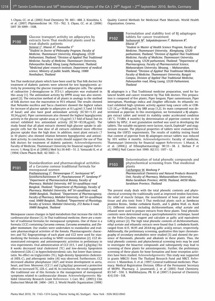
1. Chyau, CC. et al. (2002) Food Chemistry 78: 483 – 488. 2. Kinoshita, S.et al. (2007) Phytomedicine 14: 755 – 762. 3. Chyau, CC. et al. (2006)LWT 39:1099 – 1108.
P100Glucose transport activity on adipocytes byextracts form Thai medicinal plants used totreat diabetic patientsSteinrat L1, Itharat A2, Poonsatha S3
1Student in Doctor of Philosophy Program, Faculty ofMedicine, Thammasart University, Klongluang, 12120Pathumtani, Thailand; 2Division of Applied Thai TraditionalMedicine, Faculty of Medicine, Thammasart University,Pahonyothin Road, Klong Luang Pathumtani, Thailand;3Medicinal plant research institute, Department of medicalscience, Ministry of public health, Muang, 11000Nonthaburi, Thailand
Ten Thai medicinal plants which have been used by Thai folk doctors fortreatment of diabetic patients were selected for test hypoglycemic ac-tivity by promoting the glucose transport in adipocyte cells. The uptakeof radioactive 2-deoxyglucose in 3T3-L 1 adipocytes was evaluated inthis assay [1]. The antioxidant activity by DPPH assay was also investi-gated [2]. The extraction method of all plants in analogy to the methodof folk doctors was the maceration in 95% ethanol. The results showedthat Nelumbo nucifera and Tacca chantrieri showed the highest valuesfor increase of glucose uptake in adipocytes at 0.1 mg/ml (1.7and 1.6 foldof basal) and also have good antioxidant activity (EC 50 = 5.33 and10.24 mg/ml). Piper sarmentosum also showed the highest hypoglycemicactivity in the glucose uptake assay at 1.0 mg/ml (1.7 fold of basal) but itsextract exhibited less antioxidant activity (EC 50 = 83.18 mg/ml). Thestudy found that all plant extracts showed high glucose uptake to adi-pocyte cells but the low dose of all extracts exhibited more effectiveglucose uptake than the high dose. In addition, most plant extracts (7in 10 plants) also showed relation between hypoglycemic and antioxi-dant activity. In conclusion, these results should support the use of Thaifolk doctors for treatment of diabetic patients. Acknowledgements:Faculty of Medicine, Thammasart University for financial support Refer-ences: 1. Hong SJ et al (2000) J Med Sci;16:445 – 51. 2. Yamazaki K, et al(1994) Chem Pharm Bull; 42:1663 – 5.
P101Standardization and pharmacological activitiesof a Curcuma comosa traditional formula formenopausal womenPrathanturarug S1, Thirawarapan S2, Suvitayavat W2,Soonthornchareonnon N3, Piyachaturawat P4, Saralamp P1
1Department of Pharmaceutical Botany, Faculty ofPharmacy, Mahidol University, 447 Sri-ayudthaya roadBangkok, Thailand; 2Department of Physiology, Faculty ofPharmacy, Mahidol University, 447 Sri-ayudthaya road,10400 Bangkok, Thailand; 3Department of Pharmacognosy,Faculty of Pharmacy, Mahidol University, 447 Sri-ayudthayaroad, 10400 Bangkok, Thailand; 4Department of Physiology,Faculty of Science, Mahidol University, 272 Rama 6 road,10400 Bangkok, Thailand
Menopause causes changes in lipid metabolism that increase the risk forcardiovascular disease [1]. In Thai traditional medicine, there are a num-ber of traditional formulas for menopausal symptoms. One of those is aformula (CCZ) containing Curcuma comosa, Curcuma aromatica, and Zin-giber montanum. Our studies were undertaken to standardize and eval-uate pharmacological activities of the fomula. Pharmacognostic charac-ters and TLC fingerprintings of each plant and CCZ were used for stan-dardizing the formula according to WHO recommendation [2]. CCZ de-monstrated estrogenic and antiosteoporotic activities in preliminary invivo experiments. Oral administration of CCZ (0.5, 1 and 2 g/kg/day) for8 weeks decreased total cholesterol (TC) and low-density lipoproteincholesterol (LDL-C) in hypercholesterolemic rats, but lower than simvas-tatin. No effect on triglycerides (TG), high-density lipoprotein cholester-ol (HDL-C), and atherogenic index (AI) was observed. Furthermore, CCZdecreased alkaline phosphatase (ALP) and lesion development in aorta,heart, and liver. Moreover, CCZ (1 and 2 g/kg/day) possessed a protectiveeffect on increased TC, LDL-C, and AI. In conclusion, the result supportedthe traditional use of this formula in the management of menopausalsymptoms related to cardiovascular disease. Acknowledgements: Ma-hidol University Research Grant References: 1. Carr, M.C. (2003) J ClinEndocrinol Metab 88: 2404 – 2411. 2. World Health Organization (1998)
Quality Control Methods for Medicinal Plant Materials. World HealthOrganization. Geneva.
P102Formulation and stability test of BJ adaptogentablets for cancer treatmentSuthanurak M1, Sakpakdeejaroen I2, Rattarom R3,Itharat A4
1Student in Master of Health Science Program, Faculty ofMedicine, Thammasart University, Klongluang, 12120pathumtani, Thailand; 2Division of Applied Thai TraditionalMedicine, Faculty of Medicine, Thammasart University,Klong luang, 12120 pathumtani, Thailand; 3Department ofPharmacognosy, Faculty of Pharmaceutical Science,Mahasarakam University, Meuang, – Mahasarakan,Thailand; 4Division of Applied Thai Traditional Medicine,Faculty of Medicine, Thammasart University, RungsitCampus, Division of Applied Thai Traditional Medicine,Pahonyothin road, Klong Luang, 12120 Pathumtani,Thailand
BJ adaptogen is a Thai Traditional medicine preparation, used for ba-lanced health and cancer treatment by Thai folk doctors. This prepara-tion is composed of five plants; Piper longum, Piper samentosum, Piperinteruptum, Plumbago indica and Zingiber officinale. Its ethanolic ex-tract exhibited high cytotoxic activity against lung cancer cells or COR-L 23 (IC50= 19.80 mg/ml) by SRB assay [1,2]. Its main compound was de-termined as piperine. In this investigation, we formulated a BJ adapto-gen extract tablet and tested its stability under accelerated condition(45 �C, 75%RH, 4 months) by determination of piperine content in thetablets by HPLC. A wet granulation method was used in developing thetablets. The suitable excipients were lactose, starch, Explotab� and mag-nesium stearate. The physical properties of tablets were evaluated fol-lowing the USP25 requirements. The results of stability testing foundthat content of piperine from BJ adaptogen extract tablets was reducedby 10% within 4 months. Acknowledgements: Faculty of medicine,Thammasart University for financial support References: 1. Itharat, A.et al. (2004) J. of Ethnopharmacology: 90:33 – 38. 2. Skehan P etal.(1990) J Natl Cancer Inst 1990: 82(13): 1107 – 12.
P103Determination of total phenolic compounds andphytochemical screening from Thai medicinalplantsCaichampoo W, Rinthong PPharmaceutical Chemistry and Natural Products ResearchUnit, Faculty of Pharmacy, Mahasarakham University,Maha-Sarakham Province, 44150, Thailand, 44150 Maha-Sarakham Province, Thailand
The present study deals with the total phenolic contents and phyto-chemical screening the traditionally used as improved tendon functions,the relief of muscle fatique, the nourishment of bone joint and bonetissue and also tonic from 3 Thai medicinal plants such as Sambucusjavanica Reinw., Smilax corbularia Kunth., and S. glabra Wall. ex. Roxb[1]. Different solvents including dichloromethane, ethyl acetate andethanol were used to prepare extracts from these plants. Total phenoliccontents were determined using a spectrophotometric technique, basedon the Folin-Ciocalteu reagent and calculate as gallic acid equivalentsGAE/g extract [2]. The high total phenolic contents of dichloromethane,ethyl acetate and ethanolic extracts were found that S. corbularia Kunth.ranged from 4.15, 18.95 and 20.64 mg gallic acid/g extract, respectively.Additionally, the preliminary screening, qualitative thin layer chromato-graphy of secondary metabolites were studied [3] and showed the pre-sence of flavonoids, phenolic and alkaloids in these plants. Thus, thetotal phenolic contents and phytochemical screening tests may be usedto investigate the bioactive compounds and subsequently may lead toscreening of these plants for antiosteoporotic. Further, the preliminaryscreening of these plants for bone formation and bone resorption activ-ities have been studied. Acknowledgements: This study was supportedin grants MRG51 from The Thailand Research Fund and NRCT. Refer-ences: 1. Maneekun, U. et al. (1997) General traditional medicines: Phar-macy. Medical Registration Division. Office of the Permanent Secretaryof MOPH. Pharmacy. 2. Javanmardi, J. et al (2003) Food Chemistry.83:547 – 550. 3. Mallikharjuna, PB. Et al (2007) E-Journal of Chemistry4(4):510 – 518.
Planta Med 2010; 76: 1163–1374 Georg Thieme Verlag KG Stuttgart · New York · ISSN 0032-0943
1218 7th Tannin Conference and 58th International Congress of the GA | 29th August – 2nd September 2010, Berlin, Germany
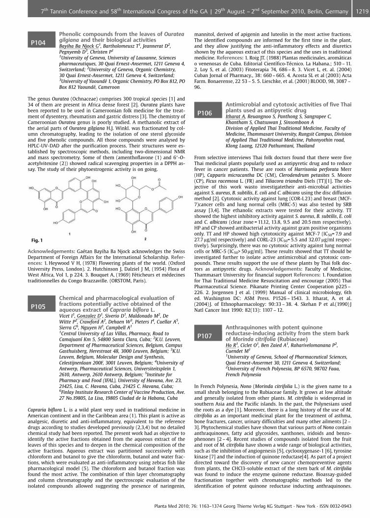
P104Phenolic compounds from the leaves of Ourateagilgiana and their biological activitiesBayiha Ba Njock G1, Bartholomeusz T1, Jeannerat D2,Pegnyemb D3, Christen P1
1University of Geneva, University of Lausanne, Sciencespharmaceutiques, 30 Quai Ernest-Ansermet, 1211 Geneva 4,Switzerland; 2University of Geneva, Organic Chemistry,30 Quai Ernest-Ansermet, 1211 Geneva 4, Switzerland;3University of Yaound� 1, Organic Chemistry, PO Box 812, POBox 812 Yaound�, Cameroon
The genus Ouratea (Ochnaceae) comprises 300 tropical species [1] and34 of them are present in Africa dense forest [2]. Ouratea plants havebeen reported to be used in Cameroonian folk medicine for the treat-ment of dysentery, rheumatism and gastric distress [3]. The chemistry ofCameroonian Ouratea genus is poorly studied. A methanolic extract ofthe aerial parts of Ouratea gilgiana H.J. Winkl. was fractionated by col-umn chromatography, leading to the isolation of one sterol glycosideand five phenolic compounds. All those compounds were analysed byHPLC-UV-DAD after the purification process. Their structures were es-tablished by spectroscopic methods, including two-dimensional NMRand mass spectrometry. Some of them (amenthoflavone (1) and 6@-O-acetylvitexine (2)) showed radical scavenging properties in a DPPH as-say. The study of their phytoestrogenic activity is on going.
Fig. 1
Acknowledgements: Ga�tan Bayiha Ba Njock acknowledges the SwissDepartment of Foreign Affairs for the International Scholarship. Refer-ences: 1. Heywood V H, (1978) Flowering plants of the world. (OxfordUniversity Press, London). 2. Hutchinson J, Dalziel J M, (1954) Flora ofWest Africa, Vol 1, p 224. 3. Bouquet A, (1969) F�ticheurs et m�decinestraditionnelles du Congo Brazzaville. (ORSTOM, Paris).
P105Chemical and pharmacological evaluation offractions potentially active obtained of theaqueous extract of Capraria biflora L.Vicet I1, Gonzalez D1, Siverio D1, Maldonado M2, DeWitte P2, Crawford A2, Dehaen W3, Pieters I4, Cuellar A5,Sierra G6, Nguyen H1, Campbell A1
1Central University of Las Villas, Pharmacy, Road toCamajuan� Km 5, 54800 Santa Clara, Cuba; 2K.U. Leuven,Department of Pharmaceutical Sciences, Belgium, CampusGasthuisberg, Herestraat 48, 3000 Leuven, Belgium; 3K.U.Leuven, Belgium, Molecular Design and Synthesis,Celestijnenlaan 200F, 3001 Leuven, Belgium; 4University ofAntwerp, Pharmaceutical Sciences, Universiteitsplein 1,2610, Antwerp, 2610 Antwerp, Belgium; 5Institute forPharmacy and Food (IFAL), University of Havana, Ave. 23,21425, Lisa, C. Havana, Cuba, 21425 C. Havana, Cuba;6Finlay Institute Research Center of Vaccine Production, Ave.27 No.19805, La Lisa, 19805 Ciudad de la Habana, Cuba
Capraria biflora L. is a wild plant very used in traditional medicine inAmerican continent and in the Caribbean area (1). This plant is active asanalgesic, diuretic and anti-inflammatory, equivalent to the referencedrugs according to studies developed previously (2,3,4) but no detailedchemical study had been reported. The present work had as objective toidentify the active fractions obtained from the aqueous extract of theleaves of this species and to deepen in the chemical composition of theactive fractions. Aqueous extract was partitioned successively withchloroform and butanol to give the chloroform, butanol and water frac-tions, which were evaluated as anti-inflammatory using zebras fish likepharmacological model (5). The chloroform and butanol fraction wasfound the most active. The combination of thin layer chromatographyand column chromatography and the spectroscopic evaluation of theisolated compounds allowed suggesting the presence of naringenin,
mannitol, derived of apigenin and luteolin in the most active fractions.The identified compounds are informed for the first time in the plant,and they allow justifying the anti-inflammatory effects and diureticsshown by the aqueous extract of this species and the uses in traditionalmedicine. References: 1. Roig JT. (1988) Plantas medicinales, arom�ticaso venenosas de Cuba. Editorial Cient�fico-T�cnico. La Habana.; 510 – 11.2. Loy S, et al. (2003) Fitoterapia 74, 686 – 8. 3. Vicet L, et. al. (2004)Cuban Jornal of Pharmacy., 38: 660 – 665. 4. Acosta SL et al (2003) ActaFarm. Bonaerense, 22 53 – 5. 5. Lieschke, et al. (2001) BLOOD, 98, 3087 –96.
P106Antimicrobial and cytotoxic activities of five Thaiplants used as antipyretic drugItharat A, Reuangnoo S, Panthong S, Sangrapee C,Khantham S, Chatsuwan J, Sinsomboon ADivision of Applied Thai Traditional Medicine, Faculty ofMedicine, Thammasart University, Rungsit Campus, Divisionof Applied Thai Traditional Medicine, Pahonyothin road,Klong Luang, 12120 Pathumtani, Thailand
From selective interviews Thai folk doctors found that there were fiveThai medicinal plants popularly used as antipyretic drug and to reducefever in cancer patients. These are roots of Harrisonia perforata Merr(HP), Capparis micracantha DC (CM), Clerodendrum petasites S. Moore(CP), Ficus racemosa L. (FR) and Tiliacora triandra Diels (TT)[1]. The ob-jective of this work wasto investigatetheir anti-microbial activitiesagainst S. aureus, B. subtilis, E. coli and C. albicans using the disc diffusionmethod [2]. Cytotoxic activity against lung (COR-L23) and breast (MCF-7)cancer cells and lung normal cells (MRC-5) was also tested by SRBassay [3,4]. The ethanolic extracts were tested for their activity. TTshowed the highest inhibitory activity against S. aureus, B. subtilis, E. coliand C. albicans (clear zone = 11.12, 13.8, 9.5 and 20.5 mm respectively).HP and CP showed antibacterial activity against gram positive organismsonly. TT and HP showed high cytotoxicity against MCF-7 (IC50= 7.9 and27.7 mg/ml respectively) and CORL-23 (IC50= 5.5 and 32.07 mg/ml respec-tively). Surprisingly, there was no cytotoxic activity against lung normalcells or MRC-5 (IC50> 50 mg/ml). These results showed that TT should beinvestigated further to isolate active antimicrobial and cytotoxic com-pounds. These results support the use of these plants by Thai folk doc-tors as antipyretic drugs. Acknowledgements: Faculty of Medicine,Thammasart University for financial support References: 1. Foundationfor Thai Traditional Medicine Resuscitation and encourage (2005) ThaiPharmaceutical Science. Pikanate Printing Center Cooperation p225 –226. 2. Jorgensen J et al. (1999) Manual of clinical microbiology, 6thed. Washington DC: ASM Press. P1526 – 1543. 3. Itharat, A. et al.(2004) J. of Ethnopharmacology: 90:33 – 38. 4. Skehan P et al.(1990) JNatl Cancer Inst 1990: 82(13): 1107 – 12.
P107Anthraquinones with potent quinonereductase-inducing activity from the stem barkof Morinda citrifolia (Rubiaceae)Ho R1, Ciclet O1, Ben Zaied A1, Raharivelomanana P2,Cuendet M1
1University of Geneva, School of Pharmaceutical Sciences,Quai Ernest-Ansermet 30, 1211 Geneva 4, Switzerland;2University of French Polynesia, BP 6570, 98702 Faaa,French Polynesia
In French Polynesia, Nono (Morinda citrifolia L.) is the given name to asmall shrub belonging to the Rubiaceae family. It grows at low altitudeand generally isolated from other plants. M. citrifolia is widespread insouthern Asia and the Pacific islands. In the past, the Polynesians usedthe roots as a dye [1]. Moreover, there is a long history of the use of M.citrifolia as an important medicinal plant for the treatment of asthma,bone fractures, cancer, urinary difficulties and many other ailments [2 –3]. Phytochemical studies have shown that various parts of Nono containanthraquinones, fatty acid glycosides, xanthones, iridoids and benzo-phenones [2 – 4]. Recent studies of compounds isolated from the fruitand root of M. citrifolia have shown a wide range of biological activities,such as the inhibition of angiogenesis [5], cyclooxygenase-1 [6], tyrosinekinase [7] and the induction of quinone reductase[4]. As part of a projectdirected toward the discovery of new cancer chemopreventive agentsfrom plants, the CHCl3-soluble extract of the stem bark of M. citrifoliawas found to induce the enzyme quinone reductase. Bioassay-guidedfractionation together with chromatographic methods led to theidentification of potent quinone reductase inducting anthraquinones.
Planta Med 2010; 76: 1163–1374 Georg Thieme Verlag KG Stuttgart · New York · ISSN 0032-0943
12197th Tannin Conference and 58th International Congress of the GA | 29th August – 2nd September 2010, Berlin, Germany
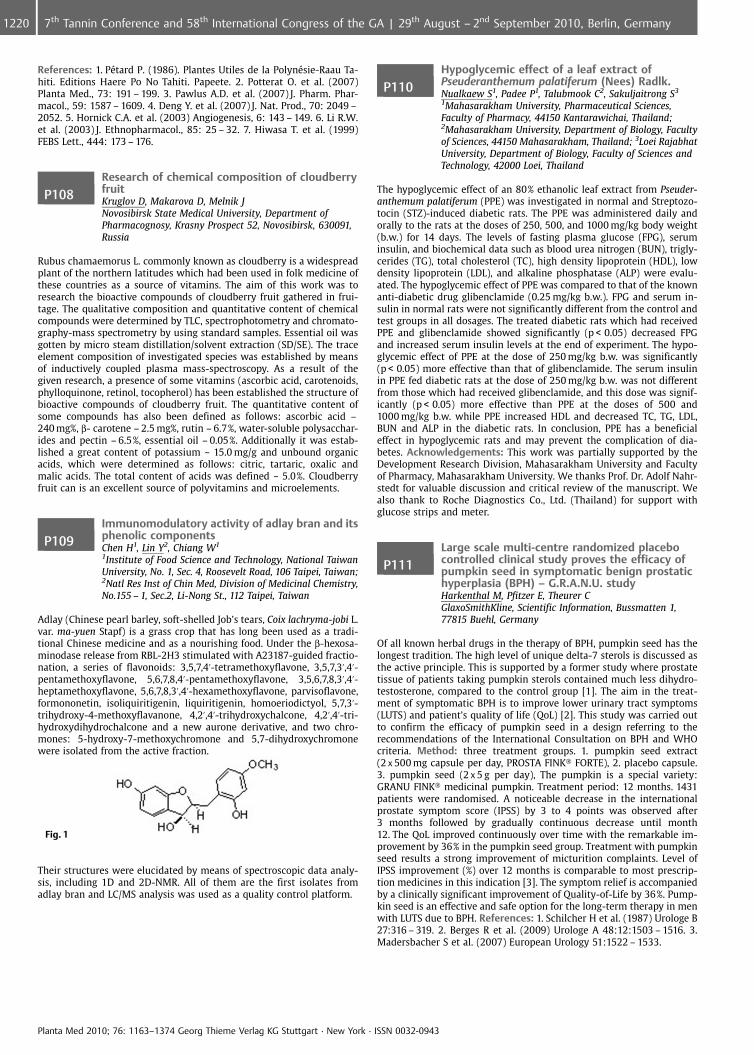
References: 1. P�tard P. (1986). Plantes Utiles de la Polyn�sie-Raau Ta-hiti. Editions Haere Po No Tahiti. Papeete. 2. Potterat O. et al. (2007)Planta Med., 73: 191 – 199. 3. Pawlus A.D. et al. (2007) J. Pharm. Phar-macol., 59: 1587 – 1609. 4. Deng Y. et al. (2007) J. Nat. Prod., 70: 2049 –2052. 5. Hornick C.A. et al. (2003) Angiogenesis, 6: 143 – 149. 6. Li R.W.et al. (2003) J. Ethnopharmacol., 85: 25 – 32. 7. Hiwasa T. et al. (1999)FEBS Lett., 444: 173 – 176.
P108Research of chemical composition of cloudberryfruitKruglov D, Makarova D, Melnik JNovosibirsk State Medical University, Department ofPharmacognosy, Krasny Prospect 52, Novosibirsk, 630091,Russia
Rubus chamaemorus L. commonly known as cloudberry is a widespreadplant of the northern latitudes which had been used in folk medicine ofthese countries as a source of vitamins. The aim of this work was toresearch the bioactive compounds of cloudberry fruit gathered in frui-tage. The qualitative composition and quantitative content of chemicalcompounds were determined by TLC, spectrophotometry and chromato-graphy-mass spectrometry by using standard samples. Essential oil wasgotten by micro steam distillation/solvent extraction (SD/SE). The traceelement composition of investigated species was established by meansof inductively coupled plasma mass-spectroscopy. As a result of thegiven research, a presence of some vitamins (ascorbic acid, carotenoids,phylloquinone, retinol, tocopherol) has been established the structure ofbioactive compounds of cloudberry fruit. The quantitative content ofsome compounds has also been defined as follows: ascorbic acid –240 mg%, b- carotene – 2.5 mg%, rutin – 6.7%, water-soluble polysacchar-ides and pectin – 6.5%, essential oil – 0.05%. Additionally it was estab-lished a great content of potassium ~ 15.0 mg/g and unbound organicacids, which were determined as follows: citric, tartaric, oxalic andmalic acids. The total content of acids was defined ~ 5.0%. Cloudberryfruit can is an excellent source of polyvitamins and microelements.
P109Immunomodulatory activity of adlay bran and itsphenolic componentsChen H1, Lin Y2, Chiang W1
1Institute of Food Science and Technology, National TaiwanUniversity, No. 1, Sec. 4, Roosevelt Road, 106 Taipei, Taiwan;2Natl Res Inst of Chin Med, Division of Medicinal Chemistry,No.155 – 1, Sec.2, Li-Nong St., 112 Taipei, Taiwan
Adlay (Chinese pearl barley, soft-shelled Job’s tears, Coix lachryma-jobi L.var. ma-yuen Stapf) is a grass crop that has long been used as a tradi-tional Chinese medicine and as a nourishing food. Under the b-hexosa-minodase release from RBL-2H3 stimulated with A23187-guided fractio-nation, a series of flavonoids: 3,5,7,4’-tetramethoxyflavone, 3,5,7,3’,4’-pentamethoxyflavone, 5,6,7,8,4’-pentamethoxyflavone, 3,5,6,7,8,3’,4’-heptamethoxyflavone, 5,6,7,8,3’,4’-hexamethoxyflavone, parvisoflavone,formononetin, isoliquiritigenin, liquiritigenin, homoeriodictyol, 5,7,3’-trihydroxy-4-methoxyflavanone, 4,2’,4’-trihydroxychalcone, 4,2’,4’-tri-hydroxydihydrochalcone and a new aurone derivative, and two chro-mones: 5-hydroxy-7-methoxychromone and 5,7-dihydroxychromonewere isolated from the active fraction.
Fig. 1
Their structures were elucidated by means of spectroscopic data analy-sis, including 1D and 2D-NMR. All of them are the first isolates fromadlay bran and LC/MS analysis was used as a quality control platform.
P110Hypoglycemic effect of a leaf extract ofPseuderanthemum palatiferum (Nees) Radlk.Nualkaew S1, Padee P1, Talubmook C2, Sakuljaitrong S3
1Mahasarakham University, Pharmaceutical Sciences,Faculty of Pharmacy, 44150 Kantarawichai, Thailand;2Mahasarakham University, Department of Biology, Facultyof Sciences, 44150 Mahasarakham, Thailand; 3Loei RajabhatUniversity, Department of Biology, Faculty of Sciences andTechnology, 42000 Loei, Thailand
The hypoglycemic effect of an 80% ethanolic leaf extract from Pseuder-anthemum palatiferum (PPE) was investigated in normal and Streptozo-tocin (STZ)-induced diabetic rats. The PPE was administered daily andorally to the rats at the doses of 250, 500, and 1000 mg/kg body weight(b.w.) for 14 days. The levels of fasting plasma glucose (FPG), seruminsulin, and biochemical data such as blood urea nitrogen (BUN), trigly-cerides (TG), total cholesterol (TC), high density lipoprotein (HDL), lowdensity lipoprotein (LDL), and alkaline phosphatase (ALP) were evalu-ated. The hypoglycemic effect of PPE was compared to that of the knownanti-diabetic drug glibenclamide (0.25 mg/kg b.w.). FPG and serum in-sulin in normal rats were not significantly different from the control andtest groups in all dosages. The treated diabetic rats which had receivedPPE and glibenclamide showed significantly (p < 0.05) decreased FPGand increased serum insulin levels at the end of experiment. The hypo-glycemic effect of PPE at the dose of 250 mg/kg b.w. was significantly(p < 0.05) more effective than that of glibenclamide. The serum insulinin PPE fed diabetic rats at the dose of 250 mg/kg b.w. was not differentfrom those which had received glibenclamide, and this dose was signif-icantly (p < 0.05) more effective than PPE at the doses of 500 and1000 mg/kg b.w. while PPE increased HDL and decreased TC, TG, LDL,BUN and ALP in the diabetic rats. In conclusion, PPE has a beneficialeffect in hypoglycemic rats and may prevent the complication of dia-betes. Acknowledgements: This work was partially supported by theDevelopment Research Division, Mahasarakham University and Facultyof Pharmacy, Mahasarakham University. We thanks Prof. Dr. Adolf Nahr-stedt for valuable discussion and critical review of the manuscript. Wealso thank to Roche Diagnostics Co., Ltd. (Thailand) for support withglucose strips and meter.
P111Large scale multi-centre randomized placebocontrolled clinical study proves the efficacy ofpumpkin seed in symptomatic benign prostatichyperplasia (BPH) – G.R.A.N.U. studyHarkenthal M, Pfitzer E, Theurer CGlaxoSmithKline, Scientific Information, Bussmatten 1,77815 Buehl, Germany
Of all known herbal drugs in the therapy of BPH, pumpkin seed has thelongest tradition. The high level of unique delta-7 sterols is discussed asthe active principle. This is supported by a former study where prostatetissue of patients taking pumpkin sterols contained much less dihydro-testosterone, compared to the control group [1]. The aim in the treat-ment of symptomatic BPH is to improve lower urinary tract symptoms(LUTS) and patient’s quality of life (QoL) [2]. This study was carried outto confirm the efficacy of pumpkin seed in a design referring to therecommendations of the International Consultation on BPH and WHOcriteria. Method: three treatment groups. 1. pumpkin seed extract(2 x 500 mg capsule per day, PROSTA FINK� FORTE), 2. placebo capsule.3. pumpkin seed (2 x 5 g per day), The pumpkin is a special variety:GRANU FINK� medicinal pumpkin. Treatment period: 12 months. 1431patients were randomised. A noticeable decrease in the internationalprostate symptom score (IPSS) by 3 to 4 points was observed after3 months followed by gradually continuous decrease until month12. The QoL improved continuously over time with the remarkable im-provement by 36% in the pumpkin seed group. Treatment with pumpkinseed results a strong improvement of micturition complaints. Level ofIPSS improvement (%) over 12 months is comparable to most prescrip-tion medicines in this indication [3]. The symptom relief is accompaniedby a clinically significant improvement of Quality-of-Life by 36%. Pump-kin seed is an effective and safe option for the long-term therapy in menwith LUTS due to BPH. References: 1. Schilcher H et al. (1987) Urologe B27:316 – 319. 2. Berges R et al. (2009) Urologe A 48:12:1503 – 1516. 3.Madersbacher S et al. (2007) European Urology 51:1522 – 1533.
Planta Med 2010; 76: 1163–1374 Georg Thieme Verlag KG Stuttgart · New York · ISSN 0032-0943
1220 7th Tannin Conference and 58th International Congress of the GA | 29th August – 2nd September 2010, Berlin, Germany
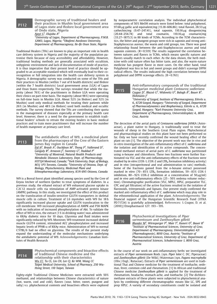
P112Demographic survey of traditional healers andtheir practices in Mushin local government areaof Lagos state and Ifedore local government areaof Ondo state, NigeriaAjayi G1, Elujoba A2
1University of Lagos, Department of Pharmacognosy, P.M.B.12003, Lagos, Nigeria; 2Obafemi Awolowo University,Department of Pharmacognosy, Ile-Ife Osun State, Nigeria
Traditional Healers (THs) are known to play an important role in healthcare delivery system in Nigeria and the majority of the population pa-tronise them because of easy accessibility and affordability. However,traditional healing methods are generally associated with occultism,unhygienic environment and lack of documentation of mode of practice.It is thus imperative that their present level of practice should be up-graded to an acceptable standard which will eventually lead to their co-recognition or full integration into the health care delivery system inNigeria. A demographic survey was conducted on some of the THs andtheir practices in Mushin (within 7 out of 8 health districts) and Ifedore(within the 7 health districts) Local government areas (LGA) of Lagosand Osun States respectively. The surveys revealed that while the ma-jority (about 76%) of the practitioners in Ifedore LGA were operatingtheir clinics on part-time basis. The majority (about 98%) were operatingon full-time basis in Mushin LGA. About 52% (in Ifedore) and 52% (inMushin) used only medical methods for treating their patients while26% (in Mushin) and 48% (in Ifedore) used both medical and occulticmethods. The survey showed that the THs are contributing their quotain the management of diseases especially at the primary health carelevel. However, there is a need for the government to establish tradi-tional healers’ schools to retrain the existing healers in basic medicalpractices and to train more practitioners that will increase the numberof health manpower at primary care level.
P113The antidiabetic effect of W9, a medicinal plantfrom the pharmacopea of the Cree of the EasternJames Bay region in CanadaEid H1, Brault A1, Ouchfoun M1, Thong F2, Vallerand D1,Ganguly R2, Arnason J3, Sweeney G2, Haddad P1
1University of Montreal, Natural Health Products andMetabolic Diseases Laboratory, Dept. of Pharmacology,H3T1J4 Montreal, Canada; 2York University, Dept. of Biology,M3 J 1P3 Toronto, Canada; 3University of Ottawa, Dept. ofBiology, Phytochemistry, Medicinal Plant andEthnopharmacology Laboratory, K1N 6N5 Ottawa, Canada
W9 is a Boreal forest plant identified among species used by the Cree ofEeyou Istchee of northern Quebec to treat symptoms of diabetes. In aprevious study, the ethanol extract of W9 enhanced glucose uptake inC 2C12 muscle cells via stimulation of AMP-activated protein kinase(AMPK) pathway. In this study, we investigated the effect of this producton the translocation of insulin-sensitive GLUT4 transporters in skeletalmuscle cells in culture. Treatment of L 6 myotubes with W9 for 18 hsignificantly increased glucose uptake and GLUT4 translocation to thecell membrane. W9 increased phosphorylation of AMPK and P38 MAPKwith no indication of increased phosphorylation of Akt. To validate theeffect of W9 in vivo, the extract (1% in drinking water) was administeredto KKAy diabetic mice for 10 days. Glycemia and fluid intakes weresignificantly reduced by W9. Moreover, W9-treatment increased skeletalmuscle GLUT4 content, stimulated ACC phosphorylation and increasedhepatic levels of PPAR-a of KKAy mice. Administration of W9 to normalC 57BL/6 had no effect on glycemia. The results of the present studyexpand the understanding of the molecular mechanisms underlyingthe antidiabetic potential of W9. Acknowledgements: Canadian Insti-tutes of Health Research
P114Phytochemical components and bioactive effectsof traditional Chinese medicine and therelationship with their characteristicsWu CC, Tu CL, Lin CH, Lee CJ, Ke WM, Wang CCTaipei Medical University, School of Pharmacy, 250 Wu-Hsing Street, 110 Taipei, Taiwan
Eighty-eight Traditional Chinese Medicines were extracted with 50%methanol, and relationships between Chinese characteristics of nature(hot, warm, cool and cold), flavors (sour, bitter, sweet, pungent andsalty) v.s. phytochemical contents and bioactives effects were explored
by nonparametric correlation analysis. The individual phytochemicalcomponents of 50% MeOH extracts were listed below: total polyphenol,97.68 mg gallic acid equivalent/mg (15.18~606.86); total flavonl, 28.53 mgcathechin/mg (0.47~247.41), total saponin, 110.16 mg diosgenin/mg(20.44~254.74) and total coumarin, 110.16 mg coumarin/mg(33.27~1875.5) in 88 kinds of TCMs. According to the TCM characteris-tics, the bitter and pungent groups were rich in saponin and strong anti-Staphylococcus aureus; the warm and cold groups, too. There was goodrelationship found between the anti-Staphylococcus aureus and totalsaponin contents. (R = 0.519) The results supported the correlation be-tween natures and flavors in TCMs, which means specific natures andflavors usually match to each other. As the data shown, the herb med-icine with cold nature often has bitter taste, and also, the warm-naturemedicine has pungent flavor in most cases. On the other hand, totalPolyphenol was less in hot and sweet groups and didn’t scavenge DPPHradical effects. The results indicated the high correlation between totalpolyphenol and DPPH scavenge effects. (R = 0.763)
P115Anti-inflammatory compounds of the traditionalHungarian medicinal plant Centaurea sadlerianaCsupor D1, Blazs� G2, Widowitz U3, Balogh �2, Bauer R3,Hohmann J1
1University of Szeged, Institute of Pharmacognosy, E�tv�s u.6., 6720 Szeged, Hungary; 2University of Szeged, Departmentof Pharmacodynamics and Biopharmacy, E�tv�s u. 6., 6720Szeged, Hungary; 3Karl-Franzens University Graz,Department of Pharmacognosy, Universit�tsplatz 4., 8010Graz, Austria
The decoction of the aerial parts of Centaurea sadleriana JANKA (Aster-aceae), a plant native to Hungary, is traditionally used to treat thewounds of sheep in the Southern Great Plain region. Phytochemicaland pharmacological studies on this plant have not been performed sofar. Only we have recently confirmed the wound healing effect of theplant on rats [1]. The objective of the present work was the in vivo andin vitro investigation of the anti-inflammatory effect of C. sadleriana andthe isolation and identification of its active compounds. The concen-trated methanol extract of aerial parts of C. sadleriana was partitionedusing n-hexane and chloroform. These two fractions were further frac-tionated via VLC and the anti-inflammatory effects of the fractions werestudied by in vitro (COX-1, COX-2 and LTB4 formation inhibitory activity)and in vivo (intraperitoneal and oral administration to rats) methods.Some of the fractions gained from the n-hexane extract possessedmarked in vitro (70 – 85% LTB4 formation inhibition, 59 – 83% COX-1inhibition, 80 – 92% COX-2 inhibition at a concentration of 50 mg/ml)and in vivo anti-inflammatory effects (25 – 50% oedema volume reduc-tion). Chromatographic purification (VLC, MPLC, HPLC, preparative TLC,CPC and gel filtration) of the active fractions resulted in the isolation offlavonoids, triterpenoids and lignans. Our present study confirmed themarked anti-inflammatory effect of C. sadleriana which may play role inthe ethnomedicinal application of the plant. Acknowledgements: Thefinancial support of the Hungarian Scientific Research Fund (OTKAPD 71724) is gratefully acknowledged. References: 1. Csupor, D. et al.(2010) J Etnopharmacol 127: 193 – 195.
P116Phytochemical investigations of Pipersarmentosum and Zanthoxyllum gilletiiBlunder M1, Wirnsperger P1, Mair T1, Kunert O2, Bauer R1
1Institute of Pharmaceutical Sciences, University of Graz,Departmanet of Pharmacognosy, Universit�tsplatz 4/I(Pharmakognosie), 8010 Graz, Austria; 2Institute ofPharmaceutical Sciences, University of Graz, Department ofPharmaceutical Sciences, Schuberstrasse 1, 8010 Graz,Austria
In the course of our work on anti-inflammatory herbs we investigatedextracts of Piper sarmentosum Roxb. (syn. Piper lolot C. DC; Piperaceae)and Zanthoxyllum gilletii (De Wild.) Waterman (syn. Fagara macrophylla(Oliv.) Engl.; Rutaceae). Extracts of Piper sarmentosum are used in tradi-tional Thai and Chinese medicine for the treatment of toothache, oede-mas, fever, common cold and rheumatism. [1] In African and traditionalChinese medicine Zanthoxyllum gilletii is applied for the treatment ofrheumatism, headache, stomach-ache, and toothache. [2] The dichloro-methane extracts of these plants were subjected to phytochemical ana-lysis by combining different chromatographic means like LC, SPE andprep HPLC. A variety of secondary constituents could be isolated and
Planta Med 2010; 76: 1163–1374 Georg Thieme Verlag KG Stuttgart · New York · ISSN 0032-0943
12217th Tannin Conference and 58th International Congress of the GA | 29th August – 2nd September 2010, Berlin, Germany
was structurally elucidated using UV, 1D (1H, 13C) and 2D NMR (DQF-COSY, HMBC, HSQC) experiments and mass spectroscopy. From Pipersarmentosum extracts of both, herba and radix et caulis, were prepared.Among the isolated compounds pyrrole and pyrrolidine amides, pheno-lic compounds, and a new ester of 3-(4’-methoxy-phenyl)-propionic acidwere identified. The investigation of the dichloromethane extract of thebark of Zanthoxyllum gilletii revealed the presence of aromatic amideslike pyrrole amide (e. g. fagaramide), alkylamides, lignans (e. g. sesamin)and coumarins. References: 1. Li, C.-Y. et al. (2007) J. Agric. Food Chem.55:9436 – 9442. 2. Wansi, J.D. et al. (2009) Planta Med. 75:517 – 521.
P117Ethnobotanical study of plants used in managingdepression within Lagos metropolis NigeriaOdukoya O, Adewumi C, Asunmo S, Badejo AUniversity of Lagos, Pharmacognosy, Faculty of Pharmacy,PMB 12003, Suru Lere, 12003 Lagos, Nigeria
Antidepressant drugs continue to raise concerns about their side effects,which include suicide, clinical worsening of depression, and unusualchanges in behavior of adolescents and children. Recently, the FDA in-structed all drug manufacturers to add black box warnings (the mostserious warning label for a prescription medicine) to their antidepres-sant drugs. In light of these findings, doctors and patients are seekingsafer alternative therapies. An ethnobotanical survey of medicinal andpoisonous plants was conducted amongst herb sellers in major openmarkets in Lagos metropolis, Nigeria. Data was collected by the authorsusing classical descriptive ethnobotanical techniques (i. e. no quantita-tive measures) through an unstructured open ended interview. A total of48 ethnomedicinal plant species distributed in 12 families are documen-ted in this study. The medicinal plants used are listed with Latin name,family, local name, parts used, mode of preparation and duration oftreatment. The majority of the remedies were prepared from freshlycollected plant material from the wild. They are mainly taken orally assingle plants, but some applications were prepared with a mixture ofplants or ingredients such as honey, salt, palm oil and pepper. Decoctionof the leaves was the main form of preparation (68%) and leaf powderwas mostly used for the preparation of infusions (16%). The need for theuse of stem and root barks increases when the leaves are not available.These plants can serve as sources of new alternatives for managingdepressive symptoms through exploitation of novel pharmacologicalagents.
P118Adaptogenic and central nervous system effectsof Potentilla alba L extract in miceShikov A1, Lazukina M1, Makarova M1, Pozharitskaya O1,Demchenko D1, Makarov V1, Golubeva O2
1St-Petersburg Institute of Pharmacy, 47/5, Piskarevskyprospect, 195067 St-Petersburg, Russian Federation; 2St-Petersburg State Medicinal Academy named after I.I.Mechnikov, 47/33, Piskarevsky prospect, 195067St-Petersburg, Russian Federation
Potentilla alba L. (Rosaceae) rhizomes are used in Russian and Ukrainianfolk medicine to reduce the thyroxin level in blood plasma as an impor-tant drug against thyroid gland diseases. The preparation shows anti-inflammatory, antioxidant, and adaptogenic effect [1,2]. P. alba containsa significant amount of proanthocyanidins, procyanidins, polyphenolics,some flavonoids, polysaccharides etc. [3]. The data about adaptogenicand central nervous system (CNS) activities of P. alba are fragmentary.The adaptogenic and CNS effects of P. alba were examined in the swim-ming to exhaustion, light/dark exploration, and open field tests on mice.The treatment groups were orally administered dry extract of P. alba (indoses of 0.3, 0.9 and 1.8 mg/kg), control group received distilled water.The swimming time after 7 days of treatment was increased by 3.3 – 5.5fold. Minimal glycogen level was observed in the group treated by0.9 mg/kg P. alba. It was estimated that glycogen was hydrolyzed toglucose, glucose level was increased in the blood plasma, and lacticacidosis was prevented by aerobic oxidation of glucose (Table 1).
Table 1: Results of fswimming test in mice
Group Swimming time,s
Glucose,mmol/L
Lactate,mmol/L
Glycogen in liver,mg/g
Control 730 € 90 6.2 € 0.5 5.8 €.0.5 0.61 €.0.15P. alba, 0.3 mg/kg 2393 € 371* 2.5 € 0.4* 6.1 € 0.8 0.18 €.0.05*P. alba, 0.9 mg/kg 4015 € 795* 4.9 € 0.8 4.9 €.0.4 0.12.€ 0.06*P. alba. 1.8 mg/kg 2597 € 572* 2.8 €.0.4* 5.6 € 0.6 0.20 €.0.10*
A dose dependant tendency for increased time spent in the light com-partment (light/dark exploration) in the treated groups was observed.However, no changes were found in the number of entries into eachcompartment. Statistical analysis revealed that in the open field testthe extract at a dose of 0.3 mg/kg induced a significant increase in loco-motor activity but alterations in the anxiety of the treated mice were notmarked. References: 1. Smyk. K.G. (1982) Chimia I Zhizn 3: 15 – 17. 2.Tomczyk. M. Latte. K.P. (2009) J. Ethnopharmacol. 122: 184 – 204. 3. Shi-kov. A.N. et al. (2009) Book of abstracts of 57 GA Congress: A5.
P119Azadirachtin-rich Neem extracts againstectoparasitesGraf-Schiller S1, Spielberger U1, Schwarze-Fiedler D2,Kleeberg H2
1Plantavet GmbH, Frauenbergstr. 45, 88339 Bad Waldsee,Germany; 2Trifolio-M GmbH, Dr.-Hans-Wilhelmi-Weg 1,35633 Lahnau, Germany
Leaves, seeds, bark and other parts of the Neem tree Azadirachta indicaA. JUSS have been used in traditional medicine in India (Ayurveda, Unaniand Homeopathy)1 since centuries against different diseases especiallyskin disorders and ectoparasites like pediculosis, scabies, fleas, ticks etc.With respect to these experiences a standardised method for extractingneem seed kernels was developed and patented. Currently the ingredi-ents of this extract called NeemAzal� are characterized and AzadirachtinA is found as the main ingredient and analytical leading substance.
Fig. 1: Azadirachtin A
Investigations show a favourable toxicological profile of NeemAzal�(acute oral toxicity in rat LD 50 > 5000 mg/kg bw, acute dermal toxicityin rabbit LD 50 > 3000 mg/kg bw; NOAEL 3.7 mg/kg bw/d)2, good com-patibility and effectiveness. Different standardised formulations areavailable and examined: – Sheep and goats are successfully treated withNeemPro�-Sheep (in accordance with Biocide Directive 98/8/EC andapproved for organic farming) against Anopheles spec., Culicoides spec.,Damalinia spec.3,4, Wolfarthia magnifica). – Ectoparasites can be con-trolled effective by ContrAcar�Pet (in accordance with Biocide Directive98/8/EC and approved for organic farming) in the pet surrounding. –Bernauer-Jacob reported NeemPro�-Dog kills adult fleas (Ctenocepha-lides felis) on dogs at doses of 83 mg AzA/kg bw and stops completelythe egg production of adult fleas.5 – The good compatiblity6 for humansof NeemExtract-FT shampoo was shown by a safety assessment includ-ing stability, physico-chemical and bacteriological tests, Het Cam Testand a 48 hour Patch-test with the finished product. In Italy a registrationprocedure for marketing authorisation (Presidi Medico Chirugici) is on-going. References: 1. Ketkar, A.Y. et al. (2002) The Neem Tree. Schmut-terer, H. Mumbai. 2. Niemann, L. (2002) The Neem Tree. Schmutterer, H.Mumbai. 3. Habluetzel, A. (2006) Vet Parasitol 144(3 – 4): 328 – 337. 4.Guerrini, V.H. (2000) OJVR 4: 133 – 138. 5. Bernauer-Jacob, V. (1999)Orientierende Untersuchungen zur Wirksamkeit von NeemAzal auf denKatzenfloh Ctenocephalides felis felis (Bouch�) und den tropischen Rat-tenfloh Xenopsylla cheopsis (Rothschild), Berlin. 6. Saboureau, D. (2006)Safety Assessment for human health’s safety evaluation of a cosmeticproduct, unpublished.
P120Quantification of some phenolic acid and rutin inthe leaves of rosemary from Turkey and CroatiaMuradic S1, Salihovic M1, Klepo L1, Sofic E1, Kalher K2
1University of Sarajevo, Faculty of Science, Department ofChemistry, Zmaja od Bosne 33 – 35, 71000 Sarajevo, BosniaAnd Herzegovina; 2Karl-Franzens University, Department ofAnalytical Chemistry, Universit�tsplatz 3, 8010 Graz, Austria
Rosemary contains a number of potentially biologically active com-pounds, such as carnosic acid and rosmarinic acid (RA), camphor, caffeicacid (CA), ursolic acid, betulinic acid, rosmaridiphenol and rosmanol.OBJECTIVES: In this study, using HPLC-ED system, analysis of gallic acid
Planta Med 2010; 76: 1163–1374 Georg Thieme Verlag KG Stuttgart · New York · ISSN 0032-0943
1222 7th Tannin Conference and 58th International Congress of the GA | 29th August – 2nd September 2010, Berlin, Germany
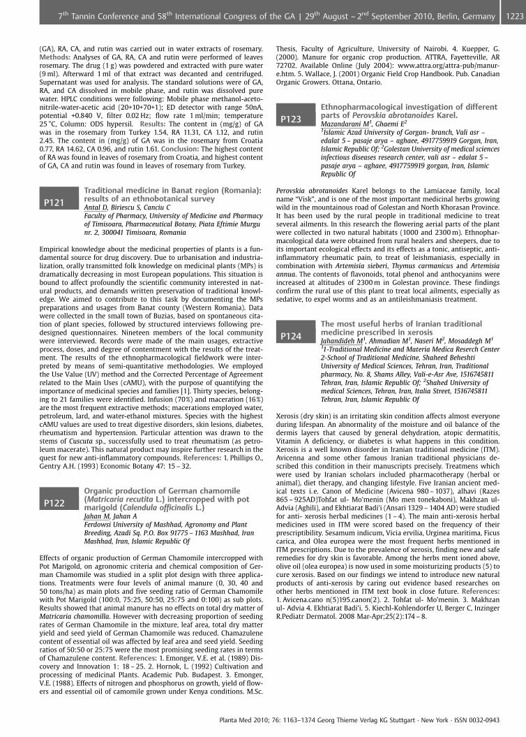
(GA), RA, CA, and rutin was carried out in water extracts of rosemary.Methods: Analyses of GA, RA, CA and rutin were performed of leavesrosemary. The drug (1 g) was powdered and extracted with pure water(9 ml). Afterward 1 ml of that extract was decanted and centrifuged.Supernatant was used for analysis. The standard solutions were of GA,RA, and CA dissolved in mobile phase, and rutin was dissolved purewater. HPLC conditions were following: Mobile phase methanol-aceto-nitrile-water-acetic acid (20+10+70+1); ED detector with range 50nA,potential +0.840 V, filter 0.02 Hz; flow rate 1 ml/min; temperature25 �C, Column: ODS hypersil. Results: The content in (mg/g) of GAwas in the rosemary from Turkey 1.54, RA 11.31, CA 1.12, and rutin2.45. The content in (mg/g) of GA was in the rosemary from Croatia0.77, RA 14.62, CA 0.96, and rutin 1.61. Conclusion: The highest contentof RA was found in leaves of rosemary from Croatia, and highest contentof GA, CA and rutin was found in leaves of rosemary from Turkey.
P121Traditional medicine in Banat region (Romania):results of an ethnobotanical surveyAntal D, Biriescu S, Canciu CFaculty of Pharmacy, University of Medicine and Pharmacyof Timisoara, Pharmaceutical Botany, Piata Eftimie Murgunr. 2, 300041 Timisoara, Romania
Empirical knowledge about the medicinal properties of plants is a fun-damental source for drug discovery. Due to urbanisation and industria-lization, orally transmitted folk knowledge on medicinal plants (MPs) isdramatically decreasing in most European populations. This situation isbound to affect profoundly the scientific community interested in nat-ural products, and demands written preservation of traditional knowl-edge. We aimed to contribute to this task by documenting the MPspreparations and usages from Banat county (Western Romania). Datawere collected in the small town of Buzias, based on spontaneous cita-tion of plant species, followed by structured interviews following pre-designed questionnaires. Nineteen members of the local communitywere interviewed. Records were made of the main usages, extractiveprocess, doses, and degree of contentment with the results of the treat-ment. The results of the ethnopharmacological fieldwork were inter-preted by means of semi-quantitative methodologies. We employedthe Use Value (UV) method and the Corrected Percentage of Agreementrelated to the Main Uses (cAMU), with the purpose of quantifying theimportance of medicinal species and families [1]. Thirty species, belong-ing to 21 families were identified. Infusion (70%) and maceration (16%)are the most frequent extractive methods; macerations employed water,petroleum, lard, and water-ethanol mixtures. Species with the highestcAMU values are used to treat digestive disorders, skin lesions, diabetes,rheumatism and hypertension. Particular attention was drawn to thestems of Cuscuta sp., successfully used to treat rheumatism (as petro-leum macerate). This natural product may inspire further research in thequest for new anti-inflammatory compounds. References: 1. Phillips O.,Gentry A.H. (1993) Economic Botany 47: 15 – 32.
P122Organic production of German chamomile(Matricaria recutita L.) intercropped with potmarigold (Calendula officinalis L.)Jahan M, Jahan AFerdowsi University of Mashhad, Agronomy and PlantBreeding, Azadi Sq. P.O. Box 91775 – 1163 Mashhad, IranMashhad, Iran, Islamic Republic Of
Effects of organic production of German Chamomile intercropped withPot Marigold, on agronomic criteria and chemical composition of Ger-man Chamomile was studied in a split plot design with three applica-tions. Treatments were four levels of animal manure (0, 30, 40 and50 tons/ha) as main plots and five seeding ratio of German Chamomilewith Pot Marigold (100:0, 75:25, 50:50, 25:75 and 0:100) as sub plots.Results showed that animal manure has no effects on total dry matter ofMatricaria chamomilla. However with decreasing proportion of seedingrates of German Chamomile in the mixture, leaf area, total dry matteryield and seed yield of German Chamomile was reduced. Chamazulenecontent of essential oil was affected by leaf area and seed yield. Seedingratios of 50:50 or 25:75 were the most promising seeding rates in termsof Chamazulene content. References: 1. Emonger, V.E. et al. (1989) Dis-covery and Innovation 1: 18 – 25. 2. Hornok, L. (1992) Cultivation andprocessing of medicinal Plants. Academic Pub. Budapest. 3. Emonger,V.E. (1988). Effects of nitrogen and phosphorus on growth, yield of flow-ers and essential oil of camomile grown under Kenya conditions. M.Sc.
Thesis, Faculty of Agriculture, University of Nairobi. 4. Kuepper, G.(2000). Manure for organic crop production. ATTRA, Fayetteville, AR72702. Available Online (July 2004): www.attra.org/attra-pub/manur-e.htm. 5. Wallace, J. (2001) Organic Field Crop Handbook. Pub. CanadianOrganic Growers. Ottana, Ontario.
P123Ethnopharmacological investigation of differentparts of Perovskia abrotanoides Karel.Mazandarani M1, Ghaemi E2
1Islamic Azad University of Gorgan- branch, Vali asr –edalat 5 – pasaje arya – aghaee, 4917759919 Gorgan, Iran,Islamic Republic Of; 2Golestan University of medical sciencesinfectious diseases research center, vali asr – edalat 5 –pasaje arya – aghaee, 4917759919 gorgan, Iran, IslamicRepublic Of
Perovskia abrotanoides Karel belongs to the Lamiaceae family, localname “Visk”, and is one of the most important medicinal herbs growingwild in the mountainous road of Golestan and North Khorasan Province.It has been used by the rural people in traditional medicine to treatseveral ailments. In this research the flowering aerial parts of the plantwere collected in two natural habitats (1000 and 2300 m). Ethnophar-macological data were obtained from rural healers and sheepers, due toits important ecological effects and its effects as a tonic, antiseptic, anti-inflammatory rheumatic pain, to treat of leishmaniasis, especially incombination with Artemisia sieberi, Thymus carmanicus and Artemisiaannua. The contents of flavonoids, total phenol and anthocyanins wereincreased at altitudes of 2300 m in Golestan province. These findingsconfirm the rural use of this plant to treat local ailments, especially assedative, to expel worms and as an antileishmaniasis treatment.
P124The most useful herbs of Iranian traditionalmedicine prescribed in xerosisJahandideh M1, Ahmadian M1, Naseri M2, Mosaddegh M1
11-Traditional Medicine and Materia Medica Reserch Center2-School of Traditional Medicine, Shaheed BeheshtiUniversity of Medical Sciences, Tehran, Iran, Traditionalpharmacy, No. 8, Shams Alley, Vali-e-Asr Ave, 1516745811Tehran, Iran, Islamic Republic Of; 2Shahed University ofmedical Sciences, Tehran, Iran, Italia Street, 1516745811Tehran, Iran, Islamic Republic Of
Xerosis (dry skin) is an irritating skin condition affects almost everyoneduring lifespan. An abnormality of the moisture and oil balance of thedermis layers that caused by general dehydration, atopic dermatitis,Vitamin A deficiency, or diabetes is what happens in this condition.Xerosis is a well known disorder in Iranian traditional medicine (ITM).Avicenna and some other famous Iranian traditional physicians de-scribed this condition in their manuscripts precisely. Treatmens whichwere used by Iranian scholars included pharmacotherapy (herbal oranimal), diet therapy, and changing lifestyle. Five Iranian ancient med-ical texts i. e. Canon of Medicine (Avicena 980 – 1037), alhavi (Razes865 – 925AD)Tohfat ul- Mo’menin (Mo men tonekaboni), Makhzan ul-Advia (Aghili), and Ekhtiarat Badi’i (Ansari 1329 – 1404 AD) were studiedfor anti- xerosis herbal medicines (1 – 4). The main anti-xerosis herbalmedicines used in ITM were scored based on the frequency of theirprescriptibility. Sesamum indicum, Vicia ervilia, Urginea maritima, Ficuscarica, and Olea europea were the most frequent herbs mentioned inITM prescriptions. Due to the prevalence of xerosis, finding new and saferemedies for dry skin is favorable. Among the herbs ment ioned above,olive oil (olea europea) is now used in some moisturizing products (5) tocure xerosis. Based on our findings we intend to introduce new naturalproducts of anti-xerosis by caring out evidence based researches onother herbs mentioned in ITM text book in close future. References:1. Avicena.cano n(5)195.canon(2). 2. Tohfat ul- Mo’menin. 3. Makhzanul- Advia 4. Ekhtiarat Badi’i. 5. Kiechl-Kohlendorfer U, Berger C, InzingerR.Pediatr Dermatol. 2008 Mar-Apr;25(2):174 – 8.
Planta Med 2010; 76: 1163–1374 Georg Thieme Verlag KG Stuttgart · New York · ISSN 0032-0943
12237th Tannin Conference and 58th International Congress of the GA | 29th August – 2nd September 2010, Berlin, Germany
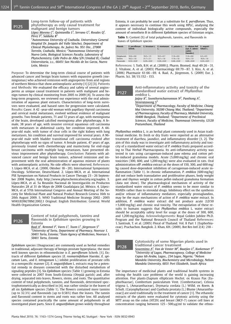
P125Long-term follow-up of patients withphytotherapy as only causal treatment formalignant and benign tumorsL�pez Moreno C1, Quintanilla L2, Serrano G1, Rosales Q1,P�rez F1, Salda�a R1
1Autonomous University of Coahuila. Universitary GeneralHospital Dr. Joaqu�n del Valle S�nchez, Department ofClinical Phytotherapy, Av. Ju�rez No. 951 Ote., 27000Torre�n, Coahuila, Mexico; 2Autonomous University ofNuevo Le�n, Biological Sciences Faculty. Laboratory ofPhytochemistry, Calle Pedro de Alba S/N (Unidad B), CiudadUniversitaria, z.c., 66451 San Nicol�s de los Garza, NuevoLe�n, Mexico
Purpose: To determine the long-term clinical course of patients withadvanced cancer and benign brain tumors with expansive growth (me-ningiomas) who achieved remission with angiosperms from arid regionsof northern Mexico that show antineoplastic activity [1] [2] [3]. Patientsand Methods: We evaluated the efficacy and safety of several angios-perms as unique causal treatment in patients with malignant and be-nign tumors by clinical monitoring from 2005 to 2009 [4]. To assess thelong-term prognosis, we reviewed our experience with the oral admin-istration of aqueous plant extracts. Characteristics of long-term survi-vors were evaluated, and hazard rates for progression were calculated.Results: Cases: A 42 -year-old woman with papillary thyroid carcinomaand cervical nodal metastases, evolved without evidence of malignantgrowths. Two female patients, 71 and 72 years of age, with meningiomaof the brain, developed calcified meningioma after phytotherapy. A fe-male, 38 years of age, with invasive cervical squamous cell carcinomawith metastases in abdominal cavity, her condition improved. A 44-year-old male, with tumor of clear cells in the right kidney with lungmetastases, his condition and survival improved for several years. A 96-year-old male with bladder transitional cell carcinoma evolved afterphytotherapy with no signs of tumor. A female patient, 47 years of age,previously treated with chemotherapy and mastectomy for end-stagebreast carcinoma with multiple lung metastases, have presented ob-vious improvement after phytotherapy. Conclusion: Patients with ad-vanced cancer and benign brain tumors, achieved remission and im-provement with the oral administration of aqueous mixture of plantswith antineoplastic activity. No side effects were observed. References:1. L pez-MCA, et al. (2000, Oktober 18 – 20). International Symposium:Oncology. Schliersee, Deutschland. 2. L pez-MCA, et al. InternationalPSE Symposium on Natural Products in Cancer Therapy 23 – 26 Septem-ber 2008 Naples, Italy http://www.phytochemicalsociety.org/naples. 3.L pez-MCA, et al. 5a Reuni n Nacional de Investigaci n en ProductosNaturales 28 al 31 de Mayo de 2008 Guadalajara Jal. M�xico. 4. L pez-MCA, et al 57th International Congress and Annual Meeting of the So-ciety for Medicinal Plant and Natural Product Research Geneva 16th to20th August 2009. 5. WHO Traditional Medicine Strategy 2002 – 2005WHO/EDM/TRM/2002.1 Original: English Distribution: General. WorldHealth Organization Geneva.
P126Content of total polyphenols, tannins andflavonoids in Epilobium species growing inEstoniaRaal A1, Remmel I2, Vares L1, Toom L1, J�rgenson S1
1University of Tartu, Department of Pharmacy, Nooruse 1,50411 Tartu, Estonia; 2State Agency of Medicines, Nooruse 1,50411 Tartu, Estonia
Epilobium species (Onagraceae) are commonly used as herbal remediesin traditional, adjuvant therapy of benign prostate hyperplasia; the mostwell known species in ethnomedicine is E. parviflorum Schreb. [1]. Ex-tracts of different Epilobium species (E. rosmarinifolium Haenke, E. spi-catum Lam., and E. tetragonum L.) inhibit proliferation of prostate cellsin a nonspecific manner [2]. E. angustifolium L. extracts may be a poten-tial remedy in diseases connected with the disturbed metabolism ofsignaling peptides [3]. Six Epilobium species (Table 1) growing in Estoniawere collected in 2007 from South-Estonia (Otep�� parish) and, afterdrying, separated into leaves, flowers, stems, and roots. The quantitativecontent of total polyphenols, tannins, and flavonoids, determined spec-trophotometrically as described in [4], was rather similar in the leaves ofall six Epilobium species (Table 1). The flowers contained more tannins(up to 32.3%) and flavonoids (up to 0.58%) than the leaves. The tanninand flavonoid content in stems and roots was rather low. All analysedspecies contained practically the same amount of polyphenols in allinvestigated plant parts. Since E. angustifolium grows widely throughout
Estonia, it can probably be used as a substitute for E. parviflorum. Thus,it appears necessary to continue this work using HPLC, analysing thecontent of individual biologically active compounds, especially theamount of oenothein B in different Epilobium species of Estonian origin.
Table 1: Content (%) of total polyphenols, tannins, and flavonoids inleaves of Epilobium species
Phenols E. parviflorumSchreb.
E. hirsutum L. E. montanumL.
E. palustre L. E. ciliatum Raf.(syn. E. adeno-caulonHausskn.)
E. angusti-folium L.
Polyphenols 10.6 € 0.8 19.3 € 1.5 10.3 € 0.7 11.9 € 0.9 11.0 € 0.8 6.3 € 0.5Tannins 18.2 € 1.8 21.7 € 2.1 16.1 € 1.6 17.9 € 1.8 19.4 € 1.9 16.0 € 1.6Flavonoids 0.32 € 0.02 0,29 € 0.02 0.30 € 0.02 0.46 € 0.02 0.69 € 0.04 0.24 € 0.01
References: 1. Toth, B.H. et al. (2009) J. Pharm. Biomed. Anal 49:26 – 31.2. Vitalone, A. et al. (2003) Pharmacology 69:79 – 87. 3. Kiss, A. et al.(2006) Pharmazie 61:66 – 69. 4. Raal, A., J�rgenson, S. (2009) Eur. J.Pharm. Sci. 38/1S:152 – 153.
P127Anti-inflammatory activity and toxicity of thestandardised water extract of Phyllanthusemblica L.Jaijoy K1, Soonthornchareonnon N2, Panthong A1,Sireeratawong S3
1Department of Pharmacology, Faculty of Medicine, ChiangMai University, 50200 Chiang Mai, Thailand; 2Departmentof Pharmacognosy, Faculty of Pharmacy, Mahidol University,10400 Bangkok, Thailand; 3Department of PreclinicalSciences, Faculty of Medicine, Thammasat University, 12120Pratumthani, Thailand
Phyllanthus emblica L. is an herbal plant commonly used in Asian tradi-tional medicine. Its fresh or dry fruits were reported as an alternativetreatment of diarrhea, jaundice, and inflammatory disorder [1, 2]. Theaim of this study was to investigate anti-inflammatory activity and toxi-city of a standardised water extract of P. emblica fruits prepared accord-ing to Thai Herbal Pharmacopoeia. Its anti-inflammatory activity wastested in rats using carrageenan-induced paw edema and cotton pel-let-induced granuloma models. Acute (5,000 mg/kg) and chronic oraltoxicities (300, 600, and 1,200 mg/kg) were also evaluated in rats. Oraladministration ofP. emblica extract at the doses of 150, 300, and 600 mg/kg caused dose-dependent inhibition of carrageenan-induced acute in-flammation (Table 1). In chronic inflammation, P. emblica (600 mg/kg)did not reduce both transudative and proliferative phases, body weightgain and thymus weight in cotton pellet-induced granuloma formation(data not show). The pharmacological mechanism of activity of thestandardised water extract of P. emblica seems to be more similar toNSAIDs rather than to steroidal drugs. Inhibitory effect on the synthesisand/or release of inflammatory mediators, especially prostaglandins,may be the main mechanisms of action of P. emblica water extract. Inaddition, P. emblica water extract did not produce acute (LD 50> 5,000 mg/kg) and chronic oral toxicity. The extrapolation of these re-sults to humans suggests that Phyllanthus emblica L. water extractshould be acceptably safety level for usage at the doses of 300, 600,and 1,200 mg/kg/day. Acknowledgements: Royal Golden Jubilee Ph.D.Program and the National Research Council of Thailand References:1. Santisuk, T. et al. (2005) Floral of Thailand. Vol. 8 Part 1 (Euphorbia-ceae). Prachachon. Bangkok. 2. Khan, KH. (2009). Bot Res Intl 2(4): 218 –28.
P128Cytotoxicity of some Nigerian plants used intraditional cancer treatmentSowemimo A1, Van de Venter M2, Baatjies L2, Koekemoer T2
1University of Lagos, Pharmacognosy, College of MedicineCapus Idi-Araba, Lagos., 234 Lagos, Nigeria; 2NelsonMandela University, Biochemistry and Microbiology, NelsonMandela University, 6031 Port Elizabeth, South Africa
The importance of medicinal plants and traditional health systems insolving the health care problems of the world is gaining increasingattention. Five plants-[Sapium ellipticum Hochst. ex Krauss Pax (Eu-phorbiaceae), Combretum paniculatum Vent. (Combretaceae), Celosiatrigyna L. (Amaranthaceae), Drymaria cordata (L.) Willd. ex Roem. &Schult. (Caryophyllaceae) and Cyathula prostata (L.) Blume (Amarantha-ceae)] are used traditionally in the treatment of cancer [1]. The ethanolicextracts of the plants were evaluated for cytotoxic activity using theMTT assay on the colon (HT29) and breast (MCF-7) cancer cell lines atconcentration ranging between 125 – 500 mg/ml to validate the ethno-
Planta Med 2010; 76: 1163–1374 Georg Thieme Verlag KG Stuttgart · New York · ISSN 0032-0943
1224 7th Tannin Conference and 58th International Congress of the GA | 29th August – 2nd September 2010, Berlin, Germany
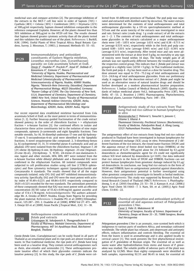
medicinal uses and compare activities [2]. The percentage inhibition ofthe extracts in the MCF-7 cell line were in order of Sapium (74%) >Cyathula (48%) > Celosia (30%) > Combretum (28%) > Drymaria (22%)at 500 mg/ml respectively. Sapium showed inhibition comparable to thereference compound Cisplatin. However, all the plants showed less than50% inhibition at 500 mg/ml in the HT29 cell line. The results showedthat Sapium showed greater cytotoxic activity than all the plants testedand this validates the traditional use of the plant. References: 1. Burkill,H.M. (1994). Useful Plants of West Tropical Africa. Royal Botanic Gar-dens, Surrey. 2. Mossman, T. (1983). J. Immunol. Methods 65: 55 – 63.
P129Immunomodulatory and antioxidantconstituents of Eastern Nigeria mistletoe,Loranthus micranthus Linn. (Loranthaceae)parasitic on Cola acuminata Schott et Endl.Omeje E1, Osadebe P1, Procksh P2, Amal H2, Debbab A2,Kawamura A3, Esimone C4, Nworu S1
1University of Nigeria, Nsukka, Pharmaceutical andMedicinal Cehmistry, Department of Pharmaceutical andMedicinal Cehmistryfaculty of PharmaceuticalSciencesuniversity of Nigeria, Nsukka, 410001 Nsukka,Nigeria; 2Heinrich-Hein University, D�sseldorf, Departmentof Pharmaceutical Biology, 40225 D�sseldorf, Germany;3Hunter College of CUNY, The City University of New York,U.S.A, Department of Chemistry, Department of Chemistry,10065 New York, United States; 4Faculty of PharmaceuticalSciences, Nnamdi Azikiwe University, 420281, Awka,Department of Pharmaceutical Microbiology andBiotechnology, 420281, Awka, 420281 Awka, Nigeria
Our recent reported data established mistletoe harvested from Colaacuminata Schott et Endl. as the most potent in terms of immunostimu-lation [1, 2]. Further bioassay-guided fractionation of the crude extractshowed potency in the order of chloroform fraction > > ethylacetatefraction > n-hexane fraction [3]. Our continued efforts to isolate andcharacterize these active constituents led to the isolation of nine majorcompounds, epimeric b-carotenoids and eight lipophilic fractions. Twonotable steroids, 5a-16, 16-dimethyl-androstan-17-one and 6b-hydroxy-17-oxo-4, 5-secoandrostan-4-oic acid were identified in the lipophilicfractions. A novel sesquiterpene-like compound; 2, 3-dimethoxy-benzo[a, b] cyclopentenyl-31, 31, 51-trimethyl pyran-4-carboxylic acid and analkaloid, CFO were isolated from the chloroform fraction. Stigmast-7, 20(21)-diene-3b-hydroxy-6-one, 3b-Hydroxy- stigmast- 23-ene (Stigmast-23-ene-3b-ol), 21b-hydroxy- 5, 9(10), 24-triene-21-nor-dammaren-3-one, lupeol and other steroids, HF4, HF6, HF7 were isolated from then-hexane fraction while dibutyl phthalate and a flavonoidal EA2 wereconfirmed in the ethylacetate fraction. All isolated compounds weresubjected to cell proliferation studies using cell line (C 57Bl/6 spleno-cytes) and flow cytometry techniques against Lipopolysaccharide andConcanavalin A standards. The results showed that of all the majorcompounds isolated; only CFO, EA2 and HF7 exhibited immunostimula-tory activity. Specifically, EA2 and CFO were the most potent with activ-ity index of 91.49 € 0.22% and 69.84 € 0.19% respectively compared to34.01 € 0.32% recorded for the two standards. The antioxidant potentialsof these compounds showed that EA2 was most potent with an effectiveconcentration (EC 50) value of 55.42 € 0.99 mg/ml against ascorbic acidvalue of 17.6 € 1.78 mg/ml. Acknowledgements: The authors acknowl-edge Mr. A. Ozioko of BDCP, Nsukka, Enugu State Nigeria for providingthe plant material. References: 1. Osadebe PO, et al (2009) J Ethnophar-macol., 125:287 – 293. 2. Osadebe et al (2008), RPRM Vol 27: 473 – 485.3. Omeje EO et al (2009) Asian Pacific J.Tropical Med., 2(4): 11 – 18.
P130Anthraquinone content and toxicity test of Cassiafistula pod extractsGritsanapan W, Sakulpanich A, Thongpraditchote SFaculty of Pharmacy, Mahidol University, Department ofPharmacognosy, 447 Sri-Ayudthaya Road, RatchatewiBangkok, Thailand
Cassia fistula Linn. (Caesalpiniaceae) can be easily found in all parts ofThailand as an ornamental plant and its ripe pods are almost treated as awaste. In Thai traditional medicine, the ripe pods of C. fistula have longbeen used as a laxative drug. They contain several anthraquinones suchas rhein, aloe-emodin and sennosides [1], These anthraquinones pro-mote laxative effect of which their glycosides contents indicate thelaxative potency [2]. In this study, the ripe pods of C. fistula were col-
lected from 10 different provinces of Thailand. The pod pulp was sepa-rated and extracted with distilled water by decoction. The water extractswere determined for the contents of total anthraquinones and totalanthraquinone glycosides by a UV-vis spectrophotometric method at515 nm and the acute toxicity of the extracts was investigated in miceand rats. Extract ratio (crude drug: 1 g crude extract) of all the extractsare 1 – 2: 1. The contents of total anthraquinones and total anthraqui-none glycosides in the extracts calculated as a major anthraquinonerhein were 1.45 – 1.85% w/w (average 1.63% w/w) and 0.38 – 0.71% w/w (average 0.53% w/w), respectively while in the fresh pod pulp con-tained 0.89 – 1.03% w/w (average 0.94% w/w) and 0.22 – 0.39% w/w(average 0.31% w/w), respectively. After tested mice and rats were ad-ministered with the extract at dose level 5 g/kg, no mortality or any signof toxicity were found within 14 days. The body weight gain of allanimals was not significantly different between the treated groups andthe respective control group. This indicats that C. fistula pod extract wasgrouped in a slightly toxic (LD 50 > 5 g/kg). In Thai traditional medicine,the fresh pod pulp 4 – 8 g has been used as a dose for laxative drug. Thisdose amount was equal to 37.6 – 75.2 mg of total anthraquinones and12.4 – 24.8 mg of total anthraquinone glycosides. From our preliminarystudy, it suggests that the decoction extract of C. fistula pod might beused as an alternative source of natural laxative drugs and should befurther developed as a modern pharmaceutical laxative preparation.References: 1. Indian Council of Medical Reseach (2005) Quality stan-dards of Indian medicinal plants Vol.2, Indraprastha Press (CBT), NewDelhi: 47 – 53. 2. Bruniton J (1995) Pharmacognosy, Phytochemistry,Medicinal plants, Lavoisier Publishing, Paris: 349 – 354.
P131Antigenotoxic study of rice extracts from ThaiSung-Yod red rice cultivar in human lymphocytesin vitroRatanavalachai T, Thitiorul S, Tanuchit S, Jansom C,Uttama S, Itharat AThammasat University, Preclinical Sciences (Biochemistry),Faculty of Medicine, Paholyothin Road, A. Klong Luang,12121 Pathumthani Province, Thailand
The antigenotoxic effect of rice extracts from Sung-Yod red rice cultivarproduced in Thailand have been investigated in human lymphocytes fora sister chromatid exchange (SCE) assay in vitro. Pretreatment of 2 dif-ferent fractions of the rice extracts, the rinsed water fraction (SYGW) andthe aqueous extract of freeze dried boiled rice bran (SYBBOIL) at theconcentrations of 6.25 – 100 mg/ml alone for 2 h followed by 0.1 mg/mldoxorubicin as chemotherapeutic agent for 2 h could not significantlyreduce SCE level induced by doxorubicin (p < 0.05). Our data indicatedthat rice extracts in the form of SYGW and SYBBOIL fractions can notprotect human lymphocytes from genotoxic damage induced by 0.1 mg/ml doxorubicin. In conclusion, our Sung-Yod rice extracts in the form ofSYGW and SYBBOIL have no antigenotoxic potential against doxorubicin.However, their antigenotoxic potential is further investigated usingother genotoxic compounds to investigate its benefit as herbal medicine.Acknowledgements: This study was supported by Research Fund, Na-tional Research Council of Thailand (NRCT) References: 1. Phutthapha-doong, S. et al. (2010) Oncol.Rep. 23: 53 – 59. 2. Kannan A. et.al. (2008) JAgric Food Chem 56:11643 – 7. 3. Nam, SH. et al. (2005) J Agric FoodChem. 53:816 – 22.
P132Chemical composition and antioxidant activity ofessential oil and aqueous extract of Pelargoniumgraveolens L’Her.Cavar S, Vidic D, Maksimovic MUniversity of Sarajevo, Faculty of Science, Department ofChemistry, Zmaja od Bosne 33 – 35, 71000 Sarajevo, BosniaAnd Herzegovina
Pelargonium graveolens L’Her is an aromatic, rose-scented herb which isindigenous to various parts of southern Africa, and nowadays cultivatedworldwide. The whole plant has relaxant, anti-depressant and antisepticeffects, reduces inflammation and controls bleeding. The essential oilfrom the leaves is used in aromatherapy and is also applied locally tocervical cancer [1]. This work presents the first phytochemical investi-gation of P. graveolens of Bosnian origin. The essential oil as well aswaste water after hydrodistillation from stems and leaves of P. grave-olens was studied. The volatile profile of odorous parts of the plant wasanalyzed by GC/MS. More than eighty compounds were identified inboth samples, representing 92.3% and 96.4% in total, for essential oil
Planta Med 2010; 76: 1163–1374 Georg Thieme Verlag KG Stuttgart · New York · ISSN 0032-0943
12257th Tannin Conference and 58th International Congress of the GA | 29th August – 2nd September 2010, Berlin, Germany
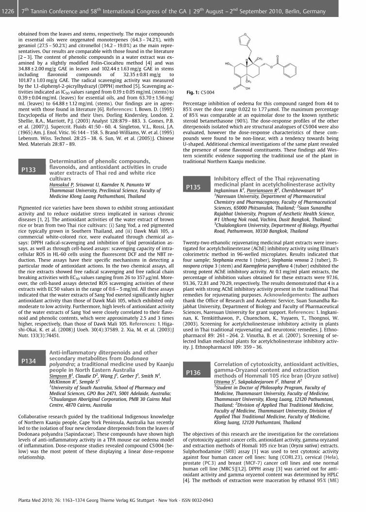
obtained from the leaves and stems, respectively. The major compoundsin essential oils were oxygenated monoterpenes (64.3 – 74.2%), withgeraniol (27.5 – 50.2%) and citronellol (14.2 – 19.0%) as the main repre-sentatives. Our results are comparable with those found in the literature[2 – 3]. The content of phenolic compounds in a water extract was ex-amined by a slightly modified Folin-Ciocalteu method [4] and was34.88 € 2.00 mg/g GAE in leaves and 102.44 € 1.63 mg/g GAE in stemsincluding flavonoid compounds of 32.35 € 0.81 mg/g to101.87 € 1.03 mg/g GAE. The radical scavenging activity was measuredby the 1,1-diphenyl-2-picrylhydrazyl (DPPH) method [5]. Scavenging ac-tivities indicated as IC50 values ranged from 0.19 € 0.05 mg/mL (stems) to0.39 € 0.04 mg/mL (leaves) for essential oils, and from 63.70 € 1.56 mg/mL (leaves) to 64.88 € 1.12 mg/mL (stems). Our findings are in agree-ment with those found in literature [6]. References: 1. Bown. D. (1995)Encyclopedia of Herbs and their Uses. Dorling Kindersley, London. 2.Shellie, R.A., Marriott, P.J. (2003) Analyst 128:879 – 883. 3. Gomes, P.B.et al. (2007) J. Supercrit. Fluids 41:50 – 60. 4. Singleton, V.L., Rossi, J.A.(1965) Am. J. Enol. Vitic. 16:144 – 158. 5. Brand-Williams, W. et al. (1995)Lebensm. Wiss. Technol. 28:25 – 38. 6. Sun, W. et al. (2005) J. ChineseMed. Materials 28:87 – 89.
P133Determination of phenolic compounds,flavonoids, and antioxidant activities in crudewater extracts of Thai red and white ricecultivarsHansakul P, Srisawat U, Kaendee N, Panunto WThammasat University, Preclinical Science, Faculty ofMedicine Klong Luang Pathumthani, Thailand
Pigmented rice varieties have been shown to exhibit strong antioxidantactivity and to reduce oxidative stress implicated in various chronicdiseases [1, 2]. The antioxidant activities of the water extract of brownrice or bran from two Thai rice cultivars: (i) Sang Yod, a red pigmentedrice typically grown in Southern Thailand, and (ii) Dawk Mali 105, acommercial white-colored rice, were evaluated through chemical as-says: DPPH radical-scavenging and inhibition of lipid peroxidation as-says, as well as through cell-based assays: scavenging capacity of intra-cellular ROS in HL-60 cells using the fluorescent DCF and the NBT re-duction. These assays have their specific mechanisms in detecting aparticular mode of antioxidant actions. In the two chemical assays, allthe rice extracts showed free radical scavenging and free radical chainbreaking activities with EC50 values ranging from 26 to 357 mg/ml. More-over, the cell-based assays detected ROS scavenging activities of theseextracts with EC 50 values in the range of 0.6 – 5 mg/ml. All these assaysindicated that the water extracts of Sang Yod exerted significantly higherantioxidant activity than those of Dawk Mali 105, which exhibited onlymoderate to low activity. Furthermore, high levels of antioxidant activityof the water extracts of Sang Yod were closely correlated to their flavo-noid and phenolic contents, which were approximately 2.5 and 3 timeshigher, respectively, than those of Dawk Mali 105. References: 1. Higa-shi-Okai, K. et al. (2008) J Uoeh. 30(4):37589. 2. Xia, M. et al. (2003) JNutr. 133(3):74451.
P134Anti-inflammatory diterpenoids and othersecondary metabolites from Dodonaeapolyandra; a traditional medicine used by Kaanjupeople in North Eastern AustraliaSimpson B1, Claudie D2, Wang J1, Gerber J1, Smith N1,McKinnon R1, Semple S1
1University of South Australia, School of Pharmacy andMedical Sciences, GPO Box 2471, 5001 Adelaide, Australia;2Chuulangun Aboriginal Corporation, PMB 30 Cairns MailCentre, 4870 Cairns, Australia
Collaborative research guided by the traditional Indigenous knowledgeof Northern Kaanju people, Cape York Peninsula, Australia has recentlyled to the isolation of four new clerodane diterpenoids from the leaves ofDodonaea polyandra (Sapindaceae). These compounds have shown highlevels of anti-inflammatory activity in a TPA mouse ear oedema modelof inflammation. Dose-response studies revealed compound CS004 (be-low) was the most potent of these displaying a linear dose-responserelationship.
Fig. 1: CS 004
Percentage inhibition of oedema for this compound ranged from 44 to85% over the dose range 0.022 to 1.77mmol. The maximum percentageof 85% was comparable at an equimolar dose to the known syntheticsteroid betamethasone (90%). The dose-response profiles of the otherditerpenoids isolated which are structural analogues of CS004 were alsoevaluated, however the dose-response characteristics of these com-pounds were found to be non-linear, with a tendency towards beingU-shaped. Additional chemical investigations of the same plant revealedthe presence of some flavonoid constituents. These findings add Wes-tern scientific evidence supporting the traditional use of the plant intraditional Northern Kaanju medicine.
P135Inhibitory effect of the Thai rejuvenatingmedicinal plant in acetylcholinesterase activityIngkaninun K1, Panriansaen R2, Cherdshewasart W3
1Naresuan University, Department of PharmaceuticalChemistry and Pharmacognosy, Faculty of PharmaceuticalSciences, 65000 Phitsanulok, Thailand; 2Suan SunandhaRajabhat University, Program of Aesthetic Health Science,#1 Uthong Nok road, Vachira, Dusit Bangkok, Thailand;3Chulalongkorn University, Department of Biology, PhyathaiRoad, Pathumwan, 10330 Bangkok, Thailand
Twenty-two ethanolic rejuvenating medicinal plant extracts were inves-tigated for acetylcholinesterase (AChE) inhibitory activity using Ellman’scolorimetric method in 96-welled microplates. Results indicated thatfour sample; Stephania erecta 1 (tuber), Stephania venosa 2 (tuber), Ti-nospora crispa 3 (stem) and Kaempferia parviflora 4 (tuber) exhibited thestrong potent AChE inhibitory activity. At 0.1 mg/ml plant extracts, thepercentage of inhibition values obtained for these extracts were 97.16,93.36, 72.81 and 70.29, respectively. The results demonstrated that 4 is aplant with strong AChE inhibitory activity present in the traditional Thairemedies for rejuvenating purposes. Acknowledgements: The authorsthank the Office of Research and Academic Service, Suan Sunandha Ra-jabhat University, Department of Biology and Faculty of PharmaceuticalSciences, Naresuan University for grant support. References: 1. Ingkani-nan, K. Temkitthawon, P., Chuenchom, K., Yuyaem, T., Thongnoi, W.(2003). Screening for acetylcholinesterase inhibitory activity in plantsused in Thai traditional rejuvenating and neurotonic remedies. J. Ethno-pharmacol 89: 261 – 264. 2. Vinutha, B. et al. (2007). Screening of se-lected Indian medicinal plants for acetylcholinesterase inhibitory activ-ity. J. Ethnopharmacol 109: 359 – 36.
P136Correlation of cytotoxicity, antioxidant activities,gamma-Oryzanol content and extractionmethods of Hommali 105 rice bran (Oryza sativa)Uttama S1, Sakpakdeejaroen I2, Itharat A2
1Student in Doctor of Philosophy Program, Faculty ofMedicine, Thammasart University, Faculty of Medicine,Thammasart University, Klong Luang, 12120 Pathumtani,Thailand; 2Division of Applied Thai Traditional Medicine,Faculty of Medicine, Thammasart University, Division ofApplied Thai Traditional Medicine, Faculty of Medicine,Klong luang, 12120 Pathumtani, Thailand
The objectives of this research are the investigation for the correlationsof cytotoxicity against cancer cells, antioxidant activity, gamma oryzanoland extraction methods of Homali 105 rice bran (Oryza sativa) extracts.Sulphorhodamine (SRB) assay [1] was used to test cytotoxic activityagainst four human cancer cell lines: lung (CORL 23), cervical (Hela),prostate (PC 3) and breast (MCF-7) cancer cell lines and one normalhuman cell line (MRC 5)[1,2]. DPPH assay [3] was carried out for anti-oxidant activity and gamma oryzenol content was determined by HPLC[4]. The methods of extraction were maceration by ethanol 95% (ME)
Planta Med 2010; 76: 1163–1374 Georg Thieme Verlag KG Stuttgart · New York · ISSN 0032-0943
1226 7th Tannin Conference and 58th International Congress of the GA | 29th August – 2nd September 2010, Berlin, Germany
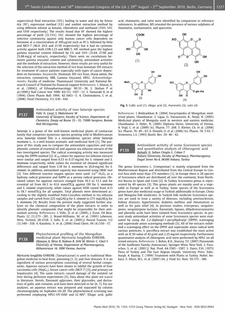
supercritical fluid extraction (SFE), boiling in water and dry by freezedry (BF), expression method (EX) and soxhlet extraction method byusing different solvent as hexane, chloroform and methanol (SXH, SXCand SXM respectively). The results found that BF showed the highestpercentage of yield (21.73%). SXC showed the highest percentage ofselective cytotoxicity against only human cancer cells dependent onhormone at a concentration of 100 mg/ml such as PC 3, followed by Helaand MCF-7 (46.9, 29.6 and 21.58 respectively) but it had no cytotoxicactivity against both COR-L23 and MRC 5. ME method gave the highestgamma oryzenol content followed by EX and SXH (31.64, 27.90 and25.88 mg/g of extracts, respectively). There were no correlations be-tween gamma oryzanol content and cytotoxicity, antioxidant activitiesand the methods of extraction. However, these results are very useful forthe selection of the extraction method of rice bran hommali 105 extractsfor treatment of cancer patients especially with types of cancer depen-dent on hormones. Keywords: Hommali 105 rice bran, Oryza sativa, theextraction, cytotoxicity, SRB, Gamma Oryzanol, HPLC. Acknowledge-ments: Faculty of medicine, Thammasart University and National Re-search Council of Thailand for financial support References: 1. Itharat, A.et al. (2004) J. of Ethnopharmacology: 90:33 – 38. 2. Skehan P etal.(1990) J Natl Cancer Inst 1990: 82(13): 1107 – 12. 3. Yamazaki K, et al(1994) Chem Pharm Bull 1994; 42:1663 – 5. 4. Chotimakorn, C et al.(2008). Food Chemistry, 111, 636 – 641.
P137Antioxidant activity of two Satureja speciesVidic D, Cavar S, Maksimovic MUniversity of Sarajevo, Faculty of Science, Department ofChemistry, Zmaja od Bosne 33 – 35, 71000 Sarajevo, BosniaAnd Herzegovina
Satureja is a genus of the well-known medicinal plants of Lamiaceaefamily that comprises numerous species growing wild in Mediterraneanarea. Satureja visianii �ilic is a stenoendemic species while Saturejamontana L., is a well known aromatic and medicinal herb [1]. The pur-pose of this study was to compare the antioxidant capacities and totalphenolic content of essential oil and aqueous tea infusion extracts of thetwo investigated species. The radical scavenging activity was measuredusing the DPPH method [2]. IC50 values for aqueous tea infusion extractswere similar and ranged from 0.33 to 0.37 mg/mL for S. visianni and S.montana respectively, while values for essential oil showed significantdifferences and ranged from 5.18 for S. montana to 26.0 mg/mL for S.visianii. In addition, antioxidant capacity was measured using ORAC test[3]. Two different reactive oxygen species were used: Cu2+-H2O2 as ahydroxy radical generator and AAPH as a peroxy radical generator. Ob-tained values for aqueous extracts reanged from 10.3 to 13.3 and foressential oil from 66.3 to 61.6 mmolTE/g against OH for S. montanaand S. visianii respectively, while values against OOH varied from 4.13to 58.7 mmolTE/g for all samples. Total phenols were determined ac-cording to the slightly modified Folin-Ciocalteu method in tea infusionsamples and varied from 225 mgGAE/g for S. visianii to 275 mgGAE/g forS. montana [4]. Results from the present study suggested further ana-lyses on the chemical composition of the plant extracts in order toidentify phenolic compounds that might be responsible for their anti-oxidant activity. References: 1. Vidic, D. et al. (2009), J. Essnt. Oil Bear.Plants 12 (3):273 – 281. 2. Brand-Williams, W. et al. (1995) Lebensm.Wiss. Technol. 28:2538. 3. Sofic, E. et. al. (2005) J. Neural Transm. 112(3):349 – 358. 4. Katalinic, V. et al. (2006) Food Chem. 94 (4):550 – 57.
P138Phytochemical profiling of the Mongolianmedicinal plant Myricaria longifolia EHRENB.Obmann A, Mraz B, Kubasa B, Zehl M, Kletter C, Glasl SUniversity of Vienna, Department of Pharmacognosy,Althanstrasse 14, 1090 Vienna, Austria
Myricaria longifolia EHRENB. (Tamaricaceae) is used in traditional Mon-golian medicine to heal fever, poisoning [1, 2], and liver diseases. It is aningredient of various prescriptions consisting of several herbal compo-nents. Aqueous extracts have been shown to inhibit the growth of livercarcinoma cells (HepG2), breast cancer cells (MCF-7) [3], and primary rathepatocytes [4]. The same extracts caused damage of the isolated ratliver during perfusion experiments [5]. Data about this plant are scarcein literature. Sterols, flavonoid aglycones, their glycosides, and deriva-tives of gallic and cinnamic acid have been detected so far [6, 7]. For ouranalyses, an aqueous extract was prepared and separated by columnchromatography on Sephadex LH-20. The phytochemical screening wasperformed employing HPLC-UV-DAD and LC-MSn. Ellagic acid, gallic
acid, rhamnetin, and rutin were identified by comparison to referencesubstances. In addition, MS revealed the presence of various sulphates ofrhamnetin, isorhamnetin, and quercetin.
Fig. 1: Gallic acid (1), ellagic acid (2), rhamnetin (3), rutin (4)
References: 1. Boldsaikhan B. (2004) Encyclopedia of Mongolian med-icinal plants. Ulaanbaatar. 2. Ligaa, U., Davaasuren, B., Ninjil, N. (2005)Medicinal plants of Mongolia used in western and eastern medicine.Ulaanbaatar. 3. Holec, N. (2005) Diploma thesis. University of Vienna.4. Vogl, C. et al. (2009) Sci. Pharm. 77: 268. 5. Kletter, Ch. et al. (2008)Sci. Pharm. 76: 49 – 63. 6. Donath, O. et al. (2006) Sci. Pharm. 74: S 93. 7.Semenova, L.S. (1993) Rastit. Res. 29: 40 – 42.
P139Antioxidant activity of some Scorzonera speciesand quantitative analysis of chlorogenic acidBahadir O, Saltan Citoglu G, Coban TAnkara University, Faculty of Pharmacy, Pharmacognosy,Dogol Street No:4, 06100 Ankara, Turkey
The genus Scorzonera L. (Compositae) is mainly originated from theMediterranean Region and distributed from the Central Europe to Cen-tral Asia with more than 175 members [1]. In Europe there is 28 speciesof Scorzonera which are distributed all over the continent, from North-ern Russia to Spain and Crete [2]. In Turkey Scorzonera genus is repre-sented by 49 species [3]. This genus plants are mainly used as a vege-table in Europe as well as in Turkey. Some species of the Scorzoneragenus have also medicinal usage in Turkish additionally in Europe, Chinaand Mongolia folk medicines. In Turkish folk medicine Scorzonera spe-cies are used to treat a variety of illnesses, including arteriosclerosis,kidney diseases, hypertension, diabetes mellitus and rheumatism aswell as for pain relief [4]. In previous studies, triterpenes, sesquiter-penes, sesquiterpene lactones, flavonoids, lignans, dihydroisocoumarinsand phenolic acids have been isolated from Scorzonera species. In pre-sent study antioxidant activities of some Scorzonera species were eval-uated by using the 2,2-diphenyl-1-picrylhydrazyl (DPPH) scavengingand superoxide anion scavenging methods [5]. All of the extracts exhib-ited a scavenging effect on the DPPH and superoxide anion radical withvarious potencies. S. parviflora extract was established the most activewith an IC50 value of 42 g/ml and 2.25 mg/ml respectively. Furthermorequantitative analysis of chlorogenic acid were performed by HPLC on alltested extracts. References: 1. Bohm, B.A., Stuessy, T.F. (2007) Flavonoidsof the Sunflower Family (Asteraceae). Springer Wien. New York. 2. Para-schos, S. et al. (2001) J. Nat. Prod. 64:1585 – 1587. 3. Davis, P.H. (1975)Flora of Turkey and The East Aegean Islands. University Press. Edin-burgh. 4. Baytop, T. (1999) Treatment with Plants in Turkey. Nobel. An-kara. 5. Altun, M.L. et al. (2007) Int. J. Food Sci. Nutr. 59:175 – 180.
Planta Med 2010; 76: 1163–1374 Georg Thieme Verlag KG Stuttgart · New York · ISSN 0032-0943
12277th Tannin Conference and 58th International Congress of the GA | 29th August – 2nd September 2010, Berlin, Germany
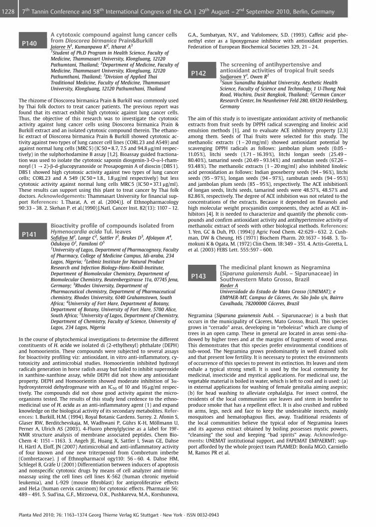
P140A cytotoxic compound against lung cancer cellsfrom Dioscorea birmanica Prain&BurkillJaiaree N1, Kumanpawa K2, Itharat A3
1Student of Ph.D Program in Health Science, Faculty ofMedicine, Thammasart University, Klongluang, 12120Pathumtani, Thailand; 2Department of Medicine, Faculty ofMedicine, Thammasart University, Klongluang, 12120Pathumthani, Thailand; 3Division of Applied ThaiTraditional Medicine, Faculty of Medicine, ThammasartUniversity, Klongluang, 12120 Pathumthani, Thailand
The rhizome of Dioscorea birmanica Prain & Burkill was commonly usedby Thai folk doctors to treat cancer patients. The previous report wasfound that its extract exhibit high cytotoxic against lung cancer cells.Thus, the objective of this research was to investigate the cytotoxicactivity against lung cancer cells using Dioscorea birmanica Prain &Burkill extract and an isolated cytotoxic compound therein. The ethano-lic extract of Dioscorea birmanica Prain & Burkill showed cytotoxic ac-tivity against two types of lung cancer cell lines (CORL23 and A549) andagainst normal lung cells (MRC 5) (IC 50 = 8.7, 7.5 and 94.8 mg/ml respec-tively) in the sulphorhodamine B assay [1,2]. Bioassay guided fractiona-tion was used to isolate the cytotoxic saponin diosgenin-3-O-a-l-rham-nosyl (1 ? 2)-b-d-glucopyranoside or Prosapogenin A of dioscin (DBS 1).DBS 1 showed high cytotoxic activity against two types of lung cancercells; CORL23 and A 549 (IC 50 = 1.8., 1.8 mg/ml respectively) but lesscytotoxic activity against normal lung cells MRC 5 (IC 50 = 37.1 mg/ml).These results can support using this plant to treat cancer by Thai folkdoctors. Acknowledgements: Thammasart University for financial sup-port References: 1. Tharat, A. et al. (2004) J. of Ethnopharmacology90:33 – 38. 2. Skehan P. et al.(1990) J.Natl. Cancer Inst. 82(13): 1107 – 12.
P141Bioactivity profile of compounds isolated fromHymenocardia acida Tul. leavesSofidiya M1, Lange C2, Sattler I2, Beukes D3, Afolayan A4,Odukoya O1, Familoni O5
1University of Lagos, Department of Pharmacognosy, Facultyof Pharmacy, College of Medicine Campus, Idi-araba, 234Lagos, Nigeria; 2Leibniz Institute for Natural ProductResearch and Infection Biology-Hans-Kn�ll-Institute,Department of Biomolecular Chemistry, Department ofBiomolecular Chemistry, Beutenbergstrasse 11a, 07745 Jena,Germany; 3Rhodes University, Department ofPharmaceutical chemistry, Department of Pharmaceuticalchemistry, Rhodes University, 6140 Grahamstown, SouthAfrica; 4University of Fort Hare, Department of Botany,Department of Botany, University of Fort Hare, 5700 Alice,South Africa; 5University of Lagos, Department of Chemistry,Department of Chemistry, Faculty of Science, University ofLagos, 234 Lagos, Nigeria
In the course of phytochemical investigations to determine the differentconstituents of H. acida we isolated di (2-ethylhexyl) phthalate (DEPH)and homoorientin. These compounds were subjected to several assaysfor bioactivity profiling viz: antioxidant, in vitro anti-inflammatory, cy-totoxicity and antimicrobial studies. Homoorientin inhibited hydroxylradicals generation in horse radish assay but failed to inhibit superoxidein xanthine-xanthine assay, while DEPH did not show any antioxidantproperty. DEPH and Homoorientin showed moderate inhibition of 3a-hydroxysteroid dehydrogenase with an IC50 of 10 and 16 mg/ml respec-tively. The compounds did not show good activity against the micro-organisms tested. The results of this study lend credence to the ethno-medicinal use of H. acida as an anti-inflammatory agent (1) and expandknowledge on the biological activity of its secondary metabolites. Refer-ences: 1. Burkill, H.M. (1994). Royal Botanic Gardens. Surrey. 2. Afonin S,Glaser RW, Berditchevskaja, M, Wadhwani P, G�hrs K-H, M�llmann U,Perner A, Ulrich AS (2003). 4-Fluoro phenylglycine as a label for 19F-NMR structure analysis of membrane associated peptides. Chem Bio-Chem 4: 1151 – 1163. 3. Angeh JE, Huang X, Sattler I, Swan GE, DahseH, H�rtl A, Eloff, JN (2007) Antimicrobial and anti-inflammatory activityof four known and one new triterpenoid from Combretum imberbe(Combretaceae). J of Ethnopharmacol ogy110: 56 – 60. 4. Dahse HM,Schlegel B, Gr�fe U (2001) Differentiation between inducers of apoptosisand nonspecific cytotoxic drugs by means of cell analyzer and immu-noassay using the cell lines cell lines K-562 (human chronic myeloidleukemia), and L-929 (mouse fibroblast) for antiproliferative effectsand HeLa (human cervix carcinom) for cytotoxic effects. Pharmazie 56:489 – 491. 5. Sud’ina, G.F., Mirzoeva, O.K., Pushkareva, M.A., Korshunova,
G.A., Sumbatyan, N.V., and Vafolomeev, S.D. (1993). Caffeic acid phe-nethyl ester as a lipoxygenase inhibitor with antioxidant properties.Federation of European Biochemical Societies 329, 21 – 24.
P142The screening of antihypertensive andantioxidant activities of tropical fruit seedsSudjaroen Y1, Owen R2
1Saun Sunandha Rajabhat University, Aesthetic HealthScience, Faculty of Science and Technology, 1 U-Thong NokRoad, Wachira, Dusit Bangkok, Thailand; 2German CancerResearch Center, Im Neunheimer Feld 280, 69120 Heidelberg,Germany
The aim of this study is to investigate antioxidant activity of methanolicextracts from fruit seeds by DPPH radical scavenging and linoleic acidemulsion methods [1], and to evaluate ACE inhibitory property [2,3]among them. Seeds of Thai fruits were selected for this study. Themethanolic extracts (1 – 20 mg/ml) showed antioxidant potential byscavenging DPPH radicals as follows: jambolan plum seeds (0.05 –11.05%), litchi seeds (1.71 – 16.39%), litchi longan seeds (25.49 –80.40%), tamarind seeds (20.49 – 93.14%) and rambutan seeds (67.26 –93.48%). The methanolic extracts (1 – 20 mg/ml) also inhibited linoleicacid peroxidation as follows: Indian gooseberry seeds (94 – 96%), litchiseeds (95 – 97%), longan seeds (94 – 97%), rambutan seeds (94 – 95%)and jambolan plum seeds (85 – 95%), respectively. The ACE inhibition%of longan seeds, litchi seeds, tamarind seeds were 48.57%, 48.57% and82.86%, respectively. The degree of ACE inhibition was not related to theconcentrations of the extracts. Because it depended on flavanols andhigh molecular weight procyanidin components, they acted as ACE in-hibitors [4]. It is needed to characterize and quantify the phenolic com-pounds and confirm antioxidant activity and antihypertensive activity ofmethanolic extract of seeds with other biological methods. References:1. Yen, GC & Duh, PD. (1994) J Agric Food Chem. 42:629 – 632. 2. Cush-man, DW & Cheung, HS (1971) Biochem Pharm. 20:1637 – 1648. 3. To-mokuni K & Ogata, M. (1972) Clin Chem. 18:349 – 351. 4. Actis-Goretta, L.et al. (2003) FEBS Lett. 555:597 – 600.
P143The medicinal plant known as Negramina(Siparuna guianensis Aubl. – Siparunaceae) inSouthwestern Mato Grosso, BrazilRieder AUniversidade do Estado de Mato Grosso (UNEMAT); eEMPAER-MT, Campus de C�ceres, Av. S¼o Jo¼o s/n, BairroCavalhada, 78200000 C�ceres, Brazil
Negramina (Siparuna guianensis Aubl. – Siparunaceae) is a bush thatoccurs in the municipality of C�ceres, Mato Grosso, Brazil. This speciesgrows in “cerrado” areas, developing in “reboleiras” which are clump oftrees in an open camp. These in general are located in areas semi-sha-dowed by higher trees and at the margins of fragments of wood areas.This demonstrates that this species prefer environmental conditions ofsub-wood. The Negramina grows predominantly in well drained soilsand that present low fertility. It is necessary to protect the environmentsof occurrence of this species to prevent its extinction. Its leaves and stemexhale a typical strong smell. It is used by the local community formedicinal, insecticide and mystical applications. For medicinal use, thevegetable material is boiled in water, which is left to cool and is used: (a)in external applications for washing of female genitalia aiming asepsis;(b) for head washing to alleviate cephalalgia. For insect control, theresidents of the local communities use leaves and stem in bomfire toproduce smoke that has a repellent effect. It is also crushed and rubbedin arms, legs, neck and face to keep the undesirable insects, mainlymosquitoes and hematophagous flies, away. Traditional residents ofthe local communities believe the typical odor of Negramina leavesand its aqueous extract obtained by boiling possesses mystic powers,“cleansing” the soul and keeping “bad spirits” away. Acknowledge-ments: UNEMAT institutional support, and FAPEMAT EMPAERMT; sup-port afforded by the whole project team PLAMED: Bonila MGO, CarnielloM, Ramos PR et al.
Planta Med 2010; 76: 1163–1374 Georg Thieme Verlag KG Stuttgart · New York · ISSN 0032-0943
1228 7th Tannin Conference and 58th International Congress of the GA | 29th August – 2nd September 2010, Berlin, Germany

P144Evaluation of the burn healing properties of fiveIranian medicinal plants in diabetic ratsGhasemi Pirbalouti A1, Azizi S2, Koohpayeh A1, Golparvar A3
1Research Centre of Medicinal Plants & Ethnoveterinary,Islamic Azad University, Shahrekord Branch, MedicinalPlants, Iran-Shahrekord-Rahmatyeh-POBox:166, 88148Shahrekord, Iran, Islamic Republic Of; 2Veterinary MedicineFaculty, Islamic Azad University, Shahrekord Branch,Shahrekord, Iran, Department of Pathology, Islamic AzadUniversity, Shahrekord Branch, POBox: 166, Shahrekord,Iran, 88148 Shahrekord, Iran, Islamic Republic Of;3Agriculture Faculty, Islamic Azad University, KhorasganBranch, Isfahan, Iran, Department of Plant Breeding, IslamicAzad University, Khorasgan Branch, Isfahan, Iran, 81595 –158 Isfahan, Iran, Islamic Republic Of
Some of the medicinal plants in Iranian traditional medicine (Unani)whose have been used as remedy against edema, burn, wound and fortheir carminative, antimicrobial and anti-inflammatory activities [1, 2].For example, Malva sylvestris L. (Malvaceae), Punica granatum L. (Puni-caceae), Prunus amygdalus L. (Rosaceae), Arnebia euchroma Rolye(Johnst) (Boraginaceae) and Scrophularia deserti Del. (Scrophulariaceae)used for wound and burn healing by tribal communities in Iran[3]. Theethanol extracts of Malva sylvestris and P. granatum flowers, P. amyg-dalus leaves, A. euchroma roots and S. deserti were used to evaluate theburn healing activity at 200 mg/kg/day dose in alloxan-induced diabeticrats. Burns were induced in Wistar rats divided into nine groups asfollowing; Group-1, normal rats were treated with simple ointmentbase. Group-2, diabetic rats were treated with simple ointment base(control). Groups-3 and -7, diabetic rats were treated with simple oint-ment base containing of extracts (diabetic animals), Groups 8, diabeticrats were treated with simple ointment base containing of mixed ex-tracts, Group-9, diabetic rats received the standard drug (Silver sulpha-diazine). The efficacy of treatment was evaluated based on burn woundarea relative and histopathological characteristics. The extract-treateddiabetic animals showed significant reduction in the wound area whencompared with control. Also, histopathological studies of the tissue ob-tained on days 9th and 16th from the extract-treated by extracts showedincreased well organized bands of collagen, more fibroblasts and fewinflammatory cells. References: 1. Ghasemi Pirbalouti A, et al. (2009)Pharmacognosy Mag. 5: 433 – 437. 2. Zargari A. (1989 – 1992) MedicinalPlants. University Publication of Tehran, Iran. 3. Ghasemi Pirbalouti A.(2009) Herba Polonica. 55: 69 – 75.
P145Antinociceptive effects of Stachys laxaKhosravi Dehaghi N1, Hajimehdipoor H2, Sahebgharani M1,Khanavi M1, Mirshaki Z1
1Department of Pharmacognosy, Faculty of Pharmacy,Tehran University of Medical Sciences, Tehran, Iran;2Department of Herbal Drugs, Food & Drug ControlLaboratories & Food & Drug Laboratory Research Center,MOH & ME, Tehran, Iran
The Labiatae family (Lamiaceae) is one of the largest and most distinc-tive families of flowering plants, with about 200 genera and almost4000 species worldwide. This family has an almost cosmopolitan dis-tribution. Many stachys plants- a genus of this family- have been used asmedicinal plants because of their broad spectrum biological activities.Some stachys have been used as remedy for painful or inflammatoryconditions in folk medicine. Anti-inflammatory and analgesic effects ofsome species of Stachys e. g. S. inflata, S. byzantina and S. Schtschegleeviihave been reported. In this study, the antinociceptive properties of totalmethanolic extracts of the aerial parts of S. laxa was investigated byformalin test in mice. Intraperitoneal injection of the methanolic extractof S. laxa at the doses of 100, 200 & 400 mg/kg, 15 min before formalintest, significantly inhibited the chronic phase of formalin- induced pain.Intraperitoneal injection of opioid antagonist (naloxone) significantlyreversed the analgesic effect of extract in chronic phases of formalintest. This results suggest that that analgesic activity of this plant maybe partly mediated by opiate system. References: 1. v. Mozaffarian(2006) A dictionary of Iranian plant names, farhang moaser, tehran. 2.A. Zargari (1996) Medicinal plants, vol 4, Tehran university, Tehran. 3. v.Mozaffarian (2005) classification of plants, vol 2, amirkabir, tehran. 4. M.Semnani, M. Saeidi, M.R. Mahdavi, F.Rahimi, J of Mazandaran UniversityMedicinal science, 2007, 17(57):57 – 66. Antimicrobial effects of meta-nolic extracts of some species of stachys &philoms. 5. M. khanvi: M.Hajimahmoodi, M. Cheraghi-Niroomand, Z. Kargar, Y. Ajani, A. Hajia-
khoondi, M.R. Oveisi. 6. African Journal of Biotechnology Vol. 8 (6), pp.1143 – 1147, 20 March, 2009 Comparison of the antioxidant activity andtotal phenolic contents in some stachys species.
P146Phytoecdysteroids from arial parts of Ajugachamaecistus subsp. tomentellaSadati N1, Khanavi M1, Shams Ardekani M1,Hadjiakhoondi A1, Akbarzadeh T2
1Department of pharmacognosy, Faculty of pharmacy,Tehran University of Medical sciences, 16th Azar St., 14155 –6451 Tehran, Iran, Islamic Republic Of; 2Department ofmedicinal chemistry, Faculty of pharmacy, Tehran Universityof Medical Sciences, 16th Azar St., 14155 – 6451 Tehran, Iran,Islamic Republic Of
The genus Ajuga (Lamiaceae) is represented by five species in the flora ofIran, of which Ajuga chamaecistus has several exclusive subspecies in-clude subsp. tomentella [1]. The plants of this genus are used tradition-ally for treatment of joints pain, gout and jaundice [2]. In this studydiethylether fraction of defatted methanolic extract (80%) from arialparts of A. chamaecistus subsp. Tomentella was chromatographed onsilicagel using a CHCL3-EtoAc-MeOH gradient system to give compound1 and 2. The structure of compound 1 and 2 were determined to be 20-Hydroxyecdysone and Cyasterone, respectively, by means of spectro-scopic analysis (13C, HNMR, IR, Mass Spectroscopy). Ecdysteriods are alarge group of natural polyhydroxysteroids with a wide spectrum ofbiological activities, which are isolated from the animal and plant king-doms [3]. In plants of genus Ajuga, a variety of phytoecdysteroids hasbeen isolated. Among them, 20-Hydroxyecdysone or Cyasterone areusually the most abundant [4]
Fig. 1: Structure of compound 1 and 2
References: 1. Mozaffarian, V (2007) A Dictionary of Iranian PlantNames. Farhang Moaser. Tehran. 2. Naghibi, F. et al. (2005) IJPR. 2:63 –79. 3. Ramazanov, N Sh. (2005) Chemistry of Nat. Comp. 41(4): 361 –369. 4. Calcagno, M.-Pia. et al. (1995) Tetrahedr. 51(44): 12119 – 12126.
P147Evaluation of chemical composition andantioxidant activity of total extract of Cuscutachinensis Lam. used in traditional medicineAmin G, Mokhtari Kondori B, Vazirian M, Abdi L,Arabshahi GTehran University of Medical Science, Pharmacognosy,Faculty of Pharmacy, Tehran University of Medical Sciences,P.O.Box 14155 – 6451, Tehran, Iran, Islamic Republic Of
Cuscuta sp. (Convolvulaceae), known as dodder, has been used in Iraniantraditional medicine for many gastrointestinal, respiratory, endocrine,skin and neurological diseases and is locally named “Davaa’’-ol-Jonoon”(Drug of mania) [1, 2, 3]. In recent experimented traditional medicinesome species are used for a wide variety of conditions such as bipolardisorder, depression, anxiety, etc. According to previous studies, oxida-tive stress is related to some neurological disorders such as schizophre-nia, psychosis and bipolar disorder [4, 5]. C. chinensis is one of Cuscutasp. used widely in traditional medicine that is rich in flavonoids as oneof the major antioxidant compounds [6]. In this study we evaluated thechemical composition and examined the antioxidant activity of thisspecies to define one of the possible mechanisms for its use in tradi-tional medicine. The total extract of C. chinensis (whole plant) wasscreened for saponins, alkaloids, flavonoids, tannins and sterols. The freeradical DPPH scavenging activity was compared with the synthetic anti-oxidant (BHA) as a positive control. Total extract showed the presence ofalkaloids, tannins, saponins, sterols and flavonoids. In a concentration of500 mg/mL it showed antioxidant activity comparable with that of BHA(220 mg/mL, p < 0.05). The IC50’s of extract and BHA were 235 mg/mL and7.9 mg/mL respectively. These data suggest that one of the mechanisms
Planta Med 2010; 76: 1163–1374 Georg Thieme Verlag KG Stuttgart · New York · ISSN 0032-0943
12297th Tannin Conference and 58th International Congress of the GA | 29th August – 2nd September 2010, Berlin, Germany
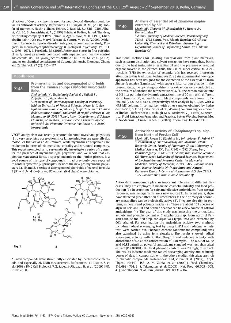
of action of Cuscuta chinensis used for neurological disorders could bevia its antioxidant activity. References: 1. Hussayni, M. M., (2008), Toh-fat-ol-momenin, Nashr-e-Shahr, Tehran. 2. Razi, M. Z., (826 – 930), Alha-vi, Vol. 20. 3. Ansarishirazi, A., (1996) Ekhtiarat Badiee, 1st ed. The drugdistributing company of Razi, Tehran. 4. Aghili-Alavi, M. H., (1996) Ghar-abadin kabir, 10th ed., Marvi, Tehran. 5. Yumru, M. et al. (2009), Oxida-tive imbalance in bipolar disorder subtypes: a comparative study, Pro-gress in Neuro-Psychopharmacology & Biological psychiatry, Vol. 33,p.1070 – 1074. 6. Parellada, M. (2010), Antioxanat status in first episodesof early onset psychosis compared with asperger and healthy controladolescents, doi: 10.1\‰16/j.schres.2010.02.6 61. 7. Ye, M., et al. (2002),studies on chemical constituents of Cuscuta chinensis, Zhongguo ZhongYao Za Zhi, Vol. 27 (2): 115 – 117.
Miscellaneous
P148Pre-myrsinanes and deoxygenated phorboidsfrom the Iranian spurge Euphorbia macrocladaBoiss.Shokoohinia Y1, Taglialatela-Scafati O2, Sajjadi S1,Zolfaghari B1, Appendino G3
1Department of Pharmacognosy, Faculty of Pharmacy,Isfahan University of Medical Sciences, Hezar jarib AveIsfahan, Iran, Islamic Republic Of; 2Dipartimento di Chimicadelle Sostanze Naturali, Universit di Napoli Federico II, ViaMontesano 49, 80131 Napoli, Italy; 3Dipartimento di ScienzeChimiche, Alimentari, Farmaceutiche e Farmacologiche,universit del Piemonte Orientale, Via Bovio 6, 3, 28100Novara, Italy
VEGFR antagonism was recently reported for some myrsinane polyesters[1], a very surprising observation since kinase inhibitors are generally flatmolecules that act as an ATP-mimics, while myrsinanes diterpenoids areexuberant in terms of tridimensional chirality and structural complexity.This report prompted us to systematically investigate a series of spurgesfor the presence of myrsinane-type polyesters, and we report that Eu-phorbia macroclada Boiss, a spurge endemic to the Iranian plateau, is agood source of this type of compounds. It had previously been reportedto contain cytotoxic [2] principles; besides the new pre-myrsinane polye-sters 1a, 1b and 2, a series of deoxygenated phorboids of general formula3 (R1 = H, Ac, 4 H =-b or -a; R2 = short alkyl chain) were obtained.
Fig. 1
All new compounds were structurally elucidated by spectroscopic meth-ods, and especially 2D NMR measuremets. References: 1. Hussain, S. etal. (2008). BMC Cell Biology.9:7. 2. Sadeghi-Aliabadi, H. et al. (2009) IJPR.5:103 – 108.
P149Analysis of essential oil of Zhumeria majdaeextracted by SFEMoein M1, Ghasemi Y1, Sharifzadeh F1, Hezave A2,Esmaeilzadeh F2
1Shiraz University of Medical Sciences, Pharmacognosy,71345 – 1735 Shiraz, Iran, Islamic Republic Of; 2ShirazUniversity, Chemical and Petroleum EngineeringDepartment, School of Engineering Shiraz, Iran, IslamicRepublic Of
Traditional methods for isolating essential oils from plant materials,such as steam distillation and solvent extraction have some draw backsdue to the heat instability of essential oil and the presence of residualorganic solvent in the extract. Thus, the use of super critical fluid ex-tractions (SFE) for extraction of essential oils has received increasingattention to this traditional techniques [1, 2]. An experimental flow-typeapparatus has been designed for the extraction of the essential oil fromZhumeria majdae (Lamiaceae) with super critical carbon dioxide. In thepresent study, the operating conditions for extraction were conducted atthe pressure of 200 bar, the temperature of 37 �C, the carbon dioxide rateof 0.3 liter per min, the dynamic extraction time of 20 min with differentstatic times of 30, 45 and 60 min. Main compounds were found to belinalool (73.8, 72.5, 41.5%, respectively) after analysis by GC/MS with aHP5-MS column. In comparison with other samples obtained by hydrodistillation, SFE oil (static times of 30, 45 min) contains higher amountof linalool. References: 1. Mchugh M A., Krukonis V J. (1986) Supercri-tical Fluid Extraction Principles and Practice, Butter Worths, Boston, MA.2. Goodarznia I, Esmaeilzadeh F. (2002) J. Chem. Eng. Data 47:333.
P150Antioxidant activity of Cladophoropsis sp. alga,from North of Persian GulfMoein M1, Moein S2, Ebrahimi N1, Sohrabipour J3, Rabiei R3
1Department of Pharmacognosy and Medicinal PlantsResearch Center, Faculty of Pharmacy, Shiraz University ofMedical Sciences, P.O. Box 71345 – 1583, Shiraz, Iran,Pharmacognosy, 71345 – 1735 Shiraz, Iran, Islamic RepublicOf; 2Hormozgan University of Medical Sciences, Departmentof Biochemistry and Research Center for MolecularMedicine, Faculty of Medicine, 79149, 64153 Bandar Abbas,Iran, Islamic Republic Of; 3Agriculture and NaturalResources Research Centre of Hormozgan, P.O. Box 79145,1577 Bandarabbas, Iran, Islamic Republic Of
Antioxidant compounds play an important role against different dis-eases. They are employed in medicine, cosmetic industry and food pro-duction (1). In searching for safe and effective antioxidants from naturalresources, marine organisms are a new source (2). In recent years, algaehave attracted great attention of researchers as their primary or second-ary metabolites can be biologically active (3). They are also rich in pro-teins, minerals and polysaccharides (2). There are about 153 species ofalgae in Persian Gulf and Arabian Sea that can be a new source of naturalantioxidants (4). The goal of this study was assessing the antioxidantactivity and phenolic content of Cladophoropsis sp., from north of Per-sian Gulf. At the first step, the algae was lyophilized and extracted by90% ethanol. For examination the antioxidant activity, two methodsincluding radical scavenging test by using DPPH and reducing activitytest, were carried out. Phenolic content (antioxidant compound) wasalso examined by using folin ciocalteu. The results showed radicalscavenging activity with IC50 = 0.9 mg/ml and reducing activity withabsorbance of 0.5 at the concentration of 1.48 mg/ml. The IC 50 of Gallicacid (0.82 mg/ml) as powerful antioxidant standard was less than algalextract (P < 0.0001). Its total phenolic content was 2.1 mg/g of extract.The results indicate moderate radical scavenging activity and reducingpower of alga. In comparison with the others studies, this algae are richin phenolic compounds. References: 1. M, Zubia. et al. (2007) J. Appl.Phycol. 19:449 – 458. 2. M, Zubia. et al. (2009) J. Food Chemistry.116:693 – 701. 3. S, Takamatsu. et al. (2003) J. Nat. Prod. 66:605 – 608.4. J, Sohrabipour. et al. Iran. Journal. Bot. 8:131 – 162.
Planta Med 2010; 76: 1163–1374 Georg Thieme Verlag KG Stuttgart · New York · ISSN 0032-0943
1230 7th Tannin Conference and 58th International Congress of the GA | 29th August – 2nd September 2010, Berlin, Germany
P151Standardized Cassia fistula pod extractsGritsanapan W, Sakulpanich AFaculty of Pharmacy, Mahidol University, Department ofPharmacognosy, 447 Sri-Ayudthaya Road, RatchatewiBangkok, Thailand
Cassia fistula L. pods have been used as a laxative drug in Thai traditionalmedicine for a long time. The pods of C. fistula were collected in summer(April, 2007) from 10 provinces in the North, North-East, Central, andSouth of Thailand. The pods were extracted by decoction which wasfound to be a suitable method for extracting anthraquinone glycosidesfrom C. fistula [1]. The pod extracts are brownish-black color with char-acteristic odour and mild sweet taste. The extract ratio (crude drug: 1 gcrude extract) were about 1 – 2: 1. All extracts contained anthraquinonecompounds identified by Borntrager’s test and TLC. Rhein was a majoranthraquinone compound in the pod extracts. Loss on drying of theextracts was not more than 1% w/w. The extracts were freely solublein water and slightly soluble in 95% ethanol. By UV-vis spectrophoto-metric analysis, total anthraquinones in the pod extracts were morethan 1% w/w, while total anthraquinone glycosides were more than0.4% w/w calculated as rhein. According to HPLC analysis, the contentof rhein in pod extracts was not less than 0.5% whereas aloe-emodincontent was not less than 0.0003% w/w. The pod extracts did not con-tain heavy metals and pesticide residues. Bacterial contamination of theextract was not more than 10 cfu/g while fungi were not more than11 cfu/g. Also, pathogenic bacteria was not found in the extracts. Theseinformations will be useful for quality assessment of the pod extracts ofC. fistula as a raw material for herbal laxative drug. References: 1. Sa-kulpanich A, Gritsanapan W. (2008) J Health Res. 22(4): 167 – 172.
P152Genomics-based approach towards the discoveryof a lipopeptide from Herpetosiphon aurantiacusNatesan L1, Nett M2, K�nig G1
1Institute for Pharmaceutical Biology, University of Bonn,Nußallee 6, 53115 Bonn, Germany; 2Leibniz-Institute forNatural Product Research and Infection Biology, Hans-Kn�ll-Institute, Beutenbergstrasse 11a, 07745 Jena, Germany
Microbes provide a rich source for compounds of therapeutic impor-tance. These metabolites are synthesized by enzymes encoded by asso-ciated biosynthetic gene clusters. Genome sequencing data from mi-crobes revealed the presence of many gene clusters, for which the sec-ondary metabolites are not known. These gene clusters are often re-ferred to as orphan gene clusters (1) whereby the structure of the me-tabolites originating from this genetic information can be predicted byalignments with conserved regions of biosynthetic genes of known me-tabolites. Recently the genome of Herpetosiphon aurantiacus, a gram-negative, gliding bacterium (2),(3) was sequenced. From its genomicsequence, a putative PKS-NRPS (polyketide synthase-non-ribosomalpeptide synthetase) orphan gene cluster involved in the biosynthesisof a lipopeptide has been identified. Bioinformatic analysis of this PKS-NRPS cluster revealed that the lipopeptide is composed of six aminoacids, including the unusual amino acid p-hydroxy phenylglycine, anda carboxylic acid moiety. One of the NRPS adenylation domains washeterologously expressed and first experiments showed that p-hydroxyphenylglycine is activated. Expression analysis at different growth con-ditions showed that the PKS-NRPS genes are expressing in casitone-yeast based medium. Furthermore, the bacterium was cultivated in ca-sitone-yeast based medium and extracted. The extract showed antibac-terial activity and LCMS analysis revealed a compound with a molecularmass of 774, coinciding with that of the proposed lipopeptide. Bioactiv-ity and LC-MS guided purification and structure elucidation of the com-pound is being carried on. References: 1. Gross H., Stockwell V. O.,Henckels M. D., Nowak-Thompson B., Loper J. E., Gerwick W. H. (2007)Chem Biol., 14: 53 – 63. 2. Nett M., Erol �., Kehraus S., K�ck M., Krick A.,Eguereva E., Neu E., K�nig G.M. (2006) Angew. Chem.Int.Ed., 45: 3863 –3867. 3. Reichenbach H., Golecki J.R. (1975) Arch. Microbiol., 102: 281 –291.
P153Composition of the volatiles and fixed oils of theseeds of Nigella speciesBaser K1, Demirci B1, D�nmez A2, Ugurlu Z2
1Anadolu University, Faculty of Pharmacy, 26470 Eskisehir,Turkey; 2Hacettepe University, Department of Biology,Beytepe, 06800 Ankara, Turkey
Nigella L. s. l. (Ranunculaceae) is represented in the world by 24 speciescomprising 31 taxa. In the flora of Turkey, there are 15 species andaltogether 19 taxa [1 – 7]. As part of a project involving a revision ofthe genus Nigella we are investigating the chemical composition ofNigella seeds. In the course of the present work, microdistillation andthe n-hexane extracted fixed oils of the seeds of the following Nigellataxa have been investigated by gas chromatography/mass spectrometry(GC/MS) and gas chromatography-flame ionization detector (GC-FID): N.arvensis L. subsp. arvensis, N. arvensis L. subsp. brevifolia Strid, N. arven-sis L. var. glauca Boiss., N. arvensis L. var. involucrata Boiss., N. arvensis L.var. tauricola P.H. Davis, N. arvensis L. assyriaca (Boiss.) Boiss., N. ciliarisDC., N. damascena L., N. degenii Vierh. subsp. degenii, N. elata Boiss., N.lancifolia Hub-Mor., N. latisecta P.H. Davis, N. orientalis L., N. nigellastrum(L.) Willk., N. oxypetala Boiss., N. sativa L., N. segetalis Bieb., N. turcicaD�nmez & Mutlu, N. unguicularis (Lam.) Spenner collected from Greece,Syria and Turkey. In general, mono- and sesquiterpenoids appeared to bethe main constituents of the volatiles. In all the fixed oils, linoleic andoleic acids were the main fatty acids. References: 1. Davis, P.H., (1965).Nigella L. Flora of Turkey, 1, 98 – 105. 2. D�nmez A.A. and Mutlu, B.(2004). A new species of Nigella L. (Ranunculaceae) from Turkey, TheLinnean Society of London, Botanical Journal of the Linnean Society. 3.Mabberley DJ (2008). Nigella L. (Ranunculaceae), Mabberley’s Plant-Book. A Portable Dictionary of Plants, Their Classification and Uses.Cambridge: Cambridge University Press. 4. Strid, A. (2002). Nigella L.Flora Hellenica, 2: 3 – 13. 5. Tamura 1993, Nigelleae in The Familiesand Genera of Vascular Plants (eds. Kubitzki and Bittrich), 2: 574. 6.Tutin, T.G and Akeroyd J.R., 1993. Nigella L., Flora of Europaea, 1: 251 –253 (2nd ed.). 7. Zohary, M. 1983. The Genus Nigella (Ranunculaceae) – ATaxonomic revision, Pl. Syst. Evol. 142: 71 – 107.
P154Cytotoxicity and mutagenicity investigation ofextracts of common South Africanethnoveterinary plantsMcGaw L1, Elgorashi E2, Eloff J1
1University of Pretoria, Department of Paraclinical Sciences,Private Bag X04Onderstepoort, 0110 Pretoria, South Africa;2Onderstepoort Veterinary Research Institute, Toxicologyand Ethnoveterinary Medicine, Private Bag X05,Onderstepoort, 0110 Pretoria, South Africa
Rural livestock keepers in southern Africa have access to an extraordin-ary diversity of plants to provide treatments for various diseases in theiranimals, particularly infections and wounds. Many of these plants havebeen shown in our previous studies to possess promising antibacterial,antifungal and antiviral properties, but have rarely been investigated fortoxicity. Cytotoxic and genotoxic effects of acetone extracts of sixteenplants used widely in South African ethnoveterinary medicine (EVM)were studied. Cytotoxicity was determined against Vero kidney cellsand bovine dermis cells using various assays. Mutagenic effects wereinvestigated using the Ames test with Salmonella typhimurium strainsTA98 and TA100. Bovine dermis cells were generally more sensitive tothe extracts than Vero cells. Combretum caffrum was the most cytotoxic,with LC50 less than 50 ug/ml against both cell types. Markhamia zanzi-barica was also fairly cytotoxic, but most of the plant extracts had LC 50values between 0.1 and 1 mg/ml. Sclerocarya birrea was not cytotoxic tothe cell lines up to the highest test concentration of 1 mg/ml. None ofthe plant extracts exhibited mutagenic effects against the strains tested.Obtaining an indication of toxicity of ethnoveterinary plant extracts aidsin the selection of plants for isolation studies of anti-infective com-pounds lacking non-specific toxicity. These studies also provide usefulindications of toxicity of presently used traditional remedies particularlyin the case of topical applications, bearing in mind the limitations of invitro tests.
Planta Med 2010; 76: 1163–1374 Georg Thieme Verlag KG Stuttgart · New York · ISSN 0032-0943
12317th Tannin Conference and 58th International Congress of the GA | 29th August – 2nd September 2010, Berlin, Germany
P155Impact of plant extracts on drug transport acrossintestinal mucosaHamman J, Viljoen A, Tarirai CTshwane University of Technology, Pharmaceutical Sciences,Private Bag X680, 0001 Pretoria, South Africa
Plant materials in the form of fruit, vegetables or herbal medicines areoften taken in conjunction with allopathic medicines. Pharmacodynamicand/or pharmacokinetic interactions may occur between phytoconstitu-ents and the co-administered drug [1]. Pharmacokinetic interactionsinclude interferences with drug delivery and distribution by means ofaltered absorption, metabolism, distribution and/or elimination [2]. Thiswork reports on the effects of extracts from traditional medicinal plants(Hypoxis hemerocallidea and Sutherlandia frutescens), fruit (Sclerocaryabirrea, Psidium guajava, Dovyalis caffra, Prunus persica, Fragaria ananas-sa, Prunus domestica), herbs (Aspalathus linearis) and vegetables (Daucuscarota, Beta vulgaris) on drug transport. The transport was measured inboth directions (apical to basolateral and basolateral to apical) acrossCaco-2 cell monolayers and pig intestinal tissues to identify effects onthe active efflux of the drug in the basolateral to apical direction. Theresults showed that some of the investigated plant extracts decreaseddrug efflux probably by inhibition of P-glycoprotein resulting in in-creased drug absorption, while others increased the efflux with a sub-sequent decrease in absorption. Although in vitro pharmacokinetic inter-actions are not always clinically significant in the in vivo situation [3], itmay indicate potential changes in the bioavailability of co-administereddrugs. References: 1. Harris R.Z. et al. (2003) Clin. Pharmacokinet.42:1071 – 1088. 2. Manzi S.F. et al. (2005) Clin. Pediatr. Emerg. Med.6:93 – 102. 3. Farkas et al. (2008) Expert Opin. Drug Metab. Toxicol.4:381 – 39.
P156Shikonin derivatives from the roots of Onosmanigricaule (Boraginaceae) zgen U1, Bulut G1, Kazaz C2, Se�en H2
1Atat�rk University, Faculty of Pharmacy, Department ofPharmacognosy, 25240 Erzurum, Turkey; 2Atat�rkUniversity, Faculty of Sciences, Deparment of Chemistry,25240 Erzurum, Turkey
The genus Onosma (Boraginaceae) is represented by 87 species in Turkey[1]. The roots of some Onosma species are used for wound healing andburns [2,3] in Turkey. Onosma nigricaule is an endemic species for Tur-key [1] and its roots are used for wound healing and burns in ErzurumProvince. In this sudy, the roots of O. nigricaule (500 g) were extractedwith n-hexane:dichloromethane (1:1) (2L x 3) and then filtered. Thefiltrate was evaporated at 40 �C. Three shikonin derivatives (deoxyshiko-nin (1), b,b-dimethylacryl shikonin (2), and acetyl shikonin (3)) wereisolated from this extract by using several chromatographic methods.The structures of the pure compounds were elucidated by using 1H-NMR and 13C-NMR Spectroscopy, and ESI-MS. Acknowledgements:The authors would like to thank Prof. Dr. Mehmet Koyuncu (AnkaraUniversity, Faculty of Pharmacy, Department of Pharmaceutical Botany,Ankara) for identification of the plant material. References: 1. Riedl H.(1978) Onosma L. Flora of Turkey and the East Aegean Islands. Vol. 6, pp.326 – 376. Edinburgh University Press, UK. 2. Ozgen U., Ikbal M., Haci-muftuoglu A., Houghton P.J., Gocer F., Dogan H., Coskun M. (2006) J.Ethnopharmacol. 104:100 – 103. 3. Cadirci E., Suleyman H., Aksoy H.,Halici Z., Ozgen U., Koc A., Ozturk N. (2007) Chem. Biol. Interact.170(1):40 – 48.
P157Evaluation of the efficiency of CROPWAT modelfor determining water requirement of cumin(Cuminum cyminum L.) in arid regionsAhmadian A1, Ghanbari A1, Tavassoli A2
1Department of Plant Production, Faculty of Technology andEngineering, Faculty of Technology and Engineering Torbat-e Heydarieh, Iran, Islamic Republic Of; 2Department ofAgronomy, Faculty of Agriculture, Faculty of Agriculture,University of Zabol Zabol, Iran, Islamic Republic Of
Shortage of water resources and increasing demand to consumption ofthis scarce resource, leads to some noticeable limitations. On the otherhand, population growth and consequently, increasing demand forwater in arid and semi arid regions, needs production in exchange oflittle amount of water consumption. To approach this objective, an ex-periment in the complete randomized blocks carried out in four replica-
tions for cumin plant growing in Zabol, southeastern Iran. Experimentaltreatments included irrigation periods at three levels. Then using CROP-WAT model, the water requirement of the plant is met. Analyzing thedata resulted from production gathered in different times of irrigationand consumption of water in the three times irrigation case with soundefficiency (1750 m3/ha), is more little than the water amount which issimulated by the CROPWAT model in 2003 (6070 m3/ha) and (5363m3/ha) in 2004. It then showed that this model is not effective indetermining the water requirement of cumin at this region. Keywords:Water requirement; Cumin; CROPWAT model; Efficiency; Simulation
P158The influence of planting time on developmentand yield of various cultivars of medicinal flaxRahimi M, Eskandari BIslamic Azad Uninersity of Yasooj Branch, Yasooj, Iran,Agronomy, 75911, 75911 Yasooj, Iran, Islamic Republic Of
In order to determine the optimum planting date for different medicinalflax cultivars, a split plot experiment with completely randomized blockdesign with 12 treatments was conducted in experimental farm of Ya-sooj Islamic Azad University. The treatments were replicated 3 times.The treatments were composed of four different combinations of plant-ing dates ((Feb. 20, Mar. 6, Mar. 21, and Apr. 4) and 3 flax cultivars(Somaco, India, and Foster). The measured traits were plant height,1000-kernel weight, harvest index, grain yield (with 12% MC), Dry Mat-ter, Leaf Area Index (LAI), Crop Growth Rate (CGR), Relative Growth Rate(RGR), and Net Photosynthesis Rate (NFR). The result showed that theplanting date has a significant effect on all the measured traits. The firstand second planting dates resulted in the highest production (1230 and1170 kg/ha) and the lowest production (600 kg/ha) was obtained by thefourth date, respectively. The maximum and minimum yields were ob-tained by Somaco and Foster cultivars, respectively. The maximum1000-kernel weight (5.853 g) was obtained in the first planting date,and the Somaco cultivar by itself produced the highest 1000-kernelweight (6.162 g). References: 1. Beighi, O.R. (2005). Production and pro-cessing medicinal plants. Vol. 1. Astane-Ghodse Rezavi Publication.P.347. 2. Zarghari, Gh. (2004). Medicinal plants. Tehran: Tehran Univer-sity Press. P.342.
P159Quality control of Hoodia gordonii raw materialand productsVermaak I, Viljoen A, Hamman JTshwane University of Technology, Department ofPharmaceutical Sciences, Private Bag X680, 0001 Pretoria,South Africa
Hoodia gordonii is a popular weight loss product highly susceptible toadulteration. This highlights the need for rapid and simple quality con-trol methods for authentication of raw material and quantification of theperceived active ingredient, P57, a steroidal glycoside. High performancethin layer chromatography (HPTLC) analysis was used to authenticateraw material and near infrared (NIR) spectroscopy and Raman spectro-scopy combined with chemometric techniques were used to attempt thequantification of P57 in raw material. The concentration of P57 deter-mined with liquid chromatography coupled to mass spectrometry (LC-MS) was used as reference data to develop calibration models based onthe partial least squares projections to latent structures (PLS) regressionalgorithm. The performance of each calibration model was evaluatedaccording to the correlation coefficient (R2) and root mean square errorof prediction (RMSEP). The HPTLC system produced good separation ofcompounds including that of the P57 band which was confirmed withpreparative TLC. For the FT-NIR spectroscopy data the PLS model with2nd derivative pre-processing predicted P57 content with an R2 value of0.9629 and an RMSEP of 0.03%. Pre-processing of the Raman data withorthogonal signal correction (OSC) yielded a PLS model with an R2 valueof 0.9986 and an RMSEP of 0.004%. The HPTLC analysis provided achemical fingerprint for authentication and confirmation of the pre-sence of P57 in H. gordonii raw material and products. The parametersof the calibration model demonstrated that both NIR and Raman spec-troscopy shows potential to rapidly quantify P57 in H. gordonii rawmaterial.
Planta Med 2010; 76: 1163–1374 Georg Thieme Verlag KG Stuttgart · New York · ISSN 0032-0943
1232 7th Tannin Conference and 58th International Congress of the GA | 29th August – 2nd September 2010, Berlin, Germany
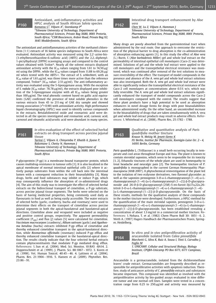
P160Antioxidant, anti-inflammatory activities andHPLC analysis of South African Salvia speciesKamatou G1, Viljoen A1, Steenkamp P2
1Tshwane University of Technology, Department ofPharmaceutical Sciences, Private Bag X680, 0001 Pretoria,South Africa; 2CSIR Biosciences, Ardeer Road, Private Bag X2,1645 Modderfontein, South Africa
The antioxidant and antiinflammatory activities of the methanol:chloro-form (1:1) extracts of 16 Salvia species indigenous to South Africa wereevaluated. Antioxidant activity was measured using the 2,2’-azino-bis-(3-ethylbenzothiazoline-6-sulfonic acid) (ABTS) and the 2,2,-diphenyl-1-picrylhydrazyl (DPPH) scavenging assays and compared to the controlvalues obtained with Trolox�. Nearly all the solvent extracts displayedantioxidant activity with the IC50 values ranging from 1.61 to 74.50 mg/ml using the DPPH·, while the IC50 values ranged from 11.88 to 69.26 mg/ml when tested with the ABTS·+. The extract of S. schlechteri, with anIC50 value of 1.61 mg/ml, was three times more active than the referencecompound, Trolox� (IC50 value: 2.51 mg/ml). The anti-inflammatory ac-tivity was evaluated using the 5-lipoxygenase assay. With the exceptionof S. radula (IC50 value: 78.78 mg/ml), the extracts displayed poor inhibi-tion of the 5-lipoxygenase enzyme with all IC50 values being greaterthan 100 mg/ml. The total phenolic content based on gallic acid equiva-lents (GAE) confirmed the presence of total soluble phenolics in thevarious extracts from 45 to 211 mg of GAE dry sample and showedstrong association (r2= 0.90) with antioxidant activity. High performanceliquid chromatography (HPLC) was used to identify various compoundsin the extracts. Betulafolientriol oxide and rosmarinic acid were de-tected in all the species investigated and rosmarinic acid, carnosic acid,carnosol and oleanolic acid/ursolic acid were abundant in many species.
P161In vitro evaluation of the effect of selected herbalextracts on drug transport across porcine jejunaltissueTarirai C, Viljoen A, Chinyemba P, Khatib A, Jiyane P,Rakomane S, Chetty N, Hamman JTshwane University of Technology, Department ofPharmaceutical Sciences, Private Bag X680, 0001 Pretoria,South Africa
P-glycoprotein (P-gp) is a membrane-bound transporter protein, whichcauses multidrug-resistance in tumour cells [1]. It is also localized in theapical membrane of normal intestinal epithelial cells [2] where it ac-tively pumps substrates from within the cell back into the intestinallumen with a consequent reduction in their bioavailability [3]. Manydrugs, herbs and food substances may inhibit or induce P-gp, whichmay consequently influence the absorption of co-administered drugs[4]. The aim of this study was to investigate the effect of selected herbalextracts on the bidirectional transport of cimetidine, a P-gp substrate,across porcine jejunal tissue segments. The herbs were selected on thebasis of having medicinal properties, being commonly used daily asflavourants in food as well as being easily accessible. The water extractsof selected herbs (garlic, cranberry, buchu and rosemary) were used todetermine their effects on the transport of cimetidine across porcinejejunal segments in both the apical-basolateral and basolateral-apicaldirections. Cimetidine alone and verapamil were included as negativeand positive control groups, respectively. The apparent permeabilitycoefficient (Papp) and flux (J) values [5] were calculated for cimetidine.Vaccinium macrocarpon (cranberry), Allium sativum (garlic) and Agathos-ma betulina (buchu) extracts inhibited P-gp efflux of cimetidine andthereby enhanced cimetidine transport in the apical-basolateral direc-tion, while Rosmarinus officinalis (rosemary) induced P-gp efflux andthereby enhanced cimetidine transport in the basolateral-apical direc-tion. The results clearly indicate that the four selected herbal extractscontain phytoconstituents that modulate P-gp mediated drug efflux.References: 1. Sun et al. (2004). Med. Sci. Monitor, 10:RA5 -RA14. 2.Doppenschmitt et al. (1999). J. Pharm. Sci. 88:1067 – 1072. 3. Koren etal. (1998). Vet. Human Toxicol. 40:45 – 46. 4. Laitinen et al. (2004).Pharm. Res. 21:1904 – 1916. 5. Hansen et al. (2009). Phytother. Res.23:86 – 91.
P162Intestinal drug transport enhancement by AloeveraChen W, Lu Z, Viljoen A, Hamman JTshwane University of Technology, Department ofPharmaceutical Sciences, Private Bag X680, 0001 Pretoria,South Africa
Many drugs are poorly absorbed from the gastrointestinal tract whenadministered by the oral route. One approach to overcome the restric-tion of the physical barrier to drug absorption is the co-administrationof absorption enhancing agents [1]. In this study the effect of Aloe vera(L.) Burm. f. (Aloe barbadensis Miller) gel and whole leaf extract on thepermeability of intestinal epithelial cell monolayers (Caco-2) was deter-mined. Solutions of gel and the whole leaf extract were applied to thecell monolayers and the transepithelial electrical resistance was moni-tored, which was continued after removal of the test solutions to mea-sure reversibility of the effect. The transport of model compounds in thepresence and absence of the A. vera gel and whole leaf extract solutionswas also investigated. Both the A. vera gel and whole leaf extract wereable to significantly reduce the transepithelial electrical resistance of theCaco-2 cell monolayers at concentrations above 0.5% w/v, which wasfully reversible. The A. vera gel and whole leaf extract solutions signifi-cantly enhanced the transport of model compounds across the Caco-2cell monolayers compared with the control. The results suggest thatthese plant products have a high potential to be used as absorptionenhancers in novel dosage forms for drugs with poor bioavailabilitieswhen administered orally. On the other hand, an uncontrolled increasein the bioavailability of drugs that are taken simultaneously with A. veragel and whole leaf extract products may result in adverse effects. Refer-ences: 1. Whitehead et al. (2008). Pharm Res, 25:1782 – 1788.
P163Qualitative and quantitative analysis of Parisquadrifolia mother tinctureKrause N, Melzig M, Jenett-Siems KFU Berlin, Institut f�r Pharmazie, Koenigin-Luise-Str. 2 – 4,14195 Berlin, Germany
Paris quadrifolia L. (Trilliaceae) is a small herb occurring locally in tem-perate and cool areas throughout Europe and Asia. The plant is known tocontain steroidal saponins, which seem to be responsible for its toxicity[1, 2]. Ethanolic tinctures of the whole plant are used in homeopathy totreat headache and neuralgic pain [2]. The homeopathic tincture of P.quadrifolia is monographed in the current German Homeopathic Phar-macopoeia (HAB 2007). A phytochemical reinvestigation of the plant ledto the isolation of two ecdysone derivatives, two flavonol glycosides aswell as the saponins pennogenin 3-O-a-L-rhamnopyranosyl-(1?4)-a-L-rhamnopyranosyl-(1?4)-[a-L-rhamnopyranosyl-(1?2)]-b-D-glucopyra-noside and 26-O-b-D-glucopyranosyl-(25R)-5-en-furost-3b,17a,22a,26-tetrol-3-O-a-L-rhamnopyranosyl-(1?4)-a-L-rhamnopyranosyl-(1?4)-[a-L-rhamnopyranosyl-(1?2)]-b-D-glucopyranoside. On the basis ofthese results, methods for the qualitative analysis of these compoundsin the mother tincture were developed. Furthermore, a HPLC-method forthe quantification of the main steroidal saponin, pennogenin 3-O-a-L-rhamnopyranosyl-(1?4)-a-L-rhamnopyranosyl-(1?4)-[a-L-rhamnopyr-anosyl-(1?2)]-b-D-glucopyranoside, has been established revealing aconcentration of about 1.3 mg/ml in a commercial mother tincture. Re-ferences: 1. Nohara, T. et al. (1982) Chem Pharm Bull 30: 1851 – 6. 2.Weth A. (1997) Hagers Handbuch der Pharmazeutischen Praxis, Spring-er, Heidelberg.
P164In vitro and in vivo antiproliferative activity ofarucanolide isolated from Calea pinnatifidaMarchetti G, Silva K, Ruiz A, Sousa I, Tinti S, Carvalho J,Foglio M1. UNICAMP, Cellular and Structural Biology, BiologyInstitute, CPQBA-Unicamp PO Box 6171, 6171 Campinas,Brazil
Arucanolide is a germacranolide, isolated from the dichloromethaneleaves’ crude extract. Germacranolides are frequently described as re-sponsible for antiproliferative activities with apoptosis induction. There-fore, study of anticancer activity of C. pinnatifida extracts and substancesbecame important. This compound was identified as involved with theantiproliferative activity by bio-guided assays evaluated in nine differ-ent tumor and one normal cell lines. Samples were tested in a concen-tration range from 0.25 to 250 mg/ml and activity was measured by
Planta Med 2010; 76: 1163–1374 Georg Thieme Verlag KG Stuttgart · New York · ISSN 0032-0943
12337th Tannin Conference and 58th International Congress of the GA | 29th August – 2nd September 2010, Berlin, Germany
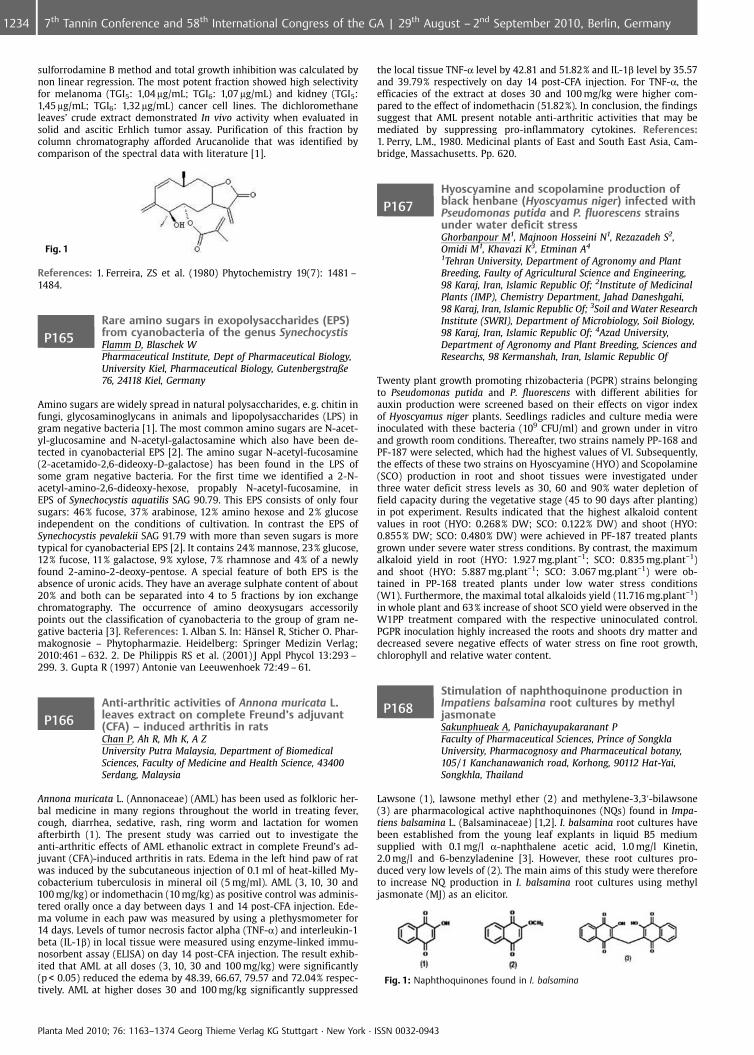
sulforrodamine B method and total growth inhibition was calculated bynon linear regression. The most potent fraction showed high selectivityfor melanoma (TGI5: 1,04 mg/mL; TGI6: 1,07 mg/mL) and kidney (TGI5:1,45 mg/mL; TGI6: 1,32 mg/mL) cancer cell lines. The dichloromethaneleaves’ crude extract demonstrated In vivo activity when evaluated insolid and ascitic Erhlich tumor assay. Purification of this fraction bycolumn chromatography afforded Arucanolide that was identified bycomparison of the spectral data with literature [1].
Fig. 1
References: 1. Ferreira, ZS et al. (1980) Phytochemistry 19(7): 1481 –1484.
P165Rare amino sugars in exopolysaccharides (EPS)from cyanobacteria of the genus SynechocystisFlamm D, Blaschek WPharmaceutical Institute, Dept of Pharmaceutical Biology,University Kiel, Pharmaceutical Biology, Gutenbergstraße76, 24118 Kiel, Germany
Amino sugars are widely spread in natural polysaccharides, e. g. chitin infungi, glycosaminoglycans in animals and lipopolysaccharides (LPS) ingram negative bacteria [1]. The most common amino sugars are N-acet-yl-glucosamine and N-acetyl-galactosamine which also have been de-tected in cyanobacterial EPS [2]. The amino sugar N-acetyl-fucosamine(2-acetamido-2,6-dideoxy-D-galactose) has been found in the LPS ofsome gram negative bacteria. For the first time we identified a 2-N-acetyl-amino-2,6-dideoxy-hexose, propably N-acetyl-fucosamine, inEPS of Synechocystis aquatilis SAG 90.79. This EPS consists of only foursugars: 46% fucose, 37% arabinose, 12% amino hexose and 2% glucoseindependent on the conditions of cultivation. In contrast the EPS ofSynechocystis pevalekii SAG 91.79 with more than seven sugars is moretypical for cyanobacterial EPS [2]. It contains 24% mannose, 23% glucose,12% fucose, 11% galactose, 9% xylose, 7% rhamnose and 4% of a newlyfound 2-amino-2-deoxy-pentose. A special feature of both EPS is theabsence of uronic acids. They have an average sulphate content of about20% and both can be separated into 4 to 5 fractions by ion exchangechromatography. The occurrence of amino deoxysugars accessorilypoints out the classification of cyanobacteria to the group of gram ne-gative bacteria [3]. References: 1. Alban S. In: H�nsel R, Sticher O. Phar-makognosie – Phytopharmazie. Heidelberg: Springer Medizin Verlag;2010:461 – 632. 2. De Philippis RS et al. (2001) J Appl Phycol 13:293 –299. 3. Gupta R (1997) Antonie van Leeuwenhoek 72:49 – 61.
P166Anti-arthritic activities of Annona muricata L.leaves extract on complete Freund’s adjuvant(CFA) – induced arthritis in ratsChan P, Ah R, Mh K, A ZUniversity Putra Malaysia, Department of BiomedicalSciences, Faculty of Medicine and Health Science, 43400Serdang, Malaysia
Annona muricata L. (Annonaceae) (AML) has been used as folkloric her-bal medicine in many regions throughout the world in treating fever,cough, diarrhea, sedative, rash, ring worm and lactation for womenafterbirth (1). The present study was carried out to investigate theanti-arthritic effects of AML ethanolic extract in complete Freund’s ad-juvant (CFA)-induced arthritis in rats. Edema in the left hind paw of ratwas induced by the subcutaneous injection of 0.1 ml of heat-killed My-cobacterium tuberculosis in mineral oil (5 mg/ml). AML (3, 10, 30 and100 mg/kg) or indomethacin (10 mg/kg) as positive control was adminis-tered orally once a day between days 1 and 14 post-CFA injection. Ede-ma volume in each paw was measured by using a plethysmometer for14 days. Levels of tumor necrosis factor alpha (TNF-a) and interleukin-1beta (IL-1b) in local tissue were measured using enzyme-linked immu-nosorbent assay (ELISA) on day 14 post-CFA injection. The result exhib-ited that AML at all doses (3, 10, 30 and 100 mg/kg) were significantly(p < 0.05) reduced the edema by 48.39, 66.67, 79.57 and 72.04% respec-tively. AML at higher doses 30 and 100 mg/kg significantly suppressed
the local tissue TNF-a level by 42.81 and 51.82% and IL-1b level by 35.57and 39.79% respectively on day 14 post-CFA injection. For TNF-a, theefficacies of the extract at doses 30 and 100 mg/kg were higher com-pared to the effect of indomethacin (51.82%). In conclusion, the findingssuggest that AML present notable anti-arthritic activities that may bemediated by suppressing pro-inflammatory cytokines. References:1. Perry, L.M., 1980. Medicinal plants of East and South East Asia, Cam-bridge, Massachusetts. Pp. 620.
P167Hyoscyamine and scopolamine production ofblack henbane (Hyoscyamus niger) infected withPseudomonas putida and P. fluorescens strainsunder water deficit stressGhorbanpour M1, Majnoon Hosseini N1, Rezazadeh S2,Omidi M1, Khavazi K3, Etminan A4
1Tehran University, Department of Agronomy and PlantBreeding, Faulty of Agricultural Science and Engineering,98 Karaj, Iran, Islamic Republic Of; 2Institute of MedicinalPlants (IMP), Chemistry Department, Jahad Daneshgahi,98 Karaj, Iran, Islamic Republic Of; 3Soil and Water ResearchInstitute (SWRI), Department of Microbiology, Soil Biology,98 Karaj, Iran, Islamic Republic Of; 4Azad University,Department of Agronomy and Plant Breeding, Sciences andResearchs, 98 Kermanshah, Iran, Islamic Republic Of
Twenty plant growth promoting rhizobacteria (PGPR) strains belongingto Pseudomonas putida and P. fluorescens with different abilities forauxin production were screened based on their effects on vigor indexof Hyoscyamus niger plants. Seedlings radicles and culture media wereinoculated with these bacteria (109 CFU/ml) and grown under in vitroand growth room conditions. Thereafter, two strains namely PP-168 andPF-187 were selected, which had the highest values of VI. Subsequently,the effects of these two strains on Hyoscyamine (HYO) and Scopolamine(SCO) production in root and shoot tissues were investigated underthree water deficit stress levels as 30, 60 and 90% water depletion offield capacity during the vegetative stage (45 to 90 days after planting)in pot experiment. Results indicated that the highest alkaloid contentvalues in root (HYO: 0.268% DW; SCO: 0.122% DW) and shoot (HYO:0.855% DW; SCO: 0.480% DW) were achieved in PF-187 treated plantsgrown under severe water stress conditions. By contrast, the maximumalkaloid yield in root (HYO: 1.927 mg.plant�1; SCO: 0.835 mg.plant�1)and shoot (HYO: 5.887 mg.plant�1; SCO: 3.067 mg.plant�1) were ob-tained in PP-168 treated plants under low water stress conditions(W1). Furthermore, the maximal total alkaloids yield (11.716 mg.plant�1)in whole plant and 63% increase of shoot SCO yield were observed in theW1PP treatment compared with the respective uninoculated control.PGPR inoculation highly increased the roots and shoots dry matter anddecreased severe negative effects of water stress on fine root growth,chlorophyll and relative water content.
P168Stimulation of naphthoquinone production inImpatiens balsamina root cultures by methyljasmonateSakunphueak A, Panichayupakaranant PFaculty of Pharmaceutical Sciences, Prince of SongklaUniversity, Pharmacognosy and Pharmaceutical botany,105/1 Kanchanawanich road, Korhong, 90112 Hat-Yai,Songkhla, Thailand
Lawsone (1), lawsone methyl ether (2) and methylene-3,3’-bilawsone(3) are pharmacological active naphthoquinones (NQs) found in Impa-tiens balsamina L. (Balsaminaceae) [1,2]. I. balsamina root cultures havebeen established from the young leaf explants in liquid B5 mediumsupplied with 0.1 mg/l a-naphthalene acetic acid, 1.0 mg/l Kinetin,2.0 mg/l and 6-benzyladenine [3]. However, these root cultures pro-duced very low levels of (2). The main aims of this study were thereforeto increase NQ production in I. balsamina root cultures using methyljasmonate (MJ) as an elicitor.
Fig. 1: Naphthoquinones found in I. balsamina
Planta Med 2010; 76: 1163–1374 Georg Thieme Verlag KG Stuttgart · New York · ISSN 0032-0943
1234 7th Tannin Conference and 58th International Congress of the GA | 29th August – 2nd September 2010, Berlin, Germany
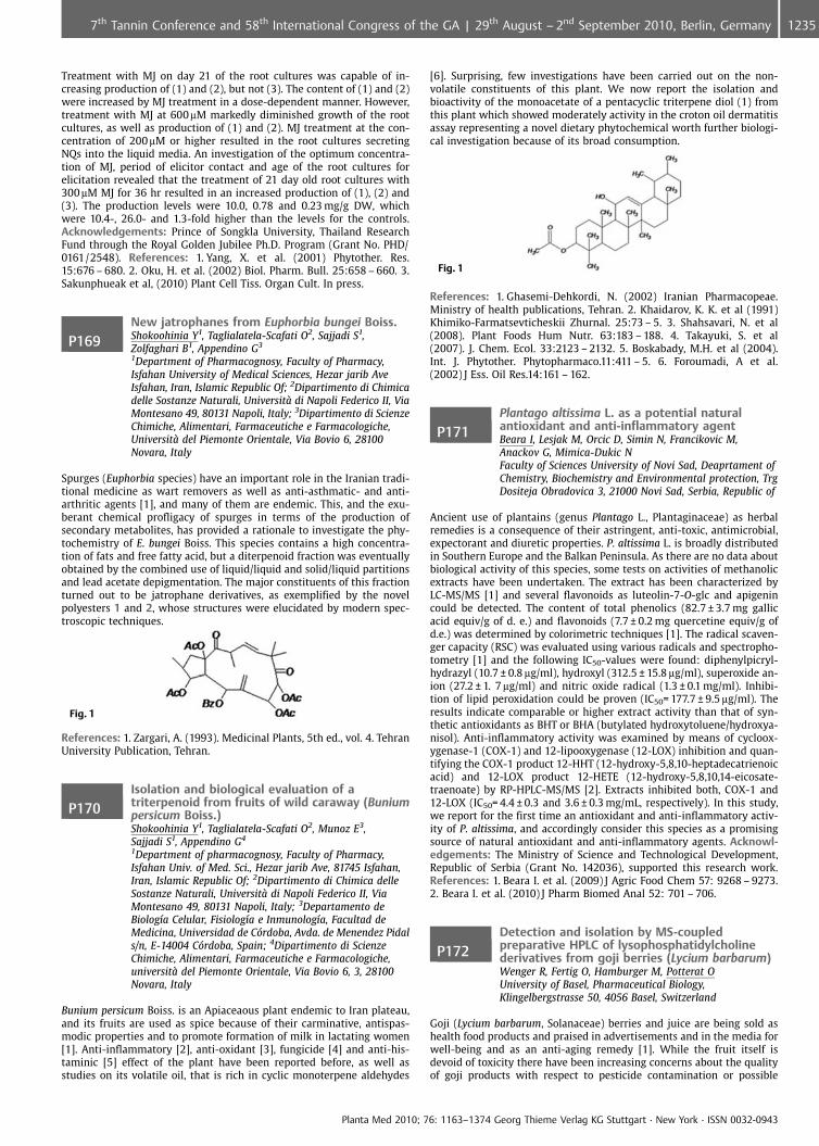
Treatment with MJ on day 21 of the root cultures was capable of in-creasing production of (1) and (2), but not (3). The content of (1) and (2)were increased by MJ treatment in a dose-dependent manner. However,treatment with MJ at 600mM markedly diminished growth of the rootcultures, as well as production of (1) and (2). MJ treatment at the con-centration of 200 mM or higher resulted in the root cultures secretingNQs into the liquid media. An investigation of the optimum concentra-tion of MJ, period of elicitor contact and age of the root cultures forelicitation revealed that the treatment of 21 day old root cultures with300 mM MJ for 36 hr resulted in an increased production of (1), (2) and(3). The production levels were 10.0, 0.78 and 0.23 mg/g DW, whichwere 10.4-, 26.0- and 1.3-fold higher than the levels for the controls.Acknowledgements: Prince of Songkla University, Thailand ResearchFund through the Royal Golden Jubilee Ph.D. Program (Grant No. PHD/0161/2548). References: 1. Yang, X. et al. (2001) Phytother. Res.15:676 – 680. 2. Oku, H. et al. (2002) Biol. Pharm. Bull. 25:658 – 660. 3.Sakunphueak et al, (2010) Plant Cell Tiss. Organ Cult. In press.
P169New jatrophanes from Euphorbia bungei Boiss.Shokoohinia Y1, Taglialatela-Scafati O2, Sajjadi S1,Zolfaghari B1, Appendino G3
1Department of Pharmacognosy, Faculty of Pharmacy,Isfahan University of Medical Sciences, Hezar jarib AveIsfahan, Iran, Islamic Republic Of; 2Dipartimento di Chimicadelle Sostanze Naturali, Universit di Napoli Federico II, ViaMontesano 49, 80131 Napoli, Italy; 3Dipartimento di ScienzeChimiche, Alimentari, Farmaceutiche e Farmacologiche,Universit del Piemonte Orientale, Via Bovio 6, 28100Novara, Italy
Spurges (Euphorbia species) have an important role in the Iranian tradi-tional medicine as wart removers as well as anti-asthmatic- and anti-arthritic agents [1], and many of them are endemic. This, and the exu-berant chemical profligacy of spurges in terms of the production ofsecondary metabolites, has provided a rationale to investigate the phy-tochemistry of E. bungei Boiss. This species contains a high concentra-tion of fats and free fatty acid, but a diterpenoid fraction was eventuallyobtained by the combined use of liquid/liquid and solid/liquid partitionsand lead acetate depigmentation. The major constituents of this fractionturned out to be jatrophane derivatives, as exemplified by the novelpolyesters 1 and 2, whose structures were elucidated by modern spec-troscopic techniques.
Fig. 1
References: 1. Zargari, A. (1993). Medicinal Plants, 5th ed., vol. 4. TehranUniversity Publication, Tehran.
P170Isolation and biological evaluation of atriterpenoid from fruits of wild caraway (Buniumpersicum Boiss.)Shokoohinia Y1, Taglialatela-Scafati O2, Munoz E3,Sajjadi S1, Appendino G4
1Department of pharmacognosy, Faculty of Pharmacy,Isfahan Univ. of Med. Sci., Hezar jarib Ave, 81745 Isfahan,Iran, Islamic Republic Of; 2Dipartimento di Chimica delleSostanze Naturali, Universit di Napoli Federico II, ViaMontesano 49, 80131 Napoli, Italy; 3Departamento deBiolog�a Celular, Fisiolog�a e Inmunolog�a, Facultad deMedicina, Universidad de C�rdoba, Avda. de Menendez Pidals/n, E-14004 C�rdoba, Spain; 4Dipartimento di ScienzeChimiche, Alimentari, Farmaceutiche e Farmacologiche,universit del Piemonte Orientale, Via Bovio 6, 3, 28100Novara, Italy
Bunium persicum Boiss. is an Apiaceaous plant endemic to Iran plateau,and its fruits are used as spice because of their carminative, antispas-modic properties and to promote formation of milk in lactating women[1]. Anti-inflammatory [2], anti-oxidant [3], fungicide [4] and anti-his-taminic [5] effect of the plant have been reported before, as well asstudies on its volatile oil, that is rich in cyclic monoterpene aldehydes
[6]. Surprising, few investigations have been carried out on the non-volatile constituents of this plant. We now report the isolation andbioactivity of the monoacetate of a pentacyclic triterpene diol (1) fromthis plant which showed moderately activity in the croton oil dermatitisassay representing a novel dietary phytochemical worth further biologi-cal investigation because of its broad consumption.
Fig. 1
References: 1. Ghasemi-Dehkordi, N. (2002) Iranian Pharmacopeae.Ministry of health publications, Tehran. 2. Khaidarov, K. K. et al (1991)Khimiko-Farmatsevticheskii Zhurnal. 25:73 – 5. 3. Shahsavari, N. et al(2008). Plant Foods Hum Nutr. 63:183 – 188. 4. Takayuki, S. et al(2007). J. Chem. Ecol. 33:2123 – 2132. 5. Boskabady, M.H. et al (2004).Int. J. Phytother. Phytopharmaco.11:411 – 5. 6. Foroumadi, A et al.(2002) J Ess. Oil Res.14:161 – 162.
P171Plantago altissima L. as a potential naturalantioxidant and anti-inflammatory agentBeara I, Lesjak M, Orcic D, Simin N, Francikovic M,Anackov G, Mimica-Dukic NFaculty of Sciences University of Novi Sad, Deaprtament ofChemistry, Biochemistry and Environmental protection, TrgDositeja Obradovica 3, 21000 Novi Sad, Serbia, Republic of
Ancient use of plantains (genus Plantago L., Plantaginaceae) as herbalremedies is a consequence of their astringent, anti-toxic, antimicrobial,expectorant and diuretic properties. P. altissima L. is broadly distributedin Southern Europe and the Balkan Peninsula. As there are no data aboutbiological activity of this species, some tests on activities of methanolicextracts have been undertaken. The extract has been characterized byLC-MS/MS [1] and several flavonoids as luteolin-7-O-glc and apigenincould be detected. The content of total phenolics (82.7 € 3.7 mg gallicacid equiv/g of d. e.) and flavonoids (7.7 € 0.2 mg quercetine equiv/g ofd.e.) was determined by colorimetric techniques [1]. The radical scaven-ger capacity (RSC) was evaluated using various radicals and spectropho-tometry [1] and the following IC50-values were found: diphenylpicryl-hydrazyl (10.7 € 0.8 mg/ml), hydroxyl (312.5 € 15.8 mg/ml), superoxide an-ion (27.2 € 1. 7 mg/ml) and nitric oxide radical (1.3 € 0.1 mg/ml). Inhibi-tion of lipid peroxidation could be proven (IC50= 177.7 € 9.5 mg/ml). Theresults indicate comparable or higher extract activity than that of syn-thetic antioxidants as BHT or BHA (butylated hydroxytoluene/hydroxya-nisol). Anti-inflammatory activity was examined by means of cycloox-ygenase-1 (COX-1) and 12-lipooxygenase (12-LOX) inhibition and quan-tifying the COX-1 product 12-HHT (12-hydroxy-5,8,10-heptadecatrienoicacid) and 12-LOX product 12-HETE (12-hydroxy-5,8,10,14-eicosate-traenoate) by RP-HPLC-MS/MS [2]. Extracts inhibited both, COX-1 and12-LOX (IC50= 4.4 € 0.3 and 3.6 € 0.3 mg/mL, respectively). In this study,we report for the first time an antioxidant and anti-inflammatory activ-ity of P. altissima, and accordingly consider this species as a promisingsource of natural antioxidant and anti-inflammatory agents. Acknowl-edgements: The Ministry of Science and Technological Development,Republic of Serbia (Grant No. 142036), supported this research work.References: 1. Beara I. et al. (2009) J Agric Food Chem 57: 9268 – 9273.2. Beara I. et al. (2010) J Pharm Biomed Anal 52: 701 – 706.
P172Detection and isolation by MS-coupledpreparative HPLC of lysophosphatidylcholinederivatives from goji berries (Lycium barbarum)Wenger R, Fertig O, Hamburger M, Potterat OUniversity of Basel, Pharmaceutical Biology,Klingelbergstrasse 50, 4056 Basel, Switzerland
Goji (Lycium barbarum, Solanaceae) berries and juice are being sold ashealth food products and praised in advertisements and in the media forwell-being and as an anti-aging remedy [1]. While the fruit itself isdevoid of toxicity there have been increasing concerns about the qualityof goji products with respect to pesticide contamination or possible
Planta Med 2010; 76: 1163–1374 Georg Thieme Verlag KG Stuttgart · New York · ISSN 0032-0943
12357th Tannin Conference and 58th International Congress of the GA | 29th August – 2nd September 2010, Berlin, Germany
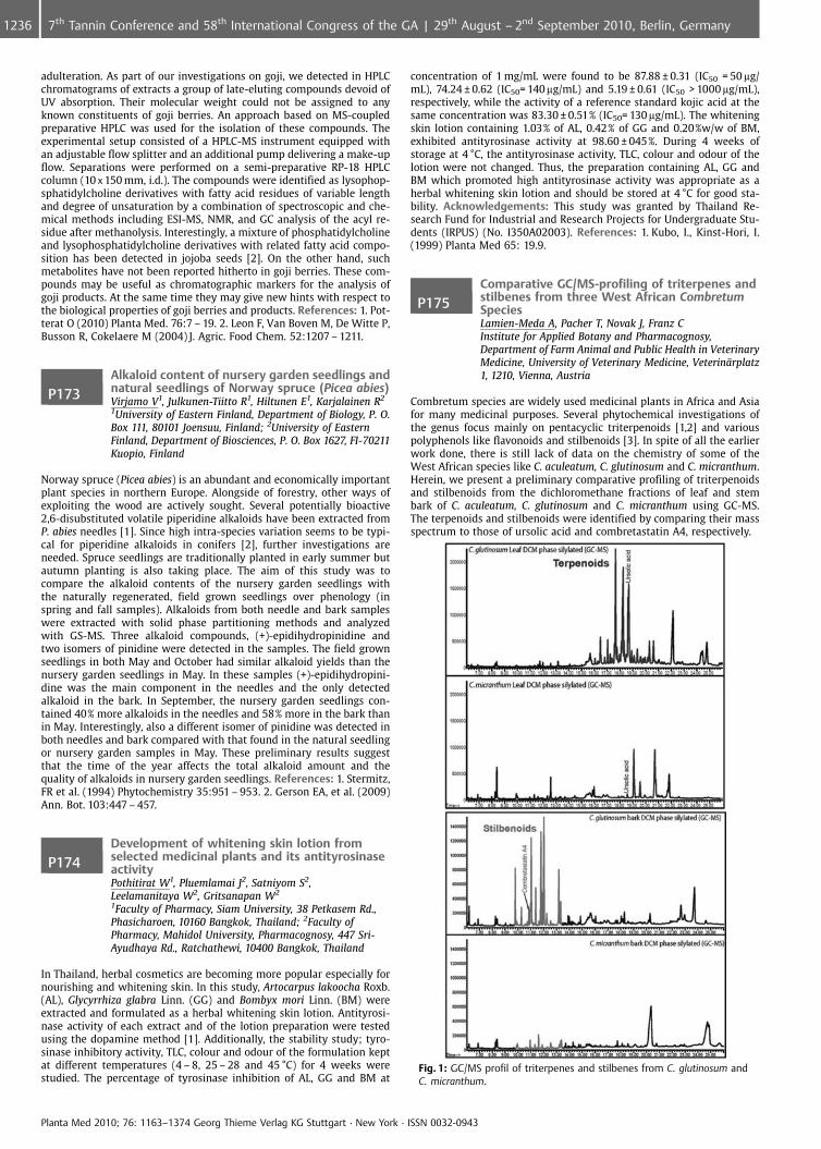
adulteration. As part of our investigations on goji, we detected in HPLCchromatograms of extracts a group of late-eluting compounds devoid ofUV absorption. Their molecular weight could not be assigned to anyknown constituents of goji berries. An approach based on MS-coupledpreparative HPLC was used for the isolation of these compounds. Theexperimental setup consisted of a HPLC-MS instrument equipped withan adjustable flow splitter and an additional pump delivering a make-upflow. Separations were performed on a semi-preparative RP-18 HPLCcolumn (10 x 150 mm, i.d.). The compounds were identified as lysophop-sphatidylcholine derivatives with fatty acid residues of variable lengthand degree of unsaturation by a combination of spectroscopic and che-mical methods including ESI-MS, NMR, and GC analysis of the acyl re-sidue after methanolysis. Interestingly, a mixture of phosphatidylcholineand lysophosphatidylcholine derivatives with related fatty acid compo-sition has been detected in jojoba seeds [2]. On the other hand, suchmetabolites have not been reported hitherto in goji berries. These com-pounds may be useful as chromatographic markers for the analysis ofgoji products. At the same time they may give new hints with respect tothe biological properties of goji berries and products. References: 1. Pot-terat O (2010) Planta Med. 76:7 – 19. 2. Leon F, Van Boven M, De Witte P,Busson R, Cokelaere M (2004) J. Agric. Food Chem. 52:1207 – 1211.
P173Alkaloid content of nursery garden seedlings andnatural seedlings of Norway spruce (Picea abies)Virjamo V1, Julkunen-Tiitto R1, Hiltunen E1, Karjalainen R2
1University of Eastern Finland, Department of Biology, P. O.Box 111, 80101 Joensuu, Finland; 2University of EasternFinland, Department of Biosciences, P. O. Box 1627, FI-70211Kuopio, Finland
Norway spruce (Picea abies) is an abundant and economically importantplant species in northern Europe. Alongside of forestry, other ways ofexploiting the wood are actively sought. Several potentially bioactive2,6-disubstituted volatile piperidine alkaloids have been extracted fromP. abies needles [1]. Since high intra-species variation seems to be typi-cal for piperidine alkaloids in conifers [2], further investigations areneeded. Spruce seedlings are traditionally planted in early summer butautumn planting is also taking place. The aim of this study was tocompare the alkaloid contents of the nursery garden seedlings withthe naturally regenerated, field grown seedlings over phenology (inspring and fall samples). Alkaloids from both needle and bark sampleswere extracted with solid phase partitioning methods and analyzedwith GS-MS. Three alkaloid compounds, (+)-epidihydropinidine andtwo isomers of pinidine were detected in the samples. The field grownseedlings in both May and October had similar alkaloid yields than thenursery garden seedlings in May. In these samples (+)-epidihydropini-dine was the main component in the needles and the only detectedalkaloid in the bark. In September, the nursery garden seedlings con-tained 40% more alkaloids in the needles and 58% more in the bark thanin May. Interestingly, also a different isomer of pinidine was detected inboth needles and bark compared with that found in the natural seedlingor nursery garden samples in May. These preliminary results suggestthat the time of the year affects the total alkaloid amount and thequality of alkaloids in nursery garden seedlings. References: 1. Stermitz,FR et al. (1994) Phytochemistry 35:951 – 953. 2. Gerson EA, et al. (2009)Ann. Bot. 103:447 – 457.
P174Development of whitening skin lotion fromselected medicinal plants and its antityrosinaseactivityPothitirat W1, Pluemlamai J2, Satniyom S2,Leelamanitaya W2, Gritsanapan W2
1Faculty of Pharmacy, Siam University, 38 Petkasem Rd.,Phasicharoen, 10160 Bangkok, Thailand; 2Faculty ofPharmacy, Mahidol University, Pharmacognosy, 447 Sri-Ayudhaya Rd., Ratchathewi, 10400 Bangkok, Thailand
In Thailand, herbal cosmetics are becoming more popular especially fornourishing and whitening skin. In this study, Artocarpus lakoocha Roxb.(AL), Glycyrrhiza glabra Linn. (GG) and Bombyx mori Linn. (BM) wereextracted and formulated as a herbal whitening skin lotion. Antityrosi-nase activity of each extract and of the lotion preparation were testedusing the dopamine method [1]. Additionally, the stability study; tyro-sinase inhibitory activity, TLC, colour and odour of the formulation keptat different temperatures (4 – 8, 25 – 28 and 45 �C) for 4 weeks werestudied. The percentage of tyrosinase inhibition of AL, GG and BM at
concentration of 1 mg/mL were found to be 87.88 € 0.31 (IC50 = 50 mg/mL), 74.24 € 0.62 (IC50= 140 mg/mL) and 5.19 € 0.61 (IC50 > 1000 mg/mL),respectively, while the activity of a reference standard kojic acid at thesame concentration was 83.30 € 0.51% (IC50= 130 mg/mL). The whiteningskin lotion containing 1.03% of AL, 0.42% of GG and 0.20%w/w of BM,exhibited antityrosinase activity at 98.60 € 045%. During 4 weeks ofstorage at 4 �C, the antityrosinase activity, TLC, colour and odour of thelotion were not changed. Thus, the preparation containing AL, GG andBM which promoted high antityrosinase activity was appropriate as aherbal whitening skin lotion and should be stored at 4 �C for good sta-bility. Acknowledgements: This study was granted by Thailand Re-search Fund for Industrial and Research Projects for Undergraduate Stu-dents (IRPUS) (No. I350A02003). References: 1. Kubo, I., Kinst-Hori, I.(1999) Planta Med 65: 19.9.
P175Comparative GC/MS-profiling of triterpenes andstilbenes from three West African CombretumSpeciesLamien-Meda A, Pacher T, Novak J, Franz CInstitute for Applied Botany and Pharmacognosy,Department of Farm Animal and Public Health in VeterinaryMedicine, University of Veterinary Medicine, Veterin�rplatz1, 1210, Vienna, Austria
Combretum species are widely used medicinal plants in Africa and Asiafor many medicinal purposes. Several phytochemical investigations ofthe genus focus mainly on pentacyclic triterpenoids [1,2] and variouspolyphenols like flavonoids and stilbenoids [3]. In spite of all the earlierwork done, there is still lack of data on the chemistry of some of theWest African species like C. aculeatum, C. glutinosum and C. micranthum.Herein, we present a preliminary comparative profiling of triterpenoidsand stilbenoids from the dichloromethane fractions of leaf and stembark of C. aculeatum, C. glutinosum and C. micranthum using GC-MS.The terpenoids and stilbenoids were identified by comparing their massspectrum to those of ursolic acid and combretastatin A4, respectively.
Fig. 1: GC/MS profil of triterpenes and stilbenes from C. glutinosum andC. micranthum.
Planta Med 2010; 76: 1163–1374 Georg Thieme Verlag KG Stuttgart · New York · ISSN 0032-0943
1236 7th Tannin Conference and 58th International Congress of the GA | 29th August – 2nd September 2010, Berlin, Germany
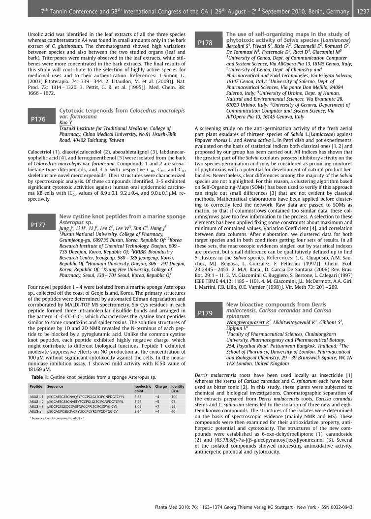
Ursolic acid was identified in the leaf extracts of all the three specieswhereas combretastatin A4 was found in small amounts only in the barkextract of C. glutinosum. The chromatograms showed high variationsbetween species and also between the two studied organs (leaf andbark). Triterpenes were mainly observed in the leaf extracts, while stil-benes were more concentrated in the bark extracts. The final results ofthis study will contribute to the selection of highly active species formedicinal uses and to their authentication. References: 1. Simon, G.(2003) Fitoterapia. 74: 339 – 344. 2. Litaudon, M. et al. (2009) J. Nat.Prod. 72: 1314 – 1320. 3. Pettit, G. R. et al. (1995) J. Med. Chem. 38:1666 – 1672.
P176Cytotoxic terpenoids from Calocedrus macrolepisvar. formosanaKuo YTsuzuki Institute for Traditional Medicine, College ofPharmacy, China Medical University, No.91 Hsueh-ShihRoad, 40402 Taichung, Taiwan
Calocetriol (1), diacetylcalocediol (2), abeoabietalignol (3), labdanecar-yophyllic acid (4), and ferrugimenthenol (5) were isolated from the barkof Calocedrus macrolepis var. formosana. Compounds 1 and 2 are secoa-bietane-type diterpenoids, and 3-5 with respective C30, C35, and C30
skeletons are novel meroterpenoids. Their structures were characterizedby spectroscopic analysis. Of these compounds identified, 3-5 exhibitedsignificant cytotoxic activities against human oral epidermoid carcino-ma KB cells with IC50 values of 8.9 € 0.1, 9.2 € 0.4, and 9.0 € 0.1mM, re-spectively.
P177New cystine knot peptides from a marine spongeAsteropus sp.Jung J1, Li H1, Li J1, Lee C2, Lee W3, Sim C4, Hong J5
1Pusan National University, College of Pharmacy,Geumjeong-gu, 609735 Busan, Korea, Republic Of; 2KoreaResearch Institute of Chemical Technology, Daejon, 609 –735 Daeojon, Korea, Republic Of; 3KRIBB, BioindustryResearch Center, Jeongeup, 580 – 185 Jeongeup, Korea,Republic Of; 4Hannam University, Daejon, 306 – 791 Daejon,Korea, Republic Of; 5Kyung Hee University, College ofPharmacy, Seoul, 130 – 701 Seoul, Korea, Republic Of
Four novel peptides 1 – 4 were isolated from a marine sponge Asteropussp., collected off the coast of Geoje Island, Korea. The primary structuresof the peptides were determined by automated Edman degradation andcorroborated by MALDI-TOF MS spectrometry. Six Cys residues in eachpeptide formed three intramolecular disulfide bonds and arranged inthe pattern -C-C-CC-C-C-, which characterizes the cystine knot peptidessimilar to some conotoxins and spider toxins. The solution structures ofthe peptides by 1D and 2D NMR revealed the N-terminus of each pep-tide to be blocked by a pyroglutamic acid. Unlike the common cystineknot peptides, each peptide exhibited highly negative charge, whichmight contribute to different biological functions. Peptide 1 exhibitedmoderate suppressive effects on NO production at the concentration of100mM without significant cytotoxicity against the cells. In the neura-minidase inhibition assay, 1 showed mild activity with IC 50 value of181.69 mM.
Table 1: Cystine knot peptides from a sponge Asteropus sp.
Peptide Sequence Isoelectricpoint
Charge Identity(%)a
ABU8 – 1 pEGCAFEGESCNVQFYPCCPGLGLTCIPGNPDGTCYYL 3.33 �4 100ABU8 – 2 pEGCAFEGESCNVEFYPCCPGLGLTCIPGNPDGTCYYL 3.26 �5 97ABU8 – 3 pEDCPGEGEQCDVEFNPCCPPLTCIPGDPYGICYII 3.09 �7 59ABU8-a pEGCAGPGEECIVGFYDCCPGYRCYPGDPGGICY 3.64 �4 60
a Sequence identity compared to ABU8 – 1
P178The use of self-organizing maps in the study ofphytotoxic activity of Salvia species (Lamiaceae)Bertolini S1, Pivetti S1, Bisio A2, Giacomelli E2, Romussi G2,De Tommasi N3, Fraternale D4, Ricci D4, Giacomini M5
1University of Genoa, Dept. of Communication Computerand System Science, Via AllOpera Pia 13, 16145 Genoa, Italy;2University of Genoa, Dept. of Chemistry andPharmaceutical and Food Technologies, Via Brigata Salerno,16147 Genoa, Italy; 3University of Salerno, Dept. ofPharmaceutical Sciences, Via ponte Don Melillo, 84084Salerno, Italy; 4University of Urbino, Dept. of Human,Natural and Environmental Sciences, Via Bramante 28,61029 Urbino, Italy; 5University of Genova, Department ofCommunication Computer and System Science, ViaAll’Opera Pia 13, 16145 Genova, Italy
A screening study on the anti-germination activity of the fresh aerialpart plant exudates of thirteen species of Salvia L.(Lamiaceae) againstPapaver rhoeas L. and Avena sativa L. in Petri dish and pot experiments,evaluated on the basis of statistical indices both classical ones [1, 2] andproposed by our group has been carried out. All indices has shown thatthe greatest part of the Salvia exudates possess inhibitory activity on thetwo species germination and may be considered as promising mixturesof phytotoxins with a potential for development of natural product her-bicides. Nevertheless, clear differences among the majority of the Salviaspecies are not highlighted. For this reason, a clustering algorithm basedon Self-Organizing-Maps (SOMs) has been used to verify if this approachcan single out small differences [3] that are not evident by classicalmethods. Mathematical elaborations have been applied before cluster-ing to correctly feed the network. Raw data are passed to SOMs asmatrix, so that if columns/rows contained too similar data, these col-umns/rows gave too few information to the process. A selection to theseelements has been applied fixing some constraints about maximum andminimum of contained values, Variation Coefficient [4], and correlationbetween data columns. After elaboration, we clustered data for bothtarget species and in both conditions getting four sets of results. In allthese sets, the macroscopic evidences singled out by statistical indexesare present, but small difference can be qualitatively defined up to find5 clusters in the Salvia species. References: 1. G. Chiapusio, A.M. San-chez, M.J. Reigosa, L. Gonzalez, F. Pellissier (1997) J. Chem. Ecol.23:2445 – 2453. 2. M.A. Ranal, D. Garcia De Santana (2006) Rev. Bras.Bot. 29:1 – 11. 3. M. Giacomini, C. Ruggiero, S. Bertone, L. Calegari (1997)IEEE TBME 44,12: 1185 – 1191. 4. M. Giacomini, J.L. McDermott, A.A. Giri,I. Martini, F.B. Lillo, O.E. Varnier (1998) J. Vir. Meth 73: 201 – 209.
P179New bioactive compounds from Derrismalaccensis, Carissa carandas and CarissaspinarumWangteeraprasert R1, Likhitwitayawuid K1, Gibbons S2,Lipipun V1
1Faculty of Pharmaceutical Sciences, ChulalongkornUniversity, Pharmacognosy and Pharmaceutical Botany,254, Payathai Road, Pattumwon Bangkok, Thailand; 2TheSchool of Pharmacy, University of London, Pharmaceuticaland Biological Chemistry, 29 – 39 Brunswick Square, WC 1N1AX London, United Kingdom
Derris malaccensis roots have been used locally as insecticide [1]whereas the stems of Carissa carandas and C. spinarum each have beenused as bitter tonic [2]. In this study, these plants were subjected tochemical and biological investigations. Chromatographic separation ofthe extracts prepared from Derris malaccensis roots, Carissa carandasstems and C. spinarum stems led to the isolation of three new and eigh-teen known compounds. The structures of the isolates were determinedon the basis of spectroscopic evidence (mainly NMR and MS). Thesecompounds were then examined for their antioxidative property, anti-herpetic potential and cytotoxicity. The structures of the new com-pounds were established as 6-oxo-dehydroelliptone (1), carandoside(2) and (6S,7R,8R)-7a-[(b-glucopyranosyl)oxy]lyoniresinol (3). Severalof the isolated compounds showed interesting antioxidative activity,antiherpetic potential and cytotoxicity.
Planta Med 2010; 76: 1163–1374 Georg Thieme Verlag KG Stuttgart · New York · ISSN 0032-0943
12377th Tannin Conference and 58th International Congress of the GA | 29th August – 2nd September 2010, Berlin, Germany
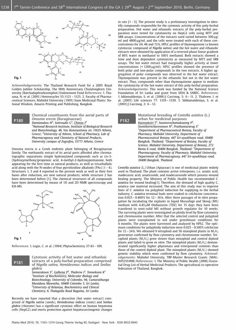
Fig. 1
Acknowledgements: The Thailand Research Fund for a 2005 RoyalGolden Jubilee Scholarship, The 90th Anniversary Chulalongkorn Uni-versity (Ratchadaphiseksomphot) Endowment Fund References: 1. Tha-sana, N. et al. (2001) Heterocycles 55:1121 – 1125. 2. Faculty of Pharma-ceutical Sciences, Mahidol University (1995) Siam Medicinal Plants: Na-tional Wisdom. Amarin Printing and Publishing. Bangkok.
P180Chemical constituents from the aerial parts ofOnosma erecta (Boraginaceae)Damianakos H2, Sotiroudis G1, Chinou I2
1National Research Institute, Institute of Biological Researchand Biotechnology, 46, Vas Konstantinou str, 11635 Athens,Greece; 2University of Athens, School of Pharmacy, Lab ofPharmacognosy and Chemistry of Natural Products,University campus of Zografou, 15771 Athens, Greece
Onosma erecta is a Greek endemic plant belonging of Boraginaceaefamily. The methanolic extract of its aerial parts afforded after chroma-tographic separations simple hydroxylated compounds such as cis-3-(hydroxymethoxy)propenoic acid, 4-methyl-2-hydroxypentanone, bothappearing for the first time as natural products, as well as tricarballylicacid along with the N-oxides of four pyrrolizidine alkaloids (PAs) (1 – 4).Structures 1, 3 and 4 reported in the present work as well as their freebases after reduction, are new natural products, while structure 2 hasbeen determined before [1]. The chemical structures of all compoundshave been determined by means of 1D and 2D NMR spectroscopy andESI-MS.
Fig. 1
References: 1. Logie, C. et al. (1994) Phytochemistry 37:43 – 109.
P181Cytotoxic activity of hot water and ethanloicextracts of a poly-herbal preparation comprisedof Nigella sativa, Hemidesmus indicus and SmilaxglabraSamarakoon S1, Galhena P2, Thabrew I1, Tennekoon K1
1Institute of Biochemistry, Molecular Biology andBiotechnology, University of Colombo, 94, CumarathungaMunidasa Mawatha, 10400 Colombo 3, Sri Lanka;2University of Kelaniya, Biochemistry and ClinicalChemistry, 6, Thalagolla Road Ragama, Sri Lanka
Recently we have reported that a decoction (hot water extract) com-prised of Nigella sativa (seeds), Hemidesmus indicus (roots) and Smilaxglabra (rhizome) has a significant cytotoxic effect on human hepatomacells (HepG2) and exerts protection against hepatocarcinogenic changes
in rats [1 – 3]. The present study is a preliminary investigation to iden-tify compounds responsible for the cytotoxic activity of this poly-herbalpreparation. Hot water and ethanolic extracts of the poly-herbal pre-paration were tested for cytotoxicity on HepG2 cells using MTT andSRB assays. Concentrations of the extracts used varied between 300 mg/ml and 4800 mg/ml, and the cells were treated with each of these con-centrations for 24, 48 and 72 h. HPLC profiles of thymoquinone (a knowncytotocxic compound of Nigella sativa) and the hot water and ethanolicextracts were obtained by application of a reversed phase linear gradientof 80% water in methanol to 100% methanol. Both extracts showed atime and dose dependent cytotoxicity as measured by MTT and SRBassays. The hot water extract had marginally higher activity at lowerconcentrations (< 1200 mg/ml). HPLC profiles showed the presence ofboth polar and non-polar compounds in the two extracts. A higher ag-gregation of polar compounds was observed in the hot water extract.Thymoquinone was present in the ethanolic but not in the hot waterextract. Thus, compounds other than thymoquinone appear to mediatethe cytotoxicity of the hot water extract of this poly-herbal preparation.Acknowledgements: This work was funded by the National ScienceFoundation of Sri Lanka and grant from SIDA & SAREC. References:1. Iddamaldeniya, S. et al. (2006) J Carcinog. 5: 6 – 11. 2. Thabrew, I. etal. (2005) Life sciences 77: 1319 – 1330. 3. SIddamaldeniya, S. et al.(2003) J Carcinog. 2: 6 – 12.
P182Mutational breeding of Centella asiatica (L.)urban for medicinal purposesKaensaksiri T1, Soontornchainaksaeng P2,Soonthornchareonnon N3, Prathanturarug S1
1Department of Pharmaceutical Botany, Faculty ofPharmacy, Mahidol University, Department ofPharmaceutical Botany, 447 Sri-ayudthaya road, 10400Bangkok, Thailand; 2Department of Botany, Faculty ofScience, Mahidol University, Department of Botany, 272Rama 6 road, 10400 Bangkok, Thailand; 3Department ofPharmacognosy, Faculty of Pharmacy, Mahidol University,Department of Pharmacognosy, 447 Sri-ayudthaya road,10400 Bangkok, Thailand
Centella asiatica (L.) Urban (Apiaceae) is one of medicinal plants widelyused in Thailand. The plant contains active triterpenes, i. e. asiatic acid,madecassic acid, asiaticoside, and madecassoside which possess woundhealing property. The Ministry of Public Health has recommended C.asiatica for wound healing[1]. Therefore, the demand of high quality C.asiatica raw material increased. The aim of this study was to improvelines of C. asiatica via polyploid induction for supplying to the herbalindustry. C. asiatica terminal buds were soaked in colchicine concentra-tion 0.025 – 0.400% for 12 – 36 h. After three passages of in vitro propa-gation by incubating the explants in liquid Murashige and Skoog (MS)medium with 4.45mM thidiazuron (TDZ) for 15 days they have beentransfered to semi-solid MS without growth regulator for 10 weeks.The surviving plants were investigated on ploidy level by flow cytometryand chromosome number. After that the selected control and polyploidplants were transplanted to soil under greenhouse conditions for4 months. The plants were harvested and analyzed by HPLC. The opti-mum conditions for polyploidy induction were 0.025 – 0.100% colchicinefor 12 – 24 h. We obtained 9 tetraploid and 56 mixoploid plants in M1V3
generation confirmed by flow cytometry and chromosome number. Tet-raploid plants (M1V3) grew slower than mixoploid and control diploidplants and failed to grow ex vitro. The mixoploid plants (M1V3) demon-strated significantly higher phytomass and triterpenoid contents thanthose of the control diploid plant. The mixoploid plants (M1V7) showedgenetic stability which were confirmed by flow cytometry. Acknowl-edgements: Mahidol University, TRF-Master Research Grants (MAG-WI515S 098) References: 1. The Ministry of Public Health (2006) Essen-tial Drug List of Herbal Medicinal Products. The agricultural co-operativefederation of Thailand, Bangkok.
Planta Med 2010; 76: 1163–1374 Georg Thieme Verlag KG Stuttgart · New York · ISSN 0032-0943
1238 7th Tannin Conference and 58th International Congress of the GA | 29th August – 2nd September 2010, Berlin, Germany
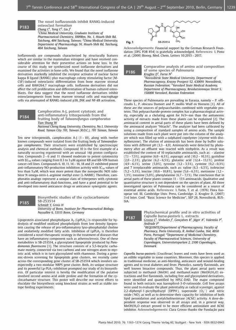
P183The novel isoflavonoids inhibit RANKL-inducedosteoclast formationChang C1, Tang C2, Liu J1
1China Medical University, Graduate Institute ofPharmaceutical Chemistry, 1009Rm, No. 1, Hsueh-Shih Rd,Taichung, 404 Taichung, Taiwan; 2China Medical University,Department of Pharmacology, 91, Hsueh-Shih Rd, Taichung,404 Taichung, Taiwan
Isoflavonoids are compounds characterised by structurally featureswhich are similar to the mammalian estrogens and have received con-siderable attention for their preventive actions on bone loss. In thecourse of this study we synthesized novel isoflavone derivatives andexamined the activities in bone cells. We found that the novel isoflavonederivatives markedly inhibited the receptor activator of nuclear factorkappa B ligand (RANKL) plus macrophage colony stimulating factor (M-CSF)-induced osteoclastic differentiation from bone marrow stromalcells and RAW264.7 macrophage cells. Isoflavone derivatives did notaffect the cell proliferation and differentiation of human cultured osteo-blasts. Our data suggest that the novel isoflavone derivatives inhibitosteoclastogenesis from bone marrow stromal cells and macrophagecells via attenuated of RANKL-induced p38, JNK and NF-kB activation.
P184Camphoratins A-J, potent cytotoxic andanti-inflammatory triterpenoids from thefruiting body of Taiwanofungus camphoratusTian Shung WNational Cheng Kung University, Chemistry, No.1, UniversityRoad, Tainan City 701, Taiwan (R.O.C.), 701 Tainan, Taiwan
Ten new triterpenoids, camphoratins A-J (1 – 10), along with twelveknown compounds were isolated from the fruiting body of Taiwanofun-gus camphorates. Their structures were established by spectroscopicanalysis and chemical methods. Compound 10 is the first example of anaturally occurring ergosteroid with an unusual 14b proton configura-tion. Compounds 2 – 6 and 11 showed moderate to potent cytotoxicitywith EC50 values ranging from 0.3 to 3mM against KB and KB-VIN humancancer cell lines. Compounds 6, 10, 11, 14 – 16, 18 and 21 exhibited potentanti-inflammatory NO-production inhibition activity with IC50 values ofless than 5mM, which was more potent than the nonspecific NOS inhi-bitor N omega-nitro-L-arginine methyl ester (L-NAME). Therefore, cam-phoratin analogs represent a unique class of triterpenes with cytotoxicand anti-inflammatory dual-functions, and have a good potential to bedeveloped into novel anticancer drugs or anticancer synergistic agents.
P185Biosynthetic studies of the cyclocarbamateSB-253514Schmidt Y, Gross HUniversity of Bonn, Institute for Pharmaceutical Biology,Nussallee 6, 53115 Bonn, Germany
Lipoprotein associated phospholipase A2 (LpPLA2) is responsible for hy-drolysis of modified oxidized phospholipids from low density lipopro-tein causing the release of pro-inflammatory lyso-phosphatidyl cholineand oxidatively modified fatty acids. Inhibition of LpPLA2 is thereforeconsidered a novel therapeutic strategy in the treatment of diseases thathave an inflammatory component such as atherosclerosis. One of thesemetabolites is SB-253514, a glycosylated lipopeptide produced by Pseu-domonas fluorescens [1]. The structure consists of a 5,5-bicyclic carba-mate moiety, connected via two carbons and one nitrogen atom to myr-istic acid, which is in turn glycosylated with rhamnose. During a geno-mic-driven screening for lipopeptide gene clusters, we recently cameacross the corresponding gene cluster of SB-253514 which involves un-expectedly a two modular NRPS gene cluster. Both, its unique structureand its powerful Lp-PLA2-inhibition prompted our study of its biosynth-esis. Of particular interest is hereby the modification of the putativeinvolved second amino acid aside proline and the formation of the cy-clocarbamate structure. This poster will describe our recent efforts toelucidate the biosynthesis using knockout strains as well as stable iso-tope feeding experiments.
Fig. 1
Acknowledgements: Financial support by the German Research Foun-dation (DFG FOR 854) is gratefully acknowledged. References: 1. Pintoet al. (2000) Bioorg. Med. Chem. Letters 10:2015 – 2017.
P186Comparative analysis of amino acid compositionof some species of PulmonariaKruglov D1, Fursa N2
1Novosibirsk State Medical University, Department ofPharmacognosy, Krasny Prospect 52, 630091 Novosibirsk,Russian Federation; 2Yaroslavl State Medical Academy,Department of Pharmacognosy, Revolutsionnaya Street 5,150000 Yaroslavl, Russian Federation
Three species of Pulmonaria are prevailing in Eurasia, namely – P. offi-cinalis L., P. obscura Dumort and P. mollis Wulf ex Hornem [1]. All ofthese are the sources of polysaccharides combined with vegetable pro-teins. This polysaccharide-protein complex has a pharmacological activ-ity, especially as a chelating agent for Fe3+-ion than the antianemicactivity of extracts made from these plants can be explained [2]. Theaminoacid content in aerial parts of these plants have been defined bythe aminoacid analyzer “Hitachi”. The analyzer had been calibrated byusing a composition of standard samples of amino acids. The samplesolutions made from each plant were put into the column of the analy-zer which was filled up with a sulphated copolymer (styrene with divi-nylbenzene). The elution of aminoacids has been done by buffer solu-tions with different pH (3,3 – 4,9). Aminoacids were detected by photo-metry after an effluent was reacted with ninhydrin. As a result wasestablished the content of 10 replaceable aminoacids, such as: – alanine(7,1 – 7,3%), arginine (6,0 – 6,3%), asparagic acid (10,2 – 10,3%), gystidine(2,0 – 2,3%), glycine (6,2 – 6,5%), glutamic acid (12,4 – 13,1%), proline(4,9 – 6,1%), serine (5,0%), tyrosine (3,2 – 3,5%), cysteine (0,2 – 0,3%)and 7 irreplaceable aminoacids, such as: valine (6,3 – 6,5%), isoleucine(5,2 – 5,3%), leucine (10,6 – 10,8%), lysine (5,6 – 6,3%), metionine (1,2 –1,3%), treonine (5,0%), phenylalanine (6,7 – 7,1%). The conclusion that inaerial parts of these plants contain 11 – 13% aminoacids. Qualitative andquantitative structure is not dependent on the species of plant. All threeinvestigated species of Pulmonaria can be considered as a source ofessential amino acids. References: 1. Tutin, T. et al. (1976) Flora Eur-opeae vol. III. Cambridge Univ. Press, Cambridge. 2. Kruglov D. (2007)3 rd Inter. Conf. “Basic Science for Medicine”, SEP 28, Novosibirsk, RUS-SIA.
P187Phytochemical profile and in vitro activities ofCapsella bursa-pastoris L. extractsGrosso C1, Vinholes J1, Gon�alves R1, J�ger A2, Valent¼o P1,Andrade P1
1REQUIMTE/Department of Pharmacognosy, Faculty ofPharmacy, Porto University, R. An�bal Cunha, 164, 4050Porto, Portugal; 2Department of Medicinal Chemistry,Faculty of Pharmaceutical Sciences, University ofCopenhagen, Universitetsparken 2, 2100 Copenhagen,Denmark
Capsella bursa-pastoris (Cruciferae) leaves and roots have been used asan edible vegetable in some countries. Moreover, this species is appliedin traditional medicine, as anti-bleeding, anticancer and wound-healingagents and to treat diabetes and fever. Phenolics, namely flavonoids, arewell known bioactive compounds. Thus, the plant aerial parts weresubjected to methanol (MeOH) and methanol:water (MeOH/H2O) ex-tractions and five flavonoids, including free and glycosylated derivatives,were identified and quantified by HPLC-DAD. The major compoundfound in both extracts was kaempferol-3-O-rutinoside. Cell free assayswere used to evaluate the plant potentiality as radical scavenger, against2,2-diphenyl-1-picrylhydrazyl (DPPH·), superoxide (O2
·�) and nitricoxide (·NO), as well as to determine their capacity for inhibition of bothlipid peroxidation and acetylcholinesterase (AChE) activity. A dose-de-pendent response was observed in all assays and, in a general way,MeOH/H2O extract proved to be more efficient antioxidant and AChEinhibitor. Acknowledgements: Clara Grosso thanks the Funda�¼o para
Planta Med 2010; 76: 1163–1374 Georg Thieme Verlag KG Stuttgart · New York · ISSN 0032-0943
12397th Tannin Conference and 58th International Congress of the GA | 29th August – 2nd September 2010, Berlin, Germany
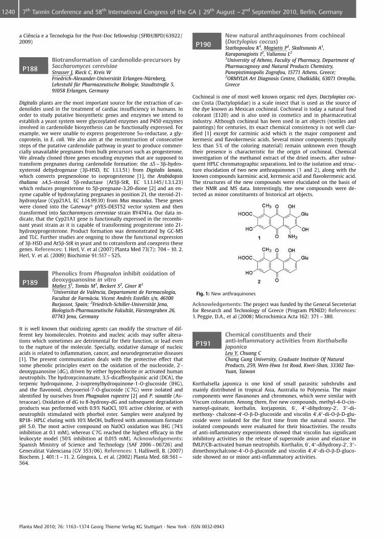
a CiÞncia e a Tecnologia for the Post-Doc fellowship (SFRH/BPD/63922/2009)
P188Biotransformation of cardenolide-precursors bySaccharomyces cerevisiaeStrasser J, Rieck C, Kreis WFriedrich-Alexander-Universit�t Erlangen-N�rnberg,Lehrstuhl f�r Pharmazeutische Biologie, Staudtstraße 5,91058 Erlangen, Germany
Digitalis plants are the most important source for the extraction of car-denolides used in the treatment of cardiac insufficiency in humans. Inorder to study putative biosynthetic genes and enzymes we intend toestablish a yeast system were glycosylated enzymes and P450 enzymesinvolved in cardenolide biosynthesis can be functionally expressed. Forexample, we were unable to express progesterone 5a-reductase, a gly-coprotein, in E. coli. We also aim at the reconstruction of consecutivesteps of the putative cardenolide pathway in yeast to produce commer-cially unavailable pregnanes from bulk precursors such as progesterone.We already cloned three genes encoding enzymes that are supposed totransform pregnanes during cardenolide formation: the D5 – 3b-hydro-xysteroid dehydrogenase (3b-HSD, EC 1.1.1.51) from Digitalis lanata,which converts pregnenolone to isoprogesterone [1], the Arabidopsisthaliana D4,5-steroid 5b-reductase (At5b-StR, EC 1.1.1.145/1.3.1.23)which reduces progesterone to 5b-pregnane-3,20-dione [2] and an en-zyme capable of hydroxylating pregnanes in position 21, the steroid-21-hydroxylase (Cyp21A1, EC 1.14.99.10) from Mus musculus. These geneswere cloned into the Gateway� pYES-DEST52 vector system and thentransformed into Saccharomyces cerevisiae strain BY4741a. Our data in-dicate, that the Cyp21A1 gene is functionally expressed in the recombi-nant yeast strain as it is capable of transforming progesterone into 21-hydroxyprogesterone. Product formation was demonstrated by GC-MSand TLC. Further studies are ongoing to show the functional expressionof 3b-HSD and At5b-StR in yeast and to cotransform and coexpress thesegenes. References: 1. Herl, V. et al (2007) Planta Med 73(7): 704 – 10. 2.Herl, V. et al. (2009) Biochimie 91:517 – 525.
P189Phenolics from Phagnalon inhibit oxidation ofdeoxyguanosine in vitroMa�ez S1, Toms M1, Beckert S2, Giner R1
1Universitat de Val�ncia, Departament de Farmacologia,Facultat de Farmcia. Vicent Andr�s Estell�s s/n, 46100Burjassot, Spain; 2Friedrich-Schiller-Universit�t Jena,Biologisch-Pharmazeutische Fakult�t, F�rstengraben 26,07743 Jena, Germany
It is well known that oxidizing agents can modify the structure of dif-ferent key biomolecules. Proteins and nucleic acids may suffer altera-tions which sometimes are detrimental for their function, or lead evento the rupture of the molecule. Specially, oxidative damage of nucleicacids is related to inflammation, cancer, and neurodegenerative diseases[1]. The present communication deals with the protective effect thatsome phenolic principles exert on the oxidation of the nucleoside, 2’-deoxyguanosine (dG), driven by either hypochlorite or activated humanneutrophils. The hydroxycinnamate, 3,5-dicaffeoylquinic acid (DCA), theterpenic hydroquinone, 2-isoprenylhydroquinone-1-O-glucoside (IHG),and the flavonoid, chrysoeriol-7-O-glucoside (C 7G) were isolated andidentified by ourselves from Phagnalon rupestre [2] and P. saxatile (As-teraceae). Oxidation of dG to 8-hydroxy-dG and subsequent degradationproducts was performed with 0.9% NaOCl, 10% active chlorine, or withneutrophils stimulated with phorbol ester. Samples were analyzed byRP18- HPLC eluting with 10% MeOH, buffered with ammonium formatepH 5.0. The most active compound on NaOCl oxidation was IHG (74%inhibition at 0.1 mM), whereas C 7G reached the highest efficacy in theleukocyte model (50% inhibition at 0.015 mM). Acknowledgements:Spanish Ministry of Science and Technology (SAF 2006 – 06726) andGeneralitat Valenciana (GV 353/06). References: 1. Halliwell, B. (2007)Biochem. J. 401:1 – 11. 2. G ngora, L. et al. (2002) Planta Med. 68:561 –564.
P190New natural anthraquinones from cochineal(Dactylopius coccus)Stathopoulou K1, Magiatis P1, Skaltsounis A1,Karapanagiotis I2, Valianou L2
1University of Athens, Faculty of Pharmacy, Department ofPharmacognosy and Natural Products Chemistry,Panepistimiopolis Zografou, 15771 Athens, Greece;2ORMYLIA Art Diagnosis Centre, Chalkidiki, 63071 Ormylia,Greece
Cochineal is one of most well known organic red dyes. Dactylopius coc-cus Costa (Dactylopiidae) is a scale insect that is used as the source ofthe dye known as Mexican cochineal. Cochineal is today a natural foodcolorant (E120) and is also used in cosmetics and in pharmaceuticalindustry. Although cochineal has been used in art objects (textiles andpaintings) for centuries, its exact chemical consistency is not well clar-ified [1] except for carminic acid which is the major component andkermesic and flavokermesic acids. Several minor components (typicallyless than 5% of the coloring material) remain unknown even thoughtheir presence is characteristic for the origin of cochineal. Chemicalinvestigation of the methanol extract of the dried insects, after subse-quent HPLC chromatographic separations, led to the isolation and struc-ture elucidation of two new anthraquinones (1 and 2), along with theknown compounds karminic acid, kermesic acid and flavokermesic acid.The structures of the new compounds were elucidated on the basis oftheir NMR and MS data. Interestingly, the new compounds were de-tected as minor constituents of historical art objects.
Fig. 1: New anthraquinones
Acknowledgements: The project was funded by the General Secreteriatfor Research and Technology of Greece (Program PENED) References:1. Peggie, D.A., et al (2008) Microchimica Acta 162: 371 – 380.
P191Chemical constituents and theiranti-inflammatory activities from KorthalsellajaponicaLeu Y, Chuang CChang Gung University, Graduate Institute Of NaturalProducts, 259, Wen-Hwa 1st Road, Kwei-Shan, 33302 Tao-Yuan, Taiwan
Korthalsella japonica is one kind of small parasitic subshrubs andmainly distributed in tropical Asia, Australia to Polynesia. The majorcomponents were flavanones and chromones, which were similar withViscum coloratum. Among them, five new compounds, methyl-4-O-cin-namoyl-quinate, korthalin, korjaponin, 6’, 4@-dihydroxy-2’, 3@-di-methoxy- chalcone-4’-O-b-D-glucoside and viscolin 4’,4@-di-O-b-D-glu-coside were isolated for the first time from the natural source. Theisolated compounds were evaluated for their bioactivities. The resultsof anti-inflammatory experiments showed that viscolin has significantinhibitory activities in the release of superoxide anion and elastase infMLP/CB-activated human neutrophils. Korthalin, 6’, 4@-dihydroxy-2’, 3@-dimethoxychalcone-4’-O-b-glucoside and viscolin 4’,4@-di-O-b-D-gluco-side showed no or minor anti-inflammatory activities.
Planta Med 2010; 76: 1163–1374 Georg Thieme Verlag KG Stuttgart · New York · ISSN 0032-0943
1240 7th Tannin Conference and 58th International Congress of the GA | 29th August – 2nd September 2010, Berlin, Germany
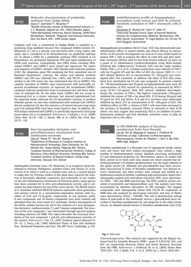
P192Molecular characterization of polyketidesynthases from Ginkgo bilobaTaura F1, Luetrakul T2, Shoyama Y3
1Kyushu University, Faculty of Pharmaceutical Sciences, 3 –1-1, Maidashi, Higashi-ku, 812 – 8582 Fukuoka, Japan;2Ubon Ratchathani University, Warin Chamrap, 34190 UbonRatchathani, Thailand; 3Nagasaki International University,Huis Ten Bosch, 859 – 3298 Sasebo, Japan
Ginkgolic acid (GA), a constituent in Ginkgo biloba, is regarded as apromising drug candidate because this compound inhibits protein SU-MOylation [1]. GA is a salicylic acid derivative with a long carbon sidechain, and is considered to be biosynthesized via polyketide pathway[2]. To obtain a cDNA for a polyketide synthase (PKS) involved in GAbiosynthesis, we performed degenerate PCR and rapid amplification ofcDNA ends reactions. Consequently, two cDNA clones encoding PKSs,named GbPKS 1 and GbPKS 2, were cloned and sequenced. The aminoacid sequence of GbPKS 1 showed > 90% identity with chalcone synthase(CHS), the ubiquitous plant PKS catalyzing the first committed step offlavonoid biosynthesis. Contrary, the amino acid identity betweenGbPKS 2 and CHS was relatively low (~40%), and Thr197, a conservedresidue in the CHS active site, was substituted into Gly in GbPKS 2. Thecatalytic functions of GbPKSs were characterized with bacterially ex-pressed recombinant enzymes. As expected, the recombinant GbPKS 1catalyzed chalcone production from 4-coumaroyl-CoA and three mole-cules of malonyl-CoA. On the other hand, GbPKS 2 did not accept 4-coumaroyl-CoA as a starter substrate, but selected palmitoleoyl-CoA,the expected starter substrate for GA biosynthetic reaction, to producetriketide pyrone via two-step condensation with malonyl-CoA. GbPKS 2did not synthesize GA, but this enzyme is of interest because long-chainacyl-CoA utilizing PKSs have been rarely found to date. In addition, RT-PCR analysis demonstrated GbPKS 2 is specifically expressed in fruit pulptissue where GA is accumulated. References: 1. Fukuda, I. et al. (2009)Chem. Biol. 16:133 – 140. 2. Austin, MB. et al. (2003) Nat. Prod. Rep.20:79 – 110.
P193New furoquinoline derivative andanti-inflammatory constituents fromZanthoxylum avicennaeChen JJ1, Wang TY2, Wu JB2, Hwang TL3
1Department of Pharmacy & Graduate Institute ofPharmaceutical Technology, Tajen University, No. 20,Weishin Rd., Yanpu Shiang, Pingtung 907, Taiwan;2Graduate Institute of Pharmaceutical Chemistry, College ofPharmacy, China Medical University, Taichung 404, Taiwan;3Graduate Institute of Natural Products, Chang GungUniversity, Taoyuan 333, Taiwan
Zanthoxylum avicennae (Lam.) DC (Rutaceae) is an evergreen shrub dis-tributed in Vietnam, Philippines, southern China, and Taiwan [1]. A de-coction of its stems is used as a stomach tonic and as a counter-poisonto snake bite [2]. Previous studies of this plant have reported the isola-tion of flavonoids, alkaloids, coumarins, and terpenoids. In our studieson the anti-inflammatory constituents of Formosan plants, many specieshave been screened for in vitro anti-inflammatory activity, and Z. avi-cennae has been found to be one of the active species. The MeOH extractof Z. avicennae inhibited fMLP/CB-induced superoxide anion generationand elastase release in a concentration-dependent manner with IC50
values of 6.34 and 15.32 mg/mL, respectively. In our previous study,8 new compounds and 18 known compounds have been isolated andidentified from the stem wood of Z. avicennae. Further investigation ofthe EtOAc-soluble fraction has led to the isolation of a new furoquino-line derivative, zanthoavicenine (1) and 7 known compounds. The struc-ture of the new compound 1 was determined through spectral analysesincluding extensive 2D NMR. This report describes the structural eluci-dation of the new compound 1 and the anti-inflammatory activities ofthe isolates. References: 1. Ho, T.C. (2007) Endemic Species Research 9:29 – 52. 2. Perry, L.M. (1980) Medicinal Plants of East and SoutheastAsia: Attributed Properties and Uses. The MIT Press: Cambridge, p. 370.
P194Antiinflammatory profile of Harpagophytumprocumbens crude extract and after its externalmetabolic activation in THP-1 monocytic cells invitroHostanska K1, Rostock M1, Suter A2, Saller R1
1University Hospital Zurich, Dept. of Internal Medicine,Institute for Complementary Medicine, Raemistrasse 100,8091 Zurich, Switzerland; 2A. Vogel Bioforce AG, PO box 76,9325 Roggwil, Switzerland
Harpagophytum procumbens (Devil’s Claw; DCE) has demonstrated anti-inflammatory effects in animal models and clinical efficacy in osteoar-thritis. In the present investigations we used an in vitro model based onhuman THP-1 cells to evaluate the effect of crude DCE and after meta-bolic activation (DCEm) with S9 mix from Aroclor-induced rat livers ona panel of 12 inflammatory cytokine/chemokine using Multi-AnalyteELISArray Kits (SABiosciences, Frederick, USA) and the single cytokineELISAs (TNF-a, IL-6, IL-8). Cytokine productions were induced in THP-1cells by 25 mg/ml LPS and the effect of DCE dry extract (DER 1.5 – 3:1;60% ethanol, Bioforce AG) at concentrations (0 – 250 mg/ml) was inves-tigated after 24 h treatment. In addition, the effect of DCE after simu-lated liver metabolism by S 9 mix was compared. As positive controls10 – 7 M dexamethasone and 10 mg/ml cyclophosphamide were used. Allconcentrations of DCE exerted no cytotoxicity as measured by WST-1assay (IC50 = 1.25 mg/ml). Both DCE extracts inhibited dose-depen-dently the secretion of TNF-a. The highest concentration (250 mg/ml)inhibited the release of TNF-a by about 80%. IC 50 beeing 98 mg/ml and47 mg/ml for DCE and DCEm. IL-8 and IL-6 secretion were moderatelyinhibited by about 25% at concentration of 50 – 100 mg/ml of DCE. Theinhibitory effect on TNF- a release in THP-1 cells have been increased inthe presence of metabolic activated DCEm as mirrored in their IC50values. Our data suggest that proprietary DCE is an inhibitor of proin-flammatory cytokines and that metabolic activation seems to play animportant role in this effect.
P195LC-SPE-NMR-MS analysis of Strychnosusambarensis fruits from RwandaCao M, Tits M, Muganga R, Angenot L, Fr�d�rich MUniversity of Li�ge, Laboratory of Pharmacognosy,Pharmacognosy, Avenue de l’Hopital, 1 (Tour 4, Bat B36),4000 Li�ge, Belgium
Strychnos usambarensis is a Rwandan tree of Loganiaceae family, whoseroots and leaves had been widely investigated. They contain a hugenumber of indolomoterpenic tertiary alkaloids possessing antimalarial[1] and antitumoral properties [2]. Nevertheless, almost no studies hadbeen carried on its fruits until now, except one which showed that de-methoxycarbonyl-3,14-dihydro-gambirtannine was the major alkaloid inthe apolar extract [3]. Alkaloids and glucoalkaloids have been success-fully characterized in fruits of Strychnos usambarensis. Two distinct ex-tracts (methanolic and ethyl acetate) were realized and studied by ahyphenated analytical method, combining high-performance liquid chro-matography coupled with solid-phase extraction (SPE), mass spectrome-try (HPLC – MS) and NMR spectroscopy. The HPLC gradient system con-sisted of acetonitrile and heptanesulfonic acid. The analytes were thenaccumulated by repetitive absorption on SPE cartridges. The trappedcompounds were subsequently eluted with CD3CN for acquisition ofNMR data and analysed by electrospray ionization and time of flightsmass spectrometry (ESI/Q – TOF/MS). This led to identification amongothers of palicoside in the methanolic extract, a glucoalkaloid never de-scribed in Strychnos usambarensis [4], and akagerine in the ethyl acetateextract, an alkaloid already known in Strychnos usambarensis roots [5,6].
Fig. 1: Palicoside
Acknowledgements: This research was supported by the Belgian Na-tional Fund for Scientific Research (FNRS – grant N 3.4533.10). M.C. andM.F. are respectively Research Fellow and Senior Research Associatefrom the FNRS. References: 1. Fr�d�rich, M. et al. (2004) Planta Med.70(6):520 – 525. 2. Balde, E-H., S. et al. (2010) Int. J. Oncol. 36(4):961 –
Planta Med 2010; 76: 1163–1374 Georg Thieme Verlag KG Stuttgart · New York · ISSN 0032-0943
12417th Tannin Conference and 58th International Congress of the GA | 29th August – 2nd September 2010, Berlin, Germany
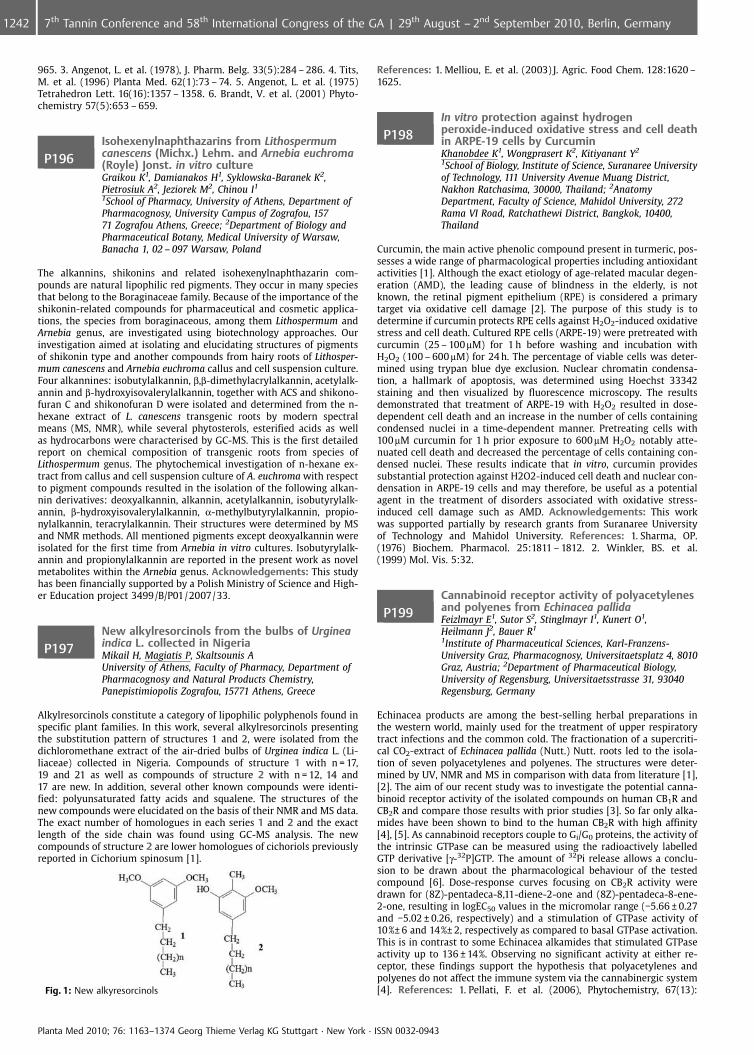
965. 3. Angenot, L. et al. (1978), J. Pharm. Belg. 33(5):284 – 286. 4. Tits,M. et al. (1996) Planta Med. 62(1):73 – 74. 5. Angenot, L. et al. (1975)Tetrahedron Lett. 16(16):1357 – 1358. 6. Brandt, V. et al. (2001) Phyto-chemistry 57(5):653 – 659.
P196Isohexenylnaphthazarins from Lithospermumcanescens (Michx.) Lehm. and Arnebia euchroma(Royle) Jonst. in vitro cultureGraikou K1, Damianakos H1, Syklowska-Baranek K2,Pietrosiuk A2, Jeziorek M2, Chinou I1
1School of Pharmacy, University of Athens, Department ofPharmacognosy, University Campus of Zografou, 15771 Zografou Athens, Greece; 2Department of Biology andPharmaceutical Botany, Medical University of Warsaw,Banacha 1, 02 – 097 Warsaw, Poland
The alkannins, shikonins and related isohexenylnaphthazarin com-pounds are natural lipophilic red pigments. They occur in many speciesthat belong to the Boraginaceae family. Because of the importance of theshikonin-related compounds for pharmaceutical and cosmetic applica-tions, the species from boraginaceous, among them Lithospermum andArnebia genus, are investigated using biotechnology approaches. Ourinvestigation aimed at isolating and elucidating structures of pigmentsof shikonin type and another compounds from hairy roots of Lithosper-mum canescens and Arnebia euchroma callus and cell suspension culture.Four alkannines: isobutylalkannin, b,b-dimethylacrylalkannin, acetylalk-annin and b-hydroxyisovalerylalkannin, together with ACS and shikono-furan C and shikonofuran D were isolated and determined from the n-hexane extract of L. canescens transgenic roots by modern spectralmeans (MS, NMR), while several phytosterols, esterified acids as wellas hydrocarbons were characterised by GC-MS. This is the first detailedreport on chemical composition of transgenic roots from species ofLithospermum genus. The phytochemical investigation of n-hexane ex-tract from callus and cell suspension culture of A. euchroma with respectto pigment compounds resulted in the isolation of the following alkan-nin derivatives: deoxyalkannin, alkannin, acetylalkannin, isobutyrylalk-annin, b-hydroxyisovalerylalkannin, a-methylbutyrylalkannin, propio-nylalkannin, teracrylalkannin. Their structures were determined by MSand NMR methods. All mentioned pigments except deoxyalkannin wereisolated for the first time from Arnebia in vitro cultures. Isobutyrylalk-annin and propionylalkannin are reported in the present work as novelmetabolites within the Arnebia genus. Acknowledgements: This studyhas been financially supported by a Polish Ministry of Science and High-er Education project 3499/B/P01/2007/33.
P197New alkylresorcinols from the bulbs of Urgineaindica L. collected in NigeriaMikail H, Magiatis P, Skaltsounis AUniversity of Athens, Faculty of Pharmacy, Department ofPharmacognosy and Natural Products Chemistry,Panepistimiopolis Zografou, 15771 Athens, Greece
Alkylresorcinols constitute a category of lipophilic polyphenols found inspecific plant families. In this work, several alkylresorcinols presentingthe substitution pattern of structures 1 and 2, were isolated from thedichloromethane extract of the air-dried bulbs of Urginea indica L. (Li-liaceae) collected in Nigeria. Compounds of structure 1 with n = 17,19 and 21 as well as compounds of structure 2 with n = 12, 14 and17 are new. In addition, several other known compounds were identi-fied: polyunsaturated fatty acids and squalene. The structures of thenew compounds were elucidated on the basis of their NMR and MS data.The exact number of homologues in each series 1 and 2 and the exactlength of the side chain was found using GC-MS analysis. The newcompounds of structure 2 are lower homologues of cichoriols previouslyreported in Cichorium spinosum [1].
Fig. 1: New alkyresorcinols
References: 1. Melliou, E. et al. (2003) J. Agric. Food Chem. 128:1620 –1625.
P198In vitro protection against hydrogenperoxide-induced oxidative stress and cell deathin ARPE-19 cells by CurcuminKhanobdee K1, Wongprasert K2, Kitiyanant Y2
1School of Biology, Institute of Science, Suranaree Universityof Technology, 111 University Avenue Muang District,Nakhon Ratchasima, 30000, Thailand; 2AnatomyDepartment, Faculty of Science, Mahidol University, 272Rama VI Road, Ratchathewi District, Bangkok, 10400,Thailand
Curcumin, the main active phenolic compound present in turmeric, pos-sesses a wide range of pharmacological properties including antioxidantactivities [1]. Although the exact etiology of age-related macular degen-eration (AMD), the leading cause of blindness in the elderly, is notknown, the retinal pigment epithelium (RPE) is considered a primarytarget via oxidative cell damage [2]. The purpose of this study is todetermine if curcumin protects RPE cells against H2O2-induced oxidativestress and cell death. Cultured RPE cells (ARPE-19) were pretreated withcurcumin (25 – 100mM) for 1 h before washing and incubation withH2O2 (100 – 600 mM) for 24 h. The percentage of viable cells was deter-mined using trypan blue dye exclusion. Nuclear chromatin condensa-tion, a hallmark of apoptosis, was determined using Hoechst 33342staining and then visualized by fluorescence microscopy. The resultsdemonstrated that treatment of ARPE-19 with H2O2 resulted in dose-dependent cell death and an increase in the number of cells containingcondensed nuclei in a time-dependent manner. Pretreating cells with100mM curcumin for 1 h prior exposure to 600mM H2O2 notably atte-nuated cell death and decreased the percentage of cells containing con-densed nuclei. These results indicate that in vitro, curcumin providessubstantial protection against H2O2-induced cell death and nuclear con-densation in ARPE-19 cells and may therefore, be useful as a potentialagent in the treatment of disorders associated with oxidative stress-induced cell damage such as AMD. Acknowledgements: This workwas supported partially by research grants from Suranaree Universityof Technology and Mahidol University. References: 1. Sharma, OP.(1976) Biochem. Pharmacol. 25:1811 – 1812. 2. Winkler, BS. et al.(1999) Mol. Vis. 5:32.
P199Cannabinoid receptor activity of polyacetylenesand polyenes from Echinacea pallidaFeizlmayr E1, Sutor S2, Stinglmayr I1, Kunert O1,Heilmann J2, Bauer R1
1Institute of Pharmaceutical Sciences, Karl-Franzens-University Graz, Pharmacognosy, Universitaetsplatz 4, 8010Graz, Austria; 2Department of Pharmaceutical Biology,University of Regensburg, Universitaetsstrasse 31, 93040Regensburg, Germany
Echinacea products are among the best-selling herbal preparations inthe western world, mainly used for the treatment of upper respiratorytract infections and the common cold. The fractionation of a supercriti-cal CO2-extract of Echinacea pallida (Nutt.) Nutt. roots led to the isola-tion of seven polyacetylenes and polyenes. The structures were deter-mined by UV, NMR and MS in comparison with data from literature [1],[2]. The aim of our recent study was to investigate the potential canna-binoid receptor activity of the isolated compounds on human CB1R andCB2R and compare those results with prior studies [3]. So far only alka-mides have been shown to bind to the human CB2R with high affinity[4], [5]. As cannabinoid receptors couple to Gi/G0 proteins, the activity ofthe intrinsic GTPase can be measured using the radioactively labelledGTP derivative [g-32P]GTP. The amount of 32Pi release allows a conclu-sion to be drawn about the pharmacological behaviour of the testedcompound [6]. Dose-response curves focusing on CB2R activity weredrawn for (8Z)-pentadeca-8,11-diene-2-one and (8Z)-pentadeca-8-ene-2-one, resulting in logEC50 values in the micromolar range (�5.66 € 0.27and �5.02 € 0.26, respectively) and a stimulation of GTPase activity of10%€ 6 and 14%€ 2, respectively as compared to basal GTPase activation.This is in contrast to some Echinacea alkamides that stimulated GTPaseactivity up to 136 € 14%. Observing no significant activity at either re-ceptor, these findings support the hypothesis that polyacetylenes andpolyenes do not affect the immune system via the cannabinergic system[4]. References: 1. Pellati, F. et al. (2006), Phytochemistry, 67(13):
Planta Med 2010; 76: 1163–1374 Georg Thieme Verlag KG Stuttgart · New York · ISSN 0032-0943
1242 7th Tannin Conference and 58th International Congress of the GA | 29th August – 2nd September 2010, Berlin, Germany
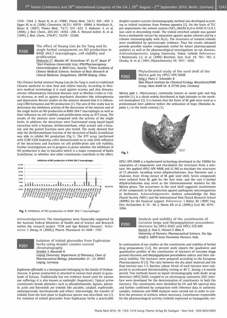
1359 – 1364. 2. Bauer R. et al. (1988), Planta Med., 54(5): 426 – 430. 3.Egger, M. et al. (2008), Chemistry, 14(35): 10978 – 10984. 4. Woelkart, K.,Bauer R. (2007), Planta Med., 73(7): 615 – 623. 5. Raduner, S. et al.(2006), J. Biol. Chem., 281(20): 14192 – 206. 6. Wenzel-Seifert, K. et al.(1999), J. Biol. Chem., 274(47): 33259 – 33266.
P200The effect of Huang Lian Jie Du Tang and itssingle herbal components on NO production inRAW 264.7 macrophages, cell viability andproliferationWidowitz U1, Blunder M1, Kretschmer N1, Lu A2, Bauer R1
1Karl-Franzens-Universit�t Graz, IPW/Pharmacognosy,Universit�tsplatz 4, 8010 Graz, Austria; 2China Academy ofChinese Medical Sciences, Institute of Basic Research inClinical Medicine, Dongzhimen, 100700 Beijing, China
The Chinese herbal mixture Huang Lian Jie Du Tang is used in traditionalChinese medicine to clear heat and relieve toxicity. According to Wes-tern medical terminology it is used against eczema and skin diseases,chronic inflammatory intestinal diseases such as Morbus Crohn or Coli-tis ulcerosa, as well as against psychiatric disorders like schizophreniaand depression. Recent studies on the mixture showed that it is influen-cing LTB4 formation and NO production [1]. The aim of this study was todetermine the inhibitory activity of the decoctions of the mixture and ofthe single herbs on NO production in RAW 264.7 macrophages as well astheir influence on cell viability and proliferation using an XTT assay. Theresults of the mixture were compared with the activity of the singleherbs. In addition, the decoctions were fractionated using liquid-liquidextraction with n-heptane, dichloromethane, ethyl acetate and n-buta-nol, and the gained fractions were also tested. The study showed thatonly the dichloromethane fraction of the decoction of Radix Scutellariaewas able to inhibit NO production (Fig.1). The XTT assay (performedwith CCRF-CEM leukemia cells) demonstrated no or only little influenceof the decoctions and fractions on cell proliferation and cell viability.Further investigations are in progress to prove whether the inhibition ofNO production is due to baicalein which is a major compound of RadixScutellariae, or whether also other constituents contribute to the effect.
Fig. 1: Inhibition of NO production in RAW 264.7 macrophages
Acknowledgements: The investigations were financially supported bythe Austrian Federal Ministries of Health and of Science and Researchwithin the research project “TCM and Age Related Diseases”. Refer-ences: 1. Zheng, H. (2009) J. Pharm. Pharmacol. 61:1699 – 1707.
P201Isolation of iridoid glucosides from Euphrasiaeherba using droplet counter-currentchromatographyStrahler S, Rauwald HLeipzig University, Department of Pharmacy, Chair ofPharmaceutical Biology, Johannisallee 21 – 23, 04103Leipzig, Germany
Euphrasia officinalis is a hemiparasite belonging to the family of Oroban-chaceae. It grows unattached or attached to various host plants in grass-lands of Eurasia. Traditionally but not evidence based used for variouseye sufferings it is also known as eyebright (Augentrost). Typical activeconstituents beside phenolics such as phenylethanoids, lignans, pheno-lic acids and flavonoids are iridoids like aucubin, catalpol, euphroside,melampyroside, boschnaloside and others. Interestingly, the transfer ofiridoids from the host plant to Euphrasia species was described, too [1].For isolation of iridoid glucosides from Euphrasiae herba a practicable
droplet counter-current chromatography method was developed accord-ing to iridoid isolations from Premna japonica [2]. On the basis of TLCpre-experiments the solvent system CHCl3:MeOH:H2O:n-PrOH 9:12:8:1was used in descending mode. The iridoid enriched sample was gainedfrom a methanolic extract by separation against apolar solvents and by acolumn chromatography with Al2O3. The structures of isolated iridoidswere established by spectroscopic evidence. Thus the results obtainedprovide possible marker compounds useful for future pharmacopoeialanalytics as well as for pharmacological investigations on eye diseases.Acknowledgements: Leipzig University, Tobias Liebold References:1. Rasmussen, L.S. et al. (2006) Biochem. Syst. Ecol. 34: 763 – 765. 2.Otsuka, H. et al. (1991) Phytochemistry 30: 1917 – 1920.
P202Phytochemical study of the seed shell of theMyrica gale by HPLC-SPE-NMRFang J, Paetz C, Schneider BMax Planck Institute for Chemical Ecology, Biosythesis/NMRGroup, Hans Kn�ll Str. 8, 07745 Jena, Germany
Myrica gale L. (Myricaseae), commonly known as sweet gale and bogmyrthle [1], is a shrub widely distributed at high latitudes in the north-ern hemisphere [2]. It is known that the fruits of M. gale were used as apredominant beer additive before the utilization of hops (Humulus lu-pulus L.) in the tenth century [1].
Fig. 1
HPLC-SPE-NMR is a hyphenated technology developed in the 1990th forseparation of components and elucidation the structures from a mix-ture. We applied HPLC-SPE-NMR and LC-MS to elucidate the structuresof 13 phenols, inculding seven dihydrochalcones, four flavonols and achalcone, from 10 mg extract of M. gale seed shell. Seven compoundswere isolated from M. gale for the first time, and the rare C-methyldihydrochalcones may serve as the chemotaxonomic markers for theMyrica genus. The occurrence in the seed shell sugguests involvementof the compounds in the protection against pathogenic microorganismsor herbivores. Acknowledgements: Authors acknowledge the MaxPlanck Society (MPG) and the International Max Planck Research School(IMPRS) for the financial support. References: 1. Behre, KE (1999) Veg.Hist. Archaeobot. 8: 35 – 48. 2. Skene, KR. et al. (2000) J. Ecol. 88: 1079 –1094.
P203Analysis and stability of the constituents ofCurcuma longa and Harpagophytum procumbenstinctures by HPLC-DAD and HPLC-ESI-MSKarioti A, Fani E, Vincieri F, Bilia AUniversity of Florence, Pharmaceutical Sciences, Via UgoSchiff 6, 50019 Sesto Fiorentino Florence, Italy
In continuation of our studies on the constituents and stability of herbaldrug preparations [1,2], the present work reports the qualitative andquantitative profiles of the constituents of tinctures of Curcuma longaground rhizomes and Harpagophytum procumbens radices and their che-mical stability. The tinctures were prepared according to the EuropeanPharmacopoeia XI [3]. The ratio between the dry plant material and thefinal tincture was 1:5. Batches (about 10 ml) of each tincture were sub-jected to accelerated thermostability testing at 40 �C. during a 6-monthperiod. Two methods based on liquid chromatography with diode arraydetection (HPLC/DAD) coupled to an electrospray ionization (ESI) inter-face were developed for the determination of constituents in both thetinctures. The constituents were identified by UV and MS spectral dataand further confirmed by comparison with reference data or authenticsamples. Isolations and NMR analyzes were carried out in order to con-firm the presence of artifacts where necessary. Constituents responsiblefor the pharmacological activity (iridoids expressed as harpagoside, ver-
Planta Med 2010; 76: 1163–1374 Georg Thieme Verlag KG Stuttgart · New York · ISSN 0032-0943
12437th Tannin Conference and 58th International Congress of the GA | 29th August – 2nd September 2010, Berlin, Germany
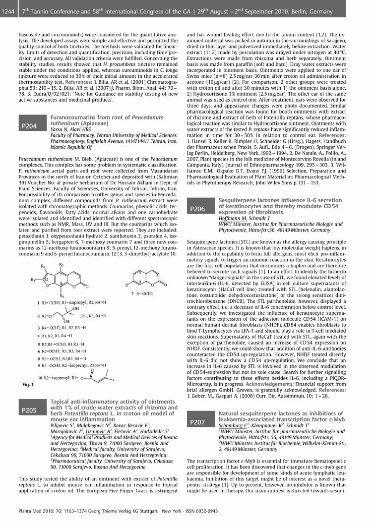
bascoside and curcuminoids) were considered for the quantitative ana-lysis. The developed assays were simple and effective and permitted thequality control of both tinctures. The methods were validated for linear-ity, limits of detection and quantification, precision, including time pre-cision, and accuracy. All validation criteria were fulfilled. Concerning thestability studies, results showed that H. procumbens tincture remainedstable under the conditions applied, whereas curcuminoids in C. longatincture were reduced to 30% of their initial amount in the acceleratedthermostability test. References: 1. Bilia, AR et al. (2001) Chromatogra-phia 53: 210 – 15. 2. Bilia, AR et al. (2007) J. Pharm. Biom. Anal. 44: 70 –78. 3. Eudra/Q/92/021: ‘Note for Guidance on stability testing of newactive substances and medicinal products’.
P204Furanocoumarins from root of Peucedanumruthenicum (Apiaceae)Yassa N, Alavi HRSFaculty of Pharmacy, Tehran University of Medical Sciences,Pharmacognosy, Enghelab Avenue, 1414714411 Tehran, Iran,Islamic Republic Of
Peucedanum ruthenicum M. Bieb. (Apiaceae) is one of the Peucedanumcomplexes. This complex has some problem in systematic classification.P. ruthenicum aerial parts and root were collected from MazandaranProvinces in the north of Iran on October and deposited with (Salimian39) Voucher No. at private herbarium of Dr. Hossain Akhani in Dept. ofPlant Sciences, Faculty of Sciencses, University of Tehran, Tehran, Iran.For possibility of its comparison to other genus and species in Penceda-num complex, different compounds from P. ruthenicum extract wereisolated with chromatographic methods. Coumarins, phenolic acids, ter-penoids, flavonoids, fatty acids, normal alkans and one carbohydratewere isolated and identified and identified with different spectroscopicmethods such as NMR, Mass, UV and IR. But the coumarins which iso-lated and purified from root extract were reported. They are included:peucedanin 1, oxypeucedanin hydrate 2, xanthotoxin 3, psoralen 4, iso-pimpinellin 5, bergapten 6, 7-methoxy coumarin 7 and three new cou-marins as 12-methoxy furanocoumarin 8; 5-prenyl, 12-methoxy furano-coumarin 9 and 5-prenyl furanocoumarin, 12 (3, 3-dimethyl) acrylate 10.
Fig. 1
P205Topical anti-inflammatory activity of ointmentswith 1% of crude water extracts of rhizoma andherb Potentilla reptans L. in croton oil model ofmouse ear inflammationPilipovic S1, Mulabegovic N2, Kovac-Besovic E3,Mornjakovic Z2, Uzunovic A1, Elezovic A1, Hadzidedic S1
1Agency for Medical Products and Medical Devices of Bosniaand Herzegovina, Titova 9, 71000 Sarajevo, Bosnia AndHerzegovina; 2Medical faculty, University of Sarajevo,Cekalusa 90, 71000 Sarajevo, Bosnia And Herzegovina;3Pharmaceutical faculty, University of Sarajevo, Cekalusa90, 71000 Sarajevo, Bosnia And Herzegovina
This study tested the ability of an ointment with extract of Potentillareptans L. to inhibit mouse ear inflammation in response to topicalapplication of croton oil. The European Five-Finger Grass is astringent
and has wound healing effect due to the tannin content (1,2). The ex-amined material was picked in autumn in the surroundings of Sarajevo,dried in thin layer and pulverized immediately before extraction. Waterextract (1: 2) made by percolation was drayed under nitrogen at 40 �C.Extractions were made from rhizoma and herb separately. Ointmentbasis was made from paraffin (soft and hard). Dray water extracts wereincorporated in ointment basis. Ointments were applied to one ear ofSwiss mice (n = 8) 2.5 mg/ear 30 min after croton oil administration inacetone (10 mg/ear) (2). For comparison, 2 other groups were treatedwith croton oil and after 30 minutes with 1) the ointment basis alone,2) Hydrocortisone 1% ointment (2,5 mg/ear). The other ear of the sameanimal was used as control one. After treatment, ears were observed forthree days, and appearance changes were photo documented. Similarpharmacological reaction was found for booth ointments with extractof rhizome and extract of herb of Potentilla reptans, whose pharmaco-logical reaction was similar to Hydrocortisone ointment. Ointments withwater extracts of the tested P. reptans have significantly reduced inflam-mation in time for 30 – 50% in relation to control ear. References:1. Hansel R, Keller K, Rimpler H, Schneider G (Hrsg.), Hagers, Handbuchder Pharmazeutischen Praxis, 5. Aufl., Bde 4 – 6, (Drogen), Springer Ver-lag Berlin, Heidelberg, New York, 1992 – 1994. 2. De Natale, A., Pollio, A.,2007. Plant species in the folk medicine of Montecorvino Rovella (inlandCampania, Italy). Journal of Ethnopharmacology 109, 295 – 303. 3. Wil-liamso E.M., Okpako D.T. Evans F.J. (1996) Selection, Preparation andPharmacological Evaluation of Plant Material in: Pharmacological Meth-ods in Phytotherapy Research; John Wiley Sons p.131 – 153.
P206Sesquiterpene lactones influence IL-6 secretionof keratinocytes and thereby modulate CD 54expression of fibroblastsHoffmann M, Schmidt TWWU M�nster, Institut f�r Pharmazeutische Biologie undPhytochemie, Hittorfstr.56, 48149 M�nster, Germany
Sesquiterpene lactones (STL) are known as the allergy causing principlein Asteraceae species. It is known that low molecular weight haptens, inaddition to the capability to form full allergens, must elicit pro-inflam-matory signals to trigger an immune reaction in the skin. Keratinocytesare the first cell population that encounters a hapten and are thereforebelieved to secrete such signals [1]. In an effort to identify the hithertounknown “danger-signals” in the case of STL, we found elevated levels ofinterleukin-6 (IL-6, detected by ELISA) in cell culture supernatants ofkeratinocytes (HaCaT cell line) treated with STL (helenalin, alantolac-tone, costunolide, dehydrocostuslactone) or the strong sensitizer dini-trochlorobenzene (DNCB). The STL parthenolide, however, displayed acontrary effect, i. e. a decrease of IL-6 concentration below control level.Subsequently, we investigated the influence of keratinocyte superna-tants on the expression of the adhesion molecule CD 54 (ICAM-1) onnormal human dermal fibroblasts (NHDF). CD 54 enables fibroblasts tobind T-Lymphocytes via LFA-1 and should play a role in T-cell mediatedskin reactions. Supernatants of HaCaT treated with STL, again with theexception of parthenolide, caused an increase of CD54 expression onNHDF. Consistently, we could show that addition of anti-IL-6-antibodiescounteracted the CD 54 up-regulation. However, NHDF treated directlywith IL-6 did not show a CD 54 up-regulation. We conclude that anincrease in IL-6 caused by STL is involved in the observed modulationof CD54-expression but not its sole cause. Search for further signallingfactors contributing to these effects besides IL-6, including a PIQOR-Microarray, is in progress. Acknowledgements: Financial support frombrial allergen GmbH, Greven, is gratefully acknowledged. References:1. Gober, M., Gaspari A. (2008) Curr. Dir. Autoimmun. 10: 1 – 26.
P207Natural sesquiterpene lactones as inhibitiors ofleukaemia-associated transcription factor c-MybSchomburg C1, Klempnauer K2, Schmidt T1
1WWU M�nster, Institut f�r pharmazeutische Biologie undPhytochemie, Hittorfstr. 56, 48149 M�nster, Germany;2WWU M�nster, Institut f�r Biochemie, Wilhelm-Klemm-Str.2, 48149 M�nster, Germany
The transcription factor c-Myb is essential for immature hematopoieticcell proliferation. It has been discovered that changes in the c-myb geneare responsible for development of some kinds of acute lymphatic leu-kaemia. Inhibition of this target might be of interest as a novel thera-peutic strategy [1]. Up to present, however, no inhibitor is known thatmight be used in therapy. Our main interest is directed towards sesqui-
Planta Med 2010; 76: 1163–1374 Georg Thieme Verlag KG Stuttgart · New York · ISSN 0032-0943
1244 7th Tannin Conference and 58th International Congress of the GA | 29th August – 2nd September 2010, Berlin, Germany
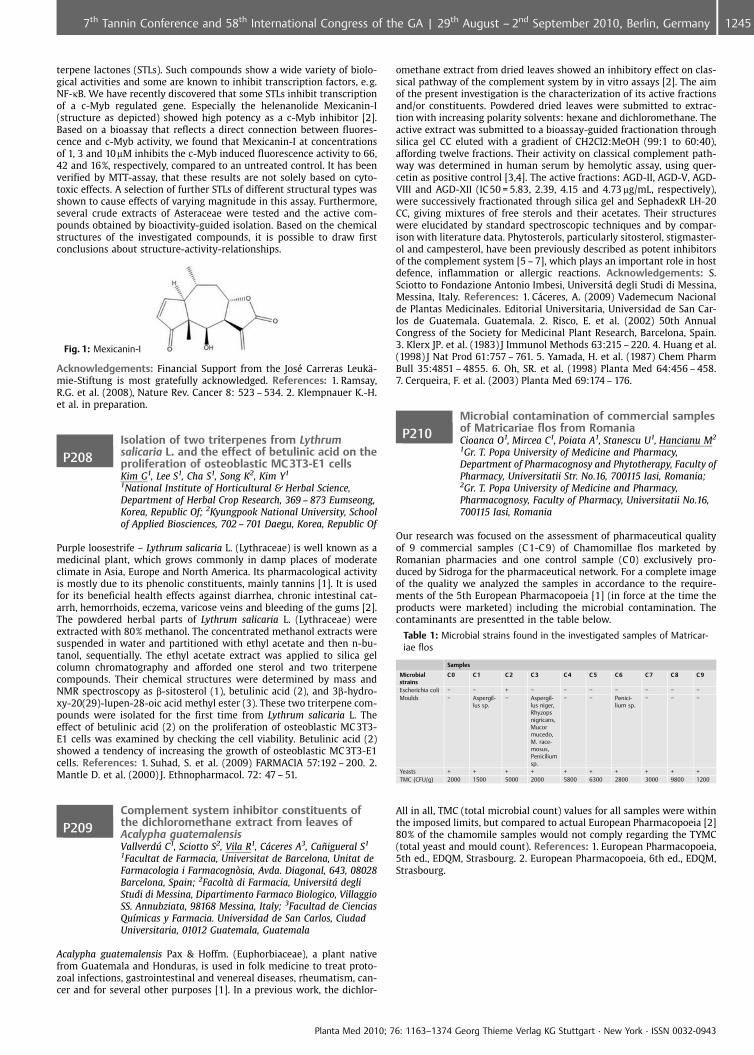
terpene lactones (STLs). Such compounds show a wide variety of biolo-gical activities and some are known to inhibit transcription factors, e. g.NF-kB. We have recently discovered that some STLs inhibit transcriptionof a c-Myb regulated gene. Especially the helenanolide Mexicanin-I(structure as depicted) showed high potency as a c-Myb inhibitor [2].Based on a bioassay that reflects a direct connection between fluores-cence and c-Myb activity, we found that Mexicanin-I at concentrationsof 1, 3 and 10mM inhibits the c-Myb induced fluorescence activity to 66,42 and 16%, respectively, compared to an untreated control. It has beenverified by MTT-assay, that these results are not solely based on cyto-toxic effects. A selection of further STLs of different structural types wasshown to cause effects of varying magnitude in this assay. Furthermore,several crude extracts of Asteraceae were tested and the active com-pounds obtained by bioactivity-guided isolation. Based on the chemicalstructures of the investigated compounds, it is possible to draw firstconclusions about structure-activity-relationships.
Fig. 1: Mexicanin-I
Acknowledgements: Financial Support from the Jos� Carreras Leuk�-mie-Stiftung is most gratefully acknowledged. References: 1. Ramsay,R.G. et al. (2008), Nature Rev. Cancer 8: 523 – 534. 2. Klempnauer K.-H.et al. in preparation.
P208Isolation of two triterpenes from Lythrumsalicaria L. and the effect of betulinic acid on theproliferation of osteoblastic MC 3T3-E1 cellsKim G1, Lee S1, Cha S1, Song K2, Kim Y1
1National Institute of Horticultural & Herbal Science,Department of Herbal Crop Research, 369 – 873 Eumseong,Korea, Republic Of; 2Kyungpook National University, Schoolof Applied Biosciences, 702 – 701 Daegu, Korea, Republic Of
Purple loosestrife – Lythrum salicaria L. (Lythraceae) is well known as amedicinal plant, which grows commonly in damp places of moderateclimate in Asia, Europe and North America. Its pharmacological activityis mostly due to its phenolic constituents, mainly tannins [1]. It is usedfor its beneficial health effects against diarrhea, chronic intestinal cat-arrh, hemorrhoids, eczema, varicose veins and bleeding of the gums [2].The powdered herbal parts of Lythrum salicaria L. (Lythraceae) wereextracted with 80% methanol. The concentrated methanol extracts weresuspended in water and partitioned with ethyl acetate and then n-bu-tanol, sequentially. The ethyl acetate extract was applied to silica gelcolumn chromatography and afforded one sterol and two triterpenecompounds. Their chemical structures were determined by mass andNMR spectroscopy as b-sitosterol (1), betulinic acid (2), and 3b-hydro-xy-20(29)-lupen-28-oic acid methyl ester (3). These two triterpene com-pounds were isolated for the first time from Lythrum salicaria L. Theeffect of betulinic acid (2) on the proliferation of osteoblastic MC 3T3-E1 cells was examined by checking the cell viability. Betulinic acid (2)showed a tendency of increasing the growth of osteoblastic MC3T3-E1cells. References: 1. Suhad, S. et al. (2009) FARMACIA 57:192 – 200. 2.Mantle D. et al. (2000) J. Ethnopharmacol. 72: 47 – 51.
P209Complement system inhibitor constituents ofthe dichloromethane extract from leaves ofAcalypha guatemalensisVallverdffl C1, Sciotto S2, Vila R1, C�ceres A3, Ca�igueral S1
1Facultat de Farmacia, Universitat de Barcelona, Unitat deFarmacologia i Farmacogn�sia, Avda. Diagonal, 643, 08028Barcelona, Spain; 2Facolt di Farmacia, Universit� degliStudi di Messina, Dipartimento Farmaco Biologico, VillaggioSS. Annubziata, 98168 Messina, Italy; 3Facultad de CienciasQu�micas y Farmacia. Universidad de San Carlos, CiudadUniversitaria, 01012 Guatemala, Guatemala
Acalypha guatemalensis Pax & Hoffm. (Euphorbiaceae), a plant nativefrom Guatemala and Honduras, is used in folk medicine to treat proto-zoal infections, gastrointestinal and venereal diseases, rheumatism, can-cer and for several other purposes [1]. In a previous work, the dichlor-
omethane extract from dried leaves showed an inhibitory effect on clas-sical pathway of the complement system by in vitro assays [2]. The aimof the present investigation is the characterization of its active fractionsand/or constituents. Powdered dried leaves were submitted to extrac-tion with increasing polarity solvents: hexane and dichloromethane. Theactive extract was submitted to a bioassay-guided fractionation throughsilica gel CC eluted with a gradient of CH2Cl2:MeOH (99:1 to 60:40),affording twelve fractions. Their activity on classical complement path-way was determined in human serum by hemolytic assay, using quer-cetin as positive control [3,4]. The active fractions: AGD-II, AGD-V, AGD-VIII and AGD-XII (IC 50 = 5.83, 2.39, 4.15 and 4.73 mg/mL, respectively),were successively fractionated through silica gel and SephadexR LH-20CC, giving mixtures of free sterols and their acetates. Their structureswere elucidated by standard spectroscopic techniques and by compar-ison with literature data. Phytosterols, particularly sitosterol, stigmaster-ol and campesterol, have been previously described as potent inhibitorsof the complement system [5 – 7], which plays an important role in hostdefence, inflammation or allergic reactions. Acknowledgements: S.Sciotto to Fondazione Antonio Imbesi, Universit� degli Studi di Messina,Messina, Italy. References: 1. C�ceres, A. (2009) Vademecum Nacionalde Plantas Medicinales. Editorial Universitaria, Universidad de San Car-los de Guatemala. Guatemala. 2. Risco, E. et al. (2002) 50th AnnualCongress of the Society for Medicinal Plant Research, Barcelona, Spain.3. Klerx JP. et al. (1983) J Immunol Methods 63:215 – 220. 4. Huang et al.(1998) J Nat Prod 61:757 – 761. 5. Yamada, H. et al. (1987) Chem PharmBull 35:4851 – 4855. 6. Oh, SR. et al. (1998) Planta Med 64:456 – 458.7. Cerqueira, F. et al. (2003) Planta Med 69:174 – 176.
P210Microbial contamination of commercial samplesof Matricariae flos from RomaniaCioanca O1, Mircea C1, Poiata A1, Stanescu U1, Hancianu M2
1Gr. T. Popa University of Medicine and Pharmacy,Department of Pharmacognosy and Phytotherapy, Faculty ofPharmacy, Universitatii Str. No.16, 700115 Iasi, Romania;2Gr. T. Popa University of Medicine and Pharmacy,Pharmacognosy, Faculty of Pharmacy, Universitatii No.16,700115 Iasi, Romania
Our research was focused on the assessment of pharmaceutical qualityof 9 commercial samples (C1-C 9) of Chamomillae flos marketed byRomanian pharmacies and one control sample (C 0) exclusively pro-duced by Sidroga for the pharmaceutical network. For a complete imageof the quality we analyzed the samples in accordance to the require-ments of the 5th European Pharmacopoeia [1] (in force at the time theproducts were marketed) including the microbial contamination. Thecontaminants are presentted in the table below.
Table 1: Microbial strains found in the investigated samples of Matricar-iae flos
Samples
Microbialstrains
C 0 C 1 C 2 C 3 C 4 C 5 C 6 C 7 C 8 C 9
Escherichia coli � � + � � � � � � �Moulds � Aspergil-
lus sp.� Aspergil-
lus niger,Rhyzopsnigricans,Mucormucedo,M. race-mosus,Peniciliumsp.
� � Penici-lium sp.
� � �
Yeasts + + + + + + + + + +TMC (CFU/g) 2000 1500 5000 2000 5800 6300 2800 3000 9800 1200
All in all, TMC (total microbial count) values for all samples were withinthe imposed limits, but compared to actual European Pharmacopoeia [2]80% of the chamomile samples would not comply regarding the TYMC(total yeast and mould count). References: 1. European Pharmacopoeia,5th ed., EDQM, Strasbourg. 2. European Pharmacopoeia, 6th ed., EDQM,Strasbourg.
Planta Med 2010; 76: 1163–1374 Georg Thieme Verlag KG Stuttgart · New York · ISSN 0032-0943
12457th Tannin Conference and 58th International Congress of the GA | 29th August – 2nd September 2010, Berlin, Germany
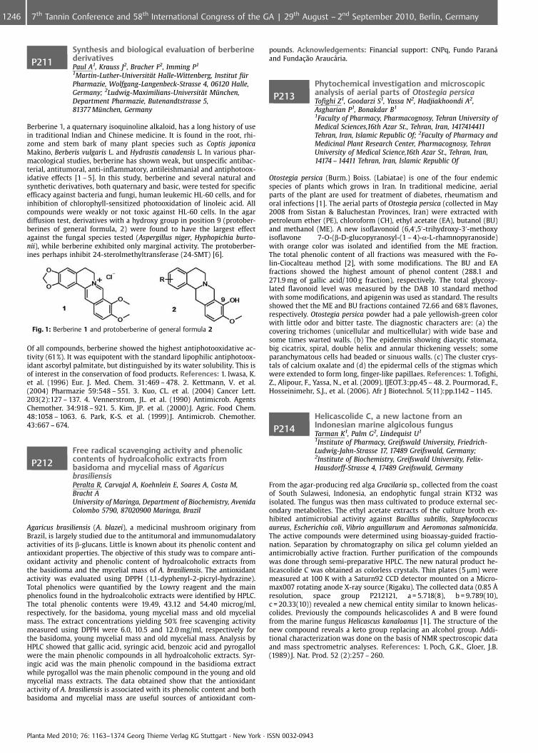
P211Synthesis and biological evaluation of berberinederivativesPaul A1, Krauss J2, Bracher F2, Imming P1
1Martin-Luther-Universit�t Halle-Wittenberg, Institut f�rPharmazie, Wolfgang-Langenbeck-Strasse 4, 06120 Halle,Germany; 2Ludwig-Maximilians-Universit�t M�nchen,Department Pharmazie, Butenandtstrasse 5,81377 M�nchen, Germany
Berberine 1, a quaternary isoquinoline alkaloid, has a long history of usein traditional Indian and Chinese medicine. It is found in the root, rhi-zome and stem bark of many plant species such as Coptis japonicaMakino, Berberis vulgaris L. and Hydrastis canadensis L. In various phar-macological studies, berberine has shown weak, but unspecific antibac-terial, antitumoral, anti-inflammatory, antileishmanial and antiphotoox-idative effects [1 – 5]. In this study, berberine and several natural andsynthetic derivatives, both quaternary and basic, were tested for specificefficacy against bacteria and fungi, human leukemic HL-60 cells, and forinhibition of chlorophyll-sensitized photooxidation of linoleic acid. Allcompounds were weakly or not toxic against HL-60 cells. In the agardiffusion test, derivatives with a hydroxy group in position 9 (protober-berines of general formula, 2) were found to have the largest effectagainst the fungal species tested (Aspergillus niger, Hyphopichia burto-nii), while berberine exhibited only marginal activity. The protoberber-ines perhaps inhibit 24-sterolmethyltransferase (24-SMT) [6].
Fig. 1: Berberine 1 and protoberberine of general formula 2
Of all compounds, berberine showed the highest antiphotooxidative ac-tivity (61%). It was equipotent with the standard lipophilic antiphotoox-idant ascorbyl palmitate, but distinguished by its water solubility. This isof interest in the conservation of food products. References: 1. Iwasa, K.et al. (1996) Eur. J. Med. Chem. 31:469 – 478. 2. Kettmann, V. et al.(2004) Pharmazie 59:548 – 551. 3. Kuo, CL. et al. (2004) Cancer Lett.203(2):127 – 137. 4. Vennerstrom, JL. et al. (1990) Antimicrob. AgentsChemother. 34:918 – 921. 5. Kim, JP. et al. (2000) J. Agric. Food Chem.48:1058 – 1063. 6. Park, K-S. et al. (1999) J. Antimicrob. Chemother.43:667 – 674.
P212Free radical scavenging activity and phenoliccontents of hydroalcoholic extracts frombasidoma and mycelial mass of AgaricusbrasiliensisPeralta R, Carvajal A, Koehnlein E, Soares A, Costa M,Bracht AUniversity of Maringa, Department of Biochemistry, AvenidaColombo 5790, 87020900 Maringa, Brazil
Agaricus brasiliensis (A. blazei), a medicinal mushroom originary fromBrazil, is largely studied due to the antitumoral and immunomudalatoryactivities of its b-glucans. Little is known about its phenolic content andantioxidant properties. The objective of this study was to compare anti-oxidant activity and phenolic content of hydroalcoholic extracts fromthe basidioma and the mycelial mass of A. brasiliensis. The antioxidantactivity was evaluated using DPPH (1,1-dyphenyl-2-picryl-hydrazine).Total phenolics were quantified by the Lowry reagent and the mainphenolics found in the hydroalcoholic extracts were identified by HPLC.The total phenolic contents were 19.49, 43.12 and 54.40 microg/ml,respectively, for the basidoma, young mycelial mass and old mycelialmass. The extract concentrations yielding 50% free scavenging activitymeasured using DPPH were 6.0, 10.5 and 12.0 mg/ml, respectively forthe basidoma, young mycelial mass and old mycelial mass. Analysis byHPLC showed that gallic acid, syringic acid, benzoic acid and pyrogallolwere the main phenolic compounds in all hydroalcoholic extracts. Syr-ingic acid was the main phenolic compound in the basidioma extractwhile pyrogallol was the main phenolic compound in the young and oldmycelial mass extracts. The data obtained show that the antioxidantactivity of A. brasiliensis is associated with its phenolic content and bothbasidoma and mycelial mass are useful sources of antioxidant com-
pounds. Acknowledgements: Financial support: CNPq, Fundo Paran�and Funda�¼o Arauc�ria.
P213Phytochemical investigation and microscopicanalysis of aerial parts of Otostegia persicaTofighi Z1, Goodarzi S1, Yassa N2, Hadjiakhoondi A2,Asgharian P1, Bonakdar B1
1Faculty of Pharmacy, Pharmacognosy, Tehran University ofMedical Sciences,16th Azar St., Tehran, Iran, 1417414411Tehran, Iran, Islamic Republic Of; 2Faculty of Pharmacy andMedicinal Plant Research Center, Pharmacognosy, TehranUniversity of Medical Science,16th Azar St., Tehran, Iran,14174 – 14411 Tehran, Iran, Islamic Republic Of
Otostegia persica (Burm.) Boiss. (Labiatae) is one of the four endemicspecies of plants which grows in Iran. In traditional medicine, aerialparts of the plant are used for treatment of diabetes, rheumatism andoral infections [1]. The aerial parts of Otostegia persica (collected in May2008 from Sistan & Baluchestan Provinces, Iran) were extracted withpetroleum ether (PE), chloroform (CH), ethyl acetate (EA), butanol (BU)and methanol (ME). A new isoflavonoid (6,4’,5’-trihydroxy-3’-methoxyisoflavone 7-O-(b-D-glucopyranosyl-(1 – 4)-a-L-rhamnopyranoside)with orange color was isolated and identified from the ME fraction.The total phenolic content of all fractions was measured with the Fo-lin-Ciocalteau method [2], with some modifications. The BU and EAfractions showed the highest amount of phenol content (288.1 and271.9 mg of gallic acid/100 g fraction), respectively. The total glycosy-lated flavonoid level was measured by the DAB 10 standard methodwith some modifications, and apigenin was used as standard. The resultsshowed thet the ME and BU fractions contained 72.66 and 68% flavones,respectively. Otostegia persica powder had a pale yellowish-green colorwith little odor and bitter taste. The diagnostic characters are: (a) thecovering trichomes (unicellular and multicellular) with wide base andsome times warted walls. (b) The epidermis showing diacytic stomata,big cicatrix, spiral, double helix and annular thickening vessels; someparanchymatous cells had beaded or sinuous walls. (c) The cluster crys-tals of calcium oxalate and (d) the epidermal cells of the stigmas whichwere extended to form long, finger-like papillaes. References: 1. Tofighi,Z., Alipour, F., Yassa, N., et al. (2009). IJEOT.3:pp.45 – 48. 2. Pourmorad, F.,Hosseinimehr, S.J., et al. (2006). Afr J Biotechnol. 5(11):pp.1142 – 1145.
P214Helicascolide C, a new lactone from anIndonesian marine algicolous fungusTarman K1, Palm G2, Lindequist U1
1Institute of Pharmacy, Greifswald University, Friedrich-Ludwig-Jahn-Strasse 17, 17489 Greifswald, Germany;2Institute of Biochemistry, Greifswald University, Felix-Hausdorff-Strasse 4, 17489 Greifswald, Germany
From the agar-producing red alga Gracilaria sp., collected from the coastof South Sulawesi, Indonesia, an endophytic fungal strain KT32 wasisolated. The fungus was then mass cultivated to produce external sec-ondary metabolites. The ethyl acetate extracts of the culture broth ex-hibited antimicrobial activity against Bacillus subtilis, Staphylococcusaureus, Escherichia coli, Vibrio anguillarum and Aeromonas salmonicida.The active compounds were determined using bioassay-guided fractio-nation. Separation by chromatography on silica gel column yielded anantimicrobially active fraction. Further purification of the compoundswas done through semi-preparative HPLC. The new natural product he-licascolide C was obtained as colorless crystals. Thin plates (5 mm) weremeasured at 100 K with a Saturn92 CCD detector mounted on a Micro-max007 rotating anode X-ray source (Rigaku). The collected data (0.85 �resolution, space group P212121, a = 5.718(8), b = 9.789(10),c = 20.33(10)) revealed a new chemical entity similar to known helicas-colides. Previously the compounds helicascolides A and B were foundfrom the marine fungus Helicascus kanaloanus [1]. The structure of thenew compound reveals a keto group replacing an alcohol group. Addi-tional characterization was done on the basis of NMR spectroscopic dataand mass spectrometric analyses. References: 1. Poch, G.K., Gloer, J.B.(1989) J. Nat. Prod. 52 (2):257 – 260.
Planta Med 2010; 76: 1163–1374 Georg Thieme Verlag KG Stuttgart · New York · ISSN 0032-0943
1246 7th Tannin Conference and 58th International Congress of the GA | 29th August – 2nd September 2010, Berlin, Germany
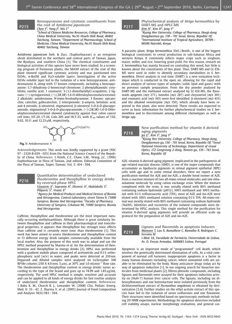
P215Benzopyranone and cytotoxic constituents fromthe root of Antidesma japonicumChen Y1, Yang J2, Wu C1
1School of Chinese Medicine Resources, College of Pharmacy,China Medical University, No.91 Hsueh-Shih Road, 40402Taichung, Taiwan; 2Department of Pharmacology, School ofMedicine, China Medical University, No.91 Hsueh-Shih Road,40402 Taichung, Taiwan
Antidesma japonicum Sieb. & Zucc. (Euphorbiaceae) is an evergreenshrub distributed in the northern and central parts of Taiwan, Japan,the Ryukyus, and southern China [1]. The chemical constituents andbiological activities of this species have never been studied. In a screen-ing program of Formosan plants, the MeOH extract of the root of thisplant showed significant cytotoxic activity and was partitioned intoEtOAc, n-BuOH and H2O-soluble layers. Investigation of the activeEtOAc-soluble layer led to the isolation of a new benzopyranone, anti-desmanone A, along with 17 known compounds, including a benzopyr-anone: 5,7-dihydroxy-2-heneicosyl-chromone; 2 phenylalkanoids: cory-lifolin, vanillic acid; 1 coumarin: 3-(1,1-dimethylallyl)-scopoletin; 2 lig-nans: (�)-syringaresinol, (�)-(2R,3R)-1,4-O-diferuloylsecoisolariciresinol;2 alkaloids: antidesmone, N-trans-feruloyltyramine; 3 flavans: epicate-chin, catechin, gallocatechin; 2 triterpenoids: b-amyrin, betulinic acid,and 4 steroids: b-sitosterol, stigmasterol, b-sitosteryl 3-O-b-D-glucopyr-anoside, stigmasteryl 3-O-b-D-glucopyranoside. (�)-(2R,3R)-1,4-O-Difer-uloylsecoisolariciresinol showed cytotoxicity against four colon cancercell lines, HT-29, CT-26, Colo 205 and HCT-25, with IC50 values of 7.02,9.62, 10.11 and 12.25mM, respectively.
Fig. 1: Antidesmanone A
Acknowledgements: This work was kindly supported by a grant (NSC97 – 2320-B-039 – 025) from the National Science Council of the Repub-lic of China. References: 1. Hsieh, C.F., Chaw, S.M., Wang, J.C. (1996)Euphorbiaceae in Flora of Taiwan, 2nd edition. Editorial Committee ofthe Flora of Taiwan, Taipei, Taiwan: Vol. 3: 414 – 504.
P216Quantitative determination of undeclaredtheobromine and theophylline in energy drinksby reversed-phase HPLCUzunovic A1, Sapcanin A2, Elezovic A1, Hadzidedic S1,Pilipovic S1, Vranic E2
1Agency for Medical Products and Medical Devices of Bosniaand Herzegovina, Control Laboratory, street Titova 9, 71000Sarajevo, Bosnia And Herzegovina; 2Faculty of Pharmacy,University of Sarajevo, Cekalusa 90, 71000 Sarajevo, BosniaAnd Herzegovina
Caffeine, theophylline and theobromine are the most important natu-rally occurring methylxanthines. Although there is great similarity be-tween theophylline and caffeine in their pharmacological and toxicolo-gical properties, it appears that theophylline has stronger toxic effectsthan caffeine and is certainly more toxic than theobromine [1]. Thiswork has been aimed to assess theobromine and theophylline contentin 13 different energy drink samples commercially available from thelocal market. Also, the purpose of this work was to adapt and use theHPLC method proposed by Sharma et al. for the determination of theo-bromine and theophylline in energy drinks [2]. HPLC was performedwith a gradient mobile phase composed of acetonitrile and 0.1% ortho-phosphoric acid (w/v) in water, and peaks were detected at 210 nm.Degassed and diluted samples were analysed on Lichrospher 100RP18e column (250 X 4.0 mm, 5 mm), at 300C and 1.0 mLmin�1 flow rate.The theobromine and theophylline content in energy drinks varies ac-cording to the type of the brand and goes up to 74.99 and 1.85 mg/mL,respectively. The used HPLC method is simple, sensitive and accurateand can be applied to all kinds of energy drinks for fast routine analysisof undeclared theobromine and theophylline content. References:1. Babu K. M., Church R. J., Lewander W. (2008) Clin. Pediatr. Emerg.Med. 9: 35 – 42. 2. Sharma V. et al. (2005) Journal of Food Compositionand Analysis 18(6):583 – 594.
P217Phytochemical analysis of Striga hermonthica byDART-MS and HPLC-MSKim H1, Kim D2, Jang Y1
1Kyung Hee University, College of Pharmacy, Hoegi-dongDongdaemun-gu, 130 – 701 Seoul, Korea, Republic Of;2International Institute of Tropical Agriculture, 30709 –00100 NairobIi, Kenya
A parasitic plant, Striga hermonthica (Del.) Benth., is one of the biggestbiological constraints to cereal production in sub-Saharan Africa andwestern Kenya. It commonly infects major crops such as sorghum,maize, millet, and rice, lowering grain yield. For this reason, reseach onS. hermonthica has mainly focused on controling this weed, but little isknown about the constituents of this plant. Thus, DART-MS and LC-ESI-MS were used in order to identify secondary metabolites in S. her-monthica. Direct analysis in real time (DART) is a new ionization tech-nique which is conducted in the open air, allowing for the rapid anddirect analysis of various types of samples (solid, lipid and gases) withno previous sample preparation. From the dry powder analysed byDART-MS and the methanol extract analysed by LC-ESI-MS, the flavo-noids apigenin (m/z 271), luteolin (m/z 287) and chrysoeriol (m/z 301)and coumaric acid (m/z 165) were first identified. b-sitosterol (m/z 415)and the alkaloid venoterpine (m/z 150), which already have been re-ported in this plant, also were detected. These results are expected toserve as basic information for further phytochemical studies on S. her-monthica and to discriminate among different chemotypes as well asStriga spp.
P218New purification method for vitamin A derivedaging pigmentsJee E1, Kim S2, Jang Y1
1Kyung Hee University, College of Pharmacy, Hoegi-dong,Dongdaemun-gu, 130 – 701 Seoul, Korea, Republic Of; 2SeoulNational University of Technology, Department of visualOptics, 172 Gongreung 2-dong, Nowon-gu, 139 – 743 Seoul,Korea, Republic Of
A2E, vitamin A-derived aging pigment, implicated in the pathogenesis ofage-related macular disease (AMD), is one of the major compounds thataccumulate as lipofuscin pigments in retinal pigment epithelial (RPE)cells with age and in some retinal disorders. Here we report a newpurification method for A2E and iso-A2E, a double bond isomer of A2E,from a reaction mixture of two all-trans-retinal molecules and one etha-nolamine molecule by using cation exchange resin. When the mixturecomplexed with the resin, it was serially eluted with 80% methanolcontaining sodium hydroxide (pH12), 100% methanol and 100% metha-nol with 0.1% trifluoroacetic acid (TFA), most of A2E and iso-A2E wereeluted with 100% methanol solution containing 0.1% TFA. All-trans-ret-inal was mostly eluted with 80% methanol containing sodium hydroxide(NaOH). Identities and recoveries of the isolated compounds were de-termined by HPLC analysis. This single method for the purification forvitamin A-derived aging pigments will provide an efficient scale upprotocol for the preparation of A2E and iso-A2E.
P219Lignans and flavonoids as apoptosis inducersMansoor T, Luo X, Ramalhete C, Ramalho R, Rodrigues C,Ferreira Mi-Med. UL, Faculdade de Farm�cia, Universidade de Lisboa,Av. D. For�as Armadas, 1600083 Lisboa, Portugal
Apoptosis is an important mode of “programmed” cell death, whichinvolves the genetically determined elimination of cells. As a vital com-ponent of normal cell turnover, inappropriate apoptosis is a factor inmany human diseases including cancer, where unwanted cells are un-able to be eliminated by the body. Many anticancer drugs today act byway of apoptosis induction [1]. In our ongoing search for bioactive mo-lecules from medicinal plants [2], fifteen phenolic compounds, includinglignans and flavonoids were assayed for their apoptosis induction activ-ity on (HuH-7) human liver cancer cells. The lignans, including six di-benzylbutanes and one butirolactone were isolated previously from thedichloromethane extract of Pycnanthus angolensis or obtained by deri-vatization [3,4]. Further studies on the ethyl acetate extract of this spe-cies have led to the isolation of seven isoflavones and one flavanone.Their structures were identified based on spectroscopic methods includ-ing 2D NMR experiments. Methodology for apoptosis detection includedcell viability assays, nuclear morphology evaluation, and general cas-
Planta Med 2010; 76: 1163–1374 Georg Thieme Verlag KG Stuttgart · New York · ISSN 0032-0943
12477th Tannin Conference and 58th International Congress of the GA | 29th August – 2nd September 2010, Berlin, Germany
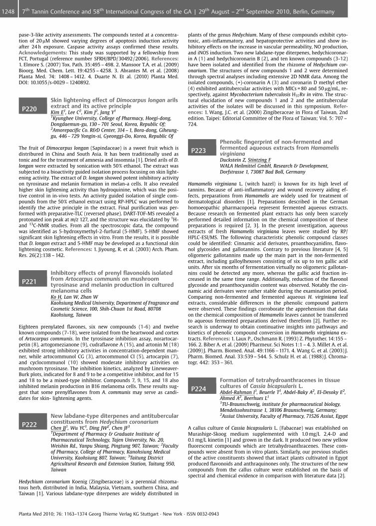
pase-3-like activity assessments. The compounds tested at a concentra-tion of 20mM showed varying degrees of apoptosis induction activityafter 24 h exposure. Caspase activity assays confirmed these results.Acknowledgements: This study was supported by a fellowship fromFCT, Portugal (reference number SFRH/BPD/30492/2006). References:1. Elmore S. (2007) Tox. Path. 35:495 – 498. 2. Mansoor T.A. et al. (2009)Bioorg. Med. Chem. Lett. 19:4255 – 4258. 3. Abrantes M. et al. (2008)Planta Med. 74: 1408 – 1412. 4. Duarte N. Et al. (2010) Planta Med.DOI: 10.1055/s-0029 – 1240892.
P220Skin lightening effect of Dimocarpus longan arilsextract and its active principleKim E1, Lee C2, Kim J2, Jang Y1
1Kyunghee University, College of Pharmacy, Hoegi-dong,Dongdaemun-gu, 130 – 701 Seoul, Korea, Republic Of;2Amorepacific Co. R&D Center, 314 – 1, Bora-dong, Giheung-gu, 446 – 729 Yongin-si, Gyeonggi-Do, Korea, Republic Of
The fruit of Dimocarpus longan (Sapindaceae) is a sweet fruit which isdistribured in China and South Asia. It has been traditionally used astonic and for the treatment of amnesia and insomnia [1]. Dried arils of D.longan were extracted by sonication with 50% ethanol. The extract wassubjected to a bioactivity guided isolation process focusing on skin light-ening activity. The extract of D. longan showed potent inhibitory activityon tyrosinase and melanin formation in melan-a cells. It also revealedhigher skin lightening activity than hydroquinine, which was the posi-tive control in in-vivo tests. An activity guided isolation of single com-pounds from the 50% ethanol extract using RP-HPLC was performed toidentify the active principle in the extract. Final purification was per-formed with preparative-TLC (reversed phase). DART-TOF-MS revealed aprotonated ion peak at m/z 127, and the structure was elucidated by 1H-and 13C-NMR studies. From all the spectroscopic data, the compoundwas identified as 5-hydroxymethyl-2-furfural (5-HMF). 5-HMF showedsignificant skin lightening effects in vitro. From the results, it is possiblethat D. longan extract and 5-HMF may be developed as a functional skinlightening cosmetic. References: 1. Jiyoung, R. et al. (2003) Arch. Pham.Res. 26(2):138 – 142.
P221Inhibitory effects of prenyl flavonoids isolatedfrom Artocarpus communis on mushroomtyrosinase and melanin production in culturedmelanoma cellsKo H, Lan W, Zhan WKaohsiung Medical University, Department of Fragrance andCosmetic Science, 100, Shih-Chuan 1st Road, 80708Kaohsiung, Taiwan
Eighteen prenylated flavones, six new compounds (1-6) and twelveknown compounds (7-18), were isolated from the heartwood and cortexof Artocarpus communis. In the tyrosinase inhibition assay, norartocar-petin (8), artogomezianone (9), cudraflavone A (15), and artonin M (18)exhibited strong inhibitory activities in concentration-dependent man-ner, while artocommunol CG (3), artocommunol CI (5), artocarpin (7),and cyclocommunol (10) showed moderate inhibitory activities onmushroom tyrosinase. The inhibition kinetics, analyzed by Lineweaver-Burk plots, indicated for 8 and 9 to be a competitive inhibitor, and for 15and 18 to be a mixed-type inhibitor. Compounds 7, 9, 15, and 18 alsoinhibited melanin production in B16 melanoma cells. These results sug-gest that some prenylflavones from A. communis may serve as candi-dates for skin- lightening agents.
P222New labdane-type diterpenes and antitubercularconstituents from Hedychium coronariumChen JJ1, Wu YC1, Ding JW2, Chen JF3
1Department of Pharmacy & Graduate Institute ofPharmaceutical Technology, Tajen University, No. 20,Weishin Rd., Yanpu Shiang, Pingtung 907, Taiwan; 2Facultyof Pharmacy, College of Pharmacy, Kanohsiung MedicalUniversity, Kaohsiung 807, Taiwan; 3Taitung DistrictAgricultural Research and Extension Station, Taitung 950,Taiwan
Hedychium coronarium Koenig (Zingiberaceae) is a perennial rhizoma-tous herb, distributed in India, Malaysia, Vietnam, southern China, andTaiwan [1]. Various labdane-type diterpenes are widely distributed in
plants of the genus Hedychium. Many of these compounds exhibit cyto-toxic, anti-inflammatory, and hepatoprotective activities and show in-hibitory effects on the increase in vascular permeability, NO production,and iNOS induction. Two new labdane-type diterpenes, hedychicoronar-in A (1) and hedychicoronarin B (2), and ten known compounds (3-12)have been isolated and identified from the rhizome of Hedychium cor-onarium. The structures of new compounds 1 and 2 were determinedthrough spectral analyses including extensive 2D NMR data. Among theisolated compounds, (+)-coronarin A (3) and coronarin D methyl ether(4) exhibited antitubercular activities with MICs = 80 and 50 mg/mL, re-spectively, against Mycobacterium tuberculosis H37Rv in vitro. The struc-tural elucidation of new compounds 1 and 2 and the antitubercularactivities of the isolates will be discussed in this symposium. Refer-ences: 1. Wang, J.C. et al. (2000) Zingiberaceae in Flora of Taiwan, 2ndedition. Taipei: Editorial Committee of the Flora of Taiwan; Vol. 5: 707 –724.
P223Phenolic fingerprint of non-fermented andfermented aqueous extracts from HamamelisvirginianaDuckstein Z, Stintzing FWALA Heilmittel GmbH, Research & Development,Dorfstrasse 1, 73087 Bad Boll, Germany
Hamamelis virginiana L. (witch hazel) is known for its high level oftannins. Because of anti-inflammatory and wound recovery aiding ef-fects, preparations from Hamamelis are widely used for treatment ofdermatological disorders [1]. Preparations described in the Germanhomoeopathic pharmacopoeia represent fermented aqueous extracts.Because research on fermented plant extracts has only been scarcelyperformed detailed information on the chemical composition of thesepreparations is required [2, 3]. In the present investigation, aqueousextracts of fresh Hamamelis virginiana leaves were studied by RP/HPLC-ESI/MS. The following characteristic phenolic compound classescould be identified: Cinnamic acid derivates, proanthocyanidins, flavo-nol glycosides and gallotannins. Contrary to previous literature [4, 5]oligomeric gallotannins made up the main part in the non-fermentedextract, including galloylhexoses consisting of six up to ten gallic acidunits. After six months of fermentation virtually no oligomeric gallotan-nins could be detected any more, whereas the gallic acid fraction in-creased in the same time range. Additionally, reduction of the flavonolglycoside and proanthocyanidin content was observed. Notably the cin-namic acid derivates were rather stable during the examination period.Comparing non-fermented and fermented aqueous H. virginiana leafextracts, considerable differences in the phenolic compound patternwere observed. These findings corroborate the apprehension that dataon the chemical composition of Hamamelis leaves cannot be transferredto aqueous fermented preparations derived therefrom [2]. Further re-search is underway to obtain continuative insights into pathways andkinetics of phenolic compound conversion in Hamamelis virginiana ex-tracts. References: 1. Laux P., Oschmann R. (1993) Z. Phytother. 14:155 –166. 2. Biber A. et al. (2009) Pharmeur. Sci Notes 1:1 – 4. 3. Millet A. et al.(2009) J. Pharm. Biomed. Anal. 49:1166 – 1171. 4. Wang G. et al. (2003) J.Pharm. Biomed. Anal. 33:539 – 544. 5. Schulz H. et al. (1988) J. Chroma-togr. 442: 353 – 361.
P224Formation of tetrahydroanthracenes in tissuecultures of Cassia bicapsularis L.Abdel-Rahman I1, Beuerle T1, Abdel-Baky A2, El-Desoky E2,Ahmed A2, Beerhues L1
1TU-Braunschweig, institute for pharmaceutical biology,Mendelssohnstrasse 1, 38106 Braunschweig, Germany;2Assiut University, Faculty of Pharmacy, 71526 Assiut, Egypt
A callus culture of Cassia bicapsularis L. (Fabaceae) was established onMurashige-Skoog medium supplemented with 1.0 mg/L 2,4-D and0.1 mg/L kinetin [1] and grown in the dark. It produced two new yellowfluorescent compounds which are tetrahydroanthracenes. These com-pounds were absent from in vitro plants. Similarly, our previous studiesof the active constituents showed that intact plants cultivated in Egyptproduced flavonoids and anthraquinones only. The structures of the newcompounds from the callus culture were established on the basis ofspectral and chemical evidence in comparison with literature data [2].
Planta Med 2010; 76: 1163–1374 Georg Thieme Verlag KG Stuttgart · New York · ISSN 0032-0943
1248 7th Tannin Conference and 58th International Congress of the GA | 29th August – 2nd September 2010, Berlin, Germany
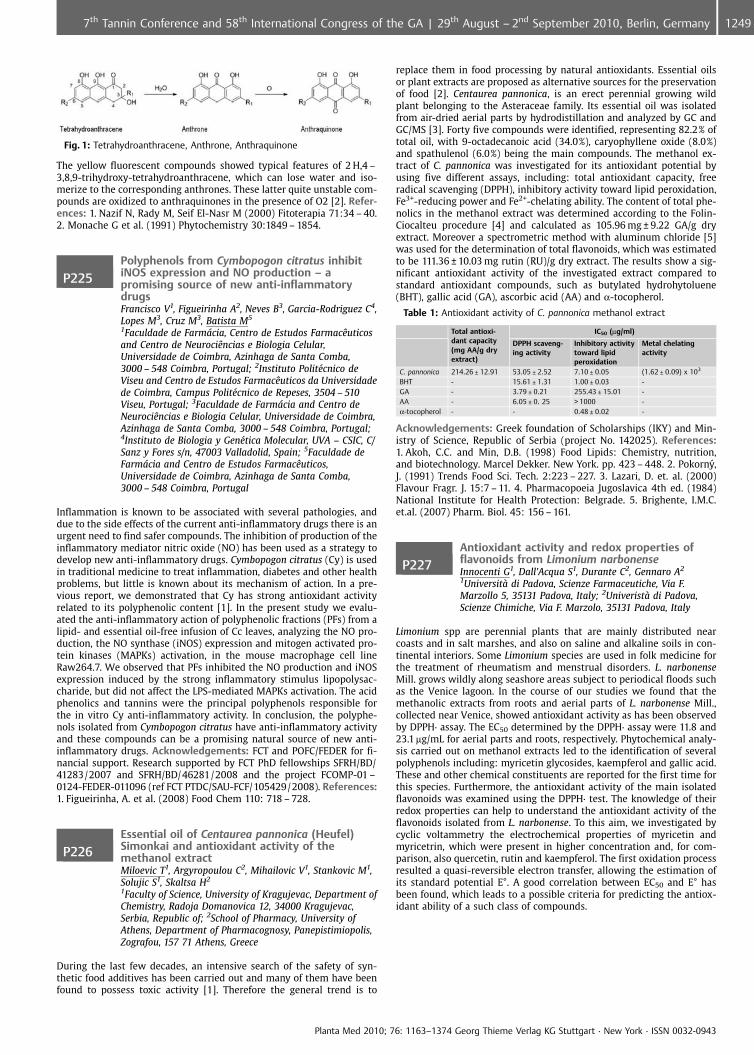
Fig. 1: Tetrahydroanthracene, Anthrone, Anthraquinone
The yellow fluorescent compounds showed typical features of 2 H,4 –3,8,9-trihydroxy-tetrahydroanthracene, which can lose water and iso-merize to the corresponding anthrones. These latter quite unstable com-pounds are oxidized to anthraquinones in the presence of O2 [2]. Refer-ences: 1. Nazif N, Rady M, Seif El-Nasr M (2000) Fitoterapia 71:34 – 40.2. Monache G et al. (1991) Phytochemistry 30:1849 – 1854.
P225Polyphenols from Cymbopogon citratus inhibitiNOS expression and NO production – apromising source of new anti-inflammatorydrugsFrancisco V1, Figueirinha A2, Neves B3, Garcia-Rodriguez C4,Lopes M3, Cruz M3, Batista M5
1Faculdade de Farm�cia, Centro de Estudos FarmacÞuticosand Centro de NeurociÞncias e Biologia Celular,Universidade de Coimbra, Azinhaga de Santa Comba,3000 – 548 Coimbra, Portugal; 2Instituto Polit�cnico deViseu and Centro de Estudos FarmacÞuticos da Universidadede Coimbra, Campus Polit�cnico de Repeses, 3504 – 510Viseu, Portugal; 3Faculdade de Farm�cia and Centro deNeurociÞncias e Biologia Celular, Universidade de Coimbra,Azinhaga de Santa Comba, 3000 – 548 Coimbra, Portugal;4Instituto de Biologia y Gen�tica Molecular, UVA – CSIC, C/Sanz y Fores s/n, 47003 Valladolid, Spain; 5Faculdade deFarm�cia and Centro de Estudos FarmacÞuticos,Universidade de Coimbra, Azinhaga de Santa Comba,3000 – 548 Coimbra, Portugal
Inflammation is known to be associated with several pathologies, anddue to the side effects of the current anti-inflammatory drugs there is anurgent need to find safer compounds. The inhibition of production of theinflammatory mediator nitric oxide (NO) has been used as a strategy todevelop new anti-inflammatory drugs. Cymbopogon citratus (Cy) is usedin traditional medicine to treat inflammation, diabetes and other healthproblems, but little is known about its mechanism of action. In a pre-vious report, we demonstrated that Cy has strong antioxidant activityrelated to its polyphenolic content [1]. In the present study we evalu-ated the anti-inflammatory action of polyphenolic fractions (PFs) from alipid- and essential oil-free infusion of Cc leaves, analyzing the NO pro-duction, the NO synthase (iNOS) expression and mitogen activated pro-tein kinases (MAPKs) activation, in the mouse macrophage cell lineRaw264.7. We observed that PFs inhibited the NO production and iNOSexpression induced by the strong inflammatory stimulus lipopolysac-charide, but did not affect the LPS-mediated MAPKs activation. The acidphenolics and tannins were the principal polyphenols responsible forthe in vitro Cy anti-inflammatory activity. In conclusion, the polyphe-nols isolated from Cymbopogon citratus have anti-inflammatory activityand these compounds can be a promising natural source of new anti-inflammatory drugs. Acknowledgements: FCT and POFC/FEDER for fi-nancial support. Research supported by FCT PhD fellowships SFRH/BD/41283/2007 and SFRH/BD/46281/2008 and the project FCOMP-01 –0124-FEDER-011096 (ref FCT PTDC/SAU-FCF/105429/2008). References:1. Figueirinha, A. et al. (2008) Food Chem 110: 718 – 728.
P226Essential oil of Centaurea pannonica (Heufel)Simonkai and antioxidant activity of themethanol extractMiloevic T1, Argyropoulou C2, Mihailovic V1, Stankovic M1,Solujic S1, Skaltsa H2
1Faculty of Science, University of Kragujevac, Department ofChemistry, Radoja Domanovica 12, 34000 Kragujevac,Serbia, Republic of; 2School of Pharmacy, University ofAthens, Department of Pharmacognosy, Panepistimiopolis,Zografou, 157 71 Athens, Greece
During the last few decades, an intensive search of the safety of syn-thetic food additives has been carried out and many of them have beenfound to possess toxic activity [1]. Therefore the general trend is to
replace them in food processing by natural antioxidants. Essential oilsor plant extracts are proposed as alternative sources for the preservationof food [2]. Centaurea pannonica, is an erect perennial growing wildplant belonging to the Asteraceae family. Its essential oil was isolatedfrom air-dried aerial parts by hydrodistillation and analyzed by GC andGC/MS [3]. Forty five compounds were identified, representing 82.2% oftotal oil, with 9-octadecanoic acid (34.0%), caryophyllene oxide (8.0%)and spathulenol (6.0%) being the main compounds. The methanol ex-tract of C. pannonica was investigated for its antioxidant potential byusing five different assays, including: total antioxidant capacity, freeradical scavenging (DPPH), inhibitory activity toward lipid peroxidation,Fe3+-reducing power and Fe2+-chelating ability. The content of total phe-nolics in the methanol extract was determined according to the Folin-Ciocalteu procedure [4] and calculated as 105.96 mg € 9.22 GA/g dryextract. Moreover a spectrometric method with aluminum chloride [5]was used for the determination of total flavonoids, which was estimatedto be 111.36 € 10.03 mg rutin (RU)/g dry extract. The results show a sig-nificant antioxidant activity of the investigated extract compared tostandard antioxidant compounds, such as butylated hydrohytoluene(BHT), gallic acid (GA), ascorbic acid (AA) and a-tocopherol.
Table 1: Antioxidant activity of C. pannonica methanol extract
Total antioxi-dant capacity(mg AA/g dryextract)
IC50 (mg/ml)
DPPH scaveng-ing activity
Inhibitory activitytoward lipidperoxidation
Metal chelatingactivity
C. pannonica 214.26 € 12.91 53.05 € 2.52 7.10 € 0.05 (1.62 € 0.09) x 103
BHT - 15.61 € 1.31 1.00 € 0.03 -GA - 3.79 € 0.21 255.43 € 15.01 -AA - 6.05 € 0. 25 > 1000 -a-tocopherol - - 0.48 € 0.02 -
Acknowledgements: Greek foundation of Scholarships (IKY) and Min-istry of Science, Republic of Serbia (project No. 142025). References:1. Akoh, C.C. and Min, D.B. (1998) Food Lipids: Chemistry, nutrition,and biotechnology. Marcel Dekker. New York. pp. 423 – 448. 2. Pokorny,J. (1991) Trends Food Sci. Tech. 2:223 – 227. 3. Lazari, D. et. al. (2000)Flavour Fragr. J. 15:7 – 11. 4. Pharmacopoeia Jugoslavica 4th ed. (1984)National Institute for Health Protection: Belgrade. 5. Brighente, I.M.C.et.al. (2007) Pharm. Biol. 45: 156 – 161.
P227Antioxidant activity and redox properties offlavonoids from Limonium narbonenseInnocenti G1, Dall’Acqua S1, Durante C2, Gennaro A2
1Universit di Padova, Scienze Farmaceutiche, Via F.Marzollo 5, 35131 Padova, Italy; 2Univerist di Padova,Scienze Chimiche, Via F. Marzolo, 35131 Padova, Italy
Limonium spp are perennial plants that are mainly distributed nearcoasts and in salt marshes, and also on saline and alkaline soils in con-tinental interiors. Some Limonium species are used in folk medicine forthe treatment of rheumatism and menstrual disorders. L. narbonenseMill. grows wildly along seashore areas subject to periodical floods suchas the Venice lagoon. In the course of our studies we found that themethanolic extracts from roots and aerial parts of L. narbonense Mill.,collected near Venice, showed antioxidant activity as has been observedby DPPH· assay. The EC50 determined by the DPPH· assay were 11.8 and23.1 mg/mL for aerial parts and roots, respectively. Phytochemical analy-sis carried out on methanol extracts led to the identification of severalpolyphenols including: myricetin glycosides, kaempferol and gallic acid.These and other chemical constituents are reported for the first time forthis species. Furthermore, the antioxidant activity of the main isolatedflavonoids was examined using the DPPH· test. The knowledge of theirredox properties can help to understand the antioxidant activity of theflavonoids isolated from L. narbonense. To this aim, we investigated bycyclic voltammetry the electrochemical properties of myricetin andmyricetrin, which were present in higher concentration and, for com-parison, also quercetin, rutin and kaempferol. The first oxidation processresulted a quasi-reversible electron transfer, allowing the estimation ofits standard potential E�. A good correlation between EC50 and E� hasbeen found, which leads to a possible criteria for predicting the antiox-idant ability of a such class of compounds.
Planta Med 2010; 76: 1163–1374 Georg Thieme Verlag KG Stuttgart · New York · ISSN 0032-0943
12497th Tannin Conference and 58th International Congress of the GA | 29th August – 2nd September 2010, Berlin, Germany
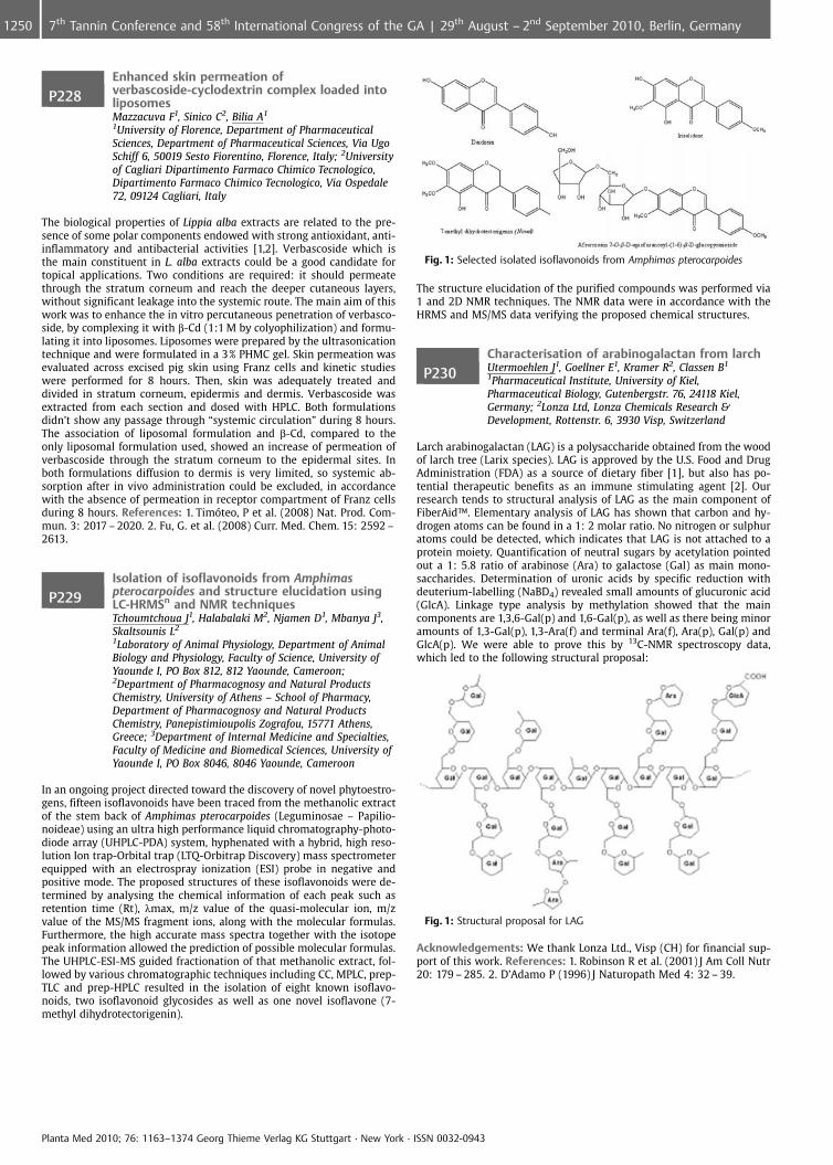
P228Enhanced skin permeation ofverbascoside-cyclodextrin complex loaded intoliposomesMazzacuva F1, Sinico C2, Bilia A1
1University of Florence, Department of PharmaceuticalSciences, Department of Pharmaceutical Sciences, Via UgoSchiff 6, 50019 Sesto Fiorentino, Florence, Italy; 2Universityof Cagliari Dipartimento Farmaco Chimico Tecnologico,Dipartimento Farmaco Chimico Tecnologico, Via Ospedale72, 09124 Cagliari, Italy
The biological properties of Lippia alba extracts are related to the pre-sence of some polar components endowed with strong antioxidant, anti-inflammatory and antibacterial activities [1,2]. Verbascoside which isthe main constituent in L. alba extracts could be a good candidate fortopical applications. Two conditions are required: it should permeatethrough the stratum corneum and reach the deeper cutaneous layers,without significant leakage into the systemic route. The main aim of thiswork was to enhance the in vitro percutaneous penetration of verbasco-side, by complexing it with b-Cd (1:1 M by colyophilization) and formu-lating it into liposomes. Liposomes were prepared by the ultrasonicationtechnique and were formulated in a 3% PHMC gel. Skin permeation wasevaluated across excised pig skin using Franz cells and kinetic studieswere performed for 8 hours. Then, skin was adequately treated anddivided in stratum corneum, epidermis and dermis. Verbascoside wasextracted from each section and dosed with HPLC. Both formulationsdidn’t show any passage through “systemic circulation” during 8 hours.The association of liposomal formulation and b-Cd, compared to theonly liposomal formulation used, showed an increase of permeation ofverbascoside through the stratum corneum to the epidermal sites. Inboth formulations diffusion to dermis is very limited, so systemic ab-sorption after in vivo administration could be excluded, in accordancewith the absence of permeation in receptor compartment of Franz cellsduring 8 hours. References: 1. Tim teo, P et al. (2008) Nat. Prod. Com-mun. 3: 2017 – 2020. 2. Fu, G. et al. (2008) Curr. Med. Chem. 15: 2592 –2613.
P229Isolation of isoflavonoids from Amphimaspterocarpoides and structure elucidation usingLC-HRMSn and NMR techniquesTchoumtchoua J1, Halabalaki M2, Njamen D1, Mbanya J3,Skaltsounis L2
1Laboratory of Animal Physiology, Department of AnimalBiology and Physiology, Faculty of Science, University ofYaounde I, PO Box 812, 812 Yaounde, Cameroon;2Department of Pharmacognosy and Natural ProductsChemistry, University of Athens – School of Pharmacy,Department of Pharmacognosy and Natural ProductsChemistry, Panepistimioupolis Zografou, 15771 Athens,Greece; 3Department of Internal Medicine and Specialties,Faculty of Medicine and Biomedical Sciences, University ofYaounde I, PO Box 8046, 8046 Yaounde, Cameroon
In an ongoing project directed toward the discovery of novel phytoestro-gens, fifteen isoflavonoids have been traced from the methanolic extractof the stem back of Amphimas pterocarpoides (Leguminosae – Papilio-noideae) using an ultra high performance liquid chromatography-photo-diode array (UHPLC-PDA) system, hyphenated with a hybrid, high reso-lution Ion trap-Orbital trap (LTQ-Orbitrap Discovery) mass spectrometerequipped with an electrospray ionization (ESI) probe in negative andpositive mode. The proposed structures of these isoflavonoids were de-termined by analysing the chemical information of each peak such asretention time (Rt), lmax, m/z value of the quasi-molecular ion, m/zvalue of the MS/MS fragment ions, along with the molecular formulas.Furthermore, the high accurate mass spectra together with the isotopepeak information allowed the prediction of possible molecular formulas.The UHPLC-ESI-MS guided fractionation of that methanolic extract, fol-lowed by various chromatographic techniques including CC, MPLC, prep-TLC and prep-HPLC resulted in the isolation of eight known isoflavo-noids, two isoflavonoid glycosides as well as one novel isoflavone (7-methyl dihydrotectorigenin).
Fig. 1: Selected isolated isoflavonoids from Amphimas pterocarpoides
The structure elucidation of the purified compounds was performed via1 and 2D NMR techniques. The NMR data were in accordance with theHRMS and MS/MS data verifying the proposed chemical structures.
P230Characterisation of arabinogalactan from larchUtermoehlen J1, Goellner E1, Kramer R2, Classen B1
1Pharmaceutical Institute, University of Kiel,Pharmaceutical Biology, Gutenbergstr. 76, 24118 Kiel,Germany; 2Lonza Ltd, Lonza Chemicals Research &Development, Rottenstr. 6, 3930 Visp, Switzerland
Larch arabinogalactan (LAG) is a polysaccharide obtained from the woodof larch tree (Larix species). LAG is approved by the U.S. Food and DrugAdministration (FDA) as a source of dietary fiber [1], but also has po-tential therapeutic benefits as an immune stimulating agent [2]. Ourresearch tends to structural analysis of LAG as the main component ofFiberAid�. Elementary analysis of LAG has shown that carbon and hy-drogen atoms can be found in a 1: 2 molar ratio. No nitrogen or sulphuratoms could be detected, which indicates that LAG is not attached to aprotein moiety. Quantification of neutral sugars by acetylation pointedout a 1: 5.8 ratio of arabinose (Ara) to galactose (Gal) as main mono-saccharides. Determination of uronic acids by specific reduction withdeuterium-labelling (NaBD4) revealed small amounts of glucuronic acid(GlcA). Linkage type analysis by methylation showed that the maincomponents are 1,3,6-Gal(p) and 1,6-Gal(p), as well as there being minoramounts of 1,3-Gal(p), 1,3-Ara(f) and terminal Ara(f), Ara(p), Gal(p) andGlcA(p). We were able to prove this by 13C-NMR spectroscopy data,which led to the following structural proposal:
Fig. 1: Structural proposal for LAG
Acknowledgements: We thank Lonza Ltd., Visp (CH) for financial sup-port of this work. References: 1. Robinson R et al. (2001) J Am Coll Nutr20: 179 – 285. 2. D’Adamo P (1996) J Naturopath Med 4: 32 – 39.
Planta Med 2010; 76: 1163–1374 Georg Thieme Verlag KG Stuttgart · New York · ISSN 0032-0943
1250 7th Tannin Conference and 58th International Congress of the GA | 29th August – 2nd September 2010, Berlin, Germany
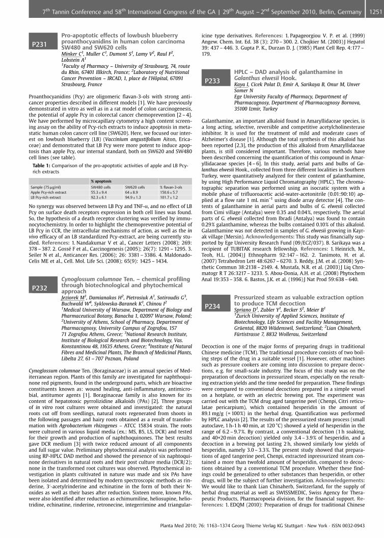
P231Pro-apoptotic effects of lowbush blueberryproanthocyanidins in human colon carcinomaSW480 and SW620 cellsMinker C1, Muller C1, Dumont S1, Lamy V2, Raul F2,Lobstein A1
1Faculty of Pharmacy – University of Strasbourg, 74, routedu Rhin, 67401 Illkirch, France; 2Laboratory of NutritionalCancer Prevention – IRCAD, 1, place de l’H�pital, 67091Strasbourg, France
Proanthocyanidins (Pcy) are oligomeric flavan-3-ols with strong anti-cancer properties described in different models [1]. We have previouslydemonstrated in vitro as well as in a rat model of colon carcinogenesis,the potential of apple Pcy in colorectal cancer chemoprevention [2 – 4].We have performed by microcapillary cytometry a high content screen-ing assay on the ability of Pcy-rich extracts to induce apoptosis in meta-static human colon cancer cell line (SW620). Here, we focused our inter-est on lowbush blueberry (LB) (Vaccinium angustifolium Aiton, Erica-ceae) and demonstrated that LB Pcy were more potent to induce apop-tosis than apple Pcy, our internal standard, both on SW620 and SW480cell lines (see table).
Table 1: Comparison of the pro-apoptotic activities of apple and LB Pcy-rich extracts
% apoptosis
Sample (75 mg/ml) SW480 cells SW620 cells % flavan-3-olsApple Pcy-rich extract 55.3 € 9.4 64 € 8.9 150.6 € 5.7LB Pcy-rich extract 92.3 € 6.1 94.9 € 1.3 101.7 € 1.2
No synergy was observed between LB Pcy and TNF-a, and no effect of LBPcy on surface death receptors expression in both cell lines was found.So, the hypothesis of a death receptor clustering was verified by immu-nocytochemistry. In order to highlight the chemopreventive potential ofLB Pcy in CCR, the intracellular mechanisms of action, as well as the invivo efficacy of an LB standardized Pcy-extract, are being currently stu-died. References: 1. Nandakumar V et al., Cancer Letters (2008); 269:378 – 387. 2. Goss� F et al., Carcinogenesis (2005); 26(7): 1291 – 1295. 3.Seiler N et al., Anticancer Res. (2006); 26: 3381 – 3386. 4. Maldonado-Celis ME et al., Cell. Mol. Life Sci. (2008); 65(9): 1425 – 1434.
P232Cynoglossum columnae Ten. – chemical profilingthrough biotechnological and phytochemicalapproachJeziorek M1, Damianakos H2, Pietrosiuk A1, Sotiroudis G3,Buchwald W4, Syklowska-Baranek K1, Chinou I2
1Medical University of Warsaw, Department of Biology andPharmaceutical Botany, Banacha 1, 02097 Warsaw, Poland;2University of Athens, School of Pharmacy, Department ofPharmacognosy, University Campus of Zografou, 15771 Zografou Athens, Greece; 3National Research Institute,Institute of Biological Research and Biotechnology, Vas.Konstantinou 48, 11635 Athens, Greece; 4Institute of NaturalFibres and Medicinal Plants, The Branch of Medicinal Plants,Libelta 27, 61 – 707 Poznan, Poland
Cynoglossum columnae Ten. (Boraginaceae) is an annual species of Med-iterranean region. Plants of this family are investigated for naphthoqui-none red pigments, found in the underground parts, which are bioactiveconstituents known as: wound healing, anti-inflammatory, antimicro-bial, antitumor agents [1]. Boraginaceae family is also known for itscontent of hepatotoxic pyrrolizidine alkaloids (PAs) [2]. Three groupsof in vitro root cultures were obtained and investigated: the naturalroots cut off from seedlings, natural roots regenerated from shoots inthe following passages and hairy roots obtained as a result of transfor-mation with Agrobacterium rhizogenes – ATCC 15834 strain. The rootswere cultured in various liquid media (ex.: MS, B5, LS, DCR) and testedfor their growth and production of naphthoquinones. The best resultsgave DCR medium [3] with twice reduced amount of all componentsand full sugar value. Preliminary phytochemical analysis was performedusing RP-HPLC DAD method and showed the presence of six naphtoqui-none derivatives in natural roots and their post culture media (DCR/2);none in the transformed root cultures was observed. Phytochemical in-vestigation in plants cultivated in nature was made and six PAs havebeen isolated and determined by modern spectroscopic methods as rin-derine, 3’-acetylrinderine and echinatine in the form of both their N-oxides as well as their bases after reduction. Sixteen more, known PAs,were also identified after reduction as echimumiline, heliosupine, helio-tridine, echinatine, rinderine, retronecine, integerrimine and triangular-
icine type derivatives. References: 1. Papageorgiou V. P. et al. (1999)Angew. Chem. Int. Ed. 38 (3): 270 – 300. 2. Chojkier M. (2003) J Hepatol39: 437 – 446. 3. Gupta P. K., Durzan D. J. (1985) Plant Cell Rep. 4:177 –179.
P233HPLC – DAD analysis of galanthamine inGalanthus elwesii Hook.Kaya I, Cicek Polat D, Emir A, Sarikaya B, Onur M, UnverSomer NEge University Faculty of Pharmacy, Department ofPharmacognosy, Department of Pharmacognosy Bornova,35100 Izmir, Turkey
Galanthamine, an important alkaloid found in Amaryllidaceae species, isa long acting, selective, reversible and competitive acetylcholinesteraseinhibitor. It is used for the treatment of mild and moderate cases ofAlzheimer’s disease [1]. Although the total synthesis of this alkaloid hasbeen reported [2,3], the production of this alkaloid from Amaryllidaceaeplants, is still considered important. Therefore, various methods havebeen described concerning the quantification of this compound in Amar-yllidacaeae species [4 – 6]. In this study, aerial parts and bulbs of Ga-lanthus elwesii Hook., collected from three different localities in SouthernTurkey, were quantitatively analyzed for their content of galanthamine,by using High Performance Liquid Chromatography (HPLC). The chroma-tographic separation was performed using an isocratic system with amobile phase of trifluoroacetic acid-water-acetonitrile (0.01:90:10) ap-plied at a flow rate 1 mL min�1 using diode array detector [4]. The con-tents of galanthamine in aerial parts and bulbs of G. elwesii collectedfrom Cimi village (Antalya) were 0.35 and 0.04%, respectively. The aerialparts of G. elwesii collected from Ibradi (Antalya) was found to contain0.29% galanthamine, whereas the bulbs contained 0.10% of this alkaloid.Galanthamine was not detected in samples of G. elwesii growing in Kayr-ak village (Mersin). Acknowledgements: This study was financially sup-ported by Ege University Research Fund (09/ECZ/037). B. Sarikaya was arecipient of TUBITAK research fellowship. References: 1. Heinrich, M.,Teoh, H.L. (2004) J Ethnopharm 92:147 – 162. 2. Tanimoto, H. et al.(2007) Tetrahedron Lett 48:6267 – 6270. 3. Reddy, J.M. et al. (2008) Syn-thetic Commun 38:2138 – 2149. 4. Mustafa, N.R. et al. (2003) J Liq Chro-matogr R T 26:3217 – 3233. 5. Abou-Donia, A.H. et al. (2008) PhytochemAnal 19:353 – 358. 6. Bastos, J.K. et al. (1996) J Nat Prod 59:638 – 640.
P234Pressurized steam as valuable extraction optionto produce TCM decoctionSpriano D1, Zubler Y1, Becker S2, Meier B1
1Zurich University of Applied Sciences, Institute ofBiotechnology, Life Sciences and Facility Management,Gr�ental, 8820 W�denswil, Switzerland; 2Lian Chinaherb,F�rtistrasse 7, 8832 Wollerau, Switzerland
Decoction is one of the major forms of preparing drugs in traditionalChinese medicine (TCM). The traditional procedure consists of two boil-ing steps of the drug in a suitable vessel [1]. However, other machinessuch as pressure cookers are coming into discussion to prepare decoc-tions, e. g. for small-scale industry. The focus of this study was on thepreparation of decoctions in pressurized steam, especially on the result-ing extraction yields and the time needed for preparation. These findingswere compared to conventional decoctions prepared in a simple vesselon a hotplate, or with an electric brewing pot. The experiment wascarried out with the TCM drug aged tangerine peel (Chenpi, Citri reticu-latae pericarpium), which contained hesperidin in the amount of89.1 mg/g (= 100%) in the herbal drug. Quantification was performedby HPLC analysis [2]. The results of the pressurized steam process (smallautoclave, 1 h-1 h 40 min, at 120 �C) showed a yield of hesperidin in therange of 6.2 – 9.7%. By contrast, a conventional decoction (1 h soaking,and 40+20 min decoction) yielded only 3.4 – 3.9% of hesperidin, and adecoction in a brewing pot lasting 2 h, showed similarly low yields ofhesperidin, namely 3.0 – 3.3%. The present study showed that prepara-tions of aged tangerine peel, Chenpi, extracted inpressurized steam con-tained a more than twofold amount of hesperidin, compared to decoc-tions obtained by a conventional TCM procedure. Whether these find-ings could be generalized to other substances than hesperidin, or otherdrugs, will be the subject of further investigation. Acknowledgements:We would like to thank Lian Chinaherb, Switzerland, for the supply ofherbal drug material as well as SWISSMEDIC, Swiss Agency for Thera-peutic Products, Pharmacopoeia division, for the financial support. Re-ferences: 1. EDQM (2010): Preparation of drugs for traditional Chinese
Planta Med 2010; 76: 1163–1374 Georg Thieme Verlag KG Stuttgart · New York · ISSN 0032-0943
12517th Tannin Conference and 58th International Congress of the GA | 29th August – 2nd September 2010, Berlin, Germany
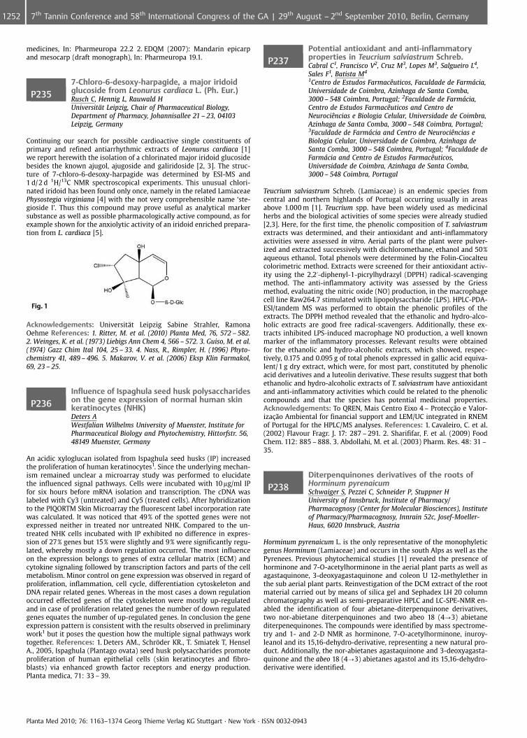
medicines, In: Pharmeuropa 22.2 2. EDQM (2007): Mandarin epicarpand mesocarp (draft monograph), In: Pharmeuropa 19.1.
P2357-Chloro-6-desoxy-harpagide, a major iridoidglucoside from Leonurus cardiaca L. (Ph. Eur.)Rusch C, Hennig L, Rauwald HUniversit�t Leipzig, Chair of Pharmaceutical Biology,Department of Pharmacy, Johannisallee 21 – 23, 04103Leipzig, Germany
Continuing our search for possible cardioactive single constituents ofprimary and refined antiarrhythmic extracts of Leonurus cardiaca [1]we report herewith the isolation of a chlorinated major iridoid glucosidebesides the known ajugol, ajugoside and galiridoside [2, 3]. The struc-ture of 7-chloro-6-desoxy-harpagide was determined by ESI-MS and1 d/2 d 1H/13C NMR spectroscopical experiments. This unusual chlori-nated iridoid has been found only once, namely in the related LamiaceaePhysostegia virginiana [4] with the not very comprehensible name ‘ste-gioside I’. Thus this compound may prove useful as analytical markersubstance as well as possible pharmacologically active compound, as forexample shown for the anxiolytic activity of an iridoid enriched prepara-tion from L. cardiaca [5].
Fig. 1
Acknowledgements: Universit�t Leipzig Sabine Strahler, RamonaOehme References: 1. Ritter, M. et al. (2010) Planta Med, 76, 572 – 582.2. Weinges, K. et al. (1973) Liebigs Ann Chem 4, 566 – 572. 3. Guiso, M. et al.(1974) Gazz Chim Ital 104, 25 – 33. 4. Nass, R., Rimpler, H. (1996) Phyto-chemistry 41, 489 – 496. 5. Makarov, V. et al. (2006) Eksp Klin Farmakol,69, 23 – 25.
P236Influence of Ispaghula seed husk polysaccharideson the gene expression of normal human skinkeratinocytes (NHK)Deters AWestfalian Wilhelms University of Muenster, Institute forPharmaceutical Biology and Phytochemistry, Hittorfstr. 56,48149 Muenster, Germany
An acidic xyloglucan isolated from Ispaghula seed husks (IP) increasedthe proliferation of human keratinocytes1. Since the underlying mechan-ism remained unclear a microarray study was performed to elucidatethe influenced signal pathways. Cells were incubated with 10 mg/ml IPfor six hours before mRNA isolation and transcription. The cDNA waslabeled with Cy3 (untreated) and Cy5 (treated cells). After hybridizationto the PIQORTM Skin Microarray the fluorescent label incorporation ratewas calculated. It was noticed that 49% of the spotted genes were notexpressed neither in treated nor untreated NHK. Compared to the un-treated NHK cells incubated with IP exhibited no difference in expres-sion of 27% genes but 15% were slightly and 9% were significantly regu-lated, whereby mostly a down regulation occurred. The most influenceon the expression belongs to genes of extra cellular matrix (ECM) andcytokine signaling followed by transcription factors and parts of the cellmetabolism. Minor control on gene expression was observed in regard ofproliferation, inflammation, cell cycle, differentiation cytoskeleton andDNA repair related genes. Whereas in the most cases a down regulationoccurred effected genes of the cytoskeleton were mostly up-regulatedand in case of proliferation related genes the number of down regulatedgenes equates the number of up-regulated genes. In conclusion the geneexpression pattern is consistent with the results observed in preliminarywork1 but it poses the question how the multiple signal pathways worktogether. References: 1. Deters AM., Schr�der KR., T. Smiatek T, HenselA., 2005, Ispaghula (Plantago ovata) seed husk polysaccharides promoteproliferation of human epithelial cells (skin keratinocytes and fibro-blasts) via enhanced growth factor receptors and energy production.Planta medica, 71: 33 – 39.
P237Potential antioxidant and anti-inflammatoryproperties in Teucrium salviastrum Schreb.Cabral C1, Francisco V2, Cruz M3, Lopes M3, Salgueiro L4,Sales F1, Batista M4
1Centro de Estudos FarmacÞuticos, Faculdade de Farm�cia,Universidade de Coimbra, Azinhaga de Santa Comba,3000 – 548 Coimbra, Portugal; 2Faculdade de Farm�cia,Centro de Estudos FarmacÞuticos and Centro deNeurociÞncias e Biologia Celular, Universidade de Coimbra,Azinhaga de Santa Comba, 3000 – 548 Coimbra, Portugal;3Faculdade de Farm�cia and Centro de NeurociÞncias eBiologia Celular, Universidade de Coimbra, Azinhaga deSanta Comba, 3000 – 548 Coimbra, Portugal; 4Faculdade deFarm�cia and Centro de Estudos FarmacÞuticos,Universidade de Coimbra, Azinhaga de Santa Comba,3000 – 548 Coimbra, Portugal
Teucrium salviastrum Schreb. (Lamiaceae) is an endemic species fromcentral and northern highlands of Portugal occurring usually in areasabove 1.000 m [1]. Teucrium spp. have been widely used as medicinalherbs and the biological activities of some species were already studied[2,3]. Here, for the first time, the phenolic composition of T. salviastrumextracts was determined, and their antioxidant and anti-inflammatoryactivities were assessed in vitro. Aerial parts of the plant were pulver-ized and extracted successively with dichloromethane, ethanol and 50%aqueous ethanol. Total phenols were determined by the Folin-Ciocalteucolorimetric method. Extracts were screened for their antioxidant activ-ity using the 2,2’-diphenyl-1-picrylhydrazyl (DPPH) radical-scavengingmethod. The anti-inflammatory activity was assessed by the Griessmethod, evaluating the nitric oxide (NO) production, in the macrophagecell line Raw264.7 stimulated with lipopolysaccharide (LPS). HPLC-PDA-ESI/tandem MS was performed to obtain the phenolic profiles of theextracts. The DPPH method revealed that the ethanolic and hydro-alco-holic extracts are good free radical-scavengers. Additionally, these ex-tracts inhibited LPS-induced macrophage NO production, a well knownmarker of the inflammatory processes. Relevant results were obtainedfor the ethanolic and hydro-alcoholic extracts, which showed, respec-tively, 0.175 and 0.095 g of total phenols expressed in gallic acid equiva-lent/1 g dry extract, which were, for most part, constituted by phenolicacid derivatives and a luteolin derivative. These results suggest that bothethanolic and hydro-alcoholic extracts of T. salviastrum have antioxidantand anti-inflammatory activities which could be related to the phenoliccompounds and that the species has potential medicinal properties.Acknowledgements: To QREN, Mais Centro Eixo 4 – Protec�¼o e Valor-iza�¼o Ambiental for financial support and LEM/UC integrated in RNEMof Portugal for the HPLC/MS analyses. References: 1. Cavaleiro, C. et al.(2002) Flavour Fragr. J. 17: 287 – 291. 2. Sharififar, F. et al. (2009) FoodChem. 112: 885 – 888. 3. Abdollahi, M. et al. (2003) Pharm. Res. 48: 31 –35.
P238Diterpenquinones derivatives of the roots ofHorminum pyrenaicumSchwaiger S, Pezzei C, Schneider P, Stuppner HUniversity of Innsbruck, Institute of Pharmacy/Pharmacognosy (Center for Molecular Biosciences), Instituteof Pharmacy/Pharmacognosy, Innrain 52c, Josef-Moeller-Haus, 6020 Innsbruck, Austria
Horminum pyrenaicum L. is the only representative of the monophyleticgenus Horminum (Lamiaceae) and occurs in the south Alps as well as thePyrenees. Previous phytochemical studies [1] revealed the presence ofhorminone and 7-O-acetylhorminone in the aerial plant parts as well asagastaquinone, 3-deoxyagastaquinone and coleon U 12-methylether inthe sub aerial plant parts. Reinvestigation of the DCM extract of the rootmaterial carried out by means of silica gel and Sephadex LH 20 columnchromatography as well as semi-preparative HPLC and LC-SPE-NMR en-abled the identification of four abietane-diterpenquinone derivatives,two nor-abietane diterpenquinones and two abeo 18 (4?3) abietanediterpenequinones. The compounds were identified by mass spectrome-try and 1- and 2-D NMR as horminone, 7-O-acetylhorminone, inuroy-leanol and its 15,16-dehydro-derivative, representing a new natural pro-duct. Additionally, the nor-abietanes agastaquinone and 3-deoxyagasta-quinone and the abeo 18 (4?3) abietanes agastol and its 15,16-dehydro-derivative were identified.
Planta Med 2010; 76: 1163–1374 Georg Thieme Verlag KG Stuttgart · New York · ISSN 0032-0943
1252 7th Tannin Conference and 58th International Congress of the GA | 29th August – 2nd September 2010, Berlin, Germany
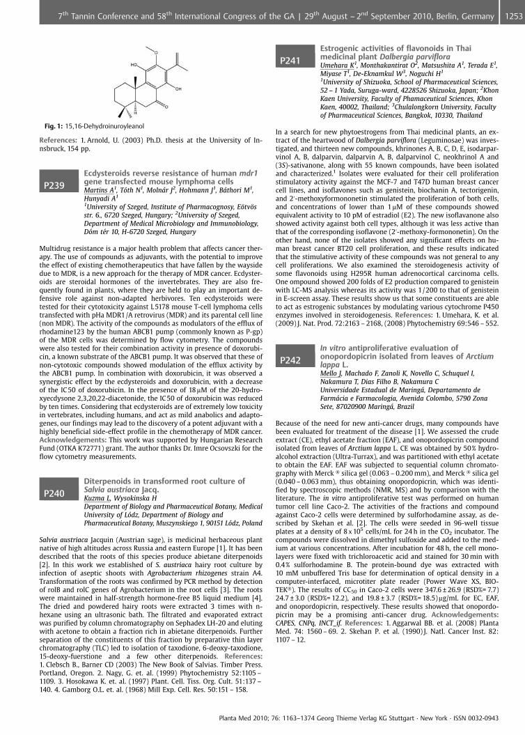
Fig. 1: 15,16-Dehydroinuroyleanol
References: 1. Arnold, U. (2003) Ph.D. thesis at the University of In-nsbruck, 154 pp.
P239Ecdysteroids reverse resistance of human mdr1gene transfected mouse lymphoma cellsMartins A1, T�th N1, Moln�r J2, Hohmann J1, B�thori M1,Hunyadi A1
1University of Szeged, Institute of Pharmacognosy, E�tv�sstr. 6., 6720 Szeged, Hungary; 2University of Szeged,Department of Medical Microbiology and Immunobiology,D�m t�r 10, H-6720 Szeged, Hungary
Multidrug resistance is a major health problem that affects cancer ther-apy. The use of compounds as adjuvants, with the potential to improvethe effect of existing chemotherapeutics that have fallen by the waysidedue to MDR, is a new approach for the therapy of MDR cancer. Ecdyster-oids are steroidal hormones of the invertebrates. They are also fre-quently found in plants, where they are held to play an important de-fensive role against non-adapted herbivores. Ten ecdysteroids weretested for their cytotoxicity against L 5178 mouse T-cell lymphoma cellstransfected with pHa MDR1/A retrovirus (MDR) and its parental cell line(non MDR). The activity of the compounds as modulators of the efflux ofrhodamine123 by the human ABCB1 pump (commonly known as P-gp)of the MDR cells was determined by flow cytometry. The compoundswere also tested for their combination activity in presence of doxorubi-cin, a known substrate of the ABCB1 pump. It was observed that these ofnon-cytotoxic compounds showed modulation of the efflux activity bythe ABCB1 pump. In combination with doxorubicin, it was observed asynergistic effect by the ecdysteroids and doxorubicin, with a decreaseof the IC 50 of doxorubicin. In the presence of 18mM of the 20-hydro-xyecdysone 2,3,20,22-diacetonide, the IC50 of doxorubicin was reducedby ten times. Considering that ecdysteroids are of extremely low toxicityin vertebrates, including humans, and act as mild anabolics and adapto-genes, our findings may lead to the discovery of a potent adjuvant with ahighly beneficial side-effect profile in the chemotherapy of MDR cancer.Acknowledgements: This work was supported by Hungarian ResearchFund (OTKA K72771) grant. The author thanks Dr. Imre Ocsovszki for theflow cytometry measurements.
P240Diterpenoids in transformed root culture ofSalvia austriaca Jacq.Kuzma L, Wysokinska HDepartment of Biology and Pharmaceutical Botany, MedicalUniversity of L�dz, Department of Biology andPharmaceutical Botany, Muszynskiego 1, 90151 L�dz, Poland
Salvia austriaca Jacquin (Austrian sage), is medicinal herbaceous plantnative of high altitudes across Russia and eastern Europe [1]. It has beendescribed that the roots of this species produce abietane diterpenoids[2]. In this work we established of S. austriaca hairy root culture byinfection of aseptic shoots with Agrobacterium rhizogenes strain A4.Transformation of the roots was confirmed by PCR method by detectionof rolB and rolC genes of Agrobacterium in the root cells [3]. The rootswere maintained in half-strength hormone-free B5 liquid medium [4].The dried and powdered hairy roots were extracted 3 times with n-hexane using an ultrasonic bath. The filtrated and evaporated extractwas purified by column chromatography on Sephadex LH-20 and elutingwith acetone to obtain a fraction rich in abietane diterpenoids. Furtherseparation of the constituents of this fraction by preparative thin layerchromatography (TLC) led to isolation of taxodione, 6-deoxy-taxodione,15-deoxy-fuerstione and a few other diterpenoids. References:1. Clebsch B., Barner CD (2003) The New Book of Salvias. Timber Press.Portland, Oregon. 2. Nagy, G. et. al. (1999) Phytochemistry 52:1105 –1109. 3. Hosokawa K. et. al. (1997) Plant. Cell. Tiss. Org. Cult. 51:137 –140. 4. Gamborg O.L. et. al. (1968) Mill Exp. Cell. Res. 50:151 – 158.
P241Estrogenic activities of flavonoids in Thaimedicinal plant Dalbergia parvifloraUmehara K1, Monthakantirat O2, Matsushita A1, Terada E1,Miyase T1, De-Eknamkul W3, Noguchi H1
1University of Shizuoka, School of Pharmaceutical Sciences,52 – 1 Yada, Suruga-ward, 4228526 Shizuoka, Japan; 2KhonKaen University, Faculty of Phamaceutical Sciences, KhonKaen, 40002, Thailand; 3Chulalongkorn University, Facultyof Pharmaceutical Sciences, Bangkok, 10330, Thailand
In a search for new phytoestrogens from Thai medicinal plants, an ex-tract of the heartwood of Dalbergia parviflora (Leguminosae) was inves-tigated, and thirteen new compounds, khrinones A, B, C, D, E, isodarpar-vinol A, B, dalparvin, dalparvin A, B, dalparvinol C, neokhrinol A and(3S)-sativanone, along with 55 known compounds, have been isolatedand characterized.1 Isolates were evaluated for their cell proliferationstimulatory activity against the MCF-7 and T47D human breast cancercell lines, and isoflavones such as genistein, biochanin A, tectorigenin,and 2’-methoxyformononetin stimulated the proliferation of both cells,and concentrations of lower than 1mM of these compounds showedequivalent activity to 10 pM of estradiol (E2). The new isoflavanone alsoshowed activity against both cell types, although it was less active thanthat of the corresponding isoflavone (2’-methoxy-formononetin). On theother hand, none of the isolates showed any significant effects on hu-man breast cancer BT20 cell proliferation, and these results indicatedthat the stimulative activity of these compounds was not general to anycell proliferations. We also examined the steroidogenesis activity ofsome flavonoids using H295R human adrenocortical carcinoma cells.One ompound showed 200 folds of E2 production compared to genisteinwith LC-MS analysis whereas its activity was 1/200 to that of genisteinin E-screen assay. These results show us that some constituents are ableto act as estrogenic substances by modulating various cytochrome P450enzymes involved in steroidogenesis. References: 1. Umehara, K. et al.(2009) J. Nat. Prod. 72:2163 – 2168, (2008) Phytochemistry 69:546 – 552.
P242In vitro antiproliferative evaluation ofonopordopicrin isolated from leaves of Arctiumlappa L.Mello J, Machado F, Zanoli K, Novello C, Schuquel I,Nakamura T, Dias Filho B, Nakamura CUniversidade Estadual de Maring�, Departamento deFarm�cia e Farmacologia, Avenida Colombo, 5790 ZonaSete, 87020900 Maring�, Brazil
Because of the need for new anti-cancer drugs, many compounds havebeen evaluated for treatment of the disease [1]. We assessed the crudeextract (CE), ethyl acetate fraction (EAF), and onopordopicrin compoundisolated from leaves of Arctium lappa L. CE was obtained by 50% hydro-alcohol extraction (Ultra-Turrax), and was partitioned with ethyl acetateto obtain the EAF. EAF was subjected to sequential column chromato-graphy with Merck � silica gel (0.063 – 0.200 mm), and Merck � silica gel(0.040 – 0.063 mm), thus obtaining onopordopicrin, which was identi-fied by spectroscopic methods (NMR, MS) and by comparison with theliterature. The in vitro antiproliferative test was performed on humantumor cell line Caco-2. The activities of the fractions and compoundagainst Caco-2 cells were determined by sulforhodamine assay, as de-scribed by Skehan et al. [2]. The cells were seeded in 96-well tissueplates at a density of 8 x 10⁵ cells/mL for 24 h in the CO2 incubator. Thecompounds were dissolved in dimethyl sulfoxide and added to the med-ium at various concentrations. After incubation for 48 h, the cell mono-layers were fixed with trichloroacetic acid and stained for 30 min with0.4% sulforhodamine B. The protein-bound dye was extracted with10 mM unbuffered Tris base for determination of optical density in acomputer-interfaced, microtiter plate reader (Power Wave XS, BIO-TEK�). The results of CC50 in Caco-2 cells were 347.6 € 26.9 (RSD%= 7.7)24.7 € 3.0 (RSD%= 12.2), and 19.8 € 3.7 (RSD%= 18.5)mg/mL for EC, EAF,and onopordopicrin, respectively. These results showed that onopordo-picrin may be a promising anti-cancer drug. Acknowledgements:CAPES, CNPq, INCT_if. References: 1. Aggarwal BB. et al. (2008) PlantaMed. 74: 1560 – 69. 2. Skehan P. et al. (1990) J. Natl. Cancer Inst. 82:1107 – 12.
Planta Med 2010; 76: 1163–1374 Georg Thieme Verlag KG Stuttgart · New York · ISSN 0032-0943
12537th Tannin Conference and 58th International Congress of the GA | 29th August – 2nd September 2010, Berlin, Germany
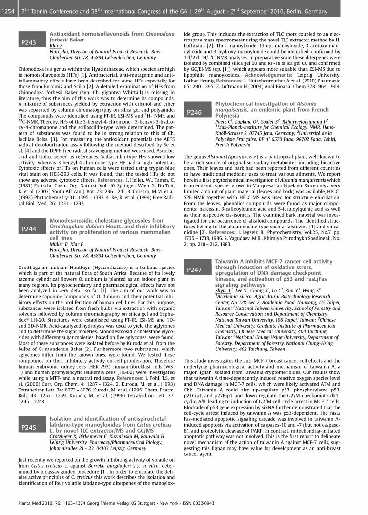
P243Antioxidant homoisoflavonoids from Chionodoxaforbesii BakerKlar FFlurepha, Division of Natural Product Research, Buer-Gladbecker Str. 78, 45894 Gelsenkirchen, Germany
Chionodoxa is a genus within the Hyacinthaceae, which species are highin homoisoflavonoids (HFs) [1]. Antibacterial, anti-mutagenic and anti-inflammatory effects have been described for some HFs, especially forthose from Eucomis and Scilla [2]. A detailed examination of HFs fromChionodoxa forbesii Baker (syn. Ch. gigantea Whittall) is missing inliterature, thus the aim of this work was to determine its compounds.A mixture of substances yielded by extraction with ethanol and etherwas separated by column chromatography on silica gel and polyamide.The compounds were identified using FT-IR, ESI-MS and 1H- NMR and13C-NMR. Thereby, HFs of the 3-benzyl-4-chromone-, 3-benzyl-3-hydro-xy-4-chromanone and the scillascillin-type were determined. The pat-tern of substances was found to be in strong relation to this of Ch.luciliae Boiss. [3]. For measuring the antioxidant potentials the ABTSradical decolourization assay following the method described by Re etal. [4] and the DPPH free radical scavenging method were used. Ascorbicacid and trolox served as references. Scillascillin-type HFs showed lowactivity, whereas 3-benzyl-4-chromone-type HF had a high potential.Cytotoxic effects of HFs on human cells were tested using trypan bluevital stain on HEK-293 cells. It was found, that the tested HFs do notshow any adverse cytotoxic effects. References: 1. Heller, W., Tamm, C.(1981) Fortschr. Chem. Org. Naturst. Vol. 40. Springer. Wien. 2. Du Toit,K. et al. (2007) South African J. Bot. 73: 236 – 241. 3. Corsaro, M.M. et al.(1992) Phytochemistry 31: 1395 – 1397. 4. Re, R. et al. (1999) Free Radi-cal Biol. Med. 26: 1231 – 1237.
P244Monodesmosidic cholestane glycosides fromOrnithogalum dubium Houtt. and their inhibitoryactivity on proliferation of various mammaliancell linesM�ller B, Klar FFlurepha, Division of Natural Product Research, Buer-Gladbecker Str. 78, 45894 Gelsenkirchen, Germany
Ornithogalum dubium Houttuyn (Hyacinthaceae) is a bulbous specieswhich is part of the natural flora of South Africa. Because of its lovelyraceme cylindrical flowers O. dubium is planted as an indoor plant inmany regions. Its phytochemistry and pharmacological effects have notbeen analyzed in very detail so far [1]. The aim of our work was todetermine saponine compounds of O. dubium and their potential inhi-bitory effects on the proliferation of human cell lines. For this purpose,substances were isolated from fresh bulbs via extraction with organicsolvents followed by column chromatography on silica gel and Sepha-dex� LH-20. Structures were established using FT-IR, ESI-MS and 1D-and 2D-NMR. Acid-catalyzed hydrolysis was used to yield the aglyconesand to determine the sugar moieties. Monodesmosidic cholestane glyco-sides with different sugar moieties, based on five aglycones, were found.Most of these substances were isolated before by Kuroda et al. from thebulbs of O. saundersie Baker [2]. Furthermore, two substances, whichaglycones differ from the known ones, were found. We tested thesecompounds on their inhibitory activity on cell proliferation. Thereforehuman embryonic kidney cells (HEK-293), human fibroblast cells (WS-1) and human promyelocytic leukemia cells (HL-60) were investigatedwhile using a MTT- and a neutral red assay. References: 1. Pohl, T.S. etal. (2000) Curr. Org. Chem. 4: 1287 – 1324. 2. Kuroda, M. et al. (1993)Tetrahedron Lett. 34: 6073 – 6076. Kuroda, M. et al. (1995) Chem. Pharm.Bull. 43: 1257 – 1259. Kuroda, M. et al. (1996) Tetrahedron Lett. 37:1245 – 1248.
P245Isolation and identification of antispirochetallabdane-type manoyloxides from Cistus creticusL. by novel TLC-extractor/MS and GC/MSGr�tzinger K, Birkemeyer C, Kuzminska M, Rauwald HLeipzig University, Pharmacy/Pharmaceutical Biology,Johannisallee 21 – 23, 04103 Leipzig, Germany
Just recently we reported on the growth inhibiting activity of volatile oilfrom Cistus creticus L. against Borrelia burgdorferi s.s. in vitro, deter-mined by bioassay guided procedure [1]. In order to elucidate the defi-nite active principles of C. creticus this work describes the isolation andidentification of four volatile labdane-type diterpenes of the manoylox-
ide group. This includes the extraction of TLC spots coupled to an elec-trospray mass spectrometer using the novel TLC extractor method by H.Luftmann [2]. Thus manoyloxide, 13-epi-manoyloxide, 3-acetoxy-man-oyloxide and 3-hydroxy-manoyloxide could be identified, confirmed by1 d/2 d-1H/13C-NMR analyses. In preparative scale these diterpenes wereisolated by combined silica gel 60 and RP-18 silica gel CC and confirmedby GC/EI-MS (cp. [1]), which appears more suitable than ESI-MS due tolipophilic manoyloxides. Acknowledgements: Leipzig University,Lothar Hennig References: 1. Hutschenreuther A et al. (2010) Pharmazie65: 290 – 295. 2. Luftmann H (2004) Anal Bioanal Chem 378: 964 – 968.
P246Phytochemical investigation of Alstoniamarquisensis, an endemic plant from FrenchPolynesiaPaetz C1, Laplane O2, Soulet S2, Raharivelomanana P2
1Max-Planck-Institute for Chemical Ecology, NMR, Hans-Kn�ll-Strasse 8, 07745 Jena, Germany; 2Universit� de laPolyn�sie Fran�aise, BP n� 6570 Faaa, 98702 Faaa, Tahiti,French Polynesia
The genus Alstonia (Apocynaceae) is a pantropical plant, well-known tobe a rich source of original secondary metabolites including bioactiveones. Their leaves and bark had been reported from different countriesto have traditional medicine uses to treat various ailments. We reportherein a first phytochemical investigation of Alstonia marquisensis whichis an endemic species grown in Marquesas archipelago. Since only a verylimited amount of plant material (leaves and bark) was available, HPLC-SPE-NMR together with HPLC-MS was used for structure elucidation.From the leaves, phenolics compounds were found as major compo-nents: narcissin, 3-caffeoylquinic acid and 5-feruloylquinic acid as wellas their respective cis-isomers. The examined bark material was inves-tigated for the occurrence of alkaloid compounds. The identified struc-tures belong to the akuammicine type such as alstovine [1] and vinca-nidine [2]. References: 1. Legseir, B., Phytochemistry, Vol.25, No.7, pp.1735 – 1738, 1986. 2. Yagudaev, M.R., Khimiya Prirodnykh Soedinenii, No.2, pp. 210 – 212, 1983.
P247Taiwanin A inhibits MCF-7 cancer cell activitythrough induction of oxidative stress,upregulation of DNA damage checkpointkinases, and activation of p53 and FasL/Fassignaling pathwaysShyur L1, Lee S1, Chang S2, Lo C1, Kuo Y3, Wang S4
1Academia Sinica, Agricultural Biotechnology ResearchCenter, No 128, Sec 2, Academia Road, Nankang, 115 Taipei,Taiwan; 2National Taiwan University, School of Forestry andResource Conservation and Department of Chemistry,National Taiwan University, 106 Taipei, Taiwan; 3ChineseMedical University, Graduate Institute of PharmaceuticalChemistry, Chinese Medical University, 404 Taichung,Taiwan; 4National Chung-Hsing University, Department ofForestry, Department of Forestry, National Chung-HsingUniversity, 402 Taichung, Taiwan
This study investigates the anti-MCF-7 breast cancer cell effects and theunderlying pharmacological activity and mechanism of taiwanin A, amajor lignan isolated from Taiwania cryptomerioides. Our results showthat taiwanin A time-dependently induced reactive oxygen species leveland DNA damage in MCF-7 cells, which were likely activated ATM andChk. Taiwanin A could also up-regulate p53, phosphorylated p53,p21Cip1, and p27Kip1 and down-regulate the G2/M checkpoint Cdk1-cyclin A/B, leading to induction of G2/M cell-cycle arrest in MCF-7 cells.Blockade of p53 gene expression by siRNA further demonstrated that thecell-cycle arrest induced by taiwanin A was p53-dependent. The FasL/Fas-mediated apoptotic signaling cascade was involved in taiwanin A-induced apoptosis via activation of caspases-10 and -7 (but not caspase-8), and proteolytic cleavage of PARP. In contrast, mitochondria-initiatedapoptotic pathway was not involved. This is the first report to delineatenovel mechanism of the action of taiwanin A against MCF-7 cells, sug-gesting this lignan may have value for development as an anti-breastcancer agent.
Planta Med 2010; 76: 1163–1374 Georg Thieme Verlag KG Stuttgart · New York · ISSN 0032-0943
1254 7th Tannin Conference and 58th International Congress of the GA | 29th August – 2nd September 2010, Berlin, Germany
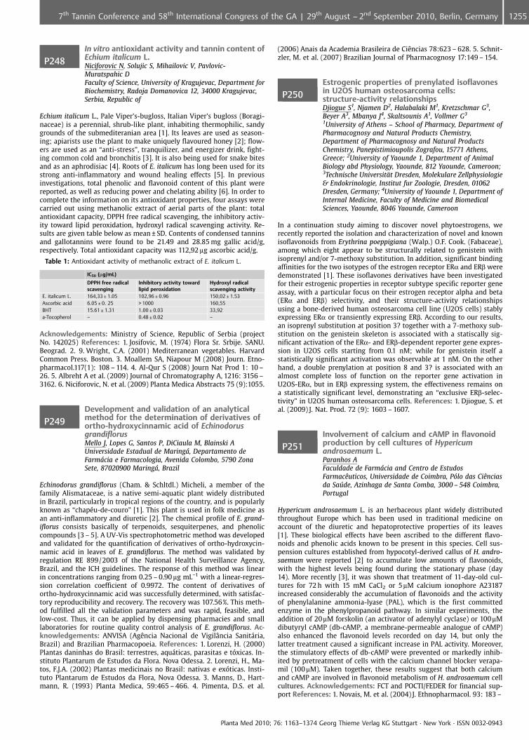
P248In vitro antioxidant activity and tannin content ofEchium italicum L.Niciforovic N, Solujic S, Mihailovic V, Pavlovic-Muratspahic DFaculty of Science, University of Kragujevac, Department forBiochemistry, Radoja Domanovica 12, 34000 Kragujevac,Serbia, Republic of
Echium italicum L., Pale Viper’s-bugloss, Italian Viper’s bugloss (Boragi-naceae) is a perennial, shrub-like plant, inhabiting thermophilic, sandygrounds of the submediteranian area [1]. Its leaves are used as season-ing; apiarists use the plant to make uniquely flavoured honey [2]; flow-ers are used as an “anti-stress”, tranquilizer, and energizer drink, fight-ing common cold and bronchitis [3]. It is also being used for snake bitesand as an aphrodisiac [4]. Roots of E. italicum has long been used for itsstrong anti-inflammatory and wound healing effects [5]. In previousinvestigations, total phenolic and flavonoid content of this plant werereported, as well as reducing power and chelating ability [6]. In order tocomplete the information on its antioxidant properties, four assays werecarried out using methanolic extract of aerial parts of the plant: totalantioxidant capacity, DPPH free radical scavenging, the inhibitory activ-ity toward lipid peroxidation, hydroxyl radical scavenging activity. Re-sults are given table below as mean € SD. Contents of condensed tanninsand gallotannins were found to be 21.49 and 28.85 mg gallic acid/g,respectively. Total antioxidant capacity was 112,92 mg ascorbic acid/g.
Table 1: Antioxidant activity of methanolic extract of E. italicum L.
IC50 (mg/mL)
DPPH free radicalscavenging
Inhibitory activity towardlipid peroxidation
Hydroxyl radicalscavenging activity
E. italicum L. 164,33 € 1.05 102,96 € 0.96 150,02 € 1.53Ascorbic acid 6.05 € 0. 25 > 1000 160,55BHT 15.61 € 1.31 1.00 € 0.03 33,92a-Tocopherol – 0.48 € 0.02 –
Acknowledgements: Ministry of Science, Republic of Serbia (projectNo. 142025) References: 1. Josifovic, M. (1974) Flora Sr. Srbije. SANU.Beograd. 2. 9. Wright, C.A. (2001) Mediterranean vegetables. HarvardCommon Press. Boston. 3. Moallem SA, Niapour M (2008) Journ. Etno-pharmacol.117(1): 108 – 114. 4. Al-Qur S (2008) Journ Nat Prod 1: 10 –26. 5. Albreht A et al. (2009) Journal of Chromatography A, 1216: 3156 –3162. 6. Niciforovic, N. et al. (2009) Planta Medica Abstracts 75 (9):1055.
P249Development and validation of an analyticalmethod for the determination of derivatives ofortho-hydroxycinnamic acid of EchinodorusgrandiflorusMello J, Lopes G, Santos P, DiCiaula M, Blainski AUniversidade Estadual de Maring�, Departamento deFarm�cia e Farmacologia, Avenida Colombo, 5790 ZonaSete, 87020900 Maring�, Brazil
Echinodorus grandiflorus (Cham. & Schltdl.) Micheli, a member of thefamily Alismataceae, is a native semi-aquatic plant widely distributedin Brazil, particularly in tropical regions of the country, and is popularlyknown as “chap�u-de-couro” [1]. This plant is used in folk medicine asan anti-inflammatory and diuretic [2]. The chemical profile of E. grand-iflorus consists basically of terpenoids, sesquiterpenes, and phenoliccompounds [3 – 5]. A UV-Vis spectrophotometric method was developedand validated for the quantification of derivatives of ortho-hydroxycin-namic acid in leaves of E. grandiflorus. The method was validated byregulation RE 899/2003 of the National Health Surveillance Agency,Brazil, and the ICH guidelines. The response of this method was linearin concentrations ranging from 0.25 – 0.90 mg mL�1 with a linear-regres-sion correlation coefficient of 0.9972. The content of derivatives ofortho-hydroxycinnamic acid was successfully determined, with satisfac-tory reproducibility and recovery. The recovery was 107.56%. This meth-od fulfilled all the validation parameters and was rapid, feasible, andlow-cost. Thus, it can be applied by dispensing pharmacies and smalllaboratories for routine quality control analysis of E. grandiflorus. Ac-knowledgements: ANVISA (AgÞncia Nacional de Vigil�ncia Sanit�ria,Brazil) and Brazilian Pharmacopoeia. References: 1. Lorenzi, H. (2000)Plantas daninhas do Brasil: terrestres, aqu�ticas, parasitas e t xicas. In-stituto Plantarum de Estudos da Flora. Nova Odessa. 2. Lorenzi, H., Ma-tos, F.J.A. (2002) Plantas medicinais no Brasil: nativas e ex ticas. Insti-tuto Plantarum de Estudos da Flora, Nova Odessa. 3. Manns, D., Hart-mann, R. (1993) Planta Medica, 59:465 – 466. 4. Pimenta, D.S. et al.
(2006) Anais da Academia Brasileira de CiÞncias 78:623 – 628. 5. Schnit-zler, M. et al. (2007) Brazilian Journal of Pharmacognosy 17:149 – 154.
P250Estrogenic properties of prenylated isoflavonesin U2OS human osteosarcoma cells:structure-activity relationshipsDjiogue S1, Njamen D2, Halabalaki M1, Kretzschmar G3,Beyer A3, Mbanya J4, Skaltsounis A1, Vollmer G3
1University of Athens – School of Pharmacy, Department ofPharmacognosy and Natural Products Chemistry,Department of Pharmacognosy and Natural ProductsChemistry, Panepistimioupolis Zografou, 15771 Athens,Greece; 2University of Yaounde 1, Department of AnimalBiology abd Physiology, Yaounde, 812 Yaounde, Cameroon;3Technische Universit�t Dresden, Molekulare Zellphysiologie& Endokrinologie, Institut fur Zoologie, Dresden, 01062Dresden, Germany; 4University of Yaounde 1, Department ofInternal Medicine, Faculty of Medicine and BiomedicalSciences, Yaounde, 8046 Yaounde, Cameroon
In a continuation study aiming to discover novel phytoestrogens, werecently reported the isolation and characterization of novel and knownisoflavonoids from Erythrina poeppigiana (Walp.) O.F. Cook. (Fabaceae),among which eight appear to be structurally related to genistein withisoprenyl and/or 7-methoxy substitution. In addition, significant bindingaffinities for the two isotypes of the estrogen receptor ERa and ERb weredemonstrated [1]. These isoflavones derivatives have been investigatedfor their estrogenic properties in receptor subtype specific reporter geneassay, with a particular focus on their estrogen receptor alpha and beta(ERa and ERb) selectivity, and their structure-activity relationshipsusing a bone-derived human osteosarcoma cell line (U2OS cells) stablyexpressing ERa or transiently expressing ERb. According to our results,an isoprenyl substitution at position 3? together with a 7-methoxy sub-stitution on the genistein skeleton is associated with a statiscally sig-nificant activation of the ERa- and ERb-dependent reporter gene expres-sion in U2OS cells starting from 0.1 nM; while for genistein itself astatistically significant activation was observable at 1 nM. On the otherhand, a double prenylation at position 8 and 3? is associated with analmost complete loss of function on the reporter gene activation inU2OS-ERa, but in ERb expressing system, the effectiveness remains ona statistically significant level, demonstrating an “exclusive ERb-selec-tivity” in U2OS human osteosarcoma cells. References: 1. Djiogue, S. etal. (2009) J. Nat. Prod. 72 (9): 1603 – 1607.
P251Involvement of calcium and cAMP in flavonoidproduction by cell cultures of Hypericumandrosaemum L.Paranhos AFaculdade de Farm�cia and Centro de EstudosFarmacÞuticos, Universidade de Coimbra, P�lo das CiÞnciasda Safflde, Azinhaga de Santa Comba, 3000 – 548 Coimbra,Portugal
Hypericum androsaemum L. is an herbaceous plant widely distributedthroughout Europe which has been used in traditional medicine onaccount of the diuretic and hepatoprotective properties of its leaves[1]. These biological effects have been ascribed to the different flavo-noids and phenolic acids known to be present in this species. Cell sus-pension cultures established from hypocotyl-derived callus of H. andro-saemum were reported [2] to accumulate low amounts of flavonoids,with the highest levels being found during the stationary phase (day14). More recently [3], it was shown that treatment of 11-day-old cul-tures for 72 h with 15 mM CaCl2 or 5mM calcium ionophore A23187increased considerably the accumulation of flavonoids and the activityof phenylalanine ammonia-lyase (PAL), which is the first committedenzyme in the phenylpropanoid pathway. In similar experiments, theaddition of 20mM forskolin (an activator of adenylyl cyclase) or 100mMdibutyryl cAMP (db-cAMP, a membrane-permeable analogue of cAMP)also enhanced the flavonoid levels recorded on day 14, but only thelatter treatment caused a significant increase in PAL activity. Moreover,the stimulatory effects of db-cAMP were prevented or markedly inhib-ited by pretreatment of cells with the calcium channel blocker verapa-mil (100mM). Taken together, these results suggest that both calciumand cAMP are involved in flavonoid metabolism of H. androsaemum cellcultures. Acknowledgements: FCT and POCTI/FEDER for financial sup-port References: 1. Novais, M. et al. (2004) J. Ethnopharmacol. 93: 183 –
Planta Med 2010; 76: 1163–1374 Georg Thieme Verlag KG Stuttgart · New York · ISSN 0032-0943
12557th Tannin Conference and 58th International Congress of the GA | 29th August – 2nd September 2010, Berlin, Germany
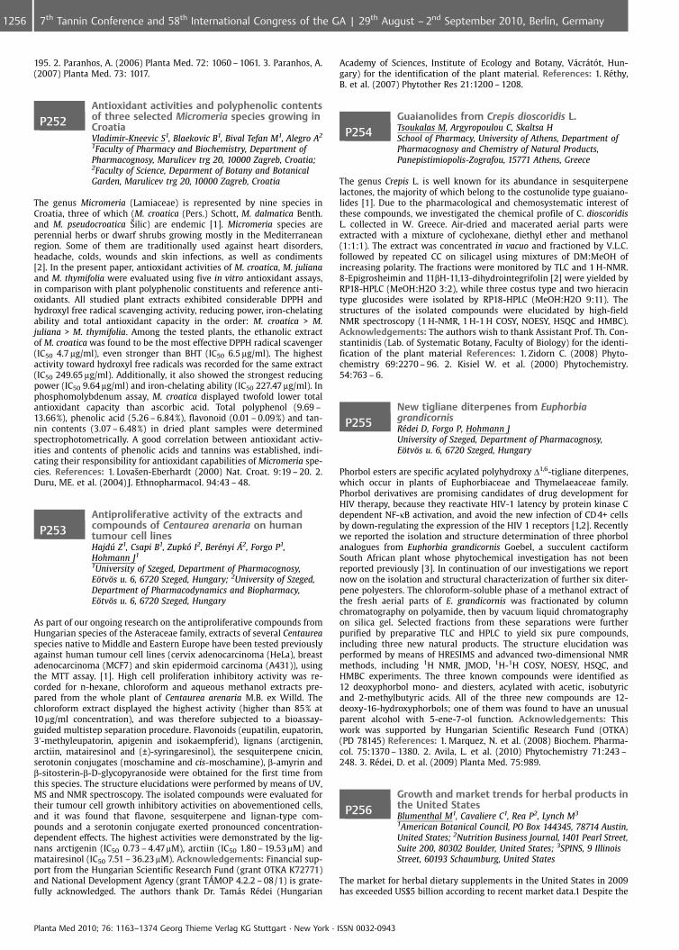
195. 2. Paranhos, A. (2006) Planta Med. 72: 1060 – 1061. 3. Paranhos, A.(2007) Planta Med. 73: 1017.
P252Antioxidant activities and polyphenolic contentsof three selected Micromeria species growing inCroatiaVladimir-Kneevic S1, Blaekovic B1, Bival Tefan M1, Alegro A2
1Faculty of Pharmacy and Biochemistry, Department ofPharmacognosy, Marulicev trg 20, 10000 Zagreb, Croatia;2Faculty of Science, Deparment of Botany and BotanicalGarden, Marulicev trg 20, 10000 Zagreb, Croatia
The genus Micromeria (Lamiaceae) is represented by nine species inCroatia, three of which (M. croatica (Pers.) Schott, M. dalmatica Benth.and M. pseudocroatica �ilic) are endemic [1]. Micromeria species areperennial herbs or dwarf shrubs growing mostly in the Mediterraneanregion. Some of them are traditionally used against heart disorders,headache, colds, wounds and skin infections, as well as condiments[2]. In the present paper, antioxidant activities of M. croatica, M. julianaand M. thymifolia were evaluated using five in vitro antioxidant assays,in comparison with plant polyphenolic constituents and reference anti-oxidants. All studied plant extracts exhibited considerable DPPH andhydroxyl free radical scavenging activity, reducing power, iron-chelatingability and total antioxidant capacity in the order: M. croatica > M.juliana > M. thymifolia. Among the tested plants, the ethanolic extractof M. croatica was found to be the most effective DPPH radical scavenger(IC50 4.7 mg/ml), even stronger than BHT (IC50 6.5 mg/ml). The highestactivity toward hydroxyl free radicals was recorded for the same extract(IC50 249.65 mg/ml). Additionally, it also showed the strongest reducingpower (IC50 9.64 mg/ml) and iron-chelating ability (IC50 227.47 mg/ml). Inphosphomolybdenum assay, M. croatica displayed twofold lower totalantioxidant capacity than ascorbic acid. Total polyphenol (9.69 –13.66%), phenolic acid (5.26 – 6.84%), flavonoid (0.01 – 0.09%) and tan-nin contents (3.07 – 6.48%) in dried plant samples were determinedspectrophotometrically. A good correlation between antioxidant activ-ities and contents of phenolic acids and tannins was established, indi-cating their responsibility for antioxidant capabilities of Micromeria spe-cies. References: 1. LovaÐen-Eberhardt (2000) Nat. Croat. 9:19 – 20. 2.Duru, ME. et al. (2004) J. Ethnopharmacol. 94:43 – 48.
P253Antiproliferative activity of the extracts andcompounds of Centaurea arenaria on humantumour cell linesHajdffl Z1, Csapi B1, Zupk� I2, Ber�nyi �2, Forgo P1,Hohmann J1
1University of Szeged, Department of Pharmacognosy,E�tv�s u. 6, 6720 Szeged, Hungary; 2University of Szeged,Department of Pharmacodynamics and Biopharmacy,E�tv�s u. 6, 6720 Szeged, Hungary
As part of our ongoing research on the antiproliferative compounds fromHungarian species of the Asteraceae family, extracts of several Centaureaspecies native to Middle and Eastern Europe have been tested previouslyagainst human tumour cell lines (cervix adenocarcinoma (HeLa), breastadenocarcinoma (MCF7) and skin epidermoid carcinoma (A431)), usingthe MTT assay. [1]. High cell proliferation inhibitory activity was re-corded for n-hexane, chloroform and aqueous methanol extracts pre-pared from the whole plant of Centaurea arenaria M.B. ex Willd. Thechloroform extract displayed the highest activity (higher than 85% at10 mg/ml concentration), and was therefore subjected to a bioassay-guided multistep separation procedure. Flavonoids (eupatilin, eupatorin,3’-methyleupatorin, apigenin and isokaempferid), lignans (arctigenin,arctiin, matairesinol and (€)-syringaresinol), the sesquiterpene cnicin,serotonin conjugates (moschamine and cis-moschamine), b-amyrin andb-sitosterin-b-D-glycopyranoside were obtained for the first time fromthis species. The structure elucidations were performed by means of UV,MS and NMR spectroscopy. The isolated compounds were evaluated fortheir tumour cell growth inhibitory activities on abovementioned cells,and it was found that flavone, sesquiterpene and lignan-type com-pounds and a serotonin conjugate exerted pronounced concentration-dependent effects. The highest activities were demonstrated by the lig-nans arctigenin (IC50 0.73 – 4.47mM), arctiin (IC50 1.80 – 19.53mM) andmatairesinol (IC50 7.51 – 36.23mM). Acknowledgements: Financial sup-port from the Hungarian Scientific Research Fund (grant OTKA K72771)and National Development Agency (grant T�MOP 4.2.2 – 08/1) is grate-fully acknowledged. The authors thank Dr. Tam�s R�dei (Hungarian
Academy of Sciences, Institute of Ecology and Botany, V�cr�t t, Hun-gary) for the identification of the plant material. References: 1. R�thy,B. et al. (2007) Phytother Res 21:1200 – 1208.
P254Guaianolides from Crepis dioscoridis L.Tsoukalas M, Argyropoulou C, Skaltsa HSchool of Pharmacy, University of Athens, Department ofPharmacognosy and Chemistry of Natural Products,Panepistimiopolis-Zografou, 15771 Athens, Greece
The genus Crepis L. is well known for its abundance in sesquiterpenelactones, the majority of which belong to the costunolide type guaiano-lides [1]. Due to the pharmacological and chemosystematic interest ofthese compounds, we investigated the chemical profile of C. dioscoridisL. collected in W. Greece. Air-dried and macerated aerial parts wereextracted with a mixture of cyclohexane, diethyl ether and methanol(1:1:1). The extract was concentrated in vacuo and fractioned by V.L.C.followed by repeated CC on silicagel using mixtures of DM:MeOH ofincreasing polarity. The fractions were monitored by TLC and 1 H-NMR.8-Epigrosheimin and 11bH-11,13-dihydrointegrifolin [2] were yielded byRP18-HPLC (MeOH:H2O 3:2), while three costus type and two hieracintype glucosides were isolated by RP18-HPLC (MeOH:H2O 9:11). Thestructures of the isolated compounds were elucidated by high-fieldNMR spectroscopy (1 H-NMR, 1 H-1 H COSY, NOESY, HSQC and HMBC).Acknowledgements: The authors wish to thank Assistant Prof. Th. Con-stantinidis (Lab. of Systematic Botany, Faculty of Biology) for the identi-fication of the plant material References: 1. Zidorn C. (2008) Phyto-chemistry 69:2270 – 96. 2. Kisiel W. et al. (2000) Phytochemistry.54:763 – 6.
P255New tigliane diterpenes from EuphorbiagrandicornisR�dei D, Forgo P, Hohmann JUniversity of Szeged, Department of Pharmacognosy,E�tv�s u. 6, 6720 Szeged, Hungary
Phorbol esters are specific acylated polyhydroxy D1,6-tigliane diterpenes,which occur in plants of Euphorbiaceae and Thymelaeaceae family.Phorbol derivatives are promising candidates of drug development forHIV therapy, because they reactivate HIV-1 latency by protein kinase Cdependent NF-kB activation, and avoid the new infection of CD 4+ cellsby down-regulating the expression of the HIV 1 receptors [1,2]. Recentlywe reported the isolation and structure determination of three phorbolanalogues from Euphorbia grandicornis Goebel, a succulent cactiformSouth African plant whose phytochemical investigation has not beenreported previously [3]. In continuation of our investigations we reportnow on the isolation and structural characterization of further six diter-pene polyesters. The chloroform-soluble phase of a methanol extract ofthe fresh aerial parts of E. grandicornis was fractionated by columnchromatography on polyamide, then by vacuum liquid chromatographyon silica gel. Selected fractions from these separations were furtherpurified by preparative TLC and HPLC to yield six pure compounds,including three new natural products. The structure elucidation wasperformed by means of HRESIMS and advanced two-dimensional NMRmethods, including 1H NMR, JMOD, 1H-1H COSY, NOESY, HSQC, andHMBC experiments. The three known compounds were identified as12 deoxyphorbol mono- and diesters, acylated with acetic, isobutyricand 2-methylbutyric acids. All of the three new compounds are 12-deoxy-16-hydroxyphorbols; one of them was found to have an unusualparent alcohol with 5-ene-7-ol function. Acknowledgements: Thiswork was supported by Hungarian Scientific Research Fund (OTKA)(PD 78145) References: 1. Marquez, N. et al. (2008) Biochem. Pharma-col. 75:1370 – 1380. 2. Avila, L. et al. (2010) Phytochemistry 71:243 –248. 3. R�dei, D. et al. (2009) Planta Med. 75:989.
P256Growth and market trends for herbal products inthe United StatesBlumenthal M1, Cavaliere C1, Rea P2, Lynch M3
1American Botanical Council, PO Box 144345, 78714 Austin,United States; 2Nutrition Business Journal, 1401 Pearl Street,Suite 200, 80302 Boulder, United States; 3SPINS, 9 IllinoisStreet, 60193 Schaumburg, United States
The market for herbal dietary supplements in the United States in 2009has exceeded US$5 billion according to recent market data.1 Despite the
Planta Med 2010; 76: 1163–1374 Georg Thieme Verlag KG Stuttgart · New York · ISSN 0032-0943
1256 7th Tannin Conference and 58th International Congress of the GA | 29th August – 2nd September 2010, Berlin, Germany
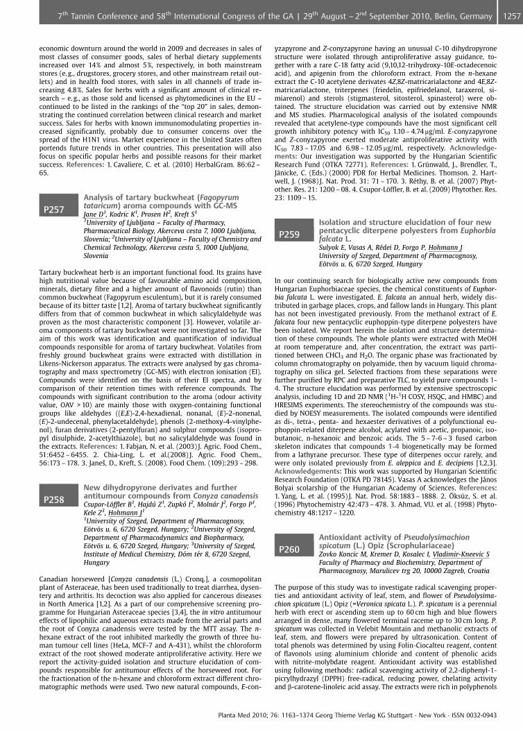
economic downturn around the world in 2009 and decreases in sales ofmost classes of consumer goods, sales of herbal dietary supplementsincreased over 14% and almost 5%, respectively, in both mainstreamstores (e.g., drugstores, grocery stores, and other mainstream retail out-lets) and in health food stores, with sales in all channels of trade in-creasing 4.8%. Sales for herbs with a significant amount of clinical re-search – e. g., as those sold and licensed as phytomedicines in the EU –continued to be listed in the rankings of the “top 20” in sales, demon-strating the continued correlation between clinical research and marketsuccess. Sales for herbs with known immunomodulating properties in-creased significantly, probably due to consumer concerns over thespread of the H1N1 virus. Market experience in the United States oftenportends future trends in other countries. This presentation will alsofocus on specific popular herbs and possible reasons for their marketsuccess. References: 1. Cavaliere, C. et al. (2010) HerbalGram. 86:62 –65.
P257Analysis of tartary buckwheat (Fagopyrumtataricum) aroma compounds with GC-MSJane D1, Kodric K1, Prosen H2, Kreft S1
1University of Ljubljana – Faculty of Pharmacy,Pharmaceutical Biology, Akerceva cesta 7, 1000 Ljubljana,Slovenia; 2University of Ljubljana – Faculty of Chemistry andChemical Technology, Akerceva cesta 5, 1000 Ljubljana,Slovenia
Tartary buckwheat herb is an important functional food. Its grains havehigh nutritional value because of favourable amino acid composition,minerals, dietary fibre and a higher amount of flavonoids (rutin) thancommon buckwheat (Fagopyrum esculentum), but it is rarely consumedbecause of its bitter taste [1,2]. Aroma of tartary buckwheat significantlydiffers from that of common buckwheat in which salicylaldehyde wasproven as the most characteristic component [3]. However, volatile ar-oma components of tartary buckwheat were not investigated so far. Theaim of this work was identification and quantification of individualcompounds responsible for aroma of tartary buckwheat. Volatiles fromfreshly ground buckwheat grains were extracted with distillation inLikens-Nickerson apparatus. The extracts were analysed by gas chroma-tography and mass spectrometry (GC-MS) with electron ionisation (EI).Compounds were identified on the basis of their EI spectra, and bycomparison of their retention times with reference compounds. Thecompounds with significant contribution to the aroma (odour activityvalue, OAV > 10) are mainly those with oxygen-containing functionalgroups like aldehydes ((E,E)-2,4-hexadienal, nonanal, (E)-2-nonenal,(E)-2-undecenal, phenylacetaldehyde), phenols (2-methoxy-4-vinylphe-nol), furan derivatives (2-pentylfuran) and sulphur compounds (isopro-pyl disulphide, 2-acetylthiazole), but no salicylaldehyde was found inthe extracts. References: 1. Fabjan, N. et al. (2003) J. Agric. Food Chem.,51:6452 – 6455. 2. Chia-Ling, L. et al.(2008) J. Agric. Food Chem.,56:173 – 178. 3. JaneÐ, D., Kreft, S. (2008). Food Chem. (109):293 – 298.
P258New dihydropyrone derivates and furtherantitumour compounds from Conyza canadensisCsupor-L�ffler B1, Hajdffl Z1, Zupk� I2, Moln�r J2, Forgo P1,Kele Z3, Hohmann J1
1University of Szeged, Department of Pharmacognosy,E�tv�s u. 6, 6720 Szeged, Hungary; 2University of Szeged,Department of Pharmacodynamics and Biopharmacy,E�tv�s u. 6, 6720 Szeged, Hungary; 3University of Szeged,Institute of Medical Chemistry, D�m t�r 8, 6720 Szeged,Hungary
Canadian horseweed [Conyza canadensis (L.) Cronq.], a cosmopolitanplant of Asteraceae, has been used traditionally to treat diarrhea, dysen-tery and arthritis. Its decoction was also applied for cancerous diseasesin North America [1,2]. As a part of our comprehensive screening pro-gramme for Hungarian Asteraceae species [3,4], the in vitro antitumoureffects of lipophilic and aqueous extracts made from the aerial parts andthe root of Conyza canadensis were tested by the MTT assay. The n-hexane extract of the root inhibited markedly the growth of three hu-man tumour cell lines (HeLa, MCF-7 and A-431), whilst the chloroformextract of the root showed moderate antiproliferative activity. Here wereport the activity-guided isolation and structure elucidation of com-pounds responsible for antitumour effects of the horseweed root. Forthe fractionation of the n-hexane and chloroform extract different chro-matographic methods were used. Two new natural compounds, E-con-
yzapyrone and Z-conyzapyrone having an unusual C-10 dihydropyronestructure were isolated through antiproliferative assay guidance, to-gether with a rare C-18 fatty acid (9,10,12-trihydroxy-10E-octadecenoicacid), and apigenin from the chloroform extract. From the n-hexaneextract the C-10 acetylene derivates 4Z,8Z-matricarialactone and 4E,8Z-matricarialactone, triterpenes (friedelin, epifriedelanol, taraxerol, si-miarenol) and sterols (stigmasterol, sitosterol, spinasterol) were ob-tained. The structure elucidation was carried out by extensive NMRand MS studies. Pharmacological analysis of the isolated compoundsrevealed that acetylene-type compounds have the most significant cellgrowth inhibitory potency with IC50 1.10 – 4.74 mg/ml. E-conyzapyroneand Z-conyzapyrone exerted moderate antiproliferative activity withIC50 7.83 – 17.05 and 6.98 – 12.05 mg/ml, respectively. Acknowledge-ments: Our investigation was supported by the Hungarian ScientificResearch Fund (OTKA 72771). References: 1. Gr�nwald, J., Brendler, T.,J�nicke, C. (Eds.) (2000) PDR for Herbal Medicines. Thomson. 2. Hart-well, J. (1968) J. Nat. Prod. 31: 71 – 170. 3. R�thy, B. et al. (2007) Phyt-other. Res. 21: 1200 – 08. 4. Csupor-L�ffler, B. et al. (2009) Phytother. Res.23: 1109 – 15.
P259Isolation and structure elucidation of four newpentacyclic diterpene polyesters from Euphorbiafalcata L.Sulyok E, Vasas A, R�dei D, Forgo P, Hohmann JUniversity of Szeged, Department of Pharmacognosy,E�tv�s u. 6, 6720 Szeged, Hungary
In our continuing search for biologically active new compounds fromHungarian Euphorbiaceae species, the chemical constituents of Euphor-bia falcata L. were investigated. E. falcata an annual herb, widely dis-tributed in garbage places, crops, and fallow lands in Hungary. This planthas not been investigated previously. From the methanol extract of E.falcata four new pentacyclic euphoppin-type diterpene polyesters havebeen isolated. We report herein the isolation and structure determina-tion of these compounds. The whole plants were extracted with MeOHat room temperature and, after concentration, the extract was parti-tioned between CHCl3 and H2O. The organic phase was fractionated bycolumn chromatography on polyamide, then by vacuum liquid chroma-tography on silica gel. Selected fractions from these separations werefurther purified by RPC and preparative TLC, to yield pure compounds 1-4. The structure elucidation was performed by extensive spectroscopicanalysis, including 1D and 2D NMR (1H-1H COSY, HSQC, and HMBC) andHRESIMS experiments. The stereochemistry of the compounds was stu-died by NOESY measurements. The isolated compounds were identifiedas di-, tetra-, penta- and hexaester derivatives of a polyfunctional eu-phoppin-related diterpene alcohol, acylated with acetic, propanoic, iso-butanoic, n-hexanoic and benzoic acids. The 5 – 7-6 – 3 fused carbonskeleton indicates that compounds 1-4 biogenetically may be formedfrom a lathyrane precursor. These type of diterpenes occur rarely, andwere only isolated previously from E. aleppica and E. decipiens [1,2,3].Acknowledgements: This work was supported by Hungarian ScientificResearch Foundation (OTKA PD 78145). Vasas A acknowledges the J�nosBolyai scolarship of the Hungarian Academy of Sciences. References:1. Yang, L. et al. (1995) J. Nat. Prod. 58:1883 – 1888. 2. �ks�z, S. et al.(1996) Phytochemistry 42:473 – 478. 3. Ahmad, VU. et al. (1998) Phyto-chemistry 48:1217 – 1220.
P260Antioxidant activity of Pseudolysimachionspicatum (L.) Opiz (Scrophulariaceae)Zovko Koncic M, Kremer D, Kosalec I, Vladimir-Kneevic SFaculty of Pharmacy and Biochemistry, Department ofPharmacognosy, Marulicev trg 20, 10000 Zagreb, Croatia
The purpose of this study was to investigate radical scavenging proper-ties and antioxidant activity of leaf, stem, and flower of Pseudolysima-chion spicatum (L.) Opiz (=Veronica spicata L.). P. spicatum is a perennialherb with erect or ascending stem up to 60 cm high and blue flowersarranged in dense, many flowered terminal raceme up to 30 cm long. P.spicatum was collected in Velebit Mountain and methanolic extracts ofleaf, stem, and flowers were prepared by ultrasonication. Content oftotal phenols was determined by using Folin-Ciocalteu reagent, contentof flavonols using aluminium chloride and content of phenolic acidswith nitrite-molybdate reagent. Antioxidant activity was establishedusing following methods: radical scavenging activity of 2,2-diphenyl-1-picrylhydrazyl (DPPH) free-radical, reducing power, chelating activityand b-carotene-linoleic acid assay. The extracts were rich in polyphenols
Planta Med 2010; 76: 1163–1374 Georg Thieme Verlag KG Stuttgart · New York · ISSN 0032-0943
12577th Tannin Conference and 58th International Congress of the GA | 29th August – 2nd September 2010, Berlin, Germany
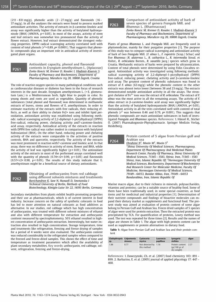
(211 – 831 mg/g), phenolic acids (2 – 27 mg/g) and flavonoids (16 –37 mg/g). In all the analyses the extracts were found to possess markedantioxidant activities. The activity of extracts in b-carotene-linoleic acidassay was significantly higher than the activity of butylated hydroxya-nisole (BHA) (ANOVA, p < 0.05). In most of the assays, activity of stemand leaf extracts was somewhat less pronounced than the activity offlower extract. However, leaf extract demonstrated the strongest redu-cing properties. DPPH radical scavenging activity correlated well withcontent of total phenols (r2= 0.89, p< 0.0001). That suggests that pheno-lic compounds play an important role in antiradical activity of investi-gated plant organs.
P261Antioxidant capacity, phenol and flavonoidcontents in Eryngium amethystinum L. (Apiaceae)Zovko Koncic M, Kremer D, Kosalec I, Vladimir-Kneevic SFaculty of Pharmacy and Biochemistry, Department ofPharmacognosy, Marulicev trg 20, 10000 Zagreb, Croatia
The role of reactive oxygen species in many pathological conditions suchas cardiovascular diseases or diabetes has been in the focus of researchinterests in the past decade. Eryngium amethystinum L. (= E. glomera-tum Lam.) is a Mediterranean herb, light blue to purple in color. Itsyoung shoots and roots are used as vegetables. Quantity of phenolicsubstances (total phenol and flavonoid) was determined in methanolicextracts of leaves, stems and flowers of E. amethystinum. In order toestimate reactivity of the extracts with free radicals, their reducing ef-fects, as well as their ability to chelate Fe2+ ions and inhibit lipid per-oxidation, antioxidant activity was established using following meth-ods: radical scavenging activity of 2,2-diphenyl-1-picrylhydrazyl (DPPH)free-radical, reducing power, chelating activity and?-carotene-linoleicacid assay, respectively. Antiradical activity of the extracts in reactionwith DPPH free radical was rather modest in comparison with butylatedhidroxianisol (BHA). On the other hand, reducing power and chelatingactivity of the extracts were comparable to the activities of positivecontrols, BHA and quercetin, respectively. The activity of the extractswas most prominent in reaction with?-carotene and linoleic acid. In thatassay, there was no difference in activity of stem, flower and BHA, whilethe activity of leaf was significantly higher (p < 0.01). The antioxidantactivity of the extracts in most of the performed assays correlated wellwith the quantity of phenols (0.74< r2< 0.99, p < 0.05) and flavonoids(0.57< r2< 0.90, p < 0.05). The results of this study indicate that E.amethystinum might be a beneficial source of dietary antioxidants.
P262Obtaining of anthocyanins from red cabbageusing different solvents mixtures and treatmentsRavichandran K, Saw N, Hunaefi D, Smetanska ITechnical University of Berlin, Methods of FoodBiotechnology, K�nigin-Luise Str 22, 14195 Berlin, Germany
Secondary metabolites from plants exhibit health-promoting propertiesand their use as pharmaceuticals, which is of current interest in foodindustry. Increase concern on the safety of synthetic colorants in foodhas led to more attention on natural colorants as food additives asalternative. In our studies, red cabbage which has high concentrationof anthocyanins, was treated with different solvent mixtures of ethanoland also with different temperature for extraction and anthocyaninconttent measured by spectrophotometry. 50% ethanol resulted in high-est concentration of anthocyanin extraction. Interestingly, hot water ex-traction also resulted in high concentration. Storage temperature, timeand treatments like refrigeration, freezing and freeze drying of samplesfor a period of 4 weeks were also evaluated. The anthocyanin contentwas reduced considerably in the refrigerated samples when compared tothe freezed and freeze-dried samples. This shows the effect of time andtemperature as treatment parameters which affect the availability ofplant secondary metabolites. Key words: anthocyanin, red cabbage, sol-vent, refrigeration, freezing and freeze-drying.
P263Comparison of antioxidant activity of bark ofseven species of genera Frangula Mill. andRhamnus L. (Rhamnaceae)Zovko Koncic M, Kremer D, Kosalec I, Vladimir-Kneevic SFaculty of Pharmacy and Biochemistry, Department ofPharmacognosy, Marulicev trg 20, 10000 Zagreb, Croatia
Plants of genus Rhamnus L. and Frangula Mill. are frequently used inphytomedicine, mainly for their purgative properties [1]. The purposeof this study was to compare radical scavenging and antioxidant activityof bark of two Frangula Mill. (F. alnus L., F. rupestris (Scop.) Schur) andfive Rhamnus (R. alaternus L., R. cathartica L., R. intermedia Steud. etHohst., R. orbiculata Bornm., R. saxatilis Jacq.) species which grow inCroatia. Methanolic extracts of barks were prepared by ultrasonication.Content of total phenols were determined by using Folin Ciocalteu re-agent. Antioxidant activity was investigated using following methods:radical scavenging activity of 2,2-diphenyl-1-picrylhydrazyl (DPPH)free-radical, reducing power, chelating activity and b-carotene-linoleicacid assay. The greatest content of phenolic substances was found inextract of F. alnus (63 mg/g) while the phenolic content in the otherextracts was almost twice lower (between 38 and 23 mg/g). The extractsdemonstrated notable antioxidant activity in all the assays. The stron-gest chelator of Fe2+ ions was the extract of R. orbiculata. The extract of F.alnus was the most active in all the remaining assays. The activity of F.alnus extract in b-carotene-linoleic acid assay was significantly higherthan the activity of butylated hydroxyanisole (BHA) (ANOVA, p< 0.05).Antioxidant activity in all the tests correlated well with content of totalphenols (r2 was between 0.46 and 0.79, p< 0.001). This suggests thatphenolic compounds are main antioxidant substances in bark of inves-tigated Frangula and Rhamnus species. References: 1. H�nsel R., Sticher�. (2007) Pharmakognosie-Phytopharmacie. Springer Medizin Verlag,Heidelberg.
P264Protein content of 5 algae from Persian gulf andArabian seaEbrahimi N1, Moein M1, Moein S2
1Shiraz University Of Medical Sciences, Pharmacognosy,Department Of Pharmacognosy And Medicinal PlantsResearch Center, Faculty Of Pharmacy, Shiraz University OfMedical Sciences, 71345 – 1583, Shiraz, Iran, 71345 – 1583Shiraz, Iran, Islamic Republic Of; 2Hormozgan University OfMedical Sciences, Biochemistry, Department Of BiochemistryAnd Research Center For Molecular Medicine, Faculty OfMedicine, Hormozgan University Of Medical Sciences,79149 – 64153, Bandar Abbas, Iran, 79149 – 64153Bandarabbas, Iran, Islamic Republic Of
Marine macro algae, due to their richness in minerals, polysaccharides,vitamins and proteins; can be a suitable source of healthy food. Some ofthem have been traditionally used, in some special countries, as fooditems and for medicinal and industrial properties [1]. Determination oftheir nutrient compounds and findings of bioactive molecules can ex-pand their dietary market as supplements and functional food. The pre-sent study was aimed at evaluation of protein content of some algaebelong to Persian Gulf and Arabian Sea. Freeze dried samples of 5 speciesof algae were used for protein extraction. Then the extracted protein wasprecipitated by TCA. For quantification of proteins, Lowry method wasused. The test was repeated for three times [2]. Results and the names ofalgae are shown in Table 1. The algae with high protein content can beused as supplements or protein alternatives in dietary food.
Table 1: Algae from Persian Gulf and Arabian Sea and their protein con-tent
Cladophropsissp.
Colpomeniasinousa
Cystoceriamyrica
Laurenciapapillosa
Iyengariastellata
algae
1.88 0.77 Not detected bythis method
1.16 Protein content(gr/ 100 gr ofdry weight)
2.27
References: 1. Dawczynski, Ch. et al. (2007) food chemistry 103: 891 –899. 2. Barbarino, E. et al. (2005) journal of applied phycology 17: 447 –460.
Planta Med 2010; 76: 1163–1374 Georg Thieme Verlag KG Stuttgart · New York · ISSN 0032-0943
1258 7th Tannin Conference and 58th International Congress of the GA | 29th August – 2nd September 2010, Berlin, Germany
P265Chemical composition and antioxidant activity ofAllium flavum L. extractsSimin N1, Mimica-Dukic N1, Lesjak M1, Beara I1, Orcic D1,Jovin E1, Bozin B2
1Faculty of Sciences, Department of chemistry, biochemistryand environmental protection, Trg d. Obradovica 3, 21000Novi Sad, Serbia, Republic of; 2Faculty of Medicine,Department of Pharmacy, Hajduk Veljkova 3, 21000 NoviSad, Serbia, Republic of
Members of the genus Allium have been used and cultured for thou-sands of years for their spiritual meaning, medicinal properties, pun-gency and characteristic taste. Only two species of genus Allium (A.sativum L. and A. cepa L.) are well researched, while data on the chemi-cal composition and biological activities of other species, including Al-lium flavum L. are very rare. In the present study we investigated che-mical composition and antioxidative properties of methanolic extractsof Allium flavum L. (subgen. Allium/sect. Codonoprasum) from five loca-tions in Serbia. Phytochemical profile was determined by measuringtotal phenolic, total flavonoid and total anthocyanin content and byHPLC-DAD-MS/MS analysis of methanolic extracts of whole plants andheadspace GC/MS analysis of volatile compounds of fresh bulbs. Flavorintensity associated with sulfur compounds was determined by measur-ing alliinase activity. The antioxidant activity was evaluated by measur-ing their 2,2-diphenyl-1-pycrylhydrazile (DPPH) radicals scavenging ca-pacity, ferric reducing ability (FRAP) and effect on lipid peroxidation(LP). High contents of total phenolics (5.5 – 14.0 mg gallic acid equiva-lents/g of dry extract) and total flavonoids (1.9 – 5.0 mg quercetinequivalents/g of dry extract) were found, while anthocianin contentwas very low (11 – 33 mg cyanidin-3-glucoside equivalents/g of dry ex-tract). Allinase activity was found to be extremely high (28 – 48 mgpyruvic acid/g of dry extract), while amount of volatile compoundswas negligibly small. Chemical profile of methanolic extracts of A. fla-vum from different locations was independent of environmental condi-tions, only small differences in quantitative composition were found.Dominant flavonoid is dihexosyl quercetin. Investigated extracts exhib-ited high antioxidant activity.
P266Application of an HPLC method for analysis of theextract from Paullinia cupana var. sorbilis (Mart.)DuckeMello J, Klein T, Lopes GUniversidade Estadual de Maring�, Departamento deFarm�cia e Farmacologia, Avenida Colombo, 5790 ZonaSete, 87020900 Maring�, Brazil
Paullinia cupana var. sorbilis (Mart.) Ducke (Sapindaceae), popularlyknown as “Guaran�”, is a tropical South American plant found in north-ern Brazil. Guaran� is widely used in medicine, cosmetics, and industrydue to its versatile biological activities, and is popularly used as a sti-mulant of the central nervous system (CNS) in cases of intellectual andphysical stress, and as an antidiarrheic, diuretic, and antineuralgic agent[1,2]. It has shown antioxidant, anti-amnesia, and antidepressive effectsin animal models [3 – 7]. These activities are mainly attributed to thepresence of polyphenols, which explains the interest in quantifyingthese constituents in guaran� preparations, as well as in validating ana-lytical methodologies. Caffeine, epicatechin, catechin, and procyanidinsB1, B2, B3, B4, A2, and C 1 have been isolated and identified from itssemipurified extract [1]. High-performance liquid chromatography(HPLC) methods have been used to quantify isolated polyphenols orthese compounds in complex biological matrices, such as herbal rawmaterials and extractive preparations. To separate and identify somesubstances present in the extract, we developed an RP-HPLC method.The chromatograms were obtained from various gradient elution sys-tems, in order to establish the ideal conditions for the analysis of guar-an� extract, using 0.05% TFA methanol: acetonitrile (25:75, v/v) and0.05% TFA water as the mobile phase. Gradient reversed-phase chroma-tography was performed using a stainless-steel column (250 x 4.6 mmi.d., 4 mm) and detection at 210 nm. The results demonstrate the effi-ciency of separation using the proposed method. Acknowledgements:CAPES, CNPq, INCT_if. References: 1. Henman, A R (1982) J. Ethnophar-macol. 6: 311 – 338. 2. Yamaguti-sasaki, E et al. (2007). Molecules 12:1950 – 1963. 3. Audi, EA, Mello, JCP (2000) Funda�¼o Universidade Esta-dual de Maring�, Cl. Int. A61P 25/24; A61K 35/78. BR #PI00066389. 28/11/2000. 4. Otobone, FJ et al. (2005) Braz. Arch. Biol. Techn. 48:723 –728. 5. Otobone, FJ et al (2007) Phitoter. Res. 21:531 – 535. 6. Espinola,
EB et al. (1997) J. Ethnopharmacol. 55:223 – 229. 7. Mattei, R et al.(1998) J. Ethnopharmacol. 60:111 – 116.
P267Semisynthesis and pharmacological investigationof lipo-alkaloids prepared from aconitine bytransesterification with eicosanoic acidanaloguesBorcsa B1, Widowitz U2, Csupor D1, Forgo P1, Bauer R2,Hohmann J1
1University of Szeged, Institute of Pharmacognosy,E�tv�s u. 6., 6720 Szeged, Hungary; 2Karl-FranzensUniversity Graz, Department of Pharmacognosy,Universit�tsplatz 4., 8010 Graz, Austria
Previously we reported the semisynthesis of 9 aconitine-derived lipo-alkaloids transesterified with fatty acids differing in the number of car-bon atoms and double bonds in their chains [1]. When these compoundswere tested for COX-1, COX-2 and LTB4 formation inhibitory activities itwas found that 14-benzoylaconine-8-O-eicosapentaenoate exhibited no-table activities through all three in vitro anti-inflammatory test systems(inhibition (%): COX-1: 54.51, COX-2: 66.07, LTB4: 45.96, at 50mM con-centration). Although no data exist on the natural occurrence of aconi-tine derived lipo-alkaloids containing eicosanoic acid analogues at C-8,the prospective of further charting the possible structure-activity rela-tionships of lipo-alkaloids is reinforced by papers dealing with the im-portance and natural roles of different unsaturated eicosanoic acid deri-vatives in the process of inflammation [2]. The present paper reports thesemisynthesis of 7 eicosanoate analogues of aconitine-derived lipo-al-kaloids, prepared according to the modified method of Bai et al [3]. Inthe reactions, aconitine was transesterified by eicosanoic, 11-eicosenoic,8,11,14-eicosatrienoic, 11,14,17-eicosatrienoic, 8,11,14,17-eicosatetraenoicand 5,8,11,14,17-eicosapentaenoic acids resulting the corresponding 14-benzoylaconin-8-O-esters and pyroaconitine. The reaction mixtureswere purified of necessity by gelfiltration, preparative TLC and centrifu-gal planar chromatography. Purity of the obtained lipo-alkaloids wasproved with the aid of NMR spectroscopy. The COX-1, COX-2 and LTB4
formation inhibitory activities of this eicosanoate lipo-alkaloid serieswere also investigated. It was observed that the 14-benzoylaconine-8-O-eicosapentaenoate has the highest activities (inhibition (%): COX-1:82.84, COX-2: 33.67, at 50 mM concentration) between the eicosanoateanalogues. References: 1. Borcsa, B. et al. (2009) Planta Med. 75: 910. 2.Rouzer, C. et al. (2009) J. Lipid Res. 50: S29 – 34. 3. Bai, Y. et al. (1994) J.Nat. Prod. 57: 963 – 970.
P268Ligusticum mutellina (L.) Crantz:a pharmacognostic overviewPeev C1, Cupara S2, Vlase L3, Feflea S1, Corina T1,Munteanu M4
1Faculty of Farmacy, Pharmacognosy, Eftimie Murgu Squareno.1, 300041 Timisoara, Romania; 2Medical FacultyUniversity of Kragujevac, Svetozara, Serbia, Svetozara,Serbia, Republic of; 3Faculty of Pharmacy, UMF Cluj NapocaIuliu Hatieganu, Department of Biofarmacy Cluj-Napoca,Romania; 4West University of Vasile Goldis Arad, Romania
Mountain lovage represents the plant Ligusticum mutellina (L.) Crantz orMeum mutellina (L.) Gaert, Apiaceae. It naturally grows in alpine areas,and is known as an antitumoral ethnobotanic remedy. Also, the roots areused in the production of a type of Schnapps in Bayern, Germany [1].The present study consists of an analysis of some pharmacognosticparameters describing the aerial part of the plant, harvested from moun-tains in northern Serbia. Polyphenolic compounds were determined inthe methanolic extract performing HPLC MS analysis. Microelementsand heavy metals were quantified through the SAA technique [2,3].The antimicrobial activity of an ethanolic extract was estimated and anevaluation of the antiproliferative potential was done by the phytobio-logic test on Lepidium sativum. Effects on the angiogenic process weredetermined by performing the chick chorioallantoic membrane assay[4]. 6 polyphenolic structures were revealed in the nonhydrolyzed sam-ple; rutoside was found in high concentration (79.23ug/ml). In the hy-drolyzed sample 4 polypheolic compounds were evidenced with quer-cetin being the most concentrated compound (13.83ug/ml). The vegetalproduct is in conformity with the official quality parameters concerningthe content of heavy metals. The ethanolic extract proved to be active onthe selected bacterial cultures. The aqueous extracts in concentrationsbetween 0,35% and 6% expressed an inhibitory effect of 70 – 89%. The
Planta Med 2010; 76: 1163–1374 Georg Thieme Verlag KG Stuttgart · New York · ISSN 0032-0943
12597th Tannin Conference and 58th International Congress of the GA | 29th August – 2nd September 2010, Berlin, Germany
CAM assay showed possible antiangiogenic activity, dependent on theconcentration of the extractive solution. The results obtained indicate anantimicrobial and a possible antiproliferative effect of different extractsof Ligusticum mutellina (L.) Crantz. References: 1. Peev, C. et al. (2007)Chem. Nat. Comp. 43(3): 259 – 262. 2. Peev, C. et al.(2006) Tim. Med. J.56(2): 233 – 36. 3. Peev, C. (2007) Mugurii foliari, materii prime in ge-moterapie. Mirton. Timisoara. 29 – 42. 4. Feflea, S.et al. (2009) Rev. Med.Farm.55(3), 346 – 349.
P269The antioxidant activity of wild rose fruitPeev C1, Dehelean C1, Tiulea C1, Feflea S1, Dragan S2
1Faculty of Farmacy, Pharmacognosy, Eftimie Murgu Squareno.1, 300041 Timisoara, Romania; 2Faculty of Medecine,University of Medicine and PharmacyVictor Babes,Department of Cardiology, Eftimie Murgu Square no.1,300041, Timisoara, Romania
The vegetal products used in the pharmaceutical and food area from thewild rose (Rosa canina, L. Rosaceae) are the fruits and the seeds, Cynos-bati fructus and Cynosbati semen. The fruits contain vitamins (C, B, PP,K), carotenoids, pectins, tannins, carbohydrates, while the seeds containfats, oil and vitamin F [1,2]. The present study aims to analyze two typesof 6% extractive solutions (hydroalchoolic maceration and hydro – infu-sion) of dried and 6 months – frozen wild rose fruits, by determining thecontent of: vitamin C (titrimetric analysis), total polyphenols (Folin Cio-c�lteu method), total carotenoids (spectrophotometrically) as well as byestablishing the antioxidant activity (DPPH method and ORAC method)[3,4]. The vegetal products were harvested in October 2009 from thehills in the Danube river area, Moldova Noua, Romania. Dried wild rosefruits showed the highest concentration of vitamin C (154.20 mg/100 g).The content of total polyphenols was similar for both dried and frozensamples. The antioxidant activity of the extractive solutions was notsignificantly influenced by the type of extraction or extractive solvent,but higher values were obtained in the case of frozen wild rose fruits(71,27 Eq Trolox/1 ml comparing to the extractive solutions of dry fruits41,03 Eq Trolox/1 ml by ORAC method). Acknowledgements: BilateralProject No. 198/2009 References: 1. Molay, K.R. et al. (2010) Int. J. FoodSc. Nutr. 61:109 – 124. 2. Kilicgun, H. et al.(2009) Pharmacogn. Res.6:417 – 420. 3. Peev, C. (2007) Mugurii foliari, materii prime in gemoter-apie. Mirton. Timisoara. 26 – 35. 4. Szajdek, A. et al. (2008) Plant FoodsHuman Nutr. 63:147 – 156.
P270Antioxidant activity and composition of essentialoils of four cultivated aromatic plants inNorth-West GreeceLazari D, Hadjipavlou-Litina DAristotle University of Thessaloniki, School of Pharmacy,Laboratory of Pharmacognosy, 54124 Thessaloniki, Greece
There is a remarkable development of interest in medicinal and aro-matic plants worldwide and their cultivation may contribute to theamelioration of the local agricultural economies. In the present study,the essential oil composition of four cultivated aromatic plants in North-West Greece has been investigated by capillary GC-MS with an HP-5column and with an EI detector: Ocimum basilicum, Salvia officinalis,Origanum vulgare subsp. hirtum and Thymus vulgaris. The major com-pounds found in essential oils were (a) for O. basilicum: linalool (55.2%)and a-bergamotene (5.9%), (b) for S. officinalis: a-pinene (6.8%), euca-lyptol (17.5%), a-thujone (30.5%), b-thujone (6.0%), camphor (12.7%) anda-caryophyllene (6.2%), (c) for O. vulgare subsp. hirtum: p-cymene(11.7%), g-terpinene (14.5%) and carvacrol (51.9%) and (d) for T. vulgaris:p-cymene (29.6%), g-terpinene (16.3%) and thymol (26.8%). Moreover,the essential oils were tested for their free radical scavenging activityusing the following in vitro assays: i) interaction with the free stableradical of DPPH (1,1-diphenyl-2-picrylhydrazyl), ii) inhibition of linoleicacid peroxidation induced by the dihydrochlroic acid of 2,2-Azabis-2-aminepropane (AAPH). Finally, their inhibitory activity toward soybeanlipoxygenase was evaluated, using linoleic acid as substrate. All thesamples presented interesting antioxidant activity. They found tostrongly inhibit lipid peroxidation as well as soybean lipoxygenase. Ac-knowledgements: Regional Development Fund of the Region of Wes-tern Macedonia for financial support
P271Study on antitumoral activity of some chemicalcompounds isolated from Origanum vulgare ssp.vulgareGuvenalp Z1, Turan M2, S�mer Z2, G�ll�ce M3, Ozbek H1,Baris O3, Orhan F3, Demirezer L4
1Ataturk University, Faculty of Pharmacy, Department ofPharmacognosy, 25240 Erzurum, Turkey; 2CumhuriyetUniversity, Faculty of Medicine, Department of Surgery,58140 Sivas, Turkey; 3Ataturk University, Faculty of Science,Department of Biology, 25240 Erzurum, Turkey; 4HacettepeUniversity, Faculty of Pharmacy, Department ofPharmacognosy, 06100 Ankara, Turkey
In vivo and in vitro studies have shown that some natural compounds,obtained from plants have regulatory roles on xenobiotic effects. Char-acterization of these compounds and determination of antimutagenicand anticarcinogenic effects of them has provided an important strategyfor decreasing the propagation of cancer disease in human beings. Thegenus Origanum (Lamiaceae) is represented by 23 species and 6 hybridesin Turkish flora and 14 of them are endemic [1 – 2]. Origanum speciesare used in infusion form (2%) as diaphoretic, di�retic, carminative andsedative in folk medicine [3]. In this study the chemical composition ofOriganum vulgare (Lamiaceae) and its anticarcinogenic effect, and effectmechanism were studied. Plant material was collected from Erzurum-Oltu in flowering period. Powdered dried aerial parts of this plant wereextracted with methanol. After evaporation of the solvent, the cruderesidue was suspended in water and successively extracted with petro-leum ether, chloroform and n-butanol, respectively. The organic layerswere evaporated to dryness. As a result of chromatographical studiesLuteolin-7-O-glucuronide, Luteolin-7-O-xyloside, Rosmarinic acid,Lithospermic acid and Lithospermic acid B were isolated. Antitumorproperties of these compounds were investigated by using MCF-7 breastcancer cell sequence. Trypan blue extraction and MTT assay methodswere used for the antitumor studies. Luteolin-7-O-glucuronide, Luteo-lin-7-O-xyloside and Rosmarinic acid have shown moderate antitumoralactivity. Acknowledgements: This study was supported by grants fromthe Scientific and Technological Research Council of Turkey (TUBITAK).(Project No: 107T203). References: 1. Ietswaart, J.H. (1982) In: “Flora ofTurkey and the East Aegean Islands”, Ed. P.H. Davis. Univ. Press. Edin-burgh. 2. Duman, H. (2000) In: “Flora of Turkey and the East AegeanIslands” (supplement 2), Eds. G�ner, A. et al. Univ. Press. Edinburgh. 3.Baytop, T. (1984) Therapy with medicinal plants in Turkey (past andpresent). Istanbul �niversitesi Yayinlari. Istanbul.
P272Antioxidant activity, total phenol and flavonoidcontents of Sideritis montana L. from SerbiaStankovic MFaculty of Science, Department of Biology and Ecology,Department of Biology and Ecology, Radoja Domanovica 12,34000 Kragujevac, Serbia, Republic of
In this study were determined the concentration of phenolic com-pounds, flavonoids and in vitro antioxidant activity of water, methanoland acetone extracts, from the whole herb of Sideritis montana usingspectrophotometric methods. Sideritis montana L. (Lamiaceae) is theannual species with low branched trunk, up to 40 cm high. Inhabits sandarid meadows and rocky in the Europe and the Mediterranean [1]. Intraditional medicine is known as ironwort and used for the antispasmo-dic, carminative [2], antimicrobial [3] and antistress [4] effects. Plantmaterial was collected from the Stara Planina Mt. in eastern Serbia.The total phenolic content of the extracts ranged from 47.35 mg/g to97.85 mg/g dry weight of extract, expressed as gallic acid equivalents.The total flavonoid concentrations varied from 24.13 mg/g to 206.4 mg/g,expressed as rutin equivalents. Acetone extract had the highest flavo-noid concentration of 206.4 mg/g. Antioxidant activity of extracts wereexpressed as IC50 values (mg/ml) and ranged from 69.37 mg/mL to229.3 mg/mL. The high contents of phenolic compounds and flavonoidsindicated that these compounds contribute to the antioxidant activity.Based on these results, Sideritis montana is a potential source of flavo-noids as a natural antioxidant substances of high value. References:1. Josifovic, M. (1974) Flora SR Srbije. SANU. Beograd. 2. Tabanca, N. etal. (1998) Turk. J. Chem. 25:201 – 208. 3. Kursat, M. et al. (2009) Turk. J.of Sci. & Tehn. 4(1):81 – 85. 4. �zt�rk, Y. et al. (1998) Phytother. Res.10(1):70 – 73.
Planta Med 2010; 76: 1163–1374 Georg Thieme Verlag KG Stuttgart · New York · ISSN 0032-0943
1260 7th Tannin Conference and 58th International Congress of the GA | 29th August – 2nd September 2010, Berlin, Germany
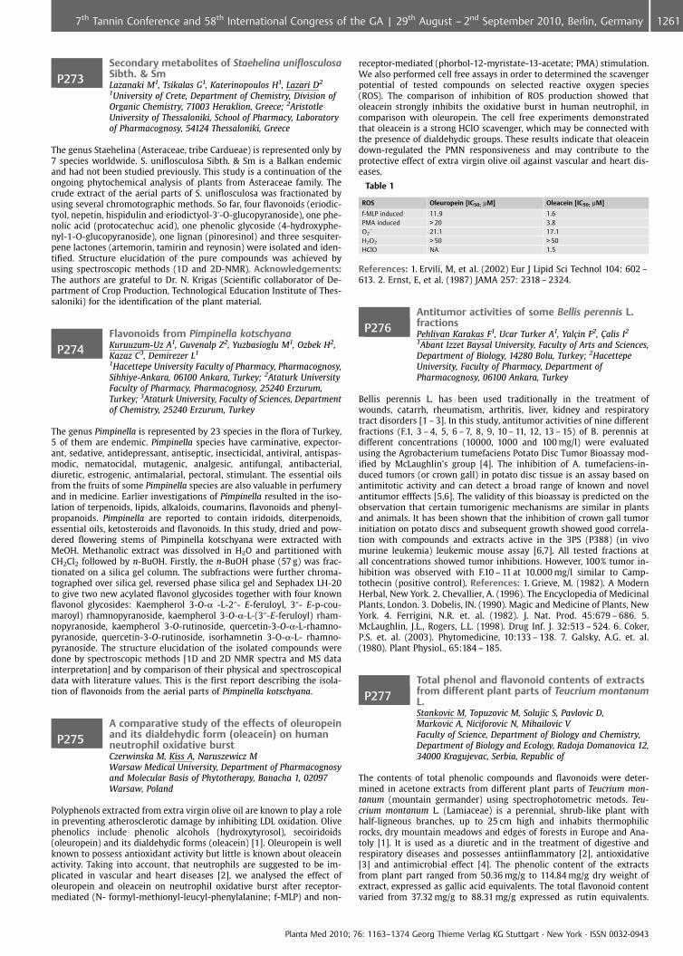
P273Secondary metabolites of Staehelina uniflosculosaSibth. & SmLazanaki M1, Tsikalas G1, Katerinopoulos H1, Lazari D2
1University of Crete, Department of Chemistry, Division ofOrganic Chemistry, 71003 Heraklion, Greece; 2AristotleUniversity of Thessaloniki, School of Pharmacy, Laboratoryof Pharmacognosy, 54124 Thessaloniki, Greece
The genus Staehelina (Asteraceae, tribe Cardueae) is represented only by7 species worldwide. S. uniflosculosa Sibth. & Sm is a Balkan endemicand had not been studied previously. This study is a continuation of theongoing phytochemical analysis of plants from Asteraceae family. Thecrude extract of the aerial parts of S. uniflosculosa was fractionated byusing several chromotographic methods. So far, four flavonoids (eriodic-tyol, nepetin, hispidulin and eriodictyol-3’-O-glucopyranoside), one phe-nolic acid (protocatechuc acid), one phenolic glycoside (4-hydroxyphe-nyl-1-O-glucopyranoside), one lignan (pinoresinol) and three sesquiter-pene lactones (artemorin, tamirin and reynosin) were isolated and iden-tified. Structure elucidation of the pure compounds was achieved byusing spectroscopic methods (1D and 2D-NMR). Acknowledgements:The authors are grateful to Dr. N. Krigas (Scientific collaborator of De-partment of Crop Production, Technological Education Institute of Thes-saloniki) for the identification of the plant material.
P274Flavonoids from Pimpinella kotschyanaKuruuzum-Uz A1, Guvenalp Z2, Yuzbasioglu M1, Ozbek H2,Kazaz C3, Demirezer L1
1Hacettepe University Faculty of Pharmacy, Pharmacognosy,Sihhiye-Ankara, 06100 Ankara, Turkey; 2Ataturk UniversityFaculty of Pharmacy, Pharmacognosy, 25240 Erzurum,Turkey; 3Ataturk University, Faculty of Sciences, Departmentof Chemistry, 25240 Erzurum, Turkey
The genus Pimpinella is represented by 23 species in the flora of Turkey,5 of them are endemic. Pimpinella species have carminative, expector-ant, sedative, antidepressant, antiseptic, insecticidal, antiviral, antispas-modic, nematocidal, mutagenic, analgesic, antifungal, antibacterial,diuretic, estrogenic, antimalarial, pectoral, stimulant. The essential oilsfrom the fruits of some Pimpinella species are also valuable in perfumeryand in medicine. Earlier investigations of Pimpinella resulted in the iso-lation of terpenoids, lipids, alkaloids, coumarins, flavonoids and phenyl-propanoids. Pimpinella are reported to contain iridoids, diterpenoids,essential oils, ketosteroids and flavonoids. In this study, dried and pow-dered flowering stems of Pimpinella kotschyana were extracted withMeOH. Methanolic extract was dissolved in H2O and partitioned withCH2Cl2 followed by n-BuOH. Firstly, the n-BuOH phase (57 g) was frac-tionated on a silica gel column. The subfractions were further chroma-tographed over silica gel, reversed phase silica gel and Sephadex LH-20to give two new acylated flavonol glycosides together with four knownflavonol glycosides: Kaempherol 3-O-a -L-2@- E-feruloyl, 3@- E-p-cou-maroyl) rhamnopyranoside, kaempherol 3-O-a-L-(3@-E-feruloyl) rham-nopyranoside, kaempherol 3-O-rutinoside, quercetin-3-O-a-L-rhamno-pyranoside, quercetin-3-O-rutinoside, isorhamnetin 3-O-a-L- rhamno-pyranoside. The structure elucidation of the isolated compounds weredone by spectroscopic methods [1D and 2D NMR spectra and MS datainterpretation] and by comparison of their physical and spectroscopicaldata with literature values. This is the first report describing the isola-tion of flavonoids from the aerial parts of Pimpinella kotschyana.
P275A comparative study of the effects of oleuropeinand its dialdehydic form (oleacein) on humanneutrophil oxidative burstCzerwinska M, Kiss A, Naruszewicz MWarsaw Medical University, Department of Pharmacognosyand Molecular Basis of Phytotherapy, Banacha 1, 02097Warsaw, Poland
Polyphenols extracted from extra virgin olive oil are known to play a rolein preventing atherosclerotic damage by inhibiting LDL oxidation. Olivephenolics include phenolic alcohols (hydroxytyrosol), secoiridoids(oleuropein) and its dialdehydic forms (oleacein) [1]. Oleuropein is wellknown to possess antioxidant activity but little is known about oleaceinactivity. Taking into account, that neutrophils are suggested to be im-plicated in vascular and heart diseases [2], we analysed the effect ofoleuropein and oleacein on neutrophil oxidative burst after receptor-mediated (N- formyl-methionyl-leucyl-phenylalanine; f-MLP) and non-
receptor-mediated (phorbol-12-myristate-13-acetate; PMA) stimulation.We also performed cell free assays in order to determined the scavengerpotential of tested compounds on selected reactive oxygen species(ROS). The comparison of inhibition of ROS production showed thatoleacein strongly inhibits the oxidative burst in human neutrophil, incomparison with oleuropein. The cell free experiments demonstratedthat oleacein is a strong HClO scavenger, which may be connected withthe presence of dialdehydic groups. These results indicate that oleaceindown-regulated the PMN responsiveness and may contribute to theprotective effect of extra virgin olive oil against vascular and heart dis-eases.
Table 1
ROS Oleuropein [IC50; mM] Oleacein [IC50; mM]
f-MLP induced 11.9 1.6PMA induced > 20 3.8O2
� 21.1 17.1H2O2 > 50 > 50HClO NA 1.5
References: 1. Ervili, M, et al. (2002) Eur J Lipid Sci Technol 104: 602 –613. 2. Ernst, E, et al. (1987) JAMA 257: 2318 – 2324.
P276Antitumor activities of some Bellis perennis L.fractionsPehlivan Karakas F1, Ucar Turker A1, Yal�in F2, �alis I2
1Abant Izzet Baysal University, Faculty of Arts and Sciences,Department of Biology, 14280 Bolu, Turkey; 2HacettepeUniversity, Faculty of Pharmacy, Department ofPharmacognosy, 06100 Ankara, Turkey
Bellis perennis L. has been used traditionally in the treatment ofwounds, catarrh, rheumatism, arthritis, liver, kidney and respiratorytract disorders [1 – 3]. In this study, antitumor activities of nine differentfractions (F.1, 3 – 4, 5, 6 – 7, 8, 9, 10 – 11, 12, 13 – 15) of B. perennis atdifferent concentrations (10000, 1000 and 100 mg/l) were evaluatedusing the Agrobacterium tumefaciens Potato Disc Tumor Bioassay mod-ified by McLaughlin’s group [4]. The inhibition of A. tumefaciens-in-duced tumors (or crown gall) in potato disc tissue is an assay based onantimitotic activity and can detect a broad range of known and novelantitumor efffects [5,6]. The validity of this bioassay is predicted on theobservation that certain tumorigenic mechanisms are similar in plantsand animals. It has been shown that the inhibition of crown gall tumorinitiation on potato discs and subsequent growth showed good correla-tion with compounds and extracts active in the 3PS (P388) (in vivomurine leukemia) leukemic mouse assay [6,7]. All tested fractions atall concentrations showed tumor inhibitions. However, 100% tumor in-hibition was observed with F.10 – 11 at 10.000 mg/l similar to Camp-tothecin (positive control). References: 1. Grieve, M. (1982). A ModernHerbal, New York. 2. Chevallier, A. (1996). The Encyclopedia of MedicinalPlants, London. 3. Dobelis, IN. (1990). Magic and Medicine of Plants, NewYork. 4. Ferrigini, N.R. et. al. (1982). J. Nat. Prod. 45:679 – 686. 5.McLaughlin, J.L., Rogers, L.L. (1998). Drug Inf. J. 32:513 – 524. 6. Coker,P.S. et. al. (2003). Phytomedicine, 10:133 – 138. 7. Galsky, A.G. et. al.(1980). Plant Physiol., 65:184 – 185.
P277Total phenol and flavonoid contents of extractsfrom different plant parts of Teucrium montanumL.Stankovic M, Topuzovic M, Solujic S, Pavlovic D,Markovic A, Niciforovic N, Mihailovic VFaculty of Science, Department of Biology and Chemistry,Department of Biology and Ecology, Radoja Domanovica 12,34000 Kragujevac, Serbia, Republic of
The contents of total phenolic compounds and flavonoids were deter-mined in acetone extracts from different plant parts of Teucrium mon-tanum (mountain germander) using spectrophotometric metods. Teu-crium montanum L. (Lamiaceae) is a perennial, shrub-like plant withhalf-ligneous branches, up to 25 cm high and inhabits thermophilicrocks, dry mountain meadows and edges of forests in Europe and Ana-toly [1]. It is used as a diuretic and in the treatment of digestive andrespiratory diseases and possesses antiinflammatory [2], antioxidative[3] and antimicrobial effect [4]. The phenolic content of the extractsfrom plant part ranged from 50.36 mg/g to 114.84 mg/g dry weight ofextract, expressed as gallic acid equivalents. The total flavonoid contentvaried from 37.32 mg/g to 88.31 mg/g expressed as rutin equivalents.
Planta Med 2010; 76: 1163–1374 Georg Thieme Verlag KG Stuttgart · New York · ISSN 0032-0943
12617th Tannin Conference and 58th International Congress of the GA | 29th August – 2nd September 2010, Berlin, Germany
The amount of phenolic compounds and flavonoids is very uneven indifferent parts of the plant. The highest content of phenolic compounds(114.84 mg/g) was recorded in acetone extract of flowers and the highestcontent of flavonoids (88.31 mg/g) was measured in acetone extract ofleaves. Herbal extract from stem contains very low concentrations ofphenolic compounds and flavonoids. Obtained values indicate that theflowers and leaves of Teucrium montanum species are very rich sourcesof phenolic compounds. References: 1. Josifovic, M. (1974) Flora SRSrbije. SANU. Beograd. 2. Saric, M. (1989) Lekovite biljke SR Srbije. SANU.Beograd. 3. Canadanovic – Brunet, J. et al. (2009) Int. J. of food. Sci. andTechn. 41(6):667 – 673. 4. Vukovic, N. et al. (2008) J Serb Chem73(3):299 – 305.
P278Phytochemical analysis of alkaloids from theIcelandic club moss Diphasiastrum alpinumHalldorsdottir E, Palmadottir R, Olafsdottir EUniversity of Iceland, Faculty of Pharmaceutical Sciences,Hofsvallagata 53, 107 Reykjavik, Iceland
More than 500 species of club mosses grow all around the world, butonly five of them are represented in Iceland: Lycopodium annotinum,Selaginella selaginoides, Diphasiastrum alpinum, Huperzia selago, andLycopodium clavatum. Club mosses produce a range of secondary me-tabolites called lycopodium alkaloids and some of them in particularhuperzine A have shown interesting anticholinesterease activity (1).The present study is a phytochemical analysis of Diphasiastrum alpinum(Lycopodium alpinum) collected in Iceland. Previous studies on D. alpi-num described the isolation of four alkaloids: lycopodine, clavolonine,lycoclavine and des-N-methyl-a-obscurine (2), however our investiga-tion has shown that it contains at least eight alkaloids. The aim of thisstudy was to isolate and elucidate the structures of these alkaloids. Theplant extract was subjected to usual fractionation, and structures of thepurified alkaloids were determined by 2D NMR spectroscopy includingCOSY, NOESY, HSQC and HMBC. This resulted in isolation of eight alka-loids including the previously reported lycopodine and clavolonine.Three of the remaining alkaloids are lycopodane type structures withtwo acetyl groups not previously reported. Lycoclavine and des-N-methyl-a-obscurine found in earlier studies could not be detected inthe Icelandic collection of D. alpinum. Acknowledgements: The Icelan-dic Research Fund, The University of Iceland Research Fund, The Icelan-dic Research Fund for Graduate Students, and Th. Scheving Thorsteins-son Fund. References: 1. Wang, et al. (2009) J. Neural Transm. 116: 457 –465. 2. Miller, et al. (1971) Phytochemistry 10:1931 – 1934.
P279New monographs proposed for the Germanhomeopathic pharmacopoeia (HAB) for themanufacture of starting materials from bacteria,yeast and fungiBader G, Akkoyun A, Wiethoff KSanum-Kehlbeck GmbH & Co. KG, Regulatory Affairs,Hasseler Steinweg 9, 27318 Hoya (Weser), Germany
Fermentation cultures of yeasts (e. g. Candida albicans, C. parapsilosis),fungi (e.g. Aspergillus niger, Mucor mucedo M. racemosus, Penicilliumbrevicompactum, P. chrysogenum, P. glabrum, P. roquefortii, Fomitopsispinicola) and bacteria (e. g. Bacillus subtilis, Mycobacterium phlei, Sta-phylococcus aureus, Streptococcus pyogenes) after preparation wereused as active substances (D3, D 4, D5 or D 6) in different isopathic drugproducts (drops, injections, tablets, capsules, suppositories and oint-ments). Isopathy is a special kind of homeopathy [1]. According to iso-pathic experience during the last 30 years, these drug products wereused in the adjuvant treatment of various disorders according to thesymptom picture of homeopathic medicine without any severe adverseeffects. Possible immunotoxic effects after repeated oral, rectal or sub-cutaneous application were tested in various guideline studies with GLPcompliance in BALB/c mice and Dunkin-Hartley guinea pigs. In general,it can be concluded that the above applications of D 3 to D 6 dilutionscan be regarded as safe. Three valid manufacturing processes are pro-posed for the German Homeopathic Pharmacopoeia [2]. Preparationsaccording to method A [3] are prepared from cultures of bacteria, yeastsor fungi as well as the fruit bodies of higher fungi. Preparations accord-ing to method B [4] are produced from bacteria, yeasts or fungi culturesafter acidic or alkaline extraction. Secreted metabolic products of bac-teria, yeasts or fungi are prepared by method C [5]. Lyophilisates areidentified by capillary electrophoresis (CE), SDS-polyacrylamide gel elec-trophoresis (SDS-PAGE) or high performance anion exchange chromato-
graphy (HPAEC). References: 1. Kracke, A., Wiethoff, K. Isopathie – Einebesondere Therapieform, Report Naturheilkunde (in press). 2. GermanHomeopathic Pharmacopoeia (HAB) draft monograph “H 5.2.6 Iso-pathics”. 3. German Homeopathic Pharmacopoeia (HAB) draft mono-graph “Prescription A: Mother tinctures made of bacteria fungi or yeasts(isopathics)”. 4. German Homeopathic Pharmacopoeia (HAB) draftmonograph “Prescription B: Mother tinctures made of bacteria fungi oryeasts after acidic or alkaline extraction (isopathics)”. 5. German Ho-meopathic Pharmacopoeia (HAB) draft monograph “Prescription C:Mother tinctures made of secreted metabolic products of bacteria fungior yeasts (isopathics)”.
P280NMR spectroscopic investigations of substituteddineolignans from Manglietia garrettiiKunert O, Voith W, Teppner H, Schuehly WKarl-Franzens-University Graz, Institute of PharmaceuticalSciences, Department of Pharmaceutical Chemistry,Universitaetsplatz 1, 8010 Graz, Austria
As part of the search for novel lignan compounds from Magnoliaceaewith potential pharmacological activity and with chemotaxonomic in-terest, the leaves of Manglietia garrettii CRAIB (Thai name montha doi)grown in the temperate house of the botanical garden in Graz werephytochemically examined. In addition to the four known neolignansmagnolol, honokiol, 5’-methylhonokiol, and obovatol two new isomericneolignans, garrettilignan A and garrettilignan B, were isolated from thedichloromethane extract and their structures were determined by NMRspectroscopy and MS. Each of the two new compounds comprised 54 car-bon atoms in six phenyl propane units, three terminal 4-allylphenolsubunits, two 4-allyl-1,2,6-trihydroxyphenyl subunits together with a4-trihydroxypropyl-1,2,6-trihydroxyphenol. As five subunits were con-nected via arylether bridges, a constitutional analysis with HMBC ex-periments was not possible. Instead, a combination of chemical modifi-cation (acetylation), which revealed the positions of three phenolic hy-droxyl groups, HMBC and selective NOE experiments was used to deter-mine the connections of the subunits in garrettilignans A and B. Com-mon feature of both compounds is a core built up of two obovatol units,one of them is substituted by two additional 4-allylphenol moieties. Thedifference between these isomeric compounds is found in the linkage ofthe two obovatol units. In conclusion, garrettilignans A and B belong tothe rare class of substituted dineolignans. References: 1. Pinto, M. M.M.; Kijjoa, A.; Mondranondra, I.-O.; Herz, W. Planta Med. 1990, 56, 417 –418.
P281Influence of fungal napthalenone derivatives onimmune cells in an in vitro model ofinflammationHaertel B1, Shushni M2, Lindequist U1
1Ernst-Moritz-Arndt University Greifswald, Pharmacy,Friedrich-Ludwig-Jahn Straße 17, 17487 Greifswald,Germany; 2Garyounis University, Pharmacognosy,Benghazie Benghazie, Libyan Arab Jamahiriya
Marine fungi are a promising source for bioactive compounds [1]. Thefungal strain 222 has been isolated from wood collected at the coast ofthe Greifswalder Bodden, Baltic Sea, Germany and produces structurallynew naphthalenone derivatives, balticols A to F. They possess antiviralactivities [2]. Since other naphthalene compounds are known for theiranti-inflammatory activities we investigated whether the balticols havean influence on inflammatory immune cells. Balticols (1 and 10 mg/ml)were added to rat mononuclear cells (F344-MNC) which were culturedalone or together with H9c2-cardiomyocytes. The latter represents amodel of inflammation similar as observed after myocardial infarction.MNC’s were collected after 48 h and analyzed for T-, B-, NK-, TH-cellsand CTL’s by flow cytometry. Dexamethasone (Dexa, 10 – 9 mol/l) servedas positive control. None of the balticols except balticol E changed thenumber of control MNC’s. The proportion of T-cells was decreased bybalticol B and D, but ICAM-1+T-cells increased. Balticol D decreased TH-and increased B-cells as Dexa which additionally decreased CTL’s. Noneof the substances influenced NK cells. After co-culture with cardiomyo-cytes TH-cells were decreased while CTL’s and ICAM-1+T-cells increased.Balticol D partly anticipated the decrease of TH. Balticol E decreased T-cells, especially TH-cells, but stimulated ICAM-1+T-cells. Dexa antici-pated the increase of CTL’s, had no influence on the proportion of TH-cells and diminished ICAM-1+T-cells. In summary, balticols B, D and Einfluence unstimulated MNC’s. Unambiguous anti-inflammatory effects
Planta Med 2010; 76: 1163–1374 Georg Thieme Verlag KG Stuttgart · New York · ISSN 0032-0943
1262 7th Tannin Conference and 58th International Congress of the GA | 29th August – 2nd September 2010, Berlin, Germany
were detected using Dexa and balticol E which exerts its effect duereduction of T-cells. References: 1. Saleem M et al. (2007) Natural Pro-ducts Report 24: 1142 – 1152. 2. Shushni MAM et al. (2009) Chemistry &Biodiversity 6: 127 – 137.
P282Determination of in vitro “anti-aging“-effects ofGanoderma pfeifferiJ�lich W1, P�rksen J2, Welzel H3, Lindequist U4
1Ernst-Moritz-Arndt University Greifswald, Pharmacy,Friedrich-Ludwig-Jahn Straße 17, 17487 Greifswald,Germany; 2Analysio GmbH, TZV Greifswald, Brandteichstr.20, 17489 Greifswald, Germany; 3HC Berlin Pharma AG, AmM�hlenberg 3, 14476 Potsdam, Germany; 4Ernst-Moritz-Arndt-University Greifswald, Pharmacy, Friedrich-Ludwig-Jahn-Str. 17, 17487 Greifswald, Germany
Although the term “anti-aging” is often used, it is not well defined. Weused a perfusion cell culture model [1] for the sensitive detection of real“anti-aging” effects and demonstrate here the results obtained with thenew medicinal mushroom Ganoderma pfeifferi BRES. G. pfeifferi is a Eur-opean relative of the medicinal mushroom Ganoderma lucidum (CURTIS:FR.) P. KARST (Reishi, LingZhi) used in Asian medicine for the prophy-laxis and treatment of several disorders including aging processes [2]. Aperfusion cell culture was characterised by the continuous addition offresh nutrient medium and the withdrawal of an equal volume of usedmedium, allowing the realization of cell cultivation conditions that areapproximated as closely as possible to the in vivo situation. We used ahuman amniotic epithelial cell line (FL cells, ATCC, CCL 62). Glucoseconsumption and lactate production were continuously measured asparameters of cell viability. Because the proliferation of the cells wasinhibited by mitomycin the cell number was nearly the same over thewhole time of the experiment. Application of extracts of Ganodermapfeifferi or of Ganoderma Maresome� [3] caused a significant increasein the glucose consumption of the cells and prolonged their life span incomparison to the untreated controls. Whereas the control cells diedabout 210 h after starting of the perfusion cell culture the treated cellslive more than 270 h. The effects occur also after prophylactic applica-tion of the mushroom preparation and those of Ganoderma pfeifferi arestronger than those of G. lucidum. References: 1. von Woedtke T et al.(2002) Pharmazie 57: 270 – 274. 2. Lindequist U et al. (2010) Med Mon-atsschr Pharm 33: 40 – 48. 3. Lukowski G et al. (2003) PCT WO 03/072118 A1; 4.09.2003.
P283Phenolic compounds in the ethyl acetate fractionof a standardized willow bark extractFreischmidt A1, J�rgenliemk G1, Kraus B1, Okpanyi S2,M�ller J2, Kelber O2, Weiser D2, Heilmann J1
1Pharmaceutical Biology, University of Regensburg, AKHeilmann, Universit�tsstrasse 31, 93053 Regensburg,Germany; 2Steigerwald Arzneimittel GmbH, Havelstr. 5,64295 Darmstadt, Germany
Recent pharmacological studies on willow bark extracts have shownthat there is more than the effect of salicin and its derivatives alone[1]. Presumably, polyphenols like flavonoids contribute considerably tothe analgetic and antirheumatic overall effect [2]. In order to investigatethese flavonoids concerning absorption behaviour and pharmacology, astandardized willow bark extract (Steigerwald STW 33-I, extractingagent: water, drug:extract ratio 16 – 23:1 comprising total salicylalcoholderivatives 23 – 26% m/m) was fractionated and the polyphenol en-riched ethyl acetate fraction used for isolation. All detectable flavanonesand flavanonols were purified by open column chromatography, flashchromatography and RP-HPLC. Structure determination was carried outby mass spectrometry and NMR. Naringenin-7-glucoside, naringenin-5-glucoside, eriodictyol-7-glucoside, naringenin-5-O-(6@-trans-p-coumar-oyl)-glucoside, dihydrokaempferol, taxifolin, naringenin, eriodictyoland the chalcones isosalipurposide and 6@-trans-p-coumaroyl-isosali-purposide were isolated, as well as catechol. Additionally, the phenol-glucoside populoside B was identified for the first time in Salix sp. Allcompounds are under investigation concerning their anti-inflammatoryactivity in an ICAM-1 assay. The absorption behaviour of the flavonoidsby using an in-vitro Caco-2 monolayer model and application of thewhole extract in comparison to the flavonoid enriched fraction and thesingle compounds are under current investigation. References:1. Khayyal MT. et al. (2005) Arzneimittelforschung 55: 677 – 87. 2. Nahr-stedt A. et al. (2007) Wien Med. Wochenschr. 157: 348 – 51.
P284Development of self-microemulsifying liquid fororal delivery of tetrahydrocurcumin (THC)Wiwattanapatapee R, Setthacheewakul S, Mahattanadul SPrince of Songkla Univeristy, Pharmaceutical Technology,Faculty of Pharm. Sci. 15 Kanchanavanit Hat Yai, Thailand
Curcumin, extracted from the rhizomes of turmeric, has been reportedto possess anti-oxidative, anti-inflammatory and anti-carcinogenic ac-tivities. Tetrahydrocurcumin (THC) is one of curcumin metabolites. THCexhibits similar pharmacological activities as curcumin; nevertheless, itshows the more potent antioxidant than curcumin [1, 2]. However, thepharmacological effect of THC is limited due to its low aqueous solubi-lity. Self-microemulsifying drug delivery systems (SMEDDS) have beenused for the improvement of oral bioavailability of insoluble lipophiliccompounds. SMEDDS are isotropic mixtures of oils and surfactants thatform fine oil-in-water microemulsions upon mild agitation in aqueousmedia such as GI fluids [3]. The optimized SMEDDS used for THC for-mulations in liquid form contained 70% mixtures of two surfactants:Cremophor EL and Labrasol (1:1), and 30% mixtures of oil: Labrafac PGand Capryol 90 (1:1). The liquid THC-SMEDDS rapidly formed fine oil-inwater-microemulsions, with particle size of less than 50 nm. The in vitrorate and extend of release of THC from liquid-SMEDDS was about 16-fold higher than that of unformulated THC. Our studies demonstratedthe potential use of self-microemulsifying drug delivery system for thedelivery of hydrophobic compounds, such as THC by the oral route.Acknowledgements: Financial support from Thailand Research Fundunder the Royal Golden Jubilee Ph.D. programme (PHD/0150/2549)and the Royal Thai Government through Prince of Songkla University.References: 1. Anand, P. et al. (2008) Molec. Pharm. 4:807 – 818. 2. Yoy-sungnoen, P. et al. (2008) World J. Gastroenterol. 14:2003 – 2009. 3.Constantinides P.P. (1997) Int. J. Pharm. 158:57 – 68.
P285Enhanced cellular uptake of paclitaxel in thepresence of macelignan, a phytoestrogen and itsimplication in cancer chemotherapyQiang F, Han HBK21 Project Team, College of Pharmacy, Chosun University,College of Pharmacy, 375 Seosuk-dong, Dong-gu, 501 – 759Gwangju, Korea, Republic Of
This study investigated the effect of macelignan, a phytoestrogen iso-lated from Myristica fragrans, on the P-glycoprotein-mediated drug ef-flux as well as CYP3A4-medated drug metabolism and subsequently itsin vivo implication on the bioavailability of paclitaxel. The inhibitioneffect of macelignan was negligible over the concentration range of0.01 – 100mM in rat liver microsome while its estimated IC50 valuewas 93.63mM in human liver microsome, implying that the interactionof macelignan with CYP3A4 might be insignificant at the physiologicallyachievable concentrations. In contrast, macelignan (20 mM) increasedthe cellular accumulation of paclitaxel by approximately 1.7 fold inNCI/ADR-RES cells overexpressing P-gp, while it did not alter the cellularaccumulation of paclitaxel in OVCAR-8 cells lacking P-gp. The effect ofmacelignan on the systemic exposure of paclitaxel was also examined inrats after the intravenous and oral administration of paclitaxel in thepresence and the absence of macelignan. The concurrent use of mace-lignan significantly (p < 0.05) enhanced the oral exposure of paclitaxel inrats while it did not affect the intravenous pharmacokinetics of pacli-taxel, implying that macelignan might be more effective to improve theintestinal absorption rather than reducing hepatic elimination. In con-clusion, macelignan appeared to be effective to improve the cellularaccumulation as well as oral exposure of paclitaxel mainly via the in-hibition of P-gp-mediated cellular efflux, suggesting that the concomi-tant use of macelignan may provide a therapeutic benefit in improvingthe anticancer efficacy of paclitaxel. Acknowledgements: This workwas supported by the National Research Foundation of Korea (NRF)grant funded by the Korea government (MEST) (No. 2009 – 0083757).
P286Chemical and biological studies on the methanolextract of Gynura pseudochina var. hispidaSiriwatanametanon N, Prieto J, Heinrich MSchool of Pharmacy, University of London, Centre forPharmacognosy and Phytotherapy, 29 – 39 BrunswickSquare, London, United Kingdom
In continuation of our search for bioactive natural products from Thaimedicinal plants as potential anti-inflammatory, anticancer and antiox-
Planta Med 2010; 76: 1163–1374 Georg Thieme Verlag KG Stuttgart · New York · ISSN 0032-0943
12637th Tannin Conference and 58th International Congress of the GA | 29th August – 2nd September 2010, Berlin, Germany
idant agents [1], Gynura pseudochina (L.) var. hispida Thv. (GPH) (Aster-aceae) was studied for potential NF-kB inhibitory compounds. Tradition-ally the fresh leaves and rhizomes have been used externally to treatinflammations of the skin as well as for viral infections and has beeninvestigated as a potential AIDS remedy [2 – 4]. Extraction of the leaveswas carried out by cold extraction using petroleum ether, ethyl acetateand methanol. Separation and isolation were achieved using SephadexLH-20 column chromatography, TLC and HPLC. For the structure eluci-dation, 1D and 2D NMR and ESI-MS experiments were used. Quercetin-rutinoside, 3, 5- dicaffeoyl quinic acid, 4, 5 dicaffeoyl quinic acid and 3-,or 5- caffeoyl quinic acid were isolated and found to possess NF-kBinhibitory properties with the IC50 values ranging from 25 to 83 mg/ml. All are reported from this species for the first time. Gynura spp. isa member of the Senecioneae, and G. segetum and G. divaricata werereported to contain hepatotoxic pyrrolizidine alkaloids (PAs) [5]. Conse-quently, the presence of PAs in the methanol extract of GPH was as-sessed. TLC with spray reagents were used and the results were com-pared with the PAs fraction separated from Comfrey (Symphytum offici-nale L.). No hepatotoxic PAs were found to present in the methanolextract of GPH. References: 1. Siriwatanametanon, N. et al. (2010). J.Ethnopharmacol. in press. 2. Saralamp, P. et al. (2000). Medicinal plantsin Thailand. Amarin printing & publishing. Bangkok, Thailand. 3. Lem-mens, R.H., Bunyapraphatsara, N. (2003). Plant Resources of South-EastAsia 12(3). Backhuys Publishers. Leiden, the Netherlands. 4. Woradu-layapinij, W. et al. (2005). J. Ethnopharmacol. 101(1 – 3): 84 – 89. 5. Jiang,Y. et al. (2006). Asian J. Pharmacodynamics & Pharmacokinetics. 6(3):187 – 192.
P287Polyketide synthases in St. John’s wort(Hypericum perforatum L.) leaves:immunochemical studies andimmunofluorescence localizationBelkheir A1, H�nsch R2, Beerhues L3
1Faculty of Pharmacy, Al-Arab Medical University,Pharmacognosy and Natural Products, 5341 Benghazi,Libyan Arab Jamahiriya; 2Institute of Plant Biology,Technical University, Institute of Plant Biology, 38106Braunschweig, Germany; 3Institute of PharmaceuticalBiology, Technical University, Institute of PharmaceuticalBiology, 38106 Braunschweig, Germany
Hypericum perforatum (St. John’s wort; Clusiaceae) is an important med-icinal plant, which is widely used as an antidepressant. The plant ischaracterized by the presence of different types of secretory tissue in-cluding translucent glands, black nodules and secretory canals. Hyperi-cum species are ideal experimental systems for studying the biosynth-esis of a diversity of aromatic polyketides. Two type III polyketidesynthases (PKSs) involved are benzophenone synthase (BPS) and chal-cone synthase (CHS), for which cDNAs were cloned and characterized.The present work describes immunochemical studies and immunofluor-escence localization of these PKSs in H. perforatum leaves. Both enzymeswere heterologously expressed in E. coli as 6xHis-tagged proteins andGST-fusion proteins. Polyclonal antibodies were raised against the6xHis-tagged PKSs in rabbits and the IgG fractions were isolated. Thespecificity of the antibodies was examined using immunoblotting andimmunotitration techniques. Protein extracts from different stages of H.perforatum leaves were subjected to SDS-PAGE and immunoblotting.While BPS expression was low, a high CHS level was found in youngleaves. The tissue-specific localization of BPS and CHS was studied indifferent developmental stages of H. perforatum leaves using the immu-nofluorescence technique and confocal laser scanning microscopy. BPSwas observed to a low extent in mesophyll cells of young leaves andstrongly expressed in the glandular cells of large translucent glandspresent inside the leaves. CHS was strongly expressed in the mesophyllcells of young leaves and was absent from glands.
P288Identification of the xanthone of the capitulaefrom Syngonatnhus nitens (Eriocaulaceae) byliquid chromatography tandem massspectrometryPacifico M1, Hilario F1, Napolitano A2, Pizza C2, Piacente S2,Vilegas W1, Santos L1
1Instituto de Quimica de Araraquara – UNESP, QuimicaOrganica Araraquara, Brazil; 2Universita degli Studi diSalerno Salerno, Italy
Syngonanthus nitens (Bong. Ruhland) or Golden grass is a grass-likespecies of Eriocaulaceae which exists in the region of Jalap¼o, state ofTocantins, Brazil. The handcrafts made from coils of S. nitens scapesrepresent important source of income in Jalap¼o [1]. The present studyaimed to investigate the chemical profile of the methanol extract fromthe capitulae of S. nitens using HPLC-ESI-IT-MS, negative mode. Capitu-lae and scapes of S. nitens were collected in Jalap�o city, Tocantins State,and authenticated by Dr. Paulo T. Sano from IB-USP (voucherSPF189975). Using this analytical approach three xanthone was detected1,3,6-trihydroxy-2-methoxyxanthone (1), 1,5,6- trihydroxy-3 – 7-di-methoxyxanthone (2) and 1,3,6 – 8-tetrahidroxy-2,5-dimethoxyx-anthone(3). Previous studies of Eriocaulaceae those [2] did not mentionthe presence of xanthone in Syngonanthus genus. Current studies pro-vide subsidies to try to better explain the relation between Leiothrix andSyngonanthus genus in Eriocaulaceae. Acknowledgements: FAPESP,CNPq, CAPES and PROPG-UNESP. References:1. Schmidt, IB. (2005) Et-nobot�nica e Ecologia Populacional de Syngonanthus nitens: Sempre-viva utilizada para artesanato no Jalap¼o, Tocantins. Disserta�¼o (Mes-trado em Ecologia), Universidade de Bras�lia. 2. Santos, LC. et al. (2001)Phytochemystry. 56:853 – 856.
P289Quantification of rutin in some plants of familyLamiaceae using high performance liquidchromatography with electrochemical detectionDilberovic B1, Salihovic M2, Krvavac J2, Toromanovic J2,Tahirovic I2, Sofic E2
1Karl-Franzens University, Graz, Austria and University ofSarajevo, Faculty of Science, Bosnia and Herzegovina,Analytical Chemistry, Univerzitaetsplatz 3, 8010 Graz,Austria; 2University of Sarajevo, Faculty of Science, Zmaja odBosne 33, 71000 Sarajevo, Bosnia And Herzegovina
Rutin is the glycoside of the flavonol quercetin and the disacchariderutinose widely distributed in various medicinal plants. Objectives: Inthis study, using high performance liquid chromatography with electro-chemical detection (HPLC-ED) system, quantitative analysis of rutin wascarried out in water extracts of sage, mint, rosemary, thyme (Thymusvulgaris L., Thymus serpyllum L., and Thymus sibthorii Benth.), marjoram,oregano, and lemon balm. Method: The drug (1 g) was powdered andextracted with pure water (9 ml). Afterward 1 ml of that extract wasdecanted and centrifuged. Supernatant was used for analysis. The stan-dard solution was rutin dissolved in pure water. HPLC conditions werefollowing: Mobile phase methanol-acetonitrile-water-acetic acid(20+10+70+1); ED detector with range 50nA, potential +0.840 V, filter0.02 Hz; flow rate 1 ml/min; temperature 25 �C. Results: The amountsof rutin in mg/g were: sage 11.8, mint 1.92, rosemary 1.61, thyme (Thy-mus vulgaris L. 24.9, Thymus serpyllum L. 9.0, Thymus sibthorii Benth.22.64), marjoram 4.44, oregano 11.16, and lemon balm 0.25. Conclusion:The highest amount of rutin was in Thymus vulgaris L. following Thymussibthorii Benth., and sage.
P290Quantification of rosmarinic acid and caffeic acidin some plants of family Lamiaceae using highperformance liquid chromatography withelectrochemical detectionSalihovic M1, Dilberovic B2, Toromanovic J1, Tahirovic I1,Krvavac J1, Sofic E1
1University of Sarajevo, Faculty of Science, Zmaja od Bosne33, 71000 Sarajevo, Bosnia And Herzegovina; 2Karl-FranzensUniversity, Graz, Austria and University of Sarajevo, Facultyof Science, Bosnia and Herzegovina, Analytical Chemistry,Universitaetplatz 3, 8010 Graz, Austria
Rosmarinic acid (RA) and caffeic acid (CA) are simple phenolic coum-pounds widely distributed in the plants. Various phenolic acids andphenolic glycosides of many different types are widerspread in nature
Planta Med 2010; 76: 1163–1374 Georg Thieme Verlag KG Stuttgart · New York · ISSN 0032-0943
1264 7th Tannin Conference and 58th International Congress of the GA | 29th August – 2nd September 2010, Berlin, Germany
and are to be found in most classes of natural compounds having aro-matic units. In this study, using HPLC-ED system, quantitative analysisof RA and CA was carried out in water extracts of some herbs and leavesof sage, mint, coleus, rosemary, thyme (Thymus vulgaris L., Thymus ser-pyllum L., and Thymus sibthorii Benth.), marjoram, oregano, and lemonbalm. All examined plants belonging Lamiaceae family. The drug (1 g)was powdered and extracted with pure water (9 ml). Afterward 1 ml ofthat extract was decanted and centrifuged. Supernatant was used foranalysis. The standard solutions were RA, and CA dissolved in mobilephase. HPLC conditions were following: Mobile phase methanol-aceto-nitrile-water-acetic acid (20+10+70+1); ED detector with range 50nA,potential +0.840 V, filter 0.02 Hz; flow rate 1 ml/min; temperature25 �C. The content in (mg/g) of RA was in the sage 5.45 and CA 0.73,mint 5.1 and CA 0.01, coleus, 0.46 and CA 0.10, rosemary 14,64 and CA0.96, thyme: (Thymus vulgaris L. 4.27 and CA 0.66, Thymus serpyllum L.,17.7 and CA 1.36, Thymus sibthorii Benth. 5.5 and CA 0.95), marjoram11.36 and CA 0.31, oregano 5.82 and CA 0.7, and lemon balm 5.1 and CA0.14. The highest content of RA and CA was found in the leaves ofThymus serpyllum L., and rosemary.
P291Screening of 50 medicinal plant extracts forquercetin-3-rutinosideSofic E1, Salihovic M1, Huseinovic S2, Cavar S1, Copra-Janicijevic A1, Kalher K2
1University of Sarajevo, Faculty of Science, Department ofChemistry, Zmaja od Bosne 33 – 35, 71000 Sarajevo, BosniaAnd Herzegovina; 2Karl-Franzens University, Department ofAnalytical Chemistry, Universit�tsplatz 3, 8010 Graz, Austria
The flavonoid rutin (quercetin-3-rutinoside), is a flavonol glycosidecomprised of the flavonol quercetin and the disaccharide rutinose. Ob-jectives: In this study, using HPLC-ED system, quantification of rutinwas carried out in different extracts of medicinal plants. Methods: Ana-lyses of rutin were performed on the leaves and flowers of 50 medicinalplants from Bosnia of rue, buckwheat, rose, sage, calendula mariogold,chamomile, elder, dandelion, feverfew, lemon balm, linden, thyme, va-lerian, stinging nettle, cloves, dog rose, pansy, parsley, cowslip, rose e.t.c.Rutin was extracted with hot water. Supernatant was used for analyses.The standard solution was rutin. HPLC conditions: Mobile phase metha-nol-acetonitrile-HPLC water-acetic acid (20+10+70+1); Potential: +0.840V; Flow rate 0.8 ml/min; Column: ODS hypersil. Results: Content ofrutin (mg/g) was highest in the leaves of rue (86.0) and follow flowersof buckwheat (53.5), the leaves from buckwheat (20.0), flowers of pansy(33.5) and flowers of rose (10.0). In all other plants the content of rutinwas lower than 0.5 mg/g. The lowest content rutin found in the leaveslemon balm 0.25 mg/g. Conclusion: The high concentration of rutin inflowers and leaves of rue, buckwheat, pansy and rose give more impor-tance to rue, buckwheat, pansy and rose as medicinal and diet plants fortheirs use to decreasing of capillary fragility.
P292Total sulphur and organosulphur compounds ingarlic and ramsons plant organs at the end ofvegetative periodMuradic S1, Karacic D2, Mahmutovic O3, Mutovic F3,Sofic E2, Kroyer G4
1Karl-Franzens University, Department of AnalyticalChemistry, Universit�tsplatz 3, 8010 Graz, Austria;2University of Sarajevo, Faculty of Science, Department ofChemistry, Zmaja od Bosne 33 – 35, 71000 Sarajevo, BosniaAnd Herzegovina; 3Pedagogical faculty, Skenderija 72, 71000Sarajevo, Bosnia And Herzegovina; 4Technical University ofVienna, Technical Chemistry, Karlsplatz 13, 1040 Vienna,Austria
In this study was analysed total sulphur and content of organosulphurcompounds such alliin, diallyl disulfide (DD), reduced glutathione (GSH)and L-cysteine in the bulb and leaves of garlic and ramsons at the end ofvegetative period. Total sulphur content was determined by ion chroma-tography in the form of sulphate ion. Analysis of alliin, DD, GSH and L-cysteine was performed by HPLC using UV-VIS, electrochemical andfluorescence detectors. Sulphur content (mg/g) in leaves and bulb ofgarlic was: bulb 0,63 and leaves 0,66. Content of alliin: bulb 4,8 x 10�2
mg/g, leaves 3,8 x 10�2 mg/g, and DD: 12,97 mg/g, but in the leaves ofgarlic DD was below of the limit of detection. Content of L-cysteine inthe bulb of garlic was 15,82 mg/g, leaves 2,31 mg/g, while the content ofGSH in the bulb of garlic was 21,9 mg/g and leaves 12,69 mg/g. Total
sulphur content (mg/g) in leaves and bulbs of ramsons at the end ofvegetative period was: bulb 0,93 and leaves 0,74. Content of alliin: bulb23,2 x 10�2 mg/g, leaves 7,3 x 10�3 mg/g. The content of DD in the bulb oframsons was 1,78 mg/g, while in the leaves content of DD was below oflimit of detection. L-cysteine in the bulb of ramsons was 14,51 mg/g andleaves 0,94 mg/g, GSH in the bulb of ramsons was 14,51 mg/g and leaves8,94 mg/g. In general the contents of total sulphur and organosulphurcompounds in the bulb of garlic and ramsons are higher than in leaves.
P293Content of some phenolic acids and rutin in theleaves and roots of Symphytum officinale L.Tahirovic I1, Rimpapa Z1, Cavar S1, Huseinovic S2,Muradic S2, Salihovic M1, Sofic E1
1University of Sarajevo, Faculty of Science, Department ofChemistry, Zmaja od Bosne 33 – 35, 71000 Sarajevo, BosniaAnd Herzegovina; 2Karl-Franzens University,Universit�tsplatz 3, 8010 Graz, Austria
Symphytum officinale L. is a perennial flowering plant of the genus Sym-phytum in the family Boraginaceae wich contains allantoin. Objectives:In this study, using HPLC-ED system, analysis of chlorogenic acid (CGA),gallic acid (GA), rosmarinic acid (RA) and caffeic acid (CA), and rutin wascarried out in water extracts in the leaves and roots of S. officinale.Methods: Analyses of CGA, GA, RA, CA and rutin were performed inthe leaves and root of S. officinale The drug (1 g) was powdered andextracted with pure water (9 ml). Afterward 1 ml of that extract wasdecanted and centrifuged. Supernatant was used for analysis. The stan-dard solutions were of CGA, GA, RA, and CA dissolved in mobile phase,and rutin was dissolved pure water. HPLC conditions were following:Mobile phase methanol-acetonitrile-water-acetic acid (20+10+70+1);ED detector with range 50nA, potential +0.840 V, filter 0.02 Hz; flowrate 1 ml/min; temperature 25 �C, Column: ODS hypersil. Results: Thecontent in (mg/g) of CGA was in the root of S. officinale 1.27, GA 0.05, RA0.85, CA 0.15, and leaves GA 0.20, RA 1.15, CA 0.29. The content of CGA inleaves, and rutin in root was below limit of detection by this method.Conclusion: The highest content of RA, CA, GA and rutin was deter-mined in leaves of S. officinale, while only was in roots determined CGA.
P294Screening of sage and thyme plant extracts forsome phenolic acids and rutinHuseinovic S1, Salihovic M2, Topcagic A2, Kalcher K1,Cavar S2, Sofic E2
1Karl-Franzens University, Universit�tsplatz 3, 8010 Graz,Austria; 2University of Sarajevo, Faculty of Science,Department of Chemistry, Zmaja od Bosne 33 – 35, 71000Sarajevo, Bosnia And Herzegovina
Objectives: In this study, using HPLC-ED system, analysis of chlorogenicacid (CGA), gallic acid (GA), rosmarinic acid (RA) and caffeic acid (CA),and rutin was carried out in water extracts of sage and different thyme.Methods: Analyses of CGA, GA, RA, CA and rutin were performed ofherbs sage and thyme. The drug (1 g) was powdered and extracted withpure water (9 ml). Afterward 1 ml of that extract was decanted andcentrifuged. Supernatant was used for analysis. The standard solutionswere of CGA, GA, RA, and CA dissolved in mobile phase, and rutin wasdissolved pure water. HPLC conditions were following: Mobile phasemethanol-acetonitrile-water-acetic acid (20+10+70+1); ED detector withrange 50nA, potential +0.840 V, filter 0.02 Hz; flow rate 1 ml/min; tem-perature 25 �C, Column: ODS hypersil. Results: The content in (mg/g) ofCGA was in the sage 0.36, GA 0.09, RA 5.45, CA 0.73, and rutin 11.8,thyme: (Thymus vulgaris L. CGA 1.21, GA 0.3, RA 4.27, CA 0.66, and rutin24.9, Thymus serpyllum L. CGA 4.6, GA 0.95, RA 17.27, CA 1.36, and rutin17.27, Thymus sibthorii Benth. CGA 1.44, GA 0.3, RA 5.5, CA 0.95, andrutin 22.64). Conclusion: The highest content of CGA, RA, CA and GAwas determined in herbs of Thymus serpyllum L., and highest content ofrutin was determined in Thymus vulgaris L.
Planta Med 2010; 76: 1163–1374 Georg Thieme Verlag KG Stuttgart · New York · ISSN 0032-0943
12657th Tannin Conference and 58th International Congress of the GA | 29th August – 2nd September 2010, Berlin, Germany
P295Processing, analytical caracteristics and skinanti-ageing properties of a purified seabuckthorn extractLucas C, Laperdrix C, Lubrano CLaboratoire Yves Rocher, 101 Quai Roosevelt, 92444 Issy LesMoulineaux Cedex, France
Sea buckthorn (Hippophae rhamnoides L. – Elaeagnaceae) is a spinyshrub native to Europe and Asia. The entire plant has been used forcenturies in traditional medicine, but lately the interest has been fo-cused on the berries, particularly for their nutritional value. They arerich in organic acids (mainly malic acid), carotenoids, phytosterols andflavono�ds. The berries are processed into 3 types of extracts: the pulpjuice for the food market, pulp and seed CO2 extracts mainly for thecosmetic market. We focused on the potential use of sea buckthorn juicein cosmetic application, particularly for its organic acids. The juice com-mercialized for food products is difficult to use in cosmetics because ofits odour and colour impacts on cosmetics products. In order to lessenthis problem, we used ion exchange resins to purify the organic acidsfrom a commercial sea buckthorn juice. The organic acids were analysedby HPLC – UV. After ion exchange resin treatment, the organic acidsrepresent the majority of the soluble solids present in the juice. Malicacid concentration is about 3 times higher than in the previous com-mercial juice. Quinic acid is also present in important concentration,along with ascorbic acid. In addition to the organic acids, the treatedjuice still contains some flavono�ds. The anti-ageing activities of thisorganic acid rich sea buckthorn extract were demonstrated on dermalcells.
P296Oil content determination in wild chamomilepopulations from Banat areaPop G1, Maior I1, Militaru A2, Peev C2, Pop D1
1Banats University of Agricultural Science and VeterinaryMedicine, Medicinal Plant, Calea Aradului 119, 300645Timisoara, Romania; 2University of Medicine and PharmacieVictor Babes, Pediatric Diseases, Eftimie Murgu Nr. 2,300041, Timisoara, Romania
Chamomile (Matricaria recutita L.) has wide ecological amplitude andthis species geological occurrence in partial all over the world. Planthabits and the creation of secondary metabolites in the chamomile areaffected by the endogenous and exogenus factors, which can be dividedin: morfo- ontogenetic variability and genetic variability. With regard tothe essential oil content the populations were very heterogeneous eventhose, which came from the same habitat. The trial was carried out inthe Romanian Timis County. We studied spontaneous chamomile popu-lations around the following localities: Carani, Gavojdia, Ghiroda andRemetea. These populations were compared with the native chamomilecultivar Margaritar, cultivated on the experimental plot of the BanatUniversity of Agricultural Science Timisoara. Comparing the values ofvolatile oil content of the Margaritar chamomile cultivar with the valuesyielded by some spontaneous populations from the Banat area we cansee that compared to the Margaritar chamomile cultivar the closestvalue of volatile oil content was in the Remetea and Ghiroda populations(1.06%), with a difference of only 0.02% volatile oil accumulated. Thevalues obtained in the Carani area and in the Gavojdia area are lower, i. e.0.99 and 1.03% respectively volatile oil. One of the causes might be theslightly low temperatures in these areas. These results are particularlyimportant not only for the valorisation of these populations but also fortheir improvement in order to get new Romanian chamomile cultivars,taking into account that at present the only cultivar available on themarket is the Margaritar chamomile cultivar.
P297A comparative analysis between historicaliatrosophia texts and modern plant use in themonasteries of CyprusLardos A, Heinrich M, Prieto JThe School of Pharmacy, Univ. of London, Centre forPharmacognosy and Phytotherapy, 29 – 39 BrunswickSquare London, United Kingdom
The iatrosophia is a genre of historical medical Greek texts rooted in theByzantine Empire [1]. Here we use a diachronic approach based on themedicinal knowledge contained in six iatrosophia related to Cyprus fromthe 17th to 20th century mostly written by monks [2]. We juxtapose thisancient knowledge with modern knowledge collected in an ethnobota-
nical field study involving 21 monasteries. In both the ancient and themodern dataset gastrointestinal and respiratory tract disorders areamong the most important conditions treated (at least 35% of the med-icinal use reports). The most remarkable change is the decline of uses indermatology (from 22% to less than 7%) and the appearance of cardio-vascular and blood system uses (from practically zero to 14%) in themodern dataset. In both datasets the majority of the twenty most fre-quently reported plants are cultivated vegetables, fruits, aromatic herbsand other crop plants with about half belonging to the Lamiaceae, Ro-saceae and Liliaceae. Mentha spicata L., Crataegus azarolus L. and Salviafruticosa Mill. are used for medicinal purposes in at least 16 of the21 monasteries. While M. spicata scores among the top ten with thehighest numbers of use reports in the historical texts, S. fruticosa is lessimportant and C. azarolus not mentioned at all. The present analysishighlights changes and similarities in plant usage patterns of two closelyrelated traditional systems separated in time and adds to our under-standing of the dynamics involved the development of herbal medicineuse in the (Eastern) Mediterranean. Acknowledgements: This studywas supported by a grant from the A. G. Leventis Foundation. Refer-ences: 1. Touwaide, A. (2007) In: Bowers, B.S. (Ed.) The Medieval Hospi-tal and Medical Practice. Ashgate. Hampshire. 2. Lardos, A. (2006) J Eth-nopharmacol 104: 387 – 406.
P298Quantitative HPLC analysis of rosmarinic acid andluteolin 5-O-b-glucoside in the aerial parts ofThymus praecox Opiz subs. großheimii (Ronniger)Jalas var. großheimii�zgen U1, Sevindik H1, �zt�rk Er H2
1Atat�rk University, Faculty of Pharmacy, Department ofPharmacognosy, Department of Pharmacognosy, 25240Erzurum, Turkey; 2Erzurum Regional Hygiene Institute,Yenisehir, 25100 Erzurum, Turkey
There are 39 (64 taxa) Thymus species in Turkey, 27 taxa of which areendemic [1 – 3]. Thymus praecox subs. großheimii var. großheimii is usedas a herbal tea for cold, stomachache, cough, infections in ErzurumProvince (Turkey). In our previous study we have isolated two triterpe-nic acid compounds (ursolic acid and oleanolic acid), three phenoliccompounds (rosmarinic acid, ethyl rosmarinate and methyl rosmari-nate), one flavon glycoside (luteolin 5-O-b-glucoside), and one monoter-pene diglycoside (thymoquinol 2,5-O-b-diglucopyranoside) from theaerial parts of the plant. In this study, the HPLC methods were appliedfor quantitative analysis of rosmarinic acid and luteolin 5-O-b-glucoside.Analysis were performed on an Agilent 1200 HPLC system, equippedwith a DAD detector. For all analysis, RP-C 18 column (250 x 4.6 mm,5 mm particle size, ACE�) was used and maintained at 30 �C. Solventsused for separation were 0.05% aqueous phosphoric acid (v/v) (A) andacetonitrile (B) for rosmarinic acid; 1% acetic acid (v/v) (A) and metha-nol (B) for luteolin 5-O-b-glucoside. Flow rate and sample volume wereset to 1.0 ml/min and 100 ml, respectively. Detection wavelength was330 nm for rosmarinic acid, 350 nm for luteolin 5-O-b-glucoside. Thecontents of the rosmarinic acid and luteolin 5-O-b-glucoside were15.2 mg/g and 57.8 mg/g of dry weight, respectively. Acknowledge-ments: The authors would like to thank Erzurum Regional Hygiene In-stitute for the technical support. References: 1. Jalas J. (1982) Thymus L.,in Flora of Turkey and the East Aegean Islands, Vol. 7, pp. 349 – 382,edited by P.H. Davis, University Press, Edinburgh, UK. 2. Davis P.H., MillR.R., Kit T. (1988) Flora of Turkey and the East Aegean Islands, Vol. 10, pp.209 – 504, University Press, Edinburgh, UK. 3. Gner A., zhatay N., EkimT., Baser K.H.C. (2000) Flora of Turkey and the East Aegean Islands, Vol.11, p. 209, University Press, Edinburgh, UK.
P299The content of total phenolics and arbutin insome fruits from BosniaToromanovic J2, Huseinovic S1, Muradic S1, Tahirovic I2,Rimpapa Z2, Selman S2, Sofic E2
1Karl-Franzens University, Analytical Chemistry,Heinrichstrasse 28, 8010 Graz, Austria; 2University ofSarajevo, Faculty of Science, Department of Chemistry,Zmaja od Bosne 33 – 35, 71000 Sarajevo, Bosnia AndHerzegovina
The aim of this study was to determine total phenolics with spectro-photometric method and arbutin using HPLC-ED method in the fruitextracts of blueberry, red currant, cowberry, sour cherry and wild cherry.The fruits were homogenized and extracted with water. Afterward of
Planta Med 2010; 76: 1163–1374 Georg Thieme Verlag KG Stuttgart · New York · ISSN 0032-0943
1266 7th Tannin Conference and 58th International Congress of the GA | 29th August – 2nd September 2010, Berlin, Germany
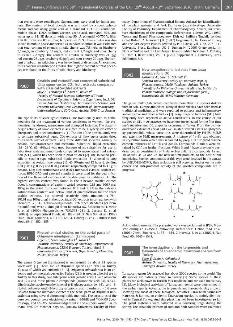
that extracts were centrifuged. Supernatants were used for futher ana-lysis. The content of total phenols was estimated by a spectrophoto-metric method using gallic acid as a standard. HPLC-ED conditions:Mobile phase: EDTA, sodium acetate, acetic acid, methanol 50%, andwater up to 1 L; ED detector with range 50 nA, potential +0.750 V, filter0.02 Hz; flow rate 0.9 ml/min; temperature 25 �C. Pure arbutin was dis-solved in mobile phase and served as standard solution. It was estimatedthat total content of phenols in wild cherry was 7,5 mg/g, in blueberry5,5 mg/g, in cowberry 5,1 mg/g, red currant 2,7 mg/g and sour cherryfruits had 2,2 mg/g. The content of arbutin in blueberry was 21 mg/g,red currant 26 mg/g, cowberry 63 mg/g and sour cherry 20 mg/g. The con-tent of arbutin in wild cherry was below limit of detection. All examinedfruits contain uroantiseptic arbutin. The highest content of total pheno-lics was found in the fruits of wild cherry and blueberry.
P300Casticin and rotundifuran content of subcriticalVitex agnus-castus CO2 fruit extracts comparedwith classical Soxhlet extractsMele A1, Feizlmayr E2, Abazi S1, Bauer R2
1Faculty of Natural Sciences, University of Tirana,Department of Chemistry, Bulevardi Zogu i pare, Nr. 2, 001Tirana, Albania; 2Institute of Pharmaceutical Science, KarlFranzens University Graz, Department of Pharmacognosy,Universit�tsplatz 4, 8010 Graz, Austria, 8010 Graz, Austria
The ripe fruits of Vitex agnus-castus L. are traditionally used as herbalmedicine for the treatment of various conditions in women, like pre-menstrual syndrome, menopause and disrupted lactation. The dopami-nergic activity of some extracts is assumed to be a synergistic effect ofditerpenes and other constitutents [1]. The aim of the present study wasto compare subcritical liquid CO2 extracts of Vitex agnus-castus driedfruits with soxhlet extracts using three different solvents, namely n-hexane, dichloromethane and methanol. Subcritical liquid extraction(25 – 26 �C, 62 – 64 bar) was used because of its suitability for use inlaboratory scale and minor costs compared to supercritical fluid extrac-tion (SFE), which has been used in prior studies [2]. The so-called peri-odic or soxhlet-type subcritical liquid extraction [3] allowed to stopextraction at certain time points (15, 45, 90 min and 12 hours), yielding0.01 g, 0.14 g, 0.25 g and 0.28 g extract, respectively compared to 1.8 g n-hexane, 1.3 g dichloromethane and 0.96 g methanol classical soxhlet ex-tracts. HPLC-DAD and external standards were used for the quantifica-tion of the flavonoid casticin and the diterpene rotundifuran [4]. Thehighest casticin content was found in the n-hexane soxhlet extract.Overall, concentrations of casticin varied between 0,15 and 106,7 mg/100 g in the dried fruits and between 0,13 and 1,18% in the extracts.Rotundifuran content was below limit of quantification for the threesoxhlet extracts, but showed relatively high amounts (11,75 –301,01 mg/100 g drug) in the subcritical CO2 extracts in comparison withliterature [2], [4]. Acknowledgements: Reference standards (casticin,rotundifuran) were a kind gift from Bionorica AG. References: 1. MeierB. et al. (2000) Phytomedicine, 7(5):373 – 381. 2. Cossuta D. et al.(2008) J. of Supercritical Fluids, 47: 188 – 194. 3. Naik S.N. et al. (1989)Fluid Phase Equilibria, 49: 115 – 126. 4. Hoberg E. et al. (2000) PlantaMed., 66(4): 352 – 355.
P301Phytochemical studies on the aerial parts ofOriganum minutiflorum (Lamiaceae)�zgen U1, Sezen Karaoglan E1, Kazaz C2
1Atat�rk University, Faculty of Pharmacy, Department ofPharmacognosy, 25240 Erzurum, Turkey; 2Atat�rkUniversity, Faculty of Sciences, Department of Chemistry,25240 Erzurum, Turkey
The genus Origanum (Lamiaceae) is represented by about 38 speciesworldwide [1]. There are 22 Origanum species (27 taxa) in Turkey,13 taxa of which are endemic [2 – 3]. Origanum minutiflorum is an en-demic and commercial species for Turkey [2]. It is used as a herbal tea inTurkey. In this study, two triterpenic acids (ursolic acid (1) and oleanolicacid (2)) and three phenolic compounds (rosmarinic acid (3), 4-(3,4-dihydroxybenzoyloxymethyl)phenyl-b-D-glucopyranoside (4), and 3-(3,4-dihydroxyphenyl)-2-hydroxy-propionic acid (danshensu) (5)) wereisolated from the methanol extract of the aerial parts of Origanum min-utiflorum using several chromatographic methods. The structures of thepure compounds were elucidated by using 1H-NMR and 13C-NMR Spec-troscopy, and ESI-MS. Acknowledgements: The authors would like tothank Prof. Dr. Mehmet Koyuncu (Ankara University, Faculty of Phar-
macy, Department of Pharmaceutical Botany, Ankara) for identificationof the plant material and Prof. Dr. Ihsan �alis (Hacettepe University,Faculty of Pharmacy, Department of Pharmacognosy, Ankara) for struc-ture elucidation of the compounds. References: 1. Evans W.C. (1989)Trease and Evans’ Pharmacognosy, 13th ed. Bailliere Tindall: London,Great Britain. 2. Ietswaart J.H. (1982) Origanum L., In: Flora of Turkeyand the East Aegean Islands, (edited by P.H. Davis), Vol. 7, pp. 297 – 313,University Press, Edinburg, UK. 3. Duman H. (2000) Origanum L., In:Flora of Turkey and the East Aegean Islands (edited by Gner A, zhatayN, Ekim T, Baser KHC), Vol. 11, p. 207, Supplement 2, University Press,Edinburgh, UK.
P302New sesquiterpene lactones from Inulamontbretiana DCG�kbulut A1, Sarer E1, Schmidt T2
1Ankara University Faculty of Pharmacy Dept. ofPharmacognosy, 06100, Tandogan Ankara, Turkey;2Westf�lische Wilhelms-Universit�t M�nster, Institut f�rPharmazeutische Biologie und Phytochemie (IPBP),Hittorfstraße 56, 48149 M�nster, Germany
The genus Inula (Asteraceae) comprises more than 100 species distrib-uted in Asia, Europe and Africa. Many of these species have been used astraditional medicines and were reported to possess anti-inflammatory,anti-infective and other activities [1]. Sesquiterpene lactones (STL) havefrequently been reported as active constituents. In the course of ourstudies on STL in Asteraceae, we have now investigated for the first timeInula montbretiana DC, a species occurring in Turkey. From the dichlor-omethane extract of aerial parts we isolated several esters of 9b-hydro-xy-parthenolide, whose structures were determined by HR-ESI-MSMSand extensive NMR measurements. A mixture of 1a-2b was obtainedin crystalline form which was separated by prep. HPLC to yield diaster-eomeric mixtures of 1a+1b and 2a+2b. Compounds 3 and 4 were ob-tained by CC from further fractions. While 3 and 4 have previously beendescribed as constituents of Inula verbascifolia [2], compounds 1a and1b as well as 2a and 2b are new natural products, to the best of ourknowledge. Further compounds of this type were detected in the extractby UHPLC-ESI-MSMS; their isolation is still ongoing. Studies on the anti-cancer and anti-protozoal activity of the isolated compounds are inprogress.
Fig. 1
Acknowledgements: The presented work was performed at IPBP, Mn-ster, during an ERASMUS fellowship. References: 1. Zhao, Y-M. et al.(2006) Chem. Biodivers. 3: 371 – 384. 2. Harvala, E. et al. (2002) J. Nat.Prod. 65: 1045 – 1048.
P303The investigation on the terpenoids andflavonoids of an endemic Taraxacum species fromTurkeySarer E, Sahin A, G�kbulut AAnkara University, Faculty of Pharmacy, Pharmacognosy,Tandogan Ankara, Turkey
Taraxacum genus (Asteraceae) has about 2000 species in the world. The48 species are naturally found in Turkey [1]. Some species of theseplants are wellknown in Turkish folk medicine and are used medicinaly[2]. Many biological activities of Taraxacum genus were determined inthe earlier reports. Actually, the terpenoids and flavonoids play a role ofshowing the most of these biological activities. Taraxacum farinosumHausskn. & Bornm., an endemic Taraxacum species, is mainly distribu-ted in Central Turkey. And this plant has not been investigated so far.The plant materials were collected in a flowering stage during thespring. The ethanolic extracts of root and herb samples were examined
Planta Med 2010; 76: 1163–1374 Georg Thieme Verlag KG Stuttgart · New York · ISSN 0032-0943
12677th Tannin Conference and 58th International Congress of the GA | 29th August – 2nd September 2010, Berlin, Germany
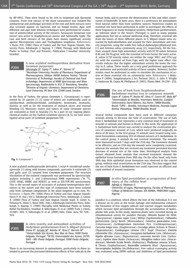
by RP-HPLC. They were found to be rich in terpenoid and flavonoidcontents. From root extract of the plant taraxasterol was isolated.Theantibacterial and antifungal activities of the root and herb extracts werealso screened against six bacterial, and six fungal strains. Agar welldiffusion and agar tube dilution techniques were performed for evalua-tion of antimicrobial activity of the extracts. Taraxacum farinosum rootextract was active to Staphylococcus aureus and Salmonella typhi. Theroot and herb extracts of the plant have shown significant activityagainst Microsporum canis and Trichophyton longifusus. References:1. Davis, P.H. (1982) Flora of Turkey and the East Aegean Islands, Uni-versity Press, Edinburgh. 2. Baytop, T. (1984) Therapy with MedicinalPlants in Turkey (Past and Present), Publication f Istanbul University,Istanbul.
P304A new acylated neohesperidose derivative fromGeranium purpureumS�hretoglu D1, Liptaj T2, Sakar K1, Sterner O3
1Hacettepe University, Faculty of Pharmacy, Department ofPharmacognosy, Sihhiye, 06100 Ankara, Turkey; 2SlovakUniversity of Technology, Faculty of Chemical and Foodtechnology, Department of NMR Spectroscopy and MassSpectroscopy, Radlinsk�ho 9, 812 37 Bratislava, Slovakia;3Division of Organic Chemistry, Department of Chemistry,Lund University, PO Box 124, 22100 Lund, Sweden
In the flora of Turkey, the genus Geranium L. (Geraniaceae) is repre-sented by 35 species [1, 2], some of which are traditionally used asantidiarrheal, antihemorrhoidal, antidiabetic, hemostatic, stomachic,diuretic as well as for the treatment of stomach ulcers and internalbleeding [3]. Moreover, leaves and tubers of some Geranium speciesare consumed as food in Anatolia [1]. As a part of our ongoing phyto-chemical studies on the Turkish Geranium species [4, 5], we have inves-tigated aerial parts of Geranium purpureum Vill.
Fig. 1: Compound 1
A new acylated neohesperoside derivative, 1-octyl-4’-isovaleroyl-neohe-speroside (1) along with known compounds, quercetin-3-rutinoside (2)and gallic acid (3) isolated from Geranium purpureum. The structureelucidation of the isolated compounds was performed by spectroscopicanalysis including 1- and 2-dimensional NMR experiments (1H, 13C,COSY, HSQC, HMBC and NOESY) as well as ESI-TOF-MS spectrometry.This is the second report of occurance of acylated neohesperidose deri-vatives in the nature and this type of compounds have been isolatedonly from Geranium caespitosum in the plant kingdom up to now. Re-ferences: 1. Davis, PH. (1966) Flora of Turkey and East Aegean Islands;Davis P. H. (Ed.), Vol 2. Edinburgh University Press. Edinburgh. 2. Gner,A. (2000) Flora of Turkey and East Aegean Islands Supll. II. Gner A.,zhatay N., Ekim T., Baser KHC. (Eds.), Edinburgh University Press. Edin-burgh. 3. Baytop, T. (1999) Theraphy with Medicinal Plants in Turkey,Nobel Tip Kitabevi. Istanbul. 4. S�hretoglu, D. et al. (2009) Turk. J. Chem.,33:685 – 692. 5. S�hretoglu D. et al. (2009) Helv. Chim. Acta, 92: 520 –524.
P305In vitro toxicity and antioxidant activities ofHedychium gardnerianum from S. Miguel (Azores)Viana H1, Arruda M2, Rainha N1, Rosa J1, Barreto M1
1Universidade dos A�ores, Rua Mae de Deus, 9500 PontaDelgada, Portugal; 2DCTD, Universidade dos A�ores, DCTD,9501 – 801 Ponta Delgada, Portugal, 9500 Ponta Delgada,Portugal
There is an increasing interest in antioxidants, particularly in those in-tended to prevent the presumed deleterious effects of free radicals in the
human body, and to prevent the deterioration of fats and other consti-tuents of foodstuffs. In both cases, there is a preference for antioxidantsfrom natural rather than from synthetic sources. Numerous studies onessential oils have been performed in order to investigate the effects ofthese fragrances on many biological systems. Hedychium gardnerianum,an infestant plant in the Azores (Portugal) is used in many popularapplications but not as natural medicinal drug. Therefore, essential oilsfrom the leaves of three different places in S. Miguel Island were ex-tracted by hydrodistillation and assayed for their antioxidant and toxi-city properties, using the stable free radical diphenylpicrylhydrazyl test.[1] and Artemia salina cytotoxicity assay [2], respectively. All the frac-tions assayed show high antioxidant activity with EC50 values between8.46 and 28.76 mg/mL with the essential oil from Furnas the most active.A. salina LC 50 toxicity values are within the range 300.1 and 379.2 mg/mL with the essential oil from Fogo with the higher toxic effect. Ourresults indicate that the higher antioxidant activity the lower the toxi-city in A. salina. These results are a very good preliminary approach forthe antitumor and pesticidal activity of these samples. However, furtherstudies must be performed in order to elucidate the mechanism of ac-tion of these essential oils on cytotoxicity tests. References: 1. Moly-neux P (2004). Songklanakarin J. Sci. Technol. 26(2). 2. Solis P, WrightC, Anderson M, Gupta M, Phillipson D (1993). Planta Med. 59:250 – 252.
P306The use of bark from Stryphonodendronbarbatiman martius tree in cutaneous woundsLopes B1, Galera P1, Rocha M1, Silva A1, Castro M1, Melo RF2
1Universidade de Bras�lia, Veterinary Medicine, CampusUniversitrio Darci Ribeiro, Asa Norte, 70000 Bras�lia,Brazil; 2UPIS – Bras�lia, Veterinary Medicine, Fazenda LagoaBonita, Planaltina, 70000 Bras�lia, Brazil
Several herbal compounds have been used in different cutaneouswounds aiming to decrease the time of cicatrization. The use of barkfrom S. barbatiman tree represents a very common kind of treatment ofwounds in animals and human in Brazil. In this work, the application ofS. barbatiman bark extract was carried to analyze the cicatrization pro-cess of cutaneous wounds of 2 cm, which were produced surgically ondorse of 20 mice. In the first group, 15 animals were treated using oint-ment formulation containing 10% of ethanol extracts of S. barbatiman. Inthe control group, 5 animals do not received treatment. The woundswere measured to each 7 days until the 21th day. The treatment showedto be effective, and on 21th day the wounds were completely cicatrized,whereas the animals that not received any treatment presented discretedecrease of wound size at the same time. Histological analysis fromtissues obtained from animals that received treatment showed theepithelial tissue formation from 10th day. On the other hand, only from18th day, little epithelial tissue formation was observed in the controlgroup, without total cicatrization on the 21th day. This data suggest thatthe use of bark from S. barbatiman tree could represent an efficient andrapid method of wound treatment.
P307In-vitro lipid peroxidation as progression of liverinjury at the cellular levelAdeniji A, Odukoya OUniversity of Lagos, Pharmacognosy, Faculty of Pharmacy,College of Medicine Campus, IDI-ARABA, PMB12003 Lagos,Nigeria
Jaundice is a condition, which affects the liver of the individual. It is nota disease on its own as the tissue damage and endotoxemia enhancesthe formation of free oxygen radicals and reactive oxygen metaboliteswhich increase lipid peroxidation through the accumulation of hydro-phobic bile acids. In this study, nineteen plants were recorded during anethnobotanical survey for jaundice therapy: Alstonia boonei De Wild.(Apocynaceae), Cajanus cajan (Linn.) Millsp (Papilionoideae), Calliandraportoricensis (Jacq) Benth (Mimosaidaea), Celastrus paniculatus Linn.(Celastraceae), Cochlospermum tinctorium Perr. (Cochlospermaceae),Curcuma longa Linn. (Zingiberaceae), Curculigo pilosa (Schum. & Thonn.)(Hypoxidaceae), Cymbopogon citratus (DC). Stapf. (Poaceae), Enantiachlorantha Oliv. (Annonaceae), Gossypium barbadense Linn. (Malvaceae),Kigelia africana (Lam.) (Bignoniaceae), Lawsonia inermis Linn. (Lythra-ceae), Lophira alata Banks (Ochnaceae), Mangifera indica Linn. (Anacar-diaceae), Morinda lucida Benth. (Rubiaceae), Phyllantus amarus Schum.& Thonn. (Euphorbiaceae), Rauwolfia vomitoria Afzel. (Apocynaceae),Sarcocephalus latifolius Sm.(Rubiaceae). Free radical scavenging activitywas evaluated using DPPH radicals and inhibition of lipid peroxidation
Planta Med 2010; 76: 1163–1374 Georg Thieme Verlag KG Stuttgart · New York · ISSN 0032-0943
1268 7th Tannin Conference and 58th International Congress of the GA | 29th August – 2nd September 2010, Berlin, Germany
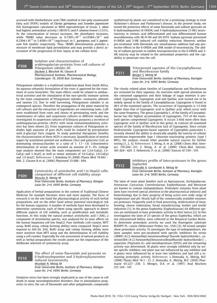
accessed with thiobarbituric acid (TBA) method in two poly unsaturatedfatty acid (PUFA) models of Clarias gariepinus and Scomber japonicumfish homogenates calculated as MDA equivalent/gm of tissue. L. alatahad highest antioxidant activity r (80.95%) and P. amarus lowest (1.53%).As the concentration of extract increases, the absorbance increases,while TBARS value decreases as 6.7305 x 10�5 to1.0384 x 10�5 and8.2304 x 10�5 to 5.4100 x 10�5 (mg/tissue) in C. gariepinus and S. japoni-cum fish models respectively. Thus, TBARS determination provides ameasure of membrane lipid peroxidation and may provide a direct as-sessment of the progression of liver injury at the cellular level.
P308Isolation and characterisation ofarabinogalactan-proteins from cell cultures ofPelargonium sidoides DCDuchow S, Blaschek W, Classen BPharmaceutical Institute, Pharmaceutical Biology,Gutenbergstr. 76, 24118 Kiel, Germany
Pelargonium sidoides is a traditional medicinal plant from South Africa.An aqueous-ethanolic formulation of the roots is approved for the treat-ment of acute bronchitis. The main effects could be related to antibac-terial activities and the stimulation of the non-specific immune systemby the main components of Pelargonium sidoides: coumarins, phenolsand tannins [1]. Due to wild harvesting, Pelargonium sidoides is anendangered species. Therefore the propagation of the plant material bycell cultures and the extraction of ingredients are interesting tasks. Calliwere established from roots and shoots of germinating seeds and themaintenance of callus and suspension cultures in different media wasinvestigated. In suspension cultures of Echinacea purpurea a secretion ofarabinogalactan-proteins (AGPs) with weak immunomodulatory activ-ities has been shown [2]. From suspension cultures of Pelargonium si-doides high amounts of pure AGPs could be isolated by precipitationwith b-glucosyl Yariv reagent. To study potential therapeutic benefitsthe characterisation of these AGPs is necessary. Quantification of neutralsugars by acetylation pointed out arabinose (Ara) and galactose (Gal) asdominating monosaccharides in a ratio of 1: 1.7 – 1.8. Colourimetricdetermination of uronic acids revealed an amount of 5 – 8%. Linkagetype analysis showed that the main components are 1,3,6-Gal(p), 1,3-Gal(p) and 1-Ara(f) as well as minor amounts of 1,6-Gal(p), 1,4-Gal(p)and 1,5-Ara(f). References: 1. Kolodziej H (2008) Planta Med 74:661 –666. 2. Classen B et al. (2006) Phytomed 13:688 – 694.
P309Cytotoxicity of aristolochic acid I in HepG2 cells:comparison of different cell viability assaysNitzsche D, Melzig MFreie Universit�t Berlin, Institute of Pharmacy, K�nigin-Luise-Str. 2+4, 14195 Berlin, Germany
Application of herbal preparations in the context of Traditional ChineseMedicine for example becomes more and more popular. The focus ofresearch is on the one hand the pharmacological activity of such herbalpreparations and on the other hand whose potential toxicological riskfor the human organism. A number of methods have been developed todetermine cytotoxicity, each of them using specific approach to detectdifferent aspects of cell viability, such as proliferation and metabolicfunctions. In this study the natural product aristolochic acid I (AAI), acomponent of Aristolochia species, was analyzed for its toxic effects onthe human hepatoma cell line HepG2. Four methods (cell counter, MTTassay, BrdU assay, colony forming ability) were compared. HepG2 wereexposed to AAI for 24 h. BrdU assay and colony forming ability weremore sensitive than MTT assay and the determination of cell viabilityusing a cell counter. Especially for risk assessment of natural products aswell as herbal preparations the results point out the importance of thedeliberate selection of cytotoxicity assay.
P310The effect of some flavonoids and pyruvate on6-hydroxydopamine and 3-hydroxykynurenineinduced neurotoxicityWszelaki N, Melzig MFreie Universit�t Berlin, Institute of Pharmacy, K�nigin-Luise-Str. 2+4, 14195 Berlin, Germany
Oxidative stress has been strongly implicated as one of the cause in celldeath in many neurodegenerative disorders. Due to antioxidative prop-erties in vitro, the use of flavonoids and other polyphenolic compounds
synthetised by plants are considered to be a promising strategy to treatAlzheimer’s disease and Parkinsons’s disease. In the present study, wetested the protective effects of some flavonoids and sodium pyruvate on6-hydroxydopamine (6-OHDA) and 3-hydroxykynurenine (3-HK) neuro-toxcicity in retinoic acid differentiated and non differentiated humanneuroblastoma cells SK-N-SH and SH-SY5Y. Sodium pyruvate prevented6-OHDA and 3-HK induced cell viability reduction. However, neitherpre-treatment with flavonoids nor simultaneous addition had any pro-tective effects in the 6-OHDA and 3HK model of neurotoxicity. The abil-ity of sodium pyruvate to exhibit neuroprotection in the 6-OHDA and 3-HK toxicity may be related to the antioxidant properties and the cap-ability to penetrate into the cell.
P311Triterpenoid saponins of the Caryophyllaceaeand Illecebraceae familyB�ttger S, Melzig MFreie Universit�t Berlin, Institute of Pharmacy, Koenigin-Luise-Str. 2+4, 14195 Berlin, Germany
The closely related plant families of Caryophyllaceae and Illecebraceaeare reviewed for their saponins. An overview with special attention onthe contained sapogenins and their linkage of sugar moieties is pro-vided. Gypsogenin, Gypsogenic acid and Quillaic acid turned out to bewidely spread in the family of Caryophyllaceae. Gypsogenin is found in46% of the examined species. The occurrence of Gypsogenin is 1.5-foldhigher than that of Gypsogenic acid or Quillaic acid, which occur withthe same frequency. The genus Gypsophila L. of the family of Caryophyl-laceae has the highest accumulation of Gypsogenin. 75% of the exam-ined species comprehend Gypsogenin. It occurs 3-fold more often thanGypsogenic acid or Quillaic acid in this genus. In contrast all examinedspecies of the family of Illecebraceae lack of Gypsogenin. Since certainbisdesmosidic Gypsogenin-based saponins of Gypsophila paniculata L.recently showed the ability to drastically amplify the toxicity of cellularmembrane-impermeable type I ribosome-inactivating proteins (type IRIPs), the analysis reveals other possible natural sources for furthertesting [1, 2, 3]. References: 1. Weng, A. et al. (2008) Chem.-Biol. Inter-act. 176:204 – 211. 2. Weng, A. et al. (2009) Chem.-Biol. Interact.181:424 – 429. 3. Hebestreit, P. et al. (2006) Toxicon 47:330 – 335.
P312Inhibitory profile of latex-proteases in the genusEuphorbiaDomsalla A, Garshasebi A, Melzig MFreie Universit�t Berlin, Institute of Pharmacy, Koenigin-Luise-Str. 2+4, 14195 Berlin, Germany
The latex of some plant families such as Apocynaceae, Asclepiadaceae,Asteraceae, Caricaceae, Convolvulaceae, Euphorbiaceae, and Moraceaeare known to contain endopeptidases. Proteolytic enzymes from plantlatex have received special attention in the pharmaceutical industry andbiotechnology due to their property of being active over wide range oftemperature and pH. Nearly half of the commercially available enzymesare proteases, frequently used in food processing, tenderization of meat,brewing, cheese elaboration, bread manufacturing, leather and textileindustries [1]. In the genus Euphorbia (Euphorbiaceae) 64 different spe-cies are mentioned to have proteolytic activity in their latices [2]. In thisinvestigation the latex of 23 species of the genus Euphorbia, which arenot characterised before, were collected in the Botanical Garden Berlin.To determine proteolytic activity we used the fluorogenic substrateBODIPY FL- casein (Moleculare Probes, Inc., USA) [3]. All tested samplesshow proteolytic activity. To investigate the type of endopeptidases, thelatex samples were pre-incubated with specific inhibitors for serine(AEBSF (4-(2-Aminoethyl)-benzenesulfonyl fluoride hydrochloride)-, cy-steine(E64 (4-(2-Aminoethyl)benzenesulfonylfluoride hydrochloride)) -,aspartatic (Pepstatin A)- and metalloprotease (EDTA) and the remainingactivity was determined. 18 plants were strongly inhibited only by ser-ine specific inhibitor, one plant was not influenced by any inhibitor and4 plants were influenced by at least two inhibitors and had still a re-maining proteolytic activity. References: 1. Domsalla, A., Melzig, M.F.(2008) Planta Med 74:1 – 13. 2. Domsalla, A., Melzig, M.F. (2010) Phar-mazie 65:227 – 230. 3. Menges, D.A. et al. (1997) Anal Biochem251:144 – 147.
Planta Med 2010; 76: 1163–1374 Georg Thieme Verlag KG Stuttgart · New York · ISSN 0032-0943
12697th Tannin Conference and 58th International Congress of the GA | 29th August – 2nd September 2010, Berlin, Germany
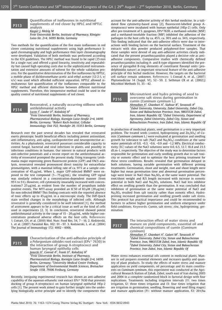
P313Quantification of isoflavones in nutritionalsupplements of red clover by HPLC and HPTLCanalysisKasper J, Melzig MFreie Universit�t Berlin, Institute of Pharmacy, K�nigin-Luise-Str. 2+4, 14195 Berlin, Germany
Two methods for the quantification of the five main isoflavones in redclover containing nutritional supplements using high performance li-quid chromatography and high performance thin layer chromatographywere developed. Validation of both methods was performed accordingto the ICH guidelines. The HPLC method was found to be rapid (25 minfor a single run) and offered a good linearity, sensitivity and repeatabil-ity but caused high operating costs due to the sophisticated instrumen-tation and extensive amounts of solvent for the whole validation pro-cess. For the quantitative determination of the five isoflavones by HPTLC,a mobile phase of dichloromethane-acetic acid-ethyl acetate (12:2:1, v/v/v) was used which afforded sufficient separation. HPTLC allowed si-multaneous fast quantification of the isoflavones comparable to theHPLC method and efficient distinction between different nutritionalsupplements. Therefore, this inexpensive method could be used in thequality control of nutritional supplements of red clover.
P314Resveratrol, a naturally occurring stilbene withantileishmanial activityLucas I1, Laube U2, Kolodziej H1
1Freie Universit�t Berlin, Institute of Pharmacy,Pharmaceutical Biology, Koenigin Luise-Straße 2+4, 14195Berlin, Germany; 2Robert Koch-Institut, CellularImmunology Unit P22, Nordufer 20, 13353 Berlin, Germany
Research over the past several decades has revealed that resveratrolexerts pleiotropic health beneficial effects including potent antioxidant,anti-inflammatory, anti-aging, cardioprotective and neuroprotective ac-tivities. As a phytoalexin, resveratrol possesses considerable capacity tocontrol fungal, bacterial and viral infections in plants, and possibly ininfectious conditions in humans. Our interest in natural products withanti-infective potentials and a recent report on the antileishmanial ac-tivity of resveratrol prompted the present study. Using transgenic Leish-mania major expressing green fluorescent protein (GFP) and FACS ana-lysis, resveratrol revealed pronounced direct toxicity for extracellularpromastigotes, with a maximal killing rate of ca. 97% at a sample con-centration of 45 mg/mL. When L. major GFP-infected BMM? were ex-posed to the test compound (5 – 75 mg/mL), the resulting GFP signalwas similarly reduced in a concentration-dependent manner (from95% to 5%). However, cell cytotoxicity invariably increased with concen-trations? 25 mg/mL as evident from the number of propidium iodidepositive events. The MTT-assay provided an IC50 of 83mM (20 mg/mL)for non-infected BMM? This finding contrasts with the reported absenceof toxic effects up to 45 mg/mL in a microscopic study. The Diff-Quickstain verified changes in the morphology of infected cells. Althoughresveratrol is generally considered to be well tolerated [1], the methodof assessment appears to be a critical issue as well as the kind of cellsused in experiments [2, 3]. In conclusion, resveratrol showed potentantileishmanial activity in the range of 15 – 20 mg/mL, while higher con-centrations produced adverse effects on the host cells. References:1. Cottart, CH. et al. (2010) Mol. Nutr. Food Res. 54, 7 – 16. 2. Kedzierski,L. et al. (2007) Parasitol Res. 102: 91 – 97. 3. Kedzierski, L. et al. (2004)The Journal of Immunology 172: 4902 – 4906.
P315Characterization of the anti-adhesive principle ofa Pelargonium sidoides root extract (EPs� 7630) inthe interaction of group A-streptococci andhuman laryngeal epithelia cellsJanecki A1, Conrad A2, Frank U2, Kolodziej H1
1Freie Universit�t Berlin, Institute of Pharmacy,Pharmaceutical Biology, Koenigin Luise-Straße 2+4, 14195Berlin, Germany; 2University Medical Center Freiburg,Department of Environmental Health Sciences, BreisacherStraße 115B, 79106 Freiburg, Germany
Recently, intriguing experimental research has shown an anti-adhesivecapability of the aqueous-ethanolic root extract EPs� 7630 that preventsdocking of group A-streptococci on human laryngeal epithelial HEp-2cells [1]. The present work aimed to gain further insight into the under-lying biologically active principle and to identify the components that
account for the anti-adhesive activity of this herbal medicine. In a vali-dated flow cytometry-based assay [2], fluorescent-labelled group A-streptococci were incubated with human epithelial (HEp-2) cells. Onlyafter pre-treatment of S. pyogenes, EPs�7630, a methanol-soluble (MSF)and a methanol-insoluble fraction (MIF) inhibited the adhesion of thepathogen to the host cells by ca. 45%, ca. 30% and ca. 34%, respectively.This finding indicated that the anti-adhesive effects were due to inter-actions with binding factors on the bacterial surface. Treatment of theextracts with skin powder produced polyphenol-free samples. Thatthese samples were devoid of any anti-adhesive activities clearly indi-cated that the present proanthocyanidins played a decisive role as anti-adhesive components. Comparative studies with chemically definedproanthocyanidins including A- and B-type oligomers identified the pre-sence of pyrogallol B-ring elements of constituent flavanyl units (pro-delphinidin nature) as important structural feature of the anti-adhesiveprinciple of this herbal medicine. However, the targets on the bacterialcell surface remain unknown. References: 1. Conrad A. et al., (2007)Phytomedicine 14 (Suppl. VI), 52 – 59. 2. Sethman CR et al. (2002) JMicrobiol Methods 51, 35 – 42.
P316Micronutrient and hydro priming of seed toovercome salt stress during germination incumin (Cuminum cyminum L.)Ahmadian A1, Ghanbari A1, Siahsar B1, Tavassoli A2
1Zabol University, University, Zabol University, Zabol City,Sistan and Balouchestan Province, Iran, 98615538 Zabol,Iran, Islamic Republic Of; 2Zabol University, Department ofAgronomy, Zabol University, Zabol City, Sistan andBalouchestan Province, Iran Zabol, Iran, Islamic Republic Of
In production of medicinal plants, seed germination is a very importantproblem. The treated seeds (control, hydropriming and Zn2SO4) of Cu-min (Cuminum cyminum L.) were evaluated at germination and seedlinggrowth for tolerance to salt (NaCl and Na2SO4) conditions at the samewater potentials of 0.0, �0.3, �0.6, �0.9 and �1.2 MPa. Electrical conduc-tivity (EC) values of the NaCl solutions were 0.0, 6.5, 12.7, 18.4 and 23.5dS.m-1, respectively. The objective of the study was to determine factorsresponsible for germination and early seedling growth due to salt toxi-city or osmotic effect and to optimize the best priming treatment forthese stress conditions. Results revealed that germination delayed inboth solutions, having variable germination with different primingtreatments. Germination, shoot and weight, root and shoot length werehigher but mean germination time and abnormal germination percen-tage were lower in NaCl than Na2SO4 at the same water potential. Theroot/shoot weight and R/S length increased with increase in osmoticpotential in both NaCl and Na2SO4 solutions. NaCl had less inhibitoreffect on seedling growth than the germination. It was concluded thatinhibition of germination at the same water potential of NaCl andNa2SO4 resulted from salt toxicity rather than osmotic effect. Hydro-priming increased germination and seedling growth under salt stress.This protocol has practical importance and could be recommended tofarmers to achieve higher germination and uniform emergence underfield conditions. Keywords: Cumin, salt stress; zinc, hydropriming; ger-mination
P317The interaction effect of water stress andmanure on yield components, essential oil andchemical compositions of cumin (Cuminumcyminum)Ahmadian A1, Ghanbari A1, Galavi M1, Tavassoli A2
1Zabol University, Zabol City, Sistan and BalouchestanProvince, Iran, 98615538 Zabol, Iran, Islamic Republic Of;2Zabol University, Zabol City, Sistan and BalouchestanProvince, Iran Zabol, Iran, Islamic Republic Of
Water stress enhances essential oils content in medicinal plants. Man-ure in soil prepares essential elements and increases quality and quan-tity of plant products. To study the effects of water stress and manureapplication on yield components, oil percentage and its main constitu-ents on Cuminum cyminum, this experiment was conducted at the Agri-cultural Research Station of Zahak, Zabol, south east of Iran during 2003and 2004 in a complete randomized block in factorial design with fourreplications. Treatments including irrigation intervals (I1: two timesirrigation, I2: three times irrigation and I3: four times irrigation thatare irrigation in germination, seedling, flowering and seed filing stages)and manure application (F1: without manure application, F2: 20 t/ha
Planta Med 2010; 76: 1163–1374 Georg Thieme Verlag KG Stuttgart · New York · ISSN 0032-0943
1270 7th Tannin Conference and 58th International Congress of the GA | 29th August – 2nd September 2010, Berlin, Germany
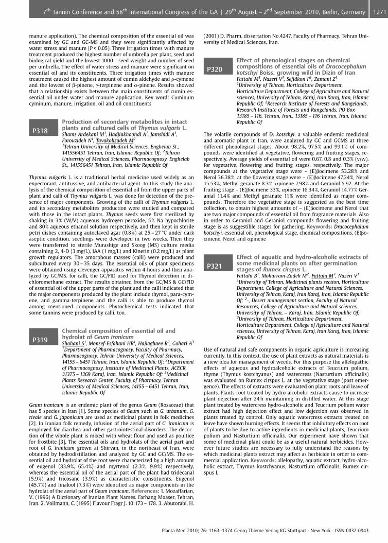
manure application). The chemical composition of the essential oil wasexamined by GC and GC-MS and they were significantly affected bywater stress and manure (P < 0.05). Three irrigation times with manuretreatment produced the highest number of umbrella per plant, seed andbiological yield and the lowest 1000 – seed weight and number of seedper umbrella. The effect of water stress and manure were significant onessential oil and its constituents. Three irrigation times with manuretreatment caused the highest amount of cumin aldehyde and r-cymeneand the lowest of b-pinene, g-terpinene and a-pinene. Results showedthat a relationship exists between the main constituents of cumin es-sential oil under water and manure application. Key word: Cuminumcyminum, manure, irrigation, oil and oil constituents
P318Production of secondary metabolites in intactplants and cultured cells of Thymus vulgaris L.Shams Ardekani M1, Hadjiakhoondi A1, Jamshidi A1,Forouzideh N1, Tavakolizadeh M2
1Tehran University of Medical Sciences, Enghelab St.,141556451 Tehran, Iran, Islamic Republic Of; 2TehranUniversity of Medical Sciences, Pharmacognosy, EnghelabSt., 141556451 Tehran, Iran, Islamic Republic Of
Thymus vulgaris L. is a traditional herbal medicine used widely as anexpectorant, antitussive, and antibacterial agent. In this study the ana-lysis of the chemical composition of essential oil from the upper parts ofplant and calli of Thymus vulgaris L. was done for detection of the pre-sence of major components. Growing of the calli of Thymus vulgaris L.and its secondary metabolites production were studied and comparedwith those in the intact plants. Thymus seeds were first sterilized byshaking in 3% (W/V) aqueous hydrogen peroxide, 5% Na hypochloriteand 80% aqueous ethanol solution respectively, and then kept in sterilepetri dishes containing autoclaved agar (0.8%) at 25 – 27 �C under darkaseptic condition. seedlings were developed in two weeks. Then theywere transferred to sterile Murashige and Skoog (MS) culture mediacontaining 2, 4-D (3 mg/L), IAA (1 mg/L) and Kinetin (0.2 mg/L) as plantgrowth regulators. The amorphous masses (calli) were produced andsubcultured every 30 – 35 days. The essential oils of plant specimenswere obtained using clevenger apparatus within 4 hours and then ana-lyzed by GC/MS. for calli, the GC/FID used for Thymol detection in di-chloromethane extract. The results obtained from the GC/MS & GC/FIDof essential oil of the upper parts of the plant and the calli indicated thatthe major components produced by the plant include thymol, para-cym-ene, and gamma-terpinene and the calli is able to produce thymolamong mentioned components. Phytochemical tests indicated thatsome tannins were produced by calli, too.
P319Chemical composition of essential oil andhydrolat of Geum iranicumShahani S1, Monsef-Esfahani HR1, Hajiaghaee R2, Gohari A3
1Department of Pharmacognosy, Faculty of Pharmacy,Pharmacognosy, Tehran University of Medical Sciences,14155 – 6451 Tehran, Iran, Islamic Republic Of; 2Departmentof Pharmacognosy, Institute of Medicinal Plants, ACECR,31375 – 1369 Karaj, Iran, Islamic Republic Of; 3MedicinalPlants Research Center, Faculty of Pharmacy, TehranUniversity of Medical Sciences, 14155 – 6451 Tehran, Iran,Islamic Republic Of
Geum iranicum is an endemic plant of the genus Geum (Rosaceae) thathas 5 species in Iran [1]. Some species of Geum such as G. urbanum, G.rivale and G. japonicum are used as medicinal plants in folk medicines[2]. In Iranian folk remedy, infusion of the aerial part of G. iranicum isemployed for diarrhea and other gastrointestinal disorders. The decoc-tion of the whole plant is mixed with wheat flour and used as poulticefor frostbite [3]. The essential oils and hydrolats of the aerial part androot of G. iranicum grown at Shirvan, in the northeast of Iran, wereobtained by hydrodistillation and analyzed by GC and GC/MS. The es-sential oil and hydrolat of the root were characterized by a high amountof eugenol (83.9%, 65.4%) and myrtenol (2.3%, 9.9%) respectively,whereas the essential oil of the aerial part of the plant had tridecanal(5.9%) and tricosane (3.9%) as characteristic constituents. Eugenol(45.7%) and linalool (7.3%) were identified as major components in thehydrolat of the aerial part of Geum iranicum. References: 1. Mozaffarian,V. (1996) A Dictionary of Iranian Plant Names. Farhang Moaser, Tehran,Iran. 2. Vollmann, C. (1995) Flavour Fragr J. 10:173 – 178. 3. Abutorabi, H.
(2001) D. Pharm. dissertation No.4247, Faculty of Pharmacy, Tehran Uni-versity of Medical Sciences, Iran.
P320Effect of phenological stages on chemicalcompositions of essential oils of Dracocephalumkotschyi Boiss. growing wild in Dizin of IranFattahi M1, Nazeri V1, Sefidkon F2, Zamani Z1
1University of Tehran, Horticulture Department,Horticulture Department, College of Agriculture and Naturalsciences, University of Tehran, Karaj, Iran Karaj, Iran, IslamicRepublic Of; 2Research Institute of Forests and Rangelands,Research Institute of Forests and Rangelands, PO Box13185 – 116, Tehran, Iran., 13185 – 116 Tehran, Iran, IslamicRepublic Of
The volatile compounds of D. kotschyi, a valuable endemic medicinaland aromatic plant in Iran, were analyzed by GC and GCMS at threedifferent phenological stages. About 98.2%, 97.5% and 99.1% of com-pounds were identified at vegetative, flowering and fruiting stages, re-spectively. Average yields of essential oil were 0.67, 0.8 and 0.3% (v/w),for vegetative, flowering and fruiting stages, respectively. The majorcompounds at the vegetative stage were – (E)bocimene 53.28% andNerol 36.38%, at the flowering stage were – (E)bocimene 47.24%, Nerol15.53%, Methyl geranate 8.3%, apinene 7.98% and Geraniol 5.92. At thefruiting stage – (E)bocimene 33%, apinene 16.34%, Geraniol 14.77% Ger-anial 11.4% and Methyl geranate 11% were identified as major com-pounds. Therefore the vegetative stage is suggested as the best timecollection, to obtain highest amounts of – (E)bocimene and Nerol thatare two major compounds of essential oil from fragrance materials. Alsoin order to Geraniol and Geraniol compounds flowering and fruitingstage is as suggestible stages for gathering. Keywords: Dracocephalumkotschyi, essential oil, phenological stage, chemical compositions, (E)bo-cimene, Nerol and apinene
P321Effect of aquatic and hydro-alcoholic extracts ofsome medicinal plants on after germinationstages of Rumex cirspus L.Fattahi B1, Moharram-Zadeh M2, Fattahi M3, Nazeri V1
1University of Tehran, Medicinal plants section, HorticultureDepartment, College of Agriculture and Natural Sciences,University of Tehran, Karaj, Iran Karaj, Iran, Islamic RepublicOf; 2-, Desert management section, Faculty of NaturalResources, College of Agriculture and Natural sciences,University of Tehran, – Karaj, Iran, Islamic Republic Of;3University of Tehran, Horticulture Department,Horticulture Department, College of Agriculture and Naturalsciences, University of Tehran, Karaj, Iran Karaj, Iran, IslamicRepublic Of
Use of natural and safe components in organic agriculture is increasingcurrently. In this context, the use of plant extracts as natural materials isa new idea for management of weeds. For this purpose the allelopathiceffects of aqueous and hydroalcoholic extracts of Teucrium polium,thyme (Thymus kostchyanus) and watercress (Nasturtium officinalis)was evaluated on Rumex cirspus L. at the vegetative stage (post emer-gence). The effects of extracts were evaluated on plant roots and leave ofplants. Plants root treated by hydro-alcoholic extracts cause to increaseplant dejection after 24 h maintaining in distilled water. At this stageplant treated by watercress hydro-alcoholic and Teucrium polium waterextract had high dejection effect and low dejection was observed inplants treated by control. Only aquatic watercress extracts treated onleave have shown burning effects. It seems that inhibitory effects on rootof plants to be due to active ingredients in medicinal plants, Teucriumpolium and Nasturtium officinalis. Our experiment have shown thatsome of medicinal plant could be as a useful natural herbicides, How-ever future studies are necessary to fully understand the reasons bywhich medicinal plants extract may affect as herbicide in order to com-mercial application. Keywords: allelopathy, aquatic extract, hydro-alco-holic extract, Thymus kostchyanus, Nasturtium officinalis, Rumex cir-spus L
Planta Med 2010; 76: 1163–1374 Georg Thieme Verlag KG Stuttgart · New York · ISSN 0032-0943
12717th Tannin Conference and 58th International Congress of the GA | 29th August – 2nd September 2010, Berlin, Germany
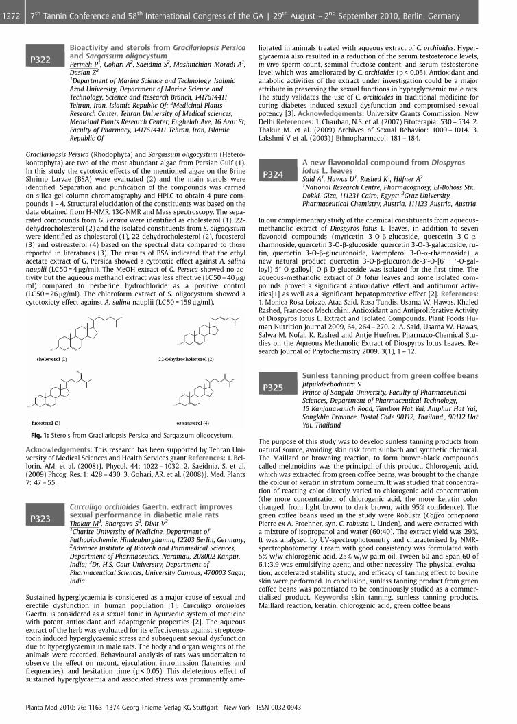
P322Bioactivity and sterols from Gracilariopsis Persicaand Sargassum oligocystumPermeh P1, Gohari A2, Saeidnia S2, Mashinchian-Moradi A1,Dasian Z2
1Department of Marine Science and Technology, IsalmicAzad University, Department of Marine Science andTechnology, Science and Research Branch, 1417614411Tehran, Iran, Islamic Republic Of; 2Medicinal PlantsResearch Center, Tehran University of Medical sciences,Medicinal Plants Research Center, Enghelab Ave, 16 Azar St,Faculty of Pharmacy, 1417614411 Tehran, Iran, IslamicRepublic Of
Gracilariopsis Persica (Rhodophyta) and Sargassum oligocystum (Hetero-kontophyta) are two of the most abundant algae from Persian Gulf (1).In this study the cytotoxic effects of the mentioned algae on the BrineShrimp Larvae (BSA) were evaluated (2) and the main sterols wereidentified. Separation and purification of the compounds was carriedon silica gel column chromatography and HPLC to obtain 4 pure com-pounds 1 – 4. Structural elucidation of the constituents was based on thedata obtained from H-NMR, 13C-NMR and Mass spectroscopy. The sepa-rated compounds from G. Persica were identified as cholesterol (1), 22-dehydrocholesterol (2) and the isolated constituents from S. oligocystumwere identified as cholesterol (1), 22-dehydrocholesterol (2), fucosterol(3) and ostreasterol (4) based on the spectral data compared to thosereported in literatures (3). The results of BSA indicated that the ethylacetate extract of G. Persica showed a cytotoxic effect against A. salinanauplii (LC50 = 4 mg/ml). The MeOH extract of G. Persica showed no ac-tivity but the aqueous methanol extract was less effective (LC 50 = 40 mg/ml) compared to berberine hydrochloride as a positive control(LC 50 = 26 mg/ml). The chloroform extract of S. oligocystum showed acytotoxicty effect against A. salina nauplii (LC 50 = 159 mg/ml).
Fig. 1: Sterols from Gracilariopsis Persica and Sargassum oligocystum.
Acknowledgements: This research has been supported by Tehran Uni-versity of Medical Sciences and Health Services grant References: 1. Bel-lorin, AM. et al. (2008) J. Phycol. 44: 1022 – 1032. 2. Saeidnia, S. et al.(2009) Phcog. Res. 1: 428 – 430. 3. Gohari, AR. et al. (2008) J. Med. Plants7: 47 – 55.
P323Curculigo orchioides Gaertn. extract improvessexual performance in diabetic male ratsThakur M1, Bhargava S2, Dixit V3
1Charite University of Medicine, Department ofPathobiochemie, Hindenburgdamm, 12203 Berlin, Germany;2Advance Institute of Biotech and Paramedical Sciences,Department of Pharmaceutics, Naramau, 208002 Kanpur,India; 3Dr. H.S. Gour University, Department ofPharmaceutical Sciences, University Campus, 470003 Sagar,India
Sustained hyperglycaemia is considered as a major cause of sexual anderectile dysfunction in human population [1]. Curculigo orchioidesGaertn. is considered as a sexual tonic in Ayurvedic system of medicinewith potent antioxidant and adaptogenic properties [2]. The aqueousextract of the herb was evaluated for its effectiveness against streptozo-tocin induced hyperglycaemic stress and subsequent sexual dysfunctiondue to hyperglycaemia in male rats. The body and organ weights of theanimals were recorded. Behavioural analysis of rats was undertaken toobserve the effect on mount, ejaculation, intromission (latencies andfrequencies), and hesitation time (p < 0.05). This deleterious effect ofsustained hyperglycaemia and associated stress was prominently ame-
liorated in animals treated with aqueous extract of C. orchioides. Hyper-glycaemia also resulted in a reduction of the serum testosterone levels,in vivo sperm count, seminal fructose content, and serum testosteronelevel which was ameliorated by C. orchioides (p < 0.05). Antioxidant andanabolic activities of the extract under investigation could be a majorattribute in preserving the sexual functions in hyperglycaemic male rats.The study validates the use of C. orchioides in traditional medicine forcuring diabetes induced sexual dysfunction and compromised sexualpotency [3]. Acknowledgements: University Grants Commission, NewDelhi References: 1. Chauhan, N.S. et al. (2007) Fitoterapia: 530 – 534. 2.Thakur M. et al. (2009) Archives of Sexual Behavior: 1009 – 1014. 3.Lakshmi V et al. (2003) J Ethnopharmacol: 181 – 184.
P324A new flavonoidal compound from Diospyroslotus L. leavesSaid A1, Hawas U1, Rashed K1, H�fner A2
1National Research Centre, Pharmacognosy, El-Bohoss Str.,Dokki, Giza, 111231 Cairo, Egypt; 2Graz University,Pharmaceutical Chemistry, Austria, 111123 Austria, Austria
In our complementary study of the chemical constituents from aqueous-methanolic extract of Diospyros lotus L. leaves, in addition to sevenflavonoid compounds (myricetin 3-O-b-glucoside, quercetin 3-O-a-rhamnoside, quercetin 3-O-b-glucoside, quercetin 3-O-b-galactoside, ru-tin, quercetin 3-O-b-glucuronoide, kaempferol 3-O-a-rhamnoside), anew natural product quercetin 3-O-b-glucuronide-3’-O-[6’ ’ ’-O-gal-loyl)-5@-O-galloyl]-O-b-D-glucoside was isolated for the first time. Theaqueous-methanolic extract of D. lotus leaves and some isolated com-pounds proved a significant antioxidative effect and antitumor activ-ities[1] as well as a significant hepatoprotective effect [2]. References:1. Monica Rosa Loizzo, Ataa Said, Rosa Tundis, Usama W. Hawas, KhaledRashed, Francseco Mechichini. Antioxidant and Antiproliferative Activityof Diospyros lotus L. Extract and Isolated Compounds. Plant Foods Hu-man Nutrition Journal 2009, 64, 264 – 270. 2. A. Said, Usama W. Hawas,Salwa M. Nofal, K. Rashed and Antje Huefner. Pharmaco-Chemical Stu-dies on the Aqueous Methanolic Extract of Diospyros lotus Leaves. Re-search Journal of Phytochemistry 2009, 3(1), 1 – 12.
P325Sunless tanning product from green coffee beansJitpukdeebodintra SPrince of Songkla University, Faculty of PharmaceuticalSciences, Department of Pharmaceutical Technology,15 Kanjanavanich Road, Tambon Hat Yai, Amphur Hat Yai,Songkhla Province, Postal Code 90112, Thailand., 90112 HatYai, Thailand
The purpose of this study was to develop sunless tanning products fromnatural source, avoiding skin risk from sunbath and synthetic chemical.The Maillard or browning reaction, to form brown-black compoundscalled melanoidins was the principal of this product. Chlorogenic acid,which was extracted from green coffee beans, was brought to the changethe colour of keratin in stratum corneum. It was studied that concentra-tion of reacting color directly varied to chlorogenic acid concentration(the more concentration of chlorogenic acid, the more keratin colorchanged, from light brown to dark brown, with 95% confidence). Thegreen coffee beans used in the study were Robusta (Coffea canephoraPierre ex A. Froehner, syn. C. robusta L. Linden), and were extracted witha mixture of isopropanol and water (60:40). The extract yield was 29%.It was analysed by UV-spectrophotometry and characterised by NMR-spectrophotometry. Cream with good consistency was formulated with5% w/w chlorogenic acid, 25% w/w palm oil. Tween 60 and Span 60 of6.1:3.9 was emulsifying agent, and other necessity. The physical evalua-tion, accelerated stability study, and efficacy of tanning effect to bovineskin were performed. In conclusion, sunless tanning product from greencoffee beans was potentiated to be continuously studied as a commer-cialised product. Keywords: skin tanning, sunless tanning products,Maillard reaction, keratin, chlorogenic acid, green coffee beans
Planta Med 2010; 76: 1163–1374 Georg Thieme Verlag KG Stuttgart · New York · ISSN 0032-0943
1272 7th Tannin Conference and 58th International Congress of the GA | 29th August – 2nd September 2010, Berlin, Germany
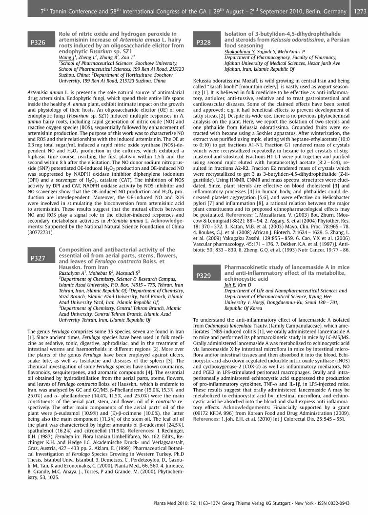
P326Role of nitric oxide and hydrogen peroxide inartemisinin increase of Artemisia annua L. hairyroots induced by an oligosaccharide elicitor fromendophytic Fusarium sp. SZ1Wang J1, Zheng L2, Zhang B1, Zou T1
1School of Pharmaceutical Sciences, Soochow University,School of Pharmaceutical Sciences, 199 Ren Ai Road, 215123Suzhou, China; 2Department of Horticulture, SoochowUniversity, 199 Ren Ai Road, 215123 Suzhou, China
Artemisia annua L. is presently the sole natural source of antimalarialdrug artemisinin. Endophytic fungi, which spend their entire life spansinside the healthy A. annua plant, exhibit intimate impact on the growthand physiology of their hosts. An oligosaccharide elicitor (OE) of oneendophytic fungi (Fusarium sp. SZ1) induced multiple responses in A.annua hairy roots, including rapid generation of nitric oxide (NO) andreactive oxygen species (ROS), sequentially followed by enhancement ofartemisinin production. The purpose of this work was to characterise NOand ROS and their relationships with the induced artemisinin. The OE at0.3 mg total sugar/mL induced a rapid nitric oxide synthase (NOS)-de-pendent NO and H2O2 production in the cultures, which exhibited abiphasic time course, reaching the first plateau within 1.5 h and thesecond within 8 h after the elicitation. The NO donor sodium nitroprus-side (SNP) potentiated OE-induced H2O2 production and OE-induced NOwas suppressed by NADPH oxidase inhibitor diphenylene iodonium(DPI) and a scavenger of H2O2, catalase (CAT). The inhibition of NOSactivity by DPI and CAT, NADPH oxidase activity by NOS inhibitor andNO scavenger show that the OE-induced NO production and H2O2 pro-duction are interdependent. Moreover, the OE-induced NO and ROSwere involved in stimulating the bioconversion from artemisinic acidto artemisinin. These results suggest that the mutual effects betweenNO and ROS play a signal role in the elicitor-induced responses andsecondary metabolism activities in Artemisia annua L. Acknowledge-ments: Supported by the National Natural Science Foundation of China(30772731)
P327Composition and antibacterial activity of theessential oil from aerial parts, stems, flowers,and leaves of Ferulago contracta Boiss. etHausskn. from IranRustaiyan A1, Mohebat R2, Masoudi S3
1Department of Chemistry, Science & Research Campus,Islamic Azad University, P.O. Box. 14515 – 775, Tehran, IranTehran, Iran, Islamic Republic Of; 2Department of Chemistry,Yazd Branch, Islamic Azad University, Yazd Branch, IslamicAzad University Yazd, Iran, Islamic Republic Of;3Department of Chemistry, Central Tehran Branch, IslamicAzad University, Central Tehran Branch, Islamic AzadUniversity Tehran, Iran, Islamic Republic Of
The genus Ferulago comprises some 35 species, seven are found in Iran[1]. Since ancient times, Ferulago species have been used in folk medi-cine as sedative, tonic, digestive, aphrodisiac, and in the treatment ofintestinal worms and haemorrhoids in different regions [2]. More overthe plants of the genus Ferulago have been employed against ulcers,snake bite, as well as headache and diseases of the spleen [3]. Thechemical investigation of some Ferulago species have shown coumarins,flavonoids, sesquiterpenes, and aromatic compounds [4]. The essentialoil obtained by hydrodistillation from the aerial parts, stems, flowers,and leaves of Ferulago contracta Boiss. et Hausskn., which is endemic toIran, was analysed by GC and GC/MS. b-Phellandrene (15.0%, 15.3%, and25.0%) and a- phellandrene (14.4%, 11.5%, and 25.0%) were the mainconstituents of the aerial part, stem, and flower oil of F. contracta re-spectively. The other main components of the aerial parts’ oil of theplant were b-eudesmol (10.9%) and (E)-b-ocimene (10.0%), the latterbeing also the main component (11.3%) of the stem oil. The leaf oil ofthe plant was characterised by higher amounts of b-eudesmol (24.5%),spathulenol (16.2%) and citronellol (11.9%). References: 1. Rechinger,K.H. (1987). Ferulago in: Flora Iranian Umbellifarea, No. 162. Edits., Re-chinger K.H. and Hedge I.C, Akademische Druck- und Verlagsanstalt,Graz, Austria, 427 – 433 pp. 2. Aklam, E. (1999). Pharmaceutical Botani-cal Investigation of Ferulago Species Growing in Western Turkey. Ph.DThesis, Istanbul Univ., Istanbul. 3. Demetzos, C., Perdetzoylou, D., Gazou-li, M., Tan, K and Economakis, C. (2000). Planta Med., 66, 560. 4. Jimenez,B. Grande, M.C. Anaya, J., Torres, P and Grande, M. (2000). Phytochem-istry, 53, 1025.
P328Isolation of 3-butyliden-4,5-dihydrophthalideand steroids from Kelussia odoratissima, a Persianfood seasoningShokoohinia Y, Sajjadi S, MehrAmiri PDepartment of Pharmacognosy, Faculty of Pharmacy,Isfahan University of Medical Sciences, Hezar jarib AveIsfahan, Iran, Islamic Republic Of
Kelussia odoratissima Mozaff. is wild growing in central Iran and beingcalled “karafs koohi” [mountain celery], is vastly used as yogurt season-ing [1]. It is believed in folk medicine to be effective as anti-inflamma-tory, antiulcer, anti-tussive, sedative and to treat gastrointestinal andcardiovascular diseases. Some of the claimed effects have been testedand approved; e. g. it had beneficial effects to prevent development offatty streak [2]. Despite its wide use, there is no previous phytochemicalanalysis on the plant. Here, we report the isolation of two sterols andone phthalide from Kelussia odoratissima. Grounded fruits were ex-tracted with hexane using a Soxhlet apparatus. After winterization, theextract was purified using mplc, eluting with heptane-ethylacetate (10:0to 0:10) to get fractions A1-N1. Fraction G1 rendered mass of crystalswhich were recrystallized repeatedly in hexane to get crystals of stig-masterol and sitosterol. Fractions H1-L1 were put together and purifiedusing second mplc eluted with heptane:ethyl acetate (8:2 – 6:4), re-sulted in fractions A2-R2. Fraction E2 rendered mass of crystals whichwere recrystallized to get 3 as 3-butyliden-4,5-dihydrophthalide (Z-li-gustilide). Using HNMR, CNMR and mass spectra, structures were eluci-dated. Since, plant sterols are effective on blood cholesterol [3] andinflammatory processes [4] in human body, and phthalides could de-creased platelet aggregation [5,6], and were effective on Helicobacterpylori [7] and inflammation [8], a rational relation between the majorplant constituents and its proposed ethnopharmacological effects maybe postulated. References: 1. Mozaffarian, V. (2003) Bot. Zhurn. (Mos-cow & Leningrad) 88(2): 88 – 94. 2. Asgary, S. et al (2004) Phytother. Res.18: 370 – 372. 3. Katan, M.B. et al. (2003) Mayo. Clin. Proc. 78:965 – 78.4. Boukes, G.J. et al. (2008) African J. Biotech. 7:1624 – 1629. 5. Zhang, L.et al. (2009) Yakugaku Zasshi. 129:855 – 859. 6. Cao, Y.X et al. (2006)Vascular pharmacology. 45:171 – 176. 7. Dekker, K.A. et al. (1997) J. Anti-biotic 50: 833 – 839. 8. Zheng, G.Q. et al. (1993) Nutr Cancer. 19:77 – 86.
P329Pharmacokinetic study of lancemaside A in miceand anti-inflammatory effect of its metabolite,echinocystic acidJoh E, Kim DDepartment of Life and Nanopharmaceutical Sciences andDepartment of Pharmaceutical Science, Kyung-HeeUniversity 1, Hoegi, Dongdaemun-Ku, Seoul 130 – 701,Republic Of Korea
To understand the anti-inflammatory effect of lancemaside A isolatedfrom Codonopsis lanceolata Trautv. (family Campanulaceae), which ame-liorates TNBS-induced colitis [1], we orally administered lancemaside Ato mice and performed its pharmacokinetic study in mice by LC-MS/MS.Orally administered lancemaside A was metabolized to echinocystic acidvia lancemaside X by intestinal microflora in mice by intestinal micro-flora and/or intestinal tissues and then absorbed it into the blood. Echi-nocystic acid also down-regulated inducible nitric oxide synthase (iNOS)and cyclooxygenase-2 (COX-2) as well as inflammatory mediators, NOand PGE2 in LPS-stimulated peritoneal macrophages. Orally and intra-peritoneally administered echinocystic acid suppressed the productionof pro-inflammatory cytokines, TNF-a and IL-1b, in LPS-injected mice.These results suggest that orally administered lancemaside A may bemetabolized to echinocystic acid by intestinal microflora, and echino-cystic acid be absorbed into the blood and shall express anti-inflamma-tory effects. Acknowledgements: Financially supported by a grant(09172 KFDA 996) from Korean Food and Drug Administration (2009).References: 1. Joh, E.H. et al. (2010) Int J Colorectal Dis. 25:545 – 551.
Planta Med 2010; 76: 1163–1374 Georg Thieme Verlag KG Stuttgart · New York · ISSN 0032-0943
12737th Tannin Conference and 58th International Congress of the GA | 29th August – 2nd September 2010, Berlin, Germany
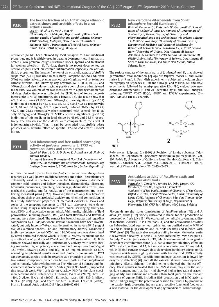
P330The hexane fraction of an Ardisia crispa ethanolicextract shows anti-arthritis effects in a ratarthritis modelLau M1, Ah R1, S S1, Ms N1, Mm N2
1University Putra Malaysia, Department of BiomedicalScience, Faculty of Medicine and Health Science, Selangor,43400 Serdang, Malaysia; 2Forest Research InstituteMalaysia (FRIM), Department of Medical Plant, SelangorDarul Ehsan, 52110 Kepong, Malaysia
Ardisia crispa has been claimed by local villagers to have medicinalproperties, and it is widely used in treating dysmenorrhea, rheumatism,orchitis, skin problem, coughs, fractured bones, sprains and treatmentfor women afterbirth [1]. To date, there is no report documented onArdisia crispa’s effect on chronic inflammation especially on anti-ar-thritic properties. The hexane fraction of an ethanolic extract of Ardisiacrispa root (ACHE) was used in this study. Complete Freund’s adjuvant(CFA) was injected onto plantar aponeurosis of right paw of rat to inducechronic arthritis. The following day onwards, ACHE at 3, 10, 30, and100 mg/kg and also indomethacin as positive control were administeredto the rats. Paw volume of rat was measured with a plethysmometer for14 days. Ankle tissue was collected for ELISA test of tumor necrosisfactor alpha (TNF-a) and interleukin-1 beta (IL-1b). The result show thatACHE at all doses (3.10.30 and 100 mg/kg) have significant (p < 0.05)inhibition of oedema by 45.3%, 64.31%, 73.12% and 49.55% respectively.At 3, 10 and 30 mg/kg, ACHE significantly reduced TNF-a by 45.2%,45.7% and 25.1% respectively when compared with control. For IL-1b,only 10 mg/kg and 30 mg/kg of ACHE elicited a significant (p < 0.05)inhibition of this mediator in local tissue by 45.9% and 36.5% respec-tively. The efficacies of those doses were comparable to the effect ofindomethacin (34.6%). Thus, it can be concluded that Ardisia crispaposseses anti- arthritic effect on specific FCA-induced arthritis modelin rats.
P331Anti-infammatory and free radical scavengingactivity of Juniperus communis L. 1753 var.communis leaves and cones extractLesjak M, Beara I, Orcic D, Balog K, Francikovic M, Simin N,Mimica-Dukic NFaculty of Sciences University of Novi Sad, Department ofChemistry, Biochemistry and Environmental Protection, TrgDositeja Obradovica 3, 21000 Novi Sad, Serbia, Republic of
All over the world plants from the Juniperus genus have always beenregarded as a well-known traditional remedy and spice. These plants areextensively used in the folk medicine for healing various disorders:common cold, urinary and kidney infections, dermatological disorders,bronchitis, pneumonia, dysentery, hemorrhage, rheumatic arthritis, sto-machache, diarrhea and for regulation of the menstruation and in re-lieving menstrual pains [1,2,3]. However, there are only few literaturedata about their pharmacological activity and chemical composition. Inthis study antioxidant properties of methanol extracts of leaves andcones of the Juniperus communis L. 1753 var. communis, were deter-mined using assays which measure free radical scavenging ability to-ward DPPH and superoxide anion radical. Additionally, inhibition of lipidperoxidation, reducing power (FRAP) and total flavonoid and flavonoidcontent were determined. The extract has been characterized regardingcomposition by LC-MS/MS where several flavonoids have been investi-gated, but only luteolin-7-O-glc was determined in leaves (96.19 mg/g ofdw) of examined species. The anti-inflammatory activity, consideringinhibitory potency toward COX-1 and 12-LOX enzymes, was determinedby novel optimized method which is using LC-MS/MS technique for thequantification of products of COX-1 and 12-LOX metabolism [4]. Bothextracts showed markedly anti-inflammatory activity, with leaves hav-ing somewhat higher potency concerning both assays, reaching IC50 at1.50 mg/mL towards COX-1 and IC50 at 1.81 mg/mL towards 12-LOX.According to obtained results examined Juniperus communis L. 1753var. communis. species could be regarded as a promising source of bioac-tive natural compounds, which can be used both as food supplementand as remedy. Acknowledgements: The Ministry of Sciences and En-vironmental Protection, Republic of Serbia (Grant No. 142036) supportedthis research work. We thank Goran Anackov, PhD for the plant speci-men determination. References: 1. Thomas, P.A et al. (2007) J. Ecol. 95:1404. 2. Akkol, E.K. et al. (2009) J. Ethnopharmacol. 125: 330. 3. Miceli,N. et al. (2009) J. Agr. Food Chem. 57: 6570. 4. Beara, I.N. et al. (2010) J.Pharm. Biomed. Anal. doi:10.1016/j.jpba.2010.02.014.
P332New clerodane diterpenoids from Salviaadenophora Fernald (Lamiaceae)Bisio A1, Damonte G2, Fraternale D3, Giacomelli E1, Salis A2,Russo E1, Cafaggi S1, Ricci D3, Romussi G1, DeTommasi N4
1University of Genoa, Dept. of of Chemistry andPharmaceutical and Food Technologies, Via Brigata Salerno13, 16147 Genova, Italy; 2University of Genova, Dept. ofExperimental Medicine and Center of Excellence forBiomedical Research, Viale Benedetto XV, 7, 16132 Genova,Italy; 3University of Urbino, Dipartimento di ScienzedellUomo, dellAmbiente e della Natura, Via Bramante 28,61029 Urbino, Italy; 4University of Salerno, Dipartimento diScienze Farmaceutiche, Via Ponte Don Melillo, 84084Salerno, Italy
The surface exudate of the aerial parts of S. adenophora [1], that showedgermination total inhibition [2] against Papaver rhoeas L. and Avenasativa L. at 5 mg/L in Petri dish experiments, subjected to column chro-matography on Sephadex LH-20 and silica gel and to HPLC-MS and MS 2experiments followed by semi-preparative RP-HPLC, yielded two newclerodane diterpenoids (1 and 2), identified by IR and NMR analysis,including TOCSY, COSY, HSQC, HMBC and ROESY experiments, ESI-TRAP-MS and HR-MS analysis.
Fig. 1
References: 1. Epling, C. (1940) A Revision of Salvia, subgenus Calo-sphace. In: Repertorium Specierum Novarum Regni Vegetabilis. Vol.110. Fedde F., University of California Press: Berkley, California. 2. Chia-pusio. G., Sanchez A.M., Reigosa M.J., Gonzalez L., Pellissier F. (1997),Journal of Chemical Ecology, 23, 2445 – 2453.
P333Antioxidant activity of Passiflora edulis andPassiflora alata fruitsYariwake J1, Zeraik M1, Serteyn D2, Deby-Dupont G2,Wauters J3, Tits M3, Angenot L3, Franck T2
1University of Sao Paulo, Institut of Chemistry of Sao Carlos,DQFM, C. P. 780, 13560970 Sao Carlos, Brazil; 2University ofLiege, CORD, Institute of Chemistry B6a, Sart Tilman, 4000Liege, Belgium; 3University of Liege, Department ofPharmacie, B36, CHU Sart-Tilman, 4000 Liege, Belgium
Flavonoids are the major constituents of Passiflora edulis (PE) and P.alata (PA) fruits [1, 2], widely cultivated in Brazil, for the production ofprocessed or fresh juice [3]. We evaluated the radical-scavenging abilityof methanol extracts (DPPH· method) [4] and the antioxidant activitieson PMA-stimulated equine neutrophils and on purified equine MPO ofPA and PE fruit pulp extracts and PE rinds (healthy and infected withPWV virus) [5]. The radical-scavenging ability followed the order: rutin> resveratrol > healthy PE peels > PE peels infected by PWV > PE pulp >PA pulp. The total juice extract of PE, which was measured by lucigenin-dependent chemiluminescence (CL), had a stronger inhibitory effect onROS production than did PA, but only at a concentration of 1 mg mL-1,while PE rind extracts showed dose-dependent inhibitory effects on CLresponse which were slightly stronger with healthy rind. MPO activitywas assessed by SIEFED (specific immunologic extraction followed byenzymatic detection) [6], and all the extracts showed dose-dependentinhibitory effects, although the rind extracts showed the highest effi-cacy. These results indicated that the PWV disease may alter the anti-oxidant content, and that fruit rind showed higher free radical scaven-ging ability and antioxidant activities than total juice on the oxidantresponse of equine PMN, including ROS production and MPO activity.These findings suggest the potential of passion fruit rind, a by-product ofthe passion fruit processing industry, as a possible functional food or asa raw material for the development of phytomedicines. Acknowledge-
Planta Med 2010; 76: 1163–1374 Georg Thieme Verlag KG Stuttgart · New York · ISSN 0032-0943
1274 7th Tannin Conference and 58th International Congress of the GA | 29th August – 2nd September 2010, Berlin, Germany
ments: CAPES, CNPq, FAPESP. References: 1. Pereira, C.A.M., Vilegas,J.H.Y. (2000) Rev. Bras. Med. 3: 1 – 12. 2. Dhawan, K. et al. (2004) J.Ethnopharm. 94: 1 – 23. 3. Mercadante, A.Z. et al. (1998) J. Agric. FoodChem. 46: 4102 – 4106. 4. Trevisan, F. et al. (2006) Plant Dis. 90: 1026 –1030. 5. Brand-Williams, W. et al. (1995) Lebensm-Wiss. Technol. 28:25 – 28. 6. Franck, T. et al. (2006) J. Vet. Diagn. Invest. 18: 326 – 334.
P336 ??Anti-diabetic activity of Pseuderanthemumpalatiferum Radlk. leaf aqueous extractChayarop K1, Temsiririrkkul R1, Peungvicha P2,Wongkrajang Y2, Chuakul W1, Amnuoypol S3
1Department of Pharmaceutical Botany, Faculty ofPharmacy, Mahidol University, 447 Sri-Ayuthaya Road.Ratchathevi, 10400 Bangkok, Thailand; 2Department ofPhysiology, Faculty of Pharmacy, Mahidol University,447 Sri-Ayuthaya Road. Ratchathevi, 10400 Bangkok,Thailand; 3Department of Pharmacognosy andPharmaceutical botany, Faculty of Pharmaceutical Sciences,Chulalongkorn University, 254 Phayathai Road.Pathumwan, 10330 Bangkok, Thailand
Fresh leaves of Pseuderanthemum palatiferum Radlk. were claimed tocure various disease including diabetes mellitus in Thai people. How-ever, hypoglycemic and anti-diabetic activities of P. palatiferum have notbeen reported. This study examined the hypoglycemic and anti-diabeticeffects of P. palatiferum leaf aqueous extracts by an oral glucose toler-ance test (OGTT) in normal and streptozotocin-induced diabetic rats [1].A single oral administration of the aqueous extract at doses of 0.25 and0.50 g/kg significantly decreased the blood glucose level in STZ-induceddiabetic rats but not in normal rats. A reference drug, glibenclamide, at adose of 5 mg/kg showed a significant blood glucose lowering effect both,in normal rats and STZ-induced diabetic rats. Phytochemical screeningsto examine compounds responsible for the biological activity were car-ried out by color and precipitation tests [2]. It was found that the extractcontained flavonoids, phenolic compounds, non-volatile lactones andsaponins. References: 1. Peungvicha, P. et al. (1998) J. Ethnopharmacol.60:27 – 32. 2. Farnsworth, NR. (1996) J. Pharm. Sci. 55(3):225 – 69.
P337 ??Triterpenoids as antiproliferative agents inclassical and atypical multidrug resistant cancercellsLage H1, Ramalhete C2, Mulhovo S3, Ferreira M2
1Institute of Pathology, Charit�platz 1, 10117 Berlin,Germany; 2iMed.UL, Faculdade de Farmcia, Universidadede Lisboa, Av. Prof. Gama Pinto, 1649 – 003 Lisboa, Portugal;3Escola Superior T�cnica, Universidade Pedaggica,Departamento de CiÞncias Agro-Pecurias, Av. deMo�ambique, – Maputo, Mozambique
Resistance of cancer cells to multiple classes of structurally and mechan-istically unrelated antitumor drugs can be defined as multidrug resis-tance (MDR), and it is one of the major causes of chemotherapy failure.The most significant mechanism of MDR, referred as typical or classicalMDR, is that resulting from altered cell membrane transport due to over-expression of the transporter protein P-glycoprotein (Pgp/MDR1) thatact as an drug efflux pump. Conversely, MDR cells without overexpres-sion of this transporter protein are referred as atypical MDR cells andtheir resistance has been associated with enhanced expression of alter-native transporter protreins, altered DNA topoisomerase II activity orother mechanisms. According to some authors, atypical MDR may alsoresult from altered expression of some metabolizing enzymes [1]. There-fore, a promising approach to overcome MDR is the development ofcompounds that are selectively cytotoxic to resistant cancer cells. Con-tinuing our search for anticancer agents from plant sources [2], the aimof this work was to evaluate the antiproliferative activity of severalcucurbitane-type triterpenoids, isolated from Momordica balsamina amedicinal African plant also used as food. The study was carried outwith human cancer cell lines derived from three tumor entities: gastric(EPG85 – 257), pancreatic (EPP85 – 181) and colon (HT-29) carcinomas.Furthermore, different multidrug-resistant variants of these cancer celllines with over-expression of MDR1/P-gp or no MDR1/P-gp expressionwere also investigated. When comparing with the parental cell lines,some of the multidrug resistant variants showed increased sensitivitiesto the studied compounds. Acknowledgements: The authors wish tothank the Science and Technology Foundation, (FCT, grant SFRH/BD/22321/2005). References: 1. Teodori, E. et al (2006) Curr Drug Targets
7: 893 – 909. Ramalhete, C. et al (2009) Bioorg. Med. Chem. 17: 6942 –6951.
P336Further insights into Ficus carica latexmetabolomeOliveira A1, Silva L1, Guedes de Pinho P2, Valent¼o P1,Silva B3, Pereira J4, Andrade P1
1REQUIMTE/Department of Pharmacognosy, Faculty ofPharmacy of Porto University, Rua An�bal Cunha, 164,4050 Porto, Portugal; 2REQUIMTE/Department ofToxicology, Faculty of Pharmacy, Porto University, RuaAn�bal Cunha, 164, 4050 Porto, Portugal; 3REQUIMTE/Department of Pharmacognosy, Faculty of Pharmacy, PortoUniversity and CEBIMED/Research Group on Toxicology andPhytochemistry, Faculty of Health Sciences, Fernando PessoaUniversity, Rua An�bal Cunha, 164, 4050 Porto, Portugal;4CIMO/Escola Superior Agrria, Instituto Polit�cnico deBragan�a, Campus de Santa Apolnia, Apartado 1172,5301 Bragan�a, Portugal
Ficus carica products are widely used both as food and as medicine inthe Middle East. All Ficus species possess latex-like material within theirvasculatures, affording protection and self-healing from physical attacks.F. carica latex has been traditionally used in the treatment of gout, ulcersand warts, among other situations, given its proteolytic and keratolyticeffects, associated to its viscosity [1]. In this work fatty acids and phy-tosterols profiles were established. Combined fatty acids were hydro-lyzed and all free compounds were derived to their methyl ester forms,prior to GC-MS analysis. Fourteen compounds were determined, beingsaturated fatty acids, namely arachidic, palmitic and behenic acids, theones present in highest contents. Phytosterols profile of saponified sam-ple was characterized by HPLC-DAD, which allowed the determinationof seven compounds. b-Sitosterol, lupeol and lanosterol were the majorphytosterols. This study provides further knowledge to the richness oflatex metabolome, namely in bioactive compounds. Acknowledge-ments: Funda�¼o para a CiÞncia e a Tecnologia, Andreia P. Oliveira(SFRH/BD/47620/2008). Funda�¼o Calouste Gulbenkian, Branca M. Silva.References: 1. Oliveira, A.P. et al (2010) J. Agric. Food Chem. 58: 3393 –3398.
P337Anxiolytic effects of fractions obtained fromPassiflora incarnata L. in the elevated plus mazein miceSampath C1, Holbik M2, Krenn L2, Butterweck V1
1University of Florida, Pharmaceutics, 1600 SW Archer Road,JHMHSC, 32610 Gainesville, United States; 2University ofVienna, Pharmacognosy, Althanstr. 14, 1090 Vienna, Austria
The anxiolytic effects of passion flower (Passiflora incarnata L.) havebeen confirmed in several pharmacological studies [1 – 3], however,the compounds responsible for this effect are still a matter of debate.The purpose of this study was to characterize the putative anxiolytic-like activity of fractions prepared from a hydroethanolic extract usingthe elevated plus-maze (EPM) in mice. The fractions were prepared aspublished recently [4], yielding into a butanol, petroleum ether andchloroform fraction. Male BL6/C57 J mice were either treated orallywith each fraction in three different concentrations according to theirpercent amount in the extract or the positive control diazepam (1.5 mg/kg). From the tested fractions, the butanol fraction showed significantincreases in the number of open arm entries in the EPM in concentra-tions of 2.1 mg/kg and 4.2 mg/kg corresponding to 150 and 300 mg/kg ofthe original extract. The highest activity was found for the chloroformfraction in doses of 0.17 mg/kg (10.0 € 1.9, p < 0.001) and 0.34 mg/kg(6.6 € 0.86; p < 0.05) which corresponds to a total extract dose of 150and 300 mg/kg respectively. Interestingly, the petroleum ether fractiondid not show any effects in the elevated plus maze. A sedative effect ofeach of the fractions could be excluded, since none of the compoundshad an influence on the total distance that the animals covered duringthe observation period. Our results suggest that the active principle ofpassion flower seems to be in the chloroform fraction and to a lowerextend in the butanol fraction. References: 1. Soulimani, R. et al. (1997) JEthnopharmacol 57(1): 11 – 20. 2. Grundmann, O. et al. (2009) Pharma-zie 64: 63 – 64. 3. Grundmann et al. (2008) Planta Medica 74(15): 1769 –1773. 4. Holbik, M. (2010) Diploma Thesis, University of Vienna.
Planta Med 2010; 76: 1163–1374 Georg Thieme Verlag KG Stuttgart · New York · ISSN 0032-0943
12757th Tannin Conference and 58th International Congress of the GA | 29th August – 2nd September 2010, Berlin, Germany
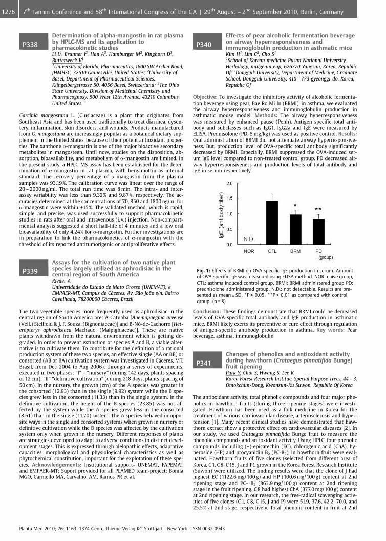
P338Determination of alpha-mangostin in rat plasmaby HPLC-MS and its application topharmacokinetic studiesLi L1, Brunner I2, Han A3, Hamburger M2, Kinghorn D3,Butterweck V1
1University of Florida, Pharmaceutics, 1600 SW Archer Road,JHMHSC, 32610 Gainesville, United States; 2University ofBasel, Department of Pharmaceutical Sciences,Klingelbergstrasse 50, 4056 Basel, Switzerland; 3The OhioState University, Division of Medicinal Chemistry andPharmacognosy, 500 West 12th Avenue, 43210 Columbus,United States
Garcinia mangostana L. (Clusiaceae) is a plant that originates fromSoutheast Asia and has been used traditionally to treat diarrhea, dysen-tery, inflammation, skin disorders, and wounds. Products manufacturedfrom G. mangostana are increasingly popular as a botanical dietary sup-plement in the United States, because of their potent antioxidant proper-ties. The xanthone a-mangostin is one of the major bioactive secondarymetabolites in mangosteen. Until now, studies on the disposition, ab-sorption, bioavailability, and metabolism of a-mangostin are limited. Inthe present study, a HPLC-MS assay has been established for the deter-mination of a-mangostin in rat plasma, with bergamottin as internalstandard. The recovery percentage of a-mangostin from the plasmasamples was 93.19%. The calibration curve was linear over the range of20 – 2000 ng/ml. The total run time was 8 min. The intra- and inter-assay variability was less than 9.32% and 9.87%, respectively. The ac-curacies determined at the concentrations of 70, 850 and 1800 ng/ml fora-mangostin were within +15%. The validated method, which is rapid,simple, and precise, was used successfully to support pharmacokineticstudies in rats after oral and intravenous (i. v.) injection. Non-compart-mental analysis suggested a short half-life of 4 minutes and a low oralbioavailability of only 4.24% for a-mangostin. Further investigations arein preparation to link the pharmacokinetics of a-mangostin with thethreshold of its reported antitumorigenic or antiproliferative effects.
P339Assays for the cultivation of two native plantspecies largely utilized as aphrodisiac in thecentral region of South AmericaRieder AUniversidade do Estado de Mato Grosso (UNEMAT); eEMPAER-MT, Campus de Cceres, Av. S¼o Jo¼o s/n, BairroCavalhada, 78200000 Cceres, Brazil
The two vegetable species more frequently used as aphrodisiac in thecentral region of South America are: A-Catuaba [Anemopaegma arvense(Vell.) Stellfeld & J. F. Souza, (Bignoniaceae)] and B-N -de-Cachorro [Het-eropterys aphrodisiaca Machado, (Malpighiaceae)]. These are nativeplants withdrawn from the natural environment which is getting de-graded. In order to prevent extinction of species A and B, a viable alter-native is to cultivate them. To contribute for the definition of a rationalproduction system of these two species, an effective single (AA or BB) orconsorted (AB or BA) cultivation system was investigated in C�ceres, MT,Brasil, from Dec 2004 to Aug 2006), through a series of experiments,executed in two phases: “I” – “nursery” (during 142 days, plants spacingof 12 cm); “II” “definitive cultivation” (during 218 days, plants spacing of50 cm). In the nursery, the growth (cm) of the A species was greater inthe consorted (12.93) than in the single (9.92) system while the B spe-cies grew less in the consorted (11.33) than in the single system. In thedefinitive cultivation, the height of the B species (23.85) was not af-fected by the system while the A species grew less in the consorted(8.61) than in the single (11.70) system. The A species behaved in oppo-site ways in the single and consorted systems when grown in nursery ordefinitive cultivation while the B species was affected by the cultivationsystem only when grown in the nursery. Different responses of plantsare strategies developed to adapt to adverse conditions in distinct devel-opment stages. This is expressed through alelopathic effects, adaptativecapacities, morphological and physiological characteristics as well asphytochemical constitution, important for the explotation of these spe-cies. Acknowledgements: Institutional support- UNEMAT, FAPEMATand EMPAER-MT; Suport provided for all PLAMED team-project: BonilaMGO, Carniello MA, Carvalho, AM, Ramos PR et al.
P340Effects of pear alcoholic fermentation beverageon airway hyperresponsiveness andimmunoglobulin production in asthmatic miceKim H1, Lim C2, Cho S1
1School of Korean medicine Pusan National University,Herbology, mulgeum eup, 626770 Yangsan, Korea, RepublicOf; 2Dongguk University, Department of Medicine, GraduateSchool, Dongguk University, 410 – 773 gyeonggi-do, Korea,Republic Of
Objective: To investigate the inhibitory activity of alcoholic fermenta-tion beverage using pear, Bae Ro Mi In (BRMI), in asthma, we evaluatedthe airway hyperresponsiveness and immunoglobulin production inasthmatic mouse model. Methods: The airway hyperresponsivenesswas measured by enhanced pause (Penh). Antigen specific total anti-body and subclasses such as IgG1, IgG2a and IgE were measured byELISA. Prednisolone (PD, 5 mg/kg) was used as positive control. Results:Oral administration of BRMI did not attenuate airway hyperresponsive-ness. But, production level of OVA-specific total antibody significantlydecreased by BRMI. Especially, BRMI suppressed the OVA-induced ser-um IgE level compared to non-treated control group. PD decreased air-way hyperresponsiveness and production levels of total antibody andIgE in serum respectively.
Fig. 1: Effects of BRMI on OVA-specific IgE production in serum. Amountof OVA-specific IgE was measured using ELISA method. NOR: naive group,CTL: asthma induced control group, BRMI: BRMI administered group PD:prednisolone administered group. N.D.: not detectable. Results are pre-sented as mean € SD. *P < 0.05, **P < 0.01 as compared with controlgroup. (n = 8)
Conclusion: These findings demonstrate that BRMI could be decreasedlevels of OVA-specific total antibody and IgE production in asthmaticmice. BRMI likely exerts its preventive or cure effect through regulationof antigen-specific antibody production in asthma. Key words: Pearbeverage, asthma, immunoglobulin
P341Changes of phenolics and antioxidant activityduring hawthorn (Crataegus pinnatifida Bunge)fruit ripeningPark Y, Choi S, Hwang S, Lee KKorea Forest Research Institue, Special Purpose Trees, 44 – 3,Omokchun-Dong, Kwonsun-Ku Suwon, Republic Of Korea
The antioxidant activity, total phenolic compounds and four major phe-nolics in hawthorn fruits (during three ripening stages) were investi-gated. Hawthorn has been used as a folk medicine in Korea for thetreatment of various cardiovascular disease, arteriosclerosis and hyper-tension [1]. Many recent clinical studies have demonstrated that haw-thorn extract show a protective effect on cardiovascular diseases [2]. Inour study, we used Crataegus pinnatifida Bunge fruit to measure thephenolic compounds and antioxidant activity. Using HPLC, four phenoliccompounds including (�)-epicatechin (EC), chlorogenic acid (ChA), hy-peroside (HP) and procyanidin B2 (PC-B2), in hawthorn fruit were eval-uated. Hawthorn fruits of five clones (selected from different area ofKorea, C 1, C 8, C 15, J and P), grown in the Korea Forest Research Institute(Suwon) were utilized. The finding results were that the clone of J hadhighest EC (1122.6 mg/100 g) and HP (100.6 mg/100 g) content at 2ndripening stage and PC- B2 (863.9 mg/100 g) content at 2nd ripeningstage in the fruit ripening. C 8 had highest ChA (377.0 mg/100 g) contentat 2nd ripening stage. In our research, the free-radical scavenging activ-ities of five clones (C 1, C 8, C 15, J and P) were 51.9, 37.6, 42.2, 70.0, and25.5% at 2nd stage, respectively. Total phenolic content in fruit at 2nd
Planta Med 2010; 76: 1163–1374 Georg Thieme Verlag KG Stuttgart · New York · ISSN 0032-0943
1276 7th Tannin Conference and 58th International Congress of the GA | 29th August – 2nd September 2010, Berlin, Germany
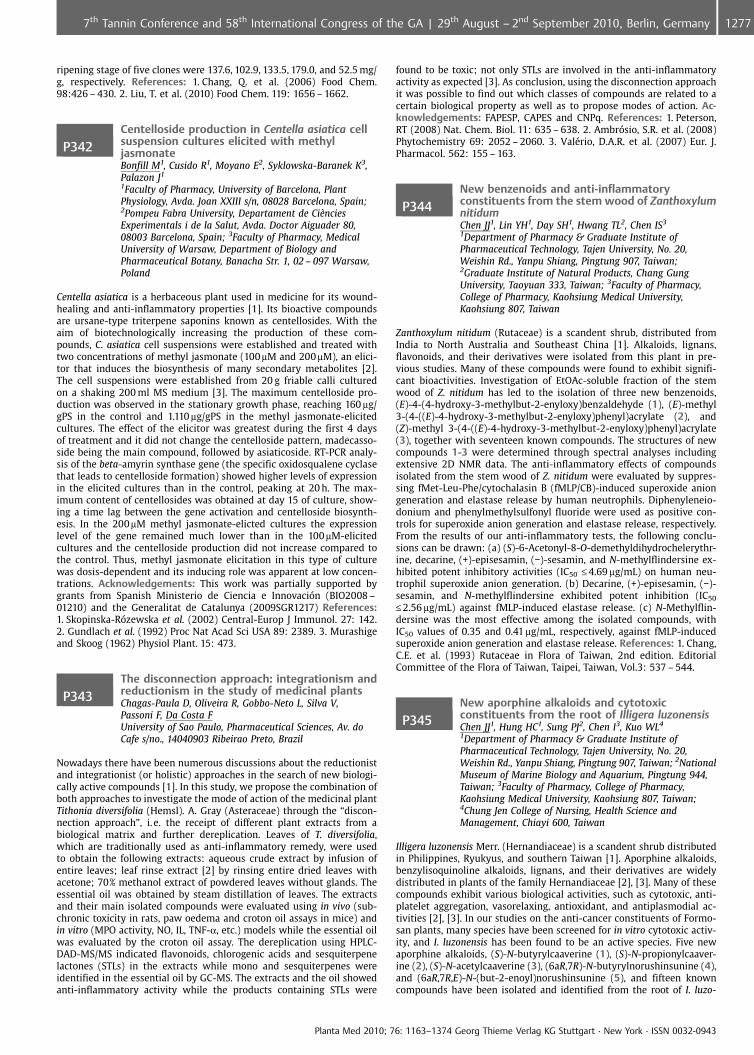
ripening stage of five clones were 137.6, 102.9, 133.5, 179.0, and 52.5 mg/g, respectively. References: 1. Chang, Q. et al. (2006) Food Chem.98:426 – 430. 2. Liu, T. et al. (2010) Food Chem. 119: 1656 – 1662.
P342Centelloside production in Centella asiatica cellsuspension cultures elicited with methyljasmonateBonfill M1, Cusido R1, Moyano E2, Syklowska-Baranek K3,Palazon J1
1Faculty of Pharmacy, University of Barcelona, PlantPhysiology, Avda. Joan XXIII s/n, 08028 Barcelona, Spain;2Pompeu Fabra University, Departament de Ci�nciesExperimentals i de la Salut, Avda. Doctor Aiguader 80,08003 Barcelona, Spain; 3Faculty of Pharmacy, MedicalUniversity of Warsaw, Department of Biology andPharmaceutical Botany, Banacha Str. 1, 02 – 097 Warsaw,Poland
Centella asiatica is a herbaceous plant used in medicine for its wound-healing and anti-inflammatory properties [1]. Its bioactive compoundsare ursane-type triterpene saponins known as centellosides. With theaim of biotechnologically increasing the production of these com-pounds, C. asiatica cell suspensions were established and treated withtwo concentrations of methyl jasmonate (100mM and 200mM), an elici-tor that induces the biosynthesis of many secondary metabolites [2].The cell suspensions were established from 20 g friable calli culturedon a shaking 200 ml MS medium [3]. The maximum centelloside pro-duction was observed in the stationary growth phase, reaching 160 mg/gPS in the control and 1.110 mg/gPS in the methyl jasmonate-elicitedcultures. The effect of the elicitor was greatest during the first 4 daysof treatment and it did not change the centelloside pattern, madecasso-side being the main compound, followed by asiaticoside. RT-PCR analy-sis of the beta-amyrin synthase gene (the specific oxidosqualene cyclasethat leads to centelloside formation) showed higher levels of expressionin the elicited cultures than in the control, peaking at 20 h. The max-imum content of centellosides was obtained at day 15 of culture, show-ing a time lag between the gene activation and centelloside biosynth-esis. In the 200mM methyl jasmonate-elicted cultures the expressionlevel of the gene remained much lower than in the 100 mM-elicitedcultures and the centelloside production did not increase compared tothe control. Thus, methyl jasmonate elicitation in this type of culturewas dosis-dependent and its inducing role was apparent at low concen-trations. Acknowledgements: This work was partially supported bygrants from Spanish Ministerio de Ciencia e Innovaci n (BIO2008 –01210) and the Generalitat de Catalunya (2009SGR1217) References:1. Skopinska-R zewska et al. (2002) Central-Europ J Immunol. 27: 142.2. Gundlach et al. (1992) Proc Nat Acad Sci USA 89: 2389. 3. Murashigeand Skoog (1962) Physiol Plant. 15: 473.
P343The disconnection approach: integrationism andreductionism in the study of medicinal plantsChagas-Paula D, Oliveira R, Gobbo-Neto L, Silva V,Passoni F, Da Costa FUniversity of Sao Paulo, Pharmaceutical Sciences, Av. doCafe s/no., 14040903 Ribeirao Preto, Brazil
Nowadays there have been numerous discussions about the reductionistand integrationist (or holistic) approaches in the search of new biologi-cally active compounds [1]. In this study, we propose the combination ofboth approaches to investigate the mode of action of the medicinal plantTithonia diversifolia (Hemsl). A. Gray (Asteraceae) through the “discon-nection approach”, i. e. the receipt of different plant extracts from abiological matrix and further dereplication. Leaves of T. diversifolia,which are traditionally used as anti-inflammatory remedy, were usedto obtain the following extracts: aqueous crude extract by infusion ofentire leaves; leaf rinse extract [2] by rinsing entire dried leaves withacetone; 70% methanol extract of powdered leaves without glands. Theessential oil was obtained by steam distillation of leaves. The extractsand their main isolated compounds were evaluated using in vivo (sub-chronic toxicity in rats, paw oedema and croton oil assays in mice) andin vitro (MPO activity, NO, IL, TNF-a, etc.) models while the essential oilwas evaluated by the croton oil assay. The dereplication using HPLC-DAD-MS/MS indicated flavonoids, chlorogenic acids and sesquiterpenelactones (STLs) in the extracts while mono and sesquiterpenes wereidentified in the essential oil by GC-MS. The extracts and the oil showedanti-inflammatory activity while the products containing STLs were
found to be toxic; not only STLs are involved in the anti-inflammatoryactivity as expected [3]. As conclusion, using the disconnection approachit was possible to find out which classes of compounds are related to acertain biological property as well as to propose modes of action. Ac-knowledgements: FAPESP, CAPES and CNPq. References: 1. Peterson,RT (2008) Nat. Chem. Biol. 11: 635 – 638. 2. Ambr sio, S.R. et al. (2008)Phytochemistry 69: 2052 – 2060. 3. Val�rio, D.A.R. et al. (2007) Eur. J.Pharmacol. 562: 155 – 163.
P344New benzenoids and anti-inflammatoryconstituents from the stem wood of ZanthoxylumnitidumChen JJ1, Lin YH1, Day SH1, Hwang TL2, Chen IS3
1Department of Pharmacy & Graduate Institute ofPharmaceutical Technology, Tajen University, No. 20,Weishin Rd., Yanpu Shiang, Pingtung 907, Taiwan;2Graduate Institute of Natural Products, Chang GungUniversity, Taoyuan 333, Taiwan; 3Faculty of Pharmacy,College of Pharmacy, Kaohsiung Medical University,Kaohsiung 807, Taiwan
Zanthoxylum nitidum (Rutaceae) is a scandent shrub, distributed fromIndia to North Australia and Southeast China [1]. Alkaloids, lignans,flavonoids, and their derivatives were isolated from this plant in pre-vious studies. Many of these compounds were found to exhibit signifi-cant bioactivities. Investigation of EtOAc-soluble fraction of the stemwood of Z. nitidum has led to the isolation of three new benzenoids,(E)-4-(4-hydroxy-3-methylbut-2-enyloxy)benzaldehyde (1), (E)-methyl3-(4-((E)-4-hydroxy-3-methylbut-2-enyloxy)phenyl)acrylate (2), and(Z)-methyl 3-(4-((E)-4-hydroxy-3-methylbut-2-enyloxy)phenyl)acrylate(3), together with seventeen known compounds. The structures of newcompounds 1-3 were determined through spectral analyses includingextensive 2D NMR data. The anti-inflammatory effects of compoundsisolated from the stem wood of Z. nitidum were evaluated by suppres-sing fMet-Leu-Phe/cytochalasin B (fMLP/CB)-induced superoxide aniongeneration and elastase release by human neutrophils. Diphenyleneio-donium and phenylmethylsulfonyl fluoride were used as positive con-trols for superoxide anion generation and elastase release, respectively.From the results of our anti-inflammatory tests, the following conclu-sions can be drawn: (a) (S)-6-Acetonyl-8-O-demethyldihydrochelerythr-ine, decarine, (+)-episesamin, (�)-sesamin, and N-methylflindersine ex-hibited potent inhibitory activities (IC50 £ 4.69 mg/mL) on human neu-trophil superoxide anion generation. (b) Decarine, (+)-episesamin, (�)-sesamin, and N-methylflindersine exhibited potent inhibition (IC50
£2.56 mg/mL) against fMLP-induced elastase release. (c) N-Methylflin-dersine was the most effective among the isolated compounds, withIC50 values of 0.35 and 0.41 mg/mL, respectively, against fMLP-inducedsuperoxide anion generation and elastase release. References: 1. Chang,C.E. et al. (1993) Rutaceae in Flora of Taiwan, 2nd edition. EditorialCommittee of the Flora of Taiwan, Taipei, Taiwan, Vol.3: 537 – 544.
P345New aporphine alkaloids and cytotoxicconstituents from the root of Illigera luzonensisChen JJ1, Hung HC1, Sung PJ2, Chen I3, Kuo WL4
1Department of Pharmacy & Graduate Institute ofPharmaceutical Technology, Tajen University, No. 20,Weishin Rd., Yanpu Shiang, Pingtung 907, Taiwan; 2NationalMuseum of Marine Biology and Aquarium, Pingtung 944,Taiwan; 3Faculty of Pharmacy, College of Pharmacy,Kaohsiung Medical University, Kaohsiung 807, Taiwan;4Chung Jen College of Nursing, Health Science andManagement, Chiayi 600, Taiwan
Illigera luzonensis Merr. (Hernandiaceae) is a scandent shrub distributedin Philippines, Ryukyus, and southern Taiwan [1]. Aporphine alkaloids,benzylisoquinoline alkaloids, lignans, and their derivatives are widelydistributed in plants of the family Hernandiaceae [2], [3]. Many of thesecompounds exhibit various biological activities, such as cytotoxic, anti-platelet aggregation, vasorelaxing, antioxidant, and antiplasmodial ac-tivities [2], [3]. In our studies on the anti-cancer constituents of Formo-san plants, many species have been screened for in vitro cytotoxic activ-ity, and I. luzonensis has been found to be an active species. Five newaporphine alkaloids, (S)-N-butyrylcaaverine (1), (S)-N-propionylcaaver-ine (2), (S)-N-acetylcaaverine (3), (6aR,7R)-N-butyrylnorushinsunine (4),and (6aR,7R,E)-N-(but-2-enoyl)norushinsunine (5), and fifteen knowncompounds have been isolated and identified from the root of I. luzo-
Planta Med 2010; 76: 1163–1374 Georg Thieme Verlag KG Stuttgart · New York · ISSN 0032-0943
12777th Tannin Conference and 58th International Congress of the GA | 29th August – 2nd September 2010, Berlin, Germany
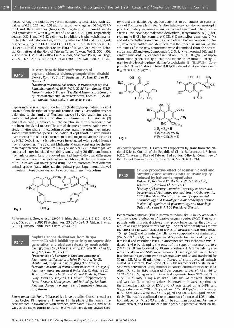
nensis. Among the isolates, (�)-yatein exhibited cytotoxicities, with IC50
values of 0.81, 0.20, and 0.59 mg/mL, respectively, against DLD-1, CCRF-CEM, and HL-60 cell lines. (6aR,7R)-N-Butyrylnorushinsunine (4) exhib-ited cytotoxicities, with IC50 values of 5.45 and 3.66 mg/mL, respectively,against DLD-1 and IMR-32 cell lines. In addition, N-phenethylcinnama-mide exhibited cytotoxicities, with IC50 values of 6.84 and 5.79 mg/mL,respectively, against DLD-1 and CCRF-CEM cell lines. References: 1. Li,H.I. et al. (1996) Hernandiaceae. In: Flara of Taiwan, 2nd edition. Edito-tial Committee of the Flora of Taiwan, Taipei, Taiwan: Vol. 2: 500 – 503.2. Conserva, L.M. et al. (2005) The Alkaloids, Academic Press, San Diego,Vol. 54: 175 – 243. 3. Lakshmi, V. et al. (2009) Rec. Nat. Prod. 3: 1 – 22.
P346In vitro hepatic biotransformation ofcepharanthine, a bisbenzylisoquinoline alkaloidBory S1, Koeut S1, Bun S1, Baghdikian B1, Elias R1, Bun H2,Ollivier E1
1Faculty of Pharmacy, Laboratory of Pharmacognosy andEthnopharmacology, UMR-MD 3, 27 Bd Jean Moulin, 13385Marseille cedex 5, France; 2Faculty of Pharmacy, Laboratoryof Toxicokinetics and Pharmacokinetics, UMR-MD 3, 27 bdJean Moulin, 13385 cedex 5 Marseille, France
Cepharanthine is a major biscoclaurine (bisbenzylisoquinoline) alkaloidisolated from the tuber of Stephania rotunda Lour., a Cambodian creeperbelonging to the family of Menispermaceae [1]. Cepharanthine exertsvarious biological effects including antiplasmodial [1], cytotoxic [2]and antioxidant [3] activies, but the metabolism of this compound hasnot been elucidated to date. The aim of the present investigation was tostudy in vitro phase I metabolism of cepharanthine using liver micro-somes from different species. Incubation of cepharanthine with humanliver microsomes led to the formation of one major metabolite, detectedby HPLC-DAD. Enzyme kinetics were investigated with pooled humanliver microsomes. The apparent Michaelis-Menten constants for the hu-man major metabolite were Km = 37.7mM and Vm = 22.7 nmol/mg/h. Weconducted inter-individual variability study using 20 different humanliver microsomes. Results showed marked inter-individual differencesin human cepharanthine metabolism. In addition, the biotransformationof this alkaloid was investigated using liver microsomes from differentanimal species (rats, mice, rabbits, guinea-pigs). Experiments showedimportant inter-species variability of cepharanthine metabolism
Fig. 1
References: 1. Chea, A. et al. (2007) J. Ethnopharmacol. 112:132 – 137. 2.Bun, S.S. et al. (2009) Phytother. Res. 23:587 – 590. 3. Gl�in, I. et al.(2010) J. Enzyme Inhib. Med. Chem. 25:44 – 53.
P347Naphthalenone derivatives from Berryaammonilla with inhibitory activity on superoxidegeneration and elastase release by neutrophilsChen JJ1, Chien SK1, Chou T2, Hwang TL3, Wei DC1, Chen IS2,Yang SZ4, Liao HC1, Ho CC1
1Department of Pharmacy & Graduate Institute ofPharmaceutical Technology, Tajen University, No. 20,Weishin Rd., Yanpu Shiang, Pingtung 907, Taiwan;2Graduate Institute of Pharmaceutical Sciences, College ofPharmacy, Kaohsiung Medical University, Kaohsiung 807,Taiwan; 3Graduate Institute of Natural Products, ChangGung University, Taoyuan 333, Taiwan; 4Department ofForest Resource, Management and Technology, NationalPingtung University of Science and Technology, Pingtung912, Taiwan
Berrya ammonilla Roxb. (Tiliaceae) is a large tree, distributed in southernIndia, Ceylon, Philippines, and Taiwan [1]. The plants of the family Tilia-ceae are rich in flavonoids with flavones, flavanones, flavans, and bifla-vans as the major constituents, some of which have demonstrated cyto-
toxic and antiplatelet aggregation activities. In our studies on constitu-ents of Formosan plants for in vitro inhibitory activity on neutrophilproinflammatory responses, B. ammonilla has been found to be an activespecies. Five new naphthalenone derivatives, berryammone A (1), ber-ryammone B (2), berryammone C (3), 6-O-methylberryammone C (4),and 4-O-methylberryammone C (5) and eleven known compounds (6 –16) have been isolated and identified from the stem of B. ammonilla. Thestructures of these new compounds were determined through spectro-scopic and MS analyses. Compounds 1, 2, 3, 5, (+)-pinoresinol (6), and 3-epi-betulinic acid (12) exhibited inhibition (IC50 £1.58 mg/mL) of super-oxide anion generation by human neutrophils in response to formyl-L-methionyl-L-leucyl-L-phenylalanine/cytochalasin B (fMLP/CB). Com-pounds 1, 2, and 5 also inhibited fMLP/CB induced elastase release withIC50 values £1.21 mg/mL.
Fig. 1
Acknowledgements: This work was supported by grant from the Na-tional Science Council of the Republic of China. References: 1. Robson,N.K.B. Tiliaceae in Flora of Taiwan. 2nd edition. Editorial Committee ofthe Flora of Taiwan, Taipei, Taiwan: 1996; Vol. 3: 694 – 714.
P348Ex vivo protective effect of rosmarinic acid andMentha x villosa water extract on tissue injuryinduced by ischaemia/reperfusionFialov S1, Sotn�kov R2, Noslov V2, Drbikov K2,Tekelov D1, Kotlov D1, Grancai D1
1Faculty of Pharmacy Comenius University in Bratislava,Department of Pharmacognosy and Botany, Odbojarov 10,83232 Bratislava, Slovakia; 2Institute of experimentalpharmacology and toxicology, Slovak Academy of Science,Institute of experimental pharmacology and toxicology,Dfflbravska cesta 9, 841 04 Bratislava, Slovakia
Ischaemia/reperfusion (I/R) is known to induce tissue injury associatedwith increased production of reactive oxygen species (ROS). Thus com-pounds with antiradical activity may prove beneficial as they help toreduce or even to prevent this damage. The aim of the study was to testthe effect of the water extract of leaves of Mentha x villosa Huds (EMV,1.3 mg/10 ml)) and its main phenolic active compound – rosmarinic acid(RA, 5 x 10�4 mol/l) on changes in ROS production induced by I/R inintestinal and vascular tissues. In anaesthetised rats, ischaemia was in-duced in vivo by clamping the onset of the superior mesenteric artery(SMA) for 60 min followed by 30 min reperfusion1. After sacrifying ani-mals, the ileum and SMA were removed. Tissue segments were placedinto the testing solutions with or without EMV and RA and incubated for30 min (SMA) or 60 min (ileum). Tissues of sham-operated animalsserved as a control. Production of ROS by segments of the ileum andSMA was determined using luminol enhanced chemiluminiscence (CL).After I/R, CL in SMA increased from control values of 7.9 € 1.66 to15.21 € 2.49 mV/mg w.w., in intestinal segments from 33.74 € 6.47 to58.53 € 11.73 mV/100 mg w.w. Both, EMV and RA reduced intestinaland vascular CL to control values. Moreover in in vitro experiments,the antioxidant activity of EMV and RA was tested using DPPH test.SC50 % values were 7.26 € 0.09 mg/ml and 1.72 € 0.13 mg/ml, respectively.In ABTS test SC50 % were 15.07 € 0.47 mg/ml and 1.93 € 0.03 mg/ml, respec-tively. The results confirmed the attenuation of increased ROS produc-tion induced by I/R in SMA and ileum by rosmarinic acid and Mentha x -villosa extract, and thus indicate their probable protective effect on tis-
Planta Med 2010; 76: 1163–1374 Georg Thieme Verlag KG Stuttgart · New York · ISSN 0032-0943
1278 7th Tannin Conference and 58th International Congress of the GA | 29th August – 2nd September 2010, Berlin, Germany
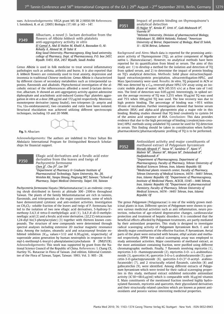
sues. Acknowledgements: VEGA grant MS SR 2/0050/09 References:1. Sotn�kov�, R. et al. (2005) Biologia (17) 60: p 145 – 147.
P349Albactam, a novel b- lactam derivative from theflowers of Albizia lebbeck with plateletsanti-aggregatory activity in vitroEl Gamal A, Abd El Halim M, Khalil A, Basoudan O, Al-Rehaily A, Ahmed M, El Tahir KKing Saud University, Pharmacognosy, King Saud university,college of Pharmacy, Dept. of Pharmaconosy, P.O. box 2457,Riyadh 11451, KSA, 2457 Riyadh, Saudi Arabia
Genus Albizia is used in folk medicine to treat several inflammatorypathologies such as asthma, arthritis and burns. It is also reported thatA. lebbeck flowers are commonly used to treat anxiety, depression andinsomnia in traditional Chinese medicine. Genus Albizia is characterizedby different classes of secondary metabolites such as triterpenoidal sa-ponins, flavonoids and alkaloids. Phytochemical investigation of the al-coholic extract of the inflorescences afforded a novel b-lactam deriva-tive, albactam. It showed an anti-aggregatory activity against adenosinediphosphate and arachidonic acid-induced Guniea Pigs’ platelets aggre-gation in vitro at a dose 208 mg/ml and 172 mg/ml respectively. Moreover,monoterpene derivative (epoxy linalol), two tritepenes (b- amyrin and11a, 12a-oxidotaraxerol), two ceramides and rutin have been isolated.Structural elucidation was achieved utilizing different spectroscopictechniques, including 1D and 2D NMR.
Fig. 1: Albactam
Acknowledgements: The authors are indebted to Prince Sultan BinAbdulaziz International Program for Distinguished Research Scholar-ships for financial support.
P350Ellagic acid derivatives and a ferulic acid esterderivative from the leaves and twigs ofPachycentria formosanaChen JJ1, Cho JY2, Lee TH2
1Department of Pharmacy & Graduate Institute ofPharmaceutical Technology, Tajen University, No. 20,Weishin Rd., Yanpu Shiang, Pingtung 907, Taiwan; 2School ofPharmacy, Taipei Medical University, Taipei 110, Taiwan
Pachycentria formosana Hayata (Melastomataceae) is an endemic creep-ing shrub distributed in forests at altitude 300 – 2100 m throughoutTaiwan. The plants of the family Melastomataceae are rich in tannins,flavonoids, and triterpenoids as the major constituents, some of whichhave demonstrated cytotoxic and anti-oxidant activities. Investigationon CH2Cl2- soluble fraction of the leaves and twigs of P. formosana hasled to the isolation of two new ellagic acid derivatives, 5-hydroxy-5’-methoxy-3,4,3’,4’-tetra-O-methylellagic acid (1), 3,4,3’,4’-di-O-methyle-neellagic acid (2) and a ferulic acid ester derivative, (2Z,2’Z)-tetracosane-1,24-diyl bis(3-phenylacrylate) (3) together with thirteen known com-pounds. The structure of new compounds were determined throughspectral analyses including extensive 2D nuclear magnetic resonancedata. Among the isolates, oleanolic acid and octacosanoyl ferulate ex-hibited inhibition (IC50 values = 3.12 and 6.30 mg/mL, respectively) ofsuperoxide anion generation by human neutrophils in response to for-myl-L-methionyl-L-leucyl-L-phenylalanine/cytochalasin B (fMLP/CB).Acknowledgements: This work was supported by grant from the Na-tional Science Council of the Republic of China. References: 1. Chang CE,Hartley TG. Rutaceae in Flora of Taiwan. 2nd edition. Editorial Commit-tee of the Flora of Taiwan, Taipei, Taiwan: 1993; Vol. 3: 905 – 28.
P351Impact of protein binding on thymoquinone’sanalytical detectionEl-Najjar N1, Ketola R1, Urtti A1, Gali-Muhtaseb H2,Vuorela H1
1Helsinki University, Division of pharmaceutical Biology,Viikinkaari 5E, 00014 Helsinki, Finland; 2AmericanUniversity of Beirut, Department of Biology, Riad El Soloh,11 – 0236 Beirut, Lebanon
Background and Aims: Much data is reported for the promising antic-ancer activity of Thymoquinone (TQ), an active component of Nigellasativa L. (Ranunculaceae). However, no analytical methods have beenreported for its quantification from blood or serum. The aims of thisstudy are: 1) to develop a method for the isolation and detection of TQfrom spiked serum, and 2) to determine the impact of protein bindingon TQ’s analytical detection. Methods: Solid phase extraction/liquid-liquid extraction/protein precipitation, ultracentrifugation-HPLC, andMass Spectrometry were used. Results: In vitro, TQ prepared in ACN orPBS was detected by a C18 reversed-phase HPLC-UV assay using an iso-cratic mobile phase of water: ACN (45:55% v/v) at a flow rate of 1 ml/min. The limit of detection was 0.05 mg/ml. Interestingly, in spiked ser-um the average recovery of TQ was 3.25% when 10 mg/ml TQ was usedand 75% when 100 mg/ml TQ was used. The low recovery was due tohigh protein binding. The percentage of binding was > 95% within10 min of incubation. Further investigation showed that bovine serumalbumin (BSA) and alpha-acid glycoprotein play a major role in thisbinding. Binding studies showed that TQ bind covalently to cystein-34of the amino acid sequence of BSA. Conclusion: This data providesevidence that due to the high percentage of binding (covalent/non-cova-lent) HPLC methods using unlabeled TQ cannot be used for TQ detectionin serum. This finding should be taken in consideration when furtherpharmacokinetic/pharmacodynamic profiling of TQ is to be performed.
P352Antioxidant activity and major constituents ofmethanol extract of Polygonum hyrcanicumMoradi-Afrapoli F1, Yassa N1, Saeidnia S2, Ajani Y3,Malmir M2, Shamsi M1, Mirjani M1, Dolatabadi R4,Hadjiakhoondi A1
1Department of Pharmacognosy, Department ofPharmacognosy, Faculty of Pharmacy, Tehran University ofMedical Sciences Tehran, Iran, Islamic Republic Of;2Medicinal plants research center, Faculty of Pharmacy,Tehran University of Medical Sciences, 14174 – 14411 Tehran,Iran, Islamic Republic Of; 3Department of Pharmacognosy,Institute of Medicinal Plants, ACECR, 13145 – 1446 Tehran,Iran, Islamic Republic Of; 4Department of pharmaceuticalchemistry, Faculty of Pharmacy, Tehran University ofMedical Sciences, 14174 – 14411 Tehran, Iran, IslamicRepublic Of
The genus Polygonum (Polygonaceae) is one of the widely grown med-icinal plants in Iran. Different species of Polygonum were shown to pos-sess various medicinal activities such as anti inflammation, neuro pro-tection, reduction of age-related degenerative changes, cardiovascularprotection and treatment of hepatic disorders. It is considered that thebeneficial effects afforded by Polygonum treatments, are partly mediatedby their antioxidant properties. This study is designed to investigateradical scavenging activity of Polygonum hyrcanicum Rech. f. and toidentify major constituents of the effective fraction. P. hyrcanicum. Aerialparts of the plant were extracted with hexane, ethyl acetate and metha-nol respectively. DPPH free radical scavenging assay was employed tostudy antioxidant activities. Major constituents of methanol extract, asthe most antioxidant containing fraction, were purified using differentchromatographic methods. Finally, 7 flavonols involving myricetin (1),myricetin-3-O- rhamnopyranoside (2), myricetin-3-O- a-L-arabinofura-noside (3), quercetin (4), quercetin-3-O-a-L-arabinofuranoside (5), quer-cetin-3-O-galactopyranoside (6), quercetin-3-O-(3”-O-acetyl- arabino-furanoside) (7), and 2 structurally related flavanols, catechin (8) andgallocatechin (9), were identified. Among different extracts of Polygo-num hyrcanicum which were tested for their radical scavenging proper-ties in this study, methanol extract exhibited noticeable antioxidantactivity (IC 50 = 68.6 mg/ml) which is comparable with 16 mg/ml vitaminE. Main constituents of the mentioned fraction involved highly hydro-xylated flavonols, myricetin and quercetin, their glycosilated derivativesand their structurally related catechins which are known as potent anti-oxsidants and possess various interesting medicinal properties.
Planta Med 2010; 76: 1163–1374 Georg Thieme Verlag KG Stuttgart · New York · ISSN 0032-0943
12797th Tannin Conference and 58th International Congress of the GA | 29th August – 2nd September 2010, Berlin, Germany
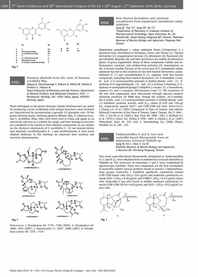
Fig. 1
P353Stemona alkaloids from the roots of Stemonasessilifolia Miq.Takeya K, Hitotsuyanagi Y, Kakuta D, Hikita M, Takeda E,Uemura G, Fukaya HTokyo University of Pharmacy and Life Sciences, Departmentof Natural Products and Medicinal Chemistry, 1432 – 1Horinouchi, Hachioji, 192 – 0392 Tokyo, Japan, 1920392Hachioji, Japan
Plants belonging to the genus Stemona (family Stemonaceae) are notedfor producing a series of alkaloids with unique structures, most of whichare characterized by incorporating a pyrrolo[ 1,2-a]azepine core. Of thegenus Stemona plants, Stemona japonica (Blume) Miq., S. tuberosa Lour.,and S. sessilifolia (Miq.) Miq. have been used in China and Japan as aninsecticide and also as a remedy for cough, and their biological activitiesare considered to be related to their alkaloid components. In our studieson the chemical constituents of S. sessilifolia [1 – 4], we isolated elevennew alkaloids, sessilifoliamides A – J and sessilifoliamine A, with novelalkaloid skeletons. In this meeting, we represent their isolation andstructure determination.
Fig. 1
References: 1. Tetrahedron 59: 7779 – 7786 (2003). 2. Tetrahedron 63:1008 – 1013 (2007). 3. Heterocycles 71: 2035 – 2040 (2007). 4. Tetrahe-dron Letters 49: 7376 – 7379.
P354New thymol derivatives and cytotoxicconstituents from Eupatorium cannabinum subsp.asiaticumChen JJ1, Tsai YC1, Sung PJ2, Ho CC1
1Department of Pharmacy & Graduate Institute ofPharmaceutical Technology, Tajen University, No. 20,Weishin Rd., Yanpu Shiang, Pingtung 907, Taiwan; 2NationalMuseum of Marine Biology and Aquarium, Pingtung 944,Taiwan
Eupatorium cannabinum L. subsp. asiaticum Kitam (Compositae) is aperennial herb, distributed in Himalaya, China, and Taiwan [1]. Thymolderivatives [2], sesquiterpene lactones [3], diterpenes [4], flavonoids [4],pyrrolizidine alkaloids [4], and their derivatives are widely distributed inplants of genus Eupatorium. Many of these compounds exhibit anti-in-flammatory, cytotoxic, and antibacterial activities [5]. Investigation onthe n-hexane-soluble fraction of the aerial part of E. cannabinum subsp.asiaticum has led to the isolation of two new thymol derivatives, canna-bithymol A (1) and cannabithymol B (2), together with five knowncompounds, including three thymol derivatives, (E)-2-methylbut-2-eno-lic acid 2-(2-acetoxymethyl-oxiranyl)-5-methyl-phenyl ester (3), 8-methoxy-9-O-angeloylthymol (4), and (Z)-3-acetoxy-2-hydeoxy-2-(2-hydroxy-4-methylphenol)propyl-2-ethylbut-2-enoate (5), a benzofuran,euparin (6), and a coumarin, 2H-chromen-2-one (7). The structures ofthe two new compounds were determined through spectral analysesincluding extensive 2D NMR data. Among the isolates, (E)-2-methyl-but-2-enolic acid 2-(2-acetoxymethyl-oxiranyl)-5-methyl-phenyl ester(3) exhibited cytotoxic activity, with IC50 values of 0.02 and 1.02 mg/mL, respectively, against DLD-1 and CCRF-CEM cell lines. References:1. Chang, C.E. et al. (1993) Compositae in Flora of Taiwan. 2nd edition.Editorial Committee of the Flora of Taiwan, Taipei, Taiwan. Vol. 2: 804 –1101. 2. Tori M. et. al. (2001) J. Nat. Prod. 64: 1048 – 1051. 3. McPhail A.T.et al. (1975) J. Chem. Soc. Perkin II 1798 – 1801. 4. Paolini J. et al. (2007)Phytochem. Anal. 18: 235 – 244. 5. Woerdenbag, H.J. (1986) Pharm.Weekbl. Sci. 8: 245 – 251.
P355Cladieunicellins A and B, two neweunicellin-based diterpenoids from anIndonesian octocoral Cladiella sp.Sung PJ, Tai C, Chen Y, Lin MNational Museum of Marine Biology and Aquairum,2 Houwan Rd. Checheng Pingtung, Taiwan
Two novel eunicellin-based diterpenoids, designated as cladieunicellinsA (1) and B (2), were obtained from an Indonesian octocoral identified asCladiella sp. The structures of eunicellins 1 and 2 were established byspectroscopic methods. These two compounds are the first metabolitesof eunicellin-related natural products found to possess 2-hydroxybuta-noxy groups. Eunicellin 1 exhibited significant cytotoxicity towardCCRF-CEM tumor cells (ED50= 3.61 mg/ml) and moderate cytotoxicity to-wards DLD-1 (ED50 = 8.50 mg/ml) and P388D 1 (ED50 = 8.32 mg/ml) tumorcells. Eunicellin 2 was also found to exhibit moderate cytotoxicity to-wards CCRF-CEM (ED 50 = 4.65 mg/ml) and DLD-1 (ED50= 10.15 mg/ml) tu-mor cells.
Fig. 1
Planta Med 2010; 76: 1163–1374 Georg Thieme Verlag KG Stuttgart · New York · ISSN 0032-0943
1280 7th Tannin Conference and 58th International Congress of the GA | 29th August – 2nd September 2010, Berlin, Germany
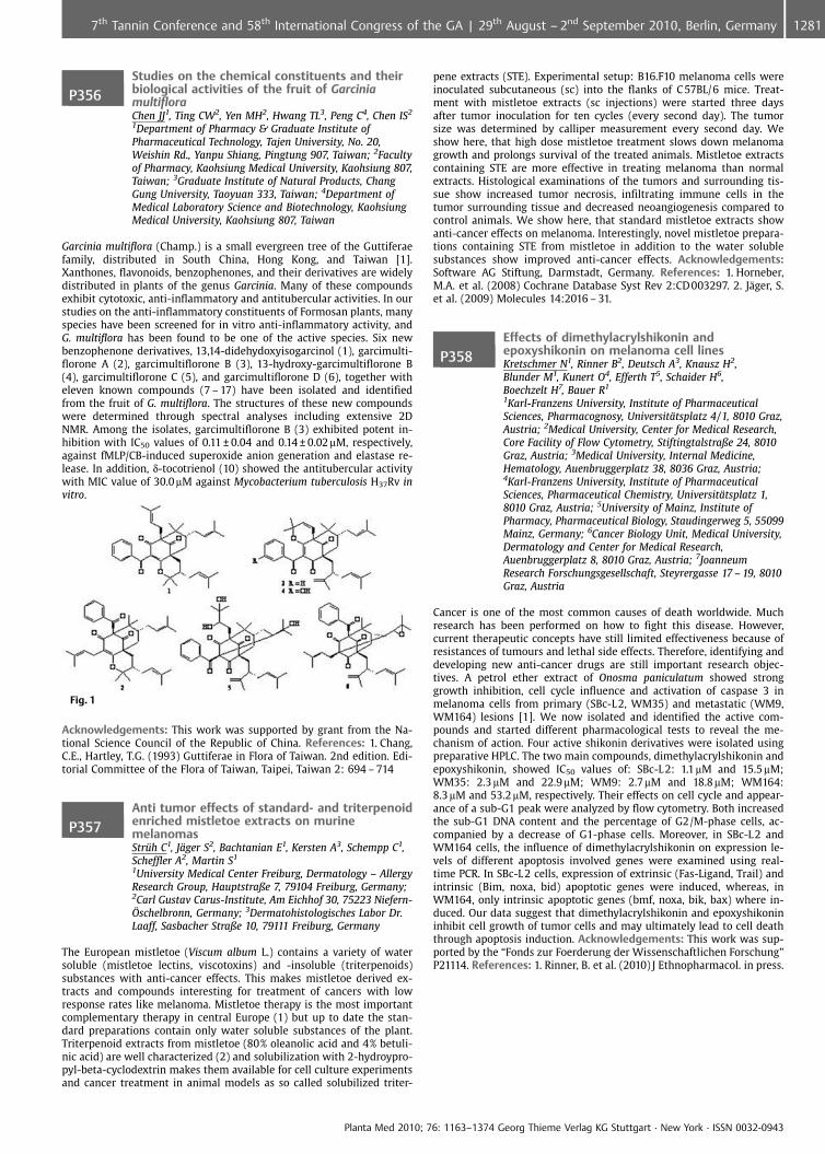
P356Studies on the chemical constituents and theirbiological activities of the fruit of GarciniamultifloraChen JJ1, Ting CW2, Yen MH2, Hwang TL3, Peng C4, Chen IS2
1Department of Pharmacy & Graduate Institute ofPharmaceutical Technology, Tajen University, No. 20,Weishin Rd., Yanpu Shiang, Pingtung 907, Taiwan; 2Facultyof Pharmacy, Kaohsiung Medical University, Kaohsiung 807,Taiwan; 3Graduate Institute of Natural Products, ChangGung University, Taoyuan 333, Taiwan; 4Department ofMedical Laboratory Science and Biotechnology, KaohsiungMedical University, Kaohsiung 807, Taiwan
Garcinia multiflora (Champ.) is a small evergreen tree of the Guttiferaefamily, distributed in South China, Hong Kong, and Taiwan [1].Xanthones, flavonoids, benzophenones, and their derivatives are widelydistributed in plants of the genus Garcinia. Many of these compoundsexhibit cytotoxic, anti-inflammatory and antitubercular activities. In ourstudies on the anti-inflammatory constituents of Formosan plants, manyspecies have been screened for in vitro anti-inflammatory activity, andG. multiflora has been found to be one of the active species. Six newbenzophenone derivatives, 13,14-didehydoxyisogarcinol (1), garcimulti-florone A (2), garcimultiflorone B (3), 13-hydroxy-garcimultiflorone B(4), garcimultiflorone C (5), and garcimultiflorone D (6), together witheleven known compounds (7 – 17) have been isolated and identifiedfrom the fruit of G. multiflora. The structures of these new compoundswere determined through spectral analyses including extensive 2DNMR. Among the isolates, garcimultiflorone B (3) exhibited potent in-hibition with IC50 values of 0.11 € 0.04 and 0.14 € 0.02mM, respectively,against fMLP/CB-induced superoxide anion generation and elastase re-lease. In addition, d-tocotrienol (10) showed the antitubercular activitywith MIC value of 30.0 mM against Mycobacterium tuberculosis H37Rv invitro.
Fig. 1
Acknowledgements: This work was supported by grant from the Na-tional Science Council of the Republic of China. References: 1. Chang,C.E., Hartley, T.G. (1993) Guttiferae in Flora of Taiwan. 2nd edition. Edi-torial Committee of the Flora of Taiwan, Taipei, Taiwan 2: 694 – 714
P357Anti tumor effects of standard- and triterpenoidenriched mistletoe extracts on murinemelanomasStr�h C1, J�ger S2, Bachtanian E1, Kersten A3, Schempp C1,Scheffler A2, Martin S1
1University Medical Center Freiburg, Dermatology – AllergyResearch Group, Hauptstraße 7, 79104 Freiburg, Germany;2Carl Gustav Carus-Institute, Am Eichhof 30, 75223 Niefern-�schelbronn, Germany; 3Dermatohistologisches Labor Dr.Laaff, Sasbacher Straße 10, 79111 Freiburg, Germany
The European mistletoe (Viscum album L.) contains a variety of watersoluble (mistletoe lectins, viscotoxins) and -insoluble (triterpenoids)substances with anti-cancer effects. This makes mistletoe derived ex-tracts and compounds interesting for treatment of cancers with lowresponse rates like melanoma. Mistletoe therapy is the most importantcomplementary therapy in central Europe (1) but up to date the stan-dard preparations contain only water soluble substances of the plant.Triterpenoid extracts from mistletoe (80% oleanolic acid and 4% betuli-nic acid) are well characterized (2) and solubilization with 2-hydroypro-pyl-beta-cyclodextrin makes them available for cell culture experimentsand cancer treatment in animal models as so called solubilized triter-
pene extracts (STE). Experimental setup: B16.F10 melanoma cells wereinoculated subcutaneous (sc) into the flanks of C 57BL/6 mice. Treat-ment with mistletoe extracts (sc injections) were started three daysafter tumor inoculation for ten cycles (every second day). The tumorsize was determined by calliper measurement every second day. Weshow here, that high dose mistletoe treatment slows down melanomagrowth and prolongs survival of the treated animals. Mistletoe extractscontaining STE are more effective in treating melanoma than normalextracts. Histological examinations of the tumors and surrounding tis-sue show increased tumor necrosis, infiltrating immune cells in thetumor surrounding tissue and decreased neoangiogenesis compared tocontrol animals. We show here, that standard mistletoe extracts showanti-cancer effects on melanoma. Interestingly, novel mistletoe prepara-tions containing STE from mistletoe in addition to the water solublesubstances show improved anti-cancer effects. Acknowledgements:Software AG Stiftung, Darmstadt, Germany. References: 1. Horneber,M.A. et al. (2008) Cochrane Database Syst Rev 2:CD 003297. 2. J�ger, S.et al. (2009) Molecules 14:2016 – 31.
P358Effects of dimethylacrylshikonin andepoxyshikonin on melanoma cell linesKretschmer N1, Rinner B2, Deutsch A3, Knausz H2,Blunder M1, Kunert O4, Efferth T5, Schaider H6,Boechzelt H7, Bauer R1
1Karl-Franzens University, Institute of PharmaceuticalSciences, Pharmacognosy, Universit�tsplatz 4/1, 8010 Graz,Austria; 2Medical University, Center for Medical Research,Core Facility of Flow Cytometry, Stiftingtalstraße 24, 8010Graz, Austria; 3Medical University, Internal Medicine,Hematology, Auenbruggerplatz 38, 8036 Graz, Austria;4Karl-Franzens University, Institute of PharmaceuticalSciences, Pharmaceutical Chemistry, Universit�tsplatz 1,8010 Graz, Austria; 5University of Mainz, Institute ofPharmacy, Pharmaceutical Biology, Staudingerweg 5, 55099Mainz, Germany; 6Cancer Biology Unit, Medical University,Dermatology and Center for Medical Research,Auenbruggerplatz 8, 8010 Graz, Austria; 7JoanneumResearch Forschungsgesellschaft, Steyrergasse 17 – 19, 8010Graz, Austria
Cancer is one of the most common causes of death worldwide. Muchresearch has been performed on how to fight this disease. However,current therapeutic concepts have still limited effectiveness because ofresistances of tumours and lethal side effects. Therefore, identifying anddeveloping new anti-cancer drugs are still important research objec-tives. A petrol ether extract of Onosma paniculatum showed stronggrowth inhibition, cell cycle influence and activation of caspase 3 inmelanoma cells from primary (SBc-L2, WM35) and metastatic (WM9,WM164) lesions [1]. We now isolated and identified the active com-pounds and started different pharmacological tests to reveal the me-chanism of action. Four active shikonin derivatives were isolated usingpreparative HPLC. The two main compounds, dimethylacrylshikonin andepoxyshikonin, showed IC50 values of: SBc-L 2: 1.1mM and 15.5 mM;WM35: 2.3 mM and 22.9mM; WM9: 2.7mM and 18.8mM; WM164:8.3 mM and 53.2mM, respectively. Their effects on cell cycle and appear-ance of a sub-G1 peak were analyzed by flow cytometry. Both increasedthe sub-G1 DNA content and the percentage of G2/M-phase cells, ac-companied by a decrease of G1-phase cells. Moreover, in SBc-L 2 andWM164 cells, the influence of dimethylacrylshikonin on expression le-vels of different apoptosis involved genes were examined using real-time PCR. In SBc-L 2 cells, expression of extrinsic (Fas-Ligand, Trail) andintrinsic (Bim, noxa, bid) apoptotic genes were induced, whereas, inWM164, only intrinsic apoptotic genes (bmf, noxa, bik, bax) where in-duced. Our data suggest that dimethylacrylshikonin and epoxyshikonininhibit cell growth of tumor cells and may ultimately lead to cell deaththrough apoptosis induction. Acknowledgements: This work was sup-ported by the “Fonds zur Foerderung der Wissenschaftlichen Forschung”P21114. References: 1. Rinner, B. et al. (2010) J Ethnopharmacol. in press.
Planta Med 2010; 76: 1163–1374 Georg Thieme Verlag KG Stuttgart · New York · ISSN 0032-0943
12817th Tannin Conference and 58th International Congress of the GA | 29th August – 2nd September 2010, Berlin, Germany
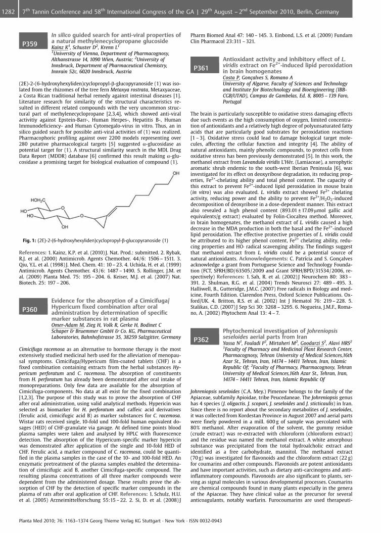
P359In silico guided search for anti-viral properties ofa natural methylenecyclopropane glucosideKainz K1, Schuster D2, Krenn L1
1University of Vienna, Department of Pharmacognosy,Althanstrasse 14, 1090 Wien, Austria; 2University ofInnsbruck, Department of Pharmaceutical Chemistry,Innrain 52c, 6020 Innsbruck, Austria
(2E)-2-(6-hydroxyhexyliden)cyclopropyl-b-glucopyranoside (1) was iso-lated from the rhizomes of the tree fern Metaxya rostrata, Metaxyaceae,a Costa Rican traditional herbal remedy against intestinal diseases [1].Literature research for similarity of the structural characteristics re-sulted in different related compounds with the very uncommon struc-tural part of methylenecyclopropane [2,3,4], which showed anti-viralactivitiy against Epstein-Barr-, Human Herpes-, Hepatitis B-, HumanImmunodeficiency- and Human Cytomegalo-virus in vitro. Thus, an insilico guided search for possible anti-viral activities of (1) was realized.Pharmacophoric profiling against over 2200 models representing over280 putative pharmacological targets [5] suggested a-glucosidase aspotential target for (1). A structural similarity search in the MDL DrugData Report (MDDR) database [6] confirmed this result making a-glu-cosidase a promising target for biological evaluation of compound (1).
Fig. 1: (2E)-2-(6-hydroxyhexyliden)cyclopropyl-b-glucopyranoside (1)
References: 1. Kainz, K.P. et al. (2010) J. Nat. Prod.: submitted. 2. Rybak,R.J. et al. (2000) Antimicrob. Agents Chemother. 44/6: 1506 – 1511. 3.Qiu, Y.L. et al. (1998) J. Med. Chem. 41: 10 – 23. 4. Uchida, H. et al. (1999)Antimicrob. Agents Chemother. 43/6: 1487 – 1490. 5. Rollinger, J.M. etal. (2009) Planta Med. 75: 195 – 204. 6. Keiser, M.J. et al. (2007) Nat.Biotech. 25: 197 – 206.
P360Evidence for the absorption of a Cimicifuga/Hypericum fixed combination after oraladministration by determination of specificmarker substances in rat plasmaOmer-Adam M, Zieg H, Volk R, Gerke H, Bodinet CSchaper & Bruemmer GmbH & Co. KG, PharmaceuticalLaboratories, Bahnhofstrasse 35, 38259 Salzgitter, Germany
Cimicifuga racemosa as an alternative to hormone therapy is the mostextensively studied medicinal herb used for the alleviation of menopau-sal symptoms. Cimicifuga/Hypericum film-coated tablets (CHF) is afixed combination containing extracts from the herbal substances Hy-pericum perforatum and C. racemosa. The absorption of constituentsfrom H. perforatum has already been demonstrated after oral intake ofmonopreparations. Only few data are available for the absorption ofCimicifuga-components. No data at all exist for the fixed combination[1,2,3]. The purpose of this study was to prove the absorption of CHFafter oral administration, using valid analytical methods. Hypericin wasselected as biomarker for H. perforatum and caffeic acid derivatives(ferulic acid, cimicifugic acid B) as marker substances for C. racemosa.Wistar rats received single, 10-fold und 100-fold human equivalent do-sages (HED) of CHF-granulate via gavage. At defined time points bloodplasma samples were taken and analysed by HPLC with fluorescencedetection. The absorption of the Hypericum-specific marker hypericinwas demonstrated after application of the single and 10-fold HED ofCHF. Ferulic acid, a marker compound of C. racemosa, could be quanti-fied in the plasma samples in the case of the 10- and 100-fold HED. Anenzymatic pretreatment of the plasma samples enabled the determina-tion of cimicifugic acid B, another Cimicifuga-specific compound. Theresulting plasma concentrations of all three marker compounds weredependent from the administered dosage. These results prove the ab-sorption of CHF by the detection of specific marker compounds in theplasma of rats after oral application of CHF. References: 1. Schulz, H.U.et al. (2005) Arzneimittelforschung 55:15 – 22. 2. Si, D. et al. (2008) J
Pharm Biomed Anal 47: 140 – 145. 3. Einbond, L.S. et al. (2009) FundamClin Pharmacol 23:311 – 321.
P361Antioxidant activity and inhibitory effect of L.viridis extract on Fe2+-induced lipid peroxidationin brain homogenatesCosta P, Gon�alves S, Romano AUniversity of Algarve, Faculty of Sciences and Technologyand Institute for Biotechnology and Bioengineering (IBB-CGB/UTAD), Campus de Gambelas, Ed. 8, 8005 – 139 Faro,Portugal
The brain is particularly susceptible to oxidative stress damaging effectsdue such events as the high consumption of oxygen, limited concentra-tion of antioxidants and a relatively high degree of polyunsaturated fattyacids that are particularly good substrates for peroxidation reactions[1 – 3]. Oxidative stress could lead to damage biological target mole-cules, affecting the cellular function and integrity [4]. The ability ofnatural antioxidants, mainly phenolic compounds, to protect cells fromoxidative stress has been previously demonstrated [5]. In this work, themethanol extract from Lavandula viridis L’H�r. (Lamiaceae), a xerophyticaromatic shrub endemic to the south-west Iberian Peninsula [6], wasinvestigated for its effect on deoxyribose degradation, its reducing prop-erties, Fe2+-chelating ability and total phenol content. The capacity ofthis extract to prevent Fe2+-induced lipid peroxidation in mouse brain(in vitro) was also evaluated. L. viridis extract showed Fe2+ chelatingactivity, reducing power and the ability to prevent Fe2+/H2O2-induceddecomposition of deoxyribose in a dose-dependent manner. This extractalso revealed a high phenol content (893.01 € 17.09 mmol gallic acidequivalents/g extract) evaluated by Folin-Ciocalteu method. Moreover,in brain homogenates, the methanol extract of L. viridis caused a highdecrease in the MDA production in both the basal and the Fe2+-inducedlipid peroxidation. The effective protective properties of L. viridis couldbe attributed to its higher phenol content, Fe2+ chelating ability, redu-cing properties and HO· radical scavenging ability. The findings suggestthat methanol extract from L. viridis could be a potential source ofnatural antioxidants. Acknowledgements: C. Patr�cia and S. Gon�alvesacknowledge a grant from Portuguese Science and Technology Founda-tion (FCT, SFRH/BD/63505/2009 and Grant SFRH/BPD/31534/2006, re-spectively) References: 1. Sah, R. et al. (2002) J Neurochem 80: 383 –391. 2. Shulman, R.G. et al. (2004) Trends Neurosci 27: 489 – 495. 3.Halliwell, B., Gutteridge, J.M.C. (2007) Free radicals in Biology and med-icine, Fourth Edition. Clarendon Press, Oxford Science Publications. Ox-ford/UK. 4. Britton, R.S. et al. (2002) Int J Hematol 76: 219 – 228. 5.Stalikas, C.D. (2007) J Sep Sci 30: 3268 – 3295. 6. Nogueira, J.M.F., Roma-no, A. (2002) Phytochem Anal 13: 4 – 7.
P362Phytochemical investigation of Johreniopsisseseloides aerial parts from IranYassa N1, Fouladi F2, Mirtaheri M2, Goodarzi S2, Alavi HRS2
1Faculty of Pharmacy and Medicinal Plant Research Center,Pharmacognosy, Tehran University of Medical Sciences,16thAzar St., Tehran, Iran, 14174 – 14411 Tehran, Iran, IslamicRepublic Of; 2Faculty of Pharmacy, Pharmacognosy, TehranUniversity of Medical Sciences,16th Azar St., Tehran, Iran,14174 – 14411 Tehran, Iran, Islamic Republic Of
Johreniopsis seseloides (C.A. Mey.) Pimenov belongs to the family of theApiaceae, subfamily Apioidae, tribe Peucedaneae. The Johreniopsis genushas 4 species (J. oligactis. J. scopari, J. seseloides and J. sticticaulis) in Iran.Since there is no report about the secondary metabolites of J. seseloides,it was collected from Kordestan Province in August 2007 and aerial partswere finely powdered in a mill. 600 g of sample was percolated with80% methanol. After evaporation of the solvent, the gummy residue(crude extract) was re-extracted with chloroform (chloroform extract)and the residue was named the methanol extract. A white amorphoussubstance was precipitated from the total hydroalcholic extract andidentified as a free carbohydrate, mannitol. The methanol extract(70 g) was investigated for flavonoids and the chloroform extract (22 g)for coumarins and other compounds. Flavonoids are potent antioxidantsand have important activities, such as dietary anti-carcinogens and anti-inflammatory compounds. Flavonoids are also significant to plants, ser-ving as signal molecules in various developmental processes. Coumarinsare chemical compounds found in many plants especially in the generaof the Apiaceae. They have clinical value as the precursor for severalanticoagulants, notably warfarin. Furocoumarins are used therapeuti-
Planta Med 2010; 76: 1163–1374 Georg Thieme Verlag KG Stuttgart · New York · ISSN 0032-0943
1282 7th Tannin Conference and 58th International Congress of the GA | 29th August – 2nd September 2010, Berlin, Germany
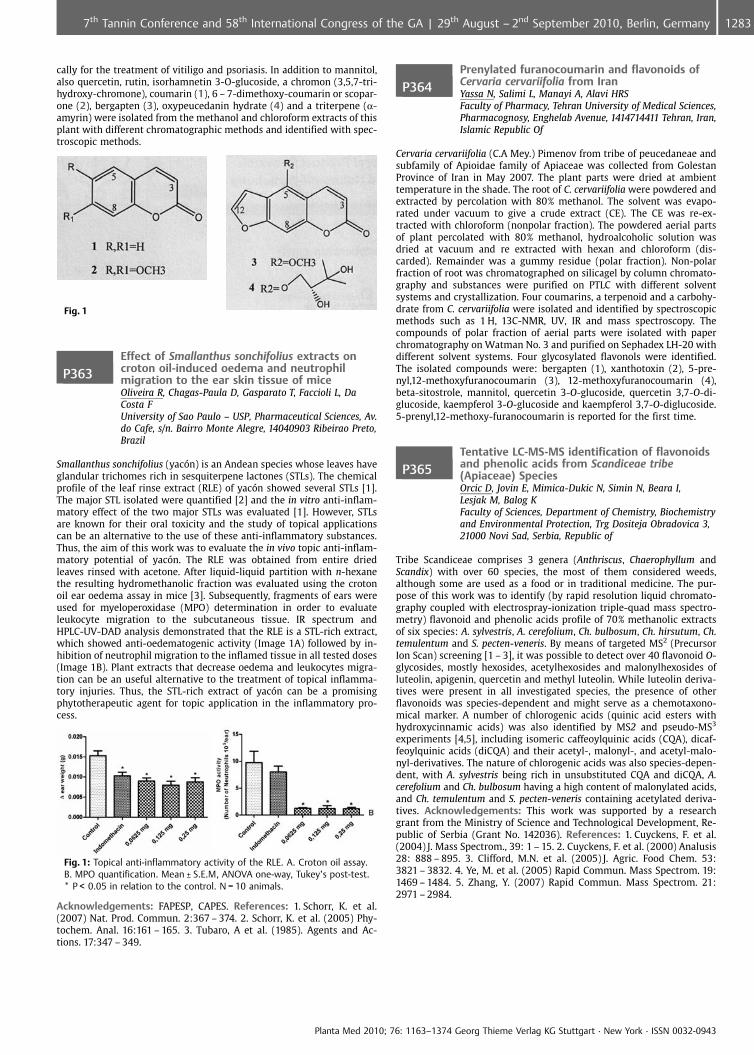
cally for the treatment of vitiligo and psoriasis. In addition to mannitol,also quercetin, rutin, isorhamnetin 3-O-glucoside, a chromon (3,5,7-tri-hydroxy-chromone), coumarin (1), 6 – 7-dimethoxy-coumarin or scopar-one (2), bergapten (3), oxypeucedanin hydrate (4) and a triterpene (a-amyrin) were isolated from the methanol and chloroform extracts of thisplant with different chromatographic methods and identified with spec-troscopic methods.
Fig. 1
P363Effect of Smallanthus sonchifolius extracts oncroton oil-induced oedema and neutrophilmigration to the ear skin tissue of miceOliveira R, Chagas-Paula D, Gasparato T, Faccioli L, DaCosta FUniversity of Sao Paulo – USP, Pharmaceutical Sciences, Av.do Cafe, s/n. Bairro Monte Alegre, 14040903 Ribeirao Preto,Brazil
Smallanthus sonchifolius (yac n) is an Andean species whose leaves haveglandular trichomes rich in sesquiterpene lactones (STLs). The chemicalprofile of the leaf rinse extract (RLE) of yac n showed several STLs [1].The major STL isolated were quantified [2] and the in vitro anti-inflam-matory effect of the two major STLs was evaluated [1]. However, STLsare known for their oral toxicity and the study of topical applicationscan be an alternative to the use of these anti-inflammatory substances.Thus, the aim of this work was to evaluate the in vivo topic anti-inflam-matory potential of yac n. The RLE was obtained from entire driedleaves rinsed with acetone. After liquid-liquid partition with n-hexanethe resulting hydromethanolic fraction was evaluated using the crotonoil ear oedema assay in mice [3]. Subsequently, fragments of ears wereused for myeloperoxidase (MPO) determination in order to evaluateleukocyte migration to the subcutaneous tissue. IR spectrum andHPLC-UV-DAD analysis demonstrated that the RLE is a STL-rich extract,which showed anti-oedematogenic activity (Image 1A) followed by in-hibition of neutrophil migration to the inflamed tissue in all tested doses(Image 1B). Plant extracts that decrease oedema and leukocytes migra-tion can be an useful alternative to the treatment of topical inflamma-tory injuries. Thus, the STL-rich extract of yac n can be a promisingphytotherapeutic agent for topic application in the inflammatory pro-cess.
Fig. 1: Topical anti-inflammatory activity of the RLE. A. Croton oil assay.B. MPO quantification. Mean € S.E.M, ANOVA one-way, Tukey’s post-test.* P < 0.05 in relation to the control. N = 10 animals.
Acknowledgements: FAPESP, CAPES. References: 1. Schorr, K. et al.(2007) Nat. Prod. Commun. 2:367 – 374. 2. Schorr, K. et al. (2005) Phy-tochem. Anal. 16:161 – 165. 3. Tubaro, A et al. (1985). Agents and Ac-tions. 17:347 – 349.
P364Prenylated furanocoumarin and flavonoids ofCervaria cervariifolia from IranYassa N, Salimi L, Manayi A, Alavi HRSFaculty of Pharmacy, Tehran University of Medical Sciences,Pharmacognosy, Enghelab Avenue, 1414714411 Tehran, Iran,Islamic Republic Of
Cervaria cervariifolia (C.A Mey.) Pimenov from tribe of peucedaneae andsubfamily of Apioidae family of Apiaceae was collected from GolestanProvince of Iran in May 2007. The plant parts were dried at ambienttemperature in the shade. The root of C. cervariifolia were powdered andextracted by percolation with 80% methanol. The solvent was evapo-rated under vacuum to give a crude extract (CE). The CE was re-ex-tracted with chloroform (nonpolar fraction). The powdered aerial partsof plant percolated with 80% methanol, hydroalcoholic solution wasdried at vacuum and re extracted with hexan and chloroform (dis-carded). Remainder was a gummy residue (polar fraction). Non-polarfraction of root was chromatographed on silicagel by column chromato-graphy and substances were purified on PTLC with different solventsystems and crystallization. Four coumarins, a terpenoid and a carbohy-drate from C. cervariifolia were isolated and identified by spectroscopicmethods such as 1 H, 13C-NMR, UV, IR and mass spectroscopy. Thecompounds of polar fraction of aerial parts were isolated with paperchromatography on Watman No. 3 and purified on Sephadex LH-20 withdifferent solvent systems. Four glycosylated flavonols were identified.The isolated compounds were: bergapten (1), xanthotoxin (2), 5-pre-nyl,12-methoxyfuranocoumarin (3), 12-methoxyfuranocoumarin (4),beta-sitostrole, mannitol, quercetin 3-O-glucoside, quercetin 3,7-O-di-glucoside, kaempferol 3-O-glucoside and kaempferol 3,7-O-diglucoside.5-prenyl,12-methoxy-furanocoumarin is reported for the first time.
P365Tentative LC-MS-MS identification of flavonoidsand phenolic acids from Scandiceae tribe(Apiaceae) SpeciesOrcic D, Jovin E, Mimica-Dukic N, Simin N, Beara I,Lesjak M, Balog KFaculty of Sciences, Department of Chemistry, Biochemistryand Environmental Protection, Trg Dositeja Obradovica 3,21000 Novi Sad, Serbia, Republic of
Tribe Scandiceae comprises 3 genera (Anthriscus, Chaerophyllum andScandix) with over 60 species, the most of them considered weeds,although some are used as a food or in traditional medicine. The pur-pose of this work was to identify (by rapid resolution liquid chromato-graphy coupled with electrospray-ionization triple-quad mass spectro-metry) flavonoid and phenolic acids profile of 70% methanolic extractsof six species: A. sylvestris, A. cerefolium, Ch. bulbosum, Ch. hirsutum, Ch.temulentum and S. pecten-veneris. By means of targeted MS2 (PrecursorIon Scan) screening [1 – 3], it was possible to detect over 40 flavonoid O-glycosides, mostly hexosides, acetylhexosides and malonylhexosides ofluteolin, apigenin, quercetin and methyl luteolin. While luteolin deriva-tives were present in all investigated species, the presence of otherflavonoids was species-dependent and might serve as a chemotaxono-mical marker. A number of chlorogenic acids (quinic acid esters withhydroxycinnamic acids) was also identified by MS2 and pseudo-MS3
experiments [4,5], including isomeric caffeoylquinic acids (CQA), dicaf-feoylquinic acids (diCQA) and their acetyl-, malonyl-, and acetyl-malo-nyl-derivatives. The nature of chlorogenic acids was also species-depen-dent, with A. sylvestris being rich in unsubstituted CQA and diCQA, A.cerefolium and Ch. bulbosum having a high content of malonylated acids,and Ch. temulentum and S. pecten-veneris containing acetylated deriva-tives. Acknowledgements: This work was supported by a researchgrant from the Ministry of Science and Technological Development, Re-public of Serbia (Grant No. 142036). References: 1. Cuyckens, F. et al.(2004) J. Mass Spectrom., 39: 1 – 15. 2. Cuyckens, F. et al. (2000) Analusis28: 888 – 895. 3. Clifford, M.N. et al. (2005) J. Agric. Food Chem. 53:3821 – 3832. 4. Ye, M. et al. (2005) Rapid Commun. Mass Spectrom. 19:1469 – 1484. 5. Zhang, Y. (2007) Rapid Commun. Mass Spectrom. 21:2971 – 2984.
Planta Med 2010; 76: 1163–1374 Georg Thieme Verlag KG Stuttgart · New York · ISSN 0032-0943
12837th Tannin Conference and 58th International Congress of the GA | 29th August – 2nd September 2010, Berlin, Germany
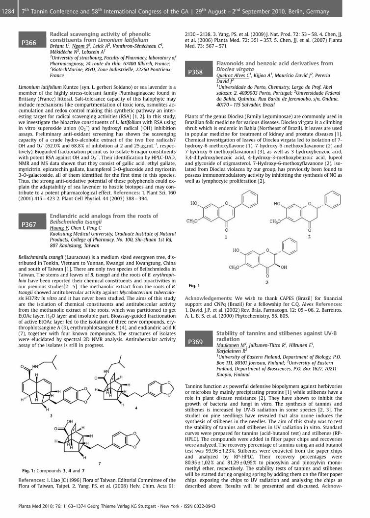
P366Radical scavenging activity of phenolicconstituents from Limonium latifoliumBr�ant L1, Ngom S2, Leick A2, Vonthron-S�n�cheau C1,M�kid�che N2, Lobstein A1
1University of strasbourg, Faculty of Pharmacy, laboratory ofPharmacognosy, 74 route du rhin, 67400 Illkirch, France;2BiotechMarine, R&D, Zone Industrielle, 22260 Pontrieux,France
Limonium latifolium Kuntze (syn. L. gerberi Soldano) or sea lavender is amember of the highly stress-tolerant family Plumbaginaceae found inBrittany (France) littoral. Salt-tolerance capacity of this halophyte mayinclude mechanisms like compartmentation of toxic ions, osmolites ac-cumulation and redox control making this synthetic pathway an inter-esting target for radical scavenging activities (RSA) [1, 2]. In this study,we investigate the bioactive constituents of L. latifolium with RSA usingin vitro superoxide anion (O2
�) and hydroxyl radical (.OH) inhibitionassays. Preliminary anti-oxidant screening has shown the scavengingcapacity of a crude hydro-alcoholic extract of the two free radicals?OH and O2
� (62.0% and 68.8% of inhibition at 2 and 25 mg.mL�1, respec-tively). Bioguided fractionation permit us to isolate 6 major constituentswith potent RSA against OH and O2
.�. Their identification by HPLC-DAD,NMR and MS data shown that they consist of gallic acid, ethyl gallate,myricitrin, epicatechin gallate, kaempferol 3-O-glucoside and myricetin3-O-galactoside, all of them identified for the first time in this species.Thus, the strong anti-oxidative potential of these polyphenols could ex-plain the adaptability of sea lavender to hostile biotopes and may con-tribute to a potent pharmacological effect. References: 1. Plant Sci. 160(2001) 415 – 423 2. Plant Cell Physiol. 44 (2003) 388 – 394.
P367Endiandric acid analogs from the roots ofBeilschmiedia tsangiiHuang Y, Chen I, Peng CKaohsiung Medical University, Graduate Institute of NaturalProducts, College of Pharmacy, No. 100, Shi-chuan 1st Rd,807 Kaohsiung, Taiwan
Beilschmiedia tsangii (Lauraceae) is a medium sized evergreen tree, dis-tributed in Tonkin, Vietnam to Yunnan, Kwangsi and Kwangtung, Chinaand south of Taiwan [1]. There are only two species of Beilschmiedia inTaiwan. The stems and leaves of B. tsangii and the roots of B. erythroph-loia have been reported their chemical constituents and bioactivities inour previous studies[2 – 5]. The methanolic extract from the roots of B.tsangii showed antitubercular activity against Mycobacterium tuberculo-sis H37Rv in vitro and it has never been studied. The aims of this studyare the isolation of chemical constituents and antitubercular activityfrom the methanolic extract of the roots, which was partitioned to getEtOAc layer, H2O layer and insoluble part. Bioassay-guided fractionationof active EtOAc layer led to the isolation of three new compounds, ery-throphlotsangine A (3), erythrophlotsangine B (4), and endiandric acid K(7), together with four known compounds. The structures of isolateswere elucidated by spectral 2D NMR analysis. Antitubercular activityassay of the isolates is still in progress.
Fig. 1: Compounds 3, 4 and 7
References: 1. Liao JC (1996) Flora of Taiwan, Editorial Committee of theFlora of Taiwan, Taipei. 2. Yang, PS. et al. (2008) Helv. Chim. Acta 91:
2130 – 2138. 3. Yang, PS. et al. (2009) J. Nat. Prod. 72: 53 – 58. 4. Chen, JJ.et al. (2006) Planta Med. 72: 351 – 357. 5. Chen, JJ. et al. (2007) PlantaMed. 73: 567 – 571.
P368Flavonoids and benzoic acid derivatives fromDioclea virgataQueiroz Alves C1, Kijjoa A1, Maur�cio David J2, PereriaDavid J2
1Universidade do Porto, Chemistry, Largo do Prof. Abelsalazar, 2, 4099003 Porto, Portugal; 2Universidade Federalda Bahia, Qu�mica, Rua Bar¼o de Jeremoabo, s/n, Ondina,40170 – 115 Salvador, Brazil
Plants of the genus Dioclea (Family Leguminosae) are commonly used inBrazilian folk medicine for various diseases. Dioclea virgata is a climbingshrub which is endemic in Bahia (Northeast of Brazil). It leaves are usedin popular medicine for treatment of kidney and prostate diseases [1].Chemical investigation of leaves of Dioclea virgata led to isolation of 7-hydroxy-6-methoxyflavone (1), 7-hydroxy-6-methoxyflavanone (2) and7-hydroxy-6 methoxyflavanonol (3), as well as 3-hydroxybenzoic acid,3,4-dihydroxybenzoic acid, 4-hydroxy-3-methoxybenzoic acid, lupeoland glycoside of stigmasterol. 7-Hydroxy-6-methoxyflavanone (2), iso-lated from Dioclea violacea by our group, has previously been found topossess immunomodulatory activity by inhibiting the synthesis of NO aswell as lymphocyte proliferation [2].
Fig. 1
Acknowledgements: We wish to thank CAPES (Brazil) for financialsupport and CNPq (Brazil) for a fellowship for C.Q. Alves References:1. David, J.P. et al. (2002) Rev. Br�s. Farmacogn. 12: 05 – 06. 2. Barreiros,A. L. B. S. et al. (2000) Phytochemistry, 55, 805.
P369Stability of tannins and stilbenes against UV-BradiationMaukonen M1, Julkunen-Tiitto R1, Hiltunen E1,Karjalainen R2
1University of Eastern Finland, Department of Biology, P.O.Box 111, 80101 Joensuu, Finland; 2University of EasternFinland, Department of Biosciences, P.O. Box 1627, 70211Kuopio, Finland
Tannins function as powerful defensive biopolymers against herbivoriesor microbes by mainly precipitating proteins [1] while stilbenes have arole in plant disease resistance [2]. They have shown to inhibit thegrowth of bacteria and fungi in vitro. The synthesis of tannins andstilbenes is increased by UV-B radiation in some species [2, 3]. Thestudies on pine seedlings have revealed that also ozone induces thesynthesis of stilbenes in the needles. The aim of this study was to testthe stability of tannins and stilbenes in UV radiation in vitro. Standardcurves were prepared for tannins (acid-butanol test) and stilbenes (RP-HPLC). The compounds were added in filter paper chips and recoverieswere analyzed. The recovery percentage of tannins using an acid butanoltest was 99,96 € 1,23%. Stilbenes were extracted from the paper chipsand analyzed by RP-HPLC. Their recovery percentages were80,95 € 1,02% and 81,29 € 0,95% to pinosylvin and pinosylvin mono-methyl ether, respectively. The stability tests of tannins and stilbeneswill be started during ongoing spring by adding them on the filter paperchips, exposing the chips to UV radiation and analyzing the chips asdescribed above. Results will be presented and discussed. Acknow-
Planta Med 2010; 76: 1163–1374 Georg Thieme Verlag KG Stuttgart · New York · ISSN 0032-0943
1284 7th Tannin Conference and 58th International Congress of the GA | 29th August – 2nd September 2010, Berlin, Germany
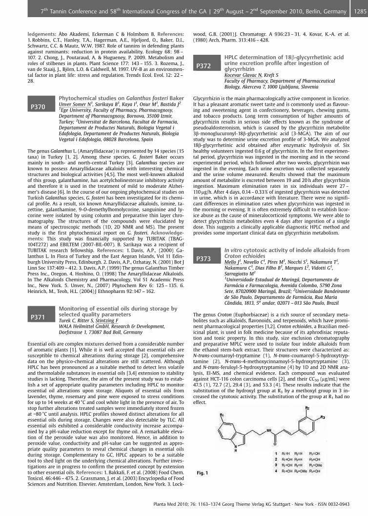
ledgements: �bo Akademi, Eckerman C & Holmbom B. References:1. Robbins, C.T., Hanley, T.A., Hagerman, A.E., Hjeljord, O., Baker, D.I.,Schwartz, C.C. & Mautz, W.W. 1987. Role of tannins in defending plantsagainst ruminants: reduction in protein availability. Ecology 68: 98 –107. 2. Chong, J., Poutaraud, A. & Hugueney, P. 2009. Metabolism androles of stilbenes in plants. Plant Science 177: 143 – 155. 3. Rozema, J.,van de Staaij, J., Bj�rn, L.O. & Caldwell, M. 1997. UV-B as an environmen-tal factor in plant life: stress and regulation. Trends Ecol. Evol. 12: 22 –28.
P370Phytochemical studies on Galanthus fosteri BakerUnver Somer N1, Sarikaya B1, Kaya I1, Onur M1, Bastida J2
1Ege University, Faculty of Pharmacy, Pharmacognosy,Department of Pharmacognosy, Bornova, 35100 Izmir,Turkey; 2Universitat de Barcelona, Facultat de Farmacia,Departament de Productes Naturals, Biologia Vegetal iEdafologia, Departament de Productes Naturals, BiologiaVegetal i Edafologia, 08028 Barcelona, Spain
The genus Galanthus L. (Amaryllidaceae) is represented by 14 species (15taxa) in Turkey [1, 2]. Among these species, G. fosteri Baker occursmainly in south- and north-central Turkey [3]. Galanthus species areknown to possess Amaryllidaceae alkaloids with interesting chemicalstructures and biological activities [4,5]. The most well-known alkaloidof this group, galanthamine, has acetylcholinesterase inhibitory activityand therefore it is used in the treatment of mild to moderate Alzhei-mer’s disease [6]. In the course of our ongoing phytochemical studies onTurkish Galanthus species, G. fosteri has been investigated for its chemi-cal profile. As a result, six known Amaryllidaceae alkaloids, ismine, ta-zettine, galanthamine, 9-O-demethylhomolycorine, sanguinine and ly-corine were isolated by using column and preparative thin layer chro-matography. The structures of the compounds were elucidated bymeans of spectroscopic methods (1D, 2D NMR and MS). The presentstudy is the first phytochemical report on G. fosteri. Acknowledge-ments: This study was financially supported by TUBITAK (TBAG-104T272) and EBILTEM (2007-BIL-007). B. Sarikaya was a recipient ofTUBITAK research fellowship. References: 1. Davis, A.P., (2000) Ga-lanthus L. In Flora of Turkey and the East Aegean Islands, Vol 11 Edin-burgh University Press, Edinburgh. 2. Davis, A.P., Ozhatay, N. (2001) Bot JLinn Soc 137:409 – 412. 3. Davis, A.P. (1999) The genus Galanthus TimberPress Inc., Oregon. 4. Hoshino, O. (1998) The Amaryllidaceae Alkaloids.In The Alkaloids Chemistry and Pharmacology, Vol 51 Academic PressInc., New York. 5. Unver, N., (2007) Phytochem Rev 6: 125 – 135. 6.Heinrich, M., Teoh, H.L. (2004) J Ethnopharm 92:147 – 162.
P371Monitoring of essential oils during storage byselected quality parametersTurek C, Ritter S, Stintzing FWALA Heilmittel GmbH, Research & Development,Dorfstrasse 1, 73087 Bad Boll, Germany
Essential oils are complex mixtures derived from a considerable numberof aromatic plants [1]. While it is well accepted that essential oils aresusceptible to chemical alterations during storage [2], comprehensivedata on the physico-chemical alterations are still scattered. AlthoughHPLC has been pronounced as a suitable method to detect less volatileand thermolabile substances in essential oils [3,4] extension to stabilitystudies is lacking. Therefore, the aim of the present study was to estab-lish a set of appropriate quality parameters including HPLC to monitoressential oil alterations upon storage. Aliquots of essential oils fromlavender, thyme, rosemary and pine were exposed to stress conditionsfor up to 14 weeks at 40 �C and cool white light in the presence of air. Tostop further alterations treated samples were immediately stored frozenat �80 �C until analysis. HPLC profiles showed distinct alterations for allessential oils during storage. Changes were also detectable by TLC. Allessential oils exhibited a considerable conductivity increase accompa-nied by a pH-value reduction except for thyme oil. A remarkable eleva-tion of the peroxide value was also monitored. Hence, in addition toperoxide value, conductivity and pH-value can be suggested as appro-priate quality parameters to reveal chemical changes in essential oilsduring storage. Complementary to GC, HPLC appears to be a suitabletool to shed light on the underlying chemical alterations. Further inves-tigations are in progress to confirm the presented concept by extensionto other essential oils. References: 1. Bakkali, F. et al. (2008) Food Chem.Toxicol. 46:446 – 475. 2. Grassmann, J. et al. (2003) Encyclopedia of FoodSciences and Nutrition. Elsevier. Amsterdam, London, New York. 3. Lock-
wood, G.B. (2001) J. Chromatogr. A 936:23 – 31. 4. Kovar, K.-A. et al.(1980) Arch. Pharm. 313:416 – 428.
P372HPLC determination of 18b-glycyrrhetinic acidurine excretion profile after ingestion ofglycyrrhizinKocevar Glavac N, Kreft SFaculty of Pharmacy, Department of PharmaceuticalBiology, Akerceva 7, 1000 Ljubljana, Slovenia
Glycyrrhizin is the main pharmacologically active component in licorice.It has a pleasant aromatic sweet taste and is commonly used as flavour-ing and sweetening agent in confectionery, beverages, chewing gums,and tobacco products. Long term consumption of higher amounts ofglycyrrhizin results in serious side effects known as the syndrome ofpseudoaldosteronism, which is caused by the glycyrrhizin metabolite3b-monoglucuronyl-18b-glycyrrhetinic acid (3-MGA). The aim of ourstudy was to determine urine excretion profile of 3-MGA. We analyzed18b-glycyrrhetinic acid obtained after enzymatic hydrolysis of. Sixhealthy volunteers ingested 0.6 g of glycyrrhizin. In the first experimen-tal period, glycyrrhizin was ingested in the morning and in the secondexperimental period, which followed after two weeks, glycyrrhizin wasingested in the evening. Each urine excretion was collected separatelyand the urine volume measured. Results showed that the maximumamount of metabolite is excreted between 19 and 20 h after glycyrrhiziningestion. Maximum elimination rates in six individuals were 27 –110 mg/h. After 4 days, 0.14 – 0.33% of ingested glycyrrhizin was detectedin urine, which is in accordance with literature. There were no signifi-cant differences in elimination rates when glycyrrhizin was ingested inthe morning or evening. It is often extremely difficult to establish licor-ice abuse as the cause of mineralocorticoid symptoms. We were able todetect glycyrrhizin metabolites even 4 days after ingestion of a singledose. This suggests a clinically applicable diagnostic HPLC method andprovides some important clinical data on glycyrrhizin metabolism.
P373In vitro cytotoxic activity of indole alkaloids fromCroton echioidesMello J1, Novello C1, Pires M1, Nocchi S1, Nakamura T1,Nakamura C1, Dias Filho B1, Marques L2, Vidotti G1,Sarragiotto M1
1Universidade Estadual de Maring, Departamento deFarmcia e Farmacologia, Avenida Colombo, 5790 ZonaSete, 87020900 Maring, Brazil; 2Universidade Bandeirantede S¼o Paulo, Departamento de Farmcia, Rua MariaC ndida, 1813, 5o andar, 02071 – 013 S¼o Paulo, Brazil
The genus Croton (Euphorbiaceae) is a rich source of secondary meta-bolites such as alkaloids, flavonoids, and terpenoids, which have promi-nent pharmacological properties [1,2]. Croton echioides, a Brazilian med-icinal plant, is used in folk medicine because of its aphrodisiac reputa-tion and tonic property. In this study, size exclusion chromatographyand preparative MPLC were used to isolate four indole alkaloids fromthe ethanol stem-bark extract. Their structures were characterized as:N-trans-coumaroyl-tryptamine (1), N-trans-coumaroyl-5-hydroxytryp-tamine (2), N-trans-4-methoxycinnamoyl-5-hydroxytryptamine (3),and N-trans-feruloyl-5-hydroxytryptamine (4) by 1D and 2D NMR ana-lysis, EI-MS, and chemical evidence. Each compound was evaluatedagainst HCT-116 colon carcinoma cells [2], and their CC50 (mg/mL) were47.5 (1), 72.7 (2), 29.4 (3), and 53.3 (4). These results indicate that thesubstitution of the hydroxyl group at R3 by a methoxyl group in 3 in-creased the cytotoxic activity. The substitution of the group at R1 had noeffect.
Fig. 1
Planta Med 2010; 76: 1163–1374 Georg Thieme Verlag KG Stuttgart · New York · ISSN 0032-0943
12857th Tannin Conference and 58th International Congress of the GA | 29th August – 2nd September 2010, Berlin, Germany
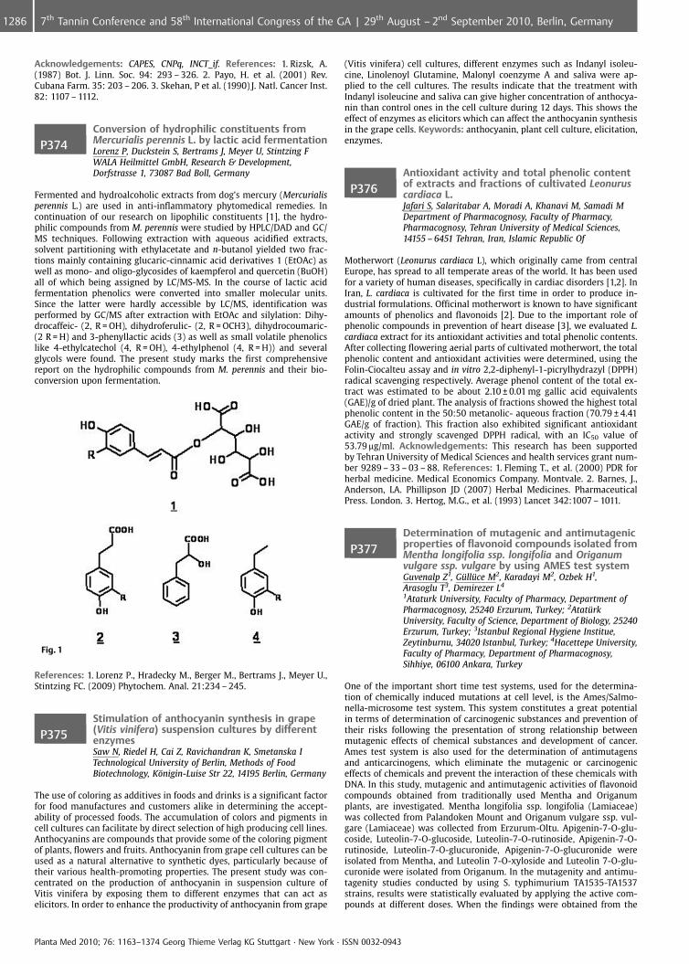
Acknowledgements: CAPES, CNPq, INCT_if. References: 1. Rizsk, A.(1987) Bot. J. Linn. Soc. 94: 293 – 326. 2. Payo, H. et al. (2001) Rev.Cubana Farm. 35: 203 – 206. 3. Skehan, P et al. (1990) J. Natl. Cancer Inst.82: 1107 – 1112.
P374Conversion of hydrophilic constituents fromMercurialis perennis L. by lactic acid fermentationLorenz P, Duckstein S, Bertrams J, Meyer U, Stintzing FWALA Heilmittel GmbH, Research & Development,Dorfstrasse 1, 73087 Bad Boll, Germany
Fermented and hydroalcoholic extracts from dog’s mercury (Mercurialisperennis L.) are used in anti-inflammatory phytomedical remedies. Incontinuation of our research on lipophilic constituents [1], the hydro-philic compounds from M. perennis were studied by HPLC/DAD and GC/MS techniques. Following extraction with aqueous acidified extracts,solvent partitioning with ethylacetate and n-butanol yielded two frac-tions mainly containing glucaric-cinnamic acid derivatives 1 (EtOAc) aswell as mono- and oligo-glycosides of kaempferol and quercetin (BuOH)all of which being assigned by LC/MS-MS. In the course of lactic acidfermentation phenolics were converted into smaller molecular units.Since the latter were hardly accessible by LC/MS, identification wasperformed by GC/MS after extraction with EtOAc and silylation: Dihy-drocaffeic- (2, R = OH), dihydroferulic- (2, R = OCH3), dihydrocoumaric-(2 R = H) and 3-phenyllactic acids (3) as well as small volatile phenolicslike 4-ethylcatechol (4, R = OH), 4-ethylphenol (4, R = H)) and severalglycols were found. The present study marks the first comprehensivereport on the hydrophilic compounds from M. perennis and their bio-conversion upon fermentation.
Fig. 1
References: 1. Lorenz P., Hradecky M., Berger M., Bertrams J., Meyer U.,Stintzing FC. (2009) Phytochem. Anal. 21:234 – 245.
P375Stimulation of anthocyanin synthesis in grape(Vitis vinifera) suspension cultures by differentenzymesSaw N, Riedel H, Cai Z, Ravichandran K, Smetanska ITechnological University of Berlin, Methods of FoodBiotechnology, K�nigin-Luise Str 22, 14195 Berlin, Germany
The use of coloring as additives in foods and drinks is a significant factorfor food manufactures and customers alike in determining the accept-ability of processed foods. The accumulation of colors and pigments incell cultures can facilitate by direct selection of high producing cell lines.Anthocyanins are compounds that provide some of the coloring pigmentof plants, flowers and fruits. Anthocyanin from grape cell cultures can beused as a natural alternative to synthetic dyes, particularly because oftheir various health-promoting properties. The present study was con-centrated on the production of anthocyanin in suspension culture ofVitis vinifera by exposing them to different enzymes that can act aselicitors. In order to enhance the productivity of anthocyanin from grape
(Vitis vinifera) cell cultures, different enzymes such as Indanyl isoleu-cine, Linolenoyl Glutamine, Malonyl coenzyme A and saliva were ap-plied to the cell cultures. The results indicate that the treatment withIndanyl isoleucine and saliva can give higher concentration of anthocya-nin than control ones in the cell culture during 12 days. This shows theeffect of enzymes as elicitors which can affect the anthocyanin synthesisin the grape cells. Keywords: anthocyanin, plant cell culture, elicitation,enzymes.
P376Antioxidant activity and total phenolic contentof extracts and fractions of cultivated Leonuruscardiaca L.Jafari S, Salaritabar A, Moradi A, Khanavi M, Samadi MDepartment of Pharmacognosy, Faculty of Pharmacy,Pharmacognosy, Tehran University of Medical Sciences,14155 – 6451 Tehran, Iran, Islamic Republic Of
Motherwort (Leonurus cardiaca L), which originally came from centralEurope, has spread to all temperate areas of the world. It has been usedfor a variety of human diseases, specifically in cardiac disorders [1,2]. InIran, L. cardiaca is cultivated for the first time in order to produce in-dustrial formulations. Officinal motherwort is known to have significantamounts of phenolics and flavonoids [2]. Due to the important role ofphenolic compounds in prevention of heart disease [3], we evaluated L.cardiaca extract for its antioxidant activities and total phenolic contents.After collecting flowering aerial parts of cultivated motherwort, the totalphenolic content and antioxidant activities were determined, using theFolin-Ciocalteu assay and in vitro 2,2-diphenyl-1-picrylhydrazyl (DPPH)radical scavenging respectively. Average phenol content of the total ex-tract was estimated to be about 2.10 € 0.01 mg gallic acid equivalents(GAE)/g of dried plant. The analysis of fractions showed the highest totalphenolic content in the 50:50 metanolic- aqueous fraction (70.79 € 4.41GAE/g of fraction). This fraction also exhibited significant antioxidantactivity and strongly scavenged DPPH radical, with an IC50 value of53.79 mg/ml. Acknowledgements: This research has been supportedby Tehran University of Medical Sciences and health services grant num-ber 9289 – 33 – 03 – 88. References: 1. Fleming T., et al. (2000) PDR forherbal medicine. Medical Economics Company. Montvale. 2. Barnes, J.,Anderson, LA. Phillipson JD (2007) Herbal Medicines. PharmaceuticalPress. London. 3. Hertog, M.G., et al. (1993) Lancet 342:1007 – 1011.
P377Determination of mutagenic and antimutagenicproperties of flavonoid compounds isolated fromMentha longifolia ssp. longifolia and Origanumvulgare ssp. vulgare by using AMES test systemGuvenalp Z1, G�ll�ce M2, Karadayi M2, Ozbek H1,Arasoglu T3, Demirezer L4
1Ataturk University, Faculty of Pharmacy, Department ofPharmacognosy, 25240 Erzurum, Turkey; 2Atat�rkUniversity, Faculty of Science, Department of Biology, 25240Erzurum, Turkey; 3Istanbul Regional Hygiene Institue,Zeytinburnu, 34020 Istanbul, Turkey; 4Hacettepe University,Faculty of Pharmacy, Department of Pharmacognosy,Sihhiye, 06100 Ankara, Turkey
One of the important short time test systems, used for the determina-tion of chemically induced mutations at cell level, is the Ames/Salmo-nella-microsome test system. This system constitutes a great potentialin terms of determination of carcinogenic substances and prevention oftheir risks following the presentation of strong relationship betweenmutagenic effects of chemical substances and development of cancer.Ames test system is also used for the determination of antimutagensand anticarcinogens, which eliminate the mutagenic or carcinogeniceffects of chemicals and prevent the interaction of these chemicals withDNA. In this study, mutagenic and antimutagenic activities of flavonoidcompounds obtained from traditionally used Mentha and Origanumplants, are investigated. Mentha longifolia ssp. longifolia (Lamiaceae)was collected from Palandoken Mount and Origanum vulgare ssp. vul-gare (Lamiaceae) was collected from Erzurum-Oltu. Apigenin-7-O-glu-coside, Luteolin-7-O-glucoside, Luteolin-7-O-rutinoside, Apigenin-7-O-rutinoside, Luteolin-7-O-glucuronide, Apigenin-7-O-glucuronide wereisolated from Mentha, and Luteolin 7-O-xyloside and Luteolin 7-O-glu-curonide were isolated from Origanum. In the mutagenity and antimu-tagenity studies conducted by using S. typhimurium TA1535-TA1537strains, results were statistically evaluated by applying the active com-pounds at different doses. When the findings were obtained from the
Planta Med 2010; 76: 1163–1374 Georg Thieme Verlag KG Stuttgart · New York · ISSN 0032-0943
1286 7th Tannin Conference and 58th International Congress of the GA | 29th August – 2nd September 2010, Berlin, Germany
studies, it was determined that the examined compounds at the applieddoses do not exhibit any mutagenic effect. On the other hand, it wasdetermined that at 20 mM dose application, Apigenin-7-O-glucosideand Apigenin-7-O-rutinoside exhibited respectively 91% and 88,55%antimutagenic activity. It was seen that the remaining compounds ex-hibited strong antimutagenic activities, which are considered statisti-cally important, at all applied dosage levels. Acknowledgements: Thisstudy was supported by grants from the Scientific and TechnologicalResearch Council of Turkey (TUBITAK). (Project No: 107T203).
P378Levels of some trace elements in someMacedonian edible wild TricholomataceaemushroomsKirovska Cigulevska OJZU CJZ Skopje, chemical labs, III Makedonska Brigada No18; 1000 Skopje, MacedoniaP.O. Box 809, 1000 Skopje,Macedonia, The Former Yugoslav Republic Of
The Chemical composition and nutritive value of some Macedonian ed-ible Boletaceae mushrooms Bauer Petrovska B1, Kirovska Cigulevska O 2,Ugrinova L 1 1Faculty of Pharmacy, Vodnjanska 17, P.O. Box 36, 1000Skopje, R. Macedonia; 2JZO ZZZ Skopje, Treta Mak. Brigada 18, P.O. Box809; 1000 Skopje, R. Macedonia; Over the last few years there has beena great interest in the collecting of wild mushrooms in Macedonia. Ex-cept for some cultivated mushrooms, little is known about the nutri-tional quality of the wild – spread, edible mushroom species in Mace-donia. Their great number enable the selection of that which are char-acterized with great nutritional quality. Therefore in the present studythe nutritionally important components were determined with variouscontemporary nutritients analyses in 15 wild species of Boletaceaemushrooms from various Macedonian areas. Macedonian Boletaceaemushrooms, contained relatively high total dietary fibre content (up to11.73%, dry basis), protein content (av. 30.98%, dry basis) with highbiological values (up to 78.94 BV, 2.60 PER) and were low in fats (av.4.77%, dry basis). 100 g dry mushrooms comprised low energetic valueof 1670.64 kJ on the average. The contents of minerals showed greatvariation (5.15 – 11.15%, dry mass) in agreement with literature data[1]. P, Mg, Cu, Fe and Zn were found in higher concentrations than otherions. Cd and Pb have lower values than the maximum concentrationsimposed by low [2]. Compared with other provisions [3], MacedonianBoletaceae mushrooms proved to be a good source of protein and miner-al matter for the human diet. They are deficient in fat, posse low energybut are nutritionally valuable food. Table 1. Content of protein, fat, ashand energy value of Macedonian Boletaceae mushrooms and other pro-visions [3] expressed in dry mass provisions Protein% Fat% Ash% Energyvalue kJ/100 g Macedonian Boletaceae mushrooms 30.98 4.77 8.181670.64 Milk 27.1 29.0 6.0 2186.6 Veal 70.7 23.9 4.4 2085.8 Soya 36.919.8 5.1 1475.4 Beans 24.6 2.5 7.4 1429.3 Wheat bread 13.3 1.7 2.7 1447.1Beef 56.4 44.4 3.1 2600.9 Acknowledgements: Faculty of NaturalScience and Mathematics, Karadelev M. References: 1. Falandysz, J., Lip-ka, K. (2006) Rocz Panstw Zakl Hig. 57(3): 217 – 33. 2. Pravilnik za opshtibaranja za bezbednost na hrana, Sl. Vesnik RM 118/05. 3. Souci, S.,Faschmann, W., Kraut, H. (1994) Food composition and tables. Med-pharm. Stuttgart.
P379Isolation of bioactive aporphinoid alkaloids inOxandra asbecki (Annonaceae)Le Ven J1, Eparvier V2, Litaudon M1, Gueritte F1
1ICSN-CNRS, P�le Substances Naturelles, 1 av de la terrase,91198 Gif sur Yvette, France; 2CNRS-UMR Ecofog, UMR-Ecofog-L3MA, BP792 IESG, 97300 Cayenne, French Guiana
Since prehistoric times, humans beneficially used natural resources fortheir daylife needs. Plants are able to synthesize complex molecules andconsequently have a unique chemical diversity. This is a source of in-spiration for new drugs discovery. Dyrk1A kinase is a target used inresearch on Alzheimer’s disease. Inhibition of this kinase is associatedwith treating symptoms of this disease [1,2]. In France, the annual num-ber of new cases is 230 000. The prevalence is expected to double inindustrialized countries and quadruple in developing countries in thecoming decades. The development of a better diagnosis and treatment isessential. The Annonaceae is a large family of tropical plants that havebeen investigated intensively. There exist 38 species in Oxandra genusand Oxandra asbecki species in Venezuela and in primary forests ofFrench Guiana. To the aim of discovering new bioactive plants fromFrench Guiana, Oxandra asbecki was selected for phytochemical study
because of its potent inhibition of DyrK1A kinase. Bioassay-directedfractionation of the ethyl acetate extract provided three bioactive alka-loids. We isolated three aporphinoid alkaloids and show for the firsttime an strong activity (with micromolar IC 50) of Velutamin undertwo kinase: CDK1 and DyrK1A, and activity of Aristolactam AII on Dyr-K1A kinase. References: 1. Lee, V.M-Y., et al. (2004). Trends in neuros-ciences 27. 2. Nam Doo Kim, N.D., et al. (2006) Biol. Med. Chem. Lett. 16,3772 – 3776.
P380Secondary metabolites from endemic Iraneciospecies from IranAsgari T1,2, Amin G1, Passreiter C2, Hauschild W2
1Tehran University of Medical Sciences, Pursina Ave, 14155 –6451 Tehran, Iran, Islamic Republic Of; 2Heinrich-Heine-University of Duesseldorf, Institute of PharmaceuticalBiology and Biotechnology, Universitaetsstrasse 1, Gebaeude26.23, 40225 Dusseldorf, Germany
The genus Iranecio belongs to the Asteraceae and contains four species,whose main habitat is Iran. Except for botanical information in the FloraIranica [1], no information about the chemistry of the plants is given inthe literature. Since many members of the Asteraceae contain secondarymetabolites which display cytotoxic effects [2,3], extracts from Iranecioelbruzensis Bioss (endemic to Iran) and I. othonae were tested for cyto-toxic activity, following the method previously reported by Azizi et al.[4], IC50 values were found at 1 mg/ml and 0.75 mg/ml, respectively.Aerial parts of I. elbruzensis and I. othonae were extracted with dichlor-omethane in a Soxhlet apparatus and the obtained extracts purified bycolumn chromatography using methanol and Sephadex LH-20. Purifica-tion of two fractions by repeated column chromatography, flash chro-matography or preparative TLC afforded the steroids b-sitosterol [5] andits glycoside daucosterol [6], as well as the sesquiterpene spathulenol[7] from I. elbruzensis. Iranecio othonae also contained daucosterol and ararely found palmitic acid ester derivate of calenduladiol (see fig.) [8].
Fig. 1
All structures were elucidated by means of their mass and NMR spectra.Calenduladiol palmitate, which was found in larger amounts, will betested on its cytotoxicity in the near future. Acknowledgements: Insti-tute of Pharmaceutical Biology and Biotechnology of Heinrich HeineUniversity of Dusseldorf, Mrs Eva Mller References: 1. Ditrich, M etal. (1982) Flora Iranica Compositae IV. Rechinger, KH (ed). AkademischeDruck und Verlagsanstalt, Graz, Austria. 2. Woerdenbag, HJ et al. (1994)Planta Med, 60:434 – 437. 3. Francois, G. et al. (1996) Planta Med.62:126 – 129. 4. Azizi, E. et al (2008) J. Biol. Sci. 8:380 – 385. 5. Seo, S.et al. (1988) J. Chem Soc. Perkin Trans I 2407 – 2414. 6. Gu, J-Q et al(2004) Z. Naturforsch. 59c: 797 – 802. 7. Brochini, CB and Roque, NF(2000) Braz.Chem.Soc. 11:361 – 364. 8. Schmidt, T. et al (2004) PlantaMed. 70:967 – 977.
P381Tussilago farfara L.: History of its therapeuticaluse and illustration in the 19th centuryNeumann CUniversit�t Bern, Philosophisch-naturwisenschaftlicheFakult�t, private: Rosenthaler Straße 19, 10119 Berlin,Germany
Historical pharmaceutical and medical sources are celebrating a com-mon renaissance not only due to their information content about oldformulations but also due to their remarkable attraction of rich andskilful illustrations. In the present study there have been selected withthe help of the Index Londinensis [1] almost 40 original historicalsources of the 19th century which have been analysed in regard to theircontent of their texts as well as of their illustrations. Although text and
Planta Med 2010; 76: 1163–1374 Georg Thieme Verlag KG Stuttgart · New York · ISSN 0032-0943
12877th Tannin Conference and 58th International Congress of the GA | 29th August – 2nd September 2010, Berlin, Germany
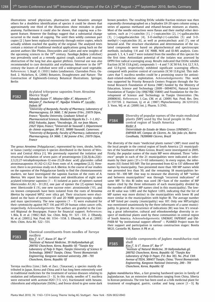
illustrations served physicians, pharmacists and botanists amongstothers for a doubtless identification of species it could be shown thatapproximately one third of the illustrations show mistakes in plantanatomy. Furthermore it could also be shown, that copying was a fre-quent feature. However the findings suggest that a substantial changeoccurred in the mode of copying. The until then wildly common partcopies [2 ] have been replaced extensively through entire copies, whichactually seldom reached the quality of the original. The analysed textscontain a mixture of traditional medical applications going back on theancient authors like Plinius, Dioscurides and Galen and new insights ofthe upcoming sciences in the 19th century. Tussilago farfara (coltsfoot)was prevalently used as a remedy against cough, catarrh and mucousobstruction of the lung but also against phthisis. External use was alsorecommended to cure dermatitis and erythemas. Moreover in the 19th
century the leaves of coltsfoot were still a widespread used vegetable.References: 1. Stapf, O. (1931) Index Londinensis. Clarendon Press. Ox-ford. 2. Nickelsen, K. (2006) Botanists, Draughtsmen and Nature: TheConstruction of Eighteenth-Century Botanical Illustrations. Springer,Berlin
P382Acylated triterpene saponins from Atroximaliberica StapfTabopda Kuiate T1, Mitaine-Offer A1, Miyamoto T2,Mirjolet J3, Duchamp O3, Ngadjui Tchaleu B4, Lacaille-Dubois M5
1University of Burgundy, Faculty of Pharmacy, Laboratory ofPharmacognosy, EA 3660, 7, Bd Jeanne D’Arc, 21079 Dijon,France; 2Kyushu University, Graduate School ofPharmaceutical Sciences, Maidashi Higashi Ku 3 – 1-1, 812 –8582 Fukuoka, Japan; 3Oncodesign, 20, rue Jean Mazen,21627 Dijon, France; 4Universit� de Yaound� 1, D�partementde chimie organique, BP 812, 30000 Yaound�, Cameroon;5University of Burgundy, Faculty of Pharmacy, Laboratory ofPharmacognosy, EA 3660, 7, Bd Jeanne d’Arc, 21079 Dijon,France
The genus Atroxima (Polygalaceae), represented by trees, shrubs, herbs,or lianas (rarely) comprises 6 species distributed in the forests of Wes-tern and Central Africa [1]. We previously reported the isolation andstructural elucidation of seven pairs of preatroxigenin [(2b,3b,4a,22b)-2,3,22,27-tetrahydroxyolean-12-ene-23,28-dioic acid] glycosides calledatroximasaponins A1/A2-G1/G2 from roots of A. congolana [2,3]. How-ever, there is no previous study on A. liberica. In the course of our studieson the saponins of the Polygalaceae family to find the chimiotaxonomicmarkers, we have investigated the saponin fraction of the roots of A.liberica. We report here the isolation and identification of eight newacylated triterpene saponins (1 – 8) isolated as pairs of epimers namedlibericosides A1/A2 – D1/D2, one pair of epimer the (Z)-epimer beingnew: libericoside E (9), one new sucrose ester: atroximoside (10), andsix known compounds have been isolated from the roots of Atroximaliberica by repeated MPLC over silica gel. Their structures were eluci-dated on the basis of extensive 1D and 2D NMR spectroscopic studiesand mass spectrometry. The new saponins (1 - 9) were evaluated fortheir cytotoxicity against HCT 116 and HT-29 human colon cancer cells,respectively. Acknowledgements: The authors are grateful to the Con-seil R�gional de Bourgogne, France for financial support. References:1. Bila, B. et al. (1982) Bull. Soc. Chim. Belg. 91: 321 – 331. 2. Elbandy,M. et al. (2003) J. Nat. Prod. 66: 1154 – 1158. 3. Elbandy, M. et al. (2003)Helv. Chim. Acta 86: 522 – 531.
P383Chemical constituents from needles of TorreyanuciferaKim J1, Si C2, Kwon D3, Bae Y3
1Institute of Natural Medicine, 39 Hallymdaehak-gil,200702 Chuncheon, Korea, Republic Of; 2Tianjin KeyLaboratory of Pulp & Paper, Tianjin University of Science &Technology, 300457 Tianjin, China; 3Forest BiomaterialsEngineering, Kangwon national university, 200 – 701Chuncheon, Korea, Republic Of
Torreya nucifera (Linn.) Sieb. et Zucc. (Taxaceae), a species mainly dis-tributed in Japan, Korea and China and it has long been extensively usedin traditional medicines for the treatment of various diseases relating tooxidation and inflammation [1 – 3]. In this study, needles of T. nuciferawere extracted with acetone-H2O (7:3, v/v), fractionated with hexane,chloroform and ethylacetate (EtOAc), and freeze dried to give some dark
brown powders. The resulting EtOAc soluble fraction mixture was thenrepeatedly chromatographed on a Sephadex LH-20 open column using aseries of aqueous methanol and ethanol-hexane mixtures as eluents.Most of the needle extractives were flavan and their methyl ether deri-vatives, such as (+)-catechin (1), (�)-epicatechin (2), (+)-gallocatechin(3), (�)-epigallocatechin (4), 3-O-methyl-(+)-catechin (5) and 3-O-methyl-(�)-epicatechin (6), as well as protocatechuic acid (7), one ofbenzoid acid. The structure elucidation and determination of the iso-lated compounds were based on physiochemical and spectroscopicmethods, including 1 H and 13C NMR, NOE and EI-MS analysis. Com-pounds 1, 3, 4, 5 and 7 were isolated from the needles of this species forthe first time. Antioxidant potential of the isolates was assessed byDPPH free radical scavenging assay. Results indicated that EtOAc solublefraction (IC 50 13.8 g/ml), compounds 1 – 4 and 7 (IC 50 9.6, 9.4, 12.7, 9.5,and 12.9 g/ml, respectively) exhibited significant scavenging effects,compared with positive control of curcumin (IC 50 13.8 g/ml. This indi-cates that T. nucifera needles could be a promising source for antioxi-dant-related-medicine exploitation. Acknowledgements: This workwas supported by Priority Research Centers Program through the Na-tional Research Foundation of Korea (NRF) funded by the Ministry ofEducation, Science and Technology (2009 – 0094074), Natural ScienceFoundation of Tianjin City (09JCYBJC 15800) and Foundation for the De-velopment of Science and Technology in Tianjin Universities (No.20080616). References: 1. Cao, CM. et al. (2009) Nat. Prod. Res. Dev.21:737739. 2. Harrison, LJ. et al. (1987) Phytochemistry. 26:12111212.3. Yoon, WJ. et al. (2009) Int. J. Pharm. 5:3743.
P384Diversity of popular names of the main medicinalplants (MP) used by the local people in thecentral region of South AmericaRieder AUniversidade do Estado de Mato Grosso (UNEMAT); eEMPAER-MT, Campus de Cceres, Av. S¼o Jo¼o s/n, BairroCavalhada, 78200000 Cceres, Brazil
The diversity of the main “medicinal plants names”-(MP) most used bythe local people in the central region of South America (21 municipali-ties of the Southwest of Mato Grosso, Brazil) were investigated in 2005.Firstly, the popular names of the main MP were obtained. Three “refer-ence” people in each of the 21 municipalities were indicated as infor-mants by their pairs (21 x 3 = 63 informants). In every region, the infor-mants (63) listed 503 MP. The list presented from 42 to 137 different MPnames by municipality. Two presented more than 100 MP names whileone presented less than 50, and the remaining 18 municipalities be-tween 50 – 100 MP. One way to measure the diversity of MP “withinand between municipalities” was through “recurrent indications” ofsame MP. To this RI index was used (RI = Sum of the number of MPnames cited by the three informants of each municipality divided bythe number of different MP names cited in this municipality). The low-est RI value was 1.085 and the highest 1.693, indicating that the list ofMP names was more distinct in the municipalities with lower RI andmore similar in the municipalities with higher RI. The average numberof MP listed per county (municipality) was 107. Only one MP(urtig¼o)was mentioned unanimously by the three informants of a same munici-pality. In general, the recurrence of indications (RI) was low. It’s reveal-ing a great informative, cultural and ethnoknowledge diversity in re-spect of medicinal plants used by these communities in central regionof South America. Acknowledgements: UNEMAT, FAPEMAT and EM-PAER-M “by institutional support offered” -; PLAMED Project team fortheir support and participation in various construction stages: BonilaMGO, Carniello M, Ramos A PR et al.
P385Volatile components of Juglans mandshurica rootshellKim J1, Si C2, Kwon D3, Bae Y3
1Institute of Natural Medicine, 39 Hallymdaehak-gil,200702 Chuncheon, Korea, Republic Of; 2Tianjin KeyLaboratory of Pulp & Paper, P.O. Box 183, No. 29 at 13thAvenue of TEDA, 300457 Tianjin, China; 3Forest BiomaterialsEngineering, Kangwon National University, 200 – 701Chuncheon, Korea, Republic Of
Juglans mandshurica Max., a fast growing hardword species in family ofJuglandaceae, has an extensive distribution ranging from China, Siberiato Korean peninsula. The tree has been used as a folk medicinal plant fortreatment of esophageal, gastric, cardiac and lung cancer [1 – 3]. By
Planta Med 2010; 76: 1163–1374 Georg Thieme Verlag KG Stuttgart · New York · ISSN 0032-0943
1288 7th Tannin Conference and 58th International Congress of the GA | 29th August – 2nd September 2010, Berlin, Germany
comparison of mass fragmentation pattern of each component with twomodern MS libaries namely Wiely 6 and NIST, the volatile compoundsfrom root shell of J. mandshurica were determined as three naphthoqui-none derivatives such as 1,4-naphthoquinone (I), 5-hydroxy-1,4-naphthoquinone (Juglone, II) and 5-hydroxy-2-methyl-1,4-naphthoqui-none (plumbagin, III), and one aliphatic alcohol, 3-ethyl-2-methyl-1-pentene-3-ol (IV). Evaluated by quantitative investigation with authen-tic compounds of the three corresponding naphthoquinone derivatives,compounds I and II were the major volatile components in J. mandshur-ica root shell and their yields were around 54.4 mg/g and 21.3 mg/g, re-spectively. Acknowledgements: This work was supported by PriorityResearch Centers Program through the National Research Foundation ofKorea (NRF) funded by the Ministry of Education, Science and Technol-ogy (2009 – 0094074), Natural Science Foundation of Tianjin City(09JCYBJC 15800) and Foundation for the Development of Science andTechnology in Tianjin Universities (No. 20080616). References: 1. Kim,TW. (1994) The Woody Plants of Korea in Color. Kyohak Press, Seoul, pp.58 – 59. 2. Kim, JK. et al. (2006) J. Kor. Wood Sci. Technol. 34(6): 51 – 60.3. Min, BS. et al. (2003) Biol. Pharm. Bull. 26(7):1042 – 1044.
P386Comparative effects of plant-derived smoke andpotassium nitrate on germination andpost-germination parameters of four medicinalspeciesAbdollahi M, Aghavaisi B, Mirzaie Asl A, Babolhavaeji HBu Ali Sina University, Agronomy and Plant Breeding,Azadegan Street, 6517833131 Hamedan, Iran, IslamicRepublic Of
The effect of smoke water and Potassium nitrate on germination andpost germination parameters of four medicinal species of the Rosaceae(Sanguisorba minor Scop.), Apiaceae (Pimpinella anisum L.), Lamiaceae(Melissa officinalis L.) and Ranunculaceae (Nigella sativa L.) was tested.Dry seeds were exposed to smoke water (1:500) and KNO3 (150 mM) for1 h. Smoke enhanced some seed germination parameters of the studyspecies. In species of S. minor, germination percentage (GP) and germi-nation speed (GS) was stimulated by 1 h of smoke water exposure whilein species of P. anisum only GS was enhanced by smoke water. In addi-tion, 1 h of smoke water exposure enhanced the post-germination para-meters including shoot length (SL), root length (RL), total seedling length(TSL), seedling fresh mass (SFM), vigour index (VI), and germinationvalue (GV) in S. minor and SL, VI, and GV in species of P. anisum. Inspecies of M. officinalis, smoke water enhanced parameters of RL, root/shoot ratio, and SFM. The species of N. sativa no had response to smoke.Potassium nitrate enhanced GS, SL, VI, and GV in P. anisum species, GS,SL, RL, TSL, SFM, VI, and GV in species of M. officinalis and SL in N. sativa.Pearson’s correlation coefficients revealed significant relation betweenresponse to smoke and Potassium nitrate for germination percentage(0.577, p < 0.01), germination speed (0.606, p < 0.01), root/shoot ratio(cm) (p < 0.01) and germination value (0.429, p < 0.05) in the examinedspecies.
P387Effects of salinity and temperature ongermination and seedling growth of ninemedicinal plantsNadjafi F1, Shabahang J2, Mahdavi Damghani A3
1Medicinal Plant and Drug Research Institute, ShahidBeheshti University, Agriculture, Medicinal Plants and DrugResearch Institute, Shahid Beheshti University, evin, Tehran,Iran., 1983963113 Tehran, Iran, Islamic Republic Of; 2Collegeof Agriculture, Ferdowsi University of Mashhad, Departmentof Agronomy, Department of Agronomy, College ofAgriculture, Ferdowsi University of Mashhad Mashhad, Iran,Islamic Republic Of; 3Environmental sciences ResearchInstitute, Shahid Beheshti University, Agroecology,Environmental sciences Research Institute, Shahid BeheshtiUniversity, G.C, Tehran, Iran Tehran, Iran, Islamic Republic Of
Salinity stress is a major environmental constraint in arid and semi-aridregions such as Iran. Excessive amounts of salt in soil severely reduceseed germination and seedling growth of crops in agricultural systems.The purpose of this research was to study the effect of temperature,salinity and their interaction on the germination and seedling growthof nine medicinal plant species including Salvia nemorosa L., Marrubiumvulgare L., Hyssopus officinalis L., Origanum majorana L., Ocimum basili-cum L., Nepeta racemosa Lam., Oenothera biennis L., Silybum marianum L.
and Cnicus benedictus L. Treatments included three temperatures (15,25 and 35 �C) and four NaCl concentrations (0, 5.3, 8.48 and 10.6 g.l-1).A completely randomized design with three replications was used. Re-sults showed that salinity treatments had significant effect on germina-tion percentage, germination rate, seedling growth and seedling vigor inall nine medicinal plant species. Germination percentage and germina-tion rate of all medicinal plant species gradually declined as the con-centration of NaCl increased. Significant decrease in germination per-centage and germination rate was observed at higher levels of salt con-centration. Ocimum basilicum L. and Salvia nemorosa L. were the onlytwo among nine medicinal plants in this study that germinated in sali-nity concentration higher than 5.3 g.l�1. Germination rate and germina-tion percentage of all species, except Ocimum basilicum were adverselyaffected by increasing temperature to 35 �C. The highest seedling vigorin most species was observed in a temperature range of 15 – 25 �C andincreasing temperature up to 35 �C, strongly decreased it. The interac-tion effect of temperature and NaCl concentration on final germinationin all species was significant, indicating that germination response tosalinity depended on temperature. The inhibitory effect of high salinityon final germination, germination rate, seedling growth and seedlingvigor was greater at 35 �C than at 15 �C. References: 1. Auld, D.Letal.(1998). Planting date and temperature effects on germination, emer-gence and seed yield of Chickpea. Agron. J. 80: 909 – 914. Almansouri,M.et al. (2001). Effect of salt and osmotic stresses on germination indurum wheat (Triticum durum Desf). Plant and Soil. 231:243 – 254.
P388Study on the effects of prepriming seed withsalicylic acid in salinity stress condition, ongermination and growth characteristics ofchamomile (Matricaria recutita L.)Tavassoli A, Ghanbari A, Mousavi Nik M, Ahmadian AZabol University, University, Zabol University, Zabol City,Sistan And Balouchestan Province, Iran Zabol, Iran, IslamicRepublic Of
In order to study the effects of prepriming seed with salicylic acid insalinity stress condition, on germination and growth characteristics ofchamomile, an experiment was conducted in 2009 in the seed labora-tory of Zabol University, Iran, as a factorial arrangement based on com-pletely randomized design with three replications. The experimentaltreatments by salicylic acid were in 5 levels (0, 0.5, 1, 1.5 and 2.5 mM)and salinity in 4 levels (0, 0.5, 1 and 1.5%). Results indicated that saltstress had an negative effect on all factors (p < 0.01). So that the highestpercentage and speed of germination, length of radicle and hypocotyle,endosperm weight and the lowest length of time of 50% germinationobtained was with 0% salinity treatment. With respect to salicylic acidtreatments, the best effect except speed of germination and length ofhypocotyle was achieved with 2.5 mM salicylic acid treatment. Keywords: Germination, Salicylic acid, Salt stress, Chamomile
P389Effect of different levels of organic and inorganicfertilizer on yield and main composition ofessential oil of chamomile (Matricaria recutita L.)Tavassoli A, Ghanbari A, Ahmadian AUniversity of Zabol, Department of Agronomy, Faculty ofAgriculture Zabol City, Iran, Islamic Republic Of
In order to study the effect of different rates of fertilizer, manure, micro-nutrients and mixtures of them, on flower and seed yields and thecomposition of essential oil of chamomile, a field experiment was con-ducted in 2006 – 2007 in a farm located 5 kilometer west of Shirvan city.The experimental design was randomized complete block with threereplications and eight treatments including: unfertilized (F1), 100% fer-tilizer (F2), 100% manure (F3), micronutrients foliar (F4), 50% fertilizer+50% manure (F5), 100% fertilizer + micronutrients foliar (F6), 100%manure + micronutrients foliar (F7), and 50% fertilizer + 50% manure +micronutrients foliar (F8). The results showed that the highest seed,fresh and dry flower yield was obtained from 100% fertilizer + micro-nutrients treatment, and there wasn’t significant difference betweenthis treatment with 100% fertilizer treatment. The highest essential oiland chamazulen content was obtained with 50% fertilizer + 50% manureand 100% manure treatments. The highest essential oil and chamazulenyield achieved from 50% fertilizer + 50% manure + micronutrients treat-ment and there wasn’t significantly different from this treatment and50% fertilizer + 50% manure treatment. Key words: Chamomile, Fertili-zer, Manure, Micronutrients, Essential oil, Yield
Planta Med 2010; 76: 1163–1374 Georg Thieme Verlag KG Stuttgart · New York · ISSN 0032-0943
12897th Tannin Conference and 58th International Congress of the GA | 29th August – 2nd September 2010, Berlin, Germany
P390Influence of planting date and nitrogen on yieldand components of medicinal flaxRahimi M, Eskandari BIslamic Azad University of yasooj Branch, Iran, Agronomy,Islamic Azad Uninersity of yasooj Branch, Iran, 75911, 75911Yasooj, Iran, Islamic Republic Of
The effect of sowing date and different nitrogen (N) levels on the qualityof medicinal flax was investigated in an experiment performed in splitplot in form of Randomized Complete Block Design with 4 replicationsin 2005 – 2006 and 2006 – 2007 in the Yasooj Agriculture Research Sta-tion. The 5 sowing dates included 4, 6,8,10, and 12 �C on the basis oftemperature in 5-cm soil depth in the main plots and 4-fertilizer levelsin sub-plots with no fertilizer, 50, 100 and 150 kg/ha pure N (urea assource) of which 50% was used at the time of sowing and 50% in theway of top-dressing. The results of complex 2-year analysis of data in-dicate that with delayed sowing the plant height, the number ofbranches, the number of fruits, the grain yield, the 1000-seed weight,the leaf area index, the dry matter, the crop growth rate and the oilpercentage were reduced significantly. The use of 100 kg/ha pure N sig-nificantly increased plant height, number of branches, number of fruits,grain yield, leaf area index, dry matter, crop growth [1]. First sowingdate with 1801.12 kg/ha resulted inhighest yields and fifth sowing datewith 760.48 in the lowest one. References: 1. Beighi, O.R. (2005). Pro-duction and processing medicinal plants. Vol. 1 Astane-Ghodse RezaviPublication p. 347. 2. Zarghari, Gh. (2004). Medicinal plants. Tehran:Tehran University Press. P.342.
P391Comparative study of rosmarinic acid content inSatureja speciesHajimehdipoor H1, Shekarchi M2, Saeidnia S3, Gohari A3,Abedi Z2
1Traditional Medicine and Materia Medica Research Centerand School of Traditional Medicine, Shahid BeheshtiUniversity of Medical Sciences, Po. Box: 14155 – 6359.Tehran, Iran, Islamic Republic Of; 2Food and DrugLaboratory Research Center and Food and Drug ControlLaboratories, MOH & ME, Imam Khomeini St., 1113615911Tehran, Iran, Islamic Republic Of; 3Medicinal PlantsResearch Center, Faculty of Pharmacy, Tehran University ofMedical Sciences, Enghelab St., 10115 Tehran, Iran, IslamicRepublic Of
Satureja species are used in traditional medicine and phytotherapymostly in gastrointestinal disorders. This genus is rich in essential oiland phenolic compounds. Rosmarinic acid which is found in this genusexhibits important biological activities such as antiviral, antioxidant,anti-inflammatory and anti HIV properties [1 – 3]. In this investigation,rosmarinic acid content of different Satureja species which grow in var-ious parts of Iran was determined by using HPLC method. Aerial parts ofsix Satureja species named S. hortensis, S. khuzestanica, S. bakhtiarica, S.atropa, S. mutica and S. macranta were collected, identified, dried andmilled. 200 mg of each plant was extracted by using water, methanoland 2- propranol in a ratio of 80:10:10 as solvent. O-phosphoric acid(0.085%) was added to the solvent subsequently. The HPLC was per-formed by using C18 column. The mobile phase was the same as extrac-tion solvent with gradient mode. UV spectra were collected across therange of 200 – 900 nm, extracting 330 nm for the chromatograms. Theresults showed that the HPLC method could separate rosmarinic acidfrom other components of the plants efficiently. By using above-men-tioned method, rosmarinic acid content in Satureja species was found0.12 – 1.90% in dried plants. The highest amount of the rosmarinic acidwas found in S. mutica, whereas S. khuzestanica contained the lowestcontent of rosmarinic acid. References: 1. Toth, J. et al. (2003) ActaFacultatis Pharmaceuticae Universitatis Comenianae 139 – 145. 2. Peter-sen, M. et al. (2003) Phytochemistry 62: 121 – 125. 3. Swarup, V. et al.(2007) Antimicrob. Agents Chemother. 3367 – 3370.
P392Assessment of fluoride content and daily intakefrom different brands of tea bags in IranAmanlou M, Bahrampour Omrany Z, Nabati F, Azizian H,Farsam HFaculty of Pharmacy, Tehran University of Medical Sciences,141556451 Tehran, Iran, Islamic Republic Of
Tea is the second most commonly consumed beverage world wide. Tea(Camellia sinensis) is a naturally rich source of fluoride. The quantity offluoride intake is important in optimizing its dental caries-preventiverole. Intense concentration of fluoride in tea, can lead to excessive fluor-ide intake which may cause health problems in turn. The measurementof fluoride intake usually requires information on the fluoride concen-tration in food and beverages. The main objective of this study was toinvestigate the fluoride content of various commercial brands of tea bagavailable on the market in Iran. Furthermore, daily fluoride intake fromthese brands is assessed. The results of this study showed that among15 brands of tea bag assessed in this study, NemoonehTM had the high-est fluoride concentration (0.41 € 0.01 mg/100 ml/3 min), whereas Ah-madTM had the lowest level (0.10 € 0.01 mg/100 ml/3 min). The averagefluoride concentration was 0.23 € 0.01 mg/100 ml/3 min. Thus, it seemsthat daily consumption of four cups of tea could provide up to 73% ofrecommended daily dose of fluoride in children 2 – 5 years old and up to50% recommended daily dose for adults. It seems that existing tea bag inIranian market contain proper amounts of fluoride and there is no toxi-city with their regular consumption.
P393Antioxidant and anticancer activity andcomposition of the essential oil of Dracocephalummulticaule from IranSonboli A1, Esmaeili M1, Gholipour A2
1Shahid Beheshti University, Biology, Evin, 1983963113Tehran, Iran, Islamic Republic Of; 2Payame Noor University,Biology, Sari, 10185 Sari, Iran, Islamic Republic Of
The genus Dracocephalum L. (Lamiaceae) is represented in flora of Iranby eight annual and perennial species [1]. Infusion of the aerial flower-ing parts of the Dracocephalum multicaule Montbar. & Auch. are usedlocally in the treatment of colds and gastrointestinal disorders. The goalsof the present study were to determine: (1) the chemical composition ofthe essential oil of the plant, (2) the antioxidant activity of the essentialoil and its major compounds using the 1,1- diphenyl-2-picrylhydrazyl(DPPH) radical scavenging assay and the?-carotene-linoleic acid blench-ing assays and (3) the protective effect of essential oil in oxidative stressdamage of K562 cells induced by hydrogen peroxide. Chemical composi-tion of the hydrodistilled essential oil of Dracocephalum multicaule wascharacterized by GC and GC-MS. Monoterpenoids including oxygenatedand hydrocarbons ones with 71.5 and 28.3%, respectively, were the prin-cipal groups. Perilla aldehyde (71.5%) and limonene (28.1%) were iden-tified as the main components. Radical scavenging capacity and b-car-otene bleaching assay was carried out to determine the antioxidantactivity of samples according to Blois’ [2] and Miller’s [3] procedures.Antioxidant studies based on DPPH assay indicated that the essential oilof D. multicaule possesses a marked antioxidant and radical scavengingactivity with an IC 50 value of 438.2 mg/ml. In addition, pre-treatmentwith essential oil and main constituents protected K562 cells 49.5%against H2O2 induced oxidative damage throughout increasing the ac-tivities of antioxidant enzymes and glutathione content in K562 cells.References: 1. Rechinger KH. (1982) Flora Iranica, Vol. 150, Graz, Akade-mische Druck-u. Verlagsanstalt. 2. Blois MS. (1958) Nature, 181: 1199 –1200. 3. Miller HE. (1971). J. Am. Chem. Soc 48: 91 – 94.
P394Phenolic compounds from Lespdesza cuneataKim J1, Kwon D2, Si C3, Bae Y2
1Institute of Natural Medicine, 39 Hallymdaehak-gil,200702 Chuncheon, Korea, Republic Of; 2Forest BiomaterialsEngineering, Kangwon national university, 200 – 702Chuncheon, Korea, Republic Of; 3Tianjin Key Laboratory ofPulp & Paper, P.O. Box 183, No.29 at 13th Avenue of TEDA,300457 Tianjin, China
Phenolic compounds, especially flavonids and its derivatives play animportant role in medicine, food addictives and cosmetic materials fab-rication and development [1]. As one chain of our continually exploringactive and functional flavonoids from plants, aerial parts of Lespdeszacuneata G. Don (Leguminosae) was studied in this work. L. cuneata
Planta Med 2010; 76: 1163–1374 Georg Thieme Verlag KG Stuttgart · New York · ISSN 0032-0943
1290 7th Tannin Conference and 58th International Congress of the GA | 29th August – 2nd September 2010, Berlin, Germany
naturally grows in fields of East and South Asian countries, such asKorea, China, Japan and India [2]. In folk medicines, the aerial parts ofL. cuneata have long been used to cure various diseases [3]. However, itschemical constituents are far from fully investigated. The aerial parts ofL. cuneata were collected, air-dired and extracted with 95% aqueousethanol. Then was successively partitioned with n-hexane, chloroform,ethyl acetate and H2O, and freeze dried. Repeat Sephadex LH-20 opencolumn chromatography on a portion of ethyl acetate and aqueous so-luble powders resulted in the isolation of four flavone glycosides, andtheir structures were elucidated as desmodin, homoadonivernith,kaempferol and quercetin based on extensive spectroscopic techniquessuch as 1H NMR, 13C NMR and Mass spectrum. Acknowledgements:This work was supported by Priority Research Centers Program throughthe National Research Foundation of Korea (NRF) funded by the Ministryof Education, Science and Technology (2009 – 0094074), Natural ScienceFoundation of Tianjin City (09JCYBJC 15800) and Foundation for the De-velopment of Science and Technology in Tianjin Universities (No.20080616). References: 1. Si, CL. et al. (2009) Planta Med. 75:1165 –1167. 2. Lee CH. (1994) The pharmacology of Chinese Herbs, CRC Press,China. 3. Ding, JL. et al. (2006) Korean J. Biotechnol. Bioeng. 21:414 – 419.
P395Inhibition of mediator release from RBL-2H3 cellsand human granulocytes by the Pelargoniumsidoides root extract EPs�7630Koch E, Augustin R, Wohn C, Erdelmeier CDr. Willmar Schwabe GmbH & Co. KG, Preclinical Research,Willmar-Schwabe-Strasse 4, 76227 Karlsruhe, Germany
The incidence of allergic disorders is increasing worldwide. This appliesespecially to type-I hypersensitivity reactions which are caused by im-munoglobulin E (IgE) antibodies against normally innocuous environ-mental substances. Cross-linking of IgE bound to high-affinity receptors(FceRI) on mast cells and basophiles by the triggering allergen leadssubsequently to the release of inflammatory mediators (e. g. histamine,eicosanoids and cytokines). EPs�7630, a proprietary aquous-ethanolicextract from P. sidoides roots, is an effective treatment for upper respira-tory tract infections. Besides other pharmacological effects, EPs�7630has been shown to possess immunomodulatory properties. Thus, itwas considered worthwhile to investigate if EPs�7630 also disposes ofanti-allergic activities. Experiments were performed with RBL-2H3 ratbasophilic leukemia cells. Dinitrophenyl (DNP)-IgE-sensitized cells weretreated with the extract for 30 min and then stimulated by DNP-BSA toinduce degranulation. Released histamine was determined using a com-mercial ELISA. EPs�7630 concentration-dependently inhibited release ofhistamine with an IC50 of about 39 mg/ml. In order to gain insight intothe contribution of different extract constituents to this effect, EPs�7630was fractionated by Sephadex LH20 column chromatography using elu-ents of different polarity. Whereas a water and an ethanol eluate exertedonly a very weak effect, a strong inhibition of histamine release wasobserved for the methanol (IC50 21 mg/ml) and acetone fraction (IC50
19 mg/ml) indicating that mainly polymeric proanthocyanidins are re-sponsible for this activity. Similarly, the extract suppressed the synthesisof eicosanoids in calcium ionophore-stimulated human granulocytes.The results suggest that EPs�7630 may have a therapeutic potential forthe treatment of allergies, such as hay fever or asthma.
P396The effect of corm weight on saffron (Crocussativus L.) production in Saudi ArabiaSharaf-Eldin M1, Fernndez J2, Al-Khedhairi A3
1Alkharj University, College of Sciences and HumanitarianStudies and National Research Centre (NRC), Biology, P.O.Box 83, 11942 Alkharj, Saudi Arabia; 2University of Castilla-La Mancha, School of Agronomy (ETSIA) & Group ofBiotechnology (IDR), Campus Universitario s/n, 02071Albacete, Spain; 3Alkharj University, College of Sciences andHumanitarian Studies, P.O. Box 83, 11942 Alkharj, SaudiArabia
The kingdom of Saudi Arabia (KSA) is one of the highest consumercountries for saffron spice. Last year in the local market the price forone kg of saffron spice reached 18,000 SR (~US$ 5,000). Here we reportfor the first time the cultivation of saffron in the KSA in particularly atAlkharj Governorate. The effect of corm weight on saffron productionwas investigated under Alkharj governorate cultivation conditions, KSA.Corms of Crocus sativus L. (Iridaceae) of Spanish origin (accession #:BCU001584 from Minaya, Albacete, Spain) were provided by Professor
J.A. Fern�ndez (Biotechnology IDR, University of CastillaLa Mancha, Al-bacete, Spain). Three different corm weights as CW1: > 10 g, CW2: ‡5 g –£10 g and CW3: < 5 g were studied. The higher weight of saffron cormsincreased the number of leaves per corm. The maximum mean values ofleave length were obtained as a result of lesser weight of saffron cormsweighting < 5 g. The highest number of sprouts was observed with theuse of saffron corms weighting > 10 g. None of the three corm weightsproduced saffron flowers, that might because of planting date was toolate in December, while the flowering period is mainly in November.Daughter corms have been produced by the three different cormweights. The higher weight of saffron corm increased the number ofdaughter corms, up to three daughter corms per mother corm wereproduced at the end of May. Acknowledgements: The authors grate-fully acknowledge Mr. Abdullah L. Al-Onazi and Mr. Ali Ibrahim for helpin professional photographing and field work, respectfully. References:1. Sharaf-Eldin, M. et al. (2008) Planta Med. 74: 1316 – 1320.
P397Effect of nitrogen fertilizer and plant density ongrowth, yield and essential oil content of newlyintroduced plant species in Egypt: 1- MonardacitriodoraSalama A, Sabry R, Sharaf-Eldin MMedicinal and Aromatic Plants Dept., National ResearchCentre (NRC), 33 Elbehooth St., 12622 Cairo, Egypt
Here we are reporting for the first time in the recent agricultural historyof the Egypt the cultivation of monarda (Monarda citriodora) in Egypt.The effect of nitrogen fertilizer and plant spacing on growth, yield, andessential oil content on monarda production was investigated underGiza governorate cultivation conditions. Seeds were obtained from Jelit-to seed company, Germany on October 2008 and were sown in thenursery on October 14th and the seedlings were cultivated in a clay-loamy soil in December. Three nitrogen doses were applied (33.5,50.25 or 67 kg N/fedden = 4200 m2) and four plant spacing (20, 25, 30,or 35 cm between plants) were studied. The highest nitrogen dose resultin the highest growth parameters and essential oil% obtained, whileplant spacing of 30 cm seems to be the optimum for monarda produc-tion.
P398Antioxidant activities of methanolic extract ofSapium ellipticumAdesegun S, Elechi N, Coker HUniversity of Lagos, Pharmacognosy, Department ofPharmacognosy, Faculty of Pharmacy, University of Lagos,Idi-Araba, Lagos, Nigeria., 12003 Surulere, Nigeria
The stem bark extract of S. ellipticum (Hochst) Pax was investigated forits antioxidant properties in this study. The extract was evaluated forantioxidant activity in vitro in terms of its ability to inhibit lipid perox-idation and its free radical scavenging reducing, metal chelation powers[1,2,3]. The total amount phenolic compounds in the extract were alsodetermined in terms of gallic acid equivalent. The extract producedeffective free radical scavenging and reducing activities in a dose depen-dent fashion. The extract exhibited noticeable inhibition of lipid perox-idation of linoleic acid emulsion. These activities were less that of as-corbic acid and BHT used as positive controls. The extract howeverdemonstrated poor iron chelating ability compared to EDTA. The totalphenolic content of the extract was 50.61 € 0.08 mg/g in terms of gallicacid. This study showed that the stem bark extract of S. ellipticum ex-hibits significant antioxidant activity and is a good source of naturalantioxidants. References: 1. Chang, LW et al. (2002). Food Chem., 78:347 – 354. 2. Lai, LS et al. (2001). J. Agric. Food Chem., 49: 963 – 968. 3.Willcox, JK et al. (2004). Crit. Rev, Food Sci., 44: 275 – 295.
Planta Med 2010; 76: 1163–1374 Georg Thieme Verlag KG Stuttgart · New York · ISSN 0032-0943
12917th Tannin Conference and 58th International Congress of the GA | 29th August – 2nd September 2010, Berlin, Germany
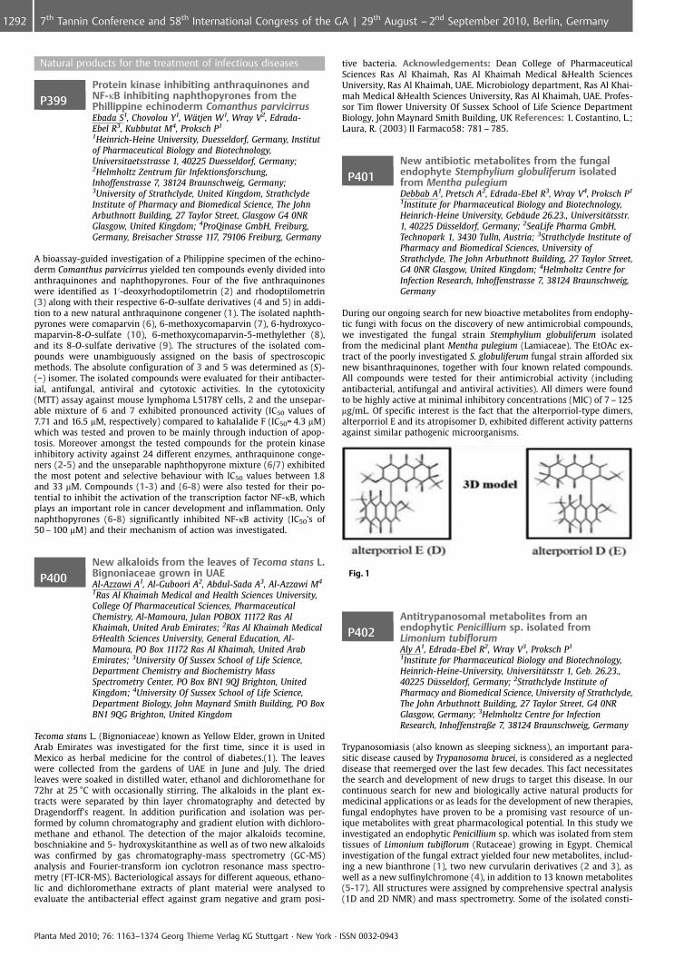
Natural products for the treatment of infectious diseases
P399Protein kinase inhibiting anthraquinones andNF-kB inhibiting naphthopyrones from thePhillippine echinoderm Comanthus parvicirrusEbada S1, Chovolou Y1, W�tjen W1, Wray V2, Edrada-Ebel R3, Kubbutat M4, Proksch P1
1Heinrich-Heine University, Duesseldorf, Germany, Institutof Pharmaceutical Biology and Biotechnology,Universitaetsstrasse 1, 40225 Duesseldorf, Germany;2Helmholtz Zentrum f�r Infektionsforschung,Inhoffenstrasse 7, 38124 Braunschweig, Germany;3University of Strathclyde, United Kingdom, StrathclydeInstitute of Pharmacy and Biomedical Science, The JohnArbuthnott Building, 27 Taylor Street, Glasgow G4 0NRGlasgow, United Kingdom; 4ProQinase GmbH, Freiburg,Germany, Breisacher Strasse 117, 79106 Freiburg, Germany
A bioassay-guided investigation of a Philippine specimen of the echino-derm Comanthus parvicirrus yielded ten compounds evenly divided intoanthraquinones and naphthopyrones. Four of the five anthraquinoneswere identified as 1’-deoxyrhodoptilometrin (2) and rhodoptilometrin(3) along with their respective 6-O-sulfate derivatives (4 and 5) in addi-tion to a new natural anthraquinone congener (1). The isolated naphth-pyrones were comaparvin (6), 6-methoxycomaparvin (7), 6-hydroxyco-maparvin-8-O-sulfate (10), 6-methoxycomaparvin-5-methylether (8),and its 8-O-sulfate derivative (9). The structures of the isolated com-pounds were unambiguously assigned on the basis of spectroscopicmethods. The absolute configuration of 3 and 5 was determined as (S)-(�) isomer. The isolated compounds were evaluated for their antibacter-ial, antifungal, antiviral and cytotoxic activities. In the cytotoxicity(MTT) assay against mouse lymphoma L5178Y cells, 2 and the unsepar-able mixture of 6 and 7 exhibited pronounced activity (IC50 values of7.71 and 16.5 mM, respectively) compared to kahalalide F (IC50= 4.3 mM)which was tested and proven to be mainly through induction of apop-tosis. Moreover amongst the tested compounds for the protein kinaseinhibitory activity against 24 different enzymes, anthraquinone conge-ners (2-5) and the unseparable naphthopyrone mixture (6/7) exhibitedthe most potent and selective behaviour with IC50 values between 1.8and 33 mM. Compounds (1-3) and (6-8) were also tested for their po-tential to inhibit the activation of the transcription factor NF-kB, whichplays an important role in cancer development and inflammation. Onlynaphthopyrones (6-8) significantly inhibited NF-kB activity (IC50’s of50 – 100 mM) and their mechanism of action was investigated.
P400New alkaloids from the leaves of Tecoma stans L.Bignoniaceae grown in UAEAl-Azzawi A1, Al-Guboori A2, Abdul-Sada A3, Al-Azzawi M4
1Ras Al Khaimah Medical and Health Sciences University,College Of Pharmaceutical Sciences, PharmaceuticalChemistry, Al-Mamoura, Julan POBOX 11172 Ras AlKhaimah, United Arab Emirates; 2Ras Al Khaimah Medical&Health Sciences University, General Education, Al-Mamoura, PO Box 11172 Ras Al Khaimah, United ArabEmirates; 3University Of Sussex School of Life Science,Department Chemistry and Biochemistry MassSpectrometry Center, PO Box BN1 9QJ Brighton, UnitedKingdom; 4University Of Sussex School of Life Science,Department Biology, John Maynard Smith Building, PO BoxBN1 9QG Brighton, United Kingdom
Tecoma stans L. (Bignoniaceae) known as Yellow Elder, grown in UnitedArab Emirates was investigated for the first time, since it is used inMexico as herbal medicine for the control of diabetes.(1). The leaveswere collected from the gardens of UAE in June and July. The driedleaves were soaked in distilled water, ethanol and dichloromethane for72hr at 25 �C with occasionally stirring. The alkaloids in the plant ex-tracts were separated by thin layer chromatography and detected byDragendorff’s reagent. In addition purification and isolation was per-formed by column chromatography and gradient elution with dichloro-methane and ethanol. The detection of the major alkaloids tecomine,boschniakine and 5- hydroxyskitanthine as well as of two new alkaloidswas confirmed by gas chromatography-mass spectrometry (GC-MS)analysis and Fourier-transform ion cyclotron resonance mass spectro-metry (FT-ICR-MS). Bacteriological assays for different aqueous, ethano-lic and dichloromethane extracts of plant material were analysed toevaluate the antibacterial effect against gram negative and gram posi-
tive bacteria. Acknowledgements: Dean College of PharmaceuticalSciences Ras Al Khaimah, Ras Al Khaimah Medical &Health SciencesUniversity, Ras Al Khaimah, UAE. Microbiology department, Ras Al Khai-mah Medical &Health Sciences University, Ras Al Khaimah, UAE. Profes-sor Tim flower University Of Sussex School of Life Science DepartmentBiology, John Maynard Smith Building, UK References: 1. Costantino, L.;Laura, R. (2003) Il Farmaco58: 781 – 785.
P401New antibiotic metabolites from the fungalendophyte Stemphylium globuliferum isolatedfrom Mentha pulegiumDebbab A1, Pretsch A2, Edrada-Ebel R3, Wray V4, Proksch P1
1Institute for Pharmaceutical Biology and Biotechnology,Heinrich-Heine University, Geb�ude 26.23., Universit�tsstr.1, 40225 D�sseldorf, Germany; 2SeaLife Pharma GmbH,Technopark 1, 3430 Tulln, Austria; 3Strathclyde Institute ofPharmacy and Biomedical Sciences, University ofStrathclyde, The John Arbuthnott Building, 27 Taylor Street,G4 0NR Glasgow, United Kingdom; 4Helmholtz Centre forInfection Research, Inhoffenstrasse 7, 38124 Braunschweig,Germany
During our ongoing search for new bioactive metabolites from endophy-tic fungi with focus on the discovery of new antimicrobial compounds,we investigated the fungal strain Stemphylium globuliferum isolatedfrom the medicinal plant Mentha pulegium (Lamiaceae). The EtOAc ex-tract of the poorly investigated S. globuliferum fungal strain afforded sixnew bisanthraquinones, together with four known related compounds.All compounds were tested for their antimicrobial activity (includingantibacterial, antifungal and antiviral activities). All dimers were foundto be highly active at minimal inhibitory concentrations (MIC) of 7 – 125mg/mL. Of specific interest is the fact that the alterporriol-type dimers,alterporriol E and its atropisomer D, exhibited different activity patternsagainst similar pathogenic microorganisms.
Fig. 1
P402Antitrypanosomal metabolites from anendophytic Penicillium sp. isolated fromLimonium tubiflorumAly A1, Edrada-Ebel R2, Wray V3, Proksch P1
1Institute for Pharmaceutical Biology and Biotechnology,Heinrich-Heine-University, Universit�tsstr 1, Geb. 26.23.,40225 D�sseldorf, Germany; 2Strathclyde Institute ofPharmacy and Biomedical Science, University of Strathclyde,The John Arbuthnott Building, 27 Taylor Street, G4 0NRGlasgow, Germany; 3Helmholtz Centre for InfectionResearch, Inhoffenstraße 7, 38124 Braunschweig, Germany
Trypanosomiasis (also known as sleeping sickness), an important para-sitic disease caused by Trypanosoma brucei, is considered as a neglecteddisease that reemerged over the last few decades. This fact necessitatesthe search and development of new drugs to target this disease. In ourcontinuous search for new and biologically active natural products formedicinal applications or as leads for the development of new therapies,fungal endophytes have proven to be a promising vast resource of un-ique metabolites with great pharmacological potential. In this study weinvestigated an endophytic Penicillium sp. which was isolated from stemtissues of Limonium tubiflorum (Rutaceae) growing in Egypt. Chemicalinvestigation of the fungal extract yielded four new metabolites, includ-ing a new bianthrone (1), two new curvularin derivatives (2 and 3), aswell as a new sulfinylchromone (4), in addition to 13 known metabolites(5-17). All structures were assigned by comprehensive spectral analysis(1D and 2D NMR) and mass spectrometry. Some of the isolated consti-
Planta Med 2010; 76: 1163–1374 Georg Thieme Verlag KG Stuttgart · New York · ISSN 0032-0943
1292 7th Tannin Conference and 58th International Congress of the GA | 29th August – 2nd September 2010, Berlin, Germany
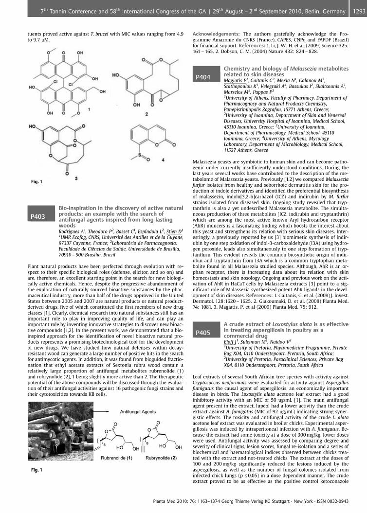
tuents proved active against T. brucei with MIC values ranging from 4.9to 9.7 mM.
Fig. 1
P403Bio-inspiration in the discovery of active naturalproducts: an example with the search ofantifungal agents inspired from long-lastingwoodsRodrigues A1, Theodoro P2, Basset C1, Esp�ndola L2, Stien D1
1UMR Ecofog, CNRS, Universit� des Antilles et de la Guyane,97337 Cayenne, France; 2Laboratrio de Farmacognosia,Faculdade de CiÞncias da Safflde, Universidade de Bras�lia,70910 – 900 Bras�lia, Brazil
Plant natural products have been perfected through evolution with re-spect to their specific biological roles (defense, elicitor, and so on) andare, therefore, an excellent starting point in the search for new biologi-cally active chemicals. Hence, despite the progressive abandonment ofthe exploration of naturally sourced bioactive substances by the phar-maceutical industry, more than half of the drugs approved in the UnitedStates between 2005 and 2007 are natural products or natural product-derived drugs, five of which constituted the first members of new drugclasses [1]. Clearly, chemical research into natural substances still has animportant role to play in improving quality of life, and can play animportant role by inventing innovative strategies to discover new bioac-tive compounds [1,2]. In the present work, we demonstrated that a bio-inspired approach for the identification of novel bioactive natural pro-ducts represents a promising biotechnological tool for the developmentof new drugs. We have studied how natural defenses within decay-resistant wood can generate a large number of positive hits in the searchfor antimycotic agents. In addition, it was found from bioguided fractio-nation that ethyl acetate extracts of Sextonia rubra wood contain arelatively large proportion of antifungal metabolites rubrenolide (1)and rubrynolide (2), 1 being slightly more active than 2. The therapeuticpotential of the above compounds will be discussed through the evalua-tion of their antifungal activities against 16 pathogenic fungi strains andtheir cytotoxicities towards KB cells.
Fig. 1
Acknowledgements: The authors gratefully acknowledge the Pro-gramme Amazonie du CNRS (France), CAPES, CNPq and FAPDF (Brazil)for financial support. References: 1. Li, J. W.-H. et al. (2009) Science 325:161 – 165. 2. Dobson, C. M. (2004) Nature 432: 824 – 828.
P404Chemistry and biology of Malassezia metabolitesrelated to skin diseasesMagiatis P1, Gaitanis G2, Mexia N1, Galanou M3,Stathopoulou K1, Velegraki A4, Bassukas I2, Skaltsounis A1,Marselos M3, Pappas P3
1University of Athens, Faculty of Pharmacy, Department ofPharmacognosy and Natural Products Chemistry,Panepistimiopolis Zografou, 15771 Athens, Greece;2University of Ioannina, Department of Skin and VenerealDiseases, University Hospital of Ioannina, Medical School,45110 Ioannina, Greece; 3University of Ioannina,Department of Pharmacology, Medical School, 45110Ioannina, Greece; 4University of Athens, MycologyLaboratory, Department of Microbiology, Medical School,11527 Athens, Greece
Malassezia yeasts are symbiotic to human skin and can become patho-genic under currently insufficiently understood conditions. During thelast years several works have contributed to the description of the me-tabolome of Malassezia yeasts. Previously [1,2] we compared Malasseziafurfur isolates from healthy and seborrhoic dermatitis skin for the pro-duction of indole derivatives and identified the preferential biosynthesisof malassezin, indolo[3,2-b]carbazol (ICZ) and indirubin by M. furfurstrains isolated from diseased skin. Ongoing study revealed that tryp-tanthrin is also a yet undescribed Malassezia metabolite. The simulta-neous production of three metabolites (ICZ, indirubin and tryptanthrin)which are among the most active known Aryl hydrocarbon receptor(AhR) inducers is a fascinating finding which boosts the interest aboutthis yeast and strengthens its relation with serious skin diseases. Inter-estingly, a previously reported by us [3] biomimetic synthesis of indir-ubin by one step oxidation of indol-3-carboxaldehyde (I3A) using hydro-gen peroxide, leads also simultaneously to one step formation of tryp-tanthrin. This evident reveals the common biosynthetic origin of indir-ubin and tryptanthrin from I3A which is a common tryptophan meta-bolite found in all Malassezia studied species. Although, AhR is an or-phan receptor, there is increasing data about its relation with skinhomeostasis and skin nosology. Ongoing and previous work on the acti-vation of AhR in HaCaT cells by Malassezia extracts [3] point to a sig-nificant role of Malassezia synthesized potent AhR ligands in the devel-opment of skin diseases. References: 1. Gaitanis, G. et al. (2008) J. Invest.Dermatol. 128:1620 – 1625. 2. Giakoumaki, D. et al. (2008) Planta Med.74: 1081. 3. Magiatis, P. et al (2009) Planta Med. 75: 912.
P405A crude extract of Loxostylus alata is as effectivein treating aspergillosis in poultry as acommercial drugEloff J1, Suleiman M1, Naidoo V2
1University of Pretoria, Phytomedicine Programme, PrivateBag X04, 0110 Onderstepoort, Pretoria, South Africa;2University of Pretoria, Paraclinical Sciences, Private BagX04, 0110 Onderstepoort, Pretoria, South Africa
Leaf extracts of several South African tree species with activity againstCryptococcus neoformans were evaluated for activity against Aspergillusfumigatus the causal agent of aspergillosis, an economically importantdisease in birds. The Loxostylis alata acetone leaf extract had a goodinhibitory activity with an MIC of 50 ug/mL [1]. The main antifungalagent present in the extract, lupeol had a lower activity than the crudeextract against A. fumigatus (MIC of 92 ug/mL) indicating strong syner-gistic effects. The toxicity and antifungal activity of the crude L. alataacetone leaf extract was evaluated in broiler chicks. Experimental asper-gillosis was induced by intraperitoneal infection with A. fumigatus. Be-cause the extract had some toxicity at a dose of 300 mg/kg, lower doseswere used. Antifungal activity was assessed by comparing degree andseverity of clinical signs, lesion scores, fungal re-isolation and a series ofbiochemical and haematological indices observed between chicks trea-ted with the extract and not-treated chicks. The extract at the doses of100 and 200 mg/kg significantly reduced the lesions induced by theaspergillosis, as well as the number of fungal colonies isolated frominfected chick lungs (p £0.05) in a dose dependent manner. The crudeextract proved to be as effective as the positive control ketoconazole
Planta Med 2010; 76: 1163–1374 Georg Thieme Verlag KG Stuttgart · New York · ISSN 0032-0943
12937th Tannin Conference and 58th International Congress of the GA | 29th August – 2nd September 2010, Berlin, Germany
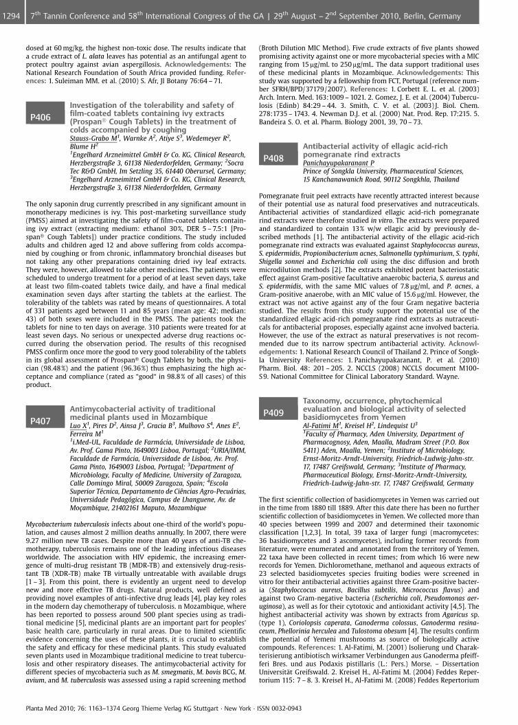
dosed at 60 mg/kg, the highest non-toxic dose. The results indicate thata crude extract of L. alata leaves has potential as an antifungal agent toprotect poultry against avian aspergillosis. Acknowledgements: TheNational Research Foundation of South Africa provided funding. Refer-ences: 1. Suleiman MM. et al. (2010) S. Afr, Jl Botany 76:64 – 71.
P406Investigation of the tolerability and safety offilm-coated tablets containing ivy extracts(Prospan� Cough Tablets) in the treatment ofcolds accompanied by coughingStauss-Grabo M1, Warnke A2, Atiye S3, Wedemeyer R2,Blume H2
1Engelhard Arzneimittel GmbH & Co. KG, Clinical Research,Herzbergstraße 3, 61138 Niederdorfelden, Germany; 2SocraTec R&D GmbH, Im Setzling 35, 61440 Oberursel, Germany;3Engelhard Arzneimittel GmbH & Co. KG, Clinical Research,Herzbergstraße 3, 61138 Niederdorfelden, Germany
The only saponin drug currently prescribed in any significant amount inmonotherapy medicines is ivy. This post-marketing surveillance study(PMSS) aimed at investigating the safety of film-coated tablets contain-ing ivy extract (extracting medium: ethanol 30%, DER 5 – 7.5:1 [Pro-span� Cough Tablets]) under practice conditions. The study includedadults and children aged 12 and above suffering from colds accompa-nied by coughing or from chronic, inflammatory bronchial diseases butnot taking any other preparations containing dried ivy leaf extracts.They were, however, allowed to take other medicines. The patients werescheduled to undergo treatment for a period of at least seven days, takeat least two film-coated tablets twice daily, and have a final medicalexamination seven days after starting the tablets at the earliest. Thetolerability of the tablets was rated by means of questionnaires. A totalof 331 patients aged between 11 and 85 years (mean age: 42; median:43) of both sexes were included in the PMSS. The patients took thetablets for nine to ten days on average. 310 patients were treated for atleast seven days. No serious or unexpected adverse drug reactions oc-curred during the observation period. The results of this recognisedPMSS confirm once more the good to very good tolerability of the tabletsin its global assessment of Prospan� Cough Tablets by both, the physi-cian (98.48%) and the patient (96.36%) thus emphasizing the high ac-ceptance and compliance (rated as “good“ in 98.8% of all cases) of thisproduct.
P407Antimycobacterial activity of traditionalmedicinal plants used in MozambiqueLuo X1, Pires D2, Ainsa J3, Gracia B3, Mulhovo S4, Anes E2,Ferreira M1
1i.Med-UL, Faculdade de Farmcia, Universidade de Lisboa,Av. Prof. Gama Pinto, 1649003 Lisboa, Portugal; 2URIA/IMM,Faculdade de Farmcia, Universidade de Lisboa, Av. Prof.Gama Pinto, 1649003 Lisboa, Portugal; 3Department ofMicrobiology, Faculty of Medicine, University of Zaragoza,Calle Domingo Miral, 50009 Zaragoza, Spain; 4EscolaSuperior T�cnica, Departamento de CiÞncias Agro-Pecurias,Universidade Pedaggica, Campus de Lhanguene, Av. deMo�ambique, 21402161 Maputo, Mozambique
Mycobacterium tuberculosis infects about one-third of the world’s popu-lation, and causes almost 2 million deaths annually. In 2007, there were9.27 million new TB cases. Despite more than 40 years of anti-TB che-motherapy, tuberculosis remains one of the leading infectious diseasesworldwide. The association with HIV epidemic, the increasing emer-gence of multi-drug resistant TB (MDR-TB) and extensively drug-resis-tant TB (XDR-TB) make TB virtually untreatable with available drugs[1 – 3]. From this point, there is evidently an urgent need to developnew and more effective TB drugs. Natural products, well defined asproviding novel examples of anti-infective drug leads [4], play key rolesin the modern day chemotherapy of tuberculosis. n Mozambique, wherehas been reported to possess around 500 plant species using as tradi-tional medicine [5], medicinal plants are an important part for peoples’basic health care, particularly in rural areas. Due to limited scientificevidence concerning the uses of these plants, it is crucial to establishthe safety and efficacy for these medicinal plants. This study evaluatedseven plants used in Mozambique traditional medicine to treat tubercu-losis and other respiratory diseases. The antimycobacterial activity fordifferent species of mycobacteria such as M. smegmatis, M. bovis BCG, M.avium, and M. tuberculosis was assessed using a rapid screening method
(Broth Dilution MIC Method). Five crude extracts of five plants showedpromising activity against one or more mycobacterial species with a MICranging from 15 mg/mL to 250 mg/mL. The data support traditional usesof these medicinal plants in Mozambique. Acknowledgements: Thisstudy was supported by a fellowship from FCT, Portugal (reference num-ber SFRH/BPD/37179/2007). References: 1. Corbett E. L. et al. (2003)Arch. Intern. Med. 163:1009 – 1021. 2. Gomez, J. E. et al. (2004) Tubercu-losis (Edinb) 84:29 – 44. 3. Smith, C. V. et al. (2003) J. Biol. Chem.278:1735 – 1743. 4. Newman D.J. et al. (2000) Nat. Prod. Rep. 17:215. 5.Bandeira S. O. et al. Pharm. Biology 2001, 39, 70 – 73.
P408Antibacterial activity of ellagic acid-richpomegranate rind extractsPanichayupakaranant PPrince of Songkla University, Pharmaceutical Sciences,15 Kanchanawanich Road, 90112 Songkhla, Thailand
Pomegranate fruit peel extracts have recently attracted interest becauseof their potential use as natural food preservatives and nutraceuticals.Antibacterial activities of standardized ellagic acid-rich pomegranaterind extracts were therefore studied in vitro. The extracts were preparedand standardized to contain 13% w/w ellagic acid by previously de-scribed methods [1]. The antibacterial activity of the ellagic acid-richpomegranate rind extracts was evaluated against Staphylococcus aureus,S. epidermidis, Propionibacterium acnes, Salmonella typhimurium, S. typhi,Shigella sonnei and Escherichia coli using the disc diffusion and brothmicrodilution methods [2]. The extracts exhibited potent bacteriostaticeffect against Gram-positive facultative anaerobic bacteria, S. aureus andS. epidermidis, with the same MIC values of 7.8 mg/ml, and P. acnes, aGram-positive anaerobe, with an MIC value of 15.6 mg/ml. However, theextract was not active against any of the four Gram negative bacteriastudied. The results from this study support the potential use of thestandardized ellagic acid-rich pomegranate rind extracts as nutraceuti-cals for antibacterial proposes, especially against acne involved bacteria.However, the use of the extract as natural preservatives is not recom-mended due to its narrow spectrum antibacterial activity. Acknowl-edgements: 1. National Research Council of Thailand 2. Prince of Songk-la University References: 1. Panichayupakaranant, P. et al. (2010)Pharm. Biol. 48: 201 – 205. 2. NCCLS (2008) NCCLS document M100-S9. National Committee for Clinical Laboratory Standard. Wayne.
P409Taxonomy, occurrence, phytochemicalevaluation and biological activity of selectedbasidiomycetes from YemenAl-Fatimi M1, Kreisel H2, Lindequist U3
1Faculty of Pharmacy, Aden University, Department ofPharmacognosy, Aden, Maalla, Madram Street (P.O. Box5411) Aden, Maalla, Yemen; 2Institute of Microbiology,Ernst-Moritz-Arndt-University, Friedrich-Ludwig-Jahn-str.17, 17487 Greifswald, Germany; 3Institute of Pharmacy,Pharmaceutical Biology, Ernst-Moritz-Arndt-University,Friedrich-Ludwig-Jahn-str. 17, 17487 Greifswald, Germany
The first scientific collection of basidiomycetes in Yemen was carried outin the time from 1880 till 1889. After this date there has been no furtherscientific collection of basidiomycetes in Yemen. We collected more than40 species between 1999 and 2007 and determined their taxonomicclassification [1,2,3]. In total, 39 taxa of larger fungi (macromycetes:36 basidiomycetes and 3 ascomycetes), including former records fromliterature, were enumerated and annotated from the territory of Yemen.22 taxa have been collected in recent times; from which 16 were newrecords for Yemen. Dichloromethane, methanol and aqueous extracts of23 selected basidiomycetes species fruiting bodies were screened invitro for their antibacterial activities against three Gram-positive bacter-ia (Staphyloccocus aureus, Bacillus subtilis, Micrococcus flavus) andagainst two Gram-negative bacteria (Escherichia coli, Pseudomonas aer-uginosa), as well as for their cytotoxic and antioxidant activity [4,5]. Thehighest antibacterial activity was shown by extracts from Agaricus sp.(type 1), Coriolopsis caperata, Ganoderma colossus, Ganoderma resina-ceum, Phellorinia herculea and Tulostoma obesum [4]. The results confirmthe potential of Yemeni mushrooms as source of biologically activecompounds. References: 1. Al-Fatimi, M. (2001) Isolierung und Charak-terisierung antibiotisch wirksamer Verbindungen aus Ganoderma pfeiff-feri Bres. und aus Podaxis pistillaris (L.: Pers.) Morse. – DissertationUniversit�t Greifswald. 2. Kreisel H., Al-Fatimi M. (2004) Feddes Reper-torium 115: 7 – 8. 3. Kreisel H., Al-Fatimi M. (2008) Feddes Repertorium
Planta Med 2010; 76: 1163–1374 Georg Thieme Verlag KG Stuttgart · New York · ISSN 0032-0943
1294 7th Tannin Conference and 58th International Congress of the GA | 29th August – 2nd September 2010, Berlin, Germany
119: 5 – 6. 4. Al-Fatimi, M. et al. (2005). Pharmazie 60: 776 – 780. 5. Al-Fatimi, M. et al. (2006) Evidence-based Complementary and AalternativeMedicine 3 (1), 87 – 92.
P410Lipid composition and biological activity ofextracts from Zygophyllum oxianumSasmakov S1, Gazizov F1, Putieva Z1, Wende K2, Alresly Z2,Lindequist U2
1S. Yu. Yunusov Institute of the Chemistry of PlantSubstances, Uzbek Academy of Sciences, Chemistry ofGlycosides, M. Ulugbek Str. 77, Tashkent, 100170 Tashkent,Uzbekistan; 2Institute of Pharmacy, University of Greifswald,Department of Pharmaceutical Biology, F.-L.-Jahn-Str. 17, D-17489 Greifswald, Germany
Zygophyllum species (Zygophyllaceae) are widely distributed in Uzbeki-stan and used in folk medicine as antiinfectious, antirheumatic andantidiabetic means. Continuing our research on the chemical constitu-ents of this genus [1,2], we have studied the lipid composition of theaerial parts of Zygophyllum oxianum Boriss. The yields of the total lipidswere: leaves – 0.75%, stems – 0.33% and fruits – 0.49% from fresh weightof organ. Phospholipids (PL) are presented by 9 classes, from themphosphatidylcholine is dominating (leaves – 55.8%, stems – 56.4%, fruits– 63.7% from sum of PL). The study of antimicrobial activity againstseveral bacterial strains and fungi showed that the n-butanol extractfrom all aerial parts exhibits significant activity against Candida sp. butonly weak antibacterial activity. In cytotoxicity assays against 5637 hu-man urinary bladder cells the n-butanol extract showed only weak ac-tivity. Otherwise the chloroform-methanol (2:1) extract of steams andleaves has a remarkable cytotoxicity against 5637 cells with an IC 50value of 6.2 mg/ml, but only weak antibacterial and antifungal activity.The chloroform-methanol extract (2:1) of the fruits was less toxic (IC 50value: 37.6 mg/ml). References: 1. Sasmakov SA et al. (2003) Pharmazie,Vol.58 (8): 602 – 603. 2. Sasmakov SA et al. (2007) Pharmazie, Vol.62(12): 957 – 959.
P411Antibacterial from Hypericum acmosepalumshowing inhibition of ATP dependent MurE ligasefrom Mycobacterium tuberculosisOsman K1, Basavannacharya C2, Envangelopoulos D2,Gupta A2, Bhakta S2, Gibbons S1
1The School of Pharmacy, Pharmaceutical and BiologicalChemistry, The School of Pharmacy University of London,29 – 39 Brunswick Square, 11 London, United Kingdom;2Birkbeck College, University of London, Departmen ofBiological Sciences in the school of Science, Malet Street,Bloomsbury London, WC 1E 7HX London, United Kingdom
In a project to characterise new antibacterial chemotypes from plants,hyperenone A and hypercalin B were isolated from the hexane andchloroform extracts of the aerial parts of H. acmosepalum. The structureof compounds were characterised by extensive 1- and 2D NMR spectro-scopy and confirmed by MS spectrometry. Both hyperenone A and hy-percalin B exhibited anti-bacterial activity against multidrug-resistantstrains of Staphylococcus aureus, with MIC values ranging from 2 –128 mg/mL to 0.5 – 128 mg/mL, respectively. Hyperenone A also showedgrowth inhibition against Mycobacterium tuberculosis H37Rv and M. bo-vis BCG at 50 mg/mL (H37Rv) and 75 mg/mL (BCG). The hyperenone A didnot inhibit growth of Escherichia coli and was not toxic to culturedmammalian macrophages cells at the concentration at which anti-my-cobacterial activity was determined. Both the compounds were alsotested for their ability to inhibit MurE and MurF enzymes of M. tubercu-losis which are crucial enzymes in the cytoplasmic steps of peptidogly-can biosynthesis. Hyperenone A inhibited MurE selectively whereas hy-percalin B did not have any effect on both the enzymes. This is firstreport of a natural product from plant source showing inhibition ofATP dependent MurE ligase from M. tuberculosis. These studies repre-sent promising starting points for further development. These highlyactive antibacterial compounds encourage further studies on the Hyper-icum genus. References: 1. Decosterd, L. et al. (1989) Photo S HelveticaActa 70 (8): 1833 – 1845. 2. Kikuche, T. et al. (1985) Chem. Pharm. Bull.33(2): 557 – 564.
P412Phytochemical and antibacterial studies onOcimum kilimandscharicumShinde K1, Shinde V2, Mahadik K2, Gibbons S1
1The School of Pharmacy, Dept. of Pharmaceutical andBiological Chemistry, University of London, 29 – 39Brunswick Square, London, 11 London, United Kingdom;2Poona College of Pharmacy, Department of Pharmacognosyand Phytochemistry, Bharati Vidyapeeth University, Pune,India, 411038 Pune, India
The development of resistance by bacteria to currently available anti-biotics is a major problem in the treatment of infectious diseases. Toexemplify this, there has been an increase in the number of deaths byMRSA world wide. As part of on-going efforts to characterize new anti-bacterials with activity against multidrug-resistant (MDR) strains ofStaphylococcus aureus, an extract of the aerial parts of Ocimum kili-mandscharicum (Lamiaceae), have been investigated. This species iscommonly known as camphor Tulsi. Ocimum has been used in theAyurvedic System of Medicine for bronchitis, bronchial asthma, skindiseases, arthritis, as an astringent, in inflammation and for fever. Theplant material was extracted with chloroform, methanol and acetonesuccessively by cold maceration. Further sub-fractionation by SPE andpurification by P-TLC led to the isolation of six compounds eugenol,quercitin, cadinol, thymol, b-sitosterol and stigmasterol. The structuresof the compounds were characterized by extensive 1 and 2D NMR spec-troscopy. The isolated constituents were evaluated for their antibacterialactivity by the broth microtitre MIC assay against a panel of multidrug-resistant (MDR) and methicillin-resistant Staphylococcus aureus (MRSA)strains (ATCC-25923, SA-1199B, XU-212, RN-4220, EMRSA-15 and EMR-SA-16) and minimum inhibitory concentrations (MICs) of eugenol andcadinol were found to be in the range of 2 – 128 mg/ml. This study cor-roborates the traditional claims of Ocimum kilimandscharicum as a topi-cal antibacterial. Acknowledgements: Kamlesh Shinde thanks the As-sociation of Commonwealth Universities for a Split Site Ph.D scholar-ship. References: 1. Prakash, P., Gupta, N. (2005) J. Physiol. Pharmacol.49: 125 – 131. 2. Smith, ECJ. et al. (2008) Phytochem. Lett. 1:49 – 53.
P413Screening of South African medicinal plants andHPLC based profiling for the identification ofleads with antiprotozoal activitiesHata Uribe Y1, Julianti T1, Mokoka T2, Moodley N2,Zimmermann S1, Adams M1, Koorbanally N3, Brun R4,Kaiser M4, Hamburger M1
1University of Basel, Pharmaceutical Sciences,Klingelbergstrasse 50, 4056 Basel, Switzerland; 2Council forScientific and Indutrial Research, P.O. Box 395, 0002Pretoria, South Africa; 3University of KwaZulu-Natal,Durban, 4041 Durban, South Africa; 4Swiss TropicalInstitute, Socinstrasse 57, 4002 Basel, Switzerland
A library of 300 extracts from one hundred and seven plant species, usedin South African folk medicine to treat malaria were collected andscreened in vitro against Plasmodium falciparum, Trypanosoma bruceirhodesiense, Trypanosoma cruzi and Leishmania donovani. Of these 102(34.0%) exhibited more than 50% growth inhibition of one of the para-sites at the screen concentration of 9.7 mg/mL and were thus consideredactive. P. falciparum against which seventy-two plant extracts (24.0%)showed activity was the most susceptible parasite, followed by L. dono-vani (forty-nine, 16.3%) and T. b. rhodesiense (thirty-six, 12.0%), with T.cruzi (zero) being the least susceptible. Twenty plants (6.6%) were se-lected for further investigation based on activity and specificity criteriaas well as on chemotaxonomic considerations. Just 350 mg of the se-lected extracts were fractionated by analytical scale HPLC into 32 one-minute fractions in a fully automated 96 well microfractionation scheme[1]. After drying the microfractions were subsequently tested against theparasite for which the extract had shown activity, to identify the peaksresponsible for the overall activity of the extracts. HPLC hyphenatedmethods (MS, UV, ELSD, HRMS and offline LC-NMR) helped to identifymany active substances online. Active compounds were isolated andtested against the parasites as well as human cancer cell lines to deter-mine their cytotoxicity. Screening results as well as selected examples ofactivity profiling and active compounds will be shown. References:1. Adams et al. (2009) Nat Prod Comm. 10:1377 – 81.
Planta Med 2010; 76: 1163–1374 Georg Thieme Verlag KG Stuttgart · New York · ISSN 0032-0943
12957th Tannin Conference and 58th International Congress of the GA | 29th August – 2nd September 2010, Berlin, Germany
P414Alkaloids and phytosterols from Cakile maritimapresent in UAEAl-Azzawi A1, Al-Guboori A2, Abdul-Sada A3, Al-Azzawi M4
1Ras Al Khaimah Medical and Health Sciences University,College Of Pharmaceutical Sciences, Ras Al Khaimah,Pharmaceutical Chemistry, Ras Al Khaimah, Al-Mamoura,Julan POBOX 11172, 11172 Ras Al Khaimah, United ArabEmirates; 2Ras Al Khaimah Medical &Health SciencesUniversity, General Education, Ras Al Khaimah, UAE, 11172Ras Al Khaimah, United Arab Emirates; 3University OfSussex School of Life Science, Department Chemistry andBiochemistry Mass Spectrometry Center, Brighton, BN1 9QJBrighton, United Kingdom; 4University Of Sussex School ofLife Science, Department Biology, John Maynard SmithBuilding, BN1 9QG Brighton, United Kingdom
Cakile maritima is a halophyte exhibiting potential for secondary meta-bolite production. Plants grown in the United Arab Emirates (UAE) wereinvestigated for the first time for its phytochemical constituents andantibacterial activity. Dried leaves and stems collected from the gardensin Ras Al-Khiama, UAE were (soxhlet) extracted using equal amounts ofethanol and water for three hours. Alkaloids were detected by thin layerchromatography with dichloromethane:ethanol:ammonia as the mobilephase and Dragendorff’s spraying reagent. Isolation and purification ofalkaloids and phytosterols was performed by column chromatographyusing dichloromethane and ethanol as the mobile phase in gradientelution technique. The detection of alkaloids and phytosterols was con-firmed by gas chromatography-mass spectrometry (GC-MS) analysis andFourier-transform ion cyclotron resonance mass spectrometry (FT-ICR-MS). The bacteriological assays for different plant extracts (aqueous,ethanolic and dichloromethane) were conducted to evaluate the anti-bacterial effect against gram negative and gram positive bacteria.Acknowledgements: Dean College of Pharmaceutical Sciences Ras AlKhaimah, Ras Al Khaimah Medical &Health Sciences University, Ras AlKhaimah, UAE. Microbiology department, Ras Al Khaimah Medical&Health Sciences University, Ras Al Khaimah, UAE. Professor Tim flowerUniversity Of Sussex School of Life Science Department Biology, JohnMaynard Smith Building, UK
P415Antifungal activity against Candida albicans andeffect on mitochondrial NADH oxidation ofgalanginChudapongse N, Klahan K, Kamkhunthod M,Ratchawong C, Nantapong NInstitute of Science, Biology, Suranaree University ofTechnology, 111 University Avenue, Muang District, 30000Nakhon Ratchasima, Thailand
Galangin is a flavonoid found in the rhizomes of Alpinia officinarum [1].In Asia, this plant has long been used as traditional medicine for gastro-intestinal ailments and infectious diseases. Galangin has been shown topossess several biological and pharmacological properties, includingantimicrobial activity [2]. Candida albicans resides in a diversity of organsurfaces of a healthy host, such as oral cavity, digestive tract, skin andvagina. As an opportunistic pathogen, it can cause life-threatening sys-temic infection in immunocompromised hosts [3], especially in HIV-infected patients. Nowadays, the number of antifungals against candi-diasis is quite limited. In the present study, we found that galanginexhibited antifungal activity against Candida albicans. The MIC valuewas 25 mg/ml (92.5 mM). Galangin has been reported to strongly interactwith rat liver mitochondrial membranes by acting as an uncouplingagent [4]. In mitochondria isolated from Candida albicans, galanginwas found to inhibit NADH oxidation and CCCP-stimulated respiration,suggesting that galangin interfered with the electron transport chain inCandida albicans mitochondria. This mitochondrial effect may contri-bute to the antifungal activity of galangin against Candida albicans.References: 1. Mutsuda, H. et al. (2006) Bioorg. Med. Chem. 95:138 –142. 2. Cushnie, TPT. et al. (2003) Microbiol. Res. 158:281 – 289. 3. Cal-derone, RA., Fonzi, WA. (2001) Trends Microbiol. 9:327 – 335. 4. Dorta,DJ. et al. (2005) Chem. Biol. Interact. 152: 67 – 78.
P416Anti-plaque activity of Piper betle leaf extractsPandita N, Varghese A, Mantri M, Kachwala YSVKM NMIMS University, Pharmaceutical chemistry, VLmehta road Vile parle Mumbai, India 400056 Mumbai, India
Leaves of Piper betle L. (Piperaceae) have been shown to possess anti-microbial, gastroprotective, wound healing, hepatoprotective and anti-oxidant properties [1]. The antimicrobial activity of betel leaves relevantin oral care lead to the investigation of its anti-plaque activity. Fivevarieties of Piper betle leaves (Banarasi, Calcutta, Desi, Maghai andPuna) were subjected to successive extractions at room temperaturewith various solvents (hexane, diethyl ether, ethyl acetate, ethanol, hy-dro-ethanol and water). Two varieties (Banarasi and Calcutta) were se-lected on the basis of their in vitro activities. Of the extracts, hexaneextract was found to be active. Banarasi and Calcutta leaves were alsosubjected to super critical extraction and steam distillation. TLC wascarried on hexane extracts of all varieties, super critical extracts andsteam distillation of Banarasi and Calcutta. A solvent system was devel-oped and plates were derivatized with Vanillin sulphuric acid. Sinceeugenol is present in all varieties this compound was used as markercompound. Anti-plaque activity was evaluated using a microbial assay(Agar Streak Plate method). Leaf extracts were tested for antimicrobialactivity against Streptococcus mutans, Streptococcus sobrinus, and Acti-nomyces viscosus (early colonizers of dental plaque) [2]. Antimicrobialactivity of Piper betle leaf extracts are summarized in the table below.
Table 1: Antimicrobial activity of Piper betle – Direct extracts
Observed MIC (mg/ml)
Extract Diluent S. mutans S. sanguis A. viscosusHexane (Banarasi) Ethyl acetate 50 75 50Hexane (Banarasi) Methanol + Tween-20 > 125 > 125 > 125Hexane (Calcutta) Ethyl acetate 100 500 500Hexane (Maghai) Ethyl Acetate 50 300 > 600Hexane (Pune) Ethyl acetate 200 > 500 > 500Hexane (Desi) Ethyl Acetate 200 > 500 > 500Di ethyl ether (Calcutta) Ethyl acetate 100 400 > 500Di ethyl ether (Pune) Ethyl acetate 250 500 1000Ethanol (Maghai) DMSO > 1000 > 1000 > 1000
Antimicrobial activity was observed for the hexane extract of Piper betleleaves (Banarasi) and could be attributed to the presence of allypyroca-techol [3] obtained in the chromatographic data of these extracts. Re-ferences: 1. Arambewala L.S.R et al. (2005), Journal of Ethnopharmacol-ogy, 102:239 – 245. 2. Svens�ter G et al. Caries Research, 2003; 37,395. 3.Ramji N. et al. (2002) Journal of Ethnopharmacology, 83: 149 – 152.
P417Antiprotozoal and cytotoxic potential of Britishand Irish red algaeAllmendinger A1, Spavieri J1, Kaiser M2, Guiry M3,Blunden G4, Tasdemir D1
1Centre for Pharmacognosy and Phytotherapy, School ofPharmacy, University of London, 29 – 39 Brunswick Square,wc1n 1ax London, United Kingdom; 2Department of MedicalParasitology and Infection Biology, Swiss Tropical Institute,Socinstrasse 57, CH-4002 Basel, Switzerland; 3Martin RyanInstitute, National University of Ireland, University Road,1 Galway, Ireland; 4School of Pharmacy and BiomedicalSciences, University of Portsmouth, White Swan Road, PO12DT Portsmouth, United Kingdom
Marine algae are a prolific source of diverse natural products, but theirbiomedical potential has barely been explored. As part of our continuingresearch on seaweeds [1], we have screened crude extracts of 23 marinered algae (Rhodophyta) collected from Britain and Ireland. Algal materialwas extracted with CHCl3: MeOH mixtures (3:1 and 1:1) at room tem-perature and evaporated to dryness in vacuo before use in bioassays. Theclinically important blood-stage life forms of Trypanosoma brucei rhode-siense, T. cruzi and Leishmania donovani were used as test organisms inthe in vitro assays. The selective toxicity of the extracts was determinedtowards mammalian skeletal myoblast (L 6) cells. All algal extractsshowed activity against T. brucei rhodesiense, with Corallina officinalis(Corallinaceae) and Ceramium virgatum (Ceramiaceae) being the mostpotent (IC50 values 4.8 and 5.4 mg/ml, respectively), whilst none of thealgal extracts inhibited the growth of T. cruzi. With the exception of aPorphyra leucosticta (Bangiaceae) extract, all seaweed extracts also dis-played leishmanicidal activity with IC50 values ranging from 16.5 to85.6 mg/ml. Corallina officinalis was the only seaweed that showed somemarginal cytotoxicity (IC50 value 88.6 mg/ml), whereas all remaining ex-tracts were non-toxic towards L6 cells at 90 mg/ml concentration. To our
Planta Med 2010; 76: 1163–1374 Georg Thieme Verlag KG Stuttgart · New York · ISSN 0032-0943
1296 7th Tannin Conference and 58th International Congress of the GA | 29th August – 2nd September 2010, Berlin, Germany
knowledge, this is the first study reporting antiprotozoal activity ofBritish and Irish red algae. References: 1. Orhan, I. et al. (2006) Phyto-medicine 13:388 – 393.
P418Phytochemical study of plants used in traditionalmedicine in the treatment of malaria in theComoros islandsMohamed Kaou A1, Mahiou-Leddet V1, Mabrouki F1,Hutter S1, Laget M1, Azas N1, Yahaya I2, Ollivier E1
1Facult� de Pharmacie, Laboratoire de Pharmacognosie etethnopharmacologie, 27 Bd Jean Moulin, 13385 Marseille,France; 2Centre National de Documentation et deRecherches Scientifiques, Laboratoire de biologie, MoroniMoroni, Comoros
An ethnobotanical survey was carried out in the Comoros Islands aboutplant species used traditionally for the treatment of various diseasesincluding malaria. The in vitro antiplasmodial activity of 76 vegetal ex-tracts obtained from 17 species traditionaly used to treat malaria symp-tomes, was evaluated using Plasmodium falciparum choloroquine-resis-tant strain (W2). The results showed that 10 plant extracts had a mod-erate activity (5 < IC 50 £10 mg/ml), and 6 showed particularly interest-ing in vitro activity with IC 50 value £5 mg/ml [1]. The phytochemicalstudy realized on three selected plants, Flueggea virosa (Roxb. Ex Willd.)Voigt subsp. Virosa (Euphorbiaceae), Piper capense L.f. (Piperaceae), andFlacourtia indica (Burm. F.) Merr. (Flacourtiaceae) allowed us to isolatecompounds from the most active extracts. The structures of isolatedcompounds were elucidated by spectrometric methods and their anti-plasmodial activity was evaluated in vitro against the chloroquine-resis-tant strain W2 of Plasmodium falciparum, according to Azas, 2002 [2].From F. virosa, rutine, gallic acid, methyl gallate, bergenin, (�)-norsecur-inine, securinol A, N-methyltetrahydro-b-carboline and N-methyltrypta-mine were isolated. The highest antiplasmodial activity was found formethyl gallate (IC 50 = 14.1mM). Together with apigenine dimethyletherand piperchabamide A, a new amide alkaloid, Kaousine and the Z formof antiepilepsirine were isolated from the aerial part of P. capense. Loweractivity was observed for kaousine and apigenine dimethylether,whereas antiepilepsirine demonstrated the same activity that the chlor-omethylenic extract of P. capense (IC 50 = 7 mg/ml) [3]. Pyrocatechol, ho-maloside D and poliothrysoside were isolated from the decoction of theaerial parts of F. indica. The poliothrysoside presented a strong antiplas-modial activity (IC50 = 7.4mM) and a good selectivity index (> 28) simi-lar to chloroquine. References: 1. Mohamed Kaou, A. et al. (2008) J.Ethnopharmacol 116: 74 – 83. 2. Azas, N et al. (2002) Parasitol Res. Ss:165 – 171. 3. Mohamed Kaou, A. et al. (2010) Fitoterapia (In press).
P419Marine algal extracts inhibit growth of thehuman pathogens P. falciparum, T. brucei and L.donovaniVonthron-S�n�cheau C1, Mussio I2, Kaiser M3, Attioua B1,Kerhuel A2, Rusig A2
1Universit� de Strasbourg, Facult� de Pharmacie/UMR UDS/CNRS 7200/Pharmacognosie, 74 route du Rhin, 67401Illkirch, France; 2Universit� de Caen Basse Normandie,Esplanade de la Paix, 14032 Caen, France; 3Swiss TropicalInstitute, Socinstrasse 57, 4002 Basel, Switzerland
Marine macrophytes have been highlighted as resources containing avariety of biologically active compounds and particularly showing invitro antiprotozoal activities1,2. As a part of a screening program tosearch new natural antiprotozoal products, we screened 20 seaweedspecies from the Normandy’s coast belonging to Rhodophyta, Pheophytaand Chlorophyta against protozoa responsible for 4 major endemic para-sitic diseases in vitro. Thus, brown, red and green seaweeds extractswere screened in vitro against P. falciparum, T. brucei, T. cruzi and L.donovani, respectively responsible of malaria, African trypanosomiasis,Chaga’s disease and visceral leishmaniasis. No or weak activity wasshown against T. cruzi and L. donovani. In contrast, more than half ofthe species tested showed good to strong activity against P. falciparumand T. brucei (0.5< IC50< 5 mg/ml). Interestingly, the active species arebrown or red algae, all sharing phyllogenetic origins with P. falciparum.Green algae were inactive. Bio-guided fractionation of the most activeextracts (IC50< 5 mg/ml) is under process. Acknowledgements: AgenceUniversitaire de la Francophonie, S. Sritharan. References: 1. Lin et al.2010 J. Nat. Prod 73:275 – 8. 2. Afolayan et al. 2009 Phytochemistry70:597 – 600.
P420Antiplasmodial evaluation andpharmacomodulation of lanaroflavone, abiflavonoid isolated from Campnospermapanamense Standl. (Anacardiaceae)Weniger B1, Seri G2, Stiebing S3, Kerhuel A3, Collot V3,Schmitt M1, Perrotey S2, Candolfi E2, Vonthron-S�n�cheau C1
1UMR UDS/CNRS 7200, Facult� de Pharmacie, Universit� deStrasbourg, 74 route du Rhin, 67401 Illkirch, France; 2Institutde Parasitologie et Pathologie Tropicale, Facult� deM�decine, Universit� de Strasbourg, 3 rue Koeberle, 67000Strasbourg, France; 3CERMN, Universit� de Caen BasseNormandie, Boulevard Becquerel, 14032 Caen, France
Activity-guided fractionation of the leaves from Campnosperma pana-mense Standl. (Anacardiaceae) was undertaken as a part of a screeningprogram to search new antiprotozoal drugs1. The biflavonoid lanarofla-vone was isolated and showed high in vitro antiplasmodial activityagainst Plasmodium falciparum erythrocytic stages and mild activityagainst Leishmania donovani amastigotes, but was inactive against Cha-ga’s disease vector, Trypanosoma cruzi2,3. A possible machanism under-lying antiplasmodial activity implicating inhibition of beta-hematin for-mation by this compound was supported by in vitro tests. The screeningof eight natural biflavonoids structurally related to lanaroflavone al-lowed us to identify the nature and the length of the interflavonoidlinkage as of high relevance regarding antiplasmodial activity. Thus, aserie of new flavone derivatives was synthetized and tested against P.falciparum resistant strains (K1, 7G8). They were also tested on theircytotoxicity effects upon mouse hepatocyte and L 6 cells. One new ami-nated flavone derivative showed significant activity in the nanomolarrange and very satisfying selectivity indexes (IC50= 80nM, SI> 100), aswell as absence of hemolytic effect indicating selective antiplasmodialactivity. The importance of the hydrophylic framework attached at theC 8 of the flavone is presently explored. The most promising derivativeswill be evaluated in vivo in P. berghei infected mice. References: 1. We-niger et al. 2001 J. Ethnopharmacol. 78:193 – 200. 2. Weniger et al 2004Fitoterapia 75:764 – 7. 3. Weniger et al 2006 Phytomedicine 13:176 –180.
P421Characterization of immunostimulatoryactivities of fractions obtained from TaraxacumofficinaleKim J, Choi G, Hwang H, Ku H, Choi C, Jung G, So BNational Veterinary Research & Quarantine Service, Anyang6 dong #480, 430824 Anyang, Korea, Republic Of
Plants of the genus Taraxacum officinale have a history of use in Korean,Chinese, Arabian and Native American traditional medicine to treat avariety of infectious diseases and cancers [1]. In Traditional Medicine,it is also known as choleretic, diuretic, anti-rheumatic and anti-inflam-matory properties [2]. In the current study, in vitro immunostimulatroyeffects of Taraxacum officinale have been investigated. The crude metha-nolic extract of the whole plant was sequentially fractionated into threesubextracts; explicitly, ethyl acetate (EtOAc), n-butanol, and remainingwater extracts. Further studies were carried out on the most active sub-extract, i. e. the EtOAc subextract, was further subjected to fractionationthrough successive column chromatographic applications on Silica gel60. For the activity assessment, each extract or fraction was submitted tobioassay systems; in vitro neutrophil migration, spleen lymphocyte pro-liferation, nitric oxide (NO) production and phagocytosis for immunos-timulatory activity assessment. Among the fractions, the ethyl acetatefraction was identified to be most effective in the migration activity ofneutrophils, spleen cell proliferation, phagocytosis and NO production,significantly greater than media control. Sequential chromatographicseparation of the ethyl acetate fraction on Silica gel column resulted ina more purified preparation that retained the immunostimulatory activ-ity. From the EtOAc subextract, a major component was isolated and itsstructure was determined as taraxinic acid b-D-glucoside by means ofspectral techniques. This study provides a new scientific data on Tarax-acum officinale extracts or individual components present in the extractsmay be of value as novel immunostimulatory agents.
Planta Med 2010; 76: 1163–1374 Georg Thieme Verlag KG Stuttgart · New York · ISSN 0032-0943
12977th Tannin Conference and 58th International Congress of the GA | 29th August – 2nd September 2010, Berlin, Germany
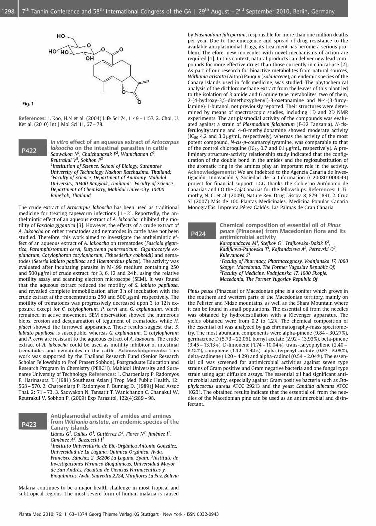
Fig. 1
References: 1. Koo, H.N et al. (2004) Life Sci 74, 1149 – 1157. 2. Choi, U.Ket al. (2010) Int J Mol Sci 11, 67 – 78.
P422In vitro effect of an aqueous extract of Artocarpuslakoocha on the intestinal parasites in cattleSaowakon N1, Chaichanasak P2, Wanichanon C2,Reutrakul V3, Sobhon P2
1Institution of Science, School of Biology, SuranareeUniversity of Technology Nakhon Ratchasima, Thailand;2Faculty of Science, Department of Anatomy, MahidolUniversity, 10400 Bangkok, Thailand; 3Faculty of Science,Department of Chemistry, Mahidol University, 10400Bangkok, Thailand
The crude extract of Artocarpus lakoocha has been used as traditionalmedicine for treating tapeworm infections [1 – 2]. Reportedly, the an-thelmintic effect of an aqueous extract of A. lakoocha inhibited the mo-tility of Fasciola gigantica [3]. However, the effects of a crude extract ofA. lakoocha on other trematodes and nematodes in cattle have not beenstudied. Therefore, this work aimed to investigate the anthelmintic ef-fect of an aqueous extract of A. lakoocha on trematodes (Fasciola gigan-tica, Paramphistomum cervi, Eurytrema pancreaticum, Gigantocotyle ex-planatum, Cotylophoron cotylophorum, Fishoederius cobboldi) and nema-todes (Seteria labiato papillosa and Haemonchus placei). The activity wasevaluated after incubating parasite in M-199 medium containing 250and 500 mg/ml of crude extract, for 3, 6, 12 and 24 h, using the relativemotility assay and scanning electron microscope (SEM). It was foundthat the aqueous extract reduced the motility of S. labiato papillosa,and revealed complete immobilization after 3 h of incubation with thecrude extract at the concentrations 250 and 500 mg/ml, respectively. Themotility of trematodes was progressively decreased upon 3 to 12 h ex-posure, except for C. cotylophorum, P. cervi and G. explanatum, whichremained in active movement. SEM observation showed the numerousblebs, erosion and desquamation of tegument of trematodes while H.placei showed the furrowed appearance. These results suggest that S.labiato papillosa is susceptible, whereas G. explanatum, C. cotylophorumand P. cervi are resistant to the aqueous extract of A. lakoocha. The crudeextract of A. lakoocha could be used as motility inhibitor of intestinaltrematodes and nematodes in the cattle. Acknowledgements: Thiswork was supported by the Thailand Research Fund (Senior ResearchScholar Fellowship to Prof. Prasert Sobhon), Postgraduate Education andResearch Program in Chemistry (PERCH), Mahidol University and Sura-naree University of Technology References: 1. Charoenlarp P, RadomyosP, Harinasuta T. (1981) Southeast Asian J Trop Med Public Health. 12:568 – 570. 2. Charoenlarp P, Radomyos P, Bunnag D. (1989) J Med AssocThai. 2: 71 – 73. 3. Saowakon N, Tansatit T, Wanichanon C, Chanakul W,Reutrakul V, Sobhon P. (2009) Exp Parasitol. 122(4):289 – 98.
P423Antiplasmodial activity of amides and aminesfrom Withania aristata, an endemic species of theCanary islandsLlanos G1, Callies O1, Guti�rrez D2, Flores N2, Jim�nez I1,Gim�nez A2, Bazzocchi I1
1Instituto Universitario de Bio-Orgnica Antonio Gonzlez,Universidad de La Laguna, Qu�mica Orgnica, Avda.Francisco Snchez 2, 38206 La Laguna, Spain; 2Instituto deInvestigaciones Frmaco Bioqu�micas, Universidad Mayorde San Andr�s, Facultad de Ciencias Farmac�uticas yBioqu�micas, Avda. Saavedra 2224, Miraflores La Paz, Bolivia
Malaria continues to be a major health challenge in most tropical andsubtropical regions. The most severe form of human malaria is caused
by Plasmodium falciparum, responsible for more than one million deathsper year. Due to the emergence and spread of drug resistance to theavailable antiplasmodial drugs, its treatment has become a serious pro-blem. Therefore, new molecules with novel mechanisms of action arerequired [1]. In this context, natural products can deliver new lead com-pounds for more effective drugs than those currently in clinical use [2].As part of our research for bioactive metabolites from natural sources,Withania aristata (Aiton) Pauquy (Solanaceae), an endemic species of theCanary Islands used in folk medicine, was studied. The phytochemicalanalysis of the dichloromethane extract from the leaves of this plant ledto the isolation of 3 amide and 6 amine type metabolites, two of them,2-(4-hydroxy-3,5-dimethoxyphenyl)-3-oxetanamine and N-4-(3-furoy-lamine)-1-butanol, not previously reported. Their structures were deter-mined by means of spectroscopic studies, including 1D and 2D NMRexperiments. The antiplasmodial activity of the compounds was evalu-ated against a strain of Plasmodium falciparum (F-32 Tanzania). N-cis-feruloyltyramine and 4-O-methyldopamine showed moderate activity(IC50 4.2 and 3.0 mg/mL, respectively), whereas the activity of the mostpotent compound, N-cis-p-coumaroyltyramine, was comparable to thatof the control chloroquine (IC50 0.7 and 0.1 mg/mL, respectively). A pre-liminary structure-activity relationship study indicated that the config-uration of the double bond in the amides and the regiosubstitution ofthe aromatic ring in the amines play an important role in the activity.Acknowledgements: We are indebted to the Agencia Canaria de Inves-tigaci n, Innovaci n y Sociedad de la Informaci n (C 200801000049)project for financial support. LGG thanks the Gobierno Aut nomo deCanarias and CO the CajaCanarias for the fellowships. References: 1. Ti-mothy, N. C. et al. (2009), Nature Rev. Drug Discov. 8, 879 – 891. 2. CruzSJ (2007) M�s de 100 Plantas Medicinales. Medicina Popular CanariaMonograf�as. Imprenta P�rez Gald s. Las Palmas de Gran Canaria.
P424Chemical composition of essential oil of Pinuspeuce (Pinaceae) from Macedonian flora and itsantimicrobial activityKarapandzova M1, Stefkov G1, Trajkovska-Dokik E2,Kadifkova-Panovska T1, Kaftandzieva A2, Petrovski O2,Kulevanova S1
1Faculty of Pharmacy, Pharmacognosy, Vodnjanska 17, 1000Skopje, Macedonia, The Former Yugoslav Republic Of;2Faculty of Medicine, Vodnjanska 17, 1000 Skopje,Macedonia, The Former Yugoslav Republic Of
Pinus peuce (Pinaceae) or Macedonian pine is a conifer which grows inthe southern and western parts of the Macedonian territory, mainly onthe Pelister and Nidze mountains, as well as the Shara Mountain whereit can be found in small populations. The essential oil from the needleswas obtained by hydrodestillation with a Klevenger apparatus. Theyields obtained were from 0.2 to 1.2%. The chemical composition ofthe essential oil was analyzed by gas chromatography-mass spectrome-try. The most abundant components were alpha-pinene (9.84 – 30.27%),germacrene D (5.73 – 22.06), bornyl acetate (2.92 – 13.93%), beta-pinene(3.45 – 13.13%), D-limonene (1.74 – 10.04%), trans-caryophyllene (2.40 –8.12%), camphene (1.32 – 7.42%), alpha-terpenyl acetate (0.57 – 5.05%),delta-cadinene (1.20 – 4.29) and alpha-cadinol (0.54 – 2.04%). The essen-tial oil was screened for antimicrobial activities against seven typestrains of Gram positive and Gram negative bacteria and one fungal typestrain using agar diffusion assays. The essential oil had significant anti-microbial activity, especially against Gram positive bacteria such as Sta-phylococcus aureus ATCC 29213 and the yeast Candida albicans ATCC10231. The obtained results indicate that the essential oil from the nee-dles of the Macedonian pine can be used as an antimicrobial and disin-fectant.
Planta Med 2010; 76: 1163–1374 Georg Thieme Verlag KG Stuttgart · New York · ISSN 0032-0943
1298 7th Tannin Conference and 58th International Congress of the GA | 29th August – 2nd September 2010, Berlin, Germany
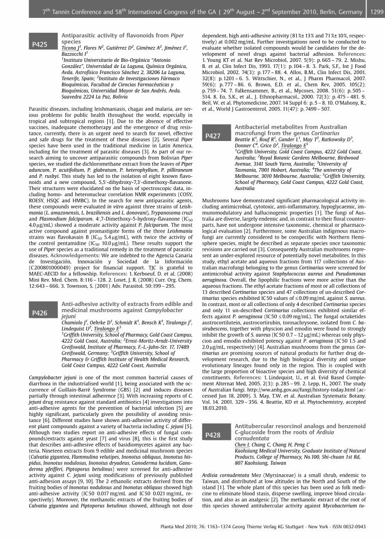
P425Antiparasitic activity of flavonoids from PiperspeciesTicona J1, Flores N2, Guti�rrez D2, Gim�nez A2, Jim�nez I1,Bazzocchi I1
1Instituto Universitario de Bio-Orgnica “AntonioGonzlez”, Universidad de La Laguna, Qu�mica Orgnica,Avda. Astrof�sico Francisco Snchez 2, 38206 La Laguna,Tenerife, Spain; 2Instituto de Investigaciones FrmacoBioqu�micas, Facultad de Ciencias Farmac�uticas yBioqu�micas, Universidad Mayor de San Andr�s, Avda.Saavedra 2224 La Paz, Bolivia
Parasitic diseases, including leishmaniasis, chagas and malaria, are ser-ious problems for public health throughout the world, especially intropical and subtropical regions [1]. Due to the absence of effectivevaccines, inadequate chemotherapy and the emergence of drug resis-tance, currently, there is an urgent need to search for novel, effectiveand safe drugs for the treatment of these diseases [2]. Several Piperspecies have been used in the traditional medicine in Latin America,including for the treatment of parasitic diseases [3]. As part of our re-search aiming to uncover antiparasitic compounds from Bolivian Piperspecies, we studied the dichloromethane extract from the leaves of Piperaduncum, P. acutifolium, P. glabratum, P. heterophyllum, P. pilliraneumand P. rusbyi. This study has led to the isolation of eight known flavo-noids and a new compound, 5,5’-dihydroxy-7,3’-dimethoxy-flavanone.Their structures were elucidated on the basis of spectroscopic data, in-cluding homo- and heteronuclear correlation NMR experiments (COSY,ROESY, HSQC and HMBC). In the search for new antiparasitic agents,these compounds were evaluated in vitro against three strains of Leish-mania (L. amazonensis, L. braziliensis and L. donovani), Trypanosoma cruziand Plasmodium falciparum. 4’,7-Dimethoxy-5-hydroxy-flavanone (IC50
4.0 mg/mL) showed a moderate activity against P. falciparum. The mostactive compound against promastigote forms of the three Leishmaniastrains was flavokavain B (IC50 5.4 mg/mL), with twice the activity ofthe control pentamidine (IC50 10.0 mg/mL). These results support theuse of Piper species as a traditional remedy in the treatment of parasiticdiseases. Acknowledgements: We are indebted to the Agencia Canariade Investigaci n, Innovaci n y Sociedad de la Informaci n(C 200801000049) project for financial support. TJC is grateful toMAEC-AECID for a fellowship. References: 1. Kerboeuf, D. et al. (2008)Mini Rev. Med. Chem. 8:116 – 128. 2. Loset, J. R. (2008) Curr. Org. Chem.12:643 – 666. 3. Townson, S. (2001) Adv. Parasitol. 50:199 – 295.
P426Anti-adhesive activity of extracts from edible andmedicinal mushrooms against CampylobacterjejuniChamiolo J1, Oehrke D1, Schmidt K1, Bensch K1, Tiralongo J1,Lindequist U2, Tiralongo E3
1Griffith University, School of Pharmacy, Gold Coast Campus,4222 Gold Coast, Australia; 2Ernst-Moritz-Arndt-UniversityGreifswald, Institute of Pharmacy, F.-L.-Jahn-Str. 17, 17489Greifswald, Germany; 3Griffith University, School ofPharmacy & Griffith Institute of Health Medical Research,Gold Coast Campus, 4222 Gold Coast, Australia
Campylobacter jejuni is one of the most common bacterial causes ofdiarrhoea in the industrialised world [1], being associated with the oc-currence of Guillain-Barr� Syndrome (GBS) [2] and induces diseasespartially through intestinal adherence [3]. With increasing reports of C.jejuni drug resistance against standard antibiotics [4] investigations intoanti-adhesive agents for the prevention of bacterial infection [5] arehighly significant, particularly given the possibility of avoiding resis-tance [6]. Different studies have shown anti-adhesive activity of differ-ent plant compounds against a variety of bacteria including C. jejuni [5].Although two studies report on anti-adhesive effects of fungal com-pounds/extracts against yeast [7] and virus [8], this is the first studythat describes anti-adhesive effects of basidiomycetes against any bac-teria. Nineteen extracts from 9 edible and medicinal mushroom species(Calvatia gigantea, Flammulina velutipes, Inonotus obliquus, Inonotus his-pidus, Inonotus nodulosus, Inonotus dryadeus, Ganoderma lucidum, Gano-derma pfeifferi, Piptoporus betulinus) were screened for anti-adhesiveactivity against C. jejuni using modifications of previously publishedanti-adhesion assays [9, 10]. The 2 ethanolic extracts derived from thefruiting bodies of Inonotus nodulosus and Inonotus obliquus showed highanti-adhesive activity (IC 50 0.017 mg/mL and IC 50 0.021 mg/mL, re-spectively). Moreover, the methanolic extracts of the fruiting bodies ofCalvatia gigantea and Piptoporus betulinus showed, although not dose
dependent, high anti-adhesive activity (81%€ 13% and 71%€ 10%, respec-tively) at 0.002 mg/mL. Further investigations need to be conducted toevaluate whether isolated compounds would be candidates for the de-velopment of novel drugs against bacterial adhesion. References:1. Young KT et al. Nat Rev Microbiol, 2007. 5(9): p. 665 – 79. 2. Mishu,B. et al. Clin Infect Dis, 1993. 17(1): p.104 – 8. 3. Park, S.F., Int J FoodMicrobiol, 2002. 74(3): p.177 – 88. 4. Allos, B.M., Clin Infect Dis, 2001.32(8): p.1201 – 6. 5. Wittschier, N., et al., J Pharm Pharmacol, 2007.59(6): p. 777 – 86. 6. Brown, E.D. et al., Chem Rev, 2005. 105(2):p. 759 – 74. 7. Falkensammer, B., et al., Mycoses, 2008. 51(6): p. 505 –514. 8. Eo, S.K., et al., J. Ethnopharmacol., 2000. 72(3): p. 475 – 481. 9.Beil, W. et al. Phytomedicine, 2007. 14 Suppl 6: p. 5 – 8. 10. O’Mahony, R.,et al., World J Gastroenterol, 2005. 11(47): p. 7499 – 507.
P427Antibacterial metabolites from Australianmacrofungi from the genus CortinariusBeattie K1, Rouf R1, Gander L1, May T2, Ratkowsky D3,Donner C4, Grice D1, Tiralongo E5
1Griffith University, Gold Coast Campus, 4222 Gold Coast,Australia; 2Royal Botanic Gardens Melbourne, BirdwoodAvenue, 3141 South Yarra, Australia; 3University ofTasmania, 7001 Hobart, Australia; 4The university ofMelbourne, 3010 Melbourne, Australia; 5Griffith University,School of Pharmacy, Gold Coast Campus, 4222 Gold Coast,Australia
Mushrooms have demonstrated significant pharmacological activity in-cluding antimicrobial, cytotoxic, anti-inflammatory, hypoglycaemic, im-munomodulatory and hallucinogenic properties [1]. The fungi of Aus-tralia are diverse, largely endemic and, in contrast to their floral counter-parts, have not undergone intensive taxonomic, chemical or pharmaco-logical evaluation [2]. Furthermore, some Australian indigenous macro-fungi are currently considered to be conspecific with Northern Hemi-sphere species, might be described as separate species once taxonomicrevisions are carried out [3]. Consequently Australian mushrooms repre-sent an under-explored resource of potentially novel metabolites. In thisstudy, ethyl acetate and aqueous fractions from 117 collections of Aus-tralian macrofungi belonging to the genus Cortinarius were screened forantimicrobial activity against Staphylococcus aureus and Pseudomonasaeruginosa. Overall, the lipophilic fractions were more active than theaqueous fractions. The ethyl acetate fractions of most or all collections of13 described Cortinarius species and 47 collections of un-described Cor-tinarius species exhibited IC 50 values of £0.09 mg/mL against S. aureus.In contrast, most or all collections of only 4 described Cortinarius speciesand only 11 un-described Cortinarius collections exhibited similar ef-fects against P. aeruginosa (IC50 £ 0.09 mg/mL). The fungal octaketidesaustrocortilutein, austrocortirubin, torosachrysone, isolated from C. ba-sirubescens, together with physcion and emodin were found to stronglyinhibit the growth of S. aureus (IC50 0.7 – 12 mg/mL) whereas only phys-cion and emodin exhibited potency against P. aeruginosa (IC50 1.5 and2.0 mg/mL, respectively) [4]. Australian mushrooms from the genus Cor-tinarius are promising sources of natural products for further drug de-velopment research, due to the high biological diversity and uniqueevolutionary lineages found only in the region. This is coupled withthe large proportion of bioactive species and high diversity of chemicalconstituents. References: 1. Lindequist, U., et al. Evid Based Comple-ment Alternat Med, 2005. 2(3): p. 285 – 99. 2. Lepp, H., 2007. The studyof Australian fungi. http://ww.anbg.gov.au/fungi/history-today.html (ac-cessed Jun 18, 2009). 3. May, T.W. et al. Australian Systematic Botany.Vol. 14. 2001. 329 – 356. 4. Beattie, KD et al. Phytochemistry, accepted18.03.2010.
P428Antitubercular resorcinol analogs and benzenoidC-glucoside from the roots of ArdisiacornudentataChen I, Chang C, Chang H, Peng CKaohsiung Medical University, Graduate Institute of NaturalProducts, College of Pharmacy, No.100, Shi-chuan 1st Rd,807 Kaohsiung, Taiwan
Ardisia cornudentata Mez (Myrsinaceae) is a small shrub, endemic toTaiwan, and distributed at low altitudes in the North and South of theisland [1]. The whole plant of this species has been used as folk medi-cine to eliminate blood stasis, disperse swelling, improve blood circula-tion, and also as an analgesic [2]. The methanolic extract of the root ofthis species showed antitubercular activity against Mycobacterium tu-
Planta Med 2010; 76: 1163–1374 Georg Thieme Verlag KG Stuttgart · New York · ISSN 0032-0943
12997th Tannin Conference and 58th International Congress of the GA | 29th August – 2nd September 2010, Berlin, Germany
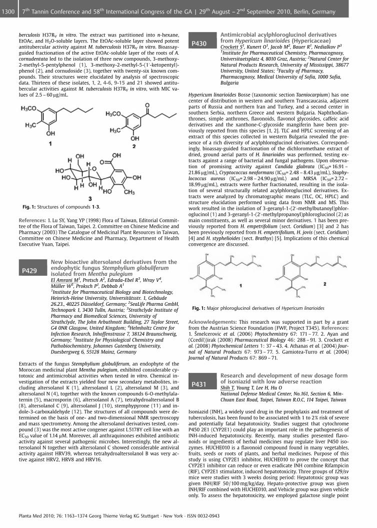
berculosis H37RV in vitro. The extract was partitioned into n-hexane,EtOAc, and H2O-soluble layers. The EtOAc-soluble layer showed potentantitubercular activity against M. tuberculosis H37RV in vitro. Bioassay-guided fractionation of the active EtOAc-soluble layer of the roots of A.cornudentata led to the isolation of three new compounds, 3-methoxy-2-methyl-5-pentylphenol (1), 3-methoxy-2-methyl-5-(1’-ketopentyl)-phenol (2), and cornudoside (3), together with twenty-six known com-pounds. Their structures were elucidated by analysis of spectroscopicdata. Thirteen of these isolates, 1, 2, 4-6, 9-15 and 21 showed antitu-bercular activities against M. tuberculosis H37RV in vitro, with MIC va-lues of 2.5 – 60 mg/mL.
Fig. 1: Structures of compounds 1-3.
References: 1. Lu SY, Yang YP (1998) Flora of Taiwan, Editorial Commit-tee of the Flora of Taiwan, Taipei. 2. Committee on Chinese Medicine andPharmacy (2003) The Catalogue of Medicinal Plant Resources in Taiwan,Committee on Chinese Medicine and Pharmacy, Department of HealthExecutive Yuan, Taipei.
P429New bioactive altersolanol derivatives from theendophytic fungus Stemphylium globuliferumisolated from Mentha pulegiumEl Amrani M1, Pretsch A2, Edrada-Ebel R3, Wray V4,M�ller W5, Proksch P1, Debbab A1
1Institute for Pharmaceutical Biology and Biotechnology,Heinrich-Heine University, Universit�tsstr. 1, Geb�ude26.23., 40225 D�sseldorf, Germany; 2SeaLife Pharma GmbH,Technopark 1, 3430 Tulln, Austria; 3Strathclyde Institute ofPharmacy and Biomedical Sciences, University ofStrathclyde, The John Arbuthnott Building, 27 Taylor Street,G4 0NR Glasgow, United Kingdom; 4Helmholtz Centre forInfection Research, Inhoffenstrasse 7, 38124 Braunschweig,Germany; 5Institute for Physiological Chemistry andPathobiochemistry, Johannes Gutenberg University,Duesbergweg 6, 55128 Mainz, Germany
Extracts of the fungus Stemphylium globuliferum, an endophyte of theMoroccan medicinal plant Mentha pulegium, exhibited considerable cy-totoxic and antimicrobial activities when tested in vitro. Chemical in-vestigation of the extracts yielded four new secondary metabolites, in-cluding altersolanol K (1), altersolanol L (2), altersolanol M (3), andaltersolanol N (4), together with the known compounds 6-O-methylala-ternin (5), macrosporin (6), altersolanol A (7), tetrahydroaltersolanol B(8), altersolanol C (9), altersolanol J (10), stemphypyrone (11) and in-dole-3-carboxaldehyde (12). The structures of all compounds were de-termined on the basis of one- and two-dimensional NMR spectroscopyand mass spectrometry. Among the altersolanol derivatives tested, com-pound (3) was the most active congener against L 5178Y cell line with anEC50 value of 1.14 mM. Moreover, all anthraquinones exhibited antibioticactivity against several pathogenic microbes. Interestingly, the new al-tersolanol N together with altersolanol C showed considerable antiviralactivity against HRV39, whereas tetrahydroaltersolanol B was very ac-tive against HRV2, HRV8 and HRV16.
P430Antimicrobial acylphloroglucinol derivativesfrom Hypericum linarioides (Hypericaceae)Crockett S1, Kunert O1, Jacob M2, Bauer R1, Nedialkov P3
1Institute for Pharmaceutical Chemistry, Pharmacognosy,Universitaetsplatz 4, 8010 Graz, Austria; 2Natural Center forNatural Products Research, University of Mississippi, 38677University, United States; 3Faculty of Pharmacy,Pharmacognosy, Medical University of Sofia, 1000 Sofia,Bulgaria
Hypericum linarioides Bosse (taxonomic section Taeniocarpium) has onecenter of distribution in western and southern Transcaucasia, adjacentparts of Russia and northern Iran and Turkey, and a second center insouthern Serbia, northern Greece and western Bulgaria. Naphthodian-thrones, simple anthrones, flavonoids, flavonol glycosides, caffeic acidderivatives and the xanthone-C-glycoside mangiferin have been pre-viously reported from this species [1, 2]. TLC and HPLC screening of anextract of this species collected in western Bulgaria revealed the pre-sence of a rich diversity of acylphloroglucinol derivatives. Correspond-ingly, bioassay-guided fractionation of the dichloromethane extract ofdried, ground aerial parts of H. linarioides was performed, testing ex-tracts against a range of bacterial and fungal pathogens. Upon observa-tion of promising activity against Candida glabrata (IC50= 16.91 –21.86 mg/mL), Cryptococcus neoformans (IC50= 2.48 – 8.43 mg/mL), Staphy-lococcus aureus (IC50= 2.98 – 24.90 mg/mL) and MRSA (IC50= 2.72 –18.99 mg/mL), extracts were further fractionated, resulting in the isola-tion of several structurally related acylphloroglucinol derivatives. Ex-tracts were analyzed by chromatographic means (TLC, OC, HPLC) andstructure elucidation performed using data from NMR and MS. Thiswork resulted in the isolation of 3-geranyl-1-(2’-methylbutanoyl)phlor-oglucinol (1) and 3-geranyl-1-(2’-methylpropanoyl)phloroglucinol (2) asmain constituents, as well as several minor derivatives. 1 has been pre-viously reported from H. empetrifolium (sect. Coridium) [3] and 2 hasbeen previously reported from H. empetrifolium, H. jovis (sect. Coridium)[4] and H. styphelioides (sect. Brathys) [5]. Implications of this chemicalconvergence are discussed.
Fig. 1: Major phloroglucinol derivatives of Hypericum linarioides
Acknowledgements: This research was supported in part by a grantfrom the Austrian Science Foundation (FWF, Project T345). References:1. �melcerovic et al. (2006) Phytochemistry 67: 171 – 77. 2. Ayan and(Ccedil))irak (2008) Pharmaceutical Biology 46: 288 – 91. 3. Crockett etal. (2008) Phytochemical Letters 1: 37 – 43. 4. Athanas et al. (2004) Jour-nal of Natural Products 67: 973 – 77. 5. Gamiotea-Turro et al. (2004)Journal of Natural Products 67: 869 – 71.
P431Research and development of new dosage formof isoniazid with low adverse reactionShih T, Young T, Lee H, Hu ONational Defense Medical Center, No.161, Section 6, Min-Chuan East Road, Taipei, Taiwan R.O.C, 114 Taipei, Taiwan
Isoniazid (INH), a widely used drug in the prophylaxis and treatment oftuberculosis, has been found to be associated with 1 to 2% risk of severeand potentially fatal hepatotoxicity. Studies suggest that cytochromeP450 2E1 (CYP2E1) could play an important role in the pathogenesis ofINH-induced hepatotoxicity. Recently, many studies presented flavo-noids or ingredients of herbal medicines may regulate liver P450 iso-zymes. HUCHE010 is a flavonoid compound found in many vegetables,fruits, seeds or roots of plants, and herbal medicines. Purpose of thisstudy is using CYP2E1 inhibitor, HUCHE010 to prove the concept thatCYP2E1 inhibitor can reduce or even eradicate INH combine Rifampicin(RIF), CYP2E1 stimulator, induced hepatotoxicity. Three groups of 129/svmice were studies with 3 weeks dosing period: Hepatotoxic group wasgiven INH/RIF 50/100 mg/kg/day, Hepato-protective group was givenINH/RIF combined with HUCHE010, and Vehicle group was given vehicleonly. To assess the hepatotoxicity, we employed galactose single point
Planta Med 2010; 76: 1163–1374 Georg Thieme Verlag KG Stuttgart · New York · ISSN 0032-0943
1300 7th Tannin Conference and 58th International Congress of the GA | 29th August – 2nd September 2010, Berlin, Germany
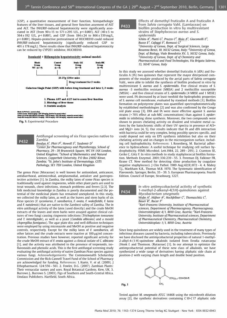
(GSP), a quantitative measurement of liver function, histopathologicfeatures of the liver tissues, and general liver function assement of ASTand ALT. The INH/RIF-induced hepatotoxicity were significantly asso-ciated in AST (from 90 € 15 to 571 € 295 U/L, p < 0.001), ALT (40 € 5 to364 € 192 U/L, p < 0.001), and GSP (from 184 € 24 to 866 € 339 mg/L,p < 0.001). Hepato-protective pretreatment of HUCHE010 could reducedINH/RIF-induced hepatotoxicity (significantly reduced GSP to401 € 178 mg/L). These results show that INH/RIF-induced hepatotoxicitycan be reduced by CYP2E1 inhibitor, HUCHE010.
Fig. 1: Experimental Procedure
P432Antifungal screening of six Ficus species native toZambiaBwalya A1, Phiri P2, Howell S3, Tasdemir D1
1Center for Pharmacognosy and Phytotherapy, School ofPharmacy, 29 – 39 Brunswick Square, WC 1N 1AX London,United Kingdom; 2School of Mathematics and NaturalSciences, Copperbelt University, P.O Box 21692 Kitwe,Zambia; 3St. John’s Institute of Dermatology, GSTSPathology, SE1 7EH London, United Kingdom
The genus Ficus (Moraceae) is well known for antioxidant, anticancer,antidiarrhoeal, antimicrobial, antiplasmodial, antiulcer and gastropro-tective activities [1]. In Zambia, the milky latex of some Ficus species istraditionally used against ringworms, while other plant parts are used totreat wounds, chest infections, stomach problems and fevers [2,3]. Thefolk medicinal knowledge in Zambia is poorly documented and the po-tential of the medicinal plants has remained unexplored. In this study,we collected the milky latex, as well as the leaves and stem barks of sixFicus species (F. sycomorus, F. sansibarica, F. ovata, F. wakefieldii, F. luteaand F. natalensis) that are native to the Zambezi valley of Zambia. The invitro antifungal activity of the latex (used directly) and the crude MeOHextracts of the leaves and stem barks were assayed against clinical cul-tures of two fungi causing ringworm infections (Trichophyton tonsuransand T. interdigitale), as well as a yeast (Candida albicans) and a mould(Aspergillus fumigatus). The agar plate disc and well diffusion techniqueswere employed by using miconazole and MeOH as positive and negativecontrols, respectively. Except for the milky latex of F. sansibarica, allother latices and the crude extracts were inactive at 100 mg/ml concen-tration. Previous studies have however, reported significant activity forthe crude MeOH extract of F. ovata against a clinical isolate of C. albicans[1], and the activity was attributed to the presence of terpenoids, iso-flavonoids and phenolic acids. This is the first antifungal screening studyevaluating the antifungal activity of native Zambian Ficus species againstvarious fungi. Acknowledgements: The Commonwealth ScholarshipCommission and the Rick-Cannell Travel Fund of the School of Pharmacyare acknowledged for funding. References: 1. Kuete, V. et al. (2009). JEthopharmacol. 124:556 – 561. 2. Fowler, D.G. (2007). Zambian Plants:Their vernacular names and uses. Royal Botanical Gardens. Kew, UK. 3.Burrows J., Burrows S. (2003). Figs of Southern and South-Central Africa.Umdaus Publishers. Hartfield, South Africa.
P433Effects of demethyl fruticulin A and fruticulin Afrom Salvia corrugata Vahl. (Lamiaceae) onbiofilm production in vitro by multiresistantstrains of Staphylococcus aureus and S.epidermidisSchito A1, Piatti G1, Pruzzo C2, Bisio A3, Giacomelli E3,Russo E3, Cafaggi S3, Romussi G3
1University of Genoa, Dept. of Surgical Sciences, LargoRosanna Benzi, 10, 16132 Genoa, Italy; 2University of Genoa,Dept. of Biology, Viale Benedetto XV, 5, 16132 Genoa, Italy;3University of Genoa, Dept. of of Chemistry andPharmaceutical and Food Technologies, Via Brigata Salerno,13, 16147 Genoa, Italy
In this study we assessed whether demethyl fruticulin A (dfA) and fru-ticulin A (fA) two quinones that represent the mayor diterpenoid com-ponents of the exudate produced by the aerial parts of Salvia corrugataVahl., were able to inhibit the synthesis of biofilm produced in vitro bymultiresistant S. aureus and S. epidermidis. Five clinical strains of S.aureus -3 methicillin resistant (MRSA) and 2 methicillin susceptible(MSSA) – and five clinical strains of S. epidermidis (4 MRSE and 1 MSSE)were used. FA decreased by at least twofold the hydrophobic propertiesof S. aureus cell membrane, evaluated by standard methods [1]. Biofilmformation on polystyrene plates was quantified spectrophotometricallyby established methodologies [2] and was also confirmed by the Congored plate assay [3]. DfA and fA were more effective against S. aureusstrains (> 70% effect at sub-MIC concentrations) than against S. epider-midis in inhibiting slime synthesis. Moreover, the two compounds wereshown to posses chelating activity on divalent and trivalent metal ca-tions by the batochromic shifts of their UV spectra in presence of Al3+and Mg2+ ions [4, 5]. Our results indicate that fA and dfA interactionwith bacteria could be very complex, being possibly species-specific, andcould depend not only on EPS synthesis inhibition but also on theirchelating activity and on changes in the microorganism surface, includ-ing cell hydrophobicity. References: 1. Rosenberg, M. Bacterial adher-ence to hydrocarbons: A useful technique for studying cell surface hy-drophobicity. FEMS Microbiol. Lett.1984, 22, 289 – 295). 2. Cramton SE,Gerke C, G�tz F. In vitro methods to study staphylococcal biofilm forma-tion. Methods Enzymol. 2001;336:239 – 55. 3. Freeman DJ, Falkiner FR,Keane CT. New method for detecting slime production by coagulasenegative staphylococci. J Clin Pathol. 1989 Aug;42(8):872 – 4. 4. MabryT.J., Markham K.R., Thomas M.B. 1970. The Systematic identification ofFlavonoids. Springer, Berlin, 35 – 39. 5. European Pharmacopoeia. FourthEdition. Council of Europe, Strasbourg. 1227.
P434In vitro antimycobacterial activity of synthetic1-methyl-2-alkenyl-4(1H)-quinolones againstMycobacterium smegmatisWube A1, H�fner A2, Hochfellner C1, Thomaschitz C1,Bauer R1, Bucar F1
1Karl-Franzens University, Institute of Pharmaceuticalsciences, Department of Pharmacognosy, Pharmacognosy,Universit�tsplatz 4/1, 8010 Graz, Austria; 2Karl-FranzensUniversity, Institute of Pharmaceutical sciences, Departmentof Pharmaceutical Chemistry, Pharmaceutical Chemistry,Universit�tzplatz 1/1, 8010 Graz, Austria
Since long quinolones are widely used in the treatment of many types ofinfectious diseases caused by bacteria, including tuberculosis. Previouslywe have disclosed the antimycobacterial properties of natural 1-methyl-2-alkyl-4-(1 H)-quinolone alkaloids isolated from Evodia rutaecarpa(Hook f. and Thomson. (Rutaceae) [1]. In our attempt to optimize theantimycobacterial potential of these new class of alkaloids, we havesynthesized a wide range of derivatives having aliphatic side chain atposition-2 with varying chain length and double bond position.
Fig. 1
Tested against M. smegmatis ATCC 14468 using the microbroth dilutionassay [2], the synthetic derivatives containing C 10-C 17 aliphatic side
Planta Med 2010; 76: 1163–1374 Georg Thieme Verlag KG Stuttgart · New York · ISSN 0032-0943
13017th Tannin Conference and 58th International Congress of the GA | 29th August – 2nd September 2010, Berlin, Germany

chain showed more potency (MIC’s values 0.5 – 16 mg/mL) compared tothe natural products and even the standard drugs ethambutol and iso-niazid. The structure-activity relationship study revealed that the size ofthe aliphatic side chain is essential for the observed antimycobacterialeffects. Acknowledgements: This work is financially supported by Aus-trian Science Fund (FWF) Project No. P21152-B18. References: 1. Adams,M. et al (2004) Planta Med. 70; 904 – 908. 2. Wube, AA. Et al. (2005)Phytochemistry 66; 2309 – 2315.
P435Isolation of curcacycline A from latex of Jatrophacurcas and its antibiotic and cytotoxic effectInsanu M1, Anggadireja J2, Kayser O1
1University of Groningen, Pharmaceutical Biology, AntoniusDeusinglaan 1, 9713 Groningen, Netherlands; 2Agency forthe Asessment Aplication of Technology, Jl.M.H. ThamrinJakarta, Indonesia
Euphorbiaceae are known as a rich source of cyclic peptides from higherplants. One of cyclic peptides was isolated from the latex of Jatrophacurcas, namely curcacycline A (C 37H66N8O9) [1]. In the past differenttechniques have been applied to determine the structure of curcacyclineA. In a new approach the structure of the isolated curcacycline A con-firmed by more sophisticated bioanalytical techniques (HPLC-MS, 2D-NMR, COSY, APT, HSQC, IR). For the first time the structure was con-firmed by a full synthetic approach and first biological assays have beencarried out to shed light on the pharmacological activity of curcacyclineA as representative of a rather unknown group of cylicpeptides. Theanalytical data indicate that curcacycline A consists of 8 amino acids,while ESI-MS data showed complete sequencing of amino acid c(Gly-Leu-Leu-Leu-Gly-Thr-Val-Leu-Leu-Gly). Screening of antibiotic activityof curcacycline A were done against several bacteria (Bacillus subtilis,Staphyloccus aureus, Eschericia coli, Pseudomonas aeruginosa) and fungi(Candida albicans) using the agar diffussion method. Curcacycline A in-hibits the growth of B. subtillis and P. aeruginosa. Cytotoxic effect wastested to two cell lines, human ovarian cancer cell-line OVCAR-3(ATCC�) and human colon cancer cell-line (Colo205). Curcacycline Adecreased the level of OVCAR 3 at a concentration of 1 mg/mL, whileno effect was found on Colo205 (ATCC�). Curcacycline A had no muta-genic activity in the non-activated and activated AMES test. References:1. van den Berg, A. J., S. F. Horsten, et al. (1995). FEBS Lett 358(3): 215 – 8.
P436Secondary metabolites from the root of Ehretialongiflora and their antitubercular activityChien Y, Peng C, Lin C, Chen I, Tsai IKaohsiung Medical University, Graduate Insituent ofNatural Products, College of Pharmacy, No.100, Shi-chuan1st Rd, 807 Kaohsiung, Taiwan
Ehretia longiflora Champ. ex Benth. (Boraginaceae) is a medium sizeddeciduous tree, distributed in south China, Indochina, and in forestsfrom low elevations to 750 m of Taiwan [1]. The methanolic extract ofthe root of this plant showed antitubercular activity against Mycobacter-ium tuberculosis H37Rv in vitro. The aim of this study is the isolation ofchemical constituents and antitubercular activity. Bioassay-guided frac-tionation of the ethyl acetate-soluble layer from the root of this speciesled to the isolation of four new compounds, including one quinonoid:ehretiquinone (1); one triterpenoid: ehretiolide (2); one alkaloid: ehre-tilonine (3); one acetamide: ehretilonamide (4), together with nineteenknown compounds, of which three compounds, comprising of 1-(4-methoxyphenyl)-4-phenylbutane-2,3-diol (5), ehretiquinonone (6),(2S,3S)-1,4-bis(4-methoxyphenyl)butan2,3-diol (7), were first isolatedfrom nature. The structures of these isolates were elucidated by spectralanalysis. Among these isolates, 1, 5 and 8 exhibited antitubercular activ-ity against M. tuberculosis H37Rv in vitro, showing MIC values of 25.0,30.0 and 50.0 mg/mL, respectively. The clinically use antitubercularagent, ethambutol, was used as a positive control. Antitubercular activ-ity assay of the isolates are still in progress.
Fig. 1: Compounds 1-7
References: 1. Hsiao JY, Liu HY (1998) Flora of Taiwan, Editorial Com-mittee of the Flora of Taiwan, Taipei.
P437Kaempferol rhamnosides from Bryophyllumpinnatum, a medicinal plant used againstinfectious diseases and as analgesic in Mbouda,CameroonTatsimo Ndendoung J1, Tane P2, Csupor D3, Forgo P3,Hohmann J3
1Higher Teachers’ Training College, university of Maroua,Depatement of chemistry, Box 55, 237 Maroua, Cameroon;2University of Dschang, Department of chemistry, Box 67,237 Dschang, Cameroon; 3University of Szeged, Departmentof pharmacognosy, E�tv�s u. 6, 6720 Szeged, Hungary
An ethnobotanical survey was made on plants used in the treatment ofinfectious diseases in Mbouda subdivision, Cameroon. According to oursurvey, one of the most important medicinal plants in that area is Bryo-phyllum pinnatum (Lank.) Oken (Crassulaceae), a succulent plant nativeto Africa. B. pinnatum (leaves or whole plant) was found to be wellknown and was used against blennorrhoea, syphilis, jaundice, candidia-sis and for the treatment of others ailments such as dysmenorrhoea,external ulcers, burns, convulsions and as analgesic. In order to identifythe biologically active compounds of the plant and to confirm or infirmits ethnopharmacological claims, phytochemical study of ethyl acetateextract of the whole plant was carried out. As a result of this kaempferolrhamnosides, kaempferol 3,7-O-bis-a-L-rhamnopyranoside (kaempferi-trin) [1], kaempferol 3-O-a-L-(3-acetyl)rhamnoside-7-O-a-L-rhamno-pyranoside [2], kaempferol 3-O-a-L-rhamnoside (afzelin) [1] andkaempferol 7-O-a-L-rhamnoside (a-rhamnoisorobin) [1] were isolatedand identified by extensive NMR and MS studies. All compounds weredescribed for the first time in this species. These isolates are reported inother plant species to possess antioxidant, antinociceptive and anti-in-flammatory activities [3, 4], therefore the kaempferol rhamnosides of B.
Planta Med 2010; 76: 1163–1374 Georg Thieme Verlag KG Stuttgart · New York · ISSN 0032-0943
1302 7th Tannin Conference and 58th International Congress of the GA | 29th August – 2nd September 2010, Berlin, Germany
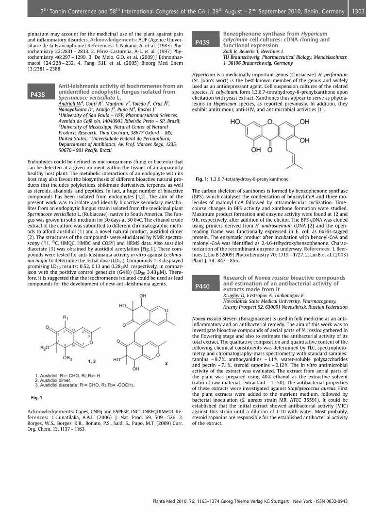
pinnatum may account for the medicinal use of the plant against painand inflammatory disorders. Acknowledgements: AUF (Agence Univer-sitaire de la Francophonie) References: 1. Nakano, A. et al. (1983) Phy-tochemistry 22:2831 – 2833. 2. P�rez-Castorena, A-L. et al. (1997) Phy-tochemistry 46:297 – 1299. 3. De Melo, G.O. et al. (2009) J Ethnophar-macol 124:228 – 232. 4. Fang, S.H. et al. (2005) Bioorg Med Chem13:2381 – 2388.
P438Anti-leishmania activity of isochromenes from anunidentified endophytic fungus isolated fromSpermacoce verticillata L.Andrioli W1, Conti R1, Manfrim V1, Toledo J1, Cruz ff1,Nanayakkara D2, Araffljo J3, Pupo M1, Bastos J1
1University of Sao Paulo – USP, Pharmaceutical Sciences,Avenida do Caf� s/n, 14040903 Ribeir¼o Preto – SP, Brazil;2University of Mississippi, Natural Center of NaturalProducts Research, Thad Cochran, 38677 Oxford – MS,United States; 3Universidade Federal do Pernambuco,Departament of Antibiotics, Av. Prof. Moraes Rego, 1235,50670 – 901 Recife, Brazil
Endophytes could be defined as microorganisms (fungi or bacteria) thatcan be detected at a given moment within the tissues of an apparentlyhealthy host plant. The metabolic interactions of an endophyte with itshost may also favour the biosynthesis of different bioactive natural pro-ducts that includes polyketides, shikimate derivatives, terpenes, as wellas steroids, alkaloids, and peptides. In fact, a huge number of bioactivecompounds has been isolated from endophytes [1,2]. The aim of thepresent work was to isolate and identify bioactive secondary metabo-lites from an endophytic fungus strain isolated from the medicinal plantSpermacoce verticillata L. (Rubiaceae), native to South America. The fun-gus was grown in solid medium for 30 days at 30 �¤C. The ethanol crudeextract of the culture was submitted to different chromatographic meth-ods to afford austidiol (1) and a novel natural product, austidiol dimer(2). The structures of the compounds were elucidated by NMR spectro-scopy (1H, 13C, HMQC, HMBC and COSY) and HRMS data. Also austidioldiacetate (3) was obtained by austidiol acetylation [Fig.1]. These com-pounds were tested for anti-leishmania activity in vitro against Leishma-nia major to determine the lethal dose (LD50). Compounds 1-3 displayedpromising LD50 results: 0.52, 0.13 and 0.28 mM, respectively, in compar-ison with the positive control geneticin (G418) (LD50 3.43mM). There-fore, it is suggested that the isochromenes isolated could be used as leadcompounds for the development of new anti-leishmania agents.
Fig. 1
Acknowledgements: Capes, CNPq and FAPESP, INCT-INBEQUIMeDI. Re-ferences: 1. Gunatilaka, A.A.L. (2006). J. Nat. Prod. 69, 509 – 526. 2.Borges, W.S., Borges, K.B., Bonato, P.S., Said, S., Pupo, M.T. (2009) Curr.Org. Chem. 13, 1137 – 1163.
P439Benzophenone synthase from Hypericumcalycinum cell cultures: cDNA cloning andfunctional expressionZodi R, Beuerle T, Beerhues LTU Braunschweig, Pharmaceutical Biology, Mendelssohnstr.1, 38106 Braunschweig, Germany
Hypericum is a medicinally important genus (Clusiaceae). H. perforatum(St. John’s wort) is the best-known member of the genus and widelyused as an antidepressant agent. Cell suspension cultures of the relatedspecies, H. calycinum, form 1,3,6,7-tetrahydroxy-8-prenylxanthone uponelicitation with yeast extract. Xanthones thus appear to serve as phytoa-lexins in Hypericum species, as reported previously. In addition, theyexhibit antitumour, anti-HIV, and antimicrobial activities [1].
Fig. 1: 1,3,6,7-tetrahydroxy-8-prenylxanthone
The carbon skeleton of xanthones is formed by benzophenone synthase(BPS), which catalyses the condensation of benzoyl-CoA and three mo-lecules of malonyl-CoA followed by intramolecular cyclization. Time-course changes in BPS activity and xanthone formation were studied.Maximum product formation and enzyme activity were found at 12 and9 h, respectively, after addition of the elicitor. The BPS cDNA was clonedusing primers derived from H. androsaemum cDNA [2] and the open-reading frame was functionally expressed in E. coli as 6xHis-taggedprotein. The enzymatic product after incubation with benzoyl-CoA andmalonyl-CoA was identified as 2,4,6-trihydroxybenzophenone. Charac-terization of the recombinant enzyme is underway. References: 1. Beer-hues L, Liu B (2009) Phytochemistry 70: 1719 – 1727. 2. Liu B et al. (2003)Plant J. 34: 847 – 855.
P440Research of Nonea rossica bioactive compoundsand estimation of an antibacterial activity ofextracts made from itKruglov D, Evstropov A, Tonkonogov ENovosibirsk State Medical University, Pharmacognosy,Krasny Prospect 52, 630091 Novosibirsk, Russian Federation
Nonea rossica Steven. (Boraginaceae) is used in folk medicine as an anti-inflammatory and an antibacterial remedy. The aim of this work was toinvestigate bioactive compounds of aerial parts of N. rossica gathered inthe flowering stage and also to estimate the antibacterial activity of itstotal extract. The qualitative composition and quantitative content of thefollowing chemical constituents was determined by TLC, spectrophoto-metry and chromatography-mass spectrometry with standard samples:tannins – 9,7%, anthocyanidins – 1,1%, water-soluble polysaccharidesand pectin – 7,1%, steroid saponins – 0,12%. The in vitro antimicrobialactivity of the extract was evaluated. The extract from aerial parts ofthe plant was prepared using 40% ethanol as the extractive solvent(ratio of raw material: extractant – 1: 50). The antibacterial propertiesof these extracts were investigated against Staphylococcus aureus. Firstthe plant extracts were added to the nutrient medium, followed bybacterial inoculation (S. aureus strain MR, ATCC 35591). It could beestablished that the initial extract showed antibacterial activity (MIC)against this strain until a dilution of 1:10 with water. Most probably,steroid saponins are responsible for the established antibacterial activityof the extract.
Planta Med 2010; 76: 1163–1374 Georg Thieme Verlag KG Stuttgart · New York · ISSN 0032-0943
13037th Tannin Conference and 58th International Congress of the GA | 29th August – 2nd September 2010, Berlin, Germany

P441Leishmanicide activity of oleanolic acid againstpromastigotes of Leishmania braziliensis andLeishmania chagasiMelo T2, Bonardo V2, Gattass C1, Magri F2, Fiorino P2,Farah V2, Fonteles M2, Delorenzi J2
1Universidade Federal Do Rio De Janeiro, Instituto DeBiof�sica Carlos Chagas Filho, Av. Brigadeiro Trompowsky, S/N – Ilha Do Fund¼o, 21944 – 970 Rio De Janeiro, Brazil;2Universidade Presbiteriana Mackenzie, Centro De CiÞnciasBiolgicas E Da Safflde – Farmcia, Rua Da Consola�¼o, 896,01302 – 907 S¼o Paulo, Brazil
Leishmaniasis is a major worldwide health problem, with around 12 mil-lion people infected and 600 thousand new cases appearing each year. InBrazil, 30 thousand new cases appear annually only in the Northeastregion. Pentavalent antimonals are the first line treatment for leishma-niasis. Disadvantages such as costs, long-term treatment, side effectsand low efficacy against many strains have been reported. Althoughgreat efforts had done along the last century to develop new drugs forleishmaniasis treatment, a drug with high efficacy and low side effects isstill need. Among all strategies used to develop new agents againstleishmaniasis, the research of natural products produced good results.This work investigated the leishmanicidal activity of oleanolic acid (OA),a natural triterpene found in a great variety of plants against promasti-gotes forms of L. braziliensis and L. chagasi. Promastigotes were incu-bated in the presence of different concentrations of OA, which wasadded only once to the cultures. After 3 days at 26 �C, parasite survivalwas estimated by counting viable or motile forms. OA exhibited a goodanti-promastigote activity both for L. braziliensis and L. chagasi, reducingparasite survival in 75 and 94%, respectively, when we used 50 mg/ml ofthe drug. DMSO 1% was used as solvent control. These results suggestthat L. braziliensis is less susceptive than L. chagasi. No toxicity to themacrophages was observed after the treatment with OA, as measured bytheir spreading and adherence to glass surface. These results reinforcethat OA could be a strong candidate for an antileishmanial drug. Ac-knowledgements: Hebron FarmacÞutica, MackPesquisa, PIBIC/Macken-zie, CNPq, FAPERJ
P442Screening of Thamnolia vermicularis var.subuliformis for antimicrobial, antioxidant andcytotoxic activitiesManojlovic N1, Vasiljevic P2, Juskovic M2, Najman S3,Bogdanovic-Duanovic G2, Joksimovic Z1, Milenkovic-Andjelkovic A2
1Medical faculty, University of Kragujevac, Department ofPharmacy, S. Markovica 69, 34000 Kragujevac, Serbia,Republic of; 2Faculty of Sciences, University of Ni,Department of Biology and Ecology, visegradska 33, 18000Ni, Serbia, Republic of; 3Faculty of Medicine, University of Ni,Institute of Biology and Human genetics, visegradska 45,18000 Ni, Serbia, Republic of
Thamnolia vermicularis (Icmadophilaceae) has commonly been used as atea with the local name of “snow tea” in some part of China, where thisspecies of lichens is also used as food [1] and for the treatment ofpsychic disorders, high blood pressure and inflammatory conditions ofthe respiratory tract [2]. In this study, T. vermicularis var. subuliformis iscollected in Serbia and chloroform, ethyl acetate and methanol extractsof this lichen were studied for the antioxidant, antimicrobial and cyto-toxic activities. The antimicrobial activity of the extracts were testedagainst twelve human and plant pathogenic bacteria using a disc-diffu-sion method, and the minimum inhibitory concentration (MIC) values ofeach active extract were determined. The inhibition zones and minimalinhibitory concentration (MIC) values of bacterial strains were in therange of 10 – 42 mm and 50 – 300 mg/ml, respectively. Furthermore, theextracts scavenged 1,1-diphenyl-2-picrylhydrazyl radical (DPPH.), result-ing in IC 50 > 50 mg/ml. All the extracts showed moderate antioxidantactivity comparable with BHT. Viability HeLa cells treated with the ethylacetate and chloroform extracts of T. vermicularis var. subuliformis de-creased with increasing concentrations of these extracts. IC 50 valueafter 24 hours for the ethyl acetate extract was 162.50 € 5.80 mg/ml,while for the chloroform extract this value was 159.32 € 5.16 mg/ml. Allthe tested concentrations of the ethyl acetate and chloroform extractsafter 72 hours of treatment of HeLa cells showed a cytotoxic effect. Themethanolic extract containing the lowest content of baeomycesic andsquamatic acid showed the lowest activity. Acknowledgements: Min-istry of Science and Environment of the Republic of Serbia (Grant No.
142025). References: 1. Hanssen HP, Sch�dler M (1985). Pflanzen in dertradi tionellen chinesischen Medizin. Dtsch Apoth Ztg. 125:1239 – 1243.2. Wang LS, et al. (2001) Bryologist 104:345 – 349.
P443Anti-MRSA and anti-VRE of some traditionalChinese medicinal (TCM) plantsMulyaningsih S, Reichling J, Wink MInstitute of Pharmacy and Molecular Biotechnology,Heidelberg University, Biology, Im Neuenheimer Feld 364,69214 Heidelberg, Germany
Methicillin-resistant Staphylococcus aureus (MRSA) and vancomycin-re-sistant Enterococcus faecalis (VRE) are the most important resistantpathogens which cause nosocomial infections [1 – 2]. Antimicrobial ac-tivity of methanol and dichloromethane extracts of 84 Traditional Chi-nese Medicine (TCM) plants was investigated against 10 strains of MRSAand 5 strains of VRE (references strains and clinical isolates). Eucalyptusglobulus (Myrtaceae) and Glycyrrhiza glabra (Fabaceae) exerted a sub-stantial activity against all strains of MRSA and VRE. All MRSA strainswere inhibited by dichloromethane extracts of G. glabra (MIC range 16 –32 mg/ml) and E. globulus (MIC range 8 – 16 mg/ml). Anti-VRE activity wasalso observed for dichloromethane extracts of G. glabra and E. globuluswith MIC values range 32 – 250 and 8 – 16 mg/ml, respectively. In addi-tion, methanol extracts of E. globulus also exhibited a strong inhibitionagainst MRSA and VRE with MIC values range 16 – 32 mg/ml. The dichlor-omethane extracts were generally more active than methanol extractsagainst the bacteria tested. In this study, we have demonstrated thatsome TCM plants exerted a promising activity as anti-MRSA and anti-VRE agents. References: 1. Jones RN. (2001) Chest 119:397S-404S. 2. MaXX, et al. (2002) Antimicrob Agents Chemother 46:1147 – 1152.
P444Antifungal activity in Juglans nigra green husksRodrigues L1, Gon�alves M2, Amaral M2, Batista M2
1Centro de Estudos FarmacÞuticos, Faculdade de Farmcia,Universidade de Coimbra, Azinhaga de Santa Comba,3000 – 548 Coimbra, Portugal; 2Faculdade de Farmcia andCentro de Estudos FarmacÞuticos, Universidade de Coimbra,Azinhaga de Santa Comba, 3000 – 548 Coimbra, Portugal
Juglans spp. (Juglandaceae) are known to be used in folk medicine totreat a wide range of health disorders. In this work, the activity againstyeasts and dermatophyte strains was assessed from fresh green husks ofJ. nigra. The material was extracted with 70% aqueous ethanol using anUltra-Turrax homogeniser. Subsequently, volatile (VF) and no volatile(NVF) fractions were obtained by extract distillation under vacuumand evaluated for their antifungal activity. Six yeasts (five Candida spp.and Cryptococcus neoformans) and seven dermatophytes (four Trichophy-ton spp., two Microsporum spp. and Epidermophyton floccosum) wereassayed by broth macrodilution methods [1] to determine minimuminhibitory concentrations (MIC), and for a reference antifungal com-pound (fluconazole). Minimum lethal concentration (MLC) was alsoevaluated. Phenolic profiles of the fractions were established by HPLC-PDA-ESI/tandem MS. MICs between 48 – 768 mg/mL and 192 – 768 mg/mL were obtained against yeasts and dermatophytes, respectively, forNVF. The VF, the most active fraction, showed MICs between 23.8 – 95.3mg/mL and 23.8 – 47.6 mg/mL for yeasts and dermatophytes, respectively.An important activity was verified from VF, the MLC values being lowerthan those of fluconazole for eight fungi: Candida albicans, C. tropicalis,C. krusei, Cryptococcus neoformans, Trichophyton rubrum, T. mentagro-phytes, Microsporum canis and M. gypseum. Chromatographic profile ofJuglans nigra VF showed that naphthoquinones are the main phenoliccompounds. These results suggest that this fraction can constitute analternative for the treatment of Candida krusei, a recurrent strain ofvulvovaginal candidosis and dermatophytes of nails and skin. Acknowl-edgements: FCT and POCTI/FEDER for financial support and LEM/UCintegrated in RNEM of Portugal for the HPLC/MS analyses. References:1. Clinical and Laboratory Standards Institute (CLSI) (2008). Wayne, Pa,USA.
Planta Med 2010; 76: 1163–1374 Georg Thieme Verlag KG Stuttgart · New York · ISSN 0032-0943
1304 7th Tannin Conference and 58th International Congress of the GA | 29th August – 2nd September 2010, Berlin, Germany
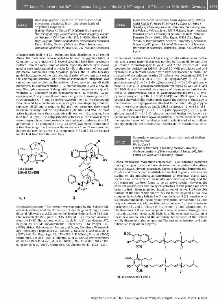
P445Bioassay-guided isolation of antiplasmodialstrychnos alkaloids from the stem bark ofStrychnos icaja Baill.Tchinda Tiabou A1, Tamze V2, Fr�d�rich M1, Angenot L1
1University of Liege, Department of Pharmacognosy, avenuede l’H�pital 1. CHU Tour 4 B t B36, B- 4000 Li�ge 1, 4000Liege, Belgium; 2Institute of Medical Research and MedicinalPlants Studies, Center of Medicinal Plants Studies andTraditional Medicine, PO Box 6163, 237 Yaounde, Cameroon
Strychnos icaja Baill. is a 20 – 100 m long liana distributed in all centralAfrica. The roots have been reported to be used by pygmies tribes inCameroon to cure malaria [1]. Several alkaloids have been previouslyisolated from the roots, some of which, especially dimers have showngood in vitro antiplasmodial activities [2 – 6]. In the search of new anti-plasmodial compounds from Strychnos species, the in vitro bioassayguided fractionation of the total alkaloid fraction of the stem bark usingthe chloroquine-sensitive 3D 7 strain of Plasmodium falciparum wascarried out and resulted in the isolation of two new tertiary alkaloidsmonomers, 15-hydroxyvomicine 1, 12-methoxyicajine 2 and a new di-mer, Nb-oxyde sungucine 3 along with the known monomers, icajine 4,vomicine 5, 15-hydroxy-19,20a-epoxynovacine 6, 12-methoxy-19,20a-epoxyicajine 7, strychnine 8 and dimers sungucine 9, isosungucine 10,strychnogucine C 11 and bisnorhydroxytoxiferine 12. The compoundswere isolated by a combination of silica gel chromatography columns,sephadex LH-20 and preparative TLC and their structures determinedbased on the analysis of their spectral data (UV, NMR and MS). The mostactive fractions were those containing dimers with IC50 ranging from0.32 to 4.31 mg/ml. The antiplasmodial activities of the known dimerswere comparable to those previously reported against other strains of P.falciparum [3 – 6]. Compound 3 (IC50 3.4 mg/ml) was about 3 times moreactive than sungucine 9 whereas the monomers 1 and 2 were inactive.Besides the new derivatives 1-3, compounds 4-7 and 9-12 are isolatedfor the first time from the stem bark.
Fig. 1
Acknowledgements: This research was supported by the ‘Subside fed-eral de la recherche’ of the University of Li�ge, Belgium through a post-doctoral fellowship to A.T.T. and by the Belgian National Fund for Scien-tific Research (FNRS – grant N. 3.4533.10). M.F. is a research associatefrom the FNRS. The authors wish to thank Mr. J.-C. Van Heugen, ATC,Belgium for ESI-MS measurements. References: 1. Neuwinger, H.D.(1996). African Ethnobotany: Poisons and Drugs, Chemistry, Pharmacol-ogy, Toxicology. Chapman & Hall, London. 2. Delaude, C. and Delaude, L.(1997) Bull. Soc. Roy. Liege. 66: 183 – 286. 3. Fr�d�rich, M. et al. (2000)Planta med. 66: 262 – 269. 4. Philippe, G. et al. (2003) Phytochemistry62: 623 – 629. 5. Frederich, M. et al. (2002). J. Nat. Prod. 65: 1381 – 1386.6. Fr�d�rich et al. (1999). Antimicrob. Ag. Chemother. 43: 2328 – 2331.
P446New steroidal saponins from Agave angustifoliaAbdel Khalik S1, Melek F2, Miyase T3, Gaber N1, Mina S1
1Faculty of Pharmacy, Helwan University, Pharmacognosy,Ein Helwan, Helwan, Egypt, 59711 Helwan, Egypt; 2NationalResearch Center, Chemistry of Natural Products, NationalResearch Center, Dokki, Giza, Egypt, 12622 Giza, Egypt;3School of Pharmaceutical Sciences, University of Schizuoka,Schizuoka,422, Japan., School of Pharmaceutical Sciences,University of Schizuoka, Schizuoka, Japan., 422 Schizuoka,Japan
Extraction of the dried leaves of Agave angustifolia with aqueous metha-nol gave a crude material that was purified by Diaion HP-20 and silicagel column chromatography to yield 1 and 2. The structure of 1 wasproposed by positive ion FABMS, 1D and 2D NMR techniques and acidhydrolysis as a steroidal saponin with new pentasaccharide chain. Thestructure of the aglycone bearing 27 carbons was determined 25R 5 aspirostan-12 -one 3 b -ol 3 -O [b – D- xylopyranosyl (1 –> 4) b -D-galactopyranosyl (1 –> 3) b -D- xylopyranosyl (1 –> 3) b -D- glucopyra-nosyl (1 –> 2) b -D- glucopyranoside]Acid hydrolysis as well as1 H and13C NMR data of 1 revealed the presence of five monosaccharide units,two b -D- glucopyranose, one b -D- galactopyranose and two b -D-xylo-pyranose assigned by 1 H- 1 H COSY, HMQC and HMBC spectra. Com-pound 2 showed similar NMR signals except the absence of those due tothe terminal b -D- xylopyranose attached to the outer b-D- glucopyra-nose, it was characterized as Cpd 2: 25R 5 a spirostan-12 -one 3 b -ol 3 -O [b -D- xylofuranosyl (1 –> 4) b -D- galactopyranosyl (1 –> 3) b -D-glucopyranosyl (1 –> 2) b -D- glucopyranoside] Further two known sa-ponins were isolated from Agave angustifolia. The methanol extract andthe saponin fraction of this plant proved to exhibit marked anti-inflam-matory, analgesic, schistosomicidal, cercaricidal & miracidicidal activ-ities.
P447Secondary metabolites from the root of HeliciarengetiensisWu H, Chen ICollege of Pharmacy, Kaohsiung Medical University,Graduate Institute of Pharmaceutical Sciences, 100, Shih-Chuan 1st Road, 807 Kaohsiung, Taiwan
Helicia rengetiensis Masamune (Proteaceae) is an endemic evergreentrees, growing in thickets at lower elevations in the central and southernparts of Taiwan. Flavonol glycosides, phenolic glycosides, benzenoid gly-cosides, and their derivatives distributed in plant of genus Helicia. In ourstudies on the antitubercular constituents of Formosan plants, 1200species have been screened for in vitro antitubercular activity, and theH. rengetiensis has been found to be an active species. However, thechemical constituents and biological activities of this plant have neverbeen studied. Bioassay-guided fractionation of active EtOAc-solublefraction of the root of this species has led to the isolation of two newcompounds, including helicinol A (1) and helicinol B (2), together withsix known compounds, including one cyclophane, kermadecin H (3), onefatty acid, stearic acid (4), one triterpene, squalene (5), one chroman, a-tocopherol (6), and a mixture of b-sitosterol (7) and stigmasterol (8).The structures of these new compounds were determined through spec-troscopic analyses including 2D-NMR data. The structural elucidation ofthese new compounds and the antitubercular activities of the isolateswill be discussed in this symposium. The successive isolation and anti-tubercular assay are in progress.
Fig. 1: Compounds 1 and 2
Planta Med 2010; 76: 1163–1374 Georg Thieme Verlag KG Stuttgart · New York · ISSN 0032-0943
13057th Tannin Conference and 58th International Congress of the GA | 29th August – 2nd September 2010, Berlin, Germany
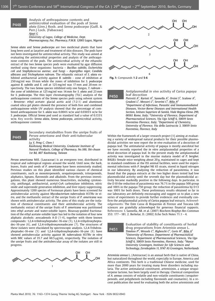
P448Analysis of anthraquinone contents andantimicrobial evaluation of the pods of Sennaalata (Linn.) Roxb. and Senna podocarpa (Guill. &Perr.) Lock. (Fabaceae)Ajayi GUniversity of Lagos, College of Medicine, Dept.Pharmacognosy, Fac. Pharmacy, P.M.B. 12003 Lagos, Nigeria
Senna alata and Senna podocarpa are two medicinal plants that havelong been used as laxative and treatment of skin diseases. The pods havenot been investigated for antimicrobial activity. This study was aimed atevaluating the antimicrobial properties and analysis of the anthraqui-none contents of the pods. The antimicrobial activity of the ethanolicextract of the two Senna species pods were evaluated by agar diffusionmethod using these organisms: bacteria – Bacillus subtilis, Escherichiacoli and Staphylococcus aureus; and fungi – Aspergillus niger, Candidaalbicans and Trichophyton rubrum. The ethanolic extract of S. alata ex-hibited antibacterial activity against B. subtilis - zone of inhibition at250 mg/ml was 19 mm while the zones of inhibition for S. podocarpaagainst B. subtilis and E. coli at 125 mg/ml was 17 mm and 20 mm re-spectively. The two Senna species inhibited only one fungus, T. rubrum -the zone of inhibition at 125 mg/ml was 14 mm for S. alata and 23 mmfor S. podocarpa. The thin layer chromatography (TLC) analysis of theanthraquinone contents of the Senna species pods using solvent system– Benzene: ethyl acetate: glacial acetic acid (7:2:1) and aluminumcoated silica gel plates showed the presence of both free and combinedanthraquinone while UV analysis showed that the percentage of com-bined anthraquinone for S. alata was 0.004% w/w and 0.0303% w/w forS. podocarpa. Official Senna pod used as standard had a value of 0.133%w/w. Key words: Senna alata, Senna podocarpa, antimicrobial activity,anthraquinone contents
P449Secondary metabolites from the unripe fruits ofPersea americana and their anti-tubercularactivityLu Y, Peng C, Chen IKaohsiung Medical University, Gradurate Institute ofNatural Products, College of Pharmacy, No.100, Shi-chuan1st Rd, 807 Kaohsiung, Taiwan
Persea americana Mill. (Lauraceae) is an evergreen tree, distributed intropical and subtropical regions around the world. Until now, the bark,leaves, fruits and seeds of P. americana have been extensively studied.Previous studies on this plant identified various classes of chemicalconstituents, such as monoterpenoids, sesquiterpenoids, triterpenoids,aliphatics, lignans, flavonoids and alkaloids. From the previous investi-gations, this plant showed numerous bioactivities, including cytotoxi-city, antifungal, antibacterial, acetyl-CoA carboxylase inhibition, nitricoxide and superoxide generation inhibition, and liver injury suppressing.Approximately 1200 species of Formosan plants have been screened forantitubercular activity against Mycobacterium tuberculosis H37Rv in vi-tro, and the methanolic extract of the unripe fruits of P. americana wasshown with antitubercular activity. The aims of this study are the isola-tion of chemical constituents and their antitubercular activity. Themethanolic extract of the unripe fruits of P. americana was partitionedinto ethyl acetate and water-soluble layers. Bioassay-guided fractiona-tion of the ethyl acetate-soluble layer has led to the isolation of four newaliphatic alcohols: avocadenols A-D (1-4), together with three knowncompounds: 1,2,4-trihdroxyheptadec-16-ene (5), 1,2,4-trihydroxyhepta-dec-16-yne (6), and 1,2,4-trihydroxynonadecane (7). The structures ofthese isolates were elucidated by spectroscopic analysis. 1,2,4-Trihdrox-yheptadec-16-ene (5) and 1,2,4-trihydroxyheptadec-16-yne (6) haveshowed antitubercular activity against M. tuberculosis H37Rv in vitrowith MIC values of 35.7 and 60.5 mg/mL, respectively. The isolation ofthe unripe fruits and the antitubercular assay of the isolates are still inprogress.
Fig. 1: Compounds 1-2 and 5-6
P450Antiplasmodial in vivo activity of Carica papayaleaf decoctionPietretti A1, Karioti A2, Sannella A1, Orsini S1, Scalone A1,Gradoni L1, Messori L3, Severini C1, Bilia A2
1Department of Infectious, Parasitic and ImmunomediatedDiseases, Vector-Borne Diseases and International HealthSection, Istituto Superiore di Sanit�, Viale Regina Elena 299,00161 Rome, Italy; 2University of Florence, Department ofPharmaceutical Sciences, Via Ugo Schiff 6, 50019 SestoFiorentino Florence, Italy; 3Department of Chemistry,University of Florence, Via della Lastruccia 3, 50019 SestoFiorentino, Florence, Italy
Within the framework of a larger research project [1] aiming at evaluat-ing a variety of widespread natural products for their possible plasmo-dicidal activities we now report the in vivo evaluation of a decoction ofpapaya leaf. The antimalarial activity of papaya is mostly anecdotal butwe have recently reported the in vitro antiplasmodial properties of adried decoction of papaya leaf. In the present study the antiplasmodialactivity was investigated in the murine malaria model of P. berghei.BALB/c female mice weighing about 20 g, maintained in cages and keptin standard conditions at the ISS animal facilities, were used for experi-mental infections with P. berghei NK 65 strain, stored in liquid nitrogenin our laboratory. By analyzing the reduction of parasitemia, it wasfound that the papaya extracts at the two higher doses tested had lowplasmodicidal activity until the seventh day but the plasmodicidal ac-tivity became markedly positive in the following days. In particular, inthe D10 the reduction of parasitemia was 76.7% in the papaya 500 groupand 100% in the papaya 750 group; the reduction of parasitemia by D13was 100% for both doses. These preliminary results obtained so far inour laboratory are definitely encouraging and suggest us to plan a newseries of experiments to provide further evidences and eventually con-firm the antiplasmodial activity of Carica papaya leaf extracts. Acknowl-edgements: The Ente Cassa di Risparmio di Firenze and Toscana Lifesciences are gratefully acknowleged for generous financial supports.References: 1. Sannella, AR. et al. (2007) Biochem Biophys Res Commun353: 177 – 181. 2. Berkelar, D. (2002) Echo Tech Notes 71:1 – 4.
P451Evaluation of stability of constituents of herbaldrug preparations from Artemisia annua L.Timteo P1, Wessels C1, Righeschi C1, Goris H2, Bilia A1
1University of Florence, Department of PharmaceuticalSciences, Department of Pharmaceutical Sciences, Via UgoSchiff 6, 50019 Sesto Fiorentino, Florence, Italy; 2HanzeUniversity Groningen, Institute for Life Sciences andTechnology, Zernikeplein 11, 9747 AS Groningen, Netherlands
Artemisia annua L. (Asteraceae) is an annual herb that is native of China,but naturalized throughout the world, especially in Europe, America andAfrica continents. This herb is a traditional Chinese medicine, used formore than 2000 years for treating many fever disorders including ma-laria. The active antimalarial constituent, artemisinin, a unique sesqui-terpene lactone, has been largely used in therapy. Chemical compositionof A. annua consists of volatile and non-volatile constituents (sesquiter-penoids, and polyphenols including flavonoids and coumarins). In a re-cent publication the need for evaluating both the active artemisinin and
Planta Med 2010; 76: 1163–1374 Georg Thieme Verlag KG Stuttgart · New York · ISSN 0032-0943
1306 7th Tannin Conference and 58th International Congress of the GA | 29th August – 2nd September 2010, Berlin, Germany
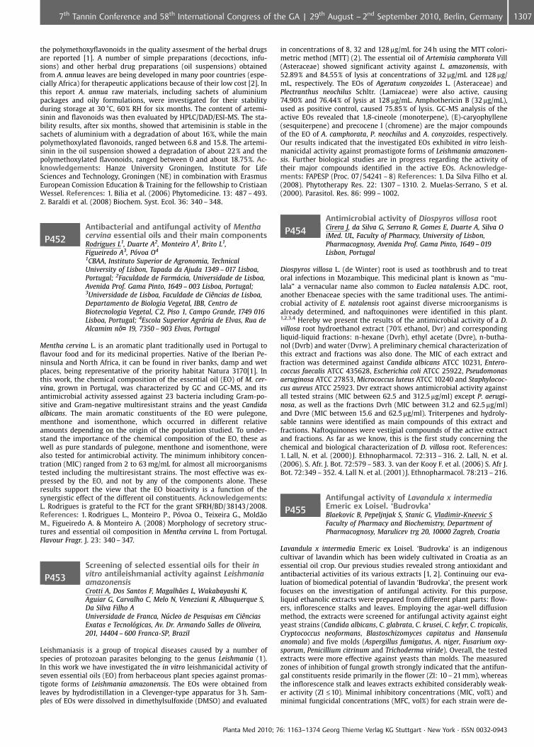
the polymethoxyflavonoids in the quality assesment of the herbal drugsare reported [1]. A number of simple preparations (decoctions, infu-sions) and other herbal drug preparations (oil suspensions) obtainedfrom A. annua leaves are being developed in many poor countries (espe-cially Africa) for therapeutic applications because of their low cost [2]. Inthis report A. annua raw materials, including sachets of aluminiumpackages and oily formulations, were investigated for their stabilityduring storage at 30 �C, 60% RH for six months. The content of artemi-sinin and flavonoids was then evaluated by HPLC/DAD/ESI-MS. The sta-bility results, after six months, showed that artemisinin is stable in thesachets of aluminium with a degradation of about 16%, while the mainpolymethoxylated flavonoids, ranged between 6.8 and 15.8. The artemi-sinin in the oil suspension showed a degradation of about 22% and thepolymethoxylated flavonoids, ranged between 0 and about 18.75%. Ac-knowledgements: Hanze University Groningen, Institute for LifeSciences and Technology, Groningen (NE) in combination with ErasmusEuropean Comission Education & Training for the fellowship to CristiaanWessel. References: 1. Bilia et al. (2006) Phytomedicine. 13: 487 – 493.2. Baraldi et al. (2008) Biochem. Syst. Ecol. 36: 340 – 348.
P452Antibacterial and antifungal activity of Menthacervina essential oils and their main componentsRodrigues L1, Duarte A2, Monteiro A1, Brito L1,Figueiredo A3, Pvoa O4
1CBAA, Instituto Superior de Agronomia, TechnicalUniversity of Lisbon, Tapada da Ajuda 1349 – 017 Lisboa,Portugal; 2Faculdade de Farmcia, Universidade de Lisboa,Avenida Prof. Gama Pinto, 1649 – 003 Lisboa, Portugal;3Universidade de Lisboa, Faculdade de CiÞncias de Lisboa,Departamento de Biologia Vegetal, IBB, Centro deBiotecnologia Vegetal, C 2, Piso 1, Campo Grande, 1749 016Lisboa, Portugal; 4Escola Superior Agrria de Elvas, Rua deAlcamim n�¤ 19, 7350 – 903 Elvas, Portugal
Mentha cervina L. is an aromatic plant traditionally used in Portugal toflavour food and for its medicinal properties. Native of the Iberian Pe-ninsula and North Africa, it can be found in river banks, damp and wetplaces, being representative of the priority habitat Natura 3170[1]. Inthis work, the chemical composition of the essential oil (EO) of M. cer-vina, grown in Portugal, was characterized by GC and GC-MS, and itsantimicrobial activity assessed against 23 bacteria including Gram-po-sitive and Gram-negative multiresistant strains and the yeast Candidaalbicans. The main aromatic constituents of the EO were pulegone,menthone and isomenthone, which occurred in different relativeamounts depending on the origin of the population studied. To under-stand the importance of the chemical composition of the EO, these aswell as pure standards of pulegone, menthone and isomenthone, werealso tested for antimicrobial activity. The minimum inhibitory concen-tration (MIC) ranged from 2 to 63 mg/mL for almost all microorganismstested including the multiresistant strains. The most effective was ex-pressed by the EO, and not by any of the components alone. Theseresults support the view that the EO bioactivity is a function of thesynergistic effect of the different oil constituents. Acknowledgements:L. Rodrigues is grateful to the FCT for the grant SFRH/BD/38143/2008.References: 1. Rodrigues L., Monteiro P., P voa O., Teixeira G., Mold¼oM., Figueiredo A. & Monteiro A. (2008) Morphology of secretory struc-tures and essential oil composition in Mentha cervina L. from Portugal.Flavour Fragr. J. 23: 340 – 347.
P453Screening of selected essential oils for their invitro antileishmanial activity against LeishmaniaamazonensisCrotti A, Dos Santos F, Magalh¼es L, Wakabayashi K,Aguiar G, Carvalho C, Melo N, Veneziani R, Albuquerque S,Da Silva Filho AUniversidade de Franca, Nfflcleo de Pesquisas em CiÞnciasExatas e Tecnolgicas, Av. Dr. Armando Salles de Oliveira,201, 14404 – 600 Franca-SP, Brazil
Leishmaniasis is a group of tropical diseases caused by a number ofspecies of protozoan parasites belonging to the genus Leishmania (1).In this work we have investigated the in vitro leishmanicidal activity ofseven essential oils (EO) from herbaceous plant species against promas-tigote forms of Leishmania amazonensis. The EOs were obtained fromleaves by hydrodistillation in a Clevenger-type apparatus for 3 h. Sam-ples of EOs were dissolved in dimethylsulfoxide (DMSO) and evaluated
in concentrations of 8, 32 and 128 mg/mL for 24 h using the MTT colori-metric method (MTT) (2). The essential oil of Artemisia camphorata Vill(Asteraceae) showed significant activity against L. amazonensis, with52.89% and 84.55% of lysis at concentrations of 32 mg/mL and 128 mg/mL, respectively. The EOs of Ageratum conyzoides L. (Asteraceae) andPlectranthus neochilus Schltr. (Lamiaceae) were also active, causing74.90% and 76.44% of lysis at 128 mg/mL. Amphothericin B (32 mg/mL),used as positive control, caused 75.85% of lysis. GC-MS analysis of theactive EOs revealed that 1,8-cineole (monoterpene), (E)-caryophyllene(sesquiterpene) and precocene I (chromene) are the major compoundsof the EO of A. camphorata, P. neochilus and A. conyzoides, respectively.Our results indicated that the investigated EOs exhibited in vitro leish-manicidal activity against promastigote forms of Leishmania amazonen-sis. Further biological studies are in progress regarding the activity oftheir major compounds identified in the active EOs. Acknowledge-ments: FAPESP (Proc. 07/54241 – 8) References: 1. Da Silva Filho et al.(2008). Phytotherapy Res. 22: 1307 – 1310. 2. Muelas-Serrano, S et al.(2000). Parasitol. Res. 86: 999 – 1002.
P454Antimicrobial activity of Diospyros villosa rootCirera J, da Silva G, Serrano R, Gomes E, Duarte A, Silva OiMed. UL, Faculty of Pharmacy, University of Lisbon,Pharmacognosy, Avenida Prof. Gama Pinto, 1649 – 019Lisbon, Portugal
Diospyros villosa L. (de Winter) root is used as toothbrush and to treatoral infections in Mozambique. This medicinal plant is known as “mu-lala” a vernacular name also common to Euclea natalensis A.DC. root,another Ebenaceae species with the same traditional uses. The antimi-crobial activity of E. natalensis root against diverse microorganisms isalready determined, and naftoquinones were identified in this plant.1,2,3,4 Hereby we present the results of the antimicrobial activity of a D.villosa root hydroethanol extract (70% ethanol, Dvr) and correspondingliquid-liquid fractions: n-hexane (Dvrh), ethyl acetate (Dvre), n-butha-nol (Dvrb) and water (Dvrw). A preliminary chemical characterization ofthis extract and fractions was also done. The MIC of each extract andfraction was determined against Candida albicans ATCC 10231, Entero-coccus faecalis ATCC 435628, Escherichia coli ATCC 25922, Pseudomonasaeruginosa ATCC 27853, Micrococcus luteus ATCC 10240 and Staphylococ-cus aureus ATCC 25923. Dvr extract shows antimicrobial activity againstall tested strains (MIC between 62.5 and 312.5 mg/ml) except P. aerugi-nosa, as well as the fractions Dvrh (MIC between 31.2 and 62.5 mg/ml)and Dvre (MIC between 15.6 and 62.5 mg/ml). Triterpenes and hydroly-sable tannins were identified as main compounds of this extract andfractions. Naftoquinones were vestigial compounds of the active extractand fractions. As far as we know, this is the first study concerning thechemical and biological characterization of D. villosa root. References:1. Lall, N. et al. (2000) J. Ethnopharmacol. 72:313 – 316. 2. Lall, N. et al.(2006). S. Afr. J. Bot. 72:579 – 583. 3. van der Kooy F. et al. (2006) S. Afr J.Bot. 72:349 – 352. 4. Lall N. et al. (2001) J. Ethnopharmacol. 78:213 – 216.
P455Antifungal activity of Lavandula x intermediaEmeric ex Loisel. ‘Budrovka’Blaekovic B, Pepeljnjak S, Stanic G, Vladimir-Kneevic SFaculty of Pharmacy and Biochemistry, Department ofPharmacognosy, Marulicev trg 20, 10000 Zagreb, Croatia
Lavandula x intermedia Emeric ex Loisel. ‘Budrovka’ is an indigenouscultivar of lavandin which has been widely cultivated in Croatia as anessential oil crop. Our previous studies revealed strong antioxidant andantibacterial activities of its various extracts [1, 2]. Continuing our eva-luation of biomedical potential of lavandin ‘Budrovka’, the present workfocuses on the investigation of antifungal activity. For this purpose,liquid ethanolic extracts were prepared from different plant parts: flow-ers, inflorescence stalks and leaves. Employing the agar-well diffusionmethod, the extracts were screened for antifungal activity against eightyeast strains (Candida albicans, C. glabrata, C. krusei, C. kefyr, C. tropicalis,Cryptococcus neoformans, Blastoschizomyces capitatus and Hansenulaanomala) and five molds (Aspergillus fumigatus, A. niger, Fusarium oxy-sporum, Penicillium citrinum and Trichoderma viride). Overall, the testedextracts were more effective against yeasts than molds. The measuredzones of inhibition of fungal growth strongly indicated that the antifun-gal constituents reside primarily in the flower (ZI: 10 – 21 mm), whereasthe inflorescence stalk and leaves extracts exhibited considerably weak-er activity (ZI £10). Minimal inhibitory concentrations (MIC, vol%) andminimal fungicidal concentrations (MFC, vol%) for each strain were de-
Planta Med 2010; 76: 1163–1374 Georg Thieme Verlag KG Stuttgart · New York · ISSN 0032-0943
13077th Tannin Conference and 58th International Congress of the GA | 29th August – 2nd September 2010, Berlin, Germany
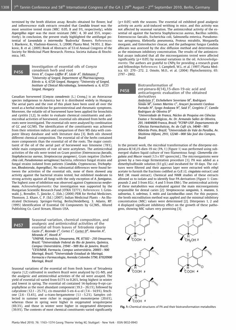
termined by the broth dilution assay. Results obtained for flower, leafand inflorescence stalk extracts revealed that Candida krusei was themost sensitive tested strain (MIC: 0.05, 8 and 9%, respectively), whileAspergillus niger was the most resistant (MIC: 4, 30 and 35%, respec-tively). In conclusion, the present study highlighted the antifungal po-tential of Lavandula x intermedia ‘Budrovka’ flowers. References:1. Blaekovic, B., Vladimir-Kneevic, S. (2008) Planta Med. 74:951. 2. Blae-kovic, B. et al. (2005) Book of Abstracts of 53 rd Annual Congress of theSociety for Medicinal Plant Research. Firenca. Societa italiana di fitochi-mica, 145.
P456Investigation of essential oils of Conyzacanadensis herb and rootVeres K1, Csupor-L�ffler B1, Lzr A2, Hohmann J1
1University of Szeged, Department of Pharmacognosy,E�tv�s u. 6, 6720 Szeged, Hungary; 2University of Szeged,Institute of Clinical Microbiology, Semmelweis u. 6, 6725Szeged, Hungary
Canadian horseweed [Conyza canadensis (L.) Cronq.] is an Asteraceaespecies indigenous to America but it is distributed widely in Hungary.The aerial parts and the root of this plant have been used all over theword as a herbal medicine for gastrointestinal and rheumatic symptoms.Moreover, the volatile oil of horseweed have been applied for bronchitisand cystitis [1,2]. In order to evaluate chemical constituents and anti-microbial activities of horseweed, essential oils obtained from herbs androots were investigated. The essential oils were analysed by combinationof GC and GC/MS. The identification of the constituents was achievedfrom their retention indices and comparison of their MS data with com-puter library database and with literature data [3]. Both oils showeddifferent chemical composition. The essential oil of the herbs containedmore components than the essential oil of the roots. The major consti-tuent of the oil of the aerial part of horseweed was limonene (78%),while main components of root oil were acetylenes. The antimicrobialactivities of the oils were tested on Gram positive (Enterococcus faecalis,Staphylococcus aureus, Streptococcus pyogenes), Gram negative (Escheri-chia coli, Pseudomonas aeruginosa) bacteria, reference fungal strains andfungal strains isolated from patients (Candida, Cryptococcus, Trichophy-ton, Rhodotorula, Aspergillus). No substantial differences were found be-tween the activities of the essential oils, none of them showed anyactivity against the bacterial strains tested, but exhibited moderate tostrong activity against all fungi with the only exception of A. fumigatus.The highest zone of inhibition was observed against Cryptococcus neofor-mans. Acknowledgements: Our investigation was supported by theHungarian Scientific Research Fund (OTKA 72771). References: 1. Grn-wald, J., Brendler, T., J�nicke, C. (Eds.) (2000) PDR for Herbal Medicines.Thomson. 2. Khare, C. P. (Ed.) (2007). Indian Medicinal Plants – An Illu-strated Dictionary. Springer-Verlag. Berlin/Heidelberg. 3. Adams, RP.(1995) Identification of Essential Oil Components by GC/MS., AlluredPublishing Co. Carol Stream, Illinois USA.
P457Seasonal variation, chemical composition, andanalgesic and antimicrobial activities of theessential oil from leaves of Tetradenia ripariaGazin Z1, Rezende C2, Cortez L3, Cortez D4, Amorim A2,Miranda A2, Hovell A1
1UNIPAR, Farmcia, Umuarama, PR, 87502 – 210 Umuarma,Brazil; 2Universidade Federal do Rio de Janeiro, Qu�mica,Campus Universitrio, 21941 – 909 Rio de Janeiro, Brazil;3CESUMAR, Farmacia, Campus universitrio, 20003 – 900Maring, Brazil; 4Universidade Estadual de Maring,Farmacia e Farmacologia, Avenida Colombo 5790, 87020900Maring, Brazil
Seasonal variations of the essential oil from fresh leaves of Tetradeniariparia (1,2) cultivated in southern Brazil were analyzed by CG-MS, andthe analgesic and antimicrobial activities of the oil were assayed. Theyield of essential oil varied from 0.17% to 0.26%, being highest in winterand lowest in spring. The essential oil contained 14-hydroxy-9-epi-car-yophyllene as the most abundant component (19.3 – 26.1%), followed bycalyculone (12.1 – 25.7%), cis-muurolol-5-en-4-a-ol (7.4 – 14.9%), fench-one (2.6 – 13.4%), and a-trans-bergamotene (1.1 – 5.2%). Samples col-lected in summer were richer in oxygenated monoterpene (20.6%),whereas those in spring were higher in oxygenated sesquiterpene(69.3%), and those in winter were higher in oxygenated diterpenes(39.9%). The contents of most chemical constituents varied significantly
(p < 0.05) with the seasons. The essential oil exhibited good analgesicactivity on acetic acid-induced writhing in mice, and this activity wasnot affected by seasonal variation. The antimicrobial activity of the es-sential oil against the bacteria Staphylococcus aureus, Bacillus subtilis,Enterococcus faecalis, Escherichia coli, Salmonella enterica, Pseudomo-nas aeruginosa, Klebsiella pneumonia, Proteus mirabilis, Morganellamorganii, and Enterobacter cloacae, and the pathogenic fungus Candidaalbicans was assessed by the disc diffusion method and determinationas the minimum inhibitory concentration. The results of the antimicro-bial assays indicated that all the microorganisms tested were affectedsignificantly (p < 0.05) by seasonal variations in the oil. Acknowledge-ments: The authors are grateful to CNPq for providing a research grantand fellowships References: 1. Campbell, W.L. et al. (1997) Planta Med-ica. 63: 270 – 272. 2. Omolo, M.O., et al. (2004) Phytochemistry, 65:2797 – 2802.
P458Biotransformation ofent-pimara-8(14),15-dien-19-oic acid andanticariogenic evaluation of the obtainedderivativesAmbrsio S1, Etchebehere Severiano M1, RodriguesSim¼o M1, Gomes Martins C1, Ara�ari Jacometti CardosoFurtado N2, Syogo Arakawa N3, Said S2, Gregrio L2, CamiloRodrigues de Oliveira D2
1Universidade de Franca, Nfflcleo de Pesquisa em CiÞnciasExatas e Tecnolgicas, Av. Dr. Armando Salles de Oliveira,201, 14404600 Franca, Brazil; 2FCFRP-USP, Departamento deCiÞncias FarmacÞuticas, Av. do Caf� s/n, 14040 – 903Ribeir¼o Preto, Brazil; 3Universidade do Vale do Para�ba, Av.Shishima Hifumi, 2911, 12240 – 000 S¼o Jos� dos Campos,Brazil
In the present work, the microbial transformation of the diterpene ent-pimara-8(14),15-dien-19 oic (PA, 1) (Figure 1) was performed using sub-merged shaken liquid culture of two filamentous fungi: Glomerella cin-gulata and Mucor rouxii (1.5 x 10⁷ spores/mL). The microorganisms weregrown by a two-stage fermentation procedure [1]. PA was added as adimethylsulfoxide solution (0.1 g/L) and incubated for 10 days. The cul-tures were filtered and their aqueous layer were extracted with ethylacetate to furnish the fractions codified as GcE (G. cingulata extract) andMrE (M. rouxii extract). Chemical and NMR studies of these extractsallowed us to isolate and to identify four PA derivatives (Figure 1: Com-pounds 2 and 3 from EGc; 4 and 5 from EMr). The antimicrobial activityof these metabolites was evaluated against the main microorganismsresponsible for dental caries [2]: Streptococcus sanguinis, S. mutans, S.salivarius, S. sobrinus, S. mitis and Lactobacillus casei. For this purpose,the broth microdilution method was applied and the minimal inhibitoryconcentration (MIC) values were determined [2]. Diterpenes 1, 2 and4 displayed significant inhibitory effect on the growth of these patho-gens, showing MIC values very promising [3].
Fig. 1: Chemical structures of PA and their biotransformation metabolites
Planta Med 2010; 76: 1163–1374 Georg Thieme Verlag KG Stuttgart · New York · ISSN 0032-0943
1308 7th Tannin Conference and 58th International Congress of the GA | 29th August – 2nd September 2010, Berlin, Germany
Acknowledgements: FAPESP (Proc. 2007/54762 – 8) References:1. Bastos, D.Z.L et al. (2007) Phytochemistry 68: 834 – 839. 2. Porto, T.S.et al. (2009) Molecules 14:191 – 199. 3. Gibbons, S. (2008) Planta Med.74: 594 – 599.
P459Interactions between pathogenic Escherichia coli,porcine intestinal cells, and a phytogenic feedadditive and its main active substanceG�rtner S, Tedin L, Zentek JInstitute for Animal Nutrition, Department of VeterinaryMedicine, Freie Universit�t Berlin, Br�mmerstraße 34, 14195Berlin, Germany
Plant derived products are claimed to improve animal performance andhealth, but the knowledge on the mode of action is still limited. The aimwas to investigate effects of a phytogenic feed additive and its mainactive substance in vitro regarding 1) their impact on adhesion proper-ties of F4+ Escherichia coli to the porcine intestinal epithelial cell lineIPEC-J2, 2) their capacity to bind E. coli, and 3) their antimicrobial char-acter in context to the pathogenic E. coli. The phytogenic additive Fres-ta� F (FF; Delacon, Austria) consists of essential oils, herbs, and spices.The main ingredient of the additive consists of 28% galactomannans(GM). The impact of the test substances was investigated in vitro bymeasuring the fluorescence intensity of CDFA-SE marked bacteria thatadhere on the IPEC-J2 cells (trial #1). A flow cytometer was used toquantify the fluorescent cells. In vitro trial #2 was performed using acoating method previously published by Becker et al. 2007. Antimicro-bial effects of FF and GM on the E. coli strain were estimated semi-quantitatively from bacterial growth curves. FF and GM significantlyreduced the adhesion of E. coli to IPEC-J2 (p < 0.05); fluorescence inten-sity was 0.96 € 0.63, 0.86 € 0.61, and 2.03 € 1.13, respectively. FF0.37 € 0.20 and GM 0.42 € 0.16 showed capacity to bind E. coli comparedwith the positive control 0.52 € 0.05. No direct antimicrobial effectscould be detected. It is concluded that the mode of action of Fresta� Fis not due to antimicrobial effect, but rather due to the anti-adhesioncapacity of galactomannans present in the products main component.References: 1. Becker, P.M., et al. (2007) J Appl Microbiol 103, 2686 –2696.
P460Plants used in popular medicine for treatment ofinfectious diseases in Central South AmericaRieder AUniversidade do Estado de Mato Grosso (UNEMAT); eEMPAER-MT, Campus de Cceres, Av. S¼o Jo¼o s/n, BairroCavalhada, 78200000 Cceres, Brazil
In communities there are people reference and popular trust, which aresought to guide the needy. This happens also for the treatment of healthproblems. In the capital (Cuiab�) from Mato Grosso, Brazil (geodesiccenter of South America) the people who lead and trading medicinalplants are known as healers (raizeiros). They have points of servicespecific and home-made to meet its demand. To know what are themajor medicinal plants listed the treatment of infectious diseases ingeneral, a study was done in the city of Cuiaba, the following speciesare mentioned for twelve healers: Angico (Anadenanthera peregrina (L.)Speg.-Fabaceae), Arnica (Arnica montana L.- Asteraceae), Arnica do cam-po (Camarea ericoides A. St.-Hil.- Malpighiaceae), Aroeira (Astroniumurundeuva (Allem¼o) Engl.-Anacardiaceae), B�lsamo da mata (Diclipterapohliana Nees – Acanthaceae), Barbatim¼o (Stryphnodendron adstringens(Mart.) Coville -Fabaceae), Cancerosa (Maytenus ilicifolia (Schrad.)Planch.- Calastraceae), Copa�ba (Copaifera langsdorffii Desf.- Fabaceae),Jequitib� (Cariniana rubra Gardner ex Miers – Lecythidaceae), Malvabranca (Waltheria americana L. – Sterculiaceae), Mangava brava (Lafoen-sia pacari A. St.-Hil. – Lythraceae), Marapuama (Ptychopetalum olacoidesBenth.- Olacaceae), N de cachorro (Heteropterys aphrodisiaca Mach. –Malpighiaceae), Pau doce (Vochysia rufa Mart. -Vochysiaceae), Sucupira(Bowdichia virgilioides Kunth – Fabaceae), Vassourinha (Scoparia dulcis L.– Plantaginaceae). It was also mentioned other plant species suitable forspecific infections: intestinal (2), renal (4), urinary (7), uterus and ovar-ies (3). It is observed predominance of species rich in tannins, and hasantiseptic and disinfectant power, which may partly explain the indica-tion of these plants to combat infectious diseases. Acknowledgements:Institutional support- UNEMAT, UFMT and EMPAER-MT; the teacher ad-visor – Guarim Neto, G; colleagues: Gon�alves MIA, de la Cruz MotaMGF – by participation in data collection and other support.
P461Dammarenolic acid, a secodammaranetriterpenoid from Aglaia sp shows potentanti-retroviral activity in vitroEsimone C1, Eck G2, Nworu C3, �berla K4, Proksch P2
1Faculty of Pharmaceutical Sciences, Nnamdi AzikiweUniversity, PMB 5025 Awka, Nigeria; 2Institutes forPharmaceutical Biology, Heinrich-Heine-University, 40225D�sseldorf, Germany; 3Department of Pharmacology andToxicology, Faculty of Pharmaceutical Sciences, University ofNigeria, 410001 Nsukka, Nigeria; 4Department of Molecularand Medical Virology, Ruhr University, 44780 Bochum,Germany
Screening of a panel of purified compounds isolated from Aglaia spp.(Meliaceae) for inhibition of early steps in the lentiviral replication cycleled to the identification of the 3, 4-secodammarane triterpenoid, ignT1,which inhibited HIV-1 infection potently (IC50 = 0.48 mg/ml), while cy-totoxic effects and inhibition of cell proliferation were only observed atconcentrations exceeding 10.69 mg/ml. Time of addition experiments re-vealed similar kinetics to the non-nucleoside RT-inhibitor (NNRTI), Ne-virapine, although the latter was significantly less cytotoxic. However,unlike Nevirapine, dammarenolic acid also potently inhibited the invitro replication of other retroviruses, including Simian immunodefi-ciency virus and Murine leukemic virus in vector-based antiviral screen-ing studies. Interestingly, the methyl ester analogue of dammarenolicacid, methyl dammarenolate had no anti-HIV-1 activity. Cell cycle ana-lysis revealed that ignT1 arrests HeLa cells at the S and G2/M phase.These results strongly suggest that dammarenolic acid could be a pro-mising lead compound for the development of novel anti-retrovirals.Acknowledgements: The authors gratefully acknowledge the Postdoc-toral Fellowship Awards to CS Nworu and CO Esimone by the Alexandervon Humboldt Foundation References: 1. Esimone, CO. et al (2009).Chemotherapy 55:119 – 126. 2. De Clercq, E. (2000). Med Res Rev 20:323 – 349.
P462Efficacy of components from leaves ofCalophyllum brasiliense against LeishmaniaamazonensisHonda P, Ferreira I, Amado C, Silveira T, Brenzan M,Lonardoni M, Cortez DUniversidade Estadual de Maring, Farmcia eFarmacologia, Avenida Colombo 5790, 87020900 Maring,Brazil
Leishmanicide potential of Calophyllum brasiliense leaves on promasti-gote and amastigote of Leishmania (Leishmania) amazonensis is evalu-ated. The LD50 of the dichloromethane extract and the hexane fractionfor promastigotes was respectively 40 mg/ml and 20 mg/ml. In mouseperitoneal macrophages infected with Leishmania amastigotes the Infec-tion Index decreased respectively 100% and 84.2% in 80 mg/ml and40 mg/ml concentrations of dichloromethane extract. The hexane frac-tion decreased the Infection Index respectively by 98.7% and 91.3% with-in the same concentrations. It was found that pretreatment with di-chloromethane extract or with hexane fraction of experimentally in-fected BALB/c mice decrease the volume of the lesions by L. amazonen-sis. Moreover, animals treated topically also revealed healing lesions.Besides, the parasite load in the animals’ popliteal lymph nodes wassignificantly reduced in treated animals, showing that plant componentsactually control infection. Results show that crude extract and hexanefraction of C. brasiliense reveal a significant in vitro and in vivo leishma-nicide activity [1]. The amentoflavones, (�)mammea A/BB and mammeaB/BB, were identified in the extracts and fractions by comparison withstandard samples and by analyses of HPLC-UV. Acknowledgements:The authors are grateful to CNPq for providing a research grant andfellowships References: 1. Honda A. (2010) Phytomedicine. 17: 333 –338.
Planta Med 2010; 76: 1163–1374 Georg Thieme Verlag KG Stuttgart · New York · ISSN 0032-0943
13097th Tannin Conference and 58th International Congress of the GA | 29th August – 2nd September 2010, Berlin, Germany
P463Antimicrobial activities of Longan (Euphorialongan L.) skin and seedsThongmuang P1, Sudjaroen Y1, Owen R2
1Suan Sunandha Rajabhat University, Aesthetic HealthScience, Faculty of Science and Technology, 1 U-Thong NokRoad, Wachira, Dusit Bangkok, Thailand; 2German CancerResearch Center, Division of Toxicology and Cancer RiskFactors, Im Neuenheimer Feld 280, 69120 Heidelberg,Germany
The methanolic extracts from seeds and skin of Longan (Euphoria longanL.) were tested for antimicrobial activity with five strains of pathogenicbacteria including Staphylococcus aureus, Bacillus cereus, Psuedomonasaeruginosa, Enterococcus faecalis, Escherichia coli and one strain ofpathogenic yeast, Candida albicans. The antimicrobial activities of eachextract were screened by agar diffusion before conducting with brothmacrodilution methods for determining minimal inhibitory concentra-tion (MIC) [1]. The phenolic content of skin and seed extracts was eval-uated by analytical high performance liquid chromatography (HPLC) [2].The results show that the methanolic extract of Longan skin (10 mg/ml)inhibited growth of S. aureus, P. aeruginosa and C. albicans were 15,11 and 9 mm of inhibition zone and MIC values were 4.42, 8.84 and1.11 mg/ml, respectively. The inhibition zones of Longan seed extract(10 mg/ml) were 17, 12 and 11 mm and MIC values were 3.19, 1.59 and1.59 mg/ml for S. aureus, P. aeruginosa, and C. albicans, respectively. Thephenolic content of skin and seed extracts were 13.38 and 88.51 g/kg ofdry weight. It was concluded that the antimicrobial activity was notrelated to content of phenolic compounds. However, it may be due totypes of phenolic compounds presented in the extracts and solubility ofextracts [3, 4]. Acknowledgements: 1, Faculty of Science and Faculty ofMedical Technology, Rangsit University, Phaholyothin Road, Lakhok,Pathumthani 12000, Thailand. 2, Associate Professor Omboon Luanrata-na, head of Department of Pharmacognosy, Faculty of Pharmacy, Mahi-dol University, Sri-Ayuthaya Road, Rajathevi, Bangkok 10400, Thailand.References: 1. NCCLS. (1998) Performance standards for AntimicrobialSusceptibility testing: Fifth Informational Supplement M100-S 8 18 (1).National Committee for Clinical Laboratory Standards. 2. Owen RW et al.(2000) Eur J Cancer 36: 1235 – 1247. 3. Cowan, MM. (1999) Clin Micro-biol Rev 12: 564 – 582. 4. Cushnie, TPT. et. al. (2003) Microbiol Res. 158:281 – 289.
P464Protein fraction from Syzygium cumini L. (Skeels)seeds active against bacteria isolated frombovine mastitisValle A1, Guimar¼es G1, Lazzari A1, Blume H1, Mulinari F1,Melo RF2
1UPIS – Bras�lia, Veterinary Medicine, Fazenda LagoaBonita, Planaltina, DF, 70000 Bras�lia, Brazil; 2UPIS –Bras�lia, Veterinary Medicine, Fazenda Lagoa Bonita,Planaltina, 70000 Bras�lia, Brazil
Mastitis, an inflammation of the mammary gland, is the costliest pro-duction disease of dairy cattle around the world. The treatment is basedon antibiotic therapy, but the therapeutic efficacies of the drugs aredecreasing due to the development of antimicrobial resistance. Aimingto evaluated the potential use of herbal compounds on infections ofmammary glands, the in vitro activity of Syzygium cumini L. (Skeels)seeds protein fraction was tested against different isolated bacteria frombovine mastitis (Staphylococcus aureus, Staphylococcus intermedius, coa-gulase-negative Staphylococcus, Staphylococcus hyicus, Streptococcusuberis, a-hemolytic Streptococcus, Streptococcus dysgalactiae, Streptococ-cus bovis and Enterococcus faecalis). The tests were carried using proteinfraction obtained by sulfate ammonium precipitation, dialysis and lyo-philization. The agar diffusion method was used and this fractionshowed activity against S. uberis, with an inhibition halo of 12 mm, a-hemolytic Streptococcus (10,5 mm), S. intermedius (15 mm), coagulase-negative Staphylococcus (16 mm) and S. aureus (18 mm). These resultsshowed that the S. cumini seeds protein fraction could represent aninteresting alternative method for mastitis control. The formulationsbased on this natural product will be tested, aiming to developmentan alternative treatment for antibiotic therapy, with reduced costs andrisk of residues on milk.
P465Antimicrobial activity of Limonium avei (De Not.)Brullo & Erben extractsFilocamo A1, Nostro A1, Giovannini A2, Catania S3, Costa C3,Marino A1, Bisignano G1
1Pharmaco-Biological Department, School of Pharmacy,University of Messina, Pharmaco-Biological Department,School of Pharmacy, Vill. Annunziata, 98168 Messina, Italy;2C.R.A. Experimental Unit for Floricolture and OrnamentalSpecies, Corso Inglesi, 508, 18038 Sanremo (IM), Italy;3Interdepartmental Centre of Experimental, Environmentaland Occupational Toxicology (CITSAL), Via C. Valeria, 98122Messina, Italy
Limonium avei (De Not.) Brullo & Erben (Plumbaginaceae) is a rare tri-ploid (2n = 27), annual halophyte, with apomictic reproduction [1], in-cluded in the Red List of Endangered Species by the IUCN [2]. The spe-cies is endemic to the central Mediterranean coast and in Liguria (Italy)it is present in only one population, with almost 1500 individuals. Theincreasing urbanization of the Ligurian population natural habitat hasprompted the adoption of measures for its conservation. As part of thiseffort ex situ seed conservation and tissue culture techniques were de-veloped for the species [3]. To the best of our knowledge, no studyreporting to biological activity is present in literature. Here we reportedfor the first time the antimicrobial activity and phytochemical profile ofLimonium avei ethanol and dichloromethane extracts. Flowering stemscollected in the natural site were compared with flowering stems col-lected in the CRA-FSO greenhouse from micropropagated acclimatisedplants. Tissues were air dry at room temperature for 60 days. The anti-microbial activity of extracts was performed by the minimum inhibitoryconcentration (MIC) and minimum bactericidal concentration (MBC) [4]against Gram-positive, Gram-negative bacteria and mycetes. The ex-tracts were submitted to phytochemical screening by LCMS and HPLC/DAD analyses. The results indicated that the ethanol extracts of bothsamples displayed higher activity than dichloromethane extracts andthis activity was more pronounced against Gram-positive than Gram-negative bacteria and mycetes. In particular, the extracts demonstratedMIC and MBC values ranging from 15.6 to 500 mg/ml and from 500 to4000 mg/ml respectively. References: 1. Brullo S. (1988). Miscellaneousnotes on the genus Limonium. Willdenowia 17(1): 17. 2. Conti F. et al.(1997). Liste Rosse Regionali delle Piante d’Italia. WWF & SBI, Camerino:64. 3. Giovannini A. et al. (2009). Ex situ conservation measures of athreatened Limonium avei (De Not.) Brullo & Erben population. In Bookof Abstract “Biodiversity Hotspots in the Mediterranean Area” Cagliari22 – 24 giugno: 284. 4. CLSI (2000). Methods for dilution antimicrobialsusceptibility tests for bacteria that grow aerobically:approved standard,Wayne, Pa 17: 10 – 13.
P466In vitro antimicrobial activity screenning ofTerminalia macroptera leafSilva O1, da Silva G1, Tani�a M1, Serrano R1, Vital J2, TeixeiraGomes E1
1iMed. UL, Faculty of Pharmacy, University of Lisbon,Laboratory of Pharmacognosy, Av. Prof. Gama Pinto, 1649 –019 Lisbon, Portugal; 2Faculty of Pharmacy, University ofLisbon, Laboratory of Microbiology, Av. Prof. Gama Pinto,1649 – 019 Lisbon, Portugal
Terminalia macroptera Guill. and Perr. (Combretaceae) is a West Africanspecies used on traditional medicine to treat infectious diseases.[1]Hereby we present the results of an antimicrobial activity screeningperformed by the twofold serial microdilution assay against seven re-ference bacterial strains and against Candida albicans, with a leaf hydro-ethanol extract (Tml) and liquid-liquid partition fractions Tml-1 (hex-ane), Tml-2 (diethyl ether), Tml-3 (ethyl acetate), Tml-4 (Tml waterfiltered fraction) and Tml-5 (Tml water precipitate fraction). Resultsare displayed on Table 1. In the range of tested concentrations (3200 to50 mg/ml), the extract was active against all tested microorganisms. Thebest results were obtained against Shigella dysenteriae and Vibrio choler-ae and the most active fraction was identified as the ethyl acetate one(Tml-3). Chemical profile of this fraction includes polyphenols as maincompounds.
Planta Med 2010; 76: 1163–1374 Georg Thieme Verlag KG Stuttgart · New York · ISSN 0032-0943
1310 7th Tannin Conference and 58th International Congress of the GA | 29th August – 2nd September 2010, Berlin, Germany
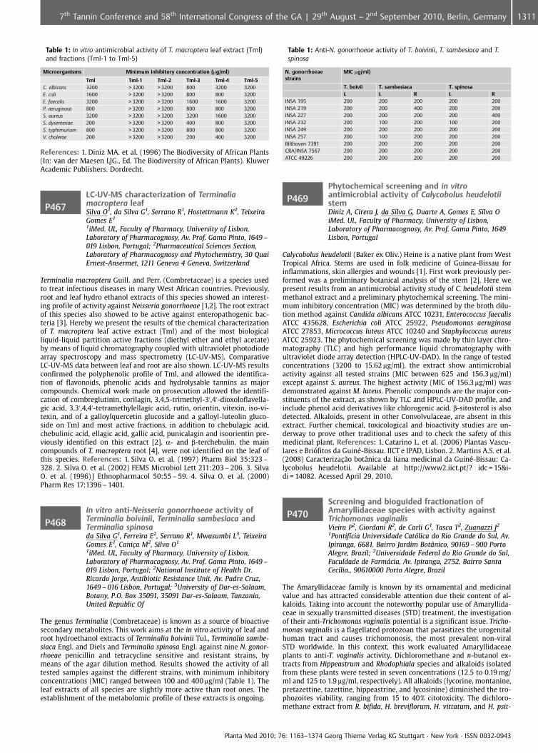
Table 1: In vitro antimicrobial activity of T. macroptera leaf extract (Tml)and fractions (Tml-1 to Tml-5)
Microorganisms Minimum inhibitory concentration (mg/ml)
Tml Tml-1 Tml-2 Tml-3 Tml-4 Tml-5C. albicans 3200 > 3200 > 3200 800 3200 3200E. coli 1600 > 3200 > 3200 800 800 3200E. faecalis 3200 > 3200 > 3200 1600 1600 3200P. aeruginosa 800 > 3200 > 3200 800 800 3200S. aureus 3200 > 3200 > 3200 3200 1600 3200S. dysenteriae 200 > 3200 > 3200 400 800 3200S. typhimurium 800 > 3200 > 3200 800 800 3200V. cholerae 200 > 3200 > 3200 200 400 3200
References: 1. Diniz MA. et al. (1996) The Biodiversity of African Plants(In: van der Maesen LJG., Ed. The Biodiversity of African Plants). KluwerAcademic Publishers. Dordrecht.
P467LC-UV-MS characterization of Terminaliamacroptera leafSilva O1, da Silva G1, Serrano R1, Hostettmann K2, TeixeiraGomes E1
1iMed. UL, Faculty of Pharmacy, University of Lisbon,Laboratory of Pharmacognosy, Av. Prof. Gama Pinto, 1649 –019 Lisbon, Portugal; 2Pharmaceutical Sciences Section,Laboratory of Pharmacognosy and Phytochemistry, 30 QuaiErnest-Ansermet, 1211 Geneva 4 Geneva, Switzerland
Terminalia macroptera Guill. and Perr. (Combretaceae) is a species usedto treat infectious diseases in many West African countries. Previously,root and leaf hydro ethanol extracts of this species showed an interest-ing profile of activity against Neisseria gonorrhoeae [1,2]. The root extractof this species also showed to be active against enteropathogenic bac-teria [3]. Hereby we present the results of the chemical characterizationof T. macroptera leaf active extract (Tml) and of the most biologicalliquid-liquid partition active fractions (diethyl ether and ethyl acetate)by means of liquid chromatography coupled with ultraviolet photodiodearray spectroscopy and mass spectrometry (LC-UV-MS). ComparativeLC-UV-MS data between leaf and root are also shown. LC-UV-MS resultsconfirmed the polyphenolic profile of Tml, and allowed the identifica-tion of flavonoids, phenolic acids and hydrolysable tannins as majorcompounds. Chemical work made on prosecution allowed the identifi-cation of combreglutinin, corilagin, 3,4,5-trimethyl-3’,4’-dioxoloflavella-gic acid, 3,3’,4,4’-tetramethylellagic acid, rutin, orientin, vitexin, iso-vi-texin, and of a galloylquercetin glucoside and a galloyl-luteolin gluco-side on Tml and most active fractions, in addition to chebulagic acid,chebulinic acid, ellagic acid, gallic acid, punicalagin and isoorientin pre-viously identified on this extract [2]. a- and b-terchebulin, the maincompounds of T. macroptera root [4], were not identified on the leaf ofthis species. References: 1. Silva O. et al. (1997) Pharm Biol 35:323 –328. 2. Silva O. et al. (2002) FEMS Microbiol Lett 211:203 – 206. 3. SilvaO. et al. (1996) J Ethnopharmacol 50:55 – 59. 4. Silva O. et al. (2000)Pharm Res 17:1396 – 1401.
P468In vitro anti-Neisseria gonorrhoeae activity ofTerminalia boivinii, Terminalia sambesiaca andTerminalia spinosada Silva G1, Ferreira E2, Serrano R1, Mwasumbi L3, TeixeiraGomes E1, Cani�a M2, Silva O1
1iMed. UL, Faculty of Pharmacy, University of Lisbon,Laboratory of Pharmacognosy, Av. Prof. Gama Pinto, 1649 –019 Lisbon, Portugal; 2National Institute of Health Dr.Ricardo Jorge, Antibiotic Resistance Unit, Av. Padre Cruz,1649 – 016 Lisbon, Portugal; 3University of Dar-es-Salaam,Botany, P.O. Box 35091, 35091 Dar-es-Salaam, Tanzania,United Republic Of
The genus Terminalia (Combretaceae) is known as a source of bioactivesecondary metabolites. This work aims at the in vitro activity of leaf androot hydroethanol extracts of Terminalia boivinii Tul., Terminalia sambe-siaca Engl. and Diels and Terminalia spinosa Engl. against nine N. gonor-rhoeae penicillin and tetracycline sensitive and resistant strains, bymeans of the agar dilution method. Results showed the activity of alltested samples against the different strains, with minimum inhibitoryconcentrations (MIC) ranged between 100 and 400 mg/ml (Table 1). Theleaf extracts of all species are slightly more active than root ones. Theestablishment of the metabolomic profile of these extracts is ongoing.
Table 1: Anti-N. gonorrhoeae activity of T. boivinii, T. sambesiaca and T.spinosa
N. gonorrhoeaestrains
MIC mg/ml)
T. boivii T. sambesiaca T. spinosaL L R L R
INSA 195 200 200 200 200 200INSA 219 200 200 400 200 200INSA 227 200 200 200 200 400INSA 232 200 100 200 100 200INSA 249 200 200 200 200 200INSA 257 200 100 200 200 200Bilthoven 7391 200 200 200 200 200CRA/INSA 7567 200 200 200 200 200ATCC 49226 200 200 200 200 200
P469Phytochemical screening and in vitroantimicrobial activity of Calycobolus heudelotiistemDiniz A, Cirera J, da Silva G, Duarte A, Gomes E, Silva OiMed. UL, Faculty of Pharmacy, University of Lisbon,Laboratory of Pharmacognosy, Av. Prof. Gama Pinto, 1649Lisbon, Portugal
Calycobolus heudelotii (Baker ex Oliv.) Heine is a native plant from WestTropical Africa. Stems are used in folk medicine of Guinea-Bissau forinflammations, skin allergies and wounds [1]. First work previously per-formed was a preliminary botanical analysis of the stem [2]. Here wepresent results from an antimicrobial activity study of C. heudelotii stemmethanol extract and a preliminary phytochemical screening. The mini-mum inhibitory concentration (MIC) was determined by the broth dilu-tion method against Candida albicans ATCC 10231, Enterococcus faecalisATCC 435628, Escherichia coli ATCC 25922, Pseudomonas aeruginosaATCC 27853, Micrococcus luteus ATCC 10240 and Staphylococcus aureusATCC 25923. The phytochemical screening was made by thin layer chro-matography (TLC) and high performance liquid chromatography withultraviolet diode array detection (HPLC-UV-DAD). In the range of testedconcentrations (3200 to 15.62 mg/ml), the extract show antimicrobialactivity against all tested strains (MIC between 625 and 156.3 mg/ml)except against S. aureus. The highest activity (MIC of 156.3 mg/ml) wasdemonstrated against M. luteus. Phenolic compounds are the major con-stituents of the extract, as shown by TLC and HPLC-UV-DAD profile, andinclude phenol acid derivatives like chlorogenic acid. b-sitosterol is alsodetected. Alkaloids, present in other Convolvulaceae, are absent in thisextract. Further chemical, toxicological and bioactivity studies are un-derway to prove other traditional uses and to check the safety of thismedicinal plant. References: 1. Catarino L. et al. (2006) Plantas Vascu-lares e Bri fitos da Guin�-Bissau. IICT e IPAD, Lisbon. 2. Martins A.S. et al.(2008) Caracteriza�¼o bot�nica da liana medicinal da Guin�-Bissau: Ca-lycobolus heudelotii. Available at http://www2.iict.pt/? idc = 15&i-di = 14082. Acessed April 29, 2010.
P470Screening and bioguided fractionation ofAmaryllidaceae species with activity againstTrichomonas vaginalisVieira P2, Giordani R2, de Carli G1, Tasca T2, Zuanazzi J2
1Pontif�cia Universidade Catlica do Rio Grande do Sul, Av.Ipiranga, 6681. Bairro Jardim Bot nico, 90169 – 900 PortoAlegre, Brazil; 2Universidade Federal do Rio Grande do Sul,Faculdade de Farmcia, Av. Ipiranga, 2752. Bairro SantaCec�lia., 90610000 Porto Alegre, Brazil
The Amaryllidaceae family is known by its ornamental and medicinalvalue and has attracted considerable attention due their content of al-kaloids. Taking into account the noteworthy popular use of Amaryllida-ceae in sexually transmitted diseases (STD) treatment, the investigationof their anti-Trichomonas vaginalis potential is a significant issue. Tricho-monas vaginalis is a flagellated protozoan that parasitizes the urogenitalhuman tract and causes trichomonosis, the most prevalent non-viralSTD worldwide. In this context, this work evaluated Amaryllidaceaeplants to anti-T. vaginalis activity. Dichloromethane and n-butanol ex-tracts from Hippeastrum and Rhodophiala species and alkaloids isolatedfrom these plants were tested in seven concentrations (12.5 to 0.19 mg/ml and 125 to 1.9 mg/ml, respectively). All alkaloids (lycorine, montanine,pretazettine, tazettine, hippeastrine, and lycosinine) diminished the tro-phozoites viability, ranging from 15 to 40% citotoxicity. The dichloro-methane extract from R. bifida, H. breviflorum, H. vittatum, and H. psit-
Planta Med 2010; 76: 1163–1374 Georg Thieme Verlag KG Stuttgart · New York · ISSN 0032-0943
13117th Tannin Conference and 58th International Congress of the GA | 29th August – 2nd September 2010, Berlin, Germany
tacinum reduced the viability of the trophozoites of T. vaginalis in about30 – 60% in the lower concentration. The H. breviflorum extracts demon-strated the highest anti-T. vaginalis activity (60% of viability reduction)and a bioguided study were realized. Nine alkaloids enriched dichloro-methane and n-butanol fractions were obtained and tested. Amongthese fractions the one butanol fraction is the most active against T.vaginalis because reduced the viability at 100%. All the active fractionscontain lycorine and lycosinine as major components and a synergismcan occur considering the higher anti-T. vaginalis activity of extractsthan alkaloids.
P471Lapachol and isomeric 5- and8-hydroxy-2-(1’-hydroxyethyl)naphtho[2,3-b]furan-4,9-diones are effective antileishmanialconstituents of Tabebuia avellanedaeAli A1, Kiderlen A2, Kolodziej H1
1Freie Universit�t Berlin, Institute of Pharmacy,Pharmaceutical Biology, Koenigin Luise-Straße 2+4, 14195Berlin, Germany; 2Robert Koch-Institut, CellularImmunology Unit P22, Nordufer 20, 13353 Berlin, Germany
The antileishmanial activity of a methanolic extract of Tabebuia avella-nedae (Bignoniaceae) and some constituents against transgenic Leish-mania major expressing green fluorescent protein (GFP) was examinedusing parasite retrieval assay and flow cytometry (FACS analysis). Leish-maniasis is a group of diseases caused by flagellate protozoan Leishma-nia spp. causing high morbidity and mortality in tropical to Mediterra-nean environments. The methanol extract was sequentially extractedwith n-hexane, dichloromethane, ethyl acetate, and n-butanol, yieldingfinally water-soluble extractives. The highest antileishmanial activityresided in the n-hexane and dichloromethane fractions with IC 50 of64 mg/ml and 41 mg/ml, respectively. Phytochemical analysis of the n-hexane fraction revealed the presence of lapachol which exhibited an-tileishmanial activity against both extra- and intracellular parasites,with IC 50 values of 33mM and 115mM, respectively. Lapachol did notshow any cytotoxicity on macrophages as host cells of Leishmania para-sites (EC 50 > 300mM). From the dichloromethane fraction an insepar-able mixture of isomeric 5- and 8-hydroxy-2-(1’-hydroxyethyl)-naphtho[2,3-b] furan-4,9-diones was isolated by a combination of col-umn chromatography and semi-preparative HPLC. This mixture was sig-nificantly more effective against both extra- and intracellular L. majorparasites (IC50 of 4mM, ca. 28 fold more active than that of lapachol),while cytotoxic effects on macrophages were not evident at 8mM. TheIC 50 of amphotericin B, a reference compound, was 2.5 mM and 0.2mMagainst promastigotes and amastigotes, respectively. It appears as if thepresence of a furan ring increases the antileishmanial activity ofnaphthoquinones.
P472Antileishmanial mode of action of lapachol andplumbaginAli A1, Kiderlen A2, Kolodziej H1
1Freie Universit�t Berlin, Institute of Pharmacy,Pharmaceutical Biology, Koenigin Luise-Straße 2+4, 14195Berlin, Germany; 2Robert Koch-Institut, CellularImmunology Unit P22, Nordufer 20, 13353 Berlin, Germany
Plumbagin, a naphthoquinone first isolated from species of the genusPlumbago, and lapachol, a prenylated hydroxynaphthoquinone encoun-tered in Tabebuia avellanedae (Bignoniaceae), were reported as antifun-gal, antibacterial, anticancer and antileishmanial agents [1, 2]. It hasbeen suggested that the antileishmanial activity of these naphthoqui-nones is due to the generation of reactive oxygen species [3]. The aim ofthis investigation was to evaluate the antileishmanial activity of lapa-chol and plumbagin with a closer view on their mode of action. Lapacholexhibited prominent antileishmanial activity against L. major promasti-gotes with an IC 50 value of 8 mg/ml (33mM) and showed a moderateactivity against amastigotes as evident from an IC 50 value of 28 mg/ml(115mM). It did not show any cytotoxicity on macrophages as host cellseven at high concentrations (EC 50 > 72 mg/ml, 300mM). On the otherhand plumbagin was significantly more effective against both extra-and intracellular L. major parasites (IC 50 0.5 mg/ml, 2.6mM), but wasrelatively more cytotoxic on macrophages (1.5 mg/ml; 8mM). Plumbaginand lapachol did not induce NO release in infected macrophages. Bothcompounds were found to be similarly antileishmanicidal when incu-bated with or without the iNOS inhibitor L-NMMA. While plumbagindid not show any antileishmanial effects in the presence of glutathione
plus ascorbic acid, no difference in activity was found for lapachol. Thisfinding suggests that lapachol and plumbagin mediate their antileish-manial activities by different mode of actions and independent from NOproduction. References: 1. Hussain, H et al. (2007), Arkivoc: 145 – 171. 2.Croft S et al. (1985), Annal Trop Med Parasitol 79: 651 – 653. 3. Inbaraj JJ,Chignell CF (2004), Chem ResToxicol. 17: 55 – 62.
P473Treatment of malaria in Iranian traditionalmedicineNaghibi F1, Mosaddegh M1, Esmaeili S2
1Traditional Medicine and Materia Medica Research, Schoolof Pharmacy, Shahid Beheshti University of MedicalSciences, Pharmacognosy, Shams Alley, Valiasr St.,1516745811 Tehran, Iran, Islamic Republic Of; 2TraditionalMedicine and Materia Medica Research, School ofTraditional Medicine, Shahid Beheshti University of MedicalSciences, Traditional Pharmacy, Shams Alley, Valiasr St.,1516745811 Tehran, Iran, Islamic Republic Of
The declining efficacy of classical medication in relation to the rapidextension of Plasmodium falciparum chloroquine-resistant strains hasled to a need for new efficient anti malarial drugs. It is a real necessity tosearch for new efficient anti malarial compounds and make them acces-sible to most of the people. An ethnobotanical study was conducted tofind plants traditionally used against malaria in Iran. In this study35 plants were found on the basis of ethnobotanical investigation andsearching in Iranian ancient traditional physician’s books (1 – 4). At thesame time the anopheles mosquito has developed resistance to manyinsecticides. There are some plants in Iran that are traditionally used asinsect repellent (1 – 4). Also matching the old medicinal plant nameswith scientific terminology was done (5 – 8). First ethnobotanical stu-dies on these plants were carried out at theTraditional Medicine andMateria Medica Research Center (TMRC). Further assessments are inprocess. References: 1. Ibn – sina, Qanoon, Lithography, 1296 A.H. rep-rinted by Institute of medical history study, Islamic and complementarymedicine. Tehran 2004. 2. Jorjani S. Zakhireh Kharazmshahi. Bonyadefarhang Iran. Tehran First Ed, 1976 PP: 256 – 273. 3. Davood Ibn-OmarAntaki, Tazkareh olel-Albab. Beirot. 4. Aqili Khorasani, Makhzan ol-Ad-vieh. Enghelab e Eslami publishing and Educational Organization. Teh-ran 1992. 5. Ghahreman A and Okhovvat A. R. Matching the old medic-inal plant names with scientific terminology Vol 1. Tehran University.Tehran. 2004. 6. Dini M. Investigation of various common names ofplants used in traditional medicine. Research institute of forests andrangelands. Tehran. 2005. 7. Amin Gh. Popular medicinal plants of Iran.Tehran University. Tehran. 2005. 8. Soltani A. Encyclopedia of traditionalmedicine (Medicinal plants). Arjmand. Tehran. 2004.
P474Investigation on pharmacological andantimicrobial activities of Galanthustranscaucasicus Fomin growing in IranSharifzadeh M1, Yousefbeyk F2, Amin G2, SalehiSormaghi M2, Azadi B2, Samadi N3, Amini MoghadamFarouj N4, Amin M5
1Department of Toxicology and Pharmacology, Faculty ofPharmacy and Research Center of Medicinal Plants, TehranUniversity of Medical Sciences, Enghelab Avenue, 16- azarStreet,14155 – 6451 Tehran, Iran, Islamic Republic Of;2Department of Pharmacognosy, Faculty of Pharmacy,Tehran University of Medical Sciences, Tehran-Iran,Enghelab Ave,16-Azar Street, 1417614411 Tehran, Iran,Islamic Republic Of; 3Department of Food and Drug Control,Faculty of Pharmacy and Research Center of MedicinalPlants, Tehran University of Medical Sciences, Tehran-Iran,Enghelab Ave,16-Azar Street, 1417614411 Tehran, Iran,Islamic Republic Of; 4Department of Pharmacognosy,Pharmaceutical Sciences Unit, Islamic Azad University,Tehran-Iran, Shariati Ave, Yakhchal street, 1417614411Tehran, Iran, Islamic Republic Of; 5Department of MolecularGenetics, Faculty of Medicine, University of Toronto, Facultyof Medicine, Toronto, Canada
Galanthus transcaucasicus Fomin (Amaryllidaceae) is an endemic spe-cies of the Caucasus region and the Alborz mountains in Iran (1). Allspecies of Galanthus are famous for their bioactive alkaloids such asgalanthamine, an acetyl cholinesterase inhibitor, which is used for thetreatment of Alzheimer’s disease (1). The bulbs of the plant were col-
Planta Med 2010; 76: 1163–1374 Georg Thieme Verlag KG Stuttgart · New York · ISSN 0032-0943
1312 7th Tannin Conference and 58th International Congress of the GA | 29th August – 2nd September 2010, Berlin, Germany
lected in February 2008 in Alborz mountain area (Rostam abad), Iran.The total extract was prepared by cool percolation method using ethanoland the chloroform fraction was obtained. Preliminary phytochemicalscreening showed the presence of alkaloids, sterols and cardiac glyco-sides in bulbs total ethanolic extract. The antimicrobial activity of theethanolic extract of bulbs and chloroform fraction were evaluated onStaphylococcus aureus, Staphylococcus piogenes, Pseudomonas aerugi-nosa, Escherichia coli, Helicobacter pylori, Shigella soneii, Salmonellatyphi, Bacillus subtilis, Proteus vulgaris and Candida albicans using cupplate diffusion method. The time course of the effects of the ethanolicextract of bulbs, administered intrapritoneally to rats, on the spatialmemory retention in the Morris water maze was investigated (2). Theresults showed that G. transcaucasicus Fomin ethanolic extract of bulbshad antibacterial activity against B. subtilis ATCC 6633 (MIC 9.275 mg/ml) and antifungal activity against C. albicans ATCC 10231 (MIC 150unit/ml). The chloroform fraction showed activity against S. aureus ATCC6538 (MIC 1.17 mg/ml). The administered doses of 5, 25 and 100 mg/kgshowed a significant reduction in escape latency (P < 0.05), and traveleddistance (P < 0.05) but not swimming speed, compared with controlsuggesting significant spatial memory retention enhancement by G.transcaucasicus Fomin. References: 1. Bastida, J. et al. (2000). The alka-loids. Elsevier Scientific Publishing, Amesterdom 63: 87 – 179. 2. Shar-ifzadeh, M. et al. (2005). Eur J Pharmacol 511:159 – 66.
P475Chemical constituents of Zanthoxylum capenseLuo X1, Pinto A1, Durn A1, Mansoor T1, Mulhovo S2,Duarte N1, Ferreira M1
1i.Med-UL, Faculdade de Farmcia, Universidade de Lisboa,Av. Prof. Gama Pinto, 1649003 Lisboa, Portugal; 2EscolaSuperior T�cnica, Departamento de CiÞncias Agro-Pecurias,Universidade Pedaggica, Campus de Lhanguene, Av. deMo�ambique, 21402161 Maputo, Mozambique
Zanthoxylum capense (Thunb.) Harv (Rutaceae) is a small to mediumtree distributed in Zimbabwe, South Africa, and Mozambique. The de-coction of its roots is used to treat gallsickness in the Eastern Cape, andits fruits are employed in the treatment of colic and paralysis. Similarlyto other Zanthoxylum species, such as Z. chalybeum Engl. and Z. davyi(Verdoorn) P.G. Waterman, Z. capense is also used in the treatment ofsnakebite and severe coughs and colds [1]. Previous studies of this genushave reported the isolation of coumarins, lignans, alkaloids, terpenoids,and flavonoids [2]. In our search for biologically active compounds fromplant species, the methanol extract of Z. Capense roots has been studied.The crude methanol extract was suspended on a methanol-water mix-ture and sequentially extracted with n-hexane and dichloromethane.Preliminary studies of the dichloromethane fraction, using combinedchromatographic techniques, have yielded some known benzophenan-thridine-type alkaloids, namely decarine, norchelerythrine, and 8-acet-onyldihydro-cherlerythrine, and one furoquinoline alkaloid, skimmia-nine. The lignan, (�)-savinin was also isolated. The structures of thesecompounds were deduced from their physical and spectroscopic data,including 1D and 2D NMR experiments (1H, 13C, COSY, HMQC, HMBC andNOESY) and comparison with reported data. Further phytochemicalstudy on this plant is going on. Acknowledgements: This study wassupported by a fellowship from FCT, Portugal (reference number SFRH/BPD/37179/2007). References: 1. Tarus, P.K. et al. (2006) S. African. J.Bot. 72:555 – 558. 2. Gray, A.I. et al. (1983) Chemistry and ChemicalTaxonomy of Rutales. Academic Press, London, p. 97.
P476Screening of medicinal plants for antibacterialactivityLuo X1, Mansoor T1, Mulhovo S2, Duarte A1, Ferreira M1
1i.Med-UL, Faculdade de Farmcia, Universidade de Lisboa,Av. Prof. Gama Pinto, 1649003 Lisboa, Portugal; 2EscolaSuperior T�cnica, Departamento de CiÞncias Agro-Pecurias,Universidade Pedaggica, Campus de Lhanguene, Av. deMo�ambique, 21402161 Maputo, Mozambique
Plant-derived compounds have long been playing a crucial role in drugdiscovery and development, representing a potential source of new anti-infective agents. It was supposed that the antimicrobial compoundsfrom plants may inhibit bacteria through different mechanisms thanconventional antibiotics, and could therefore be of clinical value in thetreatment of resistant microbial strains [1]. Herein, we report the eva-luation of antibacterial activity of fourteen medicinal plants, which werecollected in Mozambique. Seventy crude extracts were obtained by both
successive extraction methods and by decoction according to traditionaluse. Four organic solvents with varying polarity (n-hexane, dichloro-methane, ethylacetate, and 70% ethanol) were used for the extraction.The extracts were tested against two Gram-positive strains: Staphylococ-cus aureus ATCC 25923, Enterococcus faecalis ATCC 51299, and fourGram-negative strains: Klebsiella pneumoniae ATCC 9997, Klebsiellapneumoniae ID 2564, Escherichia coli ATCC 25922, and Pseudomonasaeruginosa ATCC 27853 by broth dilution method. Eight plants extractsshowed moderate to significant activity against Gram-positive strains.The n-hexane and dichloromethane extracts of Anacardium occidentale L.showed highest inhibition against E. faecalis ATCC 51299 (MIC = 31 mg/mL), and the 70% EtOH extract of Adansonia digitata L. exhibited activityto S. aureus ATCC 25923 (MIC = 62 mg/mL). However, no extract showedactivity to Gram-negative bacteria with concentrations ranging from0.9 mg/mL to 500 mg/mL. Acknowledgements: This study was supportedby a fellowship from FCT, Portugal (reference number SFRH/BPD/37179/2007). References: 1. Eloff, J.N. et al. (1998) J. Ethnopharm. 60:1 – 8.
P477Autophagy cell death process induced inTrypanosoma cruzi by piperovatine andpiperlonguminineVeiga-Santos P1, Ueda-Nakamura T1, Dias Filho B1, Silva S1,Cortez D1, Mello J1, Nakamura C2
1Universidade Estadual de Maring, CiÞncias Bsicas daSafflde, Av. Colombo 5790, 87020 – 900 Maring, Brazil;2Universidade Estadual de Maring, CiÞncias Bsicas daSafflde, Av. Colombo 5790, Bloco B-08 Sala 06, 87020900Maring, Brazil
Chagas disease is a neglected illness which currently affects 10 – 16millions of people [1]. As already demonstrated by our group, theamides piperovatine and piperlonguminine obtained from Piper ovatumVahl have potent effect against epimastigote form of T. cruzi [2]. In thepresent work, we report the activities of these compounds against pro-liferation of intracellular amastigote and also determine the mechanismof cell death in epimastigote using flow cytometry and labeling withmonodansylcadaverine (MDC) techniques. Piperovatine and piperlongu-minine concentration which inhibit 50% of growth (IC50) of amastigoteswere 35.0 € 6.9mM and 33.9 € 5.4 mM, respectively. Ultrastructural stu-dies of the parasite showed alterations in cell membrane and presenceof multiple vacuoles in the cytoplasm. Additionally, no alterations wereobserved in cell architecture of nucleus and mitochondria. For mem-brane integrity assay, treated parasites were incubated with 2 mg/mLpropidium iodide (PI) and analyzed in FACSCalibur flow cytometer (Bec-ton-Dickinson, Rutherford, NJ, USA) equipped with the CellQuest soft-ware. This study showed an increase in PI signal, with 84% for pipero-vatine and 86% for piperlonguminine, suggesting alterations in mem-brane integrity. Besides that, in the study of cell death process, epimas-tigotes treated with these compounds were incubated with MDC (0.05mM) and visualized using? uorescence microscope (Media cybernetics,US). Labeling with MDC revealed formation of autophagic vacuoles in90% of the cells. These results showed in vitro that the mechanism ofcell death can be by autophagic process, since the alterations inducedhad distinct feature from those of typical apoptosis or necrosis [3].Acknowledgements: This study was supported through grants fromCNPq, FINEP, Funda�¼o Arauc�ria, and CAPES. References: 1. WHO.(2005) Report of the Scienti? c Working Group (SWG) on Chagas Dis-ease, Buenos Aires, Argentina. 2. Veiga-Santos, P. et al. (2008) The bio-logical activity of piperovatine and piperlonguminine isolated from Pi-per ovatum Vahl in epimastigote form of Trypanosoma cruzi. PlantaMedica 75:877 – 1094. 3. Nguewa, P. A., et al. (2004) Programmed celldeath in Trypanosomatids: a way to maximize their biological? tness?TRENDS in Parasitology 20:375 – 380.
Planta Med 2010; 76: 1163–1374 Georg Thieme Verlag KG Stuttgart · New York · ISSN 0032-0943
13137th Tannin Conference and 58th International Congress of the GA | 29th August – 2nd September 2010, Berlin, Germany
P478In vivo study of effect of copaiba oil obtainedfrom Copaifera martii, on lesions caused byLeishmania amazonensisSantos A1, Costa M1, Lima M1, Ueda-Nakamura T1, DiasFilho B1, Veiga-Jr V2, Nakamura C3
1Universidade Estadual de Maringa, Ciencias Basicas daSaude, Av. Colombo 5790, 87020 – 900 Maringa, Brazil;2Universidade Federal do Amazonas, de Quimica, Av. Gal.Rodrigo Octvio Jord¼o Ramos 3000, 69077 – 000 Manaus,Brazil; 3Universidade Estadual de Maring, CiÞncias Bsicasda Safflde, Av. Colombo 5790, Bloco B-08 Sala 06, 87020900Maring, Brazil
Leishmaniasis is still a severe public health problem, with high rates ofmorbidity and mortality. Unfortunately, the treatment for this infectiousdisease is essentially limited to pentavalent antimony (Sb), in use for50 years [1]. A recent study by our research group showed that copaibaoils obtained from different species of Copaifera show in vitro activityagainst promastigote forms of Leishmania amazonensis [2]. In the pre-sent study, we demonstrated the in vivo activity of copaiba oil obtainedfrom Copaifera martii in BALB/c mice infected with L. amazonensis. Forthe in vivo tests, Balb/c mice were infected subcutaneously with L. ama-zonensis (1 x 10⁷ cells/ml) in the right hind footpad. The treatment wasstarted on the 8th week post-infection. Mice were treated topically,orally, orally and topically, or subcutaneously. Treatment with Glucan-time� was used as a positive control. The lesion size was measured witha caliper each week during one month of infection. The oral treatmentcaused a significant (p < 0.05) reduction in the average lesion size(1.1 € 0.4 mm) compared with untreated mice (4.4 € 1.3 mm). However,topical (4.9 € 0.3 mm) and subcutaneous (3.0 € 1.0 mm) treatmentsshowed no significant reduction in the average lesion size (p < 0.05).Interestingly, copaiba oil may be a promising oral treatment for cuta-neous leishmaniasis. Acknowledgements: This study was supportedthrough grants by CNPq, FINEP, PRONEX/Fund. Arauc�ria References:1. Croft, S.L., Seifert, K., Yardley, V. (2006) Indian J Med Res 123:399 –410. 2. Santos, A.O., Ueda-Nakamura, T., Dias-Filho, B.P., Veiga-Jr, V.F.,Pinto, A.C., Nakamura, C.V. (2008) J Ethnopharmacol. 120:204 – 208.
P479The trypanocidal action of eupomatenoid-5isolated from Piper regnellii var. pallescens may berelated to an imbalance between the antioxidantsystem and ROSPelizzaro-Rocha K1, Veiga-Santos P2, Rocha K2, Silva S2,Ueda-Nakamura T2, Dias Filho B2, Ximenes V3,Nakamura C4
1Universidade Estadual de Londrina, de Microbiologia, Av.Celso Garcia Cid s/n, 86051 – 990 Londrina, Brazil;2Universidade Estadual de Maringa, CiÞncias Bsicas daSafflde, Av. Colombo 5790, 87020 – 900 Maring, Brazil;3Universidade Estadual Paulista, Faculdade de Ciencias, AvEngenheiro Luis Eduardo Carrijo Coube 14 – 01, 17033 – 360Bauru, Brazil; 4Universidade Estadual de Maring, CiÞnciasBsicas da Safflde, Av. Colombo 5790, Bloco B-08 Sala 06,87020900 Maring, Brazil
Due to the severe side effects and variable efficacy, the current treat-ment for Chagas disease is still unsatisfactory. Natural compounds aregood alternative chemotherapeutic agents for treatment of this infection[1]. Our group has reported antiproliferative activity and morphologicalalterations of eupomatenoid-5 isolated from leaves of Piper regnellii var.pallescens against epimastigotes and intracellular amastigotes of T. cruzi[2]. Here, we assessed the effects of eupomatenoid-5 on trypomasti-gotes, and investigated the possible mechanism of action of this com-pound on T. cruzi. Eupomatenoid-5 exhibited activity against trypomas-tigotes, where 50% of the cells were non-viable with 40.5mM of thecompound. Ultrastructural analyses showed effects on the cytoplasmicmembrane and vacuole formation. Treatment with eupomatenoid-5 for24 h, increased lipoperoxidation 6-fold in trypomastigotes and 2-fold inepimastigotes. Cytometry analysis of rhodamine 123-stained T. cruzishowed depolarization of mitochondrial membrane potential in 29.0and 62.8% of epimastigotes treated with 34.0 and 51.0mM of eupomate-noid-5, respectively, after 96 h of incubation. Eupomatenoid-5 also in-creased G6PD activity by 265, 1,403 and 3,601% after treatment with34.0, 85.0 and 170.0mM for 24 h, followed by an increase in H2O2 con-sumption in epimastigotes. Although T. cruzi possesses different ROSdetoxifying mechanisms, they seem to be less efficient than those ofmammals. Therefore, the parasite detoxifying systems may be consid-
ered a target for drug development. Our results indicate that the trypa-nocidal action of eupomatenoid-5 may be associated with the impair-ment of antioxidant systems, causing oxidative stress that can triggerdestructive effects on biological molecules such as lipids and proteins,leading to parasite death. Acknowledgements: This study was sup-ported through grants from DECIT/SCTIE/MS and MCT by CNPq, FINEP,PRONEX/Fund. Arauc�ria References: 1. Ioset, J.R. (2008) Curr OrgChem. 12:643 – 666. 2. Luize, P.S., Ueda-Nakamura, T., Dias Filho, B.P.,Cortez, D.A.G., Morgado-Diaz, J.A., Souza, W., Nakamura, C.V. (2006)Parasitology Research 100:31 – 37.
P480Anti-promastigote activity of dillapiole andisodillapiole against Leishmania chagasiFarah D1, Parisi-Filho R1, Magri F1, Bonardo V1, Fiorino P1,Farah V1, Fonteles M1, Delorenzi J2
1Universidade Presbiteriana Mackenzie, Centro de CiÞnciasBiol�gicas e da Safflde, Rua da Consola�¼o, 896, 01302907S¼o Paulo, Brazil; 2Universidade Presbiteriana Mackenzie,Centro de CiÞncias Biolgicas e da Safflde – Farmcia, Rua daConsola�¼o, 896, 01302 – 907 S¼o Paulo, Brazil
Leishmaniasis is still a worldwide health problem with 12 million peopleinfected around the world and with 200 million people living at riskareas in 80 countries. Pentavalent antimonals are the main line treat-ment for leishmaniasis. Disadvantages such as costs, long-term treat-ment, side effects and low efficacy against and resistance to manystrains have been reported. The search for newer substances obtainedfrom natural products that can be used as prototypes potentially super-ior semi-synthetic analogues is currently an area of great interest. Dilla-piole is the major component of the essential oil extracted from Piperaduncum. The aim of this work was to evaluate the leishmanicidal ac-tivity of dillapiole and the semi-synthetic analogues isodillapiole againstpromastigotes of L. chagasi. The anti-promastigote activity was evalu-ated as proposed by Delorenzi et al (2001). Promastigotes were incu-bated in the presence of different concentrations of Dillapiole and Iso-dillapiole, which were added only once to the cultures. After 3 days at26 �C, parasite survival was estimated by counting viable or motileforms. Both drugs exhibited a strong anti-promastigote activity, redu-cing parasite survival in 99 and 96%, respectively, when we used 50 mg/ml of the drugs. Both drugs also showed a concentration dependentactivity, however, isodillapiole was less active in lower concentrations.DMSO 1% was used as solvent control. No toxicity to the macrophageswas observed after the treatment with the drugs, as measured by theirspreading and adherence to glass surface. Acknowledgements: HebronFarmacÞutica, MackPesquisa, PIBIC/Mackenzie. References: 1. DELOR-ENZI et al. DOI:10.1128/AAC.45.5.1349 – 1354.2001.
P481Antiviral activity of ethanol extracts of Ficusbenjamina and its fractions in vitroHuleihel M, Yarmolinsky U, Ben-Shabat S, Zaccai IBen-Gurion University, POB 653 Beer-Sheva, Israel
The antiviral activity of Ficus benjamina ethanol leaf extract and itsfractions against Herpes Simplex Virus -1 and 2 (HSV-1, HSV-2), Vari-cella-Zoster Virus (VZV), Murine Sarcoma Virus (MuSV) and MoloneyMurine Leukemia Virus (MuLV) were investigated in vitro. Ficus binja-mina is known to be resistant to various plant viruses (1). Leaf extractsof F. binjamina inhibited all studied viruses. The fraction eluted with80%-MeOH completely blocked HSV-1, HSV-2. There was an indirectevidence for strong interactions between the plant extracts and theviruses and weak interactions with the cell surface. It is suggested thatplant extracts exerted their anti-herpetic effect mainly by blocking thevirus access to the host cells. Three flavonoids from this fraction arethought to be responsible for antiviral activities. Identification of theirstructures is in progress. The fraction eluted with 20%- MeOH (polysac-charide fraction) significantly inhibited VZV, MuSV and MuLV, whereasthe fraction eluted with 60%- MeOH (polyphenol fraction) significantlyinhibited MuSV and MuLV. References: 1. Petrov D.B., Gerberov S.A.,Nechaev S.T., Gubarev D.K (1974) Virus infection of decorative plants,Journal of Botany, 4: 23 – 26.
Planta Med 2010; 76: 1163–1374 Georg Thieme Verlag KG Stuttgart · New York · ISSN 0032-0943
1314 7th Tannin Conference and 58th International Congress of the GA | 29th August – 2nd September 2010, Berlin, Germany
P482Antimalarial activity of triterpenoids with thecucurbitane skeletonRamalhete C1, Lopes D2, Molnr J3, Mulhovo S4, Rosrio V2,Ferreira M1
1iMed. UL, Faculdade de Farmcia, Universidade de Lisboa,Av. Prof. Gama Pinto, 1649 – 003 Lisboa, Portugal; 2CMDT.LA,Instituto de Higiene e Medicina Tropical, UNL, R. daJunqueira 96, 1349 – 008 Lisboa, Portugal; 3Department ofMedical Microbiology, University of Szeged, Dom t�r 10, H-6720 Szeged, Hungary; 4Escola Superior T�cnica,Universidade Pedaggica, Av. de Mo�ambique, – Maputo,Mozambique
Malaria, caused by protozoan parasites of the genus Plasmodium, is adevastating infectious disease in tropical and subtropical countries. Oneof the biggest problems that have hindered the control of malaria is theemergence and spread of drug resistant Plasmodium strains, particularlyPlasmodium falciparum. In order to overcome this problem, new thera-peutic agents based on new mechanisms of action or with new struc-tures are urgently needed. Plants have been an important source ofmedicines against malaria. Quinine and artemisinin, two of the mostimportant antimalarials currently in use, were derived from plants[1,2]. Momordica balsamina L. (Cucurbitaceae), a vegetable used as food,has also been widely used in traditional medicine, mainly for the treat-ment of fever and malaria in Mozambique and South Africa [3]. Conti-nuing our search for biologically active compounds from Momordicabalsamina [4,5], the bioassay-guided fractionation of the methanol ex-tract of the aerial parts of this plant led to the isolation of three newcucurbitane-type triterpenoids, balsaminols C-E. Their structures wereelucidated on the basis of spectroscopic methods including 2D NMRexperiments (COSY, HMQC, HMBC and NOESY). These compounds to-gether with ten cucurbitane-type triterpenoids previously isolated[4,5] were evaluated for their antimalarial activity against two differentP. falciparum strains (3D 7 and Dd2). Their cytotoxicity was also assayedagainst human breast cancer cells (MCF-7). Most of the compoundsdisplayed antimalarial activity against both the chloroquine-sensitivestrain 3D 7 and the chloroquine-resistant clone Dd2 of P. falciparum.They were inactive or showed weak toxicity against the MCF-7 cell line.Acknowledgements: The authors wish to thank the Science and Tech-nology Foundation, (FCT, grant SFRH/BD/22321/2005). References:1. Turschner, S. et al (2009) Mini-Rev. Med. Chem. 9: 206 – 14. 2. Wells,T.N.S. et al (2009) Nat. Rev. Drug Discov., 8: 879 – 91. 3. Bandeira, S.O. etal (2001) Pharm. Biol. 39: 70 – 3. 4. Ramalhete, C. et al (2009) Bioorg.Med. Chem. 17: 6942 – 51. 5. Ramalhete, C. et al. J. Nat. Prod. 72: 2009 –13.
P483Antibacterial activity against Staphylococcusepidermidis and inhibition of glucose-stimulatedoxygen consumption by Garcinia mangostanaextractNantapong N1, Kamkhunthod M1, Tengking S1,Gritsanapan W2, Chudapongse N1
1Institute of Science, School of Biology, 111 UniversityAvenue, Muang District Nakhon Ratchasima, Thailand;2Faculty of Pharmacy, Department of Pharmacognosy, 447Sri Ayudthaya Road, Rachathevi, 10400 Bangkok, Thailand
Staphylococcus epidermidis, a skin flora, is one of the major opportunisticpathogen which has been recognized as a leading cause of nosocomialinfections [1]. Due to multi-drug resistance of S. epidermidis [2], thesearch for medicinal plant-derived antibacterial agents against thispathogen has accelerated in recent years. It has been previously reportedthat mangosteen, Garcinia mangostana, exhibits an antimicrobial actionagainst certain bacteria, fungi and viruses [3]. Our current study is toinvestigate the antibacterial activity of G. mangostana extract against S.epidermidis. G. mangostana used in this study was collected from Chum-porn province located in the southern region of Thailand. The crudeextract was prepared by extracting the pericarps with ethanol, and theextract was then used to evaluate the antibacterial activity against S.epidermidis. The result showed that the ethanolic extract of G. mangos-tana exhibited the antimicrobial effect against S. epidermidis with MIC of50 mg/ml. In addition, we had observed that the extract inhibited theglucose-induced increase in oxygen consumption of intact S. epidermidiscells. This result suggested that the extract interfered with the bacterialglucose metabolism; and this action may play a role in the antibacterialactivity of G. mangostana extract against S. epidermidis. References:1. Ziebuhr, W., Hennig, S., Eckart, M., Kr�nzler, H., Batzilla, C., Kozitskaya,
S. (2006) Int J Antimicrob Agents. 28:14 – 20. 2. Raad, I.; Alrahwan, A.;Rolston, K. (1998) Clin. Infect. Dis. 26:1182 – 1187. 3. Pedraza-Chaverri, J.,C�rdenas-Rodr�guez, N., Orozco-Ibarra, M., P�rez-Rojas, JM. (2008) FoodChem Toxicol. 10:3227 – 3239.
P484In vitro anti-bacterial activity of cumin (Cuminumcyminum L.) and tarragon (Artemisia dracunculusL.) extracts against clinical isolates ofHelicobacter pyloriNakhaei Moghaddam MDepartment of Biology, Faculty of Basic Sciences, IslamicAzad University, Mashhad Branch, P.O. Box 91735 413Mashhad, Islamic Republic of Iran
Helicobacter pylori infection is the most common gastrointestinal bac-terial disease worldwide. Since antibiotic resistance of H. pylori is in-creasing and sometimes the cure achieved with antibiotics is not suc-cessful, so introduction of new therapeutic agents for treatment or pro-phylaxis is important. Cumin (Apiaceae family) and tarragon (Asteraceaefamily) are plants that are native to Iran and it has been reported theyare traditionally beneficial in gastric problems [1]. In this study theiractivity against H. pylori were examined. Percolated methanol and aqu-eous extracts of plant leaves were examined against 45 clinical isolatedof H. pylori. Growth inhibition was determined by the filter paper discdiffusion method on Mueller-Hinton agar containing egg yolk emulsion[2] compared with amoxicillin and metronidazole standard discs [3].The effect of both methanol extracts was significantly higher than thatof the aqueous extracts (P < 0.001). Methanol extract of tarragon andaqueous extract of cumin exhibited the most and the least anti- Helico-bacter pylori activity, respectively. The minimum inhibitory concentra-tions (MICs) of methanol extract of cumin and tarragon were 691 mg/ml.Both of the two methanolic extracts preserved their anti- Helicobacterpylori activity after autoclaving for 20 min. Preliminary phytochemicalscreening of the cumin methanol extract indicated the presence of sa-ponins and of the tarragon methanol extract indicated presence of sa-ponins and tannins. This study demonstrated that tarragon and cuminleaves inhibited the growth of H. pylori strains in vitro. Acknowledge-ments: This study was supported by Islamic Azad University, MashhadBranch, Iran for the Scientific Research Project. References: 1. Zargari A,(1997) Medicinal plants. Tehran University Press. Iran. 2. Tabak M, et al.(1999), J. Ethnopharmacol. 67(3): 269 – 77. 3. McNulty C, et al. (2002) J.Antimicrob. Chemother. 49: 601 – 4.
P485Screening of Turkish plants for antimalarial andcytotoxic effectsLauinger I1, Vivas L2, G�kt�rk R3, Tasdemir D1
1Centre for Pharmacognosy and Phytotherapy, School ofPharmacy, University of London, 29 – 39 Brunswick Square,WC 1N 1AX London, United Kingdom; 2London School ofHygiene and Tropical Medicine, Keppel Street, WC 1E 7HTLondon, United Kingdom; 3Akdeniz University, Departmentof Biology, 07058 Kampus, Antalya, Turkey
Malaria is the number one parasitic disease worldwide with half of theworld’s population at risk [1]. Natural products have had an enormousimpact in malaria chemotherapy as the majority of current antimalarialagents are natural products or derive from a natural product scaffoldisolated from plants traditionally used against malaria. Malaria che-motherapy has become problematic due to resistance development bythe parasite against many antimalarial drugs, so new drugs are despe-rately needed. Previous studies have shown that members of the Anthe-mis, Salvia and Scrophularia genera display significant antiplasmodialpotential [2 – 6]. In this study, we assessed the in vitro activity of theTurkish plants Anthemis cretica subsp. anatolica, A. pestalozzae (Astera-ceae), Salvia virgata (Lamiaceae), Scrophularia lucida and S. pinardii(Scrophulariaceae) against the chloroquine- and pyrimethamine/sulfa-doxine-resistant Plasmodium falciparum strain K1. The cytotoxicity ofthe extracts was determined against KB cells to evaluate their selectiv-ity. All plants except S. virgata are endemic to Turkish flora and unstu-died. The aerial parts and the roots of the plants were extracted sepa-rately by maceration with MeOH. All crude extracts showed good tomoderate inhibitory potential against P. falciparum with no toxicity to-wards human cells. Hence, they were subjected to a solvent partitioningscheme to yield three subextracts (n-hexane, CHCl3 and aq. MeOH). TheCHCl3 subextracts exhibited the best antiplasmodial activities with IC50
values in the range of 2.2 – 5.6 mg/ml, except for both Scrophularia roots
Planta Med 2010; 76: 1163–1374 Georg Thieme Verlag KG Stuttgart · New York · ISSN 0032-0943
13157th Tannin Conference and 58th International Congress of the GA | 29th August – 2nd September 2010, Berlin, Germany
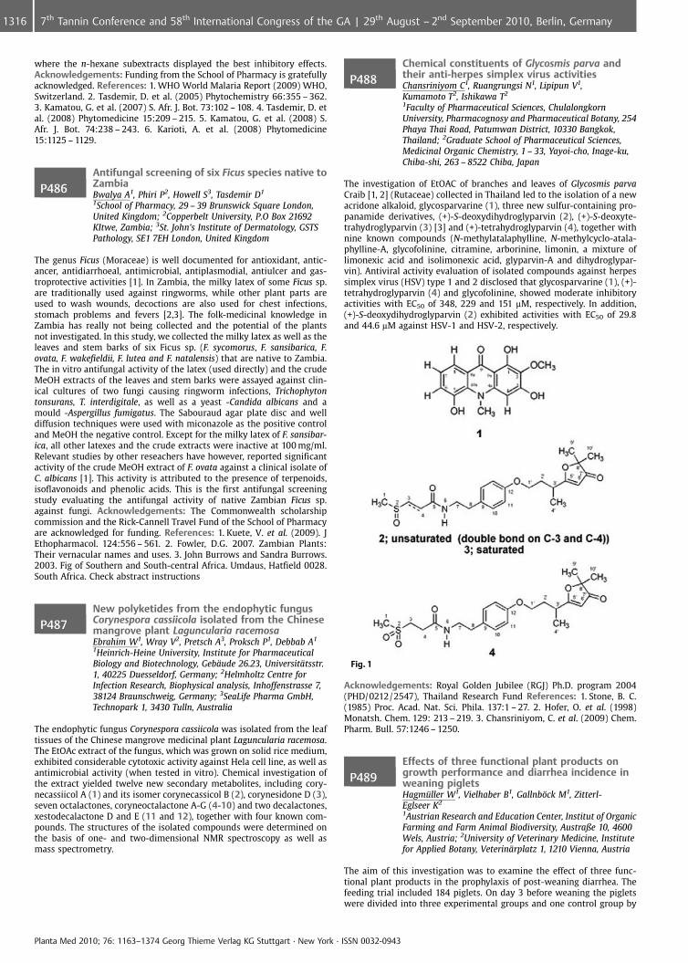
where the n-hexane subextracts displayed the best inhibitory effects.Acknowledgements: Funding from the School of Pharmacy is gratefullyacknowledged. References: 1. WHO World Malaria Report (2009) WHO,Switzerland. 2. Tasdemir, D. et al. (2005) Phytochemistry 66:355 – 362.3. Kamatou, G. et al. (2007) S. Afr. J. Bot. 73:102 – 108. 4. Tasdemir, D. etal. (2008) Phytomedicine 15:209 – 215. 5. Kamatou, G. et al. (2008) S.Afr. J. Bot. 74:238 – 243. 6. Karioti, A. et al. (2008) Phytomedicine15:1125 – 1129.
P486Antifungal screening of six Ficus species native toZambiaBwalya A1, Phiri P2, Howell S3, Tasdemir D1
1School of Pharmacy, 29 – 39 Brunswick Square London,United Kingdom; 2Copperbelt University, P.O Box 21692KItwe, Zambia; 3St. John’s Institute of Dermatology, GSTSPathology, SE1 7EH London, United Kingdom
The genus Ficus (Moraceae) is well documented for antioxidant, antic-ancer, antidiarrhoeal, antimicrobial, antiplasmodial, antiulcer and gas-troprotective activities [1]. In Zambia, the milky latex of some Ficus sp.are traditionally used against ringworms, while other plant parts areused to wash wounds, decoctions are also used for chest infections,stomach problems and fevers [2,3]. The folk-medicinal knowledge inZambia has really not being collected and the potential of the plantsnot investigated. In this study, we collected the milky latex as well as theleaves and stem barks of six Ficus sp. (F. sycomorus, F. sansibarica, F.ovata, F. wakefieldii, F. lutea and F. natalensis) that are native to Zambia.The in vitro antifungal activity of the latex (used directly) and the crudeMeOH extracts of the leaves and stem barks were assayed against clin-ical cultures of two fungi causing ringworm infections, Trichophytontonsurans, T. interdigitale, as well as a yeast -Candida albicans and amould -Aspergillus fumigatus. The Sabouraud agar plate disc and welldiffusion techniques were used with miconazole as the positive controland MeOH the negative control. Except for the milky latex of F. sansibar-ica, all other latexes and the crude extracts were inactive at 100 mg/ml.Relevant studies by other reseachers have however, reported significantactivity of the crude MeOH extract of F. ovata against a clinical isolate ofC. albicans [1]. This activity is attributed to the presence of terpenoids,isoflavonoids and phenolic acids. This is the first antifungal screeningstudy evaluating the antifungal activity of native Zambian Ficus sp.against fungi. Acknowledgements: The Commonwealth scholarshipcommission and the Rick-Cannell Travel Fund of the School of Pharmacyare acknowledged for funding. References: 1. Kuete, V. et al. (2009). JEthopharmacol. 124:556 – 561. 2. Fowler, D.G. 2007. Zambian Plants:Their vernacular names and uses. 3. John Burrows and Sandra Burrows.2003. Fig of Southern and South-central Africa. Umdaus, Hatfield 0028.South Africa. Check abstract instructions
P487New polyketides from the endophytic fungusCorynespora cassiicola isolated from the Chinesemangrove plant Laguncularia racemosaEbrahim W1, Wray V2, Pretsch A3, Proksch P1, Debbab A1
1Heinrich-Heine University, Institute for PharmaceuticalBiology and Biotechnology, Geb�ude 26.23, Universit�tsstr.1, 40225 Duesseldorf, Germany; 2Helmholtz Centre forInfection Research, Biophysical analysis, Inhoffenstrasse 7,38124 Braunschweig, Germany; 3SeaLife Pharma GmbH,Technopark 1, 3430 Tulln, Australia
The endophytic fungus Corynespora cassiicola was isolated from the leaftissues of the Chinese mangrove medicinal plant Laguncularia racemosa.The EtOAc extract of the fungus, which was grown on solid rice medium,exhibited considerable cytotoxic activity against Hela cell line, as well asantimicrobial activity (when tested in vitro). Chemical investigation ofthe extract yielded twelve new secondary metabolites, including cory-necassiicol A (1) and its isomer corynecassicol B (2), corynesidone D (3),seven octalactones, coryneoctalactone A-G (4-10) and two decalactones,xestodecalactone D and E (11 and 12), together with four known com-pounds. The structures of the isolated compounds were determined onthe basis of one- and two-dimensional NMR spectroscopy as well asmass spectrometry.
P488Chemical constituents of Glycosmis parva andtheir anti-herpes simplex virus activitiesChansriniyom C1, Ruangrungsi N1, Lipipun V1,Kumamoto T2, Ishikawa T2
1Faculty of Pharmaceutical Sciences, ChulalongkornUniversity, Pharmacognosy and Pharmaceutical Botany, 254Phaya Thai Road, Patumwan District, 10330 Bangkok,Thailand; 2Graduate School of Pharmaceutical Sciences,Medicinal Organic Chemistry, 1 – 33, Yayoi-cho, Inage-ku,Chiba-shi, 263 – 8522 Chiba, Japan
The investigation of EtOAC of branches and leaves of Glycosmis parvaCraib [1, 2] (Rutaceae) collected in Thailand led to the isolation of a newacridone alkaloid, glycosparvarine (1), three new sulfur-containing pro-panamide derivatives, (+)-S-deoxydihydroglyparvin (2), (+)-S-deoxyte-trahydroglyparvin (3) [3] and (+)-tetrahydroglyparvin (4), together withnine known compounds (N-methylatalaphylline, N-methylcyclo-atala-phylline-A, glycofolinine, citramine, arborinine, limonin, a mixture oflimonexic acid and isolimonexic acid, glyparvin-A and dihydroglypar-vin). Antiviral activity evaluation of isolated compounds against herpessimplex virus (HSV) type 1 and 2 disclosed that glycosparvarine (1), (+)-tetrahydroglyparvin (4) and glycofolinine, showed moderate inhibitoryactivities with EC50 of 348, 229 and 151 mM, respectively. In addition,(+)-S-deoxydihydroglyparvin (2) exhibited activities with EC50 of 29.8and 44.6 mM against HSV-1 and HSV-2, respectively.
Fig. 1
Acknowledgements: Royal Golden Jubilee (RGJ) Ph.D. program 2004(PHD/0212/2547), Thailand Research Fund References: 1. Stone, B. C.(1985) Proc. Acad. Nat. Sci. Phila. 137:1 – 27. 2. Hofer, O. et al. (1998)Monatsh. Chem. 129: 213 – 219. 3. Chansriniyom, C. et al. (2009) Chem.Pharm. Bull. 57:1246 – 1250.
P489Effects of three functional plant products ongrowth performance and diarrhea incidence inweaning pigletsHagm�ller W1, Vielhaber B1, Gallnb�ck M1, Zitterl-Eglseer K2
1Austrian Research and Education Center, Institut of OrganicFarming and Farm Animal Biodiversity, Austraße 10, 4600Wels, Austria; 2University of Veterinary Medicine, Institutefor Applied Botany, Veterin�rplatz 1, 1210 Vienna, Austria
The aim of this investigation was to examine the effect of three func-tional plant products in the prophylaxis of post-weaning diarrhea. Thefeeding trial included 184 piglets. On day 3 before weaning the pigletswere divided into three experimental groups and one control group by
Planta Med 2010; 76: 1163–1374 Georg Thieme Verlag KG Stuttgart · New York · ISSN 0032-0943
1316 7th Tannin Conference and 58th International Congress of the GA | 29th August – 2nd September 2010, Berlin, Germany
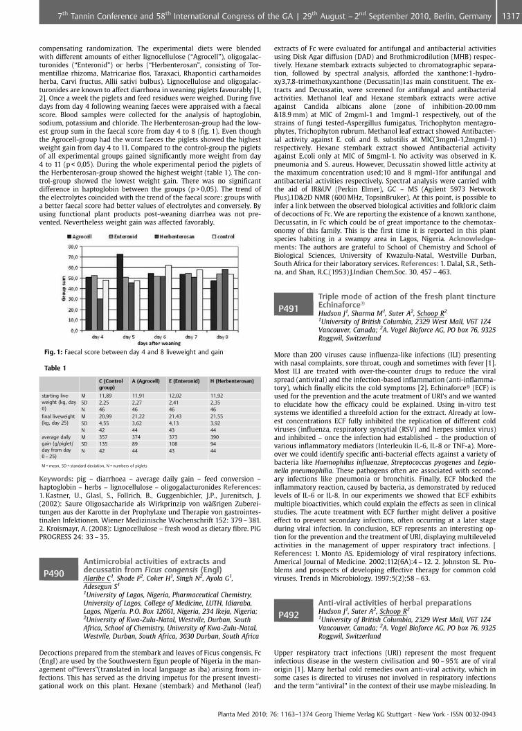
compensating randomization. The experimental diets were blendedwith different amounts of either lignocellulose (“Agrocell”), oligogalac-turonides (“Enteronid”) or herbs (“Herbenterosan”, consisting of Tor-mentillae rhizoma, Matricariae flos, Taraxaci, Rhapontici carthamoidesherba, Carvi fructus, Allii sativi bulbus). Lignocellulose and oligogalac-turonides are known to affect diarrhoea in weaning piglets favourably [1,2]. Once a week the piglets and feed residues were weighed. During fivedays from day 4 following weaning faeces were appraised with a faecalscore. Blood samples were collected for the analysis of haptoglobin,sodium, potassium and chloride. The Herbenterosan-group had the low-est group sum in the faecal score from day 4 to 8 (fig. 1). Even thoughthe Agrocell-group had the worst faeces the piglets showed the highestweight gain from day 4 to 11. Compared to the control-group the pigletsof all experimental groups gained significantly more weight from day4 to 11 (p < 0,05). During the whole experimental period the piglets ofthe Herbenterosan-group showed the highest weight (table 1). The con-trol-group showed the lowest weight gain. There was no significantdifference in haptoglobin between the groups (p > 0,05). The trend ofthe electrolytes coincided with the trend of the faecal score: groups witha better faecal score had better values of electrolytes and conversely. Byusing functional plant products post-weaning diarrhea was not pre-vented. Nevertheless weight gain was affected favorably.
Fig. 1: Faecal score between day 4 and 8 liveweight and gain
Table 1
C (Controlgroup)
A (Agrocell) E (Enteronid) H (Herbenterosan)
starting live-weight (kg, day0)
M 11,89 11,91 12,02 11,92SD 2,25 2,27 2,41 2,35N 46 46 46 46
final liveweight(kg, day 25)
M 20,99 21,22 21,43 21,55SD 4,55 3,62 4,13 3,92N 42 44 43 44
average dailygain (g/piglet/day from day0 – 25)
M 357 374 373 390SD 135 89 108 94N 42 44 43 44
M = mean, SD = standard deviation, N = numbers of piglets
Keywords: pig – diarrhoea – average daily gain – feed conversion –haptoglobin – herbs – lignocellulose – oligogalacturonides References:1. Kastner, U., Glasl, S., Follrich, B., Guggenbichler, J.P., Jurenitsch, J.(2002): Saure Oligosaccharide als Wirkprinzip von w�ßrigen Zuberei-tungen aus der Karotte in der Prophylaxe und Therapie von gastrointes-tinalen Infektionen. Wiener Medizinische Wochenschrift 152: 379 – 381.2. Kroismayr, A. (2008): Lignocellulose – fresh wood as dietary fibre. PIGPROGRESS 24: 33 – 35.
P490Antimicrobial activities of extracts anddecussatin from Ficus congensis (Engl)Alaribe C1, Shode F2, Coker H1, Singh N2, Ayola G1,Adesegun S1
1University of Lagos, Nigeria, Pharmaceutical Chemistry,University of Lagos, College of Medicine, LUTH, Idiaraba,Lagos, Nigeria. P.O. Box 12661, Nigeria, 234 Ikeja, Nigeria;2University of Kwa-Zulu-Natal, Westvile, Durban, SouthAfrica, School of Chemistry, University of Kwa-Zulu-Natal,Westvile, Durban, South Africa, 3630 Durban, South Africa
Decoctions prepared from the stembark and leaves of Ficus congensis, Fc(Engl) are used by the Southwestern Egun people of Nigeria in the man-agement of“fevers”(translated in local language as iba) arising from in-fections. This has served as the driving impetus for the present investi-gational work on this plant. Hexane (stembark) and Methanol (leaf)
extracts of Fc were evaluated for antifungal and antibacterial activitiesusing Disk Agar diffusion (DAD) and Brothmicrodilution (MHB) respec-tively. Hexane stembark extracts subjected to chromatographic separa-tion, followed by spectral analysis, afforded the xanthone:1-hydro-xy3,7,8-trimethoxyxanthone (Decussatin)1as main constituent. The ex-tracts and Decussatin, were screened for antifungal and antibacterialactivities. Methanol leaf and Hexane stembark extracts were activeagainst Candida albicans alone (zone of inhibition-20.00 mm&18.9 mm) at MIC of 2mgml-1 and 1mgml-1 respectively, out of thestrains of fungi tested-Aspergillus fumigatus, Trichophyton mentagro-phytes, Trichophyton rubrum. Methanol leaf extract showed Antibacter-ial activity against E. coli and B. substilis at MIC(3mgml-1,2mgml-1)respectively. Hexane stembark extract showed Antibacterial activityagainst E.coli only at MIC of 5mgml-1. No activity was observed in K.pneumonia and S. aureus. However, Decussatin showed little activity atthe maximum concentration used;10 and 8 mgml-1for antifungal andantibacterial activities respectively. Spectral analysis were carried withthe aid of IR&UV (Perkin Elmer), GC – MS (Agilent 5973 NetworkPlus),1D&2D NMR (600 MHz, TopsinBruker). At this point, is possible toinfer a link between the observed biological activities and folkloric claimof decoctions of Fc. We are reporting the existence of a known xanthone,Decussatin, in Fc which could be of great importance to the chemotax-onomy of this family. This is the first time it is reported in this plantspecies habiting in a swampy area in Lagos, Nigeria. Acknowledge-ments: The authors are grateful to School of Chemistry and School ofBiological Sciences, University of Kwazulu-Natal, Westville Durban,South Africa for their laboratory services. References: 1. Dalal, S.R., Seth-na, and Shan, R.C.(1953) J.Indian Chem.Soc. 30, 457 – 463.
P491Triple mode of action of the fresh plant tinctureEchinaforce�
Hudson J1, Sharma M1, Suter A2, Schoop R2
1University of British Columbia, 2329 West Mall, V6T 1Z4Vancouver, Canada; 2A. Vogel Bioforce AG, PO box 76, 9325Roggwil, Switzerland
More than 200 viruses cause influenza-like infections (ILI) presentingwith nasal complaints, sore throat, cough and sometimes with fever [1].Most ILI are treated with over-the-counter drugs to reduce the viralspread (antiviral) and the infection-based inflammation (anti-inflamma-tory), which finally elicits the cold symptoms [2]. Echinaforce� (ECF) isused for the prevention and the acute treatment of URI’s and we wantedto elucidate how the efficacy could be explained. Using in-vitro testsystems we identified a threefold action for the extract. Already at low-est concentrations ECF fully inhibited the replication of different coldviruses (influenza, respiratory syncytial (RSV) and herpes simlex virus)and inhibited – once the infection had established – the production ofvarious inflammatory mediators (Interleukin IL-6, IL-8 or TNF-a). More-over we could identify specific anti-bacterial effects against a variety ofbacteria like Haemophilus influenzae, Streptococcus pyogenes and Legio-nella pneumophilia. These pathogens often are associated with second-ary infections like pneumonia or bronchitis. Finally, ECF blocked theinflammatory reaction, caused by bacteria, as demonstrated by reducedlevels of IL-6 or IL-8. In our experiments we showed that ECF exhibitsmultiple bioactivities, which could explain the effects as seen in clinicalstudies. The acute treatment with ECF further might deliver a positiveeffect to prevent secondary infections, often occurring at a later stageduring viral infection. In conclusion, ECF represents an interesting op-tion for the prevention and the treatment of URI, displaying multileveledactivities in the management of upper respiratory tract infections. [References: 1. Monto AS. Epidemiology of viral respiratory infections.Americal Journal of Medicine. 2002;112(6A):4 – 12. 2. Johnston SL. Pro-blems and prospects of developing effective therapy for common coldviruses. Trends in Microbiology. 1997;5(2);58 – 63.
P492Anti-viral activities of herbal preparationsHudson J1, Suter A2, Schoop R2
1University of British Columbia, 2329 West Mall, V6T 1Z4Vancouver, Canada; 2A. Vogel Bioforce AG, PO box 76, 9325Roggwil, Switzerland
Upper respiratory tract infections (URI) represent the most frequentinfectious disease in the western civilisation and 90 – 95% are of viralorigin [1]. Many herbal cold remedies own anti-viral activity, which insome cases is directed to viruses not involved in respiratory infectionsand the term “antiviral” in the context of their use maybe misleading. In
Planta Med 2010; 76: 1163–1374 Georg Thieme Verlag KG Stuttgart · New York · ISSN 0032-0943
13177th Tannin Conference and 58th International Congress of the GA | 29th August – 2nd September 2010, Berlin, Germany
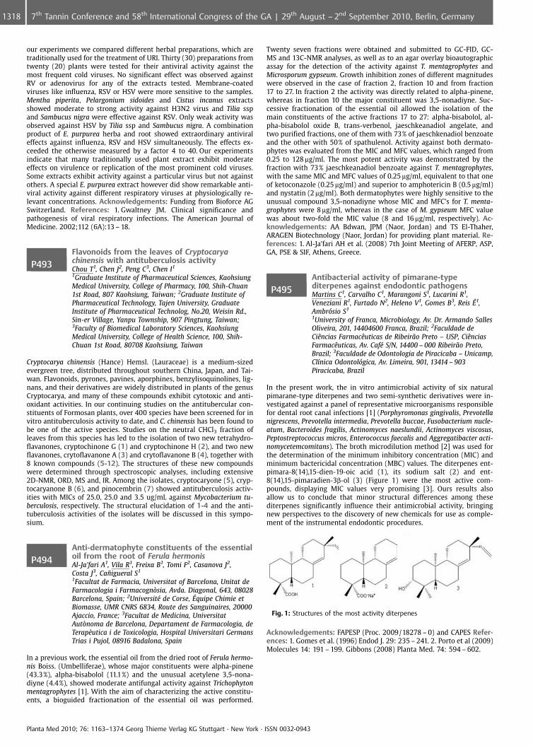
our experiments we compared different herbal preparations, which aretraditionally used for the treatment of URI. Thirty (30) preparations fromtwenty (20) plants were tested for their antiviral activity against themost frequent cold viruses. No significant effect was observed againstRV or adenovirus for any of the extracts tested. Membrane-coatedviruses like influenza, RSV or HSV were more sensitive to the samples.Mentha piperita, Pelargonium sidoides and Cistus incanus extractsshowed moderate to strong activity against H3N2 virus and Tilia sspand Sambucus nigra were effective against RSV. Only weak activity wasobserved against HSV by Tilia ssp and Sambucus nigra. A combinationproduct of E. purpurea herba and root showed extraordinary antiviraleffects against influenza, RSV and HSV simultaneously. The effects ex-ceeded the otherwise measured by a factor 4 to 40. Our experimentsindicate that many traditionally used plant extract exhibit moderateeffects on virulence or replication of the most prominent cold viruses.Some extracts exhibit activity against a particular virus but not againstothers. A special E. purpurea extract however did show remarkable anti-viral activity against different respiratory viruses at physiologically re-levant concentrations. Acknowledgements: Funding from Bioforce AGSwitzerland. References: 1. Gwaltney JM. Clinical significance andpathogenesis of viral respiratory infections. The American Journal ofMedicine. 2002;112 (6A):13 – 18.
P493Flavonoids from the leaves of Cryptocaryachinensis with antituberculosis activityChou T1, Chen J2, Peng C3, Chen I1
1Graduate Institute of Pharmaceutical Sciences, KaohsiungMedical University, College of Pharmacy, 100, Shih-Chuan1st Road, 807 Kaohsiung, Taiwan; 2Graduate Institute ofPharmaceutical Technology, Tajen University, GraduateInstitute of Pharmaceutical Technolog, No.20, Weisin Rd.,Sin-er Village, Yanpu Township, 907 Pingtung, Taiwan;3Faculty of Biomedical Laboratory Sciences, KaohsiungMedical University, College of Health Science, 100, Shih-Chuan 1st Road, 80708 Kaohsiung, Taiwan
Cryptocarya chinensis (Hance) Hemsl. (Lauraceae) is a medium-sizedevergreen tree, distributed throughout southern China, Japan, and Tai-wan. Flavonoids, pyrones, pavines, aporphines, benzylisoquinolines, lig-nans, and their derivatives are widely distributed in plants of the genusCryptocarya, and many of these compounds exhibit cytotoxic and anti-oxidant activities. In our continuing studies on the antitubercular con-stituents of Formosan plants, over 400 species have been screened for invitro antituberculosis activity to date, and C. chinensis has been found tobe one of the active species. Studies on the neutral CHCl3 fraction ofleaves from this species has led to the isolation of two new tetrahydro-flavanones, cryptochinone G (1) and cryptochinone H (2), and two newflavanones, crytoflavanone A (3) and crytoflavanone B (4), together with8 known compounds (5-12). The structures of these new compoundswere determined through spectroscopic analyses, including extensive2D-NMR, ORD, MS and, IR. Among the isolates, cryptocaryone (5), cryp-tocaryanone B (6), and pinocembrin (7) showed antituberculosis activ-ities with MICs of 25.0, 25.0 and 3.5 ug/mL against Mycobacterium tu-berculosis, respectively. The structural elucidation of 1-4 and the anti-tuberculosis activities of the isolates will be discussed in this sympo-sium.
P494Anti-dermatophyte constituents of the essentialoil from the root of Ferula hermonisAl-Ja’fari A1, Vila R1, Freixa B1, Tomi F2, Casanova J2,Costa J3, Ca�igueral S1
1Facultat de Farmacia, Universitat of Barcelona, Unitat deFarmacologia i Farmacogn�sia, Avda. Diagonal, 643, 08028Barcelona, Spain; 2Universit� de Corse, �quipe Chimie etBiomasse, UMR CNRS 6834, Route des Sanguinaires, 20000Ajaccio, France; 3Facultat de Medicina, UniversitatAut�noma de Barcelona, Departament de Farmacologia, deTerap�utica i de Toxicologia, Hospital Universitari GermansTrias i Pujol, 08916 Badalona, Spain
In a previous work, the essential oil from the dried root of Ferula hermo-nis Boiss. (Umbelliferae), whose major constituents were alpha-pinene(43.3%), alpha-bisabolol (11.1%) and the unusual acetylene 3,5-nona-diyne (4.4%), showed moderate antifungal activity against Trichophytonmentagrophytes [1]. With the aim of characterizing the active constitu-ents, a bioguided fractionation of the essential oil was performed.
Twenty seven fractions were obtained and submitted to GC-FID, GC-MS and 13C-NMR analyses, as well as to an agar overlay bioautographicassay for the detection of the activity against T. mentagrophytes andMicrosporum gypseum. Growth inhibition zones of different magnitudeswere observed in the case of fraction 2, fraction 10 and from fraction17 to 27. In fraction 2 the activity was directly related to alpha-pinene,whereas in fraction 10 the major constituent was 3,5-nonadiyne. Suc-cessive fractionation of the essential oil allowed the isolation of themain constituents of the active fractions 17 to 27: alpha-bisabolol, al-pha-bisabolol oxide B, trans-verbenol, jaeschkeanadiol angelate, andtwo purified fractions, one of them with 73% of jaeschkenadiol benzoateand the other with 50% of spathulenol. Activity against both dermato-phytes was evaluated from the MIC and MFC values, which ranged from0.25 to 128 mg/ml. The most potent activity was demonstrated by thefraction with 73% jaeschkeanadiol benzoate against T. mentagrophytes,with the same MIC and MFC values of 0.25 mg/ml, equivalent to that oneof ketoconazole (0.25 mg/ml) and superior to amphotericin B (0.5 mg/ml)and nystatin (2 mg/ml). Both dermatophytes were highly sensitive to theunusual compound 3,5-nonadiyne whose MIC and MFC’s for T. menta-grophytes were 8 mg/ml, whereas in the case of M. gypseum MFC valuewas about two-fold the MIC value (8 and 16 mg/ml, respectively). Ac-knowledgements: AA Bdwan, JPM (Naor, Jordan) and TS El-Thaher,ARAGEN Biotechnology (Naor, Jordan) for providing plant material. Re-ferences: 1. Al-Ja’fari AH et al. (2008) 7th Joint Meeting of AFERP, ASP,GA, PSE & SIF, Athens, Greece.
P495Antibacterial activity of pimarane-typediterpenes against endodontic pathogensMartins C1, Carvalho C1, Marangoni S1, Lucarini R1,Veneziani R1, Furtado N2, Heleno V1, Gomes B3, Reis �1,Ambrsio S1
1University of Franca, Microbiology, Av. Dr. Armando SallesOliveira, 201, 14404600 Franca, Brazil; 2Faculdade deCiÞncias FarmacÞuticas de Ribeir¼o Preto – USP, CiÞnciasFarmacÞuticas, Av. Caf� S/N, 14400 – 000 Ribeir¼o Preto,Brazil; 3Faculdade de Odontologia de Piracicaba – Unicamp,Cl�nica Odontolgica, Av. Limeira, 901, 13414 – 903Piracicaba, Brazil
In the present work, the in vitro antimicrobial activity of six naturalpimarane-type diterpenes and two semi-synthetic derivatives were in-vestigated against a panel of representative microorganisms responsiblefor dental root canal infections [1] (Porphyromonas gingivalis, Prevotellanigrescens, Prevotella intermedia, Prevotella buccae, Fusobacterium nucle-atum, Bacteroides fragilis, Actinomyces naeslundii, Actinomyces viscosus,Peptostreptococcus micros, Enterococcus faecalis and Aggregatibacter acti-nomycetemcomitans). The broth microdilution method [2] was used forthe determination of the minimum inhibitory concentration (MIC) andminimum bactericidal concentration (MBC) values. The diterpenes ent-pimara-8(14),15-dien-19-oic acid (1), its sodium salt (2) and ent-8(14),15-pimaradien-3b-ol (3) (Figure 1) were the most active com-pounds, displaying MIC values very promising [3]. Ours results alsoallow us to conclude that minor structural differences among thesediterpenes significantly influence their antimicrobial activity, bringingnew perspectives to the discovery of new chemicals for use as comple-ment of the instrumental endodontic procedures.
Fig. 1: Structures of the most activity diterpenes
Acknowledgements: FAPESP (Proc. 2009/18278 – 0) and CAPES Refer-ences: 1. Gomes et al. (1996) Endod J. 29: 235 – 241. 2. Porto et al (2009)Molecules 14: 191 – 199. Gibbons (2008) Planta Med. 74: 594 – 602.
Planta Med 2010; 76: 1163–1374 Georg Thieme Verlag KG Stuttgart · New York · ISSN 0032-0943
1318 7th Tannin Conference and 58th International Congress of the GA | 29th August – 2nd September 2010, Berlin, Germany
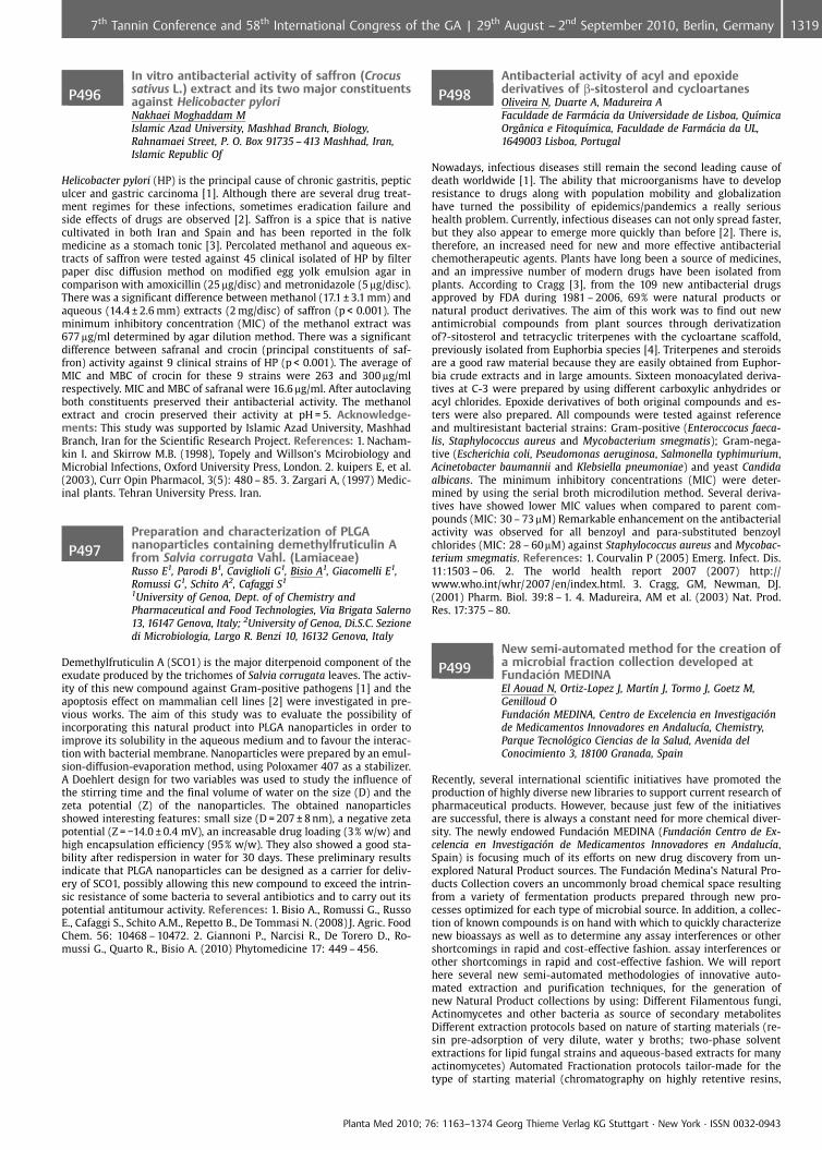
P496In vitro antibacterial activity of saffron (Crocussativus L.) extract and its two major constituentsagainst Helicobacter pyloriNakhaei Moghaddam MIslamic Azad University, Mashhad Branch, Biology,Rahnamaei Street, P. O. Box 91735 – 413 Mashhad, Iran,Islamic Republic Of
Helicobacter pylori (HP) is the principal cause of chronic gastritis, pepticulcer and gastric carcinoma [1]. Although there are several drug treat-ment regimes for these infections, sometimes eradication failure andside effects of drugs are observed [2]. Saffron is a spice that is nativecultivated in both Iran and Spain and has been reported in the folkmedicine as a stomach tonic [3]. Percolated methanol and aqueous ex-tracts of saffron were tested against 45 clinical isolated of HP by filterpaper disc diffusion method on modified egg yolk emulsion agar incomparison with amoxicillin (25 mg/disc) and metronidazole (5 mg/disc).There was a significant difference between methanol (17.1 € 3.1 mm) andaqueous (14.4 € 2.6 mm) extracts (2 mg/disc) of saffron (p < 0.001). Theminimum inhibitory concentration (MIC) of the methanol extract was677 mg/ml determined by agar dilution method. There was a significantdifference between safranal and crocin (principal constituents of saf-fron) activity against 9 clinical strains of HP (p < 0.001). The average ofMIC and MBC of crocin for these 9 strains were 263 and 300 mg/mlrespectively. MIC and MBC of safranal were 16.6 mg/ml. After autoclavingboth constituents preserved their antibacterial activity. The methanolextract and crocin preserved their activity at pH = 5. Acknowledge-ments: This study was supported by Islamic Azad University, MashhadBranch, Iran for the Scientific Research Project. References: 1. Nacham-kin I. and Skirrow M.B. (1998), Topely and Willson’s Mcirobiology andMicrobial Infections, Oxford University Press, London. 2. kuipers E, et al.(2003), Curr Opin Pharmacol, 3(5): 480 – 85. 3. Zargari A, (1997) Medic-inal plants. Tehran University Press. Iran.
P497Preparation and characterization of PLGAnanoparticles containing demethylfruticulin Afrom Salvia corrugata Vahl. (Lamiaceae)Russo E1, Parodi B1, Caviglioli G1, Bisio A1, Giacomelli E1,Romussi G1, Schito A2, Cafaggi S1
1University of Genoa, Dept. of of Chemistry andPharmaceutical and Food Technologies, Via Brigata Salerno13, 16147 Genova, Italy; 2University of Genoa, Di.S.C. Sezionedi Microbiologia, Largo R. Benzi 10, 16132 Genova, Italy
Demethylfruticulin A (SCO1) is the major diterpenoid component of theexudate produced by the trichomes of Salvia corrugata leaves. The activ-ity of this new compound against Gram-positive pathogens [1] and theapoptosis effect on mammalian cell lines [2] were investigated in pre-vious works. The aim of this study was to evaluate the possibility ofincorporating this natural product into PLGA nanoparticles in order toimprove its solubility in the aqueous medium and to favour the interac-tion with bacterial membrane. Nanoparticles were prepared by an emul-sion-diffusion-evaporation method, using Poloxamer 407 as a stabilizer.A Doehlert design for two variables was used to study the influence ofthe stirring time and the final volume of water on the size (D) and thezeta potential (Z) of the nanoparticles. The obtained nanoparticlesshowed interesting features: small size (D = 207 € 8 nm), a negative zetapotential (Z = �14.0 € 0.4 mV), an increasable drug loading (3% w/w) andhigh encapsulation efficiency (95% w/w). They also showed a good sta-bility after redispersion in water for 30 days. These preliminary resultsindicate that PLGA nanoparticles can be designed as a carrier for deliv-ery of SCO1, possibly allowing this new compound to exceed the intrin-sic resistance of some bacteria to several antibiotics and to carry out itspotential antitumour activity. References: 1. Bisio A., Romussi G., RussoE., Cafaggi S., Schito A.M., Repetto B., De Tommasi N. (2008) J. Agric. FoodChem. 56: 10468 – 10472. 2. Giannoni P., Narcisi R., De Torero D., Ro-mussi G., Quarto R., Bisio A. (2010) Phytomedicine 17: 449 – 456.
P498Antibacterial activity of acyl and epoxidederivatives of b-sitosterol and cycloartanesOliveira N, Duarte A, Madureira AFaculdade de Farmcia da Universidade de Lisboa, Qu�micaOrg nica e Fitoqu�mica, Faculdade de Farmcia da UL,1649003 Lisboa, Portugal
Nowadays, infectious diseases still remain the second leading cause ofdeath worldwide [1]. The ability that microorganisms have to developresistance to drugs along with population mobility and globalizationhave turned the possibility of epidemics/pandemics a really serioushealth problem. Currently, infectious diseases can not only spread faster,but they also appear to emerge more quickly than before [2]. There is,therefore, an increased need for new and more effective antibacterialchemotherapeutic agents. Plants have long been a source of medicines,and an impressive number of modern drugs have been isolated fromplants. According to Cragg [3], from the 109 new antibacterial drugsapproved by FDA during 1981 – 2006, 69% were natural products ornatural product derivatives. The aim of this work was to find out newantimicrobial compounds from plant sources through derivatizationof?-sitosterol and tetracyclic triterpenes with the cycloartane scaffold,previously isolated from Euphorbia species [4]. Triterpenes and steroidsare a good raw material because they are easily obtained from Euphor-bia crude extracts and in large amounts. Sixteen monoacylated deriva-tives at C-3 were prepared by using different carboxylic anhydrides oracyl chlorides. Epoxide derivatives of both original compounds and es-ters were also prepared. All compounds were tested against referenceand multiresistant bacterial strains: Gram-positive (Enteroccocus faeca-lis, Staphylococcus aureus and Mycobacterium smegmatis); Gram-nega-tive (Escherichia coli, Pseudomonas aeruginosa, Salmonella typhimurium,Acinetobacter baumannii and Klebsiella pneumoniae) and yeast Candidaalbicans. The minimum inhibitory concentrations (MIC) were deter-mined by using the serial broth microdilution method. Several deriva-tives have showed lower MIC values when compared to parent com-pounds (MIC: 30 – 73 mM) Remarkable enhancement on the antibacterialactivity was observed for all benzoyl and para-substituted benzoylchlorides (MIC: 28 – 60mM) against Staphylococcus aureus and Mycobac-terium smegmatis. References: 1. Courvalin P (2005) Emerg. Infect. Dis.11:1503 – 06. 2. The world health report 2007 (2007) http://www.who.int/whr/2007/en/index.html. 3. Cragg, GM, Newman, DJ.(2001) Pharm. Biol. 39:8 – 1. 4. Madureira, AM et al. (2003) Nat. Prod.Res. 17:375 – 80.
P499New semi-automated method for the creation ofa microbial fraction collection developed atFundaci�n MEDINAEl Aouad N, Ortiz-Lopez J, Mart�n J, Tormo J, Goetz M,Genilloud OFundacin MEDINA, Centro de Excelencia en Investigacinde Medicamentos Innovadores en Andaluc�a, Chemistry,Parque Tecnolgico Ciencias de la Salud, Avenida delConocimiento 3, 18100 Granada, Spain
Recently, several international scientific initiatives have promoted theproduction of highly diverse new libraries to support current research ofpharmaceutical products. However, because just few of the initiativesare successful, there is always a constant need for more chemical diver-sity. The newly endowed Fundaci n MEDINA (Fundacin Centro de Ex-celencia en Investigacin de Medicamentos Innovadores en Andaluc�a,Spain) is focusing much of its efforts on new drug discovery from un-explored Natural Product sources. The Fundaci n Medina’s Natural Pro-ducts Collection covers an uncommonly broad chemical space resultingfrom a variety of fermentation products prepared through new pro-cesses optimized for each type of microbial source. In addition, a collec-tion of known compounds is on hand with which to quickly characterizenew bioassays as well as to determine any assay interferences or othershortcomings in rapid and cost-effective fashion. assay interferences orother shortcomings in rapid and cost-effective fashion. We will reporthere several new semi-automated methodologies of innovative auto-mated extraction and purification techniques, for the generation ofnew Natural Product collections by using: Different Filamentous fungi,Actinomycetes and other bacteria as source of secondary metabolitesDifferent extraction protocols based on nature of starting materials (re-sin pre-adsorption of very dilute, water y broths; two-phase solventextractions for lipid fungal strains and aqueous-based extracts for manyactinomycetes) Automated Fractionation protocols tailor-made for thetype of starting material (chromatography on highly retentive resins,
Planta Med 2010; 76: 1163–1374 Georg Thieme Verlag KG Stuttgart · New York · ISSN 0032-0943
13197th Tannin Conference and 58th International Congress of the GA | 29th August – 2nd September 2010, Berlin, Germany
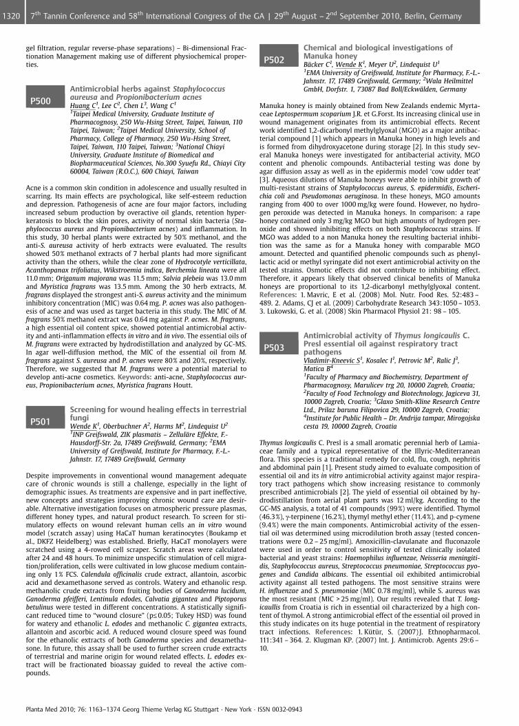
gel filtration, regular reverse-phase separations) – Bi-dimensional Frac-tionation Management making use of different physiochemical proper-ties.
P500Antimicrobial herbs against Staphylococcusaureusa and Propionibacterium acnesHuang C1, Lee C2, Chen L3, Wang C1
1Taipei Medical University, Graduate Institute ofPharmacognosy, 250 Wu-Hsing Street, Taipei, Taiwan, 110Taipei, Taiwan; 2Taipei Medical University, School ofPharmacy, College of Pharmacy, 250 Wu-Hsing Street,Taipei, Taiwan, 110 Taipei, Taiwan; 3National ChiayiUniversity, Graduate Institute of Biomedical andBiopharmaceutical Sciences, No.300 Syuefu Rd., Chiayi City60004, Taiwan (R.O.C.), 600 Chiayi, Taiwan
Acne is a common skin condition in adolescence and usually resulted inscarring. Its main effects are psychological, like self-esteem reductionand depression. Pathogenesis of acne are four major factors, includingincreased sebum production by overactive oil glands, retention hyper-keratosis to block the skin pores, activity of normal skin bacteria (Sta-phylococcus aureus and Propionibacterium acnes) and inflammation. Inthis study, 30 herbal plants were extracted by 50% methanol, and theanti-S. aureusa activity of herb extracts were evaluated. The resultsshowed 50% methanol extracts of 7 herbal plants had more significantactivity than the others, while the clear zone of Hydrocotyle verticillata,Acanthopanax trifoliatus, Wikstroemia indica, Berchemia lineata were all11.0 mm; Origanum majorana was 11.5 mm; Salvia plebeia was 13.0 mmand Myristica fragrans was 13.5 mm. Among the 30 herb extracts, M.fragrans displayed the strongest anti-S. aureus activity and the minimuminhibitory concentration (MIC) was 0.64 mg. P. acnes was also pathogen-esis of acne and was used as target bacteria in this study. The MIC of M.fragrans 50% methanol extract was 0.64 mg against P. acnes. M. fragrans,a high essential oil content spice, showed potential antimicrobial activ-ity and anti-inflammation effects in vitro and in vivo. The essential oils ofM. fragrans were extracted by hydrodistillation and analyzed by GC-MS.In agar well-diffusion method, the MIC of the essential oil from M.fragrans against S. aureusa and P. acnes were 80% and 20%, respectively.Therefore, we suggested that M. fragrans were a potential material todevelop anti-acne cosmetics. Keywords: anti-acne, Staphylococcus aur-eus, Propionibacterium acnes, Myristica fragrans Houtt.
P501Screening for wound healing effects in terrestrialfungiWende K1, Oberbuchner A2, Harms M2, Lindequist U2
1INP Greifswald, ZIK plasmatis – Zellul�re Effekte, F.-Hausdorff-Str. 2a, 17489 Greifswald, Germany; 2EMAUniversity of Greifswald, Institute for Pharmacy, F.-L.-Jahnstr. 17, 17489 Greifswald, Germany
Despite improvements in conventional wound management adequatecare of chronic wounds is still a challenge, especially in the light ofdemographic issues. As treatments are expensive and in part ineffective,new concepts and strategies improving chronic wound care are desir-able. Alternative investigation focuses on atmospheric pressure plasmas,different honey types, and natural product research. To screen for sti-mulatory effects on wound relevant human cells an in vitro woundmodel (scratch assay) using HaCaT human keratinocytes (Boukamp etal., DKFZ Heidelberg) was established. Briefly, HaCaT monolayers werescratched using a 4-rowed cell scraper. Scratch areas were calculatedafter 24 and 48 hours. To minimize unspecific stimulation of cell migra-tion/proliferation, cells were cultivated in low glucose medium contain-ing only 1% FCS. Calendula officinalis crude extract, allantoin, ascorbicacid and dexamethasone served as controls. Watery and ethanolic resp.methanolic crude extracts from fruiting bodies of Ganoderma lucidum,Ganoderma pfeifferi, Lentinula edodes, Calvatia gigantea and Piptoporusbetulinus were tested in different concentrations. A statistically signifi-cant reduced time to “wound closure” (p£0.05; Tukey HSD) was foundfor watery and ethanolic L. edodes and methanolic C. gigantea extracts,allantoin and ascorbic acid. A reduced wound closure speed was foundfor the ethanolic extracts of both Ganoderma species and dexametha-sone. In future, this assay shall be used to further screen crude extractsof terrestrial and marine origin for wound related effects. L. edodes ex-tract will be fractionated bioassay guided to reveal the active com-pounds.
P502Chemical and biological investigations ofManuka honeyB�cker C1, Wende K1, Meyer U2, Lindequist U1
1EMA University of Greifswald, Institute for Pharmacy, F.-L.-Jahnstr. 17, 17489 Greifswald, Germany; 2Wala HeilmittelGmbH, Dorfstr. 1, 73087 Bad Boll/Eckw�lden, Germany
Manuka honey is mainly obtained from New Zealands endemic Myrta-ceae Leptospermum scoparium J.R. et G.Forst. Its increasing clinical use inwound management originates from its antimicrobial effects. Recentwork identified 1,2-dicarbonyl methylglyoxal (MGO) as a major antibac-terial compound [1] which appears in Manuka honey in high levels andis formed from dihydroxyacetone during storage [2]. In this study sev-eral Manuka honeys were investigated for antibacterial activity, MGOcontent and phenolic compounds. Antibacterial testing was done byagar diffusion assay as well as in the epidermis model ‘cow udder teat’[3]. Aqueous dilutions of Manuka honeys were able to inhibit growth ofmulti-resistant strains of Staphylococcus aureus, S. epidermidis, Escheri-chia coli and Pseudomonas aeruginosa. In these honeys, MGO amountsranging from 400 to over 1000 mg/kg were found. However, no hydro-gen peroxide was detected in Manuka honeys. In comparison: a rapehoney contained only 3 mg/kg MGO but high amounts of hydrogen per-oxide and showed inhibiting effects on both Staphylococcus strains. IfMGO was added to a non Manuka honey the resulting bacterial inhibi-tion was the same as for a Manuka honey with comparable MGOamount. Detected and quantified phenolic compounds such as phenyl-lactic acid or methyl syringate did not exert antimicrobial activity on thetested strains. Osmotic effects did not contribute to inhibiting effect.Therefore, it appears likely that observed clinical benefits of Manukahoneys are proportional to its 1,2-dicarbonyl methylglyoxal content.References: 1. Mavric, E et al. (2008) Mol. Nutr. Food Res. 52:483 –489. 2. Adams, CJ et al. (2009) Carbohydrate Research 343:1050 – 1053.3. Lukowski, G. et al. (2008) Skin Pharmacol Physiol 21: 98 – 105.
P503Antimicrobial activity of Thymus longicaulis C.Presl essential oil against respiratory tractpathogensVladimir-Kneevic S1, Kosalec I1, Petrovic M2, Ralic J3,Matica B4
1Faculty of Pharmacy and Biochemistry, Department ofPharmacognosy, Marulicev trg 20, 10000 Zagreb, Croatia;2Faculty of Food Technology and Biotechnology, Jagiceva 31,10000 Zagreb, Croatia; 3Glaxo Smith-Kline Research CentreLtd., Prilaz baruna Filipovica 29, 10000 Zagreb, Croatia;4Institute for Public Health – Dr. Andrija tampar, Mirogojskacesta 19, 10000 Zagreb, Croatia
Thymus longicaulis C. Presl is a small aromatic perennial herb of Lamia-ceae family and a typical representative of the Illyric-Mediterraneanflora. This species is a traditional remedy for cold, flu, cough, nephritisand abdominal pain [1]. Present study aimed to evaluate composition ofessential oil and its in vitro antimicrobial activity against major respira-tory tract pathogens which show increasing resistance to commonlyprescribed antimicrobials [2]. The yield of essential oil obtained by hy-drodistillation from aerial plant parts was 12 ml/kg. According to theGC-MS analysis, a total of 41 compounds (99%) were identified. Thymol(46.3%), g-terpinene (16.2%), thymyl methyl ether (11.4%), and p-cymene(9.4%) were the main components. Antimicrobial activity of the essen-tial oil was determined using microdilution broth assay (tested concen-trations were 0.2 – 25 mg/ml). Amoxicillin-clavulanate and fluconazolewere used in order to control sensitivity of tested clinically isolatedbacterial and yeast strains: Haemophilus influenzae, Neisseria meningiti-dis, Staphylococcus aureus, Streptococcus pneumoniae, Streptococcus pyo-genes and Candida albicans. The essential oil exhibited antimicrobialactivity against all tested pathogens. The most sensitive strains wereH. influenzae and S. pneumoniae (MIC 0.78 mg/ml), while S. aureus wasthe most resistant (MIC > 25 mg/ml). Our results revealed that T. long-icaullis from Croatia is rich in essential oil characterized by a high con-tent of thymol. A strong antimicrobial effect of the essential oil proved inthis study indicates on its huge potential in the treatment of respiratorytract infections. References: 1. Ktr, S. (2007) J. Ethnopharmacol.111:341 – 364. 2. Klugman KP. (2007) Int. J. Antimicrob. Agents 29:6 –10.
Planta Med 2010; 76: 1163–1374 Georg Thieme Verlag KG Stuttgart · New York · ISSN 0032-0943
1320 7th Tannin Conference and 58th International Congress of the GA | 29th August – 2nd September 2010, Berlin, Germany
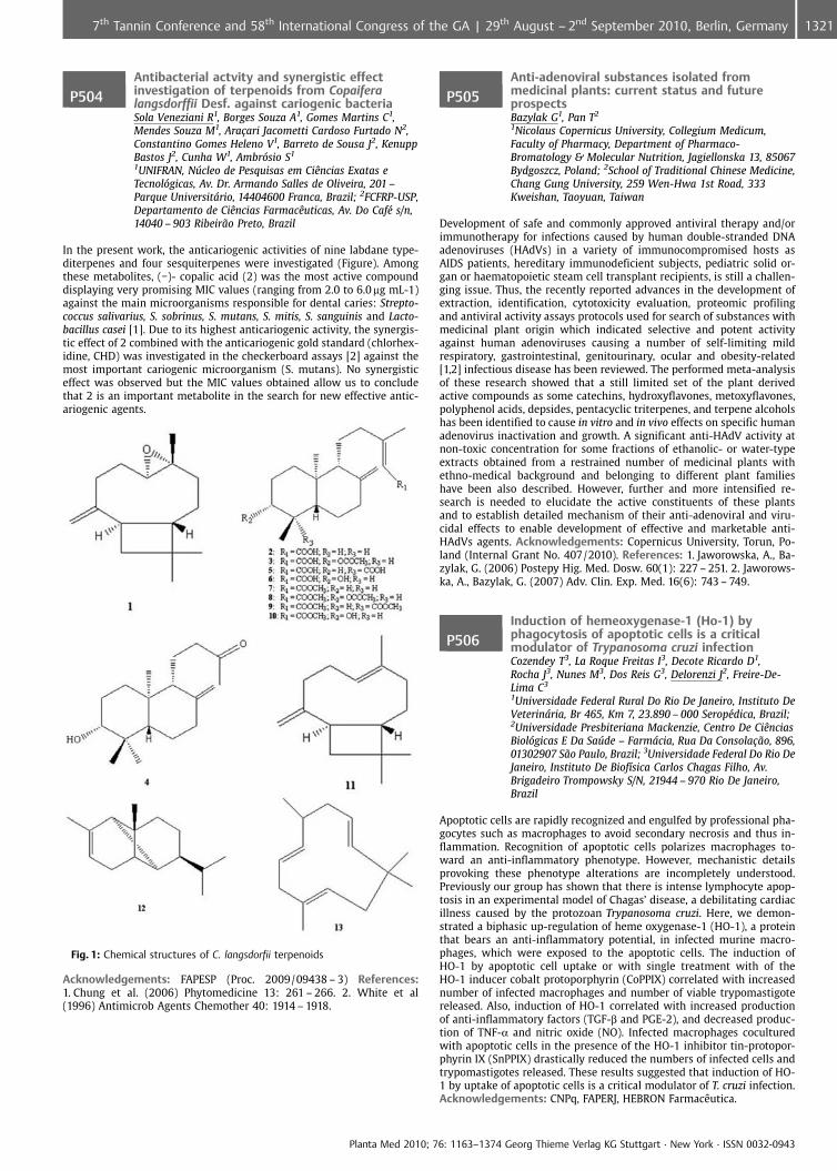
P504Antibacterial actvity and synergistic effectinvestigation of terpenoids from Copaiferalangsdorffii Desf. against cariogenic bacteriaSola Veneziani R1, Borges Souza A1, Gomes Martins C1,Mendes Souza M1, Ara�ari Jacometti Cardoso Furtado N2,Constantino Gomes Heleno V1, Barreto de Sousa J2, KenuppBastos J2, Cunha W1, Ambrsio S1
1UNIFRAN, Nfflcleo de Pesquisas em CiÞncias Exatas eTecnolgicas, Av. Dr. Armando Salles de Oliveira, 201 –Parque Universitrio, 14404600 Franca, Brazil; 2FCFRP-USP,Departamento de CiÞncias FarmacÞuticas, Av. Do Caf� s/n,14040 – 903 Ribeir¼o Preto, Brazil
In the present work, the anticariogenic activities of nine labdane type-diterpenes and four sesquiterpenes were investigated (Figure). Amongthese metabolites, (�)- copalic acid (2) was the most active compounddisplaying very promising MIC values (ranging from 2.0 to 6.0 mg mL-1)against the main microorganisms responsible for dental caries: Strepto-coccus salivarius, S. sobrinus, S. mutans, S. mitis, S. sanguinis and Lacto-bacillus casei [1]. Due to its highest anticariogenic activity, the synergis-tic effect of 2 combined with the anticariogenic gold standard (chlorhex-idine, CHD) was investigated in the checkerboard assays [2] against themost important cariogenic microorganism (S. mutans). No synergisticeffect was observed but the MIC values obtained allow us to concludethat 2 is an important metabolite in the search for new effective antic-ariogenic agents.
Fig. 1: Chemical structures of C. langsdorfii terpenoids
Acknowledgements: FAPESP (Proc. 2009/09438 – 3) References:1. Chung et al. (2006) Phytomedicine 13: 261 – 266. 2. White et al(1996) Antimicrob Agents Chemother 40: 1914 – 1918.
P505Anti-adenoviral substances isolated frommedicinal plants: current status and futureprospectsBazylak G1, Pan T2
1Nicolaus Copernicus University, Collegium Medicum,Faculty of Pharmacy, Department of Pharmaco-Bromatology & Molecular Nutrition, Jagiellonska 13, 85067Bydgoszcz, Poland; 2School of Traditional Chinese Medicine,Chang Gung University, 259 Wen-Hwa 1st Road, 333Kweishan, Taoyuan, Taiwan
Development of safe and commonly approved antiviral therapy and/orimmunotherapy for infections caused by human double-stranded DNAadenoviruses (HAdVs) in a variety of immunocompromised hosts asAIDS patients, hereditary immunodeficient subjects, pediatric solid or-gan or haematopoietic steam cell transplant recipients, is still a challen-ging issue. Thus, the recently reported advances in the development ofextraction, identification, cytotoxicity evaluation, proteomic profilingand antiviral activity assays protocols used for search of substances withmedicinal plant origin which indicated selective and potent activityagainst human adenoviruses causing a number of self-limiting mildrespiratory, gastrointestinal, genitourinary, ocular and obesity-related[1,2] infectious disease has been reviewed. The performed meta-analysisof these research showed that a still limited set of the plant derivedactive compounds as some catechins, hydroxyflavones, metoxyflavones,polyphenol acids, depsides, pentacyclic triterpenes, and terpene alcoholshas been identified to cause in vitro and in vivo effects on specific humanadenovirus inactivation and growth. A significant anti-HAdV activity atnon-toxic concentration for some fractions of ethanolic- or water-typeextracts obtained from a restrained number of medicinal plants withethno-medical background and belonging to different plant familieshave been also described. However, further and more intensified re-search is needed to elucidate the active constituents of these plantsand to establish detailed mechanism of their anti-adenoviral and viru-cidal effects to enable development of effective and marketable anti-HAdVs agents. Acknowledgements: Copernicus University, Torun, Po-land (Internal Grant No. 407/2010). References: 1. Jaworowska, A., Ba-zylak, G. (2006) Postepy Hig. Med. Dosw. 60(1): 227 – 251. 2. Jaworows-ka, A., Bazylak, G. (2007) Adv. Clin. Exp. Med. 16(6): 743 – 749.
P506Induction of hemeoxygenase-1 (Ho-1) byphagocytosis of apoptotic cells is a criticalmodulator of Trypanosoma cruzi infectionCozendey T3, La Roque Freitas I3, Decote Ricardo D1,Rocha J3, Nunes M3, Dos Reis G3, Delorenzi J2, Freire-De-Lima C3
1Universidade Federal Rural Do Rio De Janeiro, Instituto DeVeterinria, Br 465, Km 7, 23.890 – 000 Serop�dica, Brazil;2Universidade Presbiteriana Mackenzie, Centro De CiÞnciasBiolgicas E Da Safflde – Farmcia, Rua Da Consola�¼o, 896,01302907 S¼o Paulo, Brazil; 3Universidade Federal Do Rio DeJaneiro, Instituto De Biof�sica Carlos Chagas Filho, Av.Brigadeiro Trompowsky S/N, 21944 – 970 Rio De Janeiro,Brazil
Apoptotic cells are rapidly recognized and engulfed by professional pha-gocytes such as macrophages to avoid secondary necrosis and thus in-flammation. Recognition of apoptotic cells polarizes macrophages to-ward an anti-inflammatory phenotype. However, mechanistic detailsprovoking these phenotype alterations are incompletely understood.Previously our group has shown that there is intense lymphocyte apop-tosis in an experimental model of Chagas’ disease, a debilitating cardiacillness caused by the protozoan Trypanosoma cruzi. Here, we demon-strated a biphasic up-regulation of heme oxygenase-1 (HO-1), a proteinthat bears an anti-inflammatory potential, in infected murine macro-phages, which were exposed to the apoptotic cells. The induction ofHO-1 by apoptotic cell uptake or with single treatment with of theHO-1 inducer cobalt protoporphyrin (CoPPIX) correlated with increasednumber of infected macrophages and number of viable trypomastigotereleased. Also, induction of HO-1 correlated with increased productionof anti-inflammatory factors (TGF-b and PGE-2), and decreased produc-tion of TNF-a and nitric oxide (NO). Infected macrophages coculturedwith apoptotic cells in the presence of the HO-1 inhibitor tin-protopor-phyrin IX (SnPPIX) drastically reduced the numbers of infected cells andtrypomastigotes released. These results suggested that induction of HO-1 by uptake of apoptotic cells is a critical modulator of T. cruzi infection.Acknowledgements: CNPq, FAPERJ, HEBRON FarmacÞutica.
Planta Med 2010; 76: 1163–1374 Georg Thieme Verlag KG Stuttgart · New York · ISSN 0032-0943
13217th Tannin Conference and 58th International Congress of the GA | 29th August – 2nd September 2010, Berlin, Germany
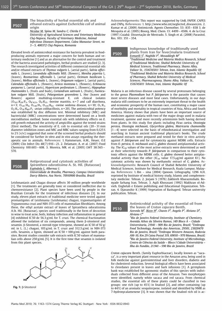
P507The bioactivity of herbal essential oils andethanol extracts against Escherichia coli of animaloriginNiculae M, Spinu M, Sandru C, Chirila FUniversity of Agricultural Sciences and Veterinary MedicineCluj-Napoca, Faculty of Veterinary Medicine, AnimalInfectious Diseases Department, Calea Manastur Street, no3 – 5, 400372 Cluj-Napoca, Romania
Elevated levels of antimicrobial resistance for bacteria prevalent in food-producing animals represent a major concern for both human and ve-terinary medicine [1] and as an alternative for the control and treatmentof the bacteria associated pathologies, herbal products are studied [2, 3].The research investigated activities of different commercial essential oilsand/or ethanol extracts of Thymus vulgaris L. (aerial parts), Salvia offici-nalis L. (leaves), Lavandula officinalis Mill. (flowers), Mentha piperita L.(leaves), Rosmarinus officinalis L. (aerial parts), Ocimum basilicum L.(leaves), Mellisa officinalis L. (leaves), Origanum vulgare L. (aerial parts),Calendula officinalis L. (flowers), Arnica montana L. (flowers), Echinaceapurpurea L. (aerial parts), Hypericum perforatum L. (flowers), Hippophaerhamnoides L. (fruits and buds), Coriandrum sativum L. (fruits), Foenicu-lum vulgare Mill. (fruits), Pelargonium graveolens L. Her (leaves andflowers) against multidrug-resistant E. coli clinical isolates(O101:K28:F5, O8:K25, O9:K35 -bovine mastitis, n = 7 and calf diarrhoea,n = 3; O141:H4, O138:H1, O139:K82 -swine oedema disease, n = 10; O1:K1,O2:K1, O78:K80:F1 -avian colibacillosis, n = 20). The antimicrobial potencywas established by disc diffusion test. Minimal inhibitory (MIC) andbactericidal (MBC) concentrations were determined based on a brothmicrodilution method. Some essential oils with inhibitory effects on E.coli growth enhanced the activity of ampicillin against the tested strains(Etest method). The sensitivity of E. coli to essential oils (20 – 31 mmdiameter inhibition zones and MIC and MBC values ranging from 0,125%to 2% (v/v)) suggested that some of the screened herbal products shouldbe considered for further in vitro and in vivo assays regarding the ther-apeutic use in animals. References: 1. Hammerum, A.M., Heuer, O.E(2009) Clin Infect Dis 48(7):916 – 21. 2. Delamare, A. et al. (2007) FoodChemistry 100:603 – 608. 3. Moreira, MR. et al. (2005) LWT 38:565 –570.
P508Antiprotozoal and cytotoxic activities ofSpiranthera odoratissima A. St. Hil. (Rutaceae)Esp�ndola L, Albernaz LUniversidade de Bras�lia, Pharmacy, Campus UniversitrioDarcy Ribeiro, Asa Norte, 70910900 Bras�lia, Brazil
Leishmaniasis and Chagas disease affects 30 million people worldwide[1]. The treatments are generally toxic or considered ineffective due tochemoresistance [2]. Plant species have been used by people in theBrazilian Cerrado for the treatment of infectious diseases [3]. In ourstudy, eleven plant extracts of traditional medicine were tested againstpromastigotes of Leishmania (Leishmania) chagasi, trypomastigotes ofTrypanosoma cruzi and NIH-3T3 cells of mammalian fibroblasts. Amongthese, the ethyl acetate extract of leaves of Spiranthera odoratissimaA.St. Hil. (Rutaceae), species used traditionally in the form of decoctionin wine to treat acne, boils, kidney infection and inflammation in general[4] exhibited IC 50 de 56.3 g/mL for T. cruzi. The chemical fractionationallowed the isolation of six compounds, among them b-sitosterol andsesamin. b-Sitosterol, a steroid-type triterpene, showed an IC50 of 92 g/mL in L. (L.) chagasi, 103 g/mL in T. cruzi and 312.5 g/mL in NIH-3T3cells. Sesamin, a lignin, showed an IC 50 > 100 g/mL against both para-sites. Recent studies consider safe extracts with IC50 values of mamma-lian cells above 250 g/mL [5]. It is the first time that sesamin is isolatedfrom this plant species.
Fig. 1: b-Sitosterol
Acknowledgements: This report was supported by UnB, FAPDF, CAPESand CNPq. References: 1. http://www.who.int/neglected_diseases/en. 2.Kumar et al. (2009) Antimicrob. Agents Chemother. 53: 835 – 838 3. deMesquita et al. (2005) Bioorg. Med. Chem. 13: 4499 – 4506. 4. de la Cruz(1997) Cuiab�: Disserta�¼o de Mestrado. 5. Singh et al. (2008) Parasitol.Res. 103: 351 – 354.
P509Indigenous knowledge of traditionally usedplants from Iran for fever/malaria treatmentEsmaeili S1, Naghibi F2, Mosaddegh M2
1Traditional Medicine and Materia Medica Research, Schoolof Traditional Medicine, Shahid Beheshti University ofMedical Sciences, Traditional Pharmacy, Shams Alley,Valiasr St., 1516745811 Tehran, Iran, Islamic Republic Of;2Traditional Medicine and Materia Medica Research, Schoolof Pharmacy, Shahid Beheshti University of MedicalSciences, Pharmacognosy, Shams Alley, Valiasr St.,1516745811 Tehran, Iran, Islamic Republic Of
Malaria is an infectious disease caused by several protozoans belongingto the genus Plasmodium but P. falciparum is the parasite that causesmost severe diseases and most fatal cases. Unfortunately, in our daysmalaria still continues to be an extremely important threat to the healthand economic prosperity of the human race, constituting a major causeof morbidity and mortality in tropical countries of Asia, Africa and SouthAmerica. History shows that plants have been an important source ofmedicines against malaria with two of the major drugs used in malariatreatment, quinine and more recently artemisinin both having derivedfrom plants. In this study five plants including Ficus carica, Otostegiapersica, Otostegia michauxii, Glycyrrhiza glabra, Matricaria chamomilla[1 – 4] were selected on the basis of ethnobotanical investigation andsearching in Iranian ancient traditional physician’s books. The crudemethanol extracts were prepared and tested for their in vitro activityagainst P. falciparum and for cytotoxicity against MDBK cells. Extractsfrom O. persica, O. michauxii and G. glabra showed antiplasmodial activ-ity. The IC50 values of the most active extracts were determined as wellas their selectivity towards P. falciparum in comparison to their cyto-toxic effects against the MDBK cells. G. glabra showed better antiplas-modial activity than the other (IC50 value 17.5 mg/ml against K1). Nocytotoxic activity was shown by methanolic extract of G. glabra. Ac-knowledgements: Research Council of Shahid Beheshti University ofMedical Sciences, Institute for Medical Research, Kuala Lumpur, Malay-sia. References: 1. Ibn – sina (2004) Qanoon. Lithography, 1296 A.H.reprinted by Institute of medical history study, Islamic and complemen-tary medicine. Tehran. 2. Jorjani S (1976) Zakhireh Kharazmshahi. Bo-nyade farhang Iran. Tehran. 3. Aqili Khorasani (1992) Makhzan ol-Ad-vieh. Enghelab e Eslami publishing and Educational Organization. Teh-ran. 4. Djavanshir K (1999) Vegetation of Bashagerd. Tehran universitypublication. Tehran.
P510Antimicrobial activity of the essential oil fromthe leaves of Croton cajucara Benth.Azevedo M1, Bizzo H2, Chaves F3, Angelo P3, Alviano D4,Alviano C4
1Rio de Janeiro Federal University, Institute of Chemistry,Avenida Athos da Silveira Ramos, 149 Bloco A – CidadeUniversitaria, 21941 – 909 Rio de Janeiro, Brazil; 2EmbrapaFood Technology, Avenida das Americas, 29501, 23020470Rio de Janeiro, Brazil; 3Embrapa Western Amazon, RodoviaAM-10, Km 29 Caixa Postal 319, 69010 – 970 Manaus, Brazil;4Rio de Janeiro Federal University, Institute of Microbiology,Centro de CiÞncias da Safflde – Bloco I Cidade Universitria –Ilha do Fund¼o, 21.941 – 590 Rio de Janeiro, Brazil
Croton cajucara Benth. (family Euphorbiaceae), locally known as “saca-ca”, is a very important plant resource in the Amazon area, being used infolk medicine against gastrointestinal and liver disorders, diabetis andfor cholesterol reduction. Several biological effects have been associatedto cleordanes present in leaves and bark infusions [1]. A germplasmbank was established for agronomic studies of this species with indivi-duals collected from different areas of the Amazon. Two morphotypeswere identified, namely white sacaca and red sacaca. From chemicalstudies, the essential oils of these plants could be classified in twogroups: one rich (up to 45%) in linalool [2], and other containing (upto 44%) of an aromatic sesquiterpene, isolated and identified by NMR as7-hydroxycalamenene [3]. It was shown that the linalool rich oil is ac-
Planta Med 2010; 76: 1163–1374 Georg Thieme Verlag KG Stuttgart · New York · ISSN 0032-0943
1322 7th Tannin Conference and 58th International Congress of the GA | 29th August – 2nd September 2010, Berlin, Germany
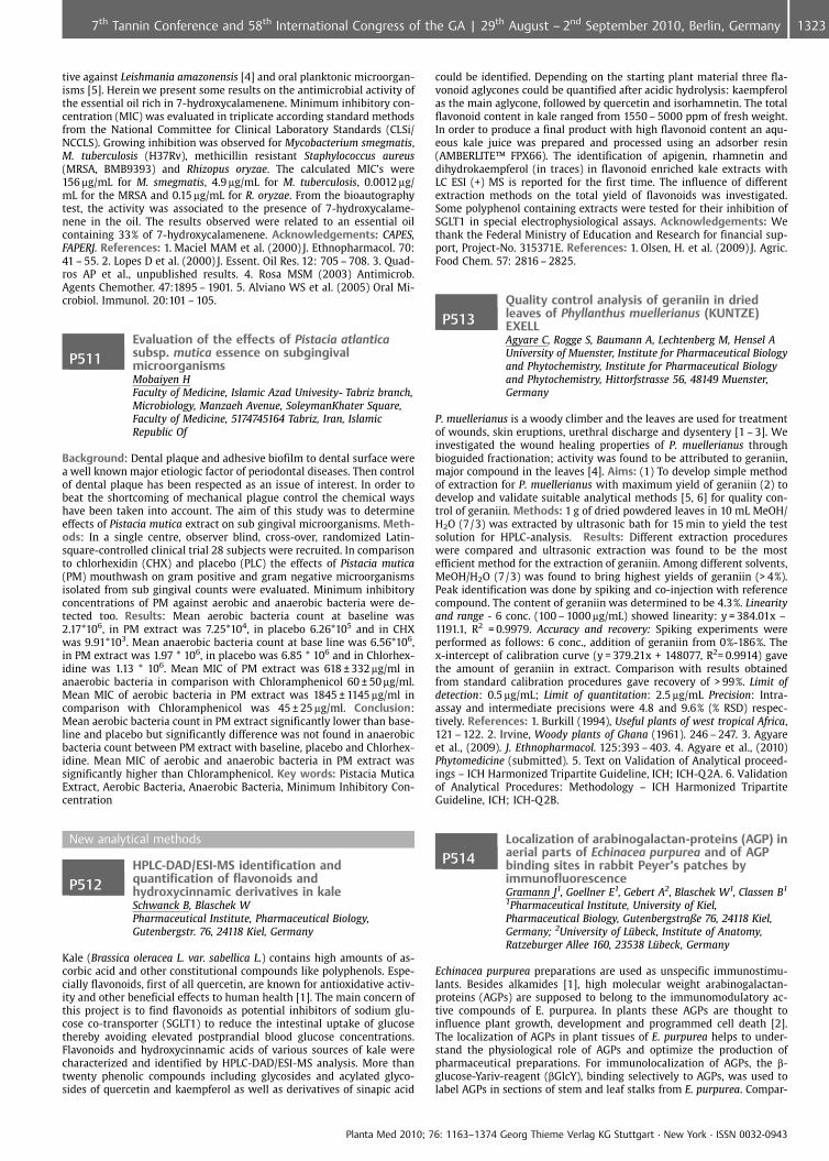
tive against Leishmania amazonensis [4] and oral planktonic microorgan-isms [5]. Herein we present some results on the antimicrobial activity ofthe essential oil rich in 7-hydroxycalamenene. Minimum inhibitory con-centration (MIC) was evaluated in triplicate according standard methodsfrom the National Committee for Clinical Laboratory Standards (CLSi/NCCLS). Growing inhibition was observed for Mycobacterium smegmatis,M. tuberculosis (H37Rv), methicillin resistant Staphylococcus aureus(MRSA, BMB9393) and Rhizopus oryzae. The calculated MIC’s were156 mg/mL for M. smegmatis, 4.9 mg/mL for M. tuberculosis, 0.0012 mg/mL for the MRSA and 0.15 mg/mL for R. oryzae. From the bioautographytest, the activity was associated to the presence of 7-hydroxycalame-nene in the oil. The results observed were related to an essential oilcontaining 33% of 7-hydroxycalamenene. Acknowledgements: CAPES,FAPERJ. References: 1. Maciel MAM et al. (2000) J. Ethnopharmacol. 70:41 – 55. 2. Lopes D et al. (2000) J. Essent. Oil Res. 12: 705 – 708. 3. Quad-ros AP et al., unpublished results. 4. Rosa MSM (2003) Antimicrob.Agents Chemother. 47:1895 – 1901. 5. Alviano WS et al. (2005) Oral Mi-crobiol. Immunol. 20:101 – 105.
P511Evaluation of the effects of Pistacia atlanticasubsp. mutica essence on subgingivalmicroorganismsMobaiyen HFaculty of Medicine, Islamic Azad Univesity- Tabriz branch,Microbiology, Manzaeh Avenue, SoleymanKhater Square,Faculty of Medicine, 5174745164 Tabriz, Iran, IslamicRepublic Of
Background: Dental plaque and adhesive biofilm to dental surface werea well known major etiologic factor of periodontal diseases. Then controlof dental plaque has been respected as an issue of interest. In order tobeat the shortcoming of mechanical plague control the chemical wayshave been taken into account. The aim of this study was to determineeffects of Pistacia mutica extract on sub gingival microorganisms. Meth-ods: In a single centre, observer blind, cross-over, randomized Latin-square-controlled clinical trial 28 subjects were recruited. In comparisonto chlorhexidin (CHX) and placebo (PLC) the effects of Pistacia mutica(PM) mouthwash on gram positive and gram negative microorganismsisolated from sub gingival counts were evaluated. Minimum inhibitoryconcentrations of PM against aerobic and anaerobic bacteria were de-tected too. Results: Mean aerobic bacteria count at baseline was2.17*106, in PM extract was 7.25*104, in placebo 6.26*105 and in CHXwas 9.91*103. Mean anaerobic bacteria count at base line was 6.56*106,in PM extract was 1.97 * 106, in placebo was 6.85 * 106 and in Chlorhex-idine was 1.13 * 106. Mean MIC of PM extract was 618 € 332 mg/ml inanaerobic bacteria in comparison with Chloramphenicol 60 € 50 mg/ml.Mean MIC of aerobic bacteria in PM extract was 1845 € 1145 mg/ml incomparison with Chloramphenicol was 45 € 25 mg/ml. Conclusion:Mean aerobic bacteria count in PM extract significantly lower than base-line and placebo but significantly difference was not found in anaerobicbacteria count between PM extract with baseline, placebo and Chlorhex-idine. Mean MIC of aerobic and anaerobic bacteria in PM extract wassignificantly higher than Chloramphenicol. Key words: Pistacia MuticaExtract, Aerobic Bacteria, Anaerobic Bacteria, Minimum Inhibitory Con-centration
New analytical methods
P512HPLC-DAD/ESI-MS identification andquantification of flavonoids andhydroxycinnamic derivatives in kaleSchwanck B, Blaschek WPharmaceutical Institute, Pharmaceutical Biology,Gutenbergstr. 76, 24118 Kiel, Germany
Kale (Brassica oleracea L. var. sabellica L.) contains high amounts of as-corbic acid and other constitutional compounds like polyphenols. Espe-cially flavonoids, first of all quercetin, are known for antioxidative activ-ity and other beneficial effects to human health [1]. The main concern ofthis project is to find flavonoids as potential inhibitors of sodium glu-cose co-transporter (SGLT1) to reduce the intestinal uptake of glucosethereby avoiding elevated postprandial blood glucose concentrations.Flavonoids and hydroxycinnamic acids of various sources of kale werecharacterized and identified by HPLC-DAD/ESI-MS analysis. More thantwenty phenolic compounds including glycosides and acylated glyco-sides of quercetin and kaempferol as well as derivatives of sinapic acid
could be identified. Depending on the starting plant material three fla-vonoid aglycones could be quantified after acidic hydrolysis: kaempferolas the main aglycone, followed by quercetin and isorhamnetin. The totalflavonoid content in kale ranged from 1550 – 5000 ppm of fresh weight.In order to produce a final product with high flavonoid content an aqu-eous kale juice was prepared and processed using an adsorber resin(AMBERLITE� FPX66). The identification of apigenin, rhamnetin anddihydrokaempferol (in traces) in flavonoid enriched kale extracts withLC ESI (+) MS is reported for the first time. The influence of differentextraction methods on the total yield of flavonoids was investigated.Some polyphenol containing extracts were tested for their inhibition ofSGLT1 in special electrophysiological assays. Acknowledgements: Wethank the Federal Ministry of Education and Research for financial sup-port, Project-No. 315371E. References: 1. Olsen, H. et al. (2009) J. Agric.Food Chem. 57: 2816 – 2825.
P513Quality control analysis of geraniin in driedleaves of Phyllanthus muellerianus (KUNTZE)EXELLAgyare C, Rogge S, Baumann A, Lechtenberg M, Hensel AUniversity of Muenster, Institute for Pharmaceutical Biologyand Phytochemistry, Institute for Pharmaceutical Biologyand Phytochemistry, Hittorfstrasse 56, 48149 Muenster,Germany
P. muellerianus is a woody climber and the leaves are used for treatmentof wounds, skin eruptions, urethral discharge and dysentery [1 – 3]. Weinvestigated the wound healing properties of P. muellerianus throughbioguided fractionation; activity was found to be attributed to geraniin,major compound in the leaves [4]. Aims: (1) To develop simple methodof extraction for P. muellerianus with maximum yield of geraniin (2) todevelop and validate suitable analytical methods [5, 6] for quality con-trol of geraniin. Methods: 1 g of dried powdered leaves in 10 mL MeOH/H2O (7/3) was extracted by ultrasonic bath for 15 min to yield the testsolution for HPLC-analysis. Results: Different extraction procedureswere compared and ultrasonic extraction was found to be the mostefficient method for the extraction of geraniin. Among different solvents,MeOH/H2O (7/3) was found to bring highest yields of geraniin (> 4%).Peak identification was done by spiking and co-injection with referencecompound. The content of geraniin was determined to be 4.3%. Linearityand range - 6 conc. (100 – 1000 mg/mL) showed linearity: y = 384.01x –1191.1, R2 = 0.9979. Accuracy and recovery: Spiking experiments wereperformed as follows: 6 conc., addition of geraniin from 0%-186%. Thex-intercept of calibration curve (y = 379.21x + 148077, R2= 0.9914) gavethe amount of geraniin in extract. Comparison with results obtainedfrom standard calibration procedures gave recovery of > 99%. Limit ofdetection: 0.5 mg/mL; Limit of quantitation: 2.5 mg/mL Precision: Intra-assay and intermediate precisions were 4.8 and 9.6% (% RSD) respec-tively. References: 1. Burkill (1994), Useful plants of west tropical Africa,121 – 122. 2. Irvine, Woody plants of Ghana (1961). 246 – 247. 3. Agyareet al., (2009). J. Ethnopharmacol. 125:393 – 403. 4. Agyare et al., (2010)Phytomedicine (submitted). 5. Text on Validation of Analytical proceed-ings – ICH Harmonized Tripartite Guideline, ICH; ICH-Q 2A. 6. Validationof Analytical Procedures: Methodology – ICH Harmonized TripartiteGuideline, ICH; ICH-Q 2B.
P514Localization of arabinogalactan-proteins (AGP) inaerial parts of Echinacea purpurea and of AGPbinding sites in rabbit Peyer’s patches byimmunofluorescenceGramann J1, Goellner E1, Gebert A2, Blaschek W1, Classen B1
1Pharmaceutical Institute, University of Kiel,Pharmaceutical Biology, Gutenbergstraße 76, 24118 Kiel,Germany; 2University of L�beck, Institute of Anatomy,Ratzeburger Allee 160, 23538 L�beck, Germany
Echinacea purpurea preparations are used as unspecific immunostimu-lants. Besides alkamides [1], high molecular weight arabinogalactan-proteins (AGPs) are supposed to belong to the immunomodulatory ac-tive compounds of E. purpurea. In plants these AGPs are thought toinfluence plant growth, development and programmed cell death [2].The localization of AGPs in plant tissues of E. purpurea helps to under-stand the physiological role of AGPs and optimize the production ofpharmaceutical preparations. For immunolocalization of AGPs, the b-glucose-Yariv-reagent (bGlcY), binding selectively to AGPs, was used tolabel AGPs in sections of stem and leaf stalks from E. purpurea. Compar-
Planta Med 2010; 76: 1163–1374 Georg Thieme Verlag KG Stuttgart · New York · ISSN 0032-0943
13237th Tannin Conference and 58th International Congress of the GA | 29th August – 2nd September 2010, Berlin, Germany
able to roots of E. purpurea [3], AGPs are mainly located in cell walls ofxylem tracheary elements, especially in pit channels. In contrast toroots, the aerial parts of the plant show additionally very strong immu-nolabeling of sclerenchyma cells and of companion cells of the phloem.The pharmacological effects of AGPs after oral application of E. purpureapreparations are thought to be mediated by Peyer’s patches of the ileum[4]. Cryo sections of rabbit Peyer’s patch tissue were incubated withAGPs from Echinacea and AGP binding sites were detected using immu-nohistochemistry and confocal microscopy. AGPs from the aerial parts ofE. purpurea strongly bind to intraepithelial lymphocytes in pockets ofmembranous (M) cells. As these sites are believed to be critically in-volved in the initiation of specific immune responses as well as thegeneration of immune tolerance [5], AGPs from E. purpurea might di-rectly influence the immune protection of the human organism. Ac-knowledgements: We thank Rottapharm/Madaus GmbH, K�ln, for fi-nancial support of this work. References: 1. Woelkart K et al. (2007)Planta Med 73: 615 – 623. 2. Nothnagel E A (1997) Int Rev Cytol 174:195 – 291. 3. Classen et al. (2009) Planta Med 75: 1526 – 1533. 4. BodinetC et al. (2004) Drug Res 54: 114 – 118. 5. Brandtzaeg P et al. (1999)Immunol Today 20: 141 – 151.
P515Localization of mistletoe lectins I-III in Viscumalbum L. by immunofluorescent labelingGramann J, Herbst B, Blaschek W, Classen BPharmaceutical Institute, University of Kiel, PharmaceuticalBiology, Gutenbergstraße 76, 24118 Kiel, Germany
Because of immunomodulatory effects mistletoe extracts play an impor-tant role as phytotherapeutic adjuvants in cancer therapy. Mistletoelectins I-III contribute to the in vivo activity of mistletoe preparations[1]. They consist of two chains: the A chain is a homologous protein forall mistletoe lectins and responsible for the inhibition of translation. TheB chain is necessary for binding to specific carbohydrate epitopes; theprotein sequence of this chain varies from lectin to lectin. The localiza-tion of bioactive lectins in Viscum album L. is of significance to under-stand the physiological role of lectins in the plant and to optimize theproduction of pharmaceutical preparations. We investigated the cellulardistribution of lectins in different parts of the plant by immunofluores-cent labeling. Leaves and internodes of shoots of Viscum album L. weretreated with a monoclonal antibody against the A-chain of mistletoelectins I-III. A secondary fluorescent-labeled antibody was added andthe sections were analyzed by confocal laser scanning microscopy. Themistletoe lectins I-III were located mainly in cells of the parenchyma ofthe cortex, in sclerenchyma tissue next to the vascular bundles and inthe companion cells of the phloem. Especially in parenchyma cells of thethird and fourth internode a high content of lectins could be detected,whereas parenchyma cells of the leaves showed weak labeling only.Acknowledgements: We thank Rottapharm/Madaus GmbH, K�ln, forfinancial support of this work. References: 1. Seifert G et al. (2008)Cancer Lett 264: 218 – 228.
P516Rapid analysis of N-phenylpropenoyl-L-aminoacids (NPA) from cocoa by UPLC and CELechtenberg M, Henschel K, Liefl�nder-Wulf U, Quandt B,Hensel AInstitute for Pharmaceutical Biology and Phytochemistry,M�nster, M�nster University (WWU), Hittorfstraße 56,48149 M�nster, Germany
The occurrence of N-phenylpropenoyl-L-amino acids (NPA) in Theobro-ma cacao L. was firstly described in 1998 [1]. Since that NPA were shownto be key contributors to the astringent taste of cocoa beans and nibs [2],and to be stimulators of mitochondrial activity and proliferation rate ofhuman liver cells and keratinocytes [3]. Furthermore the aspartic acidamide of caffeic acid showed strong antiadhesive properties against theadhesion of Helicobacter pylori to human stomach tissue [3]. Preliminarydata on absorption and bioavailability of NPA in humans are available[4]. Recently a stable isotope dilution analysis with LC-MS/MS (MRM)detection was developed and 14 NPA were quantified in cocoa samples[5]. As SIDA-methods often suffer from the availability of referencecompounds, there is need for alternative methods. The aim of our workwas to establish a fast method for analysis of NPA in cocoa and cocoaproducts. We developed two methods on the basis of alternative separa-tion principles: chromatography (? UPLC) and electrophoresis (? CE).Both methods were validated according to ICH guidelines and enabledus to quantify the main NPA in different cocoa products. After defatting
with petroleum benzine, extraction was done using acetone-water (7:3).To avoid interference caused by accompanying polyphenols subsequentSPE on quaternary amine sorbent yielded NPA-enriched fractions. UPLCseparation of compounds 1 – 5 (Image 1) was done in less than 4 min-utes. CE offers a suitable alternative with the disadvantage of a lowersensitivity. Highest amounts of NPA were found in cocoa beans, followedby nips and chocolate.
Fig. 1 UPLC/PDA (320 nm): 1 K-Asp, 2 pC-Asp, 3 K-Dopa, 4 K-Tyr, 5 pC-Tyr,I.S. Rosmarinic acid
References: 1. Sanbongi C et al. (1998) J. Agric. Food Chem. 46:454 – 7. 2.Stark T and Hofmann T (2005) J. Agric. Food Chem. 53:5419 – 28. 3. Hen-sel A et al. (2007) Planta Med 73:142 – 50. 4. Stark T et al. (2008) Mol.Nutr. Food Res. 52:1201 – 14. 5. Stark T et al. (2006) J. Agric. Food Chem.54:2859 – 67.
P517Efficient silkworm expression of a single-chainvariable fragment antibody against ginsenosideRe and its application in ELISA for quality controlof total ginsenosides in various ginsengSakamoto S1, Pongkitwitoon B1, Nakamura S2, Maenaka K3,Tanaka H1, Morimoto S1
1Graduate School of Pharmaceutical Sciences, KyushuUniversity, Department of Pharmacognosy, 3 – 1-1Maidashi, Higashi-ku, 8128582 Fukuoka, Japan; 2GraduateSchool of Medical Science, Kyushu University, Department ofMolecular Biology, 3 – 1-1 Maidashi, Higashi-ku, 812 – 8582Fukuoka, Japan; 3Medical Institute of Bioregulation, KyushuUniversity, 3 – 1-1 Maidashi, Higashi-ku, 812 – 8582Fukuoka, Japan
Panax ginseng and its related species are well known as important tradi-tional medicines which have been widely used in Asia, especially China,Japan and Korea, for thousands of years. Ginseng has been shown tohave various pharmacologic activities such as tonic, immunomodula-tory, anti-mutagenic, and anti-aging activities [1 – 2]. A single-chainvariable antibody fragment (scFv) against its major compound, ginseno-side Re (G-Re) has been successfully expressed in the silkworm larvaeusing the Bombyx mori nucleopolyhedrovirus (BmNPV) bacmid DNAsystem. The baculovirus donor vector for expression of scFv against G-Re (GRe-scFv) was constructed to contain a honeybee melittin sequencesignal (HMSS) to accelerate secretion of recombinant GRe-scFv into thehemolymph of silkworm larvae. Functional GRe-scFv was purified bycation exchange chromatography followed by immobilized metal ionaffinity chromatography. The yield of purified GRe-scFv was 6.5 mg per13 silkworm larvae, exhibiting higher yield than that expressed in E. coli(1.7 mg per liter of culture medium) [3]. GRe-scFv retained similar char-acteristics of the parental monoclonal antibody (MAb) against G-Re(MAb-4G10), making it possible to develop an indirect competitive en-zyme-linked immunosorbent assay (icELISA) for the quality control oftotal ginsenosides in various ginsengs. The detectable range for calibra-tion of G-Re by developed icELISA was 0.05 – 10 mg/ml. These resultsclearly suggested that the silkworm expression system is quite usefulfor the expression of functional scFv that frequently require time- andcost-consuming refolding when expressed in E. coli. References: 1. Lee,TK. et al. (2005) Mutagenesis 20: 237 – 243. 2. Kiefer, D. et al. (2003) Am.Fam. Physician 68: 1539 – 1542. 3. Pongkitwitoon, B. et al. (2010) J. Nat.Med. in press.
Planta Med 2010; 76: 1163–1374 Georg Thieme Verlag KG Stuttgart · New York · ISSN 0032-0943
1324 7th Tannin Conference and 58th International Congress of the GA | 29th August – 2nd September 2010, Berlin, Germany
P518Flash chromatography on cartridges for theseparation of plant extracts – Rules for theselection of chromatographic conditions, andcomparison with MPLCPotterat O1, Weber P1, Schafroth N2, Hamburger M1
1University of Basel, Pharmaceutical Biology,Klingelbergstrasse 50, 4056 Basel, Switzerland; 2B�chiLabortechnik AG, 9230 Flawil, Switzerland
Flash chromatography on cartridges has become increasingly popularfor the rapid purification of compounds, mainly of synthetic origin. Incontrast, its application for natural product isolation is poorly documen-ted, and easy-to-use procedures for optimization of the separation con-ditions are lacking. Using Sepacore� cartridges (Bchi Labortechnik), wehave established empirical rules for the selection of chromatographicconditions with an emphasis on gradient mode. Reversed phase HPLCseparations can be transposed by increasing the gradient time by afactor 2 – 4. For normal phase separations, solvent compositions result-ing in Rf values of 0.15 – 0.2 on TLC for the most lipophilic and the mosthydrophilic constituents, respectively, should be selected as gradientendpoints. We applied these rules to the separation of complex plantextracts, with Curcuma xanthorrhiza, Piper nigrum and Salvia milthior-rhiza as examples of medicinal and commercial importance. The perfor-mance of the cartridges was compared to that of classical MPLC (med-ium pressure liquid chromatography) glass columns. Sepacore cartridgesenabled a good separation of compounds with a broad range of polarity,as typically found in plant extracts. The chromatographic resolutionremained, however, lower than that achieved by MPLC on columnspacked with material of smaller particle size. For poorly soluble extracts,solid introduction gave better results than liquid injection. Despite low-er resolution as compared to MPLC, pre-packed cartridges are an attrac-tive alternative for the purification of extracts and crude fractions due totheir ease of use and speed of separation.
P519Content of antidepressant hyperforin correlateswith translucent glands in Hypericum perforatumL.Soelberg J1, Johansen B1, J�ger A2
1University of Copenhagen, Biological Institute,Osterfarimagsgade 2D, 1123 Copenhagen, Denmark;2University of Copenhagen, Medicinal Chemistry,Universitetsparken 2, 2100 Copenhagen, Denmark
Hypericum perforatum L. synthesizes two therapeutically importantcompounds, hypericin and hyperforin, which have recently been shownto accumulate in secretory structures: the dark glands and the translu-cent glands respectively [1]. Photographic images of H. perforatumleaves were computer-analyzed to produce measures of total gland areaand total leaf area. A single leaf was placed between two microscopeslides and positioned in a microscope fitted with light externally andfrom below. Since the translucent glands span the depth of the leaf, theyclearly show when lit from behind, and could be captured in high-defi-nition images (CCD camera “Evolution LC”, exposure at 700 ms). Theimages were captured as TIFF files, and processed in computer programImage-Pro Plus 5.1 (conversion to grey tones; masking of anything out-side the leaf outline; and finally count and size-measure of any brightobjects (the translucent glands) of a defined roundness (0 – 2 arbitraryunits) and within a defined size (0.001 – 1 mm2). The leaves were thensubjected to extraction and HPLC analysis for hyperforin content. Thecontent of hyperforin in a H. perforatum leaf correlated with the totalarea of translucent glands (correlation 0.71), whereas there was no cor-relation with the leaf area (correlation 0.09). This means that it is notthe biomass of a plant, but the number and size of translucent glandsthat influence the yield of hyperforin in a crop. This applied methodindicates that hyperforin content in H. perforatum leaves could be esti-mated without use of analytical chemistry, simply by observation andcalculation of translucent gland area. References: 1. Soelberg J. et al.(2007) Ann Bot 99: 1097 – 1100.
P520Simultaneous determination of eight markercomponents in the traditional herbal medicine,Sipjundaebo tang by HPLC-DADYang H1, Weon J1, Ma J2, Lee J2, Ma C1
1Department of Biomaterials Engineering, Division ofBioscience and Biotechnology, Kangwon National University,Hyoja-2-dong, 200701 Chuncheon, Korea, Republic Of; 2TKMConverging Research Division, Korea Institute of OriginalMedicine, 483 Exporo, Yuseong-gu, 305 – 811 Daejeon,Korea, Republic Of
Sipjundaebo tang, a traditional herbal medicine, has been used in thetreatment of anemia, inflammation and tumor. For simultaneous deter-mination of eight components, namely hydroxymethylfurfural (5-HMF),paeoniflorin, ferulic acid, cinnamaldehyde, decursinol, 6-gingerol, decur-sin and glycyrrhizin in Sipjundaebo tang, a high performance liquidchromatography-diode array detector method was established. To devel-op and validate this HPLC-DAD method, C 18 column (S-5 mm,4.6 x 250 mm) was used with gradient mobile phase, water and metha-nol containing 0.1% TFA at the column temperature of 35 �C. UV wave-length was set at 230, 254, 280 and 300 nm. Validation of the chroma-tography method was evaluated by linearity, precision and accuracy test.Calibration curve for eight marker components showed good linearitywith R2 > 0.9998. Limits of detection (LOD) and Limits of quantification(LOQ) ranged from 0.01 mg/ml to 0.13 mg/ml and 0.03 mg/ml to 0.41 mg/ml, respectively. The relative standard deviations (RSDs) value of preci-sion test, intra-day and inter-day test, was less than 0.61% and 0.42%,respectively. The results of accuracy test varied from 95.1% to 104.8%with RSD < 2%. In conclusion, this developed simultaneous determina-tion method was efficient for the quality evaluation of Sipjundaebo tang.
P521Quantitative detection of sinigrin in crudeextracts of horseradish (Armoracia rusticana)roots by Enzyme-linked Immunosorbent Assay(ELISA)Loebers A, Eisenbeiß W, Kreis WUniversit�t Erlangen, Pharmazeutische Biologie,Staudstrasse 5, 91058 Erlangen, Germany
The glucosinolate sinigrin is an important ingredient in roots of horse-radish (Armoracia rusticana Gaertn., Mey., & Scherb.). Glucosinolates arecharacterized as sulfur- and nitrogen-containing plant secondary meta-bolites, possessing several health benefits [1] and are predominantlyfound in members of Brassicaceae. Following disintegration of root cells,during processing of the roots, sinigrin is degraded by myrosinase there-by releasing isothiocyanate which causes the spicy taste of horseradish-containing food. HPLC has been used as the standard method for detec-tion of sinigrin or other glucosinolates. We developed an enzyme-linkedimmunosorbent assay (ELISA), rarely employed for quantitative detec-tion of glucosinolates [2, 3], in order to estimate the sinigrin content inhorseradish root extracts. We immunized Balb/c mice with BSA-linkedsinigrin to obtain sinigrin-specific polyclonal antibodies. Quantificationof sinigrin was achieved using a competitive ELISA against a sinigrin-ovalbumin conjugate used as the antigen. Values determined by thisELISA were compared with HPLC measurements suggesting that thismethod can be readily used as a rapid and high-throughput techniquefor measuring sinigrin-containing horseradish extracts. References:1. Traka M. et al. (2009): Glucosinolates, isothiocyanat and humanhealth. 2. Hassan F. et al. (1988) J. Agric. Food Chem. 36: 398 – 403. 3.van Doorn H. E. et al. (1998) J. Agric. Food Chem. 46: 793 – 800.
Planta Med 2010; 76: 1163–1374 Georg Thieme Verlag KG Stuttgart · New York · ISSN 0032-0943
13257th Tannin Conference and 58th International Congress of the GA | 29th August – 2nd September 2010, Berlin, Germany
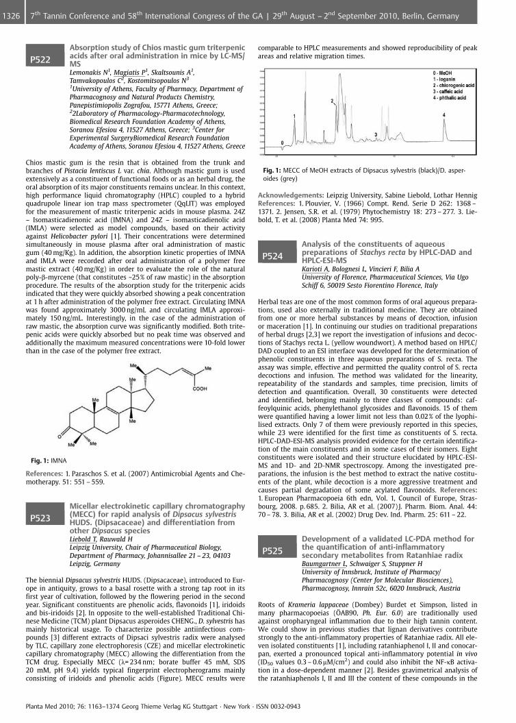
P522Absorption study of Chios mastic gum triterpenicacids after oral administration in mice by LC-MS/MSLemonakis N1, Magiatis P1, Skaltsounis A1,Tamvakopoulos C2, Kostomitsopoulos N3
1University of Athens, Faculty of Pharmacy, Department ofPharmacognosy and Natural Products Chemistry,Panepistimiopolis Zografou, 15771 Athens, Greece;22Laboratory of Pharmacology-Pharmacotechnology,Biomedical Research Foundation Academy of Athens,Soranou Efesiou 4, 11527 Athens, Greece; 3Center forExperimental SurgeryBiomedical Research FoundationAcademy of Athens, Soranou Efesiou 4, 11527 Athens, Greece
Chios mastic gum is the resin that is obtained from the trunk andbranches of Pistacia lentiscus L var. chia. Although mastic gum is usedextensively as a constituent of functional foods or as an herbal drug, theoral absorption of its major constituents remains unclear. In this context,high performance liquid chromatography (HPLC) coupled to a hybridquadrupole linear ion trap mass spectrometer (QqLIT) was employedfor the measurement of mastic triterpenic acids in mouse plasma. 24Z– Isomasticadienonic acid (IMNA) and 24Z – isomasticadienolic acid(IMLA) were selected as model compounds, based on their activityagainst Helicobacter pylori [1]. Their concentrations were determinedsimultaneously in mouse plasma after oral administration of masticgum (40 mg/Kg). In addition, the absorption kinetic properties of IMNAand IMLA were recorded after oral administration of a polymer freemastic extract (40 mg/Kg) in order to evaluate the role of the naturalpoly-b-myrcene (that constitutes ~25% of raw mastic) in the absorptionprocedure. The results of the absorption study for the triterpenic acidsindicated that they were quickly absorbed showing a peak concentrationat 1 h after administration of the polymer free extract. Circulating IMNAwas found approximately 3000 ng/mL and circulating IMLA approxi-mately 150 ng/mL. Interestingly, in the case of the administration ofraw mastic, the absorption curve was significantly modified. Both trite-penic acids were quickly absorbed but no peak time was observed andadditionally the maximum measured concentrations were 10-fold lowerthan in the case of the polymer free extract.
Fig. 1: IMNA
References: 1. Paraschos S. et al. (2007) Antimicrobial Agents and Che-motherapy. 51: 551 – 559.
P523Micellar electrokinetic capillary chromatography(MECC) for rapid analysis of Dipsacus sylvestrisHUDS. (Dipsacaceae) and differentiation fromother Dipsacus speciesLiebold T, Rauwald HLeipzig University, Chair of Pharmaceutical Biology,Department of Pharmacy, Johannisallee 21 – 23, 04103Leipzig, Germany
The biennial Dipsacus sylvestris HUDS. (Dipsacaceae), introduced to Eur-ope in antiquity, grows to a basal rosette with a strong tap root in itsfirst year of cultivation, followed by the flowering period in the secondyear. Significant constituents are phenolic acids, flavonoids [1], iridoidsand bis-iridoids [2]. In opposite to the well-established Traditional Chi-nese Medicine (TCM) plant Dipsacus asperoides CHENG., D. sylvestris hasmainly historical usage. To characterize possible antiinfectious com-pounds [3] different extracts of Dipsaci sylvestris radix were analysedby TLC, capillary zone electrophoresis (CZE) and micellar electrokineticcapillary chromatography (MECC) allowing the differentiation from theTCM drug. Especially MECC (l= 234 nm; borate buffer 45 mM, SDS20 mM, pH 9.4) yields typical fingerprint electropherograms mainlyconsisting of iridoids and phenolic acids (Figure). MECC results were
comparable to HPLC measurements and showed reproducibility of peakareas and relative migration times.
Fig. 1: MECC of MeOH extracts of Dipsacus sylvestris (black)/D. asper-oides (grey)
Acknowledgements: Leipzig University, Sabine Liebold, Lothar HennigReferences: 1. Plouvier, V. (1966) Compt. Rend. Serie D 262: 1368 –1371. 2. Jensen, S.R. et al. (1979) Phytochemistry 18: 273 – 277. 3. Lie-bold, T. et al. (2008) Planta Med 74: 995.
P524Analysis of the constituents of aqueouspreparations of Stachys recta by HPLC-DAD andHPLC-ESI-MSKarioti A, Bolognesi L, Vincieri F, Bilia AUniversity of Florence, Pharmaceutical Sciences, Via UgoSchiff 6, 50019 Sesto Fiorentino Florence, Italy
Herbal teas are one of the most common forms of oral aqueous prepara-tions, used also externally in traditional medicine. They are obtainedfrom one or more herbal substances by means of decoction, infusionor maceration [1]. In continuing our studies on traditional preparationsof herbal drugs [2,3] we report the investigation of infusions and decoc-tions of Stachys recta L. (yellow woundwort). A method based on HPLC/DAD coupled to an ESI interface was developed for the determination ofphenolic constituents in three aqueous preparations of S. recta. Theassay was simple, effective and permitted the quality control of S. rectadecoctions and infusion. The method was validated for the linearity,repeatability of the standards and samples, time precision, limits ofdetection and quantification. Overall, 30 constituents were detectedand identified, belonging mainly to three classes of compounds: caf-feoylquinic acids, phenylethanol glycosides and flavonoids. 15 of themwere quantified having a lower limit not less than 0.02% of the lyophi-lised extracts. Only 7 of them were previously reported in this species,while 23 were identified for the first time as constituents of S. recta.HPLC-DAD-ESI-MS analysis provided evidence for the certain identifica-tion of the main constituents and in some cases of their isomers. Eightconstituents were isolated and their structure elucidated by HPLC-ESI-MS and 1D- and 2D-NMR spectroscopy. Among the investigated pre-parations, the infusion is the best method to extract the native costitu-ents of the plant, while decoction is a more aggressive treatment andcauses partial degradation of some acylated flavonoids. References:1. European Pharmacopoeia 6th edn, Vol. 1, Council of Europe, Stras-bourg, 2008. p. 685. 2. Bilia, AR et al. (2007) J. Pharm. Biom. Anal. 44:70 – 78. 3. Bilia, AR et al. (2002) Drug Dev. Ind. Pharm. 25: 611 – 22.
P525Development of a validated LC-PDA method forthe quantification of anti-inflammatorysecondary metabolites from Ratanhiae radixBaumgartner L, Schwaiger S, Stuppner HUniversity of Innsbruck, Institute of Pharmacy/Pharmacognosy (Center for Molecular Biosciences),Pharmacognosy, Innrain 52c, 6020 Innsbruck, Austria
Roots of Krameria lappaceae (Dombey) Burdet et Simpson, listed inmany pharmacopoeias (AB90, Ph. Eur. 6.0) are traditionally usedagainst oropharyngeal inflammation due to their high tannin content.We could show in previous studies that lignan derivatives contributestrongly to the anti-inflammatory properties of Ratanhiae radix. All ele-ven isolated constituents [1], including ratanhiaphenol I, II and conocar-pan, exerted a pronounced topical anti-inflammatory potential in vivo(ID50 values 0.3 – 0.6mM/cm2) and could also inhibit the NF-kB activa-tion in a dose-dependent manner [2]. Besides gravimetrical analysis ofthe ratanhiaphenols I, II and III the content of these compounds in the
Planta Med 2010; 76: 1163–1374 Georg Thieme Verlag KG Stuttgart · New York · ISSN 0032-0943
1326 7th Tannin Conference and 58th International Congress of the GA | 29th August – 2nd September 2010, Berlin, Germany
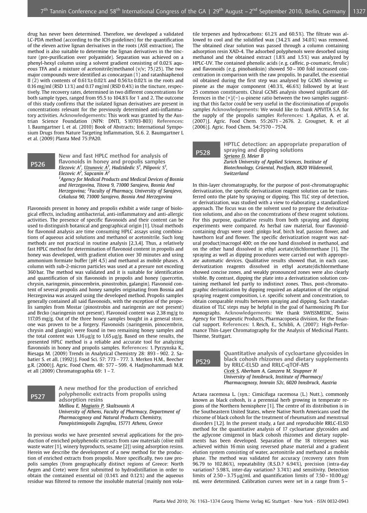
drug has never been determined. Therefore, we developed a validatedLC-PDA method (according to the ICH-guidelines) for the quantificationof the eleven active lignan derivatives in the roots (ASE extraction). Themethod is also suitable to determine the lignan derivatives in the tinc-ture (pre-purification over polyamide). Separation was achieved on aphenyl-hexyl column using a solvent gradient consisting of 0.02% aqu-eous TFA and a mixture of acetonitrile/methanol (v/v; 75/25). The twomajor compounds were identified as conocarpan (1) and ratanhiaphenolII (2) with contents of 0.61%€ 0.02% and 0.56%€ 0.02% in the roots and0.16 mg/ml (RSD 1.1%) and 0.17 mg/ml (RSD 0.4%) in the tincture, respec-tively. The recovery rates, determined in two different concentrations forboth sample types, ranged from 95.5 to 104.8% for 1 and 2. The outcomeof this study confirms that the isolated lignan derivatives are present inconcentrations relevant for the previously determined anti-inflamma-tory activities. Acknowledgements: This work was granted by the Aus-trian Science Foundation (NFN: DNTI, S10703-B03) References:1. Baumgartner L et al. (2010) Book of Abstracts; International Sympo-sium Drugs from Nature Targeting Inflammation, SL 6. 2. Baumgartner Let al. (2009) Planta Med 75:PA20.
P526New and fast HPLC method for analysis offlavonoids in honey and propolis samplesElezovic A1, Uzunovic A1, Hadzidedic S1, Pilipovic S1,Elezovic A2, Sapcanin A2
1Agency for Medical Products and Medical Devices of Bosniaand Herzegovina, Titova 9, 71000 Sarajevo, Bosnia AndHerzegovina; 2Faculty of Pharmacy, University of Sarajevo,Cekalusa 90, 71000 Sarajevo, Bosnia And Herzegovina
Flavonoids present in honey and propolis exhibit a wide range of biolo-gical effects, including antibacterial, anti-inflammatory and anti-allergicactivities. The presence of specific flavonoids and their content can beused to distinguish botanical and geographical origin [1]. Usual methodsfor flavonoid analysis are time consuming HPLC assays using combina-tions of aqueous acid solutions and methanol or acetonitrile. Such longmethods are not practical in routine analysis [2,3,4]. Thus, a relativelyfast HPLC method for determination of flavonoid content in propolis andhoney was developed, with gradient elution over 30 minutes and usingammonium formiate buffer (pH 4,5) and methanol as mobile phases. Acolumn with sub-2-micron particles was used at a presure not exceding360 bar. The method was validated and it is suitable for identificationand quantification of six flavonoids in propolis and honey (quercetin,chrysin, naringenin, pinocembrin, pinostrobin, galangin). Flavonoid con-tent of several propolis and honey samples originating from Bosnia andHerzegovina was assayed using the developed method. Propolis samplesgenerally contained all said flavonoids, with the exception of the propo-lis samples from Mostar (pinostrobin and naringenin are not present)and Brcko (naringenin not present). Flavonoid content was 2,38 mg/g to117,05 mg/g. Out of the three honey samples bought in a general store,one was proven to be a forgery. Flavonoids (naringenin, pinocembrin,chrysin and glangin) were found in two remaining honey samples andthe total content was 1,16 mg/g to 1,65 mg/g. Based on these results, thepresented HPLC method is a reliable and accurate tool for analyzingflavonoids in honey and propolis samples. References: 1. Pyrzynska K.,Biesaga M. (2009) Trends in Analytical Chemistry 28: 893 – 902. 2. Sa-batier S. et all. (1992) J. Food Sci. 57: 773 – 777. 3. Merken H.M., Beecherg.R. (2000) J. Agric. Food Chem. 48: 577 – 599. 4. Hadjmohammadi M.R.et all (2009) Chromatographia 69: 1 – 7.
P527A new method for the production of enrichedpolyphenolic extracts from propolis usingadsorption resinsMelliou E, Magiatis P, Skaltsounis AUniversity of Athens, Faculty of Pharmacy, Department ofPharmacognosy and Natural Products Chemistry,Panepistimiopolis Zografou, 15771 Athens, Greece
In previous works we have presented several applications for the pro-duction of enriched polyphenolic extracts from raw materials (olive millwaste water [1], winery byproducts, sesame [2]) using adsorption resins.Herein we describe the development of a new method for the produc-tion of enriched extracts from propolis. More specifically, two raw pro-polis samples (from geographically distinct regions of Greece: NorthAegen and Crete) were first submitted to hydrodistillation in order toobtain the contained essential oil (0.14% and 0.12%) and the aqueousresidue was filtered to remove the insoluble material (mainly non vola-
tile terpenes and hydrocarbons: 61.2% and 60.5%). The filtrate was al-lowed to cool and the solidified wax (34.2% and 34.0%) was removed.The obtained clear solution was passed through a column containingadsorption resin XAD-4. The adsorbed polyphenols were desorbed usingmethanol and the obtained extract (1.8% and 1.5%) was analyzed byHPLC-UV. The contained phenolic acids (e. g. caffeic, p-coumaric, ferulic)and flavonoids (e.g. pinobanksin) showed 50 – 100 fold increased con-centration in comparison with the raw propolis. In parallel, the essentialoil obtained during the first step was analysed by GCMS showing a-pinene as the major component (40.3%, 46.6%) followed by at least25 common constituents. Chiral GCMS analysis showed significant dif-ferences in the (+)/(�) a-pinene ratio between the two samples suggest-ing that this factor could be very useful in the discrimination of propolissamples Acknowledgements: We would like to thank APIVITA S.A. forthe supply of the propolis samples References: 1. Agalias, A. et al.(2007) J. Agric. Food Chem. 55:2671 – 2676. 2. Grougnet, R. et al(2006) J. Agric. Food Chem. 54:7570 – 7574.
P528HPTLC detection: an appropriate preparation ofspraying and dipping solutionsSpriano D, Meier BZurich University of Applied Sciences, Institute ofBiotechnology, Gr�ental, Postfach, 8820 W�denswil,Switzerland
In thin-layer chromatography, for the purpose of post-chromatographicderivatization, the specific derivatization reagent solution can be trans-ferred onto the plate by spraying or dipping. This TLC step of detection,or derivatization, was studied with a view to elaborating a standardizedapproach. The focus was on the solvent used to prepare the derivatiza-tion solutions, and also on the concentrations of these reagent solutions.For this purpose, qualitative results from both spraying and dippingexperiments were compared. As herbal raw material, four flavonoid-containing drugs were used: ginkgo leaf, birch leaf, passion flower, andhawthorn leaf and flower. The specific derivatization reagent was nat-ural product/macrogol 400; on the one hand dissolved in methanol, andon the other hand dissolved in ethyl acetate/dichlormethane [1]. Thespraying as well as dipping procedures were carried out with appropri-ate automatic devices. Qualitative results showed that, in each case,derivatization reagents dissolved in ethyl acetate/dichlormethaneshowed concise zones, and weakly pronounced zones were also clearlyvisible. By contrast, dipping the plate into a derivatization solution con-taining methanol led partly to indistinct zones. Thus, post-chromato-graphic derivatization by dipping required an adaptation of the originalspraying reagent composition, i. e. specific solvent and concentration, toobtain comparable results between spraying and dipping. Such standar-dization of TLC steps may be helpful in the goal of harmonizing Ph Eurmonographs. Acknowledgements: We thank SWISSMEDIC, SwissAgency for Therapeutic Products, Pharmacopoeia division, for the finan-cial support. References: 1. Reich, E., Schibli, A. (2007): High-Perfor-mance Thin-Layer Chromatography for the Analysis of Medicinal Plants.Thieme, Stuttgart.
P529Quantitative analysis of cycloartane glycosides inblack cohosh rhizomes and dietary supplementsby RRLC-ELSD and RRLC-qTOF-MSCicek S, Aberham A, Ganzera M, Stuppner HUniversity of Innsbruck, Institute of Pharmacy/Pharmacognosy, Innrain 52c, 6020 Innsbruck, Austria
Actaea racemosa L. (syn.: Cimicifuga racemosa (L.) Nutt.), commonlyknown as black cohosh, is a perennial herb growing in temperate re-gions of the Northern hemisphere [1]. The centre of its distribution is inthe Southeastern United States, where Native North Americans used therhizome of black cohosh for the treatment of rheumatism and menstrualdisorders [1,2]. In the present study, a fast and reproducible RRLC-ELSDmethod for the quantitative analysis of 17 cycloartane glycosides andthe aglycone cimigenol in black cohosh rhizomes and dietary supple-ments has been developed. Separation of the 18 triterpenes wasachieved within 16 min using reversed phase material and a gradientelution system consisting of water, acetonitrile and methanol as mobilephase. The method was validated for accuracy (recovery rates from96.79 to 102.86%), repeatability (R.S.D.? 6.94%), precision (intra-dayvariation? 5.98%, inter-day variation? 3.74%) and sensitivity. Detectionlimits of 2.50 – 3.75 mg/mL and quantification limits of 7.50 – 10.00 mg/mL were determined. Calibration curves were set in a range from 5 –
Planta Med 2010; 76: 1163–1374 Georg Thieme Verlag KG Stuttgart · New York · ISSN 0032-0943
13277th Tannin Conference and 58th International Congress of the GA | 29th August – 2nd September 2010, Berlin, Germany
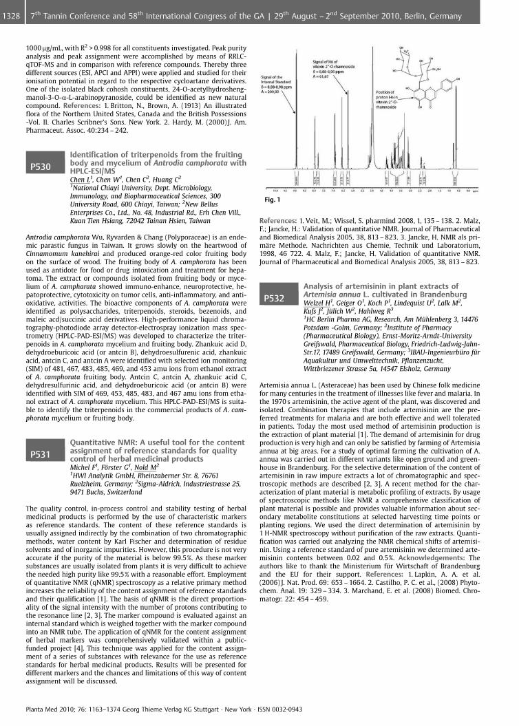
1000 mg/mL, with R2 > 0.998 for all constituents investigated. Peak purityanalysis and peak assignment were accomplished by means of RRLC-qTOF-MS and in comparison with reference compounds. Thereby threedifferent sources (ESI, APCI and APPI) were applied and studied for theirionisation potential in regard to the respective cycloartane derivatives.One of the isolated black cohosh constituents, 24-O-acetylhydrosheng-manol-3-O-a-L-arabinopyranoside, could be identified as new naturalcompound. References: 1. Britton, N., Brown, A. (1913) An illustratedflora of the Northern United States, Canada and the British Possessions-Vol. II. Charles Scribner’s Sons. New York. 2. Hardy, M. (2000) J. Am.Pharmaceut. Assoc. 40:234 – 242.
P530Identification of triterpenoids from the fruitingbody and mycelium of Antrodia camphorata withHPLC-ESI/MSChen L1, Chen W1, Chen C2, Huang C2
1National Chiayi University, Dept. Microbiology,Immunology, and Biopharmaceutical Sciences, 300University Road, 600 Chiayi, Taiwan; 2New BellusEnterprises Co., Ltd., No. 48, Industrial Rd., Erh Chen Vill.,Kuan Tien Hsiang, 72042 Tainan Hsien, Taiwan
Antrodia camphorata Wu, Ryvarden & Chang (Polyporaceae) is an ende-mic parastic fungus in Taiwan. It grows slowly on the heartwood ofCinnamomum kanehirai and produced orange-red color fruiting bodyon the surface of wood. The fruiting body of A. camphorata has beenused as antidote for food or drug intoxication and treatment for hepa-toma. The extract or compounds isolated from fruiting body or myce-lium of A. campharata showed immuno-enhance, neuroprotective, he-patoprotective, cytotoxicity on tumor cells, anti-inflammatory, and anti-oxidative, activities. The bioactive components of A. camphorata wereidentified as polysaccharides, triterpenoids, steroids, bezenoids, andmaleic acd/succinic acid derivatives. High-performance liquid chroma-tography-photodiode array detector-electrospray ionization mass spec-trometry (HPLC-PAD-ESI/MS) was developed to characterize the triter-penoids in A. camphorata mycelium and fruiting body. Zhankuic acid D,dehydroeburicoic acid (or antcin B), dehydroesulfurenic acid, zhankuicacid, antcin C, and antcin A were identified with selected ion monitoring(SIM) of 481, 467, 483, 485, 469, and 453 amu ions from ethanol extractof A. camphorata fruiting body. Antcin C, antcin A, zhankuic acid C,dehydresulfurinic acid, and dehydroeburicoic acid (or antcin B) wereidentified with SIM of 469, 453, 485, 483, and 467 amu ions from etha-nol extract of A. camphorata mycelium. This HPLC-PAD-ESI/MS is suita-ble to identify the triterpenoids in the commercial products of A. cam-phorata mycelium or fruiting body.
P531Quantitative NMR: A useful tool for the contentassignment of reference standards for qualitycontrol of herbal medicinal productsMichel F1, F�rster G1, Nold M2
1HWI Analytik GmbH, Rheinzaberner Str. 8, 76761Ruelzheim, Germany; 2Sigma-Aldrich, Industriestrasse 25,9471 Buchs, Switzerland
The quality control, in-process control and stability testing of herbalmedicinal products is performed by the use of characteristic markersas reference standards. The content of these reference standards isusually assigned indirectly by the combination of two chromatographicmethods, water content by Karl Fischer and determination of residuesolvents and of inorganic impurities. However, this procedure is not veryaccurate if the purity of the material is below 99.5%. As these markersubstances are usually isolated from plants it is very difficult to achievethe needed high purity like 99.5% with a reasonable effort. Employmentof quantitative NMR (qNMR) spectroscopy as a relative primary methodincreases the reliability of the content assignment of reference standardsand their qualification [1]. The basis of qNMR is the direct proportion-ality of the signal intensity with the number of protons contributing tothe resonance line [2, 3]. The marker compound is evaluated against aninternal standard which is weighed together with the marker compoundinto an NMR tube. The application of qNMR for the content assignmentof herbal markers was comprehensively validated within a public-funded project [4]. This technique was applied for the content assign-ment of a series of substances with relevance for the use as referencestandards for herbal medicinal products. Results will be presented fordifferent markers and the chances and limitations of this way of contentassignment will be discussed.
Fig. 1
References: 1. Veit, M.; Wissel, S. pharmind 2008, 1, 135 – 138. 2. Malz,F.; Jancke, H.: Validation of quantitative NMR. Journal of Pharmaceuticaland Biomedical Analysis 2005, 38, 813 – 823. 3. Jancke, H. NMR als pri-m�re Methode. Nachrichten aus Chemie, Technik und Laboratorium,1998, 46 722. 4. Malz, F.; Jancke, H. Validation of quantitative NMR.Journal of Pharmaceutical and Biomedical Analysis 2005, 38, 813 – 823.
P532Analysis of artemisinin in plant extracts ofArtemisia annua L. cultivated in BrandenburgWelzel H1, Geiger O1, Koch P1, Lindequist U2, Lalk M2,Kufs J2, J�lich W2, Hahlweg R3
1HC Berlin Pharma AG, Research, Am M�hlenberg 3, 14476Potsdam -Golm, Germany; 2Institute of Pharmacy(Pharmaceutical Biology), Ernst-Moritz-Arndt-UniversityGreifswald, Pharmaceutical Biology, Friedrich-Ludwig-Jahn-Str.17, 17489 Greifswald, Germany; 3IBAU-Ingenieurb�ro f�rAquakultur und Umwelttechnik, Pflanzenzucht,Wittbriezener Strasse 5a, 14547 Elsholz, Germany
Artemisia annua L. (Asteraceae) has been used by Chinese folk medicinefor many centuries in the treatment of illnesses like fever and malaria. Inthe 1970 s artemisinin, the active agent of the plant, was discovered andisolated. Combination therapies that include artemisinin are the pre-ferred treatments for malaria and are both effective and well toleratedin patients. Today the most used method of artemisinin production isthe extraction of plant material [1]. The demand of artemisinin for drugproduction is very high and can only be satisfied by farming of Artemisiaannua at big areas. For a study of optimal farming the cultivation of A.annua was carried out in different variants like open ground and green-house in Brandenburg. For the selective determination of the content ofartemisinin in raw impure extracts a lot of chromatographic and spec-troscopic methods are described [2, 3]. A recent method for the char-acterization of plant material is metabolic profiling of extracts. By usageof spectroscopic methods like NMR a comprehensive classification ofplant material is possible and provides valuable information about sec-ondary metabolite constitutions at selected harvesting time points orplanting regions. We used the direct determination of artemisinin by1 H-NMR spectroscopy without purification of the raw extracts. Quanti-fication was carried out analyzing the NMR chemical shifts of artemisi-nin. Using a reference standard of pure artemisinin we determined arte-misinin contents between 0.02 and 0.5%. Acknowledgements: Theauthors like to thank the Ministerium fr Wirtschaft of Brandenburgand the EU for their support. References: 1. Lapkin, A. A. et al.(2006) J. Nat. Prod. 69: 653 – 1664. 2. Castilho, P. C. et al., (2008) Phyto-chem. Anal. 19: 329 – 334. 3. Marchand, E. et al. (2008) Biomed. Chro-matogr. 22: 454 – 459.
Planta Med 2010; 76: 1163–1374 Georg Thieme Verlag KG Stuttgart · New York · ISSN 0032-0943
1328 7th Tannin Conference and 58th International Congress of the GA | 29th August – 2nd September 2010, Berlin, Germany
P533Development of HPTLC method forquantification of flavonoids in extracts of theselected Potentilla speciesTomczyk M1, Bazylko A2, Legas A2
1Medical University of Bialystok, Department ofPharmacognosy, Mickiewicza 2a, 15 – 230 Bialystok, Poland;2Medical University of Warsaw, Department ofPharmacognosy and Molecular Basis of Phytotherapy,Banacha 1, 02097 Warsaw, Poland
Potentilla species (Rosaceae) and their extracts have been highly valuedin many different ethnic cultures for hundreds of years throughout theworld. Extracts from P. species were and are still applied for the treat-ment of inflammations, wounds, infections due to bacteria, virus orfungi, diarrhoea, diabetes mellitus and several more ailments. Most ofthe biological activities of P. extracts can be explained with the highcontent of polyphenolics e. g. tannins (proanthocyanidins and hydroly-sable tannins), numerous flavonoids, coumarins, polyprenols, phenoliccarboxylic acids as well as triterpenoids [1]. Our previous study for thesimultaneous determination of polyphenolics from P. species has beendeveloped and validated using HPTLC precoated silica gel 60F254 plateswith toluen\ethyl formate\formic acid (6:4:1, v/v/v) [2]. The aim of thepresent study was to determinate of three flavonoids: isoquercitrin (IQ),quercetin 3-glucuronide (QG) and rutin (RT) in the ethyl acetate extractsobtained from aerial parts of the selected Potentilla species: P. nepalen-sis and Drymocallis rupestris (P. rupestris) by using HPTLC-densitome-try method. Chromatography was performed in CAMAG ADC2 (Auto-matic Development Chamber). HPTLC-DIOL silica gel F254 plates wereapplied. As a mobile phase ethyl acetate/methyl ethyl ketone/diisopro-pylether/formic acid (3:10:4:1, v/v/v/v) was used (distance of 7.5 cm).Densitometry was carried out by using of Shimadzu CS-9301PC densit-ometer. The absorption spectra were recorded at 350 nm. The proposedHPTLC method was found to be simple, precise and accurate for thequantification of these compounds in plant materials. Acknowledge-ments: This study is financially supported by the Polish Ministry ofScience and Higher Education (Grant No. N N405 621638) References:1. Tomczyk, M., Latt�, KP. (2009) J Ethnopharmacol 122:184 – 204. 2.Tomczyk, M. et al. (2010) Phytochem Anal 21:174 – 179.
P534Characterization of salvianolic acid B andverbascoside cyclodextrin complexes byinnovative NMR methods. Evaluation of theirstabilityIsacchi B, Karioti A, Bergonzi C, Bilia AUniversity of Florence, Department of PharmaceuticalSciences, Pharmaceutical Sciences, Via Ugo Schiff 6, 50019Sesto Fiorentino Florence, Italy
Salvianolic acid B is a potent antioxidant with anti-thrombotic, anti-hypertensive and hepatoprotective activities obtained from Salvia mil-tiorrhiza L. root [1]. Verbascoside is a phenylpropanoid glycoside widelyspread in nature with remarkable antioxidant, neuroprotective and he-patorpotective properties [2]. Both constituents are very water-solublebut unstable in aqueous solutions at pH > 5 [3]. In the present studyboth substances were enclosed in b-cyclodextrin in order to increasestheir water stability and the complexes were characterised by 1H-NMRcomplexation shifts analysis, 1H-NMR diffusion measurements, ROESY,NMR titration study and Job’s plot. Diffusion ordered spectroscopy(DOSY) has been used extensively to study supramolecular aggregatesand solubilisation processes in order to determine diffusion coefficientsfor the individual signals in a spectrum [4]. The DOSY spectra demon-strated that in the presence of b-CyD the translational diffusion of ver-bascoside and salvianolic acid B are sizably slowed down with respect tothe free drugs. In the case of salvianolic acid B induced shifts and inter-molecular ROE signals demonstrated that two caffeoyl moieties are dee-ply inserted in the cyclodextrin cavity, while one caffeoyl moiety hangsout from the wider rim. The Job’s plots confirmed the 3:2 inclusioncomplex. In the case of verbascoside induced proton shifts and intermo-lecular ROE signals demonstrated that the caffeoyl moiety is deeplyinserted in the cyclodextrin cavity. The Job’s plots confirmed the 1:1inclusion complex. Stability studies made by qNMR showed that bothcomplexes increased the stability of the molecules. References: 1. Lin,YL et al. (2006) J. Ethnopharm. 105: 215 – 222. 2. Guangmiao, F. et al.(2008) Cur. Med. Chem.15: 2592 – 2613. 3. Meng, W. et al. (2009) Nat.Prod. Comm. in press. 4. Bilia, AR. et al. (2002) J. Pharm. Sci 91: 2265 –2270.
P535Quantification of xanthohumol, isoxanthohumol,8-prenylnaringenin, and 6-prenylnaringenin inhop extracts and derived capsules usingsecondary standardsDhooghe L1, Naessens T1, Heyerick A2, De Keukeleire D2,Vlietinck A1, Pieters L1, Apers S1
1Department of Pharmaceutical Sciences, University ofAntwerp, Universiteitsplein 1, 2610 Antwerp, Belgium;2Faculty of Pharmaceutical Sciences, Ghent University,Harelbekestraat 72, 9000 Ghent, Belgium
Hop (Humulus lupulus) is a frequently used estrogenic herbal medicinalproduct, containing the prenylflavonoids xanthohumol (XN), isoxantho-humol (IXN), 8- and 6-prenylnaringenin (8-PN and 6-PN). Althoughmany analytical methods have been developed for the quantification ofthese compounds, there still is a lack of validated methods for routinecontrol. Therefore we have developed an accessible HPLC-DAD methodusing quercetin and naringenin as secondary standards for the analysisof hop extract and capsules. After optimization of sample preparationand HPLC conditions, the analysis was validated according to the ICHguidelines. The response function of XN, 8-PN, quercetin and naringeninshowed a linear relationship. For the determination of XN, a calibrationline of at least three concentrations of quercetin was constructed. Thecorrection factors for XN (quercetin) and 8-PN (naringenin) were vali-dated and determined to be 0.583 for XN, and 1.296 for IXN, 8-PN and 6-PN. The intermediate precision was investigated and it could be con-cluded that the standard deviation of the method was equal consideringtime and concentration (RSD of 2.5 – 5%). By means of a recovery experi-ment, it was proven that the method is accurate (recoveries of 96.1 –100.1%). Several hop-containing preparations available on the Belgianmarket were analysed in order to check their compliance to the guide-line concerning the maximal daily intake of selected medicinal plants,establised by the Advisory Committee for Plant Preparations. For hop themaximal daily dose is limited to 400 mg of 8-PN. These analyses showedthat the reported method was applicable for this conformity testing.
P536Fluorometric determination of ascorbic acid inthe absence of the oxidant in juices of commoncitrusCopra-Janicijevic A, Sofic E, Klepo L, Topcagic A, Tahirovic I,Haskovic AFaculty of Science, Chemistry, Zmaja od Bosne 33 – 35,71000 Sarajevo, Bosnia And Herzegovina
A simple and sensitive fluorimetric method for the determination ofascorbic acid (AA) in juices of common citrus is described. The methodis based on the condensation reaction between AA and o-phenylenedia-mine (OPDA) in the abscence of the oxidant. The keto-form of AA reactswith OPDA in alkaline medium and forms an N-heterocyclic compoundwith large p-bond system, which can emit strong fluorescence. Thefluorescence intensity is measured at exitation and emision wavelengthsof 330 nm and 430 nm, respectively. Under optimum conditions, a linearrelationship is obtained between the fluorescence intensity and the con-centraction of AA in the range 0.05 – 50 mg mL-1. The detection limit (3s)was found to be 0.54 mg mL-1 of AA (s from 5 determination of 10 mgmL-1). A relative standard deviation of 1.4% was recorded for 8 measure-ments of 1 mg mL-1 standard AA solution. The efect of the reaction timeis also studied. The fluorescence intensity of the system reached a max-imum immediatelly after all the reagens were added and remainedstable at least for 45 minutes. This method is simple, sensitive and easyfor the determination of AA in juices of common citrus. This is firstreport that analyses of AA in biological samples were done withoutoxidant.
P537Method development and optimization forisoflavone quantification in dry soy extractcontaining productsCsupor D1, Bognr J1, Karsai J2, Hohmann J1
1University of Szeged, Institute of Pharmacognosy, E�tv�s u.6., 6720 Szeged, Hungary; 2University of Szeged, Departmentof Medical Informatics, Kornyi fasor 9., 6720 Szeged,Hungary
Isoflavones are secondary metabolites belonging to flavonoids found in avariety of plants, especially soy (Glycine max L.). Due to the estrogen-likeactivity of these compounds several soy products are on the market and
Planta Med 2010; 76: 1163–1374 Georg Thieme Verlag KG Stuttgart · New York · ISSN 0032-0943
13297th Tannin Conference and 58th International Congress of the GA | 29th August – 2nd September 2010, Berlin, Germany
applied to relieve menopausal symptoms. The aim of our study was todevelop a simple method for the determination of isoflavones in soyextract containing products. The main isoflavones of soybean are genis-tin, glycitin, daidzin and their respective acetyl, malonyl and aglyconeforms. Quantitative measurement of each individual isoflavone is diffi-cult because some of the reference compounds are not widely commer-cially available [1]. Keeping in mind that the biological effects of soyisoflavones depend upon aglycone form, hydrolysis of glycosides is apractical method for the quantitative determination of total isoflavonesand assessment of biological value of extracts [2]. The objective of ourinvestigation was to establish an acid hydrolysis method for the quanti-tative HPLC determination of total isoflavones including daidzein, gen-istein and glycitein. To optimize the extraction and hydrolysis of isofla-vones, the effect of HCl concentration (1.5 – 6 N), hydrolysis time (25 –210 min) and temperature (30 – 100 �C) on total isoflavone aglycone con-tent was studied using an RP-HPLC-DAD method. By mathematical fit-ting and optimization methods optimum hydrolysis conditions for max-imizing the quantification of isoflavones were determined (t = 94 min,cHCl= 5.09 N, T = 80 �C). The experimentally verified model has a goodcoefficient of R2 (0,9928) and the recovery of isoflavones was 97.81 –102.76%. The developed method allows a reliable and maximum deter-mination of isoflavones in dry soy extract containing products. Refer-ences: 1. Rostagno, M.A. et al. (2009) J Chrom A 1216: 2 – 29. 2. Chiang,W.D. et al. (2001) Food Chemistry 72: 499 – 503.
P538Phytochemical analysis of the stems fromcultivar varieties of Rubus idaeusKrauze-Baranowska M, Majdan M, Glod DMedical University of Gdansk, Deparment ofPharmacognosy, Gen. J. Hallera 107, 80416 Gdansk, Poland
The stems of red raspberry (Rubus idaeus L., Rosaceae) are used in folkmedicine of several countries of Eastern Europe as anti-inflammatoryand antipyretic remedies in the treatment of flulike infections. There isno literature data on a chemical composition of this plant material, incontrary to fruits of R. idaeus rich in anthocyanins and ellagic acid deri-vatives [1]. The aim of the study was to recognize active biologicallycompounds present in the stems of twelve cultivars of red raspberry.Different groups of simple phenols and polyphenols were analysed inthe obtained methanol extracts by chromatographic methods – TLC,HPLC/DAD/MS and two dimensional techniques, including “comprehen-sive” LCxLC [2]. Sanguiin H-6 and ellagic acid were shown to be themajor compounds in all analysed cultivars. Other phenolic compounds,cinnamic and benzoic acid derivatives and flavonols, flavan-3-ols andprocyanidins were detected in minor concentrations as free forms (agly-cones) or glycosides. Among flavonols, hyperoside and isoquercetin weredominant compounds. The stems of Rubus idaeus seems to be a richsource of phenols possesing antioxidant [4] and antiviral activity [5],what can explain its traditional use as curative in a cold. Acknowledge-ments: Polish State Committee for Scientific Research (KBN) Grant Re-ferences: 1. Rao A.V., Snyder D. M. (2010) J. Agric. Food Chem. 58:3871 – 3883. 2. Dugo P. et al. (2009) J. Chromatogr. A.1216: 7483 – 7487.
P539Improved separation of green tea catechins andxanthines with calixarene bonded silica packingsin narrow-bore HPLC systemsBazylak G1, Pan T2
1Nicolaus Copernicus University, Collegium Medicum,Faculty of Pharmacy, Department of Pharmaco-Bromatology & Molecular Nutrition, Jagiellonska 13, 85067Bydgoszcz, Poland; 2School of Traditional Chinese Medicine,Chang Gung University, 259 Wen-Hwa 1st Road, 333Kweishan, Taoyuan, Taiwan
Epigallocatechin gallate (EGCg), the predominant green tea catechin, isrecommended as the health-promoting and complementary agent in thecurrent management of variety infectious diseases with approved anti-virals and antibiotics [1]. However, in commonly applied isocratic HPLCassays with octadecylsilica-C 18 packed columns the baseline separationof critical pairs as catechine/epigallocatechine and caffeine/EGCg wasoften failed. In presented isocratic narrow-bore HPLC procedure, thethree different calixarene bonded stationary phases were successfullyapplied with AcN-2.65 mM H3PO4 (10:90, v/v), pH 3.0, as mobile phaseto enable a complete separation of six catechins and six xanthines pre-sent in aqueous-AcN extracts of green tea. Compared to the retentionwith C 18 columns, the trans isomer (catechine) was eluted before the cis
isomer (epicatechine) on each calixarene-based packing studied. How-ever, when the dihydroxyphenyl substituents in catechine moiety werereplaced by trihydroxyphenyl fragment, as was also in case of its gallateesters, the cis isomers were eluted first. These phenomena showed thatselectivity of catechins separation with calixarene stationary phases wasclosely related with stability of their inclusion (1:1) host-guest com-plexes formed between analyte molecules and calixarene cavities [2].In case of xanthines the increased size of substituents attached to theN1 nitrogen atom of the pyrimidine ring, caused enhanced retention ofthese compounds in proposed HPLC systems. The distribution profiles ofcatechins and xanthines in series of commercially available green teasand catechins-based nutraceuticals were determined with the devel-oped HPLC procedure to offer more detailed data for standardization ofsuch health-promoting products. Acknowledgements: Nicolaus Univer-sity, Torun, Poland (Internal Grant No. 407/2010). References: 1. Song,J.M., Seong, B.L. (2007) Expert Rev. Anti Infect. Ther. 5(3): 497 – 506. 2.Bazylak, G. et. al. (2008) Curr. Drug Discov. Technol. 5(2): 177 – 189.
P540Quantitative determination of stachydrine viaHPTLC in cardioactive Leonurus and LeonotisdrugsKuchta K1, Volk R2, Rauwald H1
1Leipzig University, Department of Pharmaceutical Biology,Johannisallee 2123, 04103 Leipzig, Germany; 2Schaper &Br�mmer GmbH & Co. KG, Bahnhofstraße 35, 38259Salzgitter-Ringelheim, Germany
Tea preparations from aerial parts of the Central and Eastern EuropeanLamiaceae Leonurus cardiaca L. are traditionally used as a remedyagainst tachyarrhythmia, heart failure, and other cardiac disorders [1 –3]. Nevertheless, a scientific basis for these therapeutic effects had notbeen established [4] until just recently an antiarrhythmic refined extractwas prepared from this drug via bioassay guided fractionation [1], mak-ing the development of improved methods of extract characterisationnecessary. In Chin.Ph. [5] the “alkaloids” of the tea preparation of theclosely related and similarly used East Asian Leonurus japonicus Houtt.[6] are regarded as the active constituents and quantified photometri-cally after Reinecke’s complexation, calculated as stachydrine. Unfortu-nately, both the quantification method by complexation and the accom-panied TLC method of the Chin.Ph. are deemed to be “not reproducible”by current experimental literature, such as Heuberger et al. 2008 [7].Therefore, a state of the art HPTLC method was developed, using aCAMAG automatic HPTLC system with scanner and winCATS analysissoftware for reproducible stachydrine quantification. Silica gel 60 F254HPTLC plates were scanned at 517 nm after development withMeOH:CH2Cl2:NH325% (8:2:3) and derivatisation with V�gffljfalvi solu-tion. The related cardioactive [8 – 10] South African Leonotis leonurus(L.)R.Br. was co-investigated. These measurements revealed stachydrinecontents (w/w) of 0.2 to 1.1% for the L. japonicus drug, 0.3% for thematerial from L. leonurus, 0.5 to 1.5% for the L. cardiaca samples, and6.6% for the antiarrhythmic refined extract of L. cardiaca. References:1. Ritter M. et al. (2010) Planta Med 76: 572 – 82. 2. Barnes J. et al. (2002)Monograph Motherwort in: Herbal Medicines, Pharmaceutical Press,London. 3. Fuchs L. (1543) New Kreutterbuch, Basel edition. 4. Leonuricardiacae herba, in: Kommentar zum Europ�ischen Arzneibuch (2005).Wissenschaftliche Verlagsgesellschaft, Stuttgart. 5. Pharm. P. Rep. China,Eng. Edition Vol 1. Beijing: Chem. Ind. Press, 1997/2000. 6. Luo X: Com-pendium of Materia Medica, book III, Beijing: Foreign Languages Press,2003. A translation of the “Bencao Gangmu”, first published by Li Shiz-hen, 1593. 7. Heuberger H. et al. (2008) Z Arzn Gew Pfl 13 (4): 173 – 181.8. Burger A, Kabatembe J (2008) Planta Med 74: 991. 9. Watt JM, Breyer-Brandwijk MG (1962) The Medicinal and Poisonous Plants of Southernand Eastern Africa. Livingstone, London. 10. Hutchings A (1996) ZuluMedicinal Plants. Natal University Press, Pietermaritzburg.
P541Leonurine in Leonurus and Leonotis drugs?Detection and quantitative determination by anewly developed HPLC methodKuchta K1, Ortwein J2, Briel D2, Rauwald H1
1Leipzig University, Department of Pharmaceutical Biology,Johannisallee 2123, 04103 Leipzig, Germany; 2LeipzigUniversity, Department of Pharmaceutical Chemistry,Br�derstraße 34, 04103 Leipzig, Germany
Both aerial parts and seeds of Leonurus japonicus Houtt. are regarded astwo of the most efficient remedies for the treatment of abnormal men-
Planta Med 2010; 76: 1163–1374 Georg Thieme Verlag KG Stuttgart · New York · ISSN 0032-0943
1330 7th Tannin Conference and 58th International Congress of the GA | 29th August – 2nd September 2010, Berlin, Germany
struation, postpartum abdominal pain, and blood circulation disordersin TCM and Japanese Kampo medicine [1 – 3]. These pharmacologicaleffects are generally attributed to the “alkaloid” leonurine (4-[amino(i-mino)methyl]aminobutyl4-hydroxy-3,5-dimethoxybenzoate) [4]. Inter-estingly, in European herbalism Leonurus cardiaca L. (Ph.Eur.) is usedwith similar indications such as gynaecological disorders [5,6] and espe-cially heart disease [7], in which case its efficacy was recently proven bythe development of a cardioactive refined extract via bioassay guidedfractionation [8]. Although the presence of leonurine in L. cardiaca wasreported in one single publication [9], these results were never repro-duced. Nevertheless, it has repeatedly been quoted as an active consti-tuent of this drug, even in current textbooks on pharmacognosy [10,11].In the present study, a comprehensive, highly reproducible HPLC meth-od for the detection and quantitative analysis of leonurine in plant drugmaterial is reported for the first time. The South African herb Leonotisleonurus (L.)R.Br., used by native healers with similar indications [12],was co-investigated. Leonurine contends (w/w) between 0.001 and0.104% were detected in the nine different L. japonici herba samples, ofwhich only one failed to contain measurable amounts of leonurine. In-stead of their similar application, no leonurine was detected in L. japo-nicus seeds or in the L. cardiaca and L. leonurus samples, indicating thatapart from leonurine additional ingredients might contribute to the wellproven clinical efficacy of the examined traditional medical plants. Re-ferences: 1. Luo X: Compendium of Materia Medica, book III, Beijing:Foreign Languages Press, 2003. A translation of the “Bencao Gangmu”,first published by Li Shizhen, 1593. 2. Pharm. P. Rep. China, Eng. EditionVol 1. Beijing: Chem. Ind. Press, 1997/2000. 3. Kataoka N, Nakamura A(1992) Yamato Manyo Flowers. Tohoshuppan, Oosaka. 4. Kubota S, Na-kashima S (1930) Folia Pharmacologica Japonica 11: 153 – 167. 5. Culpe-per N: Culpeper’s Complete Herbal & English Physician, London: Park-gate Books, 1997. Identical text to the original publication of 1652. 6.Welch JM (1883) Transactions of the National Eclectic Medical Associa-tion 10: 79. 7. Fuchs L. (1543) New Kreutterbuch, Basel edition. 8. RitterM. et al. (2010) Planta Med 76: 572 – 82. 9. Gulabov AZ, Tchervenkova-Veleva VB (1970) Travaux scientifiques – Chimie 8 (1): 129 – 132. 10.Hiller K, L�w D: Leonuri cardiacae herba/Herzgespannkraut. In: WichtlM (editor): Teedrogen und Phytopharmaka, ein Handbuch fr die Praxisauf wissenschaftlicher Grundlage. Stuttgart: Wissenschaftliche Verlags-gesellschaft, 2009. 11. Teuscher E, Melzig MF, Lindequist U (2004) Bio-gene Arzneimittel. Wissenschaftliche Verlagsgesellschaft, Stuttgart.12. Burger A, Kabatembe J (2008) Planta Med 74: 991.
P542Quantitative HPTLC determination of rosmarinicacid and antioxidant activity of Origanum onitesL. water and 70 % methanol extractsG�ger F1, Altintas A1, Kosar M2, Kirimer N1, Baser K1
1Anadolu University, Pharmacognosy, Faculty of Pharmacydepartment of Pharmacognosy, 26470 Eskisehir, Turkey;2Erciyes University, Pharmacognosy, Faculty of PharmacyDepartment of Pharmacognosy, 38280 Kayseri, Turkey
Turkey is regarded as an important gene-center for the family Lamiaceae(Labiatae). The family is represented by 45 genera, 546 species and 730taxa in Turkey. The rate of endemism in the family is 44.2% [1 – 2]. Theflora of Turkey has 23 species of Origanum (15 endemic). They areknown and used as thyme or “kekik” which is the name given thosespecies with a thymol/carvacrol type odor in Turkey. The members ofthe Lamiaceae are common mainly in the mountainous areas of theMediterranean parts of Turkey and the composition of their essentialoils is detailed by Baser (1 – 3). In this study rosmarinic acid levels oforiganum onites water and 70%methanol extracts were determinedwith HPTLC using Camag TLC scanner at 330 nm. ethyl acetate:toluene:-formic acid (5:4:1) used as mobil phase. Extracts were also investigatedfor total phenolics content and antioxidant activity using Dpphbioauthography, and abts radical scavenger activity. Rosmarinic acidlevel of water extract and 70%Methanol extract were found 2.36% and2.29% respectively. Results can comparable with HPLC results (4) Theextracts showed remarkable antioxidant activity. References: 1. Baydar,H., et al., Food Control, 2004. 15(3): p.169 – 172. 2. Baser, K.H.C., E ActaHorticulture, 1993. 333: p. 217 – 238. 3. Baser, K.H.C., Lamiales Newslet-ter, 1994. 3 p. 6 – 11. 4. Exarchou, V., et al., J. Agric. Food Chem, 2001.49(1): p. 2 – 8.
P543CE and SDS-PAGE fingerprint analysis andidentification of several fermentation productsobtained by the Candida and Penicillium speciesused for homeopathic starting materialsWiethoff K, Irmer A, Bader GSANUM- Kehlbeck GmbH & Co. KG, Qualified Person,Hasseler Steinweg 9, 27318 Hoya/Weser, Germany
Fermentation cultures of Candida albicans (strain DSMZ 4884), C. para-psilosis (strain DSMZ 5197), Penicillium brevicompactum (strain DSMZ3961), P. chrysogenum (strain DSMZ 5753), P. glabrum (strain DSMZ5752) and P. roquefortii (strain DSMZ 5504) are prepared using variouspurification steps and cell mill treatment as well as sterilization andlyophilisation procedures to produce water-soluble substances [1 – 6].Homeopathic (isopathic) dilutions of these starting materials are usedas drug products in various European countries under the trade namesof Albicansan, Pefrakehl, Stolonikehl, Notakehl, Quentakehl, Fortakehland Exmykehl. The identification of the drug substances are performedby SDS-polyacrylamide gel electrophoresis (SDS-PAGE) and capillaryelectrophoresis (CE) producing characteristic fingerprints. The SDS-PAGEanalysis shows substance-specific bands with the following relative mo-lecule masses (Mr). The signals obtained by CE and detection at 260 nmshow fingerprint electropherograms that are specific for every substancepreparation, making it possible to identify each individual culturedstrain. Molecule Masses from extracts of Candida and Peniciliium spe-cies
Table 1
Candida sp. Mr [kDa]
Penicillium sp. > 100 100 – 90 90 – 80 80 – 70 70 – 60 60 – 50 50 – 40 40 – 30 30 – 20 < 20C. albicans 85 77 37/ 34 26 16C. parapsilosis 118 78 41 37 28P. brevicompactum 59 34 27/ 25P. chrysogenum 76 24 16P. glabrum 96 57 41 24P. roquefortii 67 50 38 24
References: 1. German Homeopathic Pharmacopoeia (HAB) draft mono-graph (2009) “Candida albicans e volumine cellulae (lyophil., steril.)”. 2.German Homeopathic Pharmacopoeia (HAB) draft monograph (2009)“Candida parapsilosis e volumine cellulae (lyophil., steril.)”. 3. GermanHomeopathic Pharmacopoeia (HAB) draft monograph (2010) “Penicil-lium brevicompactum e volumine cellulae (lyophil., steril.)”. 4. GermanHomeopathic Pharmacopoeia (HAB) draft monograph (2009) “Penicil-lium chrysogenum e volumine cellulae (lyophil., steril.)”. 5. GermanHomeopathic Pharmacopoeia (HAB) draft monograph (2009) “Penicil-lium glabrum e volumine cellulae (lyophil., steril.)”. 6. German Homeo-pathic Pharmacopoeia (HAB) draft monograph (2009) “Penicillium ro-quefortii e volumine cellulae (lyophil., steril.)”.
P544Zebrafish bioassay-guided microfractionationcombined with CapNMR a comprehensiveapproach for the identification ofanti-inflammatory and anti-angiogenicconstituents of Rhynchosia viscosaCrawford A1, Bohni N3, Maes J1, Kamuhabwa A2, Moshi M2,Esguerra C1, de Witte P1, Wolfender J3
1Chemical Genetics Initiative and Laboratory forPharmaceutical Biology, Department of PharmaceuticalSciences, University of Leuven, 3000 Leuven, Belgium;2Muhimbili University of Health and Allied Sciences, P. OBox, 35091 Dar es Salaam, Tanzania, United Republic Of;3School of Pharmaceutical Sciences, University of Geneva,University of Lausanne, School of Pharmaceutical Sciences,Quai E Ansermet 30, 1211 Geneva, Switzerland
Zebrafish have recently emerged as an attractive in vivo system forfunctional genomics and drug discovery [1]. Because of their small size,rapid development, optical transparency, and high genetic, physiologic,and pharmacologic similarity with humans, zebrafish embryos and lar-vae are also an ideal model for natural product discovery. As zebrafishbioassays require only microgram amounts of crude extracts, chromato-graphic fractions, and pure compounds, we are developing a zebrafish-based natural product discovery platform that takes advantage of mod-ern techniques for the isolation and structural elucidation of naturalproducts, such as UHPLC-TOF-MS profiling, LC-MS microfractionationand subsequent capillary NMR characterization [2]. An in vivo, zebra-fish-based anti-inflammatory screen of methanolic extracts of East Afri-can medicinal plants resulted in the identification of R. viscosa, whichrevealed potent inhibition of leukocyte migration after larval tail trans-
Planta Med 2010; 76: 1163–1374 Georg Thieme Verlag KG Stuttgart · New York · ISSN 0032-0943
13317th Tannin Conference and 58th International Congress of the GA | 29th August – 2nd September 2010, Berlin, Germany
ection in the presence of bacterial lipopolysaccharides. This extract alsodisplayed anti-angiogenic activity in transgenic zebrafish with vascula-ture-specific expression of GFP. Intriguingly, R. viscosa is used by tradi-tional healers in Tanzania for the treatment of inflammatory skin dis-orders and insect bites, corroborating our findings in zebrafish. Micro-fractionation of the crude methanolic extract was performed, and indi-vidual fractions tested for bioactivity in zebrafish. One highly activefraction was subjected to HR-ESI-MS and CapNMR analysis, resultingin the identification of genistein – a known inhibitor of inflammationand angiogenesis. These results indicate the potential of zebrafish bioas-say-guided microfractionation, in combination with sub-milligram NMRtechniques, to rapidly identify bioactive natural products. References:1. Crawford, A. et al. (2008) Planta Med., 6, 624 – 632. 2. Glauser, G. et al.(2009) J. Agric. Food Chem., 57, 1127 – 34.
P545Mass spectrometry in microbial metabolomicanalysis as an analytical tool for dereplicationstrategyCrevelin E1, Beraldo de Moraes L1, Soares de Melo I2
1University of S¼o Paulo, Chemistry, Bandeirantes Avenue,14040901 Ribeir¼o Preto, Brazil; 2Brazilian Company forAgricultural Research – Embrapa, Laboratory ofEnvironmental Microbiology, SP Highway, 13820 – 000Jaguarifflna, Brazil
Metabolomics is the quantitative analysis of wide arrays of metabolites,including seoncdary metabolites, in biological samples. Metabolic pro-filing focuses on a group of metabolites while metabolic fingerprintingdeal with global screening approach to classify samples based on meta-bolite patterns. The application of MS for analysis of secondary metabo-lites has grown over the last two decades, and today MS is the mostimportant detector method in biotechnology [1]. Dereplication acceler-ates the discovery of novel natural products by eliminating repetitivework on known compounds. MS has been used for the dereplication ofnatural products because it is sensitive and provides information aboutthe molecular mass, molecular formula and substructure of molecules[2], allowing dereplication of natural products at their early stages ofpurification and characterization. The aim of this study was to analyzemetabolite profiles of crude extracts obtained from actinomycetes indifferents cultivation medium by direct infusion MS and LC/MS. Thereare differences in the production of secondary metabolites in responseto the growth conditions. The dereplication strategy for the analysis ofthe secondary metabolites has been developed. First, full scan total ionchromatogram (TIC), scanning m/z 100 – 1200, was acquired in bothpositive and negative modes. Ionization of compounds was better inpositive mode. The dereplication was carried out in the mass range ofm/z 750 – 900. The data suggest that the secondary metabolites locatedin this mass range belong to class of macrotetrolides and macrolidesantibiotics (Figure 1). Compounds were isolated by a bioassay-guidedfractionation using the microalga Chlorella vulgaris as test organism.
Fig. 1: Chemical structures of macrotetrolides and 16-memberedmacrolide antibiotics
Acknowledgements: FAPESP, CAPES and CNPq References: 1. Dettmer,K.; Aronov, P.A.; Hammock, B.D. Mass. Spectrom. Rev. 2007, 26, 51 – 78.2. Lang, G.; Mayhudin, N.A.; Mitova, M.I.; Sun, L.; Van der Sar, S.; Blunt,J.W.; Cole, A.L.J.; Ellis, G.; Laatsch, H.; Munro, M.H.G. J. Nat. Prod. 2008,71, 1595 – 1599.
P546Specific HPLC determination of toxic alkaloids inaconite roots compared with amount ot totalalkaloids determined by titrationCsupor D1, Borcsa B1, Heydel B4, Hohmann J1, Zupk I2,Ma Y3, Widowitz U4, Bauer R4
1University of Szeged, Department of Pharmacodynamicsand Biopharmacy, E�tv�s u. 6, 6720 Szeged, Hungary;2University of Szeged, Department of Pharmacodynamicsand Biopharmacy, E�tv�s u. 6, 6720 Szeged, Hungary;3Medical University Vienna, Department ofPathophysiology, Center of Physiology, Pathophysiology andImmunology, W�hringer G�rtel 18 – 20, 1090 Vienna,Austria; 4Institute of Pharmaceutical Sciences, University ofGraz, Pharmacognosy, Universit�tsplatz 4, 8010 Graz,Austria
Aconite roots are known to be very toxic due to diterpene ester alkaloids[1]. Therefore, in Chinese medicine they are only used after processing.Hydrolysis of the ester groups decreases toxicity [2]. Nevertheless, sev-eral cases of poisoning by unprocessed or improperly processed aconiteroots have been reported [3]. Recently we suggested a specific samplepreparation method and HPLC assay for the analysis of toxic aconitealkaloids [4]. Now, an improved method with an even better resolutionis presented. Using this method, we have determined the content ofmesaconitine, aconitine and hypaconitine in 30 commercial samples ofprocessed aconite roots. In most of the samples, toxic aconite alkaloidswere not detectable, or only traces were found. However, in four sam-ples we could detect more than 0.04% of hypaconitine and mesaconi-tine, the highest with a content of 0.16%. Therefore, this method issuggested as a purity test for the European Pharmacopoeia. Acute toxi-city of batches high in hypaconitine and mesaconitine was also con-firmed in CFLP mice. In the aconite monograph of the German Homeo-pathic Pharmacopoeia, alkaloids are determined by a titration method.We compared the results of HPLC analysis of toxic alkaloids (mesaconi-tine, aconitine and hypaconitine) with the results obtained by the titra-tion method, and found no correlation. Samples which were lackingmesaconitine, aconitine and hypaconitine, still contained up to 0.2%alkaloids determined by titration. Therefore, titration of alkaloids isnot appropriate as an assay for toxic alkaloids in aconite roots.
Fig. 1
References: 1. Xiao, P.-G. et al. (2006) Acta Phytotaxon. Sin. 44, 1 – 46. 2.Bisset, N.G. (1981) J. Ethnopharmacol. 4: 247 – 336. 3. Singhuber, J. et al.(2009) J. Ethnopharmacol. 126: 18 – 30. 4. Csupor, D. et al. (2009) J. Chro-matrogr. A 1216: 2079 – 2086.
Planta Med 2010; 76: 1163–1374 Georg Thieme Verlag KG Stuttgart · New York · ISSN 0032-0943
1332 7th Tannin Conference and 58th International Congress of the GA | 29th August – 2nd September 2010, Berlin, Germany
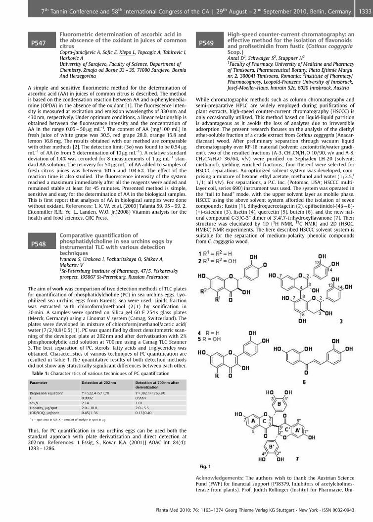
P547Fluorometric determination of ascorbic acid inthe abscence of the oxidant in juices of commoncitrusCopra-Janicijevic A, Sofic E, Klepo L, Topcagic A, Tahirovic I,Haskovic AUniversity of Sarajevo, Faculty of Science, Department ofChemistry, Zmaja od Bosne 33 – 35, 71000 Sarajevo, BosniaAnd Herzegovina
A simple and sensitive fluorimetric method for the determination ofascorbic acid (AA) in juices of common citrus is described. The methodis based on the condensation reaction between AA and o-phenylenedia-mine (OPDA) in the absence of the oxidant [1]. The fluorescence inten-sity is measured at excitation and emission wavelengths of 330 nm and430 nm, respectively. Under optimum conditions, a linear relationship isobtained between the fluorescence intensity and the concentration ofAA in the range 0.05 – 50 mg mL�1. The content of AA (mg/100 mL) infresh juice of white grape was 30.5, red grape 28.0, orange 15.8 andlemon 16.8 mg. The results obtained with our method are comparablewith other methods [2]. The detection limit (3s) was found to be 0.54 mgmL�1 of AA (s from 5 determination of 10 mg mL�1). A relative standarddeviation of 1.4% was recorded for 8 measurements of 1 mg mL�1 stan-dard AA solution. The recovery for 50 mg mL
�1of AA added to samples of
fresh citrus juices was between 101.5 and 104.6%. The effect of thereaction time is also studied. The fluorescence intensity of the systemreached a maximum immediately after all the reagents were added andremained stable at least for 45 minutes. Presented method is simple,sensitive and easy for the determination of AA in the biological samples.This is first report that analyses of AA in biological samples were donewithout oxidant. References: 1. X, W. et al. (2003) Talanta 59, 95 – 99. 2.Eitenmiller R.R., Ye, L., Landen, W.O. Jr.(2008) Vitamin analysis for thehealth and food sciences, CRC Press.
P548Comparative quantification ofphosphatidylcholine in sea urchins eggs byinstrumental TLC with various detectiontechniquesIvanova S, Urakova I, Pozharitskaya O, Shikov A,Makarov V1St-Petersburg Institute of Pharmacy, 47/5, Piskarevskyprospect, 195067 St-Petersburg, Russian Federation
The aim of work was comparison of two detection methods of TLC platesfor quantification of phosphatidylcholine (PC) in sea urchins eggs. Lyo-philized sea urchins eggs from Barents Sea were used. Lipids fractionwas extracted with chloroform/methanol (2/1) by sonification in30 min. A samples were spotted on Silica gel 60 F 254 s glass plates(Merck, Germany) using a Linomat V system (Camag, Switzerland). Theplates were developed in mixture of chloroform/methanol/acetic acid/water (7/2/0.8/0.5) [1]. PC was quantified by direct densitometric scan-ning of the developed plate at 202 nm and after derivatization with 2%phosphomolybdic acid solution at 700 nm using a Camag TLC Scanner3. The best separation of PC, sterols, fatty acids and triglycerides wasobtained. Characteristics of various techniques of PC quantification areresulted in Table 1. The quantitative results of both detection methodsdid not show any statistically significant differences between each other.
Table 1: Characteristics of various techniques of PC quantification
Parameter Detection at 202 nm Detection at 700 nm afterderivatization
Regression equation* Y = 522.4+571.7X Y = 382.1+1763.8Xr 0.9992 0.9997sdv,% 2.14 1.01Linearity, mg/spot 2.0 – 10.0 2.0 – 5.5LOD/LOQ, mg/spot 0.45 / 1.36 0.13 / 0.40
*Y – spot area in AU; X – amount of analyte in spot in mg
Thus, for PC quantification in sea urchins eggs can be used both thestandard approach with plate derivatization and direct detection at202 nm. References: 1. Essig, S., Kovar, K.A. (2001) J AOAC Int. 84(4):1283 – 1286.
P549High-speed counter-current chromatography: aneffective method for the isolation of flavonoidsand profisetinidin from fustic (Cotinus coggygriaScop.)Antal D1, Schwaiger S2, Stuppner H2
1Faculty of Pharmacy, University of Medicine and Pharmacyof Timisoara, Pharmaceutical Botany, Piata Eftimie Murgunr. 2, 300041 Timisoara, Romania; 2Institute of Pharmacy/Pharmacognosy, Leopold-Franzens University of Innsbruck,Josef-Moeller-Haus, Innrain 52c, 6020 Innsbruck, Austria
While chromatographic methods such as column chromatography andsemi-preparative HPLC are widely employed during purifications ofplant extracts, high-speed counter-current chromatography (HSCCC) isonly occasionally utilized. This method based on liquid-liquid partitionis advantageous as it avoids the loss of analytes due to irreversibleadsorption. The present research focuses on the analysis of the diethylether-soluble fraction of a crude extract from Cotinus coggygria (Anacar-diaceae) wood. After preliminary separation through vacuum liquidchromatography over RP-18 material (solvent: acetonitrile/water gradi-ent), two of the resulted fractions (A-3, CH3CN/H2O 10/90, v/v and A-8,CH3CN/H2O 36/64, v/v) were purified on Sephadex LH-20 (solvent:methanol), yielding enriched fractions; four thereof were selected forHSCCC separations. An optimized solvent system was developed, com-prising a mixture of hexane, ethyl acetate, methanol and water (1/2.5/1/1; all v/v). For separations, a P.C. Inc. (Potomac, USA; HSCCC multi-layer coil, series 690) instrument was used. The system was operated inthe “tail to head” mode, with the upper solvent layer as mobile phase.HSCCC using the above solvent system afforded the isolation of sevencompounds: fustin (1), dihydroquercetagetin (2), epifisetinidol-(4b?8)-(+)-catechin (3), fisetin (4), quercetin (5), butein (6), and the new nat-ural compound C-3/C-3” dimer of 3’,4’,7-trihydroxyflavanone (7). Theirstructure was elucidated by 1D (1H NMR, 13C NMR) and 2D (HSQC,HMBC) NMR experiments. The here described HSCCC solvent system issuitable for the separation of medium-polarity phenolic compoundsfrom C. coggygria wood.
Fig. 1
Acknowledgements: The authors wish to thank the Austrian ScienceFund (FWF) for financial support (P18379, Inhibitors of acetylcholines-terase from plants). Prof. Judith Rollinger (Institut fr Pharmazie, Uni-
Planta Med 2010; 76: 1163–1374 Georg Thieme Verlag KG Stuttgart · New York · ISSN 0032-0943
13337th Tannin Conference and 58th International Congress of the GA | 29th August – 2nd September 2010, Berlin, Germany
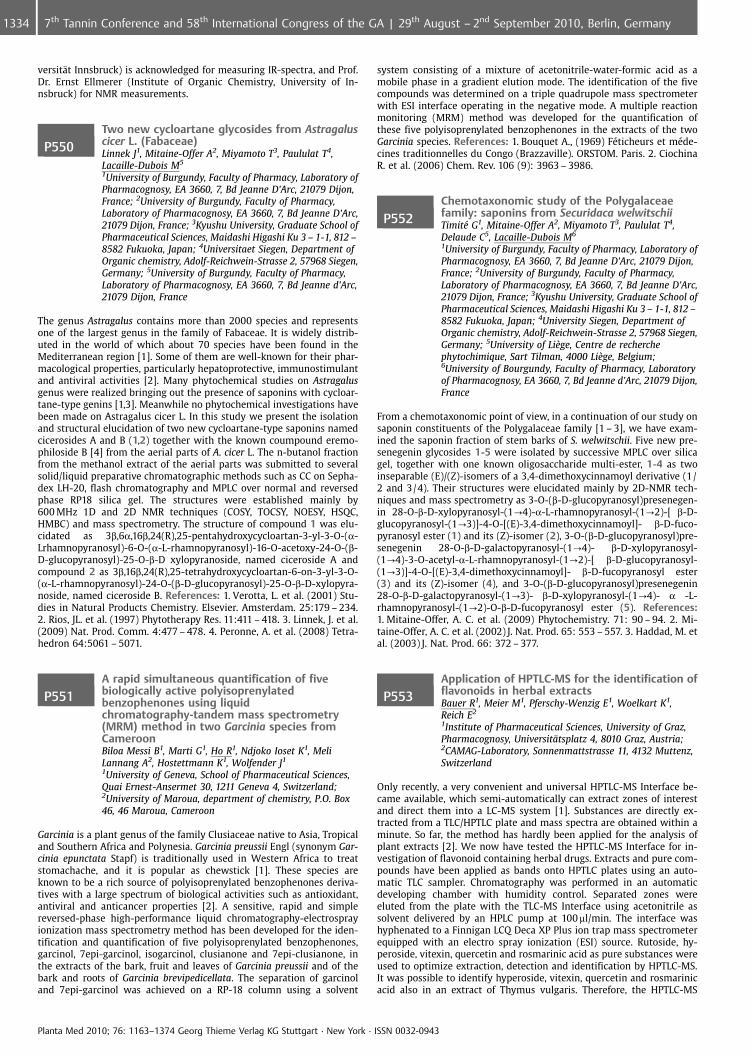
versit�t Innsbruck) is acknowledged for measuring IR-spectra, and Prof.Dr. Ernst Ellmerer (Institute of Organic Chemistry, University of In-nsbruck) for NMR measurements.
P550Two new cycloartane glycosides from Astragaluscicer L. (Fabaceae)Linnek J1, Mitaine-Offer A2, Miyamoto T3, Paululat T4,Lacaille-Dubois M5
1University of Burgundy, Faculty of Pharmacy, Laboratory ofPharmacognosy, EA 3660, 7, Bd Jeanne D’Arc, 21079 Dijon,France; 2University of Burgundy, Faculty of Pharmacy,Laboratory of Pharmacognosy, EA 3660, 7, Bd Jeanne D’Arc,21079 Dijon, France; 3Kyushu University, Graduate School ofPharmaceutical Sciences, Maidashi Higashi Ku 3 – 1-1, 812 –8582 Fukuoka, Japan; 4Universitaet Siegen, Department ofOrganic chemistry, Adolf-Reichwein-Strasse 2, 57968 Siegen,Germany; 5University of Burgundy, Faculty of Pharmacy,Laboratory of Pharmacognosy, EA 3660, 7, Bd Jeanne d’Arc,21079 Dijon, France
The genus Astragalus contains more than 2000 species and representsone of the largest genus in the family of Fabaceae. It is widely distrib-uted in the world of which about 70 species have been found in theMediterranean region [1]. Some of them are well-known for their phar-macological properties, particularly hepatoprotective, immunostimulantand antiviral activities [2]. Many phytochemical studies on Astragalusgenus were realized bringing out the presence of saponins with cycloar-tane-type genins [1,3]. Meanwhile no phytochemical investigations havebeen made on Astragalus cicer L. In this study we present the isolationand structural elucidation of two new cycloartane-type saponins namedcicerosides A and B (1,2) together with the known coumpound eremo-philoside B [4] from the aerial parts of A. cicer L. The n-butanol fractionfrom the methanol extract of the aerial parts was submitted to severalsolid/liquid preparative chromatographic methods such as CC on Sepha-dex LH-20, flash chromatography and MPLC over normal and reversedphase RP18 silica gel. The structures were established mainly by600 MHz 1D and 2D NMR techniques (COSY, TOCSY, NOESY, HSQC,HMBC) and mass spectrometry. The structure of compound 1 was elu-cidated as 3b,6a,16b,24(R),25-pentahydroxycycloartan-3-yl-3-O-(a-Lrhamnopyranosyl)-6-O-(a-L-rhamnopyranosyl)-16-O-acetoxy-24-O-(b-D-glucopyranosyl)-25-O-b-D xylopyranoside, named ciceroside A andcompound 2 as 3b,16b,24(R),25-tetrahydroxycycloartan-6-on-3-yl-3-O-(a-L-rhamnopyranosyl)-24-O-(b-D-glucopyranosyl)-25-O-b-D-xylopyra-noside, named ciceroside B. References: 1. Verotta, L. et al. (2001) Stu-dies in Natural Products Chemistry. Elsevier. Amsterdam. 25:179 – 234.2. Rios, JL. et al. (1997) Phytotherapy Res. 11:411 – 418. 3. Linnek, J. et al.(2009) Nat. Prod. Comm. 4:477 – 478. 4. Peronne, A. et al. (2008) Tetra-hedron 64:5061 – 5071.
P551A rapid simultaneous quantification of fivebiologically active polyisoprenylatedbenzophenones using liquidchromatography-tandem mass spectrometry(MRM) method in two Garcinia species fromCameroonBiloa Messi B1, Marti G1, Ho R1, Ndjoko Ioset K1, MeliLannang A2, Hostettmann K1, Wolfender J1
1University of Geneva, School of Pharmaceutical Sciences,Quai Ernest-Ansermet 30, 1211 Geneva 4, Switzerland;2University of Maroua, department of chemistry, P.O. Box46, 46 Maroua, Cameroon
Garcinia is a plant genus of the family Clusiaceae native to Asia, Tropicaland Southern Africa and Polynesia. Garcinia preussii Engl (synonym Gar-cinia epunctata Stapf) is traditionally used in Western Africa to treatstomachache, and it is popular as chewstick [1]. These species areknown to be a rich source of polyisoprenylated benzophenones deriva-tives with a large spectrum of biological activities such as antioxidant,antiviral and anticancer properties [2]. A sensitive, rapid and simplereversed-phase high-performance liquid chromatography-electrosprayionization mass spectrometry method has been developed for the iden-tification and quantification of five polyisoprenylated benzophenones,garcinol, 7epi-garcinol, isogarcinol, clusianone and 7epi-clusianone, inthe extracts of the bark, fruit and leaves of Garcinia preussii and of thebark and roots of Garcinia brevipedicellata. The separation of garcinoland 7epi-garcinol was achieved on a RP-18 column using a solvent
system consisting of a mixture of acetonitrile-water-formic acid as amobile phase in a gradient elution mode. The identification of the fivecompounds was determined on a triple quadrupole mass spectrometerwith ESI interface operating in the negative mode. A multiple reactionmonitoring (MRM) method was developed for the quantification ofthese five polyisoprenylated benzophenones in the extracts of the twoGarcinia species. References: 1. Bouquet A., (1969) F�ticheurs et m�de-cines traditionnelles du Congo (Brazzaville). ORSTOM. Paris. 2. CiochinaR. et al. (2006) Chem. Rev. 106 (9): 3963 – 3986.
P552Chemotaxonomic study of the Polygalaceaefamily: saponins from Securidaca welwitschiiTimit� G1, Mitaine-Offer A2, Miyamoto T3, Paululat T4,Delaude C5, Lacaille-Dubois M6
1University of Burgundy, Faculty of Pharmacy, Laboratory ofPharmacognosy, EA 3660, 7, Bd Jeanne D’Arc, 21079 Dijon,France; 2University of Burgundy, Faculty of Pharmacy,Laboratory of Pharmacognosy, EA 3660, 7, Bd Jeanne D’Arc,21079 Dijon, France; 3Kyushu University, Graduate School ofPharmaceutical Sciences, Maidashi Higashi Ku 3 – 1-1, 812 –8582 Fukuoka, Japan; 4University Siegen, Department ofOrganic chemistry, Adolf-Reichwein-Strasse 2, 57968 Siegen,Germany; 5University of Li�ge, Centre de recherchephytochimique, Sart Tilman, 4000 Li�ge, Belgium;6University of Bourgundy, Faculty of Pharmacy, Laboratoryof Pharmacognosy, EA 3660, 7, Bd Jeanne d’Arc, 21079 Dijon,France
From a chemotaxonomic point of view, in a continuation of our study onsaponin constituents of the Polygalaceae family [1 – 3], we have exam-ined the saponin fraction of stem barks of S. welwitschii. Five new pre-senegenin glycosides 1-5 were isolated by successive MPLC over silicagel, together with one known oligosaccharide multi-ester, 1-4 as twoinseparable (E)/(Z)-isomers of a 3,4-dimethoxycinnamoyl derivative (1/2 and 3/4). Their structures were elucidated mainly by 2D-NMR tech-niques and mass spectrometry as 3-O-(b-D-glucopyranosyl)presenegen-in 28-O-b-D-xylopyranosyl-(1?4)-a-L-rhamnopyranosyl-(1?2)-[ b-D-glucopyranosyl-(1?3)]-4-O-[(E)-3,4-dimethoxycinnamoyl]- b-D-fuco-pyranosyl ester (1) and its (Z)-isomer (2), 3-O-(b-D-glucopyranosyl)pre-senegenin 28-O-b-D-galactopyranosyl-(1?4)- b-D-xylopyranosyl-(1?4)-3-O-acetyl-a-L-rhamnopyranosyl-(1?2)-[ b-D-glucopyranosyl-(1?3)]-4-O-[(E)-3,4-dimethoxycinnamoyl]- b-D-fucopyranosyl ester(3) and its (Z)-isomer (4), and 3-O-(b-D-glucopyranosyl)presenegenin28-O-b-D-galactopyranosyl-(1?3)- b-D-xylopyranosyl-(1?4)- a -L-rhamnopyranosyl-(1?2)-O-b-D-fucopyranosyl ester (5). References:1. Mitaine-Offer, A. C. et al. (2009) Phytochemistry. 71: 90 – 94. 2. Mi-taine-Offer, A. C. et al. (2002) J. Nat. Prod. 65: 553 – 557. 3. Haddad, M. etal. (2003) J. Nat. Prod. 66: 372 – 377.
P553Application of HPTLC-MS for the identification offlavonoids in herbal extractsBauer R1, Meier M1, Pferschy-Wenzig E1, Woelkart K1,Reich E2
1Institute of Pharmaceutical Sciences, University of Graz,Pharmacognosy, Universit�tsplatz 4, 8010 Graz, Austria;2CAMAG-Laboratory, Sonnenmattstrasse 11, 4132 Muttenz,Switzerland
Only recently, a very convenient and universal HPTLC-MS Interface be-came available, which semi-automatically can extract zones of interestand direct them into a LC-MS system [1]. Substances are directly ex-tracted from a TLC/HPTLC plate and mass spectra are obtained within aminute. So far, the method has hardly been applied for the analysis ofplant extracts [2]. We now have tested the HPTLC-MS Interface for in-vestigation of flavonoid containing herbal drugs. Extracts and pure com-pounds have been applied as bands onto HPTLC plates using an auto-matic TLC sampler. Chromatography was performed in an automaticdeveloping chamber with humidity control. Separated zones wereeluted from the plate with the TLC-MS Interface using acetonitrile assolvent delivered by an HPLC pump at 100 ml/min. The interface washyphenated to a Finnigan LCQ Deca XP Plus ion trap mass spectrometerequipped with an electro spray ionization (ESI) source. Rutoside, hy-peroside, vitexin, quercetin and rosmarinic acid as pure substances wereused to optimize extraction, detection and identification by HPTLC-MS.It was possible to identify hyperoside, vitexin, quercetin and rosmarinicacid also in an extract of Thymus vulgaris. Therefore, the HPTLC-MS
Planta Med 2010; 76: 1163–1374 Georg Thieme Verlag KG Stuttgart · New York · ISSN 0032-0943
1334 7th Tannin Conference and 58th International Congress of the GA | 29th August – 2nd September 2010, Berlin, Germany
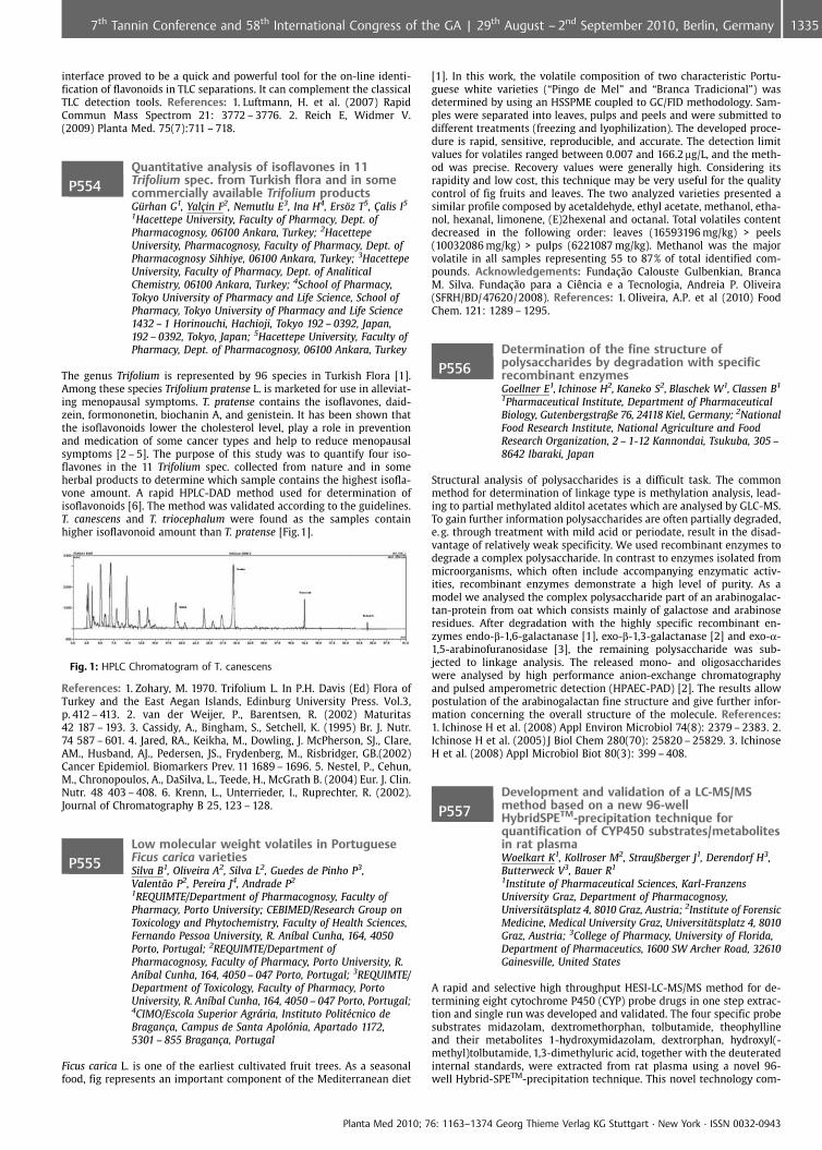
interface proved to be a quick and powerful tool for the on-line identi-fication of flavonoids in TLC separations. It can complement the classicalTLC detection tools. References: 1. Luftmann, H. et al. (2007) RapidCommun Mass Spectrom 21: 3772 – 3776. 2. Reich E, Widmer V.(2009) Planta Med. 75(7):711 – 718.
P554Quantitative analysis of isoflavones in 11Trifolium spec. from Turkish flora and in somecommercially available Trifolium productsG�rhan G1, Yal�in F2, Nemutlu E3, Ina H4, Ers�z T5, �alis I5
1Hacettepe University, Faculty of Pharmacy, Dept. ofPharmacognosy, 06100 Ankara, Turkey; 2HacettepeUniversity, Pharmacognosy, Faculty of Pharmacy, Dept. ofPharmacognosy Sihhiye, 06100 Ankara, Turkey; 3HacettepeUniversity, Faculty of Pharmacy, Dept. of AnaliticalChemistry, 06100 Ankara, Turkey; 4School of Pharmacy,Tokyo University of Pharmacy and Life Science, School ofPharmacy, Tokyo University of Pharmacy and Life Science1432 – 1 Horinouchi, Hachioji, Tokyo 192 – 0392, Japan,192 – 0392, Tokyo, Japan; 5Hacettepe University, Faculty ofPharmacy, Dept. of Pharmacognosy, 06100 Ankara, Turkey
The genus Trifolium is represented by 96 species in Turkish Flora [1].Among these species Trifolium pratense L. is marketed for use in alleviat-ing menopausal symptoms. T. pratense contains the isoflavones, daid-zein, formononetin, biochanin A, and genistein. It has been shown thatthe isoflavonoids lower the cholesterol level, play a role in preventionand medication of some cancer types and help to reduce menopausalsymptoms [2 – 5]. The purpose of this study was to quantify four iso-flavones in the 11 Trifolium spec. collected from nature and in someherbal products to determine which sample contains the highest isofla-vone amount. A rapid HPLC-DAD method used for determination ofisoflavonoids [6]. The method was validated according to the guidelines.T. canescens and T. triocephalum were found as the samples containhigher isoflavonoid amount than T. pratense [Fig.1].
Fig. 1: HPLC Chromatogram of T. canescens
References: 1. Zohary, M. 1970. Trifolium L. In P.H. Davis (Ed) Flora ofTurkey and the East Aegan Islands, Edinburg University Press. Vol.3,p. 412 – 413. 2. van der Weijer, P., Barentsen, R. (2002) Maturitas42 187 – 193. 3. Cassidy, A., Bingham, S., Setchell, K. (1995) Br. J. Nutr.74 587 – 601. 4. Jared, RA., Keikha, M., Dowling, J. McPherson, SJ., Clare,AM., Husband, AJ., Pedersen, JS., Frydenberg, M., Risbridger, GB.(2002)Cancer Epidemiol. Biomarkers Prev. 11 1689 – 1696. 5. Nestel, P., Cehun,M., Chronopoulos, A., DaSilva, L., Teede, H., McGrath B. (2004) Eur. J. Clin.Nutr. 48 403 – 408. 6. Krenn, L., Unterrieder, I., Ruprechter, R. (2002).Journal of Chromatography B 25, 123 – 128.
P555Low molecular weight volatiles in PortugueseFicus carica varietiesSilva B1, Oliveira A2, Silva L2, Guedes de Pinho P3,Valent¼o P2, Pereira J4, Andrade P2
1REQUIMTE/Department of Pharmacognosy, Faculty ofPharmacy, Porto University; CEBIMED/Research Group onToxicology and Phytochemistry, Faculty of Health Sciences,Fernando Pessoa University, R. An�bal Cunha, 164, 4050Porto, Portugal; 2REQUIMTE/Department ofPharmacognosy, Faculty of Pharmacy, Porto University, R.An�bal Cunha, 164, 4050 – 047 Porto, Portugal; 3REQUIMTE/Department of Toxicology, Faculty of Pharmacy, PortoUniversity, R. An�bal Cunha, 164, 4050 – 047 Porto, Portugal;4CIMO/Escola Superior Agrria, Instituto Polit�cnico deBragan�a, Campus de Santa Apolnia, Apartado 1172,5301 – 855 Bragan�a, Portugal
Ficus carica L. is one of the earliest cultivated fruit trees. As a seasonalfood, fig represents an important component of the Mediterranean diet
[1]. In this work, the volatile composition of two characteristic Portu-guese white varieties (“Pingo de Mel” and “Branca Tradicional”) wasdetermined by using an HSSPME coupled to GC/FID methodology. Sam-ples were separated into leaves, pulps and peels and were submitted todifferent treatments (freezing and lyophilization). The developed proce-dure is rapid, sensitive, reproducible, and accurate. The detection limitvalues for volatiles ranged between 0.007 and 166.2 mg/L, and the meth-od was precise. Recovery values were generally high. Considering itsrapidity and low cost, this technique may be very useful for the qualitycontrol of fig fruits and leaves. The two analyzed varieties presented asimilar profile composed by acetaldehyde, ethyl acetate, methanol, etha-nol, hexanal, limonene, (E)2hexenal and octanal. Total volatiles contentdecreased in the following order: leaves (16593196 mg/kg) > peels(10032086 mg/kg) > pulps (6221087 mg/kg). Methanol was the majorvolatile in all samples representing 55 to 87% of total identified com-pounds. Acknowledgements: Funda�¼o Calouste Gulbenkian, BrancaM. Silva. Funda�¼o para a CiÞncia e a Tecnologia, Andreia P. Oliveira(SFRH/BD/47620/2008). References: 1. Oliveira, A.P. et al (2010) FoodChem. 121: 1289 – 1295.
P556Determination of the fine structure ofpolysaccharides by degradation with specificrecombinant enzymesGoellner E1, Ichinose H2, Kaneko S2, Blaschek W1, Classen B1
1Pharmaceutical Institute, Department of PharmaceuticalBiology, Gutenbergstraße 76, 24118 Kiel, Germany; 2NationalFood Research Institute, National Agriculture and FoodResearch Organization, 2 – 1-12 Kannondai, Tsukuba, 305 –8642 Ibaraki, Japan
Structural analysis of polysaccharides is a difficult task. The commonmethod for determination of linkage type is methylation analysis, lead-ing to partial methylated alditol acetates which are analysed by GLC-MS.To gain further information polysaccharides are often partially degraded,e. g. through treatment with mild acid or periodate, result in the disad-vantage of relatively weak specificity. We used recombinant enzymes todegrade a complex polysaccharide. In contrast to enzymes isolated frommicroorganisms, which often include accompanying enzymatic activ-ities, recombinant enzymes demonstrate a high level of purity. As amodel we analysed the complex polysaccharide part of an arabinogalac-tan-protein from oat which consists mainly of galactose and arabinoseresidues. After degradation with the highly specific recombinant en-zymes endo-b-1,6-galactanase [1], exo-b-1,3-galactanase [2] and exo-a-1,5-arabinofuranosidase [3], the remaining polysaccharide was sub-jected to linkage analysis. The released mono- and oligosaccharideswere analysed by high performance anion-exchange chromatographyand pulsed amperometric detection (HPAEC-PAD) [2]. The results allowpostulation of the arabinogalactan fine structure and give further infor-mation concerning the overall structure of the molecule. References:1. Ichinose H et al. (2008) Appl Environ Microbiol 74(8): 2379 – 2383. 2.Ichinose H et al. (2005) J Biol Chem 280(70): 25820 – 25829. 3. IchinoseH et al. (2008) Appl Microbiol Biot 80(3): 399 – 408.
P557Development and validation of a LC-MS/MSmethod based on a new 96-wellHybridSPETM-precipitation technique forquantification of CYP450 substrates/metabolitesin rat plasmaWoelkart K1, Kollroser M2, Straußberger J1, Derendorf H3,Butterweck V3, Bauer R1
1Institute of Pharmaceutical Sciences, Karl-FranzensUniversity Graz, Department of Pharmacognosy,Universit�tsplatz 4, 8010 Graz, Austria; 2Institute of ForensicMedicine, Medical University Graz, Universit�tsplatz 4, 8010Graz, Austria; 3College of Pharmacy, University of Florida,Department of Pharmaceutics, 1600 SW Archer Road, 32610Gainesville, United States
A rapid and selective high throughput HESI-LC-MS/MS method for de-termining eight cytochrome P450 (CYP) probe drugs in one step extrac-tion and single run was developed and validated. The four specific probesubstrates midazolam, dextromethorphan, tolbutamide, theophyllineand their metabolites 1-hydroxymidazolam, dextrorphan, hydroxyl(-methyl)tolbutamide, 1,3-dimethyluric acid, together with the deuteratedinternal standards, were extracted from rat plasma using a novel 96-well Hybrid-SPETM-precipitation technique. This novel technology com-
Planta Med 2010; 76: 1163–1374 Georg Thieme Verlag KG Stuttgart · New York · ISSN 0032-0943
13357th Tannin Conference and 58th International Congress of the GA | 29th August – 2nd September 2010, Berlin, Germany
bines the simplicity of precipitation with the selectivity of SPE andhence leads to much cleaner extracts than with conventional proce-dures. The bioanalytical assay was based on reversed phase liquid chro-matography coupled with tandem mass spectrometry in the positive ionmode using selected reaction monitoring (SRM) for drug (-metabolite)quantification. All analytes were separated simultaneously in a singlerun that lasted less than 11 min. The intra- and inter-day precisions forall eight substrates/metabolites were 1.62 – 12.81% and 2.09 – 13.02%,respectively, and the relative errors (accuracy) for the eight compoundsranged from �9.62 – 7.48% and �13.84 – 8.82%. Hence, the present meth-od provides a robust, fast and reproducible analytical tool. The methodenables the determination of four major drug metabolising cytochromeP450 (3A4, 2C 9, 1A2, and 2D 6) enzymes and can be used as a commonhigh throughput analytical assay for in-vivo herb-drug interaction stu-dies.
P558Validation of TLC procedures for theidentification of Cimicifuga racemosa andHypericum per-foratum during quality control of aCimicifuga/Hypericum fixed combinationHoppenheit A, Volk R, Gerke H, Bodinet CSchaper & Br�mmer GmbH & Co. KG, Bahnhofstraße 35,38259 Salzgitter, Germany
For quality control of pharmaceuticals, validated analytical proceduresare required. Identity verification of herbal substances and phytophar-maceuticals is usually performed by thin layer chromatography (TLC).Cimicifuga/Hypericum film-coated tablets contain dry extract prepara-tions of Cimicifuga racemosa and Hypericum perforatum. Two TLC proce-dures on silica gel 60 F254 HPTLC plates were validated for this fixedcombination: (a) for detection of characteristic constituents of C. race-mosa, such as triterpene glycosides, (b) for detection of marker com-pounds typical for H. perforatum, such as hypericin. Mobile phases were(a) ethyl acetate: methanol (85:15), (b) toluol: ethyl acetate: formicacid: water (5:15:2:1). The typical validation characteristic for identifi-cation tests is their specificity. A characteristic necessary for any analy-tical procedure is its robustness [1]. For verifying the specificity of bothprocedures, test solutions of the herbal medicinal product, of herbalsubstances and of individual placebos were analysed. The procedureswere suitable to detect specifically characteristic constituents of bothherbal substances as mentioned above. Identification of both herbalsubstances was possible without any restriction. Specificity was alsotested by comparison of two different stationary phases. HPTLC plateswere essential (Cimicifuga), respectively prefered (Hypericum) insteadof aluminium foil plates. For verifying the robustness of both proce-dures, three parameters were modified: sample preparation: variationof numbers of extraction cycles during soxhlet extraction; chromatogra-phy: variation of the mobile phase; variation of detection (duration ofheating, respectively drying conditions). All tested variations led tochromatograms comparable to those obtained under standard condi-tions. Therefore, robustness was proven, too. References: 1. ICH guide-line Q 2(R1) Validation of analytical procedures: text and methodology;current step 4 version, Nov. 2005 (http://www.ich.org).
P559HPLC and 2D NMR analyzes as a tool for thequality control of herbal medicinal products: thecase of an antihypertensive herbal supplementcontaining not declared ajmaline and reserpineKarioti A1, Giocaliere E2, Pieraccini G2, Vannacci A3,Gallo E3, Bilia A1
1University of Florence, Department of PharmaceuticalSciences, Pharmaceutical Sciences, Via Ugo Schiff 6, 50019Sesto Fiorentino Florence, Italy; 2University of Florence,Mass Spectrometry Center (CISM), Mass SpectrometryCenter (CISM), Viale Pieraccini 6, 50139 Sesto FiorentinoFlorence, Italy; 3University of Florence, Department ofPreclinical and Clinical Pharmacology, Centre for MolecularMedicine (CIMMBA), Viale Pieraccini 6, 50139 Florence, Italy
In this study HPLC-DAD-ESI-MS, including HPLC-ESI-MSn analyses and2D NMR techniques were applied for the quality control of a herbalsupplement. Several reports were received by the pharmacovigilancesystem by patients who fainted after consuming the product “Olivis”, adietary supplement as an adjunctive treatment for hypertension. De-clared components of this product are extracts from Olea europea, Cra-taegus oxyacantha, Fumaria officinalis, Capsella bursa pastoris. “Olivis”
sample was subjected to 2D NMR and HPLC-DAD-ESI-MS analyses inorder to identify the marker consituents of the different Herbal Drugs.Comparison of the NMR and chromatographic profiles of the extractswith the original product showed the lack of constituents responsiblefor antihypertensive activity, such as oleuropein, protopine. Since signalscorresponding to polysaccharides submerged those of the other charac-teristic compounds, fractionation of the preparation was carried out inorder to separate the bulk of saccharides. HPLC-DAD and HPLC-ESI-MSanalyses of the fractions showed the presence of an alkaloid, ajmaline, asthe principal constituent of this product, as well as alkaloids structurallyrelated to ajmaline. HPLC-ESI-MSn analyses revealed a chromatographicpeak identified as reserpine. Phytochemical fractionations led to theisolation of ajmaline, whereas quantitation showed that ajmaline pre-vailed against reserpine indicating that a Rauwolfia species other than R.serpentina was used for this product. Rauwolfia extracts have beenbanned but they are available via internet. The present study showsthe importance of extensive controls using combined analytical toolsof the botanical products on the market to assure their quality and asa consequence their safety profiles.
P560Compared the separation efficacy of xanthonesfrom mangosteen with three gel-filtrationcolumnsHsu H, Huang J, Chen LNational Chiayi University, 300 University, 600 Chiayi,Taiwan
Mangosteen, Garcinia mangostana Linn (Guttiferae), is a popular refresh-ing juicy fruit in Taiwan. The rinds of mangosteen have been used as atraditional medicine in Thailand for the treatment of trauma, diarrhea,and skin infections. Xanthones are regarded as the major bioactive com-pounds of the mangosteen hulls which have been confirmed to consistof antimicrobial, anti-inflammatory, antioxidative, and inhibit humanimmunodeficiency virus infection activities. In present, three gel filtra-tion columns packed with Sephadex LH-20, Toyopearl HW-40, and MCI-gel CHP-20P respectively were used to compare the separative efficacyfor the xanthones from mangosteen hulls. The same amount of crudeextract of mangosteen hulls was dissolved in methanol and separated byeach column with methanol elution. Each eluate fraction was detectedby high-performance liquid chromatography coupled with electron-spray ionization mass spectrometry. The result display Sephadex LH-20 has the best separation efficacy. The elution orders for the threemajor xanthones were b-mangostin, a-mangostin, and g-magostin. Itsuggested that the separation mechanisms were complex with gel filtra-tion and adsorption. This method can be used for the xanthones fractio-nation from crude extract of mangosteen.
P561Effect of extraction conditions on the content ofsoluble oxalate in aqueous infusions of green andherbal teasBazylak G, Sperkowska BNicolaus Copernicus University, Collegium Medicum, Facultyof Pharmacy, Department of Pharmaco-Bromatology &Molecular Nutrition, Jagiellonska 13, 85067 Bydgoszcz,Poland
Green (GTs) and herbal (HTs) teas are recommended as the alternativehot beverages for subjects who tend to form calcium oxalate stones [1].The aim of study was to compare the effect of extraction procedures [2]on the content of soluble oxalate (SOX) in drinkable infusions of six GTsand seven HTs sold in Poland. The traditional method of extraction usingboiling water at 100 �C was compared with the microwave extraction at80 �C (EWPM), the ultrasound extraction at 40 �C (EWU-40) and theultrasound extraction at 60 �C. The highest amount of SOX, as deter-mined by oxidimetric and HPLC methods, was obtained in the infusionsmade by the EWPM procedure, i. e. 7.73 – 14.89 mg/g (dry mass) forstudied GTs, and in range of 3.53 – 18.11 mg/g for studied HTs. The low-est values were obtained after the EWU-40 procedure (5.06 – 10.88 mg/gfor GTs, and 1.50 – 11.03 mg/g for HTs). Highest average content of SOXwere observed in brews obtained from green whole leaf teas by theEWPM method (14.89 mg/g). The reduced content of SOX was observedin brews obtained by EWPM method from the highly crushed, ready-to-use, express green teas (7.73 mg/g). Similarly, the highest content of SOXafter the EWPM extraction was obtained for herbal whole leaf teas as inthe case of peppermint (18.11 mg/g), salvia (12.23 mg/g), and nettle(11.76 mg/g). These results could be used for standardization of analyti-
Planta Med 2010; 76: 1163–1374 Georg Thieme Verlag KG Stuttgart · New York · ISSN 0032-0943
1336 7th Tannin Conference and 58th International Congress of the GA | 29th August – 2nd September 2010, Berlin, Germany
cal methods applied to determination of SOX in imported green teas anddomestic herbal teas. Acknowledgements: Nicolaus Copernicus Univer-sity, Torun, Poland (Internal Grant No. 407/2010). References: 1. Massey,L.K. (2007) J. Am. Diet. Assoc. 107(7): 1191 – 1194. 2. Honow, R., Hesse, A.(2002) Food Chem. 78(4): 511 – 521.
P562HS-SPME method to monitoring “in vivo” volatilecompounds emitted from Achillea collina infestedby aphidsGiorgi A, Panseri S, Nanayakkarawasam C, Lozzia G,Chiesa L, Soncin S, Biondi P, Cocucci MUniversita Degli Studi di Milano, Produzione Vegetale, ViaCeloria 02, 20133, Milano, Italy, 20133 Milano, Italy
Plant leaves normally release small quantities of volatile compounds,but when it is damaged by herbivorous insects, many more volatilesare produced. The emitted volatile fraction plays a key role in plant-environment interaction, being involved in important processes in plantlife cycle [1]. Determination of volatile compounds release from livingplants are usually performed by enclosing the whole plants or some ofits parts in glass o plastic chambers, followed by collection of theemitted compounds in sorbent traps and chromatographic analysis ofthe adsorbed compounds. This general procedure has some drawbacksfor example some material employed as trapping can introduced arti-fact. This work describes the application of an Headspace Solid PhaseMicro-Extraction (HS-SPME) and Gas-Chromatographic Mass-Spectro-metric (GC-MS) method to characterize the volatile compounds emittedby leaving leaves of Achillea collina Becker ex Rchb, infested by aphids(Macrosifonella) using a sampling glass chamber specially designed forthis task. In particular, the headspaces volatiles were extracted using adivinylbenzene-carboxen-polydimethylsiloxane (DVB/CAR/PDMS, 70/30 mm) fiber. The extraction conditions including extraction temperatureand time were also optimized using the total peak area as index. Thebest response was obtained at room temperature for 240 min. As a re-sult, many volatile compounds appeared as new compounds after infes-tation (a-fenchene, aromadendrene and pinocarvone). Other com-pounds showed an increase trend after infestation by aphid such ascamazulene, followed by b-pinene, a-bergamotene and b-phellandrene.In conclusion, the present method is simple and effective and can beused to study the “in vivo” volatile compound emissions from medicinalplants. References: 1. Pareja M. et al. (2007). J Chem Ecol 33: 695 – 710.
P563Development of enzyme-based rapid screeningmethods for bioactive constituents in plantextractsChen C, Lee CTaipei Medical University, College of Pharmacy, 250 Wu XinStreet, 11031 Taipei, Taiwan
In recent years, there are more and more researches with developmentsof hyphenated techniques for the rapid identification of antioxidants incomplex plant extracts, but the limitations for this on-line analysis maybe critical to control the stability and activity of biological reactions [1].A rapid method for detection and identification of antioxidant com-pounds in plant extracts was developed by a combination of microplatearray analysis using xanthine oxidase, tyrosinase, lipoxygenase as mod-els and HPLC [2,3]. As for the enzymatic reaction with plant extracts, theactivity of the enzyme can be kept stable under the experimental con-ditions. With this simple and rapid method, we can estimate the re-quired minimum screening dose of enzyme for bioactive constituentsfrom plant extracts by HPLC. In addition to the reversible inhibitoryeffect of enzyme on plant extracts demonstrated by both microplatearray analysis and HPLC, we will testify the irreversible inhibitory effectof enzymes with trace amounts of bioactive compounds by LC-TOF/MS.References: 1. Tang, D. et al. (2008) J Sep Sci. 31:3519 – 26. 2. S. Zhu; S.et al. (2006) Fitoterapia 77:100 – 108. 3. Chen, C.H. et al.(2009) Mole-cules 14: 2947 – 2958.
New Targets for herbal medicines
P564The herbal combination preparation STW 5 isactive in DSS-induced colitisAbdel-Aziz H1, Wadie W2, Zaki H2, Vinson B3, Kelber O3,Okpanyi S3, Weiser D3, Khayyal M2
1Heliopolis University, 3 Cairo-Belbeis Desert Road, 11361Cairo, Egypt; 2Cairo University, Kasr El-Aini-Street, 11562Cairo, Egypt; 3Scientific Department, SteigerwaldArzneimittelwerk GmbH, Havelstr. 5, 64295 Darmstadt,Germany
Herbal extracts often have strong anti-inflammatory properties. To thesebelongs the phytomedicine STW 5, a combination preparation of nineherbal extracts used in the therapy of functional gastrointestinal dis-eases, including irritable bowel syndrome (IBS). Earlier pharmacologicalstudies, e. g. in TNBS induced colitis, an experimental model for Crohn’sdisease, suggest that STW 5 has anti-inflammatory properties. So wedecided to test it also in a model of Dextran Sodium Sulphate (DSS)induced colitis, which relates more to ulcerative colitis in man. DSSwas administered to rats in the drinking water for 7 days, leading tolesions in the colon, associated with changes in plasma levels of variousrelevant mediators. STW 5 was administered once daily orally in 3 doselevels (1, 2.5 and 5 ml/Kg) during DSS administration. It reduced thesymptom score, colon shortening and colon mass index in a dose de-pendent manner. The levels of TNFa and myeloperoxidase activity (asinflammation parameters) as well as the levels of glutathione, glu-tathione peroxidase, and superoxide dismutase (as oxidative stress para-meters) were measured in colonic tissue as well as in the blood. Thechanges in these parameters induced by DSS were favourably influencedby STW 5 in a manner comparable to sulfasalazine (300 mg/Kg) whichwas used as a reference standard drug. The findings point to the poten-tial therapeutic usefulness of STW 5 not only in IBS, but also in ulcera-tive colitis.
P565Rosemary extract enriched in carnosic acidshows anti-obesity and anti-diabetic effects on invitro and in vivo modelsIbarra A1, He K1, Bily A1, Cases J1, Coussaert A2, Ripoll C2,Roller M1
1NATUREX, 375 Huyler St., 07606 South Hackensack, UnitedStates; 2NATURALPHA, Parc Eurasant�, Lille M�tropole 85,Rue Nelson Mandela, 59120 Loos, France
Rosemary (Rosmarinus officinalis L.) leaf extract standardized to 20%carnosic acid (RE) has shown good antioxidant properties in both ORACin vitro and LDL oxidation ex vivo assays [1]. Regulating fat absorptionby inhibiting the action of the pancreatic lipase activity is an effectiveway to reduce body weight and obesity [2]. PPARg represents the majortarget for the glitazone type of drugs used for the treatment of type2 diabetes [3]. Therefore, we evaluated the in vitro capacities of RE toinhibit pancreatic lipase activity compared to tetrahydrolipstatin, and toactivate PPARg in a cell based assay compared to rosiglitazone. In addi-tion, we studied the effects of a low-fat diet (LFD), high-fat diet (HFD),and high-fat diet at 500 mpk RE (RED) on C 57BL/6 J mice during16 weeks. In vitro results showed that RE was able to inhibit pancreaticlipase activity by 69.8% at 100 mg/ml (P< 0.001) and activate PPARg by23% at 50 mg/ml (P< 0.05) as compared to their respective positive con-trols. In the in vivo experiment, after a 16 week treatment and comparedto both control groups, we observed that RED reduced by 67.7% the bodyweight gain (P< 0.001) and by 79.4% the adipose tissue gain (P< 0.001),without affecting the food intake. RED lowered total cholesterol increaseby 68.4 (P< 0.01). Glycemia was reduced by 72.0% (P< 0.05). RED did notshow any modification in the safety parameters measured as liverweight gain, AST and ALT levels. These results encourage conductingfurther studies on obesity and diabetes control using RE. References:1. Ibarra, A. et al. (2010) J Med Food In press. 2. Kim, HY and Kang, MH.(2005). Phytother Res 19:359 – 361. 3. Evans, RM. et al (2004). Nat Med10:355 – 361.
Planta Med 2010; 76: 1163–1374 Georg Thieme Verlag KG Stuttgart · New York · ISSN 0032-0943
13377th Tannin Conference and 58th International Congress of the GA | 29th August – 2nd September 2010, Berlin, Germany
P566Effects of some essential oils and saponins onlysozyme activity in human monocytes andepithelial cellsHelal R, Melzig MFree University of Berlin, Institute of Pharmacy,Pharmaceutical biology, K�nigin-Luise-Str. 2+4, 14195Berlin, Germany
Lysozyme is one of the most important factors of innate immunity,possessing antibacterial, antiviral, antitumor and immune modulatoryactivities. Human lysozyme is produced by phagocytic cells and a vari-ety of epithelial cells. In a recent study we have investigated the effect ofsome essential oils and saponins on the secretion and expression oflysozyme activity in the human monocytic cell line THP-1, and the hu-man colonic epithelial cell line HT-29. The selection of these naturalsubstances was based on their antibacterial, antiviral, anti-inflammatoryor immune-stimulating properties. The influence on lysozyme secretion,as an important defense molecule of the human innate immune system,may provide a new mechanism which could explain these properties.Lysozyme activity was determined using highly sensitive fluorescence-based assay [1]. Some saponins from natural origin had stimulatingeffects on lysozyme activity secretion within one-hour-incubation inboth monocytic and epithelial cells. Primulic acid, ginsenosid-Rd, quil-laja and gypsophila saponins were proved to induce lysozyme activitysecretion in both cell lines. Aescin could stimulate lysozyme activitysecretion only in the monocytic cells, while it had no effect when in-cubated with the epithelial cells. Stimulating effect of essential oils onlysozyme activity in both monocytic and epithelial cell lines could beless frequently observed. Orange blossom oil had stimulating effect onlysozyme activity secretion in both cell lines. Inducing effect on lyso-zyme activity could also be detected by tea tree oil and majoran oilwhen incubated with the epithelial cells, whereas no effect or inhibitoryeffect was observed in the monocytic cells. References: 1. Helal, R.,Melzig, M.F. (2008) Pharmazie 63(6):415 – 419.
P567The effect of ginger juice on the gastrointestinaltract is not fully explained by its knownconstituenHague T1, Naughton D1, Andrews P2
1Kingston University, Life Sciences, Main Building PenrhynRoad Kingston upon Thames, United Kingdom; 2St George’sUniversity of London, Basic Medical Sciences, CranmerTerrace, SW17 0RE London, United Kingdom
Ginger, a traditional Chinese herbal medicine, is used to treat digestivedisorders in particular to alleviate symptoms of nausea [1]. [6]-Gingerol(6G) is one of the main constituents in ginger and is reported to haveanti-oxidant properties [2]. The aims of this study were to use HPLC andICP-AES to measure the concentration of 6G, and elements in freshginger rhizome juice (GJ) and investigate their effects on the uppergastrointestinal tract. In vitro isometric recording was used to investi-gate GJ (200mL), 6G [1.59 x 10�5M), a selected combination of elements(K [4.6 x 10�2M], Mg [7.4 x 10�3M], Mn [8.3 x 10�4M], Na [1.1 x 10�3M], Ca[5.1 x 10�4M]), and a “faux” ginger juice on contractile activity of prox-imal and distal stomach and duodenal segments from Suncus murinus(house musk shrew). The concentration of 6G in GJ was 239.4 € 7.9 mg/L.GJ caused a pro-longed inhibitory effect on duodenal contractions and abiphasic effect on the stomach resulting in an overall increase in tensionat 25 minutes. “Faux” GJ did not fully account for the motility effects ofGJ, indicating other bioactive constituents were present in GJ (e. g. [6]-shogaol). GJ was most effective on the duodenum (48.3% inhibition ofcontraction tone), this could be a target for an enteric coated gingercapsule for gastrointestinal disorders. References: 1. Lien, HC. et al.(2003) Am. J. Physiol. Gastrointest. Liver Physiol. 284:G481-G489.8:125 – 132. 2. Kim, JK. et al. (2007) Free Radic. Res. 41:603 – 614.
P568Determination of dibenzylbutyrolactone-typelignans in Centraurea species and analysis ofarctigenin’s anticancer effectBorsodi Szokol L1, Sedlk �1, Boldizsr I1, Paku S2,Preininger �1, Gyurjn I1
1E�tv�s Lornd University, Budapest, Hungary, Departmentof Plant Anatomy, Pzmny P�ter s�tny 1/C, 1117 Budapest,Hungary; 2Semmelweis University, First Institute ofPathology & Experimental Cancer Research, �lloi fflt 26,1085 Budapest, Hungary
Centaurea species (Asteraceae) are mainly considered as weeds or orna-mental plants but rarely as a herb. Dibenzylbutyrolactone-type lignans,matairesionoside and arctiin, and their aglycones, matairesinol and arc-tigenin are present in Centaurea generea. Arctigenin exhibit antitumoreffect against colorectal [1], pancreatic [2] and skin cancer [3]. Isolationof lignans was published mainly for taxonomical classification. We in-vestigated Centaurea americana, C. calcitrapa, C. cyanus, C. dealbata, C.montana and C. scabiosa fruits to quantify their lignan composition byHPLC-UV [4, 5]. Matairesinoside and matairesinol were the major lignancomponents in C. scabiosa with 51.5 mg/g and 11.7 mg/g, while arctiinwas in C. dealbata with 58.7 mg/g. Arctigenin was present in all Centaur-ea species, with highest of 6.3 mg/g in C. americana. The total lignancontent ranged from 4.53 mg/g (C. cyanus) to 73.8 mg/g (C. dealbata)with RSD% between 3.2 – 6.9. Lignans of the selected Centaurea specieswere determined and from pharmaceutical point of view C. dealbataseems to be the best source of arctiin and C. scabiosa of matairesinosidefor further utilization. In the anticancer experiments with arctigenin,inbred C 57Bl/6 mice were transplanted with C 38 colorectal tumor. Tu-mor growth rate in mice treated with 50 mg arctigenin/kg body weightwas the smallest. The size of the tumors on the 21st day in group was asbig as the tumors on the 14th day in the control group. Arctigenin in50 mg/kg dose was effective against C 38 colon cancer. Arctigenin’s anti-tumor activity against C 38 colon cancer is promising in in vivo experi-ments. References: 1. Hausott, B et al. (2003) J. Cancer Res. Clin. Oncol.129: 569 – 576. 2. Awale, S. et al.(2006) Cancer Res. 66: 1751 – 1757. 3.Takasaki, M. et al. (2000) Cancer Lett. 158: 53 – 59. 4. Boldizs�r, I et al.(2010) J. Chrom A 1217:1674 – 1682. 5. Sedl�k, � et al. (2008) Chrom 68:S35-S41.
P569Suppression of histamine-induced increase ofendothelial permeability via nitric oxideproduction by Bixa orellana leaves extractYong Y, Abd Hamid R, Abdullah M, Ang K, Ahmad ZUniversiti Putra Malaysia, Biomedical Science, UniversitiPutra Malaysia 43400 UPM Serdang Selangor, Malaysia.,43400 Serdang, Malaysia
Previously reported pharmacological activity of Bixa orellana L. (Bixa-ceae) includes its ability to neutralize edema-forming effects of Bothropsasper venom (Nunez V., et al, 2004). Study on the mechanism of its anti-edema activity is thus far lacking. The purpose of this study was toexamine the effects of aqueous extract of B. orellana (AEBO) leaves onendothelial permeability and the permeability-regulator molecule, nitricoxide (NO), during inflammatory stimulation by histamine. This studydemonstrated that AEBO (0.1 mg/ml – 0.4 mg/ml) significantly (p < 0.05)suppressed histamine-induced increased endothelial permeability inhuman umbilical veins endothelial cells (HUVECs), where maximal in-hibition was 90.2% at concentration and time point of 0.4 mg/ml and15 min, respectively. Histamine-mediated NO formation in HUVECs wassignificantly reduced by all concentration of AEBO in a dose-dependentmanner. 0.4 mg/ml showed maximal inhibition where it reduced NOlevel from 12.51 € 0.07mM to 11.3 € 0.07mM (65.4% inhibition). On theother hand, 0.1 and 0.2 mg/ml of AEBO suppressed NO production at21.70% and 34.50%, respectively. To verify that AEBO will produce simi-lar effects to exogenous source of NO as to endogenous NO, NO donor,sodium nitroprusside (SNP) was used. AEBO showed significant effectsin scavenging NO radicals released by SNP where maximal inhibitionwas 51.2% at 0.4 mg/ml. These results indicate that AEBO suppressedincreased endothelial permeability by re-establishing normal NO pro-duction in HUVECs. This study justifies the use of Bixa orellana in tradi-tional medicine by showing its potential in regulating endothelial cellbarrier function. References: 1. Nunez V. et al., (2004). Braz. J. Med. Bio.Res. 37: 969 – 977.
Planta Med 2010; 76: 1163–1374 Georg Thieme Verlag KG Stuttgart · New York · ISSN 0032-0943
1338 7th Tannin Conference and 58th International Congress of the GA | 29th August – 2nd September 2010, Berlin, Germany
P570Screening of Chinese medicinal plants forinhibition of NF-kB1 gene expressionOtto N, Schinkovitz A, Bauer RKarl-Franzens-University Graz – Institute of PharmaceuticalSciences, Pharmacognosy, Universit�tsplatz 4/1, 8010 Graz,Austria
Patients suffering from widespread chronic inflammatory diseases – likerheumatoid arthritis and asthma – have to endure serious side-effectsdue to long-term therapy based on the first-line therapeutics non-ster-oidal anti-inflammatory drugs (NSAIDs) or corticosteroids. Therefore,research into alternative therapies has been intensified in recent years,especially into the Traditional Chinese Medicine (TCM), that comprises ahuge variety of anti-inflammatory plants. A crucial mediator of cellularinflammation is the Nuclear Transcription Factor kappa B (NF-kB) lead-ing to transcription of various pro-inflammatory genes. In this study68 TCM plants have been examined regarding their potential to decreasethe mRNA-level of NF-kB1 in an in vitro gene expression assay. THP-1cells were incubated with herbal extracts (20 mg/ml) and stimulatedwith LPS, whereas parthenolide served as positive control. The expres-sion level of NF-kB1 mRNA was determined by relative quantificationusing real-time PCR. The following extracts showed considerable inhibi-tory effects (> 40%) on NF-kB1 gene expression: Aristolochia debilis DCMand MeOH, Asari species DCM, Cassia species MeOH and Chrysanthemumindicum DCM. These extracts were further investigated with a cell via-bility assay (XTT). The extracts of Asari species and Aristolochia debilisshowed a high toxicity, whereas those of Cassia species and Chrysanthe-mum indicum did not affect cell survival. Since the inhibitory effect onNF-kB1 gene expression may contribute to the anti-inflammatory activ-ity, further investigations of the active compounds are in progress. Ac-knowledgements: This work was granted by the Austrian ScienceFoundation (NFN: Drugs from Nature Targeting Inflammation, S10705-B03).
P571Therapeutic and prophylactic effects of betulinemulsions on patients treated withchemotherapy causing hand-foot syndromeLaszczyk M1, Distelrath A2
1Birken GmbH, R&D, Streiflingsweg 11, 75223 Niefern-�schelbronn, Germany; 2MVZ Osthessen, Pacelliallee, 36043Fulda, Germany
Betulin-emulsions (BE-emulsion) consist of a triterpene extract (TE)from birch cork. The main TE-component is BE (81%). TE is able tostabilize a W/O emulsion without additives [1]. Experimental studiessuggest that the BE emulsion exhibits anti-inflammatory and regenera-tive effects on irritated skin [2]. Thus, TE supports the natural process ofepidermal regeneration and the reconstitution of the epidermal barrierfunction. Commonly, chemotherapy causes skin alteration accompaniedwith inflammation and the destruction of the epidermal barrier. Thehand-foot-syndrome (= inflammatory skin alterations on the palm andsole of foot) is one of the side effects of capecitabine (Cap) and doxor-ubicine liposomal (DL). Different stages are defined: 1 no impairment ofworkaday life; paresthesia, dysesthesia, tingling, painless swelling, 2 er-ythema, painful swelling, tingling or burning, flaking; 3 flaking, ulcera-tion and functional impairment. Case studies showed positive effects ofthe therapeutic and prophylactic use of BE-emulsions on skin alterationcaused by chemotherapy. 13 patients were treated with skin alterationsstage 2 – 3 and 13 patients received prophylactic application. The appli-cation of BE-emulsion for 7 and more days, decreased the alterations byreducing inflammation and ulceration of all 13 patients to stage 0 – 1.Prophylactic treatment of 13 patients protected their skin in all cases. In5 cases only alterations of stage 1 were observed after chemotherapywith Cap or DL under supportive care. The present results suggest thatthe use of BE-emulsion is a promising treatment option of skin altera-tions caused by chemotherapy and everyffective in prophylactic applica-tion. References: 1. Rolf Daniels, Melanie N. Laszczyk: Betulin fr ten-sidfreie Emuldionen. Pharmazeutische Zeitung, 2008; 153: 34 – 35. 2. M.N. Laszczyk, I. Reitenbach-Blindt, W. Gehring: Regenerative und anti-entzndliche Effekte von Betulin-Emulsionen bei gest�rter epidermalerBarrierefunktion. Aktuelle Dermatologie, 2009; 35: 1 – 5.
P572Anti-adhesive activity of herbal extracts againstCampylobacter jejuniBensch K1, Tiralongo J1, Matthias A2, Bone K2, Lehmann R2,Tiralongo E3
1Griffith University, Gold Coast Campus, 4222 Gold Coast,Australia; 2Integria Healthcare, 35 Miles Platting Road, 4113Eight Mile Plains, Australia; 3Griffith University, School ofPharmacy & Griffith Institute of Health Medical Research,Gold Coast Campus, 4222 Gold Coast, Australia
Campylobacter jejuni is one of the most common bacterial causes ofdiarrhoea in the industrialised world [1], being associated with the oc-currence of Guillain-Barr� Syndrome (GBS) [2] and induces diseasespartially through intestinal adherence [3]. With increasing reports of C.jejuni drug resistance against standard antibiotics [4], investigationsinto anti-adhesive agents for the prevention of bacterial infection [5]are highly significant. Given the consumer-driven development towardsholistic and integrative healthcare [6], research into additional anti-Campylobacter effects of phytotherapeutics that are already used fortheir beneficial effects on bowel and digestive functions is crucial. Dilu-tions of 21 herbal extracts were screened for anti-adhesive activityagainst C. jejuni using modifications of previously published anti-adhe-sion assays [7, 8]. Anti-adhesion effects with IC 50 values < 3 mg/mLwere obtained for 7 ethanolic plant extracts, with ginger, cayenne andlicorice displaying the highest anti-adhesion activity against C. jejuni(IC 50: < 0.1 mg/mL, 0.29 mg/mL and 0.65 mg/mL, respectively). Suchmarked activities could well be clinically relevant. In addition, differ-ences in anti-adhesion activity were found for two different Echinaceaspecies with E. purpurea displaying significantly higher and dose depen-dent anti-adhesion activity than E. pallida var. angustifolia. No signifi-cant anti-adhesion activity (IC 50 values > 35 mg/mL) was found for agri-mony, andrographis, chamomile, fennel, meadowsweet and wormwoodextracts. This study provides evidence for additional beneficial effects ofmarketed phytotherapeutics in gastrointestinal disorders. Further re-search is required to identify i) synergistic effects of different herbalextracts, ii) anti-adhesive potential of as yet unknown compounds andiii) anti-adhesive activities of known herbal constituents. References:1. Young KT., et al. Nat Rev Microbiol, 2007. 5(9): p. 665 – 79. 2. Mishu, B.et al. Clin Infect Dis, 1993. 17(1): p.104 – 8. 3. Park, S.F., Int J Food Micro-biol, 2002. 74(3): p.177 – 88. 4. Allos, B.M., Clin Infect Dis, 2001. 32(8):p.1201 – 6. 5. Wittschier, N., et al., J Pharm Pharmacol, 2007. 59(6):p. 777 – 86. 6. Hirschkorn, K.A., Sociol Health Illn, 2006. 28(5): p. 533 –57. 7. Beil, W. et al., Phytomedicine, 2007. 14 Suppl 6: p. 5 – 8. 8. O’Mah-ony, R., et al., World J Gastroenterol, 2005. 11(47): p. 7499 – 507.
P573The protective effect of luteolin on amyloid bprotein (25 – 35)-induced neurotoxicity inprimary rat cortical neuron cells and possiblemechanismsChen C1, Peng W2, Lee M2, Chen H3, Lee M4, Cheng H5,Chou T1
1National Defense Medical Center, Graduate Institute of LiftSciences, 161 Minchuan East Road, Sec. 6, Taipei, Taiwan,114, R.O.C., 114 Taipei, Taiwan; 2China Medical University,Graduate Institute of Chinese Pharmaceutical Science, No.91Hsueh-Shih Road, Taichung, Taiwan 40402, R.O.C, 40402Taichung, Taiwan; 3Mingchi University of Technology,Department of Safety, Health and EnvironmentalEngineering, 84 Gungjuan Rd., Taishan, Taipei 24301,Taiwan, R.O.C., 24301 Taipei, Taiwan; 4Tungs TaichungMetroHarbor Hospital, Department of Medical Research,No.699, Sec.1, Chungchi Rd., Wuchi Township, TaichungCounty 435, Taiwan, R.O.C, 435 Taichung, Taiwan; 5ChungJen College of Nursing, Health Sciences and Management,No.1 – 10, Hubei Village, Dalin Township, Chiayi County 622,Taiwan, ROC., 622 Chiayi, Taiwan
It is well known that neurodegeneration of the amyloid b peptide (Ab)plays a major part in the memory dysfunction observed in early stagesof Alzheimer’s disease which shows a significant extent of oxidativedamage. The flower bud of Lonicera japonica Thunb. has been shownto possess antibacterial, antipyretic and anti-inflammatory effects. Lu-teolin, belongs to favonoid compounds, is a main active constituent ofLonicerae Flos. In modern pharmacological studies, Luteolin possessesDNA protective effect, anti-inflammatory, anti-oxidant, and is a freeradical scavenger.
Planta Med 2010; 76: 1163–1374 Georg Thieme Verlag KG Stuttgart · New York · ISSN 0032-0943
13397th Tannin Conference and 58th International Congress of the GA | 29th August – 2nd September 2010, Berlin, Germany
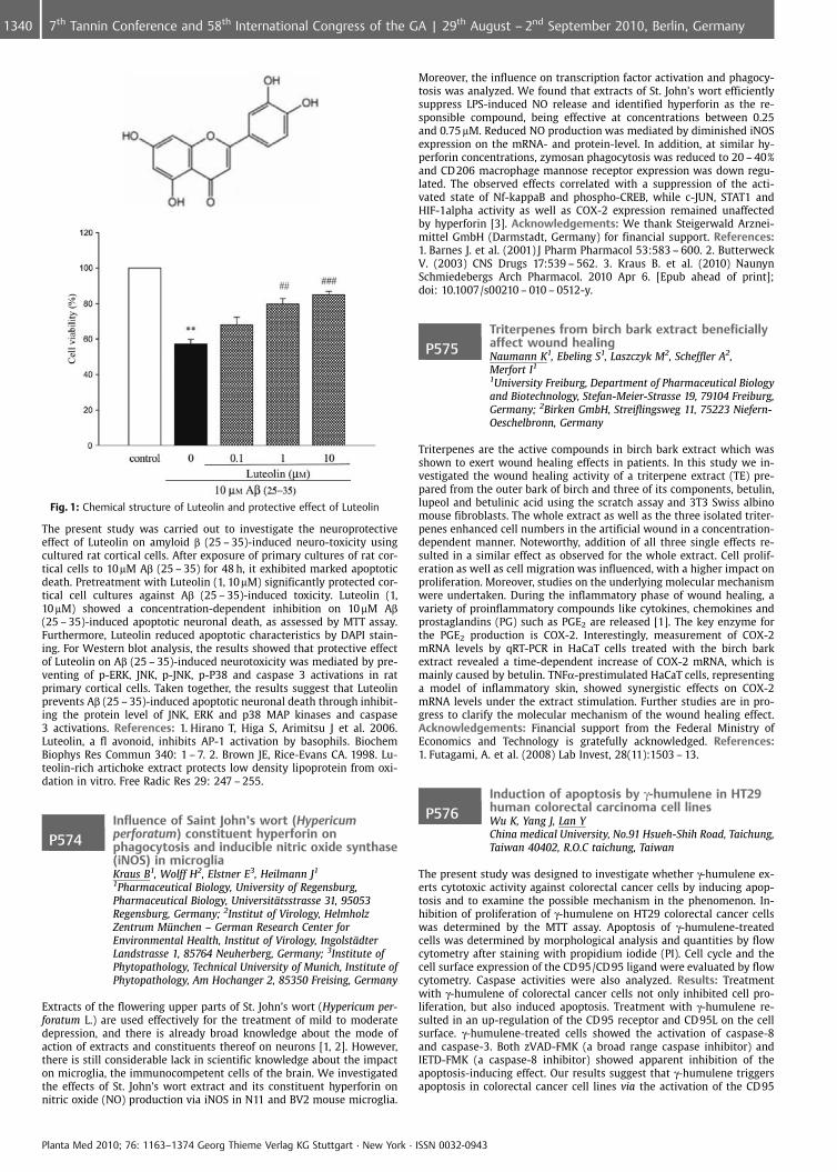
Fig. 1: Chemical structure of Luteolin and protective effect of Luteolin
The present study was carried out to investigate the neuroprotectiveeffect of Luteolin on amyloid b (25 – 35)-induced neuro-toxicity usingcultured rat cortical cells. After exposure of primary cultures of rat cor-tical cells to 10mM Ab (25 – 35) for 48 h, it exhibited marked apoptoticdeath. Pretreatment with Luteolin (1, 10mM) significantly protected cor-tical cell cultures against Ab (25 – 35)-induced toxicity. Luteolin (1,10 mM) showed a concentration-dependent inhibition on 10 mM Ab(25 – 35)-induced apoptotic neuronal death, as assessed by MTT assay.Furthermore, Luteolin reduced apoptotic characteristics by DAPI stain-ing. For Western blot analysis, the results showed that protective effectof Luteolin on Ab (25 – 35)-induced neurotoxicity was mediated by pre-venting of p-ERK, JNK, p-JNK, p-P38 and caspase 3 activations in ratprimary cortical cells. Taken together, the results suggest that Luteolinprevents Ab (25 – 35)-induced apoptotic neuronal death through inhibit-ing the protein level of JNK, ERK and p38 MAP kinases and caspase3 activations. References: 1. Hirano T, Higa S, Arimitsu J et al. 2006.Luteolin, a fl avonoid, inhibits AP-1 activation by basophils. BiochemBiophys Res Commun 340: 1 – 7. 2. Brown JE, Rice-Evans CA. 1998. Lu-teolin-rich artichoke extract protects low density lipoprotein from oxi-dation in vitro. Free Radic Res 29: 247 – 255.
P574Influence of Saint John’s wort (Hypericumperforatum) constituent hyperforin onphagocytosis and inducible nitric oxide synthase(iNOS) in microgliaKraus B1, Wolff H2, Elstner E3, Heilmann J1
1Pharmaceutical Biology, University of Regensburg,Pharmaceutical Biology, Universit�tsstrasse 31, 95053Regensburg, Germany; 2Institut of Virology, HelmholzZentrum M�nchen – German Research Center forEnvironmental Health, Institut of Virology, Ingolst�dterLandstrasse 1, 85764 Neuherberg, Germany; 3Institute ofPhytopathology, Technical University of Munich, Institute ofPhytopathology, Am Hochanger 2, 85350 Freising, Germany
Extracts of the flowering upper parts of St. John’s wort (Hypericum per-foratum L.) are used effectively for the treatment of mild to moderatedepression, and there is already broad knowledge about the mode ofaction of extracts and constituents thereof on neurons [1, 2]. However,there is still considerable lack in scientific knowledge about the impacton microglia, the immunocompetent cells of the brain. We investigatedthe effects of St. John’s wort extract and its constituent hyperforin onnitric oxide (NO) production via iNOS in N11 and BV2 mouse microglia.
Moreover, the influence on transcription factor activation and phagocy-tosis was analyzed. We found that extracts of St. John’s wort efficientlysuppress LPS-induced NO release and identified hyperforin as the re-sponsible compound, being effective at concentrations between 0.25and 0.75 mM. Reduced NO production was mediated by diminished iNOSexpression on the mRNA- and protein-level. In addition, at similar hy-perforin concentrations, zymosan phagocytosis was reduced to 20 – 40%and CD206 macrophage mannose receptor expression was down regu-lated. The observed effects correlated with a suppression of the acti-vated state of Nf-kappaB and phospho-CREB, while c-JUN, STAT1 andHIF-1alpha activity as well as COX-2 expression remained unaffectedby hyperforin [3]. Acknowledgements: We thank Steigerwald Arznei-mittel GmbH (Darmstadt, Germany) for financial support. References:1. Barnes J. et al. (2001) J Pharm Pharmacol 53:583 – 600. 2. ButterweckV. (2003) CNS Drugs 17:539 – 562. 3. Kraus B. et al. (2010) NaunynSchmiedebergs Arch Pharmacol. 2010 Apr 6. [Epub ahead of print];doi: 10.1007/s00210 – 010 – 0512-y.
P575Triterpenes from birch bark extract beneficiallyaffect wound healingNaumann K1, Ebeling S1, Laszczyk M2, Scheffler A2,Merfort I1
1University Freiburg, Department of Pharmaceutical Biologyand Biotechnology, Stefan-Meier-Strasse 19, 79104 Freiburg,Germany; 2Birken GmbH, Streiflingsweg 11, 75223 Niefern-Oeschelbronn, Germany
Triterpenes are the active compounds in birch bark extract which wasshown to exert wound healing effects in patients. In this study we in-vestigated the wound healing activity of a triterpene extract (TE) pre-pared from the outer bark of birch and three of its components, betulin,lupeol and betulinic acid using the scratch assay and 3T3 Swiss albinomouse fibroblasts. The whole extract as well as the three isolated triter-penes enhanced cell numbers in the artificial wound in a concentration-dependent manner. Noteworthy, addition of all three single effects re-sulted in a similar effect as observed for the whole extract. Cell prolif-eration as well as cell migration was influenced, with a higher impact onproliferation. Moreover, studies on the underlying molecular mechanismwere undertaken. During the inflammatory phase of wound healing, avariety of proinflammatory compounds like cytokines, chemokines andprostaglandins (PG) such as PGE2 are released [1]. The key enzyme forthe PGE2 production is COX-2. Interestingly, measurement of COX-2mRNA levels by qRT-PCR in HaCaT cells treated with the birch barkextract revealed a time-dependent increase of COX-2 mRNA, which ismainly caused by betulin. TNFa-prestimulated HaCaT cells, representinga model of inflammatory skin, showed synergistic effects on COX-2mRNA levels under the extract stimulation. Further studies are in pro-gress to clarify the molecular mechanism of the wound healing effect.Acknowledgements: Financial support from the Federal Ministry ofEconomics and Technology is gratefully acknowledged. References:1. Futagami, A. et al. (2008) Lab Invest, 28(11):1503 – 13.
P576Induction of apoptosis by g-humulene in HT29human colorectal carcinoma cell linesWu K, Yang J, Lan YChina medical University, No.91 Hsueh-Shih Road, Taichung,Taiwan 40402, R.O.C taichung, Taiwan
The present study was designed to investigate whether g-humulene ex-erts cytotoxic activity against colorectal cancer cells by inducing apop-tosis and to examine the possible mechanism in the phenomenon. In-hibition of proliferation of g-humulene on HT29 colorectal cancer cellswas determined by the MTT assay. Apoptosis of g-humulene-treatedcells was determined by morphological analysis and quantities by flowcytometry after staining with propidium iodide (PI). Cell cycle and thecell surface expression of the CD 95/CD 95 ligand were evaluated by flowcytometry. Caspase activities were also analyzed. Results: Treatmentwith g-humulene of colorectal cancer cells not only inhibited cell pro-liferation, but also induced apoptosis. Treatment with g-humulene re-sulted in an up-regulation of the CD95 receptor and CD95L on the cellsurface. g-humulene-treated cells showed the activation of caspase-8and caspase-3. Both zVAD-FMK (a broad range caspase inhibitor) andIETD-FMK (a caspase-8 inhibitor) showed apparent inhibition of theapoptosis-inducing effect. Our results suggest that g-humulene triggersapoptosis in colorectal cancer cell lines via the activation of the CD95
Planta Med 2010; 76: 1163–1374 Georg Thieme Verlag KG Stuttgart · New York · ISSN 0032-0943
1340 7th Tannin Conference and 58th International Congress of the GA | 29th August – 2nd September 2010, Berlin, Germany
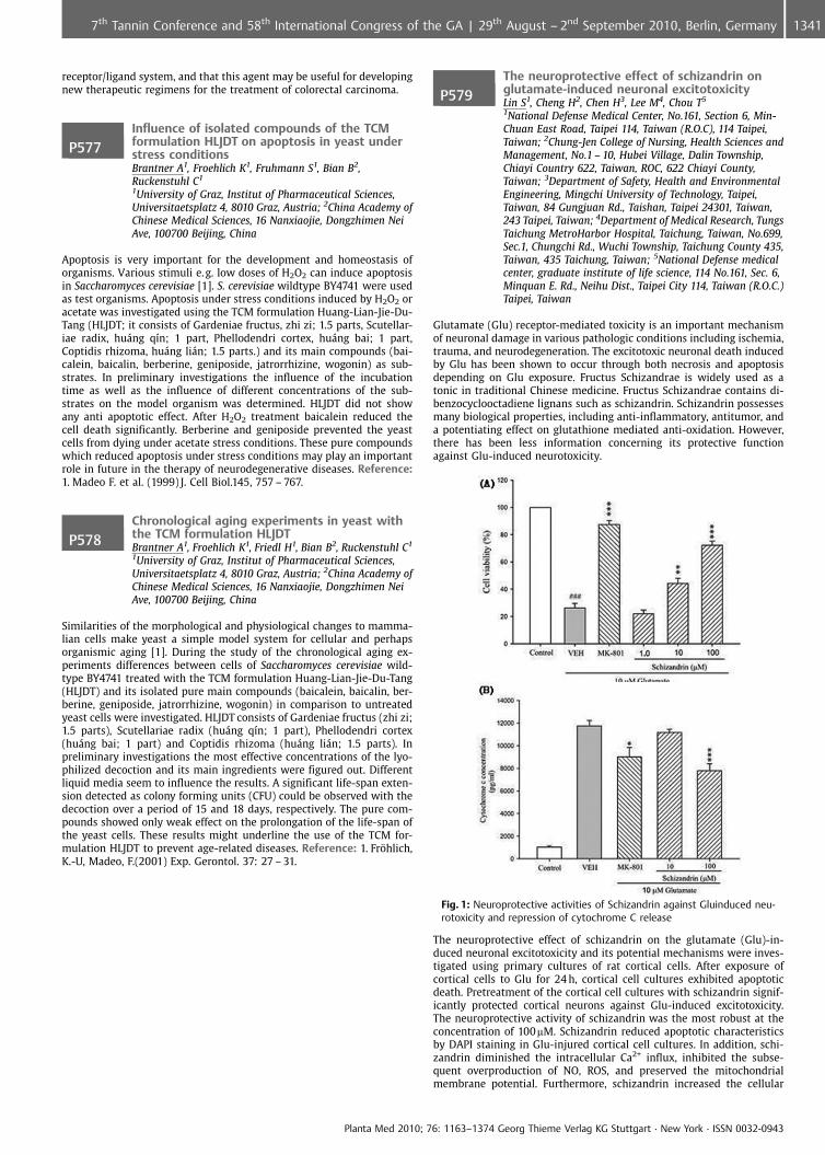
receptor/ligand system, and that this agent may be useful for developingnew therapeutic regimens for the treatment of colorectal carcinoma.
P577Influence of isolated compounds of the TCMformulation HLJDT on apoptosis in yeast understress conditionsBrantner A1, Froehlich K1, Fruhmann S1, Bian B2,Ruckenstuhl C1
1University of Graz, Institut of Pharmaceutical Sciences,Universitaetsplatz 4, 8010 Graz, Austria; 2China Academy ofChinese Medical Sciences, 16 Nanxiaojie, Dongzhimen NeiAve, 100700 Beijing, China
Apoptosis is very important for the development and homeostasis oforganisms. Various stimuli e.g. low doses of H2O2 can induce apoptosisin Saccharomyces cerevisiae [1]. S. cerevisiae wildtype BY4741 were usedas test organisms. Apoptosis under stress conditions induced by H2O2 oracetate was investigated using the TCM formulation Huang-Lian-Jie-Du-Tang (HLJDT; it consists of Gardeniae fructus, zhi zi; 1.5 parts, Scutellar-iae radix, hu�ng q�n; 1 part, Phellodendri cortex, hu�ng bai; 1 part,Coptidis rhizoma, hu�ng li�n; 1.5 parts.) and its main compounds (bai-calein, baicalin, berberine, geniposide, jatrorrhizine, wogonin) as sub-strates. In preliminary investigations the influence of the incubationtime as well as the influence of different concentrations of the sub-strates on the model organism was determined. HLJDT did not showany anti apoptotic effect. After H2O2 treatment baicalein reduced thecell death significantly. Berberine and geniposide prevented the yeastcells from dying under acetate stress conditions. These pure compoundswhich reduced apoptosis under stress conditions may play an importantrole in future in the therapy of neurodegenerative diseases. Reference:1. Madeo F. et al. (1999) J. Cell Biol.145, 757 – 767.
P578Chronological aging experiments in yeast withthe TCM formulation HLJDTBrantner A1, Froehlich K1, Friedl H1, Bian B2, Ruckenstuhl C1
1University of Graz, Institut of Pharmaceutical Sciences,Universitaetsplatz 4, 8010 Graz, Austria; 2China Academy ofChinese Medical Sciences, 16 Nanxiaojie, Dongzhimen NeiAve, 100700 Beijing, China
Similarities of the morphological and physiological changes to mamma-lian cells make yeast a simple model system for cellular and perhapsorganismic aging [1]. During the study of the chronological aging ex-periments differences between cells of Saccharomyces cerevisiae wild-type BY4741 treated with the TCM formulation Huang-Lian-Jie-Du-Tang(HLJDT) and its isolated pure main compounds (baicalein, baicalin, ber-berine, geniposide, jatrorrhizine, wogonin) in comparison to untreatedyeast cells were investigated. HLJDT consists of Gardeniae fructus (zhi zi;1.5 parts), Scutellariae radix (hu�ng q�n; 1 part), Phellodendri cortex(hu�ng bai; 1 part) and Coptidis rhizoma (hu�ng li�n; 1.5 parts). Inpreliminary investigations the most effective concentrations of the lyo-philized decoction and its main ingredients were figured out. Differentliquid media seem to influence the results. A significant life-span exten-sion detected as colony forming units (CFU) could be observed with thedecoction over a period of 15 and 18 days, respectively. The pure com-pounds showed only weak effect on the prolongation of the life-span ofthe yeast cells. These results might underline the use of the TCM for-mulation HLJDT to prevent age-related diseases. Reference: 1. Fr�hlich,K.-U, Madeo, F.(2001) Exp. Gerontol. 37: 27 – 31.
P579The neuroprotective effect of schizandrin onglutamate-induced neuronal excitotoxicityLin S1, Cheng H2, Chen H3, Lee M4, Chou T5
1National Defense Medical Center, No.161, Section 6, Min-Chuan East Road, Taipei 114, Taiwan (R.O.C), 114 Taipei,Taiwan; 2Chung-Jen College of Nursing, Health Sciences andManagement, No.1 – 10, Hubei Village, Dalin Township,Chiayi Country 622, Taiwan, ROC, 622 Chiayi County,Taiwan; 3Department of Safety, Health and EnvironmentalEngineering, Mingchi University of Technology, Taipei,Taiwan, 84 Gungjuan Rd., Taishan, Taipei 24301, Taiwan,243 Taipei, Taiwan; 4Department of Medical Research, TungsTaichung MetroHarbor Hospital, Taichung, Taiwan, No.699,Sec.1, Chungchi Rd., Wuchi Township, Taichung County 435,Taiwan, 435 Taichung, Taiwan; 5National Defense medicalcenter, graduate institute of life science, 114 No.161, Sec. 6,Minquan E. Rd., Neihu Dist., Taipei City 114, Taiwan (R.O.C.)Taipei, Taiwan
Glutamate (Glu) receptor-mediated toxicity is an important mechanismof neuronal damage in various pathologic conditions including ischemia,trauma, and neurodegeneration. The excitotoxic neuronal death inducedby Glu has been shown to occur through both necrosis and apoptosisdepending on Glu exposure. Fructus Schizandrae is widely used as atonic in traditional Chinese medicine. Fructus Schizandrae contains di-benzocyclooctadiene lignans such as schizandrin. Schizandrin possessesmany biological properties, including anti-inflammatory, antitumor, anda potentiating effect on glutathione mediated anti-oxidation. However,there has been less information concerning its protective functionagainst Glu-induced neurotoxicity.
Fig. 1: Neuroprotective activities of Schizandrin against Gluinduced neu-rotoxicity and repression of cytochrome C release
The neuroprotective effect of schizandrin on the glutamate (Glu)-in-duced neuronal excitotoxicity and its potential mechanisms were inves-tigated using primary cultures of rat cortical cells. After exposure ofcortical cells to Glu for 24 h, cortical cell cultures exhibited apoptoticdeath. Pretreatment of the cortical cell cultures with schizandrin signif-icantly protected cortical neurons against Glu-induced excitotoxicity.The neuroprotective activity of schizandrin was the most robust at theconcentration of 100mM. Schizandrin reduced apoptotic characteristicsby DAPI staining in Glu-injured cortical cell cultures. In addition, schi-zandrin diminished the intracellular Ca2+ influx, inhibited the subse-quent overproduction of NO, ROS, and preserved the mitochondrialmembrane potential. Furthermore, schizandrin increased the cellular
Planta Med 2010; 76: 1163–1374 Georg Thieme Verlag KG Stuttgart · New York · ISSN 0032-0943
13417th Tannin Conference and 58th International Congress of the GA | 29th August – 2nd September 2010, Berlin, Germany
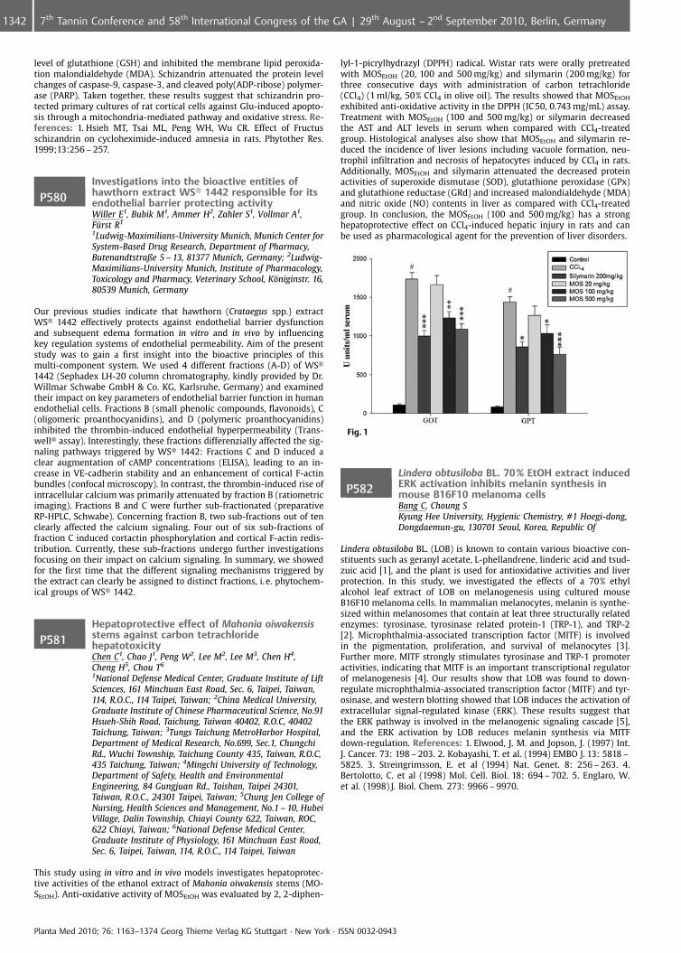
level of glutathione (GSH) and inhibited the membrane lipid peroxida-tion malondialdehyde (MDA). Schizandrin attenuated the protein levelchanges of caspase-9, caspase-3, and cleaved poly(ADP-ribose) polymer-ase (PARP). Taken together, these results suggest that schizandrin pro-tected primary cultures of rat cortical cells against Glu-induced apopto-sis through a mitochondria-mediated pathway and oxidative stress. Re-ferences: 1. Hsieh MT, Tsai ML, Peng WH, Wu CR. Effect of Fructusschizandrin on cycloheximide-induced amnesia in rats. Phytother Res.1999;13:256 – 257.
P580Investigations into the bioactive entities ofhawthorn extract WS� 1442 responsible for itsendothelial barrier protecting activityWiller E1, Bubik M1, Ammer H2, Zahler S1, Vollmar A1,F�rst R1
1Ludwig-Maximilians-University Munich, Munich Center forSystem-Based Drug Research, Department of Pharmacy,Butenandtstraße 5 – 13, 81377 Munich, Germany; 2Ludwig-Maximilians-University Munich, Institute of Pharmacology,Toxicology and Pharmacy, Veterinary School, K�niginstr. 16,80539 Munich, Germany
Our previous studies indicate that hawthorn (Crataegus spp.) extractWS� 1442 effectively protects against endothelial barrier dysfunctionand subsequent edema formation in vitro and in vivo by influencingkey regulation systems of endothelial permeability. Aim of the presentstudy was to gain a first insight into the bioactive principles of thismulti-component system. We used 4 different fractions (A-D) of WS�1442 (Sephadex LH-20 column chromatography, kindly provided by Dr.Willmar Schwabe GmbH & Co. KG, Karlsruhe, Germany) and examinedtheir impact on key parameters of endothelial barrier function in humanendothelial cells. Fractions B (small phenolic compounds, flavonoids), C(oligomeric proanthocyanidins), and D (polymeric proanthocyanidins)inhibited the thrombin-induced endothelial hyperpermeability (Trans-well� assay). Interestingly, these fractions differenzially affected the sig-naling pathways triggered by WS� 1442: Fractions C and D induced aclear augmentation of cAMP concentrations (ELISA), leading to an in-crease in VE-cadherin stability and an enhancement of cortical F-actinbundles (confocal microscopy). In contrast, the thrombin-induced rise ofintracellular calcium was primarily attenuated by fraction B (ratiometricimaging). Fractions B and C were further sub-fractionated (preparativeRP-HPLC, Schwabe). Concerning fraction B, two sub-fractions out of tenclearly affected the calcium signaling. Four out of six sub-fractions offraction C induced cortactin phosphorylation and cortical F-actin redis-tribution. Currently, these sub-fractions undergo further investigationsfocusing on their impact on calcium signaling. In summary, we showedfor the first time that the different signaling mechanisms triggered bythe extract can clearly be assigned to distinct fractions, i. e. phytochem-ical groups of WS� 1442.
P581Hepatoprotective effect of Mahonia oiwakensisstems against carbon tetrachloridehepatotoxicityChen C1, Chao J1, Peng W2, Lee M2, Lee M3, Chen H4,Cheng H5, Chou T6
1National Defense Medical Center, Graduate Institute of LiftSciences, 161 Minchuan East Road, Sec. 6, Taipei, Taiwan,114, R.O.C., 114 Taipei, Taiwan; 2China Medical University,Graduate Institute of Chinese Pharmaceutical Science, No.91Hsueh-Shih Road, Taichung, Taiwan 40402, R.O.C, 40402Taichung, Taiwan; 3Tungs Taichung MetroHarbor Hospital,Department of Medical Research, No.699, Sec.1, ChungchiRd., Wuchi Township, Taichung County 435, Taiwan, R.O.C,435 Taichung, Taiwan; 4Mingchi University of Technology,Department of Safety, Health and EnvironmentalEngineering, 84 Gungjuan Rd., Taishan, Taipei 24301,Taiwan, R.O.C., 24301 Taipei, Taiwan; 5Chung Jen College ofNursing, Health Sciences and Management, No.1 – 10, HubeiVillage, Dalin Township, Chiayi County 622, Taiwan, ROC,622 Chiayi, Taiwan; 6National Defense Medical Center,Graduate Institute of Physiology, 161 Minchuan East Road,Sec. 6, Taipei, Taiwan, 114, R.O.C., 114 Taipei, Taiwan
This study using in vitro and in vivo models investigates hepatoprotec-tive activities of the ethanol extract of Mahonia oiwakensis stems (MO-SEtOH). Anti-oxidative activity of MOSEtOH was evaluated by 2, 2-diphen-
lyl-1-picrylhydrazyl (DPPH) radical. Wistar rats were orally pretreatedwith MOSEtOH (20, 100 and 500 mg/kg) and silymarin (200 mg/kg) forthree consecutive days with administration of carbon tetrachloride(CCl4) (1 ml/kg, 50% CCl4 in olive oil). The results showed that MOSEtOH
exhibited anti-oxidative activity in the DPPH (IC50, 0.743 mg/mL) assay.Treatment with MOSEtOH (100 and 500 mg/kg) or silymarin decreasedthe AST and ALT levels in serum when compared with CCl4-treatedgroup. Histological analyses also show that MOSEtOH and silymarin re-duced the incidence of liver lesions including vacuole formation, neu-trophil infiltration and necrosis of hepatocytes induced by CCl4 in rats.Additionally, MOSEtOH and silymarin attenuated the decreased proteinactivities of superoxide dismutase (SOD), glutathione peroxidase (GPx)and glutathione reductase (GRd) and increased malondialdehyde (MDA)and nitric oxide (NO) contents in liver as compared with CCl4-treatedgroup. In conclusion, the MOSEtOH (100 and 500 mg/kg) has a stronghepatoprotective effect on CCl4-induced hepatic injury in rats and canbe used as pharmacological agent for the prevention of liver disorders.
Fig. 1
P582Lindera obtusiloba BL. 70 % EtOH extract inducedERK activation inhibits melanin synthesis inmouse B16F10 melanoma cellsBang C, Choung SKyung Hee University, Hygienic Chemistry, #1 Hoegi-dong,Dongdaemun-gu, 130701 Seoul, Korea, Republic Of
Lindera obtusiloba BL. (LOB) is known to contain various bioactive con-stituents such as geranyl acetate, L-phellandrene, linderic acid and tsud-zuic acid [1], and the plant is used for antioxidative activities and liverprotection. In this study, we investigated the effects of a 70% ethylalcohol leaf extract of LOB on melanogenesis using cultured mouseB16F10 melanoma cells. In mammalian melanocytes, melanin is synthe-sized within melanosomes that contain at leat three structurally relatedenzymes: tyrosinase, tyrosinase related protein-1 (TRP-1), and TRP-2[2]. Microphthalmia-associated transcription factor (MITF) is involvedin the pigmentation, proliferation, and survival of melanocytes [3].Further more, MITF strongly stimulates tyrosinase and TRP-1 promoteractivities, indicating that MITF is an important transcriptional regulatorof melanogenesis [4]. Our results show that LOB was found to down-regulate microphthalmia-associated transcription factor (MITF) and tyr-osinase, and western blotting showed that LOB induces the activation ofextracellular signal-regulated kinase (ERK). These results suggest thatthe ERK pathway is involved in the melanogenic signaling cascade [5],and the ERK activation by LOB reduces melanin synthesis via MITFdown-regulation. References: 1. Elwood, J. M. and Jopson, J. (1997) Int.J. Cancer. 73: 198 – 203. 2. Kobayashi, T. et al. (1994) EMBO J. 13: 5818 –5825. 3. Streingrimsson, E. et al (1994) Nat. Genet. 8: 256 – 263. 4.Bertolotto, C. et al (1998) Mol. Cell. Biol. 18: 694 – 702. 5. Englaro, W.et al. (1998) J. Biol. Chem. 273: 9966 – 9970.
Planta Med 2010; 76: 1163–1374 Georg Thieme Verlag KG Stuttgart · New York · ISSN 0032-0943
1342 7th Tannin Conference and 58th International Congress of the GA | 29th August – 2nd September 2010, Berlin, Germany
P583Oral administration of Vaccinium uliginosum L.extract inhibits the development ofDNCB-induced atopic dermatitis in NC/Nga miceKim K, Choung SDepartment of Hygenic Chemistry, College of Pharmacy,Khung Hee University, 131 – 701 Seoul, Korea, Republic Of
Vaccinium uliginosum L. (also known as Bog Bilberry or Northern Bilber-ry) has been reported to have anti-oxidant [1] and protective effectsagainst UV-induced skin photoaging [2]. To investigate the anti-atopicdermatitis effects we orally administrated Vaccinium uliginosum L. ex-tract (VU) dissolved in distilled water (90, 150, 250 mg/kg) to atopicdermatitis induced NC/Nga mice (n = 5) daily for 4 weeks. Prednisolonetreatment group (3 mg/kg, n = 5), normal group (DNCB -, VU -, n = 5), andnegative control group (DNCB+, VU-, n = 5) were compared to VU treat-ment groups. Oral administration of Vaccinium uliginosum L. extractsignificantly inhibited the exacerbation of AD-like skin lesions, thicknessof ear and scratching behavior comparing to negative control group(DNCB +, VU -). Moreover, VU treatment decreased serum IgE level in adose dependent manner, and significantly reduced IL-4 and IFN- g pro-duction in concanavalin A (Con A) stimulated splenocyte. These resultssuggest that Vaccinium uliginosum L. extract may be effective therapeu-tic agent against Atopic Dermatitis by inhibiting IgE and cytokines pro-duction. References: 1. Kim YH., et al. (2009) J. Med. Food. 12(4): 885 –892. 2. Bae JY., et al. (2009) Mol. Nutri. Food Res. 53: 726 – 738.
P584Rosmarinic acid as the effective compound inCordia americanaGeller F1, Schmidt C2, Goettert M1, Fronza M2,Heinzmann B3, Werz O4, Merfort I2, Laufer S1
1University of T�bingen, Department of Pharmaceutical andMedicinal Chemistry, Morgenstelle 8, 72076 T�bingen,Germany; 2University of Freiburg, Pharmaceutical Biologyand Biotechnology, Stefan-Meier-Str. 19, 79104 Freiburg,Germany; 3Federal University of Santa Maria,Pharmaceutical Industry, Avenida Roraima 1000, 97105 –900 Santa Maria, Brazil; 4University of T�bingen,Pharmaceutical Analytics, Morgenstelle 8, 72076 T�bingen,Germany
In Brazil medicinal plants have been widely used for the treatment ofdiseases in folk medicine. However, the effective compounds responsiblefor the biological effects are widely unknown. The objective of this studywas to characterize the phytochemical profile and to identify bioactivecompounds in the leaves of Cordia americana (Boraginaceae) [1]. Thebiological activity of the constituents and the ethanolic extract wereinvestigated for the inhibition of 5-lipoxygenase, p38a MAPK, TNFarelease and fibroblast scratch assay, targeting different aspects of in-flammatory and wound healing processes. The phytochemical studies(i. e., 1D, 2D NMR and MS) have led to the identification of flavonols,phenolic compounds and phytosterols. Quantification analysis showedthat rosmarinic acid (RA) is the main compound with an amount of8.44% in the ethanolic extract. RA as well as the ethanolic extract ex-hibited the highest inhibitory effects on 5-lipoxygenase (IC50= 0.97 and0.69 mg/ml, resp.) and p38a MAPK (IC50= 1.16 and 3.25 mg/ml, resp.). Ad-ditionally, RA inhibited the release of TNFa to 36.75 € 1.55% at a con-centration of 36.03 mg/ml, which can be considered as moderately active.The ethanolic extract showed a lower inhibition of 30.3 € 0.75% at aconcentration of 100 mg/mL. RA also exhibited slight stimulatory activityon proliferation and migration of fibroblasts indicating that it may par-tially contribute to the wound healing effects of this plant. We demon-strated for the first time pharmacological effects of C. americana and weprovided evidences for a crucial role of RA as the major player. Acknowl-edgements: Financial support from the government Baden-Wrttem-berg (Zukunftsoffensive IV) is gratefully acknowledged. References:1. Sobral, M. et al (2006). Flora arb rea e arborescente do Rio Grandedo Sul, Brasil. RiMa Publisher. Porto Alegre.
P585Aesculin from Butcher’s broom reduces thepermeability of endothelial cells in vitroBarbic M2, Willer E1, F�rst R1, J�rgenliemk G2
1Munich Center for System-Based Drug Research, Ludwig-Maximilians-University Munich, Department of Pharmacy,Pharmaceutical Biology, Butenandtstr. 5 – 13, 81377 Munich,Germany; 2University of Regensburg, PharmaceuticalBiology, Universit�tsstr. 31, 93053 Regensburg, Germany
Herbal medicinal products containing Butcher’s broom (Rusci rhizoma,Ruscus aculeatus, Ruscaceae) are used as supportive medications againstchronic venous disorders (CVD). Furo- and spirostanol glycosides havebeen suggested to be responsible for the efficacy of the drug [1]. The aimof this study was to identify phenolic ingredients in Rusci rhizomawhich could contribute to the overall effect of Butcher’s broom prepara-tions. As a first result, aesculin was isolated from a defatted methanolicextract of Rusci rhizoma by liquid-liquid chromatography between bu-tanol and water and further LC of the butanol fraction by Sephadex-LH20�, silica gel and RP-18. After structure elucidation by modern spec-troscopic methods like 1D-, 2D-NMR techniques and ESI-MS, the poten-tial of aesculin to affect endothelial barrier dysfunction in vitro wastested. Therefore, macromolecular permeability of human microvascularendothelial cells (HMECs) was measured in a Transwell� assay (0.4 mmpore size) using FITC-dextran (40 kDa, 1 mg/ml) as tracer. To induceendothelial barrier breakdown, HMECs were treated with thrombin (3U/ml) for 60 min. The pretreatment (30 min) with aesculin concentra-tion-dependently (0.1 – 10mM) inhibited the thrombin-induced rise ofendothelial permeability. In summary, aesculin was for the first timedescribed as an ingredient of Rusci rhizoma. The pharmacological re-sults indicate that phenolic compounds of Butcher’s broom, such asaesculin, might also contribute to the efficacy of the whole drug againstCVD. Ongoing research focuses on the identification of further com-pounds from Rusci rhizoma with protective activities on the humanendothelium. Acknowledgements: Thanks are due to Prof. Dr. Heil-mann for helpful discussions References: 1. ESCOP Monographs, 2ndedition (2003).
P586eNOS-activity guided fractionation of vine leavesfrom AustriaDonath O, Kastner S, Reznicek G, Dirsch VDepartment of Pharmacognosy, University of Vienna,Althanstrasse 14, UZA2, 1090 Vienna, Austria
Polyphenol fractions of Austrian red wine samples are reported to in-crease endothelial nitric oxide synthase (eNOS) activity in EA.hy926endothelial cells [1]. The present investigation aims to identify furthereNOS-activating fractions from red vine leaves. Two leaf samples of thevariety Blaufr�nkisch from the region Neusiedlersee-Hgelland wereselected for bio-assay guided fractionation using EA.hy926 endothelialcells and the [14C]L-arginine/[14C]L-citrulline conversion assay measur-ing eNOS activity. The first leaf sample was harvested late in Septembertogether with grapes as green vine leaves (GVL), and the other samplewas collected as red vine leaves (RVL) five weeks later in the samevineyard. Both vine leaf samples were dried, pulverised, and extractedconsecutively with dichloromethane and methanol using an acceleratedsolvent extractor. Further separation of the methanol-extract of the twosamples was done by liquid-liquid partition with ethylacetate and waterresulting in a polar fraction (PF) and an apolar fraction (AF). The metha-nol-extract of both vine leaf samples (GVL, RVL) showed the same levelof enhanced eNOS activity at a concentration of 600 mg/ml. The dichlor-omethane-extracts of GVL and RVL, tested in equal concentration, de-creased the activity significantly. The AF of the samples revealed anactivating effect on eNOS at 300 mg/ml, whereas the complementary PFdid not increase enzyme activity at the same concentration. Thus, thecompounds responsible for eNOS activation seem to reside in the AF.Therefore, ongoing bio-assay guided fractionation of the AF will showwhich class of compounds is responsible for the observed increasedeNOS activity. References: 1. Donath O et al. (2008) Polyphenols Com-munications, 699 – 700.
Planta Med 2010; 76: 1163–1374 Georg Thieme Verlag KG Stuttgart · New York · ISSN 0032-0943
13437th Tannin Conference and 58th International Congress of the GA | 29th August – 2nd September 2010, Berlin, Germany
P587Anti-inflammatory effects of ethanolic extractsfrom Micromeria speciesBival Tefan M1, Jelic D2, Vladimir-Kneevic S1, Trzun M2, FrkaBoric K2, Paravic-Radicevic A2, Braja K2, Blaekovic B1
1Faculty of Pharmacy and Biochemistry, Marulicev trg 20,10000 Zagreb, Croatia; 2GlaxoSmithKline Research CenterZagreb, Prilaz Baruna Filipovica 29, 100000 Zagreb, Croatia
Micromeria species are traditionally used as expectorant, antispasmodicand stimulant agents as well as condiments [1]. Our study aimed toevaluate in vitro modulation effects of three Micromeria ethanolic ex-tracts on interleukin-6 (IL-6) production in comparison with hydroxy-cinnamic acids. IL-6 is a cytokine with various essential biological activ-ities. It plays important roles in the regulation of immune response andinflammation [2]. Total contents of hydroxycinnamic acids were spectro-photometrically determined in aerial plant parts of M. croatica (6.8%), M.juliana (5.4%) and M. thymifolia (5.3%). TLC analysis showed the pre-sence of caffeic, chlorogenic and rosmarinic acids in studied plant ex-tracts. Modulation of IL-6 production in LPS-stimulated male Balb/Cmice splenocytes by plant extracts (18.75 – 300 mg/mL), as well as caffeicacid, chlorogenic acid and rosmarinic acid (125 – 2000mM) was exhib-ited using enzyme-linked immunosorbent assay (ELISA). None of theinvestigated samples showed cytotoxic effect or inhibition of cell growthof human THP-1 and HepG2 cell lines, as well as on mouse splenocytes(where we observed increased absorbance) by MTS citotoxicity test. Intested concentrations, all extracts and hydroxycinnamic acids inhibitedproduction of IL-6 in the range of 9.5 – 100% and 30.6 – 100%, respec-tively. Among the extracts, the activity decreased in order M. juliana > M.croatica > M. thymifolia, percentages of inhibition were 56.6%, 54.3% and42.1% at concentration of 75 mg/mL. These findings suggest that Micro-meria ethanolic extracts possess anti-inflammatory activity, which couldbe attributed to the presence of hidroxycinnamic acids. References:1. Vladimir-Kneevic, S. at al. (2000) Farm. Glas. 56:301 – 312. 2. Nishi-moto, N., Kishimoto, T. (2004) Curr. Opin. Pharmacol. 4:386 – 391.
P588Contribution of the components of STW 5 to itsinhibitory effect on gene expression and releaseof the pro-inflammatory cytokine TNF-aHoser S1, Michael S1, Kelber O2, Weiser D2, Nieber K1
1Universit�t Leipzig, Institut f�r Pharmazie, Pharmakologie,Talstr. 33 Leipzig, Germany; 2Steigerwald Arzneimittelwerk,Wissenschaftliche Abteilung, Havelstr.5, 64295 Darmstadt,Germany
STW 5 (Iberogast�) is successfully used in the therapy of gastrointestinaldisorders such as functional dyspepsia and irritable bowel syndrome(IBS). Pharmacological studies have demonstrated a multi-target effectof this fixed herbal combination. Actions on nerve, smooth muscle,epithelium and intestinal inflammation are revealed. Given that recentclinical data suggest an inflammatory etiology of IBS, the influence ofSTW 5 and its components on the production of the pro-inflammatorycytokine TNF-a was examined. The gene expression of TNF-a was deter-mined in rat ileum preparations by realtime-RT-PCR. TNBS was used toinduce inflammation. The release of TNF-a was measured in LPS-stimu-lated human monocytes using a commercially available ELISA. TheTNBS-induced inflammation was accompanied by an increased TNF-agene expression. STW 5 inhibited the increased gene expression andreduced significantly the release of TNF-a to 13% in LPS-stimulatedmonocytes, while having no effect in untreated cells. In further experi-ments the single herbal components of STW 5 were tested separately inconcentrations equivalent to those in the combination. Caraway (Carumcarvi L.), milk thistle (Silybum marianum L.), lemon balm (Melissa offi-cinalis) and greater celandine (Chelidonium majus L.) had no effect onthe TNF-a release. Bitter candytuft (Iberis amara L.), peppermint(Mentha piperita L.), chamomile (Matricaria recutita L.), liquorice (Gly-cyrrhiza glabra L.) and angelica (Angelica archangelica L.) reduced theTNF-a release, though less pronounced as compared to STW 5. The re-sults indicate that a group of five components, including Iberis amara,contributes relevantly to the distinct effect of the herbal combinationSTW 5.
P589Trans-crocetin is involved in the inhibition of theglutamatergic synaptic transmission on ratcortical neurones by saffron extractBerger F1, Hensel A2, Nieber K1
1University Leipzig, Pharmacology for Natural Sciences,Talstr. 33, 04315 Leipzig, Germany; 2University M�nster,Institute for Pharmaceutical Biology and Phytochemistry,Hittorfstr. 56, 48149 M�nster, Germany
Saffron is the dried stigmata of Crocus sativus L. Saffron contains a largequantity of carotenoids, the crocins, glycosides of the C20 apocarotenoidtrans-crocetin. It has been shown that ethanolic (80 vol.-%) saffron ex-tract (CSE) and trans-crocetin, interact with the phencyclidine bindingsite of the NMDA receptor [1]. The aim of the present study was toexamine the influence of CSE and trans-crocetin on the glutamatergicsynaptic transmission in rat brain slices. Postsynaptic potentials (PSPs)were elicited by focal electrical stimulation in pyramidal cells of thecingulate cortex and recorded using intracellular placed microelec-trodes. PSPs were induced by glutamate released from presynaptic term-inals which activated postsynaptic NMDA and non-NMDA receptors. Incontrast, externally added glutamate induced a depolarisation of the cellmembrane of the pyramidal cells. CSE (10 – 200 mg/ml) decreased theglutamate-induced membrane depolarisation and inhibited the evokedPSPs. CSE inhibited the isolated NMDA and non-NMDA component ofthe PSPs and decreased the NMDA and kainate induced depolarisation.This indicates an antagonistic effect of CSE on NMDA and kainate recep-tors. Trans-crocetin (1 – 50 mM) investigated under the same conditionsas CSE showed comparable inhibitory effects on the glutamate-inducedmembrane depolarisation and evoked PSPs. Trans-crocetin decreasedthe NMDA induced membrane depolarisation, but compared to CSEtrans-crocetin did not inhibit the isolated non-NMDA component ofthe PSPs. Therefore, it seems that trans-crocetin did not interact withthe non-NMDA type of glutamate receptors. We conclude that trans-crocetin is involved in the antagonistic effect of CSE on NMDA but noton kainate receptors. References: 1. Lechtenberg, M. et al. 2008, PlantaMed 74: 764 – 772.
P590Acylated flavonol monorhamnosides fromEriobotrya japonica as XIAP BIR3 inhibitorsrevealed by in silico and HPLC-SPE-NMRtechniquesPfisterer P1, Nikolovska-Coleska Z2, Schyschka L3,Schuster D1, Rudy A3, Wolber G1, Vollmar A3, Rollinger J1,Stuppner H1
1Institute of Pharmacy, CMBI, University of Innsbruck,Pharmacy/Pharmacognosy, Innrain 52c, 6020 Innsbruck,Austria; 2Department of Pathology, Medical School,University of Michigan, 1301 Catherine Road, 48109 – 5602Ann Arbor, United States; 3Department of Pharmacy,Ludwig-Maximilian University, Butenandtstr. 5 – 13, 81377Munich, Germany
Targeting the X-linked inhibitor of apoptosis proteins (XIAP), baculoviralIAP repeat (BIR) 3 groove represents an innovative strategy for increas-ing the sensitivity of resistant tumour cells to conventional chemother-apeutic drugs [1]. Using a previously generated pharmacophore model[2], 3D models of 122 reported constituents from the leaves of themedicinal plant Eriobotrya japonica Lindl. (Rosaceae) were virtuallyscreened against this target. Based on the predicted hits, we focusedon acylated flavonol monorhamnosides (AFMR) as promising phyto-chemical class. AFMRs identified in the crude methanol extract by LC-MS and enriched by chromatographic methods, showed chemosensitiz-ing potential in combination with etoposide in XIAP-overexpressing Jur-kat cells (Nicoletti assay [3]). Application of the HPLC-SPE-NMR hyphe-nated technique enabled the structure elucidation of two new AFMRsand three known ones. By means of preparative HPLC, one of the mainconstituents of the AFMR mixture, the virtual hit kaempferol-3-O-a-L-(2@,4@-di-E-p-coumaroyl)-rhamnoside (1), was isolated. Compound 1sensitizes XIAP-overexpressing Jurkat cells towards the treatment withetoposide (Nicoletti assay), binding to the XIAP BIR3 groove with a dose-dependent affinity (IC50 7.69mM, fluorescence polarization based bind-ing assay [4]). In accordance with the predicted structural requirementswithin the binding site, its substructures, kaempferol-3-O-a-L-rhamno-side and kaempferol, were inactive (IC50 > 100 mM, resp.) revealing amajor impact of the acid and sugar moieties of 1 on bioactivity. Inconclusion, this study elucidates 1 as natural, small-molecular weightinhibitor of the XIAP BIR3 groove using a combination of in silico and
Planta Med 2010; 76: 1163–1374 Georg Thieme Verlag KG Stuttgart · New York · ISSN 0032-0943
1344 7th Tannin Conference and 58th International Congress of the GA | 29th August – 2nd September 2010, Berlin, Germany
HPLC-SPE-NMR techniques. References: 1. Schimmer, A.D. et al. (2006)Cell Death Differ. 13: 179 – 188. 2. Bliem, C.B. et al. (2006) Planta Med.72: 1008. 3. Nicoletti, I. et al. (1991) J. Immunol. Methods 139: 271 – 279.4. Nikolovska-Coleska, Z. et al. (2004) J. Med. Chem. 47: 2430 – 2440.
P591A prolonged protein kinase C-mediated,opioid-related antinociceptive effect of St. John’sWort in miceGaleotti N1, Vivoli E1, Bilia A2, Bergonzi C2, Bartolini A1,Ghelardini C1
1Univeristy of Florence, Department of Preclinical andClinical Pharmacology, Department of Preclinical andClinical Pharmacology, Viale G. Pieraccini 6, I-50139Florence, Italy; 2University of Florence, Department ofPharmaceutical Sciences, Pharmaceutical Sciences, Via UgoSchiff 6, 50019 Sesto Fiorentino Florence, Italy
The antinociceptive profile of St. John’s Wort (SJW) was investigated inmice in a condition of acute thermal and chemical pain, together withthe mechanism that might underlie this effect. A standardised extract ofSJW induced a prolonged antinociception that persisted for 120 minutesafter administration. The thermal antinociception was prevented by na-loxone and by the protein kinase C (PKC) activator PMA, whereas thechemical antinociception was prevented by PMA, remaining naloxoneinsensitive. A chloroform (CHL) and a methanol (MET) fractions, con-taining hyperforins and hypericin plus flavonoids, respectively, in-creased pain threshold with a time course comparable to the total ex-tract. The CHL antinociception was prevented by naloxone, whereas theMET antinociception was antagonized by PMA. Purified hyperforin andhypericin showed an antinociceptive efficacy comparable to CHL andMET, respectively. Conversely, flavonoids were devoid of any effect. Theadministration of yohimbine and atropine did not modify SJW, CHL andMET antinociception. These results indicate that both CHL and METfractions mediate the SJW-induced antinociception. In particular, thepresence of hypericin was fundamental to induce both thermal andchemical antinociception through the inhibition of the PKC activity,whereas hyperforin selectively produced a thermal opioid antinocicep-tion. These findings identify important targets for a longer-acting acti-vation of endogenous pain systems and should potentially help clini-cians who seek safe, tolerable, and prolonged treatments for pain relief.
P592Greek mountain tea – an herbal drug for mentalenhancementWalbroel B, Feistel BFinzelberg GmbH & Co KG, Development Department,Koblenzer Str. 48 – 56, 56626 Andernach, Germany
The aerial parts of Ironwort (Sideritis ssp.) have been used for a longtime, commonly known as “Mountain tea” in Greece, Turkey, Bulgariaand Albania. Mountain farmers around eastern Mediterranean regiondrunk beverages and classical (caffeine-free) tea preparations from Her-ba Sideritis traditionally after work for calming down and to remedy thecommon cold [1]. Newer investigations on highly lipophilic extracts [2]demonstrate reuptake-effects on serotonin metabolism in-vitro. All re-sults were without relationship to the distinct used Sideritis species norof their origin. Our actual investigations used this testing model asscreening tool to evaluate potentials of different Sideritis species andaqueous tea-analogue extracts as well ethanolic-water extracts. Findingswere used to prepare study preparation (nutrifin� mental) in an in-vivomouse model. Herein an ElectroEncephaloGraphic Pharmacogram(EEGP) was made to detect possible psychopharmacological effects [3].Strongest effects were seen with respect to alpha2 waves representingan activation of dopaminergic neurotransmission. Delta, theta, and espe-cially at higher dosages also alpha1 waves were also attenuated, compa-tible with the view of activation of the cholinergic, norepinephrinergicand serotonergic transmission systems. All activities were located infrontal cortex and hypocampus regions responsible for cognitive perfor-mance. A clinical study on the acute effects of nutrifin� mental onhealthy humans is currently in progress. References: 1. Dioscurides(1st century A.D.) De Materia Medica. 2. Kn�rle R., Schnierle P. (2005)Patent application EP 1634602. 3. Dimpfel W., Profiling of Extr. Sideritisaquos. sicc. by means of EEGpower spectra of conscious freely movingrats (electropharmacogram). unpublished data (2009).
P593Efficacy of extracts of Thai medicinal plants as ananesthetics on carp fish (Cyprinus carpio)Chantong B1, Buranasinsup S1, Toniti P1, Suttiyotin P1,Sirimanapong W1, Nusuetrong P2
1Faculty of Veterinary Sciences, Mahidol University, 73170Nakornpathom, Thailand; 2Faculty of Medicine, Departmentof Physiology, Srinakharinwirot University, 10110 Bangkok,Thailand
Anesthetics are widely used in aquaculture to prevent stress duringhandling. Main objective of our research is to investigate the efficacyof Thai medicinal plants in carp fish (Cyprinus carpio). We focused onthe plant extracts from the family of Piperaceae according to their tran-quilizing and sedative potential (1, 2). Psidium guajava was also includeddue to the ability to induce CNS depression (3). The ethanolic extract ofPiper betle and Psidium guajava caused were significantly able to inducesurgical stage (stage 4) anesthesia in concentration dependent manner.Extracts of P. betle at concentrations of 0.1, 0.2, 0.3 and 0.4 mg/ml wereable to induce 76.67 € 3.66, 90.00 € 4.14, 95.00 € 5.77 and 97.50 € 2.89%of fish in to anesthesia state, respectively. P. guajava extracts caused80.00 € 10.00, 86.67 € 15.28 and 100% of fish to loss pain reflex, at con-centrations of 0.1, 0.2 and 0.3 mg/ml, respectively. The extract of P. betlesignificantly induced a faster anesthesia at 0.1 mg/ml when compared toP. guajava. The recovery time of P. guajava was longer than P. betle at thesame concentration. The acute toxicity tests were performed during20 min incubation periods. At concentrations of 0.1, 0.2, 0.3 and0.4 mg/ml P. betle caused a 30.0 € 10.0, 47.50 € 9.57 and 75.00 € 5.77%mortality, respectively. The mortality rates of fish exposed to P. guajavawere 20.0 € 10.0, 50.0 € 10.0, 83.33 € 5.77% at the concentrations of 0.1,0.2 and 0.3 mg/ml, respectively. These findings suggest that the extractfrom P. betle could be an effective anesthetic for his species. References:1. McFerren, M.A. et al. (2002) J. Ethnopharmacol. 83: 201 – 207. 2. Gar-rett, K.M. et al. (2003) Psychopharmacology (Berl). 170: 33 – 41. 3. Shah-een, H.M. et al. (2000) Phytother. Res. 14: 107 – 111.
P594Allergy-preventive effects of flowers of CampsisgrandifloraIshiguro K1, Oku H1, Iwaokab E2, Iinuma M3
1Mukogawa Womens University, School of Pharmacy andPharmaceutical Sciences, Koshien Kyuban-cho Nishinomiya,Japan; 2Hyogo University of Health Sciences, Department ofPharmacy, Minatojima Chuo-ku, 650 – 8530 Kobe, Japan;3Gifu Pharmaceutical University, Laboratory ofPharmacognosy, Mitahora-higashi, 5028585 Gifu, Japan
We used an in vivo assay system, which we had previously developed tosearch for allergy-preventive substances from natural sources. [1] Thismethod monitors the decrease in blood flow (BF) in the tail vein of micesubjected to hen egg-white lysozyme (HEL) sensitization. The BF de-crease is very complicated, involving various factors such as NO fromiNOS, TXA2, PGI2, ET-1, granulocytic elastase, COX-1, 2 and cNOS. [2]Using this assay system, we found that the MeOH extract (CG) of flowersof Campsis grandiflora Thumb. (Bignoniaceae) significantly reduced thedecrease of BF. Flowers of C. grandiflora have been used in traditionalChinese medicine to treat bruises, pruritus, diuresis and thrombosis. Inthis study, we report on the allergy-preventive effects of CG and itsactive compounds. The BF of HEL-sensitized mice (control group) gra-dually and significantly decreased, falling to about 70% of the BF ofnormal mice at day 9. CG could significantly the decrease of BF. Active-guided fractionation of CG led to isolation of apigenin (1), acteoside (2)from the most active AcOEt fraction and rengyoside (3), rengyol (4) andisorengyol (5) from a few active n-BuOH extracts. Among these com-pounds, 1 and 2 significantly reduced the decrease of BF. 1 has beenreported to inhibit platelet aggregation and inhibit the expression ofiNOS or COX-2, while 2 have been reported to have anti-inflammatoryand antioxidant effects. Our findings show that CG could be useful forpreventing allergy. Details of the mode of action are under investigation.References: 1. Ishiguro, K., Oku, H., Ueda, Y., Iwaoka, E., Kunitomo M.,(2005) Biol. Pharm. Bull., 28;1490 – 1495. 2. Oku, H., Ogawa, Y., Iwaoka,E., Kunitomo, M., Ueda, H., Okamura, H., Ishiguro, K., (2007) Biol. Pharm.Bull., 30; 1324 – 1328.
Planta Med 2010; 76: 1163–1374 Georg Thieme Verlag KG Stuttgart · New York · ISSN 0032-0943
13457th Tannin Conference and 58th International Congress of the GA | 29th August – 2nd September 2010, Berlin, Germany
P595The in vivo and in vitro angiogenic evaluation ofthe essential oil of Echinophora tournefortiiDemirci B1, Kiyan T1, Koparal A2, Kaya M2, Demirci F1,Baser K1
1Anadolu University, Faculty of Pharmacy, Pharmacognosy,Anadolu University, 26470 Eskisehir, Turkey; 2AnadoluUniversity, Faculty of Science, Biology, Anadolu University,26470 Eskisehir, Turkey
Angiogenesis is the formation of new blood vessels occuring in embryogrowth and wound healing. It has a vital role in certain pathologies likechronic inflammation and cancer. The antiangiogenic approach as a newadvance and target to treat or prevent such pathologies looks very pro-mising (1). In this study, the essential oil from the aerial parts of Echi-nophora tournefortii Jaub.& Spach. was investigated for its in vivo and invitro angiogenic/antiangiogenic properties to support its use for woundhealing in folk medicine. The essential oil was obtained by hydrodistilla-tion, which was analyzed both by GC and GC-MS. The main constituentsof the oil were identified as myrcene (29%) and a-pinene (28%). Usingthe in vivo Chorio Allantoic Membrane (CAM) assay and in vitro toxicity(MTT), cell migration and tube formation tests (HUV-EC-C cell lines) theoil and its main constituent myrcene were tested at various concentra-tions. Cortisone, suramin, sodium dodecyl sulphate and thalidomidewere used as standards in both assays. The oil showed a weak antian-giogenic effect with slight irritation in vivo and antiangiogenic activityin a dose dependent manner in vitro. It did not show embryotoxicity invivo but cytotoxicity in vitro. Myrcene showed a weak antiangiogeniceffect in vivo as well as a wound healing effect in a dose dependentmanner but no antiangiogenic activity in vitro. Acknowledgements:This work was financially supported by TUBITAK Project No: SBAG-107S262 References: 1. Paper, D., Natural products as angiogenesis in-hibitors, Planta Med., 64, 686 – 695 (1998).
P596Genome-scale microRNA identify small RNAmodulators of the effect of berberine onpancreatic cancer cell growthYouns M1, Hoheisel J2, Efferth T1
1Institute of Pharmacy and Biochemistry, University ofMainz., Department of Pharmaceutical Biology, StaudengerWeg 5, 55128 Mainz, Germany; 2German Cancer ResearchCenter, INF580, 692120 Heidelberg, Germany
MicroRNAs (miRNAs) are newly identified small RNA molecules up to22 nucleotides in length. miRNAs have emerged as important regulatorsof genes involved in many biological processes, including development,cell proliferation and differentiation, apoptosis and metabolism. Distur-bance of microRNA expression may play a role in the initiation andprogression of certain diseases including cancer. Human cancers com-monly exhibit an altered expression profile of miRNAs with oncogenic(miR-21) or tumor-suppressive (miR-34a) activity. Pancreatic cancer isone of the most aggressive human malignancies with an incidence ratesalmost identical to mortality rates. This discouraging information is dueto the lack of improvement in detection and diagnosis strategies and thepaucity of breakthroughs in treatment regimens. A successful drug de-velopment in this disease continues to be a major challenge. Recently, amicroRNA expression signature has been identified that is associatedwith pancreatic cancer. Berberine is an isoquinoline alkaloid, isolatedfrom Rhizoma coptidis, and reported to have anti-cancer effects in dif-ferent human cancer cells. There is however, no available information onthe effect of berberine on global miRNA expression pattern in pancreaticcancer. Here we carried out a global miRNA-based expression profilingstudy in order to identify novel molecular miRNA targets mediating thegrowth inhibitory effects of berberine on pancreatic cancer cells. Amongthe 59 significantly expressed genes, 14 were significantly up-regulatedand 45 were significantly down-regulated in their expression. Our re-sults showed, for the first time, that berberine was able to modulate themiRNA signature of pancreatic cancer cells.
P597Two novel phloroglucinols from Hypericumempetrifolium with antiproliferative activity onendothelial cellsSchmidt S1, Skaltsa H2, J�rgenliemk G1, Heilmann J1
1Universitiy of Regensburg, Pharmaceutical Biology,Universit�tsstraße 31, 93053 Regensburg, Germany;2University of Athens, Pharmacognosy & Chemistry ofNatural Products, Panepistimiopolis-Zografou, 15771Athens, Greece
Acylphloroglucinols are often accumulated in the familiy Clusiaceae.They seem to contribute to the antidepressant efficacy of St. John’s wortand have antibacterial activities. More recently, a potent anti-angiogenicactivity has been described [1]. Aim of the present study was to identifycompounds from the endemic greek Hypericum species H. empetrifolium(Clusiaceae) with antiproliferative activity on endothelial cells to iden-tify possible new anti-angiogenic substances. As a first result, two novelacylphloroglucinol derivatives were isolated from a petrol ether extractof H. empetrifolium after fractionation by LC on silica gel, RP-18 andpurification by preparative HPLC (RP-18), namely 1-O-(p-1-menthen-8-yl)-4-geranyl-2-(2-methylpropionyl)-phloroglucinol (1) and 1-O-(p-1-menthen-8-yl)-4-geranyl-2-(2-methylbutyryl)-phloroglucinol (2). Theirstructures were elucidated by 1D-, 2D-NMR techniques and ESI-MS. Todetermine a possible anti-angiogenic activity, the inhibition of endothe-lial cell proliferation was measured. Subconfluent grown HMEC-1 cells(human microvascular endothelial cell line) were treated with (1) and(2) for 3 days before cells were stained with crystal violet. Both sub-stances showed a concentration dependent activity by inhibiting the cellproliferation between 1 and 20 mM.
Fig. 1: 1-O-(p-1-menthen-8-yl)-4-geranyl-2-(2-methylpropionyl)-phloro-glucinol
Fig. 2: 1-O-(p-1-menthen-8-yl)-4-geranyl-2-(2-methylbutyryl)-phloroglu-cinol
Acknowledgements: Thanks are due to Prof. Theophanis Constantini-dis, University of Athens, Faculty of Biology, for botanical classificationas well as to Dr. E. Ades and Mr. F. J. Candal of CDC (USA) and Dr. T.Lawley of Emory University (USA) for providing the HMEC-1 cells Refer-ences: 1. Martinez-Poveda, B. et al. (2010) PLoS One 5(3): e9558.
P598Influence of hyperoside and hyperforin on theregulation of b1-adrenergic receptors on livingC 6 glioblastoma cells: A new postsynapticapproach for the mode of action of St. John’swortH�berlein H1, Jakobs D1, Hage-H�lsmann A1, Kolb C2
1University of Bonn, Institute of Biochemistry andMicrobiology, Nussallee 11, 53115 Bonn, Germany;2Steigerwald Arzneimittelwerk GmbH, Havelstr. 5, 64295Darmstadt, Germany
In depressive disorders hyperactivity of the hypothalamic-pituitary-adrenal axis is one of the most consistent findings which can be cor-rected with standard antidepressant drugs. One of the receptors in-
Planta Med 2010; 76: 1163–1374 Georg Thieme Verlag KG Stuttgart · New York · ISSN 0032-0943
1346 7th Tannin Conference and 58th International Congress of the GA | 29th August – 2nd September 2010, Berlin, Germany
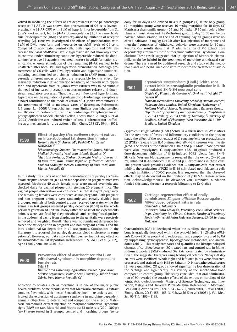
volved in mediating the effects of antidepressants is the b1-adrenergicreceptor (b1-AR). It was shown that pretreatment of C 6-cells (overex-pressing the b1-AR-GFP) with 1mM of hyperforin or hyperoside from St.John’s wort extract, led to b1-AR downregulation [1], the same holdstrue for desipramine (DMI) and was explained by inhibition of receptorrecycling [2]. Here we investigated the effects of preincubation with1mM of DMI, hyperforin and hyperoside on cAMP-levels of C 6-cells.Compared to non-treated control cells, both hyperforin and DMI de-creased the basal cAMP-level, while hyperoside did not show any effect.In contrast, under stimulating conditions hyperoside reduced the dobu-tamine (selective b1-agonist) mediated increase in cAMP formation sig-nificantly, whereas stimulation of the remaining b1-AR seemed to beunaffected after both DMI and hyperforin preincubation. Although pre-treatment of C 6-cells with DMI, hyperforin and hyperoside under sti-mulating conditions led to a similar reduction in cAMP formation, ap-parently different modes of action are responsible for this effect. Re-markably, reduction in b1-adrenergic sensitivity of C 6-cells with hyper-forin and hyperoside from St. John’s wort extracts was found withoutthe need of increased presynaptic neurotransmitter release and down-stream regulatory processes. Thus, the direct influence of hyperforin andhyperoside on the regulation of postsynaptic b1-adrenergic receptors isa novel contribution to the mode of action of St. John’s wort extracts inthe treatment of mild to moderate cases of depression. References:1. Prenner L., (2006) Untersuchungen zum Einfluss von Inhaltsstoffenaus Hypericum perforatum L. auf das b-adrenerge Rezeptorsystem ampostsynaptischen Modell lebender Zellen, Thesis, Bonn. 2. Brgi, S. et al.(2003) Antidepressant-induced switch of beta 1-adrenoceptor traffick-ing as a mechanism for drug action, J. Biol. Chem. 278 (2), 1044 – 1052.
P599Effect of parsley (Petroselinum crispum) extracton intra-abdominal fat deposition in miceHosseini Biuki S1, Anvari M2, Dashti-R M2, ZeinaliNasrabadi F3
1Pharmacology Student, Pharmaceutical School, IsfahanMedical University Yazd, Iran, Islamic Republic Of;2Assistant Professor, Shaheed Sadoughi Medical UniversityOf Yazd Yazd, Iran, Islamic Republic Of; 3Medical Student,Shaheed Sadoughi Medical University Of Yazd Yazd, Iran,Islamic Republic Of
In this study the effects of non toxic concentrations of parsley (Petrose-linum crispum) decoction (0.5%) on fat deposition in pregnant mice wasassessed. Methods: 40 adult female mice were mated overnight andchecked daily for vaginal plaque until yielding 20 pregnant mice. Thevaginal plaque observation was considered as the1st day of pregnancy.The remaining females were considered as non pregnant. Both pregnantand non pregnant animals were randomly and equally divided into2 groups. Animals of both control groups received tap water while theanimals in test groups received parsley decoction (0.5%) in the wholeperiod of gestation ad libitum. 18 days after the onset of the experimentsanimals were sacrificed by deep anesthesia and striping fats depositedin the abdominal cavity from diaphragm to the genitalia were preciselyremoved and weighted. Result: There was no significant difference be-tween the fat deposition in pregnant and non pregnant animals and theintra abdominal fat deposition in all test groups. Conclusion: In theliterature it is reported that parsley decreases blood cholesterol in somepatients1. However, our data indicate that parsley has not any effect onthe intraabdominal fat deposition. References: 1. Suido, H. et al. (2002) JAgric Food Chem. 50: 3346 – 50.
P600Prevention effect of Matricaria recutita L. onwithdrawal syndrome in morphine dependentfemale ratsSafari EIslamic Azad University, Agriculture science, AgricultureScience department, Islamic Azad University, Tabriz branchTabriz, Iran, Islamic Republic Of
Addiction to opiates such as morphine is in one of the major publichealth problems. Some reports show that Matrivaria chamomilla extractcontains flavonoids, which exert Benzodiazepine – like activity and in-hibited the expression of abstinence syndrome in morphine dependentanimals. Objective: to determined and comparison the effect of Matri-caria chamomilla extract injection on morphine withdrawal syndromesigns (MWS) in rats. Material and Methods: 32 male rats (200 – 300gr)(n = 8) were tested in 2 groups: control and morphine groups (twice
daily for 10 days) and divided in 4 sub groups: (1) saline only group,(2) morphine group were received 10 mg/kg morphine for 10 days, (3)Matricaria chamomilla group – 25 and 50 mg/kg I.P 30 min before Mor-phine administration and (4) Methadone group. In day 10, 30 min beforenaloxan administration. In the end of training day all groups were re-ceived naloxane (5 mg/kg I.P) 3 h after last injection of morphine andthen the frequencies of withdrawal behavior were assessed for 30 min.Results: Our results show that I.P administration of MC extract dosedependently attenuates most of morphine withdrawal syndrome. Con-clusion: These result suggested that ip injection of Matricaria chamo-milla might be helpful in the treatment of morphine withdrawal syn-drome. There is a need for additional research and study of the medic-inal plants and herbal medicine with respect to the treatment of addic-tion.
P601Cryptolepis sanguinolenta (Lindl.) Schltr. rootextract inhibits prostaglandin production in IL-1bstimulated SK-N-SH neuronal cellsOlajide O1, Pinheiro de Oliveira A2, Unekwe J2, Wright C3,Fiebich B2
1London Metropolitan University, School of Human Sciences,Holloway Road London, United Kingdom; 2University ofFreiburg Medical School, Neurochemistry Research Group,Department of Psychiatry and Psychotherapy, Hauptstrasse5, 79104 Freiburg, 79104 Freiburg, Germany; 3University ofBradford, School of Pharmacy, West Yorkshire, BD 7 1DPBradford, United Kingdom
Cryptolepis sanguinolenta (Lindl.) Schltr. is a shrub used in West Africafor the treatment of fevers and inflammatory conditions. In the presentstudy the effect of the root extract of C. sanguinolenta on prostaglandinE2 (PGE2) release from IL-1b-stimulated SK-N-SH neurons was investi-gated. The effects of the extract on COX-2 and p38 MAP Kinase proteinswere also investigated. C. sanguinolenta (2.5 – 10 mg/ml) produced adose-dependent inhibition of IL-1b-induced PGE2 release from SK-N-SH cells. Western blot experiments revealed that the extract (5 – 20 mg/ml) inhibited IL-1b-induced COX -2 and p38 expressions in these cells.The present work provides evidence that C. sanguinolenta root extractinhibits the production of PGE2 in IL-1b-stimulated neuroblastoma cellsthrough inhibition of COX-2 protein. It is suggested that the observedeffects may be dependent on the inhibition of p38 MAP Kinase activa-tion. Acknowledgements: The Alexander von Humboldt Foundationfunded this study through a research fellowship to Dr Olajide
P602Cartilage regeneration effect of orallyadministered Zingiber officinale Roscoe againstMIA-induced osteoarthritis in ratsGanabadi S, Fakurazi SUniversiti Putra Malaysia, Veterinary Pre-Clinical Sceinces,Dept. Veterinary Pre-Clinical Sciences, Faculty of VeterinaryMedicineUniversiti Putra Malaysia, Serdang, 43400 Serdang,Malaysia
Osteoarthritis (OA) is developed when the cartilage that protects thebone is gradually destroyed within the synovial joint [1]. Zingiber offici-nale Roscoe (ZO) is potential to reduce pain and symptoms caused by OAby suppressing cyclooxygenase, lipooxygenase metabolites, and arachi-donic acid [2]. This study compares and quantifies the histopathologicalchanges of cartilage between ZO treated rats and control rats in Mono-sodium idoacetate (MIA)-induced OA. Rats were treated by administra-tion of the suggested therapies using feeding catheter for 28 days. At day28, rats were sacrificed. Whole right and left knee joints were dissected,processed, and stained with H&E or Safranin-O. Histopathological scores[3] were quantified. ZO group showed significantly low degeneration ofthe cartilage and significantly less severity of the subchondral bonecompared to control group. This study concluded that oral administra-tion of ZO revealed the curative effects of the extract on cartilage of OAjoints. Acknowledgements: Ministry of Science, Technology and Inno-vation, Malaysia and Universiti Putra Malaysia. References: 1. Moreland,LW. (2003). Arthritis Res. Ther. 5:54 – 67. 2. Tjendraputra, E. et al. (2001).Bioorg. Chem. 29(3):156 – 163. 3. Kobayashi K. et al. (2003). J. Vet. Med.Sci. 65(11): 1195 – 1199.
Planta Med 2010; 76: 1163–1374 Georg Thieme Verlag KG Stuttgart · New York · ISSN 0032-0943
13477th Tannin Conference and 58th International Congress of the GA | 29th August – 2nd September 2010, Berlin, Germany
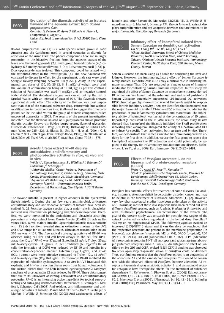
P603Evaluation of the diurectic activity of an isolatedflavonol of the aqueous extract from Boldoapurpurascens Cav.Gonzalez D, Dehaen W, Apers S, Kilonda A, Pieters L,Compernolle F, Toppet SUniversity, Road to camajuani km 5 1/2, 50400 Santa Clara,Cuba
Boldoa purpurascens Cav. (1) is a wild species which grows in LatinAmerica and the Caribbean; used in several countries as diuretic foraffections of the urine tract. The phenol compounds here are in largerproportion in the bioactive fraction. From the aqueous extract of theleave one flavonoid glycoside (2,3) with group benzodioxalane (4’,5-di-hydroxy-6,7-methylendioxyflavonol-3-O-a-L-rhamnopyranosil-(1�2)-b-D-xylopyranoside was isolated. This compound could be related withthe attributed effect in the investigation. (4). The new flavonoid wasevaluated to discern its effect, for the experiment, male rats were used,Sprague Dawley weighing between 160 y 220 g. Assay. In the experi-mental test doses of 100, 50, 25 12, 6, 3 mg/Kg of weight were used,the volume of administration being of 10 mL/Kg; as positive control asolution of Furosemide was used (4 mg/Kg) and as negative control,water distilled. The statistical analysis was carried out by the test ofKruskal-Wallis with an interval of trust of 99%. The flavonol; showedsignificant diuretic effect. The activity of the flavonol was most impor-tant than that of the standard reference drug, Furosemide but withoutmodifications in the excretions ions, it allowed to suggest that the com-pound can be included within the group of diuretic compounds calleduncovered acuaretics in 2003. The results of the present investigationindicated that the flavonol isolated of B. purpurascens shows profounddiuretic activity Keywords: Boldoa purpurascens, flavonol glycosides,Furosemide References: 1. Roig, J. T,(1988). Dictionary of Cuban Com-mon Yams, pp 225 – 226. 2. Niassy, B.; Um, B. – H, et al, (2004). C. R.Chimie 7, 993 – 996. 3. Jpn. Kokai Tokkyo Koho,(1988), JP63203682 A2. 4.Magalh¼es AF, Tozzi AM, et al,(2007) Acad Bras Cienc; 79,351 – 675.
P604Reseda luteola extract RF-40 displaysantioxidative, antiinflammatory andphotoprotective activities in vitro, ex vivo andin vivoW�lfle U1, Simon-Haarhaus B1, W�hling A2, Behnam D3,Lademann J4, Schempp C1
1University Medical Center Freiburg, Department ofDermatology, Hauptstr. 7, 79104 Freiburg, Germany; 2NIGGmbH, Wasserkunststr. 26, 39124 Magdeburg, Germany;3Aquanova AG, Birkenweg 8 – 10, 64295 Darmstadt,Germany; 4Charit� – Universit�tsmedizin Berlin,Department of Dermatology, Charit�platz 1, 10117 Berlin,Germany
The flavone luteolin is present in higher amounts in the dyers weld,Reseda luteola L. During the last few years antimicrobial, anticancer,antiinflammatory and antioxidative activities of luteolin have been de-scribed [1, 2]. Reactive oxygen species play a major role in ultraviolet-induced skin inflammation, photoaging and photocarcinogenesis. There-fore, we were interested in the antioxidant and ultraviolet-absorbingproperties of a dry extract from Reseda luteola (RF-40) [3] rich in fla-vones (40% w/w), mainly luteolin. Spectrophotometric measurementswith 1% (v/v) solution revealed similar extinction maxima in the UVBand UVA range for RF-40 and luteolin. Ultraviolet transmission below370 nm was < 10%. The free radical scavenging activity of RF-40 wasassessed using cell-free and cell-based assays. In the cell-free DPPHassay the IC50 of RF-40 was 37 mg/ml (Luteolin:12 mg/ml, Trolox: 25 mg/ml; N-acetylcystein: 34 mg/ml). In UVB irradiated (60 mJ/cm2) HaCaTcells the formation of DCFH was reduced by RF-40 and luteolin in aconcentration-dependent manner. Luteolin (IC50 3 mg/ml) and RF-40(IC50 4 mg/ml) were more effective compared to Trolox (IC50 12 mg/ml)and N-acetylcystein (IC50 847 mg/ml). Furthermore RF-40 inhibited theexpression of inducible cyclooxygenase-2 ex vivo in UVB irradiated skinexplants, and in vivo in suction blister roofs from human volunteers. Inthe suction blister fluid the UVB induced, cyclooxygenase-2 catalyzedsynthesis of prostaglandin E2 was reduced by RF-40. These data suggestthat due to its ultraviolet absorbing, antioxidant and antiinflammatoryproperties RF-40 is an interesting active ingredient for topical sun-pro-tecting and anti-aging dermocosmetics. References: 1. Seelinger G, Mer-fort I, Schempp CM (2008) Anti-oxidant, anti-inflammatory and anti-allergic activities of luteolin. Planta Med 74:1667 – 1677. 2. Seelinger G,Merfort I, W�lfle U, Schempp CM (2008) Anti-carcinogenic effects of
luteolin and other flavonoids. Molecules 13:2628 – 51. 3. W�lfle U, Si-mon-Haarhaus B, Merfort I, Schempp CM. Reseda luteola L. extract dis-plays antiproliferative and pro-apoptotic activities that are related to itsmajor flavonoids. Phytotherapy Research (in press).
P605Inhibitory effect of kaempferol isolated fromSemen Cuscutae on dendritic cell activationLin M1, Chang W1, Lee M1, Yang M1, Chu C2
1China Medical University, School of Chinese MedicineResources, No.91 Hsueh-Shih Road, 40402 Taichung,Taiwan; 2National Health Research Institutes, ImmunologyResearch Center, No.35 Keyan Road, 350 Zhunan, MiaoliCounty, Taiwan
Semen Cuscutae has been using as a tonic for nourishing the liver andkidneys. However, the immunoregulatory effect of Semen Cuscutae israrely studied. Dendritic cells (DCs) play a critical role in initiating im-mune response. Thus, DCs are regarded as a major target of immuno-modulator for controlling harmful immune responses. In this study, weexamined the effect of Semen Cuscutae on mouse bone marrow-derivedDC activation. We found that the n-butanol and methanol partitions ofSemen Cuscutae potentially suppressed LPS-induced DC activation.HPLC chromatography showed that several flavonoids might be respon-sible for this inhibitory activity. Then, we identified that kaempferol wasthe major flavonoid to inhibit LPS-induced DC activation by reducing theproduction of pro-inflammatory cytokines and maturation. The inhibi-tory ability of kaempferol was tested at the concentration of 10 ug/ml.Importantly, consistent to the in vitro results, the recall assay in vivoshowed that kaempferol significantly inhibited the T cell proliferation,indicating that kaempferol abrogated the ability of LPS-stimulated DCsto induce Ag-specific T cell activation, both in vitro and in vivo. There-fore, we demonstrate that Semen Cuscutae has immunosuppressive ac-tivity for the first time. In addition, we are the first group to report thatkaempferol attenuates the DC activation and could potentially be ap-plied in the therapy for inflammatory and autoimmune diseases. Refer-ences: 1. Yu YL, et al., 2009. Eur J Immunol. 39(9):2482 – 2491.
P606Effects of Passiflora incarnata L. on rathippocampal G protein-coupled receptors(GPCRs)Weiss G1, Appel K2, Rose T2, Kammler T1
1PASCOE pharmazeutische Pr�parate GmbH, Research &Development, Schiffenberger Weg 55, 35394 Gießen,Germany; 2VivaCell Biotechnology GmbH, Ferdinand-Porsche-Str. 5, 79211 Denzlingen, Germany
Passiflora has potential effects for treatment of some diseases like anxi-ety, insomnia, attention-deficit hyperactivity disorder, cancer, and maybe helpful in the treatment of substance addictions [1], [2]. However,very few pharmacological studies have been undertaken on the activityof P. incarnata; most of these investigations have been carried out withdifferent Passiflora species, such as P. edulis, P. alata, or P. coerulea andwith insufficient phytochemical characterization of the extracts. Thegoal of the present study was to search for possible new targets of theextract contained as active ingredient in the herbal drug Pascoflair�425 mg on rat hippocampal GPCRs. The following agonists evoked anincreased [35S]-GTP? S signal and it can therefore be concluded thatthe respective receptors are present in the membrane preparation (inbrackets): acetylcholine (muscarinic M2 or M4), DAGO (m-opioid), ADP(P2Y12 or P2Y13), HU-210 (cannabinoid CB1 > CB2), CCPA (adenosineA1), serotonin (serotonin 5-HT1all subtypes) and glutamate (metabotro-pic glutamate receptors, mGlu2,3,4,6,7,8). An antagonistic effect of Pas-siflora on Hu-210 and CCPA-evoked [35S]-GTP? S binding was observed.No other agonistic/antagonistic action of Passiflora could be detected.Thus, our findings suggest that the Passiflora extract is an antagonist ofthe adenosine A1 and the cannabinoid receptors. This would be consis-tent with the observed effects of Passiflora, because adenosine A1 re-ceptor antagonists display anxiolytic activity [3] and cannabinoid recep-tor antagonist have therapeutic effects for the treatment of substancedependence [4]. References: 1. Dhawan, K. et al. (2004) J Ethnopharma-col. Sep;94(1):1 – 23. 2. Patel, S. et al. (2009) Int J Green Pharm 3:277 –80. 3. Maemoto, T. et al (2004). J Pharmacol. Sci. 96, 42 – 52. 4. Schindleret al. (2010) Eur J Pharmacol. May 10;633(1 – 3):44 – 9.
Planta Med 2010; 76: 1163–1374 Georg Thieme Verlag KG Stuttgart · New York · ISSN 0032-0943
1348 7th Tannin Conference and 58th International Congress of the GA | 29th August – 2nd September 2010, Berlin, Germany
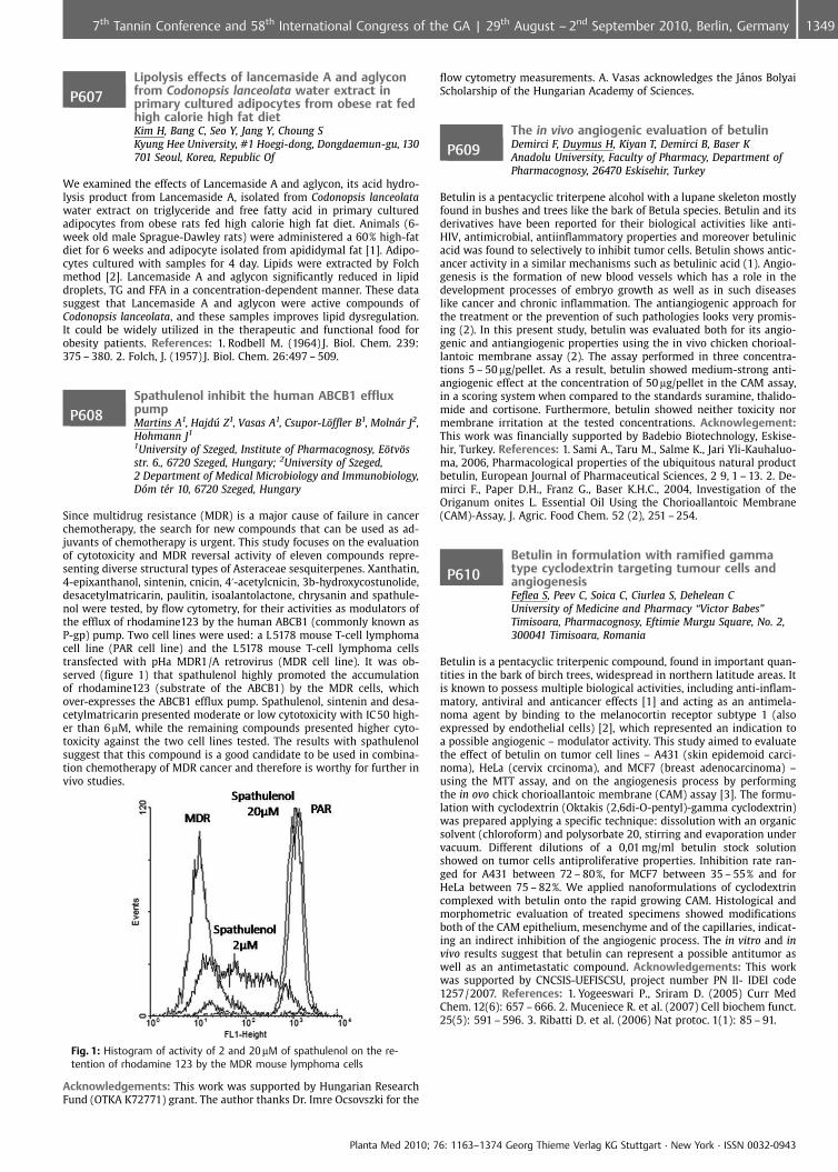
P607Lipolysis effects of lancemaside A and aglyconfrom Codonopsis lanceolata water extract inprimary cultured adipocytes from obese rat fedhigh calorie high fat dietKim H, Bang C, Seo Y, Jang Y, Choung SKyung Hee University, #1 Hoegi-dong, Dongdaemun-gu, 130701 Seoul, Korea, Republic Of
We examined the effects of Lancemaside A and aglycon, its acid hydro-lysis product from Lancemaside A, isolated from Codonopsis lanceolatawater extract on triglyceride and free fatty acid in primary culturedadipocytes from obese rats fed high calorie high fat diet. Animals (6-week old male Sprague-Dawley rats) were administered a 60% high-fatdiet for 6 weeks and adipocyte isolated from apididymal fat [1]. Adipo-cytes cultured with samples for 4 day. Lipids were extracted by Folchmethod [2]. Lancemaside A and aglycon significantly reduced in lipiddroplets, TG and FFA in a concentration-dependent manner. These datasuggest that Lancemaside A and aglycon were active compounds ofCodonopsis lanceolata, and these samples improves lipid dysregulation.It could be widely utilized in the therapeutic and functional food forobesity patients. References: 1. Rodbell M. (1964) J. Biol. Chem. 239:375 – 380. 2. Folch, J. (1957) J. Biol. Chem. 26:497 – 509.
P608Spathulenol inhibit the human ABCB1 effluxpumpMartins A1, Hajdffl Z1, Vasas A1, Csupor-L�ffler B1, Molnr J2,Hohmann J1
1University of Szeged, Institute of Pharmacognosy, E�tv�sstr. 6., 6720 Szeged, Hungary; 2University of Szeged,2 Department of Medical Microbiology and Immunobiology,Dm t�r 10, 6720 Szeged, Hungary
Since multidrug resistance (MDR) is a major cause of failure in cancerchemotherapy, the search for new compounds that can be used as ad-juvants of chemotherapy is urgent. This study focuses on the evaluationof cytotoxicity and MDR reversal activity of eleven compounds repre-senting diverse structural types of Asteraceae sesquiterpenes. Xanthatin,4-epixanthanol, sintenin, cnicin, 4’-acetylcnicin, 3b-hydroxycostunolide,desacetylmatricarin, paulitin, isoalantolactone, chrysanin and spathule-nol were tested, by flow cytometry, for their activities as modulators ofthe efflux of rhodamine123 by the human ABCB1 (commonly known asP-gp) pump. Two cell lines were used: a L 5178 mouse T-cell lymphomacell line (PAR cell line) and the L 5178 mouse T-cell lymphoma cellstransfected with pHa MDR1/A retrovirus (MDR cell line). It was ob-served (figure 1) that spathulenol highly promoted the accumulationof rhodamine123 (substrate of the ABCB1) by the MDR cells, whichover-expresses the ABCB1 efflux pump. Spathulenol, sintenin and desa-cetylmatricarin presented moderate or low cytotoxicity with IC 50 high-er than 6mM, while the remaining compounds presented higher cyto-toxicity against the two cell lines tested. The results with spathulenolsuggest that this compound is a good candidate to be used in combina-tion chemotherapy of MDR cancer and therefore is worthy for further invivo studies.
Fig. 1: Histogram of activity of 2 and 20mM of spathulenol on the re-tention of rhodamine 123 by the MDR mouse lymphoma cells
Acknowledgements: This work was supported by Hungarian ResearchFund (OTKA K72771) grant. The author thanks Dr. Imre Ocsovszki for the
flow cytometry measurements. A. Vasas acknowledges the J�nos BolyaiScholarship of the Hungarian Academy of Sciences.
P609The in vivo angiogenic evaluation of betulinDemirci F, Duymus H, Kiyan T, Demirci B, Baser KAnadolu University, Faculty of Pharmacy, Department ofPharmacognosy, 26470 Eskisehir, Turkey
Betulin is a pentacyclic triterpene alcohol with a lupane skeleton mostlyfound in bushes and trees like the bark of Betula species. Betulin and itsderivatives have been reported for their biological activities like anti-HIV, antimicrobial, antiinflammatory properties and moreover betulinicacid was found to selectively to inhibit tumor cells. Betulin shows antic-ancer activity in a similar mechanisms such as betulinic acid (1). Angio-genesis is the formation of new blood vessels which has a role in thedevelopment processes of embryo growth as well as in such diseaseslike cancer and chronic inflammation. The antiangiogenic approach forthe treatment or the prevention of such pathologies looks very promis-ing (2). In this present study, betulin was evaluated both for its angio-genic and antiangiogenic properties using the in vivo chicken chorioal-lantoic membrane assay (2). The assay performed in three concentra-tions 5 – 50 mg/pellet. As a result, betulin showed medium-strong anti-angiogenic effect at the concentration of 50 mg/pellet in the CAM assay,in a scoring system when compared to the standards suramine, thalido-mide and cortisone. Furthermore, betulin showed neither toxicity normembrane irritation at the tested concentrations. Acknowlegement:This work was financially supported by Badebio Biotechnology, Eskise-hir, Turkey. References: 1. Sami A., Taru M., Salme K., Jari Yli-Kauhaluo-ma, 2006, Pharmacological properties of the ubiquitous natural productbetulin, European Journal of Pharmaceutical Sciences, 2 9, 1 – 13. 2. De-mirci F., Paper D.H., Franz G., Baser K.H.C., 2004, Investigation of theOriganum onites L. Essential Oil Using the Chorioallantoic Membrane(CAM)-Assay, J. Agric. Food Chem. 52 (2), 251 – 254.
P610Betulin in formulation with ramified gammatype cyclodextrin targeting tumour cells andangiogenesisFeflea S, Peev C, Soica C, Ciurlea S, Dehelean CUniversity of Medicine and Pharmacy “Victor Babes”Timisoara, Pharmacognosy, Eftimie Murgu Square, No. 2,300041 Timisoara, Romania
Betulin is a pentacyclic triterpenic compound, found in important quan-tities in the bark of birch trees, widespread in northern latitude areas. Itis known to possess multiple biological activities, including anti-inflam-matory, antiviral and anticancer effects [1] and acting as an antimela-noma agent by binding to the melanocortin receptor subtype 1 (alsoexpressed by endothelial cells) [2], which represented an indication toa possible angiogenic – modulator activity. This study aimed to evaluatethe effect of betulin on tumor cell lines – A431 (skin epidemoid carci-noma), HeLa (cervix crcinoma), and MCF7 (breast adenocarcinoma) –using the MTT assay, and on the angiogenesis process by performingthe in ovo chick chorioallantoic membrane (CAM) assay [3]. The formu-lation with cyclodextrin (Oktakis (2,6di-O-pentyl)-gamma cyclodextrin)was prepared applying a specific technique: dissolution with an organicsolvent (chloroform) and polysorbate 20, stirring and evaporation undervacuum. Different dilutions of a 0,01 mg/ml betulin stock solutionshowed on tumor cells antiproliferative properties. Inhibition rate ran-ged for A431 between 72 – 80%, for MCF7 between 35 – 55% and forHeLa between 75 – 82%. We applied nanoformulations of cyclodextrincomplexed with betulin onto the rapid growing CAM. Histological andmorphometric evaluation of treated specimens showed modificationsboth of the CAM epithelium, mesenchyme and of the capillaries, indicat-ing an indirect inhibition of the angiogenic process. The in vitro and invivo results suggest that betulin can represent a possible antitumor aswell as an antimetastatic compound. Acknowledgements: This workwas supported by CNCSIS-UEFISCSU, project number PN II- IDEI code1257/2007. References: 1. Yogeeswari P., Sriram D. (2005) Curr MedChem. 12(6): 657 – 666. 2. Muceniece R. et al. (2007) Cell biochem funct.25(5): 591 – 596. 3. Ribatti D. et al. (2006) Nat protoc. 1(1): 85 – 91.
Planta Med 2010; 76: 1163–1374 Georg Thieme Verlag KG Stuttgart · New York · ISSN 0032-0943
13497th Tannin Conference and 58th International Congress of the GA | 29th August – 2nd September 2010, Berlin, Germany
P611Purinergic agents of hydroalcoholic Agaricusbrasiliensis extracts: identification, quantificationand biological assaysPeralta R, Eler G, Oliveira A, Bracht AUniversity of Maringa, Department of Biochemistry, AvenidaColombo 5790, 87020900 Maringa, Brazil
In the present work the effects of an hydroalcoholic extract of Agaricusbrasiliensis (formerly known as A. blazei) on metabolic parameters in theperfused rat liver have been examined with emphasis on its content onnucleotides and nucleosides, which act as purinergic agents. Severalnucleosides and nucleotides were identified in the A. brasiliensis extract.Consistently, the extract is active on several liver functions in a relativelycomplex way. It increased perfusion pressure, oxygen consumption, gly-cogenolysis, glycolysis, ureogenesis, gluconeogenesis, and shifted theredox state of the cytosolic NAD-NADH couple toward a more reducedstate. A significant part of these effects seems to be the result of pur-inergic action, as indicated mainly by experiments with inhibitors andantagonists: pressure changes, glycogenolysis control (glucose and lac-tate release) and the redox state changes. Another set of phenomena,namely the increased gluconeogenesis and ureogenesis and especiallythe ubiquitous stimulation of oxygen consumption, are more likely theconsequence of metabolic transformation of several substrates con-tained within the extract, especially amino acids. The results of thepresent work have implications for both the consumer of A. brasiliensisas a functional and nutraceutic food and for the experimentator inter-ested in the physiologic and pharmacologic effects of the mushroom. Itseems apparent that consumption of A. brasiliensis represents not onlythe ingestion of metabolic precursors, but also the ingestion of sub-stances that, even at low concentrations, can exert important signallingfunctions not only in the liver but in the organism as a whole. Acknowl-edgements: Financial support: CNPq and Funda�ao Araucaria.
P612Comparative pathological study of Matricariarecutita L. and methadone effects on liver andkidney of morphine dependent ratsKhabbaz MStudents scientific association; Islamic Azad University,Veterinary Science Department, No. 24 Shagayeg 3 street;Babay 2 Avenue; Shahrak Parvaz; Elgoly Tabriz, Iran, IslamicRepublic Of
Introduction Addiction to opiates such as morphine is one of the majorworld health problems. Some reports show that Matricaria chamomillaextract contains flavonoids, which exert Benzodiazepine-like activityand inhibited the expression of abstinence syndrome in morphine-de-pendent animals. The objective of this study was to determined andcompare the effect of injection of Matricaria chamomilla extract onmorphine withdrawal syndrome signs (MWS) in rats. In addition tocompare the pathological effects of Matricaria chamomilla and metha-done on the liver of morphine dependent rats. Material and Methods:32 male rats (200 – 300 gr) (n = 8) were tested in 4 groups: (1) salineonly group, (2) morphine group were received 10 mg/kg morphine for10 days, (3) Matricaria chamomilla group – 20 mg/kg I.P 30 min beforeMorphine administration and (4) Methadone group. At the end of thetraining day all groups received naloxane (5 mg/kg I.P) 3 h after lastinjection of morphine and then the frequencies of withdrawal behaviorwere assessed for 30 min using the Open field test. In addition hepaticand renal pathology was studied. Results: Microscopically there weresevere pathological effects, such as necrosis and hemorrhage, acute pre-portal necrosis, atrophy of pre-portal hepatocytes, hyperplasia of bileducts and infiltration of lymphocytes and granulocytes, in the liver ofmorphine – treated group. No hepatic lesions were observed in theMatricaria chamomilla – treated group. No kidney lesions were ob-served in either group. Discussion: These result suggested that ip injec-tion of Matricaria chamomilla might be helpful in the treatment ofmorphine withdrawal syndrome and appears to be safer than metha-done. There is a need for additional research and study of the medicinalplants and herbal medicine with respect to the treatment of addiction.Acknowledgments: Dr. Sharif Azadeh (Head of Students Scientific asso-ciation of islamic Azad University) Dr. Rahim vakilzadeh, Miss ranamokhtarzadeh.
P613Effect of Hypericum perforatum aqueous extracton formalin induced pain and inflammation inmiceKhabbaz MStudents scientific association; Islamic Azad University,Veterinary Science Department, No. 24 Shagayeg 3 street;Babay 2 Avenue; Shahrak Parvaz; Elgoly Tabriz, Iran, IslamicRepublic Of
Introduction: Hypericum perforatum is a medical plant species con-taining many polyphenolic compounds, specially flavonoids and pheno-lic acids. A number of studies have shown that polyphenolic compoundshave high antioxidant potential and anti inflammatory effects in some ofthe animal models (but not all the animals). Also in traditional medicinethis herb has been used to decrease pain and inflammation. The aim ofthis study was to investigate the effect of Hypericum perforatum aqu-eous extract on formalin induced pain and inflammation in mice. Ma-terial & Methods: Animals were pretreated with Hypericum perfora-tum aqueous extract (0.5 g/kg) orally before induction of pain by intra-padly formalin (20 ml, 0.5%) injection. The time of licking and biting ofinjected paw was measured as pain response at 5 min intervals for anhour. Results: The results have shown that formalin induces a biphasicpain response (first phase; 0 – 5 min & second phase; 15 – 45 min afterinjection) and oral administration of Hypericum perforatum aqueousextract before formalin injection, reduces the second phase of pain re-sponse significantly (p < 0.05) but it didn’t have significant effect on firstphase (p‡0.05). Conclusion: Our results suggest that the effect of Hy-pericum perforatum aqueous extract is probably because of its highantioxidant, antinociceptive and anti inflammatory properties. Key-words: Hypericum perforatum, aqueous extract, pain, inflammatory,mice
Pharmacology
P614Anti-inflammatory effect of an ethanol extractfrom rhizomes of Stahlianthus involucratus in ratsKunanusorn P1, Pingsusaen P1, Khonsung P1,Chiranthanut N1, Panthong A1, Rujjanawate C2
1Chiang Mai University, Department of Pharmacology,Faculty of Medicine, 110 Indhawaroros Road, 50200 ChiangMai, Thailand; 2Mae Fah Luang University, School of HealthScience, 333 Moo1, Thasud, Muang, 57100 Chiang Rai,Thailand
Stahlianthus involucratus (King) Craib ex Loes, a perennial herbaceousplant of the Zingiberaceae family, is widely distributed in Asia [1]. Sinceno pharmacological and phytochemical studies have been reported, thisstudy aimed to investigate the anti-inflammatory activity of an ethanolextract from its rhizomes in rats using the ethyl phenylpropiolate (EPP)-induced ear edema model [2] and the carrageenin-induced hind pawedema model [3]. The topical application of the extract (5 mg/ear) sig-nificantly inhibited EPP-induced ear edema, at all evaluation timepoints, with comparable percentages of inhibition to those of diclofenac(5 mg/ear), the reference nonsteroidal anti-inflammatory drug. The ex-tract (75, 150 and 300 mg/kg p. o.) as well as diclofenac (10 mg/kg p. o.)also significantly inhibited carrageenin-induced hind paw edema, at allevaluation time points. Moreover, the latter effect of the extract ap-peared to occur in a dose-dependent manner. The present study re-vealed an anti-inflammatory effect of the ethanolic extract from S. in-volucratus rhizomes in rats possibly through inhibition of cyclooxygen-ase similar to diclofenac. Acknowledgements: This work was sup-ported by the Faculty of Medicine Endowment Fund, Faculty of Medi-cine, Chiang Mai University. References: 1. Chaveerach, A. et al. (2007)Taiwania 52:315 – 319. 2. Brattsand, R. et al. (1982) J. Steroid. Biochem.16:779 – 786. 3. Winter, C.A. et al. (1962) Proc. Soc. Exp. Biol. Med.111:544 – 547.
Planta Med 2010; 76: 1163–1374 Georg Thieme Verlag KG Stuttgart · New York · ISSN 0032-0943
1350 7th Tannin Conference and 58th International Congress of the GA | 29th August – 2nd September 2010, Berlin, Germany
P615Influence of Ginkgo biloba extract EGb 761� onNADH levels in the brain of Mapt-transgenic miceKrautscheid Y1, Lecher B1, Bartel C1, Brinckmann A1,Melzig M2
1mfd Diagnostics GmbH, Im Biotechnologiepark TGZ III,14943 Luckenwalde, Germany; 2Freie Universit�t Berlin,Institut f�r Pharmazie, Pharmazeutische Biologie, K�nigin-Luise-Str. 2+4, 14195 Berlin, Germany
Impaired mitochondrial function can be improved by Ginkgo biloba ex-tract EGb 761� [1]. NADH is an indicator for the course of the mitochon-drial respiratory chain. We investigated the influence of Ginkgo bilobaextract EGb 761� on NADH levels in the brains of Mapt-trasgenic mice,using the newly designed OMC 02 (mfd Diagnostics). OMC 02 causesfluorescence of NADH by means of a nitrogen laser and detects thefluorescence over time. The long-term objective of the OMC 02 is thenoninvasive measurement of cerebral energy metabolism state in thebrain. Wild type mice (n = 6) and Mapt-transgenic mice (n = 6) receivedGinkgo biloba extract (30 mg/kg/week) through the drinking water on3 – 4 days/week over a 4 week period. The identical composed controlgroup received water without any additives. Water maze test was usedto verify the results obtained from laser-induced NADH fluorescencemeasurements. The results from the water maze test point to an im-provement of retentivity in Ginkgo biloba-treated mice. However thisresult does not seem to be reflected in the LIF-measurement. Refer-ences: 1. Abdel-Kader R., Hauptmann S., Keil U. et al. (2007) Stabiliza-tion of mitochondrial function by Ginkgo biloba extract (EGb 761�) Phar-macol. Res. 56: 493 – 502.
P616Effects of the special Cimicifuga racemosa extractBNO 1055 on hot flashes on ovariectomized ratsWuttke W, Kapur P, Seidlov-Wuttke DUniversity Medical Center G�ttingen, Department ofEndocrinology, Robert-Koch-Str. 40, 37099 G�ttingen,Germany
Hot flashes occur due to the lack of estrogens and are the most commonclimacteric complaint. Hormone replacement therapy is the gold stan-dard treatment but now its use is limited due to severe side effects. It iswell established that extracts of Cimicifuga racemosa (CR) ease climac-teric complaints but solid animal experimental data supporting sucheffects are not available. The availability of temperature sensitive trans-ponders enables experiments in rats to establish whether they have hotflashes following ovariectomy (ovx) and if so, whether they can be in-fluenced by the extract CR BNO 1055. Intact Sprague Dawley rats (n = 16)were implanted with transponders under the skin of the nape. Subcuta-neous temperature was measured in 5 min intervals for 3 h. Thereafter,the rats were ovx and fed either with soy free (sf) or CR BNO 1055(11.3 mg/animal/day) food. Temperature was recorded again after acuteand sub-acute application of CR. In intact animals temperature wasstable over the 3 h recording period. Following ovx temperature pulsesappeared with peaks occurring every 20 – 40 min. These fluctuationswere not seen in intact or CR BNO 1055 treated animals resulting insignificantly higher mean temperatures. The reduction of hot flashesby BNO 1055 outlasted the experimental period of 3 weeks. These re-sults suggest that the ovx rats and the new temperature sensitive trans-ponders may be useful for the study of hot flashes. Furthermore theyprove that the CR BNO 1055 exerts hot flash reducing effects.
P617Effects of echinacea supplementation onalkylamide pharmacokineticsAgnew L1, Addison R2, Matthias A3, Bone K1, Lehmann R3,Watson K1
1University of New England, School of Science andTechnology, Armidale, 2351 Armidale, Australia; 2RoyalBrisbane and Womens Hospital, Centre for IntegratedPreclinical Drug Development, Level 7, Block 6, 4029Brisbane, Australia; 3Integria Healthcare, Research, PO Box4854, Eight Mile Plains, 4113 Brisbane, Australia
Echinacea preparations are widely used for the prevention and treat-ment of colds and flu. Studies have shown that alkylamides are presentin plasma after oral dosing with echinacea preparations [1,2]. It is wellknown that continued use of certain drugs can result in changes to theirclearance rate and hence their pharmacokinetic profiles and therapeuticeffects. Pharmacokinetic data such as this does not exist for the prophy-
lactic use of echinacea. In this study, the effects of supplementation withechinacea on alkylamide bioavailability and pharmacokinetics were ex-amined before and after prophylactic use of echinacea. Six healthy vo-lunteers aged 24 to 66 years with a body mass index ranging from 22.7to 24.6 participated in the study. They consumed 2 echinacea tabletstwice a day for 14 days. On days 0 and 15, participants consumed 4 echi-nacea tablets (a total of 17.67 mg of alkylamides). Blood samples weretaken at 0, 30, 90, 120, 180, 240 and 360 minutes post dose and alkyla-mide levels were determined as previously described [1]. There was noevidence for either the induction or inhibition of alkylamide metabolismas evidenced by consistent elimination half life data. There was a trendtowards more rapid alkylamide absorption and achievement of a higherCmax before the prophylactic use of echinacea but these changes werenot statistically significant and there was no difference to the extent ofabsorption (AUC). Plasma ratios of the tetraene alkylamides were alsoexamined. These were similar to those found in the tablets used in thestudy and were not altered after prophylactic use of echinacea. In con-clusion, prophylactic use of echinacea for 2 weeks does not appear toalter the pharmacokinetics of the alkylamides in echinacea. References:1. Matthias, A. et al. (2005) Life Sci 77:2018 – 29. 2. Woelkart, K. et al.(2005) J Clin Pharmacol 45: 683 – 9.
P618Iberis amara L. constituents reduce excitatorycholinergic and inhibitory purinergicneurotransmissionSibaev A1, Yuece B1, Kelber O2, Okpanyi S2, Storr M3
1Department of Internal Medicine II, Ludwig MaximiliansUniversity Munich, Klinikum Großhadern, Marchioninistr.15, 81377 Munich, Germany; 2Scientific Department,Steigerwald Arzneimittelwerk GmbH, Havelstr. 5, 64295Darmstadt, Germany; 3Department of Medicine, Division ofGastroenterology, University of Calgary, 6D 56, TRWBuilding, 3280 Hospital Drive N.W., AB T2N 4N1 Calgary,Canada
An extract of Iberis amara L. (extraction solvent ethanol 50%, 1:1.5 – 2.5)is, together with eight other herbal extracts, component of STW 5 (Iber-ogast�), a phytomedicine with clinically proven efficacy and safety infunctional gastrointestinal diseases (1, 2). The objective of this studywas to evaluate the pharmacological effects of Iberis amara and someif its characteristic constituents (glucoiberin, kaempferol, cucurbitacin Eand I, and kaempferol-3,4’-diglucopyranoside-7-O-rhamnopyranoside)on cholinergic neurotransmission in the enteric nervous system of ratand mouse. Cholinergic contractions of isolated muscle strips of ileumwere studied in an organ bath. Myenteric reflex responses elicited byEFS were studied in a perfused organ bath using a four inch long ileumsegment. Intracellular recordings of EJP and IJP in smooth muscle cells ofthe circular muscle layer of the proximal colon were performed. Iberisamara and its components kaempferol and cucurbitacin E reduced cho-linergic contractions in organ bath studies in a concentration dependentmanner. Kaempferol and the cucurbitacins E and I reduced the ampli-tude and latency of ascending and descending myenteric reflex contrac-tions in a concentration dependent manner. Kaempferol reduced choli-nergic EJP and increased the purinergic fast part of the IJP. Thus, Iberisamara and its characteristic components interact with mechanisms ofexcitatory cholinergic and inhibitory purinergic neurotransmission inthe enteric nervous system. Characterization of these distinct effectson gastrointestinal neurotransmission helps to understand how thecombination phytomedicine STW 5 alleviates patient symptoms. Refer-ences: 1. Roesch W et al.: Phytotherapy for functional dyspepsia: A re-view of the clinical evidence for the herbal preparation STW 5. Phyto-medicine 2006, 13, 114 – 121. 2. Schmulson, MJ: How safe and effective isthe herbal drug STW 5 for patients with functional dyspepsia? NatureClinical Practice Gastroenterology & Hepatology 2008, 5, 136 – 137.
P619The effect of rice bran extract on beta cells ofrats fed with high-fat dietSaenthaweesuk S, Jitvaropas R, Kandee N,Lerdvuthisopon NFaculty of Medicine, Thammasat University, Biochemistry,95 Paholyothin Rd. Pathumthani, Thailand
Both water extract of rice bran (RBW) and rice bran oil (RBO) werepreviously shown to reduce fat deposition and area under the glucose-clearance curve in Sprague-Dawley rats fed with high-fat diet (65% oftotal calories) for four weeks [1]. To elucidate the effect on pancreatic
Planta Med 2010; 76: 1163–1374 Georg Thieme Verlag KG Stuttgart · New York · ISSN 0032-0943
13517th Tannin Conference and 58th International Congress of the GA | 29th August – 2nd September 2010, Berlin, Germany
islets, the study on the histology of these rat tissues was conducted. Ratsin group 2 to 9 were high-fat fed. Group 3 to 6 were separately either co-treated daily with three doses (22.05, 220.5, 2205 mg/kg) of RBW or9.55 mg/kg of metformin, twice daily. The high-fat feed in group 7 to9 contained 20, 40 and 50% of total calories as cold pressed RBO, respec-tively. After four weeks of treatment, the insulin staining showed sig-nificant amount of vacuoles within beta cells (arrow head) as well as thedilated blood vessel (arrow) in tissues of rat fed with high-fat diet alone.RBW at the minimum daily dose of 220.5 mg/kg, metformin and coldpressed RBO (group 8 and 9) were able to abolish these abnormalities.There was no positive Ki-67 staining in any tissue indicating no cellproliferative effect. It was concluded that rice bran extract amelioratesinsulin resistance partly by protecting beta-cell function [3, 4]. Ac-knowledgements: Research Unit, Faculty of Medicine, Thammasat Uni-versity, The National Research Council of Thailand. References: 1. Kan-dee N., et al (2009) Thamm Med J 9:140 – 7. 2. Sone H., Kagawa Y. (2005)Diabetologia 48:58 – 67. 3. Karger C., Ktorza A. (2008) Diabetes ObesMetab 10 (suppl 4):43 – 53. 4. Bonora E. (2008) Nutr Metab CardiovascDis 18:74 – 83.
P620Preclinical investigations of possible druginteractions with WS� 1375, a proprietary dryextract from Rhodiola rosea rootsNoeldner M1, Appel K2, Fuchs K3, Koch E1
1Dr. Willmar Schwabe GmbH & Co. KG, Preclinical Research,P.O. Box 41 09 25, 76209 Karlsruhe, Germany; 2VivacellBiotechnologie GmbH, Ferdinand-Porsche-Str. 5, 79211Denzlingen, Germany; 3GenPharmTox BioTech AG,Fraunhoferstrasse 9, 82152 Planegg, Germany
WS�1375 is the pharmaceutically active ingredient in Vitango�, a tradi-tional herbal medicinal product used as adaptogen for the treatment offatigue and other stress associated symptoms. The extract is preparedfrom the roots and rhizomes of Rhodiola rosea L. The aim of the inves-tigation was to determine potential drug interaction effects of WS�1375.Of the 57 cytochrome P450 isoenzymes encoded by the human genomeonly 5 enzymes are responsible for the oxidative metabolism of 95% ofall drugs (CYP1A2, CYP2C 9, CYP2C19, CYP2D 6 and CYP3A4).1 We inves-tigated the inhibitory/stimulatory effects of WS�1375 in microsomes,prepared from freshly isolated human hepatocytes. To get comprehen-sive information on the inhibitory effects the activity of all five majorCYP isoenzymes was measured whereas the possible induction potentialof WS�1375 was investigated only on the inducible isoenzymes CYP1A2and CYP3A4. The IC50 values for inhibition were 1A2 = 63 mg/ml,2C9 = 77 mg/ml, 2C19 = 75 mg/ml, 2D 6 = 25 mg/ml and 3A4 = 104 mg/ml. Amore than 7-fold induction was observed for positive controls in theinduction experiments confirming the functional state and the validityof the test system. The extract did not induce the catalytic activity of thecytochrome isoenzymes tested up to a concentration of 100 mg/ml. Con-clusion: The IC 50 values for all CYP isoenzymes in the inhibition experi-ments were between 25 mg/ml and 104 mg/ml and thus far away fromclinical relevant plasma concentrations. These data in combination withthe lack of induction potential clearly demonstrates that WS�1375 isdevoid of a clinical relevant interaction potential. References: 1. John-son, W.W. (2008) Drug Metabolism Reviews, 40:101 – 147.
P621Influence of the herbal component of acommercial feed additive on serum parameters,fertility and longevity of dairy cowsWalkenhorst M1, Ivemeyer S1, Spranger J2, Arndt G3,Schaette R3
1Research Institute of Organic Agriculture, Animal HealthDivision, Ackerstrasse, 5070 Frick, Switzerland; 2Jurastrasse19, 4053 Basel, Switzerland; 3Dr. Schaette GmbH,Stahlstrasse 5, 88339 Bad Waldsee, Germany
Fertility dysfunction is a main culling reason for dairy cows [1]. A ran-domized placebo controlled trial investigates a herbal mixture contain-ing mainly Urtica dioica L. (herba), Trigonella foenum-greacum L. (se-men), Silybum marianum (L.) Gaert. (fructus) and Achillea millefolium L.(herba). It was fed cows daily (50 g per cow and day) about 60 d fromdry off to calving date. 63 dairy cows (32 in the herb-(h)- and 31 in theplacebo-(p)-group) of one Swiss organic dairy farm were included in thestudy. Cows were differentiated depending on their lactation number(L); L 1: first lactating cows (h: n = 10, p: n = 9), L 2 – 4: cows of secondto fourth lactation (h: n = 11, p: n = 12) and L> 4: cows with more than
four lactations (h: n = 11, p: n = 10). Blood samples were taken and arectal palpation according to Rosenberger [2] of uterus and ovarieswas done once between day 21 and 35 after calving. Furthermore allcows were observed until culling or next calving. Table 1 shows thesignificant differences (p < 0.05) between the herb and placebo group.The fed herbal mixture seems to have an impact on the postpartal meta-bolic status of dairy cows which is a main factor influencing fertility [3].The herbs seem to prolong the postnatal anoestrus but also to decreasethe culling rate particularly for fertility reasons. Overall the prolongedintercalving period had a lower economic effect than the decreased cul-ling rate.
Table 1: Significant differences (p < 0.05) between herb and placebogroup
herb fed group placebo group
abnormalities in at least one of 7 tested serum parameters(glucose, bilirubin, aspartate aminotransferase, glutamatedehydrogenase, calcium, phosphate, magnesium) in thelactating group L 1
0 % 44%
serum phosphate contents in the lactating group L > 4 7.2 mg/dl 5.9 mg/dlovaries with palpable follicles in the lactating group L > 4 18% 63%uterus dimension score in the lactating group L > 4 3 2intercalving period 387 days 344 daysoverall culling rate 17% 50%culling rate for fertility reasons 3 % 23%
References: 1. Seegers, H. et al. (1998) Prev Vet Med. 9:257 – 271. 2.Rosenberger, G. (1990) Die klinische Untersuchung des Rindes. VerlagPaul Parey. Berlin und Hamburg. 3. Westwood, C. T. et al. (2002) J. DairySci. 85:3225 – 3237.
P622Identification of g-aminobutyric acid type A(GABAA) receptor modulators: HPLC-basedactivity profiling of Asarum heterotropoidesRueda D1, Zaugg J1, Hering S2, Hamburger M1
1Division of Pharmaceutical Biology, University of Basel,Divsion of Pharmaceutical Biology, Klingelbergstrasse 50,4056 Basel, Switzerland; 2Institute of Pharmacology andToxicology, University of Vienna, Institute of Pharmacologyand Toxicology, Althanstrasse 14, 1090 Vienna, Austria
g-Aminobutyric acid type A (GABAA) receptors are the major mediatorsof fast synaptic inhibition in the central nervous system and thus play acrucial role in controlling the excitability of the brain. Diverse types ofthese ligand-gated chloride ion channels are a target for several thera-peutically relevant drugs, such as benzodiazepines, barbiturates, neuro-steroids and various general anaesthetics. However, these drugs interactin a non-selective way with several GABAA receptor subtypes and hencecause side effects such as reduced coordination, cognitive impairment,increased accident proneness, and development of dependence andabuse liability [1, 2]. In a search for new natural product derived scaf-folds for GABAA receptor modulators we screened a plant extract libraryfor GABAA receptor activity by means of a two-microelectrode voltageclamp assay using Xenopus laevis oocytes, which transiently express thetarget receptors of desired subunit composition. With the aid of anHPLC-based activity profiling approach [3], an active dichloromethaneextract of Asarum heterotropoides herb (Aristolochiaceae) was fractio-nated in a time-based manner and submitted to the assay. In a nextstep, some of the compounds of the active time-based fractions wereisolated and identified by on-line high-resolution mass spectrometryand off-line microprobe 1D and 2D NMR spectroscopy, using only milli-gram amounts. Dose-response experiments were carried out with thesecompounds in order to determine EC50 values and maximum potentia-tion of GABA-induced chloride influx, with the aid of the oocyte func-tional assay. References: 1. M�hler, H. (2006) J. Recept. Signal Transduct.26:731 – 740. 2. Sieghart, W. (2000) TiPS. 21: 411 – 412 3. Kim, H. et al.(2008) Planta Med. 74:521 – 526.
Planta Med 2010; 76: 1163–1374 Georg Thieme Verlag KG Stuttgart · New York · ISSN 0032-0943
1352 7th Tannin Conference and 58th International Congress of the GA | 29th August – 2nd September 2010, Berlin, Germany
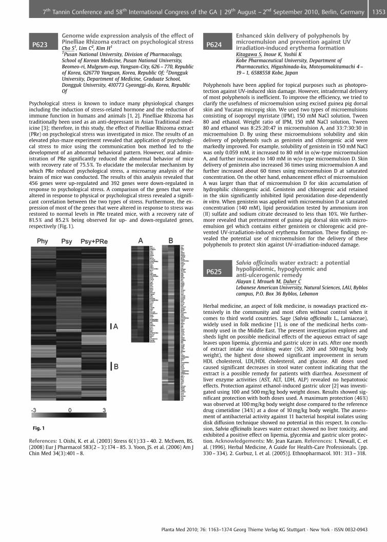
P623Genome wide expression analysis of the effect ofPinelliae Rhizoma extract on psychological stressCho S1, Lim C2, Kim H1
1Pusan National University, Division of Pharmacology,School of Korean Medicine, Pusan National University,Beomeo-ri, Mulgeum-eup, Yangsan-City, 626 – 770, Republicof Korea, 626770 Yangsan, Korea, Republic Of; 2DonggukUniversity, Department of Medicine, Graduate School,Dongguk University, 410773 Gyeonggi-do, Korea, RepublicOf
Psychological stress is known to induce many physiological changesincluding the induction of stress-related hormone and the reduction ofimmune function in humans and animals [1, 2]. Pinelliae Rhizoma hastraditionally been used as an anti-depressant in Asian Traditional med-icine [3]; therefore, in this study, the effect of Pinelliae Rhizoma extract(PRe) on psychological stress was investigated in mice. The results of anelevated plus-maze experiment revealed that application of psychologi-cal stress to mice using the communication box method led to thedevelopment of an abnormal behavioral pattern. However, oral admin-istration of PRe significantly reduced the abnormal behavior of micewith recovery rate of 75.5%. To elucidate the molecular mechanism bywhich PRe reduced psychological stress, a microarray analysis of thebrains of mice was conducted. The results of this analysis revealed that456 genes were up-regulated and 392 genes were down-regulated inresponse to psychological stress. A comparison of the genes that werealtered in response to physical or psychological stress revealed a signifi-cant correlation between the two types of stress. Furthermore, the ex-pression of most of the genes that were altered in response to stress wasrestored to normal levels in PRe treated mice, with a recovery rate of81.5% and 85.2% being observed for up- and down-regulated genes,respectively (Fig.1).
Fig. 1
References: 1. Oishi, K. et al. (2003) Stress 6(1):33 – 40. 2. McEwen, BS.(2008) Eur J Pharmacol 583(2 – 3):174 – 85. 3. Yoon, JS. et al. (2006) Am JChin Med 34(3):401 – 8.
P624Enhanced skin delivery of polyphenols bymicroemulsion and prevention against UVirradiation-induced erythema formationKitagawa S, Inoue K, Yoshii KKobe Pharmaceutical University, Department ofPharmaceutics, Higashinada-ku, Motoyamakitamachi 4 –19 – 1, 6588558 Kobe, Japan
Polyphenols have been applied for topical purposes such as photopro-tection against UV-induced skin damage. However, intradermal deliveryof most polyphenols is inefficient. To improve the efficiency, we tried toclarify the usefulness of microemulsion using excised guinea pig dorsalskin and Yucatan micropig skin. We used two types of microemulsionsconsisting of isopropyl myristate (IPM), 150 mM NaCl solution, Tween80 and ethanol. Weight ratio of IPM, 150 mM NaCl solution, Tween80 and ethanol was 8:25:20:47 in microemulsion A, and 33:7:30:30 inmicroemulsion D. By using these microemulsions solubility and skindelivery of polyphenols such as genistein and chlorogenic acid weremarkedly improved. For example, solubility of genistein in 150 mM NaClwas only 0.059 mM, it increased to 80 mM in o/w-type microemulsionA, and further increased to 140 mM in w/o-type microemulsion D. Skindelivery of genistein also increased 36 times using microemulsion A andfurther increased about 60 times using microemulsion D at saturatedconcentration. On the other hand, enhancement effect of microemulsionA was larger than that of microemulsion D for skin accumulation ofhydrophilic chlorogenic acid. Genistein and chlorogenic acid retainedin the skin significantly inhibited lipid peroxidation dose-dependentlyin vitro. When genistein was applied with microemulsion D at saturatedconcentration (140 mM), lipid peroxidation tested by ammonium iron(II) sulfate and sodium citrate decreased to less than 10%. We further-more revealed that pretreatment of guinea pig dorsal skin with micro-emulsion gel which contains either genistein or chlorogenic acid pre-vented UV-irradiation-induced erythema formation. These findings re-vealed the potential use of microemulsion for the delivery of thesepolyphenols to protect skin against UV-irradiation-induced damage.
P625Salvia officinalis water extract: a potentialhypolipidemic, hypoglycemic andanti-ulcerogenic remedyAlayan I, Mroueh M, Daher CLebanese American University, Natural Sciences, LAU, Bybloscampus, P.O. Box 36 Byblos, Lebanon
Herbal medicine, an aspect of folk medicine, is nowadays practiced ex-tensively in the community and most often without control when itcomes to third world countries. Sage (Salvia officinalis L., Lamiaceae),widely used in folk medicine [1], is one of the medicinal herbs com-monly used in the Middle East. The present investigation explores andsheds light on possible medicinal effects of the aqueous extract of sageleaves upon lipemia, glycemia and gastric ulcer in rats. After one monthof extract intake via drinking water (50, 200 and 500 mg/kg bodyweight), the highest dose showed significant improvement in serumHDL cholesterol, LDL/HDL cholesterol, and glucose. All doses usedcaused significant decreases in stool water content indicating that theextract is a possible remedy for patients with diarrhea. Assessment ofliver enzyme activities (AST, ALT, LDH, ALP) revealed no hepatotoxiceffects. Protection against ethanol-induced gastric ulcer [2] was investi-gated using 100 and 500 mg/kg body weight doses. Results showed sig-nificant protection with both doses used. A maximum protection (46%)was observed at 100 mg/kg body weight dose compared to the referencedrug cimetidine (34%) at a dose of 10 mg/kg body weight. The assess-ment of antibacterial activity against 11 bacterial hospital isolates usingdisk diffusion technique showed no potential in this respect. In conclu-sion, Salvia officinalis leaves water extract showed no liver toxicity, andexhibited a positive effect on lipemia, glycemia and gastric ulcer protec-tion. Acknowledgements: Mr. Jean Karam. References: 1. Newall, C. etal. (1996). Herbal Medicine, A Guide for Health-Care Professionals. (pp.330 – 334). 2. Gurbuz, I. et al. (2005) J. Ethnopharmacol. 101: 313 – 318.
Planta Med 2010; 76: 1163–1374 Georg Thieme Verlag KG Stuttgart · New York · ISSN 0032-0943
13537th Tannin Conference and 58th International Congress of the GA | 29th August – 2nd September 2010, Berlin, Germany
P626Potential anti-inflammatory, anti-ulcerogenicand antioxidant activity of Nasturtium officinaliswater extractFares N, Mroueh M, Mroueh M, Daher C1Lebanese American University, School of Pharmacy, P.O.Box 36 Byblos, Lebanon
Nasturtium officinalis is a widely used edible plant in the Mediterraneanregion and well known for its medicinal value in folk medicine. Thepresent study explores the effects of the water extract of N. officinalisupon rat blood lipid profile, glycemia, liver enzymes, gastric ulcer, in-flammation and antioxidant activities. One month of aqueous plant ex-tract intake (100, 250 and 500 mg/kg body weight) via drinking water,showed no significant changes in the serum lipids and glucose level.Liver enzyme activities (AL T, ALP, AST) were not negatively affected thusassuring that the extract has no hepatotoxic effects over the study per-iod. Doses of 100, 250, and 500 mg/kg body weight exhibited substantialanti-inflammatory effects against carrageenan induced inflammation(60.0, 51.1 and 46.11%) compared to 73.33% for diclofenac [1]. Similardoses caused respectively 4.7, 51.5 and 57.1% anti-inflammatory effectsagainst formalin induced inflammation [2]. For the anti-ulcerogenic ac-tivity, pre-treatment of fasted rats with100 and 250 mg/kg body weightdemonstrated significant protection (32.1% and 51% respectively)against ethanol-induced gastric ulcer, compared to 10 mg/kg cimetidine(50.24%) and 3 mg/kg omeprazole (47.81%)3. The extract also exhibiteda strong scavenging activity against DPPH radicals (55%)4. In conclusion,our data demonstrates the beneficial anti-ulerogenic, anti-inflammatoryand antioxidant activities of Nasturtium officinalis. Acknowledgements:Mr. Jean Karam. References: 1. Winter, C. et al. (1962) Proc Soc Exp BiolMed 111:544 547. 2. Northover, B. et al. (1961) Brit J Pharmacol. 16:163 –169. 3. Gurbuz, I. et al. (2005) J. Ethnopharmacol. 101: 313 – 318. 4. Pier-oni, A. (2000) J. Ethnopharmacol. 70: 235 – 273.
P627Cardiovascular effects induced by Camelliasinensis in experimental diabetesCalassi N, Dudas P, Parisi-Filho R, Magri F, Fiorino P,Ginosa M, Fonteles M, Delorenzi J, Farah VUniversidade Presbiteriana Mackenzie, Centro de CiÞnciasBiolgicas e da Safflde – Farmcia, Rua da Consola�¼o, 896,01302907 S¼o Paulo, Brazil
Diabetes mellitus is a metabolic disorder syndrome characterized byhigh glucose blood levels because of a reduced insulin secretion bypancreas beta cells and/or reducing biologic activity of this hormone.This syndrome affects carbohydrates, fat and protein metabolism. Dia-betic patients exhibit important endothelial alterations associated withthe oxidative stress, being one of the most important cause of cardio-vascular disorder in these people. Green tea (leaves of Camellia sinensis)is a popular beverage in East Asia, also used as an herbal remedy inEurope and North America. Green tea is being widely studied for itsbeneficial effect in the treatment and prevention of human diseases. Itis considered to be anti-inflammatory, anti-oxidative, anti-mutagenicand anti-carcinogenic and can prevent cardiac disorders. The aim of thisstudy was to evaluate the effects of C. sinensis tea on cardiovascularparameters in diabetic animals induced by a single injection of strepto-zotocin (STZ, 50 mg/kg, i. v.) administered 21 days before the experi-ments. Induction of diabetes mellitus in animals was confirmed byblood glucose value above 250 mg/dl 48 h after STZ induction. STZ pro-moted attenuation in mean BP (~10% of reduction) as well as in HR(~20% of reduction) when compared with control animals. Green teaintake prevented the reduction of both parameters, MAP and HR indiabetic animals. These results suggest that although green tea presentsimportant antioxidant action, the presence of caffeine in its extract mayhave a sympathomimetic action determining changes in blood pressurein animals. Acknowledgements: MackPesquisa, PIBIC/CNPq
P628Mechanisms involved on the hypotensive effectof a standardized fraction from Hancorniaspeciosa leavesSilva G, Braga F, Pesquero J, Lima M, Lemos V, Cortes SFederal University of Minas Gerais, Pharmacology, Av.Antonio Carlos, 6627, 31270901 Belo Horizonte, Brazil
Hancornia speciosa Gomes is popularly known in Brazil as “Mangaba”.The barks, roots and leaves of the species are traditionally used to treatseveral diseases, including hypertension and diabetes. Previous works of
our group demonstrated that the crude ethanolic extract from H. specio-sa leaves was able to inhibit the activity of angiotensin-converting en-zyme (ACE) and to induce vasodilatation in large and small arteries[1,2,3]. In the present work, we evaluated the hypotensive effect andits mechanism of action for a standardized fraction from H. speciosaleaves (SFH) in normotensive Swiss mice. SFH (10 mg/Kg) reduced thesystolic blood pressure (SBP), measured by tail pletismography, in32.3 € 11.7 and 28.2 € 4.9 mm Hg, after single administration by ip andpo routes, respectively. The plasmatic activity of ACE as well as theplasmatic level of angiotensin II was strongly inhibited by SFH (1 mg/Kg; po). Meanwhile, the plasmatic level of nitrite, an indirect indicator ofNO production, was significantly increased by SFH (1 mg/Kg; po). Thehypotensive effect was significantly inhibited by L-NAME (20 mg/Kg;po), whereas nitrite contents were reduced. In mouse mesenteric ar-teries, SFH induced a concentration-dependent vasodilatation(IC50= 32.0 € 0.2 mg/mL), partially dependent on functional endotheliumand on production of NO. The present results demonstrate that thehypotensive effect induced by SFH involves the inhibition of the renin-angiotensin system, the increase on production or bioavailability of NOand a direct vasodilator effect in normotensive mice. Acknowledge-ments: CNPq and PAPPE/FAPEMIG (Brazil) References: 1. Serra, CP. etal. (2005) Phytomedicine 12:424 – 432. 2. Ferreira, HC. et al. (2007)Phytomedicine 14:473 – 478. 3. Ferreira HC. et al. (2007) J Ethnopharma-col 109:161 – 164.
P629Echinacea induced hsp70 alterations inleukocytesAgnew L1, Matthias A3, Shipp C1, Kauter K1, Bone K2,Watson K1, Lehmann R3
1University of New England, School of Science andTechnology, Armidale, 2351 Armidale, Australia; 2Universityof New England, School of Health, Armidale, 2351 Armidale,Australia; 3Integria Healthcare, Research, PO Box 4854,Eight Mile Plains Brisbane, Australia
Heat shock proteins (hsp) are expressed constitutively as well as in-duced in response to mild stress. These stress factors include inflamma-tion, viral and bacterial infection, oxidative stress and cytotoxins. Al-tered hsp expression has been found in a number of disease states [1].Echinacea is used as a prophylactic to boost the immune system and hasbeen previously shown to enhance hsp70 expression in leucocytes aftermild heat shock [2]. In this study, the effects of echinacea supplementa-tion on hsp70 expression in the different leucocyte subpopulations hasbeen examined. Twenty four healthy volunteers aged between 20 and66 years with a body mass index ranging from 20.2 to 30.7 participatedin the study. They consumed 2 echinacea tablets (4.42 mg total alkyla-mides per tablet) twice a day for 14 days. Blood samples were taken ondays 1 and 15 and hsp70 expression was determined by flow cytometryin whole blood with and without in vitro heat shock. Significant differ-ences were observed in the percentage of several white cell subsets thatexpressed hsp70. CD4 and CD8 lymphocytes expressed significantlymore hsp70 in both males and females after supplementation but innatural killer cells and B lymphocytes, expression of hsp70 were onlysignificantly increased in the male participant cohort. These results in-dicate that Echinacea may play a role in activating the immune systemwhen the body encounters a challenge such as a virus. References:1. Srivastava, PK. (2002) Ann Rev Immunol 20: 395 – 425. 2. Agnew, LL.(2006) J Clin Pharm Ther 30: 363 – 9.
P630Acute toxicity study of the crude extract of thefruit rind of rambutan (Nephelium lappaceum L.)in male Wistar ratsThinkratok A1, Srisawat R2
1Institute of Science, Suranaree University of Technology 111University Avenue, Suranaree District, Amphur Muang,30000 Nakhon Ratchasima Province, Thailand; 2Institute ofScience, Suranaree University of Technology, School ofBiology, 111 University Avenue, Suranaree District, AmphurMuang, 30000 Nakorn Ratchasima Province, Thailand
Rambutans (Nephelium lappaceum L.) have various uses in medicine andthe fruit rind of rambutan can be considered as an easily accessiblesource of natural antioxidant and antibacterial agent [1]. Since toxicolo-gical data of this extract have not been reported, this study aimed toinvestigate acute toxicity and liver function effects of crude extract fromthe fruit rind of rambutan in male Wistar rats. Acute toxicity was stu-
Planta Med 2010; 76: 1163–1374 Georg Thieme Verlag KG Stuttgart · New York · ISSN 0032-0943
1354 7th Tannin Conference and 58th International Congress of the GA | 29th August – 2nd September 2010, Berlin, Germany

died in 42 male rats that received distilled water, 1, 2, 3, 4 or 5 g/kg oframbutan rind extracts by oral gavage. The number of deaths in eachgroup was recorded within 24 h. Survived rats were further investigatedfor 14 days. For liver function test, 24 male rats received distilled water,1 g/kg tannin or 1 g/kg rambutan rind extract by oral gavage. Five hourslater, trunk bloods were collected and the plasmas were measured forGPT (Glutamic-pyruvic transaminase) and GOT (glutamic-oxaloacetictransaminase) by automatic analysis apparatus. The rambutan rindwas extracted by 85% ethanol and the extract had total phenolic contentof 18.69 € 0.2 mg gallic acid/g dry sample extract and 50% inhibitor con-centration (IC50) values of 0.288 € 0.04 mg/ml sample extract. Oral acutetoxicity study revealed that there was no toxicity found at doses up to5 g/kg and there was no abnormal clinical signs. Tannin and rambutanrind extract didn’t have effect on plasma GPT and GOT levels. The resultssuggest that rambutan rind extract should be safe to be used in cosmes-tic, nutraceutical and pharmaceutical applications. References: 1. Pala-nisamy, U. et al. (2008) Food Chem. 109:54 – 63.
P631Some therapeutic effects of herbal basedointment “Dermoplant G“on venous ulcersKundakovic T1, Binic I2, Milenkovic M1, Zlatkovic S3,Nikolic G4
1Faculty of Pharmacy, Pharmacognosy, Vojvode Stepe 450,11000 Beograd, Serbia, Republic of; 2Clinic of Dermatologyand Venereology, Clinical Center Ni, Bld. Zorana Djindjica48, 18000 Ni, Serbia, Republic of; 3Actavis Trading ltd,Djordja Stanojevica 12, 11070 Belgrade, Serbia, Republic of;4Faculty of Technology, Department of Organic ChemicalTechnology, Bulevar oslobodenja 124, 16000 Leskovac,Serbia, Republic of
Venous ulcers (ulcus cruris venosum) are a common chronic diseaserequiring continuing treatment, significantly influencing patients’ wayof life. After the first signs like heavy and achy legs, ankle and leg swel-ling, the disease progresses, and the patient frequently develops legulcers, which can become infected. We tested the therapeutic effectsof the ointment “Dermoplant G“on the epithelization (reducing the af-fected area, ulcers score and microbial flora) of venous ulcers in 10 hu-man patients (4 men and 6 women) during 7 weeks. The major compo-nents of the ointment were dry water extract of Allii bulbus, dry ethanolextract of Hyperici herba and oil extract of Calendulae flos. The patientswere older than 18 years, with ulceration no longer then 2 months orwith recurrent ulcers during last 6 months, localized in lower leg. Theinvolved patients did not use any other phytomedicine or supportivetherapy. The parameters were evaluated before the treatment (afterclinical diagnosis of venous ulcers), and every 2 weeks during the periodof 7 weeks. The ulcers score was evaluated on the basis of epithelization,granulation (0-very marked, 1-moderate marked, 2-little marked, 3-noeffect), fibrin deposits, exudation and eczema (0-no effect, 1-little ex-pressed, 2-good expressed, 3- very expressed). The percentage of epithe-lization of venous ulcers was 61,53 € 28,26 after 7 weeks, without sig-nificant effects on microbial flora (Staphylococcus aureus and Pseudomo-nas aeruginosa). After 7 weeks of treatment, the ulcers score and symp-toms score improvement was almost maximal-99,0%. The total score oftreatment with “Dermoplant G” was 86,37% after 7 weeks.
P632Beneficial effects of carrots (Daucus carota) onadipocyte differentiation, glucose uptake, andfat accumulationChristensen K1, Kotowska D2, Olsen L3, Bhattacharya S4,Frett� X1, Færgeman N3, Kristiansen K2, Oksbjerg N4,Grevsen K5, Christensen L1
1University of Southern Denmark, Institute of ChemicalEngineering, Biotechnology and Environmental Technology,Niels Bohrs All� 1, 5230 Odense M, Denmark; 2University ofCopenhagen, Department of Biology, Ole Maaloes Vej 5,2200 Copenhagen N, Denmark; 3University of SouthernDenmark, Department of Biochemistry and MolecularBiology, Campusvej 55, 5230 Odense M, Denmark; 4AarhusUniversity, Department of Food Science, Blichers All� P.O.Box 50, 8830 Tjele, Denmark; 5Aarhus University,Department of Horticulture, Kirstinebjergvej 10, 5792Aarslev, Denmark
Consumption of carrots (Daucus carota L.) is in general believed to bebeneficial for human health and several bioactive compounds have been
identified e. g. the polyacetylene falcarinol, which exhibits anticanceractivities both in vitro and in vivo [1,2]. Here we report a range of resultssuggesting a potential new field of application for carrots towards con-ditions associated with the metabolic syndrome e. g. insulin resistanceand abdominal obesity. Dichloromethane (DCM) and methanol (MeOH)extracts of two varieties of carrots (var. bolero and purple haze) weremade and tested in a number of different bioassays and one in vivomodel system. DCM extracts of both carrot varieties were found to acti-vate peroxisome proliferator-activated receptor (PPAR)g without stimu-lating adipocyte differentiation, suggesting that they can have a positiveeffect on insulin sensitivity. At low concentrations (< 0.5 mg/mL) theDCM extract was able to enhance glucose uptake (GU) in porcine myo-tubes but at higher concentrations this was impaired. Biphasic concen-tration-dependent bioactivity has previously been reported for e. g. fal-carinol [2]. The nematode Caenorhabditis elegans is a good model systemfor studying lipid metabolism and fat accumulation in vivo. At 200 mg/mL, the DCM extract of var. Bolero was able to reduce fat accumulation,as measured by lipophilic dye Nile red, in C. elegans by 50%. The carrotpolyacetylenes have structures similar to both the endogenous ligandsof PPARg as well as recently identified alkamides able to activate PPARgand enhance insulin-stimulated GU in adipocytes [3], indicating thatthese could be responsible for the observed effects. References: 1. Ko-baek-Larsen M et al. (2005) J. Agric. Food Chem. 53: 1823 – 1827. 2.Hansen SL et al. (2003) J. Sci. Food Agric. 83: 1010 – 1017. 3. ChristensenKB et al. (2009) J. Nat. Prod. 72: 933 – 937.
P633Elderflowers (Sambucus nigra L.) have asignificant impact on cellular mechanismsrelated to lipid storage and insulin resistanceChristensen K1, Olsen L2, Kotowska D3, Bhattacharya S4,Frett� X1, Færgeman N2, Kristiansen K3, Oksbjerg N4,Christensen L1
1University of Southern Denmark, Institute of ChemicalEngineering, Biotechnology and Environmental Technology,Niels Bohrs All� 1, 5230 Odense M, Denmark; 2University ofSouthern Denmark, Department of Biochemistry andMolecular Biology, Campusvej 55, 5230 Odense M,Denmark; 3University of Copenhagen, Department ofBiology, Ole Maaloes Vej 5, 2200 Copenhagen N, Denmark;4Aarhus University, Department of Food Science, BlichersAll� P.O. Box 50, 8830 Tjele, Denmark
Preparations of elderflowers (Sambucus nigra L.) have traditionally beenused as diuretics however recent studies suggest that they also have apotential use in the treatment/prevention of conditions associated withthe metabolic syndrome. Extracts of elderflowers are known to activatethe key regulator of adipocyte differentiation, peroxisome proliferator-activated receptor (PPAR)g, without stimulating adipocyte differentia-tion, and furthermore to enhance insulin-stimulated glucose uptake(GU) in adipocytes [1]. The flavanone naringenin was identified as oneof the bioactive components whereas major elderflower metaboliteswere not active [2]. In the present study, we have further investigatedthe effects of elderflower extracts and metabolites on mechanisms asso-ciated with lipid storage and insulin resistance in relation to the meta-bolic syndrome. Both dichloromethane (DCM) and methanol (MeOH)extracts of elderflowers were found to be able to enhance GU in pigmyotubes both with and without insulin-stimulation correspondingwith previous studies on mouse abdominal muscle [3]. The nematodeCaenorhabditis elegans provides an excellent model system for studyinglipid metabolism and fat accumulation in vivo. Extracts of elderflowerswere able to significantly reduce fat accumulation as measured by thelipophilic dye Nile red, in C. elegans at concentrations of 200 mg/mL, andin particular the DCM extract was one of the most potent plant extractswith more than a 50% reduction of Nile red fluorescence [4]. However,on individual basis major elderflower metabolites do not seem to pos-sess the same magnitude of bioactivity in the tests performed as theextracts. Hence, synergistic effects might be at play warranting furtherinvestigations. References: 1. Christensen KB et al. (2009) Phytother.Res. 23: 1316 – 1325. 2. Christensen KB et al. (2010) Phytother. Res. inpress. 3. Gray AM et al. (2000) J. Nutri. 130: 15 – 20. 4. Boelt SG et al.(2008) Chem. Physics Lipids 154: S32-S 32.
Planta Med 2010; 76: 1163–1374 Georg Thieme Verlag KG Stuttgart · New York · ISSN 0032-0943
13557th Tannin Conference and 58th International Congress of the GA | 29th August – 2nd September 2010, Berlin, Germany
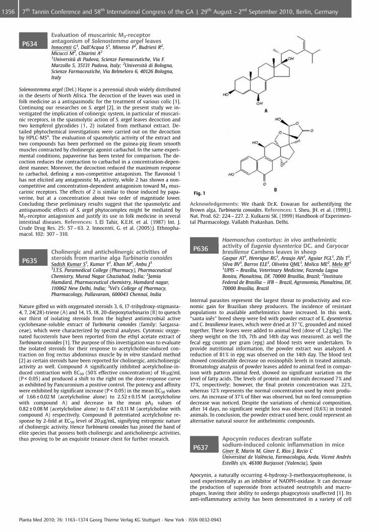
P634Evaluation of muscarinic M3-receptorantagonism of Solenostemma argel leavesInnocenti G1, Dall’Acqua S1, Minesso P1, Budriesi R2,Micucci M2, Chiarini A2
1Universit� di Padova, Scienze Farmaceutiche, Via F.Marzollo 5, 35131 Padova, Italy; 2Universit� di Bologna,Scienze Farmaceutiche, Via Belmeloro 6, 40126 Bologna,Italy
Solenostemma argel (Del.) Hayne is a perennial shrub widely distributedin the deserts of North Africa. The decoction of the leaves was used infolk medicine as a antispasmodic for the treatment of various colic [1].Continuing our researches on S. argel [2], in the present study we in-vestigated the implication of colinergic system, in particular of muscari-nic receptors, in the spasmolytic action of S. argel leaves decoction andtwo kempferol glycodides (1, 2) isolated from methanol extract. De-tailed phytochemical investigations were carried out on the decoctionby HPLC-MSn. The evaluation of spasmolytic activity of the extract andtwo compounds has been performed on the guinea-pig ileum smoothmuscles contracted by cholinergic agonist carbachol. In the same experi-mental conditions, papaverine has been tested for comparison. The de-coction reduces the contraction to carbachol in a concentration-depen-dent manner. Moreover, the decoction reduced the maximum responseto carbachol, defining a non-competitive antagonism. The flavonoid 1has not elicited any antagonistic M3 activity, while 2 has shown a non-competitive and concentration-dependent antagonism toward M3 mus-carinic receptors. The effects of 2 is similar to those induced by papa-verine, but at a concentration about two order of magnitude lower.Concluding these preliminary results suggest that the spasmolytic andantispasmodic effects of S. argel phytocomplex might be mediated byM3-receptor antagonism and justify its use in folk medicine in severalintestinal diseases. References: 1. El Tahir, K.E.H. et al. (1987) Int. J.Crude Drug Res. 25: 57 – 63. 2. Innocenti, G. et al. (2005) J. Ethnopha-macol. 102: 307 – 310.
P635Cholinergic and anticholinergic activities ofsteroids from marine alga Turbinaria conoidesSadish Kumar S1, Kumar Y1, Khan M2, Anbu J3
1I.T.S. Paramedical College (Pharmacy), PharmaceuticalChemistry, Murad Nagar Ghaziabad, India; 2JamiaHamdard, Pharmaceutical chemistry, Hamdard nagar,110062 New Delhi, India; 3Vel’s College of Pharmacy,Pharmacology, Pallavaram, 600043 Chennai, India
Nature gifted us with oxygenated steroids 3, 6, 17-trihydroxy-stigmasta-4, 7, 24(28)-triene (A) and 14, 15, 18, 20-diepoxyturbinarin (B) to quenchour thirst of isolating steroids from the highest antimicrobial activecyclohexane-soluble extract of Turbinaria conoides (family: Sargassa-ceae), which were characterized by spectral analyses. Cytotoxic oxyge-nated fucosterols have been reported from the ethyl acetate extract ofTurbinaria conoides [1]. The purpose of this investigation was to evaluatethe isolated steroids for their response to acetylcholine-induced con-traction on frog rectus abdominus muscle by in vitro standard method[2] as certain steroids have been reported for cholinergic, anticholinergicactivity as well. Compound A significantly inhibited acetylcholine-in-duced contraction with EC50 (50% effective concentration) of 16 mg/mL(P < 0.05) and produced a shift to the right on the dose-response curveas exhibited by Pancuronium a positive control. The potency and affinitywere exhibited by significant increase (P < 0.05) in the mean EC50 valuesof 1.66 € 0.02 M (acetylcholine alone) to 2.52 € 0.15 M (acetylcholinewith compound A) and decrease in the mean pA2 values of0.82 € 0.08 M (acetylcholine alone) to 0.47 € 0.11 M (acetylcholine withcompound A) respectively. Compound B potentiated acetylcholine re-sponse by 2-fold at EC50 level of 20 mg/mL, signifying estrogenic natureof cholinergic activity. Hence Turbinaria conoides has joined the band ofelite species that possess both cholinergic and anticholinergic activities,thus proving to be an exquisite treasure chest for further research.
Fig. 1
Acknowledgements: We thank Dr.K. Eswaran for authentifying theBrown alga, Turbinaria conoides. References: 1. Sheu, JH. et al. (1999) J.Nat. Prod. 62: 224 – 227. 2. Kulkarni SK. (1999) Handbook of Experimen-tal Pharmacology. Vallabh Prakashan. Delhi.
P636Haemonchus contortus: in vivo anthelminticactivity of Eugenia dysenterica DC. and Caryocarbrasiliense Cambess leaves in sheepGaspar AT1, Henrique RG1, Araujo AH1, Aguiar FGL1, Zils T1,Silva BV1, Barros ELE1, Oliveira QML1, Molica ME2, Melo RF1
1UPIS – Bras�lia, Veterinary Medicine, Fazenda LagoaBonita, Planaltina, DF, 70000 Bras�lia, Brazil; 2InstitutoFederal de Bras�lia – IFB – Brazil, Agronomia, Planaltina, DF,70000 Bras�lia, Brazil
Internal parasites represent the largest threat to productivity and eco-nomic gain for Brazilian sheep producers. The incidence of resistantpopulations to available anthelmintics have increased. In this work,“santa inÞs” breed sheep were fed with powder extract of E. dysentericaand C. brasiliense leaves, which were dried at 37 �C, grounded and mixedtogether. These leaves were added to animal feed (dose of 1,2 g/kg). Thesheep weight on the 1th, 7th and 14th day was measured; as well thefecal egg counts per gram (epg) and blood tests were undertaken. Toprovide nutritional information, the powder extract was analyzed. Areduction of 81% in epg was observed on the 14th day. The blood testshowed considerable decrease on eosinophils levels in treated animals.Bromatology analysis of powder leaves added to animal feed in compar-ison with pattern animal feed, showed no significant variation on thelevel of fatty acids. The levels of protein and minerals decreased 7% and17%, respectively; however, the final protein concentration was 22%,whereas 12% represents the normal concentration used by most produ-cers. An increase of 37% of fiber was observed, but no feed consumptiondecrease was noticed. Despite the variations of chemical composition,after 14 days, no significant weight loss was observed (0,6%) in treatedanimals. In conclusion, the powder extract used here, could represent analternative natural source for anthelmintic compounds.
P637Apocynin reduces dextran sulfatesodium-induced colonic inflammation in miceGiner R, Marin M, Giner E, R�os J, Recio CUniversitat de Val�ncia, Farmacologia, Avda. Vicent Andr�sEstell�s s/n, 46100 Burjassot (Valencia), Spain
Apocynin, a naturally occurring 4-hydroxy-3-methoxyacetophenone, isused experimentally as an inhibitor of NADPH-oxidase. It can decreasethe production of superoxide from activated neutrophils and macro-phages, leaving their ability to undergo phagocytosis unaffected [1]. Itsanti-inflammatory activity has been demonstrated in a variety of cell
Planta Med 2010; 76: 1163–1374 Georg Thieme Verlag KG Stuttgart · New York · ISSN 0032-0943
1356 7th Tannin Conference and 58th International Congress of the GA | 29th August – 2nd September 2010, Berlin, Germany

and animal models [2]. In this study, we investigated whether apocyninexhibits anti-inflammatory activity on a model of inflammatory boweldisease (IBD) using dextran sulfate sodium (DSS)-induced colitis in mice.Acute colitis was induced in the mice through administration of 5% DSSin water for 7 days. The mice were fed a control diet or a diet supple-mented with 2% apocynin or 2% rutin (a flavonoid used as reference) [3].The animals were sacrificed on day 8 and their colons were removed,immediately snap-frozen on liquid nitrogen, and stored until use. Thedisease activity index score (DAI) was analyzed taking weight loss, stoolblood, and rectal bleeding into account. The removed colons were sub-jected to extraction of proteins and COX-2 expression was analyzed withthe aid of a Western blot assay. Treatment with apocynin amelioratedthe course of colonic inflammation with results similar to those of rutin,as can be seen by reductions in the DAI scores (60% reduction for bothcompounds), colon length (47% and 42% reduction vs. blank, respec-tively), and COX-2 levels (65% and 75% inhibition, respectively). Theseresults are promising for further experimental studies on treating gas-trointestinal diseases and the potential protective effects of apocynin.Acknowledgements: Spanish Government, MICIIN (SAF 2009 – 13059-C 03 – 01). References: 1. Simons, J.M., et al. (1990) Free Rad. Biol. Med.8:251 – 258. 2. Stefanska, J., Pawliczak, R. (2008) Mediators Inflamm. ID106507. 3. Xu, L., et al. (2009) Phytomedicine 16:989 – 995.
P638Effects of the crude extract of the fruit rind oframbutan (Nephelium lappaceum L.) on bloodpressure, heart rate and respiratory rate inanaesthetized male ratsSrisawat R, Puengpai S, Nontamart N, Thinkratok AInstitute of Science, Suranaree University of Technology,School of Biology, 111 University Avenue, Suranaree District,Amphur Muang, 30000 Nakhon Ratchasima province,Thailand
The bark of rambutan (Nephelium lappaceum L.), which is normallydiscarded was found to contain extremely high antioxidant activity [1].Since polyphenols found in plants are widely known to affect the cardi-ovascular system, there is no evidence suggesting how polyphenolsfound in the crude extract of the fruit rind of rambutan did. We, there-fore, investigated acute effects of rambutan on cardiovascular and re-spiratory responses in male rats. The ethanolic rambutan bark extractwith a total phenolic content of 18.69 € 0.2 mg gallic acid/g dry sampleextract were used in this experiment. Invasive arterial blood pressure,heart rate and respiratory rate were recorded in pentobarbital-anaesthe-tized male rats for 2 hours after single i. p. injected with vehicle (n = 8)and 1 g/kg rambutan bark extract (n = 8). Intragroup comparison showedthat rambutan rind extract markedly increased mean arterial bloodpressure (MABP) and systolic blood pressure (p < 0.05, compared tobaseline). In comparison between groups, significant increases in MABP,systolic blood pressure, diastolic blood pressure and heart rate can beobserved 60 minutes after rambutan bark extract (p < 0.05, two wayrepeated measures ANOVA). Significant increases in those parameterswere found over the rest period of investigation. However, there were nochanges in respiratory rate In conclusion, this study provided the firstevidence of prolonged cardiovascular response (increases in MABP, sys-tolic blood pressure and heart rate) following acute rambutan rind ex-tract in anesthetized male rats. The mechanism of which rambutan rindextract regulating blood pressure in normotensive, hypotensive and hy-pertensive conditions should be further investigated. References: 1. Pa-lanisamy, U. et al. (2008) Food Chem. 109:54 – 63.
P639Naringin protects against dextran sodiumsulfate-induced colitis in miceGiner E, Marin M, Recio C, R�os J, Giner RUniversitat de Val�ncia, Farmacologia, Avda. Vicent Andr�sEstell�s s/n, 46100 Burjassot (Valencia), Spain
Naringin (4’,5,7-trihydroxyflavanone 7-rhamnoglucoside) is a well-known natural flavonoid that acts as a potent anti-oxidant, superoxidescavenging, anti-apoptotic, anti-atherogenic and metal chelating agent[1]. Orally administered naringin is metabolized to its aglycone, narin-genin, which has a wide range of pharmacological properties, includingan anti-inflammatory effect through inhibition of nitric oxide and pros-taglandin E2 production [2]. The aim of this study is to determine theeffect of naringin on a model of inflammatory bowel disease (IBD),namely dextran sulfate sodium (DSS)-induced colitis in mice. FemaleBalb-C mice were randomized to receive either normal water or 5%
DSS drinking water to induce colitis. The mice were fed a control dietor a diet supplemented with 2% naringin or 2% rutin for 7 days [3,4]. Onday 8, the mice were sacrificed and their colons were removed, imme-diately snap-frozen on liquid nitrogen and stored until use. The diseaseactivity index score (DAI) was analyzed taking weight loss as well asstool blood and rectal bleeding into consideration. The removed colonswere subjected to protein extraction and COX-2 expression was ana-lyzed with the aid of Western blot techniques. Naringin was found tosignificantly reduce the extent and severity of injury to the colon asshown by the DAI score (49% reduction), colon length (40% reductionvs. blank) and COX-2 levels (78% inhibition). Rutin, a polyphenolic fla-vonoid used as reference, also diminished these parameters by percen-tages of 60, 42 and 75, respectively. These results indicate that naringinmay prove to be a useful agent for treating IBD. Acknowledgements:Spanish Government, MICIIN (SAF 2009 – 13059-C 03 – 01). References:1. Jagetia, G.C., Reddy, T.K., (2005) Life Sci. 77:780 – 794. 2. Raso, GM., etal. (2001) Life Sci. 68:921 – 931. 3. Claire Billerey-Larmonier, M.S., et al.(2008) Inflamm. Bowel Dis. 14:780 – 793. 4. Xu, L., et al. (2009) Phyto-medicine 16:989 – 995.
P640Isolation, structure determination andcannabinoid receptor activating effect of newmetabolites of Echinacea purpurea rootHohmann J1, R�dei D1, Forgo P1, Bojnik E2, Benyhe S2
1University of Szeged, Department of Pharmacognosy,E�tv�s u. 6, 6720 Szeged, Hungary; 2Hungarian Academy ofSciences, Biological Research Centre, Institute ofBiochemistry, POB 521, 6701 Szeged, Hungary
Extracts of Echinacea purpurea are extensively used in the therapy ofcommon cold and upper respiratory tract infections as immunomodula-tors and anti-inflammatory agents. The extracts of the root and herb ofE. purpurea have a complex chemical composition, including alkamides,caffeic acid conjugates, and polysaccharides. Alkamides, the main lipo-philic compounds, are the modulators of the endocannabinoid system inconsequence of their structural similarity to the endogenous ligand ana-ndamide. Recently, receptor binding assays demonstrated the CB2 re-ceptor affinity of Echinacea alkamides, proving by this the CB2-receptor-dependent immunomodulatory effect [1,2]. The aim of the present studywas to reinvestigate the chemical composition of the roots of E. purpureaand to gain new information about the affinity of the isolated com-pounds to the cannabinoid system. The nhexane-soluble extract of theroot was subjected to multiple chromatographic separations affording19 compounds. The structures were analysed by extensive NMR and MSstudies resulting the identification of four new natural products (3 alka-mides and nitidanin-diisovalerianate). In addition, five compounds weredetected for the first time in this species, and ten known E. purpureametabolites were identified. Cannabinoid receptor activation of thirteenisolated compounds has been studied by [35S]GTPgS binding assays inrat brain membranes. This test measures (i) receptor-mediated G-pro-tein activation induced by agonists and (ii) inhibition by antagonistligands. A number of E. purpurea compounds turned out to be weak-to-moderate partial agonists, while others displayed inverse-agonist ac-tions. In the presence of a reference CB1 receptor agonist, both sort ofcompounds exhibited concentration-dependent competitive antagonisteffects. References: 1. Woelkart, K., Bauer, R. (2007) Planta Med.73:615 – 623. 2. Raduner, S., et al. (2006) J. Biol. Chem. 281:14192 –14206.
P641High-dose St. John’s wort extract STW 3-VI as adaily single-dose is safe and effective in thetreatment of mild to moderate depression indifferent age-groups – results of a re-evaluationM�ller J, Kolb C, Kelber O, Weiser DClinical Research, Steigerwald Arzneimittelwerk GmbH,Havelstr. 5, 64295 Darmstadt, Germany
Objectives: In a post-marketing surveillance study (1) 4188 patients of793 general practitioners were included. The patients suffered mainlyfrom mild to moderate depression and dysthymia according to ICD10 and were treated with St. John’s wort extract (STW 3-VI, 900 mgextract) 1 x 1 tablet daily for 12 weeks. This revaluation assessed thechange of the Hamilton Depression Rating Scale (HAMD) scores overtime to different age-groups. Methods: An analysis of variance (ANOVA)for repeated measures (SPSS 15.0) was conducted. Only patients withcompleted data for all measurements were revaluated. Results: 1701
Planta Med 2010; 76: 1163–1374 Georg Thieme Verlag KG Stuttgart · New York · ISSN 0032-0943
13577th Tannin Conference and 58th International Congress of the GA | 29th August – 2nd September 2010, Berlin, Germany
patients with mild depression, 1433 patients with moderate depressionand 194 patients with dysthymia were revaluated (913 male, 2415 fe-male patients). The 3 main ICD-10 disease classes showed a significantand comparable change of the HAMD-scores over time with very similarscores in moderate depression and dysthymia (mild depression: HAMD-means: 13.5 at baseline vs. 3.3 after 12 weeks; moderate depression:HAMD-means: 18.2 at baseline vs. 5.5 after 12 weeks). In a 2. analysisthe patients were separated into 7 age-groups (from < 18 years to > 65years). All age-groups, including the elderly, showed the above men-tioned significant and comparable change of the HAMD-scores overtime. Conclusion: This revaluation indicates that there is neither aninfluence of the disease classification (i. e. mild or moderate depressivedisorders) nor of the age to the time course of the treatment outcome inpatients treated with St. John’s wort extract (STW 3-VI). References:1. Demling et al., Nervenheilkunde 2004, 23:160.
P642Extract and fractions from birch bark affectstimulation of human dendritic cells and theiractivation of allogeneic CD 4+ T cells in vitroOmarsdottir S1, Sigurpalsson M2, Eggertsdottir A1,Runarsson J2, Hardardottir I3, Vikingsson A2, Olafsdottir E1,Freysdottir J2
1University of Iceland, Faculty of Pharmaceutical Sciences,Hagi, Hofsvallagata 53, 170 Reykjavik, Iceland; 2LandspitaliUniversity Hospital, Centre for Rheumatology Research andDepartment of Immunology, Hringbraut, house 14, 101Reykjavik, Iceland; 3University of Iceland, Faculty ofMedicine, Laeknagardur, Vatnsmyrarvegi 16, 101 Reykjavik,Iceland
Birch bark has been used in traditional medicine to treat skin disordersand rheumatism. Several components have been isolated from birchbark with only few being tested for their effects on the immune system.Dendritic cells play a major role in the regulation of an immune re-sponse, e. g. by directing the differentiation of naive T cells. Therefore,the effect of extract and fractions from the bark of Betula pubescens wastested in a human monocyte-derived dendritic cell model. Dendriticcells cultured with ethanol extract from birch bark secreted less IL-6,IL-10 and IL-12p40 than cells cultured with stimulation factors alone.These effects were also observed for fractions III and IV obtained withVLC using dichloromethane:methanol gradient. Allogeneic CD+ T cellsco-cultured with dendritic cells treated with fraction IV secreted lessIFN-g than T cells co-cultured with dendritic cells treated with stimula-tion factors alone. Further fractionation revealed that the active frac-tions are mainly composed of triterpenes and further characterizationof these compounds is in progress.
P643Toxicological studies on the Eugenia dysentericaDC and Caryocar brasiliense Cambess leaves inratsElias F1, Zils T1, Aguiar FGL1, Barros ELE1, Molica ME2,Melo RF3
1UPIS – Bras�lia, Fazenda Lagoa Bonita, Planaltina,700000 Bras�lia, Brazil; 2(1) UPIS – Bras�lia, Agronomia,Instituto Federal de Bras�lia, 70000 Planaltina, Brazil;3UPIS – Bras�lia, Veterinary Medicine, Fazenda LagoaBonita, Planaltina, 70000 Bras�lia, Brazil
Powder extract of E. dysenterica and C. brasiliense leaves proved to beactive against Haemonchus contortus, in vivo, in sheep, according toprevious work performed by our group. Therefore, toxicology studiesbecome necessary. To this aim, twenty male rats received a powderextract of E. dysenterica and C. brasiliense leaves for 15 days. The leaveswere dried at 37 �C, ground, mixed and added to animal feed at concen-tration of 10%, 20% and 30%. A control group was fed only with dietwithout the mixture of leaves. After 15 days, only rats that received thefeed with 30% of powder leaves showed significant weight loss, prob-ably due to nutritional deficit since no feed reduction consumption wasobserved. A reduction on thymus weight, a discrete degeneration of liverand decrease on creatinin levels were observed on rats that received themajor dose after 15 days. No important variation on hemoglobin, hema-tocrit, leukocytes and total protein was observed. According to thesedata we can suggest some toxicity when we use a mixture of 30% ofpowder leaves added to rat feed, and although a reduction of 81% infecal egg counts per gram has been observed when we used the dose of
only 1,2 g/kg in sheep, we intent to perform new experiments to analyzethe treatment during a extend time period.
P644In vitro and in vivo evidence of synergy betweenHypericum and Passiflora in antidepressantpharmacological modelsFiebich B1, Kn�rle R2, Weiss G3, McGregor G4
1VivaCell Biotechnology GmbH, Ferdinand-Porsche-Str. 5,79211 Denzlingen, Germany; 2IBAM, Ferdinand-Porsche-Str.5, 79211 Denzlingen, Germany; 3PASCOE pharmazeutischePr�parate GmbH, Schiffenberger Weg 55, 35394 Gießen,Germany; 4Philipps-University of Marburg, Faculty ofMedicine, Deutschhausstrasse 2, 35037 Marburg, Germany
Neurapas�balance is a combination of special extracts of Hypericumperforatum (St. John’s wort), Passiflora incarnata (passion flower) andValeriana officinialis (valerian); it is used for the treatment of mild de-pression, mild anxiety and sleep disorders. Since the daily dose of Hy-pericum in Neurapas� balance is lower than the doses of Hypericum insingle extract phytomedicines, we were interested whether a combina-tion of extracts of Hypericum and Passiflora exerts comparable effects toNeurapas� balance. We used two well-established models for investi-gating extracts for their anti-depressant activity, namely the effects onsynaptic uptake of serotonin and the forced swimming test; in both testsHypericum has previously been shown to exert pharmacological effects.We show here for the first time, that the Passiflora extract significantlyenhances the pharmacological potency of Hypericum in both models.The potency of Hypericum to inhibit serotonin uptake depended onthe hyperforin content of the test batches. The dose-dependent en-hancement of the Hypericum effect by Passiflora (50 mg/ml) resulted inan IC 50 of 14 mg/ml (Hypericum containing 1% hyperforin) and 9.7 mg/ml(Hypericum containing 2.1% hyperforin). A similar synergy between Pas-siflora and Hypericum extracts was observed in the in vivo model. Agreater maximal effect (60% reduced immobility) was observed withHypericum in a fixed (2:1) combination with Passiflora (90 mg/kg) thanwith higher concentrations (180/360 mg/kg) of Hypericum alone (max-imal 45% reduced immobility). Our data prove that the anti-depressivetherapeutic effects of Hypericum are possible with lower doses, when itis combined with Passiflora, than with mono-preparations of Hypericum.
P645Gemmotherapy-complementary treatment injuvenile rheumatoid arthritisMilitaru A1, Simedrea I1, Daescu C1, Peev C1, Pop G2
1University of Medicine and Pharmacie Victor Babes,Pedriatic Clinic, Eftimie Murgu No. 2, 300041 Timisoara,Romania; 2Banat’s University of Agricultural Science andVeterinary Medicine, Agriculture, 119 calea aradului,300645 Timisoara, Romania
Rheumatoid arthritis is a chronic disease that damagesandeventuallydestroys the joints of the body. Juvenile rheumatoid arthritis (JRA) isnot a single disease, but a group of diseases with symptoms and signsdeveloped in children younger than 16 years. There are three majorforms: pauciarticular disease, poliarticular disease, and systemic dis-ease. The aim of treatment is to stop or slow down the progress ofinflammation, improving function, and preventing joint damage. Themost usefull drugs are: 1) nonsteroidal anti-inflammatorydrugs(NSAIDs) reduce inflammation, swelling, and pain, 2) disease-modifyingantirheumatic drugs (DMARDs), in children Methotrexat is the mostused drug, it interferes in the immune processes that cause inflamma-tion and JRA. 3) Biologic response modifiers are a newer, specializedtype of immunosuppressive drugs, carefully designed to block the ac-tions of natural substances that are part of the immune response, suchas tumor necrosis factor. A variety of complementary approaches can bevery effective in relieving pain and improving the outcome of the dis-ease. One of these complementary therapy is gemmoterapy, a scientificuse of a special glycerin extracts from plant buds. In our study,19 pa-cients with diagnosed JRA (15 with pauciarticular disease and 4 withpoliarticular form) was treated with specific antirheumatic drugs asso-ciating gemmotherapy. It has been used three types of bud extracts(Ribes nigrum, Buxus sempervirens, Vitis vinifera) with proved anti-rheumatic effects, during a period of 3 months. In all the pacients weobserved a clinical amelioration, permitting a slight reduction of thedose of the NSAIDs and the DMARDs.
Planta Med 2010; 76: 1163–1374 Georg Thieme Verlag KG Stuttgart · New York · ISSN 0032-0943
1358 7th Tannin Conference and 58th International Congress of the GA | 29th August – 2nd September 2010, Berlin, Germany
P646Topical anti-inflammatory agents from the alpineflavouring plant Artemisia umbelliformis Lam.Sosa S1, Giangaspero A1, Ponti C2, Del Favero G1,Pollastro F3, Appendino G3, Tubaro A1, Della Loggia R1
1University of Trieste, Dep. of Materials and NaturalResources, Via A. Valerio 6, 34127 Trieste, Italy; 2Universityof Trieste, Dipartimento Universitario Clinico diBiomedicina, Via Manzoni 16, 34138 Trieste, Italy;3Universit� del Piemonte Orientale, Dipartimento di ScienzeChimiche, Alimentari, Farmaceutiche e Farmacologiche, ViaBovio 6, 28100 Novara, Italy
Artemisia umbelliformis Lam. (Asteraceae) is an alpine flavouring plantused to produce the bitter liqueur genepy. The lipophilic flavonoid eu-patilin, a series of sesquiterpenes lactones and the sesterpene lactonegenepolide, isolated from this plant [1,2], were investigated for theirtopical anti-inflammatory activity (inhibition of the Croton oil-inducedmouse ear dermatitis) in comparison to the steroidal and non steroidalanti-inflammatory drugs hydrocortisone and indomethacin [3]. Sixhours after dermatitis induction, the anti-oedema potency of eupatilinwas comparable to that of indomethacin (ID50= 0.30 and 0.26mmol/cm2),and only one order of magnitude lower than that of hydrocortisone(ID50= 0.03 mmol/cm2). A slightly lower effect was observed for genepo-lide and the sesquiterpenes anhydroverlotorin, santamarin, 5-deoxy-5-hydroperoxy-telekin, 5-deoxy-5-hydroperoxy-epitelekin and costuno-lide (ID50 ranged from 0.35 to 0.73mmol/cm2), the most active beinganhydroverlotorin and genepolide (ID50= 0.35 and 0.40mmol/cm2). Theoverall effect of eupatilin (0.3mmol/cm2), anhydroverlotorin (0.4mmol/cm2) and genepolide (0.4mmol/cm2) on oedema development up to 48 hwas intermediate between those of equimolar doses of indomethacinand hydrocortisone at one order of magnitude lower doses, but theiractivity profile was similar to the latter. Eupatilin reduced also the leu-kocytes infiltrate in the ear tissue, similarly to the reference drugs. Theeffect of anhydroverlotorin and genepolide on inflammatory cells infil-tration will also be discussed. References: 1. Appendino, G. et al.(2009) J. Nat. Prod. 72: 340 – 344. 2. Rubiolo, P. et al. (2009) J. Agric. FoodChem. 57: 3436 – 3443. 3. Tubaro, A. et al. (1985) Agents Actions 17:347 – 349.
P647Analgesic effects of Stachys turcomanica extractin miceHajimehdipoor H1, Sahebgharani M2, Khanavi M3,Mirshaki Z2
1Traditional Medicine and Materia Medica Research Centerand School of Traditional Medicine, Shahid BeheshtiUniversity of Medical Sciences, Po. Box: 14155 – 6359.Tehran, Iran, Islamic Republic Of; 2Department ofPharmacology, Facalty of Medicine, Tehran University ofMedical Sciences, Enghelab St., 15911 Tehran, Iran, IslamicRepublic Of; 3Department of Pharmacognosy, Faculty ofPharmacy, Tehran University of Medical Sciences, EnghelabSt., 15911 Tehran, Iran, Islamic Republic Of
Pain and inflammation are the complex biological responses to harmfulstimulants. In fact, they are a protective attempt by the organism toremove the injurious stimulant. Natural products have long been recog-nized as an important source of therapeutically effective medicines.Different approaches have been developed to analyze analgesic andanti-inflammatory potential of plants in the past years. Stachys species(Lamiaceae) have been used as medicinal plants because of their biolo-gical activities. Some Stachys have been used as remedy for painful orinflammatory conditions in folk medicine [1,2]. In this study, the anti-nociceptive property of S. turcomanica was investigated by formalin testin mice. Aerial parts of S. turcomanica were collected from its growingarea and identified. Dried and milled plant material was extracted byusing methanol 80% and different concentrations of the dried extractwere prepared. During the test, formalin solution (0.5%) was injectedinto the plantar surface of the right hind paw of the mice, 15 minutesafter peritoneal injection of the plant methanol extract (100, 200 and400 mg/kg). In order to elucidate the probable mechanism of action ofthe plant extract, naloxone was administered with extract. The resultsshowed that injection of the plant extract at the doses of 100, 200 &400 mg/kg significantly inhibited the chronic phase of formalin- inducedpain. Intraperitoneal injection of opioid antagonist (naloxone) signifi-cantly reversed the analgesic effect of the extract in chronic phase. Thisresult suggests that analgesic activity of this plant may be partlymediated by opioid system. References: 1. Rezazadeh, Sh. et al.
(2009) J. Med. Plants Res. 3: 368 – 376. 2. Hajhashemi, V. et al. (2007)Res. Pharmac. Sci. 2: 92 – 98.
P648Antiproliferative effect of Bryonia aspera Stev. exLedebSahranavard S1, Ghafari S2, Moazzeni H2, Naghibi F1
1Traditional medicine and materia medica research center,School of Pharmacy, Shahid Beheshti University of MedicalSciences, Pharmacognosy Department, Vali asr streetTehran, Iran, Islamic Republic Of; 2Traditional medicine andmateria medica research center, Shahid Beheshti Universityof Medical Sciences, valiasr street, shams alley, 1516745811Tehran, Iran, Islamic Republic Of
This study was designed to evaluate antiproliferative effects from theroots of Bryonia aspera Stev. ex Ledeb which is used traditionally in thetreatment of gastrointestinal disorders, cardiac disorders and cancer [1].Methanol, chloroform and petroleum ether fractions were prepared andinvestigated for antiproliferative activity against MCF7 (human breastadenocarcinoma), HepG2 (human hepatocellular carcinoma), WEHI(mouse fibrosarcoma) and MDBK (normal kidney epithelial cell) celllines by MTT assay and cisplatin and tamoxifen were used as positivecontrol [2]. All the fractions showed significant antiproliferative activityon MCF7 whereas they were inactive on HepG2 and WEHI cell lines(IC50> 50 mg/ml). Our study showed that the CHCl3 fraction was the mostpotent one against the MCF7 cell line. In our previous study, we isolatedsome triterpene cucurbitane-type compounds from the CHCl3 fraction[3]. Further studies are in progress to evaluate the antiproliferative ac-tivity of these pure compounds. References: 1. Ghorbani A. (2005) J.Ethnopharmacol. 102:58. 2. Carmichael J.et al (1987) Cancer. Res.47:936 – 42. 3. Sahranavard S. et al (2010) Planta Med. 76: 1 – 4.
P649Pharmacological and histological study ofCentaurea bruguierana ssp. belangerana onindomethacin-induced peptic ulcer in ratsAhmadi R, Rajabi A, Khanavi M, Hassanzadeh G,Khademi R, Hadjiakhoondi A, Sharifzadeh MTehran University of Medical Sciences, Department ofPharmacognosy, Tehran, Iran, 1417614411 Tehran, Iran,Islamic Republic Of
The species Centaurea bruguierana (DC.) Hand.-Mzt. ssp. belangerana(DC.) Bornm. (CBB) (Asteraceae) [1], namely “Baad-Avard” in Bushehrprovince, south of Iran, is used in folk medicine as hypoglycemic indiabetes and remedy for peptic ulcer disorders. The inhibitory effectsof total extract and fractions of dried flowering samples of CBB collectedfrom Borazjan, Bushehr Province, Iran on indomethacin-induced pepticulcer in rats were studied. The 80% EtOH extract and petroleum ether,CHCl3, EtOAc, n-BuOH, and remaining MeOH fractions obtained by sol-vent-solvent fractionation of dried flowering samples of CBB was inves-tigated for anti-ulcer activity against indomethacin-induced ulcerogen-esis in rats. The percentage inhibition for total extract with dose of100 mg/kg (99%) and CHCl3 fraction with dose of 42 mg/kg (98.95%)were found to be more prominent (***p < 0.001) compared to the refer-ence group (cimetidine 100 mg/kg, 87.62% ulcer inhibition). A dramaticdecrease in ulcer index was observed following the administration oftotal extract (100 mg/kg, ***p < 0.001) and CHCl3 fraction (42 mg/kg,***p < 0.001) compared to control group. Pharmacoilogical and histolo-gical results of the present study proved that the aerial flowering partsof CBB possess preventive activity against peptic ulcer supporting thefolkloric assertion in southern Iranian folk medicine. However, theseeffects are not limited to this species and previous studies have revealedthat some other Centaurea species are active as well [2,3,4]. Acknowl-edgements:Pharmaceutical Sciences Research Center, Tehran Universityof Medical Sciences grant number: 6091 – 33 – 03 – 86, Department ofAnatomy, Faculty of Medicine, Tehran University of Medical Sciences,Tehran, Iran. References: 1. Rechinger, K.H., 1980. Flora Iranica, No.139b. Akademische Druck-u. Verlagsanstalt, Graz. 2. Yesilada E. et al.(1995). J Ethnopharmacol 39: 31 – 38. 3. Yesilada E. et al. (2004). J Eth-nopharmacol 95: 213 – 219. 4. Yesilada E. et al. (1995). J Ethnopharmacol46: 133 – 152.
Planta Med 2010; 76: 1163–1374 Georg Thieme Verlag KG Stuttgart · New York · ISSN 0032-0943
13597th Tannin Conference and 58th International Congress of the GA | 29th August – 2nd September 2010, Berlin, Germany
P650Anti-spasmodic properties of Matricaria recutitaL. cultivated in Golestan province of Iran:potential role of nitrergic and cholinergicpathwaysKhori V1, Hossini F2, Biat H3, Changizi S4, Mazandarani M2,Sifi A1
1Golestan University Of Medical Sciences, Pharmacology,Golestan Cardiovascular Research Center, Faculty OfMedicine, Department Of Pharmacology, 49175553 Gorgan,Iran, Islamic Republic Of; 2Azad University, Pharmacognosy,Khargosh Hill, 49175553 Gorgan, Iran, Islamic Republic Of;3Niak Pharmaceutical Company, Aghghala Highway,49175553 Gorgan, Iran, Islamic Republic Of; 4SocialInsurance Research Deputy, Finacial Manengment, GolestanCardiovascular Reseach Center, 49175553 Gorgan, Iran,Islamic Republic Of
Introduction: Previous studies revealed anti-inflammatory and anti-spasmodic effects of Matricaria recutita. The objective of present studyis to determine: 1) the effect of spray dried total extract of M. recutitacollected in Golestan province of Iran on contractile phase of isolatedjejunum of rabbit 2) the role of Cholinergic and Nitregic systems onanti-spasmodic effects of M. recutita. Material &Methods: In firstgroups (N = 6), the effects of increasing concentrations of plant extracton the contractile responses of jejunum of rabbits induced by cumula-tive addition of extracellular calcium and high K+ were studied. The roleof the nitrergic and cholinergic systems was assessed in the presence ofvarious concentrations of M. recutita (0.003 – 0.013 mg/ml) and choli-nerhic and nitrergic modulators at the second and third groups (N = 12).Results: Significant concentration-dependet inhibitory effects of M. re-cutita on contraction of smooth muscle caused by cumulative concen-trations of calcium (0.03 – 3 mM) and to 50 mM Kcl was observed. Pre-treatments with L-Name (100 mm) and atropine (3 mm) abrogated effec-tively the spasmolytic effect of M. recutita. Although, the M. recutitaeffect was significantly reduced in preparations pretreated with L-Arg-ening (100mM), a precursor of Nitric Oxide production. Interestingly,contractile response of smooth muscle to Ach was completely revertedby M. recutita Conclusion:Our results indicate that the anti-spasmodiceffects of Matricaria recutita arise from its direct inhibitory effects onCa2+ channels. however, modulation of cholinergic and nitregic path-way could be involved. These observations suggest that Matricaria re-cutita inhibit contractile elements of peristalsis by facilitating inhibitoryrole of nitrergic pathway.
P651Adjuvant potentials of AcF1, animmunostimulant fraction of Alchornea cordifoliaextractNworu C1, Esimone C2, Temchura V3, Kamdem Toukam D3,Tenbusch M3, �berla K3
1Department of Pharmacology and Toxicology, Faculty ofPharmaceutical Sciences, University of Nigeria, 410001Nsukka, Nigeria; 2Faculty of Pharmaceutical Sciences,Nnamdi Azikiwe University, PMB 5025 Awka, Nigeria;3Department of Molecular and Medical Virology, RuhrUniversity, 44780 Bochum, Germany
As a result of strong experimental data supporting effectiveness andsafety, herb-based immunomodulators are paving way as alternativesources of potent adjuvants for vaccines. In this study, the immunosti-mulatory and adjuvant properties of AcF1, a flavonoid-rich fraction ofAlchornea cordifolia extract, were evaluated. AcF1 was shown to activatetotal splenocytes, CD 4+ T cells, and B cells; inducing remarkable in-creases in CD69 expression, proliferation, cytokine (IL-4 and IFN-g) ex-pression by na�ve splenic cells in a concentration-dependent manner.Lympho-activation and proliferation induced by AcF1 was partly inhib-ited by U0126, a selective mitogen activated protein kinase kinase (MKK)inhibitor. Additionally, AcF1 was shown to induce structural and func-tional maturation of bone-marrow derived dendritic cells (BM-DCs) andalso increased their specific-antigen presentation functions in vitro.When employed as an adjuvant in a homologous prime-boost OVA im-munisation in C 57BL/6 mice, AcF1 significantly (P< 0.05) increased thelevel of OVA-specific IgG1 and IgG2a titres in the sera of immunisedmice, compared to the control group immunised with OVA alone. Theantigen-specific CTL responses, measured by intracellular staining forCD 8+/IFN-g+, did not show any significant difference. The improvementin antibody responses and strong immune cells activating propertiesobserved in the study show that AcF1 could be employed as a potential
vaccine adjuvant and requires further study in combination with othervaccine antigens capable of eliciting strong CTL responses. Acknowl-edgements: The Alexander von Humboldt Foundation is gratefully ac-knowledged for postdoctoral fellowship award to Dr C.S Nworu Refer-ences: 1. Allison AC, Byars NE (1991). Mol. Immunol 28: 279 – 284 2.Petrovsky N, Aguilar JC (2004). Immunol and Cell Biol 82: 488 – 496.
P652Attenuation of stress-induced excitatorybehaviors in mice by valerena-4,7(11)-dienefrom spikenardTakemoto H1, Ito M2, Akaike A2, Kobayashi Y1
1Kitasato University, School of Pharmaceutical Sciences, 5 –9-1 Shirokane Minato-ku, Japan; 2Kyoto University,Graduate School of Pharmaceutical Science, 46 – 29Yoshida-Shimoadachi-cho, Sakyo-ku, 606 – 8501 Kyoto,Japan
Valerena-4,7(11)-diene (VLD) is a sesquiterpenoid obtained from spike-nard (Nardostachys grandiflora DC., syn. Nardostachys chinensis). Vola-tised spikenard oil had shown sedative effects on mice in the open fieldtest, and VLD was determined to be the principal active constituent [1].The volatised VLD at the concentration of 300 mg/cage reduced micelocomotor activity by 78% and prolonged the continuous sleep time ofpentobarbital-treated mice by 2.7 times. In addition, VLD showed anti-depressant effect comparable to imipramine in the forced swimmingtest. Immobilized time of VLD group was reduced by 33% in comparisonwith that of control group. Considering the following results, these ef-fects of VLD were seemed to be expressed through both olfactory stimu-lation and direct CNS stimulation. In the experimentally induced anos-mic mice, the effects of VLD were reduced by 52%. The intra venousinjection of 0.1 mg/ml VLD reduced mice locomotor activity by 53%,and the VLD concentration in the blood was 3.0 ng/ml. On the otherhand, the mice kept for 60 min with volatised VLD had shown muchless spontaneous activity, but the VLD blood level was 1.0 ng/ml. Inaddition, VLD had dose dependently reduced the stress-induced beha-vioural changes in the mice subjected to immobilisation stress. Thisstress shortened sleeping time in the pentobarbital sleeping test by47%, and immobilised time in the forced swimming test by 43%. Thevaporised VLD completely suppressed these stress-induced excitatorybehaviours. Moreover, vaporised VLD reduced this stress-induced riseof serum cortisol level. Our data suggest that valerena-4,7(11)-dienewould be useful as an anti-stress agent. References: 1. Hiroaki T et al.(2009) Journal of Natural Medicines (63)4: 380 – 5.
P653Topical application for treatment of atopicdermatitis with flavononol galloyl glycoside fromthe leaves of Acer ginnala in NC/Nga micePark K1, Choi S1, Jeoung M2, Kim E2, Seo S2, Lee M1
1College of Pharmacy, Chung-Ang University, 221, Heukseok-Dong, Dongjak-Gu, Seoul, 156 – 756 Seoul, Korea, RepublicOf; 2College of Medicine, Chung-Ang University, 221,Heukseok-Dong, Dongjak-Gu, 156 – 756 Seoul, Korea,Republic Of
Acer ginnala (Korean maple) have been used as folk medicine for eyediseases related to inflammatory and infection. As part of our continuingresearch for new anti-atopic natural products based on their traditionaltherapeutic usages, we isolated several components and evaluated theiranti-atopic activities in vivo. Activity guided isolation of 80% methanolextract from the leaves of Acer ginnala (AGL) yielded two flavononolgalloyl glycosides and three unusual gallotannins including a new com-ponent (2,4,6-trigalloyl-1,5-anhydroglucitol). In order to evaluate anti-atopic activities, 80% methanol extract from the AGL and quercetin 3-O-(2”-galloyl)-a-L-rhamnopyranoside, which was the main compound ofthe AGL, were applied to atopic dermatitis-like skin lesion in Nc/Ngamice which have recently been recognized to be a model for atopicdermatitis [1,2]. As a result, clinical skin severity score decreased bythe treatment of them. They also lowered eosinophils, IgE and Th 2cytokines levels in serum, significantly (p< 0.05). In addition, bothCOX-2 and iNOS in mouse skin tissues and their mRNA expressions weresuppressed by them, significantly (p< 0.05). These results demonstratethat AGL and its main component, quercetin 3-O-(2@-galloyl)-a-L-rham-nopyranoside may be useful for treatment to skin allergies as a novelimmunomodulator. References: 1. Simon, D. et al. (2004) Allergy.59:561 – 570. 2. Thepen, T. et al. (2006) J. Allergy. Clin. Immunol. 97:828 – 837.
Planta Med 2010; 76: 1163–1374 Georg Thieme Verlag KG Stuttgart · New York · ISSN 0032-0943
1360 7th Tannin Conference and 58th International Congress of the GA | 29th August – 2nd September 2010, Berlin, Germany
P654Effects of intraperitoneal administration ofSilexan, an essential oil from flowers ofLavandula angustifolia on extracellular levels ofnoradrenaline, dopamine and serotonin in theprefrontal cortex of freely moving ratsKehr J1, Yoshitake T2, Koch E3, Noeldner M3
1Pronexus Analytical AB, Karolinska Institutet Science Park,Fogdevreten 2a, 171 77 Stockholm, Sweden; 2KarolinskaInstitut, Dep. of Physiology and Pharmacology, 17177Stockholm, Sweden; 3Dr. Willmar Schwabe GmbH & Co. KG,Preclinical Research, P.O. Box 41 09 25, 76209 Karlsruhe,Germany
Silexan is an essential oil of selected quality produced from Lavandulaangustifolia flowers by steam distillation. The essential oil is the activepharmaceutical ingredient of Lasea�, a new phytochemical preparation,which is approved in Germany for the treatment of restlessness andmild anxiety. Native preparations of lavender oil have traditionally beenused in aromatherapy as the scent has a calming effect. The aim of thepresent study was to investigate the effects of acute treatment withSilexan on extracellular levels of noradrenalin (NA), dopamine (DA)and serotonin (5-HT) in the prefrontal cortex of freely moving rats ap-plying microdialysis technique. The microdialysis probe was insertedinto the medial prefrontal cortex as described elsewhere (1). Followinga stabilization period, the samples were collected every 60 minutes. Thefirst sample was taken for determination of basal levels. Thereafter,Silexan was given at doses of 3,10 or 30 mg/kg intraperitoneally andadditional fractions were collected 60 and 120 min after Silexan admin-istration. Silexan at doses of 10 and 30 mg/kg, significantly increased theNA concentrations by 30% and 36%, respectively, whereas the DA con-centrations increased already at the dose of 3 mg/kg by 34% and by 43%and 44%, respectively at the two higher doses. The 5-HT levels increasedby 34% only at the highest dose tested. These data demonstrate thatSilexan after a single acute administration increases the extracellularlevels of monoamines in the prefrontal cortex of awake rats and thisactivity may contribute to the clinically observed relaxing and anxiolyticaction of Silexan. References: 1. Kehr, J. (1999).
P655Evaluation of anti-inflammatory activity ofCymbopogon citratus (DC) Stapf on an in vivoacute inflammation modelGarcia A1, Veloso M1, Figueirinha A2, Costa G1,Caramona M3, Castel-Branco M3, Figueiredo I3, Batista M3
1Faculdade de Farmcia, Universidade de Coimbra,Azinhaga de Santa Comba, 3000 – 548 Coimbra, Portugal;2Instituto Polit�cnico de Viseu and Centro de EstudosFarmacÞuticos da Universidade de Coimbra, CampusPolit�cnico de Repeses, 3504 – 510 Viseu, Portugal;3Faculdade de Farmcia and Centro de EstudosFarmacÞuticos, Universidade de Coimbra, Azinhaga de SantaComba, 3000 – 548 Coimbra, Portugal
Lemongrass (Cymbopogon citratus (DC) Stapf.), an Indian native herbbelonging to the family of Poaceae, is used in popular medicine with awide range of indications such as digestive and nervous disorders, in-flammation, fever, diabetes [1,2]. In a previous study it was proved that,under inflammatory conditions, an essential oil-free infusion of C. citra-tus leaves allowed the cell viability and conferred significant reductionof the nitric oxide production in dendritic cells stimulated with lipopo-
lysaccharide [3]. Based on these findings, the aim of this work was toevaluate the anti-inflammatory activity of an essential oil-free infusionof C. citratus leaves (CcE) in an animal model of acute inflammation. Thecarrageenan-induced rat paw oedema model was used [4], employingdiclofenac as reference drug (10 mg/kg). The test groups received theaqueous extracts at the doses of 200 mg/kg (D1) and 400 mg/kg (D2)p. o. The anti-inflammatory potency of the drugs was calculated at 4 hafter carrageenan administration and was expressed as percentage ofoedema inhibition for the treated animals, with respect to the carragee-nan control group. The results obtained were 39.00% for D1, 73.10% forD 2 and 75.88% for positive control group, suggesting that oral treatmentwith CcE significantly prevents carrageenan-induced swelling in a dose-dependent manner, the potency of the higher CcE dose tested beingcomparable with that of the reference drug used. Acknowledgements:FCT and POFC/FEDER for financial support. Research supported by FCTPhD fellowship SFRH/BD/41283/2007 and the project FCOMP-01 –0124-FEDER-011096 (ref FCT PTDC/SAU FCF/105429/2008). References:1. Carvajal D, et al. (1989) J Ethnopharmacol. 25: 103 – 107. 2. Lorenzetti,BB et al. (1991) J Ethnopharmacol. 34: 43 – 48. 3. Figueirinha, A. et al.(2010) J Med Food 13 (in press). 4. Green, AY et al. (1971) Br J Pharmac41: 132 – 139.
P656Willow bark extract STW 33-I in the long termtreatment of osteoarthritic and back painM�ller J1, Kelber O1, Weiser D1, Stange R2, Uehleke B2,3
1Klinische Forschung, Steigerwald Arzneimittelwerk GmbH,Havelstr. 5, 64295 Darmstadt, Germany; 2Abt.Naturheilkunde, Charit� – Universit�tsmedizin, Berlin, undImmanuel-Krankenhaus, K�nigstraße 63, 14109 Berlin,Germany; 3Abt. Naturheilkunde, Universit�tsspital Z�rich,R�mistrasse 100, 8091 Z�rich, Switzerland
The efficacy and safety of willow bark extract in the therapy of painfuldisorders of the musculosceletal system has been shown in a largenumber of clinical studies, but only for shorter treatment periods. Theaim of this long term postmarketing surveillance study with STW 33-I(Proaktiv�, extraction solvent water; drug extract ratio 16 – 23:1) wasthe documentation of the treatment of patients suffering mainly fromosteoarthritic and back pain for a period of up to 6 month. An extensivecase report form with pain questionnaires and patient diary was used toevaluate the outcome of the treatment. 350 patients were included. 62%of them were treated with STW 33-I as a mono therapy, 28% with acombination of STW 33-I and NSAIDs, and 5% with opioids in addition.A first evaluation of the pain intensity (measured by Visual AnalogueScale VAS 0 – 100) over the treatment period shows mean improvementsof resp. 23.5 for STW 33-I only, 18.8 for STW 33-I and NSAIDs, 21.2 forSTW 33-I, NSAIDs and opioids simultaneously. Pain intensity was alsoevaluated with other scales for pain intensity at rest and in motion andfor pain duration, and with respect to further subgroups and correla-tions. Adverse events were registered as well. Within the limitations of apragmatic design of postmarketing surveillance, these data give a hint toan effectiveness of willow bark extract over 6 months, as a monotherapyand even in combination with conventional analgetics.
Planta Med 2010; 76: 1163–1374 Georg Thieme Verlag KG Stuttgart · New York · ISSN 0032-0943
13617th Tannin Conference and 58th International Congress of the GA | 29th August – 2nd September 2010, Berlin, Germany
Authors’ Index Planta Med 2010; 76: 1163–1374 Georg Thieme Verlag KG Stuttgart · New York · ISSN 0032-0943
A
A Z 1234Abazi S 1267Abd El Halim M 1279Abd Hamid R 1338Abdel Khalik S 1305Abdel-Aziz H 1337Abdel-Baky A 1248Abdel-Rahman I 1248Abdessamad D 1182Abdi L 1229Abdollahi M 1289Abdullah M 1338Abdul-Sada A 1292, 1296Abedi Z 1290Aberham A 1327Abissi B 1212Abrahamyan A 1199, 1203Abu Dahab R 1168Adams M 1173, 1180, 1217, 1295Addison R 1351Adeniji A 1268Adesegun S 1291, 1317Adewumi C 1222Afifi-Yazar F 1168Afolayan A 1228Aghavaisi B 1289Agnew L 1351, 1354Agostinho A 1196Aguiar FGL 1356, 1358Aguiar G 1307Agyare C 1165, 1166, 1323Ah R 1234, 1274Ahmad Z 1338Ahmadi R 1359Ahmadian A 1232, 1270, 1289Ahmadian M 1223Ahmed A 1248Ahmed M 1279Ainsa J 1294Ajali U 1182Ajani Y 1279Ajayi G 1221, 1306Akah P 1186Akaike A 1360Akbarzadeh T 1229Akkoyun A 1262Alaribe C 1317Alavi HRS 1244, 1282, 1283Alayan I 1353Al-Azzawi A 1292, 1296Al-Azzawi M 1292, 1296Albernaz L 1322Albuquerque S 1307Alegro A 1256Alexi X 1183Alexis M 1183Al-Fatimi M 1294Al-Guboori A 1292, 1296Ali A 1312Aligiannis N 1172Al-Ja’fari A 1318Al-Khedhairi A 1291Allmendinger A 1296Almeida R 1210Al-Rehaily A 1279Alresly Z 1295Altintas A 1331Alviano C 1322Alviano D 1322Aly A 1292Amado C 1309Amal H 1182, 1225Amanlou M 1290Amaral M 1304Ambr sio S 1308, 1318, 1321
Amin G 1214, 1229, 1287, 1312Amin M 1312Amini Moghadam Farouj N 1312Ammer H 1191, 1342Amnuoypol S 1275Amorim A 1308Anackov G 1235Anbu J 1356Andrade P 1239, 1275, 1335Andr� P 1208Andrews P 1338Andrioli W 1303Anes E 1294Ang K 1338Angelo P 1322Angenot L 1171, 1241, 1274, 1305Anggadireja J 1302Antal D 1223, 1333Anvari M 1214, 1347Apers S 1329, 1348Appel K 1190, 1348, 1352Appendino G 1211, 1230, 1235, 1359Aprotosoaie A 1207Arabshahi G 1229Ara�ari Jacometti Cardoso Furtado N 1308,
1321Arapogianni N 1183Arasoglu T 1286Araujo AH 1356Araffljo J 1303Araujo L 1216Argyropoulou C 1249, 1256Arnason J 1221Arndt G 1352Arnhold J 1212Arruda M 1211, 1268Arunpairojana V 1189Asakawa Y 1198As�i A 1182Asgari T 1287Asgharian P 1246Ashour M 1213Asunmo S 1222Atanasov A 1170Atiye S 1294Attioua B 1297Auamcharoen W 1207Aufrere J 1166Auger C 1174Augustin R 1291Aykurt C 1203Ayola G 1317Azadi B 1312Azas N 1297Azevedo M 1322Azizi M 1207Azizi S 1229Azizian H 1290Azosiri I 1186
B
Baatjies L 1224Babolhavaeji H 1289Baburin I 1170Bachran D 1179Bachtanian E 1281B�cker C 1320Badejo A 1222Bader G 1262, 1331Bae Y 1288, 1290Baghdikian B 1171, 1278Bahadir O 1227Bahrampour Omrany Z 1290Baj T 1196Bald M 1175Balog K 1274, 1283
Balogh � 1217, 1221Bandoni A 1205Bang C 1342, 1349Baptista J 1211Barbic M 1343Baris O 1260Barreto de Sousa J 1321Barreto M 1211, 1268Barros ELE 1356, 1358Barros S 1210Bartel C 1351Bartholomeusz T 1219Bartolini A 1345Basavannacharya C 1295Baser K 1231, 1331, 1346, 1349Basoudan O 1279Basset C 1172, 1197, 1293Bassler G 1204Bassukas I 1293Bastida J 1285Bastos J 1303B�thori M 1253Batista M 1249, 1252, 1304, 1361Batkhuu J 1210Batsatsashvili K 1201Bauer J 1211Bauer P 1199Bauer R 1194, 1209, 1213, 1221, 1242, 1243,
1259, 1267, 1281, 1300, 1301, 1332, 1334,1335, 1339
Baumann A 1323Baumberger J 1210Baumgartner L 1326Bayiha Ba Njock G 1219Bay-Smidt M 1178Bazylak G 1321, 1330, 1336Bazylko A 1329Bazzocchi I 1216, 1298, 1299Beara I 1235, 1259, 1274, 1283Beattie K 1186, 1299BeauchÞne J 1172Becker S 1251Beckert S 1240Beckett K 1187Beerhues L 1202, 1248, 1264, 1303Behnam D 1348Belkheir A 1264Ben Jannet H 1205Ben Zaied A 1219Benedek B 1193Bensch K 1299, 1339Ben-Shabat S 1314Benyhe S 1357Beraldo de Moraes L 1332Ber�nyi � 1256Berger F 1344Bergonzi C 1187, 1329, 1345Bertolini S 1237Bertrams J 1286Beuerle T 1248, 1303Beukes D 1228Beyer A 1255Bezakova L 1210Bhakta S 1295Bhargava S 1272Bhattacharya S 1355Bian B 1341Bianchini J 1198, 1208Biat H 1360Bihari P 1191Bilia A 1187, 1243, 1250, 1306, 1326, 1329,
1336, 1345Biloa Messi B 1334Bily A 1337Binder B 1171Binic I 1355Biondi P 1337
1362 Authors’ Index | 7th Tannin Conference and 58th International Congress of the GA
Authors’ Index
Authors’ Index Planta Med 2010; 76: 1163–1374 Georg Thieme Verlag KG Stuttgart · New York · ISSN 0032-0943
Biriescu S 1223Birkemeyer C 1254Bisignano G 1169, 1310Bisio A 1237, 1274, 1301, 1319Bival Tefan M 1256, 1344Bizzo H 1322Blaekovic B 1211, 1256, 1307, 1344Blainski A 1255Blaschek W 1234, 1269, 1323, 1324, 1335Blazs G 1221Blume H 1294, 1310Blumenthal M 1256Blunden G 1296Blunder M 1209, 1221, 1243, 1281Bodinet C 1282, 1336Boechzelt H 1194, 1281Bogdanovic-Duanovic G 1304Bogn�r J 1329Bohni N 1331Bojnik E 1357Boldizs�r I 1338Bolognesi L 1326Bolzani V 1180, 1188, 1189, 1212Bombarda I 1208Bonakdar B 1246Bonardi C 1195Bonardo V 1304, 1314Bonaterra G 1190Bone K 1339, 1351, 1354Bonfill M 1201, 1277Bonn G 1170Bont� F 1208Borcsa B 1259, 1332Borges Souza A 1321Borsch T 1167Borsodi Szokol L 1338Bory S 1278Botchway S 1167B�ttger S 1269Bozin B 1259Braca A 1192Bracher F 1246Bracht A 1246, 1350Braga F 1354Braja K 1344Brandst�tter S 1181Brantner A 1210, 1211, 1341Brault A 1221Br�ant L 1284Brendler T 1186Brenzan M 1309Briel D 1330Brinckmann A 1351Bringmann G 1165Brito L 1307Brown R 1166Bruhn T 1165Brun R 1173, 1180, 1217, 1295Brunner I 1276Bruzual De Abreu M 1192Brydziun M 1199Bubik M 1191, 1342Bucar F 1209, 1210, 1301Buchbauer G 1186Buchwald W 1251Buchwald-Werner S 1187Bucinski A 1200Budriesi R 1356Bulut G 1232Bun H 1278Bun S 1278Buongiorno L 1169, 1170Buranasinsup S 1345Burgos P 1167Burgos R 1188Butterweck V 1275, 1276, 1335Bwalya A 1301, 1316
C
Cabane B 1164Cabral C 1252C�ceres A 1245Cafaggi S 1274, 1301, 1319Cai Z 1286Caichampoo W 1194, 1218Cala O 1165Calassi N 1354�alis I 1261, 1335Callies O 1298Camilo Rodrigues de Oliveira D 1308Campbell A 1219Canciu C 1223Candolfi E 1297Cani�a M 1311Ca�igueral S 1245, 1318Cantrell C 1172Cao M 1241Caramona M 1361Cardeal L 1210Cardoso P 1188Carles M 1195Carnevale F 1188Carrupt P 1178Carvajal A 1246Carvalho C 1307, 1318Carvalho J 1233Casanova J 1318Cases J 1337Cassidy A 1174Castel-Branco M 1361Castro M 1268Castro-Gamboa I 1188Catania S 1169, 1310Cavaliere C 1256Cavar S 1225, 1227, 1265Caviglioli G 1319Cawood M 1208Cha S 1245Chagas-Paula D 1277, 1283Chaichanasak P 1298Chamiolo J 1299Chan P 1234Chandrapatya A 1207Chang C 1239, 1299Chang H 1299Chang S 1254Chang W 1195, 1348Changizi S 1360Chansriniyom C 1316Chantong B 1345Chao J 1195, 1342Chao W 1185Chataigneau T 1174Chatsuwan J 1219Chauhan N 1190, 1214Chaves F 1322Chayarop K 1275Chen C 1328, 1337, 1339, 1342Chen H 1195, 1217, 1220, 1339, 1341, 1342Chen I 1277, 1284, 1299, 1302, 1305, 1306,
1318Chen IS 1277, 1278, 1281Chen J 1318Chen JF 1248Chen JJ 1241, 1248, 1277, 1278, 1279, 1280,
1281Chen K 1195Chen L 1320, 1328, 1336Chen M 1195Chen W 1233, 1328Chen Y 1247, 1280Chen Z 1217Cheng H 1339, 1341, 1342Cheng Y 1185Cherdshewasart W 1226Chetty N 1233
Chiang W 1220Chiarini A 1356Chien SK 1278Chien Y 1302Chiesa L 1337Chinou I 1196, 1198, 1238, 1242, 1251Chinyemba P 1233Chira K 1166Chiranthanut N 1350Chirila F 1322Chizzola R 1204Cho JY 1279Cho S 1276, 1353Choi C 1297Choi G 1297Choi S 1276, 1360Chou T 1278, 1318, 1339, 1341, 1342Choung S 1342, 1343, 1349Chovolou Y 1292Christen P 1219Christen-Clottu O 1183Christensen K 1355Christensen L 1355Christensen S 1171, 1187Chu C 1348Chuakul W 1275Chuang C 1240Chudapongse N 1296, 1315Cicek Polat D 1251Cicek S 1327Ciclet O 1219Cioanca O 1245Cirera J 1196, 1307, 1311Ciurlea S 1349Classen B 1250, 1269, 1323, 1324, 1335Claudie D 1226Claus D 1200Coban T 1227Cocucci M 1337Coker H 1291, 1317Collot V 1297Compernolle F 1348Connellan P 1186Conrad A 1270Constantino Gomes Heleno V 1321Conti R 1303Cook R 1195Copra-Janicijevic A 1265, 1329, 1333Coqueiro A 1180Cordell G 1167Corina T 1259Cortes S 1354Cortez D 1308, 1309, 1313Cortez L 1308Costa C 1310Costa G 1361Costa J 1318Costa M 1246, 1314Costa P 1282Cotinguiba F 1212Coussaert A 1337Cozendey T 1321Cramer R 1166Crawford A 1219, 1331Crevelin E 1332Crockett S 1199, 1201, 1202, 1203, 1300Crotti A 1307Crouch N 1177Cruz ff 1303Cruz M 1249, 1252Csapi B 1256Csupor D 1221, 1259, 1302, 1329, 1332Csupor-L�ffler B 1257, 1308, 1349Cuellar A 1219Cuendet M 1212, 1219Cunha W 1321Cupara S 1259Cusido R 1201, 1277Czepnik M 1176
Authors’ Index | 7th Tannin Conference and 58th International Congress of the GA 1363
Authors’ Index Planta Med 2010; 76: 1163–1374 Georg Thieme Verlag KG Stuttgart · New York · ISSN 0032-0943
Czerwinska M 1261
D
da Costa F 1180, 1277, 1283Da Silva Filho A 1307da Silva G 1196, 1197, 1307, 1310, 1311Daescu C 1358Daher C 1353, 1354Dal Piaz F 1192Dall’Acqua S 1249, 1356Damianakos H 1238, 1242, 1251Damonte G 1274Danila D 1207Dashti-R M 1214, 1347Dasian Z 1272Day SH 1277de Carli G 1311De Keukeleire D 1329De Pasquale R 1169De Tommasi N 1192, 1237De Witte P 1219, 1331Debbab A 1225, 1292, 1300, 1316Deby-Dupont G 1274Decote Ricardo D 1321De-Eknamkul W 1253Dehaen W 1219, 1348Dehelean C 1260, 1349Dehm F 1211Del Favero G 1359Delaude C 1334Della Loggia R 1359Delorenzi J 1304, 1314, 1321, 1354Demchenko D 1215, 1222Demirci B 1231, 1346, 1349Demirci F 1346, 1349Demirezer L 1260, 1261, 1286Derendorf H 1335Deseo M 1186Deters A 1165, 1166, 1180, 1252DeTommasi N 1274Deutsch A 1281Dhooghe L 1329Di Leo Lira P 1205Diallo D 1209Dias Filho B 1253, 1285, 1313, 1314DiCiaula M 1255Dilberovic B 1264Ding JW 1248Diniz A 1311Dirnboeck S 1210Dirsch V 1170, 1343Distelrath A 1339Dixit V 1190, 1214, 1272Djiogue S 1183, 1255Doble M 1191Dolatabadi R 1279Domsalla A 1269Donath O 1343D�nmez A 1231Donner C 1299Dos Reis G 1321Dos Santos F 1307Dr�bikov� K 1278Dragan S 1260Duarte A 1307, 1311, 1313, 1319Duarte N 1313Duchamp O 1205, 1288Duchow S 1269Duckstein S 1286Duckstein Z 1248Dudai N 1204Dudas P 1354Duerbeck K 1198Dufourc E 1165Dumont S 1251Dur�n A 1313Durante C 1249Duymus H 1349
E
Ebada S 1292Ebadi M 1207Ebeling S 1340Ebrahim W 1316Ebrahimi N 1230, 1258Eck G 1309Eckl D 1204Edrada-Ebel R 1292, 1300Edwards S 1178Efferth T 1185, 1213, 1281, 1346Eggertsdottir A 1358Ehrhardt C 1186Ehteshamnia A 1206Eid H 1221Eid S 1213Einarsdottir E 1204Eisenbeiß W 1325El Amrani M 1300El Aouad N 1319El Gamal A 1279El Tahir K 1279El-Desoky E 1248Eldridge G 1184Elechi N 1291Eler G 1350Elezovic A 1244, 1247, 1327Elgorashi E 1231Elias F 1358Elias R 1278El-Najjar N 1279Eloff J 1231, 1293El-Readi M 1213Elsebai MF 1192Elstner E 1340Elujoba A 1221Emadi F 1191Emir A 1251Envangelopoulos D 1295Eparvier V 1197, 1287Erdelmeier C 1291Ernst M 1199Ers�z T 1335Esguerra C 1331Esimone C 1182, 1225, 1309, 1360Eskandari B 1232, 1290Esmaeili M 1290Esmaeili S 1312, 1322Esmaeilzadeh F 1230Esp�ndola L 1293, 1322Etchebehere Severiano M 1308Etienne-Selloum N 1174Etminan A 1234Eugster P 1178Evstropov A 1303Ezike A 1186
F
Fabbri V 1187Fabre S 1165Faccioli L 1283Fakhrudin N 1170Fakurazi S 1347Familoni O 1228Fang J 1243Fani E 1243Farah D 1314Farah V 1304, 1314, 1354Fares N 1354Færgeman N 1355Farsam H 1290Fattahi B 1271Fattahi M 1271Feflea S 1259, 1260, 1349Feistel B 1193, 1345Feizlmayr E 1242, 1267Fern�ndez J 1291Ferreira D 1164
Ferreira E 1311Ferreira I 1309Ferreira M 1247, 1275, 1294, 1313, 1315Fertig O 1235Feucht W 1167Fialov� S 1278Fiebich B 1190, 1347, 1358Figueiredo A 1307Figueiredo I 1361Figueirinha A 1249, 1361Filocamo A 1169, 1310Finnsson B 1204Fiorino P 1304, 1314, 1354Flamm D 1234Flausino Jr O 1212Flemmig J 1212Flores N 1298, 1299Foglio M 1233Fokialakis N 1172Fonteles M 1304, 1314, 1354Forgo P 1256, 1257, 1259, 1302, 1357Forouzideh N 1271F�rster G 1328Forster P 1186Fouladi F 1282Fouquet E 1165Francikovic M 1235, 1274Francisco V 1249, 1252Franck T 1274Frank U 1270Franz C 1178, 1236Fraternale D 1237, 1274Fr�d�rich M 1171, 1241, 1305Freire-De-Lima C 1321Freischmidt A 1263Freixa B 1318Frett� X 1355Freysdottir J 1204, 1358Friedl H 1341Frka Boric K 1344Froehlich K 1341Fronza M 1165, 1183, 1343Fruhmann S 1341Fuchs H 1179Fuchs K 1352Fukaya H 1280Furlan M 1212Fursa N 1239Frst R 1191, 1342, 1343Furtado N 1318
G
Gaber N 1305Gaitanis G 1293Galambosi B 1199Galanou M 1293Galavi M 1270Galeotti N 1345Galera P 1268Galhena P 1238Gali-Muhtaseb H 1279Gallnb�ck M 1316Gallo E 1336Ganabadi S 1347Gander L 1299Ganguly R 1221Ganjipoor P 1206Ganzera M 1327Garcia A 1361Garcia-Rodriguez C 1249Garshasebi A 1269G�rtner S 1309Gaspar AT 1356Gasparato T 1283Gattass C 1304Gazin Z 1308Gazizov F 1295Gebert A 1323
1364 Authors’ Index | 7th Tannin Conference and 58th International Congress of the GA
Authors’ Index Planta Med 2010; 76: 1163–1374 Georg Thieme Verlag KG Stuttgart · New York · ISSN 0032-0943
Geiger O 1328Geller F 1343Genilloud O 1319Gennaro A 1249Gerber J 1226Gerke H 1282, 1336Gerth A 1200Gertsch J 1171Gescher K 1166Gessner A 1184Ghaderian R 1198Ghaemi E 1223Ghafari S 1359Ghanavati M 1198Ghanbari A 1232, 1270, 1289Ghasemi Pirbalouti A 1206, 1229Ghasemi Y 1230Ghelardini C 1187, 1345Gholipour A 1290Ghorbanpour M 1234Giacomelli E 1237, 1274, 1301, 1319Giacomini M 1237Giangaspero A 1359Gibbons S 1180, 1237, 1295Gille E 1207Gim�nez A 1298, 1299Giner E 1356, 1357Giner R 1240, 1356, 1357Ginosa M 1354Giocaliere E 1336Giordani R 1311Giorgi A 1337Giovannini A 1310Glasl S 1227Glauser G 1178Glod D 1330Glos K 1196Glowniak K 1196Gobbo-Neto L 1277Godejohann M 1175Goellner E 1250, 1323, 1335Goettert M 1343Goetz M 1319G�ger F 1331Gohari A 1271, 1272, 1290G�kbulut A 1267G�ktrk R 1315Golparvar A 1206, 1229Golubeva O 1222Gomes Martins C 1308, 1321Gomes B 1318Gomes E 1196, 1197, 1307, 1311Gomes K 1212Gon�alves M 1304Gon�alves R 1239Gon�alves S 1282Gonceariuc M 1207Gonzalez D 1219, 1348Goodarzi S 1246, 1282Goris H 1306Gortzi O 1198Gorynski K 1200Gorzalczany S 1205Govindaraghavan S 1195Gracia B 1294Gradoni L 1306Graf-Schiller S 1222Graikou K 1196, 1198, 1242Gramann J 1323, 1324Grancai D 1278Greg rio L 1308Grevsen K 1355Grice D 1299Grienke U 1179Griffiths L 1190Gritsanapan W 1173, 1189, 1216, 1225, 1231,
1236, 1315Gross H 1169, 1239Grosso C 1239
Gr�tzinger K 1254Gruber C 1170Gschwind S 1217Gualdi V 1195Guedes de Pinho P 1275, 1335Gueritte F 1287Guillarme D 1178Guimar¼es G 1310Guiry M 1296Gllce M 1260, 1286Gunawan-Puteri M 1182Gnther S 1183Gupta A 1295Gupta M 1168Gurel E 1188Grhan G 1335Guti�rrez D 1298, 1299Guvenalp Z 1260, 1261, 1286Gyurj�n I 1338
H
H�berlein H 1346Haddad P 1221Hadian J 1206Hadjiakhoondi A 1229, 1246, 1271, 1279,
1359Hadjipavlou-Litina D 1260Hadzidedic S 1244, 1247, 1327Haertel B 1262Hage-Hlsmann A 1346Hagels H 1176Hagerman A 1167Hagmller W 1316Hague T 1338Hahlweg R 1328Hajdffl Z 1256, 1257, 1349Hajiaghaee R 1271Hajimehdipoor H 1229, 1290, 1359Halabalaki M 1183, 1250, 1255Halldorsdottir E 1212, 1262Hamburger M 1173, 1180, 1217, 1235, 1276,
1295, 1325, 1352Hamman J 1232, 1233Han A 1276Han H 1263Hancianu M 1200, 1207, 1245Hanna M 1190Hansakul P 1226H�nsch R 1264Hansen E 1171Hardardottir I 1204, 1358Harkenthal M 1220Harms M 1320Harsulkar A 1179Harzallah Skhiri F 1205Haskovic A 1329, 1333Hassanzadeh Khayat M 1207Hassanzadeh G 1359Hata Uribe Y 1295Hata Y 1180Hatano T 1216Hata-Uribe Y 1173Haunschild J 1213Hauschild W 1287Hawas U 1272Hayes W 1166Hayot C 1166He K 1337Heilmann J 1242, 1263, 1340, 1346Heinle H 1193Heinrich M 1178, 1263, 1266Heinzmann B 1165, 1183, 1343Heiss E 1170Helal R 1338Heleno V 1318Henkel T 1184Hennell J 1195Hennig L 1252
Henrique RG 1356Henschel K 1324Hensel A 1165, 1166, 1180, 1323, 1324, 1344Herbst B 1324Hering A 1200Hering S 1170, 1352Herl V 1199Herman R 1180Heydel B 1332Heyerick A 1329Hezave A 1230Higgins V 1195Hikita M 1280Hilario F 1264Hildebrand W 1190Hiltunen E 1236, 1284Hiltunen R 1199Hitotsuyanagi Y 1280Ho CC 1278, 1280Ho R 1219, 1334Hochfellner C 1301Hoffmann M 1244Hoheisel J 1346Hohmann J 1217, 1221, 1253, 1256, 1257,
1259, 1302, 1308, 1329, 1332, 1349, 1357Holbik M 1275Holkova I 1210Holm Y 1199Holubarsch C 1175Honda P 1309Hong J 1237Hoppenheit A 1336Hoser S 1344Hosseini Biuki S 1214, 1347Hossini F 1360Hostanska K 1241Hostettmann K 1212, 1311, 1334Houghton P 1181Hovell A 1308Howell S 1301, 1316Hsieh T 1217Hsu H 1336Hu O 1300Huang C 1320, 1328Huang J 1336Huang Y 1192, 1284Huck C 1170Hudson J 1317Hfner A 1209, 1272, 1301Hule A 1182Huleihel M 1187, 1314Hunaefi D 1258Hung HC 1277Hunyadi A 1217, 1253Huseinovic S 1265, 1266Hutter S 1297Hwang H 1297Hwang J 1181Hwang S 1276Hwang TL 1241, 1277, 1278, 1281
I
Ibarra A 1337Ichinose H 1335Iguera R 1195Iinuma M 1345Imming P 1246Ina H 1335Ince AG 1201, 1202, 1203Ingert N 1208Ingkaninun K 1209, 1226Innocenti G 1249, 1356Inoue K 1353Insanu M 1302Irmer A 1331Isacchi B 1187, 1329Ishiguro K 1345Ishikawa T 1316
Authors’ Index | 7th Tannin Conference and 58th International Congress of the GA 1365
Authors’ Index Planta Med 2010; 76: 1163–1374 Georg Thieme Verlag KG Stuttgart · New York · ISSN 0032-0943
Ishizu T 1164Itharat A 1195, 1218, 1219, 1225, 1226, 1228Ito H 1165, 1216Ito M 1360Ivanova S 1333Ivemeyer S 1352Iwaokab E 1345
J
Jabrane A 1205Jacob M 1300Jafari S 1286J�ger A 1171, 1178, 1217, 1239, 1325J�ger S 1281Jahan A 1223Jahan M 1223Jahandideh M 1223Jaiaree N 1228Jaijoy K 1224Jain A 1214, 1215Jain N 1214, 1215Jakobs D 1346Jamil A 1170Jamshidi A 1271Jandaghi D 1193Jane D 1257Janecki A 1270Jang Y 1247, 1248, 1349Jansom C 1209, 1225Jansom V 1209Jarikasem S 1189Jaroszewski J 1175, 1212Jauch J 1181Jeannerat D 1219Jee E 1247Jelic D 1344Jenett-Siems K 1179, 1233Jensen C 1187Jensen S 1208Jeoung M 1360Jeziorek M 1242, 1251Jimenez C 1216Jim�nez I 1216, 1298, 1299Jitpukdeebodintra S 1272Jitvaropas R 1351Jiyane P 1233Joh E 1273Johansen B 1325Joksimovic Z 1304Jonville M 1171Jovin E 1259, 1283Julianti T 1173, 1180, 1295Jlich W 1263, 1328Julkunen-Tiitto R 1236, 1284Jung G 1297Jung J 1237Jrgenliemk G 1191, 1263, 1343, 1346Jrgenson S 1224Juskovic M 1304Juvekar A 1182, 1188, 1192, 1215
K
Kachwala Y 1296Kadifkova-Panovska T 1298Kaendee N 1226Kaensaksiri T 1238Kaftandzieva A 1298Kainz K 1282Kaiser M 1295, 1296, 1297Kakuta D 1280Kalcher K 1265Kalher K 1222, 1265Kamatou G 1233Kamdem Toukam D 1360Kamkhunthod M 1296, 1315Kammler T 1348Kamuhabwa A 1331Kandee N 1351
Kaneko S 1335Kappe S 1180Kapur P 1351Karaca M 1201, 1202, 1203Karacic D 1265Karadayi M 1286Karapanagiotis I 1240Karapandzova M 1298Karioti A 1187, 1243, 1306, 1326, 1329, 1336Karjalainen R 1236, 1284Karsai J 1329Kasabri V 1168Kasper J 1270Kasper S 1184Kastner S 1343Katerinopoulos H 1261Kato E 1182Kato M 1212Kauter K 1354Kawabata J 1182Kawamura A 1182, 1215, 1225Kaya I 1251, 1285Kaya M 1346Kaya Y 1182Kayser O 1176, 1302Kazaz C 1182, 1232, 1261, 1267Kazemi K 1198Ke WM 1221Kehr J 1361Kehraus S 1192, 1197Kelber O 1190, 1193, 1263, 1337, 1344, 1351,
1357, 1361Kele Z 1257Kenupp Bastos J 1321Kerhuel A 1297Kersten A 1281Ketola R 1279Khabbaz M 1350Khademi R 1359Khalil A 1279Khan M 1356Khanavi M 1229, 1286, 1359Khanobdee K 1242Khantham S 1219Khatib A 1233Khavazi K 1234Khayyal M 1337Khonsung P 1350Khoo C 1195Khori V 1360Khosravi Dehaghi N 1229Kiderlen A 1312Kijjoa A 1207, 1284Kilonda A 1348Kim D 1247, 1273Kim E 1248, 1360Kim G 1245Kim H 1247, 1276, 1349, 1353Kim J 1248, 1288, 1290, 1297Kim K 1343Kim S 1247Kim Y 1245Kimura Y 1216Kinghorn D 1276Kinscherf R 1190Kirchmair J 1179Kirimer N 1331Kirovska Cigulevska O 1287Kiss A 1261Kitagawa S 1353Kitiyanant Y 1242Kiyan T 1346, 1349Klahan K 1296Klar F 1254Kleeberg H 1222Klein K 1193Klein T 1259Klempnauer K 1244Klepo L 1222, 1329, 1333
Kletter C 1227Klocke P 1183Knapp K 1196, 1197Knausz H 1281Kn�rle R 1358Kn�ss W 1196, 1197Ko H 1248Kobayashi Y 1360Kocevar Glavac N 1285Koch E 1291, 1352, 1361Koch P 1328Kodric K 1257Koeberle A 1211Koehnlein E 1246Koekemoer T 1224Koeut S 1278K�hler S 1185Kokotkiewicz A 1200Kolb C 1346, 1357Kollroser M 1335Kolodziej H 1270, 1312Komala I 1198K�nig G 1192, 1197, 1231Konkin D 1182Koohpayeh A 1229Koorbanally N 1295Koparal A 1346Kopeinig B 1213Kopp B 1170, 1171Korsangruang S 1197Kortenkamp A 1213Kosalec I 1257, 1258, 1320Kosar M 1331Kostomitsopoulos N 1326Kot�lov� D 1278Kotowska D 1355Kovac-Besovic E 1244Kramer R 1250Kratou H 1178Kratschmar D 1181Kraus B 1263, 1340Krause N 1233Krauss J 1246Krautscheid Y 1351Krauze-Baranowska M 1204, 1330Kreft S 1257, 1285Kreis W 1199, 1240, 1325Kreisel H 1294Kremer D 1257, 1258Krenn L 1275, 1282Kretschmer N 1243, 1281Kretzschmar G 1183, 1255Kriechbaum M 1204Krieg C 1170Kristiansen K 1355Kristiansen U 1217Krombach F 1191Kroyer G 1265Kruglov D 1220, 1239, 1303Krvavac J 1264Krydsfelt K 1178Ku H 1297Kubasa B 1227Kubbutat M 1292Kuchta K 1212, 1330Kenzi P 1180Kufs J 1328Khn J 1166Kulevanova S 1298Kumamoto T 1316Kumanpawa K 1228Kumar Y 1356Kunanusorn P 1350Kundakovic T 1355Kunert O 1201, 1210, 1221, 1242, 1262, 1281,
1300Kuo WL 1277Kuo Y 1237, 1254Kurane J 1179
1366 Authors’ Index | 7th Tannin Conference and 58th International Congress of the GA
Authors’ Index Planta Med 2010; 76: 1163–1374 Georg Thieme Verlag KG Stuttgart · New York · ISSN 0032-0943
Kuruuzum-Uz A 1261Kuzma L 1201, 1253Kuzminska M 1254Kwon D 1288, 1290
L
La Roque Freitas I 1321Lacaille-Dubois M 1205, 1288, 1334Lademann J 1348Laekeman G 1173Lage H 1275Laget M 1297Laguerre M 1165Lalas S 1198Lalk M 1328Lamien-Meda A 1236Lamy E 1183Lamy V 1251Lan W 1248Lan Y 1340Lang F 1175Lange C 1228Lanzana F 1196Laperdrix C 1266Laplane O 1254Lardos A 1266Larssen S 1181Laszczyk M 1339, 1340Lau M 1274Laube U 1270Laufer S 1165, 1183, 1193, 1343Lauinger I 1180, 1315Lazanaki M 1261L�z�r A 1308Lazari D 1260, 1261Lazukina M 1222Lazzari A 1310Le Ven J 1287Lea R 1190Leach D 1186Leach G 1186Lecher B 1351Lechtenberg M 1165, 1323, 1324Lee C 1237, 1248, 1320, 1337Lee CJ 1221Lee H 1300Lee J 1325Lee K 1276Lee M 1195, 1339, 1341, 1342, 1348, 1360Lee S 1185, 1195, 1245, 1254Lee T 1192Lee TH 1279Lee W 1237Leelamanitaya W 1236Legas A 1329Legault J 1171Lehmann R 1339, 1351, 1354Leick A 1284Lemonakis N 1326Lemos V 1354Leplanquais V 1208Lerdvuthisopon N 1351Lesjak M 1235, 1259, 1274, 1283Lettmayer M 1210Leu Y 1240Li C 1167Li H 1237Li J 1237Li L 1276Liao HC 1278Liebold T 1326Liedl K 1179Liefl�nder-Wulf U 1324Likhitwitayawuid K 1237Liktor-Busa E 1217Lim C 1276, 1353Lima E 1211Lima M 1314, 1354
Lin C 1217, 1302Lin CH 1221Lin M 1195, 1280, 1348Lin S 1341Lin Y 1192, 1220Lin YH 1277Lindauer T 1208Lindequist U 1246, 1262, 1263, 1294, 1295,
1299, 1320, 1328Linnek J 1334Lipipun V 1237, 1316Liptaj T 1268Litaudon M 1287Liu H 1187Liu J 1239Liu X 1193LLanos G 1216, 1298Lo C 1254Lobstein A 1208, 1251, 1284Lodhi S 1214, 1215Loebers A 1325L�hr G 1166Lonardoni M 1309Lopes B 1268Lopes D 1315Lopes G 1255, 1259Lopes M 1249, 1252L pez Monz n A 1201L pez Moreno C 1224Lorenz P 1286Losini I 1195Lozzia G 1337Lu A 1209, 1243Lu Y 1306Lu Z 1233Lubrano C 1266Lucarini R 1318Lucas C 1266Lucas I 1270Luczkiewicz M 1200Ludwiczuk A 1198Luetrakul T 1241Luo X 1247, 1294, 1313Lynch M 1256
M
Ma C 1325Ma J 1325Ma Y 1332Maas M 1180Mabrouki F 1297Machado F 1253Madureira A 1319Maenaka K 1324Maes J 1331Magalh¼es L 1307Magiatis P 1240, 1242, 1293, 1326, 1327Magri F 1304, 1314, 1354Mahadik K 1179, 1295Mahattanadul S 1263Mahdavi Damghani A 1289Mahiou-Leddet V 1297Mahmutovic O 1265Maimone P 1169, 1170Maior I 1266Mair T 1221Majdan M 1330Majnoon Hosseini N 1234Makarov V 1215, 1222, 1333Makarova D 1220Makarova M 1222Maksimovic M 1225, 1227Maldonado M 1219Malmir M 1279Malterud K 1209Manach C 1174Manayi A 1283Ma�ez S 1240
Manfrim V 1303Manojlovic N 1304Mansoor T 1247, 1313Mantri M 1296Marangoni S 1318Marchetti G 1233Marin M 1356, 1357Marino A 1310Markovic A 1261Marques L 1285Marqui S 1189Marselos M 1293Marston A 1208Martel S 1178Marti G 1334Mart�n J 1319Martin S 1281Martinou E 1200Martins A 1253, 1349Martins C 1318Mashinchian-Moradi A 1272Masoudi S 1273Matica B 1320Matsushita A 1253Matthias A 1339, 1351, 1354Matthys H 1185Maukonen M 1284Maur�cio David J 1284Mavi A 1182May T 1299Mazandarani M 1223, 1360Mazzacuva F 1250Mbanya J 1250, 1255McGaw L 1231McGregor G 1358McKinnon R 1226Medeiros J 1211MehrAmiri P 1273Meier B 1251, 1327Meier M 1334M�kid�che N 1284Melchini A 1169, 1170Mele A 1267Melek F 1305Meli Lannang A 1334Melillo E 1176Melliou E 1327Mello J 1253, 1255, 1259, 1285, 1313Melnik J 1220Melo N 1307Melo RF 1268, 1310, 1356, 1358Melo T 1304Melzig M 1179, 1233, 1269, 1270, 1338, 1351Mendes Souza M 1321Meng G 1174Merfort I 1165, 1183, 1340, 1343Merkel K 1193Messori L 1306Mexia N 1293Meyer U 1286, 1320Mh K 1234Miceli N 1170Michael S 1344Michel F 1328Michl J 1178Micucci M 1356Mihailovic V 1249, 1255, 1261Mihaly-Bison J 1171Mikail H 1242Milenkovic M 1355Milenkovic-Andjelkovic A 1304Militaru A 1266, 1358Milling C 1184Miloevic T 1249Mimica-Dukic N 1235, 1259, 1274, 1283Mina S 1305Minesso P 1356Minker C 1251Miranda A 1308
Authors’ Index | 7th Tannin Conference and 58th International Congress of the GA 1367
Authors’ Index Planta Med 2010; 76: 1163–1374 Georg Thieme Verlag KG Stuttgart · New York · ISSN 0032-0943
Mircea C 1245Mirjani M 1279Mirjolet J 1205, 1288Mirshaki Z 1229, 1359Mirtaheri M 1282Mirzaie Asl A 1289Mitaine-Offer A 1205, 1288, 1334Miyamoto T 1205, 1288, 1334Miyase T 1253, 1305Mm N 1274Moazzeni H 1359Mobaiyen H 1323Modarai M 1178Moein M 1230, 1258Moein S 1230, 1258Moesby L 1171Mohamed Kaou A 1297Moharram-Zadeh M 1271Mohebat R 1273Mojab F 1214Mokhtari Kondori B 1229Mokoka T 1295Molica ME 1356, 1358Møller J 1187Moln�r J 1253, 1257, 1315, 1349Monsef-Esfahani HR 1271Monteiro A 1307Monthakantirat O 1253Moodley N 1295Moradi A 1286Moradi-Afrapoli F 1279Moraes M 1189Moretti C 1208Morimoto S 1324Mornjakovic Z 1244Mosaddegh M 1223, 1312, 1322Moshi M 1331Mosoabisane T 1164Moujir L 1216Mousavi Nik M 1289Moustafa M 1192Moyano E 1277Mraz B 1227Mroueh M 1353, 1354Ms N 1274Mucaji P 1210Mueller-Harvey I 1166, 1167Muganga R 1241Muhammad S 1170Mulabegovic N 1244Mulhovo S 1275, 1294, 1313, 1315Mulinari F 1310Mller B 1254Muller C 1251Mller J 1190, 1263, 1357, 1361Mller W 1300Mller-Uri F 1199Mulyaningsih S 1304Munkert J 1199Munoz E 1235Munteanu M 1259Muradic S 1222, 1265, 1266Murillo R 1165, 1183Mussio I 1297Mutovic F 1265Mwasumbi L 1311
N
Nabati F 1290Nadjafi F 1207, 1289Naengchomnong W 1207Naessens T 1329Nagel K 1216Naghibi F 1312, 1322, 1359Nagmoti D 1188, 1192Naidoo V 1293Najafi F 1206Najman S 1304
Nakamura C 1253, 1285, 1313, 1314Nakamura S 1324Nakamura T 1253, 1285Nakhaei Moghaddam M 1315, 1319Nanayakkara D 1303Nanayakkarawasam C 1337Nantapong N 1296, 1315Napolitano A 1264Naruszewicz M 1261Naseri M 1223Nasser Lopes M 1188Natesan L 1231Naughton D 1338Naumann K 1340Nazeri V 1271Ndjoko Ioset K 1334Necula R 1207Nedialkov P 1300Nejad Ebrahimi S 1206Nemutlu E 1335Nett M 1231Neumann C 1287Neves B 1249Ngadjui Tchaleu B 1205, 1288Ngom S 1284Nguyen H 1219Niciforovic N 1255, 1261Nickavar B 1214Niculae M 1322Nieber K 1344Nielsen H 1187Nikolic G 1355Nikolovska-Coleska Z 1344Nirma C 1197Nitzsche D 1269Njamen D 1250, 1255Nocchi S 1285Noeldner M 1352, 1361Noguchi H 1253Nold M 1328Nontamart N 1357Norcross J 1184Nos�lov� V 1278Nostro A 1310Novak J 1176, 1194, 1236Novello C 1253, 1285Nualkaew S 1220Nunes M 1321Nunes R 1211Nusuetrong P 1345Nwodo J 1182Nwodo N 1186Nworu C 1309, 1360Nworu S 1182, 1225
O
Oberbuchner A 1320Obmann A 1227Ochocka R 1200Odermatt A 1181Odukoya O 1222, 1228, 1268Oehrke D 1299Ogmundsdottir H 1208Ojike F 1186Okoli C 1186Okoye F 1215Okoye T 1182Okpanyi S 1190, 1263, 1337, 1351Oksbjerg N 1355Oku H 1345Olafsdottir E 1204, 1208, 1212, 1262, 1358Olajide O 1347Oliva M 1177Oliveira A 1275, 1335, 1350Oliveira N 1319Oliveira QML 1356Oliveira R 1277, 1283Ollivier E 1171, 1278, 1297
Olsen L 1355Omarsdottir S 1204, 1208, 1358Omeje E 1182, 1215, 1225Omer-Adam M 1282Omidbaigi R 1207Omidi M 1234O’Neil-Johnson M 1184Onur M 1251, 1285Onus AN 1201, 1203Oprava A 1186Orcic D 1235, 1259, 1274, 1283Orhan F 1260Orland A 1197Orsini S 1306Ortiz-Lopez J 1319Ortwein J 1330Osadebe P 1182, 1215, 1225Osman K 1295Ottens B 1198Otto N 1339Ouchfoun M 1221Owen R 1228, 1310Ozbek H 1260, 1261, 1286zgen U 1182, 1232, 1266, 1267ztrk Er H 1266
P
Pacher T 1236Pacifico M 1264Padee P 1220Paetz C 1243, 1254Page J 1182Paktipat W 1194Paku S 1338Palazon J 1201, 1277Palm G 1246Palmadottir R 1262Pan T 1321, 1330Pandita N 1188, 1189, 1296Panichayupakaranant P 1234, 1294Panriansaen R 1226Panseri S 1337Panthong A 1224, 1350Panthong S 1219Panunto W 1226Pappas P 1293Paramapojn S 1173Paranhos A 1255Paravic-Radicevic A 1344Parisi-Filho R 1314, 1354Park K 1360Park Y 1276Parker A 1167Parodi B 1319Paschali A 1183Passoni F 1277Passreiter C 1287Paterniti Mastrazzo G 1169, 1170Paul A 1246Paulsen B 1209Paululat T 1334Pavlovic D 1261Pavlovic-Muratspahic D 1255Peck T 1184Peev C 1259, 1260, 1266, 1349, 1358Pegnyemb D 1219Pehlivan Karakas F 1261Pelizzaro-Rocha K 1314Peng C 1281, 1284, 1299, 1302, 1306, 1318Peng W 1339, 1342Pepeljnjak S 1307Peralta R 1246, 1350Pereira J 1275, 1335Pereria David J 1284P�rez F 1224Permeh P 1272Perrotey S 1297Pesquero J 1354
1368 Authors’ Index | 7th Tannin Conference and 58th International Congress of the GA
Authors’ Index Planta Med 2010; 76: 1163–1374 Georg Thieme Verlag KG Stuttgart · New York · ISSN 0032-0943
Petereit F 1165Petrovic M 1320Petrovski O 1298Peungvicha P 1275Pezzei C 1252Pferschy-Wenzig E 1334Pfisterer P 1181, 1344Pfitzer E 1220Pham A 1198, 1209Phillips L 1177Phiri P 1301, 1316Piacente S 1264Pianet I 1165Piatti G 1301Picker P 1171Pieraccini G 1336Pieri V 1178Pierreclos M 1212Pieters I 1219Pieters L 1167, 1329, 1348Pietretti A 1306Pietrosiuk A 1201, 1242, 1251Piffanelli P 1195Pilipovic S 1244, 1247, 1327Pilon A 1188Pingsusaen P 1350Pinheiro de Oliveira A 1347Pinto A 1313Pires D 1294Pires M 1285Pivetti S 1237Piyachaturawat P 1218Pizza C 1264Ploetz C 1177Pluemlamai J 1236Pohl R 1185Poiata A 1245Pollastro F 1211, 1359Polster J 1167Poncet-Legrand C 1164Pongkitwitoon B 1324Ponti C 1359Poonsatha S 1218Pop D 1266Pop G 1266, 1358P�rksen J 1263Pothitirat W 1189, 1236Potterat O 1235, 1325P voa O 1307Pozharitskaya O 1215, 1222, 1333Prabhakar B 1188, 1189Prathanturarug S 1197, 1218, 1238Preininger � 1338Pretsch A 1292, 1300, 1316Prieto J 1263, 1266Procksh P 1182, 1225Proksch P 1292, 1300, 1309, 1316Prosen H 1257Pruzzo C 1301Puengpai S 1357Pupo M 1303Putieva Z 1295
Q
Qiang F 1263Quandt B 1324Queiroz Alves C 1284Quintanilla L 1224
R
Raal A 1216, 1224Rabiei R 1230Rachmilevitch S 1204Raharivelomanana P 1198, 1208, 1219, 1254Rahimi M 1232, 1290Rahte S 1213Rainha N 1211, 1268Rajabi A 1359
Rakomane S 1233Ralic J 1320Ramalhete C 1247, 1275, 1315Ramalho R 1247Rashed K 1272Ratanavalachai T 1225Ratchawong C 1296Ratkowsky D 1299Rattarom R 1218Raul F 1251Rauwald H 1212, 1243, 1252, 1254, 1326,
1330Ravichandran K 1258, 1286Rea P 1256Recio C 1356, 1357R�dei D 1256, 1257, 1357Regasin L 1212Reich E 1334Reichling J 1304Reis � 1318Remmel I 1224Retta D 1205Reuangnoo S 1219Reutrakul V 1298Rezazadeh S 1234Rezende C 1308Reznicek G 1171, 1343Ricci D 1237, 1274Rico Santos M 1201Rieck C 1240Riedel H 1286Rieder A 1193, 1206, 1228, 1276, 1288, 1309Righeschi C 1306Rimpapa Z 1265, 1266Rinner B 1281Rinthong P 1194, 1218R�os J 1356, 1357Ripoll C 1337Ritter S 1285Rivelli D 1210Rivero Rosales A 1201Robu S 1200Rocha J 1321Rocha K 1314Rocha M 1268Rode S 1213Rodrigues Sim¼o M 1308Rodrigues A 1172, 1197, 1293Rodrigues C 1247Rodrigues L 1304, 1307Rodriguez J 1216Rogge S 1323Roller M 1337Rollinger J 1173, 1179, 1181, 1344Romano A 1282Romussi G 1237, 1274, 1301, 1319Rønsted N 1178Rosa J 1268Rosales Q 1224Ros�rio V 1315Rose T 1348Rossi A 1211Rostock M 1241Rothwell J 1174Rouf R 1299Rozema E 1170Ruangrungsi N 1316Ruckenstuhl C 1341Rudaz S 1178Rudy A 1344Rueda D 1352Ruiz A 1233Rujjanawate C 1350Runarsson J 1358Rusch C 1252Rusig A 1297Russo E 1274, 1301, 1319Rustaiyan A 1273Ruzicka J 1194
Ryd�n A 1176
S
S S 1274Saaby L 1171Sabry R 1291Sadati N 1229Sadish Kumar S 1356Saeidnia S 1272, 1279, 1290Saenthaweesuk S 1351Safari E 1347Sahebgharani M 1229, 1359Sahin A 1267Sahranavard S 1359Said A 1272Said S 1308Saito K 1197Sajjadi S 1230, 1235, 1273Sakamoto S 1324Sakar K 1268Sakat S 1182, 1188, 1192, 1215Sakpakdeejaroen I 1195, 1218, 1226Sakuljaitrong S 1220Sakulpanich A 1225, 1231Sakunphueak A 1234Salama A 1291Salaritabar A 1286Salda�a R 1224Salehi Sormaghi M 1312Salehnia A 1206Sales F 1252Salgueiro L 1252Salihovic M 1222, 1264, 1265Salimi L 1283Salis A 1274Saller R 1241Salmen Espindola L 1172Saltan Citoglu G 1227Salvicchi A 1187Samadi M 1286Samadi N 1312Samarakoon S 1238Sampath C 1275Sandru C 1322Sangrapee C 1219Sannella A 1306Santos A 1314Santos L 1189, 1264Santos P 1255Saowakon N 1298Sapcanin A 1247, 1327Saralamp P 1218Sarer E 1267Sarikaya B 1251, 1285Sarragiotto M 1285Sasaki N 1216Sasmakov S 1295Satniyom S 1236Sattler I 1228Saukel J 1171Sautebin L 1211Saw N 1258, 1286Sawant L 1188, 1189Scalbert A 1174Scalone A 1306Schaette R 1352Schafroth N 1325Schaider H 1281Scheffler A 1281, 1340Schempp C 1281, 1348Schini-Kerth V 1174Schinkovitz A 1339Schito A 1301, 1319Schmidt C 1165, 1343Schmidt K 1299Schmidt S 1346Schmidt T 1180, 1244, 1267Schmidt Y 1239
Authors’ Index | 7th Tannin Conference and 58th International Congress of the GA 1369
Authors’ Index Planta Med 2010; 76: 1163–1374 Georg Thieme Verlag KG Stuttgart · New York · ISSN 0032-0943
Schmidtke M 1179Schmitt M 1297Schneider B 1243Schneider P 1252Schomburg C 1244Schoop R 1317Schou J 1217Schr�der S 1199Schuehly W 1262Schumann A 1200Schuquel I 1253Schuster D 1170, 1173, 1179, 1181, 1282, 1344Schwaiger S 1252, 1326, 1333Schwanck B 1323Schwarze-Fiedler D 1222Schyschka L 1344Sciotto S 1245Se�en H 1182, 1232Sedl�k � 1338Seelanan T 1195Sefidkon F 1271Seger C 1178Seidlov�-Wuttke D 1186, 1351Seitz S 1181Selman S 1266Semple S 1226Seo S 1360Seo Y 1349Seri G 1297Serrano G 1224Serrano R 1196, 1197, 1307, 1310, 1311Serteyn D 1274Setthacheewakul S 1263Severini C 1306Sevindik H 1266Sezen Karaoglan E 1267Shabahang J 1289Shahani S 1271Shams Ardekani M 1229, 1271Shamsi M 1279Shanmugam H 1191Sharaf-Eldin M 1291Sharifzadeh F 1230Sharifzadeh M 1191, 1312, 1359Sharma M 1317Sharma V 1190, 1214Shaukat I 1170Shaukat R 1170Sheikhjabbari A 1206Sheikhjabbari H 1206Shekarchi M 1290Shepherd K 1186Shih T 1300Shikh E 1215Shikov A 1215, 1222, 1333Shinde K 1179, 1295Shinde V 1179, 1295Shipp C 1354Shode F 1317Shokoohinia Y 1230, 1235, 1273Shoyama Y 1241Shushni M 1262Shvarzbeyn J 1187Shyur L 1185, 1254Si C 1288, 1290Siahsar B 1270Sibaev A 1351Sierra G 1219Sifi A 1360Sigurpalsson M 1358Silva A 1268Silva B 1275, 1335Silva BV 1356Silva D 1188, 1189Silva G 1354Silva K 1233Silva L 1275, 1335Silva O 1196, 1197, 1307, 1310, 1311Silva S 1313, 1314
Silva V 1277Silveira T 1309Sim C 1237Simedrea I 1358Simin N 1235, 1259, 1274, 1283Simmler C 1208Simmonds M 1181Simon-Haarhaus B 1348Simpson B 1226Singh N 1317Singhai A 1214, 1215Singhuber J 1170Sinico C 1250Sinsomboon A 1219Sireeratawong S 1224Sirimanapong W 1345Siriwatanametanon N 1263Sithisarn P 1189Siverio D 1219Skaltsa H 1249, 1256, 1346Skaltsounis A 1240, 1242, 1255, 1293, 1326,
1327Skaltsounis L 1172, 1183, 1250Skytte D 1187Smetanska I 1258, 1286Smith G 1177Smith L 1166Smith N 1226So B 1297Soares de Melo I 1332Soares A 1246Soares C 1189Sobhon P 1298Soelberg J 1325Sofic E 1222, 1264, 1265, 1266, 1329, 1333Sofidiya M 1228Sohrabipour J 1230S�hretoglu D 1268Soica C 1349Sola Veneziani R 1321Solujic S 1249, 1255, 1261Sonboli A 1290Soncin S 1337Sonderegger H 1170Song K 1245Soonthornchareonnon N 1218, 1224, 1238Soontornchainaksaeng P 1238Sosa S 1359Sotiroudis G 1238, 1251Sotn�kov� R 1278Soukand R 1216Soulet S 1254Sousa I 1233Sowemimo A 1224Spac A 1207Sparzak B 1204Spavieri J 1296Sperkowska B 1336Spielberger U 1222Spinu M 1322Spranger J 1352Spriano D 1251, 1327Srisawat R 1213, 1354, 1357Srisawat U 1226Stafford G 1178, 1217Stanescu U 1207, 1245Stange R 1361Stanic G 1307Stankovic M 1249, 1260, 1261Stathopoulou K 1240, 1293Staubli F 1198Stauss-Grabo M 1294Steenkamp P 1233Stefkov G 1298Steinhoff B 1174Steinrat L 1218Sterner O 1268Stiebing S 1297Stien D 1172, 1197, 1293
Stinglmayr I 1242Stintzing F 1248, 1285, 1286Storr M 1351Strahler S 1243Strasser J 1240Straußberger J 1335Stringano E 1166Strh C 1281Stuppner H 1173, 1178, 1179, 1181, 1252, 1326,
1327, 1333, 1344Sturm S 1178Suarez de Tangil M 1201Sucher N 1195Sudjaroen Y 1228, 1310Suleiman M 1293Sulyok E 1257Sumbul H 1203Smer Z 1260Sung PJ 1277, 1280Supatanakul W 1189Supkamonseni N 1213Suter A 1213, 1241, 1317Suthanurak M 1218Sutor S 1242Suttiyotin P 1345Suvitayavat W 1218Svavarsson J 1204Svenningsen L 1187Sweeney G 1221Syklowska-Baranek K 1201, 1242, 1251, 1277Syogo Arakawa N 1308
T
Tabopda Kuiate T 1205, 1288Taglialatela-Scafati O 1230, 1235Tahirovic I 1264, 1265, 1266, 1329, 1333Tai C 1280Takeda E 1280Takemoto H 1360Takeya K 1280Talubmook C 1220Tamvakopoulos C 1326Tamze V 1305Tanaka H 1324Tane P 1302Tang C 1239Tangyuenyongwatana P 1216Tani�a M 1196, 1310Tanuchit S 1225Tappayuthpijarn P 1195, 1209Tarirai C 1232, 1233Tarman K 1246Tarun A 1180Tasca T 1311Tasdemir D 1180, 1213, 1296, 1301, 1315, 1316Tatsimo Ndendoung J 1302Taura F 1241Tavakolizadeh M 1271Tavassoli A 1232, 1270, 1289Taviano M 1170Tchinda Tiabou A 1305Tchoumtchoua J 1183, 1250Tebrencu C 1207Tedin L 1309Teissedre P 1166Teixeira Gomes E 1310, 1311Tekelov� D 1278Temchura V 1360Temraz A 1192Temsiririrkkul R 1275Tenbusch M 1360Tengking S 1315Tennekoon K 1238Teppner H 1262Terada E 1253Terzi Z 1182Thabrew I 1238Thakur M 1190, 1214, 1272
1370 Authors’ Index | 7th Tannin Conference and 58th International Congress of the GA
Authors’ Index Planta Med 2010; 76: 1163–1374 Georg Thieme Verlag KG Stuttgart · New York · ISSN 0032-0943
Theodoridou K 1166Theodoro P 1293Theurer C 1220Thinkratok A 1213, 1354, 1357Thirawarapan S 1218Thisayakorn K 1189Thitiorul S 1225Thomaschitz C 1301Thong F 1221Thongmuang P 1310Thongpraditchote S 1225Tian Shung W 1239Ticona J 1299Timit� G 1334Tim teo P 1306Ting CW 1281Tinti S 1233Tiralongo E 1190, 1299, 1339Tiralongo J 1299, 1339Tits M 1241, 1274Tiulea C 1260Tofighi Z 1246Toledo J 1303Tom�s M 1240Tomczyk M 1329Tomi F 1318Toniti P 1345Tonkonogov E 1303Toom L 1224Topcagic A 1265, 1329, 1333Toppet S 1348Topuzovic M 1261Tormo J 1319Toromanovic J 1264, 1266Torres L 1189T th N 1253Trajkovska-Dokik E 1298Trovato A 1170Trzun M 1344Tsai I 1302Tsai YC 1280Tsikalas G 1261Tsoukalas M 1256Tu CL 1221Tubaro A 1359Tupe P 1182, 1188, 1192, 1215Turan M 1260Turek C 1285Turgut K 1202, 1203Tzakou O 1200
U
�berla K 1309, 1360Ucar Turker A 1261Ueda-Nakamura T 1313, 1314Uehleke B 1361Uemura G 1280Ugurlu Z 1231Umehara K 1253Unekwe J 1347Unver Somer N 1251, 1285Urakova I 1333Urban E 1171Urtti A 1279Utermoehlen J 1250Uttama S 1225, 1226Uzunovic A 1244, 1247, 1327
V
Valent¼o P 1239, 1275, 1335Valianou L 1240Valle A 1310Vallerand D 1221Vallverdffl C 1245Van Baren C 1205Van de Venter M 1224van der Westhuizen J 1164, 1208Van Diermen D 1212
van Heerden F 1217van Staden J 1178van Vuuren S 1168Vannacci A 1336Vares L 1224Varghese A 1296Vasas A 1257, 1349Vasiljevic P 1304Vazirian M 1229Veiga-Jr V 1314Veiga-Santos P 1313, 1314Velegraki A 1293Veloso M 1361Veneziani R 1307, 1318Veres K 1308Verhoff M 1181Vermaak I 1232Vernhet A 1164Viana H 1211, 1268Vicet I 1219Vidic D 1225, 1227Vidotti G 1285Vieira P 1311Vielhaber B 1316Vikingsson A 1358Vila R 1245, 1318Vilegas W 1264Viljoen A 1168, 1232, 1233Vincieri F 1243, 1326Vinholes J 1239Vinson B 1193, 1337Virjamo V 1236Vital J 1310Vivas L 1315Vivoli E 1187, 1345Vladimir-Kneevic S 1211, 1256, 1257, 1258,
1307, 1320, 1344Vlase L 1259Vlietinck A 1329Vogl S 1171Voith W 1262Volk R 1282, 1330, 1336Vollmar A 1191, 1342, 1344Vollmer G 1183, 1255Vonthron-S�n�cheau C 1284, 1297Vougogiannopoulou K 1172Vranic E 1247Vuorela H 1279
W
Wadie W 1337Wagner S 1194W�hling A 1348Wakabayashi K 1307Walbroel B 1193, 1345Walkenhorst M 1352Waltenberger B 1173Wang C 1320Wang CC 1221Wang H 1217Wang J 1226, 1273Wang M 1193Wang Q 1193, 1194Wang S 1254Wang TY 1241Wangensteen H 1209Wangteeraprasert R 1237Wanichanon C 1298Warnke A 1294Warzecha H 1176W�tjen W 1292Watson K 1351, 1354Wauters J 1274Wawrosch C 1171Weber P 1325Wedemeyer R 1294Wee S 1190Wei DC 1278
Weiser D 1190, 1263, 1337, 1344, 1357, 1361Weiss G 1348, 1358Welzel H 1263, 1328Wende K 1295, 1320Weng A 1179Wenger R 1235Weniger B 1297Weon J 1325Werz O 1169, 1181, 1211, 1343Wessels C 1306Widowitz U 1209, 1213, 1221, 1243, 1259,
1332Wiesner J 1173Wiethoff K 1262, 1331Willer E 1342, 1343Wink M 1213, 1304Wirnsperger P 1221Wiwattanapatapee R 1263Woelkart K 1334, 1335Wohn C 1291Wolber G 1173, 1179, 1181, 1344Wolf T 1176Wolfender J 1178, 1331, 1334Wolff H 1340W�lfle U 1348Wongkrajang Y 1275Wongprasert K 1242Wray V 1165, 1292, 1300, 1316Wright C 1347Wszelaki N 1269Wu C 1247Wu CC 1221Wu H 1305Wu JB 1241Wu K 1340Wu YC 1248Wube A 1209, 1301Wuttke W 1186, 1351Wysokinska H 1253
X
Ximenes V 1314Xue C 1193Xue H 1194, 1200
Y
Yahaya I 1297Yal�in F 1261, 1335Yamazaki M 1197Yan A 1200Yang H 1325Yang J 1247, 1340Yang M 1348Yang N 1168Yang SZ 1278Yanti Y 1181Yariwake J 1274Yarmolinsky U 1314Yassa N 1191, 1244, 1246, 1279, 1282, 1283Yen MH 1281Yong Y 1338Yosef- Friedjung A 1204Yoshii K 1353Yoshitake T 1361Young M 1189Young T 1300Youns M 1346Yousefbeyk F 1312Yu Y 1200Yucesan B 1188Yuece B 1351Yuzbasioglu M 1261
Z
Zaccai I 1314Zahler S 1191, 1342Zahradnikova A 1210Zaki H 1337
Authors’ Index | 7th Tannin Conference and 58th International Congress of the GA 1371
Authors’ Index Planta Med 2010; 76: 1163–1374 Georg Thieme Verlag KG Stuttgart · New York · ISSN 0032-0943
Zamani Z 1271Zanoli K 1253Zaugg J 1352Zehl M 1170, 1171, 1227Zeinali Nasrabadi F 1214, 1347Zentek J 1309Zeraik M 1274Zhan W 1248Zhang B 1273
Zhang G 1193Zheng L 1273Zieg H 1282Zietsman P 1208Zils T 1356, 1358Zimmermann S 1173, 1180, 1217, 1295Zitterl-Eglseer K 1316Zlatkovic S 1355Zodi R 1303
Zolfaghari B 1230, 1235Zou T 1273Zovko Koncic M 1257, 1258Zuanazzi J 1311Zubler Y 1251Zgel S 1190Zumdick S 1166Zupk I 1217, 1256, 1257, 1332
1372 Authors’ Index | 7th Tannin Conference and 58th International Congress of the GA


