Embed Size (px)
Citation preview
Rideout et al. Lipids in Health and Disease 2014, 13:5http://www.lipidworld.com/content/13/1/5
RESEARCH Open Access
Phytosterols protect against diet-inducedhypertriglyceridemia in Syrian golden hamstersTodd C Rideout1*, Vanu Ramprasath2, John D Griffin1, Richard W Browne3, Scott V Harding4 and Peter JH Jones2
Abstract
Background: In addition to lowering LDL-C, emerging data suggests that phytosterols (PS) may reduce bloodtriglycerides (TG), however, the underlying mechanisms are not known.
Methods: We examined the TG-lowering mechanisms of dietary PS in Syrian golden hamsters randomly assignedto a high fat (HF) diet or the HF diet supplemented with PS (2%) for 6 weeks (n = 12/group). An additional subsetof animals (n = 12) was provided the HF diet supplemented with ezetimibe (EZ, 0.002%) as a positive control as it isa cholesterol-lowering agent with known TG-lowering properties.
Results: In confirmation of diet formulation and compound delivery, both the PS and EZ treatments lowered(p < 0.05) intestinal cholesterol absorption (24 and 31%, respectively), blood non-HDL cholesterol (61 and 66%,respectively), and hepatic cholesterol (45 and 55%, respectively) compared with the HF-fed animals. Blood TGconcentrations were lower (p < 0.05) in the PS (49%) and EZ (68%)-treated animals compared with the HF group.The TG-lowering response in the PS-supplemented group was associated with reduced (p < 0.05) intestinal SREBP1cmRNA (0.45 fold of HF), hepatic PPARα mRNA (0.73 fold of HF), hepatic FAS protein abundance (0.68 fold of HD),and de novo lipogenesis (44%) compared with the HF group. Similarly, lipogenesis was lower in the EZ-treatedanimals, albeit through a reduction in the hepatic protein abundance of ACC (0.47 fold of HF).
Conclusions: Study results suggest that dietary PS are protective against diet-induced hypertriglyceridemia, likelythrough multiple mechanisms that involve modulation of intestinal fatty acid metabolism and a reduction inhepatic lipogenesis.
Keywords: Phytosterols, Ezetimibe, Triglycerides, Lipogenesis
BackgroundAlthough LDL-C levels have decreased among US adultsin recent years largely due to the widespread and effectiveuse of lipid lowering medication, hypertriglyceridemia isincreasingly prevalent with 33.1% of Americans havingborderline high triglyceride (TG) levels [1]. This mean in-crease in TG concentrations amongst men and womenover the past 20 years has coincided with the alarmingobesity trend and is further associated with increased riskof acute pancreatitis and cardiovascular disease (CVD) [2].Data from the National Health and Nutrition ExaminationSurvey (NHANES) suggests that > 80% of overweight(BMI 25–30 kg/m2) and obese (BMI ≥30 kg/m2) subjectshave TG concentrations ≥1.69 mmol/L [3].
* Correspondence: [email protected] of Exercise and Nutrition Sciences, University at Buffalo, Buffalo,NY 14214, USAFull list of author information is available at the end of the article
© 2014 Rideout et al.; licensee BioMed CentraCommons Attribution License (http://creativecreproduction in any medium, provided the orwaiver (http://creativecommons.org/publicdomstated.
Moderate hypertriglyceridemia (1.69-2.24 mmol/L) istreated with body weight reduction through lifestylemodifications of diet and physical activity while severecases (>5.6 mmol/L) require first-line drug therapy witha range of pharmaceuticals including statins, fibrates,niacin, and prescription n-3 fatty acids [4]. Marine-derived n-3 fatty acids are considered the most effectivenutraceutical TG-lowering option (~30%), however, phy-tosterols (PS) have recently been explored for their poten-tial TG-lowering effects beyond their established LDL-Clowering efficacy. Although the majority of clinical PSintervention studies have failed to observe significant TGlowering responses, two studies specifically designedto examine the TG-lowering potential of PS reportedreductions in the range of 11-27%, depending on baselineTG concentration [5,6]. The precise mechanism (s) re-sponsible for the TG-lowering effects of PS are not known,
l Ltd. This is an Open Access article distributed under the terms of the Creativeommons.org/licenses/by/2.0), which permits unrestricted use, distribution, andiginal work is properly cited. The Creative Commons Public Domain Dedicationain/zero/1.0/) applies to the data made available in this article, unless otherwise
Rideout et al. Lipids in Health and Disease 2014, 13:5 Page 2 of 11http://www.lipidworld.com/content/13/1/5
however, we recently observed an increase in fecalsaturated fatty acid excretion in C57Bl6 mice fed a PS-enriched diet, suggesting a possible interference withintestinal fat absorption [7]. Furthermore, althoughprevious work suggests that PS may be effective in re-ducing TG in subjects with established, overt hypertri-glyceridemia, it is unknown if PS provides protectionagainst diet-induced increases in TG following high fatfeeding. Considering the effectiveness of PS in redu-cing LDL-C by interfering with intestinal cholesterolabsorption, knowledge of the TG-lowering potential ofPS and the underlying mechanisms involved will becritical in ascertaining the utility of PS as a potentialtherapy against mixed dyslipidemia. Therefore, the object-ive of this study was to examine the effectiveness of PS inprotecting against diet-induced hypertriglyceridemia inSyrian golden hamsters fed a high fat TG-raising diet.We utilized EZ as a comparative control as it is anotherwell-characterized cholesterol-absorptive inhibitor thatis recognized to reduce circulating TG concentrationsin humans and a previous study using the Syrian goldenhamster [8].
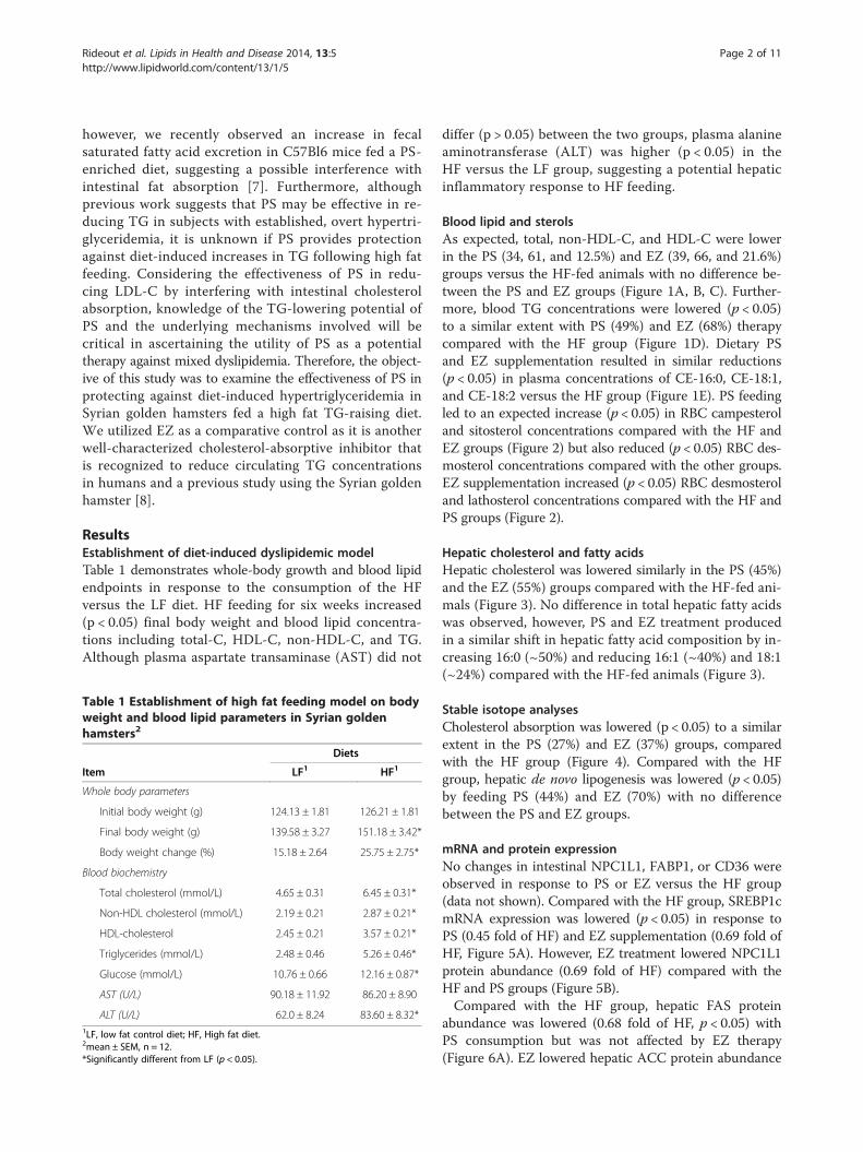
ResultsEstablishment of diet-induced dyslipidemic modelTable 1 demonstrates whole-body growth and blood lipidendpoints in response to the consumption of the HFversus the LF diet. HF feeding for six weeks increased(p < 0.05) final body weight and blood lipid concentra-tions including total-C, HDL-C, non-HDL-C, and TG.Although plasma aspartate transaminase (AST) did not
Table 1 Establishment of high fat feeding model on bodyweight and blood lipid parameters in Syrian goldenhamsters2
Diets
Item LF1 HF1
Whole body parameters
Initial body weight (g) 124.13 ± 1.81 126.21 ± 1.81
Final body weight (g) 139.58 ± 3.27 151.18 ± 3.42*
Body weight change (%) 15.18 ± 2.64 25.75 ± 2.75*
Blood biochemistry
Total cholesterol (mmol/L) 4.65 ± 0.31 6.45 ± 0.31*
Non-HDL cholesterol (mmol/L) 2.19 ± 0.21 2.87 ± 0.21*
HDL-cholesterol 2.45 ± 0.21 3.57 ± 0.21*
Triglycerides (mmol/L) 2.48 ± 0.46 5.26 ± 0.46*
Glucose (mmol/L) 10.76 ± 0.66 12.16 ± 0.87*
AST (U/L) 90.18 ± 11.92 86.20 ± 8.90
ALT (U/L) 62.0 ± 8.24 83.60 ± 8.32*1LF, low fat control diet; HF, High fat diet.2mean ± SEM, n = 12.*Significantly different from LF (p < 0.05).
differ (p > 0.05) between the two groups, plasma alanineaminotransferase (ALT) was higher (p < 0.05) in theHF versus the LF group, suggesting a potential hepaticinflammatory response to HF feeding.
Blood lipid and sterolsAs expected, total, non-HDL-C, and HDL-C were lowerin the PS (34, 61, and 12.5%) and EZ (39, 66, and 21.6%)groups versus the HF-fed animals with no difference be-tween the PS and EZ groups (Figure 1A, B, C). Further-more, blood TG concentrations were lowered (p < 0.05)to a similar extent with PS (49%) and EZ (68%) therapycompared with the HF group (Figure 1D). Dietary PSand EZ supplementation resulted in similar reductions(p < 0.05) in plasma concentrations of CE-16:0, CE-18:1,and CE-18:2 versus the HF group (Figure 1E). PS feedingled to an expected increase (p < 0.05) in RBC campesteroland sitosterol concentrations compared with the HF andEZ groups (Figure 2) but also reduced (p < 0.05) RBC des-mosterol concentrations compared with the other groups.EZ supplementation increased (p < 0.05) RBC desmosteroland lathosterol concentrations compared with the HF andPS groups (Figure 2).
Hepatic cholesterol and fatty acidsHepatic cholesterol was lowered similarly in the PS (45%)and the EZ (55%) groups compared with the HF-fed ani-mals (Figure 3). No difference in total hepatic fatty acidswas observed, however, PS and EZ treatment producedin a similar shift in hepatic fatty acid composition by in-creasing 16:0 (~50%) and reducing 16:1 (~40%) and 18:1(~24%) compared with the HF-fed animals (Figure 3).
Stable isotope analysesCholesterol absorption was lowered (p < 0.05) to a similarextent in the PS (27%) and EZ (37%) groups, comparedwith the HF group (Figure 4). Compared with the HFgroup, hepatic de novo lipogenesis was lowered (p < 0.05)by feeding PS (44%) and EZ (70%) with no differencebetween the PS and EZ groups.
mRNA and protein expressionNo changes in intestinal NPC1L1, FABP1, or CD36 wereobserved in response to PS or EZ versus the HF group(data not shown). Compared with the HF group, SREBP1cmRNA expression was lowered (p < 0.05) in response toPS (0.45 fold of HF) and EZ supplementation (0.69 fold ofHF, Figure 5A). However, EZ treatment lowered NPC1L1protein abundance (0.69 fold of HF) compared with theHF and PS groups (Figure 5B).Compared with the HF group, hepatic FAS protein
abundance was lowered (0.68 fold of HF, p < 0.05) withPS consumption but was not affected by EZ therapy(Figure 6A). EZ lowered hepatic ACC protein abundance
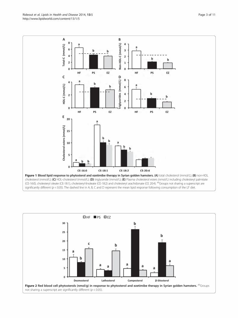
Figure 1 Blood lipid response to phytosterol and ezetimibe therapy in Syrian golden hamsters. (A) total cholesterol (mmol/L); (B) non-HDLcholesterol (mmol/L); (C) HDL-cholesterol (mmol/L); (D) triglyceride (mmol/L); (E) Plasma cholesterol esters (nmol/L) including cholesteryl palmitate(CE-16:0), cholesteryl oleate (CE-18:1), cholesteryl-linoleate (CE-18:2) and cholesteryl arachidonate (CE 20:4). abGroups not sharing a superscript aresignificantly different (p < 0.05). The dashed line in A, B, C and D represent the mean lipid response following consumption of the LF diet.
Figure 2 Red blood cell phytosterols (nmol/g) in response to phytosterol and ezetimibe therapy in Syrian golden hamsters. abGroupsnot sharing a superscript are significantly different (p < 0.05).
Rideout et al. Lipids in Health and Disease 2014, 13:5 Page 3 of 11http://www.lipidworld.com/content/13/1/5
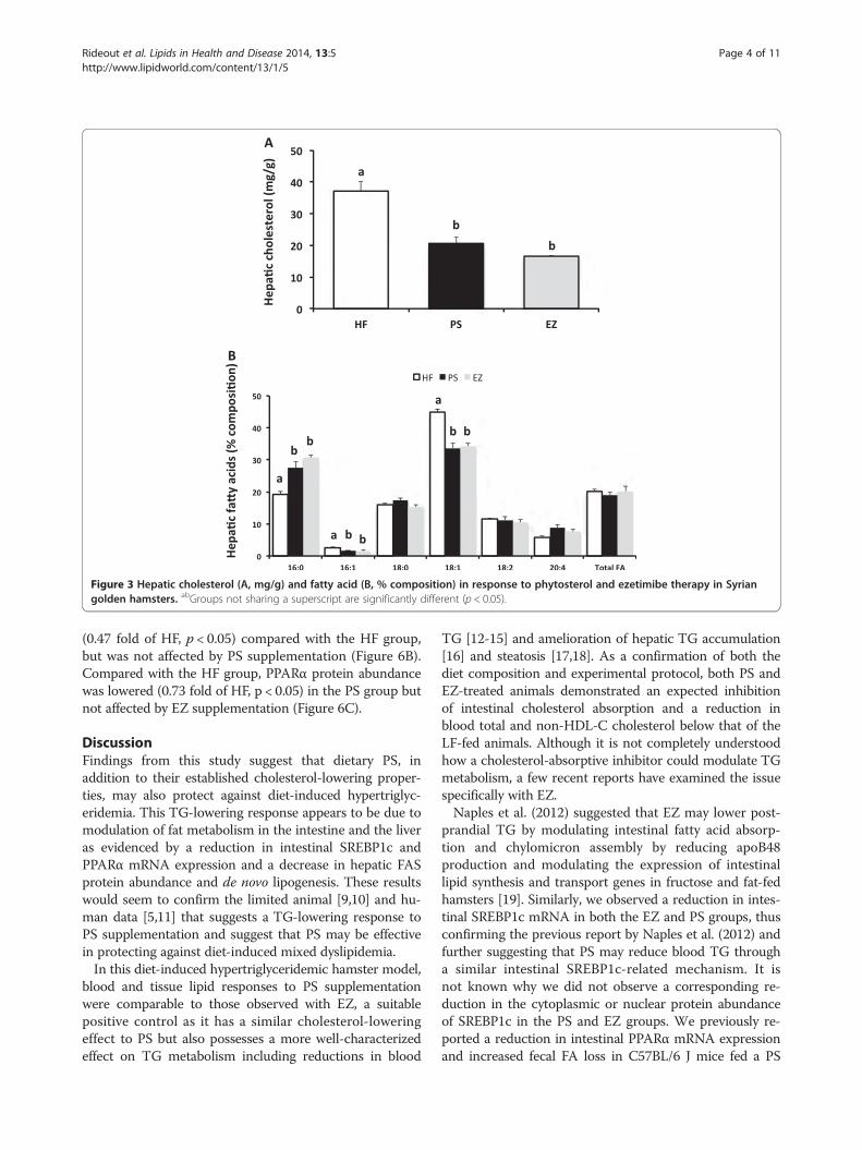
Figure 3 Hepatic cholesterol (A, mg/g) and fatty acid (B, % composition) in response to phytosterol and ezetimibe therapy in Syriangolden hamsters. abGroups not sharing a superscript are significantly different (p < 0.05).
Rideout et al. Lipids in Health and Disease 2014, 13:5 Page 4 of 11http://www.lipidworld.com/content/13/1/5
(0.47 fold of HF, p < 0.05) compared with the HF group,but was not affected by PS supplementation (Figure 6B).Compared with the HF group, PPARα protein abundancewas lowered (0.73 fold of HF, p < 0.05) in the PS group butnot affected by EZ supplementation (Figure 6C).
DiscussionFindings from this study suggest that dietary PS, inaddition to their established cholesterol-lowering proper-ties, may also protect against diet-induced hypertriglyc-eridemia. This TG-lowering response appears to be due tomodulation of fat metabolism in the intestine and the liveras evidenced by a reduction in intestinal SREBP1c andPPARα mRNA expression and a decrease in hepatic FASprotein abundance and de novo lipogenesis. These resultswould seem to confirm the limited animal [9,10] and hu-man data [5,11] that suggests a TG-lowering response toPS supplementation and suggest that PS may be effectivein protecting against diet-induced mixed dyslipidemia.In this diet-induced hypertriglyceridemic hamster model,
blood and tissue lipid responses to PS supplementationwere comparable to those observed with EZ, a suitablepositive control as it has a similar cholesterol-loweringeffect to PS but also possesses a more well-characterizedeffect on TG metabolism including reductions in blood
TG [12-15] and amelioration of hepatic TG accumulation[16] and steatosis [17,18]. As a confirmation of both thediet composition and experimental protocol, both PS andEZ-treated animals demonstrated an expected inhibitionof intestinal cholesterol absorption and a reduction inblood total and non-HDL-C cholesterol below that of theLF-fed animals. Although it is not completely understoodhow a cholesterol-absorptive inhibitor could modulate TGmetabolism, a few recent reports have examined the issuespecifically with EZ.Naples et al. (2012) suggested that EZ may lower post-
prandial TG by modulating intestinal fatty acid absorp-tion and chylomicron assembly by reducing apoB48production and modulating the expression of intestinallipid synthesis and transport genes in fructose and fat-fedhamsters [19]. Similarly, we observed a reduction in intes-tinal SREBP1c mRNA in both the EZ and PS groups, thusconfirming the previous report by Naples et al. (2012) andfurther suggesting that PS may reduce blood TG througha similar intestinal SREBP1c-related mechanism. It isnot known why we did not observe a corresponding re-duction in the cytoplasmic or nuclear protein abundanceof SREBP1c in the PS and EZ groups. We previously re-ported a reduction in intestinal PPARα mRNA expressionand increased fecal FA loss in C57BL/6 J mice fed a PS
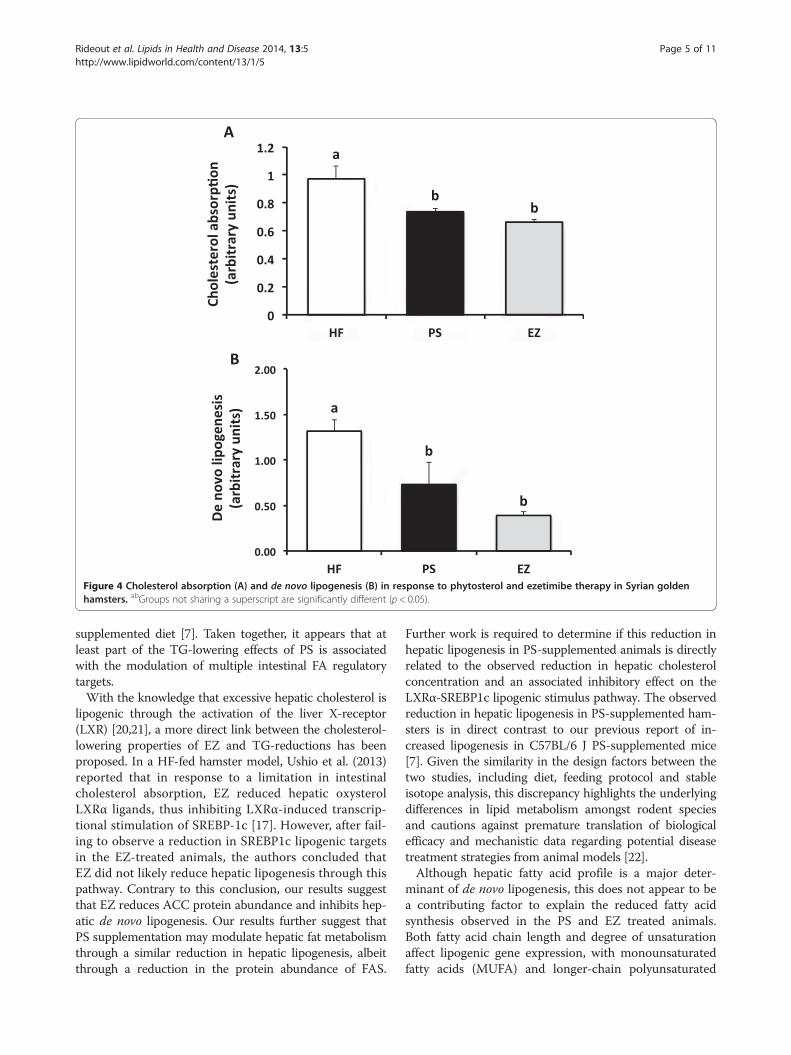
Figure 4 Cholesterol absorption (A) and de novo lipogenesis (B) in response to phytosterol and ezetimibe therapy in Syrian goldenhamsters. abGroups not sharing a superscript are significantly different (p < 0.05).
Rideout et al. Lipids in Health and Disease 2014, 13:5 Page 5 of 11http://www.lipidworld.com/content/13/1/5
supplemented diet [7]. Taken together, it appears that atleast part of the TG-lowering effects of PS is associatedwith the modulation of multiple intestinal FA regulatorytargets.With the knowledge that excessive hepatic cholesterol is
lipogenic through the activation of the liver X-receptor(LXR) [20,21], a more direct link between the cholesterol-lowering properties of EZ and TG-reductions has beenproposed. In a HF-fed hamster model, Ushio et al. (2013)reported that in response to a limitation in intestinalcholesterol absorption, EZ reduced hepatic oxysterolLXRα ligands, thus inhibiting LXRα-induced transcrip-tional stimulation of SREBP-1c [17]. However, after fail-ing to observe a reduction in SREBP1c lipogenic targetsin the EZ-treated animals, the authors concluded thatEZ did not likely reduce hepatic lipogenesis through thispathway. Contrary to this conclusion, our results suggestthat EZ reduces ACC protein abundance and inhibits hep-atic de novo lipogenesis. Our results further suggest thatPS supplementation may modulate hepatic fat metabolismthrough a similar reduction in hepatic lipogenesis, albeitthrough a reduction in the protein abundance of FAS.
Further work is required to determine if this reduction inhepatic lipogenesis in PS-supplemented animals is directlyrelated to the observed reduction in hepatic cholesterolconcentration and an associated inhibitory effect on theLXRα-SREBP1c lipogenic stimulus pathway. The observedreduction in hepatic lipogenesis in PS-supplemented ham-sters is in direct contrast to our previous report of in-creased lipogenesis in C57BL/6 J PS-supplemented mice[7]. Given the similarity in the design factors between thetwo studies, including diet, feeding protocol and stableisotope analysis, this discrepancy highlights the underlyingdifferences in lipid metabolism amongst rodent speciesand cautions against premature translation of biologicalefficacy and mechanistic data regarding potential diseasetreatment strategies from animal models [22].Although hepatic fatty acid profile is a major deter-
minant of de novo lipogenesis, this does not appear to bea contributing factor to explain the reduced fatty acidsynthesis observed in the PS and EZ treated animals.Both fatty acid chain length and degree of unsaturationaffect lipogenic gene expression, with monounsaturatedfatty acids (MUFA) and longer-chain polyunsaturated
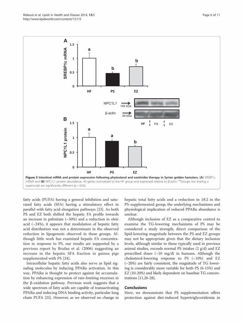
Figure 5 Intestinal mRNA and protein expression following phytosterol and ezetimibe therapy in Syrian golden hamsters. (A) SREBP1cmRNA and (B) NPC1L1 protein abundance. All genes normalized to the HF group and expressed relative to β-actin. abGroups not sharing asuperscript are significantly different (p < 0.05).
Rideout et al. Lipids in Health and Disease 2014, 13:5 Page 6 of 11http://www.lipidworld.com/content/13/1/5
fatty acids (PUFA) having a general inhibition and satu-rated fatty acids (SFA) having a stimulatory effect inparallel with fatty acid elongation pathways [23]. As bothPS and EZ both shifted the hepatic FA profile towardsan increase in palmitate (~50%) and a reduction in oleicacid (~24%), it appears that modulation of hepatic fattyacid distribution was not a determinant in the observedreduction in lipogenesis observed in these groups. Al-though little work has examined hepatic FA concentra-tion in response to PS, our results are supported by aprevious report by Brufau et al. (2006) suggesting anincrease in the hepatic SFA fraction in guinea pigssupplemented with PS [24].Intracellular hepatic fatty acids also serve as lipid sig-
naling molecules by inducing PPARα activation. In thisway, PPARα is thought to protect against fat accumula-tion by enhancing expression of rate-limiting enzymes inthe β-oxidation pathway. Previous work suggests that awide spectrum of fatty acids are capable of transactivatingPPARα and inducing DNA binding activity, particular longchain PUFA [25]. However, as we observed no change in
hepatic total fatty acids and a reduction in 18:2 in thePS-supplemented group, the underlying mechanisms andphysiological implication of reduced PPARα abundance isunclear.Although inclusion of EZ as a comparative control to
examine the TG-lowering mechanisms of PS may beconsidered a study strength, direct comparison of thelipid-lowering magnitude between the PS and EZ groupsmay not be appropriate given that the dietary inclusionlevels, although similar to those typically used in previousanimal studies, exceeds normal PS intakes (2 g/d) and EZprescribed doses (~10 mg/d) in humans. Although thecholesterol-lowering response to PS (~10%) and EZ(~20%) are fairly consistent, the magnitude of TG lower-ing is considerably more variable for both PS (6-15%) andEZ (10-20%) and likely dependent on baseline TG concen-trations [11,26-28].
ConclusionsHere, we demonstrate that PS supplementation offersprotection against diet-induced hypertriglyceridemia in
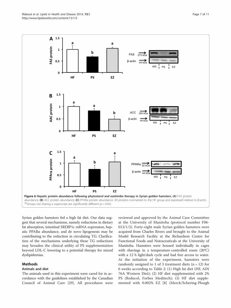
Figure 6 Hepatic protein abundance following phytosterol and ezetimibe therapy in Syrian golden hamsters. (A) FAS proteinabundance; (B) ACC protein abundance; (C) PPARα protein abundance. All proteins normalized to the HF group and expressed relative to β-actin.abGroups not sharing a superscript are significantly different (p < 0.05).
Rideout et al. Lipids in Health and Disease 2014, 13:5 Page 7 of 11http://www.lipidworld.com/content/13/1/5
Syrian golden hamsters fed a high fat diet. Our data sug-gest that several mechanisms, namely reductions in dietaryfat absorption, intestinal SREBP1c mRNA expression, hep-atic PPARα abundance, and de novo lipogenesis may becontributing to the reduction in circulating TG. Clarifica-tion of the mechanisms underlying these TG reductionsmay broaden the clinical utility of PS supplementationbeyond LDL-C lowering to a potential therapy for mixeddyslipidemias.
MethodsAnimals and dietThe animals used in this experiment were cared for in ac-cordance with the guidelines established by the CanadianCouncil of Animal Care [29]. All procedures were
reviewed and approved by the Animal Care Committeeat the University of Manitoba (protocol number F06-013/1/2). Forty-eight male Syrian golden hamsters wereacquired from Charles Rivers and brought to the AnimalModel Research Facility at the Richardson Centre forFunctional Foods and Nutraceuticals at the University ofManitoba. Hamsters were housed individually in cageswith shavings in a temperature-controlled room (20°C)with a 12 h light/dark cycle and had free access to water.At the initiation of the experiment, hamsters wererandomly assigned to 1 of 3 treatment diets (n = 12) for6 weeks according to Table 2: (1) High fat diet (HF, AIN76A Western Diet); (2) HF diet supplemented with 2%PS (Reducol, Forbes Meditech); (3) HF diet supple-mented with 0.002% EZ [8] (Merck/Schering-Plough
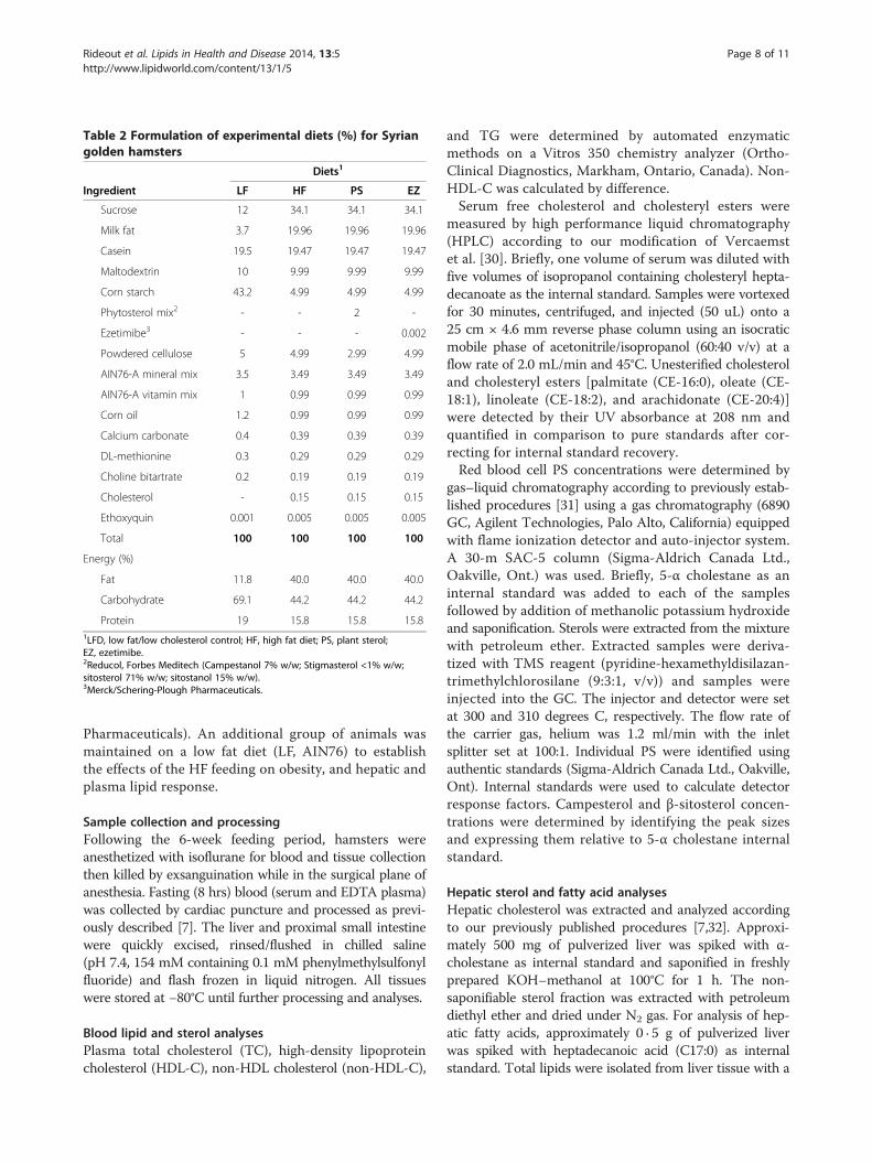
Table 2 Formulation of experimental diets (%) for Syriangolden hamsters
Diets1
Ingredient LF HF PS EZ
Sucrose 12 34.1 34.1 34.1
Milk fat 3.7 19.96 19.96 19.96
Casein 19.5 19.47 19.47 19.47
Maltodextrin 10 9.99 9.99 9.99
Corn starch 43.2 4.99 4.99 4.99
Phytosterol mix2 - - 2 -
Ezetimibe3 - - - 0.002
Powdered cellulose 5 4.99 2.99 4.99
AIN76-A mineral mix 3.5 3.49 3.49 3.49
AIN76-A vitamin mix 1 0.99 0.99 0.99
Corn oil 1.2 0.99 0.99 0.99
Calcium carbonate 0.4 0.39 0.39 0.39
DL-methionine 0.3 0.29 0.29 0.29
Choline bitartrate 0.2 0.19 0.19 0.19
Cholesterol - 0.15 0.15 0.15
Ethoxyquin 0.001 0.005 0.005 0.005
Total 100 100 100 100
Energy (%)
Fat 11.8 40.0 40.0 40.0
Carbohydrate 69.1 44.2 44.2 44.2
Protein 19 15.8 15.8 15.81LFD, low fat/low cholesterol control; HF, high fat diet; PS, plant sterol;EZ, ezetimibe.2Reducol, Forbes Meditech (Campestanol 7% w/w; Stigmasterol <1% w/w;sitosterol 71% w/w; sitostanol 15% w/w).3Merck/Schering-Plough Pharmaceuticals.
Rideout et al. Lipids in Health and Disease 2014, 13:5 Page 8 of 11http://www.lipidworld.com/content/13/1/5
Pharmaceuticals). An additional group of animals wasmaintained on a low fat diet (LF, AIN76) to establishthe effects of the HF feeding on obesity, and hepatic andplasma lipid response.
Sample collection and processingFollowing the 6-week feeding period, hamsters wereanesthetized with isoflurane for blood and tissue collectionthen killed by exsanguination while in the surgical plane ofanesthesia. Fasting (8 hrs) blood (serum and EDTA plasma)was collected by cardiac puncture and processed as previ-ously described [7]. The liver and proximal small intestinewere quickly excised, rinsed/flushed in chilled saline(pH 7.4, 154 mM containing 0.1 mM phenylmethylsulfonylfluoride) and flash frozen in liquid nitrogen. All tissueswere stored at −80°C until further processing and analyses.
Blood lipid and sterol analysesPlasma total cholesterol (TC), high-density lipoproteincholesterol (HDL-C), non-HDL cholesterol (non-HDL-C),
and TG were determined by automated enzymaticmethods on a Vitros 350 chemistry analyzer (Ortho-Clinical Diagnostics, Markham, Ontario, Canada). Non-HDL-C was calculated by difference.Serum free cholesterol and cholesteryl esters were
measured by high performance liquid chromatography(HPLC) according to our modification of Vercaemstet al. [30]. Briefly, one volume of serum was diluted withfive volumes of isopropanol containing cholesteryl hepta-decanoate as the internal standard. Samples were vortexedfor 30 minutes, centrifuged, and injected (50 uL) onto a25 cm × 4.6 mm reverse phase column using an isocraticmobile phase of acetonitrile/isopropanol (60:40 v/v) at aflow rate of 2.0 mL/min and 45°C. Unesterified cholesteroland cholesteryl esters [palmitate (CE-16:0), oleate (CE-18:1), linoleate (CE-18:2), and arachidonate (CE-20:4)]were detected by their UV absorbance at 208 nm andquantified in comparison to pure standards after cor-recting for internal standard recovery.Red blood cell PS concentrations were determined by
gas–liquid chromatography according to previously estab-lished procedures [31] using a gas chromatography (6890GC, Agilent Technologies, Palo Alto, California) equippedwith flame ionization detector and auto-injector system.A 30-m SAC-5 column (Sigma-Aldrich Canada Ltd.,Oakville, Ont.) was used. Briefly, 5-α cholestane as aninternal standard was added to each of the samplesfollowed by addition of methanolic potassium hydroxideand saponification. Sterols were extracted from the mixturewith petroleum ether. Extracted samples were deriva-tized with TMS reagent (pyridine-hexamethyldisilazan-trimethylchlorosilane (9:3:1, v/v)) and samples wereinjected into the GC. The injector and detector were setat 300 and 310 degrees C, respectively. The flow rate ofthe carrier gas, helium was 1.2 ml/min with the inletsplitter set at 100:1. Individual PS were identified usingauthentic standards (Sigma-Aldrich Canada Ltd., Oakville,Ont). Internal standards were used to calculate detectorresponse factors. Campesterol and β-sitosterol concen-trations were determined by identifying the peak sizesand expressing them relative to 5-α cholestane internalstandard.
Hepatic sterol and fatty acid analysesHepatic cholesterol was extracted and analyzed accordingto our previously published procedures [7,32]. Approxi-mately 500 mg of pulverized liver was spiked with α-cholestane as internal standard and saponified in freshlyprepared KOH–methanol at 100°C for 1 h. The non-saponifiable sterol fraction was extracted with petroleumdiethyl ether and dried under N2 gas. For analysis of hep-atic fatty acids, approximately 0 · 5 g of pulverized liverwas spiked with heptadecanoic acid (C17:0) as internalstandard. Total lipids were isolated from liver tissue with a
Rideout et al. Lipids in Health and Disease 2014, 13:5 Page 9 of 11http://www.lipidworld.com/content/13/1/5
modified Dole mixture (3 hepatane:12 propanol:3 DDH2O,vol:vol) followed by extraction with heptane: DDH2O (3:1vol:vol) [33]. Fatty acid extracts were methylated withmethanolic boron trifluoride (Sigma Aldrich, St. Louis,MO).Sterol and fatty acid fractions were analyzed using a
Shimadzu GC-17A gas chromatograph fitted with aflame ionization detector. A SAC-5 capillary column(30 m × 0 · 25 mm × 0 · 25 mm, Supelco, Bellefonte, CA,USA) was used for cholesterol analyses. Fatty acid methylesters were separated using a Supelcowax 10 column(30 m × 0 · 25 mm with 0 · 25 m film thickness; Supelco,Bellefonte, PA, USA). Relative hepatic fatty acid contentwas calculated by using individual FA peak area relative tothe total area and expressed as the percentage of total fattyacids.
Intestinal RNA preparation and real-time RT-PCRTotal RNA was isolated from whole intestinal tissueusing TRIzol reagent (Invitrogen Canada Inc., Burlington,ON). RNA concentration and integrity was determinedwith spectrophotometry (260 nm) and agarose gel elec-trophoresis, respectively. RNA preparation and real-timeRT-PCR was conducted using a one-step QuantiTectSYBR Green RT-PCR kit (Qiagen Inc., Mississauga, ON,Canada) on a Biorad MyiQ real time PCR system accord-ing to previously established protocols [34]. Sequences ofsense and antisense primers for target and housekeepinggenes were based on previously published reports forNPC1L1 [35], CD36, FABP2 [36], SREBP1c, [37], andβ-actin [38].
Immunoblot analysis of intestinal and hepatic regulatoryproteinsImmunoblots were prepared as previously described [34].Nuclear and cytoplasmic extracts for immunoblot ana-lyses of peroxisome proliferator-activated receptor alpha(PPARα, SC-9000, Santa Cruz Biotechnology), SREBP1c(Novus Biologicals, NB600-582), fatty acid synthase (FAS,C2OG5, Cell Signaling), and acetyl-CoA carboxylase(ACC, C83B10, Cell Signaling), were separated using theCelLytic™ NuCLEAR™ extraction kit (Sigma, Saint Louis,Missouri, USA). Intestinal apical membrane extractswere extracted according to a previously establishedprotocol probed for NPC1L1 (Santa Cruz, sc-67237)[39]. Target proteins were normalized to β-actin andquantified using Image J (National Institutes of Health,Bethesda, Maryland).
Stable isotope analysesHamsters were given an intraperitoneal injection of deu-terium (100 μl) 2-hours prior to euthanization. Lipogenesisrates (%/day) were quantified using the uptake rate ofdeuterium from body water into newly synthesized
hepatic-palmitate extracts over 2 h at the end of thefeeding experiment [40,41]. Deuterium enrichment ofhepatic-palmitate was quantified using an on-line gaschromatography/combustion/isotope ratio mass spec-trometry approach (Agilent 6890 N chromatographinterfaced with a Thermo Delta V Plus isotope ratiomass spectrometer (Bremen, Germany). Isotope abun-dance, expressed in delta (δ) per mil (‰), was calculatedin hepatic-palmitate and plasma water (precursor pool)using H2 as a reference gas and further corrected againstthe international reference, Standard Mean Ocean Water(SMOW). De novo lipogenesis rates were calculated withthe following equation:
De novo lipogenesis %=dayð Þ¼ ΔTGFA ‰ð Þ=Δplasma ‰ð Þ � 0:477 � 100
Where ΔTGFA is the change in deuterium enrichmentin hepatic-palmitate; Δplasma is the change in the deu-terium enrichment of the precursor plasma water; and0.477 is derived from 0.87 g-atom 3H per g-atom carbonincorporated into adipose tissue fatty acids and a correc-tion factor to account for the glycerol moiety as previouslydescribed [42]. Lipogenesis rates are expressed relative tothe HF group.Forty-eight hours prior to euthanization, hamsters were
given an oral gavage of safflower oil containing 5 mg of[3,4]-13C cholesterol (99% enriched; CDN Isotopes). Asan indicator of cholesterol absorption, GC-combustion-isotope ratio MS (Delta V Plus, Thermo Scientific) wasused to determine the 13C enrichment (13C/12C ratio) offree cholesterol in RBC compared with the non-enrichedhamster RBC 13C-cholesterol enrichment over 48 h [43].
Statistical analysesData were analyzed with a general linear model ANOVAusing experimental block as a fixed factor [44]. To estab-lish the effect of HF-feeding, responses between the LFversus the HF groups were compared using a paired t-test.Multiple comparisons between treatment groups wereanalyzed with Tukey’s post-hoc test. Data were analyzedwith SPSS 16 for Mac (SPSS Inc, Chicago IL). Data arepresented as mean ± SEM. All results are the meansfrom 12 animals unless otherwise stated. Differenceswere considered significant at p ≤ 0.05.
Competing interestsThe authors declare that they have no competing interests.
Authors’ contributionsTCR designed and conducted the research, analyzed the data, and wrote theinitial draft manuscript; VR conducted the research; JG conducted theimmunoblot and RNA analyses; RWB conducted the lipid analyses; SVHconducted the stable isotope analysis; PJ designed the research. All authorsread and approved the final manuscript.
Rideout et al. Lipids in Health and Disease 2014, 13:5 Page 10 of 11http://www.lipidworld.com/content/13/1/5
AcknowledgementsThe technical assistance of Amy Raslawsky and Marie-Lou Bodziac is greatlyappreciated.
Financial supportFunded by the Natural Sciences and Engineering Research Council ofCanada (to PJ) and a KO1 grant from the National Institute forComplementary and Alternative Medicine (to TCR).
Author details1Department of Exercise and Nutrition Sciences, University at Buffalo, Buffalo,NY 14214, USA. 2Richardson Centre for Functional Foods and Nutraceuticals,University of Manitoba, Winnipeg, Manitoba R3T 2 N2, Canada. 3Biotechnicaland Clinical Laboratory Sciences, University at Buffalo, Buffalo, NY 14214, USA.4Diabetes and Nutritional Sciences Division, School of Medicine, King’sCollege London, London SE1 9NH, UK.
Received: 19 November 2013 Accepted: 13 December 2013Published: 6 January 2014
References1. Miller M, Stone NJ, Ballantyne C, Bittner V, Criqui MH, Ginsberg HN,
Goldberg AC, Howard WJ, Jacobson MS, Kris-Etherton PM, et al: Triglyceridesand cardiovascular disease: a scientific statement from the American HeartAssociation. Circulation 2011, 123(20):2292–2333.
2. Maki KC, Bays HE, Dicklin MR: Treatment options for the management ofHypertriglyceridemia: strategies based on the best-available evidence.J Clin Lipidol 2012, 6(5):413–426.
3. Ford ES, Li C, Zhao G, Pearson WS, Mokdad AH: Hypertriglyceridemia andits pharmacologic treatment among US adults. Arch Intern Med 2009,169(6):572–578.
4. Wierzbicki AS, Clarke RE, Viljoen A, Mikhailidis DP: Triglycerides: a case fortreatment? Curr Opin Cardiol 2012, 27(4):398–404.
5. Plat J, Brufau G, Dallinga-Thie GM, Dasselaar M, Mensink RP: A plant stanolyogurt drink alone or combined with a low-dose statin lowers serumtriacylglycerol and non-HDL cholesterol in metabolic syndrome patients.J Nutr 2009, 139(6):1143–1149.
6. Theuwissen E, Plat J, van der Kallen CJ, van Greevenbroek MM, Mensink RP:Plant stanol supplementation decreases serum triacylglycerols insubjects with overt hypertriglyceridemia. Lipids 2009, 44(12):1131–1140.
7. Rideout TC, Harding SV, Jones PJ: Consumption of plant sterols reducesplasma and hepatic triglycerides and modulates the expression of lipidregulatory genes and de novo lipogenesis in C57BL/6 J mice. Mol NutrFood Res 2010, 54(Suppl 1):S7–S13.
8. van Heek M, Austin TM, Farley C, Cook JA, Tetzloff GG, Davis HR: Ezetimibe,a potent cholesterol absorption inhibitor, normalizes combineddyslipidemia in obese hyperinsulinemic hamsters. Diabetes 2001,50(6):1330–1335.
9. Awaisheh SS, Khalifeh MS, Al-Ruwaili MA, Khalil OM, Al-Ameri OH, Al-GroomR: Effect of supplementation of probiotics and phytosterols alone or incombination on serum and hepatic lipid profiles and thyroid hormonesof Hypercholesterolemic rats. J Dairy Sci 2013, 96(1):9–15.
10. Ntanios FY, van de Kooij AJ, de Deckere EA, Duchateau GS, Trautwein EA:Effects of various amounts of dietary plant sterol esters on plasma andhepatic sterol concentration and aortic foam cell formation ofcholesterol-fed hamsters. Atherosclerosis 2003, 169(1):41–50.
11. Demonty I, Ras RT, van der Knaap HC, Meijer L, Zock PL, Geleijnse JM,Trautwein EA: The effect of plant sterols on serum triglycerideconcentrations is dependent on baseline concentrations: a pooledanalysis of 12 randomised controlled trials. Eur J Nutr 2013, 52(1):153–160.
12. Nakou ES, Filippatos TD, Agouridis AP, Kostara C, Bairaktari ET, Elisaf MS: Theeffects of Ezetimibe and/or orlistat on triglyceride-rich lipoproteinmetabolism in obese Hypercholesterolemic patients. Lipids 2010,45(5):445–450.
13. Dujovne CA, Ettinger MP, McNeer JF, Lipka LJ, LeBeaut AP, Suresh R, Yang B,Veltri EP: Efficacy and safety of a potent new selective cholesterolabsorption inhibitor, Ezetimibe, in patients with primaryhypercholesterolemia. Am J Cardiol 2002, 90(10):1092–1097.
14. Goldberg AC, Sapre A, Liu J, Capece R, Mitchel YB: Efficacy and safety ofEzetimibe co administered with simvastatin in patients with primary
hypercholesterolemia: a randomized, double-blind, placebo-controlledtrial. Mayo Clin Proc 2004, 79(5):620–629.
15. Park H, Shima T, Yamaguchi K, Mitsuyoshi H, Minami M, Yasui K, Itoh Y,Yoshikawa T, Fukui M, Hasegawa G, et al: Efficacy of long-term Ezetimibetherapy in patients with non-alcoholic fatty liver disease. J Gastroenterol2011, 46(1):101–107.
16. Chan DC, Watts GF, Gan SK, Ooi EM, Barrett PH: Effect of Ezetimibe onhepatic fat, inflammatory markers, and apolipoprotein B-100 kinetics ininsulin-resistant obese subjects on a weight loss diet. Diabetes Care 2010,33(5):1134–1139.
17. Ushio M, Nishio Y, Sekine O, Nagai Y, Maeno Y, Ugi S, Yoshizaki T, Morino K,Kume S, Kashiwagi A, et al: Ezetimibe prevents hepatic steatosis inducedby a high-fat but not a high-fructose diet. Am J Physiol Endocrinol Metab2013, 305(2):E293–E304.
18. Zheng S, Hoos L, Cook J, Tetzloff G, Davis H Jr, van Heek M, Hwa JJ:Ezetimibe improves high fat and cholesterol diet-induced non-alcoholicfatty liver disease in mice. Eur J Pharmacol 2008, 584(1):118–124.
19. Naples M, Baker C, Lino M, Iqbal J, Hussain MM, Adeli K: Ezetimibeameliorates intestinal chylomicron overproduction and improvesglucose tolerance in a diet-induced hamster model of insulin resistance.Am J Physiol Gastrointest Liver Physiol 2012, 302(9):G1043–G1052.
20. Jia L, Ma Y, Rong S, Betters JL, Xie P, Chung S, Wang N, Tang W, Yu L:Niemann-Pick C1-Like 1 deletion in mice prevents high-fat diet-inducedfatty liver by reducing lipogenesis. J Lipid Res 2010, 51(11):3135–3144.
21. Repa JJ, Liang G, Ou J, Bashmakov Y, Lobaccaro JM, Shimomura I, Shan B,Brown MS, Goldstein JL, Mangelsdorf DJ: Regulation of mouse sterolregulatory element-binding protein-1c gene (SREBP-1c) by oxysterolreceptors, LXRalpha and LXRbeta. Genes Dev 2000, 14(22):2819–2830.
22. van der Worp HB, Howells DW, Sena ES, Porritt MJ, Rewell S, O’Collins V,Macleod MR: Can animal models of disease reliably inform humanstudies? PLoS Med 2010, 7(3):e1000245.
23. Collins JM, Neville MJ, Hoppa MB, Frayn KN: De novo lipogenesis andstearoyl-CoA desaturase are coordinately regulated in the humanadipocyte and protect against palmitate-induced cell injury. J Biol Chem2010, 285(9):6044–6052.
24. Brufau G, Canela MA, Rafecas M: A high-saturated fat diet enriched withphytosterol and pectin affects the fatty acid profile in guinea pigs. Lipids2006, 41(2):159–168.
25. Mochizuki K, Suruga K, Fukami H, Kiso Y, Takase S, Goda T: Selectivity offatty acid ligands for PPARalpha which correlates both with binding tocis-element and DNA binding-independent transactivity in Caco-2 cells.Life Sci 2006, 80(2):140–145.
26. Bruckert E, Giral P, Tellier P: Perspectives in cholesterol-lowering therapy:the role of Ezetimibe, a new selective inhibitor of intestinal cholesterolabsorption. Circulation 2003, 107(25):3124–3128.
27. Pandor A, Ara RM, Tumur I, Wilkinson AJ, Paisley S, Duenas A, DurringtonPN, Chilcott J: Ezetimibe monotherapy for cholesterol lowering in 2,722people: systematic review and meta-analysis of randomized controlledtrials. J Intern Med 2009, 265(5):568–580.
28. Naumann E, Plat J, Kester AD, Mensink RP: The baseline serum lipoproteinprofile is related to plant stanol induced changes in serum lipoproteincholesterol and triacylglycerol concentrations. J Am Coll Nutr 2008,27(1):117–126.
29. Olfert ED, BMC, McWilliam AA: Canadian Council on Animal Care. Guide tothe care and use of experimental animals 1. 2nd edition. Ontario, Canada:Ottawa; 1993.
30. Vercaemst R, Union A, Rosseneu M, De Craene I, De Backer G, Kornitzer M:Quantitation of plasma free cholesterol and cholesteryl esters by highperformance liquid chromatography. Study of a normal population.Atherosclerosis 1989, 78(2–3):245–250.
31. Harding SV, Zhao HL, Marinangeli CP, Day AG, Dillon HF, Jain D, Jones PJ:Red algal cellular biomass lowers circulating cholesterol concentrationsin Syrian golden hamsters consuming Hypercholesterolaemic diets. Br JNutr 2009, 102(6):842–847.
32. Harding SV, Rideout TC, Jones PJ: Hepatic nuclear sterol regulatorybinding element protein 2 abundance is decreased and that of ABCG5increased in male hamsters fed plant sterols. J Nutr 2010,140(7):1249–1254.
33. van der Vusse GJ, Roemen TH, Reneman RS: The content of non-esterifiedfatty acids in rat myocardial tissue. A comparison between the Dole andFolch extraction procedures. J Mol Cell Cardiol 1985, 17(5):527–531.
Rideout et al. Lipids in Health and Disease 2014, 13:5 Page 11 of 11http://www.lipidworld.com/content/13/1/5
34. Rideout TC, Yuan Z, Bakovic M, Liu Q, Li RK, Mine Y, Fan MZ: Guar gumconsumption increases hepatic nuclear SREBP2 and LDL receptorexpression in pigs fed an atherogenic diet. J Nutr 2007, 137(3):568–572.
35. Valasek MA, Repa JJ, Quan G, Dietschy JM, Turley SD: Inhibiting intestinalNPC1L1 activity prevents diet-induced increase in biliary cholesterol inGolden Syrian hamsters. Am J Physiol Gastrointest Liver Physiol 2008,295(4):G813–G822.
36. Laugerette F, Passilly-Degrace P, Patris B, Niot I, Febbraio M, Montmayeur JP,Besnard P: CD36 involvement in orosensory detection of dietary lipids,spontaneous fat preference, and digestive secretions. J Clin Invest 2005,115(11):3177–3184.
37. Morgan K, Uyuni A, Nandgiri G, Mao L, Castaneda L, Kathirvel E, French SW,Morgan TR: Altered expression of transcription factors and genesregulating lipogenesis in liver and adipose tissue of mice with high fatdiet-induced obesity and non-alcoholic fatty liver disease. Eur JGastroenterol Hepatol 2008, 20(9):843–854.
38. Feng D, Wang Y, Mei Y, Xu Y, Xu H, Lu Y, Luo Q, Zhou S, Kong X, Xu L:Stearoyl-CoA desaturase 1 deficiency protects mice from immune-mediated liver injury. Lab Invest 2009, 89(2):222–230.
39. Cheeseman CI, O’Neill D: Isolation of intestinal brush-border membranes.Curr Protoc Cell Biol 2006, Chapter 3:Unit 3–Unit 21.
40. Jones PJ, Leitch CA, Li ZC, Connor WE: Human cholesterol synthesismeasurement using deuterated water. Theoretical and proceduralconsiderations. Arterioscler Thromb 1993, 13(2):247–253.
41. Jones PJ: Tracing lipogenesis in humans using deuterated water. Can JPhysiol Pharmacol 1996, 74(6):755–760.
42. Jungas RL: Fatty acid synthesis in adipose tissue incubated in tritiatedwater. Biochemistry 1968, 7(10):3708–3717.
43. Zhao HL, Harding SV, Marinangeli CP, Kim YS, Jones PJ:Hypocholesterolemic and anti-obesity effects of saponins fromPlatycodon grandiflorum in hamsters fed atherogenic diets. J Food Sci2008, 73(8):H195–H200.
44. Kuehl RO: Design of Experiments: Statistical Principles of Research DesignAnalysis. 2nd edition. Baltimore, MD: Brooks/Cole Publishing Company; 2000.
doi:10.1186/1476-511X-13-5Cite this article as: Rideout et al.: Phytosterols protect against diet-induced hypertriglyceridemia in Syrian golden hamsters. Lipids in Healthand Disease 2014 13:5.
Submit your next manuscript to BioMed Centraland take full advantage of:
• Convenient online submission
• Thorough peer review
• No space constraints or color figure charges
• Immediate publication on acceptance
• Inclusion in PubMed, CAS, Scopus and Google Scholar
• Research which is freely available for redistribution
Submit your manuscript at www.biomedcentral.com/submit


