Embed Size (px)
Citation preview
Platelet Adhesion to Vascular CellsThe Role of Exogenous von Willebrand Factor in Platelet Adhesion
Patricia F.E.M. Nievelstein, Philip G. de Groot, Patrizia D'Alessio,Harry F.G. Heijnen, Elisabetta Orlando, and Jan J. Sixma
Platelet deposition on cultured fibroblasts and on their extracellular matrix (FBM)was Investigated in a flow system with citrated blood and was compared with plateletdeposition on cultured endothelial cells, smooth muscle cells, and their extracellularmatrices. Platelet deposition was present at all surfaces except on intact endothelialcells. Deposition on FBM consisted of contact platelets, spread platelets, and a fewsmall aggregates. On Intact fibroblasts cells, the surface coverage was lower, andplatelets formed aggregates. Factors Involved In primary hemostasls, particularly thewall shear rate, von Willebrand factor (vWF), and fibronectin, were Investigated onFBM. The reactivity of FBM was determined by the passage number of the culturedcells. The vWF was involved In platelet adhesion on FBM at only the high shear rate(>800 8~1). Platelet deposition was independent of plasma fibronectin at all shearrates tested. Matrix-associated fibronectin was Involved in adhesion at low and highwall shear rates. We conclude that FBM can be used as a platelet adhesive surfaceespecially to study the contribution of exogenous vWF to platelet adhesion becauseFBM does not contain vWF. (Arteriosclerosis 10:462-469, May/June 1990)
B lood vessels are lined with a monolayer of endothe-lial cells, which prevent platelet deposition and clot
formation. The endothelial cells produce extracellularmatrix, which makes up the subendothelium. Loss ofendothelial cells or contraction of these cells upon injuryresults in a rapid adhesion of platelets to the exposedsubendothelium; subsequently these platelets undergodegranulation.1 Injuries penetrating the subendotheliumand going through the media and/or adventitia lead tohemostatjc plugs.2 So in contrast to superficial injuries,which result in a thrombotic response, deep injuries showa hemostatic response.
In recent years, extensive use has been made of theextracellular matrix of vascular endothelial cells (ECM) as amodel for the vessel wall in perfusion studies with a rectan-gular perfusion chamber.3-6 Platelet adhesion and throm-bus formation on the connective tissue matrix, which rep-resents the deeper layers of the vascular wall, have beenless well documented. In this study, we focus on thehemostatic response of fibroblasts and smooth muscle cell(SMC) cultures and their extracellular matrices in compari-son to endothelial cell cultures and their matrices using theflow model with citrated blood. Important parameters forplatelet adhesion, such as von Willebrand factor (vWF) andfibronectin, were studied on fibroblast matrix (FBM). FBMwas preferred because of its uniform thickness as com-pared with smooth muscle cell matrix (SMCM), which variesin thickness. A special point of interest was investigating theusability of the FBM as a surface for the study of the role of
From the Department of Haematology, University HospitalUtrecht, Utrecht The Netherlands.
This study was supported by the Dutch Diabetes FoundationGrant 86-27.
Address for correspondence: Jan J. Sixma, Department ofHaematology, University Hospital Utrecht, P.O. Box 85500, 3508GA Utrecht, The Netherlands.
Received April 19, 1989; revision accepted January 22, 1990.
exogenous vWF in platelet adhesion. Many studies on vWFdependency have been performed with isolated vessel wallcomponents.7'8 To study the vWF dependency in a morenatural condition, it was necessary to block the vWF presentin the matrix itself.4'6 Since the FBM lacks endogenousvWF, it is well suited for a study on the vWF dependency onnative vessel wall components.
MethodsCell Culture and Isolation of Extracellular Matrix
Human vascular endothelial cells derived from umbilicalveins were isolated and cultured as described.4 Fibroblastsderived from human fetal lung tissue or human skin werecultured in RPMI-1640 supplemented with 10% fetal calfserum and antibiotics. Human SMC were derived fromhuman aorta and cultured as described by Loesberg et al.9
Experiments with endothelial cells and SMC were carriedout with cells of the third passage. Fibroblasts were usedfrom passages 3 to 23. To isolate the extracellular matrix,cells grown to confluence were exposed to 0.1 M NH<OHfor 30 minutes at room temperature with gentle shaking.The cell layer was completely removed by this procedure.6
The extracellular matrix was washed three times withphosphate-buffered saline (PBS) before use. In someexperiments, the FBM was incubated at 37°C in 100 /il ofKrebs-Ringer buffer, pH 7.35 (with 2.5 mM CaCl2 and19 mM citrate) containing vWF (0.5, 1, 2, 3, or 5 UvWF^RiCof) for 2 hours at room temperature. Controlswere incubated with buffer only. In another set of experi-ments, FBM was incubated with antifibronectin F(ab')2fragments or nonimmune rabbit F(ab'>2 fragments. Incu-bations were performed with 0.78mg/ml antibody in PBSovernight at room temperature.
Immunofluorescence StudiesImmunofluorescence studies were performed as
described by Houdijk et al.4 The rabbit polyclonal anti-
462

PLATELET ADHESION TO VASCULAR CELLS Nievelstein et al. 463
bodies against collagen type I and type III were a gener-ous gift of Jurgen Rautenberg (Munster, FRG). Theantibodies against collagen type IV and V, laminin, andnidogen were a generous gift of Rupert Timpl (MaxPlanck Institute, Martinsried, FRG).
Purification of von Willebrand Factor andCharacteristics of Antl-von Willebrand Factor
The vWF was purified from human cryoprecipitate asdescribed before.10 The monoclonal antibody CLB-RAg 35 is directed against the platelet binding domainof human vWF (generous gift from Jan A. van Mourik,CLB, Amsterdam) and inhibits the interaction of vWFwith glycoprotein Ib.11
Purification of Flbronectln and Characteristicsof Antl-flbronectln
Fibronectin was isolated from human plasma by affinitychromatography on gelatin-Sepharose as previouslydescribed.12
F(ab')2 fragments of rabbit antiserum against humanplasma fibronectin were purchased lyophilized (CappelLaboratories, Cochranville, PA) and were reconstituted in0.02 M PBS (pH 7.3). The protein concentration was7.8 mg/ml.
Von Willebrand Disease
Characterization of the diverse types of von Willebranddisease (vWD) was performed according to the criteriarecently described by Ruggeri.13
Perfuslon Chamber
Perfusions were earned out in a rectangular perfusionchamber containing two knobs,12 a modification of thechamber of Sakariassen et al.3
Preparation of Periusates
Blood from normal healthy donors or patients with vWDwas antJcoagulated with one-tenth vol 110 mM trisodiumcitrate. Part of the perfusions were performed with wholeblood and part with reconstituted blood. Reconstitutedblood was prepared as follows. Platelet-rich plasma (PRP)was obtained from whole blood by centrifugatjon (10 min-utes at 200 g, 20°C). Aspirin (10 /M) was added to the PRPto prevent aggregate formation on surfaces during perfu-sion without influencing adhesion to subendothelium14 or tocollagen.15 To one volume of PRP, one volume of Krebs-Ringer buffer (4 mM KCI, 107 mM NaCI, 20 mM NaHCO3,and 2 mM Na^OJ containing 19 mM citrate (pH 5.0) wasadded, with a final pH of about 6. A platelet pellet wasobtained by centrifugation (10 minutes at 500 g, 20°C).Platelets were resuspended in Krebs-Ringer buffer con-taining 19 mM citrate and 5 mM glucose (pH 6.0). Foreach wash step, 100 ml of resuspension buffer wasused, and the platelets were washed twice by centrifu-gation (10 minutes at 500 g, 20°C). After the secondwash, platelets were resuspended to a platelet count of190 000//il in plasma, human albumin solution (HAS)composed of 4% (wt/vol) human albumin in Krebs-Ringerbuffer containing 19 mM citrate, 2.5 mM CaC^, and 5 mMglucose (pH 7.35), or HAS with the addition of purified
fibronectin in plasma concentration (300 ^9/™!)- Platelet-poor plasma (PPP) was obtained from PRP by centrifuga-tion (10 minutes at 2000 g, 20°C). The plasma pH wasadjusted with 1 mM HCI to 7.35. Plasma contamination ofred cells was avoided by washing them three times in salinecontaining 5 mM glucose (2000 g, 20°C, twice for 5 min-utes, the last time for 15 minutes). The washed red cellswere added to a hematocrit of 0.4 at 15 minutes beforeperfusion. Perfusates were pre-warmed just before perfu-sion at 37°C for 10 minutes. Perfusions were performed atwall shear rates ranging from 100 s~1 to 1300 s"1, for 1,3,5,or 10 minutes. Before and immediately after perfusion, thesystem was rinsed with 20 ml of 10 mM HEPES, 0.154 mMNaCI buffer (pH 7.35).
Evaluation
Platelet deposition was determined morphologically witha light microscope connected to an image analyzer (Quan-timet 720, Imago, Royston, U.K.) as described before.4 Forthis purpose, coverslips covered with platelets were fixedin 0.5% glutaraldehyde in PBS immediately after perfusionat room temperature for at least 30 minutes and subse-quently in a refrigerator (6° to 8°C) overnight. They werewashed with PBS once and stained with May-Grunwald(Merck, Darmstadt, FRG; 1:1 in distilled water) andGiemsa (Merck, 1 -A in distilled water) for 5 and 15 min-utes, respectively. The coverslips were washed with dis-tilled water at least three times to avoid staining contami-nations. They were air-dried and enclosed.
Electron Microscopy
For electron microscopic evaluation, fibroblasts werecultured on Thermanox coverslips (Miles, Naperville, IL).After perfusion, fibroblasts or FBM were fixed with1% paraformaldehyde and 1% glutaraldehyde in 0.1 Mphosphate buffer (pH 7) for 30 minutes at room temperaturefollowed by fixation in the refrigerator overnight. They wereembedded in epon as described for vessel segments16 withsome modifications. After osmium tetroxide fixation (1 % indistilled water) and dehydration as described,16 vesselsegments were totally embedded in epon, whereas in thecase of the coverslips, only the perfusate surface wascovered with epon. This allowed separation between thecoverslips and the embedded cells or matrix after polymer-ization of the epon. Separation was attained by a differencein shrinkage between the two materials caused by heatingthe embedded coverslip on a hot plate (70°C) followed by arapid transfer to liquid nitrogen. Epon-embedded cells andmatrices were treated with a mixture of 1 % osmium tetrox-ide and 1% ruthenium red (ICN Biochemicals, Plainview,NY) in distilled water for 30 minutes at room temperaturebefore re-embedding. Ultrathin sections were stained withuranyl acetate and Reynolds lead citrate and were viewed ina JEOL 1200-EX electron microscope.
Statistical Analysis
The significance of differences between the meanswas calculated with Student's f test.

464 ARTERIOSCLEROSIS VOL 10, No 3, MAY/JUNE 1990
- \ , ' *> * , */^^ttt
• • • •
B
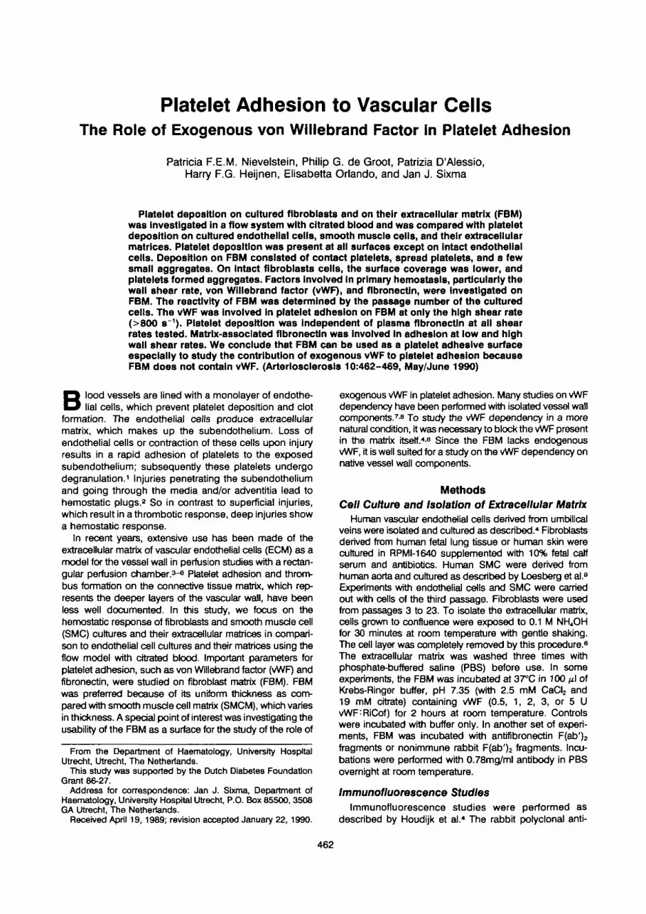
Figure 1. Morphology (en face) of platelet deposition on coverslips with cuttured cells or their extracellular matrices. Perfusions wereperformed with whole blood at a wall shear rate of 1300 s~1 for 5 minutes. After perfusion, the coverslips were fixed and stained aspreviously described.4 A. Endothelial cell matrix. B. Smooth muscle cell matrix. C. Fibroblast matrix. D. Endothelial cells. E. Smoothmuscle cells. Arrow indicates platelet aggregate. F. Fibroblasts. Single arrow indicates spread platelets, double arrow, plateletaggregates, x 1000

PLATELET ADHESION TO VASCULAR CELLS Nievelstein et al. 465
Results
Morphology of Platelet Deposition onEndothellal Cells, Smooth Muscle Cells,Fibroblasts, and Their Extracellular Matrices
The extracellular matrices of confluent vessel wall cellswere exposed to flowing blood at a wall shear rate of1300 s~1 for 5 minutes. On ECM, platelets were spread onthe surface with little or no aggregate formation (Figure1A). Adhesion to SMCM (Figure 1B) and FBM (Figure 1C)was lower than to ECM. On SMCM, however, plateletswere less spread out than on ECM. On FBM, contactplatelets or platelets which begin to spread, spreadplatelets, and small aggregates were found, the deposi-tion appearing more patchy than on ECM and SMCM.The absolute values of deposition on FBM were depen-dent upon the number of passages of the cells in culture,as will be described later.
Confluent cell cultures were exposed to flowing bloodat a wall shear rate of 1300 s~1 for 5 minutes. Plateletdeposition was observed on SMC (Figure 1E) and onfibroblasts (Figure 1F), but not on endothelial cells (Fig-ure 1D).
Electron microscopic evaluation of cross-sectionedfibroblasts showed an abluminal matrix only. Therefore,platelet deposition was due to direct deposition on thefibroblasts (Figures 2A and 2B). Ultrathin cross-sectionsof FBM showed deposition of small aggregates andspread platelets (Figure 2C).
Characterization of Flbroblast Matrix
The presence and distribution of different matrix pro-teins in the isolated extracellular matrix of confluentcultures of fibroblasts was studied by indirect immuno-fluorescence. With the use of specific antibodies, a strongfibrillar fluorescence pattern was seen for collagens typeIII, type IV, and type VI and fibronectin. The matrices alsogave a positive fluorescence for collagen type I. Thematrices were negative for vWF, laminin, nidogen, andcollagen type V (data not shown).
Quantification of Platelet Deposition onFibroblasts Compared to Their Matrix
Platelet deposition on fibroblasts and FBM was deter-mined morphologically after perfusions at wall shear ratesof 300 s"1 and 1300 s~1 for 1, 3, 5, and 10 minutes. Theplatelet surface coverage was less on fibroblasts than onFBM under all conditions (Table 1).
Effect of Passage Number on Flbroblast Matrix
The shear-rate dependence of FBM was determinedby the passage of the cultured cells. Early passages (3 to10) produced a very reactive matrix. Optimal adhesionwas already obtained at 300 s~1 and decreased slightly athigher shear rates. FBM derived from cells with higherpassage numbers (10 to 20) gave lower absolute adhe-sions. The shear-rate dependence was more or lesshyperbolic and leveled off at 1300 s~\ Higher passagesproduced a hardly reactive matrix.
Von Willebrand Factor Dependence for PlateletDeposition on Flbroblast Matrix
Three approaches were used to study the role ofexogenous vWF for platelet deposition on FBM.
To study the shear-rate dependence of platelet depo-sition on vWF, monoclonal antibody CLB:RAg:35, whichis directed against the platelet binding site of vWF,11 wasadded to unfractionated blood. Perfusions were per-formed at wall shear rates of 100 s~\ 300 s~\ 800 s~\and 1300 s~1 for 5 minutes. Significant inhibition ofplatelet deposition by CLB:RAg:35 was obtained at800 s"1 and 1300 s~1 but not at the lower wall shear rates(Table 2).
The concentration dependence for platelet adhesionon the amount of exogenous vWF was investigated bythe addition of purified vWF to reconstituted blood pre-pared with HAS and by performing perfusions for5 minutes. Perfusion with HAS alone gave about 60% ofthe adhesion obtained with normal plasma (surface cov-erage HAS=7.6%±0.7%, normal plasma=12.7%± 1.3%).Adhesion after the addition of 1 U purified vWF/ml of HASwas higher than in normal plasma (surface coverage24.0%±2.1%). The addition of 2 or 3 U vWF did not givesignificantly higher levels than 1 U vWF/ml.
The ability of the FBM to bind functional vWF wasinvestigated by pre-incubation of the matrix with vWF andperfusions in the absence of exogenous vWF (Table 3).Perfusions were performed with reconstituted blood con-taining human albumin solution as a plasma substitute ata wall shear rate of 1300 s"1 for 5 minutes. An increase inplatelet deposition was shown with increasing amounts ofvWF for pre-incubation reaching a maximum after incu-bation of the FBM with 2 U vWF/ml. Above this concen-tration, platelet deposition remained the same.
Fibronectin Dependence for Platelet Depositionon Flbroblast Matrix
Perfusions performed with HAS on FBM pre-incubatedwith a control antibody resulted in normal adhesion at300 s~1 and decreased adhesion at 1300 s"1 of 26%. Thedecreased adhesion was not normalized by the additionof purified fibronectin to HAS (in plasma concentration)(Table 4), indicating that plasma fibronectin was notinvolved. Thus, the inhibition was totally due to lack ofexogenous vWF as shown above.
Matrix-associated fibronectin, however, was involved inplatelet adhesion at both shear rates. This is shown in theperfusions performed with HAS. Incubation of the FBMwith antifibronectin F(ab')2 fragments resulted in adecrease in platelet deposition at 300 s"1 (surface cover-age control, 16.0%±1.0%; antifibronectin, 9.9%±0.8%,p<0.05) and 1300 s"1 (surface coverage control, 11.1%±0.9%; anti-fibronectin, 7.7%±0.8%, p<0.05).
Von Willebrand Disease
To test the FBM as a model for study of the functionalrole of mutated vWF, we studied platelet adhesion toFBM in blood of patients with vWD (Table 4). Plateletadhesion to fibroblast matrices was impaired in wholeblood from all patients.

466 ARTERIOSCLEROSIS VOL 10, No 3, MAY/JUNE 1990
c;
B
/i
Figure 2. Transmission electron microscopic cross-section of perfused fibroblasts (A, B) or fibrobiast matrix (C) (1300 s~\ 5 minutes).A. Platelet aggregate (T) on fibrobiast (F). X 1 4 000. B. Detail from inset in A. Interaction between thrombus and fibrobiast. x i 10 000C. Small platelet aggregates on fibrobiast matrix indicated by arrows, x 9300

PLATELET ADHESION TO VASCULAR CELLS Nievelstein et al. 467
Table 1. Time Dependence of Platelet Deposition on Flbroblasts and Their Extracellular Matrix
Perfusion time(min) FB
(300 S"1)
Percentage
FBM
surface coverage
FB
(1300 s-1)
FBM
1
35
10
0.8±0.05.7+1.34.8±0.37.0±0.3
5.9±0.89.2±1.2
14.3±3.746.5±2.4
0.5±0.3
1.4±0.5
0.7±0.0
1.7±0.6
3.8±1.1
11.1 ±1.2
13.5±1.5
37.5±7.8
The values are means±SEM (n=3).Perfuslons were performed at wall shear rates of 300 s~' and 1300 s~1 at the indicated perfusion times. Platelet deposition on
fibroblasts (FB) (passage number 6) or fibroblast extracellular matrix (FBM) was evaluated morphologically.
Table 2. Effect of Addition of Arrtl-von WlllebrandFactor to Whole Blood on Platelet Deposition onFibroblast Matrix
Wall shearrate (s~1)
100
300
800
1300
Surface coverageAddition to perfusate
Control
100±3.4(3)
100 ±6.4 (5)
100 ±6.3 (6)
100±5.4(7)
CLB:RAg:35
111.9±1.9(3)
99.1 ±11.2 (6)
18.3±4.9*(4)
9.5±1.8*(4)
The values are means±SEM. The values in parentheses arethe numbers of experiments.
Perfusions were performed at the indicated wall shear rates for5 minutes. Ascites (control or CLB:RAg:35) was added in 1 MOOdilution to whole blood, and the perfusates were Incubated for10 minutes at 37°C. Values were normalized by taking the meansof control perfusions as 100% (passage number Fb:7).
*p<0.001.
Table 4. Platelet Deposition on Fibroblast Matrixwtth Blood from Patients with von Wlllebrand Disease
Relative coverage(%) compared with
controlGroup n vWF:Ag(%)
ControlsvWD type I
vWD type Ma
vWD type III(severe)
761
1
93.2±48.8±2
68.0
3.0
100
65±1436
The values are means±SEM.Perfusions on fibroblast matrix (FBM) (passage numbers
5 and 11) were performed at a wall shear rate of 1300"' for5 minutes. Perfusates consisted of washed platelets from healthydonors or patients with diverse types of von Willebrand disease(vWD).
Values were normalized by taking the mean of control perfu-sions as 100%.
Table 3. Platelet Deposition on Pre-lncubated Fibro-blast Matrix after Perfusion with Reconstituted BloodContaining Human Albumin Solution
Pre-incu batonFBM
Percentage surfacecoverage
Buffer
0.5UvWF
LOUvWF
2.0UvWF
3.0UvWF
5.0 U vWF
6.2±1.1
9.5±1.8
12.8±1.5
18.5±9.1
19.8±6.6
17.4±3.6
The values are means±SEM (n=4).Fibroblast matrix (FBM) (passage number 6) was Incubated
for 2 hours at 37°C with buffer or purified von Willebrand factor(vWF). Perfusions were performed with reconstituted bloodcontaining washed platelets resuspended in human albuminsolution (190 000/jd) and 40% washed red cells. Perfusionswere performed at a wall shear rate of 1300 s"1 for 5 minutes.
DiscussionPrevious observations of platelet aggregate formation
on damaged vessel walls as studied in biopsy materialindicated that the platelet response to vascular injury invivo depended on the depth of the vascular injury and thetype of exposed cells and their matrices.12 To gain moreinsight into platelet adhesion to vessel wail components,we used a rectangular perfusion chamber in which cov-
erslips containing cultured vessel wall cells or their extra-cellular matrices were exposed to blood under well-defined flow conditions.3 In the past few years, muchknowledge has been obtained about endothelial cellsand ECM.3-6 In the study presented here, perfusionexperiments were expanded to cultured cells fromdeeper layers of the vessel wall and their extracellularmatrices, that is, fibroblasts and SMC. By using citratedblood, platelet deposition was mainly restricted to pri-mary adhesion, which is the first step of a thrombotic orhemostatic response.17
The results of perfusions on vessel wall cells in cultureappear to match very well with their in vivo localization.On fibroblasts and SMC, platelet aggregates were depos-ited, as appeared in en face evaluation (Figures 1B and1C), and this is in accordance with the observation ofthrombi formed in larger injuries in biopsy sections.1
Direct fibroblast-platelet interaction was shown in elec-tron microscopic cross-sections (Figure 2). On endothe-lial cells, no platelet deposition was observed (Figure 1 A)as has been previously described.18 This is in agreementwith their localization as a lining of the vasculature withantithrombotic and antiaggregatory properties.
Platelet deposition was present on all the extracellularmatrices of the cells mentioned above. Deposition onFBM was unevenly distributed and consisted of contactplatelets, spread platelets, and a few small aggregates,while on ECM and SMCM, coverage was more homoge-

468 ARTERIOSCLEROSIS VOL 10, No 3, MAY/JUNE 1990
neous, and aggregates were absent. The deposition onFBM and SMCM was lower than expected on the basis oftheir in vivo localization. This was not due to partialcoverage of the coverslips with matrix as was shown inthe immunofluorescence studies. This indicates that FBMand SMCM composition from cells in culture may differfrom their connective tissue matrix in vivo, e.g., in colla-gen composition. Preliminary data have shown that theaddition of vitamin C to culture medium influences syn-thesis and post-translational modification of collagen19
and results in aggregate formation on FBM and SMCM.20
Optimal conditions approximating the in vivo situation aredifficult to determine, which implies that direct extrapola-tion to the in vivo situation is not allowed. Reactivity ofFBM was also influenced by the passage number of thecells; the higher the passage number, the lower theplatelet deposition. This indicates that FBM compositionmay alter with increased passage number and meansthat experiments can only be compared when fibroblastsoriginate from the same passage and are grown undersimilar conditions. On an endothelial cell matrix, plateletadhesion levels off after 5 minutes, while on FBM there isa pronounced increase in platelet coverage at 10 minutescompared to 5 minutes, although the absolute amount ofplatelets adhering after 5 minutes on ECM is the same asafter 10 minutes on FBM.7 The slower adhesion to FBM isprobably due to the absence of vWF in the FBM. Thismight also explain the difference in the shear-rate depen-dence of platelet adhesion between FBM and ECM. Dueto the presence of vWF in the matrix, there is a strongerincrease in platelet adhesion with increasing shear rate toECM than to FBM because vWF determines the extentand rate of platelet adhesion at higher shear rates.
The absence of vWF in FBM makes this surface verywell suited for a study of the role of exogenous vWF.SMCM, on the other hand, contains some vWF obtainedfrom the culture medium, which contributes to plateletadhesion (De Groot PG, personal communication). Fur-thermore, FBM was preferred to SMCM because fibro-blasts grow in a monolayer, whereas SMC form hills andvalleys, causing a matrix of variable thickness after har-vesting that makes en face evaluation on this surfaceunreliable. Platelet adhesion on FBM was dependent onvWF and fibronectin, as has been previously shown forplatelet adhesion on subendothelium, ECM, and purifiedvessel wall collagens type I and III.-*-1011-12-21
vWF was involved in platelet adhesion on FBM at highshear rates only (800 s~\ 1300 s~1), as determined by theaddition of an antibody against the platelet binding sitefor vWF (CLB:RAg:35) to the perfusate (Table 2). Thisresembles the situation for subendothelium and purifiedcollagen1021 and is different from ECM, where involve-ment of vWF was also present at a lower wall shear rate.4
vWF dependence was also apparent in perfusion withHAS containing purified vWF at a shear rate of 1300 s~1
and after pre-incubation of FBM with purified vWF andperfusions with HAS (1300 s~\ Table 3). Adhesion withpurified vWF in HAS was about twice as high as com-pared with the same amount of vWF in normal plasma,indicating that vWF in plasma works less well. A possibleexplanation may be the competition of vWF with other
plasma components for platelet adhesion on FBM. Plate-let adhesion on FBM after perfusion with HAS was higheras compared with ECM incubated with anti-vWF.4 Thisindicates that the reactivity of the FBM is higher ascompared with ECM without available vWF. In a prelimi-nary experiment, FBM was perfused with whole blood ofpatients with different forms of vWD. No correlation wasfound between the amount of plasma vWF and thenumber of platelets adhering to FBM. This indicates aprominent role of platelet vWF in some types of vWD. Thisis currently under investigation.
Platelet deposition on FBM was dependent on matrix-associated fibronectin at low and high shear rate (300 s~\1300 s~1) and not on plasma fibronectin, as shown byomission of fibronectin from the perfusate and incubationof the FBM with antifibronectin F(ab')2 fragments. Thus,the quantity of fibronectin in FBM was sufficient fornormal adhesion, as shown previously for subendothe-lium22 and in contrast to ECM, where plasma fibronectinwas also required.4 However, on subendothelium vesselwall, fibronectin was only necessary at high wall shearrates, whereas on FBM and ECM, involvement of matrix-associated fibronectin was also present at a low shearrate.
We conclude that the FBM can be used as a plateletadhesion model based on morphological observationsand dependence on factors involved in platelet adhesion.The absence of vWF in the FBM matrix but dependenceon vWF in plasma makes this an attractive model for usein studies in which exogenous vWF is a variable, forexample, in various types of vWD13 or with mutant vWF ofsite-mutated mRNA.
AcknowledgmentsWe thank Annemarie van de Hoeven-Kaiser and Mieke
Ottenhof-Rovers for their excellent technical assistance.
References1. Slxma JJ, Wester J. The hemostatic plug. Semln Hematol
1977; 14:265-2992. Wester J, Slxma JJ, Geuze JJ, Heljnen H. Morphology of
the hemostatic plug in human skin wounds: Transformationof the plug. Lab Invest 1979;41:182-192
3. Sakariasaen KS, Aarts PAMM, De Groot PG, HoudljkWPM, Slxma JJ. A perfusion chamber developed to Inves-tigate platelet interaction in flowing blood with human vesselwall cells, their extracellular matrix, and purified compo-nents. J Lab Clin Med 1983; 102:522-535
4. Houdl|k WPM, De Groot PG, Nlevelsteln PFEM, Sakar-lassen KS, Slxma JJ. Subendothelial proteins and plateletadhesion. Von Willebrand factor and fibronectin, not thronvbospondin are involved in platelet adhesion to the extracel-lular matrix of human vascular endothelial cells. Arterioscle-rosis 1986;6:24-33
5. De Groot PG, Relnders JH, Sixma JJ. Perturbation ofhuman endothelial cells by thrombin or PMA changes thereactivity of their extracellular matrix towards platelets. J CellBlol 1987;104:697-704
6. Slxma JJ, Nlevelsteln PFEM, Zwaglnga JJ, De Groot PG.Adhesion of blood platelets to the extracellular matrix ofcultured human endothelial cells. Ann NY Acad Sci 1987;516:39-51
7. Slxma JJ, Nlevelsteln PFEM, Houdljk WPM, Van BreugelH, Hindriks G, De Groot PG. Adhesion of blood platelets to

PLATELET ADHESION TO VASCULAR CELLS Nievelstein et al. 469
isolated components of the vessel wall. Ann NY Acad Sci1987;509:103-117
8. Meyer D, Fresslnaud E, Sakarlassen KS, BaumgartnerHR, Glrma JP. Role of von Willebrand factor in plateletvessel-wall interaction. Ann NY Acad Sci 1987;509:118-141
9. Loesberg C, Conserves MD, Zandbergen J, et al. Theeffect of calcium on the secretion of Factor Vlll-relatedantigen by cultured human endothelial cells. Btochim Bic-phys Acta 1983;763:160-168
10. Houdijk WPM, Sakariassen KS, Nievelstein PFEM, SlxmaJJ. Role of factor Vlll-von Willebrand factor and fibronectin inthe interaction of platelets in flowing blood with monomericand fibrillar collagen types I and III. J Clin Invest 1985;75:531-540
11. Stel HV, Sakarlassen KS, Schorte BJ, Veerman ECI,Slxma JJ, Van Mourlk JA. Characterization of 25 monoclo-nal antibodies against human factor Vlll-von Willebrandfactor. Relation between ristocetin-induced platelet aggre-gation and platelet adherence to subendothelium. Blood1984;63:1408-1416
12. Nlevelstoln PFEM, D'Alesslo PA, Slxma JJ. Flbronectin inplatelet adhesion to human collagen types I and III. Use ofnonfibrillar and fibrillar collagen in flowing blood studies.Arteriosclerosis 1988;8:200-206
13. Ruggeri ZM. Classification of von Wlllebrand's disease. In:Verstraete M, Vermylen J, Lynen R, Arnout J, eds. Throm-bosis and haemostasis 1987. Leuven: Leuven UniversityPress, 1987:419-445
14. Baumgartner HR, Muggli R. Adhesion and aggregation:Morphological demonstration and quantitation in vivo and invitro. In: Gordon SL, ed. Platelets in biology and pathology.Amsterdam: North-Holland, 1976:23-60
15. Muggli R, Baumgartner HR, Tschopp TB, Keller H. Auto-mated assay as a measure for platelet adhesion and aggre-gation on collagen-coated slides under controlled flow con-ditions. J Lab Clin Med 1980;95:195-207
16. Baumgartner HR. The role of blood flow in platelet adhe-sion, fibrin deposition and formation of mural thrombi.Microvasc Res 1973;5:167-179
17. Turttto VT, Baumgartner HR. Platelet-surface interactions.In: Colman RW, Hirsch J, Marder VJ, Salzman EW, eds.Hemostasis and thrombosis: basic principles and clinicalpractice. Philadelphia: Lippincott, 1982:364-379
18. De Groot PG. Interaction of platelets with cultured endothe-lial cells and subendothelial matrix. In: Zilla PP, Fasol RD,Deutsch M, eds. Endothelialization of vascular grafts. Base):Kragel, 1987:47-56
19. Schwartz E, Brenkovskl RS, Coltorf-Schlller B, Gold-fisher S, Blumenfeld OO. Changes In the composition ofextracellular matrix and in growth properties of culturedaortic smooth muscle cells upon ascorbate feeding. J CellBlol 1982;92:462-470
20. Hlndriks GA, De Boer HC, Nleuwenhuls HK, Slxma JJ, DeGroot PG. Adhesion of blood platelets to the extracellularmatrix of cultured cells in which collagen synthesis isimpaired or induced [abstr]. Thromb Haemost 1987;58:206
21. Weiss HJ, Turttto VT, Baumgartner HJ. Effect of shear rateon platelet interaction with subendothelium in citrated andnative blood. I. Shear rate dependent decrease of adhesionin von Willebrand's disease and Bernard-Soulier Syndrome.J Lab Clin Med 1978;92:750-764
22. Houdijk WPM, Slxma JJ. Fibronectin in artery subendothe-lium is important for platelet adhesion. Blood 1985;65:598-604
Index Terms: platelet adhesion • extracellular matrix • endothelial cells • smooth muscle cells • fibroblasts


