Embed Size (px)
Citation preview

Revised 9-3-2006 Ms. G-00282-2006R1
POLYAMINES ARE REQUIRED FOR PHOSPHOLIPASE-Cγ1 EXPRESSION
PROMOTING INTESTINAL EPITHELIAL RESTITUTION AFTER WOUNDING
Jaladanki N. Rao,1,3 Lan Liu,1,3 Tongtong Zou,1,3 Bernard S. Marasa,1,2 Dessy Boneva,1 Shelley
R. Wang,3 Debra L. Malone,1,4 Douglas J. Turner,1,3 and Jian-Ying Wang1,2,3
1Cell Biology Group, Department of Surgery, 2Department of Pathology, University of Maryland
School of Medicine, and 3Baltimore Veterans Affairs Medical Center, Baltimore, MD; and
4Medical Service, US Air Force, Bethesda, MD
Running Head: PLC-γ1 and intestinal epithelial restitution
Submitted to: Am J Physiol-GI and Liver Physiology
Address Correspondence to: Dr. Jian-Ying Wang
Department of Surgery
Baltimore Veterans Affairs Medical Center
10 North Greene Street,
Baltimore, MD 21201
Phone: 410-605-7000 Ext. 5678
Fax: 410-605-7949
E-mail: [email protected]
1
Page 1 of 32Articles in PresS. Am J Physiol Gastrointest Liver Physiol (September 14, 2006). doi:10.1152/ajpgi.00282.2006
Copyright © 2006 by the American Physiological Society.

Revised 9-3-2006 Ms. G-00282-2006R1
ABSTRACT
Intestinal mucosal restitution occurs by epithelial cell migration, rather than proliferation,
to reseal superficial wounds after injury. Polyamines are essential for stimulation of intestinal
epithelial cell (IEC) migration during restitution in association with their ability to regulate Ca2+
homeostasis, but the exact mechanism by which polyamines induce cytosolic free Ca2+ ([Ca2+]cyt)
concentration remains unclear. Phospholipase-Cγ1 (PLC-γ1) catalyzes the formation of inositol-
1,4,5-trisphosphate (IP3) that is implicated in the regulation of [Ca2+]cyt by modulating Ca2+ store
mobilization and Ca2+ influx. The current study tested the hypothesis that polyamines are
involved in PLC-γ1 activity regulating [Ca2+]cyt and cell migration after wounding. Depletion of
cellular polyamines by α-difluoromethylornithine (DFMO) inhibited PLC-γ1 expression in
differentiated IECs (stable Cdx2-transfected IEC-6 cells) as indicated by substantial decreases in
levels of PLC-γ1 mRNA and protein, and its enzyme product IP3. Polyamine-deficient cells also
displayed decreased [Ca2+]cyt and inhibited cell migration. Decreased levels of PLC-γ1 by
treatment with U-73122 or transfection with siRNA specifically targeting PLC-γ1 (siPLC-γ1)
also decreased IP3, reduced resting [Ca2+]cyt and Ca2+ influx after store depletion, and suppressed
cell migration in control cells. In contrast, stimulation of PLC-γ1 by 2,4,6-trimethyl-N-(meta-3-
trifluoromethylphenyl)-benzenesulfonamide (m-3M3FBS) induced IP3, increased [Ca2+]cyt, and
promoted cell migration in polyamine-deficient cells. These results indicate that polyamines are
absolutely required for PLC-γ1 expression in IECs and that polyamine-mediated PLC-γ1
signaling stimulates cell migration during restitution as a result of increased [Ca2+]cyt.
Key Words: mucosal injury; early mucosal repair; cell migration; capacitative Ca2+ entry; Ca2+
influx; Cdx2 gene, intestinal epithelium
2
Page 2 of 32
Revised 9-3-2006 Ms. G-00282-2006R1
INTRODUCTION
Early epithelial restitution is an important repair modality in the gastrointestinal mucosa
and occurs as a consequence of epithelial cell migration over the damaged area after superficial
injury, a process that is independent of cell proliferation (6,8,20,39,48). Defective regulation of
this process underlies various critical pathological states such as mucosal bleeding and ulcers,
disruption of epithelial integrity, and gut barrier dysfunction. This rapid reepithelialization is a
complex process that is highly regulated by numerous extracellular and intracellular factors, but
its exact mechanism is still unclear. Polyamines, including spermidine, spermine and their
precursor putrescine, are organic cations found in all eukaryotic cells and have been intimately
implicated in a wide variety of distinct biological functions (16,42). Polyamines are shown to
stimulate early mucosal repair of gastric and duodenal injury in vivo (50,51) and enhance
epithelial cell migration in an in vitro model (18,27,52) that mimics the early cell division-
independent stage of epithelial restitution. Studies from our laboratory (29,30,53) have further
demonstrated that cellular polyamines stimulate epithelial cell migration during restitution
primarily by controlling intracellular free Ca2+ ([Ca2+]cyt) concentration. However, little is
known about the exact process by which polyamines modulate [Ca2+]cyt in intestinal epithelial
cells (IECs) except these compounds are involved in the activation of voltage-gated K+ (Kv)
channels (29).
Phospholipase C (PLC) is an important regulatory enzyme that catalyzes hydrolysis of
the phospholipid phosphatidylinositol-4,5-bisphosphate (PIP2) to generate diacylglycerol (DAG)
and inositol-1,4,5-trisphosphate (IP3), both of which are implicated in the regulation of a variety
of cellular processes (3,11,17,54,56). It is well known that DAG functions as the protein kinase
C (PKC) activator (15) and that IP3 acts as the Ca2+-mobilizing messenger resulting in the release
3
Page 3 of 32

Revised 9-3-2006 Ms. G-00282-2006R1
of Ca2+ from the IP3-sensitive intracellular Ca2+ stores and activation of Ca2+ influx via plasma
membrane Ca2+ permeable channels (21,47). To date, three isoforms of PLC have been
identified in mammalian cells: β, γ, and δ; but their expression is cell type-dependent in various
tissues. Activity of these PLC isoforms is regulated through different signaling pathways and
has distinct biological roles in the signal transduction cascade. Although the activation
mechanism of PLC-δ is unknown, PLC-β isoenzymes are activated by agonists whose receptors
are coupled to heterotrimeric G proteins, while regulation of PLC-γ activity is implicated in its
activation with, and phosphorylation by, receptor tyrosine kinases (12,19). Among the PLC-γ
isoenzymes, the PLC-γ1 is expressed ubiquitously, whereas PLC-γ2 is expressed commonly in
cells of hematopoietic origin (36,38). Several studies have shown that treatment with growth
factor induces PLC-γ1 activation, resulting in the enhancement of cell motility (1,55). In
contrast, pharmacological inhibition of PLC-γ1 activity represses cell migration and reduces cell
invasiveness in breast, prostate and glioblastoma multiform cancer cell lines (25,45). Piccolo et
al (24) have reported that EGF induces a phosphoinositide-3 kinase dependent translocation of
PLC-γ1 at the leading edge of migrating cells in a wound healing assay, suggesting that induced
PLC-γ1 is relevant in cell migration during epithelial repair.
Receptor-operated (ROC) and store-operated (SOC) Ca2+ influx pathways have been
described in non-excitable cells including IECs for many years, but functional properties and
molecular identity of channels supporting ROC and SOC Ca2+ influx remain elusive and are the
focus of intensive investigation. Our previous studies (29,33) have demonstrated that canonical
transient receptor potential (TRPC)1 protein is highly expressed in IECs and functions as SOC
channels mediating capacitative Ca2+ entry (CCE) after store depletion. Recently, it has also
been found that PLC-γ1 is necessary for activation of TRPC channels in human keratinocytes
4
Page 4 of 32

Revised 9-3-2006 Ms. G-00282-2006R1
and is implicated in the regulation of SOC-mediated Ca2+ influx (46). The current study
determined whether polyamines regulate [Ca2+]cyt by altering PLC-γ1 activity and if polyamine-
induced PLC-γ1 plays a role in intestinal epithelial restitution after wounding. The data
presented herein demonstrate that polyamines are absolutely required for PLC-γ1 expression in
IECs and that induced PLC-γ1 signaling stimulates cell migration during epithelial restitution as
a result of increased [Ca2+]cyt through CCE. Some of these data have been published previously
in abstract form (32).
MATERIALS AND METHODS
Chemicals and supplies. Disposable culture ware was purchased from Corning Glass
Works (Corning, NY). Tissue culture media, isopropyl-β-D-thiogalactopyranoside (IPTG),
LipofectAMINE, and dialyzed fetal bovine serum (dFBS) were obtained from Invitrogen
(Carlsbad, CA), and biochemicals were from Sigma (St. Louis, MO). The primary antibody, an
affinity-purified mouse monoclonal antibody against PLC-γ1, PLC-γ2, or PLC-β1 was purchased
from Upstate Biotechnology (Lake Placid, NY). U-73122 was purchased from Biomol Research
Laboratories (Plymouth Meeting, PA), while m-3M3FBS was obtained from Calbiochem (San
Diego, CA). D-myo-Inositol-1,4,5-triphosphote (IP3) [3H] Biotrak assay kit was purchased from
Amersham Biosciences (Arlington Heights, IL). D,L-α-difluoromethylornithine (DFMO) was
purchased from Ilex Oncology Inc. (San Antonio, TX).
Cell culture. Stable Cdx2-transfected IEC-6 cells were developed and characterized by
Suh and Traber (41) and were a kind gift from Dr. Peter G. Traber (Baylor College of Medicine,
Houston, TX). The expression vector, the LacSwitch System (Stratagene, La Jolla, CA), was
5
Page 5 of 32

Revised 9-3-2006 Ms. G-00282-2006R1
used for directing the conditional expression of the Cdx2 gene, and isopropyl-β-D-
thiogalactopyranoside (IPTG) served as the inducer for the gene expression (43). IEC-6 cells,
derived from normal rat intestinal crypts, were transfected with pOPRSVCdx2 by electroporation
technique, and clones resistant to selection medium containing 0.6 mg G418/ml and 0.3 mg
hygromycin B/ml were isolated and screened for Cdx2 expression by Northern blot, RNase
protection assays, and electrophoretic mobility shift assay. Stock-stable Cdx2-transfected IEC-6
(IEC-Cdx2L1) cells were grown in DMEM supplemented with 5% heat-inactivated FBS, 10
μg/ml insulin, and 50 μg/ml gentamicin sulfate. Before experiments, IEC-Cdx2L1 cells were
grown in DMEM containing 4 mM IPTG for 16 days to induce cell differentiation as described
in our earlier publications (27-29, 33).
RNA Interference. The siRNA that was designed to specifically cleave PLC-γ1 mRNA
(siPLC-γ1) was synthesized and purchased from Dharmacon Inc (Lafayette, CO). Scrambled
control siRNA (C-siRNA), which had no sequence homology to any known genes, was used as
the control. For each 60-mm cell culture dish, 20 µl of the 5 µM stock siPLC-γ1 or C-siRNA
was mixed with 500 µl of Opti-MEM medium (Invitrogen). This mixture was gently added to a
solution containing 6 µl of LipofectAMINE 2000 in 500 µl of Opti-MEM. The solution was
incubated for 15 min at room temperature and gently overlaid onto monolayers of cells in 3 ml of
medium, and cells were harvested for various assays after a 24- or 48-h incubation.
Reverse transcription and PCR. Total RNA was isolated by using RNeasy Mini Kit
(Qiagen, Valencia, CA). Equal amounts of total RNA (5 μg) were transcribed to synthesize
single-strand cDNA with an RT-PCR kit (Invitrogen, Carlsbad, CA). The specific sense and
antisense primers for PLC-γ1 included 5’-ACACGCTGTCTTTTTGGC-3’ and 5’-
CCTTGTAGTCGAAGAGAG-3’, and the expected size of PLC-γ1 fragments was 627 bp.
6
Page 6 of 32
Revised 9-3-2006 Ms. G-00282-2006R1
Reverse transcription and PCR was performed as described in our earlier publications (29,33).
To quantify the PCR products (the amounts of mRNA) of PLC-γ1, an invariant mRNA of β-actin
was used as an internal control. The optical density (OD) values for each band on the gel were
measured by a gel documentation system (UVP Inc, Upland, CA), and their signals were
normalized to the OD values in the β-actin signals.
Western blot analysis. Cell samples, placed in SDS sample buffer (50 mM Tris/HCl, pH
7.4, 150 mM NaCl, 1 mM DTT, 0.5 mM EDTA, 1.0% NP40, 0.5% sodium deoxycholate, 0.1%
SDS, 2 mM phenylmethyl-sulfonyl fluoride, 20 μg/ml aprotinin, 2 μg/ml leupeptin, and 2 mM
sodium orthovanadate), were sonicated and centrifuged (12,000 rpm) at 4oC for 15 min. The
supernatant from cell samples was boiled for 5 min and then subjected to electrophoresis on
7.5% SDS-PAGE gels according to Laemmli (13). After the transfer of protein onto
nitrocellulose filters, the filters were incubated for 1 h in 5% non-fat dry milk in 1× phosphate-
buffered saline/Tween 20 (PBS-T: 15 mM NaH2PO4, 80 mM Na2HPO4, 1.5 M NaCl, pH 7.5, and
0.5% (vol/vol) Tween 20). Immunological evaluation was then performed for 1 h in 1%
BSA/PBS-T buffer containing 1 μg/ml of specific antibody against PLC-γ1 protein. The filters
were subsequently washed with 1× PBS-T and incubated for 1 h with the second antibody
conjugated with horseradish peroxidase for 1 h at room temperature. The immunocomplexes on
the membranes were reacted for 1 min with enhanced chemiluminiscence reagent (NEL-100;
DuPont NEN).
Measurement of cellular IP3. Cellular IP3 levels were measured by using Biotrak assay
system that was purchased from Amersham Biosciences. After different treatments, cells were
rapidly mixed with ice-cold 20% perchloric acid and kept on ice for 20 min. The preparations
were centrifuged (2,000 rpm) at 4ºC for 15 min, and the supernatants were removed and
7
Page 7 of 32

Revised 9-3-2006 Ms. G-00282-2006R1
neutralized with KOH (10 M) to pH 7.5. The preparations were re-centrifuged, and the
supernatants were collected and utilized for IP3 assay. Levels of IP3 were measured by the
competitive binding assay system with highly isomeric specificity. Assays were assessed for
their linearity with respect to various incubation conditions and the results were expressed as
pmol/mg protein.
Measurement of [Ca2+]cyt. Details of the digital imaging methods employed for
measuring [Ca2+]cyt were described in our previous publications (28,29,33,53). Briefly, cells
were plated on 25-mm cover slips and incubated in culture medium containing 3.3 μM fura-2
AM for 30-40 min at room temperature (22-24°C) under an atmosphere of 10% CO2 in air. The
fura-2 AM loaded cells were then superfused with standard bath solution for 20-30 min at 22-
24°C to wash away extracellular dye and permit intracellular esterases to cleave cytosolic fura-2
AM into active fura-2. Fura-2 fluorescence from the cells and background fluorescence were
imaged using a Nikon Diaphot microscope equipped for epifluorescence. Fluorescent images
were obtained using a microchannel plate image intensifier (Amperex XX1381; Opelco,
Washington, DC) coupled by fiber optics to a Pulnix charge-coupled device video camera
(Stanford Photonics, Stanford, CA). Image acquisition and analysis were performed with a
Metamorph Imaging System (Universal Imaging). The ratio imaging of [Ca2+]cyt was obtained
from fura-2 fluorescent emission excited at 380 and 340 nm (17,46).
Measurement of cell migration. The migration assays were carried out as described in
our earlier publications (27-29,33,53). Cells were plated at 6.25 × 10⁴/cm2 in DMEM plus dFBS
on 60-mm dishes thinly coated with Matrigel according to the manufacturer’s instructions and
were incubated as described for stock cultures. The cells were fed on day 2 and migration was
tested on day 4. To initiate migration, the cell layer was scratched with a single edge razor blade
8
Page 8 of 32

Revised 9-3-2006 Ms. G-00282-2006R1
cut to ~27 mm in length. The scratch was made over the diameter of the dish and extended over
an area 7-10 mm wide. The migrating cells in six contiguous 0.1-mm squares were counted at
×100 magnification beginning at the scratch line and extending as far out as the cells had
migrated. All experiments were carried out in triplicate, and the results were reported as number
of migrating cells per millimeter of scratch.
Polyamine analysis. The cellular polyamines content was analyzed by high-performance
liquid chromatographic (HPLC-) analysis as previously described (18,49). Briefly, after the cells
were washed three times with ice-cold D-PBS, 0.5 M perchloric acid was added, and the cells
were frozen at -80°C until ready for extraction, dansylation, and HPLC- analysis. The standard
curve encompassed 0.31-10 µM. Values that fell >25% below the curve were considered
undetectable. The results are expressed as nanomoles of polyamines per milligram of protein.
Statistical analysis. All data are expressed as means ± SE from six dishes. PCR and
immunoblotting results were repeated three times. The significance of the difference between
means was determined by analysis of variance. The level of significance was determined using
the Duncan’s multiple-range test (10).
RESULTS
Changes in PLC-γ1 expression and its enzyme product IP3 following polyamine
depletion. Induced expression of the Cdx2 gene by treatment of stable IEC-Cdx2L1 cells with 4
mM IPTG for 16 days resulted in a significant development of differentiated phenotype. These
differentiated IEC-Cdx2L1 cells exhibited multiple morphological and molecular characteristics
of intestinal epithelial differentiation as indicated by polarization, development of lateral
9
Page 9 of 32

Revised 9-3-2006 Ms. G-00282-2006R1
membrane interdigitations and microvilli at the apical pole, and expression of brush-border
enzymes such as sucrase-isomaltase (data not shown). Because these differentiated IEC-Cdx2L1
cells migrate over the wounded edge much faster than undifferentiated parental IEC-6 cells after
injury (27,29,31), they provide an excellent model for the current study.
To determine the role of cellular polyamines in the regulation of PLC-γ1 expression,
differentiated IEC-Cdx2L1 cells were cultured in the DMEM containing 5 mM DFMO, a
specific inhibitor of polyamine synthesis, for 4 and 6 days. Exposure to DFMO completely
depleted putrescine within 48 h, but it took 4 days to totally deplete spermidine and substantially
decreased spermine (by ~60%) (data not shown). Similar results have been published in our
previous studies (27,29,48,49). Results presented in Fig. 1 show that depletion of cellular
polyamines by DFMO significantly inhibited PLC-γ1 expression in differentiated IEC-Cdx2L1
cells. The levels of PLC-γ1 mRNA in the cells treated with DFMO for 4 and 6 days were
decreased by ~80% (Fig. 1Aa), which were paralleled by decreases in PLC-γ1 protein (Fig.
1Ab). The levels of PLC-γ1 protein in the cells exposed to DFMO for 4 and 6 days were
decreased by ~75%. Consistently, the decreased levels of PLC-γ1 protein in polyamine-deficient
cells were associated with a reduction of its enzyme product IP3 (Fig. 1B). The levels of IP3
were decreased by ~60% in cells exposed to DFMO for 4 and 6 days. In the presence of DFMO,
addition of exogenous putrescine (10 µM) to the cultures not only prevented the decreased levels
of PLC-γ1 mRNA and protein but also restored the IP3 levels to near normal. Spermidine (5
µM) had an effect equal to putrescine on levels of PLC-γ1 when it was added to cultures that
contained DFMO (data not shown). We also examined changes in other mammalian PLC-
isozymes, including PLC-γ2 and PLC-β1, in the presence or absence of cellular polyamines and
demonstrated that there were no significant differences in levels of these PLC proteins between
10
Page 10 of 32

Revised 9-3-2006 Ms. G-00282-2006R1
control cells and cells exposed to DFMO alone or DFMO plus putrescine for 4 and 6 days (Fig.
1C). These results clearly indicate that polyamines are required for PLC-γ1 expression and that
decreasing cellular polyamines inhibits PLC-γ1 formation, thus leading to the reduction of IP3 in
intestinal epithelial cells.
Polyamine depletion-mediated reduction of IP3 was associated with decreases in
[Ca2+]cyt and cell migration. As shown in Fig. 2, reduced levels of IP3 following polyamine
depletion decreased the resting [Ca2+]cyt and inhibited Ca2+ influx after Ca2+ store depletion
induced by cyclopiazonic acid (CPA). Exposure to CPA resulted in an initial transient increase
in [Ca2+]cyt in the absence of extracellular Ca2+, which was apparently due to Ca2+ mobilization
from intracellular Ca2+ stores. Addition of extracellular Ca2+ to the cell superfusate, when the
CPA-induced transient rise in [Ca2+]cyt returned to basal level, caused a sustained increase in
[Ca2+]cyt because of the capacitative Ca2+ entry. In polyamine-deficient cells, levels of resting
[Ca2+]cyt and store depletion-induced Ca2+ influx were decreased by ~50%. These decreased
levels of [Ca2+]cyt were also accompanied by a significant inhibition of cell migration after
wounding (Fig. 2C). The numbers of cells migrating over the wounded edge were decreased by
~70% in DFMO-treated cells. Restoration of IP3 by exogenous putrescine given together with
DFMO not only returned the resting [Ca2+]cyt and store depletion-induced Ca2+ influx to near
normal levels but also abolished the inhibition of cell migration in polyamine-deficient cells.
Effect of PLC-γ1 inhibition on [Ca2+]cyt and cell migration. To elucidate the exact
relationship between PLC-γ1 and intestinal epithelial restitution, the following two studies were
carried out. First, we examined the effects of decreased levels of PLC-γ1 by treatment with its
specific chemical inhibitor U-73122 on [Ca2+]cyt and cell migration. Results presented in Fig. 3A
show that exposure of control differentiated IEC-Cdx2L1 cells (without DFMO) to U-73122
11
Page 11 of 32

Revised 9-3-2006 Ms. G-00282-2006R1
dose-dependently decreased the levels of PLC-γ1 protein, which were also associated with a
significant decrease in IP3 (Fig. 3B). When U-73122 at different concentrations was added to the
medium, levels of IP3 were decreased by ~17% at 1 µM, ~68% at 2 µM, and ~75% at 5 µM,
respectively. The reduced levels of IP3 by U-73122 decreased resting [Ca2+]cyt and inhibited the
store depletion-induced Ca2+ influx (Fig. 3C). Levels of resting [Ca2+]cyt in cells exposed to U-
73122 was decreased by ~25%, while Ca2+ influx after Ca2+ store depletion was decreased by
~50%. Treatment with U-73122 also inhibited cell migration after wounding (Fig. 3D). In U-
73122-treated cells, the numbers of cells migrating over the wounded edge were decreased by
~21% at 1 µM, ~66% at 2 µM, and ~80% at 5 µM, respectively.
Second, we examined changes in levels of [Ca2+]cyt and cell migration after inhibition of
PLC-γ1 expression by siRNA specifically targeting PLC-γ1 mRNA (siPLC-γ1). These specific
siPLC-γ1 nucleotides were designed to cleave rat PLC-γ1 mRNA by activating endogenous
RNase H and to have a unique combination of specificity, efficacy, and reduced toxicity (33).
Initially, we determined the transfection efficiency of the siRNA nucleotides in differentiated
IEC-Cdx2L1 cells and demonstrated that >95% of cells were positive when they were
transfected with a fluorescent FITC-conjugated siPLC-γ1 for 24 h (data not shown). As shown
in Fig. 4A, transfection with the siPLC-γ1 inhibited expression of PLC-γ1 in differentiated IEC-
Cdx2L1 cells. Levels of PLC-γ1 protein were decreased by ~70% at 24 h and ~85% at 48 h after
the transfection. To determine the specificity of siPLC-γ1 used in this study, we reprobed the
membrane with anti-PLC-β1 antibody and showed that levels of PLC-β1 protein were not
affected when cells were transfected with siPLC-γ1 (Fig. 4Aa). Inhibition of PLC-γ1 expression
by siPLC-γ1 also decreased IP3, and its levels were decreased by ~70% compared with those
observed in control cells and cells transfected with control siRNA (C-siRNA). Decreased levels
12
Page 12 of 32

Revised 9-3-2006 Ms. G-00282-2006R1
of IP3 by siPLC-γ1 reduced resting [Ca2+]cyt and inhibited Ca2+ influx after Ca2+ store depletion
(Fig. 5A and B). The levels of resting [Ca2+]cyt were decreased by ~40%, while store depletion-
induced Ca2+ influx was decreased by ~50% in differentiated IEC-Cdx2L1 cells transfected with
siPLC-γ1 for 48 h. Furthermore, inhibition of PLC-γ1 expression and the subsequent decrease in
[Ca2+]cyt by siPLC-γ1 suppressed cell migration after wounding (Fig. 5C). The rate of cell
migration was decreased by ~36% in cells transfected with the siPLC-γ1 for 48 h (Fig. 5D).
Transfection with C-siRNA at the same concentrations showed no inhibitory effects on PLC-γ1
expression, [Ca2+]cyt, and cell migration. In addition, neither siPLC-γ1 nor C-siRNA affected
cell viability as measured by Trypan blue staining (data not shown). These findings indicate that
inhibition of PLC-γ1 expression decreases [Ca2+]cyt and represses cell migration during
restitution after wounding.
Effect of increased PLC-γ1 on [Ca2+]cyt and cell migration in polyamine-deficient cells.
In this study, the synthetic compound m-3M3FBS, which is shown to specifically increase PLC-
γ (2), was used to stimulate PLC-γ1 expression in polyamine-deficient cells. Results presented
in Fig. 6 show that stimulation of PLC-γ1 by treatment with m-3M3FBS not only prevented the
decrease in store depletion-induced Ca2+ influx but also stimulated cell migration in polyamine-
deficient cells. When different concentrations of m-3M3FBS were added to the culture medium
containing DFMO, they dose-dependently increased levels of PLC-γ1 protein and IP3. Levels of
IP3 in polyamine-deficient cells were increased by ~40% at 5 µM m-3M3FBS, ~50% at 10 µM,
and ~110% at 25 µM. Induced IP3 by m-3M3FBS also consistently increased resting [Ca2+]cyt
and promoted Ca2+ influx after store depletion (Fig. 6C). When polyamine-deficient cells were
exposed to 25 µM m-3M3FBS for 6 h, the resting [Ca2+]cyt was increased by ~25%, while the
store depletion-induced Ca2+ influx was increased by ~65%. Furthermore, treatment with m-
13
Page 13 of 32

Revised 9-3-2006 Ms. G-00282-2006R1
3M3FBS increased cell migration after wounding in polyamine-deficient cells (Fig. 6D). The
numbers of cells migrating over the wounded edge were increased by ~30% at 5 µM, ~45% at 10
µM, and ~68% at 25 µM. These results indicate that polyamine-induced PLC-γ1 expression
increases [Ca2+]cyt and promotes intestinal epithelial cell migration after wounding.
DISCUSSION
Epithelial cell migration is a primary process during early rapid mucosal repair after
superficial wounds in the gastrointestinal tract, which absolutely requires cellular polyamines.
Our previous studies have demonstrated that polyamines enhance epithelial cell migration, at
least partially, by regulating [Ca2+]cyt concentration, because decreased levels of cellular
polyamines reduced [Ca2+]cyt and inhibited cell migration after wounding (29,30,33,53). The
present study supports and extends our previous observations by demonstrating that polyamines
are necessary for PLC-γ1 expression and that induced PLC-γ1 plays a critical role in the
regulation of Ca2+ homeostasis during intestinal epithelial restitution. Decreased expression of
PLC-γ1 by polyamine depletion with DFMO decreased the formation of IP3 (Fig. 1), which was
associated with significant decreases in resting [Ca2+]cyt and Ca2+ influx through CCE (Fig. 2).
Furthermore, inhibition of PLC-γ1 signaling in normal IEC-Cdx2L1 cells (without DFMO) by
either treatment with its chemical inhibitor, U-73122, (Fig. 3) or transfection with siRNA
targeting the specific coding region of PLC-γ1 mRNA also decreased [Ca2+]cyt and repressed cell
migration (Fig. 5). In contrast, increased levels of PLC-γ1 protein in DFMO-treated cells by its
specific chemical activator, m-3M3FBS, increased [Ca2+]cyt and promoted cell migration in the
absence of cellular polyamines (Fig. 6).
14
Page 14 of 32
Revised 9-3-2006 Ms. G-00282-2006R1
The findings reported herein clearly show that depletion of cellular polyamines inhibits
expression of PLC-γ1 in differentiated IECs. To provide insight into the molecular basis for
PLC-γ1 inhibition after polyamine depletion, the results presented in Fig. 1A indicate that levels
of PLC-γ1 mRNA decreased significantly in cells treated with DFMO for 4 and 6 days, which
was paralleled by a reduction of PLC-γ1 protein. This inhibition of PLC-γ1 expression in
DFMO-treated cells were completely prevented by addition of exogenous putrescine, indicating
that the observed changes in PLC-γ1 expression must be related to polyamine depletion rather
than to the nonspecific effect of DFMO. This inhibitory effect of polyamine depletion on PLC-
γ1 expression is specific, because there were no significant differences in levels of other
mammalian PLC-isoforms such as PLC-γ2 and PLC-β1 between control cells and cells exposed
to DFMO alone or DFMO plus putrescine for 4 and 6 days (Fig. 1C). Although the exact
mechanism by which polyamine depletion decreases PLC-γ1 mRNA remains unknown, the
current study suggests that regulation of PLC-γ1 expression by polyamines appears to occur at
the transcriptional level. In support of this possibility, our previous studies (14,23,57) and others
(4,5) have shown that polyamines are implicated in both transcription and posttranscription of
various genes encoding different cellular signaling proteins and that decreases in mRNAs
following polyamine depletion result predominantly from the inhibition of their gene
transcription. On the other hand, decreasing polyamines increases cellular signaling factors
primarily by stabilizing their mRNAs and proteins (57,58). For example, polyamine depletion
decreases c-myc and c-jun mRNAs in IECs by repressing their gene transcription but failing to
affect their mRNA stability (23), while decreasing cellular polyamines increases levels of p53
and JunD by stabilizing their mRNAs without effect on gene transcription (14,57). Clearly,
15
Page 15 of 32

Revised 9-3-2006 Ms. G-00282-2006R1
further studies are needed to define the molecular process by which polyamines regulate
transcription of the PLC-γ1 gene in IECs.
The data from the current studies also show that polyamine-modulated PLC-γ1 plays a
critical role in the regulation of [Ca2+]cyt concentration, at least in part, through IP3-sensitive
signaling pathway in IECs. Inhibition of PLC-γ1 expression by polyamine depletion decreased
the level of IP3, which was associated with a decrease in [Ca2+]cyt due to the reduction of CCE
(Fig. 2). Consistently, inhibition of PLC-γ1 expression in normal IEC-Cdx2L1 cells by
treatment with U-73122 (Fig. 3) or transfection with siRNA targeting PLC-γ1 mRNA also
decreases IP3 and reduced [Ca2+]cyt (Fig. 5), while stimulation of PLC-γ1 by m-3M3FBS in
polyamine-deficient cells increased IP3 and promoted Ca2+ influx through CCE (Fig. 6). These
findings are consistent with results from others who have demonstrated that IP3 triggers the
release of Ca2+ from intracellular Ca2+ store through binding to IP3 receptors and results in
activation of Ca2+ influx via SOC channels (21,22,26). However, it also has been reported that
PLC-γ1 augments Ca2+ entry induced by either a G protein-coupled receptor agonist or Ca2+
store depletion through its direct interaction with other signaling molecules such as TRPC3 and
TRPC4, but independent of its lipase activity (17,44). Several studies further show that the
interaction of PLC-γ1 with TRPC3 requires the partial pleckstrin homology (PH) domain and
that the partial PH domain of PLC-γ1 interacts with a complementary partial PH-like domain in
TRPC3 to elicit lipid binding and cell surface expression of TRPC3 (9,35,46). Our previous
studies have demonstrated that IECs do not express TRPC3, but highly express TRPC1 that
functions as SOC channels mediating Ca2+ influx after store depletion. Interestingly, Tu et al
(44) have recently found that PLC-γ1 activates SOC channels in human keratinocytes by
16
Page 16 of 32

Revised 9-3-2006 Ms. G-00282-2006R1
interacting with TRPC1, but not with TRPC4. It is unclear at present whether polyamine-
modulated PLC-γ1 directly binds to and regulates TRPC1 channels in IECs.
It is of physiological significance that polyamines regulate expression of PLC-γ1 in IECs,
because inhibition of PLC-γ1 signaling by polyamine depletion (Fig. 2) or specific siRNA
targeting PLC-γ1 (Fig. 5) decreased [Ca2+]cyt and inhibits intestinal epithelial cell migration after
wounding. Under biological conditions, the pool of intracellular polyamines is dynamically
regulated by polyamine biosynthesis, uptake, and degradation (40). Cellular levels of
polyamines are changed rapidly, either increased or decreased, in response to various
physiological and pathological stimuli, leading to the activation or inactivation of different
cellular signaling pathways. It has been shown that levels of tissue polyamines in the damaged
gastrointestinal mucosa are dramatically increased, which stimulates early rapid mucosal
restitution in rats (50,51). As reported in our previous studies (28-30,53), elevated [Ca2+]cyt is a
major mediator for the stimulation of intestinal epithelial cell migration following an increase in
cellular polyamines, but the exact mechanisms by which polyamines regulate Ca2+ influx and
store Ca2+ release remain largely unknown. A series of studies from our laboratory has
demonstrated that polyamines regulate [Ca2+]cyt partially by governing membrane potential (Em)
through control of Kv channel expression in IECs (29,34,53). Depletion of cellular polyamines
inhibits Kv channel activity as indicated by a decrease in voltage-gated K+ currents and
membrane depolarization, contributing to the reduction of [Ca2+]cyt through shrinkage of the
driving force for Ca2+ influx. The current study provides a strong evidence for the role of
polyamine-modulated PLC-γ1 signaling in the control of [Ca2+]cyt concentration during epithelial
restitution after injury.
17
Page 17 of 32

Revised 9-3-2006 Ms. G-00282-2006R1
In summary, these results indicate that polyamines are necessary for PLC-γ1 expression
and that induced PLC-γ1 is implicated in the signaling pathway of control of intracellular Ca2+
homeostasis during epithelial restitution after wounding. Depletion of cellular polyamines
inhibits PLC-γ1 expression, reduces levels of IP3, and decreases [Ca2+]cyt, thereby repressing
intestinal epithelial cell migration. In addition, inhibition of PLC-γ1 expression by treatment
with its chemical inhibitor or transfection with the specific siRNA targeting PLC-γ1 mRNA in
normal IECs also decreases [Ca2+]cyt and causes the inhibition of cell migration. In contrast,
increased PLC-γ1 by m-3M3FBS increases [Ca2+]cyt and promotes cell migration in polyamine-
deficient cells. These findings suggest that PLC-γ1 is a biological regulator for control of
[Ca2+]cyt in IECs under physiological and pathological conditions and plays a major role in
polyamine-dependent intestinal epithelial cell migration during restitution.
ACKNOWLEDGEMENTS
This work was supported by Merit Review Grant from the Department of Veterans
Affairs and by National Institutes of Health Grants DK-57819, DK-61972, and DK-68491. J-Y.
Wang is a Research Career Scientist, Medical Research Service, US Department of Veterans
Affairs.
REFERENCES
1. Anand-Apte B and Zetter B. Signaling mechanisms in growth factor-stimulated cell motility. Stem Cells 15: 259-267, 1997.
2. Bae YS, Lee TG, Park JC, Hur JH, Kim Y, Heo K, Kwak JY, Suh PG, and Ryu SH. Identification of a compound that directly stimulates phospholipase C activity. Mol Pharmacol 63: 1043-1050, 2003.
3. Berridge MJ. Elementary and global aspects of calcium signaling. J Physiol 499: 291-306, 1997.
18
Page 18 of 32
Revised 9-3-2006 Ms. G-00282-2006R1
4. Bhattacharya S, Ray R, and Johnson LR. STAT3-mediated transcription of Bcl-2, Mcl-1 and c-IAP2 prevents apoptosis in polyamine-depleted cells. Biochem J 392: 335-344, 2005.
5. Chen C, Young BA, Coleman CS, Pegg AE, and Sheppard D. Spermidine/spermine N1-acetyltransferase specifically binds to the integrin α9 subunit cytoplasmic domain and enhances cell migration. J Cell Biol 167: 161-170, 2004.
6. Ciacci C, Lind SE, and Podolsky DK. Transforming growth factor β regulation of migration in wounded rat intestinal epithelial monolayers. Gastroenterology 105: 93-101, 1993.
7. Clapham DE. Calciuim signaling. Cell 80: 259-268, 1995. 8. Dignass AU, Tsunekawa S, and Podolsky DK. Fibroblast growth factors modulate
intestinal epithelial cell growth and migration. Gastroenterology 106: 1254-1262, 1994. 9. DiNitto JP, Cronin TC, and Lambright DG. Membrane recognition and targeting by
lipid-binding domains. Sci STKE 16: 2003. 10. Harter JL. Critical values for Duncan’s new multiple range test. Biometrics 16: 671-685,
1960. 11. Khare S, Bolt MJ, Wali RK, Skarosi SF, Roy HK, Niedziela S, Scaglione-Sewell B,
Aquino B, Abraham C, Sitrin MD, Brasitus TA, and Bissonnette M. 1,25 dihydroxyvitamin D3 stimulates phospholipase Cγ in rat colonocytes: role of c-Src in PLC-γ activation. J Clin Invest 99: 1831-1841, 1997.
12. Kim JW, Sim SS, Kim UH, Nishibe S, Wahl MI, Carpenter G, and Rhee SG. Tyrosine residues in bovine phospholipase Cγ phosphorylated by the epidermal growth factor receptor in vitro. J Biol Chem 265: 3940-3943, 1990.
13. Laemmli UK. Cleavage of structural proteins during the assembly of the head of bacteriophage T4. Nature 227: 680-685, 1990.
14. Li L, Liu L, Rao JN, Esmaili A, Strauch ED, Bass BL, and Wang JY. JunD stabilization results in inhibition of normal intestinal epithelial cell growth through p21 after polyamine depletion. Gastroenterology 123: 764-779, 2002.
15. Liu WS and Heckman CA. The sevenfold way of PKC regulation. Cell Signal 10: 529-542, 1998.
16. Luk, GD, Marton LJ, and Baylin SB. Ornithine decarboxylase is important in intestinal mucosal maturation and recovery from injury in rats. Science 210: 195-198, 1980.
17. Ma HT, Venkatachalam K, Rys-Sikora KE, He LP, Zheng F, and Gill DL. Modification of phospholipase Cγ-induced Ca2+ signal generation by 2-aminoethoxydiphenyl borate. Biochem J 376: 667-676, 2003.
18. McCormack SA, Wang JY, and Johnson LR. Polyamine deficient causes reorganization of F-actin and tropomyosin in IEC-6 cells. Am J Physiol Cell Physiol 267: C715-C722, 1994.
19. Nishibe S, Wahl MI, Hernandez-Sotomayor SM, Tonks NK, Rhee SG, and Carpenter G. Increase of the catalytic activity of phospholipase Cγ1 by tyrosine phosphorylation. Science 250: 1253-1256, 1990.
20. Nusrat A, Delp C, and Madara JL. Intestinal epithelial restitution: characterization of cell culture model and mapping of cytoskeletal elements in migrating cells. J Clin Invest 89: 1501-1511, 1992.
19
Page 19 of 32

Revised 9-3-2006 Ms. G-00282-2006R1
21. Parekh AB and Penner R. Store depletion and calcium influx. Physiol Rev 77: 901-930, 1997.
22. Parekh AB and Putney JW Jr. Store-operated calcium channels. Physiol Rev 85: 757-810, 2005.
23. Patel AR and Wang JY. Polyamines modulate transcription but not posttranscription of c-myc and c-jun in IEC-6 cells. Am J Physiol Cell Physiol 273:C1020-C1029, 1997.
24. Piccolo E, Innominato PF, Mariggio MA, Maffucci T, Iacobelli S, and Falasca M. The mechanism involved in the regulation of phospholipase Cγ1 activity in cell migration. Oncogene 21: 6520-6529, 2002.
25. Price JT, Tiganis T, Agarwal A, Djakiew D, and Thompson EW. Epidermal growth factor promotes MDA-MB-231 breast cancer cell migration through a phosphatidylinositol 3'-kinase and phospholipase C-dependent mechanism. Cancer Res 59: 5475-5478, 1999.
26. Putney JW Jr, Broad LM, Braun F-J, Lievremont J-P, and Bird GSJ. Mechanisms of capacitative calcium entry. J Cell Sci 114: 2223-2229, 2001.
27. Rao JN, Li L, Li J, Bass BL, and Wang J-Y. Differentiated intestinal epithelial cells exhibit increased migration through polyamines and myosin II. Am J Physiol Gastrointest Liver Physiol 277: G1149-G1158, 1999.
28. Rao JN, Li L, Golovina VA, Platoshyn O, Strauch ED, Yuan J. X-J, and Wang J-Y. Ca2+-RhoA signaling pathway required for polyamine-dependent intestinal epithelial cell migration. Am J Physiol Cell Physiol 280: C993-C1007, 2001.
29. Rao JN, Platoshyn O, Li L, Guo X, Golovina VA, , Yuan J. X-J, and Wang J-Y. Activation of K+ channels and increased migration of differentiated intestinal epithelial cells after wounding. Am J Physiol Cell Physiol 282: C885-C898, 2002.
30. Rao JN and Wang J-Y. Ca2+ signaling in epithelial restitution. In: Gastrointestinal Mucosal Repair and Experimental Therapeutics, edited by CH. Cho and J-Y.Wang, KARGER AG, Switzerland, pp29-42, 2002.
31. Rao JN, Guo X, Liu L, Zou T, Murthy KS, Yuan J X-Y, and Wang J-Y. Polyamines regulate Rho-kinase and myosin phosphorylation during intestinal epithelial restitution. Am J Physiol Cell Physiol 284: C848-C859, 2003.
32. Rao JN, Liu L, Zou T, Marasa BS, Boneva D, Liu S, and Wang J-Y. Involvement of phospholipase Cγ1-activated signaling pathway in polyamine-dependent intestinal epithelial cell migration during restitution. Gastroenterology 128: A600, 2005.
33. Rao JN, Platoshyn O, Golovina VA, Liu L, Zou T, Marasa BS, Turner DJ, Yuan J. X-J, and Wang J-Y. TRPC1 functions as a store-operated Ca2+ channel in intestinal epithelial cells and regulates early mucosal restitution after wounding. Am J Physiol Gastrointest Liver physiol 290: G782-G792, 2006.
34. Rao JN and Wang J-Y. Regulation of Kv channel activity and intercellular junctions by polyamines in intestinal epithelial cells. In: Polyamine Cell Signaling, edited by J-Y. Wang and RA Casero Jr, Humana Press, Totowa, NJ, USA, pp363-381, 2006.
35. Rebecchi MJ and Scarlata S. Pleckstrin homology domains: a common fold with diverse functions. Annu Rev Biophys Biomol Struct 27: 503-528, 1998.
36. Rhee SG and Bae YS. Regulation of phosphoinositide-specific phospholipase C isozymes. J Biol Chem 272: 15045-15048, 1997.
37. Sekiya F, Poulin B, Kim YJ, and Rhee SG. Mechanism of tyrosine phosphorylation and activation of phospholipase C-γ1. J Biol Chem 279:32181-32190, 2004.
20
Page 20 of 32

Revised 9-3-2006 Ms. G-00282-2006R1
38. Sekiya F, Bae YS, and Rhee SG. Regulation of phospholipase C isozymes: activation of phospholipase Cγ in the absence of tyrosine-phosphorylation. Chem Phys Lipids 98: 3-11, 1999.
39. Silen W and Ito S. Mechanism for rapid-epithelialization of the gastric mucosal surface. Annu Rev Physiol 47: 217-229, 1985.
40. Soulet D and Rivest S. Polyamines play a critical role in the control of the innate immune response in the mouse central nervous system. J Cell Biol 162: 257-268, 2003.
41. Suh E and Traber PG. An intestine-specific homeobox gene regulates proliferation and differentiation. Mol Cell Biol 16: 619-625, 1996.
42. Tabor CW and Tabor H. Polyamines. Annu Rev Biochem 53: 749-790, 1984. 43. Traber PG and Wu GD. Intestinal development and differentiation: In:
Gastrointestinal Cancers: Biology, Diagnosis, and Therapy, edited by Rustgi AK. Lippincott Raven, Philadelphia, PA, USA, pp. 21-43, 1995.
44. Tu CL, Chang W, and Bikle DD. Phospholipase Cγ1 is required for activation of store-operated channels in human keratinocytes. J Invest Dermatol 124: 187-97, 2005.
45. Turner T, Epps-Fung MV, Kassis J, and Wells A. Molecular inhibition of phospholipase Cγ signaling abrogates DU-145 prostate tumor cell invasion. Clin Cancer Res 3: 2275-2282, 1997.
46. van Rossum DB, Patterson RL, Sharma S, Barrow RK, Kornberg M, Gill DL, and Snyder SH. Phospholipase Cγ1 controls surface expression of TRPC3 through an intermolecular PH domain. Nature 434: 99-104, 2005.
47. Venkatachalam K, van Rossum DB, Patterson RL, Ma HT, and Gill DL. The cellular and molecular basis of store-operated calcium entry. Nat Cell Biol 4: E263-E272, 2002.
48. Wang J-Y and Johnson LR. Luminal polyamines stimulate repair of gastric mucosal stress ulcers. Am J Physiol Gastrointest Liver Physiol 259: G584-G592, 1990.
49. Wang J-Y and Johnson LR. Polyamines and ornithine decarboxylase during repair of duodenal mucosa after stress in rats. Gastroenterology 100: 333-343, 1991.
50. Wang J-Y and Johnson LR. Luminal polyamines substitute for tissue polyamines in duodenal mucosal repair after stress in rats. Gastroenterology 102: 1109-1117, 1992.
51. Wang J-Y, Viar MJ, and Johnson LR. Transglutaminase in response to hypertonic NaCl-induced gastric mucosal injury in rats. Gastroenterology 104: 65-74, 1993.
52. Wang J-Y, McCormack SA, and Johnson LR. Role of nonmuscle myosin II in polyamine-dependent intestinal epithelial cell migration. Am J Physiol Gastrointest Liver Physiol 270: G355-G362, 1996.
53. Wang J-Y, Wang J, Golovina VA, Li L, Platoshyn O, and Yuan JX-J. Role of K+ channel expression in polyamine-dependent intestinal epithelial cell migration. Am J Physiol Cell Physiol 278: C303-C314, 2000.
54. Wilde JI and Watson SP. Regulation of phospholipase C γ isoforms in haemotopoietic cells Why one, not the other? Cellular Signaling 13: 691-701, 2001.
55. Xie H, Pallero MA, Gupta K, Chang P, Ware MF, Witke W, Kwiatkowski DJ, Lauffenburger DA, Murphy-Ullrich JE, and Wells A. EGF receptor regulation of cell motility: EGF induces disassembly of focal adhesions independently of the motility-associated PLC-γ signaling pathway. J Cell Sci 111: 615-624, 1998.
21
Page 21 of 32

Revised 9-3-2006 Ms. G-00282-2006R1
56. Zitt C, Halaszovich CR, and Luckhoff A. The TRP family of cation channels: probing and advancing the concepts on receptor-activated calcium entry. Prog Neurobiol 66: 243-264, 2002.
57. Zou T, Rao JN, Liu L, Marasa BS, Keledjian KM, Zhang AH, Xiao L, Bass BL, and Wang J-Y. Polyamine depletion induces nucleophosmin modulating stability and transcriptional activity of p53 in intestinal epithelial cells. Am J Physiol Cell Physiol 289: C686-C696, 2005.
58. Zou T, Mazan-Mamczarz K, Rao JN, Liu L, Marasa BS, Zhang AH, Xiao L, Pullmann R, Gorospe M, and Wang J-Y. Polyamine depletion increases cytoplasmic levels of RNA-binding protein HuR leading to stabilization of nucleophosmin and p53 mRNAs. J Biol Chem 281:19387-19394, 2006.
FIGURE LEGENDS
Fig. 1. Changes in phospholipase-Cγ1 (PLC-γ1) expression and inositol-1,4,5-trisphosphate (IP3)
levels in control differentiated IEC-Cdx2L1 cells and cells treated with either α-
difluoromethylornithine (DFMO) alone or DFMO plus putrescine (PUT). Before
experiments, stable IEC-Cdx2L1 cells were grown in DMEM containing 4 mM isopropyl
β-D-thiogalactopyranoside (IPTG) for 16 days to induce cell differentiation. These
differentiated IEC-Cdx2L1 cells were grown in DMEM containing either DFMO (5 mM)
alone or DFMO plus PUT (10 µM) for 4 and 6 days, and then total RNA and whole cell
lysates were harvested for various measurements. A: changes in PLC-γ1 expression. a,
mRNA levels as measured by RT-PCR analysis. The first-strand cDNAs, synthesized
from total cellular RNA, were amplified with the specific sense and antisense primers,
and PCR-amplified products displayed in agarose gel for PLC-γ1 (~627 bp) and β-actin
(~244 bp). b: representative immunoblots of Western analysis in cells from described in
Aa. Twenty micrograms of total protein were applied to each lane, and immunoblots
22
Page 22 of 32

Revised 9-3-2006 Ms. G-00282-2006R1
were hybridized with the antibody specific for PLC-γ1 (~135 kDa). After the blot was
stripped, actin (~42 kDa) immunoblotting was performed as an internal control for equal
loading. c, quantitative analysis derived from densitometric scans of immunoblots of
PLC-γ1 as described in Ab. Values are means ± SE of data from 3 separate experiments;
relative levels of PLC-γ1 protein were corrected for loading as measured by densitometry
of actin. B. levels of IP3 in cells described in A. Values are means ± SE of data from 6
dishes. * P < 0.05 compared with control and DFMO + PUT. C. representative
immunoblots of Western analysis for PCL-γ2 and PCL-β1 proteins in cells described in
A. Three experiments were performed that showed similar results.
Fig. 2. Changes in resting [Ca2+]cyt, Ca2+ influx after cyclopiazonic acid (CPA)-induced Ca2+
store depletion, and cell migration in differentiated IEC-Cdx2L1 cells described in Fig. 1.
A: representative records of [Ca2+]cyt changes measured in peripheral areas of control
cells and cells exposed to DFMO or DFMO plus PUT for 4 days. The Ca2+ stores were
depleted by treatment with CPA in the absence of extracellular Ca2+ (0Ca2+). B:
summarized data showing resting [Ca2+]cyt concentrations (left) and the amplitude of
CPA-induced Ca2+ influx (right) from cells described in A. Values were means ± SE (n =
20). C: changes in cell migration after wounding in cells described in A. Cell migration
was assayed 6 h after part of the monolayer was removed, as described in MATERIALS
AND METHODS. Values were means ± SE of data from 6 dishes. * P < 0.05 compared
with control cells and cells exposed to DFMO plus PUT.
23
Page 23 of 32
Revised 9-3-2006 Ms. G-00282-2006R1
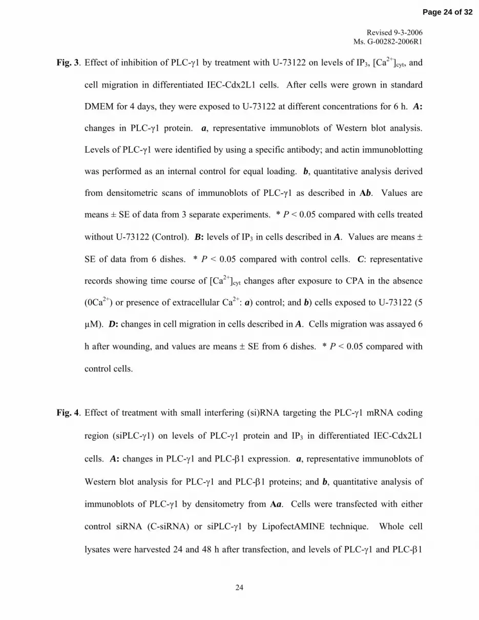
Fig. 3. Effect of inhibition of PLC-γ1 by treatment with U-73122 on levels of IP3, [Ca2+]cyt, and
cell migration in differentiated IEC-Cdx2L1 cells. After cells were grown in standard
DMEM for 4 days, they were exposed to U-73122 at different concentrations for 6 h. A:
changes in PLC-γ1 protein. a, representative immunoblots of Western blot analysis.
Levels of PLC-γ1 were identified by using a specific antibody; and actin immunoblotting
was performed as an internal control for equal loading. b, quantitative analysis derived
from densitometric scans of immunoblots of PLC-γ1 as described in Ab. Values are
means ± SE of data from 3 separate experiments. * P < 0.05 compared with cells treated
without U-73122 (Control). B: levels of IP3 in cells described in A. Values are means ±
SE of data from 6 dishes. * P < 0.05 compared with control cells. C: representative
records showing time course of [Ca2+]cyt changes after exposure to CPA in the absence
(0Ca2+) or presence of extracellular Ca2+: a) control; and b) cells exposed to U-73122 (5
µM). D: changes in cell migration in cells described in A. Cells migration was assayed 6
h after wounding, and values are means ± SE from 6 dishes. * P < 0.05 compared with
control cells.
Fig. 4. Effect of treatment with small interfering (si)RNA targeting the PLC-γ1 mRNA coding
region (siPLC-γ1) on levels of PLC-γ1 protein and IP3 in differentiated IEC-Cdx2L1
cells. A: changes in PLC-γ1 and PLC-β1 expression. a, representative immunoblots of
Western blot analysis for PLC-γ1 and PLC-β1 proteins; and b, quantitative analysis of
immunoblots of PLC-γ1 by densitometry from Aa. Cells were transfected with either
control siRNA (C-siRNA) or siPLC-γ1 by LipofectAMINE technique. Whole cell
lysates were harvested 24 and 48 h after transfection, and levels of PLC-γ1 and PLC-β1
24
Page 24 of 32

Revised 9-3-2006 Ms. G-00282-2006R1
proteins were measured by Western immunoblot analysis. Actin immunoblotting was
performed as an internal control for equal loading. Values are means ± SE of data from 3
separate experiments. B: levels of IP3 in cells described in A. Values are means ± SE of
data from 6 dishes. * P < 0.05 compared with cells transfected with C-siRNA.
Fig. 5. Changes in [Ca2+]cyt and cell migration in differentiated IEC-Cdx2L1 cells described in
Fig. 4. A: representative records showing the time course of [Ca2+]cyt changes induced by
exposure to CPA in the absence (0Ca2+) or presence of extracellular Ca2+: a) control
cells; b), cells transfected with C-siRNA for 48 h; and c) cells transfected with siPLC-γ1
for 48 h. B: summarized data showing resting [Ca2+]cyt (left) and the amplitude of CPA-
induced Ca2+ influx (right) from cells described in A. Values are means ± SE (n = 25).
*P < 0.05 compared with control cells and cells transfected with C-siRNA. C: images of
cell migration 6 h after wounding by removal of part of the monolayer: a) control cells;
b) cells transfected with C-siRNA; and c) cells transfected with siPLC-γ1. After cells
were transfected for 48 h, cell migration was assayed 6 h after wounding. D:
summarized data showing rates of cell migration after wounding in cells described in C.
Values are means ± SE of data from 6 dishes. * P < 0.05 compared with controls and
cells transfected with C-siRNA.
Fig. 6. Effect of induction of PLC-γ1 by treatment with m-3M3FBS on levels of IP3, [Ca2+]cyt,
and cell migration in polyamine-deficient IEC-Cdx2L1 cells. A: representative
immunoblots of Western blot analysis for PLC-γ1 protein. Differentiated IEC-Cdx2L1
cells were grown in culture containing 5 mM DFMO for 4 days and then exposed to
25
Page 25 of 32

Revised 9-3-2006 Ms. G-00282-2006R1
different concentrations of m-3M3FBS for 6 h. Levels of PLC-γ1 protein were measured
by Western immunoblot analysis, while actin immunoblotting was performed as an
internal control for equal loading. B: levels of IP3 in cells described in A. Values are
means ± SE of data from 6 dishes. * P < 0.05 compared with cells treated without m-
3M3FBS (control). C: representative records showing the time course of [Ca2+]cyt
changes after exposure to CPA in the absence (0Ca2+) or presence of extracellular Ca2+:
a) cells treated with DFMO alone; and b) cells treated with DFMO and then exposed to
m-3M3FBS (25 μM). Three separate experiments were performed that showed similar
results. D: summarized data showing rates of cell migration 6 h after wounding in cells
described in A. Values are means ± SE of data from 6 dishes. * P < 0.05 compared with
controls.
26
Page 26 of 32

Fig. 1
A
627-
244-
4 Days 6 Days
bp DF
MO
+P
UT
Con
trol
DF
MO
DF
MO
+P
UT
Con
trol
DF
MO
-PLC-γγγγγ1
-βββββ-actin
B
C
a. PLC-γγγγγ1 mRNA
140- -PLC-γγγγγ2
kDa
-PLC-βββββ1150-
42- -Actin
1 2 3 4 5 6
0
4
8
12
16
IP
3 le
vels
(pm
ol/m
g pr
otei
n)
* *
1 2 3 4 5 6
b. PLC-γγγγγ1 ProteinkDa
135-
42-
-PLC-γγγγγ1
-Actin
1 2 3 4 5 6c. Relative Protein Levels
PL
C- γγγγ γ
1 pr
otei
n
* *
0.0
0.5
1.0
1.5
2.0
1 2 3 4 5 6
Page 27 of 32

BC
ell m
igra
tion
(cel
ls/m
m)
0
200
400
A
Fig. 2
Rat
io (F
340/F
380)
0
1
2
3
CPA0Ca2+
CPA0Ca2+
a. Control b. DFMO c. DFMO+PUT
0
1
2
3
CPA0Ca2+ 2 min
Rat
io (F
340/F
380)
Con
trol
DFM
O
DFM
O+P
UT
**
*
Resting [Ca2+]cyt
Ca2+ influxControl DFMO DFMO
+PUT
C
Page 28 of 32

Fig. 3
C
B R
atio
(F34
0/F38
0)
A
Cel
l mig
rati
on (c
ells
/mm
)
0 1 2 5
*
U-73122 (μμμμμM)
0
150
300
450
600
*
IP3 l
evel
s (pm
ol/m
g pr
otei
n)
0 1 2 5
0
1
2
3
CPA0Ca2+
CPA0Ca2+
a. Control b. U-73122
2 min
D
0
6
12
18
* *
U-73122 (μμμμμM)
kDa 0 1 2 5
-PLC-γγγγγ1
-Actin
135-
42-
U-73122 (μμμμμM)
0.0
0.6
1.2
1.8
PL
C- γγγγ γ
1 pr
otei
n
1 2 3 4 1 2 3 4
a. PLC-γγγγγ1 protein b. Relative levels
*
*
Page 29 of 32

Fig. 4
B
**
a. PLC proteins
Rel
ativ
e P
LC
- γγγγ γ1
leve
ls
-PLC-γγγγγ1
A
1 2 3 4-Actin
kDa
135-
42-
Control
C-siRNA
siPLC-γγγγγ1
(24 h
)
siPLC-γγγγγ1
(48 h
)
0
1
2
1 2 3 4
b. Relative levels
0
5
10
15
20
IP
3 le
vels
(pm
ol/m
g pr
otei
n)
**
1 2 3 4
-PLC-βββββ1150-
Page 30 of 32

Fig. 5
B
0
1
2
3
0Ca2+CPA
Rat
io (F
340/F
380)
a. Control b. C-siRNA c. siPLC-γγγγγ1A
0Ca2+CPA
0
1
2
3
Rat
io (F
340/F
380)
Con
trol
C-s
iRN
A
siP
LC
- γγγγ γ1
*
*
Resting [Ca2+]cyt Ca2+ influx
a. Control b. C-siRNA c. siPLC-γγγγγ1C
0
150
300
450
Control C-siRNA siPLC-γγγγγ1
Cel
l mig
rati
oin
(cel
ls/m
m)
D
*
0Ca2+CPA
2 min
Page 31 of 32

Fig. 6
C
A
B-Actin
-PLC-γγγγγ1
42-
0 5 10 25
m-3M3FBS (μμμμμM)
Cel
l mig
rati
on (c
ells
/mm
)
0
100
200
300
135-
kDa
0
1
2
3
Rat
io (F
340/F
380)
2 min0Ca2+
CPA
0Ca2+
CPA
a. DFMO b. DFMO+m-3M3FBS
D
**
*
0
5
10
15
20
0 5 10 25
m-3M3FBS (μμμμμM)
0 5 10 25
m-3M3FBS (μμμμμM)
IP
3 lev
els
(pm
ol/m
g pr
otei
n)
*
**
Page 32 of 32

![Differential protein metabolism and gene expression in tomato [Lycopersicon esculentum] fruit during wounding stress](https://img.pdfslide.net/doc/110x75/6352f3641d81cb29f50d7550/differential-protein-metabolism-and-gene-expression-in-tomato-lycopersicon-esculentum.jpg)


