Embed Size (px)
Citation preview
Biomaterials 24 (2003) 4273–4281
ARTICLE IN PRESS
*Correspondin
62788784.
E-mail addres
0142-9612/03/$ -
doi:10.1016/S014
Poly(hydroxybutyrate-co-hydroxyhexanoate) promoted productionof extracellular matrix of articular cartilage chondrocytes in vitro
Ying Denga, Xing-Sun Linb, Zhong Zhenga, Jin-Guang Denga, Jin-Chun Chena, Hui Mab,Guo-Qiang Chena,*
aDepartment of Biological Sciences and Biotechnology, Tsinghua University, Beijing 100084, Chinab Department of Physics, Tsinghua University, Beijing 100084, China
Received 25 November 2002; accepted 13 May 2003
Abstract
The present investigation describes the production of extracellular matrix of rabbit articular cartilage chondrocytes grown on
scaffolds of polyhydroxybutyrate (PHB) blended with poly(hydroxybutyrate-co-hydroxyhexanoate) (PHBHHx) for up to 7 days.
The mRNA level of type II collagen of chondrocytes seeded on all scaffolds consisting of PHBHHx were obviously higher than that
of PHB-only scaffold throughout the culture period, suggesting the positive effect of PHBHHx on extracellular matrix production.
Second-harmonic generation (SHG) imaging technique, combined with confocal fluorescence microscopy (CFM) revealed that
PHBHHx in PHB scaffold provided better surface properties for anchoring type II collagen filaments and their penetration into
internal layers of the scaffolds. Glycosaminoglycan (GAG), a major composition of extracellular matrix, showed a sharp increase in
construct of 1:2 PHB/PHBHHx scaffold after 7 day cultivation, while only a small increase was observed in all other tested
scaffolds. At the same time, total collagen contents in all scaffolds containing PHBHHx increased with time, with the maximum
collagen production of 742.1799.2mg/g dry weight observed in construct of 1:2 PHB/PHBHHx scaffold inoculated for 7 days, this
was almost 4-fold higher than that in scaffold of PHB only. It appears that the presence of right proportion of PHBHHx in the
composite system of PHB/PHBHHx highly favored the production of extracellular matrix of articular cartilage chondrocytes.
r 2003 Elsevier Ltd. All rights reserved.
Keywords: Polyhydroxyalkanoates; PHB; Type II Collagen; SHG; Chondrocytes
1. Introduction
Tissue engineering of articular cartilage represents anexciting direction in the efforts to solve the complexproblem of cartilage regeneration. To promote healingof cartilage defects, many researchers are turning towarda tissue engineering approach involving ex vivo fabrica-tion of cartilage constructs by culturing cells on porous,resorbable scaffolds. Several candidate materials areavailable including glass [1,2], porous calcium ceramics[3,4], and biodegradable polymers. Biodegradable poly-mers are attractive for several reasons. They may beused to support cell growth in vitro [5–7], to conducttissue growth in vivo [8–10]. In terms of bone regenera-tion, especially cartilage regeneration, numerous biode-
g author. Tel: +86-10-62783844; fax: +86-10-
s: [email protected] (G.-Q. Chen).
see front matter r 2003 Elsevier Ltd. All rights reserved.
2-9612(03)00367-3
gradable polymers have been investigated for thesepurposes [11–13].
Polyhydroxybutyrate (PHB) is finding some interest-ing potential applications as an implant material due toits biocompatibility and resorbability [14,15]. PHBappears suitable for use as temporary stents, boneplates, patches, nails and screws [16,17]. In some cases,its brittle mechanical properties limit its use. For thatreason, materials such as triethylcitrate have been usedas a typical plasticizer [18]. Blend copolymer of PHAconsisting of 3-hydroxybutyrate and 3-hydroxyvalerate(P(HB-co-HV)) were reported to be widely used with itsimproved mechanical properties over that of PHB [19].Therefore, PHA with better elastomeric properties,such as poly(hydroxybutyrate-co-hydroxyhexanoate)(PHBHHx), is expected to meet special application asscaffolds for cartilage engineering [20]. Our previousstudies have shown better biocompatibility of PHBHHxcontaining materials for fibroblasts cell line: L929 cells
ARTICLE IN PRESSY. Deng et al. / Biomaterials 24 (2003) 4273–42814274
and rabbit articular cartilage-derived chondrocytes[21,22].
Second-harmonic generation (SHG) imaging techni-que is beginning to emerge as a powerful contrastmechanism in nonlinear optical microscopy [23]. It hasbeen commonly used to obtain shorter wavelengths andhas become a standard spectroscopic tool for character-izing material surfaces and probing dynamics at inter-faces [23]. This is the first report describing the use ofSHG and confocal fluorescence microscopy (CFM) toobtain high-resolution three-dimensional (3D) images oftype II collagen distribution on PHB/PHBHHx polymerscaffolds.
This study expanded previous work utilizing homo-and blend polymer scaffolds of PHB/PHBHHx for cellgrowth [21,22] to observe the effect of PHBHHxcontaining blends on chondrocyte growth at theproteins synthesis and gene transcription levels.
2. Materials and methods
2.1. Polymer scaffold preparation
PHB/PHBHHx scaffolds were manufactured using acombination of published salt leaching techniques [24].Briefly, 2 g PHBHHx (Mw=750,000+10%) and PHB(Mw=1,000,000+10%) (supplied by Jiangmen Centerfor Biotech Development, Guangdong, China) in aseries of weight ratios (1:0, 2:1, 1:1, 1:2, 0:1), respec-tively, were dissolved in 16 ml chloroform and refluxedat 60�C for 0.5 h. The PHA solution was then pouredonto a bed of sieved sodium chloride particles witha size of 200 mm. The scaffolds were placed undervacuum in a desiccator for 24 h, followed by repeatedlywashing with large amounts of distilled water forleaching the salt. After the salt-leaching process, themicroporous polymer scaffolds were obtained andvacuum dried.
2.2. Cell isolation and in vitro cultivation
The articular cartilages were removed from the kneesand hip joints of young (3–5 days old) New Zealandwhite rabbits. They were cut into small pieces asdescribed [25]. Chondrocytes were released from carti-lage slices by using (0.2% w/v) collagenase II digestion.The isolated cells were then cultured in DMEMsupplemented with 10% fetal calf serum (FCS), 100 U/ml penicillin, and 100 mg/ml streptomycin. Cells wereincubated at 37�C in a 5% CO2 incubator and themedium was changed every 4 days. When the cellsreached the plateau phase of growth, they wereharvested by trypsinization followed by addition offresh culture medium to create a new single cellsuspension with desired seeding cell number per
100 mg. Inoculation was performed in polystyrene 24-well flat-bottom culture plates. Scaffolds were placed inthe center of the wells added with 1ml cell suspension toallow full attachment of cells to scaffolds. Cultivationwas conducted for 3 and 7 days. Culture media werechanged every 4 days.
2.3. Biochemical analysis
Constructs of cells/(PHB/PHBHHx scaffold) wereremoved at 3 and 7 days from the tissue culture plates,frozen (�20�C) and lyophilized for at least 36 h afterwhich dry weights (dw) were determined. The sampleswere then digested in 1ml papain (125 mg/ml in 100 mm
phosphate-10 mm EDTA(PBE) buffer with 10 mm cy-steine, pH 6.5) overnight at 60�C. Glycosaminoglycan(GAG) content was estimated by chondroitin sulfateusing dimethylmethylene blue dye and a spectrophot-ometer (525 nm wavelength) [26,27]. Total collagencontent was determined based on hydroxyprolineconcentration released after acid hydrolysis of collagen(6n HCl at 115�C for 18 h) using a spectrophotometer(550 nm wavelength) [26,27].
2.4. Type II collagen mRNA expression studies
After 3 and 7 days inoculation, the scaffolds wereremoved from culture, and RNA was harvested asdescribed [28]. First of all, total RNA harvested wasquantitated using an UV/Vis spectrophotometer (Ultra-spec 3300, Biochrom, Cambridge, England) at 260 nm.Then, reverse transcription (RT) was performed using acommercially available kit (Invitrogen, California,USA). Briefly, 1 mg of extracted RNA was reversetranscribed to generate full-length first strand cDNAfor use as template for PCR (polymerase chain reaction)amplification. After RT reaction, 3 ml RT product wasamplified using 25 pmol each of primers described belowand 0.5 U Ex Taq DNA polymerase (TaKaRa, Dalian,China) in a Mastercycler Gradient Autorisierter Ther-mocycler (Eppendorf, Hamburg, Germany) for both thetarget molecule pro alpha1 type II collagen and thereference molecule beta-actin. The codon sequence ofpro alphal type II collagen and beta–actin for theprimers, given with their respective PCR conditions (30cycles for all conditions): pro alphal collagen II(Genbank S83370) was as follows: sense primer: 50-GCACCCATGGACATTGGAGG-30, antisense pri-mer: 50-AGCCCCGCACGGTCTTGCTT (323 bp;94�C, 45 s, 55�C, 45 s, 72�C, 60 s); beta-actin: senseprimer: 50-TGGACTTCGAGCAAGAGA-30, antisenseprimer: 50-CTGCTTGCTCATCCACAT-30 (450 bp;94�C, 45 s, 58�C, 45 s, 72�C, 60 s). The marker usedwas 100 bp ladder (Bebco, MBI, UK).
ARTICLE IN PRESS
250
200
150
100
50
01:0 2:1 1:1 1:2 0:1
GA
G C
onte
nt (
mg/
g)
3d
7d
PHB/PHBHHx
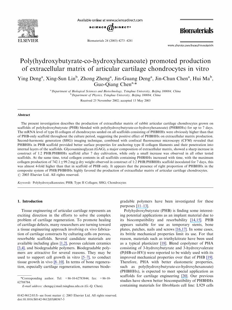
Fig. 1. GAG content in various constructs of cell/(PHB/PHBHHx
scaffold) after various culture times. Values given represent the mean,
and the error bars indicate the standard deviation estimated.
600
800
900
700
onte
nt (
mg/
g)
3d
7d
Y. Deng et al. / Biomaterials 24 (2003) 4273–4281 4275
2.5. Studies of 3D distribution of type II collagen in
PHB/PHBHHx scaffolds
Samples were dissected from constructs of cell/PHAat 3 and 7 days, followed by fixation in ethanol andacetic acid (99:1, v/v) for 2 h at room temperature.Subsequently, they were washed three times with PBSand inoculated with mouse monoclonal antibodiesagainst human type II collagen (Neomarker, USA)diluted 1:200 with PBS for 1 h at 37�C. After washingwith PBS, the second immunoreaction was performedfor 1 h at 37�C with fluorescein isothiocyanate (FITC)-conjugated antibodies raised in goat against mouse IgG(Dake, USA) diluted 1:100 with PBS for 1 h at 37�C.After rinsing with PBS, samples were placed on glassmicroscope slides to form uniform 1 cm� 1 cm squaresample surfaces of less than 1 mm thickness [29]. TheSHG imaging experiments were performed on modifiedBiorad MRC1024 confocal scan head mounted on aNikon inverted microscope (PE300, Nikon, Tokyo,Japan). The laser system is a Spectra-Physics argonion (Millennia, Spectra-Physics, USA) pumped femto-second titanium sapphire oscillator (Tsunami, SpectraPhysics, USA), characterized by pulse width of approxi-mately 120 fs at 82 MHz repetition rate at 811 nm. Two20� 0.4 N.A. lens were used for excitation and signalcollection, respectively. Data acquisition times was 1 sper 512� 512 frame [30]. The distance between twoadjacent layers was 5 mm. By switching the imagingchannels, the confocal fluorescence signals in situ werecollected by the same lens and went through a pinhole tothe photomultiplier tube (PMT) inside the scan head.3D image stacks acquired were processed and analyzedin Biorad Confocul Assistant v2.0 and Adobe Photo-shop v6.0.
2.6. Statistical analysis
Data are presented as means7standard error ofmean. Statistical comparisons were performed usingStudents t-test. P-values o0.01 were considered statis-tically significant.
200
100
400
300
500
01:0 2:1 1:1 1:2 0:1
Tot
al C
olla
gen
C
PHB/PHBHHx
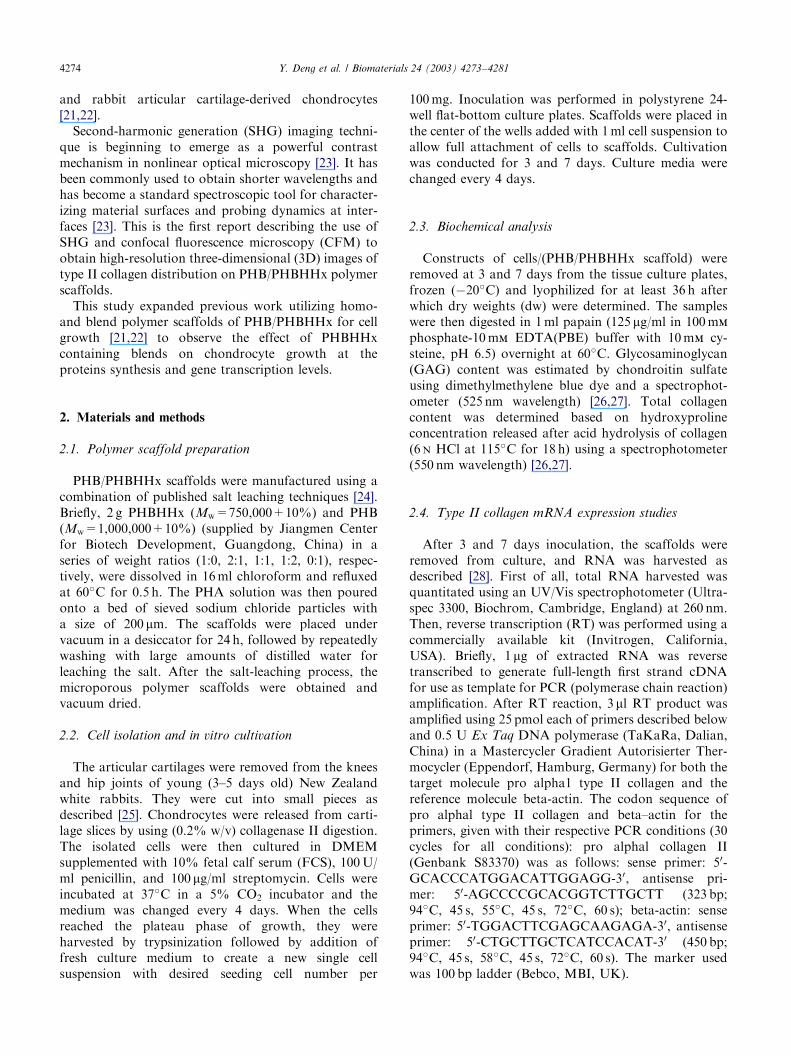
Fig. 2. Total collagen content in various constructs of cell/(PHB/
PHBHHx scaffold) after various culture times. Values given represent
the mean, and the error bars indicate the standard deviation estimated.
3. Results
3.1. Biochemical characterization of chondrocyte-
composite scaffolds
Chondrocytes seeded on the polymer scaffolds ad-hered to the matrix and continued to grow duringincubation period.
To determine the effect of polymer scaffolds on GAGproduction by chondrocytes seeded on each scaffold, theconstructs of cells/(PHB/PHBHHx scaffold) were col-lected, digested and analyzed using dimethylmethylene
blue dying method. As shown in Fig. 1, on day 3, totalGAG content in the constructs of blend polymer (PHB/PHBHHx) scaffolds was significantly different from thatof the PHBHHx or PHB-only cell construct groups(Po0:05). The total GAG content was then examinedon day 7. Slight increase of total GAG content could beobserved in each scaffold compared with that of thecorresponding scaffold analyzed on day 3.
For the same purpose, total collagen content of theconstructs of cells/(PHB/PHBHHx scaffold) was deter-mined by chloramines-T assay. Between days 3 to day 7,the total collagen content increased significantly inPHBHHx containing constructs, while it decreased
ARTICLE IN PRESSY. Deng et al. / Biomaterials 24 (2003) 4273–42814276
significantly in PHB-only constructs (cells/PHB)(Fig. 2). On day 7, total collagen content in PHB/PHBHHx constructs was significantly higher than thaton PHB or PHBHHx-only constructs.
3.2. 3D distribution of type II collagen in PHB/
PHBHHx scaffolds
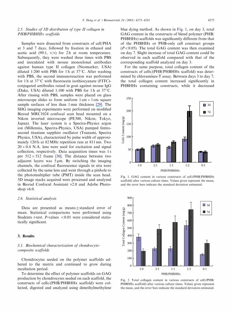
SHG signals were observed in scaffolds made of PHB,PHBHHx and their blends. The SHG intensity is strongwhere the image is bright. Fig. 3 shows that microporeswith sizes ranging from 5 to 10 mm were observable oneach scaffold. The porosity of each scaffold wasobviously dependent on the amount of PHBHHxcontents in the blend polymer scaffolds. With theincrease of PHBHHx component in the scaffolds, moremicropores appeared.
In order to reveal type II collagen produced bychondrocytes seeded on various PHB/PHBHHx scaf-folds, the constructs of cells/(PHB/PHBHHx scaffold)cultured for 3 and 7 days were studied by SHG imagingtechnique to view the scaffold in depth, CFM was also
(a)
(b) (c)
(d) (e)
Fig. 3. SHG image of PHB/PHBHHx scaffolds in various weight
ratios. (a) PHB; (b) 2:1 PHB/PHBHHx; (c) 1:1 PHB/PHBHHx; (d) 1:2
PHB/PHBHHx; (e) PHBHHx. Scale bar=50mm.
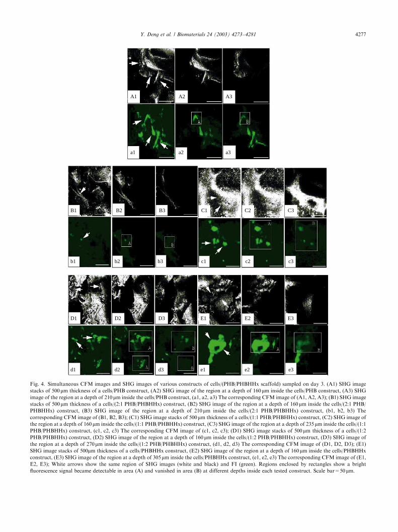
employed in situ to detect type II collagen. Fig. 4 showsgreen fluorescence images (FI) and SHG imagesobtained simultaneously on precisely the same areaand at the same depth for each construct of cells/(PHB/PHBHHx scaffold) on day 3. Mechanically dissectedsamples of each construct was optically sectionedthrough its full thickness of B500 mm. Stacks of thefull thickness optical sections were recorded as SHGimages (Fig. 4: A1, B1, C1, D1, E1) and thecorresponding FI (Fig. 4: a1, b1, c1, d1, e1). The imagesshowed that there was a correlation between thefluorescence intensities and their positions on eachtested construct, better distribution of fluorescencesignal could be observed in porous areas (white arrow)than in non-porous area. Although some similarfluorescence signal distribution appeared among variousconstructs, obvious differences were also described. Fig.4 (a2 and a3) show the region at a depth of 160 mm insidethe cells/PHB construct, corresponding to SHG image(Fig. 4 A2) where a bright fluorescence signal (A region)became detectable. This fluorescence signal vanished inB region corresponding to a depth of 210 mm (Fig. 4A3). The penetration depth of the fluorescence signalwas approximately 50 mm, signifying that collagenextended 50 mm into the cells/PHB construct. Thepenetration depths of the fluorescence signal into otherPHB/PHBHHx scaffolds of 2:1, 1:1, 1:2, and 0:1 were50, 75, 110 and 145 mm, respectively (Fig. 4: b2, b3, c2,c3, d2, d3, e2 and e3).
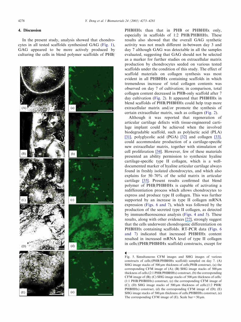
After cultivation for 7 days, type II collagen (green)was widely detected in PHB/PHBHHx scaffolds of 1:0,2:1, 1:1, 1:2 and 0:1 (Figs. 5a–e). The position wheretype II collagen distributed could be observed insimultaneous SHG imaging for each correspondingconstructs of cells/(PHB/PHBHHx scaffold) as type IIcollagen was invisible in SHG (Fig. 5A–E). More type IIcollagen was detected in PHB/PHBHHx (1:2) scaffoldthan that in other tested scaffolds (Fig. 5d).
3.3. Regulation of type II collagen mRNA expression
during the early culture period of chondrocytes
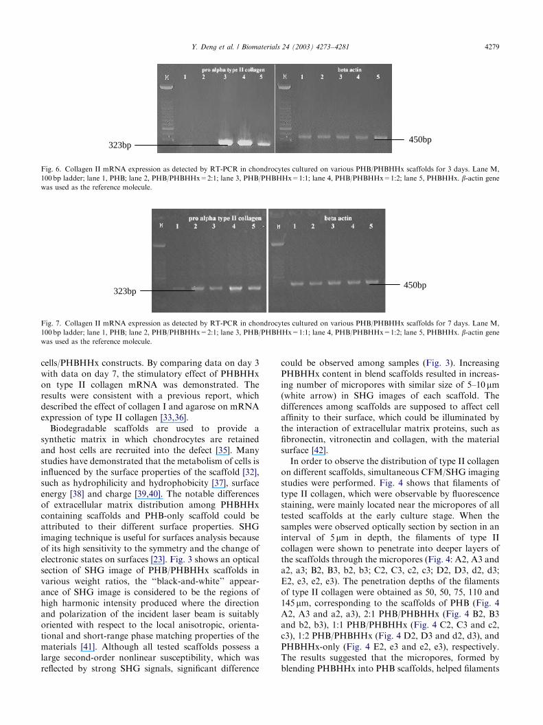
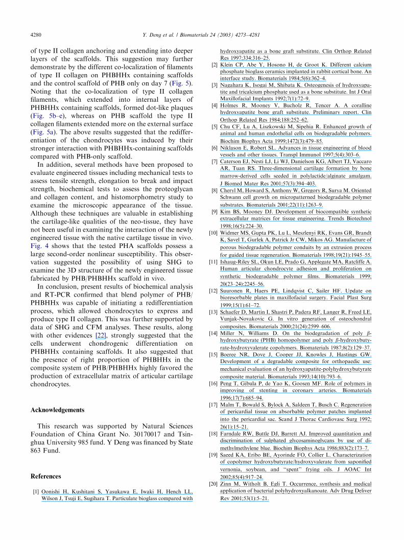
Total mRNA from each construct of cells/PHAcultured for 3 day and 7 day, respectively, was analyzedby RT-PCR using rabbit-specific primers for type IIcollagen mRNA with beta-actin mRNA as control.Type II collagen gene expression in chondrocytes seededon 1:1 and 1:2 PHB/PHBHHx scaffolds, respectively,was stronger than on the other tested scaffolds on day 3(Fig. 6). The strongest gene expression for type IIcollagen was observed on 1:2 PHB/PHBHHx scaffoldafter 7 days of cultivation (Fig. 7), while on PHB controlscaffold on day 7, the expression level was the lowest,even lower than the expression on day 3.
ARTICLE IN PRESS
A1 A2 A3
a1 a2 a3
B1 B2 B3
b1 b2 b3
C1 C2 C3
D1 D2 D3 E1 E2 E3
d1 d2 d3 e1 e2 e3
c1 c2 c3
Fig. 4. Simultaneous CFM images and SHG images of various constructs of cells/(PHB/PHBHHx scaffold) sampled on day 3. (A1) SHG image
stacks of 500mm thickness of a cells/PHB construct, (A2) SHG image of the region at a depth of 160 mm inside the cells/PHB construct, (A3) SHG
image of the region at a depth of 210mm inside the cells/PHB construct, (a1, a2, a3) The corresponding CFM image of (A1, A2, A3); (B1) SHG image
stacks of 500mm thickness of a cells/(2:1 PHB/PHBHHx) construct, (B2) SHG image of the region at a depth of 160 mm inside the cells/(2:1 PHB/
PHBHHx) construct, (B3) SHG image of the region at a depth of 210mm inside the cells/(2:1 PHB/PHBHHx) construct, (b1, b2, b3) The
corresponding CFM image of (B1, B2, B3); (C1) SHG image stacks of 500mm thickness of a cells/(1:1 PHB/PHBHHx) construct, (C2) SHG image of
the region at a depth of 160mm inside the cells/(1:1 PHB/PHBHHx) construct, (C3) SHG image of the region at a depth of 235 mm inside the cells/(1:1
PHB/PHBHHx) construct, (c1, c2, c3) The corresponding CFM image of (c1, c2, c3); (D1) SHG image stacks of 500 mm thickness of a cells/(1:2
PHB/PHBHHx) construct, (D2) SHG image of the region at a depth of 160 mm inside the cells/(1:2 PHB/PHBHHx) construct, (D3) SHG image of
the region at a depth of 270mm inside the cells/(1:2 PHB/PHBHHx) construct, (d1, d2, d3) The corresponding CFM image of (D1, D2, D3); (E1)
SHG image stacks of 500mm thickness of a cells/PHBHHx construct, (E2) SHG image of the region at a depth of 160mm inside the cells/PHBHHx
construct, (E3) SHG image of the region at a depth of 305mm inside the cells/PHBHHx construct, (e1, e2, e3) The corresponding CFM image of (E1,
E2, E3); White arrows show the same region of SHG images (white and black) and FI (green). Regions enclosed by rectangles show a bright
fluorescence signal became detectable in area (A) and vanished in area (B) at different depths inside each tested construct. Scale bar=50mm.
Y. Deng et al. / Biomaterials 24 (2003) 4273–4281 4277
ARTICLE IN PRESSY. Deng et al. / Biomaterials 24 (2003) 4273–42814278
4. Discussion
In the present study, analysis showed that chondro-cytes in all tested scaffolds synthesized GAG (Fig. 1),GAG appeared to be more actively produced byculturing the cells in blend polymer scaffolds of PHB/
(A) (a)
(B) (b)
(C) (c)
(D) (d)
(E) (e)
PHBHHx than that in PHB or PHBHHx only,especially in scaffolds of 1:2 PHB/PHBHHx. Theseresults also showed that the overall GAG syntheticactivity was not much different in-between day 3 andday 7 although GAG was detectable in all the samplesevaluated, suggesting that GAG should not be selectedas a marker for further studies on extracellular matrixproduction by chondrocytes seeded on various testedscaffolds under the condition of this study. The effect ofscaffold materials on collagen synthesis was mostevident in all PHBHHx containing scaffolds in whichtremendous increase of total collagen contents wasobserved on day 7 of cultivation; in comparison, totalcollagen content decreased in PHB-only scaffold after 7day cultivation (Fig. 2). It appeared that PHBHHx inblend scaffolds of PHB/PHBHHx could help trap moreextracellular matrix and/or promote the synthesis ofcertain extracellular matrix, such as collagen (Fig. 2).
Although it was reported that regeneration ofarticular cartilage defects with tissue-engineered carti-lage implant could be achieved when the involvedbiodegradable scaffold, such as polylactic acid (PLA)[31], polyglycolic acid (PGA) [32] and collagen [33],could accommodate production of a cartilage-specificnew extracellular matrix, together with stimulation ofcell proliferation [34]. However, few of these materialspresented an ability permission to synthesize hyalinecartilage-specific type II collagen, which is a well-documented marker of hyaline articular cartilage alwaysfound in freshly isolated chondrocytes, and which alsoexplains for 50–70% of the solid matrix in articularcartilage [35]. Present results confirmed that blendpolymer of PHB/PHBHHx is capable of activating aredifferentiation process which allows chondrocytes toexpress and produce type II collagen. This was furthersupported by an increase in type II collagen mRNAexpression (Figs. 6 and 7), which was followed by theproduction of the secreted type II collagen, as detectedby immunofluorescence analysis (Figs. 4 and 5). Theseresults, along with other evidences [22], strongly suggestthat the cells underwent chondrogenic differentiation onPHBHHx containing scaffolds. RT-PCR data (Figs. 6and 7) indicated that increased PHBHHx contentresulted in increased mRNA level of type II collagenin cells/(PHB/PHBHHx scaffold) constructs, except for
Fig. 5. Simultaneous CFM images and SHG images of various
constructs of cells/(PHB/PHBHHx scaffold) sampled on day 7. (A)
SHG image stacks of 500mm thickness of cells/PHB construct, (a) the
corresponding CFM image of (A); (B) SHG image stacks of 500mm
thickness of cells/(2:1 PHB/PHBHHx) construct, (b) the corresponding
CFM image of (B); (C) SHG image stacks of 500mm thickness of cells/
(1:1 PHB/PHBHHx) construct, (c) the corresponding CFM image of
(C); (D) SHG image stacks of 500mm thickness of cells/(1:2 PHB/
PHBHHx) construct, (d) the corresponding CFM image of (D); (E)
SHG image stacks of 500mm thickness of cells/PHBHHx construct, (e)
The corresponding CFM image of (E). Scale bar=50mm.
ARTICLE IN PRESS
450bp323bp
Fig. 6. Collagen II mRNA expression as detected by RT-PCR in chondrocytes cultured on various PHB/PHBHHx scaffolds for 3 days. Lane M,
100bp ladder; lane 1, PHB; lane 2, PHB/PHBHHx=2:1; lane 3, PHB/PHBHHx=1:1; lane 4, PHB/PHBHHx=1:2; lane 5, PHBHHx. b-actin gene
was used as the reference molecule.
323bp450bp
Fig. 7. Collagen II mRNA expression as detected by RT-PCR in chondrocytes cultured on various PHB/PHBHHx scaffolds for 7 days. Lane M,
100bp ladder; lane 1, PHB; lane 2, PHB/PHBHHx=2:1; lane 3, PHB/PHBHHx=1:1; lane 4, PHB/PHBHHx=1:2; lane 5, PHBHHx. b-actin gene
was used as the reference molecule.
Y. Deng et al. / Biomaterials 24 (2003) 4273–4281 4279
cells/PHBHHx constructs. By comparing data on day 3with data on day 7, the stimulatory effect of PHBHHxon type II collagen mRNA was demonstrated. Theresults were consistent with a previous report, whichdescribed the effect of collagen I and agarose on mRNAexpression of type II collagen [33,36].
Biodegradable scaffolds are used to provide asynthetic matrix in which chondrocytes are retainedand host cells are recruited into the defect [35]. Manystudies have demonstrated that the metabolism of cells isinfluenced by the surface properties of the scaffold [32],such as hydrophilicity and hydrophobicity [37], surfaceenergy [38] and charge [39,40]. The notable differencesof extracellular matrix distribution among PHBHHxcontaining scaffolds and PHB-only scaffold could beattributed to their different surface properties. SHGimaging technique is useful for surfaces analysis becauseof its high sensitivity to the symmetry and the change ofelectronic states on surfaces [23]. Fig. 3 shows an opticalsection of SHG image of PHB/PHBHHx scaffolds invarious weight ratios, the ‘‘black-and-white’’ appear-ance of SHG image is considered to be the regions ofhigh harmonic intensity produced where the directionand polarization of the incident laser beam is suitablyoriented with respect to the local anisotropic, orienta-tional and short-range phase matching properties of thematerials [41]. Although all tested scaffolds possess alarge second-order nonlinear susceptibility, which wasreflected by strong SHG signals, significant difference
could be observed among samples (Fig. 3). IncreasingPHBHHx content in blend scaffolds resulted in increas-ing number of micropores with similar size of 5–10 mm(white arrow) in SHG images of each scaffold. Thedifferences among scaffolds are supposed to affect cellaffinity to their surface, which could be illuminated bythe interaction of extracellular matrix proteins, such asfibronectin, vitronectin and collagen, with the materialsurface [42].
In order to observe the distribution of type II collagenon different scaffolds, simultaneous CFM/SHG imagingstudies were performed. Fig. 4 shows that filaments oftype II collagen, which were observable by fluorescencestaining, were mainly located near the micropores of alltested scaffolds at the early culture stage. When thesamples were observed optically section by section in aninterval of 5 mm in depth, the filaments of type IIcollagen were shown to penetrate into deeper layers ofthe scaffolds through the micropores (Fig. 4: A2, A3 anda2, a3; B2, B3, b2, b3; C2, C3, c2, c3; D2, D3, d2, d3;E2, e3, e2, e3). The penetration depths of the filamentsof type II collagen were obtained as 50, 50, 75, 110 and145 mm, corresponding to the scaffolds of PHB (Fig. 4A2, A3 and a2, a3), 2:1 PHB/PHBHHx (Fig. 4 B2, B3and b2, b3), 1:1 PHB/PHBHHx (Fig. 4 C2, C3 and c2,c3), 1:2 PHB/PHBHHx (Fig. 4 D2, D3 and d2, d3), andPHBHHx-only (Fig. 4 E2, e3 and e2, e3), respectively.The results suggested that the micropores, formed byblending PHBHHx into PHB scaffolds, helped filaments
ARTICLE IN PRESSY. Deng et al. / Biomaterials 24 (2003) 4273–42814280
of type II collagen anchoring and extending into deeperlayers of the scaffolds. This suggestion may furtherdemonstrate by the different co-localization of filamentsof type II collagen on PHBHHx containing scaffoldsand the control scaffold of PHB only on day 7 (Fig. 5).Noting that the co-localization of type II collagenfilaments, which extended into internal layers ofPHBHHx containing scaffolds, formed dot-like plaques(Fig. 5b–e), whereas on PHB scaffold the type IIcollagen filaments extended more on the external surface(Fig. 5a). The above results suggested that the rediffer-entiation of the chondrocytes was induced by theirstronger interaction with PHBHHx-containing scaffoldscompared with PHB-only scaffold.
In addition, several methods have been proposed toevaluate engineered tissues including mechanical tests toassess tensile strength, elongation to break and impactstrength, biochemical tests to assess the proteoglycanand collagen content, and histomorphometry study toexamine the microscopic appearance of the tissue.Although these techniques are valuable in establishingthe cartilage-like qualities of the neo-tissue, they havenot been useful in examining the interaction of the newlyengineered tissue with the native cartilage tissue in vivo.Fig. 4 shows that the tested PHA scaffolds possess alarge second-order nonlinear susceptibility. This obser-vation suggested the possibility of using SHG toexamine the 3D structure of the newly engineered tissuefabricated by PHB/PHBHHx scaffold in vivo.
In conclusion, present results of biochemical analysisand RT-PCR confirmed that blend polymer of PHB/PHBHHx was capable of initiating a redifferentiationprocess, which allowed chondrocytes to express andproduce type II collagen. This was further supported bydata of SHG and CFM analyses. These results, alongwith other evidences [22], strongly suggested that thecells underwent chondrogenic differentiation onPHBHHx containing scaffolds. It also suggested thatthe presence of right proportion of PHBHHx in thecomposite system of PHB/PHBHHx highly favored theproduction of extracellular matrix of articular cartilagechondrocytes.
Acknowledgements
This research was supported by Natural SciencesFoundation of China Grant No. 30170017 and Tsin-ghua University 985 fund. Y Deng was financed by State863 Fund.
References
[1] Oonishi H, Kushitani S, Yasukawa E, Iwaki H, Hench LL,
Wilson J, Tsuji E, Sugihara T. Particulate bioglass compared with
hydroxyapatite as a bone graft substitute. Clin Orthop Related
Res 1997;334:316–25.
[2] Klein CP, Abe Y, Hosono H, de Groot K. Different calcium
phosphate bioglass ceramics implanted in rabbit cortical bone. An
interface study. Biomaterials 1984;5(6):362–4.
[3] Nagahara K, Isogai M, Shibata K. Osteogenesis of hydroxyapa-
tite and tricalcium phosphate used as a bone substitute. Int J Oral
Maxillofacial Implants 1992;7(1):72–9.
[4] Holmes R, Mooney V, Bucholz R, Tencer A. A coralline
hydroxyapatite bone graft substitute. Preliminary report. Clin
Orthop Related Res 1984;188:252–62.
[5] Chu CF, Lu A, Liszkowski M, Sipehia R. Enhanced growth of
animal and human endothelial cells on biodegradable polymers.
Biochim Biophys Acta 1999;1472(3):479–85.
[6] Niklason E, Robert SL. Advances in tissue engineering of blood
vessels and other tissues. Transpl Immunol 1997;5(4):303–6.
[7] Caterson EJ, Nesti LJ, Li WJ, Danielson KG, Albert TJ, Vaccaro
AR, Tuan RS. Three-dimensional cartilage formation by bone
marrow-derived cells seeded in polylactide/alginate amalgam.
J Biomed Mater Res 2001;57(3):394–403.
[8] Cheryl M, Howard S, Anthony W, Gregory R, Surya M. Oriented
Schwann cell growth on micropatterned biodegradable polymer
substrates. Biomaterials 2001;22(11):1263–9.
[9] Kim BS, Mooney DJ. Development of biocompatible synthetic
extracellular matrices for tissue engineering. Trends Biotechnol
1998;16(5):224–30.
[10] Widmer MS, Gupta PK, Lu L, Meszlenyi RK, Evans GR, Brandt
K, Savel T, Gurlek A, Patrick Jr CW, Mikos AG. Manufacture of
porous biodegradable polymer conduits by an extrusion process
for guided tissue regeneration. Biomaterials 1998;19(21):1945–55.
[11] Ishaug-Riley SL, Okun LE, Prado G, Applegate MA, Ratcliffe A.
Human articular chondrocyte adhesion and proliferation on
synthetic biodegradable polymer films. Biomaterials 1999;
20(23–24):2245–56.
[12] Suuronen R, Haers PE, Lindqvist C, Sailer HF. Update on
bioresorbable plates in maxillofacial surgery. Facial Plast Surg
1999;15(1):61–72.
[13] Schaefer D, Martin I, Shastri P, Padera RF, Langer R, Freed LE,
Vunjak-Novakovic G. In vitro generation of osteochondral
composites. Biomaterials 2000;21(24):2599–606.
[14] Miller N, Williams D. On the biodegradation of poly b-
hydroxybutyrate (PHB) homopolymer and poly b-hydroxybuty-
rate-hydroxyvalerate copolymers. Biomaterials 1987;8(2):129–37.
[15] Boeree NR, Dove J, Cooper JJ, Knowles J, Hastings GW.
Development of a degradable composite for orthopaedic use:
mechanical evaluation of an hydroxyapatite-polyhydroxybutyrate
composite material. Biomaterials 1993;14(10):793–6.
[16] Peng T, Gibula P, de Yao K, Goosen MF. Role of polymers in
improving of stenting in coronary arteries. Biomaterials
1996;17(7):685–94.
[17] Malm T, Bowald S, Bylock A, Saldeen T, Busch C. Regeneration
of pericardial tissue on absorbable polymer patches implanted
into the pericardial sac. Scand J Thorac Cardiovasc Surg 1992;
26(1):15–21.
[18] Farndale RW, Buttle DJ, Barrett AJ. Improved quantitation and
discrimination of sulphated glycosaminoglycans by use of di-
methylmethylene blue. Biochim Biophys Acta 1986;883(2):173–7.
[19] Saeed KA, Eribo BE, Ayorinde FO, Collier L. Characterization
of copolymer hydroxybutyrate/hydroxyvalerate from saponified
vernonia, soybean, and ‘‘spent’’ frying oils. J AOAC Int
2002;85(4):917–24.
[20] Zinn M, Witholt B, Egli T. Occurrence, synthesis and medical
application of bacterial polyhydroxyalkanoate. Adv Drug Deliver
Rev 2001;53(1):5–21.
ARTICLE IN PRESSY. Deng et al. / Biomaterials 24 (2003) 4273–4281 4281
[21] Yang X, Zhao K, Chen GQ. Effect of surface treatment on the
biocompatibility of microbial polyhydroxyalkanoates. Biomater-
ials 2002;23(5):1391–7.
[22] Deng Y, Zhao K, Zhang XF, Hu P, Chen GQ. Study on the three-
dimensional proliferation of rabbit articular cartilage-derived
chondrocytes on polyhydroxyalkanoate scaffolds. Biomaterials
2002;23(20):4049–56.
[23] Campagnola PJ, Millard AC, Terasaki M, Hoppe PE, Malone CJ,
Mohler WA. Three-dimensional high-resolution second-harmonic
generation imaging of endogenous structural proteins in biologi-
cal tissues. Biophys J 2002;82(1):493–508.
[24] Hile DD, Amirpour ML, Akgerman A, Pishko MV. Active growth
factor delivery from poly(d,l-lactide-co-glycolide) foams prepared
in supercritical CO(2). J Control Release 2000;66(2–3):177–85.
[25] Liang HJ, Tsai CL, Chen PQ, Lu FJ. Oxidative injury induced by
synthetic humic acid polymer and monomer in cultured rabbit
articular chondrocytes. Life Sci 1999;65(11):1163–73.
[26] Ameer GA, Mahmood TA, Langer R. A biodegradable composite
scaffold for cell transplantation. J Orthop Res 2002;20(1):16–9.
[27] Elisseeff J, McIntosh W, Fu K, Blunk BT, Langer R. Controlled-
release of IGF-I and TGF-beta1 in a photopolymerizing hydrogel
for cartilage tissue engineering. J Orthop Res 2001;19(6):1098–104.
[28] Saldanha V, Grande DA. Extracellular matrix protein gene
expression of bovine chondrocytes cultured on resorbable
scaffolds. Biomaterials 2000;21(23):2427–31.
[29] Ishizeki K, Kubo M, Yamamoto H, Nawa T. Immunocytochem-
ical expression of type I and type II collagens by rat Meckel’s
chondrocytes in culture during phenotypic transformation. Arch
Oral Biol 1998;43(2):117–26.
[30] Campagnola PJ, Millard AC, Terasaki M, Hoppe PE, Malone CJ,
Mohler WA. Three-dimensional high-resolution second-harmonic
generation imaging of endogenous structural proteins in biologi-
cal tissues. Biophys J 2002;82(1 Pt 1):493–508.
[31] Grande DA, Halberstadt C, Naughton G, Schwartz R, Manji R.
Evaluation of matrix scaffolds for tissue engineering of articular
cartilage grafts. J Biomed Mater Res 1997;34(2):211–20.
[32] Freed LE, Vunjak Novakovic G, Biron RJ, Eagles DB, Lesnoy
DC, Barlow SK, Langer R. Biodegradable polymer scaffolds for
tissue engineering. Biotechnology 1994;12(7):689–93.
[33] Frenkel SR, Toolan B, Menche D, Pitman MI, Pachence JM.
Chondrocyte transplantation using a collagen bilayer matrix for
cartilage repair. J Bone Jt Surg Br 1997;79(5):831–6.
[34] Minas T, Nehrer S. Current concepts in the treatment of articular
cartilage defects. Orthopedics 1997;20(6):525–38.
[35] LeBaron RG, Athanasiou KA. Ex vivo synthesis of articular
cartilage. Biomaterials 2000;21(24):2575–87.
[36] Lee DA, Bader DL. Compressive strains at physiological
frequencies influence the metabolism of chondrocytes seeded in
agarose. J Orthop Res 1997;15(2):181–8.
[37] Webb K, Hlady V, Tresco PA. Relative importance of surface
wettability and charged functional groups on NIH 3T3 fibroblast
attachment, spreading, and cytoskeletal organization. J Biomed
Mater Res 1998;41(3):422–30.
[38] Daw R, Candan S, Beck AJ, Devlin AJ, Brook IM, MacNeil S,
Dawson RA, Short RD. Plasma copolymer surfaces of acrylic
acid/1,7 octadiene: surface characterization and the attachment
of ROS 17/2.8 osteoblast-like cells. Biomaterials 1998;19(19):
1717–25.
[39] Shelton RM, Rasmussen AC, Davies JE. Protein adsorption at
the interface between charged polymer substrate and migrating
osteoblasts. Biomaterials 1988;9(1):24–9.
[40] Van Wachem PB, Hogt AH, Beugeling T, Feijen J, Bantjes A,
Detmers JP, Van Aken WG. Adhesion of cultured human
endothelial cells onto methacrylate polymers with varying surface
wettability and charge. Biomaterials 1987;8(5):323–8.
[41] Gauderon R, Lukins PB, Sheppard CJR. Simultaneous multi-
channel nonlinear imaging: combined two-photon excited fluor-
escence and second-harmonic generation microscopy. Micron
2001;32(7):685–9.
[42] Jian Y, Jian-Zhong B, Shen-Guo W. Enhanced cell affinity of
poly (d,l-lactide) by combining plasma treatment with collagen
anchorage. Biomaterials 2002;23(12):2607–14.


