Embed Size (px)
Citation preview
Kidney International, Vol. 44 (1993), pp. 997—1005
Polymerase chain reaction and focal contact formation indicateintegrin expression in mesangial cells
ARNDT PETERMANN, HANS FEES, HARALD GRENZ, SIMON L. GOODMAN,and R. BERND STERZEL
Nephrology Research Laboratory, Medizinische KIlnik IV, University of Erlangen-Nurnberg, Erlangen, and E. Merck, PreclinicalPharmaceutical Research, Darmstadt, Germany
Polymerase chain reaction and focal contact formation indicate integrinexpression in mesangial cells. Cultured kidney glomerular mesangialcells (MCs) allow the role of extracellular matrix (ECM) and growthfactors in glomerular inflammatory disease to be studied. To investigatethe potential of MCs to interact with matrix components, the expressionof integrin mRNA in cultured MCs was examined by polymerase chainreaction (PCR), by Northern blotting and by immunofluorescence. Inaddition, the effect of matrix substrates on mRNA expression wasassessed by PCR. Northern blots with cDNA probes to integnna-chains revealed that MCs expressed al, a3 and aS integrin mRNA.al and a3 were the major messages. No a2, a4 or a6 were detectable.RT-PCR revealed that a2 and a6 were also expressed at low levels. Thecontrol cells, HTIO8O, expressed a2, a3, a4, a5 and a6 mRNA, andRugli expressed al, a3, and a5, supporting previous studies. Immuno-cytochemistry confirmed that alf3l, a2/3l, a3/31 and a5l integrinswere expressed and that they were concentrated into focal adhesions(alf3l on type I collagen and laminin; a2f3l on type I collagen; a3f31 ontype I collagen, laminin and fibronectin; a5/31 on fibronectin). a6131 wasnot detected in focal contacts. Attachment, spreading, and formation oftalin and integrin containing focal contacts still occurred when endog-enous protein synthesis was blocked with 30 ml' cycloheximide.Variation of substrate did not lead to a rapid degradation of integrina-chain mRNA as assessed by RT-PCR. These results provide a basisfor studying the regulation of interactions between MCs and extracel-lular matrix mediated by integrins.
Regulated cell interaction with the extracellular matrix oc-curs in diverse processes of tissue development and remodel-ing; failure of the regulation characterizes many pathologicalconditions. Matrix components can influence cell attachment,motion, differentiation and proliferation, and resemble solidphase analogues of the soluble growth factors [1]. In recentyears it has become apparent that the integrins are a major classof cell-surface receptors involved in cell-matrix interactions.
The smooth muscle-like cells of the glomerular mesangiumare embedded in and interact with a basement membrane-likeextracellular matrix. This interaction contributes to the perme-ability of the glomerulus, both in regulation of fluid flow throughthe glomerular capillary tuft, and restriction of access foinvading leukocytes [2, 3]. The mesangial matrix contains
Received for publication August 16, 1992and in revised form June 22, 1993Accepted for publication June 24, 1993
© 1993 by the International Society of Nephrology
collagen type IV, laminin, fibronectin and various proteogly-cans [4, 5]. In the chronic glomerular diseases that lead toglomerulosclerosis, inflammatory processes induce changes inthe matrix and the proliferation of the mesangial cells [4—6].These events contribute to scarring, blocking of the glomerularcapillary tuft and kidney failure. Extracellular matrix can mod-ulate cell behavior, however, little is known about mesangialcell (MC) interactions with matrix molecules. 131-series inte-grins often mediate cell-matrix interaction. Integrins are a-13heterodimeric transmembrane receptors whose specificity re-flects the a- and 13-chains of the complex [7]. a1131 and a2j31[8—11] bind collagens and laminin [12—14]. a3/31 may bindlaminin, collagen type I, fibronectin, entactin, epiligrin and thea2/3l integrin [15—19]. a413l [20] and a5/3l [21] bind fibronectin.a6131 and a7/31 bind laminin [22, 23].
Immunohistochemical studies show that MCs express inte-grins in situ [24—26] and use multiple receptors to attach to thesame matrix component [24], but integrin expression, organi-zation and the matrix partners involved have not been fullycharacterized; there is still debate about exactly which integrinsare expressed in MCs and under what circumstances. Therehave been few attempts to use molecular biological methods toanalyze integrins in MCs. The use of antibodies for identifyingintegrins can be problematical as blocked epitopes, antibodycross reactivity, lack of sensitivity and functional irrelevancemay all influence immunocytochemical analysis. In addition,the amount of a given integrin on the cell may be constant, butits function may change followng activation [7, 27, 28].
Thus, supporting non-immunologically based data are helpfulwhen probing for integrins. We have studied mRNA transcriptsof the integrin a-chains in MCs and integrins in focal adhesions,morphologically well defined structures [29—31]. NeithermRNA expression nor the focal contact distribution of integrinsin MCs has previously been examined. We cloned 131-seriesintegrin cDNAs from human and rat cells using polymerasechain reaction (RT-PCR), and then employed them as probes inNorthern hybridization and in RT-PCR analysis of MCs. Inaddition, we used RT-PCR to assess whether there was activa-tion or inactivation of transcription of integrin messages in MCsgrown on different matrix substrates. Here we describe that al,a2, a3, and aS integrin chains are transcribed in MCs and areco-opted into focal adhesions. The transcription is not rapidlyaltered as a short-term response to substrate.
997
brought to you by COREView metadata, citation and similar papers at core.ac.uk
provided by Elsevier - Publisher Connector
43612027 —1353-1078 i_872603,.310281 :
4361 —2027 —13531078 —872603310281
A
•x ul a2 a3 a4 aS aS •X
B
XHItX al a2 aS •XPs ApI B/E Ps B/E Pv
1353 —1078 —872 —
603 —
AHI4X cz4 aS a6 XE Ps B Pv E Xh
998 Petermann et a!: Integrin PCR in ,nesangial cells
MCS BamIII(1)a25
MCS EcoRI(1)a35 i
MCS EcoRI(1)cc45 I
SacI(733) PvuII(994) MCS
NcoI(815) BamIII(1 199)
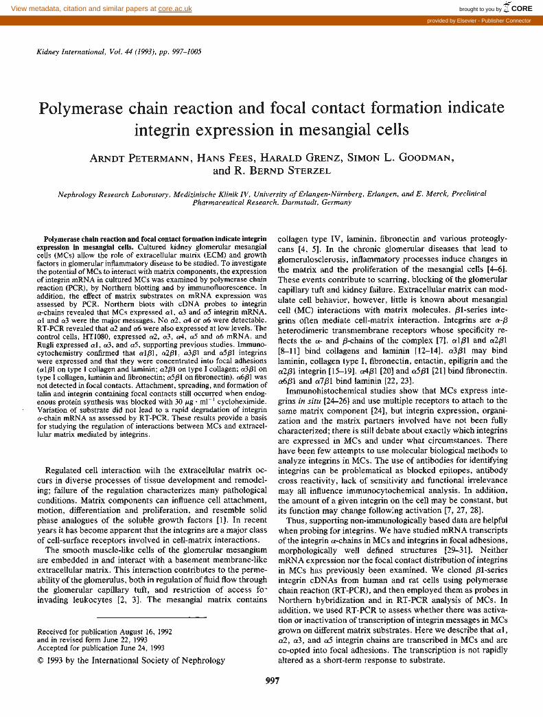
Fig. 1. Molecular cloning of integrin alphachains. (A) RT-PCR amplification of integrinmRNA from Rugli and HT1O8O cells. Reversetranscription of 5 ig of total RNA, 30 cyclePCR reaction. Ten percent of the reactionvolume has been loaded on a 1% agarose geland the bands visualized with ethidiumbromide. 4X = 4Xl74 markers; al = al-integrin primers on Rugli RNA; a2, a3, a4,a5, a6 = a2-a6 integrin primers on HTIO8ORNA. Marker sizes in base pairs are shown.(B) Cloned integnn insert and partialrestriction maps. al-6 are integrin a-chainsfrom which clones were derived. Enzymedigests of clones alternate with digests of theirinserts. PstI fragment a! cx pUC19 (pRIl.l);BamHI-EcoRI fragment a2 ex pUCI8(pHI2. 1); BamHI-EcoRI fragment a3 cxpUC18 (pHI3. 1); fragment GkaS EcoRIfragment a4 ex pUCI8 (pHI4.1). BamHI-EcoRI fragment aS cx pUCl8 (pHI5. 1).EcoRI fragment a6 ex pUCl8 (pHI6.l). 4X =4Xl74 markers: AH = A-HindIII markers.ApI = ApAI, E = EcoRl, B = BamHI, Ps =PstI, Pv = PvuI, X = XhoI. (C) Partialrestriction maps of cloned inserts.
Methods
Unless otherwise stated, chemicals were of analytical gradefrom Merck (Darmstadt) and biochemicals were from Sigma.Tissue-culture-quality water was used throughout. Reagents forpolymerase chain reaction (PCR) and molecular cloning werefrom Boehringer (Mannheim). Falcon tissue culture plastic wasfrom Becton-Djckenson. Tissue culture media and sera werefrom Gibco-BRL.
Cell culture
Cell culture of HT1O8O (human fibrosarcoma), Rugli (ratglioblastoma) and B 16 (mouse melanoma) cells have been
described elsewhere [11, 23, 32]. HT1O8O express VLA2,3,5and 6. Rugli express the rat homologues of VLA-1,3,5 anda7/31. B16 express the mouse homologues of VLA 2,5 and 6.
Preparation of MCs. Standard techniques were used [33, 34].The cortex of rat kidneys was cut into fragments and pressedthrough stainless steel sieves. Glomeruli retained between 105jim and 75 jim sieves were gathered, washed and plated in D20(DMEM, 20% heat-inactivated FCS, 2 m glutamate, 5 nglmlinsulin, 100 U/ml penicillin, 1000 jig/ml streptomycin) at 3 X i04glomeruli per 75 cm2 flask. MCs grew out from the glomeruliwithin three to six weeks. After three to four passages, thepopulation was shifted to D10 (D20, but 10% in FCS). Human
MCScci 5
Pst(1) ApaI(271)
ApaI(1 00) AatII(330)
StuI(530) MCS—
I
PstI(736)
PstI(1 47)
PstI(1 120) MCS
EcoRI(1 227)
PvuII(i 56)
PstI(346)
MCS BamIII(1)BgIII(131)PvuII(335)cz5S1
XhoI(1 08)BgIII(287)
MCS EcoRI(i) EcoRI(502) MCScx654 I
KpnI(723) MCS
EcoRI(882)
BamIII(1Q16) MCS
SacI(859)
XhoI(362)
C
I a4
aS
a6
4 fl y-Actin
p
S.
Petermann et a!: Integrin PCR in mesangial cells 999
MC cultures from kidneys not suitable for transplantation wereprepared in a similar manner, but were maintained in RiO[RPMI in place of DMEM, plus ITS (Boehringer; 10 sg ml)and HEPES (15 mM; pH 7.3)]. MCs were withdrawn from thecell cycle by incubation for 120 hours in D0.5 (D20, but 0.5%FCS). Rat and human MCs showed positive staining for Thy!.1,vascular smooth muscle cell myosin, actin and desmin, and hadcharacteristic morphology [33, 34]. The cultures showed uni-form marker expression and no expression of MAC-i and factorVIII, which are, respectively, markers for macrophages andendothelial cells [34].
Molecular biological methodsStandard methods were used, where not otherwise detailed
[35].Reverse transcription-polymerase chain reaction. PCR prim-
ers with minimal secondary structure or cross homology wereselected from integrin alpha-chain sequences (Rat al [36], andhuman a2 [37], a3 [38], a4 [39], aS [40] and a6 [41]) andsynthesized with terminal recognition sites for the restrictionendonucleases BamHI, EcoRl, or PstI. The primers used were:Rat al-up 5'CTACAAGATGGAGGATGGG 3' (bp. 1817-1835+ PstI), Rat al-down S'GCCTCAGTGAATCAAGGG 3'(bp2590-2607 + PstI); Hu a2-up 5'AGCTGGATCCACTCTGCT-TCAGTGCAAAGTTCAGA 3 '(bp.2082-2 106 + BamHI), Hua2-down S'GTCAGAATITCAAAGTCCCGTTCCAAATFCTG-GTAG 3' (bp. 3275-3299 + EcoRI); Hu a3-up 5'ATGCGAAT-TCTGTGGTCAGGGTCAGAAGACG 3' (bp. 5 16-535 + EcoRI),Hu a3-down 5' TCATGGATCCGGAGAAGAAGCCGTGG-AAGACAGCG 3'(bp. 1681-1705 + BamHI); Hu a4-up5 'GATCGGATCCGACGTGATTACAGGAAGCATACAGG3'(bp.1666-l691 + BamHI), Hu a4-down5' TGACGAATTCGC-AGTACAATAGCCTCTTATCAGTC 3' (bp. 2688-2713 + EcoRI)[39]; Hu aS-up 5'TCAGGGATCCAACTTCAGCTGGACTG-GCAGAAGCA 3'(bp.1657-l675 + BamHI) Hu aS-down5 'GATCGAATTCGGGCATCTTCAGGGCTTTGTACACA3'(bp.2900-2925 + EcoRl); Hu a6-up 5'TCAGGGATCCGTGT-TGCCAACCAGAATGGCTCGC 3'(bp.2323-2346 + BamHI) Hua6-down 5 'GATCGAATTCCAGTCACTCGAACCTGAGT-GCCTGC 3'(bp.3123-3l47 + EcoRI).
For the validation of the PCR primers, total RNA (1 to 5 p.g)[42] from HT!080 or Rugli cells was transcribed (1 hr x 37°C)with MMLV reverse transcriptase (200 U; Superscript, Strate-gene) using integrin downstream primer (30 pmol) in a volumeof 20 d (75 mi KC1, 3 mi MgCI2, 50 m Tris Cl; pH 7.3). Aquarter of the reverse transcription mix was used for PCR (30cycles; 35 seconds x 95°C, 30 seconds x 50°C, 150 seconds X72°C) by adding dNTPs (20 nmol), up and downstream primers(to 50 pmol) and Taq-polymerase (2.5 U; Stratagene) in 100 d(50 mst KCI, 1.5 m MgCI2, 0.1% Triton X-iOO, 10 mri Tris-Cl;pH 8.8). Amplified fragments were analyzed by electrophoresison agarose gels.
Cloning. cDNAs amplified by PCR were purified, cleavedwith restriction endonucleases (to generate PstI fragments forrat-al and BamHI-EcoRI fragments for a2, a3, a4, aS and a6),purified on agarose gels, and cloned into pUC18 or pUC19.Inserts were analyzed by restriction mapping and by sequenc-ing using the dideoxy method, which confirmed their identitywith the published sequences [36—41].
Northern hybridization analysis. Cloned integrin cDNA frag-
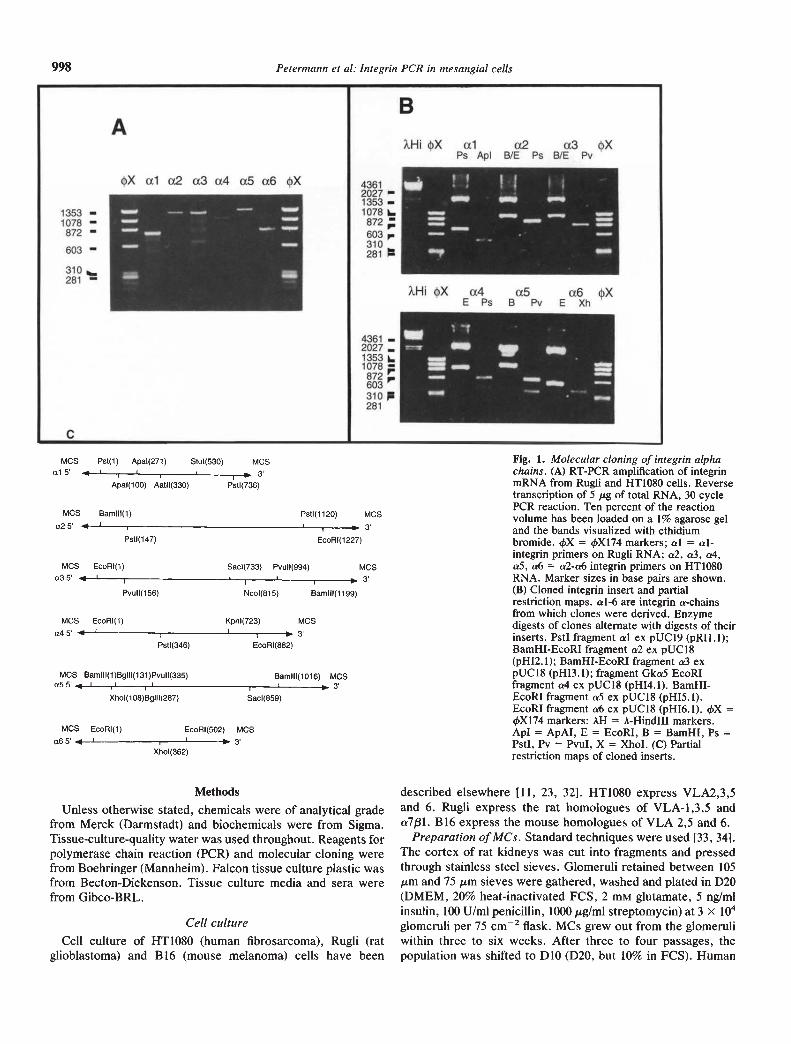
Fig. 2. Northern hybridization analysis of integrin expression in MCs.Total RNA (30 sg) was run on 1% agarose-formaldehyde gels, trans-ferred to nylon membranes and serially probed with radiolabeledintegrin inserts shown in Figure lB. HMC = human MCs, RMC = ratMCs. All films were exposed for 72 hours. The nature of the probesused is shown (al-a6).
ments were labeled with P32-ATP (Amersham) by randompriming (Gibco) to 1 X 10⁸ dpm . rg'. Total RNA (30 g)was resolved on agarose/formaldehyde gels, transferred tonylon membranes (Hybond-N; Amersham) by capillary wick-ing, and fixed by baking (2 hr x 80°C). After pre-hybridization(2 hr x 38°C; 50% formamide, 5 x SSC, 0.5 x Denhardtssolution, 200 sg m1' herring sperm DNA, 1% SDS, 50 mMphosphate buffer; pH 6.5) the membranes were hybridized withthe labeled cDNA probes (16 hr x 38°C). The membranes werewashed (38°C: 2 x 15 mm, 2 x SSC, 0.1% SDS; 2 x 30 mm, 0.2x SSC, 0.1% SDS) and exposed to X-ray film (Kodak, XAR-5)at —80°C. Filters were rehybridized following stripping inboiling SDS (0.1%; 3 x 1 mm). Hybridization with gamma-actincDNA probes revealed comparable quantities of gamma-actinmessage in each lane. The size of the hybridizing species wasestimated from the migration of the 28S and 18S rRNA [35].
Effect of substrate on inte grin gene expressionHuman vitronectin [43], fibronectin [44], murine EHS-tumor
laminin [45] and human collagen type I (gift of Dr. K. von derMark, Max-Planck, Erlangen, Germany) were >95% pure bySDS-PAGE. The proteins were dissolved in PBS (20 g ml')and coated (1 hr x 37°C) on 6-well tissue culture plates(Falcon). After blocking of free protein binding sites (2% heatdenatured BSA in PBS; 2 hr x 4°C), quiescent MCs were added(5 X iO cells/well) in A-buffer (DMEM, 0.5% BSA, HEPES, 20mM; pH 7.3). At six hours the cells were harvested, and totalcellular RNA was extracted [42]. Integrin cDNAs were ampli-fied using RT-PCR (=40 cells °°i0 ng RNA per PCR) and theproducts were resolved by agarose gel electrophoresis. Parallelamplifications with RNA from HT1O8O and Rugli cells provided
1000 Petermann et a!: Integrin PCR in mesangial cells
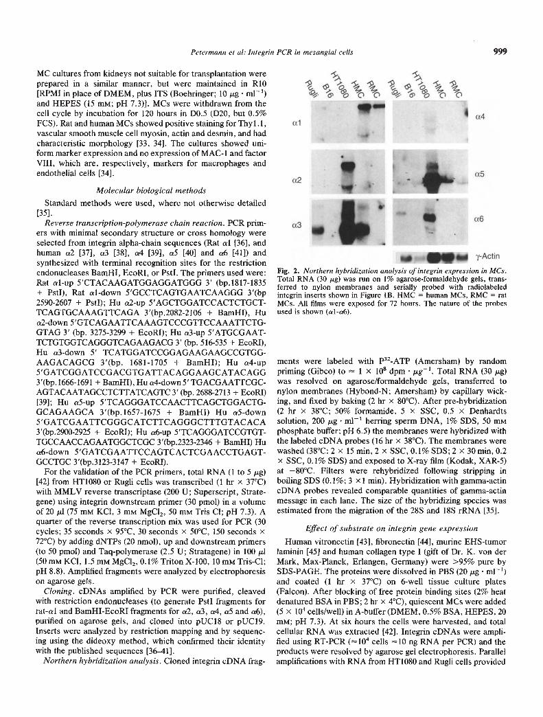
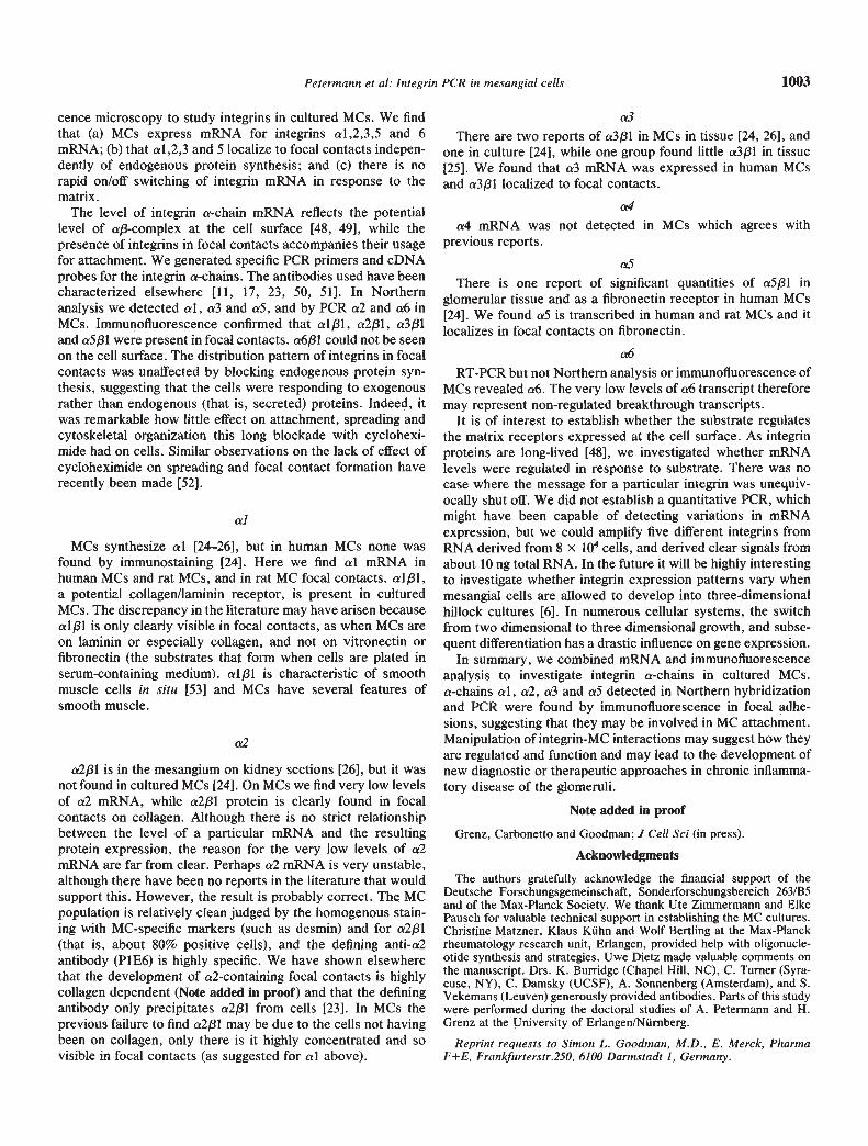
Fig. 3. Immunofluorescent labeling ofintegrins in MCs. MCs were plated on (A, B)collagen or (C, D) fibronectin for 4 hoursbefore fixation and labeling for (A) al, (B) a2,(C) cr3, (D) a5 integrin chains. Rat MCs areshown in A and human MCs in B through D.Scale bar = 10 /.LM.
markers for the amplified products, which after Southern trans.fer were confirmed by hybridization with the subcloned integrincDNAs.
Antibodies
Munne primary antibodies against human integnn alphachains P1E6 (cr2), P1B5 (cr3), and P1D6 (cr5) [15] and rabbitanti-fibronectin were from Telios (La Jolla, California, USA).Other antibodies were gifts of our colleagues: 7C7C7 (mouseanti-human cr4; Dr. J. Cassiman and S. Vekemans, Leuven),GOH3 (rat anti-mouse cr6 [46]), 3A3 (mouse anti-rat cr1 [9]),AIIB2 mouse anti-1 integrin [10] and rabbit anti-talin [47].Second layer fluorescent labeled antibodies were Texas-reddonkey-anti-mouse (Amersham) and Texas-red goat-anti-rat(Dianova). Rabbit anti-collagen-I was the gift of Dr. K. von derMark (Erlangen) and rabbit anti-laminin was produced byimmunization with EHS-laminin. TRITC-phalloidin was fromMolecular Probes (Oregon)
ImmunofluorescenceImmunofluorescence microscopy will be described elsewhere
(Note added in proof). Briefly, MCs were plated for four hoursin A-buffer on multichamber slides (Nunc) coated with matrixprotein (20 pg . mF' in PBS). After washing and fixation withparaformaldehyde (3% wt/vol in PBS: 10 mm x 0°C), the cellswere permeabilized (Triton X-l00; 1% (wt/vol) in PBS; 10 mmx 20°C), and incubated with primary antibody followed byfluorescently labelled secondary antibody before mounting inPVA (Moviol 4-88, pH 8.6; Hoechst). The cultures were exam-ined under a Leitz Axioplan microscope equipped for epifluo-rescence microscopy, and photographed at 3200 ASA on
TMAX film (Eastman-Kodak). Staining patterns qualitativelyas those described here developed as soon as the cells could befixed (0.5 to 1 hr) but were most extensive by four hours.
Cycloheximide treatment
Three hours before plating for immunofluorescence, cyclo-heximide to a final concentration of 30 jsg m1' was added toMC cultures. Subsequently, it was present in all solutions thatcame into contact with the cells until they were fixed forimmunostaining. Endogenously produced matrix componentslaminin, fibronectin and collagen type I were no longer detect-able after this procedure (Note added in proof).
Results
Human and rat MCs transcribe inte grin message
RT-PCR was used to amplify mRNA transcripts from Rugli(al) and HT1O8O cells (cr2-cr6). Integrin fragments of the pre-dicted sizes (al, 790 bp; cr2, 1237 bp; cr3, 1209 bp; cr4, 1066 bp;cr5, 1287 bp; cr6, 844 bp) could be amplified (Fig. la). Whencloned into pUC18 and pUC19 to generate unique cDNAprobes, the restriction maps (Fig. 1 b and c) and partial 2 to 300bp sequences matched published integrin data (AP, HF andSLG, unpublished observations).
The cloned integrin cDNAs were used to probe MCs usingNorthern hybridization (Fig. 2). Integrin a! cDNA probesyielded signals from both human and rat MCs, and from Rugli.A strong signal was seen at 11.5 2 kb and a weak signal at 5.5kb. Integrin a2 probes detected a signal only from HT1O8O cells,at 9.0 2 kb. cr3 probes detected signals in human MCs and in
k&cct
Petermann et a!: Integrin PCR in mesangial cells 1001
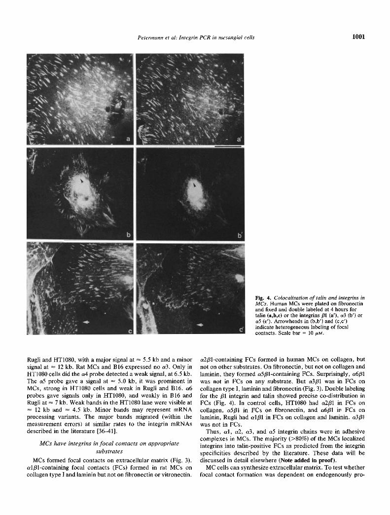
Fig. 4. Colocalisation of talin and integrins inMCs. Human MCs were plated on fibronectinand fixed and double labeled at 4 hours fortalin (a,b,c) or the integrins /31 (a'), a3 (b') ora5 (c'). Arrowheads in (b,b') and (c,c')indicate heterogeneous labeling of focalcontacts. Scale bar = 10 sM.
Rugli and HT1080, with a major signal at 5.5 kb and a minorsignal at 12 kb. Rat MCs and B16 expressed no a3. Only inHT1O8O cells did the a4 probe detected a weak signal, at 6.5 kb.The aS probe gave a signal at 5.0 kb, it was prominent inMCs, strong in HT1O8O cells and weak in Rugli and B16. a6probes gave signals only in HT1O8O, and weakly in B 16 andRugli at 7 kb. Weak bands in the HTIO8O lane were visible at
12 kb and 4.5 kb. Minor bands may represent mRNAprocessing variants. The major bands migrated (within themeasurement errors) at similar rates to the integrin mRNAsdescribed in the literature [36—41].
MCs have inte grins in focal contacts on appropriatesubstrates
MCs formed focal contacts on extracellular matrix (Fig. 3).a1J31-containing focal contacts (FCs) formed in rat MCs oncollagen type I and laminin but not on fibronectin or vitronectin.
a2j31-containing FCs formed in human MCs on collagen, butnot on other substrates. On fibronectin, but not on collagen andlaminin, they formed a5/31-containing FCs. Surprisingly, a6/31was not in FCs on any substrate. But a3/31 was in FCs oncollagen type I, laminin and fibronectin (Fig. 3). Double labelingfor the /31 integrin and talin showed precise co-distribution inFCs (Fig. 4). In control cells, HT1O8O had a2f31 in FCs oncollagen, a5f31 in FCs on fibronectin, and a6f31 in FCs onlaminin, Rugli had al/31 in FCs on collagen and laminin. a3/31was not in FCs.
Thus, al, a2, cr3, and aS integrin chains were in adhesivecomplexes in MCs. The majority (>80%) of the MCs localizedintegrins into talin-positive FCs as predicted from the integrinspecificities described by the literature. These data will bediscussed in detail elsewhere (Note added in proof).
MC cells can synthesize extracellular matrix. To test whetherfocal contact formation was dependent on endogenously pro-
1002 Petermann et a!: Integrin PCR in mesangial cells
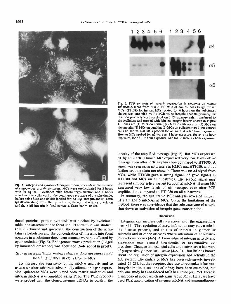
Fig. 6. PCR analysis of inte grin expression in response to matrixsubstrates. RNA from 8 x iO MCs or control cells (Rugli for ratMCs; HT1O8O for human MCs) plated for 6 hours on the substratesshown was amplified by RT-PCR using integrin specific-primers, thereaction products were resolved on 1.5% agarose gels, transferred tonitrocellulose and probed with labeled integrin inserts shown in Figure1. Lanes are (1) MCs on serum; (2) MCs on fibronectin; (3) MCs onvitronectin; (4) MCs on laminin; (5) MCs on collagen type I; (6) controlcells on serum. Rat MCs probed for a! were at a 0.5 hour exposure.Human MCs probed for a2 were an 8 hour exposure, for a4 a 16 hourexposure, for aS a 16 hour exposure, and for a6 were a 7 hour exposure.
Fig. 5. Inte grin and cytoskeleta! organization proceeds in the absenceof endogenous protein synthesis. MCs were preincubated for 3 hourswith 30 g m11 cycloheximide before trypsinization and 4 hoursattachment to collagen I in the continuous presence of cycloheximide,before being fixed and double labeled for (A) alp! integrin and (B) actin(phalloidin stain). Note the spread cells, the normal actin cytoskeletonand the alp! integrin in focal contacts. Scale bar = 10 M.
duced proteins, protein synthesis was blocked by cyclohexi-mide, and attachment and focal contact formation was studied.Cell attachment and spreading, the construction of the actin-talin cytoskeleton and the concentration of integrins into focalcontacts in a substrate-dependent manner were not affected bycycloheximide (Fig. 5). Endogenous matrix production (judgedby immunofluorescence) was abolished (Note added in proof).
Growth on a particular matrix substrate does not cause rapidswitching of integrin expression in MCs
To increase the sensitivity of the mRNA analysis and toassess whether substrate substantially affected integrin expres-sion, quiescent MCs were plated onto matrix molecules andintegrin mRNA was amplified using PCR. The PCR productswere probed with the cloned integrin cDNAs to confirm the
identity of the amplified message (Fig. 6). Rat MCs expressedal by RT-PCR. Human MC expressed very low levels of a2message even after PCR amplification compared to HT1O8O. Asignal was seen using a3 primers in HMCs and HTIO8O, withoutfurther probing (data not shown). There was no a4 signal fromMCs, while HT1O8O gave a strong signal. aS gave signals inHT1O8O and MCs on all substrates. The second signal mayrepresent a minor splice variant form of a5 mRNA. Human MCexpressed very low levels of a6 message, even after PCRamplification, compared to HT1O8O on all substrates.
In summary, the qualitative PCR analysis revealed integrinal,2,3,5 and 6 mRNAs in MCs. Given the limitations of themethod, there was no evidence that the substrate caused a rapidshut down or activation of integrin gene transcription.
Discussion
Integrins can mediate cell interaction with the extcacellularmatrix [7]. The regulation of integrin function may play a role inthe disease process, and this is of interest in glomerularsclerosis and in other diseases where alteration of cell-matrixinteractions occurs [4—6]. A knowledge of integrin activity andexpression may suggest therapeutic or preventative ap-proaches. Changes in mesangial cells and matrix are a hallmarkof progressive glomerular disease [4—6, 34], but little is knownabout the regulation of integrin expression and activity in theMC system. The matrix of MCs has been extensively investi-gated [24-26], but the receptors they use to recognize it has not.Integrins in tissue sections of kidney have been examined, butonly one study has considered MCs in culture [24]. Yet, there isdisagreement about which integrins are in MCs. Here, we haveused PCR amplification of integrin mRNA and immunofluores-
123456 123456
a6
Petermann et a!: Integrin PCR in mesangial cells 1003
cence microscopy to study integrins in cultured MCs. We findthat (a) MCs express mRNA for integrins al,2,3,5 and 6mRNA; (b) that a! ,2,3 and 5 localize to focal contacts indepen-dently of endogenous protein synthesis; and (c) there is norapid on/off switching of integrin mRNA in response to thematrix.
The level of integrin a-chain mRNA reflects the potentiallevel of af3-complex at the cell surface [48, 49], while thepresence of integrins in focal contacts accompanies their usagefor attachment. We generated specific PCR primers and cDNAprobes for the integrin a-chains. The antibodies used have beencharacterized elsewhere [11, 17, 23, 50, 51]. In Northernanalysis we detected al, a3 and a5, and by PCR a2 and a6 inMCs. Immunofluorescence confirmed that all, a2J31, a33land a5f3l were present in focal contacts. a6/31 could not be seenon the cell surface. The distribution pattern of integrins in focalcontacts was unaffected by blocking endogenous protein syn-thesis, suggesting that the cells were responding to exogenousrather than endogenous (that is, secreted) proteins. Indeed, itwas remarkable how little effect on attachment, spreading andcytoskeletal organization this long blockade with cyclohexi-mide had on cells. Similar observations on the lack of effect ofcycloheximide on spreading and focal contact formation haverecently been made [521.
alMCs synthesize al [24-26], but in human MCs none was
found by immunostaining [24]. Here we find a! mRNA inhuman MCs and rat MCs, and in rat MC focal contacts. al/31,a potential collagen/laminin receptor, is present in culturedMCs. The discrepancy in the literature may have arisen becauseal/31 is only clearly visible in focal contacts, as when MCs areon laminin or especially collagen, and not on vitronectin orfibronectin (the substrates that form when cells are plated inserum-containing medium). al/31 is characteristic of smoothmuscle cells in situ [53] and MCs have several features ofsmooth muscle.
a2
a2/31 is in the mesangium on kidney sections [26], but it wasnot found in cultured MCs [24]. On MCs we find very low levelsof a2 mRNA, while a2f31 protein is clearly found in focalcontacts on collagen. Although there is no strict relationshipbetween the level of a particular mRNA and the resultingprotein expression, the reason for the very low levels of a2mRNA are far from clear. Perhaps a2 mRNA is very unstable,although there have been no reports in the literature that wouldsupport this. However, the result is probably correct. The MCpopulation is relatively clean judged by the homogenous stain-ing with MC-specific markers (such as desmin) and for a2/3l(that is, about 80% positive cells), and the defining anti-a2antibody (P1E6) is highly specific. We have shown elsewherethat the development of a2-containing focal contacts is highlycollagen dependent (Note added in proof) and that the definingantibody only precipitates a2f3l from cells [23]. In MCs theprevious failure to find a2f31 may be due to the cells not havingbeen on collagen, only there is it highly concentrated and sovisible in focal contacts (as suggested for al above).
a3There are two reports of a3f31 in MCs in tissue [24, 26], and
one in culture [24], while one group found little a3131 in tissue[25]. We found that a3 mRNA was expressed in human MCsand a3131 localized to focal contacts.
a4a4 mRNA was not detected in MCs which agrees with
previous reports.aS
There is one report of significant quantities of a5f3l inglomerular tissue and as a fibronectin receptor in human MCs[24]. We found aS is transcribed in human and rat MCs and itlocalizes in focal contacts on fibronectin.
a6RT-PCR but not Northern analysis or immunofluorescence of
MCs revealed a6. The very low levels of a6 transcript thereforemay represent non-regulated breakthrough transcripts.
It is of interest to establish whether the substrate regulatesthe matrix receptors expressed at the cell surface. As integrinproteins are long-lived [48], we investigated whether mRNAlevels were regulated in response to substrate. There was nocase where the message for a particular integrin was unequiv-ocally shut off. We did not establish a quantitative PCR, whichmight have been capable of detecting variations in mRNAexpression, but we could amplify five different integrins fromRNA derived from 8 x LO cells, and derived clear signals fromabout 10 ng total RNA. In the future it will be highly interestingto investigate whether integrin expression patterns vary whenmesangial cells are allowed to develop into three-dimensionalhillock cultures [6]. In numerous cellular systems, the switchfrom two dimensional to three dimensional growth, and subse-quent differentiation has a drastic influence on gene expression.
In summary, we combined mRNA and immunofluorescenceanalysis to investigate integrin a-chains in cultured MCs.a-chains a!, a2, a3 and aS detected in Northern hybridizationand PCR were found by immunofluorescence in focal adhe-sions, suggesting that they may be involved in MC attachment.Manipulation of integrin-MC interactions may suggest how theyare regulated and function and may lead to the development ofnew diagnostic or therapeutic approaches in chronic inflamma-tory disease of the glomeruli.
Note added in proof
Grenz, Carbonetto and Goodman; J Cell Sci (in press).
Acknowledgments
The authors gratefully acknowledge the financial support of theDeutsche Forschungsgemeinschaft, Sonderforschungsbereich 263/B5and of the Max-Planck Society, We thank Ute Zimmermann and ElkePausch for valuable technical support in establishing the MC cultures.Christine Matzner, Klaus KUhn and Wolf Bertling at the Max-Planckrheumatology research unit, Erlangen, provided help with oligonucle-otide synthesis and strategies. Uwe Dietz made valuable comments onthe manuscript. Drs. K. Burridge (Chapel Hill, NC), C. Turner (Syra-cuse, NY), C. Damsky (UCSF), A. Sonnenberg (Amsterdam), and S.Vekemans (Leuven) generously provided antibodies. Parts of this studywere performed during the doctoral studies of A. Petermann and H.Grenz at the University of Erlangen/NUrnberg.
Reprint requests to Simon L. Goodman, M.D., E. Merck, PharinaF+E, Frankfurterstr.250, 6100 Darmstadt 1, Germany.
1004 Petermann et a!.' !ntegrin PCR in mesangial cells
References
1. NATHAN C, SPORN M: Cytokines in context. J Cell Biol 113:981—986, 1991
2. SAIC&I T, KRIZ W: The structural relationship between mesangialcells and basement membrane of the renal glomerulus. Anat Em-btyol 176:373-386, 1987
3. LATTA H: An approach to the structure and function of theglomerular mesangium. JAm Soc Nephrol 2:S65—S73, 1992
4. STERZEL RB, LOVETT DH: Interactions of inflammatory and gb-merular cells in the response to glomerular injury, in Immunopa-thology of Renal Diseases, edited by WILsoN C, New York,Churchill Livingstone, 1988, pp. 137—173
5. MENE P, SIMONSON MS, DUNN MJ: Physiology of the mesangialcell. Physiol Rev 69:1347—1424, 1989
6. STERZEL RB, SCHULZE-LOHOFF E, WEBER M, GOODMAN SL:Interactions between glomerular mesangial cells, cytokines andextracellular matrix. JAm Soc Nephrol 2:S 126—S 131, 1992
7. HYNEs RO: Integrins: Versatility, modulation, and signaling in celladhesion. Cell 69:11—25, 1992
8. TURNER DC, FLIER LA, CARBONETFO S: Magnesium-dependentattachment and neurite outgrowth by PC 12 cells on collagen andlaminin substrata. Dev Biol 121:510—525, 1987
9. TURNER DC, FLIER LA, CARBONETTO S: Identification of a cell-surface protein involved in PC12 cell-substratum adhesion andneurite outgrowth on laminin and collagen. J Neurosci 9:3287—3296,1989
10. HALL DE, REICHARDT LF, CROWLEY E, HOLLEY B, MOEZZI H,SONNENBERG A, DAMSKY CH: The alpha-1/beta-l and alpha-6/beta-l integrin heterodimers mediate cell attachment to distinctsites on laminin. J Cell Biol 110:2175—2184, 1990
11. GOODMAN SL, AUMAILLEY M, VON DER MARK H: Multiple cellsurface receptors for the short arms of laminin: a1f31 integrin andRGD-dependent proteins mediate cell attachment only to domainsIII in murine tumour laminin. J Cell Biol 113:931—941, 1991
12. LANGUINO LR, GEHLSEN KR, WAYNER E, CARTER WG, ENGVALLE, RUOSLAHTI E: Endothelial cells use alpha-2-beta-1 integrin as alaminin receptor. J Cell Biol 109:2455—2462, 1989
13. STAATZ WD, FOX KF, ZUTFER MM, ADAMS SP, RODRIGUEZ BA,SANTORO SA: Identification of a tetrapeptide recognition sequencefor the alpha 2 beta 1 integrin in collagen. J Biol Chem 266:7363—7367, 1991
14. CHAN BMC, KASSNER PD, SCHIRO JA, BYERS HR, KUPPER TS,HEMLER M: Distinct cellular functions mediated by different VLAintegrin alpha subunit cytoplasmic domains. Cell 68:1051—1060,1992
15. WAYNER EA, CARTER WG: Identification of multiple cell adhesionreceptors for collagen and fibronectin in human fibrosarcoma cellspossessing unique alpha and common beta subunits. J Cell Biol105: 1873—1884, 1987
16. CARTER WG, RYAN MC, GALIR PJ: Epiligrin, a new cell adhesionligand for integrin alpha 3 beta 1 in epithelial basement membranes.Cell 65:599—610, 1991
17. ELICES MJ, URRY LA, HEMLER ME: Receptor functions for theintegrin VLA-3: Fibronectin, collagen, and laminin binding aredifferentially influenced by Arg-Gly-Asp peptide and by divalentcations. J Cell Biol 112:169—181, 1991
18. GEHLSEN KR, SRIRAMARAO P, FURCIIT LT, SKUBITZ APN: Asynthetic peptide derived from the carboxy terminus of the lamininA chain represents a binding site for the a3/31 integrin. J Cell Biol117:449—459, 1992
19. SYMINGTON BE, TAKADA Y, CARTER WG: Interaction of integrinsa3131 and a2131: Potential role in keratinocyte intercellular adhe-sion. J Cell Rio! 120:523—535, 1993
20. ELICES MJ, OSBORN L, TAKADA Y, CROUSE C, LUHOWSKYJ S,HEMLER ME, LOBE RR: VCAM-1 on activated endothelium inter-acts with the leukocyte integrin VLA-4 at a site distinct from theVLA-4/flbronectin binding site. Cell 60:577—584, 1990
21. PYTELA R, PIERSCHBACHER MD, RUOSLAHTI E: Identification andisolation of a 140 kd cell surface glycoprotein with the propertiesexpected of a fibronectin receptor. Cell 40:191—198, 1985
22. SONNENBERG A, LINDERS CJT, MODDERMAN PW, DAMSKY CH,AUMAILLEY M, TIMPL R: Integrin recognition of different cell-binding fragments of laminin (P1 ,E3 ,E8) and evidence that alpha-6beta-i but not alpha-6-beta-4 functions as a major receptor forfragment E8. J Cell Biol 110:2145—2155, 1990
23. VON DEE MARK H, DUita J, SONNENBERG A, VON DER MARK K,DEUTZMANN R, GOODMAN SL: Skeletal muscle myoblasts utilize anovel /31-series integrin, and not a'6/3l for binding to the E8 and T8fragments of laminin. J Biol Chem 35:23593—23601, 1991
24. Costo FG, SEDMAK DD, NAHMAN NSJ: Cellular receptors formatrix proteins in normal human kidney and human mesangialcells. Kidney mt 38:886—895, 1990
25. KORHONEN M, YLANNE J, LAITINEN L, VITANEN I: The aiphal-alpha6 subunits of integrins are characteristically expressed indistinct segments of developing and adult human nephron. J CellBiol 111:1245—1254, 1990
26. SIMON EE, MCDONALD JA: Extracellular matrix receptors in thekidney cortex. Am J Physiol 259:F783—F792, 1990
27. DRANSFIELD I, HOGG N: Regulated expression of Mg2 bindingepitope on leukocyte integrin alpha subunits. EMBO J 8:3759—3765,1989
28. SPRINGER TA: The sensation and regulation of interactions with theextracellular environment: The cell biology of lymphocyte adhesionreceptors. Annu Rev Cell Biol 6:359—402, 1990
29. BURRIDGE K, FATH K, KELLY T, NUCKOLLS G, TURNER C: Focaladhesions: Transmembrane junctions between the extracellularmatrix and the cytoskeleton. Annu Rev Cell Biol 4:487—525, 1988
30. OTEY CA, BURRIDGE K: Patterning of the membrane cytoskeletonby the extracellular matrix. Semin Cell Biol 1:391—399, 1990
31. OTEY CA, PAVALKO FM, BURRIDGE K: An interaction betweenalpha-actinin and the beta-l integrin subunit in vitro. J Cell Biol111:721—729, 1990
32. BROWN JC, GOODMAN SL: Different cellular receptors for humanplacental laminin and murine EHS laminin. FEBS Lett 282:5—8,1991
33. LOVETT DH, RYAN JL, STERZEL RB: A thymocyte-activatingfactor derived from glomerular mesangial cells. J Immunol 130:1796—1801, 1983
34. ISHIMURA E, STERZEL RB, BUDDE K, KASHGARIAN M: Formationof extracellular matrix by cultured rat mesangial cells. Am J Pathol134:843—855, 1989
35. SAMBROOK J, FRITSCH EE, MANIATIS T: Molecular Cloning, ALaboratory Manual, Cold Spring Harbor, Cold Spring HarborLaboratory Press, 1989
36. IGNATIUS Mi, LARGE TH, HOUDE M, TAWIL JW, BARTON A,ESCH F, CARaONETTO S, REICHARDT LF: Molecular cloning of therat integrin alphal-subunit: A receptor for laminin and collagen. JCell Biol 111:709—720, 1990
37. TAKADA Y, HEMLER ME: The primary structure of the VLA-2/collagen receptor alpha-2 subunit (platelet GPla): Homology toother integrins and the presence of a possible collagen-bindingdomain. J Cell Biol 109:397—407, 1989
38. TAKADA Y, MURPHY E, PIL P. CHEN C, GINSBERG MH, HEMLERME: Molecular cloning and expression of the cDNA for alpha 3subunit of human alpha 3 beta I (VLA-3), an integrin receptor forfibronectin, laminin, and collagen. J Cell Biol 115:257—266, 1991
39. TAKADA Y, ELICES MJ, CROUSE C, HEMLER ME: The primarystructure of the alpha 4 subunit of VLA-4: Homology to otherintegrins and a possible cell-cell adhesion function. EMBO J8:1361—1368, 1989
40. ARGRAVES WS, SUZUKI S, A H, THOMPSON K, PIERSCHBACHERMD, RUOSLAHTI E: Amino acid sequence of the human fibronectinreceptor. J Cell Biol 105:1183—1190, 1987
41. HOGERVORST F, KUIKMAN I, VAN-KESSEL AG, SONNENBERG A:Molecular cloning of the human alpha 6 integrin subunit. Alterna-tive splicing of alpha 6 mRNA and chromosomal localization of thealpha 6 and beta 4 genes. Eur J Biochem 199:425—433, 1991
42. CHOMCZYNSKI P, SACCHI N: Single-step method of RNA isolationby acid guanidinium thiocyanate-phenol-chloroform extraction.Anal Biochem 162:156—159, 1987
43. YATOHGO T, IzuMI M, KASHIWAGI H, HAYASHI M: Novel purifi-cation of vitronectin from human plasma by heparin affinity chro-matography. Cell Struct Fund 13:281—292, 1988
Petermann et al: Inte grin PCR in mesangial cells 1005
44. ENGVALL E, RUOSLAHTI E: Binding of soluble form of fibroblastsurface protein, fibronectin, to collagen. mt j Cancer 20:1—5, 1977
45. PAULSSON M, AUMAILLEY M, DEUTZMANN R, TIMPL R, BECK K,ENGEL J: Laminin-nidogen complex extraction with chelatingagents and structural characterization. Eur J Biochem 166:11—19,1987
46, SONNENBERG A, MODDERMAN PW, HOGERVORST F: Lamininreceptor on platelets is the intergrin VLA-6. Nature (London)336:487—489, 1988
47. Buiuiro K, CONNELL L: A new protein of adhesion plaques andruffling membranes. J Cell Biol 359:367, 1983
48. ROSA JP, MCEVER RP: Processing and assembly of the integrin,glycoprotein lib-IHa, in HEL cells. J Biol Chem 264:12596—12603,1989
49. SONNENBERG A, HOGERVORST F, OSTEROP A, VELTMAN FE:Identification and characterization of a novel antigen complex on
mouse mammary tumor cells using a monoclonal antibody againstplatelet glycoprotein Ic. J Biol Chem 263:14030—14038, 1988
50. GEHLSEN KR, DILLNER L, ENGVALL E, RUOSLAHTI E: The humanlaminin receptor is a member of the integrin family of cell adhesionreceptors. Science 241:1228—1229, 1988
51. TAKADA Y, WAYNER EA, CARTER WG, HEMLER ME: Extracellu-lar matrix receptors, ECMRII and ECMRI, for collagen andfibronectin correspond to VLA-2 and VLA-3 in the VLA family ofheterodimers, J Cell Biochem 37:385—393, 1987
52. BURRIDGE K, TURNER CE, ROMER LH: Tyrosine phosphorylationof paxillin and ppl25fak accompanies cell adhesion to extracellularmatrix: A role in cytoskeletal assembly. J Cell Biol 119:893—903,1992
53. BELKIN VM, BELKIN AM, KOTELIANSKY VE: Human smoothmuscle VLA-l integrin: Purification, substrate specificity. J CellBiol 111:2159—2170, 1990


![Escherichia coli DNA Polymerase III Holoenzyme Subunits alpha, beta, and [IMAGE] Directly Contact the Primer-Template](https://img.pdfslide.net/doc/110x75/635e348ea0f1eac29f0c94c6/escherichia-coli-dna-polymerase-iii-holoenzyme-subunits-alpha-beta-and-image.jpg)


