Embed Size (px)
Citation preview
Review Article
Post-Genomics Nanotechnology Is Gaining Momentum:Nanoproteomics and Applications in Life Sciences
Firas H. Kobeissy,1,5,* Basri Gulbakan,2,3,* Ali Alawieh,4,* Pierre Karam,5 Zhiqun Zhang,1
Joy D. Guingab-Cagmat,6 Stefania Mondello,7 Weihong Tan,2,8,9 John Anagli,6 and Kevin Wang1
Abstract
The post-genomics era has brought about new Omics biotechnologies, such as proteomics and metabolomics, aswell as their novel applications to personal genomics and the quantified self. These advances are now alsocatalyzing other and newer post-genomics innovations, leading to convergences between Omics and nano-technology. In this work, we systematically contextualize and exemplify an emerging strand of post-genomicslife sciences, namely, nanoproteomics and its applications in health and integrative biological systems. Nano-technology has been utilized as a complementary component to revolutionize proteomics through differentkinds of nanotechnology applications, including nanoporous structures, functionalized nanoparticles, quantumdots, and polymeric nanostructures. Those applications, though still in their infancy, have led to several highlysensitive diagnostics and new methods of drug delivery and targeted therapy for clinical use. The present articlediffers from previous analyses of nanoproteomics in that it offers an in-depth and comparative evaluation of theattendant biotechnology portfolio and their applications as seen through the lens of post-genomics life sciencesand biomedicine. These include: (1) immunosensors for inflammatory, pathogenic, and autoimmune markers forinfectious and autoimmune diseases, (2) amplified immunoassays for detection of cancer biomarkers, and (3)methods for targeted therapy and automatically adjusted drug delivery such as in experimental stroke and braininjury studies. As nanoproteomics becomes available both to the clinician at the bedside and the citizens who areincreasingly interested in access to novel post-genomics diagnostics through initiatives such as the quantifiedself, we anticipate further breakthroughs in personalized and targeted medicine.
Introduction
The post-genomics era has realized that the sequencedgenome is not enough to discern the global biological
processes fully at a systems level (Collins et al., 2003; Gandhiand Wood, 2012). New ‘‘omics’’ fields characterized by data-intensive research and biotechnologies enabling omics in-vestigation have come into existence to narrow the existinggaps between discovery science and the attendant clinicalapplications. One of the significant contributors in the post-genomics era is the field of proteomics. The rising interest inprotein science is believed to be secondary to the far biologicaldistance between genes and phenotypes on the one hand and
the dynamic nature of proteins on the other (Aebersold andMann, 2003; Altelaar et al., 2012).
Chief among the aims of proteomics is the analysis of cel-lular proteins in terms of abundance and dynamics in re-sponse to physiological and pathological changes, as well asenvironmental influences. Proteins are central cellular com-ponents in biological networks, with diverse functions in-cluding cytoskeletal building blocks, enzymes catalyzingbiochemical reactions, antibodies contributing to immunity,or transcription factors affecting gene expression. Proteomicsby definition is the systematic identification and character-ization of protein sequence, abundance, post-translationalmodifications, interactions, activity, subcellular localization,
1Center for Neuroproteomics and Biomarkers Research, Department of Psychiatry, McKnight Brain Institute, Departments of 2Chemistryand Center for Research at Bio/Nano Interface, and 8Physiology and Functional Genomics, University of Florida, Gainesville, Florida.
3Department of Chemistry and Applied Biosciences ETH Zurich, Switzerland.4Department of Neurosciences, Medical University of South Carolina, Charleston, South Carolina.5American University of Beirut, Department of Chemistry, Beirut, Lebanon.6Center of Innovative Research, Banyan Biomarkers, Inc., Alachua, Florida.7University of Messina, Department of Neurosciences, Messina, Italy.9Moffitt Cancer Center and Research Institute, Tampa, Florida.*These authors contributed equally to this work.
OMICS A Journal of Integrative BiologyVolume 18, Number 00, 2014ª Mary Ann Liebert, Inc.DOI: 10.1089/omi.2013.0074
1
and structure in a given cell type at a particular time point(Zhang et al., 2013). Protein profiles at both physiological andpathophysiological processes characterize the informationflow in a cell, tissue, or organism (Petricoin et al., 2002). Pro-teomics studies utilize several available techniques for theidentification, validation, quantification, and expression ofcertain protein(s). Such techniques are highly sensitiveachieving targeted proteins analysis; among these tools are:Western blotting, ELISA, and protein arrays, which are usedfor identification and quantification of proteins. On the otherhand, proteomics can be of high throughput nature where aset of proteins are globally evaluated (expression and quan-tification) by methods including mass spectrometry, proteinarrays and 1D and 2-D gel electrophoresis (Kobeissy et al.,2008b; Lamond et al., 2012; Smith and Figeys, 2006).
It is generally accepted that the human genome consists ofaround 40,000 genes (Lander et al., 2001; Yates, 2013), yet asingle gene does not necessarily translate into one protein,and once proteins are synthesized, many undergo post-translational modification (PTM) by phosphates, carbohydrates,lipids, or other groups, which tremendously complicates theglobal proteome profiling (Mann and Jensen, 2003).
Similar to the Human Genome Project, a Human ProteomeProject (Cottingham, 2008) was initiated by a group of scien-tists from the Human Proteome Organization (HUPO) andwas launched at the 2011 World Congress of Proteomics inGeneva, Switzerland (Omenn, 2012). In this project, scientistshave to deal with approximately more than 1,000,000 pro-teins, which can then be further complicated by several pro-tein modifications. The time, effort, and money it takes for aprotein to be fully identified, sequenced, validated, andstructurally characterized impose a true challenge for re-searchers (Lemoine et al., 2012; Yau, 2013). Consequently, thevery early hope of characterizing the whole human proteomeshifted focus on trying to find molecular differences betweenone functional state of a biological proteome system to an-other aided by systems biology analysis, which certainlyprovided more precise comprehensive data of the proteomeprofile (Cox and Mann, 2007).
Challenges in Proteomics
The rapidly growing field of proteomics has excelled inseveral disciplines in biology, including injury, cancer, aging,and different neurological conditions, as well as psychiatricconditions including drug/substance abuse, schizophrenia,and depression (Abul-Husn and Devi, 2006; Becker et al.,2006; Becker, 2006; Choudhary and Grant, 2004; Cochranet al., 2003; Dean and Overall, 2007; Dumont et al., 2004).Proteomics is one of the fastest growing fields of biochemicalsciences; a PubMed search reveals 287,021 articles publishedin the past 2 years containing the word ‘‘protein,’’ comparedto 156,200 articles using the term ‘‘gene’’ ( January, 2011–April, 2013), which may also reflect a shift in genomics studiestowards proteomics investigations. Interestingly, the field ofproteomics is in a continual growth with the introduction ofsome more specialized disciplines and subdisciplines such asneuroproteomics, psychoproteomics and nanoproteomics(Kobeissy et al., 2008a; 2008c).
Proteomics analysis is more complicated than genomicsdue to a number of challenges occurring at different levels ofprotein post-translational modifications (Choudhary and
Grant, 2004; Zhao and Jensen, 2009). For example, on the levelof brain proteome, it is estimated that there exist around20,000 brain proteins that are differentially expressed in thevarious regions of the brain (Wang et al., 2005a). Furthermore,it is difficult to associate mRNA expression to the proteinexpression levels (Denslow et al., 2003; Freeman et al., 2005;Morrison et al., 2002; Wang et al., 2004). This is due to severalfactors, including ‘‘alternative splicing,’’ which is highlycommon in brain tissue, generating several copies of highlyrelated splices from a single gene (Hunnerkopf et al., 2007;Missler and Sudhof, 1998; Morrison et al., 2002; Wu andManiatis, 1999). It is estimated that a single gene can generateup to 10 protein isoforms (Kim et al., 2004; Williams et al.,2004). Thus, knowing the gene sequence is not sufficient topredict the possible translation pattern of that specific protein.Currently, there are approximately 430 possible protein post-translational modifications (PTMs) that can contribute to thiscomplexity (Khoury et al., 2011; Woodsmith et al., 2013).
Post-translational modifications are defined as integral‘‘chemical modifications of proteins that have implications onnew protein functions in response to a specific cellular con-dition such as activation, turnover, downregulation, confor-mation, and localization (Berretta and Moscato, 2010; Husiand Grant, 2001; Khoury et al., 2011; Morrison et al., 2002;Witze et al., 2007). Furthermore, the different expression lev-els of certain proteins leading to huge dynamic range differ-ence hamper the analysis of low expression proteins (Hortinand Sviridov, 2010; Zubarev, 2013). For example, there is0.5 pg/mL of IL-6 compared to 35 mg/mL of albumin presentin the serum that exemplifies the dynamic range difference ofsome protein expression levels analyzed using traditionalproteomics techniques including mass spectrometry, ELISA,Western blotting, and protein arrays (Anderson and Ander-son, 2002). Therefore, many of the potential biomarkers haveconcentrations in the femtomolar range while being im-mersed in a complexity of other biological components withconcentrations that span 12–15 orders of magnitude (Mitchell,2010; Rifai et al., 2006).
Furthermore, some protein classes are notoriously verydifficult to analyze due to their intrinsic characteristics. Forinstance, membrane proteins constitute almost 30% of theopen reading frames in the sequenced genome (Bagos et al.,2004; Lai, 2013; Vuckovic et al., 2013); however, they are veryhydrophobic and buried in the lipid bilayer and tend to pre-cipitate in aqueous buffers; thus, are harder to isolate. In ad-dition, the field of proteomics lacks a DNA–PCR-liketechnique, which brings about sensitivity problems, associ-ated with low abundant proteins.
Role of Nanotechnologies in Proteomics
Nanotechnology is defined as the systematic study of aparticular system at the nanometer scale (1–100 nm) (Nieet al., 2007; Vo-Dinh, 2005). Considering that average bondlengths range in the picometer range (74 picometer for H–Hbond, 200 picometer for C–I bond); this is basically the limit ofthe metrics at which molecules cannot be further manipulatedat the molecular level.
Nanotechnology opens up unique opportunities, not onlyfor material science research, but also for biology, medi-cine, and many other disciplines by manipulating individualatoms and molecules in a specific way that can fit into a
2 KOBEISSY ET AL.
certain application (Petros and DeSimone, 2010). So, what isthe relationship between nanotechnology and proteomics andwhy will nanotechnology be beneficial for proteomics appli-cations?
As mentioned previously, proteomics technology is chal-lenged by several limitations (PTMs, dynamic range, biolog-ical complexities, etc.), which in turn makes it incapable ofachieving some of its goals in elucidating protein changesunless it is coupled with other methods.
The attractive point of nanotechnology is that it can reducethese difficulties and therefore help to draw new informationout of biological systems that otherwise would not be possibleby using conventional techniques. The field of nanotechnol-ogy has been associated with several proteomics applicationssuch as phosphoproteomics/metal oxide nanoparticles, na-nostructure surfaces for protein separation, and analyticaldetection of biomarker proteins using arrays techniques(Leitner, 2010; Nelson et al., 2009; Northen et al., 2007; Rissinet al., 2010). This merging between nanotechnology andproteomics has generated nanoproteomics, which is defined asa discipline of science involving the application of proteomicstechniques aided by nanotechnology to enhance probing andevaluating protein systems (Archakov, 2007). As discussed byVo-Dinh and colleagues (2005), several basic cellular struc-tures (proteins, polymers, carbohydrates, and lipids) aremolecules with similar sizes to various nanostructures. Thesimilarity between these biological nanosystems and nano-structures have important implications in designing andmanufacturing of the next generation nano-assemblies(nanotechnology tool kits, lipid vesicles, and dendritic poly-mers) that may have important medical and biotechnologicalapplications (Chen et al., 2013; Zhang et al., 2009).
In this report, we will summarize the recent nano-technology applications that have been applied in differentproteomics-related applications. We will discuss nanostructuredsurfaces, nanoporous particles, magnetic nanomaterials, goldnanoparticles, carbon-based nanomaterials, polymeric nano-structures, quantum dots technology, and finally clinicalutility of nanoproteomics, along with their technologycommercialization.
Nanostructured Surfaces
Conventionally, the size of typical structures is on the orderof micrometers; however, technologies in the area of nano-technology have enabled us to achieve surface structure in therange of nanometers. Nanostructured surfaces have attractedbig attention owing to their unique physical, chemical, andstructural properties, including enormously high surface area,quantum confinement, and interaction with light (Hoheiselet al., 2010; Parker and Townley, 2007; Tawfick et al., 2012; Vo-Dinh et al., 2005). Not surprisingly, nanoparticles have foundwidespread use in proteomics applications that can be sum-marized into three basic areas: (a) scaffold for protein bio-sensing, (b) sample purification and enrichment tool, and (c)substrate for mass spectrometry analysis (Luong-Van et al.,2013). These diverse uses enhance the efficiencies of sev-eral proteomics applications, including ELISA and massspectrometry-related techniques, as will be discussed.
Protein biosensing changes the morphology of a surfacefrom plain to a nanostructured form and alters the sensingproperties of that particular material by (1) increasing the
surface area and, hence, increasing the available binding sites,and (2) enhancing the accessibility of the target to the surface-immobilized probe. This, in return, allows the detection ofspecific target(s) with higher sensitivity and faster kinetics.Kang et al. (2005) reported that protein arrays prepared bysilica nanotube membranes significantly improved the signal-to-noise ratio compared to plain analogs. Similarly, Kim et al.(2010) also showed that three-dimensional surfaces providehigher aspect ratios, improving the immobilization capacityof the capturing probe 5-fold and enhancing its detectionability to almost 15-fold. Another example of nanostructuredmaterial is porous silicon that was first discovered by BellLaboratories in 1960s; it has been used for many proteinsensing applications ( Jane et al., 2009). Unique fluorescenceproperties and tremendous surface areas of porous silicon(500–800 m2/g) made it an ideal candidate for protein sensors(Sailor, 2007). Ressine et al. (2007) used porous silicon as ascaffold for antibody arrays and obtained detection limitsdown to 1 picomolar for IgG and 20 picomolar for prostate-specific antigen (PSA) in clinical samples.
Hill et al. (2009) explored the curvature effect of sphericalgold nanoparticles on the loading density of probes. Theyshowed that the binding to the nanoparticle surface is similarto that of a planar structure when the diameter of the particleis greater than 60 nm. Kelley et al. further explored this con-cept by controlling the topography of gold microelectrodes atthe nanostructure level. Neither the increase of surface areanor the probe density were found to be the dominant factor inimproving on the sensitivity but the nanotopography intro-duced onto the microelectrodes (Bin et al., 2010; Das andKelley, 2011). Using the optimized topography, ovarian can-cer biomarker CA-125 was detected to the limit of 0.1 U/mLwithout the need to covalent label the target or the probe norto resort to sandwich complexes techniques (Das and Kelley,2011).
Another application of nanoparticles is femtoliter arrays.This technology has been developed in studying single en-zyme molecules, detection of low abundance protein bio-markers in biological fluids, and single cell analysis (Malhotraet al., 2012; Rusling et al., 2013). Since the properties of singlemolecules significantly differ from the bulk solution, thismethod enabled detailed studies of single proteins and theirkinetic properties, as illustrated by Gorris and Walt (2010).Also, detection of low femtomolar biomarker proteins wasachieved utilizing such techniques (for reviews, see Gorrisand Walt, 2010; LaFratta and Walt, 2008; Rissin and Walt,2006). The principle of detection enhancement basically relieson Poisson Statistics, dictating that at very low concentrationvalues, femtoliter size reaction chambers may contain eitherone or no molecules (Rissin et al., 2010). Recently, this tech-nology has been coupled with classical enzyme-linked im-munosorbent assay (ELISA). ‘‘Femtoliter arrays’’ platform,used for protein detection, has been called ‘‘single moleculearrays,’’ which can enhance ELISA assay sensitivity up to68,000 times, reaching zeptomolar (10 - 21) detection limits thatenable identification of markers specific to prostate cancer(Rissin et al., 2010).
Matrix-assisted laser desorption ionization mass spec-trometry (MALDI-MS) has been a workhorse in proteomicsstudies; since the early days of its discovery (Karas et al.,2000). However, the presence of the matrix often causes het-erogeneous co-crystallization of the matrix and analytes,
POST-GENOMICS NANOTECHNOLOGY 3
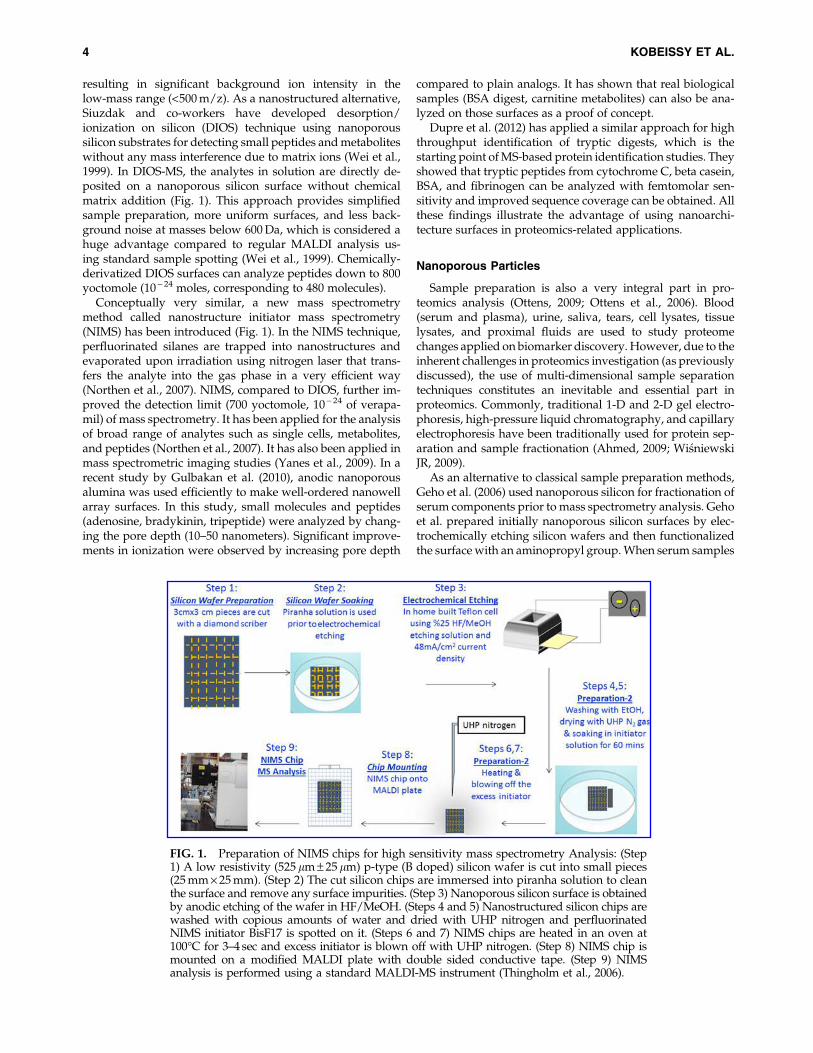
resulting in significant background ion intensity in thelow-mass range (<500 m/z). As a nanostructured alternative,Siuzdak and co-workers have developed desorption/ionization on silicon (DIOS) technique using nanoporoussilicon substrates for detecting small peptides and metaboliteswithout any mass interference due to matrix ions (Wei et al.,1999). In DIOS-MS, the analytes in solution are directly de-posited on a nanoporous silicon surface without chemicalmatrix addition (Fig. 1). This approach provides simplifiedsample preparation, more uniform surfaces, and less back-ground noise at masses below 600 Da, which is considered ahuge advantage compared to regular MALDI analysis us-ing standard sample spotting (Wei et al., 1999). Chemically-derivatized DIOS surfaces can analyze peptides down to 800yoctomole (10 - 24 moles, corresponding to 480 molecules).
Conceptually very similar, a new mass spectrometrymethod called nanostructure initiator mass spectrometry(NIMS) has been introduced (Fig. 1). In the NIMS technique,perfluorinated silanes are trapped into nanostructures andevaporated upon irradiation using nitrogen laser that trans-fers the analyte into the gas phase in a very efficient way(Northen et al., 2007). NIMS, compared to DIOS, further im-proved the detection limit (700 yoctomole, 10 - 24 of verapa-mil) of mass spectrometry. It has been applied for the analysisof broad range of analytes such as single cells, metabolites,and peptides (Northen et al., 2007). It has also been applied inmass spectrometric imaging studies (Yanes et al., 2009). In arecent study by Gulbakan et al. (2010), anodic nanoporousalumina was used efficiently to make well-ordered nanowellarray surfaces. In this study, small molecules and peptides(adenosine, bradykinin, tripeptide) were analyzed by chang-ing the pore depth (10–50 nanometers). Significant improve-ments in ionization were observed by increasing pore depth
compared to plain analogs. It has shown that real biologicalsamples (BSA digest, carnitine metabolites) can also be ana-lyzed on those surfaces as a proof of concept.
Dupre et al. (2012) has applied a similar approach for highthroughput identification of tryptic digests, which is thestarting point of MS-based protein identification studies. Theyshowed that tryptic peptides from cytochrome C, beta casein,BSA, and fibrinogen can be analyzed with femtomolar sen-sitivity and improved sequence coverage can be obtained. Allthese findings illustrate the advantage of using nanoarchi-tecture surfaces in proteomics-related applications.
Nanoporous Particles
Sample preparation is also a very integral part in pro-teomics analysis (Ottens, 2009; Ottens et al., 2006). Blood(serum and plasma), urine, saliva, tears, cell lysates, tissuelysates, and proximal fluids are used to study proteomechanges applied on biomarker discovery. However, due to theinherent challenges in proteomics investigation (as previouslydiscussed), the use of multi-dimensional sample separationtechniques constitutes an inevitable and essential part inproteomics. Commonly, traditional 1-D and 2-D gel electro-phoresis, high-pressure liquid chromatography, and capillaryelectrophoresis have been traditionally used for protein sep-aration and sample fractionation (Ahmed, 2009; WisniewskiJR, 2009).
As an alternative to classical sample preparation methods,Geho et al. (2006) used nanoporous silicon for fractionation ofserum components prior to mass spectrometry analysis. Gehoet al. prepared initially nanoporous silicon surfaces by elec-trochemically etching silicon wafers and then functionalizedthe surface with an aminopropyl group. When serum samples
FIG. 1. Preparation of NIMS chips for high sensitivity mass spectrometry Analysis: (Step1) A low resistivity (525 lm – 25 lm) p-type (B doped) silicon wafer is cut into small pieces(25 mm · 25 mm). (Step 2) The cut silicon chips are immersed into piranha solution to cleanthe surface and remove any surface impurities. (Step 3) Nanoporous silicon surface is obtainedby anodic etching of the wafer in HF/MeOH. (Steps 4 and 5) Nanostructured silicon chips arewashed with copious amounts of water and dried with UHP nitrogen and perfluorinatedNIMS initiator BisF17 is spotted on it. (Steps 6 and 7) NIMS chips are heated in an oven at100�C for 3–4 sec and excess initiator is blown off with UHP nitrogen. (Step 8) NIMS chip ismounted on a modified MALDI plate with double sided conductive tape. (Step 9) NIMSanalysis is performed using a standard MALDI-MS instrument (Thingholm et al., 2006).
4 KOBEISSY ET AL.
interact with these surfaces, unique MS profiles were ob-tained. This technique was a good demonstration of enrich-ment of labile and carrier-protein-bound molecules inbiological samples. Similar to this work, Hu et al. (2009) usedmesoporous silica chips for proteome fractionation based onenhanced sieving properties of these nanostructure surfaces.Hu et al showed that by surfactant-functionalized mesopor-ous silica, low molecular weight peptides can be specificallyisolated and fractionated from complex biological samples.After fractionation, the response of mass spectrometry greatlyimproved by achieving better sensitivity in detecting lowmolecular weight peptides (Hu et al., 2009). In another study,Finnskog et al. (2006) showed that highly improved sequencecoverage for prostate-specific antigen (PSA) and humanglandular kallikrein-2 protein can be achieved by trappingtrypsin protein in a porous silicon nanovials with proteinamounts as low as 8 fmol. The technique was not only sensi-tive but also very fast. The digestion was achieved within30 sec, as opposed to conventional trypsin digestion protocolsthat usually take up to 16–24 h with low sample amounts(fmol levels). In a very recent work, Fan et al. utilized a na-noporous silica-based method to isolate low molecular weightpeptides from high molecular weight proteins in serum bio-fluids of metastatic melanoma patients as molecular signa-tures. This was followed by proteomics identification (Fanet al., 2012b). MALDI mass spectrometry analysis and rigor-ous bioistatictical analysis led to the identification of 27 pep-tides that might be used as potential biomarkers in metastaticmelanoma. Yin et al. (2012) used C8-modified graphene@mSiO2 nanoconjugates for enriching endogenous peptidesprior to mass spectrometry. These nanoconjugates provide avery huge surface area (632 m2/g) and excellent capturingproperties that can fish out peptides from standard proteindigests, as well as from real biological mixtures such as mousebrain tissue. The Ferrari group has reported a similar strategyfor finding low molecular weight biomarkers for breast cancersamples (Fan et al., 2012a). Peptides from the serum of nudemice with MDA-MB-231 human breast cancer were isolatedby nanoporous silica particles and analyzed by matrix-assisted laser desorption/ionization time-of-flight mass spec-trometry. Protein signatures unique to different stages ofcancer development were identified. Their approach and re-sults reported in this study possess a significant potential forthe discovery of proteomic biomarkers that may significantlyenhance personalized medicine targeted at metastatic breastcancer.
In a conceptually very similar work, Tan et al. (2012) hasused nanoporous silicon particles. They tested the efficacy ofthese nanoparticles for enriching the low molecular weightproteome of serum from colorectal cancer patients and foundthat patient samples can be can be clearly distinguished fromcontrol patients by statistical analysis.
Magnetic Nanomaterials
Protein expression, purification, and modification has beenwell established with the existing biotechnologies; however,methods for low abundant protein enrichment and separationare still challenging. Magnetic materials hold great promise.First of all, they have high surface/volume ratio that provideshigh surface area for coating/binding of different substrates.Second, several affinity tags (antibodies, aptamers, lectins,
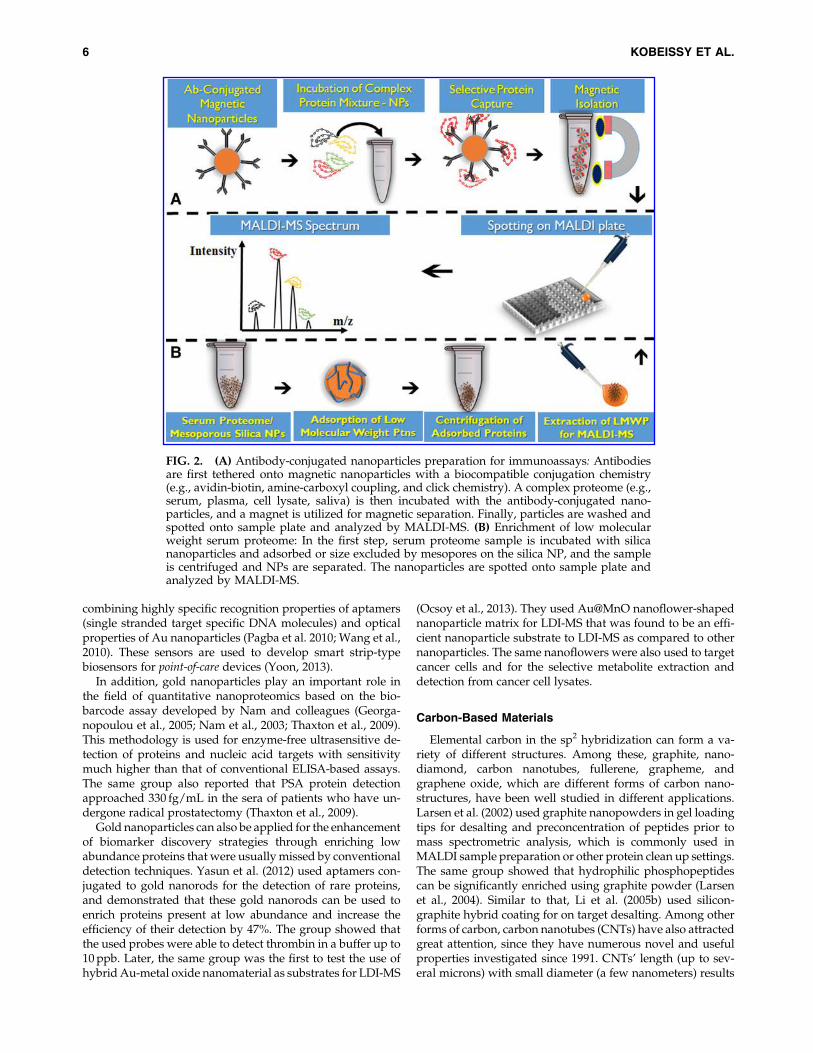
and affibodies) can be introduced on their surface with rela-tively easy chemistries. A third and probably most importantfeature is that the separation can be easily performed with asimple magnet. Owing to these properties, paramagneticparticles have been widely applied for protein isolationshown in Figure 2.
A widely used method for such purification is nickel ni-trilotriacetic acid (NTA/Ni2 + )-based magnetic separation forhistidine-tagged proteins (Gu et al., 2006). Pioneering work byXu et al. has been applied in developing a general strategyusing NTA-modified iron platinum (FePt) nanoparticles forseparation of histidine-tagged proteins at concentrations aslow as 0.5 pM (Xu et al., 2004a; 2004b). Lee et al. (2004) in-troduced bimetallic nanorods comprising of gold and nickelto fish out proteins magnetically using the similar Ni-NTAchemistry. In this work Lee et al. used Ni-Au nanorods toseparate IgG antibodies. In another report, Shukoor et al.(2008) employed multifunctional copolymer functionalizedsuperparamagnetic Fe2O3 nanoparticles for immobilizing andsubsequently isolating His-tagged recombinant protein sili-cate from marine sponge Suberites domuncula. This highlyversatile biomagnetic separation methodology also allows there-use of the magnetic nanocrystals. Antibody-conjugatedmagnetic particles were also used to isolate biomarker pro-teins from plasma samples of cancer patients. Ranzoni et al.(2012) has synthesized PSA antibody conjugated nano-particles and isolated PSA from plasma samples in conjunc-tion with pulsed magnetic fields.
Chou et al. (2005) synthesized antibody-conjugated mag-netic nanoparticles as a tool for isolating cancer proteins be-fore MALDI mass spectrometric detection. C-reactive protein(CRP) and amyloid P component were efficiently isolatedfrom unfractionated human plasma and sub-nanomolar leveldetection was achieved in MALDI analyses. In a follow-upstudy from the same group, serum amyloid A (SAA), C-reactive protein (CRP), and serum amyloid P (SAP) antigenswere captured, isolated from human plasma, and detected byMALDI-MS in a multiplexed immunoassay format. In a recentstudy, Bamrungsap et al. (2011) used aptamer-conjugatedsuperparamagnetic particles to detect lysozyme by usingmagnetic relaxation upon target capture.
Gold Nanoparticles
Another form of nanomaterials are gold (Au) nanoparticlesthat have been utilized for protein immobilization owing totheir high affinity to thiol (-SH) and disulfide (S–S) groupspresent in various molecules. Their exceptional optical, scat-tering, and agglomeration-dispersion properties render themoptimal candidates for various types of biolabeling applica-tions (Kneipp et al., 1999; Rosi et al., 2004). Protein-mediatedagglomeration of Au nanoparticles generates large localelectromagnetic fields between immediate neighbors whenilluminated known as ‘‘hot spots‘‘ (Lee et al., 2006). Thistechnology enables researchers to analyze proteins withininterparticle spaces using surface-enhanced Raman spectro-scopy (SERS) reaching detection levels down to single mole-cule level (Gunnarsson et al., 2005; Kneipp et al., 1998; Kneippet al., 1997; Nie and Emory, 1997; Podstawka et al., 2004;Wang et al., 2005b). Various colorimetric sensors for detectingmetabolites, proteins, small molecules, and whole cells insolution as well as in real samples were developed by
POST-GENOMICS NANOTECHNOLOGY 5
combining highly specific recognition properties of aptamers(single stranded target specific DNA molecules) and opticalproperties of Au nanoparticles (Pagba et al. 2010; Wang et al.,2010). These sensors are used to develop smart strip-typebiosensors for point-of-care devices (Yoon, 2013).
In addition, gold nanoparticles play an important role inthe field of quantitative nanoproteomics based on the bio-barcode assay developed by Nam and colleagues (Georga-nopoulou et al., 2005; Nam et al., 2003; Thaxton et al., 2009).This methodology is used for enzyme-free ultrasensitive de-tection of proteins and nucleic acid targets with sensitivitymuch higher than that of conventional ELISA-based assays.The same group also reported that PSA protein detectionapproached 330 fg/mL in the sera of patients who have un-dergone radical prostatectomy (Thaxton et al., 2009).
Gold nanoparticles can also be applied for the enhancementof biomarker discovery strategies through enriching lowabundance proteins that were usually missed by conventionaldetection techniques. Yasun et al. (2012) used aptamers con-jugated to gold nanorods for the detection of rare proteins,and demonstrated that these gold nanorods can be used toenrich proteins present at low abundance and increase theefficiency of their detection by 47%. The group showed thatthe used probes were able to detect thrombin in a buffer up to10 ppb. Later, the same group was the first to test the use ofhybrid Au-metal oxide nanomaterial as substrates for LDI-MS
(Ocsoy et al., 2013). They used Au@MnO nanoflower-shapednanoparticle matrix for LDI-MS that was found to be an effi-cient nanoparticle substrate to LDI-MS as compared to othernanoparticles. The same nanoflowers were also used to targetcancer cells and for the selective metabolite extraction anddetection from cancer cell lysates.
Carbon-Based Materials
Elemental carbon in the sp2 hybridization can form a va-riety of different structures. Among these, graphite, nano-diamond, carbon nanotubes, fullerene, grapheme, andgraphene oxide, which are different forms of carbon nano-structures, have been well studied in different applications.Larsen et al. (2002) used graphite nanopowders in gel loadingtips for desalting and preconcentration of peptides prior tomass spectrometric analysis, which is commonly used inMALDI sample preparation or other protein clean up settings.The same group showed that hydrophilic phosphopeptidescan be significantly enriched using graphite powder (Larsenet al., 2004). Similar to that, Li et al. (2005b) used silicon-graphite hybrid coating for on target desalting. Among otherforms of carbon, carbon nanotubes (CNTs) have also attractedgreat attention, since they have numerous novel and usefulproperties investigated since 1991. CNTs’ length (up to sev-eral microns) with small diameter (a few nanometers) results
FIG. 2. (A) Antibody-conjugated nanoparticles preparation for immunoassays: Antibodiesare first tethered onto magnetic nanoparticles with a biocompatible conjugation chemistry(e.g., avidin-biotin, amine-carboxyl coupling, and click chemistry). A complex proteome (e.g.,serum, plasma, cell lysate, saliva) is then incubated with the antibody-conjugated nano-particles, and a magnet is utilized for magnetic separation. Finally, particles are washed andspotted onto sample plate and analyzed by MALDI-MS. (B) Enrichment of low molecularweight serum proteome: In the first step, serum proteome sample is incubated with silicananoparticles and adsorbed or size excluded by mesopores on the silica NP, and the sampleis centrifuged and NPs are separated. The nanoparticles are spotted onto sample plate andanalyzed by MALDI-MS.
6 KOBEISSY ET AL.
in a large aspect ratio. This makes CNTs very attractive ma-terial for proteomics applications via their wide surface areathat enables better immobilization and capturing of proteinsthrough functionalizing its surface with a capturing proteinprobe. For instance, Okuno et al. (2007) exploited the propertyof CNTs and designed a CNT-based electrochemical systemto detect prostate-specific antigen from clinical samples.Furthermore, Chen et al. (2008) showed that a very sensitiveRaman sensor that was able to detect low fM levels of proteinbiomarkers can be constructed by using CNTs.
In another study, Drouvalakis et al. (2008) designed a CNTsensor to detect autoantibodies in rheumatoid arthritis pa-tients. In an elegant study, Guo et al. (2009) used carbon na-notubes to improve the resolution of native PAGE detectingserum proteins. Other proteomics application include: carbonbased-nanostructures, which has also been applied as sub-strates for ionization in laser-based ionization techniques. Xuet al. (2003) used carbon nanotubes as a substrate for ioniza-tion. Sunner et al. (1995) used graphite for the same purpose.Hsu et al. (2010) used carbon nanotubes for screening long-chain fatty acids in patient samples. In another study, Najam-ul-Haq sputter coated nanostructured diamond like carbon oncommercially available DVD disks and used this substrate forlaser desorption ionization of peptides with a detection sensi-tivity of femtomolar levels (Najam-ul-Haq et al., 2008).
Graphene (G) and graphene oxide (GO) have been recentlyused in protein science. Tang et al. (2010) employed G & GO as ascaffold for enriching DNA and proteins in solution, yieldinghigh recoveries which were four times higher than nanodia-mond particles. Finally, Dong et al. (2010) used graphene as asubstrate for ionization for the analysis of small peptides andsmall metabolites. Compared to conventional MALDI matrices,graphene exhibited higher desorption ionization efficiencieswith very low background ions. Furthermore, it eliminatedfragmentation and provided better tolerance to salts.
Carbon-based materials are also used in field effect tran-sistors that allows for the real-time and fast detection of label-free proteins and biomolecules (Cui et al., 2001; Hahm Jong-in,2004; Patolsky et al., 2004). A change in charge or electric po-tential at the nanomaterial surface is induced by the binding ofa biomacromolecule. This in turn leads to a change in thecharge carrier concentration in the semiconductor material thatcan be detected by conductometry measurements. Carbon na-notubes are relatively easy to prepare when compared to sili-cone nanowires and have a long shelf life (Tans et al., 1998).Chen et al. (2004) reported the detection of protein adsorptionon single-walled carbon nanotube using field effect transistors(FETs). Furthermore, Chen et al. enhanced their work bymodifying carbon nanotubes with DNA aptamers to recognizespecific macrobiomolecules selectively (Chen and Chen, 2005).
Polymeric Nanostructures
Polymeric materials, including polylysines, polyethyl-enimine, and cationic dendrimers, have been used in proteomicsstudies (Tao et al., 2005). However, the nano-architectured na-nomaterials turned out to be better alternatives to bulk poly-mers. Li et al. (2005a) used radiofrequency plasma polymerscoated on a traditional MALDI plate for selective capture anddetection of proteins that they termed as ‘‘on probe affinitycapturing.’’ The advantage of using this technique is that it al-lows the capture of different molecules by tuning the properties
of the polymers used (e.g., these can be thermo-responsivematerials). For instance, N-isopropyl acrylamide is hydrophilicbelow a certain temperature and hydrophobic above that par-ticular temperature. As a result, selective capture of hydrophilicor hydrophobic proteins can be achieved. Li et al. (2007a)showed that this thermo-responsive polymer coating in com-bination with temperature control can be used to clean upprotein samples prior to mass spectrometry analysis.
Another interesting class of nanostructured polymericmaterial is the polymer brushes composed of a layer ofpolymers attached with one end to a surface ( Jain et al., 2009).Polymeric nanostructures are also used as biomarker har-vester; Luchini et al. (2008) used N-isopropylacrylamide na-nogels in conjunction with affinity baits that functioned asmolecular size sieve for capturing abundant proteins such asalbumin. Longo et al. (2009) used the same nanogels to con-centrate and preserve platelet-derived growth factor (PDGF)from serum in order to magnify the detectable level of themarker. This was performed in one single step and in solutionphase (Longo et al., 2009) and has been commercialized underthe trade name Nanotrap� (Shaffer, 2011).
Metal Oxide Nanoparticlesand Phosphonanoproteomics
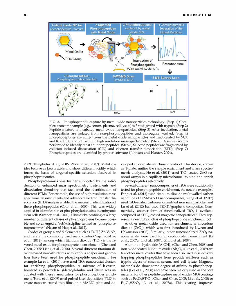
Another area that has been addressed by nanotechnologyand proteomics is the identification of PTMs and their struc-tural characterization. More specifically, nanoparticles havebeen applied in characterization of protein phosphorylation,involving phosphopeptide isolation and subsequently iden-tification by mass spectrometry as illustrated in Figure 3(Olsen et al., 2010; Oppermann et al., 2009)
Protein phosphorylation is a ubiquitous and central processthat regulates several different cellular functions such as sig-nal transduction, cell growth and differentiation. Protein ki-nase activities are significantly elevated in different types ofcancer and many new pharmaceuticals target phosphorylatedproteins to help curb the disease progression. The state of theart method for identifying protein phosphorylation sites ismass spectrometry. However analysis and characterization ofphosphoproteins from complex samples using MS is a realchallenge. The ionization efficiency of cellular phosphopro-teins is significantly hampered in mass spectrometry owing totheir very low abundance relative to unphosphorylated pro-teins and to their negatively charged phosphate groups.Therefore, phosphoproteins are largely missed in high through-put proteomics experiments and phosphoproteomics has notbeen previously possible, due to the challenge of capturingand enrichment of low abundant phosphopeptides (Taoet al., 2005). The best way to address this problem is to use acapturing tool to pre-concentrate phosphopeptides prior toanalysis by mass spectrometry (Engholm-Keller and Larsen,2013; Johnson and Hunter, 2004).
The chemistry behind phosphopeptide enrichment re-mained immature until immobilized metal ion affinity chro-matography (IMAC) was introduced (Feuerstein et al., 2005;Jin et al., 2004; Mann et al., 2002). The underlying principle ofphosphopeptide enrichment is to form reversible and stronginteraction between the metal and phosphorylation site of theprotein. The task has been originally achieved by the use ofIMAC resins that are mostly replaced with metal oxide-basedaffinity chromatography (MOAC) (Lin et al., 2008; Lin et al.,
POST-GENOMICS NANOTECHNOLOGY 7
2009; Thingholm et al., 2006; Zhou et al., 2007). Metal ox-ides behave as Lewis acids and show different acidity whichforms the basis of targeted-specific selection observed inphosphoproteomics.
Phosphoproteomics was further supported by the intro-duction of enhanced mass spectrometry instruments anddissociation chemistry that facilitated the identification ofdifferent PTMs. For example, the use of high resolution massspectrometry instruments and advanced electron transfer dis-sociation (ETD) analysis enabled the successful identification ofthese phosphopeptides (Coon et al., 2005). This was widelyapplied in identification of phosphorylation sites in embryonicstem cells (Swaney et al., 2009). Ultimately, profiling of a largenumber of different classes of phosphoproteins became possi-ble and so emerged a (sub)-discipline termed as ‘phosphona-noproteomics’ (Najam-ul-Haq et al., 2012).
Oxides of group 4 and 5 elements such as Ti, Hf, Zr, V, Nb,and Ta are the commonly used metal oxides (Najam-ul-Haqet al., 2012), among which titanium dioxide (TiO2) is the fa-vored metal oxide for phosphoprotein enrichment (Chen andChen, 2005; Liang et al., 2006). Several different titanium di-oxide-based nanostructures with various shapes and geome-tries have been used for phosphopeptide enrichment. Forexample Lu et al. (2010) have used TiO2 nanocrystal clustersfor enriching phosphopeptides. A mixture of b-casein,horseradish peroxidase, b-lactoglobulin, and fetuin was in-cubated with these nanoclusters for phosphpeptides enrich-ment. Torta et al. (2009) used pulsed laser deposition (PLD) tocreate nanostructured thin films on a MALDI plate and de-
veloped an on-plate enrichment protocol. This device, knownas T-plate, unifies the sample enrichment and mass spectro-metric analysis. He et al. (2011) used TiO2-coated ZnO na-norod arrays in a capillary microchannel to bind and enrichphosphopeptides selectively.
Several different nanocomposites of TiO2 were additionallytested for phosphopeptide enrichment. As notable examples,Fang et al. (2012) used titanium dioxide-multiwalled carbonnanotube (TiO2-MWNT) nanocomposites, Zeng et al. (2012)used TiO2-coated carbon-encapsulated iron nanoparticles, andLu et al. (2012) has used TiO(2)/graphene composites. Com-mercially, another form of functionalized TiO2 is availablecomposed of ‘‘TiO2 coated magnetic nanoparticles.’’ They rep-resent a new hybrid class of phosphopeptide enrichment tool.
Another metal oxide used for enrichment is zirconiumdioxide (ZrO2), which was first introduced by Kweon andHakansson (2008). Similarly, other functionalized ZrO2 na-nomaterials were used for phosphoprotein enrichment (Liet al., 2007a; Li et al., 2007b; Zhou et al., 2007).
Aluminum hydroxide (AlOH)3 (Chen and Chen, 2008) andiron oxide coated-Niobium oxide (Nb2O5) (Lin et al., 2009) arefew other metal oxides that have been also used in selectivelytrapping phosphopeptides from peptide mixtures such astryptic digest of caseins, serum, and cell lysate. Magneticmaterials do show some degree of affinity to phosphopep-tides (Lee et al., 2008) and have been majorly used as the corematerial for other peptide capture metal oxide (MO) coatingssuch as Fe3O4@TiO2 (Chen and Chen, 2005; Li et al., 2008) orFe3O4@ZrO2 (Li et al., 2007a). This coating improves
FIG. 3. Phosphopeptide capture by metal oxide nanoparticles technology: (Step 1) Com-plex proteome sample (e.g., serum, plasma, cell lysate) is first digested with trypsin. (Step 2)Peptide mixture is incubated metal oxide nanoparticles. (Step 3) After incubation, metalnanoparticles are isolated from non-phosphopeptides and thoroughly washed. (Step 4)Phosphopeptides are eluted from the metal oxide nanoparticles and fractionated by SCXand RP-HPLC and infused into high resolution mass spectrometry. (Step 5) A survey scan isperformed to identify most abundant peptides. (Step 6) Selected peptides are fragmented bycollision induced dissociation (CID) and electron transfer dissociation (ETD). (Step 7)Phosphopeptides are identified by proper software ( Johnson and Hunter, 2004).
8 KOBEISSY ET AL.
purification and separation of target peptides upon selectivebinding from a complex peptide mixture, using externalmagnetic field.
When the performance of metal oxide nanoparticles werecompared to that of conventional IMAC resins, it was foundthat metal oxide nanoparticles out-perform IMAC resins.Larsen et al. (2005) compared the performance of titaniumoxide micro-columns with IMAC resins and found that sig-nificantly higher number of non-phosphorylated peptideswere observed in the IMAC experiments. They reported thatperformance of the TiO2 based-techniques significantly sur-passed the IMAC method with respect to the number of de-tected phosphorylated peptides and reduction of the numberof nonphosphorylated peptides. This is attributed to the moreselective binding of phosphorylated peptides to TiO2 micro-columns than IMAC resins.
Quantum Dots
Fluorescence-based techniques for protein sensing appli-cations have gained an ever increasing popularity due to theirsimplicity and exquisite sensitivity (De et al., 2009; Ibraheemand Campbell, 2010; You et al., 2007). In particular, quantumdots (QDs), also called semiconductor nanoparticles, areemerging as a new class of fluorescent probes (Boenemanet al., 2009; Larson et al., 2003; Pinaud et al., 2006). Whencompared to organic dyes and fluorescent proteins, QDs haveunique optical and electronic properties that make thembrighter and highly resistive to photobleaching and chemicaldegradation (Leutwyler et al., 1996; Murray et al., 1993). Themaximum emission depends on the size of the electron gapthat is tuned by the particle core diameter. Smaller nano-particles have a maximum emission in the blue region andbigger particles tend to emit in the red or near-IR.
These unique optical properties make QDs an excellentfluorescent probes for applications in the field of diagnosis(Bruchez et al., 1998; Chan and Nie, 1998), in vivo and in vitroimaging (Kim et al., 2003; Levene et al., 2004; Rosenthal et al.,2002), multicolor cell imaging (Hanaki et al., 2003; Jaiswal et al.,2002; Sukhanova et al., 2004; Wu et al., 2002), cell and proteintracking (Dahan et al., 2003; Voura et al., 2004), and DNA andprotein sensing (Medintz et al., 2003; Zhang et al., 2005).
More recently, Liu et al. (2010) reported the detection andcharacterization of four low-abundant protein biomarkers(CD15, CD30, CD45, and Pax5) in Hodgkin’s lymphomausing the multiplexing capabilities of QDs. QDs were alsoused to detect apolipoprotein E, the most important knowngenetic risk factor for Alzheimer disease, by designing asandwich immunocomplex microarray assay based oncadmium-selenide/zinc-sulfide (CdSe@ZnS) quantum dots.The assay provided a low detection limit of 62 pg mL–1, seventimes more than that of the ELISA (470 pg mL–1) when testedunder the same conditions (Morales-Narvaez et al., 2012).
A major drawback, however, to the utilization of QDs in thefield of proteomics is the nonspecific binding onto their sur-face (Pathak et al., 2007). To minimize such nonspecificbinding, QDs are often modified with polyethyleneglycol(PEG)-based polymers (discussed later) (Geho et al., 2005; Liuet al., 2008). More recently, Breus et al. (2009) capped QDswith small zwiterinonic molecules. This step rendered thenanoparticles water soluble and dramatically diminishednonspecific binding.
Nanoproteomics in Clinical Applications
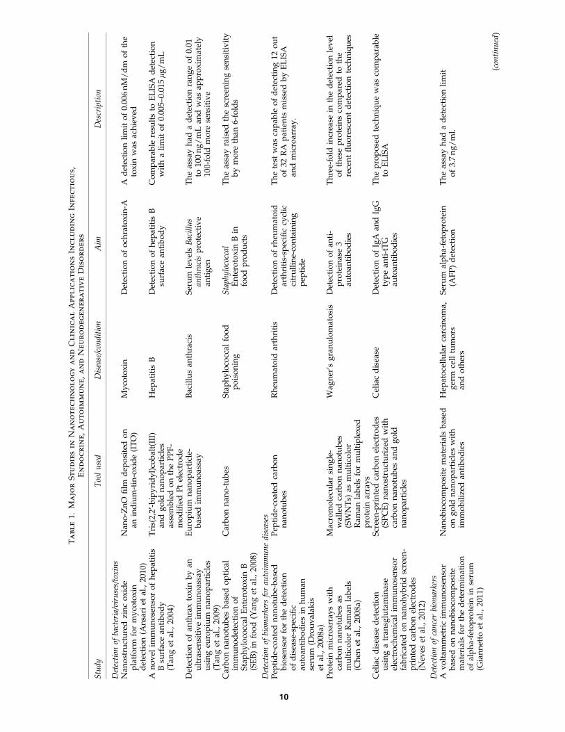
Nanoproteomics technology has been applied to variousclinical settings, mainly for enhancing biomarker discoverycapabilities. In this regard, nanoproteomics are seen to befunctioning as protein amplification techniques similar toPCR. Potential clinical applications include infectious, endo-crine, autoimmune, and neurodegenerative diseases, as wellas brain injury and several types of tumors (Ray et al., 2011).Some of these applications are illustrated in Table 1 andFigure 4.
As applied to infectious diseases, Ansari et al. (2010),Kaushik et al. (2009a), and others used nano-structured sur-faces for the detection of ochratoxin-A mycotoxin pro-duced by Aspergillus. Ansari et al. utilized Nano-ZnO filmdeposited on an indium-tin-oxide (ITO) with immobilizedrabbit-immunoglobulin and bovine serum albumin to reach adetection limit of 0.006 nM/dm of the toxin. Similarly,Kaushik et al. used nanostructured cerium oxide to achieve adetection limit of 0.25ng/dL of ochratoxin (Kaushik et al.,2009a). Tang et al. (2004; 2005) proposed different im-munosensors based on gold nanoparticles for the detection ofhepatitis B surface antibody in blood. Reported detection limitof the applied techniques ranged between 5 and 15 ng/mL,which demonstrates a much higher sensitivity than the stan-dard diagnostic serology techniques, allowing for better dis-ease detection and monitoring. The same group also appliedthe immunosensors approach for the detection of serum levelsof Bacillus anthracis protective antigen as well as HIV-capsidp24 antigen (Tang and Hewlett, 2010; Tang et al., 2009). Theseimmunosensors approaches demonstrated rapid detectionand much higher sensitivity compared to the traditionalELISA techniques that can reach up to 100 folds. Moreover, aspart of a disease preventative measure, Yang et al. (2008) usedcarbon nanotubes for the detection of Staphylococcal En-terotoxin B in food products that were traditionally tested byELISA. The new technique enhanced toxin detection limit andraised screening sensitivity by more than six-fold.
In the field of autoimmune diseases, many of the currentdiagnostic serology techniques do not demonstrate optimalsensitivity for diseases such as rheumatoid arthritis, celiacdisease, Wegener’s granulomatosis, and others. This could bein part due to the limited ability of these techniques to detectlow levels of autoantibodies in the sera of suspected patients.Here, the application of nanoproteomics techniques could bepromising. In fact, Drouvalakis et al. (2009) utilized peptide-coated nanotubes for the detection of rheumatoid arthritis-specific cyclic citrulline-containing peptide. The proposedtechnique was capable of detecting 12 out of 32 RA patientsmissed by ELISA and microarray (Drouvalakis et al., 2008).Several other studies have also reported the use of nanopro-teomics for the detection of RA-related immunoglobulins (deGracia Villa et al., 2011; Jimenez et al., 2012). Carbon nano-tubes were also suggested as a screening tool for Wagner’sgranulomatosis (Chen et al., 2008). The authors used macro-molecular single-walled carbon nanotubes (SWNTs) as mul-ticolor Raman labels for multiplexed protein arrays for thedetection of anti-proteinase 3 autoantibodies that are markersof Wagner’s granulomatosis. Results have shown a three-foldincrease in the detection level of these proteins compared tothe recent fluorescent detection techniques. In addition, na-noproteomics techniques were also assessed for utilization in
POST-GENOMICS NANOTECHNOLOGY 9
Ta
bl
e1.
Ma
jo
rS
tu
die
sin
Na
no
te
ch
no
lo
gy
an
dC
lin
ic
al
Ap
pl
ic
at
io
ns
In
cl
ud
in
gIn
fe
ct
io
us,
En
do
cr
in
e,
Au
to
im
mu
ne
,a
nd
Ne
ur
od
eg
en
er
at
iv
eD
iso
rd
er
s
Stu
dy
Too
lu
sed
Dis
ease
/con
dit
ion
Aim
Des
crip
tion
Det
ecti
onof
bact
eria
/vir
use
s/to
xin
sN
ano
stru
ctu
red
zin
co
xid
ep
latf
orm
for
my
coto
xin
det
ecti
on
(An
sari
etal
.,20
10)
Nan
o-Z
nO
film
dep
osi
ted
on
anin
diu
m-t
in-o
xid
e(I
TO
)M
yco
tox
inD
etec
tio
no
fo
chra
tox
in-A
Ad
etec
tio
nli
mit
of
0.00
6n
M/
dm
of
the
tox
inw
asac
hie
ved
An
ov
elim
mu
no
sen
sor
of
hep
atit
isB
surf
ace
anti
bo
dy
(Tan
get
al.,
2004
)
Tri
s(2,
2¢-b
ipy
rid
yl)
cob
alt(
III)
and
go
ldn
ano
par
ticl
esas
sem
ble
do
nth
eP
PF
-m
od
ified
Pt
elec
tro
de
Hep
atit
isB
Det
ecti
on
of
hep
atit
isB
surf
ace
anti
bo
dy
Co
mp
arab
lere
sult
sto
EL
ISA
det
ecti
on
wit
ha
lim
ito
f0.
005–
0.01
5l
g/
mL
Det
ecti
on
of
anth
rax
tox
inb
yan
ult
rase
nsi
tiv
eim
mu
no
assa
yu
sin
geu
rop
ium
nan
op
arti
cles
(Tan
get
al.,
2009
)
Eu
rop
ium
nan
op
arti
cle-
bas
edim
mu
no
assa
yB
acil
lus
anth
raci
sS
eru
mle
vel
sB
acil
lus
anth
raci
sp
rote
ctiv
ean
tig
en
Th
eas
say
had
ad
etec
tio
nra
ng
eo
f0.
01to
100
ng
/m
Lan
dw
asap
pro
xim
atel
y10
0-fo
ldm
ore
sen
siti
ve
Car
bo
nn
ano
tub
esb
ased
op
tica
lim
mu
no
det
ecti
on
of
Sta
ph
ylo
cocc
alE
nte
roto
xin
B(S
EB
)in
foo
d(Y
ang
etal
.,20
08)
Car
bo
nn
ano
-tu
bes
Sta
ph
ylo
cocc
alfo
od
po
iso
nin
gS
tap
hy
loco
ccal
En
tero
tox
inB
info
od
pro
du
cts
Th
eas
say
rais
edth
esc
reen
ing
sen
siti
vit
yb
ym
ore
than
6-fo
lds
Det
ecti
onof
biom
arke
rsfo
rau
toim
mu
ne
dis
ease
sP
epti
de-
coat
edn
ano
tub
e-b
ased
bio
sen
sor
for
the
det
ecti
on
of
dis
ease
-sp
ecifi
cau
toan
tib
od
ies
inh
um
anse
rum
(Dro
uv
alak
iset
al.,
2008
a)
Pep
tid
e-co
ated
carb
on
nan
otu
bes
Rh
eum
ato
idar
thri
tis
Det
ecti
on
of
rheu
mat
oid
arth
riti
s-sp
ecifi
ccy
clic
citr
ull
ine-
con
tain
ing
pep
tid
e
Th
ete
stw
asca
pab
leo
fd
etec
tin
g12
ou
to
f32
RA
pat
ien
tsm
isse
db
yE
LIS
Aan
dm
icro
arra
y.
Pro
tein
mic
roar
ray
sw
ith
carb
on
nan
otu
bes
asm
ult
ico
lor
Ram
anla
bel
s(C
hen
etal
.,20
08a)
Mac
rom
ole
cula
rsi
ng
le-
wal
led
carb
on
nan
otu
bes
(SW
NT
s)as
mu
ltic
olo
rR
aman
lab
els
for
mu
ltip
lex
edp
rote
inar
ray
s
Wag
ner
’sg
ran
ulo
mat
osi
sD
etec
tio
no
fan
ti-
pro
tein
ase
3au
toan
tib
od
ies
Th
ree-
fold
incr
ease
inth
ed
etec
tio
nle
vel
of
thes
ep
rote
ins
com
par
edto
the
rece
nt
flu
ore
scen
td
etec
tio
nte
chn
iqu
es
Cel
iac
dis
ease
det
ecti
on
usi
ng
atr
ansg
luta
min
ase
elec
tro
chem
ical
imm
un
ose
nso
rfa
bri
cate
do
nn
ano
hy
bri
dsc
reen
-p
rin
ted
carb
on
elec
tro
des
(Nev
eset
al.,
2012
)
Scr
een
-pri
nte
dca
rbo
nel
ectr
od
es(S
PC
E)
nan
ost
ruct
uri
zed
wit
hca
rbo
nn
ano
tub
esan
dg
old
nan
op
arti
cles
Cel
iac
dis
ease
Det
ecti
on
of
IgA
and
IgG
typ
ean
ti-t
TG
auto
anti
bo
die
s
Th
ep
rop
ose
dte
chn
iqu
ew
asco
mp
arab
leto
EL
ISA
Det
ecti
onof
can
cer
biom
arke
rsA
vo
ltam
met
ric
imm
un
ose
nso
rb
ased
on
nan
ob
ioco
mp
osi
tem
ater
ials
for
the
det
erm
inat
ion
of
alp
ha-
feto
pro
tein
inse
rum
(Gia
nn
etto
etal
.,20
11)
Nan
ob
ioco
mp
osi
tem
ater
ials
bas
edo
ng
old
nan
op
arti
cles
wit
him
mo
bil
ized
anti
bo
die
s
Hep
ato
cell
ula
rca
rcin
om
a,g
erm
cell
tum
ors
and
oth
ers
Ser
um
alp
ha-
feto
pro
tein
(AF
P)
det
ecti
on
Th
eas
say
had
ad
etec
tio
nli
mit
of
3.7
ng
/m
l.
(con
tin
ued
)
10
Ta
bl
e1.
(Co
nt
in
ue
d)
Stu
dy
Too
lu
sed
Dis
ease
/con
dit
ion
Aim
Des
crip
tion
Inte
gra
ted
mic
rofl
uid
icsy
stem
sw
ith
anim
mu
no
sen
sor
mo
difi
edw
ith
carb
on
nan
otu
bes
for
det
ecti
on
of
pro
stat
esp
ecifi
can
tig
en(P
SA
)in
hu
man
seru
msa
mp
les
(Pan
ini
etal
.,20
08)
Gla
ssy
carb
on
elec
tro
de
mo
difi
edw
ith
mu
ltiw
all
carb
on
nan
otu
bes
Pro
stat
eca
nce
rP
rost
ate
spec
ific
anti
gen
(PS
A)
Th
iste
chn
iqu
ew
asa
qu
ick
det
ecti
on
tech
niq
ue
wit
hh
igh
erse
nsi
tiv
ity
then
trad
itio
nal
EL
ISA
.
Nan
og
old
-en
wra
pp
edg
rap
hen
en
ano
com
po
site
sas
trac
ela
bel
sfo
rse
nsi
tiv
ity
enh
ance
men
to
fel
ectr
och
emic
alim
mu
no
sen
sors
incl
inic
alim
mu
no
assa
ys:
Car
cin
oem
bry
on
ican
tig
enas
am
od
el(Z
ho
ng
etal
.,20
10)
Nan
og
old
-en
wra
pp
edg
rap
hen
en
ano
com
po
site
sC
olo
rect
alca
nce
rC
arci
no
emb
ryo
nic
anti
gen
(CE
A)
Alo
wd
etec
tio
nli
mit
of
0.01
ng
/m
Lw
asd
emo
nst
rate
d
Insi
tuam
pli
fied
elec
tro
chem
ical
imm
un
oas
say
for
carc
ino
emb
ryo
nic
anti
gen
usi
ng
ho
rser
adis
hp
ero
xid
ase-
enca
psu
late
dn
ano
go
ldh
oll
ow
mic
rosp
her
esas
lab
els
(Tan
gan
dR
en,
2008
)
Ho
rser
adis
hp
ero
xid
ase-
enca
psu
late
dn
ano
-go
ldh
oll
ow
mic
rosp
her
es
Co
lore
ctal
can
cer
Car
cin
oem
bry
on
ican
tig
en(C
EA
)T
he
assa
ylo
wer
edth
ed
etec
tio
nli
mit
of
CE
Ad
ow
nt0
1.5
pg
/m
L.
Dru
gd
eliv
ery
and
dis
ease
mon
itor
ing
Alo
gic
alm
ole
cula
rci
rcu
itfo
rp
rog
ram
mab
lean
dau
ton
om
ou
sre
gu
lati
on
of
pro
tein
acti
vit
yu
sin
gD
NA
apta
mer
–pro
tein
inte
ract
ion
s(H
anet
al.,
2012
)
Lo
gic
alci
rcu
itb
ased
on
DN
Aap
tam
er–p
rote
inin
tera
ctio
ns
Dif
fere
nt
app
lica
tio
ns
Au
ton
om
ou
s,se
lf-s
ust
ain
edan
dp
rog
ram
mab
lem
anip
ula
tio
no
fp
rote
inac
tiv
ity
inv
itro
An
exam
ple
of
app
lica
tio
nin
vo
lves
mo
nit
ori
ng
the
lev
els
and
acti
vit
yo
fth
rom
bin
inp
lasm
aan
dd
eliv
ers
anin
hib
ito
ryan
ti-c
oag
ula
nt
acco
rdin
gly
Qu
antu
m-d
ot-
con
jug
ated
gra
ph
ene
asa
pro
be
for
sim
ult
aneo
us
can
cer-
targ
eted
flu
ore
scen
tim
agin
g,
trac
kin
gan
dm
on
ito
rin
gd
rug
del
iver
y(C
hen
etal
.,20
13)
Qu
antu
m-d
ot-
con
jug
ated
gra
ph
ene
Can
cer
chem
oth
erap
yT
arg
eted
ther
apy
wit
hd
ox
oru
bic
inT
he
syst
emca
nd
eliv
erth
ean
tin
eop
last
icd
rug
do
xo
rub
icin
toca
nce
rce
lls
and
mo
nit
or
its
rele
ase
ou
to
fth
ece
llal
low
ing
for
asa
fer
and
mo
reta
rget
edth
erap
y
Mes
op
oro
us
sili
con
nan
ote
chn
olo
gy
for
can
cer
app
lica
tio
n(B
ou
amra
ni
etal
.,20
09)
Mu
ltis
tag
ed
eliv
ery
syst
ems
usi
ng
mes
op
otr
ou
ssi
lico
nan
dp
rote
inp
latf
orm
chip
sw
ith
MS
/M
AL
DI
Tar
get
edd
rug
ther
apy
Th
ep
rote
inp
latf
orm
hel
ps
det
ect
pro
tein
chan
ges
that
can
allo
wd
isea
sem
on
ito
rin
gin
asso
ciat
ion
wit
hM
S/
MA
LD
Ian
dth
em
ult
ista
ge
del
iver
ysy
stem
allo
ws
for
targ
eted
dru
gd
eliv
ery
.
11
celiac disease through the detection of anti-gliadin auto-antibodies using different techniques where high sensitivitydetection potentials were demonstrated (Neves et al., 2012;Ortiz et al., 2011).
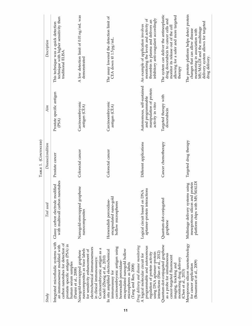
An important application of nanoproteomics tools residesin cancer screening ( Ji et al., 2010). Several plasma proteinbiomarkers have been associated with different types of can-cer, yet their detection by current diagnostic and screeningtechniques is not satisfactorily sensitive. Therefore, nanopro-teomics tools have been investigated for their possiblescreening potentials in these cases. For example, Giannettoet al. (2011) utilized nanobiocomposite materials based ongold nanoparticles with immobilized antibodies for the de-velopment of serum alpha-fetoprotein (AFP) immunosensors(Giannetto et al., 2011). Serum levels of alpha-feto protein areassociated with various types of tumors including germ celltumors, liver tumors, and others. Two other areas that showmajor potential for cancer nanoproteomics application are thedetection of prostate-specific antigen (PSA) and carcinoem-bryonic antigen (CEA). Panini et al. (2008) used carbon na-notubes for the detection of PSA in serum. The utilization ofglassy carbon electrode modified with multiwall carbon na-notubes provided a quick detection technique and a highersensitivity then traditional ELISA. On the other hand, Zhonget al. (2010) employed highly sensitive electrochemical im-munosensor with a sandwich-type immunoassay format todetect CEA that is associated with multiple tumors includingcolorectal cancer. This technique lowered the detection limitof CEA down to 0.01 ng/mL. Furthermore, Tang et al. (2008)utilized horseradish peroxidase-encapsulated nano-gold hol-low microspheres as labels for the detection of CEA, lowering
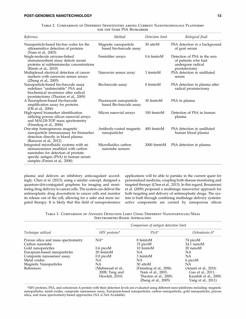
the detection limit of this tumor marker down to 1.5 pg/mL(Tang and Ren, 2008). Several studies have reported the use ofnanoproteomics as means for biomarker detection as in breastcancer, prostate cancer as well as others; for a detailed review,see Liu et al., (2013). Of interest, these nanoproteomic appli-cations have been reported with different sensitivities rangingfrom 30 attoM to 2000 fMA even for the same antigen (PSA),however, using different assays as shown in Table 2.
A further detailed comparison among different biomarkerdetection sensitivities utilizing different nanomaterial-basedapproaches is presented in Table 3. In this table, HIV proteins,PSA, and Ochratoxin-A protein levels are compared andevaluated for their sensitivity detection limits using magneticnanoparticles, metal oxides, composite nanosensor assay,Europium-based nanoparticles, carbon nanoparticles, goldnanoparticles, and porous silica coupled with mass spec-trometry-based approaches.
Added to the value of nanoproteomics application in bio-marker discovery, drug delivery, and personalized medicineare other fields where nanotechnology also holds futurepromise (Ferrari, 2005b; Kim et al., 2013). Nanotechnology-based ‘‘theranostics’’ applications were recently reviewed byKim et al. (2013). Several applications for nanoparticle-baseddrug delivery have been proposed for therapeutics, includingblood coagulation monitoring, cancer therapy, and stroketreatment; an eminent example of which is the use of liposo-mal nano-carriers (Alaouie and Sofou 2008). Han et al. (2012)described a logical circuit that enables autonomous, self-sustained, and programmable manipulation of protein activ-ity in vitro. An example of such application is the use of acircuit that monitors the levels and activity of thrombin in
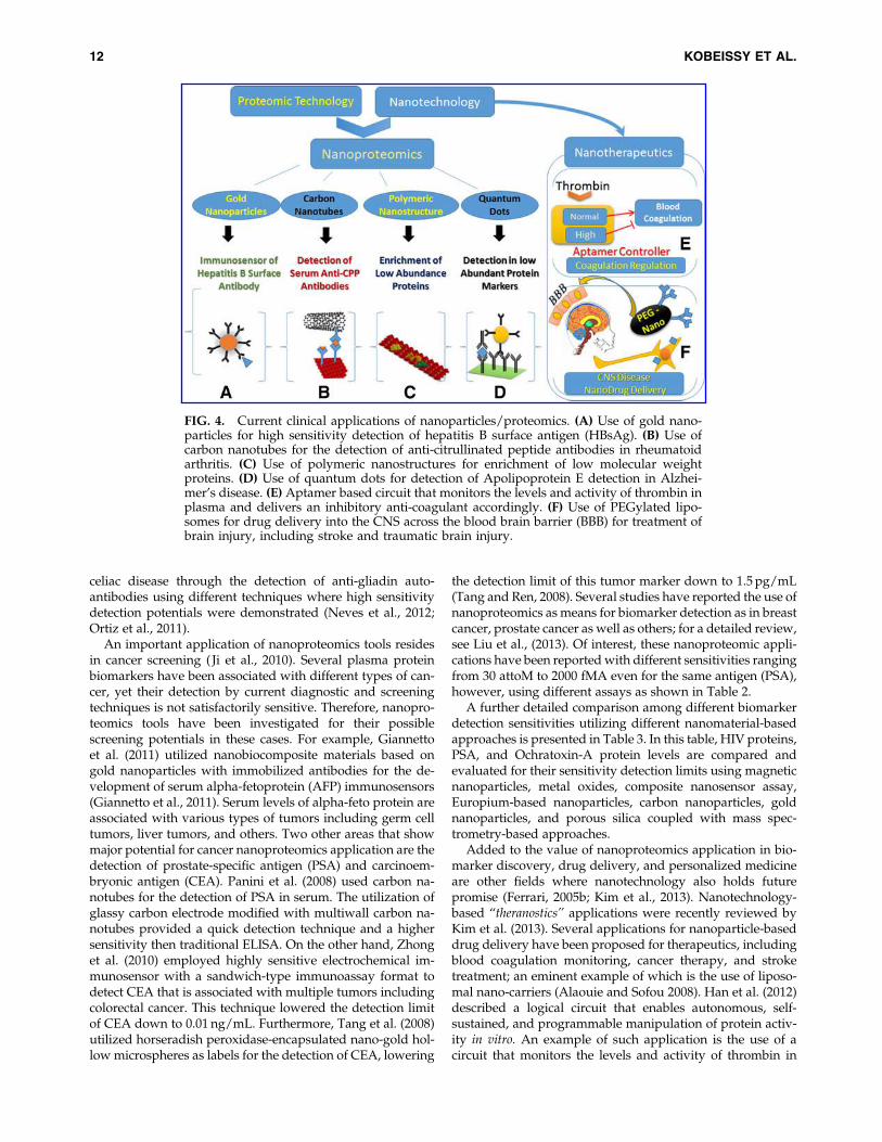
FIG. 4. Current clinical applications of nanoparticles/proteomics. (A) Use of gold nano-particles for high sensitivity detection of hepatitis B surface antigen (HBsAg). (B) Use ofcarbon nanotubes for the detection of anti-citrullinated peptide antibodies in rheumatoidarthritis. (C) Use of polymeric nanostructures for enrichment of low molecular weightproteins. (D) Use of quantum dots for detection of Apolipoprotein E detection in Alzhei-mer’s disease. (E) Aptamer based circuit that monitors the levels and activity of thrombin inplasma and delivers an inhibitory anti-coagulant accordingly. (F) Use of PEGylated lipo-somes for drug delivery into the CNS across the blood brain barrier (BBB) for treatment ofbrain injury, including stroke and traumatic brain injury.
12 KOBEISSY ET AL.
plasma and delivers an inhibitory anticoagulant accord-ingly. Chen et al. (2013), using a similar concept, designed aquantum-dot-conjugated graphene for imaging and moni-toring drug delivery to cancer cells. The system can deliver theantineoplastic drug doxorubicin to cancer cells and monitorits release out of the cell, allowing for a safer and more tar-geted therapy. It is likely that this field of nanoproteomics
applications will be able to partake in the current quest forpersonalized medicine, coupling both disease monitoring andtargeted therapy (Chen et al., 2013). In this regard, Bouamraniet al. (2009) proposed a multistage nanovector approach forboth targeting and delivery of antineoplastic drugs. The sys-tem is built through combining multistage delivery systems:active components are carried by nanoporous silicon
Table 3. Comparison of Antigen Detection Limit Using Different Nanoparticles/Mass
Spectrometry-Based Approaches
Comparison of antigen detection limit
Technique utilized HIV proteins* PSA* Ochratoxin-A*
Porous silica and mass spectrometry NA* 8 femtoM 74 picoMCarbon nanotube 15 picoM 24.1 nanoMGold nanoparictles 0.6 picoM 10 femtoM 20 nanoMEuropium-based nanoparticles 20 femtoM NA NAComposite nanosensor assay 0.8 picoM 3 femtoM NAMetal oxides NA NA 6 picoMMagnetic Nanoparticles NA 30 attoM NAReferences (Mahmoud et al.,
2008; Tang andHewlett, 2010)
(Finnskog et al., 2006;Nam et al., 2003;Thaxton et al., 2009;Zheng et al., 2005)
(Ansari et al., 2010;Guo et al., 2011;Kaushik et al., 2009;Yang et al., 2011)
*HIV proteins, PSA, and ochratoxin-A protein with their detection levels are evaluated using different nano-platforms including: magneticnanoparticles, metal oxides, composite nanosensor assay, Europium-based nanoparticles, carbon nanoparticles, gold nanoparticles, poroussilica, and mass spectrometry-based approaches (NA is Not Available).
Table 2. Comparison of Different Sensitivities among Current Nanotechnology Platforms
for the Same PSA Biomarker
Reference Method Detection limit Biological fluid
Nanoparticle-based bio-bar codes for theultrasensitive detection of proteins(Nam et al,. 2003)
Magnetic nanoparticlebased bio-barcode assay
30 attoM PSA detection in a backgroundof goat serum
Single-molecule enzyme-linkedimmunosorbent assay detects serumproteins at subfemtomolar concentrations(Rissin et al,. 2010)
Femtoliter arrays 0.4 femtoM Detection of PSA in the seraof patients who hadundergone radicalprostatectomy
Multiplexed electrical detection of cancermarkers with nanowire sensor arrays(Zheng et al,. 2005)
Nanowire sensor array 3 femtoM PSA detection in undilutedserum
Nanoparticle-based bio-barcode assayredefines ‘‘undetectable’’ PSA andbiochemical recurrence after radicalprostatectomy (Thaxton et al,. 2009)
Bio-barcode assay 8 femtoM PSA detection in plasma afterradical prostatectomy
A fluorophore-based bio-barcodeamplification assay for proteins(Oh et al,. 2006)
Fluorescent nanoparticlebased Bio-barcode assay
30 femtoM PSA in plasma
High-speed biomarker identificationutilizing porous silicon nanovial arraysand MALDI-TOF mass spectrometry(Finnskog et al,. 2006)
Silicon nanovial arrays 100 femtoM Detection of PSA in humanplasma
One-step homogeneous magneticnanoparticle immunoassay for biomarkerdetection directly in blood plasma(Ranzoni et al,. 2012)
Antibody-coated magneticnanoparticles
400 femtoM PSA detection in undilutedhuman blood plasma
Integrated microfluidic systems with animmunosensor modified with carbonnanotubes for detection of prostatespecific antigen (PSA) in human serumsamples (Panini et al,. 2008)
Microfluidics carbonnanotube sensors
2000 femtoM PSA detection in plasma
POST-GENOMICS NANOTECHNOLOGY 13
particles. Then, microparticles are used to recognize ‘‘zipcodes’’ on vascular endothelium, to detect environmentalchanges and allow extravasation of particles (Bouamraniet al., 2009).
Nanoparticles studies have been recently integrated in thearea of neuroscience, particularly in the fields of stroke andbrain injury, where the blood brain barrier (BBB) represents amajor obstacle for drug delivery. Yun et al. (2013) generatednanoparticles that carry superoxide dismutase enzyme andtargeted anti-NMDA (N-methyl-D-aspartate) receptor 1(NR1) antibody. These nanoparticles were applied to a mousemodel of cerebral ischemia where they limited reperfusioninjury, and reduced apoptosis and inflammation, improvingbehavioral outcome. In addition, utilized coated PEG-basedliposomes were also found to be localized to the CA region ofthe hippocampus, suggesting a probable mode of targeteddelivery of reactive oxidative species quenchers in the treat-ment of stroke (Yun et al., 2013). Zhao et al. (2013) have testedPuerarin (PUE), a compound that suffers low availability inbrain post-administration, as a candidate drug for treatingstroke. In their work, PUE-loaded poly (butylcyanoacrylate)nanoparticles (PBCN) coated with polysorbate 80 (Ps 80),were tested for BBB crossing and effects on the cerebralischemia/reperfusion injury. It was found that the vein in-jection of PUE-loaded PBCN exerted an improved neuro-protective effect in rats with focal cerebral ischemic injury,significantly decreasing neurological deficit and reducing theinfarct volumes (Zhao et al., 2013). Similarly, Yin et al. (2012)used poly (n-butyl-2-cyanoacrylate) (PBCA) nanoparticles todeliver drugs for treatment of traumatic brain injury (TBI).The investigators tested the capacity of nanoparticles incrossing the blood brain barrier (BBB) and transporting largemolecules into normal and injured brains. Of interest, at 4 hpost-injury, the PBCA nanoparticle-delivered drugs showeda wide distribution near injured sites, indicative of an efficientdelivery system for large molecules able to overcome the BBBobserved in traumatic brain injury (Lin et al., 2012). Takentogether, all these findings suggest that nanoparticles might beused as a delivery system to improve the transport of neu-rotherapeutic drugs to brain and have a potential as candidateneuroprotective agents in brain injury and stroke (Fig. 4).
Technology Commercialization
As has been reported in the previous sections, nanotech-nology has made great contributions in the area of pro-teomics. Several of the shortcomings and limitations ofproteomics studies were addressed. However, one aspect stillto be discussed is the use of these technologies for the devel-opment of point-of care diagnostic devices, personalizedmedicine, and biomarker discovery.
While the future still hold great promise, to date, fewtechnologies are used in clinical settings, and efforts havebeen largely restricted to academic research. One of thetechnologies currently in market is the BioBarcode technology(Hill and Mirkin, 2006). This is considered to be the mostsensitive biological assay capable of detecting proteins andoligonucleotides in the attomolar concentration range. Theassay is based on gold nanoparticles modified with a specificshort oligonucleotide sequence and with an antibody that isspecific to the target protein of interest. In a routine assay, thetarget is sandwiched between the magnetic and the gold na-
noparticles with a large ratio of bar-code DNA to target-protein. Upon the application of an external magnetic field,only the reacted gold nanoparticles from the reaction mixtureare separated. In a subsequent step, the short oligonucleotidesare released, amplified, identified, and quantified, allowingthe magnification of the detection signal. This elegant sensingstrategy is therefore extremely sensitive for protein detection.When compared to ELISA assays, the biobarcode assay (BCA)has been reported to be several orders of magnitude moresensitive, reaching low attomolar concentrations. Nam et al.(2003) reported a 30 attomolar concentration limit for theprostate-specific antigen (PSA). HIV-1 capside protein (p24antigen) were also detected with a detection limit of 0.1picogram/mL, 150 fold more sensitive than conventionalELISA methods (Kim et al., 2008; Tang et al., 2007).
A major drawback to the BCA assay is the long time itrequires to achieve a low detection limit. Indeed, it takes 3days to execute all the steps; of that time, approximately 14 his active and the rest is incubation time (Hill and Mirkin,2006). Other versions of the assay have been reported with anassay time of 90 minutes but with a lower sensitivity (around300 aM) (Oh et al., 2006).
The BCA is not solely a sensing assay but it is becoming anessential biological technique to understand diseases better byaccessing low detection limits of specific markers. In thespecific case of Alzheimer disease, for example, Georgano-poulou et al. (2005) detected with attomolar sensitivitythe concentration of amyloid-b-derived diffusible ligands(ADDLs). A significant difference was shown between con-centrations of ADDL in Alzheimer’s diagnosed subjects andin age-matched healthy controls, highlighting ADDL as apotential marker for Alzheimer disease.
Another technology that has made its way to clinicalsettings is CellSearch�. This technology detects circulatingtumor cells in blood samples for early diagnosis of cancer. Thetechnology relies upon incubating the blood sample withferrofluids that consist of a magnetic core surrounded by apolymeric layer coated with EpCAM antibodies for capturingcells (Farace et al., 2011).
Finally, a new paradigm called P4 medicine was coined byLeroy Hood at the Institute for Systems Biology (Hood andFriend, 2011). P4 stands for predictive, preventive, personal-ized, and participatory medicine. Key to the success of P4medicine is to detect the disease at an earlier stage, when it iseasier and less expensive to treat effectively. This is wherenanotechnology platforms would be greatly useful. NIH hasinitiated a program called ‘‘nanomedicine’’ in 2005 (Webster,2006). The program aims at understanding the cellular bio-logical machinery and its functionality at the nanoscale levels;data collected will be used to reconstruct cellular structuresand help in initiating new projects and technologies that havetranslational dimensions in treatment and therapeutic po-tentials. Nanoproteomics is likely to become a big portion ofthese efforts, especially with the advent of the CommonFund’s Protein Capture Reagents program introduced re-cently by NIH, as well as other affinity proteomics initiatives(Stoevesandt and Taussig, 2012).
Conclusion
The availability of nanotechnology materials and their ap-plications in proteomics research has proved successful in
14 KOBEISSY ET AL.
overcoming several technical limitations and road blocks as-sociated with proteomics analysis including biological com-plexities, sensitivity, and dynamic range. These ultrasensitiveand high-throughput technological platforms have openeddoors to the rapid detection of low abundance proteins fromcomplex and clinically relevant biological samples, facilitatingidentification and characterization of biomarkers for humandiseases, potential candidates for clinical applications. Al-though this novel analytical approach has proved powerful,much more remains to be learned about the exact mechanismof action for the commonly used nanomaterials, and significantdevelopment is still required for incorporating such researchinto clinical care. Nevertheless, we anticipate an immense po-tential of nanoproteomics that may provide a greatly expandedapproach to disease biomarker discovery, elucidating patho-genesis and mechanistic pathways in a variety of disease pro-cesses. In addition, these technologies might provide thepossibility for early and accurate diagnosis, tailored drug se-lection, and monitoring of disease progression and therapy.
Despite that, a major challenge associated with the growingfield of nanotechnology is increased human exposure toparticulate matter and associated respiratory and cardiovas-cular toxicity (Kipen and Laskin, 2005; Medina et al., 2007).The smaller those nanoparticles go, the more reactive and thustoxic they are to the cellular environment. It is not only the useof these particles in vivo that will contribute to toxicity, as thisis tightly regulated by FDA and other agencies (Ferrari,2005a), but also the associated environmental pollution withthese particles that will increase potential risks upon inhala-tion, especially among industrial workers (Donaldson et al.,2006). For instance, carbon nanotubes were found to be toxicto different cell types once tested in vitro such as keratinocytes(Monteiro-Riviere et al., 2005), T-lymphocytes (Bottini et al.,2006), and kidney cells (Cui et al., 2005). The toxicity of theseparticles is not only limited to the respiratory system, as theymay evade the phagocytic mechanisms in the lung and leakinto the systemic circulation and possibly the central nervoussystem (CNS) through various mechanisms (Medina et al.,2007). Eventually, this provides an avenue for nanoparticle-mediated neurotoxicity that may involve oxidative stress,inflammation, and apoptosis. Similar mechanisms are alsothought to contribute to cardiovascular toxicity and athero-sclerosis (Yamawaki and Iwai, 2006).
Therefore, despite the promises that nanotechnology holdsfor both basic science and clinical research, the escalatingtrend in using nanoparticles should be cautious and shouldtake into concern environmental and toxicological ‘‘sideeffects.’’ Eventually, this trend tries to fit within the toxicity-efficacy profile as any other drug or biological product. Sev-eral challenges face regulators in the field of nanotoxicology.These include the variability in behavior of the nanoparticles,bioavailability, neurotoxicity, biofluids solubility, and cross-ing blood brain barrier, which depends on several factorsincluding size, encapsulation, and biochemical contents.
Acknowledgments
Special thanks go to Mr. Andrew Bartles M.S. at theAmerican University of Beirut for revising and editing themanuscript. This work was funded in part by Florida StateCenter for Nano-Bio Sensors (CNBS)’’ Center of ExcellenceGrant Project#2 (KKW).
Author Disclosure Statement
Drs. Zhang, Mondello, and Kobeissy were employees andreceived salaries from Banyan Biomarkers, Inc.; Dr. JohnAnagli and Joy Guingab-Cagmat are employees at BanyanBiomarkers. Dr. Kevin Wang owns stock at Banyan Bio-markers Inc.
References
Abul-Husn NS, and Devi LA. (2006). Neuroproteomics of thesynapse and drug addiction. J Pharmacol Exp Ther 318,461–468.
Aebersold R, and Mann M. (2003). Mass spectrometry-basedproteomics. Nature 422,198–207.
Ahmed FE. (2009). Sample preparation and fractionation forproteome analysis and cancer biomarker discovery by massspectrometry. J Sep Sci 32, 771–798.
Alaouie AM, and Sofou S. (2008). Liposomes with triggeredcontent release for cancer therapy. J Biomed Nanotechnol 4,234–244.
Altelaar AF, Munoz J, and Heck AJ. (2013). Next-generationproteomics: Towards an integrative view of proteome dy-namics. Nat Rev Genet 14, 35–48.
Anderson NL, and Anderson NG. (2002). The human plasmaproteome: History, character, and diagnostic prospects. MolCell Proteomics 1, 845–867.
Ansari AA, Kaushik A, Solanki PR, and Malhotra BD. (2010).Nanostructured zinc oxide platform for mycotoxin detection.Bioelectrochemistry 77, 75–81.
Archakov A. (2007). Introducing Nanoproteomics, a new sectionin proteomics. Proteomics 7, 4409–4412.
Bagos PG, Liakopoulos TD, Spyropoulos IC, and HamodrakasSJ. (2004). A Hidden Markov Model method, capable of pre-dicting and discriminating beta-barrel outer membrane pro-teins. BMC Bioinformatics 5, 29.
Bamrungsap S, Shukoor MI, Chen T, Sefah K, and Tan W. (2011).Detection of lysozyme magnetic relaxation switches based onaptamer-functionalized superparamagnetic nanoparticles.Anal Chem 83, 7795–7799.
Becker M, Schindler J, and Nothwang HG. (2006). Neuroproteomics—The tasks lying ahead. Electrophoresis 27, 2819–2829.
Becker RC. (2006). Preoteomics, metabolomics and circulatingendothelial progenitor cells in acute coronary syndromes. JThromb Thrombolysis 21, 203–206.
Berretta R, and Moscato P. (2010). Cancer biomarker discovery:The entropic hallmark. PLoS One 5, e12262.
Bin X, Sargent EH, and Kelley SO. (2010). Nanostructuring ofsensors determines the efficiency of biomolecular capture.Anal Chem 82, 5928–5931.
Boeneman K, Mei BC, Dennis AM, et al. (2009). Sensing caspase3 activity with quantum dot-fluorescent protein assemblies. JAm Chem Soc 131, 3828–3829.
Bouamrani A, Serda R, and Ferrari M. (2009). Mesoporous sili-con nanotechnology for cancer application. Oncologie 11, 143–147.
Breus VV, Heyes CD, Tron K, and Nienhaus GU. (2009). Zwit-terionic biocompatible quantum dots for wide pH stabilityand weak nonspecific binding to cells. ACS Nano 3, 2573–2580.
Bruchez M, Moronne M, Gin P, Weiss S, and Alivisatos AP.(1998). Semiconductor nanocrystals as fluorescent biologicallabels. Science 281, 2013–2016.
Chan WC, and Nie S. (1998). Quantum dot bioconjugates forultrasensitive nonisotopic detection. Science 281, 2016–2018.
Chen C-T, and Chen Y-C. (2005). Fe3O4/TiO2 core/shell na-noparticles as affinity probes for the analysis of phospho-
POST-GENOMICS NANOTECHNOLOGY 15
peptides using TiO2 surface-assisted laser desorption/ioniza-tion mass spectrometry. Anal Chem 77, 5912–5919.
Chen CT, and Chen YC. (2008). A two-matrix system for MALDIMS analysis of serine phosphorylated peptides concentratedby Fe3O4/Al2O3 magnetic nanoparticles. J Mass Spectrom 43,538–541.
Chen M-L, He Y-J, Chen X-W, and Wang J-H. (2013). Quantum-dot-conjugated graphene as a probe for simultaneous cancer-targeted fluorescent imaging, tracking and monitoring drugdelivery. Bioconjug Chem 24, 387–397.
Chen RJ, Choi HC, Bangsaruntip S, et al. (2004). An investigationof the mechanisms of electronic sensing of protein adsorptionon carbon nanotube devices. J Am Chem Soc 126, 1563–1568.
Chen Z, Tabakman SM, Goodwin AP, et al. (2008). Protein mi-croarrays with carbon nanotubes as multicolor Raman labels.Nat Biotech 26, 1285–1292.
Chou PH, Chen SH, Liao HK, et al. (2005). Nanoprobe-basedaffinity mass spectrometry for selected protein profiling inhuman plasma. Anal Chem 77, 5990–5997.
Choudhary J, and Grant SG. (2004). Proteomics in postgenomicneuroscience: The end of the beginning. Nat Neurosci 7, 440–445.
Clement CC, Cannizzo ES, Nastke M-D, et al. (2010). An ex-panded self-antigen peptidome is carried by the humanlymph as compared to the plasma. PloS One 5, e9863.
Cochran DA, Evans CA, Blinco D, et al. (2003). Proteomicanalysis of chronic lymphocytic leukemia subtypes with mu-tated or unmutated Ig V(H) genes. Mol Cell Proteomics 2,1331–1341.
Collins FS, Morgan M, and Patrinos A. (2003). The Human GenomeProject: Lessons from large-scale biology. Science 300, 286–290.
Coon JJ, Ueberheide B, Syka JE, et al. (2005). Protein identifica-tion using sequential ion/ion reactions and tandem massspectrometry. Proc Natl Acad Sci USA 102, 9463–9468.
Cottingham K. (2008). HUPOA’s Human Proteome Project: Thenext big thing? J Proteome Res 7, 2192.
Cox J, and Mann M. (2007). Is proteomics the new genomics?Cell 130, 395–398.
Cui Y, Wei Q, Park H, and Lieber CM. (2001). Nanowire nano-sensors for highly sensitive and selective detection of biolog-ical and chemical species. Science 293, 1289–1292.
Dahan M, Levi S, Luccardini C, Rostaing P, Riveau B, and TrillerA. (2003). Diffusion dynamics of glycine receptors revealed bysingle-quantum dot tracking. Science 302, 442–5.
Das J, and Kelley SO. (2011). Protein detection using arrayedmicrosensor chips: Tuning sensor footprint to achieve ultra-sensitive readout of CA-125 in serum and whole blood. AnalChem 83, 1167–1172.
de Gracia Villa M, Jimenez-Jorquera C, Haro I, et al. (2011).Carbon nanotube composite peptide-based biosensors as pu-tative diagnostic tools for rheumatoid arthritis. BiosensorsBioelectron 27, 113–118.
De M, Rana S, Akpinar H, et al. (2009). Sensing of proteins inhuman serum using conjugates of nanoparticles and greenfluorescent protein. Nature Chem 1, 461–465.
Dean RA, and Overall CM. (2007). Proteomics discovery ofmetalloproteinase substrates in the cellular context by iTRAQlabeling reveals a diverse MMP-2 substrate degradome. MolCell Proteomics 6, 611–623.
Denslow N, Michel ME, Temple MD, Hsu CY, Saatman K, andHayes RL. (2003). Application of proteomics technology to thefield of neurotrauma. J Neurotrauma 20, 401–407.
Dong X, Cheng J, Li J, and Wang Y. (2010). Graphene as a novelmatrix for the analysis of small molecules by MALDI-TOF MS.Anal Chem 82, 6208–6214.
Drouvalakis KA, Bangsaruntip S, Hueber W, Kozar LG, Utz PJ,and Dai H. (2008). Peptide-coated nanotube-based biosensorfor the detection of disease-specific autoantibodies in humanserum. Biosens Bioelectron 23, 1413–1421.
Dumont D, Noben JP, Raus J, Stinissen P, and Robben J. (2004).Proteomic analysis of cerebrospinal fluid from multiple scle-rosis patients. Proteomics 4, 2117–2124.
Dupre M, Coffinier Y, Boukherroub R, Cantel S, Martinez J, andEnjalbal C. (2012). Laser desorption ionization mass spec-trometry of protein tryptic digests on nanostructured siliconplates. J Proteomics 75, 1973–1990.
Engholm-Keller K, and Larsen MR. (2013). Technologies andchallenges in large-scale phosphoproteomics. Proteomics 13,910–931.
Fan J, Deng X, Gallagher JW, et al. (2012). Monitoring the pro-gression of metastatic breast cancer on nanoporous silicachips. Philos Trans A Math Phys Eng Sci 370, 2433–2447.
Fan J, Huang Y, Finoulst I, et al. (2013). Serum peptidomic bio-markers for pulmonary metastatic melanoma identified bymeans of a nanopore-based assay. Cancer Lett 334, 202–210.
Fang G, Gao W, Deng Q, Qian K, Han H, and Wang S. (2012).Highly selective capture of phosphopeptides using a nano ti-tanium dioxide–multiwalled carbon nanotube nanocomposite.Anal Biochem 423, 210–217.
Farace F, Massard C, Vimond N, et al. (2011). A direct com-parison of CellSearch and ISET for circulating tumour-celldetection in patients with metastatic carcinomas. Br J Cancer105, 847–853.
Ferrari M. (2005). Cancer nanotechnology: Opportunities andchallenges. Nat Rev Cancer 5, 161–171.
Feuerstein I, Morandell S, Stecher G, et al. (2005). Phosphopro-teomic analysis using immobilized metal ion affinity chroma-tography on the basis of cellulose powder. Proteomics 5, 46–54.
Finnskog D, Jaras K, Ressine A, et al. (2006). High-speed bio-marker identification utilizing porous silicon nanovial arraysand MALDI-TOF mass spectrometry. Electrophoresis 27,1093–1103.
Freeman WM, Brebner K, Amara SG, Reed MS, Pohl J, andPhillips AG. (2005). Distinct proteomic profiles of amphet-amine self-administration transitional states. Pharmacoge-nomics J 5, 203–214.
Gandhi S, and Wood NW. (2012). The human genome project—What it really means and where next. Neurogenetics: A Guidefor Clinicians, 1.
Geho D, Lahar N, Gurnani P, et al. (2005). Pegylated, steptavidin-conjugated quantum dots are effective detection elementsfor reverse-phase protein microarrays. Bioconjug Chem 16,559–566.
Geho D, Ming-Cheng Cheng M, Killian K, et al. (2006). Frac-tionation of serum components using nanoporous substrates.Bioconjug Chem 17, 654–661.
Georganopoulou DG, Chang L, Nam JM, et al. (2005). Nano-particle-based detection in cerebral spinal fluid of a solublepathogenic biomarker for Alzheimer’s disease. Proc Natl AcadSci USA 102, 2273–2276.
Giannetto M, Elviri L, Careri M, Mangia A, and Mori G. (2011).A voltammetric immunosensor based on nanobiocompositematerials for the determination of alpha-fetoprotein in serum.Biosens Bioelectron 26, 2232–2236.
Gorris HH, and Walt DR. (2010). Analytical chemistry on thefemtoliter scale. Angew Chem Int Ed Engl 49, 3880–3895.
Gu H, Xu K, Xu C, and Xu B. (2006). Biofunctional magneticnanoparticles for protein separation and pathogen detection.Chem Commun (Camb) 941–949.
16 KOBEISSY ET AL.
Gulbakan B, Park D, Kang M, et al. (2010). Laser desorptionionization mass spectrometry on silicon nanowell arrays. AnalChem 82, 7566–7575.
Gunnarsson L, Rindzevicius T, Prikulis J, et al. (2005). Confinedplasmons in nanofabricated single silver particle pairs: Ex-perimental observations of strong interparticle interactions. JPhys Chem B 109, 1079–1087.
Guo Y, Huang L, Baeyens WR, Delanghe JR, He D, and OuyangJ. (2009). Novel application of carbon nanotubes for improvingresolution in detecting human serum proteins with nativepolyacrylamide gel electrophoresis. Nano Lett 9, 1320–1324.
Guo Z, Ren J, Wang J, and Wang E. (2011). Single-walled carbonnanotubes based quenching of free FAM-aptamer for selectivedetermination of ochratoxin A. Talanta 85, 2517–2521.
Hahm Jong-in MLC. (2004). Direct ultrasensitive electrical de-tection of DNA and DNA sequence variations using nanowirenanosensors. Nano Lett 4, 51–54.
Han D, Zhu Z, Wu C, et al. (2012). A logical molecular circuit forprogrammable and autonomous regulation of protein activityusing DNA aptamer–protein interactions. J Am Chem Soc 134,20797–20804.
Hanaki K-i, Momo A, Oku T, et al. (2003). Semiconductorquantum dot/albumin complex is a long-life and highlyphotostable endosome marker. Biochem Biophys Res Com-mun 302, 496–501.
He Z, Zhang Q, Wang H, and Li Y. (2011). Continuous high-throughput phosphopeptide enrichment using microfluidicchannels modified with aligned ZnO/TiO2 nanorod arrays.Biomed Microdevices 13, 865–875.
Hill HD, Millstone JE, Banholzer MJ, and Mirkin CA. (2009). Therole radius of curvature plays in thiolated oligonucleotideloading on gold nanoparticles. ACS Nano 3, 418–424.
Hill HD, and Mirkin CA. (2006). The bio-barcode assay for thedetection of protein and nucleic acid targets using DTT-induced ligand exchange. Nature Protocols 1, 324–336.
Hoheisel TN, Schrettl S, Szilluweit R, and Frauenrath H. (2010).Nanostructured carbonaceous materials from molecular pre-cursors. Angew Chem Int Ed Engl 49, 6496–6515.
Hood L, and Friend SH. (2011). Predictive, personalized, pre-ventive, participatory (P4) cancer medicine. Nat Rev ClinOncol 8, 184–7.
Hortin GL, and Sviridov D. (2010). The dynamic range problemin the analysis of the plasma proteome. J Proteomics 73, 629–636.
Hsu W-Y, Lin W-D, Hwu W-L, Lai C-C, and Tsai F-J. (2010).Screening assay of very long chain fatty acids in humanplasma with multiwalled carbon nanotube-based surface-assisted laser desorption/Ionization mass spectrometry. AnalChem 82, 6814–6820.
Hu Y, Bouamrani A, Tasciotti E, Li L, Liu X, and Ferrari M.(2009). Tailoring of the nanotexture of mesoporous silica filmsand their functionalized derivatives for selectively harvestinglow molecular weight protein. ACS Nano 4, 439–451.
Hunnerkopf R, Grassl J, and Thome J. (2007). [Proteomics: bio-marker research in psychiatry]. Fortschr Neurol Psychiatr 75,579–586.
Husi H, and Grant SG. (2001). Proteomics of the nervous system.Trends Neurosci 24, 259–266.
Ibraheem A, and Campbell RE. (2010). Designs and applicationsof fluorescent protein-based biosensors. Curr Opin Chem Biol14, 30–36.
Jain P, Baker GL, and Bruening ML. (2009). Applications ofpolymer brushes in protein analysis and purification. AnnuRev Anal Chem (Palo Alto Calif) 2, 387–408.
Jaiswal JK, Mattoussi H, Mauro JM, and Simon SM. (2002).Long-term multiple color imaging of live cells using quantumdot bioconjugates. Nature Biotechnol 21, 47–51.
Jane A, Dronov R, Hodges A, and Voelcker NH. (2009). Poroussilicon biosensors on the advance. Trends Biotechnol 27, 230–239.
Ji SR, Liu C, Zhang B, et al. (2010). Carbon nanotubes in cancerdiagnosis and therapy. Biochim Biophys Acta 1806, 29–35.
Jimenez V, de Gracia M, Jimenez Jorquera C, et al. (2012). Car-bon nanotube composite peptide-based biosensors as putativediagnostic tools for rheumatoid arthritis. Biosens Bioelectron27, 113–118.
Jin WH, Dai J, Zhou H, Xia QC, Zou HF, and Zeng R. (2004).Phosphoproteome analysis of mouse liver using immobilizedmetal affinity purification and linear ion trap mass spec-trometry. Rapid Commun Mass Spectrom 18, 2169–2176.
Johnson SA, and Hunter T. (2004). Phosphoproteomics finds itstiming. Nat Biotechnol 22, 1093–1094.
Kang M, Trofin L, Mota MO, and Martin CR. (2005). Proteincapture in silica nanotube membrane 3-D microwell arrays.Anal Chem 77, 6243–6249.
Karas M, Gluckmann M, and Schafer J. (2000). Ionization inmatrix-assisted laser desorption/ionization: Singly chargedmolecular ions are the lucky survivors. J Mass Spectrom 35,1–12.
Kaushik A, Solanki PR, Ansari AA, Ahmad S, and Malhotra BD.(2009a). A nanostructured cerium oxide film-based im-munosensor for mycotoxin detection. Nanotechnology 20,055105.
Kaushik A, Solanki PR, Sood K, Ahmad S, and Malhotra BD.(2009b). Fumed silica nanoparticles–chitosan nanobiocompo-site for ochratoxin-A detection. Electrochem Commun 11,1919–1923.
Khoury GA, Baliban RC, and Floudas CA. (2011). Proteome-wide post-translational modification statistics: Frequencyanalysis and curation of the swiss-prot database. Sci Rep 1.
Kim EY, Stanton J, Korber BT, et al. (2008). Detection of HIV-1p24 Gag in plasma by a nanoparticle-based bio-barcode-amplification method. Nanomedicine (Lond) 3, 293–303.
Kim S, Lim YT, Soltesz EG, et al. (2004a). Near-infrared fluo-rescent type II quantum dots for sentinel lymph node map-ping. Nat Biotechnol 22, 93–97.
Kim SI, Voshol H, van Oostrum J, Hastings TG, Cascio M, andGlucksman MJ. (2004b). Neuroproteomics: Expression profil-ing of the brain’s proteomes in health and disease. NeurochemRes 29, 1317–1331.
Kim SY, Yu J, Son SJ, and Min J. (2010). Signal enhancement in aprotein chip array using a 3-D nanosurface. 11th InternationalScanning Probe Microscopy Conference 110, 659–665.
Kim TH, Lee S, and Chen X. (2013). Nanotheranostics for per-sonalized medicine. Expert Rev Mol Diagn 13, 257–269.
Kneipp K, Kneipp H, Itzkan I, Dasari RR, and Feld MS. (1999).Ultrasensitive chemical analysis by Raman spectroscopy.Chem Rev 99, 2957–2976.
Kneipp K, Kneipp H, Kartha VB, et al. (1998). Detection andidentification of a single DNA base molecule using surface-enhanced Raman scattering (SERS). Phys Rev E 57, R6281.
Kneipp K, Wang Y, Kneipp H, et al. (1997). Single moleculedetection using surface-enhanced Raman scattering (SERS).Phys Rev Lett 78, 1667.
Kobeissy FH, Sadasivan S, Liu J, Gold MS, and Wang KK.(2008a). Psychiatric research: Psychoproteomics, degradomicsand systems biology. Expert Rev Proteomics 5, 293–314.
Kobeissy FH, Sadasivan S, Oli MW, et al. (2008b). Neuropro-teomics and systems biology-based discovery of protein bio-
POST-GENOMICS NANOTECHNOLOGY 17
markers for traumatic brain injury and clinical validation.Proteomics Clin Appl 2, 1467–1483.
Kobeissy FH, Warren M, Wang KKW, and Gold MS. (2008c).Using psychoproteomics to identify protein pathways rele-vant to methamphetamine neurotoxicity. Biological Psychiatry63, 91s–91s.
Kobeissy FH, Warren MW, Ottens AK, et al. (2008d). Psycho-proteomic analysis of rat cortex following acute metham-phetamine exposure. J Proteome Res 7, 1971–1983.
Kweon HK, and Hakansson K. (2008). Metal oxide-based en-richment combined with gas-phase ion-electron reactions forimproved mass spectrometric characterization of proteinphosphorylation. J Proteome Res 7, 749–755.
LaFratta CN, and Walt DR. (2008). Very high density sensingarrays. Chem Rev 108, 614–637.
Lai X. (2013). Reproducible method to enrich membrane proteinswith high purity and high yield for an LC-MS/MS approachin quantitative membrane proteomics. Electrophoresis 34,809–817.
Lamond AI, Uhlen M, Horning S, et al. (2012). Advancing cellbiology through proteomics in space and time (PROSPECTS).Mol Cell Proteomics 11, O112 017731.
Lander ES, Linton LM, Birren B, et al. (2001). Initial sequencingand analysis of the human genome. Nature 409, 860–921.
Larsen MR, Cordwell SJ, and Roepstorff P. (2002). Graphitepowder as an alternative or supplement to reversed-phasematerial for desalting and concentration of peptide mixturesprior to matrix-assisted laser desorption/ionization-massspectrometry. Proteomics 2, 1277–1287.
Larsen MR, Graham ME, Robinson PJ, and Roepstorff P. (2004).Improved detection of hydrophilic phosphopeptides usinggraphite powder microcolumns and mass spectrometry. MolCell Proteomics 3, 456–465.
Larsen MR, Thingholm TE, Jensen ON, Roepstorff P, and Jør-gensen TJ. (2005). Highly selective enrichment of phosphory-lated peptides from peptide mixtures using titanium dioxidemicrocolumns. Mol Cell Proteomics 4, 873–886.
Larson DR, Zipfel WR, Williams RM, et al. (2003). Water-solublequantum dots for multiphoton fluorescence imaging in vivo.Science 300, 1434–1436.
Lee A, Yang HJ, Lim ES, Kim J, and Kim Y. (2008). Enrichment ofphosphopeptides using bare magnetic particles. Rapid Com-mun Mass Spectrom 22, 2561–2564.
Lee KB, Park S, and Mirkin CA. (2004). Multicomponent mag-netic nanorods for biomolecular separations. Angew Chem IntEd Engl 43, 3048–3050.
Lee SJ, Morrill AR, and Moskovits M. (2006). Hot spots in silvernanowire bundles for surface-enhanced Raman spectroscopy.J Am Chem Soc 128, 2200–2201.
Leitner A. (2010). Phosphopeptide enrichment using metal oxideaffinity chromatography. TrAC Trends Anal Chem 29, 177–185.
Lemoine J, Fortin T, Salvador A, Jaffuel A, Charrier J-P, andChoquet-Kastylevsky G. (2012). The current status of clinicalproteomics and the use of MRM and MRM3 for biomarkervalidation. Expert Rev Mol Diagnos 12, 333–342.
Leutwyler WK, Burgi SL, and Burgl H. (1996). Semiconductorclusters, nanocrystals, and quantum dots. Science 271, 933.
Levene MJ, Dombeck DA, Kasischke KA, Molloy RP, and WebbWW. (2004). In vivo multiphoton microscopy of deep braintissue. J Neurophysiol 91, 1908–1912.
Li M, Fernando G, van Waasbergen LG, Cheng X, Ratner BD,and Kinsel GR. (2007a). Thermoresponsive MALDI probesurfaces as a tool for protein on-probe purification. Anal Chem79, 6840–6844.
Li M, Timmons RB, and Kinsel GR. (2005a). Radio frequencyplasma polymer coatings for affinity capture MALDI massspectrometry. Anal Chem 77, 350–353.
Li X, Wilm M, and Franz T. (2005b). Silicone/graphite coatingfor on-target desalting and improved peptide mapping per-formance of matrix-assisted laser desorption/ionization-massspectrometry targets in proteomic experiments. Proteomics 5,1460–1471.
Li Y, Leng T, Lin H, et al. (2007b). Preparation of Fe3O4@ZrO2core shell microspheres as affinity probes for selective en-richment and direct determination of phosphopeptides usingmatrix-assisted laser desorption ionization mass spectrometry.J Proteome Res 6, 4498–4510.
Li Y, Liu Y, Tang J, et al. (2007c). Fe3O4@Al2O3 magnetic core-shell microspheres for rapid and highly specific capture ofphosphopeptides with mass spectrometry analysis. J Chro-matogr A 1172, 57–71.
Li Y, Xu X, Qi D, Deng C, Yang P, and Zhang X. (2008). NovelFe3O4@TiO2 core shell microspheres for selective enrichmentof rhosphopeptides in rhosphoproteome analysis. J ProteomeRes 7, 2526–2538.
Liang SS, Makamba H, Huang SY, and Chen SH. (2006). Nano-titanium dioxide composites for the enrichment of phospho-peptides. J Chromatogr A 1116, 38–45.
Lin B, Li T, Zhao Y, Huang FK, Guo L, and Feng YQ. (2008).Preparation of a TiO2 nanoparticle-deposited capillary col-umn by liquid phase deposition and its application in phos-phopeptide analysis. J Chromatogr A 1192, 95–102.
Lin H-Y, Chen W-Y, and Chen Y-C. (2009). Iron oxide/niobiumoxide core shell magnetic nanoparticle-based phosphopeptideenrichment from biological samples for MALDI MS analysis. JBiomed Nanotechnol 5, 215–223.
Lin Y, Pan Y, Shi Y, Huang X, Jia N, and Jiang J-y. (2012). De-livery of large molecules via poly (butyl cyanoacrylate) na-noparticles into the injured rat brain. Nanotechnology 23,165101.
Liu J, Lau SK, Varma VA, Kairdolf BA, and Nie S. (2010).Multiplexed detection and characterization of rare tumor cellsin Hodgkin’s lymphoma with multicolor quantum dots. AnalChem 82, 6237–6243.
Liu W, Howarth M, Greytak AB, et al. (2008). Compact bio-compatible quantum dots functionalized for cellular imaging.J Am Chem Soc 130, 1274–1284.
Liu Y, Solomon M, and Achilefu S. (2013). Perspectives andpotential applications of nanomedicine in breast and prostatecancer. Med Res Rev 33, 3–32.
Longo C, Patanarut A, George T, et al. (2009). Core-shell hy-drogel particles harvest, concentrate and preserve labile lowabundance biomarkers. PLoS One 4, e4763.
Lu J, Wang M, Li Y, and Deng C. (2012). Facile synthesis ofTiO2/graphene composites for selective enrichment of phos-phopeptides. Nanoscale 4, 1577–1580.
Lu Z, Duan J, He L, Hu Y, and Yin Y. (2010). Mesoporous TiO2nanocrystal clusters for selective enrichment of phosphopep-tides. Anal Chem 82, 7249–7258.
Luchini A, Geho DH, Bishop B, et al. (2008). Smart hydrogelparticles: Biomarker harvesting: One-step affinity purification,size exclusion, and protection against degradation. Nano Lett8, 350–361.
Luong-Van E, Rodriguez I, Low HY, et al. (2013). Review: Micro-and nanostructured surface engineering for biomedical ap-plications. J Materials Res 28, 165–174.
Mahmoud KA, Hrapovic S, and Luong JH. (2008). Picomolardetection of protease using peptide/single walled carbon
18 KOBEISSY ET AL.
nanotube/gold nanoparticle-modified electrode. ACS Nano 2,1051–1057.
Malhotra R, Patel V, Chikkaveeraiah BV, et al. (2012). Ultra-sensitive detection of cancer biomarkers in the clinic by use of ananostructured microfluidic array. Anal Chem 84, 6249–6255.
Mann M, and Jensen ON. (2003). Proteomic analysis of post-translational modifications. Nat Biotechnol 21, 255–261.
Mann M, Ong S-E, Grønborg M, Steen H, Jensen ON, andPandey A. (2002). Analysis of protein phosphorylation usingmass spectrometry: Deciphering the phosphoproteome.Trends Biotechnol 20, 261–268.
Mattigod SV, Fryxell GE, Alford K, et al. (2005). FunctionalizedTiO2 nanoparticles for use for in situ anion immobilization.Environ Sci Technol 39, 7306–7310.
Medintz IL, Clapp AR, Mattoussi H, Goldman ER, Fisher B, andMauro JM. (2003). Self-assembled nanoscale biosensors basedon quantum dot FRET donors. Nat Mater 2, 630–638.
Missler M, and Sudhof TC. (1998). Neurexins: Three genes and1001 products. Trends Genet 14, 20–26.
Mitchell P. (2010). Proteomics retrenches. Nat Biotechnol 28,665–670.
Morales-Narvaez E, Monton H, Fomicheva A, and Merkoci A.(2012). Signal enhancement in antibody microarrays usingquantum dots nanocrystals: Application to potential Alzhei-mer’s disease biomarker screening. Anal Chem 84, 6821–6827.
Morrison RS, Kinoshita Y, Johnson MD, et al. (2002). Proteomicanalysis in the neurosciences. Mol Cell Proteomics 1, 553–560.
Murray C, Norris D, and Bawendi MG. (1993). Synthesis andcharacterization of nearly monodisperse CdE (E = sulfur, se-lenium, tellurium) semiconductor nanocrystallites. J AmChem Soc 115, 8706–8715.
Najam-ul-Haq M, Jabeen F, Hussain D, et al. (2012). Versatilenanocomposites in phospho-proteomics: A review. Anal ChimActa 747, 7–18.
Najam-ul-Haq M, Rainer M, Huck CW, Hausberger P, Kraush-aar H, and Bonn GnK. (2008). Nanostructured diamond-likecarbon on digital versatile sisc as a matrix-free target for laserdesorption/ionization mass spectrometry. Anal Chem 80,7467–7472.
Nam JM, Thaxton CS, and Mirkin CA. (2003). Nanoparticle-based bio-bar codes for the ultrasensitive detection of proteins.Science 301, 1884–1886.
Nelson CA, Szczech JR, Xu Q, Lawrence MJ, Jin S, and Ge Y.(2009). Mesoporous zirconium oxide nanomaterials effectivelyenrich phosphopeptides for mass spectrometry-based phos-phoproteomics. Chem Commun (Camb) 43, 6607–6609.
Neubert H, Gale J, and Muirhead D. (2010). Online high-flowpeptide immunoaffinity enrichment and nanoflow LC-MS/MS: Assay development for total salivary pepsin/pepsinogen.Clin Chem 56, 1413–1423.
Neves MM, Gonzalez-Garcıa MB, Nouws H, and Costa-GarcıaA. (2012). Celiac disease detection using a transglutaminaseelectrochemical immunosensor fabricated on nanohybridscreen-printed carbon electrodes. Biosensors Bioelectron 31,95–100.
Nie S, and Emory SR. (1997). Probing single molecules andsingle nanoparticles by surface-enhanced Raman scattering.Science 275, 1102–1106.
Nie S, Xing Y, Kim GJ, and Simons JW. (2007). Nanotechnologyapplications in cancer. Annu Rev Biomed Eng 9, 257–288.
Northen TR, Yanes O, Northen MT, et al. (2007). Clathrate na-nostructures for mass spectrometry. Nature 449, 1033–1036.
Ocsoy I, Gulbakan B, Shukoor MI, et al. (2013). Aptamer con-jugated multifunctional nanoflowers as a platform for target-
ing, capture and detection in laser desorption ionization massspectrometry. ACS nano7 (1), pp. 417–427.
Oh BK, Nam JM, Lee SW, and Mirkin CA. (2006). A fluorophore-based bio-barcode amplification assay for proteins. Small2,103–8.
Okuno J, Maehashi K, Kerman K, Takamura Y, Matsumoto K, andTamiya E. (2007). Label-free immunosensor for prostate-specificantigen based on single-walled carbon nanotube array-modifiedmicroelectrodes. Biosens Bioelectron 22, 2377–2381.
Olsen JV, Vermeulen M, Santamaria A, et al. (2010). Quantitativephosphoproteomics reveals widespread full phosphorylationsite occupancy during mitosis. Sci Signal 3, ra3.
Omenn GS. (2012). The HUPO Human Proteome Project (HPP),a Global Health Research Collaboration. Central Asian J Glo-bal Health 1.
Oppermann FS, Gnad F, Olsen JV, et al. (2009). Large-scaleproteomics analysis of the human kinome. Mol Cell Pro-teomics 8, 1751–1764.
Ortiz M, Fragoso A, and O’Sullivan CK. (2011). Detection ofantigliadin autoantibodies in celiac patient samples using acyclodextrin-based supramolecular biosensor. Anal Chem 83,2931–2938.
Ottens AK. (2009). The methodology of neuroproteomics.Methods Mol Biol 566, 1–21.
Ottens AK, Kobeissy FH, Golden EC, et al. (2006). Neuropro-teomics in neurotrauma. Mass Spectrom Rev 25, 380–408.
Pagba CV, Lane SM, Cho H, and Wachsmann-Hogiu S. (2010).Direct detection of aptamer-thrombin binding via surface-en-hanced Raman spectroscopy. J Biomed Opt 15, 047006.
Panini NV, Messina GA, Salinas E, Fernandez H, and Raba J.(2008). Integrated microfluidic systems with an immunosensormodified with carbon nanotubes for detection of prostatespecific antigen (PSA) in human serum samples. BiosensBioelectron 23, 1145–1151.
Parker AR, and Townley HE. (2007). Biomimetics of photonicnanostructures. Nat Nanotechnol 2, 347–353.
Pathak S, Davidson MC, and Silva GA. (2007). Characterizationof the functional binding properties of antibody conjugatedquantum dots. Nano Lett 7, 1839–1845.
Patolsky F, Zheng G, Hayden O, Lakadamyali M, Zhuang X,and Lieber CM. (2004). Electrical detection of single viruses.Proc Natl Acad Sci USA 101, 14017–14022.
Petricoin EF, Zoon KC, Kohn EC, Barrett JC, and Liotta LA.(2002). Clinical proteomics: Translating benchside promiseinto bedside reality. Nature Rev Drug Disc 1, 683–695.
Petros RA, and DeSimone JM. (2010). Strategies in the design ofnanoparticles for therapeutic applications. Nat Rev DrugDiscov 9, 615–627.
Pinaud F, Michalet X, Bentolila LA, et al. (2006). Advances influorescence imaging with quantum dot bio-probes. Bioma-terials 27, 1679–1687.
Podstawka E, Ozaki Y, and Proniewicz LM. (2004). Adsorption of S-S containing proteins on a colloidal silver surface studied by sur-face-enhanced Raman spectroscopy. Appl Spectrosc 58, 1147–1156.
Ranzoni A, Sabatte G, van Ijzendoorn LJ, and Prins MW. (2012).One-step homogeneous magnetic nanoparticle immunoassayfor biomarker detection directly in blood plasma. ACS Nano 6,3134–3141.
Ray S, Reddy PJ, Choudhary S, Raghu D, and Srivastava S. (2011).Emerging nanoproteomics approaches for disease biomarkerdetection: A current perspective. J Proteomics 74, 2660–2681.
Ressine A, Corin I, Jaras K, et al. (2007). Porous silicon surfaces:A candidate substrate for reverse protein arrays in cancerbiomarker detection. Electrophoresis 28, 4407–4415.
POST-GENOMICS NANOTECHNOLOGY 19
Rifai N, Gillette MA, and Carr SA. (2006). Protein biomarkerdiscovery and validation: The long and uncertain path toclinical utility. Nat Biotechnol 24, 971–983.
Rissin DM, Kan CW, Campbell TG, et al. (2010). Single-moleculeenzyme-linked immunosorbent assay detects serum proteinsat subfemtomolar concentrations. Nature Biotechnol 28, 595–599.
Rissin DM, and Walt DR. (2006). Digital concentration readoutof single enzyme molecules using femtoliter arrays and Pois-son statistics. Nano Lett 6, 520–523.
Rosenthal SJ, Tomlinson I, Adkins EM, et al. (2002). Targetingcell surface receptors with ligand-conjugated nanocrystals. JAm Chem Soc 124, 4586–4594.
Rosi NL, Thaxton CS, and Mirkin CA. (2004). Control of nano-particle assembly by using DNA-modified diatom templates.Angew Chem Int Ed Engl 43, 5500–5503.
Rusling JF, Munge B, Sardesai NP, Malhotra R, and Chikka-veeraiah BV. (2013). Nanoscience-based electrochemical sen-sors and arrays for detection of cancer biomarker proteins. In:Nanobioelectrochemistry, Springer. pp 1–26.
Sailor MJ. (2007). Color me sensitive: Amplification and dis-crimination in photonic silicon nanostructures. ACS Nano 1,248–252.
Sander J. Tans, Alwin R. M. Verschueren, and Dekker C. (1998).Room-temperature transistor based on a single carbon nano-tube. Nature 393, 49–52.
Shaffer C. (2011). Mass spec makes its way into the clinic: Out-of-the-box solutions ease transition by lowering costs andimproving user friendliness. Genetic Engineer BiotechnolNews 31, 16–18.
Shukoor MI, Natalio F, Tahir MN, et al. (2008). Multifunctionalpolymer-derivatized [gamma]-Fe2O3 nanocrystals as a meth-odology for the biomagnetic separation of recombinant His-tagged proteins. J Magnet Magnetic Materials 320, 2339–2344.
Smith JC, and Figeys D. (2006). Proteomics technology in sys-tems biology. Mol Biosyst 2, 364–370.
Stern E, Klemic JF, Routenberg DA, et al. (2007). Label-free im-munodetection with CMOS-compatible semiconducting na-nowires. Nature 445, 519–522.
Stoevesandt O, and Taussig MJ. (2012). European and interna-tional collaboration in affinity proteomics. N Biotechnol 29,511–514.
Sukhanova A, Devy J, Venteo L, et al. (2004). Biocompatiblefluorescent nanocrystals for immunolabeling of membraneproteins and cells. Anal Biochem 324, 60–67.
Sunner J, Dratz E, and Chen YC. (1995). Graphite surface-assisted laser desorption/ionization time-of-flight mass spec-trometry of peptides and proteins from liquid solutions. AnalChem 67, 4335–4342.
Swaney DL, Wenger CD, Thomson JA, and Coon JJ. (2009).Human embryonic stem cell phosphoproteome revealed byelectron transfer dissociation tandem mass spectrometry. ProcNatl Acad Sci USA 106, 995–1000.
Tabakman SM, Lau L, Robinson JT, et al. (2011). Plasmonic sub-strates for multiplexed protein microarrays with femtomolarsensitivity and broad dynamic range. Nat Commun 2, 466.
Tan J, Zhao WJ, Yu JK, Ma S, Sailor MJ, and Wu JM. (2012).Capture, enrichment, and mass spectrometric detection oflow-molecular-weight biomarkers with nanoporous siliconmicroparticles. Adv Healthc Mater 1, 742–750.
Tang D, and Ren J. (2008). In situ amplified electrochemicalimmunoassay for carcinoembryonic antigen using horseradishperoxidase-encapsulated nanogold hollow microspheres aslabels. Anal Chem 80, 8064–8070.
Tang D, Yuan R, Chai Y, Dai J, Zhong X, and Liu Y. (2004). Anovel immunosensor based on immobilization of hepatitis Bsurface antibody on platinum electrode modified colloidalgold and polyvinyl butyral as matrices via electrochemicalimpedance spectroscopy. Bioelectrochemistry 65, 15–22.
Tang D, Yuan R, Chai Y, et al. (2005). New amperometric andpotentiometric immunosensors based on gold nanoparticles/tris(2, 2’-bipyridyl) cobalt (III) multilayer films for hepatitis B sur-face antigen determinations. Biosensors Bioelectron 21, 539–548.
Tang LA, Wang J, and Loh KP. (2010). Graphene-based SELDIprobe with ultrahigh extraction and sensitivity for DNAoligomer. J Am Chem Soc 132, 10976–10977.
Tang S, and Hewlett I. (2010). Nanoparticle-based immunoas-says for sensitive and early detection of HIV-1 capsid (p24)antigen. J Infect Dis 201 Suppl 1, S59–64.
Tang S, Moayeri M, Chen Z, et al. (2009). Detection of anthraxtoxin by an ultrasensitive immunoassay using europium na-noparticles. Clin Vaccine Immunol 16, 408–413.
Tang S, Zhao J, Storhoff JJ, et al. (2007). Nanoparticle-basedbiobarcode amplification assay (BCA) for sensitive and earlydetection of human immunodeficiency type 1 capsid (p24)antigen. J Acquir Immune Defic Syndr 46, 231–237.
Tao WA, Wollscheid B, O’Brien R, et al. (2005b). Quantitativephosphoproteome analysis using a dendrimer conjugationchemistry and tandem mass spectrometry. Nat Methods 2,591–598.
Tawfick S, De Volder M, Copic D, et al. (2012). Engineering ofmicro- and nanostructured surfaces with anisotropic geome-tries and properties. Adv Mater 24,1628–1674.
Thaxton CS, Elghanian R, Thomas AD, et al. (2009).Nanoparticle-based bio-barcode assay redefines ‘‘undetect-able’’ PSA and biochemical recurrence after radical prosta-tectomy. Proc Natl Acad Sci USA 106, 18437–18442.
Thingholm TE, Jørgensen TJ, Jensen ON, and Larsen MR. (2006).Highly selective enrichment of phosphorylated peptides usingtitanium dioxide. Nature Prot 1, 1929–1935.
Torta F, Fusi M, Casari CS, Bottani CE, and Bachi A. (2009).Titanium dioxide coated MALDI plate for on target analysis ofphosphopeptides. J Proteome Res 8, 1932–1942.
Vo-Dinh T. (2005). Protein nanotechnology: The new frontier inbiosciences. Methods Mol Biol 300, 1–13.
Voura EB, Jaiswal JK, Mattoussi H, and Simon SM. (2004).Tracking metastatic tumor cell extravasation with quantumdot nanocrystals and fluorescence emission-scanning micros-copy. Nat Med 10, 993–998.
Vuckovic D, Dagley LF, Purcell AW, and Emili A. (2013). Mem-brane proteomics by high performance liquid chromatography-tandem mass spectrometry: Analytical approaches and chal-lenges. Proteomics 13, 404–423.
Wang G, Wang Y, Chen L, and Choo J. (2010). Nanomaterial-assisted aptamers for optical sensing. Biosens Bioelectron 25,1859–1868.
Wang H, Levin CS, and Halas NJ. (2005a). Nanosphere arrayswith controlled sub-10-nm gaps as surface-enhanced Ramanspectroscopy substrates. J Am Chem Soc 127, 14992–14993.
Wang KK, Ottens A, Haskins W, et al. (2004). Proteomics studiesof traumatic brain injury. Int Rev Neurobiol 61, 215–240.
Wang KK, Ottens AK, Liu MC, et al. (2005b). Proteomic identi-fication of biomarkers of traumatic brain injury. Expert RevProteomics 2, 603–614.
Webster TJ. (2006). Nanomedicine: What’s in a definition? Int JNanomed 1, 115–116.
Wei J, Buriak JM, and Siuzdak G. (1999). Desorption-ionizationmass spectrometry on porous silicon. Nature 399, 243–246.
20 KOBEISSY ET AL.
Williams K, Wu T, Colangelo C, and Nairn AC. (2004). Recentadvances in neuroproteomics and potential application tostudies of drug addiction. Neuropharmacology 47 Suppl 1,148–166.
Wisniewski JR, Zougman A, Nagaraj N, and Mann M. (2009).Universal sample preparation method for proteome analysis.Nat Methods 6, 359–362.
Witze ES, Old WM, Resing KA, and Ahn NG. (2007). Mappingprotein post-translational modifications with mass spectrom-etry. Nat Methods 4, 798–806.
Woodsmith J, Kamburov A, and Stelzl U. (2013). Dual coordi-nation of post translational modifications in human proteinnetworks. PLoS Comput Biol 9, e1002933.
Wu Q, and Maniatis T. (1999). A striking organization of a largefamily of human neural cadherin-like cell adhesion genes. Cell97, 779–790.
Wu X, Liu H, Liu J, et al. (2003). Immunofluorescent labeling ofcancer marker Her2 and other cellular targets with semicon-ductor quantum dots. Nat Biotechnol 21, 41–46.
Xu C, Xu K, Gu H, et al. (2004a). Dopamine as a robust anchor toimmobilize functional molecules on the iron oxide shell ofmagnetic nanoparticles. J Am Chem Soc 126, 9938–9939.
Xu C, Xu K, Gu H, et al. (2004b). Nitrilotriacetic acid-modifiedmagnetic nanoparticles as a general agent to bind histidine-tagged proteins. J Am Chem Soc 126, 3392–3393.
Xu S, Li Y, Zou H, Qiu J, Guo Z, and Guo B. (2003). Carbonnanotubes as assisted matrix for laser desorption/ionizationtime-of-flight mass spectrometry. Anal Chem 75, 6191–6195.
Yanes O, Woo HK, Northen TR, et al. (2009). Nanostructureinitiator mass spectrometry: Tissue imaging and direct bio-fluid analysis. Anal Chem 81, 2969–2675.
Yang C, Wang Y, Marty J-L, and Yang X. (2011). Aptamer-basedcolorimetric biosensing of Ochratoxin A using unmodified goldnanoparticles indicator. Biosensors Bioelectron 26, 2724–2727.
Yang M, Kostov Y, and Rasooly A. (2008). Carbon nanotubesbased optical immunodetection of Staphylococcal EnterotoxinB (SEB) in food. Int J Food Microbiol 127, 78–83.
Yasun E, Gulbakan B, Ocsoy I, et al. (2012). Enrichment anddetection of rare proteins with aptamer-conjugated gold na-norods. Anal Chem 84, 6008–6015.
Yates JR, 3rd. (2013). The revolution and evolution of shotgunproteomics for large-scale proteome analysis. J Am Chem Soc135, 1629–1640.
Yau Y. (2013). Current status and advances in quantitativeproteomic mass spectrometry. Intl J Proteomics 2013, 180605.
Yin P, Wang Y, Li Y, Deng C, Zhang X, and Yang P. (2012).Preparation of sandwich-structured graphene/mesoporoussilica composites with C8-modified pore wall for highly effi-cient selective enrichment of endogenous peptides for massspectrometry analysis. Proteomics 12, 2784–2791.
Yoon J-Y. (2013). Immunosensors. In: Introduction to Biosensors,Springer. pp 199–223.
You C-C, Miranda OR, Gider B, et al. (2007). Detection andidentification of proteins using nanoparticle–fluorescent poly-mer ‘chemical nose’sensors. Nature Nanotechnol 2, 318–323.
Yun X, Maximov VD, Yu J, Zhu H, Vertegel AA, and Kindy MS.(2013). Nanoparticles for targeted delivery of antioxidant en-zymes to the brain after cerebral ischemia and reperfusioninjury. J Cereb Blood Flow Metab 33, 583–592.
Zeng YY, Chen HJ, Shiau KJ, Hung SU, Wang YS, and Wu CC.(2012). Efficient enrichment of phosphopeptides by magneticTiO(2)-coated carbon-encapsulated iron nanoparticles. Pro-teomics 12, 380–390.
Zhang CY, Yeh HC, Kuroki MT, and Wang TH. (2005). Single-quantum-dot-based DNA nanosensor. Nat Mater 4, 826–831.
Zhang J, Li S, and Li X. (2009). Polymeric nano-assemblies asemerging delivery carriers for therapeutic applications: A re-view of recent patents. Recent Patents Nanotechnol 3, 225–231.
Zhang Y, Fonslow BR, Shan B, Baek MC, and Yates JR, 3rd.(2013). Protein analysis by shotgun/bottom-up proteomics.Chem Rev 113, 2343–2394.
Zhao LX, Liu AC, Yu SW, et al. (2013). The permeability ofpuerarin loaded poly(butylcyanoacrylate) nanoparticlescoated with polysorbate 80 on the blood-brain barrier and itsprotective effect against cerebral ischemia/reperfusion injury.Biol Pharm Bull 36, 1263–1270.
Zhao Y, and Jensen ON. (2009). Modification-specific pro-teomics: Strategies for characterization of post-translationalmodifications using enrichment techniques. Proteomics 9,4632–4641.
Zheng G, Patolsky F, Cui Y, Wang WU, and Lieber CM. (2005).Multiplexed electrical detection of cancer markers with na-nowire sensor arrays. Nat Biotechnol 23, 1294–1301.
Zhong Z, Wu W, Wang D, et al. (2010). Nanogold-enwrappedgraphene nanocomposites as trace labels for sensitivity en-hancement of electrochemical immunosensors in clinical im-munoassays: Carcinoembryonic antigen as a model.Biosensors Bioelectron 25, 2379–2383.
Zhou H, Tian R, Ye M, et al. (2007). Highly specific enrichmentof phosphopeptides by zirconium dioxide nanoparticles forphosphoproteome analysis. Electrophoresis 28, 2201–2215.
Zubarev RA. (2013). The challenge of the proteome dynamicrange and its implications for in-depth proteomics. Proteomics13, 723–726.
Address correspondence to:Dr. Kevin K. Wang
andDr. Firas H. Kobeissy
Center for Neuroproteomics and Biomarkers ResearchDepartment of PsychiatryMcKnight Brain Institute
4000 SW 23rd Street, #5-204University of Florida
Gainesville 32608, FL
E-mail: [email protected]
and
POST-GENOMICS NANOTECHNOLOGY 21