Embed Size (px)
Citation preview

POSTER SESSION 1
Wednesday 3 December 2014, 09:00–16:00
Location: Poster area
GENERAL PRINCIPLES
P178Multi-line transmit beam forming for cardiac mechanical activation imaging: apilot study in vivo
L. Tong1; C. Huang1; A. Ramalli2; P. Tortoli2; J. Luo1; J. D’hooge3
1Tsinghua University, Department of Biomedical Engineering, Beijing, China, People’sRepublic of; 2University of Florence, Department of Information Engineering , Florence,Italy; 3Catholic University of Leuven, Department of Cardiovascular Sciences, Leuven,Belgium
Purpose: We have previously demonstrated that multi-line transmit (MLT) beam formingcan provide high quality full field-of-view (908 sector) B-mode images at very high framerates, i.e. up to 500 fps. The purpose of this study was to test the feasibility of this techniquein imaging the mechanical intraventricular waves such as the one associated with activa-tion of the left ventricle.Methods: A dedicated pulse sequence using MLT was implemented on the ULA-OP re-search scanner equipped with a 2.0 MHz phased array to obtain 908 sector images at aframe rate of 436 fps. The left ventricle of a healthy volunteer was imaged from theapical 4 chamber view and the RF data was acquired. Subsequently, the strain rate wasextracted from the RF data using a normalized cross-correlation method.Results: As expected, during the early filling phase, myocardium lengthening (positivestrain rate) was observed propagating from the base of the septum to the apex andback (Figure a). A similar wave was detected in the lateral wall, although a brief shortening(negative strain rate) was detected in the mid-wall which could be the result of reverbera-tions (Figure b). During isovolumetric contraction, the septal wall shortened before thelateral wall (as expected) - moreover - there seemed to be an intra-wall base-apex short-ening gradient (Figure c and d).Conclusions: Our preliminary results show that visualization of the cardiac mechanicalactivation could be feasible using MLT based high frame rate imaging. Further researchis required to examine this in depth, which is the topic of on-going work.
P179Prognostic value of coronary CTangiography and calcium scoring in patientsadmitted with troponin negative acute chest pain and inconclusive exercisetolerance test
N. Tzemos; I. MordiCardiovascular Research Centre of Glasgow, Glasgow, United Kingdom
Purpose: Attendances to hospital with acute chest pain are an extremely frequent occur-rence. In the majority of these patients the aim is to exclude acute coronary syndrome
(ACS) using ECG and diagnostic biomarkers (e.g. troponin). Most patients in whomACS is excluded proceed to have risk stratification tests to exclude prognostically signifi-cant underlying coronary artery disease. In Europe this is most often done using exercisetolerance testing (ETT), however, many patients are either unable to undergo ETTor haveinconclusive tests, for example due to resting bundle branch block. The aim of this studywas to prospectively evaluate the prognostic value of coronary CTangiography in patientsunable to undergo ETT.Methods: We prospectively assessed 232 patients referred to our centre for coronary CTfollowing admission with troponin negative chest pain who were either unable to undergoETT or had inconclusive tests. All patients underwent calcium scoring and CT angiog-raphy. All patients were followed up for a combined outcome of CV mortality, MI, unstableangina requiring hospitalization or late revascularisation (.90 days).Results: All 232 patients completed the study. There were 98 males (42.1%) and theaverage age was 54.1 years. Mean time from admission to CT was 13.5 days. Meanfollow up was 2.5 years. 26 patients met the primary outcome over the follow up period(11.2%).Both calcium score (HR 1.00; 95% CI 1.00-1.00, p=0.025) and the presence of CAD (non-obstructive CAD HR 4.52; 95% CI 1.30-15.73, p=0.018; obstructive CAD HR 17.00; 95% CI4.60-62.85, p,0.001) were significant predictors of adverse outcome. Patients with acalcium score .400 also had adverse outcome (HR 3.08; 95% CI 1.16-8.17, p=0.045).Patients with no CAD and a calcium score ,400 have an extremely low event rate overthe follow up period (3.3%) compared to patients with obstructive CAD and calciumscore .400 (38.5%, HR 33.94; 95% CI 7.58-1.51.94, p,0.001).Conclusions: Both calcium scoring and coronary CTangiography have prognostic valuein patients admitted with chest pain who cannot undergo exercise tolerance testing. Add-itionally, the combination of the two tests can predict a cohort at very low risk of futureevents.
THE IMAGING EXAMINATION
P180Long-term follow-up of ASD closure after PBMV by TEE
T. BishayNational Heart Institute, Cardiology, cairo, Egypt
The aim of this study was to evaluate the 3 years follow-up of ASD closure after PBMV byTEE. 200 consecutive patients with rheumatic mitral stenosis (MS) who underwent suc-cessful PBMV by using the Inoue balloon catheter were studied prospectively. ASD withsmall L-R atrial shunting occurred in all the patients (100%) immediately after PBMV. Allthe ASDs were small in size (£ 5 mm). The puncture site (ASD site) occurred in thefossa ovalis (Fo.Ov.) in 120 patients (60%), while it occurred outside the Fo.Ov. (eitherin the superior limbus or in the inferior limbus of the interatrial septum (IAS)) in the other80 patients (40%). 180 patients presented at 6 month follow-up. ASD was closed in 117patients (65%), while it was persisted in 63 patients (35%). 95 patients presented at 3years follow-up. ASD was closed in 76 patients (80%) (group I), while it was persisted in19 patients (20%) (group II). All the 74 patients who had ASD immediately after PBMV inthe Fo.Ov., presented with ASD closure at 3 years follow-up. Only 2 patients who hadASD immediately after PBMV outside the Fo.Ov., presented with ASD closure at 3 yearsfollow-up. All the 19 patients who presented at 3 years follow-up with ASD persistence,had ASD immediately after PBMV outside the Fo.Ov. (14 in the superior limbus and 5 inthe inferior limbus). No patient presented at 3 years follow-up with ASD persistence,had ASD immediately after PBMV in the Fo.Ov.. Large LAD, high total echocardiographic(echo) score of the mitral valve (MV), thick Fo.Ov., thick superior limbus, thick inferiorlimbus and ASD site immediately after PBMV outside the Fo.Ov. were significant predic-tors of ASD persistence at 3 years follow-up.
Abstract P178 Figure. Curved M-mode of strain rate
Eur Heart J Cardiovasc Imaging Abstracts Supplement, December 2014
doi:10.1093/ehjci/jeu248
Published on behalf of the European Society of Cardiology. All rights reserved. & The Author 2014. For permissions please email: [email protected]
Dow
nloaded from https://academ
ic.oup.com/ehjcim
aging/article/15/suppl_2/ii25/2399658 by guest on 09 September 2022

P181Does mitral valve commissural calcification predicts restenosis at long-termfollow-up after PBMV by TEE?
T. BishayNational Heart Institute, Cardiology, cairo, Egypt
The aim of this study was to determine whether the presence of calcium in the MV coms.,as demonstrated echocardiographically, could predict restenosis at 3 years follow-upafter PBMV. 220 consecutive patients with rheumatic MS who underwent successfulPBMV by using the Inoue balloon catheter were studied prospectively. Com. calcification(calc.) was present in 70 patients (32%).140 patients presented at 3 years follow-up. Com. calc. was present in 35 patients (25%)while the other 105 patients (75%) had no com. calc. Bilateral com. splitting waspresent more significantly in patients without com. calc. than in patients with com. calc.(P , 0.001). Severe MR was present in 20 patients (14.3%). It was present more signifi-cantly in patients with com. calc. than in patients without com. calc. (P , 0.001). Resten-osis occurred in 30 patients (21.4%). The patients were classified into 2 groups. Group I(restenosis group) included 30 patients (21.4%) with restenosis. Group II (no restenosisgroup) included 110 patients (78.6%) without restenosis. Old age, large LAD, high totalechocardiographic (echo) score of the MV, MV score3 8, low mitral valve area (MVA)before PBMV, low incidence of bilateral com. splitting, low MVA after PBMV and the pres-ence ofcom. calc. were significant predictors of restenosis at3years follow-up after PBMV.New York Heart Association (NYHA), functional class (F.C.) . II was present more signifi-cantly in patients with restenosis than in patients without restenosis (P , 0.001). SevereMR occurred more significantly in patients with restenosis than in patients without resten-osis (P , 0.001).
P182Is speckle strain ready for mainstream use? An international multicenter study ofreproducibility of global strain and ejection fraction
T. Negishi1; K. Hristova2; K. Kurosawa3; M. Bansal4; P. Thavendiranathan5; S. Yuda6;BA. Popescu7; D. Vinereanu7; M. Penicka8; TH. Marwick1
1University of Tasmania, Menzies Research Institute Tasmania, Hobart, Australia; 2NationalHeart Hospital, Sofia, Bulgaria; 3Gunma University, Gunma, Japan; 4Medanta The Medicity,Gurgaon, India; 5Toronto General Hospital, Toronto, Canada; 6Sapporo Medical University,Sapporo, Japan; 7University of Medicine and Pharmacy Carol Davila, Bucharest, Romania;8Cardiovascular Center Aalst, Aalst, Belgium
Purpose: Assessments of LV function, gathered and interpreted from different centers,are often used in clinical practice and research. Ejection fraction (EF) is used uniformlyfor this purpose, but its limitations are well known. Speckle strain is a sensitive measureof LV function, which is automated and may be less variable. We sought whether straincould be used to reduce inter-observer variabilities between different centers.
Methods: 108 GLS and EF measurements were made by 18 experienced readers from 10different institutes (4 Europe, 4 Asia, 1 North America and 1 Australia) in 9 cases - 4 withgood image quality, 2 with adequate and 3 with inadequate quality. Global longitudinalstrain (GLS) and EF were measured blinded to each other and to clinical data. Intraclasscorrelation coefficients (ICCs) were used to determine concordance.Results:GLS was -17.9+3.4%, whileEFwas57+10%.Noneof the readersattempted the3 cases with poor image quality and all examiners did the remaining 6 cases. The overallICCs in GLS and 2DEF were 0.99 [95%CI 0.98, 0.99] and 0.89 [0.71, 0.98] (p,0.001).The ICCs from good and borderline image quality in GLS were similar (0.99 [0.98, 1.00];0.99 [0.92, 1.00]) (p=0.99), as were those in EF 0.87[0.57, 0.99] vs. 0.69 [0.80, 1.00](p=0.09). Borderline quality images showed greater variations in strain curves than wereidentified with good quality images (Figure). Two main sources of discordance in GLSwere the width and location of regions of interest, especially at mitral annulus.Conclusions: Observers show a reassuring uniformity in judging the image quality suit-able for strain analysis. GLS had better precision than EF. Careful observation of myocar-dial movement before tracing and careful evaluation of tracking quality would improve theagreement of GLS.
P183Assessment of hemodialysis effect on left ventricular mechanical dyssynchronyin patients with end stage renal disease
W. Hamed1; MKA. Kamel1; RIY. Yaseen1; HSE. El-Barbary2
1Menoufiya University, cardiology, Shebin El-kom, Egypt; 2Menoufiya University, Internalmedicine , Shebin El-kom, Egypt
Background: Abnormal myocardial loading can contribute to left ventricular (LV) mech-anical dyssynchrony in patients with end-stage renal disease (ESRD) and may be afactor contributing to the high incidence of cardiac deaths in these patients. The studyaims to evaluate the possible presence of LV dyssynchrony in ESRD patients, andacute effect of hemodialysis (HD) on LV synchronicity using tissue synchronizationimaging (TSI).Methods: Twenty patients with ESRD (11 males and 9 females) with mean age 63.1+4.41 were underwent echocardiographic examination before and immediately after asingle HD session. Echocardiography was done using two dimensional strain imaging,global longitudinal systolic strain was measured in the apical views. LV mechanical dys-synchrony was assessed using TSI analysis enabling the retrieval of regional intraventri-cular systolic delay data. LV mechanical dyssynchrony was defined as a maximumregional difference in time to peak systolic velocity .105 ms and all segments standarddeviation (SD) . 34.4 ms.Results: All patients had dyssynchronous LV segments before HD. A single HD sessioninduced decrease in the global LV systolic strain from 219.65+3.03 to 216.29+2.75(P,0.001), it also reduced the all segments maximum difference from 123.65+33.94to 102.60+20.84 (P,0.001), the all segments SD was also reduced from 52.2+12.31to 40.15+8.51 (P,0.001). The dyssynchronous LV segments correlated positively tothe global longitudinal systolic strain (r=0.63, P,0.05) and LV end-diastolic diameter(r=0.49, P,0.05).Conclusion: LV dyssynchrony is frequently present in patients with ESRD. The severity ofLV dyssynchrony decreases after a single session of HD and correlates with the LV end-diastolic diamter suggesting the deleterious effect of volume overload and may be the ac-cumulating toxins on LV myocardium in such patients.
ANATOMY AND PHYSIOLOGY OF THE HEART AND GREATVESSELS
P184Vasograph-derived pulsation-mediated dilation correlates with arterialdistensibility parameters as assessed by arteriograph and echocardiography
A. Nemes; O. Kis; H. Gavaller; E. Kanyo; T. Forster2nd Department of Medicine and Cardiology Center, University of Szeged, Szeged,Hungary
Introduction: There is an increased scientific interest on the evaluation of parameterscharacterizing arterial elasticity. Several methodologies are accepted in clinical practiceincluding pulse wave analysis and assessment of vessel diameter changes by animaging technique together with blood pressure measurement. Vasograph is a new clin-ical tool which employs a near-infrared photoplethysmographic sensor to record volumechanges in digital arteries, from which ‘pulsation-mediated dilation‘ (PMD) is computed.
Abstract P181 Figure.
Abstract P182 Figure.
Abstract P180 Figure.
ii26 Abstracts
Eur Heart J Cardiovasc Imaging Abstracts Supplement, December 2014
Dow
nloaded from https://academ
ic.oup.com/ehjcim
aging/article/15/suppl_2/ii25/2399658 by guest on 09 September 2022

The current study was designed to compare parameters, that are characteritics of arterialdistensibility, as assessed by three different methodology. Vasograph-derived PMD wascompared to oscillometry-based Arteriograph-derived pulse wave velocity (PWV) andaortic elastic properties obtained by echocardiography.Methods: The study included 23 volunteers (10 males, mean age: 30.1+12.0 years). Inall cases, transthoracic echocardiography was used to calculate aortic strain (AS), disten-sibility (AD) and stiffness index (ASI). In parallel, all patients were examined by Arterio-graph and Vasograph as well.Results: The PMD was found to be 0.22+0.07, while PWV was 7.78+1.90 m/s. Echo-cardiographic AS, AD and ASI were 6.9+5.3%, 2.01+1.82 cm2dynes(-1)10(-6) and7.82+3.82, respectively. Vasograph-derived PMD correlated with PWV (r =0.40, p,0.05) and echocardiographic ASI (r =0.39, p ,0.05). In agreement with previous find-ings, PWV correlated with echocardiographic AS (r =-0.45, p =0.05), AD (r =-0.55, p=0.01) and ASI (r =0.56, p =0.03).Conclusion: Correlations could be demonstrated between Vasograph-derived PMD,Arteriograph-derived PWV and aortic elastic properties measured by echocardiography.
P185Atheromatosis, arteriosclerosis and deterioration of cardiac structure andperformance in erectile dysfunction patients; a pivotal contribution of themediterranean diet in cardiovascular health.
A. Angelis; C. Vlachopoulos; N. Ioakimidis; I. Felekos; C. Chrysohoou; K. Aznaouridis;M. Abdelrasoul; D. Terentes; K. Ageli; C. StefanadisHippokration Hospital, University of Athens, 1st Department of Cardiology, Athens, Greece
Purpose: Atheromatosis and arteriosclerosis applied to changes in the intima and mediavessel wall respectively, as part of the atherosclerotic process. Mediterranean diet is adietary pattern for cardiovascular disease prevention. Aim of our study is to investigatewhether left ventricular (LV) and peripheral vascular parameters associate to adherenceto Mediterranean diet in erectile dysfunction patients, a vascular damaged population.Methods: 75 males (56+11 years) underwent cardiac ultrasound examination. Dopplerdiastolic parameters (E/A, E/ E’), LV mass (LVM) and LV mass index (LVMI) were obtained.Diameter of the ascending aorta was assessed and aortic distensibility was calculated. Allpatients underwent carotid-femoral pulse wave velocity (PWV) and carotid intima- mediathickness (IMT) evaluation. Overall assessment of dietary habits was evaluated through aspecial diet score (Med-Diet score, range 0–55), which assesses adherence to the Medi-terranean dietary pattern. Higher values indicate greater adherence to this pattern.Results: According to the Med-Diet Score, three groups were formed (high, ≥30, inter-mediate: 21-29 and low≤20) with no significant differences in main risk factors betweenthem. Patients with low score had significant higher LVM, LVMI and E/E’ compared toothers. Regarding vascular parameters, aortic stiffness and IMTwere inversely correlatedto the Med-Diet score. Associations between cardiac and vascular parameters remainedsignificant after adjustment for age.Conclusion: Low adherence to the Mediterranean type of diet is significantly associatedto impaired left ventricular and vascular structure and performance. Physicians shouldadvise patients for healthier dietary life-style habits and identify those who may needmore intensive follow up.
ASSESSMENT OF DIAMETERS, VOLUMES AND MASS
P186Left Atrial reverse remodelling is an early result of weight loss after bariatricsurgery in young women with morbid obesity
K. Kurnicka; J. Domienik-Karlowicz; B. Lichodziejewska; S. Goliszek; K. Grudzka;M. Krupa; O. Dzikowska-Diduch; M. Ciurzynski; P. PruszczykDept of Internal Medicine and Cardiology, Medical University of Warsaw, Warsaw, Poland
Purpose: Obesity causes intrinsic changes in the heart including diastolic dysfunction,high cardiac output and dilatation of left atrium (LA). Aim was to determine by ECHOthe effect of weight loss (WL) on LA morphology in morbidly obese women 6 monthsafter bariatric surgery.Methods: We studied 60 women (age 37, BMI 47,5) in III class of obesity, without overtheart disease, with sinus rhytm and good ejection fraction before and 6 months aftergastric bypass or vertical gastric banding. Clinical parameters, LA diameter (LAD), area(LAA), volume (LAV), volume index (LAVI) and also mitral lateral E/E’ratio and plasmaNT-pro-BNP level, reflecting left ventricular filling pressure were assessed.Results: Average WL was 35,7kg (26,9%) and BMI decreased to 34,8. Signifficant reduc-tion of heart rate (79,9+9,3vs 72,3+7,7 beats/min, p,0,001), LAD (35,7+3,4 vs34,0+3,2 mm, p=0,007), LAA (17,3+2,6 vs 16,2+2,4cm2, p=0,02), LAV (45,0+
11,5 vs 39,9+9,8 ml, p=0,01) and lateral E/E’ (7,5+2,2 vs 6,6+2,1, p=0,04) wereobserved after surgery. Reduction of LAV correlating with WL (r=0,42,p=0,01). Decreaseof one BMI unit was associated with 0,046cm reduction of LAD and 1,022ml decrease ofLAV. Pre- and postoperative values of LAVI and NT-pro-BNP didn’t differ signifficantly.Conclusion: Early postoperative weight loss in young morbidly obese women results in alower heart rate, reverse left atrial remodelling and lower left ventricular filling pressure, in-dicating improvement of LV diastolic function and decrease of cardiovascular risk.
P187Different Left Atrial remodelingpatternsdependingon theunderlying cardiopathyand its implications on Left Atrial measurement
F. Gual Capllonch; J. Lopez Ayerbe; A. Teis; E. Ferrer; N. Vallejo; G. Junca; R. Pla;A. Bayes-GenisGermans Trias i Pujol University Hospital, Badalona, Spain
Purpose: left atrial (LA) enlargement may follow different patterns depending on its eti-ology. Biplanar LA volume is preferred over LA anteroposterior (AP) dimensionbecause it better characterizes its asymmetric remodeling and better predicts cardiovas-cular outcomes. Considering that LA AP dimension is still widely used, we hypothesizedthat its inaccuracy varies depending on the underlying cardiopathy.Methods: we prospectively recruited patients with significant mitral regurgitation (MR),hypertrophic cardiomyopathy (HM) and atrial fibrillation (AF), all these conditions beingmutually exclusive. We measured the AP, superior-inferior (SI) and medial-lateral (ML)dimensions and calculated the eccentricity index (APx2/SI+ML; more elongated as thisindex increases), and compared the indexed AP dimension with the indexed biplanarLA volume using the Simpson rule in these 3 conditions.Results: we included 114 patients, 34 with MR, 33 with HM and 47 with AF (mean volumes60,4+15,1 ml/m2, 49,6+13,2 ml/m2 and 56,5+11,9 ml/m2, respectively). Eccentricityindices were 1.29 in MR, 1.39 in HM and 1.59 in AF (p,0,05). Taking into account differentclinical and echo parameters, the etiology of the LA enlargement was the unique predictorof LA eccentricity. The correlation of indexed AP dimension with indexed LA volume wasmoderate (r= 0,63 for MR, r=0,81 for HM, and r=0,54 for AF, all p,0,05). Regression ana-lysis demonstrated underestimation of indexed LA dimension compared to indexed LAvolumes in all three conditions; very severely dilated LA (60 ml/m2) corresponded to mod-erately dilated in MR and HM (26,33 mm/m2 and 26,28 mm/m2 respectively) and mildlydilated in AF (24,77 mm/m2) when using indexed AP dimension.Conclusions: LA remodeling patterns differ depending on its etiology, with AF entailingmore elongated LA. LA AP dimension underestimates LA volume in all groups, especiallyin AF, underlying the importance of LA volume assessment in this condition.
P188Knowledge-based 3D echocardiography reconstruction of the right ventricledocuments improvement of right ventricular volumes in response to intervention
JP. Schwaiger; DS. Knight; A. Gallimore; BE. Schreiber; C. Handler; JG. CoghlanRoyal Free Hospital , Dept. of Cardiology, London, United Kingdom
Introduction: Right ventricular (RV) function is the key determinant of symptoms and sur-vival in pulmonary hypertension (PH). Cardiac MRI, the gold standard for volumetric quan-tification of the RV chambers, is costly, resource-intensive and not widely available.Furthermore, the technique is unsuitable for claustrophobic patients and those withimplanted ferromagnetic devices. We have undertaken a pilot evaluation of a novel two-dimensionalechocardiography technique that involves knowledge-based 3Dreconstruc-tion (3DR) of the RV to follow up volumetric indices in PH patients. We have previouslydemonstrated that in test-re-test scenarios, this technique can reliably documentchanges in RV volumes or function of greater than 10%.Patients: We performed baseline and follow-up 3DR in 25 PH patients (19 in group 1; 6 ingroup 4). 16 patients experienced an intervention during follow up: 10 were newly com-menced on disease-targeted therapy, 4 had escalation or change of disease-targetedtherapy, and 2 underwent pulmonary endarterectomy (PEA). 9 patients were routinely fol-lowed up without any change in therapy.Results:Three patientshad tobeexcluded fromreconstruction due to poor imagequality.12 out of 22 patients (54%) experienced important reductions in their end-diastolic volumeindex (EDVI) of . 10% during a mean follow-up period of six months. This included: bothpatients who underwent PEA, six out of eight patients who were newly started on disease-targeted therapy (all group 1 PAH - CTD and POPH), one out of four patients who had achange or escalation of therapy and three out of eight patients who were routinely followedup. All patients who improved their EDVI by . 10% reduced their NT-proBNP levels by atleast two thirds (66% reduction) or levels were already normal or near normal at baseline.Conclusion: 3DR may be a useful 3D echocardiography technique for follow up of RVvolumes in PH patients. In a short-term follow-up of a mixed PH patient population weobserved reductions of EDVI in 54% of patients, including patients who had an interven-tion or were routinely followed up. All patients who improved their EDVI by . 10%reduced their NT-proBNP levels by at least two thirds or levels were already normal ornear normal at baseline.
P189Role of altered vascular reactivity in the pathophysiology of acute mountainsickness
R M. Bruno1; G. Giardini2; S. Malacrida2; B. Catuzzo2; S. Armenia3; R. Brustia2; L. Ghiadoni3; E. Cauchy4; L. Pratali11Institute of Clinical Physiology of CNR, Pisa, Italy; 2Della Valle d’Aosta Hospital UmbertoParini, Aosta, Italy; 3University of Pisa, Pisa, Italy; 4IFREMMONT, Chamonix-Mont Blanc,France
Abstract P185 Figure.
Abstracts ii27
Eur Heart J Cardiovasc Imaging Abstracts Supplement, December 2014
Dow
nloaded from https://academ
ic.oup.com/ehjcim
aging/article/15/suppl_2/ii25/2399658 by guest on 09 September 2022

Purpose: To test the hypothesis that impaired vascular adaptation to high altitude mightplay a role in the pathophysiology of acute mountain sickness (AMS).Methods: 34 healthy volunteers (age 38+11years, 13 women) were studied at the sea-level and after passive ascent (cable-car) to 3842 m (Aguille du Midi, France). Flow-mediated dilation (FMD), carotid stiffness (CS), and carotid-femoral pulse wave velocity(PWV), peak systolic velocity in the middle cerebral artery (MCA-PSV) were performedat sea level and after 4-h hypobaric hypoxia (HH4). AMS was defined as a Lake-LouiseScore.6 after 24-h hypobaric hypoxia (HH24).Results: At HH24 12 individuals developed AMS (AMS+). In AMS-, FMD was preservedafter 4HH, through asignificant increase in shear ratearea under the curve (SR AUC).Con-versely SR AUC did not increase in AMS+, leading to significantly reduced FMD. Carotiddiameter, but not brachial diameter, was increased after 4HH in both groups. CS wasreduced in AMS- but not in AMS+, while PWV was unchanged in both groups.MCA-PSV was increased in AMS-, but not in AMS-.Conclusions: In healthy asymptomatic individuals exposed to high altitude, endothelialfunction is preserved, probably through an enhanced microcirculatory response. Further-more, vasodilatation, increased elasticity and blood flow occurs only in the cerebral dis-trict, possibly in order to maintain cerebral oxygenation despite hypoxaemia.Thiscompensatory response is early blunted in AMS+ individuals, before symptoms onset,thus suggesting a pathogenetic role. Thus, an altered vascular reactivity might be impli-cated in the pathophysiology of acute mountain sickness.
Abstract P189 Table.
Parameters AMS- (N=22) AMS+ (N=12)Sea level 3842 m Sea level 3842 m
Baseline BA diameter (mm) 3,87+1,23 4,07+1,00 3,90+0,95 4,08+0,88FMD (%) 4,74+2,53 4,02+2,46 6,37+3,09 3,17+1,97*SR AUC (*103) 25,4+20,7 48,6+22,3* 24,7+12,3 40,1+30,1Carotid mean diameter (mm) 6,77+0,48 7,27+0,53* 6,88+0,58 7,49+0,52*Carotid stiffness 5,86+1,23 5,64+1,09* 5,92+1,11 6,06+1,04#PWV (m/s) 7,84+2,39 7,90+1,51 7,19+1,00 7,73+0,23MCA-PSV (cm/s) 87+15 94+20* 86+15 83+18#
* : p,0.05 vs sea level. #: p,0.05 vs AMS-
P190Usefulness of carotid 2D speckle tracking strain measurement in the evaluation ofvascular ageing
KH.Kim1;KJ.Lee1; JY.Cho1;HJ.Yoon1;Y.Ahn1;MH.Jeong1;JG.Cho1;JC.Park1;SK.Cho2
1Chonnam National University Hospital, Gwangju, Korea, Republic of; 2Gwangju ChristianHospital, Gwangju, Korea, Republic of
Background: To compare the usefulness of carotid 2D speckle tracking strain measure-ment in the evaluation of arterial stiffness as compared with the conventional echocardio-graphic parameters.Methods: Fifty patients with newly diagnosed hypertension (HT) and 35 controls were en-rolled. Heart-femoral (HFPWV), brachial-ankle pulse wave velocity (BAPWV), and aug-mentation index (AI) were used as standard measures of arterial stiffness.Circumferential strain (CS) of carotid artery was measured by using 2D speckle trackingmethod. Conventional echocardiographic parameters were measured as follows; diam-eter change (DC) = systolic diameter (SD) - diastolic diameter (DD), strain (%) = (SD-DD)/(DD) × 100, elastic modulus (Ep) = (peripheral pulse pressure)/strain, elastic modulus(Ec) = (central pulse pressure)/strain.Results: CS was 5.1+2.3%. DC, strain, Ep, and Ec was 0.55+0.17mm, 10.1+3.3%,6.2+2.7, and 4.9+2.4, respectively. CS and strain was significantly lower, whereasEp and Ec was significantly higher in HT than in controls. DC was not different betweenthe groups. Overall, CS and strain showed significant negative correlation with HFPWV(r=-0.572, -0.307, p,0.01), BAPWV (-0.656, -0.376, p,0.01), and AI (r=-0.577, -0.322,p,0.01). Ep and Ec showed significant positive correlation with HFPWV (r=0.492,0.555, p,0.01), BAPWV (r=0.620, 0.666, p,0.01), and AI (r=0.248, 0.450, p,0.01).CS showed significant correlation with HFPWV, BAPWV, and AI in both HTand controls.Strain showed significant with HFPWV, BAPWV, and AI only in controls, but not in HT.Conclusion: Both CS and conventional echocardiographic parameters of arterial stiff-ness showed good correlation with PWV and AI in controls. However, carotid CS mea-sured by 2D speckle tracking showed better correlation with PWV and AI thanconventional echocardiographic parameters, especially in patients with HT. Thepresent study suggested that carotid CS would be a useful and better parameter for arter-ial stiffness than conventional echocardiographic parameters in both HTand controls.
P191The impact of pulmonary trunk dimensions on pulmonary arterial function inbicuspid aortic valve patients with aortic regurgitation
O. Nastase1; R. Enache2; AD. Mateescu1; D. Botezatu1; BA. Popescu3; C. Ginghina3
1Institute of Emergency for Cardiovascular Diseases ”Prof. Dr. C. C.Iliescu",Euroecolab,Bucharest, Romania; 2University of Medicine and Pharmacy ”Carol Davila“, Euroecolab,Bucharest, Romania; 3Institute of Emergency for Cardiovascular Diseases “Prof.Dr. C. C.Iliescu” , University of Medicine and Pharmacy “Carol Davila”, Euroecolab,Bucharest, Romania
Background: Bicuspid aortic valve (BAV) is included among developmental abnormal-ities of the great vessels. Aortic and pulmonary roots share a common embryologicorigin, therefore combined histopathologic changes of the media are seen in the ascend-ing aorta and pulmonary artery (PA) in BAV. There are few data on PA stiffness in thesepatients (pts).
Purpose: To assess the correlates of PA dilation and elastic properties by echocardiog-raphy in BAVs.Methods: We enrolled 53 consecutive BAV pts (38+12 years, 62% men) with aortic re-gurgitation. Exclusion criteria were inadequate acoustic window, right chambers dila-tion/dysfunction, PA hypertension or disease. Dilated ascending aorta and PA weredefined by dimensions .36 mm and .23 mm, respectively, measured at end-diastole.From 2D and Doppler echocardiography, PA areas and indexes of stiffness were mea-sured as follow: PA pulsatility(%)=(PAs-PAd)/PAd*100; PA elastic modulus(mmHg)=PP*PAd/(PAs-PAd); PA distensibility(%/mm Hg)=[(PAs-PAd)/pp*PAd]*100; PAcapacitance(mm3/mm Hg)=SV/PP; PA dynamic compliance(mmHg-1)=(PAs-PAd)*10000/PAd*sPAP; PA elastance(mm Hg/ml) =sPAP/SV, stiffness index b (SI)=LN(sPAP/dPAP)/[(PAs-PAd)/PAd], where PAs and PAd–systolic and diastolic PA area re-spectively, SV- right ventricular stroke volume, PP – pulmonary pulse pressure, sPAP-systolic PA pressure, dPAP- diastolic PA pressure.Results: Mean end-diastolic ascending aorta and PA diameters were 38.5+7.2 mm and22.7+3.3 mm, respectively. In the study group, 57% pts had dilated ascending aorta and38% PA dilation. At univariate analysis, PA diameter correlated with PA capacitance(r=.60, p,.001), pulsatility (r=-.33, p=.017), dynamic compliance (r=-.34, p=.014), dis-tensibility (r=-.34, p=.015), elasticmodulus (r=.44,p=.001) andSI (r=-.281,p=.017).Ptswith dilated PA trunk had higher ascending aorta diameter (p=.004) and PA length(p=.007) and impaired PA elastance (p=.001), capacitance (p,.001), pulsatility(p=.017), dynamic compliance (p=.018), distensibility (p=.02) and elastic modulus(p=.006). The PA stiffness parameters did not correlate with PA pressures.Conclusions: In BAV pts with aortic regurgitation, PA diameter correlated with the elasticproperties of PA. Moreover, PA dilation significantly correlated with ascending aorta dila-tion and PA stiffness parameters irrespective of PA pressures. Further prospective studiesare needed to assess the prognostic significance of these findings.
ASSESSMENT OF SYSTOLIC FUNCTION
P192Elevated ejection-phase myocardial wall stress in children with Chronic KidneyDisease
H. Gu1; MD. Sinha2; JM. Simpson3; PJ. Chowienczyk1
1King’s College London, British Heart Foundation Centre, London, United Kingdom;2King’s College London, British Heart Foundation Centre, Evelina London Children’sHospital, Paediatric Nephrology, London, United Kingdom; 3Evelina London Children’sHospital, Paediatric and Fetal Cardiology, London, United Kingdom
Background: Patients with CKD are at high risk for adverse CV outcomes which isbelieved to have its origins in childhood. We investigated whether myocardial wallstress (MWS) throughout systole, thought to be a primary determinant of LV remodelingmay be elevated in children with CKD.Methods and Results: MWS, a function of left ventricle (LV) pressure, myocardial wallvolume and cavity volume was obtained using carotid tonometry to estimate LV pressureand 2D transthoracic echocardiographic wall tracking analysis (Tomtec). Ninety-two chil-dren (59 boys) aged 11.2+3.2 (mean+SD) years, including healthy controls (n=16),those with estimated glomerular filtration rate (eGFR, ml/min per 1.73m2) weredivided into 3 groups according CKD stage, eGFR . 90 (group 1, n=27), eGFR 60-90(group 2, n=23 and eGFR , 60 (group 3, n=27) were studied. There was no significantdifference in age, height, weight and systolic (central and peripheral) and diastolicblood pressure between groups. LV mass (p=0.582), LV mass index (p=0.55) werealso similar in the 4 study groups. By contrast peak, mean and end-systolic MWS werehigher in children with CKD and increased across stages of CKD (peak MWS 338.8+18.5 and 397.5+14.3 kdyne/cm2, in controls and group 3 respectively, p=0.011).Higher systolic MWS was explained by a form of eccentric remodeling wherebydynamic values of the ratio of wall volume to cavity size during systole were lower in chil-dren with CKD compared to those without (p=0.001).Conclusions: Left ventricular mass may be within normal limits in children with CKD butthere is evidence of a blood-pressure independent LV remodeling resulting in increasedsystolic wall stress and which may predispose to LVH in later life.
Abstract P192 Figure. Peak, mean and end-systolic Stress
ii28 Abstracts
Eur Heart J Cardiovasc Imaging Abstracts Supplement, December 2014
Dow
nloaded from https://academ
ic.oup.com/ehjcim
aging/article/15/suppl_2/ii25/2399658 by guest on 09 September 2022

P193The comparison of strain and strain rate imaging and conventionalechocardiography in cardiac monitoring of breast cancer patients receivingdoxorubicin
A. Fazlinezhad; AHMAD. Tashakori Behesthi; FATEME. Homaei; H. Mostafavi;G. Hosseini; M. BakaeiyanGhaem Hospital, Mashad, Iran (Islamic Republic of)
Objective : While serial conventional echocardiography has been suggested as the reli-able monitoring modality for detecting cardiac side effects of doxorubicin, a cardiotoxicagent which is used in chemotherapy regimen for breast cancer treatment, recentresearches introduced strain and strain rate parameters of echocardiography as earliermarkers of cardiac dysfunction (1, 2). The aim of this study was to evaluate the alterationsof strain and strain rate parameters in breast cancer patients receiving doxorubicin andcomparing them with serial conventional echocardiography changes.Material and Methods: This was a pre-experimental study conducted at ouruniversity hospital. All consecutive breast cancer patients from 2010 to 2013 who had re-ferred to the oncology clinic and meet the inclusion criteria were enrolled in the study aftersigning the written informed consent. Strain and strain rate imaging was performed byechocardiography for all 52 patients, one week before commencing chemotherapy andone week after completing it.Results: Fifty-five patients with no previous cardiac risk factors were included in this studyfrom which 3 patients did not continue their echocardiography. The mean (SD) age was40.98 (7.26). Comparison of the results of pre and after chemotherapy demonstratedthat the strain and strain rate parameters were significantly reduced (Mean difference:Basal septal strain= 2.58% (2.15), Basal lateral strain= 3.20% (1.94), Basal inferiorstrain= 4.13% (3.48), Basal anterior strain= 2,86% (2.65), Basal septal strain rate= 0.18s-1 (0.17), Basal lateral strain rate= 0.17 s-1 (0.17), Basal inferior strain rate= 0.26 s-1(0.19), Basal anterior strain rate= 0.19 s-1 (0.14), All p values were less than 0.001),while there was no significant change in patients’ cardiac ejection fraction (EF) afterchemotherapy (Mean difference= 0.52% (4.41), p-value= 0.389).Conclusion: Although cardiac EF showed no significant change after treatment withDoxorubicin, strain and strain rate parameters demonstrated a significant reduction,which is suggested to be a representation of subclinical heart failure. Whether the strainand strain rate imaging should replace the conventional echocardiography for early mon-itoring of cardiotoxicity of Doxorubicin requires further investigations.
P194long term follow up in patients with dilated cardiomyopathy. echocardiographicprognostic indices and exercise capacity
M. Boutsikou; E. Petrou; A. Dimopoulos; A. Dritsas; E. Leontiadis; G. KaratasakisOnassis Cardiac Surgery Center, Department of Cardiology, Athens, Greece
Introduction: Echocardiographic, Doppler, deformation and speckle tracking indiceshave been used for risk stratification in pts with dilated cardiomyopathy (DCM). Peakoxygen consumption during treadmill exercise (VO2) is considered an important prog-nosticator.Aim: The aim of our study is to evaluate the prognostic value of echo derived deformationand speckle tracking indices in relation to VO2 over a long follow up (F/U) period.Methods: We studied 57 pts (41 males, aged 48+15 years) with previously diagnosedDCM, normal coronaries and left ventricular ejection fraction (LVEF) ,45%. They allhad measurement of left ventricular dimensions (LVD, LVS), LVEF by the Simpson’srule, early transmitralDoppler velocity anddeceleration (E,DTE), systolicmitral and tricus-pidannular tissue velocities (Sm, Str), isovolumic mitral and tricuspid velocities (SmI, StrI),early diastolic tissue mitral annular velocity (Em), left ventricular global longitudinal strain(LVGLS) and VO2. The E/Em ratio was calculated.Results: F/U duration was 8.7+1.4 years. During this period, 9 pts had orthotopiccardiac transplantation, 1 pt had ventricular assist device implantation and 4 pts died.These 14 pts were considered to have a positive end-point (EP+group), while the remain-ing 43 comprised the (EP—group). EP+group pts had greater LVD (76.2+15.3mm vs66.2+8.1mm p=0.003) and E/Em values (17.8+6.8mm vs 12.7+5.7 p=0.021),greater LVS (67.7+13.0mm vs 53.56+8.47mm p,0.001), decreased LVEF (22+10.2% vs 34.7+9.1%, p,0.001), Sm (3.1+1.0cm/s vs 5.3+1.7cm/s, p,0.001), SmI(2.3+1.2cm/s vs 4.9+1.8cm/s p,0.001), Str (6.5+2.0cm/s vs 9.0+2.3cm/sp=0.001), StrI (5.3+3.0cm/s vs 8.6+2.9cm/s p=0.002), VO2 (13.2+3.8 vs 21.3+4.7ml/kg/min, p,0.001), LVGLS (-5.2+2.8% vs -11.6+4.4% p,0.001) and similarDTE (171+60ms vs 215.5+94.1ms, p=0.144). By Cox regression analysis among uni-variate predictors of EP+, LVGLS was the only independent prognosticator of adverseoutcome during the 9 years of F/U (HR 0.71, CI 95% 0.578-0.871, p=0.001).Conclusion: DCM pts with severe initial impairment of echocardiographic, speckle track-ing and deformation indices exhibit worst long term outcome when compared to pts withlesser degree of initial impairment. However LVGLS is the only independent predictor ofoutcome when classical echo Doppler and gas exchange indices are included in themodel.
P195Subclinical biventricular systolic function is impaired in patients with systemicsclerosis: a speckle tracking-based echocardiographic study
S T. Sahin1; S. Yurdakul1; N. Yilmaz2; B. Cengiz3; Y. Cagatay2; S. Aytekin3; S. Yavuz3
1Istanbul Bilim University, cardiology, Istanbul, Turkey; 2Istanbul Bilim University,rheumatalogy, Istanbul, Turkey; 3Florence Nightingale Hospital, cardiology, Istanbul,Turkey
Background: Myocardial involvement is associated with poor prognosis in patients withsystemic sclerosis (Ssc). In the present study we aimed to evaluate subclinical left ven-tricular (LV) and right ventricular (RV) systolic dysfunction in patients with Ssc, withoutany cardiovascular disease and with normal LV ejection fraction (EF), by using a strainimaging method, "speckle tracking echocardiography" (STE).Methods: We studied 40 patients with SSc (10 % male, age 49.5 years) and 20 age andsex-matched healthy controls (HC). Conventional echocardiography and STE were per-formed to assess biventricular deformation analyse.Results: LV conventional echocardiographic measurements were similar between SScand HC. Regarding RV conventional parameters, right atrium was significantly enlarged,tricuspidal annular plane systolic excursion (TAPSE) was decreased and systolic pulmon-ary artery pressure was increased in SSc compared to HC (p=0.001). Both LVand RV lon-gitudinal peak systolic strain/ strain rate were significantly impaired in SSc, demonstratingsubclinical LVandRVsystolicdysfunction (p=0.001) (table).We obtainedsignificant posi-tive correlation between TAPSE and RV longitudinal peak systolic strain/strain rate(r=0.753, and r=0.71, respectively, p=0.0001). Systolic PAB was negatively correlatedwith both LV and RV longitudinal peak systolic strain/strain rate (LV: r=20.554 andr=20.642, respectively, p=0.001 and RV: r=20.554 and r=20.642, respectively,p=0.001).Conclusions: Ssc is associated with myocardial systolic dysfunction. Deformation ana-lysis by STE-based strain imaging can allow for Scc patients, for detailed measurementof early deterioration in biventricular systolic function.
Abstract P195 Table. table
Scc Patients Healthy Controls P Value
Right Atrium (cm) 3,75+0,30 3,43+0,20 0,003TAPSE (cm) 2,05+0,43 2,82+0,54 0,0001Systolic PAP (mmHg) 35,14+8,64 22,07+3,87 0,0001LV longitudinal peak systolic strain (%) 12,9+1,6 18,87+3,78 0,001LV strain rate (1/s) 0,31+0,15 1,77+0,54 0,0001RV longitudinal peak systolic strain (%) 11,45+1,97 14,19+2,29 0,001RV strain rate (1/s) 0,31+0,15 2,66+0,4 0,0001
Conventional echocardiography and Speckle tracking echocardiography (STE) results of SScpatients and healthy controls
P196Evaluation of Left Ventricular function by Global Longitudinal Strain is morereproducible than measurement of Left Ventricular Ejection Fraction.
S. Karlsen1; T. Dahlslett1; B. Grenne2; B. Sjoli1; OA. Smiseth3; T. Edvardsen3; H. Brunvand1
1Sorlandet Hospital, Arendal, Norway; 2St Olavs Hospital, Trondheim, Norway; 3OsloUniversity Hospital, Oslo, Norway
Purpose: Left ventricular (LV) ejection fraction (EF) has traditionally been a cornerstone inpredicting outcome and selecting treatment for patients with coronary artery disease andheart failure. EF has acceptable intra-observer variability but less favorable featuresregarding inter-observer variability. Several publications have established global longitu-dinal strain (GLS) by speckle tracking echocardiography as an improved tool in the evalu-ation of left ventricular function. This study aimed to examine inter-observer variability ofEF and GLS by speckle tracking echocardiography, by comparing analysis from oneexperienced and one trainee sonographer.Method: 49 patients with previous non-ST-elevation acute coronary syndrome(NSTE-ACS) were included in this study. All patients underwent echocardiographic exam-ination by both an experienced and a trainee sonographer at the same consultation at 5years follow up. The examiners were blinded for each other’s findings. EF was calculatedand peak systolic GLS was measured using speckle tracking by designated software. Theexaminers analyzed the sonographic recordings, blinded for patient and clinical data andprevious echocardiographic findings.Result: The trainee and expert demonstrated a mean GLS of -19.6%+3 and -18.6%+3.3 (p,0.05), and EF of 50.2%+7 and 55.0%+9 (p,0.05), respectively. Intra-class cor-relation for GLS and LVEF was 0.86 (0.75-0.92) and 0.60 (0.38-0.75), respectively.
Abstract P196 Figure.
Abstracts ii29
Eur Heart J Cardiovasc Imaging Abstracts Supplement, December 2014
Dow
nloaded from https://academ
ic.oup.com/ehjcim
aging/article/15/suppl_2/ii25/2399658 by guest on 09 September 2022

Conclusion: These results indicate that LV global strain is more reproducible than ejec-tion fraction for assessment of LV systolic function regardless of echocardiographic train-ing level.
P197Myocardial performance index , aortic root diameter and carotid intima-mediathickness in pregnancy-induced hypertension
G. Nasr1; A. Nasr2; A. Eleraki2; S. Elrefai31Suez Canal University, Ismailia, Egypt; 2Assuit University , Assuit, Egypt; 3Suez CanalAuthority , Ismailia, Egypt
Background and aim: Pregnancy-induced hypertension offers a natural model of transi-ent hypertension. This study aimed to assess the ability of echocardiographic Doppler tounmask left ventricular function impairment as well as both left atrium and aortic rootdimensions and carotid intma-media thickness as echocardiographic markers.Patients &Methods: 160 women aged28.6+2.42 years with pregnancy-induced hyper-tension defined as blood pressure higher than 140/90 mm Hg after 20 weeks gestationwithout a history of hypertension. 60 normal pregnant women, aged 27.17+4.94years, were the controls. Left ventricular diastolic & systolic diameters, Ejection fraction,Interventricular septum, Posterior wall, Relative wall thickness, Left ventricular massindex, E velocity, A velocity, E/A ratio, isovolumetric relaxation time (IRT), isovolumetriccontraction time (ICT), ejection time (ET), and the combined index of myocardial perform-ance (Tei index = IRT + ICT/ET), were calculated by echocardiography Doppler 2 to 4days postpartum. Left atrium & aortic root dimensions and carotid intima-media thicknesswere also assessed. Lipid profile was compared and the relation to parity andpregravid body mass index were also assessed.Results: There were statistically significant differences between groups in the all pra-meters apart from both diastolic and systolic diameters, ejection fraction, left atrium andaortic root dimensions. Highly significant differences existed in the Tei Index &IRT andless significant relation regarding carotid intima-media thickness and E/A ratio. A highlypositive association with pregravid body mass index, cholesterol, LDL, triglyceridesand not HDL was found. A less positive relationship between parity was noticedConclusion: Pregnancy-induced hypertension evaluated 2 to 4 days after deliveryshowed left ventricular dysfunction, mainly diastolic. The Tei index is a useful parameterto unmask left ventricular dysfunction. Carotid intima-media thickness as well asE/A ratio are also of value. Obesity and to a lesser extent parity are also predictors.
P198Echocardiographic left ventricular strain predicts the onset of new heart failure inpatients with suspected myocarditis
I. Mordi1; P. Sonecki2; N. Tzemos1
1Cardiovascular Research Centre of Glasgow, Glasgow, United Kingdom; 2WesternInfirmary, Glasgow, United Kingdom
Purpose: We had previously evaluated the rule of longitudinal strain (LS) to support thediagnosis of myocarditis in patients presenting with acute chest pain. We hypothesisethat persistence of globally impaired LS would predict the onset and occurrence ofglobal myocardial dysfunction.Methods: 72 patients [(45 Male/27 Female, age 31(11)] with acute chest pain and symp-toms of acute myocarditis had formed the initial cohort. Obstructive coronary arteriog-raphy was excluded by either CTA and/or invasive coronary arteriography. Cardiac MRIwas used as the gold standard diagnostic test. Longitudinal strain with speckle trackingand late gadolinium enhancement (LGE) with CMR were evaluate on admission at 4weeks, 3, 6, 12, 24 months after the index event (figure 1). Total follow up was 32 (8)months. All results were compared with matched controls.Results: Global longitudinal strain was reduced on admission (LS = -19 (3) vs 23 (3) %,p= 0.001) and correlated well with late gadolinium enhancement (r= 0.7). At follow up,9 patients were readmitted with signs of heart failure (EF , 45%). In those patients,radial strain had remained reduced (LS = -18% (3) % throughout the regular assessmentsdespite near normal EF. In contrast, the LS remained normalized to the rest of the patients(LS = -21 (3)%, p= ,0.05). Similarly, LGE was either reduced or disappeared in allpatients but those readmitted with heart failure.Conclusion: Regular and sequential assessment of echocardiographic LS strain seemsto predict the onset of symptomatic new heart failure. Perhaps, the persistence of low LSreflects a state of latent inflammation later to develop to heart failure.
P199Detecting changes in the contraction pattern of the left ventricle by assessing therotation axis. A comparison between atrial and ventricular pacing
U. Gustafsson; J. Naar; M. StahlbergKarolinska University Hospital, Cardiology, Stockholm, Sweden
Background: By using a new method to calculate the rotation axis of the left ventricle (LV)we wanted to test its ability to detect changes in rotation pattern when changing the acti-vation pattern of the LV and compare it to other measurements of dyssynchrony. The ro-tation axis describes the rotation pattern of the LV and is dynamic during the cardiac cycleas it reflects the motion of the myocardium. The more differences there are in regionalmotion the more movement there is of the rotation axis. These are preliminary results ofan ongoing study.Method: Five patients with pacemaker due to sick sinus syndrome were studied withechocardiography when in atrial pacing (AAI) and in atrial and ventricular pacing(DDD). Isovolumic contraction and relaxation time (IVCT and IVRT) was measured inDoppler recordings. Time to peak systolic velocity (PSV) was measured in tissueDoppler recordings at anterior, lateral, inferior and septal basal sites. The rotation axiswas calculated at the basal level of the LV using data from speckle tracking analysis of ro-tation in short axis images of the LV.The direction of the rotation axis at aortic valve openingandclosure (AVOand AVC)wascalculated for describing the rotation pattern.Thevelocity(8/s) of the rotation axis during IVCTand the period between 25% of ejection timeand mitralvalve opening (25%-MVO) was calculated as a measurement of synchrony.Results: IVCTwas 108+20 ms and 135+30 ms at AAI respectively DDD. IVRTwas 84+13 ms and 111+15 ms at AAI respectively DDD. The time difference in PSV was 96+33ms and 98+38 ms at AAI respectively DDD. The direction of the rotation axis at AVO was57+718 (anteriolateral) and 191+778 (septal) at AAI respectively DDD. The direction ofthe rotation axis at AVC was 219+148 (inferioseptal) and 294+648 (inferiolateral) at AAIrespectively DDD. The velocity during IVCTwas 353+178 8/s with AAI and 383+279 8/swith DDD and during 25%-MVO 244+107 8/s and 305+114 8/s at AAI respectively DDD.Conclusion:The rotation axis seems to identifychanges inelectrical dispersion andprob-ably changes in the contraction pattern of the LV and also indicates a less synchronousmotion when pacing in the right ventricle. The direction of the rotation axis was differentwithactivepacing. However,nochange in timedifferences ofPSVwasseen,butprolonga-tion of both IVCT and IVRT indicate a disturbed mechanical motion when paced. Theresults gives hope that the rotation axis might be a new tool for detecting mechanical dys-ynchrony and thereby hopefully improving the success rate of cardiac resynchronizationtherapy.
P200Regional left ventricle wall function in acute myocarditis as assessed by2-dimensional speckle tracking echocardiography
A. CerneUniversity Medical Centre Ljubljana, Department of Cardiology, Ljubljana, Slovenia
Objective: Left ventricle (LV) ejection fraction (EF) is not an ideal measure of subtledecrease in regional LV function due to focal subepicardial inflammation in acutemyocarditis.Aim:This study was aimed to assess regional LV function by 2-dimensional speckle track-ing echocardiography in 20 consecutive patients with acute myocarditis and to correlatethese findings with the cardiac magnetic resonance imaging (MRI) results.Methods and Results: 20 patients with acute myocarditis mimicking acute coronary syn-drome were studied: coronary angiography excluded obstructive coronary artery diseasein all patients. Cardiac MRI showed subepicardial or intramural late gadolinium enhance-ment, consisted with myocardial inflammation, in all patients. LVEF was preserved in allpatients. Regional LV wall functional was assessed by longitudinal and radial speckletracking echocardiography. All patients showed a reduction in global systolic longitudinalstrain (LS, -8.36+ -3.47%) and strain rate (LSR, 0.53+0.29 1/s). Segmental wall distribu-tion (anterolateral, inferolateral and septal in 42%, 48% and 10% of patients, respectively)was consistent with MRI results in 92% of patients.In conclusion, speckle tracking echocardiography is a useful adjunctive assisting tool forevaluation of intramyocardial inflammation in patients with acute myocarditis.
P201Right ventricular function assessment in chronic pulmonary hypertension bythree-dimensional and speckle tracking echocardiography
L. Capotosto; E. Rosato; I. D’angeli; A. Azzano; G. Truscelli; M. De Maio; F. Salsano;C. Terzano; E. Mangieri; A. VitarelliSapienza University, Rome, Italy
Background: The aim of the present study was to compare three-dimensional (3D) andspeckle tracking (STE) echocardiographic parameters with conventional right ventricular(RV) indexes in patients with chronic pulmonary hypertension (PH), and investigatewhether these techniques could result in better correlation with hemodynamic variablesindicative of disease severity.Methods: Seventy-two adult patients (pts) with chronic PH of different etiologies werestudied (pulmonary arterial hypertension, 24 pts; obstructive pulmonary heart disease,23 pts; postcapillary PH from mitral regurgitation, 25 pts). Thirty healthy subjects servedas controls. Pre-capillary or post-capillary PH was diagnosed according to current guide-lines on the basis of the invasive hemodynamic evaluation. Patients with pre-capillary PHwere patients with mean pulmonary arterial pressure (mPAP) ≥25 mmHg and pulmonarycapillary wedge pressure (PCWP) ≤15 mmHg, patients with post-capillary PH wereAbstract P198 Figure.
ii30 Abstracts
Eur Heart J Cardiovasc Imaging Abstracts Supplement, December 2014
Dow
nloaded from https://academ
ic.oup.com/ehjcim
aging/article/15/suppl_2/ii25/2399658 by guest on 09 September 2022

patients with mPAP ≥25 mmHg and PCWP .15 mmHg. Standard 2D measurements andmitral and tricuspid TDI annular velocities were obtained. RV 3D volumes, and global andregional ejection fraction (3D-RVEF) were determined. Peak-systolic velocities and strainwere measured in the RV outflow and free-wall segments.Results: Global-free-wall RV longitudinal strain (GFW-RVLS) and 3D-RVEF were signifi-cantly lower in patients compared to controls. Higher decrease in RV peak-systolicstrain and 3D-RVEF was obtained in pts with precapillary PH compared to pts with post-capillary PH. Both GFW-RVLS and 3D-RVEF correlated similarly with mean pulmonaryartery pressure (r=-0.59 and r=-0.61; p,0.005 for both) and pulmonary vascular resist-ance (r=-0.64 and r=-0.66; p,0.001 for both). A positive correlation was shownbetween GFW-RVLS and transpulmonary gradient, between GFW-RVLS and mPAP,between apical RV free-wall longitudinal strain and mPAP, and between basal RV free-walllongitudinal strain and mPAP. By multivariate analysis, 3D-RVEF (p=0.002) andGFW-RVLS (p=0.003) were independent predictors of cardiac index. ROC curvesshowed excellent diagnostic accuracy of 3DE and RV strain for detecting hemodynamicsigns of RV failure. The sensitivity for prediction of pre-capillary PH increased significantlycombining RVSP with E/Ea , GFW-RVLS, and 3D-RVEFConclusions: 3DE and STE parameters correlate well with multiple hemodynamic vari-ables indicative of disease severity and are more sensitive in predicting hemodynamicsigns of RV failure compared to conventional RV indices. Different forms of PH (pre-capillary, post-capillary) can differently affect RV deformation.
P202Assessment and prognostic value of Right Ventricular dysfunction in precapillarypulmonary hypertension. Comparison between speckle tracking imaging andMRI-derived RV ejection fraction
S. Renard1; H. Najih2; J. Mancini3; A. Jacquier4; J. Haentjens1; JY. Gaubert4; G. Habib1
1Hospital La Timone of Marseille, Department of Cardiology, Marseille, France; 2Ibn RochdUniversity Hospital, Casablanca, Morocco; 3Hospital La Timone of Marseille, Service deBiostatistique , Marseille, France; 4Hospital La Timone of Marseille, RadiologyDepartment, Marseille, France
Background: Right ventricular (RV) dysfunction is a poor prognostic factor in pulmonaryhypertension (PH). Accurate non invasive evaluation of RV function remains challenging.Longitudinal systolic strain (LSS) determined by speckle tracking imaging (STI) is a prom-ising new echocardiographic parameter to assess systolic RV function with demonstratedprognostic value in PAH. However comparison between systolic RV LSS and right ven-tricular ejection fraction (RVEF) determined by cardiac MRI, currently considered asgold standard for RV function measurement, is untested.Purpose: (1) to assess RV LSS in PH patients with various degrees and types of pulmon-ary hypertension (2) to compare RV LSS to MRI-RVEF and (3) to determine prognosticvalue of each method.Methods: Consecutive PH patients evaluated by cardiac MRI (RVEF calculation) andechocardiography (including RV LSS) were prospectively included. Using STI, globalRV longitudinal peak systolic strain (RV-GLSS) and mean of the 3 RV free wall segments(RV-mean FWLSS) were analyzed. Other usual RV parameters were obtained by standardechocardiography and Tissue Doppler Imaging.Results: Seventy-four PH patients were included during the study period. Aetiologieswere Group I PAH for 61 patients and group IV PH (chronic thromboembolic pulmonaryhypertension) for the others. Mean age was 57+ (SD)16; women were 39 (53%); mPAPwas 49+15 mmHg, cardiac index 2.6+0.6 l/mn/m2, 6-MWD 322+110 meters andBNP 332+396 pg/ml. Mean MRI RVEF was 36+13% in the global population(n=74). Concerning longitudinal strain, RV-GLSS and RV-mean FWLSS were -15.0+5.2% (n=74) and -13.9+7.8% (n=70) significantly correlated to MRI RVEF (r=-0.68,p=0,001 and r=-0.59 p= 0,01 ; Pearson). MRI RVEF and RV-GLSS were also correlatedto invasive cardiac index (r=0.44 and r=0.44; n=68); BNP: (r= 0.54 and r=0.56;n=73); 6-MWD (r=0.39 and r=0.40 n =71), RVFAC (r=0.51 and r=0.54, n=68), TAPSE(r=0.48 and r=0.64 n =48) and S maximal velocity (r =0.56, and r=0.64, n=70).Survival was analyzed in the only PAH group (n=61), median of follow up was 24 monthsand 12 deaths were related to PAH. In univariate Cox model, RVEF (p=0.047) and Globalsystolic strain (p=0.037) were both predictors of death as well as other clinical prognosticfactors: age (p=0.002) and 6-MWD (p=0.004). Among others echocardiographic para-meters only maximal S tricuspid velocity predicted survival significantly (p=0.05).Conclusion: RV longitudinal systolic strain parameters correlate well with MRI RVEF. Bothpredict prognosis in patients with PAH.
P203Right systolic dysfunction is related to exercise intolerance in patients withchronic
G. Caminiti; V. D’antoni; V. D’antoni; V. Cardaci; V. Cardaci; V. Conti; V. Conti; M. Volterrani;M. VolterraniIRCCS San Raffaele Pisana Hospital, Rome, Italy
Aim: to evaluate the impact of of right ventricle disfunction on exercise tolerance and ex-ercise recovery, in patients with COPD undergoing rehabilitation.Methods: 44 with history of symptomatic COPD (GOLD stage II-IV) median age 70.2+5;M/F=29/15, ejection fraction (EF) 56.7+6. All subjects attended an aerobic exercisetraining (ET) program, lasting 4 weeks. Right ventricle function was evaluated at admis-sion by echocardiography using tricuspid annular plane systolic excursion (TAPSE). Ex-ercise tolerance was evaluated at admission and at discharge by six minute walking test(6MWT). Exercise recovery was defined as difference between 6MWTatdischarge- 6MWT
at admission (D6MWT). Patients were divided into two groups according to the presenceof right ventricle disfunction TAPSE(,16 mm).Results: Fourteen out of forty-four (31.8%) patients had TAPSE ,16. At baseline patientswith TAPSE ,16 had similar FEV1 and EF than patients with TAPSE ,16. Baseline 6MWTdistance was lower in the group with TAPSE ,16 compared to TAPSE ,16 (110.2+34 vs185.7+41; p 0.02). After the training program 6MWT distance increased in both groupsbut there was a lower increase in the group with TAPSE ,16 compared to TAPSE ,16(+24.3% vs +32.8%; p0.001). TAPSE was directly related to distance walked at baseline6MWT (r 0.44. p 0.002) and to D6MWT (r 0.36; p 0.006).Conclusion: TAPSE is a marker of lower exercise tolerance and exercise recovery inCOPD patients undergoing rehabilitation. This relation seems to be independent frompulmonary function.
ASSESSMENT OF DIASTOLIC FUNCTION
P204Role of BNPand echocardiography for estimating LV filling pressure in chronic AFpatients
J. Ahn; DH. Kim; HO. LeeSoonchunhyang University Gumi Hospital, Gumi, Korea, Republic of
Background: Echocardiographic assessments including E/e’ and BNP are good predic-tors of elevated left ventricular filling pressure during sinus rhythm. However, the evalu-ation of LV filling pressure using classical echocardiographic assessment has beenchallenging in the setting of AF. The aim of this study was to investigate the methods forpredicting LV filling pressure in the patients with chronic AF.Methods: Clinical data, echocardiography, and brain natriuretic peptide (BNP) levelswere obtained in 82 patients with chronic AF who were undergoing diagnostic left-heartcatheterization. LVend-diastolic filling pressure (LVEDP) and standard echocardiograph-ic measurements including pulmonary arterial systolic pressure (PASP) were measured.Blood samples were taken for serum BNP measurements with 24 hours of the echocardio-graphic examination.Results: E/e’ (r = 0.580, P , 0.001), PASP (r = 0.503, P , 0.001) and BNP (r = 0.481, P ,
0.001) correlated well with LVEDP. Using receiver operating characteristic analysis, theoptimal cut-off for E/e’ was 14 (sensitivity, 72%; specificity, 70%) and BNP was 315 pg/ml(sensitivity,66%;specificity,65%) topredict . 15mmHgLVEDP.AlsoPASP . 31mmHgpre-dicted elevated LVEDP (.15 mmHg) with a sensitivity of 66% and a specificity of 68%.Conclusions: The E/e’,BNP and PASP were well correlated with LVEDP in patients withAF. PASP . 31 mmHg, BNP . 315 pg/ml and E/e’ .14 may suggest elevated LVEDP(.15 mmHg) in patients with chronic AF.
P205Prognosticsignificance of left ventricular diastolic functionevaluated byTDI earlyand late after surgical ventricular reconstruction
L. IliutaUniversity of Medicine and Pharmacy Carol Davila , Bucharest, Romania
Aim: (1) To evaluate left ventricle (LV) diastolic function dynamics in patients with ischemiccardiomyopathy undergoing coronary artery bypass grafting (CABG) and surgical ven-tricular reconstruction (SVR) according to TDI results. (2) To investigate the impact of pre-operative LV diastolic performance on early and late outcomes in these patients (3) Toassess the echographic predictors for persistence of the restrictive LV diastolic fillingpattern (LVDFP) late after SVR and CABG.Material and Method: Prospective study on 157 patients with LV systolic dysfunction(LVEF,30%) who underwent CABG and SVR, evaluated including TDI preoperativelyand early and late postoperatively (mean 4.8 years). Statistical analysis used SYSTATand SPSS.Results: (1) Conventional transmitral diastolic Doppler indices before and after CABG+SVR remained unchanged. TDI showed significant improvement before and in 3 and 12months postoperatively of both LV systolic (S: 6.1+0.9, 7.5+1.1 and 7.3+1.2cm/sec) and diastolic function (e’: 7.2+1.8, 8.3+1.4 and 8.8+1.5cm/s.; E/e’ ratio:17.8+2.1, 13.1+1.7 and 11.3+1.8; Vp 3.2+0.55, 2.4+0.28 and 1.9+0.26,p,0.01). (2) The evolution of LVEF, LV end-diastolic volume (LVEDV) and mitral regurgi-tation (MR) severity was better in nonrestrictive group compared with restrictive group(in which these variables significantly deteriorated late after surgery): LVEF from 27+8% to 22+6%, LVEDV from 181+49 to 234+63 cm3 and MR degree from 0.9+0.6to 1.8+0.7; p , 0.005). (3) The preoperative restrictive LVDFP was an independent pre-dictor for increasing the postoperative risk of cardiovascular events (p=0.001). At 5 yearspostoperatively, cardiovascular event-free survival was higher in patients with nonrestrict-ive LVDFP (75%) compared with restrictive LVDFP (55.74%) (p, 0.0001) 4. Predictors forpersistence of a restrictive LVDFP late after surgery were: E/E’ ratio.14 (RR=19.3), LA
Abstract P203 Figure.
Abstracts ii31
Eur Heart J Cardiovasc Imaging Abstracts Supplement, December 2014
Dow
nloaded from https://academ
ic.oup.com/ehjcim
aging/article/15/suppl_2/ii25/2399658 by guest on 09 September 2022

dimension index.30mm/m2 (RR=9.2), LVEDV .200cm3 (RR=9.6), severe PHT(RR=11.4), 2 degree MR (RR=14.8)Conclusions: (1) TDI evaluation demonstrates significant improvement of LV function inpatients undergoing CABG+SVR, regardless of transmitral flow pattern. TDI is more sen-sitive and preload independent method of LV myocardial function evaluation. (2) The pre-operative LVDFP has an independent and incremental prognostic value in CABG+SVRpatients, strongly related to higher mortality with aggravation of LV systolic function, MRseverity or LV remodeling. (3) The predictors for persistence of a restrictive LVDFP lateafter CABG+SVR were: E/E’.14, LA dimension index.30mm/m2, LVEDV .200 cm3,severe PHTand 2 degree MR.
P207Very long deceleration time of E velocity predicts left ventricular filling pressureincrease in hypertensive patients with pattern of delayed relaxation
F. Lo Iudice1; R. Esposito1; M. Lembo1; C. Santoro1; PC. Ballo2; S. Mondillo3;G. De Simone1; M. Galderisi11University Hospital Federico II, Naples, Italy; 2Santa Maria Annunziata Hospital, Florence,Italy; 3Universita di Siena, Siena, Italy
Purpose: The prolongation of Doppler-derived deceleration time of transmitral E velocity(DT) is a very common abnormality in hypertensive heart. However, the relation betweenDTand non-invasively estimated left ventricular (LV) filling pressure (LVFP) is known to bepoor in patients without advanced heart failure. Aim of our study was to evaluate the rela-tionship between DTand LVFP in native uncomplicated hypertensive patients with a widerange of DT.Methods: Three-hundred-ninety-eight subjects (193 normotensive and 205 newly diag-nosed, never treated uncomplicated hypertensive patients, mean age = 50 years) under-went echo-Doppler examination including pulsed Tissue Doppler of the mitral annulus.Doppler indices of LV filling were measured, including DT. Systolic (s’) and early diastolic(e’) peak velocities were measured at septal and lateral mitral annulus and averaged. E/e’ratio was calculated as an estimate of LVFP. Left atrial volume index (LAVi) was determinedas a marker of left atrial hemodynamic load. Patients with pseudonormal/restrictive fillingpattern (transmitral E/A ratio . 1 + E/e’ ratio ≥ 13 or = 9-13 with LAVi ≥ 34 mL/m2) wereexcluded.
Results: The study population was divided into DT tertiles: 131 subjects with DT ,177.6msec, 133 with DT between 177.6 and 218.1 msec, 134 with DT.218.1 msec (range =218-390). The three groups were comparable for body mass index and heart rate. Age,blood pressure (BP), E/e’ ratio, LAVi and LV mass index progressively increased across ter-tiles of DT. Both midwall shortening (p=0.03) and s’ (p=0.001) progressively decreased.Afteradjusting for age,heart rate, diastolic BP,LVmass indexand midwall shortening bysep-arate multiple linear regression analyses, DT (b=0.281, p,0.0001), LAVi (b=0.183,p,0.01)ands’(b=-0.295,p,0.0001)wereindependentpredictorsofE/e’ratio (cumulativeR2 = 0.52; p,0.0001) in the highest DT tertile whereas only s’ (b= -0.159, p,0.01) andmidwall fractional shortening (b= -0.136,p=0.04) independentlypredictedE/e’ ratio (cumu-lative R2 = 0.193; p ,0.0001) in the pooled first and second tertiles.Conclusions: Our study highlights that substantially prolonged DT is associated withearly elevation of LVFP in the presence of delayed relaxation of uncomplicated hyperten-sives. The independent association of prolonged DT to increased LVFP is only evident atthe highest values of DT, whereas the contribution of longitudinal systolic function remainsimportant at every level of DT.
P208The change of cardiac hemodynamics and morphology after closure of atrialseptal defect
YM. Hwang; JH. Kim; JH. Kim; KW. Moon; KD. Yoo; CM. KimCatholic medical center, St. Vincent’s Hosp., Suwon, Korea, Republic of
Background: Cardiac geometric changes after closure of atrial septal defect (ASD),which is the second most common congenital heart anomaly in adults were known.However, the change of cardiac hemodynamics and morphology in left ventricle (LV)after closure of ASD according to treatment has not yet been established.Methods: Twenty eight subjects (M:F=6:22, mean age=58.9+16.7 years) who under-went ASD closure in the Department of Internal Medicine, St. Vincent’s Hospital fromJanuary 2004 to December 2012 were enrolled. Transthoracic echocardiography (TTE)was performed before and after the correction of ASD.Results: We analyzed all the patients who underwent ASD closure in our hospital, afterclosing ASD, LV dimension and E/Em (peak velocity of early diastolic mitral inflow (E) /Early diastolic mitral annular (Em) velocity; indirect measure of LV end diastolic pressure(LVEDP)) increased, on the other hand, significant decrease in right atrium volume(RAVol) and maximum regurgitation velocity of tricuspid valve (TR Vmax) was noted.Further subgroup analysis comparing surgical correction group and treated by devicegroup was done.Conclusions: Compared to pre-treatment TTE parameters, parameters measured byTTE after treatment of ASD showed significant cardiac remodeling. Most importantly,mainly because of increased preload, LV dimension increased with trend for LVEDP ele-vation. By subgroup analysis regarding to treatment options, showed consistent changesin cardiac geometry and hemodynamics without significant difference between the twogroups. In conclusion, increased preload to left heart resulted in LV enlargement andchanges in diastolic function, which eventually leads to LV volume increase in spite ofthere was significant decrease in RA volume, TR velocity, and RVSP, after ASD closure.Therefore, corrected ASD patients should be followed up serially with particular interestin hemodynamic and morphologic changes of LV.
ISCHEMIC HEART DISEASE
P209Relationship between HbA1c and doppler-derived coronary flow and coronaryflow reserve in patients with type 2 diabetes mellitus
E. Tagliamonte1; F. Rigo2; T. Cirillo1; A. Caruso1; C. Astarita3; G. Cice4; G. Quaranta1;C. Romano1; N. Capuano1; R. Calabro’41Hospital Umberto I, Division of Cardiology, Nocera Inferiore, Italy; 2Hospital "dell’Angelo",Department of Cardiology, Mestre-Venice, Italy; 3Santa Maria della Misericordia Hospital,Operative Unit of Cardiology, Sorrento, Italy; 4Second University of Naples, Division ofCardiology, Naples, Italy
Background: Patients with type 2 diabetes mellitus are known to have coronary micro-vascular dysfunction, also before obstructive coronary artery disease. Doppler-derivedcoronary flow velocity (CFV) and coronary flow reserve (CFR) can be useful to assessearly microvascular dysfunction in these patients. Diastolic-to-systolic peak Doppler vel-ocity ratio (DSVR) of basal coronary flow is a simple method to assess the severity of cor-onary artery stenosis. On the other hand, transthoracic Doppler-derived CFR is an index ofcoronary arterial reactivity and decreases in both microvascular dysfunction and coronaryartery stenosis. It is known to provide independent prognostic information in diabeticpatients.The aim of our study was to assess the relationship between glycosylated haemoglobin(HbA1c) and both DSVR and CFR in type 2 diabetes mellitus patients, using transthoracicDoppler echocardiography.Methods:Weprospectlyenrolled 62patients (40M,22F, meanage69+9years) with type2 diabetes mellitus, without signs or symptoms of myocardial ischemia. All patients under-went measurement of HbA1c and, within 3 days, complete transthoracic echocardiog-raphy. Noninvasive assessment of DSVR and CFR of the left anterior descendingcoronary artery was performed using an ultrasound system (Vivid 7, GE MedicalSystems). CFR were determined as the ratio of hyperemic, induced by intravenous dypir-idamole administration, to baseline diastolic coronary flow velocity.
ii32 Abstracts
Eur Heart J Cardiovasc Imaging Abstracts Supplement, December 2014
Dow
nloaded from https://academ
ic.oup.com/ehjcim
aging/article/15/suppl_2/ii25/2399658 by guest on 09 September 2022

Results: CFR was successfully performed in all patients. Patients were classified in twogroups, if their HbA1c levels were higher or lower than 7%. HbA1c was . 7.0% in 32%patients. There were no differences in described clinical characteristics between thetwo groups. Patients with HbA1c . 7.0% showed CFR values significantly lower thanpatients with HbA1c ≤ 7.0 (2.05+0.36 vs 2.42+0.44 – p,0.01). Moreover, HbA1cshowed a significant correlation with coronary flow reserve (r - 0.45 p,0.01). No correl-ation was found between HbA1c and DSVR.Conclusions: Optimal glycemic control (defined as HbA1c ≤ 7%) is associated withbetter microvascular dysfunction, with improved preservation of CFR as determined byDoppler echocardiography. There is no correlation, instead, between HbAc1 and basalcoronary flow, assessed by DSVR.
P210Non-invasive predictors of coronary artery lesions in patients without wall motionabnormality at rest
A. Zagatina; N. Zhuravskaya; O. GusevaCardiocenter Medika, Saint Petersburg, Russian Federation
Patients (pts) with coronary artery disease (CAD) may have decreased tissue velocities atrest even without visible wall motion abnormalities, as it was established previously. The-oretically, the hibernated myocardium has not only decreased velocity and lower ampli-tudes of contraction, but delayed moving. The aim of the study was to assess the time,velocity and amplitudes parameters of contraction that are different at rest in pts with sig-nificant stenosis of coronary artery in comparison with subjects without CAD.Methods: there were 58 subjects in the study (57+9 yrs, 45 men). We included 31 ptswith significant coronary artery stenosis by coronary angiography without wall motion ab-normality at rest that canbedefined before the angiography,and27 subjectswithout signsof CAD. Three methods: pulsed-wave Tissue Doppler Imaging (TDI), Strain (S), and StrainRate (SR) were applied in the apical 2-chamber and apical 4-chamber cine-loops with"Tissue Synchronization Imaging visible" options. We evaluated times-to-peak andvalues of the maximal peak wave.Results: The groups had significance difference in values of Doppler peak contractionparameters: TDI velocity of lateral basal segment (6.1+2.5 vs. 7.7+2.5 cm/s,p,0.02), inferior basal segment (6.0+1.1 vs. 6.7+1.2 cm/s, p,0.03), anterior basalsegment (5.5+2.5 vs. 7.3+2.3 cm/s, p,0.009), time-to-peak of lateral basal segment(153+75 vs. 117+37 ms, p,0.03), of anterior basal segment (168+92 vs. 114+28ms, p,0.006), time to peak SR wave of inferior mid-septum (197+60 vs. 153+35 ms,p,0.002), S value of apical septum (-21.7+5.3 vs. -18.9+3.9 %, p,0.04), of lateral mid-segment (-9.4+3.6 vs. -13.6+6.9 %, p,0.05), of anterior mid-segment (-13.1+6.3 vs.-17.1+7.0 %, p,0.04), time to peak S wave of inferior basal septum (425+56 vs. 391+48 ms, p,0.03), of inferior mid-septum (396+76 vs. 350+35 ms, p,0.008), of mid-lateral segment (425+90 vs. 376+47 ms, p,0.02), of mid-anterior segment (412+72 vs. 359+52 ms, p,0.004). The best predictors for coronary artery lesions weretime-to peak SR wave of inferior mid-septum and S wave of anterior mid-segment, thecut-off points were 190 and 455 ms, respectively. The alteration one of them or both para-meters were more than cut-off points in 76% pts of group with coronary artery significantlesions, and they were less than the defined cut-off points in 89% pts of controls.Conclusion: the time and amplitude echocardiographic parameters of contraction at restdiffered in pts with CAD and without wall motion abnormalities in comparison with controlsubjects.
P211Early, end and post systolic deformation in acute and late phase of STelevationmyocardial infarction. A 2D speckle tracking and delayed-enhancement magneticresonance imaging study
O. Huttin; M. Benichou; D. Voilliot; C. Venner; E. Micard; N. Girerd; N. Sadoul; F. Moulin;Y. Juilliere; C. Selton-SutyInstitut lorrain du coeur et des vaisseaux Louis Mathieu, nancy, France
Background: Speckle analysis echocardiography improves information on left ventricle(LV) deformation. Identification of the transmural extent of myocardial necrosis and non-viability after myocardial infarction (AMI) is clinically important.Aims: To evaluate the progression of 2D deformation parameters after an MI at acutephase (V1) and at 6 months follow-up (V2) and to define diagnostic value of Pre-STretch(PST) and post systolic deformation index (PSI) as compared to peak longitudinal strain(PLS). Transmural extent of MI was assessed by contrast-enhanced magnetic resonanceimaging (CMR).Methods: A complete echocardiography (GE Vivid E9) was performed with measure-ments of PLS, PSTand PSI derived from 2D speckle analysis at a global and segmentallevel. For each segment myocardial delayed enhancement (MDE) was defined as trans-mural (MDE. 66%) or non-transmural (,66%). Adjacent segments were defined asthose vascularized from the culprit vessel.Results:98STEMIwithameanLVEFCMRof46.9+8.6%andasmall infarctsize(transmuralscar extent 15%) were included. At 6 months follow-up (V2) we observed a significant im-provement of LVEF (52,6+9,5) and of all the global deformation values (PLS -14.6+4.5% vs-19+4.3%, PSI 13.4+13 vs 7.4+9,6% and PST 8.4+12.6 vs 3.8+7.8p,0.001). All global parameters values were correlated with CMR LVEF at V1 and V2 withbest association for PLS at acute phase (r=0.725) and for TTE LVEF at 6 months follow-up(r=0.709). All global parameters correlated significantly with scar extent withbestcorrelationfor PSI at acute phase (r=0.624) and WMS at 6 months follow-up (r=0.586). For segmentalanalysis we observed significant lower value of PLS and higher PSI and PST in necrotic seg-mentsatV1and V2 as compared tocontrolandremotesegments. Inadjacentsegments,we
observedsignificant lowervaluesofPLSandhigherPSIandPSTascompared toremoteandcontrol segments at V1, but only PSI was significant from control segments at V2. Best para-meters to predict transmural extent were PSI and PLS with a cut-off value of 8% at V1(AUC:0.84) and 3% at V2 (AUC:0.85) for PSI and a cut-off value of –13% at V1 (AUC:0.86)and–16% at V2 (AUC:0.85).PSTshowedhighspecificitybutpoorsensitivity topredict trans-mural extent at CMR.Conclusions: Deformation systolic values are interesting tools to assess LV regional andglobal deformation and function with good feasibility, reliability and reproducibility. PLSand PSI seemed to be the most interesting parameters to predict transmural extent ofMI, with threshold and diagnostic values varying between early and late phase of MI.
P212Which indexes of Left Ventricular systolic function best reflect the size of AcuteMyocardial Infarction even if ejection fraction is normal? - results from largeconsecutive cohort
T. Baron; C. Christersson; K. Johansson; FA. FlachskampfUppsala Clinical Research Center, Uppsala University, Department of Medical Sciences,Uppsala, Sweden
Background: Left ventricular ejection fraction (LV-EF) is widely clinically used as the onlyparameter of systolic LV-function. More effective treatment strategies of myocardial infarc-tion (MI) minimize myocardial damage preserving LV-EF.Aim: The aim of the study was to investigate if other echo parameters of systolic impair-ment than LV-EF obtained from routine study at ICU, such as wall motion score index(WMSI), mitral annular plane systolic excursion (MAPSE), indexed left ventricular end-systolicand end-diastolic volumes (LVESVi, LVEDVi)can help to determine the magnitudeof myocardial damage.Materials and Methods: Four hundred twenty one consecutive patients with MI hospita-lized at department of cardiology at our university hospital during 2011-2012 wereincluded in the REBUS (RElevance of Biomarkers for future risk of thromboembolicevents in UnSelected post-myocardial infarction patients) study. 2D-echo wasperformed within 72 hours after admission and 356 patients (84.6%) with satisfactoryimage quality were analyzed. We compared left- and right ventricular echo indexes withmaximal cardiac troponin I (max cTnI) elevation during hospital admission.Results: Normal LV-EF (≥55%) was present in 236 (66.3%) of the patients. Those patientswith normal LV-EF presented with lower max cTnI (median; interquartile range) 6.77(1.44-24.9) vs. 11.96 (2.90-50.0), p=0.003), had less often history of congestive heartfailure (3.0 vs. 12.5%, p,0.001) but not MI (16.9 vs. 22.5%, p=ns), compared with patientswith impaired LV-EF. Among the patients with normal ejection fraction (LV-EF≥55%) the re-gional systolic LV-dysfunction (WMSI.1) was observed in 69.9% of cases and impaired lon-gitudinal function (MAPSE ,1,1cm in women and 1,3cm in men) in 62.6% of patients.Myocardial infarction size estimated by max cTnI correlated strongest with WMSI(R=0.38, p,0.001), followed by LV-EF (R=-0.27, p,0.001), MAPSE (R=-0.23, p,0.001)andLVESVi (R=0.21,p,0.001).Afteradjusting forhistoryofMIandCHF,maxcTnI remainedstrongest associated with WMSI (b=0.35), as followed by LV-EF (b=-0.29), MAPSE(b=-0.23) and LVESVi (b=0.21; p,0.001 for all the models). No associations betweencTnI and LVEDVi or indexes of right ventricle impairment (TAPSE) were observed.Conclusions: The index of regional wall motion reflects the size of MI better than LV-EFand parameters of longitudinal systolic LV-function in a large cohort of patients with MI.In spite of preserved LV-EF in the majority of patients other functional parameters were fre-quently pathologic. The prognostic significance of our observation is being investigated.
P213Incidence and risk factors of development of left ventricular thrombus after acuteanterior wall myocardial infarction
S. Lee1; J. Lee2; S. Hur3; J. Park4; JY. Yun1; SK. Song1; WH. Kim1; JK. Ko1
1Chonbuk National University Hospital, Jeonju, Korea, Republic of; 2Chungnam NationalUniversity Hospital, Cardiology, Daejeon, Korea, Republic of; 3Keimyung UniversityHospital Dongsan Medical Center, Cardiolgy, Daegu, Korea, Republic of; 4YeungnamUniversity Hospital, Daegu, Korea, Republic of
Purpose: Left ventricular thrombus (LVT) formation is still one of major complications ofacute anterior wall myocardial infarction (ant-AMI) even in the current early invasive inter-vention era. In this study, we aimed to evaluate the incidence of LVTand clinico-laboratoryparameters on LVT formation after ant-AMI in Korean population.Methods: 2,061 patients were enrolled in the Korean Acute Myocardial Infarction Registryfrom 4 centers of Korea from 2005 January to 2007 December. Among them a total of 956acute ant-AMI patients were selected. We analyzed echocardiographic, clinical and la-boratory data. LVTwas defined as a distinct echodense mass adjacent to abnormally con-tracting myocardial segments that were seen clearly throughout the cardiac cycle.Investigators of each center judged the presence of LVT.Results: LVTwere detected in 29 (3.0 %) ant-AMI patients. Independent predictors for LVTformation by multiple logistic regression analysis were reduced LV systolic function (EF,47%, OR 2.35, p=0.03, 95% CI 1.079-5.17; WMSI, OR 1.23, p=0.009, 95% CI1.18-1.80), MR of more than grade II (OR 2.46, p=0.032, 95% CI 1.08-5.58), dilated LV(LVEDD .55 mm, OR 2.77, p=0.01, 95% CI 1.28-6.0) and male gender (OR 7.18,p=0.008, 95% CI 1.68-30.65) (Table).Conclusions: This is a large-scale report to demonstrate the incidence and risk factors ofLVT formation after ant-AMI in Korean population. The incidence of LVT formation was only3.0% in this study. Our data shows that older age, decreased LV EF and WMSI, significantMR, dilated LV and male gender were associated with LVT formation in patients with
Abstracts ii33
Eur Heart J Cardiovasc Imaging Abstracts Supplement, December 2014
Dow
nloaded from https://academ
ic.oup.com/ehjcim
aging/article/15/suppl_2/ii25/2399658 by guest on 09 September 2022

ant-AMI. Prospective studies using mutimodality imaging for detecting LVTafter ant-AMIare needed.
Abstract P213 Table. Independent variables for LVT formation
Independent variables OR P value 95% CI
Age 1.04 0.015* 1.01–1.08Male gender 4.98 0.043* 1.05–23.67Reduced LV EF (,47%) 2.35 0.03* 1.07–5.17WMSI 1.23 0.009* 1.18–1.80MR, grade 2-4 3.95 0.12 1.35–11.50Dilated LV (LVEDD .55 mm) 2.78 0.01* 1.28–5.99
*p ,0.05; CI, confidence interval; LVEDD, left ventricular end-diastolic dimension; LV EF, left ven-tricular ejection fraction; LVT, LV thrombus; MR, mitral regurgitation; OR, odd ratio; WMSI, wallmotion score index
P214Prognostic value of myocardial viability by GE-CMR in patients with chronicischaemic myocardial dysfunction: impact of delayed revascularization therapy
E. Nyktari1; S. Bilal2; SA. Ali1; C. Izgi1; SK. Prasad1
1Royal Brompton Hospital, CMR Unit, London, United Kingdom; 2Imperial CollegeLondon, London, United Kingdom
Objectives: To evaluate the impact of prolonged waiting time on survival in patients withchronic ischaemic heart failure (HF) directed to revascularization based on GadoliniumEnhanced (GE)-CMR viability imaging.Background: Studies have shown that in the presence of viability, there is a relative reduc-tion in death with revascularization. However, the timing of revascularization, can affect thefunctional outcome and survival.Methods: Prospective evaluation of the survival of 94 consecutive patients (55 males, age64.2+10.6) fulfilling the Felker criteria for ischaemic HF with LVEF ,40% undergoingGE-CMR was determined. Patients were divided into groups according to the presenceof viability as assessed by GE-CMR and treatment option (late revascularization (LR) vs.medical therapy). Forty-five patients underwent complete revascularization whereas theremaining 49 remained under medical therapy. Primary end-point was all cause mortality.Retrospective analysiswas performedonamedian 61months of observation to assess fora difference in all cause mortality depending on type of therapy and the presence of morethan 4 viable segments.Results: Among the 45 patients with LR there were 18 deaths (40%) vs 26 deaths amongthe 49 patients (53%) under medical treatment. In both groups, the presence of ,4 viablesegments appeared to be protective against mortality, more markedly in the LR group (HR0.47, [0.11, 2.05]) than the medical therapy group (HR 0.87 [0.39, 1.9] ), but this was notstatistically significant (p=0.32 and p=0.72 respectively). Univariate cox regression ana-lysis showed that patients with severe HF (NYHA class III-IV) were more likely to reach theprimary end point in the group receiving medical therapy (HR 2.56, [1.10, 5.93], p=0.029)compared to those undergoing late revascularization (HR 0.51, [0.16, 1.56], p=0.24). TheKaplan Meier survival curve plotted the 10 year survival for the following sub-groups: norevascularization with non-viable myocardium (NR+NV), no revascularization withviable myocardium (NR+V) and late revascularization with viable myocardium (LR+V),with viable myocardium referring to ≥4 hibernating segments. In the first 3 years, survivalwas best in the NR+NV group and worst in the LR+V group. At 10 years, the three groupsare almost indistinguishable. The results of the Kaplan Meier analysis were non-significant.Conclusion: Apart from patients with severe symptoms (NYHA III-IV), late revasculariza-tion does not reduce mortality compared to medical treatment in patients with ischaemicheart failure and presence of significant hibernation.
P215Comparison between three-dimensional speckle tracking echocardiography anddelayed contrast enhanced cardiac magnetic resonance for quantification ofmyocardial scar
MFA. Aly1; SAK. Kleijn1; HIK. Kandil2; OK. Kamp1
1VU University Medical Center, Cardiology, Amsterdam, Netherlands; 2Cairo University,cardiology, Cairo, Egypt
Purpose: We correlated three–dimensional speckle tracking (3DSTE) strain as ameasure of functional impairment to the transmural scar extent by cardiac magnetic res-onance (CMR) delayed contrast enhancement (DCE) in patients with ischemic and non-ischemic left ventricular (LV) dysfunction.Methods: Complete studies were performed in 120 patients with LV dysfunction (ejectionfraction [EF] ,50%). Patients were divided into 2 groups; ischemic (n = 80) and non-ischemic patients (n = 40). They prospectively underwent both CMR DCE for myocardialscar identification and 3DSTE for global and regional LV strain analysis. In addition, ageand gender matched normal controls (n = 28) were included.Results: DCE analysis revealed 157 segments with transmural enhancement (.50%hyperenhancement), 398 segments with non-transmural enhancement (≤50% hyperen-hancement), and 645 segments without enhancement. All global 3D strains were lower inboth groups of patients compared to normal controls but no significant difference wasobserved between ischemic and non-ischemic patients. The correlations between3DSTE global strains and either the total or the percentage enhanced LV mass were
modest and for 3DSTE regional strains were poor. All 3DSTE regional strain valuesexcept for radial strain were lower in segments with compared to segments without trans-mural hyperenhancement (–15+8 vs. –18+9 for circumferential strain, P ,0.001;–9+6 vs. –11+7 for longitudinal strain, P =0.007; 20+16 vs. 23+17 for radialstrain, P= 0.06; 22+16 vs. 25+17 for three-dimensional strain, P= 0.03; and –22+10 vs. –26+11 for area strain (AS), P , 0.001, respectively). However the area underthe curve was insufficient to define a good sensitivity and specificity for all strains to differ-entiate between segments with different percentage of scarring.Conclusions: Functional impairment by 3DSTE-derived myocardial strain does not cor-relate well with scar extent detected by CMR DCE. Myocardial deformation is attenuated inLV dysfunction regardless of the presence or the extent of myocardial scarring.
HEART VALVE DISEASES
P216Systemichypertension inpatientswith severeaorticstenosis:does theseverityofhypertension make a difference?
CC. Beladan1; A. Calin1; M. Rosca1; AM. Craciun2; MM. Gurzun2; C. Calin1; R. Enache1;A. Mateescu2; C. Ginghina1; BA. Popescu1
1University of Medicine and Pharmacy Carol Davila , Bucharest, Romania; 2Institute ofEmergency for Cardiovascular Diseases"Prof. Dr. C.C.Iliescu", Bucharest, Romania
Purpose: The detrimental impact of coexistent systemic hypertension (HTN) on aorticstenosis (AS) progression, left ventricular (LV) remodelling and clinical outcome inpatients (pts) with mild or moderate AS was previously demonstrated. It was also sug-gested that symptoms of AS develop with larger valve area and lower stroke work lossin hypertensive pts. However, data regarding the influence of HTN on clinical status andLV function in pts with severe AS are scarce. We aimed at testing whether there is afurther influence of the severity of HTN on clinical status and echocardiographic para-meters of LV function, including rotational parameters, in pts with severe AS.Methods: We prospectively studied 127 consecutive pts (65+11 years, 76 men) withsevere AS (indexed aortic valve area: AVAi,0.6 cm2/m2, 0.39+0.11 cm2/m2) and pre-served LVEF (.50%), without coronary artery disease or significant aortic or mitral regur-gitation. A detailed history regarding hypertensive status (according to ESC guidelines)was available in all patients. A comprehensive echocardiogram was performed in all, in-cluding the assessment of global LV longitudinal strain and torsional deformation para-meters by speckle tracking echocardiography.Results: SystemicHTN was found in 76% of ptswith severe AS(ofwhich 10% had grade 1,34% grade 2 and 56% grade 3 HTN). Symptoms were present in most of the included pts(82.7%) without a statistically significant difference between HTN and non-HTN. Hyper-tensive pts were older (67+9 vs 58+13 yrs, p,0.001), had a larger body mass index(p=0.002), and more frequently diabetes mellitus (p=0.02). Echocardiographic para-meters of LV diastolic function (septal e’ and EDT) were more impaired in HTN pts.Despite similar values of AVAi (p=0.3) transvalvular mean gradients were lower in HTNpts (52+20 vs 66+26 mmHg, p=0.002).Compared to pts with grade 1 and 2 HTN, pts with severe HTN had higher NYHA class(p=0.02), larger left atria (p=0.05) and more delayed LV untwisting (p=0.04), althoughAS severity, LV geometry (diameters, relative wall thickness, volumes and mass index)and systolic function parameters were not significantly different between groups(p.0.1 for all).Conclusion: The prevalence of coexistent HTN in pts with severe AS is high. Patients withsevere AS and severe systemic HTN had worse functional status and worse LV diastolicfunction compared to those with mild/moderate HTN. Further studies are needed toclarify if effective HTN treatment may add prognostic benefit beyond AVR in this setting.
P217Prognostic value of a new Tissue Doppler index in patients with unoperatedsevere Aortic Stenosis
C. Mornos1; A. Mornos2; A. Ionac1; D. Cozma1; S. Crisan1; I. Popescu2; G. Ionescu1;L. Petrescu1
1University of Medicine & Pharmacy Victor Babes, Clinic of Cardiology, Timisoara,Romania; 2Institute of Cardiovascular Diseases, Timisoara, Romania
It has been shown that a new tissue Doppler index, E/(E’×S’), including the ratio betweenearly diastolic transmitral and mitral annular velocity (E/E’), and the systolic mitral annularvelocity (S’), has a good accuracy to predict cardiac death in patients with heart failure.Puprpose: To evaluate the prognostic value of E/(E’×S’) ratio in patients with unoperatedsevere aortic stenosis.Methods: Echocardiography was performed in 125 patients with isolated unoperatedsevere aortic stenosis (mean transaortic pressure gradient .40 mm Hg, aortic valve ef-fective orifice area ,1.0 cm2), in sinus rhythm. Tissue Doppler parameters were deter-mined at the septal corner of the mitral annulus. Patients were followed up for 36months. The end point was cardiac death.Results: Tissue Doppler-derived indices could not be obtained in 14 patients. In theremaining 111 patients, the 3-year survival rate was 48.6%. Symptomatic status was notsignificantly different between survivors and nonsurvivors (77.1% vs. 79.6%, p = 0.46).Survivors compared with nonsurvivors were younger (64.6+11.1 years vs. 69.7+9.4years, p = 0.01), had a greater left ventricular ejection fraction (LVEF) (43+12% vs.37+13%, p = 0.014), a lower E/E’ ratio (11.1+3.7 vs. 13.9+4.2, p ,0.001), and alower E/(E’×S’) ratio (1.64+0.74 vs. 2.68+1.26, p,0.001). The area under receiver op-erating characteristic curve (AUC) to predict cardiac death was maximal for E/(E’×S’)
ii34 Abstracts
Eur Heart J Cardiovasc Imaging Abstracts Supplement, December 2014
Dow
nloaded from https://academ
ic.oup.com/ehjcim
aging/article/15/suppl_2/ii25/2399658 by guest on 09 September 2022

(AUC=0.76, 95% confidence interval = 0.66 - 0.84, p ,0.001). E/E’, S’, E’ and LVEF werealso significant for predicting cardiac death (AUC=0.69, 0.68, 0.60 and 0.63, respectively,each p ,0.001). The optimal cut-off value for E/(E’×S’) to predict cardiac death was 1.89(72% sensitivity, 71% specificity). Cardiac death was significantly higher in the group ofpatients with E/(E’×S’).1.89 (57 patients) than in those with E/(E’×S’)≤1.89 (54patients) (40 deaths, 70.1% vs. 17 deaths, 31.4%, p ,0.001). By multivariate Cox regres-sion analysis including variables that affected outcome in univariate analysis [LVEF, E’, S’,E/E’ and E/(E’×S’)], E/(E’×S’) was the best independent predictor of cardiac death(hazard ratio = 2.92, 95% confidence interval = 1.68 - 5.07, p,0.001).Conclusions: In patients with isolated unoperated severe aortic stenosis, E/(E’×S’) ratiois a powerful predictor of cardiac death.
P218Ratio of acceleration time to ejection time: prognostic value in aortic stenosis
S. Camacho1; S. Gamaza Chulian1; R. Carmona1; E. Diaz1; A. Giraldez1; A. Gutierrez1;R. Toro2; J. Benezet11General Hospital de Jerez, Jerez, Spain; 2University of Cadiz, Cadiz, Spain
Introduction and Purpose: Aortic stenosis is the most common valvular heart disease indeveloped countries. Inconsistencies in the grading of aortic valve stenosis are frequent(24-38%). Guidelines highlight the waveform shape when discrepancies are present. Ouraim was to evaluate the ratio of acceleration time to ejection time (Tac/Tej) as prognosticfactor in aortic stenosis.Methods: Between January 2011 and January 2013, patients with at least moderate aorticstenosis (valve area,1.5 cm2) evaluated in our echocardiography laboratory were en-rolled. Other significant valve diseases were excluded. Quantitative Doppler parametersincluded ejection dynamics (acceleration time and ejection time) and conventional para-meters (valve area, mean gradient, high velocity). The primary end-point was a combinedone of cardiovascular death or aortic valve replacement.Results: One hundred and eight patients were recruited (mean age 77+7 years; 57%women). Comorbidity of the patients was frequent: 85% hypertension, 59% diabetes,31% chronic renal failure, 26% smokers, mean body mass index 30.0+6.6 kg/m2. Com-plete follow-upwasachieved in102patients (94%) during ameanof 219days (range: 18 to1295 days). Fifth-four patients (53%) reached the primary end-point: 21 cardiovasculardeaths (21%), 26 surgical aortic valve replacement (25%) and 8 transcatheter aorticvalve implantation (8%). Tac/Tej values were higher in patients who reached the primaryend-point (0.41+0.06 vs 0.35+0.06, p,0.001). The event-free survival rates werehigher when Tac/Tej was less than 0.35 (260+27 days vs 797+137 days, p=0.009).Conclusions: Ejection dynamics through aortic valve, particularly Tac/Tej ratio can helpevaluate aortic valve function and identify aortic stenosis. Higher values of Tac/Tej wasassociated with more cardiovascular events.
P219Role of carotid artery Wave Intensity in patients with calcific aortic valve stenosis
F. Antonini-Canterin1; O. Vriz2; S. La Carrubba3; S. Poli4; E. Leiballi1; C. Zito5; S. Careri5;R. Caruso5; M. Pellegrinet1; GL. Nicolosi11Santa Mariadegli AngeliHospital, Cardiology,Pordenone, Italy; 2Cardiology,San Daniele,Italy; 3Villa Sofia Hospital, Palermo, Italy; 4Postgraduate School in CardiovascularSciences, Trieste, Italy; 5University of Messina, Cardiology, Messina, Italy
Background: There is an increasing interest regarding the interaction between valvularand arterial hemodynamics in patients with aortic stenosis (AS). Carotid artery wave inten-sity (WI) has been recently introduced as a new index of ventricular-arterial coupling, asthe first peak (W1) represents the forward compression wave, reflecting left ventricle(LV) contractile function, while the second peak (W2) relates to the ability of the LV tostop aortic blood flow. The aim of the study was to evaluate WI indexes in patients with AS.Methods: We evaluated 139 patients with severe AS (49.3% males, mean age 76+9years, mean transvalvular peak velocity 4.1+0.7 m/s, mean valve area 0.8+0.2 cm ^2) and 150 controls homogeneous for age, sex and co-morbidities (50 % males , meanage 75 +7). All patients underwent an echocardiographic and carotid artery ultrasoundstudy, assessing LV function, AS severity and carotid artery WI. We determined WI as(dp/dt) × (dU/dt) at the level of right common carotid artery (using a high definition echo-tracking system) and measuring the first (W1) and the second (W2) peak.Results: Patients with AS showed significantly lower WI values compared to controls: W16.7+3.9 vs 14.7+8.0 (p,0.001) and W2 2.6+1.7 vs 3.6+2.9 (p,0.001). In patientswith AS, W1 inversely correlated with end-systolic volume index (r = -0.208, p=0.014 )and with mean transvalvular gradient (r = -0.222, p=0.008); W1 directly correlated withthe ejection fraction ( r = 0.173 ; p = 0.024) and aortic valve area (r = 0.239 , p ,
0.001). Conversely, W2 did not correlate with parameters of AS severity.Conclusion: In patients with severe AS. values of WI, both W1 and W2, are significantlyreduced. W1, unlike W2, correlates significantly with parameters of AS severity and leftventricular ejection fraction . Further studies are needed to assess the prognosticimpact of this observation.
P220Left ventricular reverse remodeling after transcatheter aortic valve implantation: acomprehensive transthoracic echocardiography study
W. Kong; K. Kyu; R. Wong; E. Tay; J. Yip; TC. Yeo; KK. PohNational University Heart Centre, Department of Cardiology, Singapore, Singapore
Background: In patients with severe calcific degenerative aortic stenosis, left ventricularhypertrophy (LVH) is associated with increased myocardial stiffness and dysfunction
linked to cardiac morbidity and mortality. Our aim is to systematically investigating thedegree of left ventricular mass regression and changes in left ventricular function sixmonths after transcatheter aortic valve implantation (TAVI) by transthoracic echocardiog-raphy (TTE).Methods: Left ventricular mass indexed to body surface area (LVMi), left ventricular ejec-tion fraction (LVEF) and LV diastolic function (Lateral wall E/e and septal E/e) were inves-tigated by TTE before and six months after TAVI in patients with severe aortic stenosis andcontraindications for surgical aortic valve replacement.Results: Thirty six patients had paired TTE at baseline and at 6-month follow-up(n=36).The mean agewas 80.1+4years. LVMi decreased from 144+43g/m (2)at base-line to 128+36g/m (2) at six-months follow up (p=0.016). The septal E/e ratio decreasedfrom 27+13 to 20+7 (p=0.005), the lateral wall E/e ratio decreased from 19+10 to16+7 (p=0.041). The mean LV ejection fraction improved from 52+17% to 56+17%during follow-up (P =0 .016). However, pulmonary artery systolic pressure (PASP) onlydecreased from 46.5+17.4 mmHg to 40+12.4 mmHg (P=0.1).Conclusions: Based on TTE, significant left ventricular reverse remodeling and improve-ment of both systolic and diastolic function of LV occurs six months after TAVI.
Abstract P220 Table.
Before TAVR 6 months after TAVR p-value
LV mass indexed (g/m2) 144+43 128+36 0.016LVEF (%) 52+17 56+17 0.016Septal E/e ratio 27+13 20+7 0.005LV lateral wall E/e ratio 19+10 16+7 0.041PASP (mmHg) 46.5+17.4 40+12.4 0.1 (ns)
P221Optimizing the continuity equation with three-dimensional echocardiography
M. Correia; A. Delgado; B. Marmelo; E. Correia; L. Abreu; C. Cabral; P. Gama; O. SantosHospital Sao Teotonio, Viseu, Portugal
Introduction: The measurement of the cross-sectional area of the left ventricular outflowtract (LVOT) is essential to calculate the functional aortic valve area by the continuity equa-tion. The usual application of this equation assumes that the LVOT is circular. Severalrecent papers, published with the help of different imaging techniques, have shownthat the LVOT is often not circular, which puts into question the classical use of the continu-ity equation.Objectives: To determine the configuration of the LVOT, using three-dimensional trans-thoracic echocardiography data, in a group of patients followed in an echocardiographylaboratory, and its implications for the calculation of aortic valve area using the continuityequation.Methods: Review of the echocardiography laboratory database, looking for transthoracicexams with registered three-dimensional images of the left ventricle that included theLVOT, beyond the traditional two-dimensional and Doppler study. Three-dimensionalimages were analyzed using a multiplanar reformatting system, to measure directly thearea (and diameters) of the LVOT.Results: we found 28 patients with standard transthoracic studies that additionally con-tained full-volume three-dimensional images of the left ventricle. In three of theseexams, the image quality didn’t allow proper analysis of the LVOT. We studied the remain-ing 25 patients. They were mostly male (56,0%), with a mean age of 68,5 (+ 16,1) years.The aortic valve was tricuspid in all of these exams. In 44% of the cases, patients werebeing studied in the context of aortic stenosis. We found that in most cases (68,0%), theLVOTwas not circular but had an elliptical shape. In fact, we calculated an average eccen-tricity index of the LVOT in this sample of 1,4 (+ 0,3). Thus, in all the analyzed cases, it wasfound that assuming a circular configuration of the LVOT underestimated its area, onaverage, on 0,9 (+ 0,8) cm2, compared with direct measurement of the area using three-dimensional images. Therefore, in these patients, assuming a circular configuration of theLVOT underestimated the aortic functional area, on average, on 0,5 (+ 0,5) cm2. In thegroup of patients with aortic stenosis, we found that direct measurement of the LVOTarea implied a reclassification of the degree of stenosis in 36,4% of the cases.Conclusion: As described in other studies, it was found in this small sample that the LVOTis often not circular, as it is usually assumed, which has implications for determining thefunctional area of the aortic valve and can have impact on the treatment of patients.
P222Immediate and in-hospital echocardiographic and clinical results ofpercutaneous transvenous mitral commissurotomy in juvenile rheumatic mitralstenosis.
MT. RahmanNATIONAL INSTITUTE OF CARDIOVASCULAR DISEASES, DHAKA, Bangladesh
Background: Percutaneous Transvenous mitral Commissurotomy (PTMC) is an estab-lished non-surgical modality for the treatment of severe rheumatic mitral valve stenosis.Mitral stenosis is rarely seen in children and adolescents except in developing countrieswhere rheumatic fever is still endemic. It is well known that mitral stenosis at this age isoften associated with frequent episodes of pulmonary edema prompting the need fortherapeutic intervention.
Abstracts ii35
Eur Heart J Cardiovasc Imaging Abstracts Supplement, December 2014
Dow
nloaded from https://academ
ic.oup.com/ehjcim
aging/article/15/suppl_2/ii25/2399658 by guest on 09 September 2022

Aims: To evaluate the safety, efficacy and in-hospital echocardiographic and clinicalresults of percutaneous transvenous mitral commissurotomy in young patients withsevere rheumatic mitral stenosis.Methods: The study group included 540 consecutive patients who underwent PTMC atthe National Instituteof cardiovascular Diseases (NICVD), between May2003 andNovem-ber, 2013. Safety, efficacy and in-hospital results of percutaneous transvenous mitral com-missurotomy were analyzed in 96 patients 20 years old or younger (group 1) andcompared with those of 444 adults (group 2).Results:Young patients were less frequently in atrial fibrillation (6.1% vs. 35.7%,P,0.001)and had less mitral valve deformities, echo score: .8 (18.7% vs. 34.8%, P,0.0001). Mitralvalve area index by 2D-echo was of 0.61+0.15cm2/m2in group 1 and 0.60+0.12cm2/m2in group 2 (P=ns) and was larger in group 1 (1.57+0.2 cm2/m2 vs. 1.37+0.1 cm2/m2) after the procedure (P,0.001). Procedural success was obtained in 93 (96.9%)patients of group 1 vs. 414(90.8%) patients of group 2 (P,0.001).Conclusion: Balloon mitral commissurotomy is safe and effective in young with rheumat-ic mitral stenosis and provides better immediate and in-hospital results than in adults.
P223Echocardiographic Score, survival and event free-survival in mitral balloonvalvuloplasty. Follow-up of 25 years
I P. Borges1; ECS. Peixoto2; RTS. Peixoto3; RTS. Peixoto4; VF. Marcolla1
1Rio de Janeiro State Government, Rio de Janeiro, Brazil; 2Fluminense Federal University,Rio de Janeiro, Brazil; 3Aloysio de Castro State Institute of Cardiology, Rio de Janeiro,Brazil; 4Policia Militar do Rio de Janeiro, Rio de Janeiro, Brazil
Introduction: Percutaneous mitral balloon valvotomy (PMBV)has emerged as an alterna-tive to surgical treatment of mitral stenosis.Objectives:To dentify the independentspredictors of deathand combined events (death,new mitral balloon valvotomy, or mitral valve surgery) in long-term follow-up of patientsundergoing PMBV.Methods: From 1987 to 2013 a total of 312 patientes were followed-up 54.0+31.0 (1 to126) months. The techniques were the single-balloon (84.4%), Inoue-balloon(13.8%),and double-balloon techniques (1.7%). The total group was divided in two: echo-cardiographic score .8and ≤ 8points groups. Multivariate Cox regression analysiswereperformed to identify independent risk factors of long-term survival and event free survival.Results: The mean age were 38.0+12.6 years old (range, 13 to 83). Before the proced-ure, 84,42% patients had echo score ≤ 8, and 15.57% score. 8. Females comprised85%, and 84% patients were in sinus rhythm. During follow-up, survival of the totalgroup was 95.5%, echo score group ≤ 8 was 98.0% and echo score . 8 was 82.2%(p,0.0001), whereas combined event-free survival was 83.4%, 86.1%, and 68.9%, re-spectively (p,0.0001). In multivariate analysis, independent risk factors of death werepre procedure echo score . 8 and the presence of severe mitral valve regurgitationduring the procedure. The predictors of combined events were a previous history ofmitral valvular commissurotomy, atrial fibrillation, the presence of severe mitral valve re-gurgitation during the procedure and post procedure mitral valve area , 1.5 m2.Conclusion: PMBV is an effective procedure. Survival was high, even higher in the groupwith lower echocardiographic scores. Over 2/3 of the patients were event-free at the end offollow-up. Independents predictors ofsurvival were preprocedure echo score ≤ 8and theabsence of severe mitral valve regurgitation during the procedure.
P224Impact of mild aortic regurgitation on long-term mortality in patients withmyocardial infarction
H. Okura1; M. Kanai2; E. Murata2; T. Kataoka2
1Kawasaki Medical School, Kurashiki, Japan; 2Bell Land General Hospital, Sakai, Japan
Background: Presence of aortic regurgitation (AR) after transcatheter aortic valve im-plantation (TAVI) may be related to long-term prognosis in patients with aortic stenosis.Although functional mitral regurgitation is a known prognosticator in patients with myocar-dial infarction (MI), impact of AR on long-term mortality in patients with MI is unknown.Purpose: The aim of this study was to investigate impact of aortic regurgitation on long-term prognosis in patients with MI.
Methods: A total of 953 consecutive patients after MI (.2 weeks after the onset) were en-rolled. By using echocardiography, AR was graded as either no/trivial (group 1), mild(group2), or moderate / severe (group3). Primary endpoint was cardiovascular eventdefined as a composite of all-cause death and hospitalization due to congestive heart failure.Results: Group 1 was significantly younger than group 2 or 3 (66.5+11.0 vs. 75.9+9.4vs. 75.4+10.9years, p,0.0001). Group 1 had significantly lower cardiovascular eventsthan group 2 or 3 (Figure).Conclusions: Even mild AR was associated with poor long-term prognosis in patientsafter MI.
P225Analysis of excentric left ventricular hypertrophy in patients with aorticregurgitation by deformation imaging
S. Stoebe; A. Tarr; D. Pfeiffer; A. HagendorffUniversity of Leipzig, Department of Cardiology/Angiology, Leipzig, Germany
Background: The aim of the present analysis was to characterise excentric left ventricular(LV) hypertrophy by Bull‘s eye patterns of deformation imaging in patients (pts) withchronic aortic regurgitation (AR).Methods: In 23 consecutive pts with compensated chronic moderate and severe AR con-ventional and multidimensional echocardiography was performed. Coronary arterydisease was excluded by coronary angiography or dynamic stress echocardiography.The biplane Simpson analysis of LV was performed to evaluate total stroke volume(toSV) representing the amount of the effective stroke volume and regurgitant volume.Speckle tracking analysis was performed for determination of global and regional peaksystolic strain (PSS). Bull‘s eye patterns of PSS, post systolic shortening index (PSI), lon-gitudinal (LD) and transverse displacement (TD), wall motion state, pre-stretch index andtime-to-peak longitudinal strain were analysed. All patterns were compared to pts of anage-matched control group (n=20).Results: LV enddiastolic volume (187ml+19) and toSV (125ml+28) are correlated withnegative global PSS (-19.4%+3.3) in pts with AR. PSS and PSI bull‘s eye patterns do notchange and were comparable to the control group. In contrast, LD shows a significant in-crease in basal magenda staining and TD shows a characteristic apical alteration (Fig.1).Time-to-peak longitudinal strain was increased in all regions in comparison to the controlgroup.Conclusion: Global longitudinal PSS is pathophysiologically increased in pts with compen-sated chronic moderate and severe AR. In addition, the patterns of LD and TD andtime-to-peak longitudinal strain seem to show characteristic alterations of Bull‘s eye patternsin comparison to the control group characterising the degree of excentric LV hypertrophy.
P226Echocardiographic and cardiopulmonary phenotypes related to the severity offunctional Mitral Regurgitation during maximal exercise testing in Heart Failure
G. Generati; F. Bandera; M. Pellegrino; E. Alfonzetti; V. Labate; M. GuazziIRCCS, Policlinico San Donato, Heart Failure Unit, San Donato Milanese, Italy
Background: Inheart failure(HF)patients theseverityofmitral regurgitation(MR)at resthasawell established prognostic value and its increase during exercise further adds to anincreased risk. Our goal was to define the relationship between the degree ofexercise MR se-verity with cardiopulmonary and echocardiographic related phenotypes in a cohort of HFpatients.Methods: 71 HFreduced ejection fractionpatients (mean age 67+11; male 72%; ischemicetiology61%;NYHAclassI, II, IIIandIV13%,36%,39%and12%,meanejectionfraction33+9%)underwentcardiopulmonaryexercise test (CPET)ontiltablecycle-ergometer combinedwith echocardiography at rest and during exercise. The population was divided into twogroups according to the degree of functional peak MR: no to mild/moderate MR (no MR,MR1+ and MR2+) vs moderate/severe MR (MR3+ and MR4+).
Abstract P226 Table.
Peak exercise variables No MR(n=11)
MR1+ (n=19)
MR2+ (n=7)
MR3+ (n=15)
MR4+ (n=19)
P coeff.Anova
Pulmonary artery systolicpressure (PASP), mmHg
44+7 55+23 57+10 62+17 71+16 0.005
Cardiac Output, l/min 7.4+2 6.1+3 0.015Cardiac Power Output, Watt 1.75+0.6 1.3+0.7 0.018Oxygen consumption(VO2), ml/kg/min
13.8+3.4 11.6+3 0.036
O2 pulse, ml/beat 9.6+2.2 8+2.8 0.008Workload, Watt 70+23 54+19 0.002VE/VCO2, slope 33+9 37+8 0.07
Abstract P224 Figure.
Abstract P225 Figure. TD (left) and LD (right)
ii36 Abstracts
Eur Heart J Cardiovasc Imaging Abstracts Supplement, December 2014
Dow
nloaded from https://academ
ic.oup.com/ehjcim
aging/article/15/suppl_2/ii25/2399658 by guest on 09 September 2022

Results: A goodcorrelation (r coefficient= 0.49) was found between the degree of dynamicMRandPASPatpeakexercise.Despitesimilarechocardiographicprofileat restpatientswithsignificant peak MR (MR≥3+) had worse exercise performance (lower peak VO2, O2 pulseand workload) and impaired ventilatory efficiency (higher VE/VCO2 slope).Conclusions: In HF patients the severity of exercise-induced MR is associated with themost unfavorable performance and pulmonary hemodynamic response. A combined ap-proach with CPET and echocardiographic assessment can help to early unmask andtarget functional MR and its related unfavorable phenotypes.
P227Mitral regurgitation is not associated with coronary stenosis in women withprevious myocardial infarction
VA. Kuznetsov; EI. Yaroslavskaya; GS. Pushkarev; DV. Krinochkin; IP. ZyrianovTyumen Cardiology Center, Tyumen, Russian Federation
The data about factors associated with mitral regurgitation (MR) in post-myocardial infarc-tion patients are contradictive, especially in terms of localization of coronary lesions. Pre-viously we have established the relationship between MR and significant coronary lesionof right coronary or left circumflex coronary arteries in men.Purpose: To determine the relationship between significant chronic MR and clinical,echocardiographic and angiographic parameters in women with previous Q-wave myo-cardial infarction (MI).Methods: This is a retrospective cohort study of consecutive 15283 patients who under-went coronary angiography. We selected women with previous Q-wave MI without mildMR, congenital heart disease or acquired valvular disease. They were divided into twogroups: 84 patients with no MR and 53 patients with moderate or severe MR.Results: Women with MR were significantly older (57.6+8.5 vs 52.2+8.5 years,p=0.001), more frequently had severe heart failure (NYHA functional class III/IV 46.2 vs18.5%, p=0.001) and repeated MI (22.6 vs 7.1%, p=0.010). The echocardiographicindex of left ventricular (LV) dimension (30.7+3.0 vs 27.2+2.7 mm/m2, p,0.001) andthe extent of LV wall motion abnormalities (28.7+14.7 vs 22.4+12.2%, p=0.016) werehigher in patients with MR as well as the prevalence of reduced LV systolic function (LVejection fraction ,50% - 54.7 vs 17.9%, p,0.001). Significant lesions of the left main cor-onary artery were found only in women with MR (9.4 vs 0%, p=0.008). According to theresults of multivariate analysis, MR was independently associated with NYHA class of con-gestive heart failure (OR 4.26; 95% CI 1.40-12.88; p=0.010) and LV dimension (OR 1.64;95% CI 1.24-2.17; p=0.001).Conclusions: Worse NYHA class of congestive heart failure and increased index of LVsize were related to chronic MR in women with previous MI. MR in these patients wasnot independently associated with the localization of significant coronary lesions.
P228Repeated transesophageal echocardiographic examination among patients withsuspected infective endocarditis: an issue for complications and contribution forthe diagnosis
S. Carigi1; F. Baldazzi1; F. Bologna1; S. Amati1; P. Venturi1; D. Grosseto1; C. Biagetti2;E. Fabbri3; M. Arlotti2; G. Piovaccari11Rimini Hospital, Department of Cardiology, Rimini, Italy; 2Infermi Hospital of Rimini,Department of infection disease, Rimini, Italy; 3Infermi Hospital, Statistic, Rimini, Italy
Objective: evaluation of repeated transesephageal echocardiography (TEE) versussingle TEE among patients with suspected infective endocarditis (IE).Methods: we analysed retrospectively, data of TEE collected in all patients with IE from2010 to 2013. TEE included detection of data related to dimension of vegetation, entityof valvular regurgitation and complications. Clinical and prognostic data were alsorecorded. The final diagnosis of IE was made according to the modified Duke criteria.We analysed the contribution of repeated TEE to confirm the diagnosis and to follow upthe disease in order to detect new complications or worsening valve regurgitation.Results: we collected data from 106 TEE, among those 43 patients (68%) had repeatedTEE and 20 (32%) had a single TEE. The median time for a repeated TEE was 28+13days. The final diagnosis of IE was made in 39 patients (62%). The first TEE was positivein 21 cases (33%), negative in 18 (28%) and non conclusive in 24 (38%). Repeated TEEadded diagnostic information in 28 patients (65%). Different size of vegetation wasdescribed in 28% of patients, complication (new or worsening dehiscence of a prostheticvalve or abscess or regurgitation) has been detected in 16% of patients and 20% had adisappearance of previously described vegetation. Repeated TEE led to diagnosis of IEin 14patients (33%) whohad, at the first TEE, anegative ornon conclusive echo diagnosis.Conclusions: IE is a challenging diagnosis and even more in hospital follow up becausethe epidemiological profile has changed substantially affecting more often elderlypatients or with a history of multiple health care procedures. The present study showedthat a repeated TEE adds important information regarding diagnosis among those witha first TEE negative or non conclusive. Moreover repeated TEE might detect new compli-cations or evolution of those previously described. Complications and changing in vege-tation size might have important implication in the prognosis.
P229Valve surgery in patients with infective endocarditis: optimal timing and mortalitybenefit
H. Rahbi1; A. Bin Abdulhaq2; I. Tleyjeh3
1Bristol Heart Institute, Bristol Royal Infirmary, Bristol, United Kingdom; 2University ofMissouri – Kansas City, Kansas City, United States of America; 3Mayo Clinic, Rochester,United States of America
Infective endocarditis (IE) is often a fatal and disabling disease associated with significantburden. Worldwide, IE incidence ranges between 1.5-11.6 cases per 100,000 people andis steadily rising particularly in the USA. Globally, IE has been estimated to be responsiblefor a total of 1.58 million disability adjusted life years (DALYs) for the year 2010, 35.9 thou-sands death for the year 1990, and 48.3 thousands death for the year 2010.Background: The timing for surgery in infective endocarditis is controversial. Little rando-mised data is available and observational studies are prone to bias and confounding. Ac-cordingly some studies used propensity score analysis (PSA) to account for baselinedifferences among patients. However, standard propensity analysis is insufficient for over-coming survivor treatment selection bias, which tends to occur as people who live longerare more likely to undergo surgery leading to the wrong impression that treatmentimproves survival.Methods: A literature review was carried out from the year 2000 through the first week ofMarch 2014. We only included observational studies that used propensity score analysisand adjusted for survival bias.Results: Six observational studies and one RCT that enrolled a total of 7946 patients wereincluded in the final analysis. Valve surgery combined with conventional medical therapywhen compared to conventional medical therapy alone in IE patients was not associatedwith statistically significant difference in all cause mortality, HR 0.77 [CI 0.58-1.02], I2 = 88%, moreover, significant in between studies heterogeneity was evident. However, whenthe analysis was limited only to the observational studies, valve surgery was associatedwith statistically significant improvement in short-term mortality and interestingly lowdegree of in between studies heterogeneity when compared to conventional therapy,HR 0.80 [CI 0.65-0.99], I2 = 39 %.Conclusion: The findings suggest that valve surgery is not associated with a statisticallysignificant survival benefit though a trend toward improvement in all cause morality, par-ticularlyshort term, hasbeen noted.Given thefindings, the decision tooperate on infectiveendocarditis patients should be strictly individualized and surgical candidate shouldcarefully be chosen to afford the desirable benefit and avoid the risk of high perioperativemortality.
P230Impact of patient prosthesis mismatch on left ventricular filling pressure increaseafter transcatheter aortic valve implantation
C. Santoro1; M. Galderisi1; MF. Costantino2; G. Tarsia2; P. Innelli2; E. Dores1; G. Esposito1;A. Matera2; G. De Simone1; B. Trimarco1
1University Hospital Federico II, Naples, Italy; 2San Carlo Hospital, Potenza, Italy
Purpose: Transcatheter aortic valve implantation (TAVI), the treatment of choice in high-risk patients with severe, symptomatic aortic stenosis, may be complicated by patientprosthesis mismatch (PPM) more frequently than expected after conventional aorticvalve replacement. However, the hemodynamic impact of PPM in TAVI patients has notbeen investigated. Accordingly, we evaluated echo-Doppler features of patients with orwithout PPM, 2 months after TAVI.Methods: One-hundred-one patients (M/F = 52/49, age = 80.4+5.2 years) underwentstandard echo-Doppler examination 2 months after TAVI. Doppler indices of left ventricu-lar (LV)filling were measured and the ratio of transmitral E velocity to early diastolic velocity(e’) of the mitral annulus was calculated as an estimate of LV filling pressure (LVFP). Leftatrial volume index (LAVi) was determined as a marker of left atrial hemodynamic load.Post-TAVI effective orifice area index (EOAi) was calculated according to standardizedprocedures. Valvulo-arterial impedance (Zva, mmHg /mL × m2) was estimated by theformula: (mean pressure gradient + systolic BP) / stroke volume index. Based on estab-lished EOAi values, patients were divided in 2 groups: 69 without PPM (EOAi ≥ 0.85 cm2/m2) and 32 with PPM (EOAi, 0.85 cm2/m2).Results: The two groups were comparable for age, body mass index,blood pressure (BP)and heart rate. There was no between-group difference in relative diastolic wall thickness,LV mass index and ejection fraction. Also transmitral E/A ratio and E velocity decelerationtime were similar between the two groups, but e’ velocity was lower in PPM group (9.6+2.4 cm/s) than in patients without PPM (11.1+1.5 cm/s) (p,0.0001). Thus, E/e’ ratio washigher in PPM patients (10.7+2.4 versus 9.2+1.4, p,0.0001), without significant differ-ence of left atrial volume index (33.5+4.9 vs. 33.9+6.0 ml/m2). In the pooled populationsample, low EOAi was significantly associated with high E/e’ ratio (r = -0.29, p=0.003).This association remained significant (standardized b coefficient = -0.25), even afteradjusting for age, heart rate, LV mass index and Zva (cumulative R2 = 0.15, p,0.01) bya multiple linear regression analysis.Conclusions: Patient prosthesis mismatch exhibits significant hemodynamic impact asearly as 2 month after TAVI. PPM induces early increase in LVFP, which is independentlyassociated to the low EOAi. In relation with the recognized prognostic value of LVFP,these findings could have important reflections on the outcome after TAVI implantation.
P231Assessment of left and right ventricular function with two-dimensional andthree-dimensional speckle-tracking echocardiography after MitraClipimplantation in functional mitral regurgitation
L. Capotosto; A. Azzano; K. Mukred; R. Ashurov; G. Tanzilli; E. Mangieri; A. VitarelliSapienza University, Rome, Italy
Background: The MitraClip device (Abbott, Abbott Park, IL) is a novel percutaneous systemto treatmitral regurgitation(MR).Thegoalof thisanalysiswas todetermine thechangesof left(LV)and right (RV)ventricular function with two-dimensional (2DSTE)and three-dimensionalspeckle-tracking echocardiography (3DSTE) after percutaneous mitral valve repair with theMitraClip system in high-risk surgical patients with severe functional MR.
Abstracts ii37
Eur Heart J Cardiovasc Imaging Abstracts Supplement, December 2014
Dow
nloaded from https://academ
ic.oup.com/ehjcim
aging/article/15/suppl_2/ii25/2399658 by guest on 09 September 2022

Methods: Patients underwent 2D and 3D transthoracic echocardiography before Mitra-Clip implantation and after 6 months of follow-up. LV and RV systolic and diastolic 2Dstrain and strain rate parameters were obtained. LV longitudinal, circumferential andradial strains were calculated by 3DSTE. Global area strain (GAS) was calculated by3DSTE as the percentage variation in the surface area defined by the longitudinal and cir-cumferential strain vectors. Data analysis was performed offline.Results: Sixteen patients with moderate-to-severe or severe MR undergoing MitraClip(mean age 73.1+7.2 years, mean body mass index 27.3+5.6 kg/m2, mean logisticEuropean System for Cardiac Operative Risk Evaluation score 24.3+11.7%) were pro-spectively included. Device success was achieved in 15 patients (94%). There was 1death during follow-up. New York Heart Association functional class improved acutelyat discharge (from 3.1+0.7 to 2.6+0.4, p , 0.005) and continued to improve progres-sively during follow-up (2.3+0.6, p,0.001). Echocardiography was performed at dis-charge and at six months. The primary efficacy end point (MR reduction of at least 1.0grade or reduction of regurgitant orifice area by 0.1 cm2 or LV end-diastolic volume by10% compared with baseline) was obtained in 12 patients. A significant improvementwas shown in 3D LV ejection fraction (27.1+7.4 vs 37.2+9.8%, p,0.005), 3D LVvolumes (end-diastolic volume, 140.6+37.9 vs 109.3+38.8mL, p,0.005, end-systolicvolume, 110.2+35.9 vs 72.3+37.4mL, p,0.001), 3D left atrial volume (106.2 to 84.9mL,p,0.005), 2D global longitudinal strain (-9.4+2.6 vs -14.1+4.2%, p,0.005), 3D globallongitudinal strain (-8.3+2.9 vs -12.4+3.5%, p,0.001), and 3D GAS (-27.2+4.8 vs-31.5+5.6%, p,0.001). A significant improvement was also shown in 3D RV ejectionfraction (from 41.7+7.9 to 53.8+8.1%, p,0.005) and 2D global free-wall RV strain(-17.8+4.4 vs -23.8+4.3%, p,0.001).Conclusions: After percutaneous mitral valve repair patients with pre-existing LV and RVimpairment demonstrate reverse remodeling and improved LV and RV deformation para-meters as determined by 2D and 3D speckle-tracking echocardiography.
CARDIOMYOPATHIES
P232Clinical and instrumental characterization and the long-term prognosis of mildlydilated cardiomyopathy
M. Merlo; M. Gigli; D. Stolfo; B. Pinamonti; F. Antonini Canterin; M. Muca; GA. D’angelo;S. Scapol; M. Di Nucci; G. SinagraUniversity Hospital Riuniti, Cardiovascular Department, Trieste, Italy
Objective: to define the clinical and instrumental characterization and the long-term prog-nosis of Mildly Dilated Cardiomyopathy (MDCM).Background: MDCM is a subgroup of idiopathic dilated cardiomyopathy (IDCM) charac-terized by slightly dilated left ventricle and presenting systolic dysfunction. The long-termevolution and the prognosis of the disease is unknown in the current treatment and clinicalmanagement era of IDCM.Methods: From 1988 to 2008 we enrolled 659 patients with IDCM; MDCM was consideredin presence of LVEF ,50% and LV end-diastolic volume index (LVEDVI) , 86 ml/m2 atechocardiographic evaluation.Results: 252 patients (38%) fulfill the pre-specified criteria for MDCM. At baseline evalu-ation MDCM patients were less symptomatic than IDCM patients (NYHA III_IV 15% vs30% respectively, p, 0.001) and had a slightly higher LVEF (36+8 vs 30+12%, p,0.001). Interestingly MDCM patients initially improved under optimal therapy, thenwere stable at mid-term, followed by a progression in the long term approaching the con-dition of other IDCM patients. At 10 years follow up mortality for all causes death was 21 %in MDCM and 39 % in IDCM (p , 0.001); heart failure death/HTx and sudden death/malig-nant ventricular tachycardia rates were 10% vs 18% (p 0. 002) and 12vs 20% (p 0.005) inMDCM and IDCM patients respectively. MDCM with baseline LVEF ≤35%, compared tothe other MDCM patients, presented lower survival rates (p=0.001) but similar rates oflong-term sudden death and malignant ventricular arrhythmias (p 0.6).Conclusions: MDCM identifies a consistent subgroup of IDCMs discovered in an earlyphase rather than a specific disease and it presents benign long-term outcome. Initiallyit is characterized by a less adverse evolution, although presenting a long time progres-sion approaching the other IDCMs. Baseline LVEFcut-off of 35% is nothelpful inpredictingthe risk of major arrhythmic events in MDCM.
P233Value of left atrial systolic strain rate in determining patients how will probably notrespond to CRTaccording to current guidelines
A. Behaghel; D. Feneon; M. Fournet; C. Thebault; RP. Martins; P. Mabo; C. Leclercq;C. Daubert; E. DonalHospital Pontchaillou of Rennes, 35000 , Rennes, France
Background: While the majority of participants in clinical trials derived benefits from CRT,approximately 30% failed to respond. Several echocardiographic indices of mechanicaldyssynchrony have been proposed to prospectively identify the responders to therapy.Speckle tracking echocardiography is a novel noneDoppler-based method that allowsan objective quantification of LA myocardial deformation, becoming useful for LA func-tional analysis. Aim of this study was to evaluate contribution of this new parameter inselecting the non-response patient to CRT.Methods and Results: We studied 78 consecutive patients with heart failure in sinusrhythm and QRS ≥120 ms (mean=160+17) undergoing CRT device implantation.Before and 6 month after implantation of the pacemaker, three-dimensional echocardiog-raphy and LAstrainwereperformed.Response toCRTwas definedasadecrease inLVend-
systolicvolume .15%at follow-up.Twoseparategroupswereconsistingbyusingthevalueof strain separating by the median (median=-1). Group 1 with value of strain rate , -1 andGroup 2 with value of strain rate between 0 and 1. Groups were comparable for left ejectionfraction (26.6+0.7), QRS width (162+18), NYHA class (2.8+0.4). Group 1 had a signifi-cant increase of ejection fraction (42+11 vs 32+11 p,0.05) and identified 10% (4/38) ofnonresponders whereas group 2 identified 54% (21/38) of non responders (p,0.05).Conclusions: Cardiac resynchronization significantly improved LV function and reversedLV remodeling during long-term follow-up. Use of the LA strain by speckle tracking has astrong predictive value for predicting non-response to cardiac resynchronization therapy.This value could be a reflect of myocardial fibrosis and could be contribute to lower theproportion of non-responders to CRT to a maximum.
P234Roleofsystolicstrain imaging indetectionofmyocardialfibrosisandpredictionofspontaneous ventricular arrhythmias in patients of Non- Ischemic heart failure
SINGH. Davinder Pal1; NEGI. Prakash Chand1; ASOTRA. Sanjeev1; MERWAH. Rajeev1;DWIVED. Ankur2; SOOD. Ram Gopal21Indira Gandhi Medical College, Cardiology, Shimla, India; 2Indira Gandhi MedicalCollege, Radiology, Shimla, India
Background: Structural remodeling of LV forms an important substrate for spontaneousventricular arrhythmias in Non ischemic systolic heart failure (NISHF). Abnormal myocar-dial remodeling could be detected by recording alterations in global and regional systolicstrain through speckle tracking method.Purpose:Presentstudy aims toevaluate the role of global andregional LV systolic strain inlongitudinal, circumferential and radial axis in detection of myocardial fibrosis and for pre-diction of spontaneous ventricular arrhythmias.Methods: LV global systolic strain in longitudinal, circumferential and radial axis was esti-mated using speckle tracking method in 2 D images obtained in apical four chamber,parasternal short axis view at mid cavity level in LV 16-segment model and myocardial fi-brosis was evaluated using DE MRI in randomly selected 36 patients of NISHF. LVEF, LVMass and LV diastolic function was measured by pulse Doppler and tissue Dopplerderived indices (E wave DT, E/A ratio and E/e)with echocardiography study.Spontaneousventricular arrhythmia was detected by 24 hours holter monitoring. NSVTand SustainedVT were labeled as spontaneous ventricular arrhythmias.Result:27.8%of thepatientshadspontaneousarrhythmias(C.I.:14.2 -45.2%).Presenceofmyocardial fibrosiswas significantlyassociatedwith spontaneous arrhythmias (Odds ratio-5.2, 95% C.I.: 1.0 - 25.7%) There was no significant difference in global mean systolic strainin longitudinal ( 7.1+2.3 vs 8.7+3.5, p value: 0.1 ), circumferential (9.4+4.2 vs 12.3+6.4, p value:0.1) and radial axis( 12.2+11.9 vs 11.3+11.3, p value: 0.8 ) among groupwith and without spontaneous arrhythmia although there was a trend of lower globalstrain among group with spontaneous arrhythmias. ROC for regional strain imaging for dis-crimination for presence ofmyocardial fibrosis detected byDEMRI was 0.74 for radial strainin mid anterior segment and 0.72 for axial strain for mid anterolateral segment. Global sys-tolic strain in axial, circumferential and radial axis hadpoor discrimination forpresence of LVfibrosis with ROC of 0.43, 0.43 and 0.50 respectively. Indices of systolic LVEF (33.1+7.0 vs36.4+7.4 ,p value- 0.2 ) and diastolic functions (E/E’- 29.3+35.1 vs 12.7+4.3, p value-0.4) were not significantly correlated with spontaneous arrhythmias.Conclusion: Myocardial fibrosis is significantly associated with presence of spontaneousventricular arrhythmias in patients of NISHF. Regional systolic strain imaging has reason-able accuracy in detection of myocardial fibrosis in NISHF.
P235Right atrial deformation and atrial fibrillation in patients with non-ischemic dilatedcardiomyopathy
K. Mzoughi; I. Zairi; M. Jabeur; F. Ben Moussa; A. Ben Chaabene; S. Kamoun; K. Mrabet;S. Fennira; A. Zargouni; S. KraiemHabib Thameur Hospital, Department of Cardiology, Tunis, Tunisia
Background: Atrial function can be primarily or secondarily affected in many cardiac dis-orders.Strain rate imaging is a new method used to evaluate the atrial function.Objectives: We sought to evaluate the regional longitudinal strain of right atrial and to de-termine the relation between right atrial deformation and the occurrence of atrial fibrillationin non ischemic dilated cardiomyopathy.Methods: It is a prospective study including forty patients with the diagnosis of non ische-mic dilated cardiomyopathy. Echocardiography was performed to obtain right atrial de-formation indices.The deformity indices obtained consisted of the right atrial peak systolic strain (RAS), rightatrial peak systolic strain rate (RASSR), right atrial early diastolic strain rate (RAEDSR) andright atrial late diastolic strain rate (RALDSR).Results: The average age of patients was 60 years. Thesexe ratio was 4/1. 60% of patientswere smokers. Half of patients has hypertension. Only 30% of them have diabete mellituswhich was insulino-dependant in the majority of our patients. The average of ejection frac-tion of left ventricle (SIMPSON BP) was 35%.The rightatrial deformation indices were significatlycompromised in the patientswith non-ischemic dilated cardiomyopathy (RAS= 64,6 ; p=0,05 ; RAEDSR= 5,75; p=0,05).There was a significant correlation beteween RAEDSR and the occurrence of atrial fibril-lation (p=0,003) in patients with non ischemic dilated cardiomyopathy.Conclusion: In light of our findings, we conclude that right atrial early diastolic strain ratepredicts atrial fibrillation in patients with non-ischemic dilated cardiomyopathy.
ii38 Abstracts
Eur Heart J Cardiovasc Imaging Abstracts Supplement, December 2014
Dow
nloaded from https://academ
ic.oup.com/ehjcim
aging/article/15/suppl_2/ii25/2399658 by guest on 09 September 2022
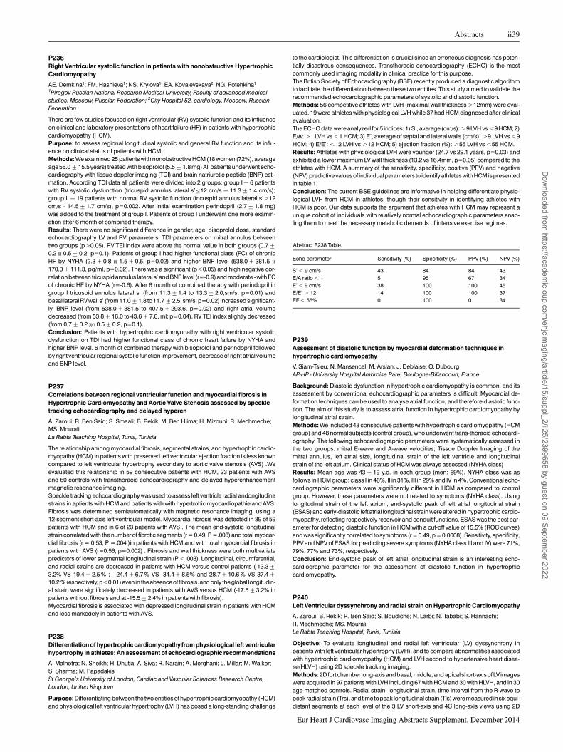
P236Right Ventricular systolic function in patients with nonobstructive HypertrophicCardiomyopathy
AE. Demkina1; FM. Hashieva1; NS. Krylova1; EA. Kovalevskaya2; NG. Potehkina1
1Pirogov Russian National Research Medical University, Faculty of advanced medicalstudies, Moscow, Russian Federation; 2City Hospital 52, cardiology, Moscow, RussianFederation
There are few studies focused on right ventricular (RV) systolic function and its influenceon clinical and laboratory presentations of heart failure (HF) in patients with hypertrophiccardiomyopathy (HCM).Purpose: to assess regional longitudinal systolic and general RV function and its influ-ence on clinical status of patients with HCM.Methods: We examined 25 patients with nonobstructive HCM (18 women (72%), averageage 56.0+15.5 years) treated with bisoprolol (5.5+1.8 mg) All patients underwent echo-cardiography with tissue doppler imaging (TDI) and brain natriuretic peptide (BNP) esti-mation. According TDI data all patients were divided into 2 groups: group I – 6 patientswith RV systolic dysfunction (tricuspid annulus lateral s’≤12 cm/s – 11.3+1.4 cm/s);group II – 19 patients with normal RV systolic function (tricuspid annulus lateral s’.12cm/s - 14.5+1.7 cm/s), p=0.002. After initial examination perindopril (2.7+1.8 mg)was added to the treatment of group I. Patients of group I underwent one more examin-ation after 6 month of combined therapy.Results: There were no significant difference in gender, age, bisoprolol dose, standardechocardiography LV and RV parameters, TDI parameters on mitral annulus betweentwo groups (p.0.05). RV TEI index were above the normal value in both groups (0.7+0.2 j 0.5+0.2, p=0.1). Patients of group I had higher functional class (FC) of chronicHF by NYHA (2.3+0.8 j 1.5+0.5, p=0.02) and higher BNP level (538.0+381.5 j
170.0+111.3, pg/ml, p=0.02). There was a significant (p,0.05) and high negative cor-relation between tricuspid annulus lateral s’ and BNP level (r=-0.9) and moderate - with FCof chronic HF by NYHA (r=-0.6). After 6 month of combined therapy with perindopril ingroup I tricuspid annulus lateral s’ (from 11.3+1.4 to 13.3+2.0,sm/s; p=0.01) andbasal lateral RVwall s’ (from 11.0+1.8 to 11.7+2.5, sm/s; p=0.02) increased significant-ly. BNP level (from 538.0+381.5 to 407.5+293.6, p=0.02) and right atrial volumedecreased (from 53.8+16.0 to 43.6+7.8, ml; p=0.04). RV TEI index slightly decreased(from 0.7+0.2 ep 0.5+0.2, p=0.1).Conclusion: Patients with hypertrophic cardiomyopathy with right ventricular systolicdysfunction on TDI had higher functional class of chronic heart failure by NYHA andhigher BNP level. 6 month of combined therapy with bisoprolol and perindopril followedby right ventricular regional systolic function improvement, decrease of right atrial volumeand BNP level.
P237Correlations between regional ventricular function and myocardial fibrosis inHypertrophic Cardiomyopathy and Aortic Valve Stenosis assessed by speckletracking echocardiography and delayed hyperen
A. Zaroui; R. Ben Said; S. Smaali; B. Rekik; M. Ben Hlima; H. Mizouni; R. Mechmeche;MS. MouraliLa Rabta Teaching Hospital, Tunis, Tunisia
The relationship among myocardial fibrosis, segmental strains, and hypertrophic cardio-myopathy (HCM) in patients with preserved left ventricular ejection fraction is less knowncompared to left ventricular hypertrophy secondary to aortic valve stenosis (AVS) .Weevaluated this relationship in 59 consecutive patients with HCM, 23 patients with AVSand 60 controls with transthoracic echocardiography and delayed hyperenhancementmagnetic resonance imaging.Speckle tracking echocardiography was used to assess left ventricle radial andongitudinastrains in aptients with HCM and patients with with hypertrohic myocardiopathie and AVS.Fibrosis was determined semiautomatically with magnetic resonance imaging, using a12-segment short-axis left ventricular model. Myocardial fibrosis was detected in 39 of 59patients with HCM and in 6 of 23 patients with AVS . The mean end-systolic longitudinalstrain correlated with the number of fibrotic segments (r = 0.49, P =.003) and total myocar-dial fibrosis (r = 0.53, P =.004 )in patients with HCM and with total myocardial fibrosis inpatients with AVS (r=0.56, p=0.002) . Fibrosis and wall thickness were both multivariatepredictors of lower segmental longitudinal strain (P ,.003). Longitudinal, circumferential,and radial strains are decreased in patients with HCM versus control patients (-13.3+3.2% VS 19.4+2.5 % ; - 24.4+6.7 % VS -34.4+8.5% and 28.7+10.6 % VS 37.4+10.2 % respectively, p,0.01)even in theabsenceoffibrosis.and only theglobal longitudin-al strain were significately decreased in patients with AVS versus HCM (-17.5+3.2% inpatients without fibrosis and at -15.5+2.4% in patients with fibrosis).Myocardial fibrosis is associated with depressed longitudinal strain in patients with HCMand less markedely in patients with AVS.
P238Differentiationof hypertrophiccardiomyopathy from physiological left ventricularhypertrophy in athletes: An assessment of echocardiographic recommendations
A. Malhotra; N. Sheikh; H. Dhutia; A. Siva; R. Narain; A. Merghani; L. Millar; M. Walker;S. Sharma; M. PapadakisSt George’s University of London, Cardiac and Vascular Sciences Research Centre,London, United Kingdom
Purpose: Differentiating between the two entities of hypertrophic cardiomyopathy (HCM)and physiological left ventricular hypertrophy (LVH) has posed a long-standing challenge
to the cardiologist. This differentiation is crucial since an erroneous diagnosis has poten-tially disastrous consequences. Transthoracic echocardiography (ECHO) is the mostcommonly used imaging modality in clinical practice for this purpose.The British Society of Echocardiography (BSE) recently produced a diagnostic algorithmto facilitate the differentiation between these two entities. This study aimed to validate therecommended echocardiographic parameters of systolic and diastolic function.Methods: 56 competitive athletes with LVH (maximal wall thickness .12mm) were eval-uated. 19 were athletes with physiological LVH while 37 had HCM diagnosed after clinicalevaluation.The ECHO data were analyzed for 5 indices: 1) S’, average (cm/s): .9 LVH vs ,9 HCM; 2)E/A: .1 LVH vs ,1 HCM; 3) E’, average of septal and lateral walls (cm/s): .9 LVH vs ,9HCM; 4) E/E’: ,12 LVH vs .12 HCM; 5) ejection fraction (%): .55 LVH vs ,55 HCM.Results: Athletes with physiological LVH were younger (24.7 vs 29.1 years, p=0.03) andexhibited a lower maximum LV wall thickness (13.2 vs 16.4mm, p=0.05) compared to theathletes with HCM. A summary of the sensitivity, specificity, positive (PPV) and negative(NPV) predictive values of individual parameters to identify athletes with HCM is presentedin table 1.Conclusion: The current BSE guidelines are informative in helping differentiate physio-logical LVH from HCM in athletes, though their sensitivity in identifying athletes withHCM is poor. Our data supports the argument that athletes with HCM may represent aunique cohort of individuals with relatively normal echocardiographic parameters enab-ling them to meet the necessary metabolic demands of intensive exercise regimes.
Abstract P238 Table.
Echo parameter Sensitivity (%) Specificity (%) PPV (%) NPV (%)
S’ , 9 cm/s 43 84 84 43E/A ratio , 1 5 95 67 34E’ , 9 cm/s 38 100 100 45E/E’ . 12 14 100 100 37EF , 55% 0 100 0 34
P239Assessment of diastolic function by myocardial deformation techniques inhypertrophic cardiomyopathy
V. Siam-Tsieu; N. Mansencal; M. Arslan; J. Deblaise; O. DubourgAP-HP - University Hospital Ambroise Pare, Boulogne-Billancourt, France
Background: Diastolic dysfunction in hypertrophic cardiomyopathy is common, and itsassessment by conventional echocardiographic parameters is difficult. Myocardial de-formation techniques can be used to analyse atrial function, and therefore diastolic func-tion. The aim of this study is to assess atrial function in hypertrophic cardiomyopathy bylongitudinal atrial strain.Methods: We included 48 consecutive patients with hypertrophic cardiomyopathy (HCMgroup) and 48 normal subjects (control group), who underwent trans-thoracic echocardi-ography. The following echocardiographic parameters were systematically assessed inthe two groups: mitral E-wave and A-wave velocities, Tissue Doppler Imaging of themitral annulus, left atrial size, longitudinal strain of the left ventricle and longitudinalstrain of the left atrium. Clinical status of HCM was always assessed (NYHA class)Results: Mean age was 43+19 y.o. in each group (men: 69%). NYHA class was asfollows in HCM group: class I in 46%, II in 31%, III in 29% and IV in 4%. Conventional echo-cardiographic parameters were significantly different in HCM as compared to controlgroup. However, these parameters were not related to symptoms (NYHA class). Usinglongitudinal strain of the left atrium, end-systolic peak of left atrial longitudinal strain(ESAS) and early-diastolic left atrial longitudinal strain were altered in hypertrophic cardio-myopathy, reflecting respectively reservoir and conduit functions. ESAS was the best par-ameter for detecting diastolic function in HCM with a cut-off value of 15.5% (ROC curves)andwassignificantly correlated to symptoms(r =0.49, p =0.0008). Sensitivity, specificity,PPV and NPV of ESAS for predicting severe symptoms (NYHA class III and IV) were 71%,79%, 77% and 73%, respectively.Conclusion: End-systolic peak of left atrial longitudinal strain is an interesting echo-cardiographic parameter for the assessment of diastolic function in hypertrophiccardiomyopathy.
P240Left Ventricular dyssynchrony and radial strain on Hypertrophic Cardiomyopathy
A. Zaroui; B. Rekik; R. Ben Said; S. Boudiche; N. Larbi; N. Tababi; S. Hannachi;R. Mechmeche; MS. MouraliLa Rabta Teaching Hospital, Tunis, Tunisia
Objective: To evaluate longitudinal and radial left ventricular (LV) dyssynchrony inpatients with left ventricular hypertrophy (LVH), and to compare abnormalities associatedwith hypertrophic cardiomyopathy (HCM) and LVH second to hypertensive heart disea-se(HLVH) using 2D speckle tracking imaging.Methods:2D fort chamber long-axis and basal, middle, and apical short-axis of LV imageswere acquired in 97 patients with LVH including 67 with HCM and 30 with HLVH, and in 30age-matched controls. Radial strain, longitudinal strain, time interval from the R-wave topeak radial strain (Trs), and time topeak longitudinal strain (Tls)were measured insix equi-distant segments at each level of the 3 LV short-axis and 4C long-axis views using 2D
Abstracts ii39
Eur Heart J Cardiovasc Imaging Abstracts Supplement, December 2014
Dow
nloaded from https://academ
ic.oup.com/ehjcim
aging/article/15/suppl_2/ii25/2399658 by guest on 09 September 2022

speckle tracking analysis. To assess LV dyssynchrony, Trs(rs)-18SD, the standard devi-ation (SD) of Tls(ls) was calculated.Results: Regional radial strain in the middle and apical short-axis segments wassignificantly less in patients with HCM than in those with HHD (56% +/2 23 VS45% +/2 21 and 47% +/2 19 VS 38 % +/2 17respectevely, p,0.01). Regionallongitudinal basal strain was also less in HCM (213 +/2 3.3 % VS -17 +/2 2.9 %,p=0.002). Trs-18SD and Tls were significantly longer in patients with HCM than in age-matched controls and patients with HLVH (Trs-18SD: HCM: 68 +/2 22 ms, HHD:21 +/2 11 ms, control: 15 +/2 12 ms P , 0.001, Tls-: HCM: 72 +/2 12 ms, HHD:44 +/2 11 ms, control: 33 +/2 13 ms P , 0.001).Conclusions: The presence of LVH is thus not always associated with LV dyssynchrony.However, the greater reduction of regional strain andsevere LVdyssynchrony in HCM maycontribute to the adverse cardiovascular outcomes associated with this disease.
P241Evaluation of left ventricular function by two-dimensional echocardiographyand three-dimensional speckle-tracking echocardiography in noncompactioncardiomyopathy and dilated myocardiopathy
R. Mechmeche; A. Zaroui; T. Chalbia; M. Ben Halima; B. Rekik; R. Boussada; MS. MouraliLa Rabta Teaching Hospital, Tunis, Tunisia
Introduction: Noncompaction cardiomyopathy develops due to the absence of myocar-dial compaction, and is associated with left ventricular dysfunction.Aim: The aim of the study was to evaluate comparatively left ventricular dysfunction inpatients with noncompaction cardiomyopathy using two-dimensional echocardiographyand three-dimensional speckle-tracking echocardiography.Method: The present study comprised of 27 patients withnoncompaction cardiomyop-athy (45.9+7.3 years, 13 males), 30 patientswith dilated myocardiopathy (42+6.4years, 14 males) 20 age- and gender-matched healthy controls (50.7+7.4 years, 12males). The echocardiographic diagnosis of LVNC was confirmed by magnetic reson-ance imaging. In all subjects standard echocardiography and tissue Doppler imaging(TDI) to study regional LV deformation and two and three-dimensional speckle-trackingechocardiography for the evaluation of left ventricular function were makedResults: two-dimensional speckle-tracking echocardiography-derived strainparametersof patients with noncompaction cardiomyopathy were found to be reduced as comparedto the values of controls. with significantly higher values in the LV base compared with theapex was observed in patients with LVNC by deformation measurements with TDI. Thisgradient was found particularly in the lateral and inferior wall but spared the anteroseptalwall; non-compaction was not found in basal segments throughout the ventricle and alsospared the anteroseptal midventricular wall. In DCM the strain andstrain rate values werehomogeneously reduced in all LV segments Three-dimensional speckle-trackingechocardiography-derived rotational parameters showed movements of the apical andbasal segments in the same direction suggesting ’rigid body rotation’ in all noncompac-tion cardiomyopathy cases.Conclusions: Left ventricular function and contractility are severely reduced in patientswith noncompaction cardiomyopathy. we releaved a preserved deformation in basal seg-ments of LVNC that can a difference between the LVNC and DCM. Absence of left ventricu-lar twist are a sign of noncompaction cardiomyopathy.
P243Relation between Left Atrial Volume and complications of type 2 diabetes
P. Lipari1; S. Bonapace1; F. Valbusa2; A. Rossi3; L. Zenari4; L. Lanzoni1; G. Targher5;G. Canali1; G. Molon1; E. Barbieri11"Sacred Heart" Hospital of Negrar, Department of Cardiology, Negrar-Verona, Italy;2Division of Internal Medicine, negrar, Italy; 3University City Hospital, Department ofCardiology, Verona, Italy; 4"Sacred Heart" Hospital of Negrar, Diabetes Center,Negrar-Verona, Italy; 5University of Verona, Department of Endocrinology and Metabolism,Verona, Italy
Background: Left atrial volume ( LAV ) is an independent predictor of cardiovascularevents than conventional indices of systolic and diastolic function in various pathologicalconditions.Moreover, the relationship between LAV and complications of type 2 diabetes(DM ) has never been studied in great detail.Objective: To investigate the potential relationship between LAV and complications oftype 2 diabetes.Methods: We enrolled 158 patients with DM with no history of atrial fibrillation and signifi-cant mitral valve disease . The maximum left atrial volume indexed to body surface area (LAVi ) was calculated before the opening of the mitral valve in systole from the apical 4and 2 chambers view by Simpson’s method. Microvascular complications were soughtwith ophthalmoscopy, macrovascular complications with carotid and lower limbs arterieswith echo-colour Doppler ultrasound, peripheral neuropathy with biothesiometer andnephropathy as serum creatinine and urinary albumin excretion.Results: mean age 68.9+6.2 years, males (74.5 %), mean body mass index (BMI)28.6+4.7 kg/m2 , mean diabetes duration 14.5+9.7 years, diabetic nephropathy(29.2 %), diabetic neuropathy (14%), microangiopathy (39.8 %), macroangiopathy(71%), glycated hemoglobin (7.3+1.3 %), creatinine (86.4+46.0 mmol / l), LAVi(32.2+10.3 ml/m2), ejection fraction of left ventricle (EF): 63.5+7.9% . LAVi correlatedwith diabetes duration (p = 0.002), glycated hemoglobin (p = 0.0004), microvascularcomplications (0.0003), diabetic nephropathy (p = 0.001) and the diabetic neuropathy(p = 0.004).Conclusions: In type 2 diabetes, left atrial volume not only correlates with the duration ofillness and the degree of glycemic control but also with the major microvascular compli-cations of diabetes. Our data suggest that this simple echocardiographic parameter canbecome an important marker in the overall assessment of the diabetic patient.
P244Troponin I/ ejection fraction ratio: a new index to differentiate Takotsubocardiomyopathy from Myocardial Infarction
G. Novo1; S. Giambanco1; MR. Sutera1; V. Bonomo1; F. Giambanco2; A. Rotolo1; S. Evola1;P. Assennato1; S. Novo1
1University of Palermo, Department of Cardiology, Palermo, Italy; 2Ingrassia Hospital,Division of Cardiology, Palermo, Italy
Purpose: Takotsubo cardiomyopathy (TC) is a frequently stress-induced cardiacdisorder, whose symptoms resemble those of acute myocardial infarction (AMI).Aim of our study was to investigate whether a non invasive tool, the ratio peak troponin Iand ejection fraction, could be useful to distinguish TC from AMI.Methods: We enrolled 53 cases of TC and as a control group 53 AMI patients, bothSTEMI and NSTEMI, matched for ejection fraction (EF), admitted to our institutionbetween 2007 and 2014.For each patient cardiovascular risk factors were recorded, a cardiologicalevaluation including electrocardiogram and transthoracic echocardiogram was per-formed, and serial troponin I levels were measured. Moreover, the ratio between peaktroponin I and left ventricular ejection fraction (LVEF) at admission was calculated (TEFR).Results:Thepeak troponin I level wassignificantly lower inpatientswith TCthan in theAMIgroup (6.52+7.25 vs. 91.11+117.91 ng/dl, p, 0.001). The TEFR was 16.31+19.58 inTC and 230.83+323.74 in AMI patients (p, 0.001). A TEFR value ≤ 60, derived fromthe receiver operating characteristic (ROC) curve analysis, was the cut-off value withthe best sensitivity (96.23%) and specificity (84.91%) to differentiate TC from AMI.Conclusions: The TEFR could be useful in differentiating TC from AMI at an early stage.
P245Clinical presentation, echocardiographic findings and in-hospital outcomes inpatients with takotsubo cardiomyopathy in comparision to patients with anteriorwall myocardial infarction.
M. Budnik; R. Piatkowski; J. Kochanowski; G. OpolskiMedical University of Warsaw, 1st Department of Cardiology, Warsaw, Poland
Takotsubo cardiomyopathy (TTC) mimics acute myocardial infarction (MI) and is mostoften characterized by chest pain, ST segment elevation, cardiac enzymes increaseand left ventricular dysfunction with reduction in ejection fraction. There are 3 types ofTTC depending on the type of wall motion abnormalities- classic if concerns apical seg-ments, inverse if concerns basal segments and atypical which means mid-segments akin-esia.
ii40 Abstracts
Eur Heart J Cardiovasc Imaging Abstracts Supplement, December 2014
Dow
nloaded from https://academ
ic.oup.com/ehjcim
aging/article/15/suppl_2/ii25/2399658 by guest on 09 September 2022

Purpose: The aim of the study was to anylyze echocardiographic findings in patients (pts)with TTC, determine the incidence of complications and compare the function of the leftventricle in pts with TTC and anterior wall acute MI with STsegment elevation (STEMI).Methods: The analysis included 94 consecutive pts with TTC and 103 consecutive ptswith STEMI. All pts were diagnosed with TTC based on the Mayo clinic criteria.Results: The mean age of pts was 69 years in TTC group and 68.2 in STEMI group (p.
0.05). There were 3 patients with inverse form of TTC and 3 pts with atypical form. Theremaining pts had classic form od TTC (88 pts). In the inverse form group EF was44,67% and in atypical form 39,67%. In 6 patients systolic anterior motion (SAM) waspresent which resolved in each case. One patient had coexisting hypertrophic cardiomy-opathy. Two pts had an apical thrombus, which resolved after improvement in systolicfunction of the left ventricle. Four cases was complicated by fluid in the pericardium.Comparison of the echo, clinical and prognostic factors between TTand STEMI group isincluded in the table 1.Conclusion: In patients with TTC despite lower EF there was better prognosis than inSTEMI group. In TTC patients none of the observed in Echo complications resulted indeath.
Abstract P245 Table.
Tako-tsubo group STEMI group p value
EF (%) 42,19 45,59 ,0,05LVDD (cm) 4,32 4,94 ,0,05Systolic RR (mmHg) 133,13 124,14 ,0,05TnI (ng/ml) 3,09 63,17 ,0,05CKMBmass (ng/ml) 12,92 181,51 ,0,05In-hospital VF (%) 4,2 9,71 ,0,05In- hospital AF (%) 5,32 7,77 ,0,05In-hospital mortality (%) 2,13 3,88 ,0,05
Comparison of the echocardiographic, clinical and prognostic factors.
SYSTEMIC DISEASES AND OTHER CONDITIONS
P246Advanced arteriolar retinal damage as a marker of Left Ventricular Hypertrophy
E. Chatzistamatiou1; I. Mpampatseva Vagena1; K. Manakos1; G. Moustakas2;D. Konstantinidis1; G. Memo1; O. Mitsakis1; A. Kasakogias1; P. Syros1; I. Kallikazaros1
1Hippokration General Hospital, Cardiology Department, Athens, Greece; 2SismanoglionHospital, Cardiology Department, Athens, Greece
Objective: Aim of our study was to determine the relationship between Scheie fundo-scopic classification and left ventricular hypertrophyin essential hypertension.Design and Methods: We studied 780 consecutive newly diagnosed, never-treated, non-diabetic, hypertensive patients stage I-III (51+13 years, 45.3% females). Echocardio-graphic left ventricular mass calculation was performed from parasternal long axis andnormalized for height in meters to the power of 2.7(LVMi). Using established cutoffs(.49 for males and .45 g/m2.7 for females), the study population was split in groupwith normal (n=649) and increased LVMi(n=131). All patients underwent fundoscopicexamination and alterations were classified according to Scheie grading system, as 0,1, 2 and 3.Results: Compared to hypertensives without LVH, patients with LVH were older (54+13vs. 51+13, p=0.018), with higher general and abdominal obesity, metabolic profile andtarget organ damage frequency and severity, while there was no difference regardinggender (females 43.7 vs. 45.6, p=0.693). Fundoscopic grade severity was significantlycorrelated with age (r=0.189, p,0.001), flow-mediated dilation of brachial artery(r=-0.166, p=0.001), LVMi (r=0.097, p=0.039), carotid IMT (0.212, p,0.001), TDI Em/Am ratio (r=-0.108, p=0.022), pulse pressure amplification ratio radial to central(r=-0.140, p=0.042), 24 hour SAP (r=0.119, p=0.011), 24 hour pulse pressure(r=0.128, p=0.006) and BP severity (r=0.170, p,0.001). Prevalence of grades 0 and 1was higher in patients with no LVH (45.3% vs. 36.1% and 31.8% vs. 23.6%, respectively),while grades 2 and 3 were more prevalent in patients with LVH (20.1% vs. 33.3% and 2.9%vs. 5.6%, respectively), p=0.007 for all.Conclusion: In the early course of essential hypertension, advanced arteriolar retinaldamage is associated with left ventricular hypertrophy.
P248Left ventricular mass index in hypertensive patients is related to increasedmobilization of mesenchymal stem cells
M. Marketou; F. Parthenakis; N. Kalyva; CH. Pontikoglou; S. Maragkoudakis; E. Zacharis;A. Patrianakos; F. Maragoudakis; H. Papadaki; P. VardasHeraklion University Hospital, Heraklion, Greece
Purpose: Stem and progenitor cells are implicated in ventricular remodelling and havegreat clinical significance in many cardiovascular diseases. However, there are limiteddata regarding the involvement of mesenchymal stem cells (MSCs) in the pathophysi-ology of arterial hypertension. The aim of this study was to investigate the circulation ofMSCs in patients with essential hypertension and left ventricular hypertrophy.Methods: We included 24 patients with untreated essential hypertension and 19 healthyindividuals. All subjects underwent a complete echocardiographic study. In addition, per-ipheral blood samples from all participants were immunostained with antibodies againstthe cell surface markers CD34, CD45 and CD90. Using flow cytometry, we measuredMSCs as a population of CD45-/CD34-/CD90+ cells and also as a population of CD45-/CD34-/CD105+ cells. The resulting counts were translated into the % percentage ofMSCs in the total cells of peripheral blood.Results: Hypertensive patients were shown to have increased circulating CD45-/CD34-/CD90+ compared to controls (0.0069+0.012% compared to 0.00085+0.0015%, re-spectively, p=0.039). No statistically significant difference in circulating CD45-/CD34-/CD105+ cells was found between hypertensives’ and normotensives’ peripheral blood(0.018+0.013% compared to 0.015+0.014%, respectively, p=0.53). Notably, CD45-/CD34-/CD90+ circulating cells were positively correlated with left ventNricular massindex (LVMI) (r=0.516, p,0.001).Conclusions: Patients with essential hypertension have increased circulating MSCscompared to normotensives. LVMI in hypertensive patients is related to increased mobil-ization of mesenchymal stem cells. Our findings contribute to the understanding of thepathophysiology of hypertension and might suggest a future therapeutic target.
P249Pericardial fat in systemic lupus erythematosus: relationship with inflammatorycytokines
AC. Rodrigues1; LA. Perandini2; TR. Souza1; AL. Sa-Pinto2; E. Borba2; AL. Arruda1;M. Furtado1; F. Carvalho1; E. Bonfa2; JL. Andrade1
1InRad- HC- University of Sao Paulo, Sao Paulo, Brazil; 2University of Sao Paulo Faculty ofMedicine (FMUSP), Rheumatology Division, Sao Paulo, Brazil
Pericardial adipose tissue may function as an endocrine organ and has been shown to bean important source of cytokines, contributing to increased inflammatory burden. In thisstudy, we compared the amount of epicardial fat between systemic lupus erythematosus(SLE) patients and healthy control(HC) individuals and the relashionship of epicardial fatwith cytokines and soluble tumor necrosis factor receptors (sTNFRs).Methods: Seventeen patients with SLE and low disease activity (Disease Activity Index of3.8+2.9) and 9 age and body mass index(BMI)-matched HC underwent echocardiog-raphy to assess systolic performance by left ventricular ejection fraction (LVEF) usingTeichholz formula and diastolic function using transmitral flow and tissue Doppler myocar-dial velocities. Measurements of epicardial fat were undertaken with 2-dimensional echo-cardiography from the parasternal long-axis view at the level of aortic root. Serumcytokines [interferon-g (INF-g), interleukin-6 (IL-6), tumor necrosis factor-a (TNF-a),interleukin-33 (IL-33)] and sTNFRs (sTNFR1 and sTNFR2) were assessed by multiplextechnique.Results: All patients were female and had normal systolic function (LVEF . 55%). Milddiastolic disfunction was found in 2 SLE patients. Though tissue Doppler velocitieswere still within normal limits, lower e’ velocities were found for SLE patients (p = 0.03).Epicardial fat was slightly but significantly increased in SLE patients compared to HC (p= 0.0003). Regarding cytokines and sTNFRs, TNF-a (p= 0.007) and (sTNFRII) were
Abstracts ii41
Eur Heart J Cardiovasc Imaging Abstracts Supplement, December 2014
Dow
nloaded from https://academ
ic.oup.com/ehjcim
aging/article/15/suppl_2/ii25/2399658 by guest on 09 September 2022

elevated in serum from SLE patients, with IL 23 and 33 showing a trend to higher levels(Table). TNFa was positively correlated to the amount of epicardial fat (r = 0.53, p,
0.05) and inversely correlated (r = - 0.62, p,0.05) with LVEF.Conclusion: These findings suggest the presence of inflammatory burden even in latentdisease and an association between pericardial fat and the activation of the inflammatoryresponse in SLE patients.
Abstract P249 Table. Clinical and laboratories variables
Age (years) BMI LVEF (%) Septal e’(cm/s)
Epicardial fat(mm)
TNFas sTNFRII
SLE 32+7 26+4 61+2 11+3 5.6+1.2 15.0+6.7 5769+2797HC 32+6 24+2 65+3 14+2 4.2+1.6 7.3+3.2 3264+803p NS NS NS 0.03 0.0003 0.005 0.02
BMI: body mass index; LVEF: left ventricular ejection fraction;TNFa: tumor necrosis factor;sTN-FRII: soluble tumor necrosis factor II
P250Echocardiographic assessment of pulmonary hypertension in Gaucher disease
Z. Hlubocka1; V. Malinova2; T. Palecek1; V. Danzig1; P. Kuchynka1; G. Dostalova1;J. Zeman2; A. Linhart11Charles University Prague, 1st Faculty of Med., 2nd Dept of Medicine-Dept of Cardiology& Angiology, Prague, Czech Republic; 2Charles University of Prague , First Faculty ofMedicine, Pediatric Clinic, Prague, Czech Republic
Purpose: Gaucher disease (GD) is the most common lysozomal storage disease charac-terized by accumulation of glucocerebroside in the body. Pulmonary arterial hypertension(PAH) is a known complication of the GD. However, the relationship between GD and PAHin regard to incidence, confounding factors and the effect of enzyme replacement therapy(ERT) is the subject of debate. The purpose of our study was to evaluate the incidence,severity and progression of PAH in the GD. Furthermore, association with clinical factrorsand the relation to ERT was assessed.Methods: From 2006 to 2013 we examined and prospectively followed 33 patients withthe GD (92% of all known cases in our country). Patients were followed annually usingechocardiography, ECG, clinical and laboratory examination. GD was diagnosed onthe basis of deficient levels of b-glucocerebrosidase; genotyping had also been per-formed. Severity score index (SSI) that reflects severity of disease parameters was calcu-lated. A complete echocardiographic examination was performed using Vivid 9 GEultrasound. Pulmonary artery systolic pressure (PASP) was assessed as the sum of thetricuspid regurgitation gradient plus right atrial pressure estimated from the inferiorvena cava measurements. PAH hypertension was defined as PASP . 35mmHg.Results: Thirty-two patients (97%) presented with type I Gaucher disease, the mean agewas 34+14 years and 22 (67%) were females. The mean time of follow-up was 6.2+1.2years. There were 23 patients (76%) receiving ERT, the mean duration of therapy was3.8+3.1 years. The mean value of PASP was 26+6mmHg. Three patients (9%) werediagnosed with mild PAH, all three were asymptomatic. One of these patients had border-line values of PASP during follow-up and in two other patients PAH did not progress sig-nificantly. PASP did not differ between patients receiving ERT (25+5 mmHg) anduntreated patients (27+7mmHg, p = n.s.) and did not correlate with the severity of theGD (SSI). There were no significant changes in other measured echocardiographic para-meters (chambers size, systolic function, diastolic dysfunction, valvular involvement).PASP did not rise during follow-up (26+6mmHg vs. 25+5mmHg, p=n.s.) and nonew cases of PAH were detected.Conclusion: In conclusion, our study reveals a predisposition for pulmonary hyperten-sion in type I Gaucher disease. However, progression to severe forms is probably rare.The presence of PAH was not related to GD severity or use of ERT. Echocardiographymay help with early identification of patients at risk of development of severe PAH.
P251Essential hypertension:When ingredients contains a little more sugar
E. Chatzistamatiou1; D. Konstantinidis1; G. Memo1; I. Mpampatzeva Vagena1;G. Moustakas2; K. Manakos1; K. Trachanas1; N. Vergi1; A. Feretou1; I. Kallikazaros1
1Hippokration General Hospital, Cardiology Department, Athens, Greece; 2SismanoglionHospital, Cardiology Department, Athens, Greece
Background: Sustained hyperglycemia, even in the prediabetic range, is associated withendothelial dysfunction, vascular stiffness and adverse cardiovascular outcomes. Aim ofour study was to explore the additive effects of impaired glycated-hemoglobin (HbA1c) onessential hypertension sequelae.Methods: We studied 465 never treated, newly diagnosed, essential hypertensivepatients stage I-III (mean age 52+13 years, 47% female), non-diabetic and withoutknown cardiovascular disease. Evaluation of hypertension was performed according toESH Guidelines. Traditional glycemic indices (fasting plasma glucose-FPG, HBA1c and2hr oral glucose tolerance-OGTT) were performed in all patients. Euglycemic group (E)included patients with all the above glycemic indices normal(FPG,100 mg/dl +HBA1c,5.7% + OGTT,140mg/dl), while impaired HBA1c group (IH) included patientswith HBA1c in prediabetic range (5.7-6.4%).Results: The mean values of HBA1c and FPG were higher in IH compared to E (92+7 vs.90+7, p=0.001), while they did not differ regarding OGTT (p=0.455). Moreover, IH com-pared to E were older (54+12 vs. 49+14, p=0.003),had higher ultrasonographic-derived preperitoneal and visceral fatthickness (18.9+6.4 vs. 17+7 mm, p=0.019
and 70.1+29 vs. 60.3+24.5 mm, p=0.003, respectively), with higher prevalence ofmetabolic syndrome (80,5% vs. 26,1%, p,0,001), while they didn’t differ regardinggender (males 48,4% vs. 50,6%, p=0.723), BMI (28+5 vs. 29+5 kg/m2, p=0.133),waist and hip circumference, waist to hip ratio and prevalence of abdominal obesity(p.0.05 for all). With the only exception of the family history of cardiovascular disease(54% vs. 41%, p=0.004),the two groups didn’t differ significantly regarding classical car-diovascular risk factors, including office and 24hr blood pressure and heart rate (p.0.05for all). However, IH presented higher prevalence of diastolic dysfunction (76% vs. 58%,p=0.008), c-f PWV (8.9+2.4 vs. 8.2+1.6, p=0.002) and carotid plaques (55% vs.40%, p=0.017), and lower TDI Em/Am (0.89+0.2 vs. 1+0.4, p=0.007), while they didnot differ regarding LVMI, carotid IMT, ankle-brachial index, ACR and Aix (p.0.05 for all).Conclusion: In essential hypertension, presence of impaired HbA1c augments arterialstiffness, carotid atherosclerosis and left ventricular diastolic dysfunction. Compared toanthropometric measurements, preperitoneal and fat thicknesses represents more sen-sitive indices of impaired glycated-hemoglobin.
P252Comparison of invasive Doppler echocardiographic and hemodynamic methodsfor optimization in patients receiving CRT
H. Corut; LE. Sade; B. Ozin; I. Atar; O. Turgay; H. MuderrisogluBaskent University, Faculty of Medicine, Ankara, Turkey
Purpose: We aimed to compare acute and mid-term effects of invasive and echocardio-graphic optimization methods on hemodynamic and volume response after cardiacresynchronization therapy (CRT).Methods: In this prospective clinical trial, we randomized patients to either echocardio-graphic (n=20)or invasive method (n=20) for atrioventricular (AV)and ventriculoventricu-lar (VV) delay programming. AV delays from 60 to 160 ms, VV delays from -80 to +80mswere tested by both methods in all patients initially, then the patients were randomlyassigned to either invasive or echo driven results for follow-up. Optimal AV and VVdelays were defined as the ones that yielded the largest LV outflow tract time velocity inte-gral (LVOT-TVI) and diastolic filling time (DFT) by echocardiography and the largest LV dP/dtmax by invasive method. End systolic volume (ESV) decrease ≥15%, ejection fraction(EF) increase .5% were considered as significant volume response, NYHA class in-crease ≥1 as clinical response at 6 months.Results: Initially determined optimal AV delays were concordant in 58% of patientswithin+10ms, and VV delays in 65% of patients within+20ms. We observed significantimprovement in acute hemodynamic response by echo-guided optimization (DFT: from360+123ms to 467+137ms; p,0.001, LVOT-TVI: from 13.5+4cm to 16+4.4cm;p,0.001) and by invasive optimization (LV dP/dtmax: from 1088+327dynes/s to1336+327dynes/s; p,0.001). At 6 months, clinical, volume and EF response rates byinvasive optimization were 70%, 40%, 70%, and by echo-guided optimization theserates were 45%, 60%, 60% respectively (p=NS). Optimization method did not predict clin-ical or volume response at 6 months.Conlusion: Both invasive hemodynamic and echocardiographic Doppler methods arecomparable and effective for CRToptimization.
P253Lung ultrasound as a non-invasive bedside approach in the diagnosis ofpulmonary embolism
A. Ledakowicz-Polak; L. Polak; G. Krauza; M. ZielinskaMedical University, Department of Intensive Cardiac Therapy, Lodz, Poland
Background: Clinical manifestation of pulmonary embolism (PE) is often non-specific.Despite the prevalence ofmultidetector CTpulmonaryangiography (MCTPA), thediagno-sis of PE still presents as a considerable challenge. Furthermore MCTPA exposes patientsto high- dose radiation and to potential serious complications. Among alternative imagingtechniques the lungultrasound seems to be the most promisingand provides anattractivenon-invasive bedside approach especially in the emergency conditions.We aimed to determine the diagnostic utility of lung ultrasound in PE diagnosis.Methods: The current study comprised 50 consecutive adult patients with suspection ofPE. The initial diagnosis was based on Wells score .4 and a positive D-dimer test in eachpatient. Final diagnosis was obtained with MCTPA. Lung ultrasound was performedbefore MCTPA in accordance with BLUE (Bedside Lung Ultrasonography in Emergency)protocol. It was considered diagnostic for PE if one or more subpleural infarcts weredetected.
Abstract P252 Figure.
ii42 Abstracts
Eur Heart J Cardiovasc Imaging Abstracts Supplement, December 2014
Dow
nloaded from https://academ
ic.oup.com/ehjcim
aging/article/15/suppl_2/ii25/2399658 by guest on 09 September 2022

Results: PE was diagnosed in 33 of 50 enrolled patients (66%) with a mean age 62,5 yearswith a predominance of females (63.6%). Lung ultrasound yielded a sensitivity of 86% anda specificity of 92.8%, respectively.Conclusions: Our findings suggest that lung ultrasound seems to be a useful diagnostictool in the management of patients with suspected PE. Furthermore this technique mayprove a valuable bedside method in the diagnosis of PE, thus facilitating immediate treat-ment decisions. Its increasing accuracy and utility can gradually lead to the MCTPAburden reduction.
P254Exercise-induced changes in RV and LV mechanical dispersion in Brugadasyndrome
M. Szulik; W. Streb; A. Wozniak; R. Lenarczyk; A. Sliwinska; Z. Kalarus; T. KukulskiSilesian Center for Heart Diseases, Medical University of Silesia; Department ofCardiology, Zabrze, Poland
Aim: Exercise-induced changes in RV and LV mechanical dispersion in Brugada syn-drome (BS) are not well studied. The differences between patients, who did(‘arrhythmia+’) and did not (‘arrhythmia-’) experience adequate ICD intervention areunknown.Methods: 14 BS patients underwent rest and bicycle ergometer echocardiography andwere followed by 2 years (0.08–4.3). The SD of time to maximum myocardial longitudinalstrain in speckle tracking – was a parameter of mechanical dispersion for LV: in a16-segment model (or 6-segment model for apical views); for RV – 3 RV free wall seg-ments (in apical 4-chamber view). Among 13 with ICD implanted, 4 were ‘arrhythmia+’.Results: In all ‘arrhythmia+’ patients (100%) postsystolic shortening in RV basal segmentwas observed, whilst among ‘arrhythmia-’ in 3 patients (33%) (p=0.018). Diastolic func-tion comparison – see table (only significant differences).Dispersion - fig 1 (dependences not shown - not significant).Conclusions: (1) RV diastolic dysfunction (RV E/E’ . 6) and post-systolic shortening areobserved significantly more often among Brugada syndrome pts with ICD adequateinterventions. (2) Significant decrease of RV dispersion during exercise occurs only inin event free patients. (3) LV strain dispersion at rest is predominantly present in pts withadequate ICD intervention. It decreases with exercise.
Abstract P254 Table. Diastole in BS - comparison of groups
Arrhythmia + Arrhythmia 2
RV E’ [cm/s] 27.5+10.6 210.2+1.3 *RV E/E’ [-] 7.46+1.4 4.7+0.7 *
* - p , 0.05
P255Role of tissue doppler and strain echocardiographic parameters in pulmonaryhypertension
MA. Nogueira1; LM. Branco1; A. Agapito1; A. Galrinho1; A. Borba2; PP. Teixeira1;AV. Monteiro1; R. Ramos1; D. Cacela1; R. Cruz Ferreira1
1Hospital Santa Marta, Department of Cardiology, Lisbon, Portugal; 2Hospital Santa Marta,Pulmonology Department, Lisbon, Portugal
Purpose: Analyse different echocardiographic parameters that evaluate mainly the rightventricular functionandtheircorrelationwithprognosticmarkers inpulmonaryhypertension.Methods: We studied 46 patients (pts) consecutively observed in a Pulmonary Hyperten-sion Clinic, with theneed fora transthoracic echocardiogram (TTE), since the beginning ofAugust 2013 until the beginning of May 2014. The echocardiographic variables analysedwere: Absolute and Indexed Right Atrial (RA) Area and Volume, RA Strain, TricuspidAnnular Plane Systolic Excursion (TAPSE), Right Ventricular Fractional Area Change(RVFAC), RV Longitudinal Strain, RV Tei Index, RV Tei Index by Tissue Doppler Imaging(TDI), Tricuspid e’, s’ and E/e’, Pulmonary Artery Systolic Pressure (PASP), Aortic VelocityTime Integral (VTI), Cardiac Output (CO),Left Ventricle Eccentricity Index (LVEI) in diastoleand systole, Mitral mean e’, s’ and E/e’. We also collected data regarding demographiccharacteristics, clinical status of the pts, six-minute walk test (6MWT) results and serumlevels of brain natriuretic peptide (BNP), in the same period of time of TTE.
Results:Themajority ofptswere female (32pts=70%), with ameanageof54+16years.Most pts (31 pts = 67%) were classified in group 1 of Dana Point Classification and were infunctional class II of the New York Heart Association. There was a statistically significantcorrelation between 6MWT (mean value of 363+106 meters) and the following variables:RV Tei Index (0.42+0.24), with p = 0.008; PASP (78.09+27.45 mm Hg), with p = 0.025;LVEI in diastole (1.32+0.32), with p = 0.042 and mitral mean e’ (8.40+2.99 cm/s), with p= 0.017. There was also a statistically significant correlation between BNP levels (meanvalue of 304.98+345.24 pg/mL) and RA Strain (21.55+13.31 %), with p = 0.001;TAPSE (17.93+4.58 mm), with p = 0.006; mitral E/mean e’ (10.38+5.26), with p =0.037 and tricuspid s’ (10.98+3.33 cm/s), with p = 0.018.Conclusions: In this population of patients with pulmonary hypertension, standard para-meters such as right ventricular Tei Index, as well as more recent parameters, particularlyright atrial strain, have an important correlation with prognostic markers well studied–six-minute walk test and brain natriuretic peptide serum levels. However, there will be neededfurther studies in order to confirm the incremental value of the more recent echocardio-graphic parameters on the prognostic stratification of this clinical entity.
P256Patient-specific heart-arterial tree modeling
A. Guala1; C. Camporeale1; F. Tosello2; C. Canuto3; L. Ridolfi1
1Politecnico di Torino, DIATI, Torino, Italy; 2University Hospital S. Giovanni Battista, Torino,Italy; 3Politecnico di Torino, Mathematical Sciences, Torino, Italy
The cardiovascular system has been studied by the fluid mechanics community since longtime, but it is just recently that the physical-based mathematical modeling has reached theaccuracy and effectiveness required by the medical science. This has been due to the im-pressive advancing of cardiovascular imaging and computational capacities.We here propose a new multi-scale mathematical model for the simulation of the leftventricle-arterial tree system. The left ventricle is described by the concept of time-varyingelastance. The aortic valve dynamics is modeled by a balance of forces on the leaflets. Thenetwork of 48 large-to-medium conducting arteries is described by a spatially-extendedone-dimensional modeling. Nonlinear viscoelasticity of the arterial wall is accounted bya new constitutive relation. The 24 distal circulation volumes are modeled bythree-element Windkessel models.About the setting of the geometrical and mechanical properties of the left ventricle, aorticvalve, arterial tree and distal elements involved in the model, we referred to data collectedby different researchers in the last sixty years, corresponding to a statistically averagedhealthy young men. Starting from this typical parameter setting, the description of a spe-cific patient is obtained by a suitable tuning of a limited subset of parameters. Such subsetincludes vessels lengths, diameters and mechanical properties (pulse pressure velocity),heart rate, QT duration, left-ventricle contraction time and maximum elastance, ejectionperiods and overall resistance of the distal volumes.The patient-specific parameter setting process has been carefully designed keeping inmind the modern non-invasive measurement techniques. An objective and simple pro-cedure is defined, adapting the model to the specific patient by use of echocardiographicimages, anthropometric data, and peripheral pressure measures.We validate the patient-specific model on six young man. Several modeled quantities arecompared to the measured ones. About left ventricle, we compare end-diastolic and end-systolic volumes, ejection fraction, and mean outflow. About arterial tree, central, brachial,radial, femoral, and tibial pressures are compared. The root mean square error aboutpressure and ventricular quantities was always less than 10% for all the patients and itoften was less than 5%, demonstrating that the model and its patient-specific settingare sufficiently accurate to reproduce the left ventricle-arterial interaction. These good per-formances induce to test further the capabilities of the model and its possible use forclinical routine.
P257Ultrasonography in abdominal adiposity as determinants of cardiovascular risk
E. Chatzistamatiou1; G. Moustakas2; G. Memo1; D. Konstantinidis1;I. Mpampatzeva Vagena1; K. Manakos1; K. Traxanas1; N. Vergi1; A. Feretou1;I. Kallikazaros1
1Hippokration General Hospital, Cardiology Department, Athens, Greece; 2SismanoglionHospital, Cardiology Department, Athens, Greece
Objective: We sought to compare the various obesity measures as predictors of dyslipi-demia, dysglycemia and essential hypertension.Design and Methods: We studied 1140 consecutive, non-diabetic subjects (51+13years, 49% males), referred to our antihypertensive unit. Body mass index (BMI), bodyfat content (FAT, using a portable bioelectric impedance body composition analyzer),waist (WC) and hip (HC) circumferenceand waist-to-hip circumferenceratio(WHR) weremeasured/calculated. Visceral (V), perirenal (R) and mesenteric (M) fat thicknesseswere also measured using a linear ultrasound probe.Results: The prevalence of dyslipidemia, dysglycemia and essential hypertension was88.1%, 44.2% and 67.2%, respectively. The prevalence of 0, 1, 2 or all 3 dysmetabolic con-ditions was 3.1%, 22.3%, 43.5% and 31.1%, respectively. Compared to patients with 1 and2, patients with all 3 dysmetabolic conditions were predominantly male (39% vs. 45% vs.68%, p,0.001), older (50+14 vs. 51+12 vs. 55+12years, p,0.001), with higher BMI(27+4 vs. 28+5 vs. 29+5kg/m2, p,0.001), FAT (32+8% vs. 34+8% vs. 35+7%,p=0.004), WC (90+13 vs. 93+13 vs. 98+13cm, p,0.001), HC (103+10 vs. 105+10 vs. 107+9cm, p,0.001), WHR (0.87+0.08 vs. 0.88+0.08 vs. 0.91+0.08,p,0.001), V (57+24 vs. 62+24 vs. 72+26mm, p,0.001), R (10+4 vs. 11+4 vs.13+5mm, p,0.001) and Mfat (11+4 vs. 12+4 vs. 13+5mm, p,0.001). All the
Abstract P254 Figure. LV and RV diseprsion - rest and exercise
Abstracts ii43
Eur Heart J Cardiovasc Imaging Abstracts Supplement, December 2014
Dow
nloaded from https://academ
ic.oup.com/ehjcim
aging/article/15/suppl_2/ii25/2399658 by guest on 09 September 2022

obesity markers significantly (p,0.001) discriminated subjects with all 3 dysmetabolicconditions except FAT (p=0.082). WHR and WC had the highest area under the curve fol-lowed by V, BMI, R, Mand FAT (Table).Conclusions: Among the various obesity markers, WHR, WC and Visceral fat emerge asthe most significant determinants of dysmetabolic diseases while measures of total bodyfat (i.e. BMI and FAT) have less predictive power.
Abstract P257 Table.
Table Area Under theCurve (AUC)
95% ConfidenceInterval (95% CI)
p-value
Waist-to-hip ratio 0.627 0.585-0.669 ,0.001Waist circumference 0.623 0.581-0.665 ,0.001Visceral fat 0.620 0.575-0.665 ,0.001Body mass index 0.599 0.557-0.642 ,0.001Perirenal fat 0.594 0.546-0.641 ,0.001Mesenteric fat 0.592 0.545-0.638 ,0.001Body fat content 0.573 0.492-0.654 0.082
P258Evaluation of left ventricular torsional mechanics using speckle trackingechocardiography in pregnancy
K. Hristova1; R. Marinov1; G. Stamenov2; M. Mihova2; S. Persenska2; A. Racheva2
1National Heart Hospital, Sofia, Bulgaria; 2Nadezhda Women’s Health Hospital , Sofia,Bulgaria
Normal pregnancy is associated with reversible changes in both systolic and diastolicmechanics, consistent with an increase in preload and decrease in afterload and systemicvascular resistance.The aim of this study is to evaluate left ventricular twist and untwist mechanics via speckletracking echocardiography in a population of healthy pregnant women during differingstages of a healthy pregnancy.Methods: The study population included 22 pregnant patients, 10 with multiplepregnancy (mean age 36.5+3 years) and 20 healthy non pregnant women (mean age33+4years). Apical and basal short axis for 2D images were acquired (frame rate65+7 frames/s) and serial images will be obtained during the first, second, and third tri-mester of the pregnancy, as well as up to two months post partum. The curves of LVT/LVUR were extracted using a commercial software.Results: Peak LV twist and peak untwisting rate increased significantly in the 3rd trimesterof normal pregnancy (13.48+2.908, 13.12+3.308, 16.83+3.618, P , 0.001; and-111.52+23.548/sec, -107.40+26.588/sec, -144.30+45.148/sec, P , 0.001; in the1st, 2nd, and 3rd trimester, respectively).The pregnants with twins have the highervalue for LVT and LVUR compare with other pregnant (p,0.01), but in the last trimester,the time to peak LVUR is prolonged compared with other pregnant women and controlgroup. An independent association was found between the change in LV twist and thechange in LV end-systolic volume between the 1st and 3rd trimester. Peak untwistingrate at the 3rd trimester correlated significantly with peak twist and LV end-diastolicvolume. Multiple regression analysis indicate that only systolic blood pressure (r =0.394, P = 0.005) was an independent predictor for increased LV torsion.Conclusions: During normal pregnancy, LV twist and peak untwisting rate increase in the3rd trimesterandcorrelate withend-systolicandend-diastolic volume, respectively.Bloodpressure and condition of multiple pregnancy are independently associated withincreased torsion during pregnancy.
P259Cardiac and obstetric care during pregnancy in women with congenital heartdisease.
KJ. Plaskota; O. Trojnarska; A. Bartczak; S. GrajekPoznan University of Medical Sciences, Ist Department of Cardiology, Poznan, Poland
Purpose: evaluation of the range of cardiac and obstetric care during pregnancy inwomen with congenital heart disease.Methods: Data of 350 completed pregnancies in 241 women with CHD aged 18-40 years,followed-up at the Grown-up Congenital Heart Disease Outpatient Clinic between1993-2013, were analysed. All patients were divided into four WHO classification categor-ies of potential risk of complications during pregnancy and delivery. Data regarding fre-quency of follow-up visits at outpatient clinic and rate of urgent hospitalizations due tocardiac and obstetric indications during pregnancy were obtained from medicalrecords and information from patients during follow-up visits.Results:Therewere813outpatientclinic follow-up visits during 350 completed pregnancies(mean 2,3+1,2). Single follow-up visit during pregnancy was the most common in patientswith low risk of pregnancy complications (44,2%). High cardiac complications risk duringpregnancy patients (WHO III, WHO IV) were followed up the most frequently (meannumber of follow-up visits in WHO class III patients was 3,1+1,4; 3,9+1,2 in WHO classIV patients, respectively). Frequency of follow-up visits increased between WHO classifica-tioncategoryI, IIandIII, therewasnostatisticallysignificantdifferencebetweenclass IIIandIV(p=0,93). 33 pregnant patients (9,4%) required urgent hospitalizations: 18 of them due tocardiac indications (5,1%), 15 of them due to obstetric reasons (4,3%). Hospitalization rateincreased with increasing WHO scale category (3,9% vs 8,2% vs 13,6% vs 87,5%;p=0,0001) with the highest rate of urgent hospitalization in WHO class IV patients.Conclusions: Pregnant patients with congenital heart disease should be provided withmeticulous cardiac care which is essential for favorable outcome of pregnancy and
delivery. Follow-up visits and urgent hospitalization incidence increase with progressingseverity of cardiac defect.
P260Prenatal diagnosis of fetuses with tricuspid atresia - Single centre experience in acountry with limited recources
RA. Ramush Bejiqi; R. Retkoceri; H. Bejiqi; A. Beha; SH. SurdulliUniversity Clinical centre Prishtine, Prishtina, Kosovo, Republic of
Introduction: Tricuspid atresia (TrA) is a congenital heart disease where there is no directcommunication between the right atrium and right ventricle. Prenatal diagnosis contributein organizing delivery and planning for palliative treatment.Aim of this presentation was retrospective analysis of diagnosis, features and outcomes offetuses with TrA diagnosed in our clinic.Method and material Between January 2001 and December 2012 in our Clinic were studiedaround 326 fetal cases with congenital heart disease (CHD), and in 23 (7.0%) were found tohave TrA. Age of gestation at time of diagnosis was 16 -38 weeks gestation. Retrospectivelywere analyzed characteristics and outcomes of 18 cases with known follow-up.Results Characteristics: Three fetuses were twins, 2 fetuses were triple. Fife fetuses(21.7%) had restrictive interatrial communication and balloon atrioseptostomy immedi-ately were performed. Twelve of them had heart failure already at presentation, due to re-strictive communication. All fetuses had nonrestrictive VSD, 5 of them had additionalmuscular restrictive VSD. Five fetuses (17.4%) had transposition of great arteries with non-restrictive VSD, 8 fetuses (34.8%) had pulmonary stenosis, 2 (8.7%) had pulmonaryatresia, 9 were (39.1%) with patent ductus, one with aortic coarctation, 6 had associatedextracardiac anomalies.Outcomes: Seven pregnancies (30.4%) were terminated, 6 with extracardiac anomalies.Outof16 fetuses thatcontinued pregnancies, 3died inutero, 3diedshortly afterbirth and3died in second month of live waiting for surgery. The remaining 10 cases were operatedwith palliative procedures (shunt or pulmonary band). All they underwent surgery(Glenn operation or Fontan procedure). Total intrauterine and postnatal mortality, with ter-minated pregnancies was 16/23 (69.6%).Conclusion: Despite an improvement in perinatal diagnosis and management of fetusesand children with TrA in Kosovo, outcomes remain still poor, especially in situation wherethe surgical treatment is depends from treatment abroad Kosovo. Negative prognosticfactors were restrictive atrial communication, long term waiting for surgery, type andform of transport from Kosovo to destination center for surgery and still poor technicalresources for follow up of this specific condition.
CONGENITAL HEART DISEASE
P262Evaluation of the tricuspid annulus size: clinical implications from comparisonbetween 2D-transthoracic and 3D-transesophageal echocardiography
J. Dreyfus; G. Durand-Viel; C. Cimadevilla; E. Brochet; A. Vahanian; D. Messika-ZeitounAP-HP-Bichat Hospital-Cardiology Department, Paris, France
Background: Tricuspid annuloplasty is recommended during left-heart valve surgerywhen tricuspid annulus (TA) is dilated, independently of the degree of tricuspid regurgita-tion, but the methodology to measure TA and thresholds arenot clearly defined. We aimedto compare TA diameter (TAD) measurements performed using bi-dimensional
ii44 Abstracts
Eur Heart J Cardiovasc Imaging Abstracts Supplement, December 2014
Dow
nloaded from https://academ
ic.oup.com/ehjcim
aging/article/15/suppl_2/ii25/2399658 by guest on 09 September 2022

transthoracic echocardiography (2D-TTE) in the 4 different views to three-dimensionalmeasurements performed during transesophageal echocardiography (3D-TEE) and todefine thresholds of TA enlargement for routine practice.Methods: 2D-TTE measurement of the TAD was performed in parasternal long-axis viewof the right ventricle inflow, parasternal short-axis, apical 4-chamber (A4C) and sub-costalviews in 195 prospectively enrolled patients and 66 healthy volunteers. 3D dynamic volu-metric datas of the TA were also acquired by TEE using a matrix array transducer (X7-2t,Philips) in the 195 patients. Multiplanar reconstructions were performed offline using dedi-cated software (QLab7, Philips) to measure the long-axis (LA) of the TA.Results: In the 195 patients, TAD measurements were not different between the 4 TTEviews (P=0.13), but A4C was the most feasible and the most reproducible method(Table). TAD measurement in A4C view by TTE (3.90+0.62cm) was well correlated(r=0.84, p,0.0001) to LA by 3D-TEE (4.33+0.63cm), but with a systematic 4mm under-estimation. In the healthy volunteers, mean value of TAD in A4C was 3.2+0.4cm or 1.8+0.23cm/m2 and the upper limit of 95% confidence interval was 4.2cm or 2.3cm/m2.Conclusion: TAD measurement in A4C view by 2D-TTE was highly feasible, reproducibleand accurately reflected TA size, even if it was systematically underestimating its maximaldiameter. Based on measurements in healthy volunteers, we suggest to consider tricuspidannuloplastyduring left-heartvalvesurgerywhenTA ismorethan2.3cm/m2 or4.2cminA4C.
P263Mitral annular disjunction and unsaddling in myxomatous mitral valve prolapse: a3-dimensional transesophageal echocardiographic study
CN. Jin1; F. Fang1; FX. Meng1; K. Kam1; JP. Sun1; GK. Tsui2; KK. Wong2; S. Wan3; CM. Yu1;AP. Lee1
1The Chinese University of Hong Kong, Division of Cardiology, Institute of VascularMedicine, Li Ka Shing Institute of Health Sciences, Hong Kong, Hong Kong SAR, People’sRepublic of China; 2The University of Hong Kong, Department of Computer Sciences,Hong Kong, Hong Kong SAR, People’s Republicof China; 3TheChinese University ofHongKong, Division of Cardiothoracic Surgery, Department of Surgery, Hong Kong, Hong KongSAR, People’s Republic of China
Background: In patients with myxomatous mitral valve prolapse (MVP), mitral annulardisjunction (MAD), defined as a separation between the atrial-mitral valve (MV) junctionand ventricular attachment, is a common finding that may be associated with disturbanceof annular function and may affect surgical repair approach. However, data on the three-dimensional (3D) structure of annular disjunction is limited.Methods: 3D transesophageal echocardiography was performed in 96 consecutivepatients with MVP and severe mitral regurgitation undergoing MV repair and 24 age, sex-matched normal controls. The 3D geometry of MV apparatus was measured with dedi-cated quantification software.Results: Obvious MAD was evident in 20 (21%) MVP patients. Compared to MVP patientswithout MAD, those withMAD had significantly greater commissural width, shorterannularheight, but similar annular anteroposterior diameter, area, and circumference, resulting ina flatter and more elliptical annular shape. Annular unsaddling (defined as annularheight-to-commissural width ratio,15%) was significantly more prevalent in MADgroup than without (90% vs. 46%, x2=9.674, P=0.002). MAD was also associated withsignificantly greater leaflet billowing volume, and longer chordal lengths (Table).Conclusion: MAD has disturbed 3D geometry with reduced annular height and loss of thesaddle-shape. These findings may have important implications in progression of MVPlesions and in selecting mitral annuloplasty rings and repair techniques.
Abstract P263 Table. 3D geometry of mitral valve apparatus
Variables Normal(n=24)
No MAD(n=76)
MAD(n=20)
ANOVA P
Commissural width (CW), mm 33.1+3.7 37.8+4.6* 41.8+6.1*† ,0.001Anteroposterior (AP)diameter, mm
27.8+2.7 36.3+4.6* 36.4+6.2* ,0.001
AP-to-CW ratio (ellipticity), % 0.84+0.06 0.96+0.08* 0.87+0.08† ,0.001Annular height (AH), mm 8.1+1.9 5.6+1.5* 4.8+1.3*† ,0.001AH-to-CW ratio (AHCWR), % 24.7+5.4 14.9+4.2* 11.5+2.7*† ,0.001Annular unsaddling(AHCWR,15%), n (%)
0 (0) 35 (46)* 18 (90)*† ,0.001
Annular circumference, mm 106.4+9.6 121.3+13.6* 128.2+19.5* ,0.001Annular area, mm2 733+132 1088+255* 1212+390* ,0.001Leaflet billowing volume, ml 0.08+0.07 0.70+0.74* 1.91+1.99*† ,0.001Chordal length (anterolateral),mm
18.2+3.3 22.6+5.3* 25.9+4.0*† ,0.001
Chordal length(posteromedial), mm
18.7+3.9 24.5+6.1* 27.8+5.2*† ,0.001
* P,0.05 versus normal controls † P,0.05 versus patients without MAD
P264Difference in prevalence of prosthesis-patient mismatch after mitral valvereplacement according to the defining methods for effective orifice area
I J. Cho; HM. Chung; R. Heo; SJ. Ha; GR. Hong; CY. Shim; HJ. Chang; JW. Ha; N. ChungYonsei University College of Medicine, Cardiology Division, Seoul, Korea, Republic of
Background: The incidence of prosthesis-patient mismatch (PPM) after mitral valve re-placement (MVR) has been reported variably and the method used for calculating effect-ive orifice area (EOA) has not standardized. The purpose of the current study was tocompare the incidence of mitral PPM according to different defining methods of EOA
including continuity equation (CE) calculation, pressure half time (PHT) calculation anduse of reference EOA in patients with mitral stenosis after MVR.Methods: We retrospectively reviewed all the patients who performed MVR due to rheum-atic mitral stenosis from January 2004 to December 2012. Among the patients, 166patients who performed postoperative echocardiography between 12 and 60 monthsafter MVR were comprised the study population. EOA was determined by CE (EOA-CE)and PHT (EOA-PHT) using Doppler echocardiography. Referred EOA (EOA-R) was deter-mined from the literature or values offered by manufacturer. Indexed EOA was used toPPM as not significant if . 1.2 cm2/m2, as moderate if . 0.9 cm2/m2 and ≤1.2 cm2/m2,and as severe if ≤ 0.9 cm2/m2.Results: Prevalence of PPM was different according to the methods used to define EOA,ranging from 7% in PHT method, 49% in referred EOA method, to 62% in CE methods.Intraclass correlation coefficient was low between the methods (0.430 between indexedEOA-CE and indexed EOA-PHT, 0.320 between indexed EOA-CE and indexed EOA-R).Among the indexed EOAs, only indexed EOA-CE showed independent correlation withpostoperative systolic pulmonary artery pressure(p,0.001).Conclusion: The incidence of mitral PPM was variable according to defining method ofEOA, and only EOA-CE was found to be independently associated with postoperativehemodynamic variable. Therefore, indexed EOA-CE should be used to define PPM.
DISEASES OF THE AORTA
P265Imaging predictors of long-term evolution of type B acute aortic syndromes
S. Moral; D. Gruosso; V. Galuppo; G. Teixido; JF. Rodriguez-Palomares; L. Gutierrez;A. EvangelistaUniversity Hospital Vall d’Hebron, Barcelona, Spain
Background: Despite aortic guidelines recommend a similar treatment in type B aorticdissection (AD) and intramural haematoma (IMH) in patients with the same clinical risk,the differences in prognosis in a long-term follow-up between acute aortic syndromes(AAS) remain unknown.Objectives:Tocomparemorbidityandmortalityofdifferent typeBAASin long-termfollow-up.Methods: 152 patients with an AAS were included prospectively in a clinical and imagingprotocol: 67(44%) AD (fig.1A), 53(35%) IMH without ID (fig.1B) and 32(21%) IMH with ID(fig.1C). ID was defined as the presence of an intimal disruption in the aorticwall.3mm. Aortic and overall mortality, required invasive treatments, visceral/peripheralischemia and maximum aortic diameter (MAD) were collected in a mean follow-up of 58+30 months (range 6-132).Results: Patients with IMH were older than those with AD (IMH with ID: 65.2+9.0, IMHwithout ID: 65.5+10.9, AD: 55.5+12.5 years; p,0.001). MAD was similar betweengroups in the acute phase (p=0.15). During the follow-up, global mortality was similarbetween groups (p=0.34), but AD presented a higher aortic mortality than IMH withand without ID (15% vs 0% vs 2%; p=0.002), a higher number of cases with visceral/per-ipheral ischaemia (8% vs 3% vs 0%; p=0.04) and higher growth rate of MAD during follow-up (AD: 3.9+5.0, IMH with LD: 1.8+2.0, IMH without LD: 0.7+1.9mm/years; p,0.001).Conclusions: In type B AAS, AD implied a higher risk of aortic complications in mid- andlong-term follow-up than IMH with and without ID, and more aggressive management isadvisable in these patients in subacute phase. IMH with ID presents a higher growthrate of MAD during follow-up than those without ID, and a close follow-up with imagingtechniques is extremely important in these cases.
Abstract P264 Figure.
Abstract P265 Figure 1.
Abstracts ii45
Eur Heart J Cardiovasc Imaging Abstracts Supplement, December 2014
Dow
nloaded from https://academ
ic.oup.com/ehjcim
aging/article/15/suppl_2/ii25/2399658 by guest on 09 September 2022

P266Prognosis implications of intimal disruption in type B acute aortic syndrome
S. Moral; D. Gruosso; V. Galuppo; G. Teixido; JF. Rodriguez-Palomares; L. Gutierrez;A. EvangelistaUniversity Hospital Vall d’Hebron, Barcelona, Spain
Background: Intimal disruption (ID) in the acute phase of type B acute aortic syndromes(AAS) is detected in all patients with aortic dissection (AD), but only in a few cases withintramural haematoma (IMH).Objectives: To compare morbidity and mortality of type B AD vs IMH with and without ID inacute phase.Methods: One hundred and seventy-one patients, 80 (47%) AD (fig.1A), 86 (50%) IMHwithout ID (fig.1B) and 5 (3%) IMH with ID (fig.1C), were included prospectively in a clinicaland imaging protocol. ID was defined as the presence of an intimal disruption in the aorticwall .3mm. Aortic and overall mortality, required invasive treatments, visceral/peripheralischemia and maximum aortic diameter (MAD) were analysed during the acute phase.Results: Patients with IMH and ID were older (IMH with ID: 73.6+13.6, IMH without ID:65.5+10.1, AD: 56.3+12.4 years; p,0.001) and presented a higher MAD at diagnosis(IMH with ID: 55.6+12.7, IMH without ID: 41.5+8.3, AD: 41.1+8.8 years; p=0.02).During the hospitalization, IMH with ID showed a higher aortic mortality in comparisonwith AD and IMH without ID (60% vs 14% vs 0%; p,0.001) and a higher number ofcases treated invasively (60% vs 8% vs 1%; p,0.001).Conclusions: Although AD has higher mortality than IMH in type B AAS, development ofID in acute phase of IMH implies a high risk of aortic complications. Urgent invasive treat-ment is advisable in these cases.
P267Clinical implications of intimal disruption in type B intramural haematoma
S. Moral; D. Gruosso; V. Galuppo; G. Teixido; JF. Rodriguez-Palomares; L. Gutierrez;A. EvangelistaUniversity Hospital Vall d’Hebron, Barcelona, Spain
Background and Objectives: Intimal disruption (ID) has been described as a complica-tion of type B intramural haematoma (IMH) with an unknown prognosis. The aim of thepresent study was to evaluate the short- and long-term evolution of medically-treatedtype B IMH with and without ID.Methods: Ninety-two consecutive patients with an acute type B IMH were prospectivelyincluded in a strict protocol of clinical and imaging techniques follow-up. Aortic andoverall mortality, required invasive treatments and visceral/peripheral ischemia were ana-lysed. ID was defined as a focal, contrast material-filled outpouching that projectedoutside the opacified aortic lumen.Results: Thirteen ID (14%) were diagnosed in the first 14 days after the clinical presenta-tion: 6 (42%) with a communicating orifice size .3mm and 7 (58%) without this feature. Nodifferences were found in basal clinical or other imaging characteristics between patientswith and without ID. Nevertheless, cases who presented an ID in this phase, required ahigher rate of invasive treatment and/or died for aortic causes (39%; p,0.001), 5 with acommunicating orifice .3mm (83%; p,0.001). During follow-up (mean: 53+60months) forty patients developed a new ID: 32 with a communicating orifice .3mmand 8 ,3mm. No differences were found in clinical complications and mortalitybetween groups. However, ID patients with orifice size .3mm showed a higher aorticgrowth rate during the follow-up (1.8+2.0mm/year vs 0.6+1.9mm/year; p=0.007).Conclusions:Prognosis of ID in type B IMH is related to communicating orifice size .3mmand early presentation. After acute phase, ID yields to more benign and slow progressiveaortic dilation. Therefore these cases should be treated invasively in acute phase whiletheycanbemanagedwithmedical treatmentandstrict imagingtechniqueduring follow-up.
STRESS ECHOCARDIOGRAPHY
P2682D speckle tracking echocardiography and prevention of unnecessary coronaryangiograms
Alexan. Alexopoulos; David. Dawson; Petros. NihoyannopoulosImperial College Healthcare NHS Trust, Department of Cardiovascular Medicine, London,United Kingdom
Background: Dobutamine stress echocardiography (DSE) is well established for detect-ing inducible ischemia. Nevertheless, interpretation of DSE is subjective and strongly de-pendent on the skills of the reader. 2D speckle tracking echocardiography (STE) hasemerged as a promising technique for evaluation of cardiac function. However, its use
on stress echocardiography has several limitations due to suboptimal frame rate andcontradictory results in certain conditions such as arrhythmias and previous coronaryartery bypass graft surgery (CABG).Methods: Sixty patients (n=60) had positive DSE by visual assessment and subsequent-ly underwent coronary angiography (CAA). Regarding these patients, ten (n=10) had pre-vious CABG and ten (n=10) had atrial fibrillation (AF). As controls, ten patients (n=10)without angiographically obstructive coronary artery disease underwent DSE within sixmonths due to recurrent angina symptoms. We studied myocardial deformation in allabove patients using a new universal strain software applied to DICOM images (EchoIn-sight, Epsilon Imaging). More specifically, peak-systolic strain (longitudinal and radial)was calculated. Strain values were calculated for each segment (segmental strain), foreach of the theoretical vascular distribution areas (territorial strain) and as the averagevalue of all segmental strains (global strain).Results: Results showed that standard procedure (visual assessment of stress echocar-diograms) and 2D strain analysis had a significant concordance of sensitivity (Se) andspecificity (Sp) in patients without AF or previous CABG (Se=81%vs83%,Sp=85%vs87%). Nevertheless, in patients with aforementioned cardiac conditionsstudy of myocardial deformation was superior to visual assessment particularly in termsof specificity (82%vs70%). Further analysis demonstrated that territorial strain gave lessfalse positive results with regards to right coronary artery territory.Conclusion: Currently, strain imaging is a very demanding technique which requiresoff-line processing of images and depends largely on experience of interpreter. Underthese circumstances it would be inappropriate to substitute current practice (visual as-sessment). Therefore, strain’s real contribution will be in avoiding several limitationssuch as detection of single vessel disease or recognition of ischemia within areas ofresting wall motion abnormalities.
P269Usefullness of wall motion score index during dobutamine stressechocardiography in predicting syntax score
H A. Zainal Abidin; JOHAN. Ismail; KAMAL. Arshad; ZUBIN. Ibrahim; CW. Lim;E. Abd Rahman; SAZZLI. KasimUniversiti Teknologi MARA, Cardiology Department, Selangor, Malaysia
Background: The SYNTAX score (SXscore) has established its role as a tool to character-ize the coronary vasculature with respect to the number of lesions, location and complex-ity. However, prognostic value of wall motion score index (WMSI) in predicting thecomplexity of coronary artery disease (CAD) has never been assessed.Objective: To look at the pattern of WMSI during dobutamine stress echocardiography(DSE) at rest and peak stress and its relation to SXscore.Materialsand Methods: 229 diagnostics DSE were conducted in our centre in 2013. 86 ofthem were subjected for coronary angiogram. 23 of those have to be excluded due to un-available DSE images to be reviewed or suboptimal study. Patients with graft study alsowere excluded.Each DSE imageswere analysed individually andscoring for 17segmentsWMSI at rest and peak. All angiographic images were assigned to their SXscore. SPSSwas used to performed statistical analysis.Results: Mean age of the subjects in the study is 57.10+10.48 with predominant malepatients of 79.4%. 46% and 69% of them are diabetics and hypertensives. The SXscorewere divided into three categories; low (0-22), intermediate (≥23 to 32) and high risk(≥33). There is a trend of increasing mean of WMSI at stress across the SXscore groupfrom low to high risk; 1.206, 1.251 and 1.344 but no significant difference across thesyntax group. There is a low correlation between stress WMSI with r value of 0.26 with sig-nificant p value 0.04. The study also able to derived the value of stress WMSI at 1.02 toachieve 90% sensitivity and 80% specificity in predicting the severity of CAD.Conclusions: WMSI can be a useful tool to predict severity and complexity of CAD in anon-invasive manner.
P270Does ECG exercise testing add to imaging in patients with a normal exerciseechocardiogram?
J. Peteiro; A. Barrio; A. Escudero; A. Bouzas-Mosquera; J. Yanez; D. Martinez;A. Castro-BeirasUniversity Hospital A Coruna, Department of Cardiology, A Coruna, Spain
Pivotal markers during standard exercise ECG testing are the development of symptomsand ECG changes during the test. We sought to investigate the importance of an exerciseECG testing positivity for predicting mortality in patients with a normal exercise echocar-diography (ExE).Methods: Retrospective analysis of prospectively collected data on 7,178 patients (age61+13 years; 3,763 men) with a normal ExE, extracted from a database of 13,442patients who were submitted to a clinically indicated first ExE. Normal ExE was definedas the absence of wall motion abnormalities at rest and at exercise. A (+) exercise ECGtesting was defined as the development of symptoms or ECG changes during the testin patients with normal baseline ECG. The end point was overall mortality. Cox regressionanalysis was performed to assess independent predictors of any cause of death.Results: During a mean follow-up of 4.1+4.6 years, there were 719 deaths (annualizedmortality rate 2.43%). Independent predictors of death were age (hazard ratio [HR]= 1.06,95% confidence intervals [CI]= 1.05-1.07, p,0.001), male gender (HR=2.40, 95%CI=2.06-2.80, p,0.001), diabetes mellitus (HR=1.31, 95% CI=1.09-1.57, p=0.004),typical angina at presentation (HR=1.36, 95% CI=1.00-1.84, p=0.049), treatment withangiotensin converting enzyme inhibitors (HR=1.19, 95% CI=1.01-1.41, p=0.04), D indouble product from rest to exercise (HR=0.97, 95% CI=0.95-0.98, p,0.001) andachieved workload in metabolic equivalents (METs) (HR=0.86, 95% CI=0.84-0.89,
Abstract P266 Figure 1.
ii46 Abstracts
Eur Heart J Cardiovasc Imaging Abstracts Supplement, December 2014
Dow
nloaded from https://academ
ic.oup.com/ehjcim
aging/article/15/suppl_2/ii25/2399658 by guest on 09 September 2022
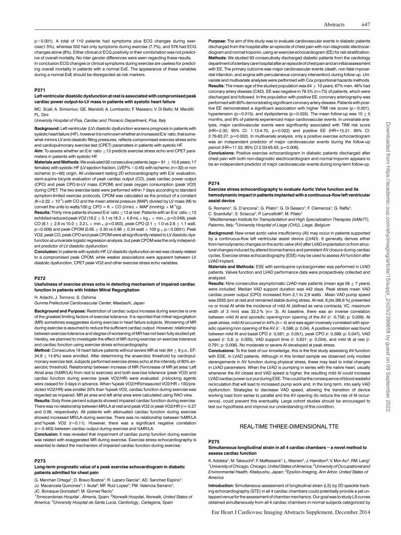
p,0.001). A total of 110 patients had symptoms plus ECG changes during exer-cise(1.5%), whereas 552 had only symptoms during exercise (7.7%), and 576 had ECGchanges alone (8%). Either clinical or ECG positivity or their combination was not predict-ive of overall mortality. No inter gender differences were seen regarding these results.In conclusion ECG changes or clinical symptoms during exercise are useless for predict-ing overall mortality in patients with a normal ExE. The appearance of these variablesduring a normal ExE should be disregarded as risk markers.
P271Left ventricular diastolic dysfunction at rest is associated with compromisedpeakcardiac power output-to-LV mass in patients with systolic heart failure
MC. Scali; A. Simioniuc; GE. Mandoli; A. Lombardo; F. Massaro; V. Di Bello; M. Marzilli;FL. DiniUniversity Hospital of Pisa, Cardiac and Thoracic Department, Pisa, Italy
Background: Left ventricular (LV) diastolic dysfunction worsens prognosis in patients withsystolicheart failure(HF),however it isnotknownwhetheran increasedE/e’ ratio, thatsome-what mirrors LV end-diastolic filling pressures, reflects compromised exercise stress echoand cardiopulmonary exercise test (CPET) parameters in patients with systolic HF.Aim: To assess whether an E/e’ ratio ≥13 predicts exercise stress echo and CPET para-meters in patients with systolic HF.Materials and Methods: We evaluated 92 consecutive patients (age= 61+10.6 years; 17females) with systolic HF (LV ejection fraction, LVEF% ,0.45) with ischemic (n=32) or nonischemic (n=60) origin. All underwent resting 2D echocardiography with E/e’ evaluation,semi-supine bicycle evaluation of peak cardiac output (CO), peak cardiac power output(CPO) and peak CPO-to-LV mass (CPOM) and peak oxygen consumption (peak VO2)during CPET. The two exercise tests were performed within 7 days according to standardsymptom-limited exercise protocols. CPOM was calculated as the product of a constant(K=2.22 × 10⁻¹) with CO and the mean arterial pressure (MAP) divided by LV mass (M) toconvert the units to watts/100 g: CPO = K × CO (l/min) × MAP (mmHg) × M-1(g).Results: Thirty-nine patients showed E/e’ ratio ≥13 at rest. Patients with an E/e’ ratio ≥13exhibited reduced peak VO2 (16.2+5.1 vs 18.3+4.8 mL× kg-1 × min-1, p=0.049), peakCO (8.1+2.6 vs 10.0+3.2 L × min-1, p=0.003), peak CPO (2.1+1.0 vs 2.6+1.1 watt,p=0.008) and peak CPOM (0.65+0.30 vs 0.96+0.34 watt × 100 g-1, p,0.0001). PeakVO2, peak CO, peak CPO and peak CPOM were all significantly related to LV diastolic dys-function at univariate logistic regression analysis, but peak CPOM was the only independ-ent predictor of LV diastolic dysfunction.Conclusion: In patients with systolic HF, LV diastolic dysfunction at rest was closely relatedto a compromised peak CPOM, while weaker associations were apparent between LVdiastolic dysfunction, CPET peak VO2 and other exercise stress echo variables.
P272Usefulness of exercise stress echo in detecting mechanism of impaired cardiacfunction in patients with hidden Mitral Regurgitation
H. Adachi; J. Tomono; S. OshimaGunma Prefectural Cardiovascular Center, Maebashi, Japan
Background and Purpose: Restriction of cardiac output increase during exercise is oneof the greatest limiting factors of exercise tolerance. It is reported that mitral regurgitation(MR) sometimes exaggerates during exercise in heart failure subjects. Worsening of MRduring exercise is assumed to reduce the sufficient cardiac output. However, relationshipbetween exercise tolerance and degree of worsening of MR has not been fully studied yet.Hereby, we planned to investigate the effect of MR during exercise on exercise toleranceand cardiac function using exercise stress echocardiography.Method: Consecutive 16 heart failure patients without severe MR at rest (64+8 y.o., EF:34.8+14.6%) were enrolled. After determining the anaerobic threshold by cardiopul-monary exercise test, subjects performed exercise stress echo at the intensity of 80% an-aerobic threshold. Relationship between increase of MR (%increase of MR jet area/ LeftAtrial area (%MR/LA) from rest to exercise) and both exercise tolerance (peak VO2) andcardiac function during exercise (peak VO2/HR) were evaluated. b-blocking agentswere ceased for 3 days in advance. When %peak VO2/HR(measured VO2/HR×100/pre-dicted VO2/HR) was smaller 20% than %peak VO2, cardiac function during exercise wasregarded as impaired. MR jet area and left atrial area were calculated using RAO view.Results: Sixty three percent subjects showed impaired cardiac function during exercise.There was no relationship between MR/LA at rest and peak VO2 or peak VO2/HR (r=-0.27and 0.06, respectively). All patients with attenuated cardiac function during exerciseshowed increased MR/LA during exercise. There was no relationship between %MR/LAand %peak VO2 (r=0.11). However, there was a significant negative correlation(r=-0.463) between cardiac output during exercise and %MR/LA.Conclusion: It was revealed that impairment of cardiac pump function during exercisewas related with exaggerated MR during exercise. Exercise stress echocardiography isessential to detect the mechanism of impaired cardiac function during exercise.
P273Long-term prognostic value of a peak exercise echocardiogram in diabeticpatients admitted for chest pain
G. Merchan Ortega1; D. Bravo Bustos1; R. Lazaro Garcia1; AD. Sanchez Espino1;JJ. Macancela Quinones1; I. Ikuta2; MF. Ruiz Lopez1; FM. Valencia Serrano1;JC. Bonaque Gonzalez3; M. Gomez Recio1
1Torrecardenas Hospital , Almeria, Spain; 2Norwalk Hospital, Norwalk, United States ofAmerica; 3University Hospital de Santa Lucıa, Cardiology , Cartagena, Spain
Purpose: The aim of this study was to evaluate cardiovascular events in diabetic patientsdischarged from the hospital after an episode of chest pain with non-diagnostic electrocar-diogram and normal troponin, using an exercise echocardiogram (EE) for risk stratification.Methods: We studied 93 consecutively discharged diabetic patients from the cardiologydepartmentofa tertiarycarehospitalafteranepisodeofchestpainandaninitialassessmentwith EE. The primary outcome was major cardiovascular events (death, non-fatal myocar-dial infarction, and angina with percutaneous coronary intervention) during follow-up. Uni-variate and multivariate analyses were performed with Cox proportional hazards methods.Results: The mean age of the studied population was 64+10 years, 67% men. 46% hadcoronary artery disease (CAD). EE was negative in 78.5% (n=73) of patients, which weredischarged and followed. In the population with positive EE, coronary arteriography wasperformed with 60% demonstrating significant coronary artery disease. Patients with posi-tive EE demonstrated a significant association with higher TIMI risk score (p,0.001),hypertension (p=0.015), and dyslipidemia (p=0.035). The mean follow-up was 10+5months, and 9% of patients experienced major cardiovascular events. In univariate ana-lysis, major cardiovascular events were significantly associated with TIMI risk score(HR=2.30, 95% CI: 1.13-4.70, p=0.022) and positive EE (HR=15.21, 95% CI:2.78-83.27, p=0.002). In multivariate analysis, only a positive exercise echocardiogramwas an independent predictor of major cardiovascular events during the follow-upperiod (HR=11.53, 95% CI 2.03-65.63, p=0.006).Conclusions: Positive exercise echocardiogram in diabetic patients discharged afterchest pain with both non-diagnostic electrocardiogram and normal troponin appears tobe an independent predictor of major cardiovascular events during long-term follow-up.
P274Exercise stress echocardiography to evaluate Aortic Valve function and itshemodynamic impact in patients implanted with a continuous-flow left ventricularassist device
G. Romano1; G. D’ancona1; G. Pilato1; G. Di Gesaro1; F. Clemenza1; G. Raffa1;C. Scardulla1; S. Sciacca1; P. Lancellotti2; M. Pilato1
1Mediterranean Institute for Transplantation and High Specialization Therapies (IsMeTT),Palermo, Italy; 2University Hospital of Liege (CHU), Liege, Belgium
Background: New-onset aortic valve insufficiency (AI) may occur in patients supportedby a continuous-flow left ventricular assist device (LVAD). It generally derives eitherfromhemodynamicchangeson theaortic valve (AV)afterLVAD implantation or fromstruc-tural changes induced by altered biomechanics and persistent AV closure during cardiaccycles. Exercise stress echocardiography (ESE) may be used to assess AV function afterLVAD implant.Materials and Methods: ESE with semisupine cycloergometer was performed in LVADpatients. Valves function and LVAD performance data were prospectively collected andanalyzed.Results: Nine consecutive asymptomatic LVAD male patients (mean age 56+7 years)were included. Median VAD support duration was 442 days. Peak stress mean VADcardiac power output (CPO) increased from 2,1 to 2,9 watts . Mean VAD pump speedwas 2555 rpm at rest and remained stable during stress. At rest, 6 pts (66,6 %) presentedno or trivial AI while the incidence of mild AI (defined as vena contracta, VC, maximumwidth of 3 mm) was 33,3 % (n= 3). At baseline, there was an inverse correlationbetween mild AI and sporadic opening/non opening of the AV (r: -0,756; p: 0,009). Atpeak stress, mild AI occurred in 44% (n= 4) and was again inversely correlated with spor-adic opening/non opening of the AV (r: - 0,598; p: 0,04). A positive correlation was foundbetween mild AI and basal CPO (r: 0,581; p: 0,051), peak CPO (r: 0,589; p: 0,047), VADspeed (r: 0,8; p: 0,005), VAD support time (r: 0,631; p: 0,034), and mild IA at rest (r:0,791; p: 0,006). No moderate or severe AI developed at peak stress.Conclusions: To the best of our knowledge, this is the first study assessing AV functionwith ESE, in LVAD patients. Although in this limited sample we observed only modestderangements in AV function during physical stress, these may lead to initial changesin LVAD parameters. When the LVAD is pumping in series with the native heart, usuallywhenever the AV closes and VAD speed is higher, the resulting mild AI could increaseLVADcardiac powerout-put during ESE.This couldbe theconsequence ofbloodwastefulrecirculation that will lead to increased pump work and, in the long term, into early VADdysfunction. Strategies to decrease VAD speed, allowing the transition of deviceworking load from series to parallel and the AV opening (to reduce the risk of AI occur-rence), could prevent this eventuality. Large cohort studies should be encouraged totest our hypothesis and improve our understanding of this condition.
REAL-TIME THREE-DIMENSIONAL TTE
P275Simultaneous longitudinal strain in all 4 cardiac chambers – a novel method toassess cardiac function
K. Addetia1; M. Takeuchi2; F. Maffessanti1; L. Weinert1; J. Hamilton3; V. Mor-Avi1; RM. Lang1
1Universityof Chicago,Chicago, United StatesofAmerica; 2University ofOccupational andEnvironmental Health, Kitakyushu, Japan; 3Epsilon-Imaging, Ann Arbor, United States ofAmerica
Introduction: Simultaneous assessment of longitudinal strain (LS) by 2D speckle track-ing echocardiography (STE) in all 4 cardiac chambers could potentially provide a yet un-tapped venue for the assessment of chamber mechanics. Our goal was to study LS curvesobtained simultaneously from all 4 cardiac chambers in normal subjects categorized by
Abstracts ii47
Eur Heart J Cardiovasc Imaging Abstracts Supplement, December 2014
Dow
nloaded from https://academ
ic.oup.com/ehjcim
aging/article/15/suppl_2/ii25/2399658 by guest on 09 September 2022

age and gender, in order to gain insight into the functional relationships between cham-bers.Methods: We studied 109 normal subjects (58 men, 51 women; age 39+13) in whom itwas possible to obtain apical 4-chamber views that contained the entire left and right ven-tricles (LV, RV) and both atria (LA, RA) in the same sector. 2D STE was performed in all 4chambers in the same cardiac cycle to measure LS using vendor-independent software(Epsilon Imaging). The inter-ventricular septum was considered part of the LV, while RVstrain was measured only in the free-wall segments. The inter-atrial septum was includedin the LS measurements of both atria.Results: Strain curves of the RV and RA were larger in magnitude than the correspondingleft heart chambers (figure), resulting in larger peak systolic LS values (table).LS for the LApeaked significantly later than the LV, RV and RA. Gender differences were found only inthe LV and LA, where peak systolic LS values were significantly larger in magnitude infemales than males (table). Age differences were significant only in the LA, where theyounger age groups (,40) showed larger magnitude strain values than the oldergroup (≥60) (table, stars).Conclusions: Simultaneous measurement of LS provides new insight into inter-chamberrelationships including age and gender differences in normal subjects. This new tool mayprove useful in evaluating diseases that affect cardiac chambers differently.
P276Three-dimensional speckle tracking echocardiography derived strainparameters could assess infarct transmurality and predict functional recovery inpatients with ST-elevation myocardial infarction
A. Sugano1; Y. Seo2; H. Watabe1; Y. Kakefuda1; H. Aihara1; H. Nishina1; T. Ishizu2;Y. Fumikura1; Y. Noguchi1; K. Aonuma2
1Tsukuba Medical Center Hospital, Department of Cardiology, Tsukuba, Japan; 2Universityof Tsukuba, Cardiovascular Division, Faculty of Medicine, Tsukuba, Japan
Background: In patients with acute ST-elevation myocardial infarction (STEMI), infarctsize and presence of transmural infarct relate with prognosis. Myocardial strain by three-dimensional speckle-tracking echocardiography (3D-STE) has been shown to accuratelyassess the infarct transmurality in patients with chronic ischemic heart disease. However,few studies have shown the usefulness of 3D-STE in patients with STEMI.Objective: The aim of this study was to evaluate whether 3D-STE could assess infarcttransmurality and predict segmental functional recovery in patients with STEMI.Methods: Sixty patients (63+14y.o, 83% male) with STEMI treated by primary percutan-eous coronary intervention (PCI) were prospectively enrolled. Comprehensive Dopplerechocardiographic examinations including 3D-STE were performed by an ARTIDA TMsystem (Toshiba Medical Co.). Global and segmental endomyocardial peak systolicstrain including longitudinal strain (LS), circumferential strain (CS) and area strain (AS)were measured. Segmental function was assessed by wall motion score (WMS) asfollows: 1=normal, 2=hypokinesis, 3=akinesis, 4=dyskinesis. Gadolinium enhancedcardiac magnetic resonance (CMR) was performed to determine the infarct transmuralityand amount of scar by late gadolinium enhancement. Transmural infarction was definedas late gadolinium enhancement .50%. Echocardiography and CMR were sequentiallyperformed at 7 days and 6 months after PCI.Results: In acute phase analysis, all 3D global strain indexes were correlated with ejectionfraction (EF) and wall motion score index (global LS r=-0.37 p=0.02, r=0.44 p=0.006, CSr=-0.43 p=0.007, r=0.61 p,0.001, AS r=-0.48 p=0.003, r=0.66 p,0.001). In segmentalanalysis (796 segments), CS and AS but not LS in transmural infarct segments were signifi-cantly lower than those in non-transmural infarct segments (CS; -16.7+9.3 vs. -21.5+8.9p,0.001,AS; -25.2+11.7vs. -30.9+9.8p,0.001,LS;-10.7+5.8vs. -12.1+5.2p=0.14).In segments with improvement of WMS at 6 months follow-up, CS and AS at baseline weresignificantly higher than those in segments without improvement of WMS (CS; -19.9+9.0vs. -15.9+8.8, p=0.02, AS; -29.3+10.4 vs. -23.6+10.7, p=0.007). Area under thecurve by ROC analysis in assessing the accuracy to predict recovery of WMS was 0.63in CS and 0.66 in AS.Conclusion:3D-STEcould assess infarct transmurality inpatients withSTEMI andpredictsegmental functional recovery.
P277Multidirectional left ventricular performance detected with three-dimensionalechocardiography in patients with chronic right ventricular pacing
XX. Luo; F. Fang; APW. Lee; Q. Shang; CM. YuIVM, Div of Cardiology, Dept of M&T, PWH, The Chinese University of Hong Kong, HongKong, Hong Kong SAR, People’s Republic of China
Purpose: The impact of chronic right ventricle (RV) pacing on left ventricle (LV) massand deformation has not been assessed previously. The changes of LV mass and
cardiac function were hence assessed by three-dimensional (3D) speckle-trackinganalysis.Method: A total of 82 consecutive bradycardia patients (age 71+10 years; 47.6% male)received RV-based dual-chamber pacemakers were enrolled. 3D transthoracic echocar-diography (GE Vingmed Ultrasound AS, Horten, Norway) was performed before and 12months after RV pacing to determine LV volumes, mass as well as LV global deformation(longitudinal, circumferential and radial strain and area strain).Result: At12-month followup, theend-systolic volume(ESV)(28.6+9.9vs33.5+21.1ml,P = 0.02) and the LV mass (141+31 vs 149+32 g, P = 0.02) were increased significantly,and was accompanied by a significant reduction in LV ejection fraction (66.4+5.8% vs60.4+7.7%, P , 0.001) as measured by 3D echo. Furthermore, global longitudinal (LS),circumferential (CS), radial strain (RS) as well as area strain (AS) derived from 3D speckle-tracking (3D-STE) were significantly decreased at 12 months (all P,0.001) (Table 1).Conclusions: RV pacing induced LVadverse remodeling as evident by the increase in LVmass and enlargement of LV cavity. There was also impairment of LV global function aswell as myocardial deformation.
Abstract P277 Table. 3D speckle-tracking analysis
Baseline (n=82) 12-month follow up (n=82) p-value
Global AS (%) 229.6+5.6 225.0+6.4 ,0.001Global CS (%) 217.6+3.6 214.7+3.9 ,0.001Global LS (%) 216.7+4.2 213.8+4.1 ,0.001Global RS (%) 48.9+12.8 38.6+12.4 ,0.001
P278Quantification of myocardial deformation: A comparison between 3D speckletracking and 3D tagged MRI in patients with heart failure
E C. Sammut1; R. Chabinok1; T. Jackson1; M. Siarkos2; L. Lee2; G. Carr-White2; R. Rajani2;S. Kapetanakis2
1King’s College London, London, United Kingdom; 2St Thomas’ Hospital, London, UnitedKingdom
Introduction: 3D speckle tracking to derive Global Longitudinal Strain (GLS) and GlobalCircumferential Strain (GCS) is emerging as a potentially valuable tool for quantifying LVfunction. This study evaluates a new algorithm for 3D strain quantification against 3DTagged MRI in patients with heart failure.Methods: Nine consecutive patients with significant heart failure were assessed with real-time 3D echo (3DE) and 3D tagged MRI (3DtCMR). 3DE data sets were acquired with aPhilips iE33 platform and speckle tracking was performed using Tomtec 4D LV analysis(Panel B). 3DtCMR sequences were acquired on a Philips Achieva 1.5T (TX) system onthe same day.The CMR protocol included 3-dimensional tagging of the whole heart, with voxel size3.4 × 7.7 × 7.7mm, temporal resolution 30-35ms. 3D Tag datawere analysed using apro-prietary application (Panel A).Results: The median GLS and GCS by 3DE were 27.3 (IQR 24.4 to 29.2) and 29.9 (IQR28.4 to 215.3) respectively. Median 3DtCMR GLS and GCS were 26.8 (IQR 23.6 to210.6) and 27.3 (IQR 23.8 to 28.9) respectively.There was excellent correlation between GLS by 3DE and 3DtCMR, with an R2 of 0.7.Bland-Altman analysis showed excellent agreement between methods and no biasbetween these. Mean difference was 0.06 (95% confidence interval 2.35 to -2.22), PanelC. There was also very good correlation in GCS quantified by these 2 techniques (R20.63), although there was a persistent bias for 3DE with a mean difference of 4.6 (95% con-fidence intervals 6.7 to 2.5).Conclusion: There appears to be a very strong correlation between strain parametersderived by 3DE and 3DtCMR. This early data suggests that quantification of myocardialdeformation by 3D echo is accurate and potentially a clinical valuable tool.
P279Improvements in radial strain in patients with Hereditary Haemochromatosisfollowing venesection, detected by speckle tracking echocardiography
D. Byrne1; JP. Walsh2; L. Ellis3; S. Mckiernan3; S. Norris3; G. King4; RT. Murphy4
1Trinity College Centre for Health Sciences, Centre for Advanced Medical Imaging, Dublin,Ireland; 2St James Hospital, Department of Radiology, Dublin, Ireland; 3Department ofGastroenterolgy, St James Hospital, Dublin 8, Dublin, Ireland; 4Department of Cardiology,St James Hospital, Dublin 8, Dublin, Ireland
Purpose: To investigate whether patients with hereditary haemochromatosis without signsof heart failure exhibit subclinical alterations of systolic left ventricular (LV) dysfunction.
Abstract P275 Figure.
Abstract P278 Figure 1.
ii48 Abstracts
Eur Heart J Cardiovasc Imaging Abstracts Supplement, December 2014
Dow
nloaded from https://academ
ic.oup.com/ehjcim
aging/article/15/suppl_2/ii25/2399658 by guest on 09 September 2022

Methods: In the context of iron overload in Beta Thalassemia Major, radial strain has pre-viously been shown to be a better prognostic marker than conventional measurements.We performed a comprehensive evaluation of systolic and diastolic cardiac functionusing Tissue Doppler Imaging (TDI) and deformation imaging (strain) at initial diagnosisand one year after commencing a treatment programme of venesection.Results: 56 patients have been assessed at baseline and 15 patients have so far com-pleted follow-up. In the 15 patients who have undergone repeat echocardiography,radial strain showed a significant improvement following venesection from 32.8 (SD+14.2) to 52.3 (SD+21.3) (p = 0.006). Average ferritin showed a significant decreasefrom a mean value of 957mg/L (SD+779) pre-venesection to 188mg/L (SD+73.7) post-venesection (p = 0.0007). There was no significant change in longitudinal strain or LVEF.Conclusion: Patients with hereditary haemochromatosis have subclinical alterations ofsystolic and diastolic LV function. Among all parameters, radial strain was shown to signifi-cantly improve following a 1 year course of venesection. This suggests that radial strainwhich is synonymous with myocardial twist could be used to demonstrate improvementsin cardiac function in patients with iron overload following venesection.
P280Is the real-time three-dimensional strain superior to two-dimensional strain fordetection on early recovery in patients with Acute Myocardial Infarction ?
K. Hristova1; TZ. Katova1; I. Simova1; V. Kostova1; I. Shuie2; V. Ferferieva3; V. Bogdanova4;X. Castelon5
1National Heart Hospital, Sofia, Bulgaria; 2Heriot-Watt University, School of Life Sciences,Edinburgh, United Kingdom; 3Hasselt University, Hasselt, Belgium; 4Private HospitalParis-Montmartre, of Cardiology, Paris, France; 5Private Hospital Athis Mons, Paris, France
The ability of 2D strain imaging has been demonstrated to be effective for diagnosis of re-covery after AMI, no information is available about the effectiveness of 3D strain in this clin-ical setting.The present study aimed to assess the diagnostic power of the RT-3D speckletracking echocardiography in comparison with 2DSTE in detection of subclinical recoveryin patients after AMI.Methods: Were included 66 consecutive patients (M/F 59/7,mean age 63 years) withSTEMI – anterior and inferior(26/40) and 20 healthy volunteers (mean age 23 years).2D images were acquired (65+7 fr/s) in addition to apical four, three and two chamberviews and SAX - apical, middle and basal view, within 36 h after revascularization(PCI)and after treatment (4-mFU).The patients underwent standard echo examination includ-ing 2D and 3D determination of EF%,2DSTE with global longitudinal strain (GLS) by auto-mated function imaging software and 3DSTE with measurements of 3D GLS,GCS,globalarea strain(GAS)and GRS.Finally, the infarct size (IS) was estimated based on MRIdelayed enhancement and expressed as a percentage of the total LV volume.Results: After infarct, adverse remodeling (progressive increase in LV size, mass andreduced EF)was found.The radial and circumferential strain decreased in the infarct peri-nfarct and remote regions acutely in comparison with controls.The reduction was numer-ically lower for all types of strain 2D and 3D.2DSTE derived GLS was marginally changed(-8.2+3.7%vs.-11.3+2.9%,p,0.01).3DSTE derived GLS(-9.9+2.0%vs 12.3+2.5%,p,0.0001), GCS (p,0.001),GAS (-28.1+5.2%vs-31.4+3.4%,p,0.0001)andGRS (p,0.0001) were all significantly reduced at baseline and 4mFU.Also 3D EF waslower on baseline(44.1+3.0%) than 4mFU (52.5+3.9%,p,0.0001).The longitudinal2D and 3D strain only was significantly decreased in infarct regions. There was a signifi-cant correlation between the infarct size (31,1+3,4%) and longitudinal 3D strain(r=0.49,p, 0.01), radial(r=0.37,p,0,01) and circumferential strain(r=-0.39,p,0.05).Among the different 3D strain components, GAS showed the best independent associa-tions with mean IS (b=20.502,P,0.0001) and LVMi (b=20.385,P, 0.001, R2 =0.55,P, 0.0001) in the pooled population.Conclusions: Our study demonstrates the superiority of 3D volumetric echocardiog-raphy and 3DSTE in comparison with both standard 2DE and 2DSTE in detecting subclin-ical recovery of STEMI patients undergoing PCI.Not only the different 3D-derived straincomponents but even the simple 3D volumetric EF are in fact significantly reduced afterinfarction, highlighting the cardiac damage induced by ischemia.
P281Correlations between three-dimensional speckle trackingechocardiography-derived left atrial functional properties and aortic stiffness inhealthy subjects
A. Nemes1; V. Sasi1; P. Domsik1; A. Kalapos1; C. Lengyel2; A. Orosz3; T. Forster112nd Department of Medicine and Cardiology Center, University of Szeged, Szeged,Hungary; 2University ofSzeged, 1stDepartment ofMedicine, Szeged, Hungary; 3Universityof Szeged, Department of Pharmacology and Pharmacotherapy, Szeged, Hungary
Introduction: Left atrial (LA) distension has been demonstrated to be associated withaortic stiffness in different patient populations. Three-dimensional (3D) speckle trackingechocardiography (STE) seems to bea promising tool for volumetric and functional evalu-ation of the LA. The objective of the present study was to find correlations between 3DSTE-derived LA volume-based and strain parameters characterizing all LA functions and echo-cardiographic aortic elastic properties in healthy subjects.Methods: The current study comprised 19 healthy volunteers (mean age: 37.9+11.4years, 11 men). All subjects had undergone complete two-dimensional (2D) Dopplertransthoracic echocardiography. Systolic and diastolic ascending aortic diameters (SDand DD) were recorded in Mmode 3cm above the aortic valve froma parasternal long-axisview. The SD and DD were measured at the time of maximum anterior motion of the aortaand at the start of the QRS complex, respectively. Aortic strain (AS), aortic distensibility(AD) and aortic stiffness index (ASI) were calculated. from aortic diameter and blood pres-sure data. 3DSTE was used for evaluation of LA volumes and strain parameters.
Results: AS, AD and ASI proved to be 0.127+0.091, 4.58+3.21 cm2dynes(-1)10(-6)and 5.17+3.45, respectively. Maximum and minimum LA volumes and LA volumebefore atrial contraction were 35.6+6.4 ml, 16.3+4.9 ml, 23.8+6.7 ml, respectively.Global peak LA radial, circumferential, longitudinal, 3D and area strains proved to be-21.8+11.8%, 28.7+10.0%, 24.2+6.6%, 57.7+17.6% and -13.9+10.8%, respect-ively, while global pre-atrial contraction LA radial, circumferential, longitudinal, 3D andarea strains were -8.5+8.3%, 10.7+11.4%, 9.0+9.4%, 16.5+16.5% and -5.3+5.4%, respectively. None of LA volumes correlated with echocardiographic aorticelastic properties. Active atrial stroke volume correlated with ASI (r =0.45, p =0.05).None of other volume-based functional properties correlated with echocardiographicaortic elastic properties. Global peak 3D strain correlated with AS (r =-0.46, p =0.05).Global radial pre-atrial contraction strain correlated with ASI (r =-0.49, p =0.04) and AS(r =-0.50, p =0.04).Conclusions: Correlations exist between 3DSTE-derived LA functional parameters andeschocardiographic aortic elastic properties in healthy subjects.
P2823-dimensional imaging of different RV remodeling and tricuspid valve:precapillary versus postcapillary pulmonary hypertension
J. Grapsa1; O. Demir1; D. Dawson1; R. Sharma2; R. Senior2; P. Nihoyannopoulos1
1Hammersmith Hospital, London, United Kingdom; 2Royal Brompton and Harefield NHSTrust Hospital, Imperial College London, London, United Kingdom
Purpose: The aim of this study was to compare right ventricular (RV) remodelingin pre-capillary (PAH) versus post-capillary (PH-HF) pulmonary hypertension with3-dimensional speckle tracking and 3D-tricuspid assessment.Methods: Forty PAH patients were compared to 42 PH-HF patients. All patients underwent2D, 3D-speckle tracking and 3D tricuspid assessment. 3D-PISA and vena contracta weremeasured. The results were indexed to the RV end-diastolic volume to avoid the volumeload effect (according to Frank-Starling principle) and indexed for body surface area (BSA).Results: PH-HF patients had greater left atrial diameter when compared to PAH (55.9+6.7 vs. 35.6+5.3, p,0.001). Right ventricular (RV) mass was greater in PAH patients(PAH: 156.8+22.1 mls vs. PH-HF: 98+19.4 mls, p,0.001) and PAH patients hadmore impaired RV function (RVEF: PAH: 32.1+10.7% vs. PH-HF: 52.4+9.6%,p,0.001). 3Dspeckle tracking demonstrated thatglobal RVstrain (indexed toRV-enddia-stolic volume) was lower in PAH patients (PAH: -0.05+0.01 vs. PH-HF: -0.09+0.02,p=0.008) however RV-free wall strain was similar for both populations: (PAH: -0.09+0.02 vs. PH-LV: -0.15+0.06, p=0.8). 3-dimensional PISA was greater in PH-HF patients(PH-HF: 0.9+0.42 cm vs. PAH: 0.6+0.2 cm, p=0.02). 3-dimensional tricuspid vena con-tracta was similar in both groups (PH-HF: 0.75+0.28 vs. PAH: 0.8+0.31, p=0.18).Conclusion: 3D vena contracta is superior to 3D PISA for the assessment of eccentric tri-cuspid regurgitation. RV free wall strain may be more significant when compared to globalstrain for the assessment of RV function, due to the interventicular dependence. Theseshould be taken into consideration when assessing the different RV remodeling.
TISSUE DOPPLER AND SPECKLE TRACKING
P283Echocardiographically assessed improvementofLeftAtrial functionandstiffnessin patients with paroxysmal atrial fibrillation successfully treated with catheterablation
E. Pilichowska; B. Zaborska; J. Baran; S. Stec; P. Kulakowski; A. BudajGrochowski Hospital, Postgraduate Medical School, Department of Cardiology, Warsaw,Poland
Purpose: Atrial fibrillation is associated with reduced left atrial (LA) function and remodel-ling increasing LA stiffness. Speckle tracking echocardiography (STE) enables quantita-tive assessment of LA myocardial deformation. The aim of the study was to evaluate theeffects of catheter ablation (CA) on LA function and reverse remodelling.Methods: Patients (pts) with non-valvular AF treated with CA were studied with transthor-acic echocardiography.Peak atrial longitudinal strain (PALS) wasassessed using speckletracking analysis by averaging values observed in all LA segments in 4- and 2- chamberviews. E/E’ and LA stiffness index (LAs)- the ratio of E/E’ to PALS were assessed in Doppler(D) and tissue D. All parameters were analyzed at baseline and 12 months after CA duringsinus rhythm. Responders were defined as partial or complete symptom improvementand no AF at 3, 6, 12 month 24-hour Holter ECG.
Abstract P283 Table. Echocardiographic parameters
Study group n= 54 Responders n=35 Nonresponders n=19Baseline 12 m P Baseline 12 m P Baseline 12 m P
E/E’ 8.31+2.57
7.96+2.3
0.107 7.74+2.46*
7.41+1.94
0.283 9.43+2.47 *
9.01+2.62
0.163
PALS % 26.12+8.97
27.47+9.95
0.039 29.85+7.47**
32.02+7.68
≤0.002 18.45+6.71**
18.10+7.19
0.435
LAs 0.41+0.36
0.39+0.44
0.016 0.3+0.23**
0.25+0.11
≤0.004 0.64+0.46**
0.7+0.66
0.538
*P.0.001**P,0.001 for baseline parameters responders vs. nonresponders
Results: Study group consisted of 54 pts (64 % males, mean age 57.8 +/- 10.1).CA was effective in 35 (65 %) pts. When baseline and 12-month measurments were com-pared, the analyzed parameters significantly improved only in responders whereas in
Abstracts ii49
Eur Heart J Cardiovasc Imaging Abstracts Supplement, December 2014
Dow
nloaded from https://academ
ic.oup.com/ehjcim
aging/article/15/suppl_2/ii25/2399658 by guest on 09 September 2022

non-responders remained stable (table). Baseline PALS was significantly higher, LAs sig-nificantly lower in responders than in nonresponders (table).Conclusion: 1.Successful CA improves LA function and is associated with LA reverseremodeling. 2. Patients in whom CA occurred effective have more favourable echocardio-graphic profile at baseline than nonresponders. It may suggest that detailed echocardio-graphic assessment before planned CA may help in proper selection of candidates for thisinvasive procedure.
P285Incremental value of Left Atrial strain for prediction of atrial fibrillation in patientsreceiving cardiac pacing: a model of complete ascertainment
W. Kosmala1; G. Kaye2; M. Saito3; K. Negishi3; TH. Marwick3
1Wroclaw Medical University, Wroclaw, Poland; 2University of Queensland, Brisbane,Australia; 3University of Tasmania, Menzies Research Institute Tasmania, Hobart, Australia
Background: Right ventricular (RV) stimulation during cardiac pacing might predisposeto atrial fibrillation (AF), and a risk evaluation process might guide prophylactic therapy.We sought to investigate factors associated with a first episode of AF (including measure-ment of left atrial volume (LAV) and LA strain) in pts undergoing dual chamber pacemakerimplantation.Methods: Clinical data were obtained prospectively in 147 pts (73+10yrs). Echocardio-grams and 2D-strain analysis were performed post implantation and at 2 yrs. Completeascertainment ofAF during follow-upwas identified from interrogation ofpermanent pace-makers.Results: During a 2-yr follow-up, episodes of AF were noted in 29 pts (20%). Pts with AFdemonstrated significantly lower left ventricular (LV) ejection fraction (p,0.03), LA strain(p,0.001) and higher LAV (p,0.002). RV lead location (apical vs. septal) was similar inthe groups with and without AF (p,0.80). The independent associations of AF (Cox re-gression) were: LAV (HR 1.02,95% CI 1.00-1.04,p,0.02), LA strain at atrial contraction(LASac;HR 0.91,95% CI 0.84-0.99,p,0.04) and change in LAV during follow-up (HR1.02,95% CI 1.00-1.04,p,0.04). No independent contribution was found for patientage, BMI, diabetes and hypertension, and use of antiarrhythmic drugs. Higher LASac(above the optimal cutpoint from ROC curve of 8.6%) had a negative predictive valuefor AF of 89%, contributing to the discrimination process both below and above theoptimal cutpoint for LAV of 62 ml (Figure).Conclusions: The risk of AF in pts receiving dual chamber pacing is independently asso-ciated with LA size and function, not LV structural and functional characteristics or RVlead location. Measurement of LA strain may be helpful to stratify the risk of AF in thispopulation.
P286Effect of years of cocaine use on the likelihood of developing cocainecardiotoxicity. Cardiac magnetic resonance study
A M. Maceira Gonzalez1; C. Ripoll2; J. Cosin-Sales3; B. Igual1; J. Salazar4; V. Belloch1
1Cardiac Imaging Unit - ERESA, Valencia, Spain; 2Unidad de Conductas Adictivas,Valencia-La Fe, Valencia, Spain; 3Hospital Arnau de Vilanova, Valencia, Spain; 4UniversityGeneral Hospital of Valencia, Valencia, Spain
Cocaine is a known cardiotoxic drug. In a previous study we observed that the prevalenceof cardiac involvement due to cocaine abuse is 71%, being the most frequent findings leftventricular (LV) hypertrophy, dilatation and biventricular systolic dysfunction, as well asfocal myocardial gadolinium enhancement indicative of myocardial injury. Our objectivenow was to determine the effect of several variables regarding cocaine use on the likeli-hood of development of cocaine cardiotoxicity.Methods: In our series of 94 cocaine consecutive subjects (81 males, 36.6+7yrs) who hadbeenstudiedwithaCMRprotocolat3T, informationwascollectedregardingcocaineuse:ageat firstuse, frequencyof use in the last month and in the last3 months prior to CMR,maximumfrequencyofuseduring life,amountconsumedin the lastmonth,yearsof regularuseandwayof use (inhaled, intravenous, sniffed). Logistic regression analysis was carried out for threedichotomic dependent variables (1) existence of LV systolic dysfunction, (2) presence of LVsystolic dysfunction or dilatation, and (3) presence of LV systolic dysfunction, dilatation orstress myocardial perfusion defect. ROC curves for all variables were obtained.Results: Of all the variables of cocaine use, the only one that showed a significant effect inthe multivariable analysis was number of years of consumption. The table shows the areasunder thecurve derived fromthe ROCcurves.A cutoff of10.5years of cocaineuse showeda sensitivity of 80% and a specificity of 35% for presence of cardiac involvement.Conclusion: number of years of cocaine use is an important factor in the development ofcardiovascular involvement by cocaine that should be taken into account when planninghealth care in the rehabilitation process of these subjects. In view of these results, it wouldseem reasonable to recommend a basic cardiac check-up in cocaine addicts with over10.5 years of history of use.
Abstract P286 Table. ROC results for years of cocaine use
Variable AUC P
LV systolic dysfunction 0.66 0.01LV systolic dysfunction or dilatation 0.67 0.004LV systolic dysfunction, dilatation or stress perfusion defect 0.70 0.001
COMPUTED TOMOGRAPHY & NUCLEAR CARDIOLOGY
P287CTcoronary calcium scoring is a useful first line investigation for assessment ofcoronary artery disease in patients presenting with non-anginal chest pain
R S. Dulai; A. Taylor; S. GuptaBarts Health NHS Trust, London, United Kingdom
Purpose:UKNICE andESC guidelines currently recommend patientswithchest pain thatis deemed to be non-anginal should not routinely undergo cardiac investigations.However a previous study has shown that the NICE guidance underestimates the preva-lence of coronary artery disease (CAD). There is also limited evidence on whether coron-ary artery calcification (CAC) varies between different ethnic groups.The aim was to assess the prevalence of CAC in patients with non-anginal chest pain pre-senting to a rapid access chest pain clinic and to evaluate if CAC scoring is a useful first lineinvestigation for these patients. To our knowledge this is the first study to assess CACscoring in this population. The secondary aim was to investigate if there were any differ-ences in CAC amongst ethnic groups (Caucasian, South Asian, Afro-Caribbean andothers).Methods: Retrospective study; CAC was assessed in 167 consecutive patients(mean age 51.9yrs; 46.7% male) with non-anginal chest pain, as defined by NICEguidelines. All patients underwent CAC scoring using the Agatston method. Calcium
Abstract P285 Figure. Incidence of AF according to LAV and LAS
ii50 Abstracts
Eur Heart J Cardiovasc Imaging Abstracts Supplement, December 2014
Dow
nloaded from https://academ
ic.oup.com/ehjcim
aging/article/15/suppl_2/ii25/2399658 by guest on 09 September 2022

score, demographics, risk factors and results of further investigations (myocardialperfusion scan and coronary angiography) and incidence of major adverse cardiacevents (MACE) were evaluated as a whole group and then compared across ethnicgroups.Results: 31/167 (18.6%) patients had evidence of CAC of which 6 patients went on to haveCAD on angiography. The mean CAC score for the whole group was 21.5+84.5. Meanfollow up was 360+110 days .There were no recorded episodes of MACE in this period.Inamultivariate logistic regression analysisage(OR1.143, 95%CI1.030–1.268,p=.012)and hypertension (OR 7.492, 95% CI 2.445– 22.954, p=,0.01) were significant predic-tors of CAC in the whole group.
There were no significant differences in risk factors between Caucasian (n=88) and SouthAsian (n=60) patients. Afro-Caribbean patients (n=16) were significantly more likely to behypertensive compared to Caucasian and South Asian patients (p=0.01). There was nosignificant difference in the presence of calcification or distribution of calcium scorebetween the ethnic groups (p= .50 and .46 respectively).Conclusion: This analysis shows that the use of CT coronary calcium scoring can beextended to patients with non-anginal chest pain as a first line investigation for the detec-tion and risk assessment of CAD. It not only identifies patients who may need coronaryrevascularisation but also patients who may benefit from lifestyle advice and medicationsfor cardiovascular risk reduction.
Abstracts ii51
Eur Heart J Cardiovasc Imaging Abstracts Supplement, December 2014
Dow
nloaded from https://academ
ic.oup.com/ehjcim
aging/article/15/suppl_2/ii25/2399658 by guest on 09 September 2022


