Embed Size (px)
Citation preview

Forensic Science International xxx (2009) xxx–xxx
G Model
FSI-5589; No of Pages 7
Postmortem identification of hyperglycemia
B. Zilg a, K. Alkass a, S. Berg b, H. Druid a,*a Department of Forensic Medicine, Karolinska Institute, Retzius v. 3, SE-171 77 Stockholm, Swedenb Department of Clinical and Experimental Medicine, Linkoping University, University Hospital, SE-581 85 Linkoping, Sweden
A R T I C L E I N F O
Article history:
Received 11 August 2008
Received in revised form 16 December 2008
Accepted 18 December 2008
Available online xxx
Keywords:
Glucose
Lactate
Vitreous
Hyperglycemia
Postmortem
Diabetes
A B S T R A C T
The detection of diabetic coma postmortem requires accurate biochemical analysis. Due to continuous
consumption of glucose by surviving cells postmortem, blood glucose levels decrease rapidly. Therefore,
vitreous fluid has been used as a substitute in forensic practice, since it has a very low cell count. It has
been repeatedly reported that the sum value of vitreous glucose and lactate should be used to estimate
the original antemortem blood glucose level, based on the assumption that pre-existing glucose is
gradually converted to lactate under anaerobic conditions during agonal phase and the early
postmortem period. In this study, we applied a strategy including consistent sampling of vitreous fluid
from the centre of both eyes of deceased subjects as soon as possible after arrival at the morgue, and
immediate bedside analysis using a blood gas instrument. In total, 3076 cases were included during
2004–2006. We found that, after an initial drop of vitreous glucose during the very early postmortem
period, the levels stayed stable for appreciable time postmortem. Analysis of a second sample collected
at autopsy 1–3 days later gave similar results (R2 = 0.90). In contrast, the vitreous lactate levels showed a
steady increase. This implies that the sum value of glucose and lactate increases with postmortem time,
as reflected by vitreous potassium level. In fact, a statistically significant difference in the sum value was
seen between subjects with potassium below 10 mmol/L (n = 1086) and above 20 mmol/L (n = 531),
p < .001. In addition, in this large material, we did not identify a single case with circumstantial
indication of hyperglycemia that only showed high vitreous lactate. We therefore suggest that vitreous
glucose alone should be used to diagnose hyperglycemia postmortem and that the limit of 10 mmol/L
should have a good specificity for diabetic coma, which theoretically would equal an original blood
glucose value of about 26 mmol/L. As to the methodology, we found that sonication, centrifugation and
addition of fluoride to the samples are unnecessary procedures when using a blood gas instrument. The
strategy resulted in a doubling of the number of diabetic coma identified at our department compared to
preceding period when analysis only was performed on selected cases.
� 2008 Elsevier Ireland Ltd. All rights reserved.
Contents lists available at ScienceDirect
Forensic Science International
journa l homepage: www.e lsevier .com/ locate / forsc i in t
1. Introduction
The diagnosis of various metabolic conditions is often difficultto make postmortem due to major changes in the blood and othertissues [1]. In particular, the disintegration of cell membranes,causing an exchange of intra- and extracellular constituents,severely hampers many tests that are clinically performed on e.g.blood or serum. In addition, continuous cell metabolism duringagony and shortly after death in surviving cells will also makepostmortem analytical results different from those found inantemortem samples. The postmortem spread of bacteria to theblood will also cause further changes in the concentrations ofvarious endogenous compounds.
* Corresponding author. Tel.: +46 8 5248 7770; fax: +46 8 3214 52.
E-mail address: [email protected] (H. Druid).
Please cite this article in press as: B. Zilg, et al., Postmortem identifidoi:10.1016/j.forsciint.2008.12.017
0379-0738/$ – see front matter � 2008 Elsevier Ireland Ltd. All rights reserved.
doi:10.1016/j.forsciint.2008.12.017
As opposed to blood, the vitreous fluid has a very low cell content,and is less affected by postmortem changes due to its isolatedposition. It is also easily accessible for sampling. Hence, a largenumber of postmortem chemistry studies have been conducted onvitreous fluid, aiming to provide keys to various diagnoses. A stillvery informative review of this field was published by Coe in 1993[2], and a large part of the studies included there regard vitreousanalyses.
In order to take advantage of postmortem chemistry in forensiccasework, it is desirable to obtain the results as soon as possible,and preferably before the autopsy is conducted. We decided to usea blood gas instrument to run vitreous analyses on samplescollected from consecutive cases at the earliest convenience afterthe body arrived at the morgue. The main questions we liked toaddress were (1) the feasibility of performing ‘‘bedside’’ analysis atthe morgue, (2) the possibility to identify cases with hypergly-cemia, (3) the possible influence of different sample handling andtreatment for the analytical results, such as centrifugation and
cation of hyperglycemia, Forensic Sci. Int. (2009),

B. Zilg et al. / Forensic Science International xxx (2009) xxx–xxx2
G Model
FSI-5589; No of Pages 7
addition of preservatives, (4) the changes of glucose levels withpostmortem time, and (5) to compare the reliability of the resultsobtained using the blood gas instrument with analysis at a clinicalchemistry laboratory.
In this communication, we focus on the diagnosis ofhyperglycemia. It has repeatedly been stressed that vitreouslevels of lactate should be determined in addition to glucose inorder to compensate for the postmortem breakdown of glucose.Hence, almost invariably, the use of the sum value of vitreousglucose and lactate has been recommended as the mostappropriate means to estimate antemortem blood glucose levels[3–7]. In this study, we show that that notion can lead toerroneous diagnosis of hyperglycemia. We suggest that thevitreous glucose level alone should be used to estimate theantemortem blood glucose concentration.
2. Materials and methods
2.1. Study design
During 2004–2006, vitreous fluid was systematically collected from consecutive
deceased subjects brought to the department of forensic medicine in Stockholm as
soon as possible after their arrival at the morgue. Using a 1 mL syringe equipped
with an 18-gauge needle, 0.2 mL vitreous fluid was aspirated from the centre of
each eye and pooled in the same syringe. Samples from severely decomposed
bodies, and from infants were not consistently included, since toxicological
analyses were prioritized in several of these cases. The vitreous samples were
analyzed with a blood gas instrument, ABL 625 (Radiometer, Copenhagen), by direct
injection of the content of the syringe.
The glucose electrode contains a chamber where glucose oxidase converts
glucose to gluconic acid and hydrogen peroxide. At the anode, the hydrogen
peroxide will be oxidized, and the liberated electrons will cause a current in
proportion to the glucose concentration in the sample. The lactate is also measured
with an amperometric electrode equipped with an enzymatically active membrane,
a method that has been shown to yield readings that correlate very well with those
of one of the most widely used standard laboratory methods [8]. The Radiometer
instrument measures all electrolytes with ion-selective electrodes. Inter-assay
imprecision for potassium, glucose and lactate was less than 0.3 mmol/L. Limit of
quantification for these analytes was 0.1 mmol/L. In total, complete results for
glucose, lactate, potassium, sodium and chloride were obtained for 3076 cases.
2.2. Evaluation of procedures
To investigate the robustness of the methodology the following studies were
conducted. In almost all cases a second vitreous sample was collected for
toxicology, during autopsy, typically performed 1–3 days after the first sample was
collected. To this end, the whole vitreous from both eyes was collected in tubes
containing NaF and submitted for toxicology. In 49 cases, these samples were
Fig. 1. Vitreous glucose concentration plotted against vitreous potassium concentration.
no history of diabetes; blue diamonds = cause of death: other; green diamonds = cause of
postmortem interval (PMI).
Please cite this article in press as: B. Zilg, et al., Postmortem identifidoi:10.1016/j.forsciint.2008.12.017
analyzed for glucose and lactate with standard clinical chemistry technique based
on enzymatic conversion yielding NADH, measured as the absorbance at 340 nm
with a Hitachi 917 instrument. The comparison of the first and second sample thus
included different sampling techniques, different sampling times, difference in
additives and difference in analytical methods. Similar results regarding glucose
would thus strongly support robustness of the methodology described in the first
section.
To address the possible importance of the immediacy of analysis, portions of
vitreous fluid were analyzed immediately and after 4 h, respectively. To investigate
the possible influence of cell debris that to a variable extent may be included in the
aspirate, 21 whole vitreous samples were transferred to Eppendorff tubes and
centrifuged at 8000 � g for 10 min and the pellet and supernatant were analyzed
separately. Ten of the samples were subjected to sonication for 30 min before
centrifugation. Another 23 samples were aliquoted to tubes with or without the
addition of 1% NaF and analyzed after 4 weeks storage at +4 8C.
2.3. Statistics
All analytical data were compiled in a file to which data from the Swedish
forensic pathology database [9] were linked. This database comprises detailed
information of the decedent including – but not limited to – age, sex, cause(s) of
death, manner of death, circumstantial information, medical history, additional
findings and diagnoses, histopathologic and forensic toxicology results.
Kruskall–Wallis’ test was used for statistical comparisons of non-normally
distributed groups. Simple linear regression analysis was used to compare glucose
concentrations obtained by the blood gas instrument and a standard routine
laboratory method. A p-value <.05 was considered statistically significant.
Statistica 7.1 (Statsoft Inc., Tulsa, TX) was used for statistical analyses.
2.4. Ethical aspects
Ethical aspects were discussed with the Regional Ethics Committee. Since all
analyses were performed as a part of the forensic medicine investigation and
immediately reported to – and used by – the responsible forensic pathologist, no
ethical permission was required for these studies. All person-identifiable data were
excluded when compiling the results.
3. Results
3.1. Vitreous glucose concentrations
In total, 3076 cases were included in this study. Median age was57 years and 73% were male. Fig. 1 shows vitreous glucoseconcentrations plotted against the vitreous potassium concentra-tion, which is used as a substitute for the postmortem interval. Inthis graph, the cases with a history of diabetes are indicated.Several cases with elevated vitreous glucose levels did not have a
Red circles = cause of death: diabetes; orange circles = cause of death: diabetes with
death other with history of diabetes. Insert shows glucose concentrations related to
cation of hyperglycemia, Forensic Sci. Int. (2009),

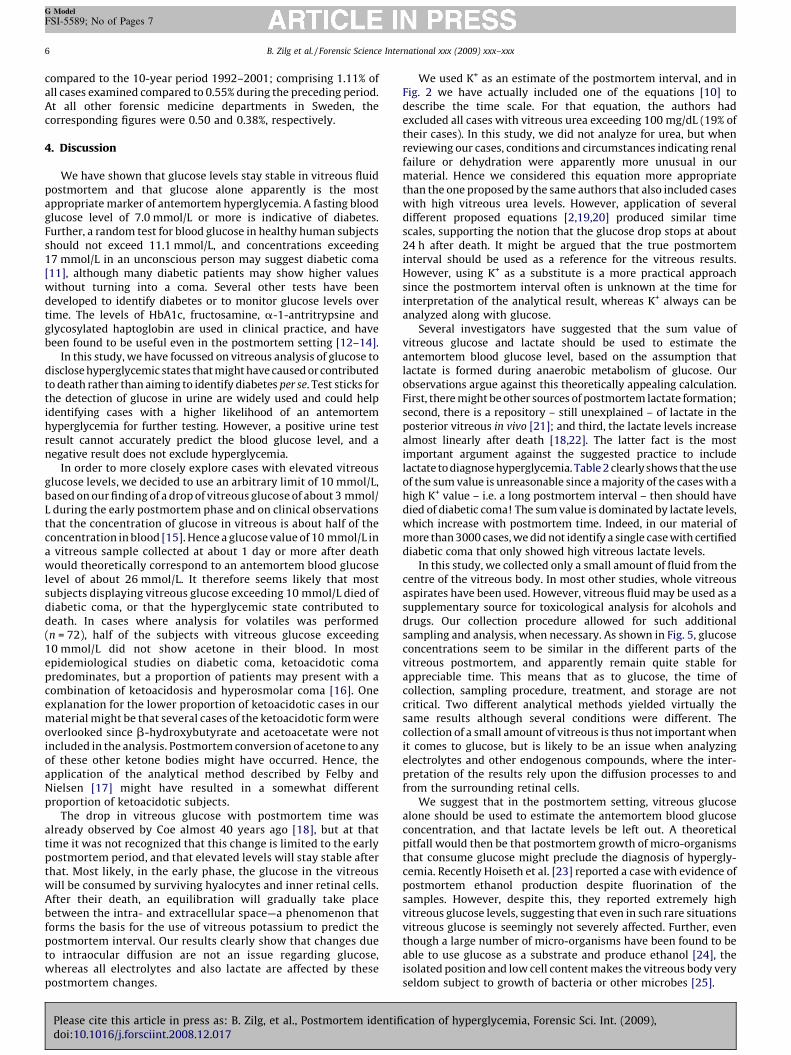
Fig. 2. Vitreous glucose concentration plotted against potassium concentration in the early postmortem interval. The scale at the top shows an arbitrary scale of the
postmortem interval calculated from the potassium values according to the equation suggested by Madea and Henssge [10].
B. Zilg et al. / Forensic Science International xxx (2009) xxx–xxx 3
G Model
FSI-5589; No of Pages 7
known history of diabetes. The same graph also illustrates that wedid not see any general decrease in glucose levels with increasingpotassium concentration. The insert shows cases with glucoseconcentrations exceeding 10 mmol/L and with known postmorteminterval. The pattern is the same – obviously the glucose levelsremain stable in the vitreous for an appreciable time, at leastduring the period covered by the graph. However, a closer look atthe cases with a very short postmortem interval (as reflected bylow potassium level), demonstrates a small, but conspicuous dropin glucose concentrations shortly after death (Fig. 2). Forcomparison, a scale representing the theoretical postmorteminterval according to the equation by Madea et al. [10] is included,starting at their proposed constant term ([K+] = 5.88 mmol/L). InTable 1, the median glucose concentrations for discrete potassiumranges are shown. Although there are few data points very closeto death (and of course none before death) the results shown inthe table suggest a median glucose concentration at the time of
Fig. 3. Vitreous lactate concentration plotted against vitreous potassium concentration. R
no history of diabetes; blue diamonds = cause of death: other; green diamonds = cause
Please cite this article in press as: B. Zilg, et al., Postmortem identifidoi:10.1016/j.forsciint.2008.12.017
death of about 3 mmol/L and an exponential decrease to 0 as thepotassium values reaches 12 mmol/L. The drop in glucoseconcentration corresponds to a steeper rise in lactate concentra-tion with increased potassium level in the early phase (Fig. 3) – apattern that can be explained by anaerobic metabolism in vitreouscells and possibly cells of the inner retinal layer shortly after death.The cases with a history of diabetes are also indicated in this figure,and as opposed to the pattern in Fig. 1, these cases conform moreto the general distribution of lactate levels across the range ofpotassium levels.
3.2. Evaluation of the sum value of vitreous glucose and lactate
To explore the assumption that the sum value of vitreousglucose and lactate would reflect the original antemortem glucoselevel, Fig. 4 shows this sum (glucose + lactate/2) plotted againstpotassium. It should be observed that the initial drop in glucose in
ed circles = cause of death: diabetes; orange circles = cause of death: diabetes with
of death other with history of diabetes.
cation of hyperglycemia, Forensic Sci. Int. (2009),

Fig. 4. The sum value of vitreous glucose and lactate/2 plotted against the vitreous potassium concentration. Red circles = cause of death: diabetes; orange circles = cause of
death: diabetes with no history of diabetes; blue diamonds = cause of death: other; green diamonds = cause of death other with history of diabetes.
Table 1Median vitreous glucose concentrations in discrete groups of vitreous potassium
ranges. All values are mmol/L.
K+ Glucose N
�5.0 3.0 8
5.1–6.0 1.9 80
6.1–7.0 1.4 194
7.1–8.0 0.7 318
8.1–9.0 0.4 277
9.1–10.0 0.3 238
10.1–11.0 0.1 220
11.1–12.0 0.1 179
12.0–13.0 0.0 159
13.1–14.0 0.0 169
>14.0 0.0 1234
B. Zilg et al. / Forensic Science International xxx (2009) xxx–xxx4
G Model
FSI-5589; No of Pages 7
the left-hand part of the graph compensates for the early, steeperrise in lactate levels, producing a straighter curve as compared toFig. 3. It should be appreciated, however, that the sum value is notconstant, but increases with increasing potassium level. In fact, thesum value is significantly correlated with the potassium level. Thisis also illustrated in Table 2, where medians, and the 25th and 75thpercentiles, of the different analytes are shown for categorizedpotassium ranges. The reason is easily appreciated when theglucose, lactate and sum value curves are compared; the sum valuecurve is dominated by increasing lactate levels with increasingpotassium level, whereas the drop in glucose levels is limited to thevery early phase, and comes to a complete stop at about 24 h.
Table 2Glucose, lactate and glucose + lactate/2 concentrations in vitreous samples from subjec
K+ � 10 (n = 1086) K+
Median 25/75 perc. Me
Glucose 0.7 0.3/1.6 0
Lactate 14.0 11.4/16.8 19
Glucose + lactate/2 7.9 6.7/9.5 10
All values are expressed as mmol/L. 25/75 perc. = 25th and 75th percentile, respectively
are in mmol/L.* Significantly different from cases with K+ � 10, p < .001.** Significantly different from cases with K+ � 10, p < .001, but not different from cay Significantly different from cases with K+ � 10, p < .001.yy Significantly different from cases with K+ � 10, p < .001, and from cases with case
Please cite this article in press as: B. Zilg, et al., Postmortem identifidoi:10.1016/j.forsciint.2008.12.017
Table 2 also shows that the medians for glucose were generally lowacross the total range of potassium levels.
3.3. Hyperglycemia cases
In total, 76 subjects had a vitreous glucose level equal to, orexceeding 10 mmol/L. Blood analysis for volatiles was performedin 72 of these. In 36 cases, acetone was detected, ranging 0.10–0.68promille. In six of these cases, isopropanol was also detected inblood at low concentrations (range 0.10–0.24 promille). BMI wassignificantly lower, 21.4, in acetone-positive cases (n = 37) ascompared to 27.7 among acetone-negative cases (n = 36). Thevitreous chloride concentrations were on average higher(108 � 16 mmol/L) in acetone-negative cases than in acetonepositive cases (101 � 13), but the difference was not statisticallysignificant, and the means did not differ from the population as awhole either. Even when comparing the difference between observedand expected chloride value (based on simple linear regressionanalysis of chloride vs. potassium; [Cl�] = 127.5–0.987*[K+])) nostatistical difference was seen between hyperglycemia cases andcases with glucose below 1.0 mmol/L (data not shown). Hence,dehydration pattern was not more frequent among these subjectsthan in non-hyperglycemia cases. It should be noted, however, thatosmolarity was not measured.
In several cases elevated vitreous glucose levels were seen insubjects with no history of diabetes (Fig. 1), and a few of these wereactually considered to have died of diabetic coma. In other caseswith moderately elevated levels, strong physiological stress, e.g.
ts with different potassium levels.
10.1–20 (n = 1459) K+ > 20 (n = 531)
dian 25/75 perc. Median 25/75 perc.
.0* 0.0/0.3 0.0** 0.0/0.5
.9y 15.5/24.8 29.3yy 21.8/34.9
.3y 8.1/13.0 15.1yy 11.5/18.2
. Lactate/2 is used to back-calculate the glucose concentration, since measurements
ses with K+ 10.1–20.
s with K+ 10.1–20, p < .001.
cation of hyperglycemia, Forensic Sci. Int. (2009),

Table 3Effects of centrifugation and sonication on analytical results for glucose, lactate, sodium and potassium in vitreous fluid.
Sonication Macro Glu-P Glu-S Lac-P Lac-S Na+-P Na+-S K+-P K+-S
Yes 2 0.1 0.1 30 29.1 137 134 24.3 24.0
Yes 1 0.2 0.3 43.4 42.7 130 135 24.9 26.8
Yes 4 0.1 0.1 23.3 23.1 140 138 13.6 13.4
Yes 4 0.2 0.2 27.8 28.5 137 139 22.1 22.3
Yes 1 0.0 0.1 30.9 30.0 127 131 22.3 22.7
Yes 2 2.7 2.4 17.5 16.8 150 148 12.8 13.1
Yes 2 0.0 0.1 33.0 32.3 131 135 27.6 28.7
Yes 1 39.1 37.2 45.2 43.0 136 144 34.2 37.2
Yes 3 1.3 1.3 38.8 38.9 143 144 10.6 10.5
Yes 3 0.8 0.8 44.8 43.5 140 141 14.9 14.9
Yes 1 0.2 0.2 41.1 39.3 137 134 28.7 28.6
Median 0.2 0.2 33.0 32.3 137.0 138.0 22.3 22.7
25/75 perc. 0.1/1.1 0.1/1.1 28.9/42.3 28.8/41.0 133.5/140.0 134.5/142.5 14.3/26.3 14.2/27.7
No 2 0.9 0.3 37.0 45.5 127 135 21.1 21.1
No 1 1.1 1.1 27.3 27.7 143 141 13.3 13.1
No 1 0.2 0.2 28.4 28.9 142 141 21.5 21.4
No 4 0.7 0.7 22.3 24.1 126 121 7.1 6.3
No 4 0.5 0.1 24.2 24.6 143 146 24.3 24.8
No 1 0.8 0.8 20.8 20.7 141 140 16.6 16.5
No 2 0.5 0.3 24.1 24.0 140 140 18.4 18.3
No 1 0.2 0.4 26.8 27.4 126 130 20 20.5
No 1 0.1 0.1 23.1 22.6 134 139 20.9 21.7
No 1 0.3 0.1 18.0 17.6 138 144 20.4 21.1
Median 0.5 0.3 24.2 24.4 139.0 140.0 20.2 20.8
25/75 perc. 0.2/0.8 0.1/0.6 22.5/27.2 23.0/27.6 128.8/141.8 136.0/141.0 17.1/21.1 17.0/21.3
All values are mmol/L. P = pellet; S = supernatant, Macro = rough grading of the macroscopically visible amounts of cell debris and/or bloody tinge. There was no statistical
difference between pellet and supernatant for any of the analytes.
Fig. 5. Comparison of vitreous glucose concentration in the first sample collected
from the centre of the eye at the morgue and the glucose concentration in the
second sample collected at autopsy, usually 1–3 days later, and consisting of all
vitreous. N = 49. All values are mmol/L. Regression analysis yielded R2 = 0.90.
Equation: y = 1.25x � 1.67. A diagonal is inserted to show the tendency for the
second samples to give slightly higher values than the first samples in the upper
part of the plot.
B. Zilg et al. / Forensic Science International xxx (2009) xxx–xxx 5
G Model
FSI-5589; No of Pages 7
due to brain trauma, could be assumed to explain for this finding.In cases with certified hypothermia, a condition known to inducephysiological stress, and also to slow down postmortem glucoseconsumption, the highest vitreous glucose value was 6.0 mmol/Land in total this group only showed slightly higher averageglucose level, 1.75 � 1.82 mmol/L (n = 25), than the bulk of the studypopulation. Among the cases with vitreous glucose exceeding10 mmol/L, 12 showed a positive result and one case (vitreousglucose 15.5 mmol/L) a negative result with Combur test on the urine.In most of these cases, however, this test was either not performed (inseven cases there was no urine available) or the result not recorded.
3.4. Evaluation of methodology
The samples used in this study were consistently collected fromthe centre of the eye to reduce errors possible caused by aspirationof detached cell fractions. However, sometimes it might benecessary to use samples that are whole vitreous aspiratesintended for toxicology. Such samples may contain a high amountof detached retinal cells. In order to investigate if disequilibriumbetween the intra- and extracellular compartments might affectthe results, samples from the whole vitreous were analyzed aftercentrifugation with (n = 11) or without (n = 10) preceding sonica-tion. No significant differences were seen regarding the glucose,lactate or potassium concentrations when analyzing the super-natant and the pellet separately (Table 3), and in fact, wholevitreous samples with macroscopic presence of cellular debris, didnot differ from those with transparent and colourless fluidregarding the analytes measured. Further, the addition of sodiumfluoride to aliquots of 23 samples did not affect these readingseither; the absolute difference between salted and unsaltedsamples were 0.17 � 0.19 and 1.16 � 1.50 for glucose and lactate,respectively.
Studies were also performed to address the issue of analytestability. Samples that were analyzed with 4 h delay did not differfrom those analyzed immediately. The absolute difference inreadings were 0.07 � 0.11 mmol/L and 0.51 � 0.72 mmol/L (n = 38)for glucose and lactate, respectively. Finally, a comparison between
Please cite this article in press as: B. Zilg, et al., Postmortem identifidoi:10.1016/j.forsciint.2008.12.017
the results of the introduced strategy with results from analysis ofselect samples collected at autopsy showed only small differences invitreous glucose concentrations, while lactate levels had increasedduring the postmortem storage of the bodies (Fig. 5).
3.5. Practical outcome
As a consequence of the introduced strategy, the frequencyof deaths in diabetic coma at the department of Forensic Medicinein Stockholm doubled during the study period 2004–2006 as
cation of hyperglycemia, Forensic Sci. Int. (2009),
B. Zilg et al. / Forensic Science International xxx (2009) xxx–xxx6
G Model
FSI-5589; No of Pages 7
compared to the 10-year period 1992–2001; comprising 1.11% ofall cases examined compared to 0.55% during the preceding period.At all other forensic medicine departments in Sweden, thecorresponding figures were 0.50 and 0.38%, respectively.
4. Discussion
We have shown that glucose levels stay stable in vitreous fluidpostmortem and that glucose alone apparently is the mostappropriate marker of antemortem hyperglycemia. A fasting bloodglucose level of 7.0 mmol/L or more is indicative of diabetes.Further, a random test for blood glucose in healthy human subjectsshould not exceed 11.1 mmol/L, and concentrations exceeding17 mmol/L in an unconscious person may suggest diabetic coma[11], although many diabetic patients may show higher valueswithout turning into a coma. Several other tests have beendeveloped to identify diabetes or to monitor glucose levels overtime. The levels of HbA1c, fructosamine, a-1-antritrypsine andglycosylated haptoglobin are used in clinical practice, and havebeen found to be useful even in the postmortem setting [12–14].
In this study, we have focussed on vitreous analysis of glucose todisclose hyperglycemic states that might have caused or contributedto death rather than aiming to identify diabetes per se. Test sticks forthe detection of glucose in urine are widely used and could helpidentifying cases with a higher likelihood of an antemortemhyperglycemia for further testing. However, a positive urine testresult cannot accurately predict the blood glucose level, and anegative result does not exclude hyperglycemia.
In order to more closely explore cases with elevated vitreousglucose levels, we decided to use an arbitrary limit of 10 mmol/L,based on our finding of a drop of vitreous glucose of about 3 mmol/L during the early postmortem phase and on clinical observationsthat the concentration of glucose in vitreous is about half of theconcentration in blood [15]. Hence a glucose value of 10 mmol/L ina vitreous sample collected at about 1 day or more after deathwould theoretically correspond to an antemortem blood glucoselevel of about 26 mmol/L. It therefore seems likely that mostsubjects displaying vitreous glucose exceeding 10 mmol/L died ofdiabetic coma, or that the hyperglycemic state contributed todeath. In cases where analysis for volatiles was performed(n = 72), half of the subjects with vitreous glucose exceeding10 mmol/L did not show acetone in their blood. In mostepidemiological studies on diabetic coma, ketoacidotic comapredominates, but a proportion of patients may present with acombination of ketoacidosis and hyperosmolar coma [16]. Oneexplanation for the lower proportion of ketoacidotic cases in ourmaterial might be that several cases of the ketoacidotic form wereoverlooked since b-hydroxybutyrate and acetoacetate were notincluded in the analysis. Postmortem conversion of acetone to anyof these other ketone bodies might have occurred. Hence, theapplication of the analytical method described by Felby andNielsen [17] might have resulted in a somewhat differentproportion of ketoacidotic subjects.
The drop in vitreous glucose with postmortem time wasalready observed by Coe almost 40 years ago [18], but at thattime it was not recognized that this change is limited to the earlypostmortem period, and that elevated levels will stay stable afterthat. Most likely, in the early phase, the glucose in the vitreouswill be consumed by surviving hyalocytes and inner retinal cells.After their death, an equilibration will gradually take placebetween the intra- and extracellular space—a phenomenon thatforms the basis for the use of vitreous potassium to predict thepostmortem interval. Our results clearly show that changes dueto intraocular diffusion are not an issue regarding glucose,whereas all electrolytes and also lactate are affected by thesepostmortem changes.
Please cite this article in press as: B. Zilg, et al., Postmortem identifidoi:10.1016/j.forsciint.2008.12.017
We used K+ as an estimate of the postmortem interval, and inFig. 2 we have actually included one of the equations [10] todescribe the time scale. For that equation, the authors hadexcluded all cases with vitreous urea exceeding 100 mg/dL (19% oftheir cases). In this study, we did not analyze for urea, but whenreviewing our cases, conditions and circumstances indicating renalfailure or dehydration were apparently more unusual in ourmaterial. Hence we considered this equation more appropriatethan the one proposed by the same authors that also included caseswith high vitreous urea levels. However, application of severaldifferent proposed equations [2,19,20] produced similar timescales, supporting the notion that the glucose drop stops at about24 h after death. It might be argued that the true postmorteminterval should be used as a reference for the vitreous results.However, using K+ as a substitute is a more practical approachsince the postmortem interval often is unknown at the time forinterpretation of the analytical result, whereas K+ always can beanalyzed along with glucose.
Several investigators have suggested that the sum value ofvitreous glucose and lactate should be used to estimate theantemortem blood glucose level, based on the assumption thatlactate is formed during anaerobic metabolism of glucose. Ourobservations argue against this theoretically appealing calculation.First, there might be other sources of postmortem lactate formation;second, there is a repository – still unexplained – of lactate in theposterior vitreous in vivo [21]; and third, the lactate levels increasealmost linearly after death [18,22]. The latter fact is the mostimportant argument against the suggested practice to includelactate to diagnose hyperglycemia. Table 2 clearly shows that the useof the sum value is unreasonable since a majority of the cases with ahigh K+ value – i.e. a long postmortem interval – then should havedied of diabetic coma! The sum value is dominated by lactate levels,which increase with postmortem time. Indeed, in our material ofmore than 3000 cases, we did not identify a single case with certifieddiabetic coma that only showed high vitreous lactate levels.
In this study, we collected only a small amount of fluid from thecentre of the vitreous body. In most other studies, whole vitreousaspirates have been used. However, vitreous fluid may be used as asupplementary source for toxicological analysis for alcohols anddrugs. Our collection procedure allowed for such additionalsampling and analysis, when necessary. As shown in Fig. 5, glucoseconcentrations seem to be similar in the different parts of thevitreous postmortem, and apparently remain quite stable forappreciable time. This means that as to glucose, the time ofcollection, sampling procedure, treatment, and storage are notcritical. Two different analytical methods yielded virtually thesame results although several conditions were different. Thecollection of a small amount of vitreous is thus not important whenit comes to glucose, but is likely to be an issue when analyzingelectrolytes and other endogenous compounds, where the inter-pretation of the results rely upon the diffusion processes to andfrom the surrounding retinal cells.
We suggest that in the postmortem setting, vitreous glucosealone should be used to estimate the antemortem blood glucoseconcentration, and that lactate levels be left out. A theoreticalpitfall would then be that postmortem growth of micro-organismsthat consume glucose might preclude the diagnosis of hypergly-cemia. Recently Hoiseth et al. [23] reported a case with evidence ofpostmortem ethanol production despite fluorination of thesamples. However, despite this, they reported extremely highvitreous glucose levels, suggesting that even in such rare situationsvitreous glucose is seemingly not severely affected. Further, eventhough a large number of micro-organisms have been found to beable to use glucose as a substrate and produce ethanol [24], theisolated position and low cell content makes the vitreous body veryseldom subject to growth of bacteria or other microbes [25].
cation of hyperglycemia, Forensic Sci. Int. (2009),

B. Zilg et al. / Forensic Science International xxx (2009) xxx–xxx 7
G Model
FSI-5589; No of Pages 7
As opposed to diabetic coma, hypoglycemia may be contractedvery rapidly, e.g. after an insulin injection. Since hyaluronic acid isvery hygroscopic and reduces the water exchange [26], there is adelay until the vitreous reflects the conditions in blood in vivo [27].Further, glucose is crossing the blood-vitreous barrier via afacilitated transport mechanism, which also retards the equili-brium. In diabetics, a passive transport occurs, but the facilitatedtransport becomes reduced resulting in a net rate that seems to bethe same as for non-diabetic subjects [28]. Therefore, it seemslikely that many cases of hypoglycemia will go undetected if onlyvitreous analyses are performed. We originally came up with theidea that cases with a low lactate level despite a high K+ value (‘‘lowlactaters’’, lower right in Fig. 3) could represent such hypoglycemicsubjects, but preliminary exploration of these cases resulted in anelusive mix of cases with highly variable circumstances surround-ing death.
5. Conclusion
We have presented a strategy to consistently collect andanalyze postmortem vitreous fluid using a blood gas instrument,providing bedside support for the forensic pathologist. Thisresulted in increased detections of hyperglycemia including deathsin diabetic coma before autopsy. When evaluating the results weconcluded that vitreous glucose alone should be used to diagnosehyperglycemia. Further studies are warranted to understand theimplications of the vitreous lactate levels.
Acknowledgements
We thank Elias Palm, Sebastian Kunz and Zandra Wildemyr forhelp with the initial studies during this project. We are alsoindebted to Midia Hussein for validation of the procedures. Theassistance of the staff at the Department of Forensic Medicine inStockholm is deeply acknowledged. These studies were supportedin part by grants by the Swedish National Board of ForensicMedicine and the Swedish Medical Society.
References
[1] G. Kernbach-Wighton, Postmortale biochemische untersuchungen, in: B. Brink-mann, B. Madea (Eds.), Handbuch Gerichtiche Medizin, Springer–Verlag, Berlin,2004, pp. 1060–1069.
[2] J.I. Coe, Postmortem chemistry update. Emphasis on forensic application, Am. J.Forensic Med. Pathol. 14 (1993) 91–117.
[3] E.A. De Letter, M.H. Piette, Can routinely combined analysis of glucose and lactatein vitreous humour be useful in current forensic practice? Am. J. Forensic Med.Pathol. 19 (1998) 335–342.
[4] M.Z. Karlovsek, Diagnostic values of combined glucose and lactate values incerebrospinal fluid and vitreous humour—our experiences, Forensic Sci. Int.146 (Suppl.) (2004) S19–S23.
[5] E. Osuna, A. Garcıa-Vıllora, M. Perez-Carceles, J. Conejero, J. Maria Abenza, P.Martınez, A. Luna, Glucose and lactate in vitreous humor compared with the
Please cite this article in press as: B. Zilg, et al., Postmortem identifidoi:10.1016/j.forsciint.2008.12.017
determination of fructosamine for the postmortem diagnosis of diabetes mellitus,Am. J. Forensic Med. Pathol. 22 (2001) 244–249.
[6] C. Peclet, P. Picotte, F. Jobin, The use of vitreous humor levels of glucose, lactic acidand blood levels of acetone to establish antemortem hyperglycemia in diabetics,Forensic Sci. Int. 65 (1994) 1–6.
[7] H. Sippel, M. Mottonen, Combined glucose and lactate values in vitreoushumour for postmortem diagnosis of diabetes mellitus, Forensic Sci. Int. 19(1982) 217–222.
[8] J.K. Sinn, J. Lloyd, D.A. Todd, R. Lazarus, A. Maesel, E. John, Umbilical cord bloodlactate in normal infants: comparison between two methods of measurement, J.Paediatr. Child Health 37 (2001) 24–27.
[9] H. Druid, P. Holmgren, P. Lowenhielm, Computer-assisted systems for forensicpathology and forensic toxicology, J. Forensic Sci. 41 (1996) 830–836.
[10] B. Madea, C. Henssge, W. Honig, A. Gerbracht, References for determining the timeof death by potassium in vitreous humor, Forensic Sci. Int. 40 (1989) 231–243.
[11] WHO, Definition, Diagnosis and Classification of Diabetes Mellitus and its Com-plications. Part 1. Diagnosis and Classification of Diabetes Mellitus, WorldHealth Organization, Department of Noncommunicable Disease Surveillance,Geneva, 1999.
[12] H.M. Khuu, C.A. Robinson, R.M. Brissie, R.J. Konrad, Postmortem diagnosis ofunsuspected diabetes mellitus established by determination of decedent’s hemo-globin A1c level, J. Forensic Sci. 44 (1999) 643–646.
[13] S. Ritz, H.J. Kaatsch, Postmortem diagnosis of fatal diabetic metabolic dyscontrol:what is the significance of cerebrospinal fluid and vitreous body total values andHbA1? Pathologe 11 (1990) 158–165.
[14] E. Osuna, E.A. Garcıa-Vıllora, M.D. Perez-Carceles, J. Conejero, J.M. Abenza, P.Martınez, A. Luna, Vitreous humor fructosamine concentrations in the autopsydiagnosis of diabetes mellitus, Int. J. Legal Med. 112 (1999) 275–279.
[15] O. Lundquist, S. Osterlin, Glucose concentration in the vitreous of nondiabetic anddiabetic human eyes, Graefes Arch. Clin. Exp. Ophthalmol. 232 (1994) 71–74.
[16] R.J. MacIsaac, L.Y. Lee, K.J. McNeil, C. Tsalamandris, G. Jerums, Influence of age onthe presentation and outcome of acidotic and hyperosmolar diabetic emergen-cies, Intern. Med. J. 32 (2002) 379–385.
[17] S. Felby, E. Nielsen, Determination of ketone bodies in postmortem blood by head-space gas chromatography, Forensic Sci. Int. 64 (1994) 83–88.
[18] J.I. Coe, Postmortem chemistries on human vitreous humor, Am. J. Clin. Pathol. 51(1969) 741–750.
[19] W.Q. Sturner, A.B. Dowdey, R.S. Putnam, J.L. Dempsey, Osmolality and otherchemical determinations in postmortem human vitreous humor, J. ForensicSci. 17 (1972) 387–393.
[20] J.J. Gamero Lucas, J.L. Romero, H.M. Ramos, M.I. Arufe, M.A. Vizcaya, Precision ofestimating time of death by vitreous potassium—comparison of various equa-tions, Forensic Sci. Int. 56 (1992) 137–145.
[21] A. Gagajewski, M.M. Murakami, J. Kloss, M. Edstrom, M. Hillyer, G.F. Peterson, J.Amatuzio, F.S. Apple, Measurement of chemical analytes in vitreous humor:stability and precision studies, J. Forensic Sci. 49 (2004) 371–374.
[22] J.T. Karkela, Critical evaluation of postmortem changes in human autopsy cis-ternal fluid. Enzymes, electrolytes, acid-base balance, glucose and glycolysis, freeamino acids and ammonia. Correlation to total brain ischemia, J. Forensic Sci. 38(1993) 603–616.
[23] G. Hoiseth, L. Kristoffersen, B. Larssen, M. Arnestad, N.O. Hermansen, J. Mørland,In vitro formation of ethanol in autopsy samples containing fluoride ions, Int. J.Legal Med. 122 (2008) 63–66.
[24] J.E. Corry, A review. Possible sources of ethanol ante- and post-mortem: itsrelationship to the biochemistry and microbiology of decomposition, J. Appl.Bacteriol. 44 (1978) 1–56.
[25] D.R. Harper, A comparative study of the microbiological contamination of post-mortem blood and vitreous humour samples taken for ethanol determination,Forensic Sci. Int. 43 (1989) 37–44.
[26] J. Sebag, The Vitreous. Structure, Function and Pathobiology, Springer–Verlag,New York, 1989.
[27] V.N. Reddy, Dynamics of transport systems in the eye. Friedenwald Lecture,Invest. Ophthalmol. Vis. Sci. 18 (1979) 1000–1018.
[28] J. DiMattio, J.A. Zadunaisky, N. Altszuler, Onset of changes in glucose transportacross ocular barriers in streptozotocin-induced diabetes, Invest. Ophthalmol.Vis. Sci. 25 (1984) 820–826.
cation of hyperglycemia, Forensic Sci. Int. (2009),


