Embed Size (px)
Citation preview

Biochimica et Biophysica Acta 1850 (2015) 118–128
Contents lists available at ScienceDirect
Biochimica et Biophysica Acta
j ourna l homepage: www.e lsev ie r .com/ locate /bbagen
Potential biological role of laccase from the sponge Suberites domunculaas an antibacterial defense component☆
Qiang Li a, Xiaohong Wang b,⁎, Michael Korzhev b, Heinz C. Schröder b, Thorben Link b,Muhammad Nawaz Tahir c, Bärbel Diehl-Seifert d, Werner E.G. Müller b,⁎a Institute of Karst Geology, CAGS, No. 50 Qixing Road, 541004 Guilin, Guangxi, Chinab ERC Advanced Investigator Grant Research Group at Institute for Physiological Chemistry, University Medical Center of the Johannes Gutenberg University, Duesbergweg 6, D-55128 Mainz, Germanyc Johannes Gutenberg University Mainz, Institute of Inorganic Chemistry and Analytical Chemistry, Duesbergweg 10-14, D-55128 Mainz, Germanyd NanotecMARIN GmbH, Duesbergweg 6, D-55128 Mainz, Germany
☆ Laccase cDNA: The cDNAof encoding the Suberites domuis deposited under the accession number LM994828 at EMB⁎ Corresponding authors at: Institute for Physiological
Center of the Johannes Gutenberg University, DuesbergwTel.: +49 6131 39 25910; fax: +49 6131 39 25243.
E-mail address: [email protected] (W.E.G. Müll
http://dx.doi.org/10.1016/j.bbagen.2014.10.0070304-4165/© 2014 Elsevier B.V. All rights reserved.
a b s t r a c t
a r t i c l e i n f oArticle history:
Received 2 September 2014Received in revised form 7 October 2014Accepted 8 October 2014Available online 16 October 2014Keywords:LaccaseCopperSpongesAnti-bacterial defenseLigninFerromagnetic particles
Background: Laccases are copper-containing enzymes that catalyze the oxidation of a wide variety ofphenolic substrates.Methods: We describe the first poriferan laccase from the marine demosponge Suberites domuncula.Results: This enzyme comprises three characteristic multicopper oxidase homologous domains.Immunohistological studies revealed that the highest expression of the laccase is in the surface zone ofthe animals. The expression level of the laccase gene is strongly upregulated after exposure of the animals tothe bacterial endotoxin lipopolysaccharide. To allow the binding of the recombinant enzyme to ferromagneticnanoparticles, a recombinant laccase was prepared which contained in addition to the His-tag, a Glu-tag at theN-terminus of the enzyme. The recombinant laccase was enzymatically active. The apparent Michaelis constantof the enzyme is 114 μM, using syringaldazine as substrate. Exposure of E. coli to the nanoparticles, coated withGlu-tagged laccase, and to the mediator 2,2′-azino-bis(3-ethylbenzothiazoline-6-sulfonic acid) in the presence oflignin, as the oxidizable substrate, resulted in an almost complete inhibition of colony formation. Quantitative stud-
ies of the effect of the laccase-coated iron oxide nanoparticles were performed using E. coli grown in suspension inreaction tubes within a magnetic nanoparticle separator.Conclusions: This newly designedmagnetic nanoparticle separator allowed a removal of the nanoparticles after ter-minating the reaction. Using this system, a strong dose-dependent inhibition of the growth of E. coli by the laccaseiron oxide nanoparticles was determined.General significance: From our data we conclude that the sponge laccase is involved in the anti-bacterial defense ofthe sponge organism.© 2014 Elsevier B.V. All rights reserved.
1. Introduction
Sponges [phylum: Porifera] are the first animal taxawhich branchedoff from the common metazoan ancestor about 800 MYA [1]. As activefilter feeders which are devoid of any blood vessels, they rely on anefficient aqueous canal system through which they suck in organic par-ticles and microorganisms that serve as nutrients. The water filtrationsystem of sponges is amazingly efficient; the water pumping ratesrange from 0.002 to 0.84 cm3 s−1 per cm3 of sponge tissue [2]. Duringthe passage of the environmental water which contains high amounts
ncula laccase (termed SDLACC-l)L/GenBank.Chemistry, University Medicaleg 6, D-55128 Mainz, Germany.
er).
of bacteria (N106 mL−1 [3]) and viruses (likewise ≈ 10·106 mL−1 [4]),the sponges eliminate all particulate or cellular particles.
In order to control the adverse influences of microorganismssponges have developed an efficient immune system which is ableto discriminate between self–self and self–non-self. Allograft and xeno-graft studies revealed that these basal animals can distinguish betweendifferent species, e.g. between Microciona prolifera and Haliclonaocculata [5] and between individuals, e.g. shown for Callyspongia diffusa[6–8]. The immune system of the Porifera and Cnidaria is restricted toinnate defense mechanisms. These systems rely on receptors and theirdownstream molecules [9]. It was Metchnikoff [10] who succeededfirst in demonstrating that sponges eliminate bacteria by phagocytosisthrough their macrophage-related archeocytes. The molecular biologi-cal basis for the sponge-microorganism defense systems has beenworked out by using the demosponges Suberites domuncula and Geodiacydonium asmodel organisms (reviewed in: [1]). S. domuncula has beenshown to recognize Gram-positive bacteria through binding to the

119Q. Li et al. / Biochimica et Biophysica Acta 1850 (2015) 118–128
bacterial proteoglycan surface [11]; the sponge cells respond with anincreased synthesis of lysozyme and with an enhanced endocytoticactivity. Similarly efficient is the repertoire of sponges that recognizeand eliminate Gram-negative bacteria. Our group demonstrated thatin S. domuncula, the mitogen-activated protein kinase pathway isactivated after binding of lipopolysaccharide (LPS) to cells [12,13]; thep38 kinase and the c-JunN-terminal kinase are rapidly phosphorylated.Furthermore, S. domuncula responds to lipopolysaccharide [LPS]with anup-regulation of the defense molecule tachylectin, a D-GlcNAc-bindinglectin [14]. It had been proven that sponges can recognize and react tothe interaction with fungi via a cell surface receptor that recognizes(1 → 3)-β-D-glucans [15]; in turn, a signal transduction pathway isactivated that causes an increased expression of genes encoding afibrinogen-like protein and an epidermal growth factor. Finally, like invertebrates [16], also Toll-like receptor(s) [TLRs] are involved insponges to detect and eliminate microbes [17]. Finally, after activationof TLRs by LPS, the mitogen-activated protein kinase and the NF-κBpathways are activated [18], and interferon production increases. InS. domuncula, three end point molecules involved in these processeshave been identified: the mitogen-activated protein kinase [19], NF-κB,and the (2–5)A polymerase [20]. Like in vertebrates the LPS–TLR interac-tion involves the pattern recognition receptor which interacts withMyD88 [21].
Since the discovery of the first clinically relevant secondarymetabo-lite isolated from the sponge Cryptotethya crypta (phylumPorifera) [22],1-β-D-arabinofuranosylthymine [ara-T], a cornucopia of unique chemi-cal compounds has been identified in sponges. They have been provento act potently and specifically on target receptors/enzymes of attackingorganisms [23]. In contrast to the secondary metabolites, the proteina-ceous bioactive substances have been given less attention despite theirpresumed higher biological and biotechnological importance [24]. Thischanged gradually with the first cloning of such a bioactive polypeptide,the hemagglutinin from G. cydonium [25] and, more recently, the ASABF[Ascaris suum antibacterial factor]-type antimicrobial peptide fromS. domuncula [26]. Since their genetic blueprints can be identified in astraightforward way, the proteinaceous compounds have the advantageover secondarymetabolites in that they can bemodified bymolecular bi-ological techniques [27].
In vertebrates, the production of free radicals, e.g. during the down-stream reaction pathways of the (1 → 3)-β-D-glucan receptor, is anefficient system, especially in macrophages and during inflammation,to eliminate microorganisms [28]. This defense/response system viafree radicals has not been studied thoroughly in sponges, in spite ofthe fact that free radical detoxification enzymes are abundant in theseorganisms and have been identified on the molecular level [29]. Byscreening the S. domuncula EST (expressed sequence tag) database(http://spongebase.genoserv.de/) it is striking that ESTs encoding theenzyme laccase are frequently found. This enzyme, together with its“mediator”, catalyzes the oxidation of non-phenolic and phenolic sub-strates via free radical mechanisms [30]. The mediators are simulta-neously substrates for the enzyme, like the compound to be oxidized[31]. This property that the laccase can act in different combinationswith substrates and mediators makes this enzyme attractive for cleanbioprocessing and remediation procedures [32]. At present, it is espe-cially the lignolytic activity that is under intensive biotechnologicalattention, since lignin is nature's dominant aromatic polymer, found inlarge amounts inmost terrestrial andmarine plants andmight representa natural source for the generation of value-added products [33]. Besidesits (potential) role in lignin degradation, this enzyme plays a crucial rolein fungal developmental and morphogenetic processes, as well as indetoxification [34–36]. In the marine ecosystem, lignin is an importantcause of eutrophication and ecotoxicity [37,38].
In the present study we show that the lowest metazoan taxon, thePorifera, owns the laccase which it uses to metabolize lignin, a processduring which free radicals can be formed [30,39]. While this enzymeis apparently absent in mammals, several isoforms had been described
from molluscs [40]. In turn, the sponges have the potential to utilizethe laccase for the detoxification and elimination of lignin-derivedproducts, but also very likely in combination with a mediator(s) as asystem to kill bacteria.
2. Materials and methods
2.1. Sponge and exposure to LPS
Live specimens of S. domuncula (Porifera, Demospongiae,Hadromerida) were collected by SCUBA near Rovinj (Croatia) fromdepths between 20 and 35 m. After the transfer of the animals toMainz (Germany) they were kept in aquaria (103 L) at 17 °C under con-tinuous aeration for more than 6 months prior to use in the experi-ments. In the natural environment S. domuncula is abundantly found,in the region of Rovinj, at a depth of ~20 m in the disphotic zone.
For the experiments the animals were separately kept in small500mL incubation beakers (under aeration) and exposed to lipopolysac-charide [LPS] from E. coli 055:B5 (#L2880 [41]; Sigma, Taufkirchen;Germany) at a concentration of 3 μg/ml. Then tissue samples weretaken and processed for immunohistological analyses, as well as for thequantification of gene expression by reverse transcription-quantitativereal-time polymerase chain reaction (RT-qPCR).
2.2. Laccase gene from S. domuncula
The complete laccase cDNA, termed SDLACC-l, was obtained byusing expressed sequence tags [EST] from the EST database (http://spongebase.genoserv.de/) as the start. The sequence was completedby application of the 3′- and 5′-racing technique [12,15]. The total nucle-otide sequence of 2612 nt, encoding the complete open reading frame[ORF], was obtained and spanned 2355 bp. The ORF nt130–132 tont2482–2484[stop] encodes a 785 aa-long polypeptide with a calculatedsize of 87,230 Da and a theoretical isoelectric point [pI] of 4.80. ThecDNA/ORF was found to be complete, as proven by Northern blot analy-sis (2.8 kb; results not shown). The deduced sponge protein was termedLACC-l_SUBDO. The similarity/identity of the S. domuncula laccase to thecorresponding sponge laccase-17-like sequence from Amphimedonqueenslandica (accession number gi 340378577) is 42%/26%.
2.3. Sequence analysis and phylogenetic relationship of the sponge laccase
Homology searches were performed via the European Bioinformat-ics Institute, Hinxton, United Kingdom and the National Center forBiotechnology Information (NCBI), Bethesda, MD [42]. The sequencealignment was performed with ClustalW version 1.6 [43]. After aasequence alignments, applying the Neighbor-Joining method todistance matrices that were calculated using the Dayhoff PAM matrixmodel [44,45], the phylogenetic tree was constructed. The degree ofsupport for internal brancheswas assessed by bootstrapping [46]. Final-ly, the graphical output of the bootstrap figures was processed throughthe “Treeview” software [47] and GeneDoc [48]. Potential domains,subunits, patterns, and transmembrane regionswere predicted searchingthe Pfam [49] or the SMART database [50].
2.4. Heterologous expression of sponge laccase with Glu-tag in E. coli
The Gateway System from Invitrogen (Karlsruhe; Germany) wasused to facilitate subcloning of the laccase cDNA SDLACC-l. With respectto the cDNA, the intracellular part of the laccase encoding cDNA, with-out signal peptidewas amplified by two step polymerase chain reaction[PCR]. The primers for the first amplification round were designed inVector NTI program and forward primer contained the 8× Glu-tagencoding sequence. Forward primer (laccase_Glu_frw) for the Glu-tagencoded sequence 5 ′-GAAGAAGAGGAAGAGGAAGAAGAGTTTCAGCCGATTGTTGCAG-3′ (the 8× Glu-tag is underlined) and reverse primer
120 Q. Li et al. / Biochimica et Biophysica Acta 1850 (2015) 118–128
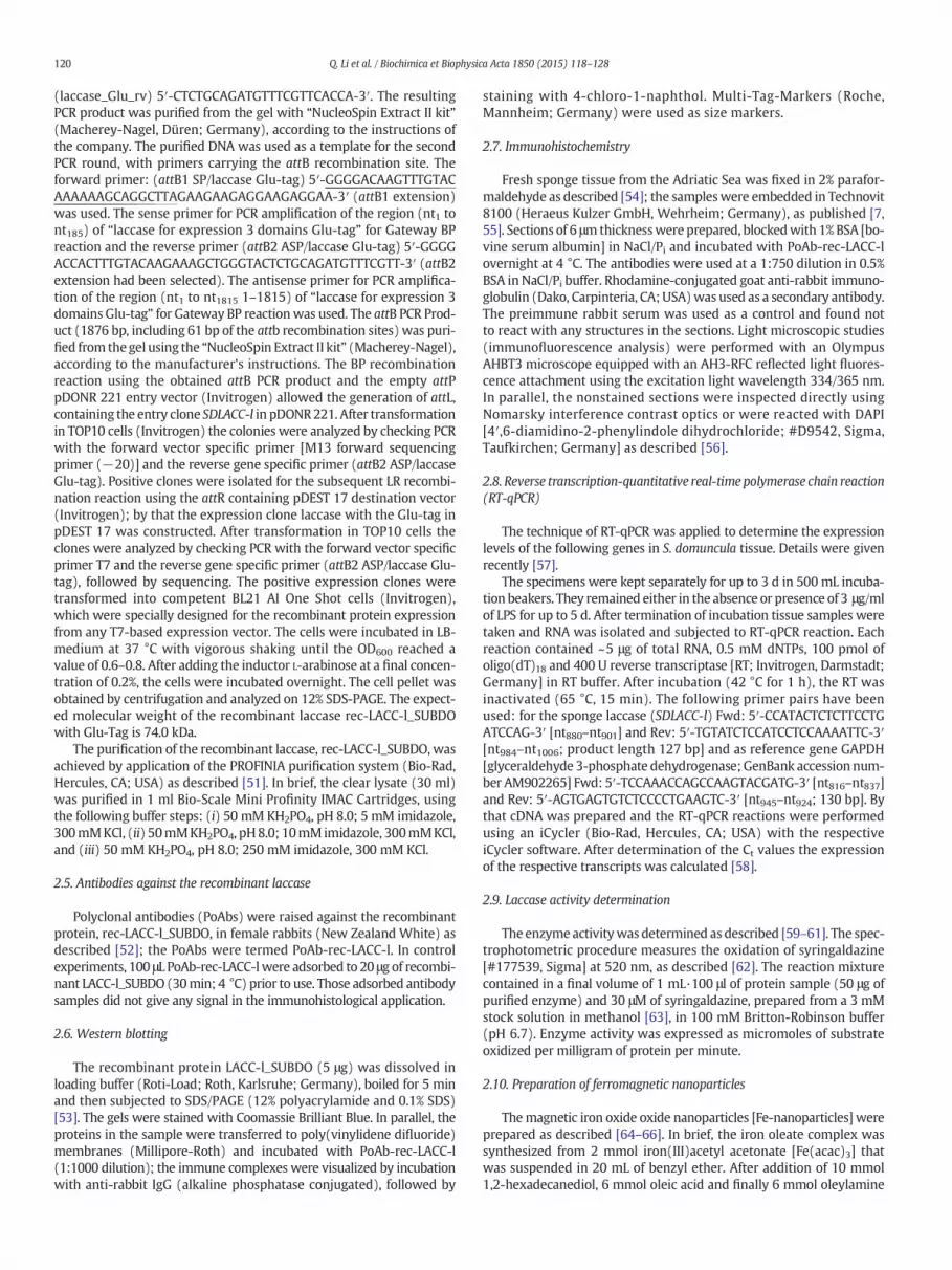
(laccase_Glu_rv) 5′-CTCTGCAGATGTTTCGTTCACCA-3′. The resultingPCR product was purified from the gel with “NucleoSpin Extract II kit”(Macherey-Nagel, Düren; Germany), according to the instructions ofthe company. The purified DNA was used as a template for the secondPCR round, with primers carrying the attB recombination site. Theforward primer: (attB1 SP/laccase Glu-tag) 5′-GGGGACAAGTTTGTACAAAAAAGCAGGCTTAGAAGAAGAGGAAGAGGAA-3′ (attB1 extension)was used. The sense primer for PCR amplification of the region (nt1 tont185) of “laccase for expression 3 domains Glu-tag” for Gateway BPreaction and the reverse primer (attB2 ASP/laccase Glu-tag) 5′-GGGGACCACTTTGTACAAGAAAGCTGGGTACTCTGCAGATGTTTCGTT-3′ (attB2extension had been selected). The antisense primer for PCR amplifica-tion of the region (nt1 to nt1815 1–1815) of “laccase for expression 3domains Glu-tag” for Gateway BP reactionwas used. The attB PCR Prod-uct (1876 bp, including 61 bp of the attb recombination sites) was puri-fied from the gel using the “NucleoSpin Extract II kit” (Macherey-Nagel),according to the manufacturer's instructions. The BP recombinationreaction using the obtained attB PCR product and the empty attPpDONR 221 entry vector (Invitrogen) allowed the generation of attL,containing the entry clone SDLACC-l in pDONR221. After transformationin TOP10 cells (Invitrogen) the colonies were analyzed by checking PCRwith the forward vector specific primer [M13 forward sequencingprimer (−20)] and the reverse gene specific primer (attB2 ASP/laccaseGlu-tag). Positive clones were isolated for the subsequent LR recombi-nation reaction using the attR containing pDEST 17 destination vector(Invitrogen); by that the expression clone laccase with the Glu-tag inpDEST 17 was constructed. After transformation in TOP10 cells theclones were analyzed by checking PCR with the forward vector specificprimer T7 and the reverse gene specific primer (attB2 ASP/laccase Glu-tag), followed by sequencing. The positive expression clones weretransformed into competent BL21 AI One Shot cells (Invitrogen),which were specially designed for the recombinant protein expressionfrom any T7-based expression vector. The cells were incubated in LB-medium at 37 °C with vigorous shaking until the OD600 reached avalue of 0.6–0.8. After adding the inductor L-arabinose at a final concen-tration of 0.2%, the cells were incubated overnight. The cell pellet wasobtained by centrifugation and analyzed on 12% SDS-PAGE. The expect-ed molecular weight of the recombinant laccase rec-LACC-l_SUBDOwith Glu-Tag is 74.0 kDa.
The purification of the recombinant laccase, rec-LACC-l_SUBDO, wasachieved by application of the PROFINIA purification system (Bio-Rad,Hercules, CA; USA) as described [51]. In brief, the clear lysate (30 ml)was purified in 1 ml Bio-Scale Mini Profinity IMAC Cartridges, usingthe following buffer steps: (i) 50 mM KH2PO4, pH 8.0; 5 mM imidazole,300mMKCl, (ii) 50mMKH2PO4, pH8.0; 10mMimidazole, 300mMKCl,and (iii) 50 mM KH2PO4, pH 8.0; 250 mM imidazole, 300 mM KCl.
2.5. Antibodies against the recombinant laccase
Polyclonal antibodies (PoAbs) were raised against the recombinantprotein, rec-LACC-l_SUBDO, in female rabbits (New Zealand White) asdescribed [52]; the PoAbs were termed PoAb-rec-LACC-l. In controlexperiments, 100 μL PoAb-rec-LACC-lwere adsorbed to 20 μg of recombi-nant LACC-l_SUBDO (30min; 4 °C) prior to use. Those adsorbed antibodysamples did not give any signal in the immunohistological application.
2.6. Western blotting
The recombinant protein LACC-l_SUBDO (5 μg) was dissolved inloading buffer (Roti-Load; Roth, Karlsruhe; Germany), boiled for 5 minand then subjected to SDS/PAGE (12% polyacrylamide and 0.1% SDS)[53]. The gels were stained with Coomassie Brilliant Blue. In parallel, theproteins in the sample were transferred to poly(vinylidene difluoride)membranes (Millipore-Roth) and incubated with PoAb-rec-LACC-l(1:1000 dilution); the immune complexes were visualized by incubationwith anti-rabbit IgG (alkaline phosphatase conjugated), followed by
staining with 4-chloro-1-naphthol. Multi-Tag-Markers (Roche,Mannheim; Germany) were used as size markers.
2.7. Immunohistochemistry
Fresh sponge tissue from the Adriatic Sea was fixed in 2% parafor-maldehyde as described [54]; the sampleswere embedded in Technovit8100 (Heraeus Kulzer GmbH, Wehrheim; Germany), as published [7,55]. Sections of 6 μmthicknesswere prepared, blockedwith 1% BSA [bo-vine serum albumin] in NaCl/Pi and incubated with PoAb-rec-LACC-lovernight at 4 °C. The antibodies were used at a 1:750 dilution in 0.5%BSA in NaCl/Pi buffer. Rhodamine-conjugated goat anti-rabbit immuno-globulin (Dako, Carpinteria, CA; USA)was used as a secondary antibody.The preimmune rabbit serum was used as a control and found notto react with any structures in the sections. Light microscopic studies(immunofluorescence analysis) were performed with an OlympusAHBT3 microscope equipped with an AH3-RFC reflected light fluores-cence attachment using the excitation light wavelength 334/365 nm.In parallel, the nonstained sections were inspected directly usingNomarsky interference contrast optics or were reacted with DAPI[4′,6-diamidino-2-phenylindole dihydrochloride; #D9542, Sigma,Taufkirchen; Germany] as described [56].
2.8. Reverse transcription-quantitative real-time polymerase chain reaction(RT-qPCR)
The technique of RT-qPCR was applied to determine the expressionlevels of the following genes in S. domuncula tissue. Details were givenrecently [57].
The specimens were kept separately for up to 3 d in 500 mL incuba-tion beakers. They remained either in the absence or presence of 3 μg/mlof LPS for up to 5 d. After termination of incubation tissue samples weretaken and RNA was isolated and subjected to RT-qPCR reaction. Eachreaction contained ~5 μg of total RNA, 0.5 mM dNTPs, 100 pmol ofoligo(dT)18 and 400 U reverse transcriptase [RT; Invitrogen, Darmstadt;Germany] in RT buffer. After incubation (42 °C for 1 h), the RT wasinactivated (65 °C, 15 min). The following primer pairs have beenused: for the sponge laccase (SDLACC-l) Fwd: 5′-CCATACTCTCTTCCTGATCCAG-3′ [nt880–nt901] and Rev: 5′-TGTATCTCCATCCTCCAAAATTC-3′[nt984–nt1006; product length 127 bp] and as reference gene GAPDH[glyceraldehyde 3-phosphate dehydrogenase; GenBank accession num-ber AM902265] Fwd: 5′-TCCAAACCAGCCAAGTACGATG-3′ [nt816–nt837]and Rev: 5′-AGTGAGTGTCTCCCCTGAAGTC-3′ [nt945–nt924; 130 bp]. Bythat cDNA was prepared and the RT-qPCR reactions were performedusing an iCycler (Bio-Rad, Hercules, CA; USA) with the respectiveiCycler software. After determination of the Ct values the expressionof the respective transcripts was calculated [58].
2.9. Laccase activity determination
The enzymeactivitywas determined as described [59–61]. The spec-trophotometric procedure measures the oxidation of syringaldazine[#177539, Sigma] at 520 nm, as described [62]. The reaction mixturecontained in a final volume of 1 mL·100 μl of protein sample (50 μg ofpurified enzyme) and 30 μM of syringaldazine, prepared from a 3 mMstock solution in methanol [63], in 100 mM Britton-Robinson buffer(pH 6.7). Enzyme activity was expressed as micromoles of substrateoxidized per milligram of protein per minute.
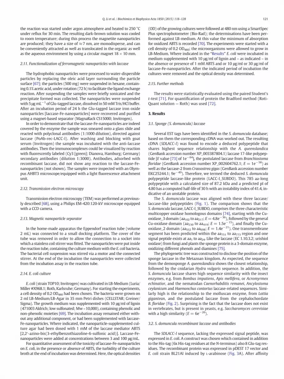
2.10. Preparation of ferromagnetic nanoparticles
Themagnetic iron oxide oxide nanoparticles [Fe-nanoparticles] wereprepared as described [64–66]. In brief, the iron oleate complex wassynthesized from 2 mmol iron(III)acetyl acetonate [Fe(acac)3] thatwas suspended in 20 mL of benzyl ether. After addition of 10 mmol1,2-hexadecanediol, 6 mmol oleic acid and finally 6 mmol oleylamine
121Q. Li et al. / Biochimica et Biophysica Acta 1850 (2015) 118–128
the reaction was started under argon atmosphere and heated to 250 °Cunder reflux for 30 min. The resulting dark-brown solution was cooledto room temperature; during this process the magnetite nanoparticlesare produced; they have a size of ≈7 nm, are monodisperse, and canbe conveniently attracted as well as translocated in the organic as wellas the aqueous environment by using a circular magnet 18 × 10 mm.
2.11. Functionalization of ferromagnetic nanoparticles with laccase
The hydrophobic nanoparticles were processed to water-dispersibleparticles by replacing the oleic acid layer surrounding the particlesurface [67]; the particles (500 mg) were suspended in hexane, contain-ing0.1% acetic acid, under rotation (72h) to facilitate the ligand exchangereaction. After suspending the samples were briefly sonicated and theprecipitate formed was collected. The nanoparticles were suspendedwith 5 μgmL−1 of Glu-tagged laccase, dissolved in 50mMTris/HCl buffer.After an incubation period of 24 h the Glu-tagged laccase iron oxidenanoparticles [laccase-Fe-nanoparticles] were recovered and purifiedusing a magnet-based separator (MagnaRack CS15000; Invitrogen).
In order to demonstrate that the laccase-Fe-nanoparticles are indeedcovered by the enzyme the sample was smeared onto a glass slide andreacted with polyclonal antibodies (1:1000 dilution), directed againstlaccase (PoAb-rec-LACC-l). After washing and blocking with goatserum (Invitrogen) the sample was incubated with the anti-laccaseantibodies. Then the immunocomplexes could be visualized by reactionwith fluorescently labeled (Cy3 [green]; Dianova, Hamburg; Germany)secondary antibodies (dilution 1:3000). Antibodies, adsorbed withrecombinant laccase, did not show any reaction to the laccase-Fe-nanoparticles (not shown). The samples were inspected with an Olym-pus AHBT3 microscope/equipped with a light fluorescence attachmentunit.
2.12. Transmission electron microscopy
Transmission electronmicroscopy (TEM)was performed as previous-ly described [68], using a Philips EM-420:120-kV microscope equippedwith a CCD camera.
2.13. Magnetic nanoparticle separator
In the home-made apparatus the Eppendorf reaction tube (volume2 mL) was connected to a small docking platform. The cover of thetube was removed in order to allow the connection to a socket intowhich a stainless coil stirrerwasfitted. The nanoparticleswere put insidethe reaction tube, containing the culturemediumwith the E. coli bacteria.The bacterial cell suspension was stirred via a motor and the connectedstirrer. At the end of the incubation the nanoparticles were collectedfrom the incubation assay in the reaction tube.
2.14. E. coli culture
E. coli (strain TOP10; Invitrogen)was cultivated in LB-Medium (Luria/Miller #X968.1; Roth, Karlsruhe;Germany). For starting the experiments,a cell density of 0.2 OD480 had been chosen. The bacteria were grown on2 ml LB-Medium/LB-Agar in 35 mm Petri dishes (CELLSTAR; Greiner/Sigma). The growth medium was supplemented with 10 μg/ml of lignin(471003Aldrich; low sulfonated;Mw~10,000), containing phenolic andnon-phenolic moieties [69]. The incubation assay remained either with-out any additional component, or had been supplemented with laccase-Fe-nanoparticles. Where indicated, the nanoparticle-supplemented cul-ture agar had been dosed with 1 mM of the laccase mediator ABTS[2,2′-azino-bis(3-ethylbenzothiazoline-6-sulfonic acid)]. Laccase-Fe-nanoparticles were added at concentrations between 3 and 100 μg/mL.
For quantitative assessment of the toxicity of laccase-Fe-nanoparticleson E. coli, in the presence or absence of ABTS, the turbidity of the culturebroth at the end of incubationwas determined. Here, the optical densities
(OD) of the growing cultureswere followed at 480 nmusing a SmartSpecPlus spectrophotometer (Bio-Rad); the determinations have been per-formed against LB-medium. At this value the minimum of absorptionfor oxidized ABTS is recorded [70]. The experiments were started with acell density of 0.2 OD480; the microorganisms were allowed to grow inLB-Medium. Where indicated in the “Results” E. coli were incubated inmedium supplemented with 10 μg/ml of lignin and – as indicated – inthe absence or presence of 1 mM ABTS and or 10 μg/ml or 30 μg/ml oflaccase-Fe-nanoparticles. After the indicated period of incubation thecultures were removed and the optical density was determined.
2.15. Further methods
The results were statistically evaluated using the paired Student'st-test [71]. For quantification of protein the Bradford method (Roti-Quant solution — Roth) was used [72].
3. Results
3.1. Sponge (S. domuncula) laccase
Several EST tags have been identified in the S. domuncula database;based on them the corresponding cDNA was worked out. The resultingcDNA (SDLACC-l) was found to encode a deduced polypeptide thatshares highest sequence relationship with the A. queenslandica(GenBank accession number XP_003387804.1) laccase 17-like polypep-tide [E value [73] of 1e−64], the postulated laccase from Branchiostomafloridae (GenBank accession number XP_002604762.1; E = 1e−58) aswell as the laccase-2 from Crassostrea gigas (GenBank accession numberEKC25244.1; 9e−49). Therefore, we termed the deduced S. domunculapolypeptide laccase-like protein (LACC-l_SUBDO). This 785 aa-longpolypeptide with a calculated size of 87.2 kDa and a predicted pI of4.80 has a computed half-life of 30 hwith an instability index of 41.4, in-dicative of an unstable protein.
The S. domuncula laccase was aligned with these three laccase/laccase-like polypeptides (Fig. 1). The comparison shows that theS. domuncula laccase, LACC-l_SUBDO, comprises the three characteristicmulticopper oxidase homologous domains [74], starting with the Cu-oxidase_3 domain (aa102 to aa217; E=4.8e−36), followedby the generalCu-oxidase domain (aa230 to aa419; E = 1.5e−15), and finally the Cu-oxidase_2 domain (aa552 to aa698; E = 1.4e−21). One transmembranesegment has been predicted within the aa751 to aa773 region and onesignal peptide exists at aa1 to aa24. Like the laccase (EC 1.10.3.2; urishioloxidase) from fungi andplants the sponge protein is a 3-domain enzyme,oxidizing different phenols and diamines [75].
The phylogenetic treewas constructed to disclose the position of thesponge laccase in the Metazoan kingdom. As expected, the sequencefrom the demosponge A. queenslandica shows the closest relationship,followed by the cnidarian Hydra vulgaris sequence. In addition, theS. domuncula laccase shares high sequence similarity with the insectenzymes, e.g. from Bombus impatiens, Apis mellifera, or Acromyrmexechinatior, and the nematodan Caenorhabditis remanei, Ancylostomaceylanicum and Haemonchus contortus laccase-related sequences. Simi-larly high is the relationship to the molluscan enzyme, from Lottiagigantean, and the postulated laccase from the cephalochordateB. floridae (Fig. 2). Surprising is the fact that the laccase does not existin vertebrates, but is present in yeasts, e.g. Saccharomyces cerevisiaewith a high similarity (E = 6e−26).
3.2. S. domuncula recombinant laccase and antibodies
The SDLACC-l sequence, lacking the expressed signal peptide, wasexpressed in E. coli. A construct was chosenwhich contained in additionto the His-tag (6xHis-tag residues at theN-terminus) also 8Glu-tag res-idues. The recombinant protein was expressed in pDEST 17 vector andE. coli strain BL21AI induced by L-arabinose (Fig. 3A). After affinity

Fig. 1. The S. domuncula laccase; alignment. The similarities of the S. domuncula deduced laccase (LACC-l_SUBDO) with the next related sequences from A. queenslandica (LACC-p_AMPHI;GenBank accession no. XP_003387804.1), B. floridae (LACC-2_CRASSGI; GenBank accession no. XP_002604762.1) as well as C. gigas (LACC-p_BRAFL; GenBank accession no. EKC25244.1).These sequences were aligned. Residues conserved (identical or similar with respect to their physico-chemical properties) in all sequences are shown in white on black. Those sequenceswhich share similarity to at least three residues are in white on gray and at least to two sequences in black on gray. The borders {signal} of the signal peptide, the transmembrane region(TM region) as well as the three characteristic domains, the Cu-oxidase_3 domain [Cu-ox-3], the general Cu-oxidase domain [Cu-ox], and the Cu-oxidase_2 domain [Cu-ox-2] are highlighted.
122 Q. Li et al. / Biochimica et Biophysica Acta 1850 (2015) 118–128
purification the 658 aa long laccase, LACC-l_SUBDO, comprising theC-terminal Glu-tag [1.05 kDa], as well as the His-tag [0.84 kDa], atotal size of 74.08 kDa for the protein has been calculated and alsoexperimentally found.
In order to localize the laccase within the sponge tissue, antibodieswere prepared in rabbits. Western blotting revealed that only the anti-bodies from the challenged animals reacted with the recombinantLACC-l_SUBDO (Fig. 3B; lane b), while the preimmune serum did not(Fig. 3B; lane a). By that the specific 74 kDa protein was visualized.
3.3. Upregulation of laccase gene in sponge tissue after exposure to LPS
3.3.1. ImmunohistologyTissue sliceswere prepared froman animal, kept for 5 dwith 3 μg/mL
of LPS or, in the control, in the absence of the endotoxin. Then the sliceswere reacted with anti-laccase IgG (PoAb-rec-LACC-l). The analysesshow that in tissue from the LPS-treated animal the brightest reactions(highest level of laccase) are in the surface zone; only a lower quantityof immunocomplexes is visualized in the central part of the animal(Fig. 4C). In parallel the histology is represented by Nomarskyinterference contrast optics (Fig. 4A) or by DAPI staining (Fig. 4B). Thecontrol animal, not exposed to LPS, shows a significant lower expressionof laccase (Fig. 4F); again the histology is visualized by interferencecontrast optics (Fig. 4D) and by staining with DAPI (Fig. 4E). In controls
it was established that the pre-immune serum did not react with thestructures within the slices (not shown).
3.3.2. RT-qPCR analysisThe laccase gene expression depends on the exposure to the endo-
toxin LPS. Specimens were kept separately for up 5 d in beakers in theabsence or presence of LPS. Tissue samples were removed after 0 to5 d, followed by RNA extraction. Then the steady-state expressionlevel of laccase was determined by RT-qPCR and the values werenormalized to GAPDH expression (Fig. 5). The data show that in tissuefrom a non-treated animal the level for laccase only slightly increasedfrom 0.06 ± 0.01 (day 0) to 0.14 ± 0.02 (day 5). In contrast, in LPS-exposed specimens the increase of laccase is significantly higher, also ifcompared to the controls. At day 0 the expression level is 0.08 ± 0.02,while at day 5 the steady-state expression value is 0.26 ± 0.04.
3.4. Laccase activity of the recombinant enzyme
The enzymatic activity of the recombinant laccase (LACC-l_SUBDO)was determined in the spectrophotometric assay, as described under“Material and methods”, by using syringaldazine as a substrate. In thepresence of syringaldazine the sponge laccase has a Vmax of 65 μM/minand an apparent Michaelis constant (Km) of 114 μM. These values aresomehow lower than those described for the bacterial laccase [60] butstill close to the laccase from mushrooms, determined earlier [76,77].

Fig. 2. The S. domuncula laccase; phylogenetic tree. A tree was constructed using, in addition to those sequences in Fig. 1, the sequences from the laccase-like protein from H. vulgaris (LACC-l_HYDRA;GenBank accessionno. XP_002159531.2) and the laccase-postulatedprotein from L. gigantean (LACC-p_LOTTIA;GenBank accessionno. ESO89911.1),A. ceylanicum (LACC-p_ANCYLO;GenBank accession no. EYB82076.1), A. mellifera (LACC-p_APIS; GenBank accession no. XP_001120790.2), B. impatiens (LACC-p_BOMBUS; GenBank accession no. XP_003490974.1) andC. remanei (LACC-p_CAEEL; GenBank accession no. XP_003092481.1). In addition, the multicopper oxidase domain containing protein from H. contortus (LACC-p_HAEMO; GenBank accessionno. CDJ83113.1) and laccase-4 from A. echinatior (LACC-4_ACROM; GenBank accession no. EGI60467.1) were included. The multicopper oxidase from S. cerevisiae (MCO-A_YEAST; GenBankaccession no. P38993.2) was included and used as outgroup to root the tree. The scale bar indicates an evolutionary distance of 0.1 aa substitutions per position in the sequence.
123Q. Li et al. / Biochimica et Biophysica Acta 1850 (2015) 118–128
3.5. Preparation of nanoparticles, coated with recombinant laccase
The ferromagnetic nanoparticles were covered with Glu-taggedlaccase. In the first step the iron oxide nanoparticles were prepared asiron oleate complex. The iron oxide nanoparticles formed have an
Fig. 3. The recombinant sponge laccase. The S. domuncula SDLACC-lwas expressed in E. coli.(A) The bacterial protein extract, obtained from non-induced (lane a) or from arabinose-induced cultures (lane b) was size-separated by SDS/PAGE analysis (12% gels). The 74 kDafusion protein is marked. (B) The purified fusion protein rec-LACC-l_SUBDO was used toraise antibodies. The resulting PoAb-rec-LACC-l protein (immune serum [i]) was found toreact with a 74-kDa protein in the Western blot assay with the recombinant protein (laneb), while the pre-immune serum (pi) did not (lane a). The sizemarkers (M) are given in (A).
average size of≈ 7 nm (Fig. 6). They display strongmagnetic propertieswhich can be utilized to collect them back from the reaction mixtureafter incubating them with bacteria (see below). The hydrophobicsurface residues were transferred to hydrophilic residues by acid treat-ment, allowing the coating of the particleswith laccase. The particles arestained in darkish brown (not shown here). After transfer of the oleateferromagnetic particles to acetic acid the particles were covered by Glu-tagged laccase. During this functionalization with laccase the particlesincreased in size to 14–18 nm (not shown here).
In order to demonstrate that the nanoparticles are indeed coatedwith laccase the particles were incubated with antibodies, directedagainst the laccase (PoAb-rec-LACC-l). The samples were smeared ontoa glass slide (Fig. 7A); after applying the antibodies and incubating theantigen/antibody complexes with a labeled secondary antibody, thecomplexes strongly lighted up in green (Fig. 7B). In the controls, incuba-tion with the preimmune serum, no signals were seen (not shown).
3.6. Antimicrobial effect of laccase
The effect of laccase that had been coated around iron oxide nano-particleswas directly checked under agar-growth conditions. An oxidiz-able substrate 10 μg/ml of lignin was added. A suspension of E. coliwasplated onto the agar; where indicated the agar was supplemented withthe laccase mediator ABTS and/or the laccase-Fe-nanoparticles. Theresults show that after an incubation period of 4 h the number of coloniesthat were formed in the control cultures, containing neither ABTS norlaccase, is relatively dense (Fig. 8). If ABTS and, especially, the laccase-Fe-nanoparticles are added separately to the bacteria a reduction of thenumber of bacterial colonies is observed. Co-addition of the mediator(ABTS) to the laccase-Fe-nanoparticles results in a dose-dependentstrong inhibition of the E. coli growth. In controls it was establishedthat lignin does not affect the growth of the bacteria (not shown).

Fig. 4. Immunohistological detection of laccase in S. domuncula tissue. The specimenwas kept for 5 d in the presence of 3 μg/mL of LPS (A, B, C) or in the absence of the endotoxin (D, E, F).Then slices through the tissue (ti)were prepared and inspected byNomarsky interference contrast optics (A, D), or after DAPI staining (B and E) to highlight the cell nuclei, or after reactionwith the laccase antibodies (PoAb-rec-LACC-l). In addition, the cell surface (s) and the surrounding water (w) environment are marked. Size bars are given.
124 Q. Li et al. / Biochimica et Biophysica Acta 1850 (2015) 118–128
3.7. Effect of laccase iron oxide nanoparticles in a magneticnanoparticle separator
To analyze the effect of the laccase-Fe-nanoparticles on the growthof bacteria in suspension, a magnetic nanoparticle separator has beenused. This device allows the incubation of the bacteria in a reactiontube under controlled stirring (Figs. 9A to C). A coiled stirrer that hasbeen inserted into the reaction tube is connectedwith amotor, allowinga tuned mixing of the cultures. In the incubation chamber, used here, ahole has been insertedwhich allows the insertion of an ultraviolet lamp.The coiled stirrer allows the collection of the nanoparticles by electro-magnetic forces (Figs. 9D and E). In control experiments it had beenascertained that the bacteria did not attach to the nanoparticles,surrounded by the laccase.
Fig. 5. Influence of LPS on the expression of the laccase gene in sponge tissue. In parallelexperiments the sponge specimens were incubated in the absence (open bars) or presenceof 3 μg/mL of LPS (closed bars) for 0 (start of the experiment), 1 d, 3 d or 5 d. Subsequently,RNA was extracted and subjected to RT-qPCR analysis for quantification of the steady-state-expression level of both laccase (SDLACC-l) and GAPDH. The expression level of laccasewasnormalized to the expression of GAPDH. Data are expressed as mean values ± SD for fiveindependent experiments. Differences between two groups were evaluated using anunpaired t-test, *P b 0.05.
A quantitative assessment of the effect of different concentrations oflaccase-Fe-nanoparticles was performed in the magnetic nanoparticleseparator, in which the bacteria were growing in the reaction tube insuspension. The medium was supplemented with the laccase substrate10 μg/ml of lignin. As a measure for bacterial growth the OD480 wasdetermined (Fig. 10). In the absence of either ABTS or laccase-Fe-nanoparticles the bacteria grew from ≈ 0.42 ± 0.5 to 1.73 ± 0.19 after100 min incubation and to 2.14 ± 0.19 after 200 min. If the mediatorABTS is added a non-significant reduction is measured with 1.47 ± 0.17(100min) and 1.93± 0.21 (200 min), respectively. However, after expo-sure of the bacteria in the absence of ABTS to 10 μg/ml laccase-Fe-nanoparticles a significant reduction was measured with 1.12 ± 0.16(100 min) and 1.47 ± 0.16 (200 min). Co-addition of ABTS and laccase-Fe-nanoparticles strongly increased the inhibitory activity of the enzyme;after 200min the density of the bacteriameasures only 0.62±0.08 (ABTSplus 10 μg/ml of laccase-Fe-nanoparticles) and 0.28 ± 0.04 (ABTS plus30 μg/ml of laccase-Fe-nanoparticles), respectively.
Fig. 6. Iron oxide nanoparticleswere prepared from iron(III)acetyl acetonate and oleylamine.

Fig. 7. Identification of laccase on a smear of laccase-Fe-nanoparticles. (A) An aliquot of laccase-Fe-nanoparticles was applied on a glass slide and (B) incubated with antibodies againstsponge laccase. The immunocomplexes were visualized with a labeled secondary antibody.
125Q. Li et al. / Biochimica et Biophysica Acta 1850 (2015) 118–128
4. Discussion
It could be indicative that the S. domuncula laccase with its domainstructure and phylogenetic relationship comprises a high sequencesimilarity besides to other poriferan enzymes also to cnidarian laccases.While a significant, even though more distant relationship also existsto insect and molluscan laccase, no homologous or similar enzyme hasbeen described for the evolved vertebrates. In sponges the laccase is ap-parently frequently present; in the demosponge species A. queenslandicaseveral isoforms have been cloned (see e.g. GenBank accession numberXP_003387804.1). This fact could imply that the laccase gene has beenlost during vertebrate evolution. Such a gain and loss of a gene has
Fig. 8. Effect of laccase (added as laccase-Fe-nanoparticles [10 μg/ml]) and of the mediator ABTcontained neither laccase-Fe-nanoparticles nor ABTS. As oxidizable substrate 10 μg/ml of ligninABTS together with 10 μg/ml, 30 μg/ml or 100 μg/ml of laccase-Fe-nanoparticles [Lac-Fe-NP] a
beendescribed to have happened for the strombine dehydrogenase, like-wise in S. domuncula [78]. However, the existence of laccase in spongesand cnidarians could also signify that this enzyme plays an importantrole in thephysiology of these basalmetazoans. The laccase is also presentin yeast [79,80]. Common to all of these laccases is the presence of threecharacteristic multicopper oxidase homologous domains that are crucialfor the one-electron oxidation of a broad range of compounds, includingsubstituted phenols, arylamines and aromatic thiols during which thecorresponding radicals are formed [81]. It is very obvious that insponges and cnidarians, laccase has a functional role in detoxificationand elimination of lignin, coming from the plants that surroundsponges, e.g. S. domuncula. In a future study we will determine why
S [1 mM], in separate, on the colony formation of E. coli (upper row); the upper left assayhad been added to all assays. It is apparent that if the medium ismixedwith themediatorn almost complete inhibition of bacterial growth is measured (lower row).

Fig. 9.Magnetic nanoparticle separator. (A) Separatorwhich features in the functional center of thedevice a reaction tube (rt) intowhich the bacteria grow in suspension. The reaction tube isflush-mounted in an incubation chamber (ic). The stirring of the bacterial suspension is carried out by a coil stirrer that is linked to a motor (m). (B and C) Incubation chamber, at highermagnification, into which the reaction tube can be steeped in. A drilled hole allows the introducing of an ultraviolet lamp (UV-l). This lamp can be protected by a protection ring (pr) thatcan be put over the feeding hole. (D and E) Reaction tube harboring the bacterial cultures with the laccase-Fe-nanoparticles; a coil stirrer (cs) is shown in E. The nanoparticles (np) in(E) have been attracted by applying electromagnetic forces.
126 Q. Li et al. / Biochimica et Biophysica Acta 1850 (2015) 118–128
the phenolic avarol, a secondary metabolite from sponges, comprisesantimicrobial activity but is not toxic to the host [82]. It might be antic-ipated that it is the laccase that “detoxifies avarol” in the sponge.
The broad substrate specificity of laccases is unusual for an enzyme[81] and suggested to us that the laccase in S. domunculamight have thefunction to kill bacteria, or control bacterial growth. In order to test thishypothesis the S. domuncula laccase was heterologously expressed in
Fig. 10.Growthof E. coli cells, grown in 10 μg/ml of lignin, in the absence of ABTS and laccase-Fe-nanoparticles (control; open bars), or – in separate – either 1mMABTS (ABTS; shaded tothe left) or 30 μg/ml laccase-Fe-nanoparticles (Lac-Fe-NP; shaded to the right). In two addi-tional series ABTS and the nanoparticles, either 10 μg/ml of laccase-Fe-nanoparticles [Lac-Fe-NP (10 μg/ml+ ABTS); cross hatched bars] or 30 μg/ml laccase-Fe-nanoparticles [Lac-Fe-NP(30 μg/ml+ ABTS);filledbars]. After an incubationperiodof 0min (start of the experiment),100min or 200min the sampleswere taken and the optical density (OD480)was determined.Values represent the means (±SD) from 10 separate experiments each (*P b 0.01).
E. coli. In order to allow subsequent binding of the enzyme to iron oxidenanoparticles a Glu-tag was genetically engineered to the N-terminus ofthe enzyme [83]. The recombinant laccase obtained displayed enzymaticfunction, if the mediator syringaldazine was added to the enzyme reac-tion. The activity of the sponge recombinant laccase is in the range ofactivities measured for other recombinant enzymes [60] and naturalenzymes [76,77,84]. The redox potential among the laccases vary dras-tically; there are both enzymes with a high redox potential, e.g. themushroom Trametes versicolor laccase, and enzymes with a low redoxpotential, like the plant Rhus vernicifera enzyme [85]. Adapted to thisinherent property the mediator has to be selected [81]. The recombi-nant sponge enzyme is significantly more active if the mediators ABTSand syringaldazine are added; both compounds are universalmediatorsfor laccases [86]. The Michaelis constant (Km) of the sponge enzyme is114 μM, somewhat higher than the one found for bacterial laccase[60], but close to the one from the mushroom Clitocybe maxima [87].
Having obtained the recombinant sponge laccase the elucidation ofthe distribution of the enzyme within the sponge became possible. Incontinuation, immunohistological studies have been performed reveal-ing that the enzyme is highly expressed at the rim of the animals. Inter-estingly enough, the level of tissue expression of the laccase is stronglyupregulated if the animals are exposed to bacterial LPS. This findingunderscores our hypothesis that the sponge laccase has a role in host-defense against microorganisms. Such a role has been proposed for theinsect laccase [88]. A quantitative approach to assess the steady-statelevel of the sponge laccase by RT-qPCR revealed that the enzyme is high-ly expressed in response to the LPS load in the aqueous environment.Until now, this is the first report that in metazoans the laccase geneexpression can be upregulated by LPS. From the functionally relatedenzyme, the phenoloxidase from sea cucumber Apostichopus japonicas,an increased expression has beenmeasured after challenging the animalswith LPS [89], also implying that this oxidoreductase is involved indefense against infection from bacteria or fungi.
Prior to a subsequent application of the sponge recombinant laccase ina biological assay the Glu-tagged laccase had been linked to iron oxidenanoparticles. The rational behind is the fact that the application of ferro-magnetic nanoparticles is very efficient with respect to a cost-effectivetesting. The selection of these nanoparticles allows a future sustainable

127Q. Li et al. / Biochimica et Biophysica Acta 1850 (2015) 118–128
application by the magnetic removal and recovery of the particles afterterminating the enzyme reaction.
In order to further substantiate the view that the sponge laccase isinvolved in bacterial defense the enzyme reaction was directly coupledwith the bacteria. An oxidizable substrate 10 μg/ml of lignin, as a sourcefor free radicals during laccase reaction, was added to the broth in allassays. Lignin is non-toxic for E. coli [90]. Exposure of E. coli to the ironoxide nanoparticles coated with enzymatically active Glu-taggedlaccase revealed that the bacterial growth is slightly inhibited by thenanoparticles and only a little by the mediator ABTS. However, if bothcomponents are added to the agar onto which the bacteria are growingan almost complete inhibition of growth is seen. A quantitative assess-ment of the effect of the laccase has been achieved by adding thelaccase-Fe-nanoparticles to E. coli, growing in suspension in reactiontubes, hooked to a magnetic nanoparticle separator. In this system thebacterial growth, mediated by laccase in the presence of the mediatorABTS, is completely inhibited. This finding can be taken as direct proofthat the sponge laccase displays anti-bacterial activity if the two compo-nents enzyme and mediator are brought into close contact with themicroorganisms.
In conclusion, the data presented show that the S. domuncula laccaseis most likely involved in the antibacterial defense of the sponge organ-ism. The laccase had been immobilized to iron oxide nanoparticles toallow us to repeatedly and sustainably use the enzyme and to applythe laccase-Fe-nanoparticles togetherwith a second set of nanoparticlesthat are surrounded by titania. Those titania-iron oxide nanoparticlesallow the photocatalytic killing of the bacteria, if the cultures areexposed to UV-light (in progress). Again, the data on the S. domunculalaccase support the view that sponges have optimized their biosynthet-ic/biodegradation capabilities to the highest efficiency and diversity,especially if the stress-response system of the animals is challenged.The aquatic environment where sponges live is characterized by amuch higher density of organismic populations in comparison to organ-isms living in the air, due to the higher density of the surroundingmedium. This implies that these animalsmust havedeveloped an efficientrepertoire of defense strategies to copewith these adverse environmentalconditions.
Acknowledgments
W.E.G. M. is a holder of an ERC Advanced Investigator Grant (no.268476 BIOSILICA). This work was supported by grants from theEuropean Commission (Grant no. 311848 “BlueGenics” and Grant no.286059 “CoreShell”), as well as the European Commission/EUREKA(EUROSTARS Grant no. 4289 “SILIBACTS”).
References
[1] W.E.G. Müller, M. Wiens, T. Adell, V. Gamulin, H.C. Schröder, I.M. Müller, Bauplan ofurmetazoa: basis for genetic complexity ofmetazoa, Int. Rev. Cytol. 235 (2004) 53–92.
[2] R. Osinga, el.H. Belarbi, E.M. Grima, J. Tramper, R.H. Wijffels, Progress towards acontrolled culture of the marine sponge Pseudosuberites andrewsi in a bioreactor,J. Biotechnol. 100 (2003) 141–146.
[3] S.W.Watson, T.J. Novitsky, H.L. Quinby, F.W. Valois, Determination of bacterial numberand biomass in themarine environment, Appl. Environ.Microbiol. 33 (1977) 940–946.
[4] M. Breitbart, Marine viruses: truth or dare, Ann. Rev. Mar. Sci. 4 (2012) 425–448.[5] A.A. Moscona, Cell aggregation: properties of specific cell-ligands and their role in
the formation of multicellular systems, Dev. Biol. 18 (1968) 250–277.[6] W.H. Hildemann, I.S. Johnston, P.L. Jokiel, Immunocompetence in the lowest meta-
zoan phylum: transplantation immunity in sponges, Science 204 (1979) 420–422.[7] Z. Pancer, M. Kruse, H. Schäcke, U. Scheffer, R. Steffen, P. Kovács, W.E.G. Müller, Poly-
morphism in the immunoglobulin-like domains of the receptor tyrosine kinase fromthe sponge Geodia cydonium, Cell Adhes. Commun. 4 (1996) 327–339.
[8] W.E.G.Müller, B. Blumbach, I.M.Müller, Evolution of the innate andadaptive immunesystems: relationships between potential immunemolecules in the lowestmetazoanphylum [Porifera] and those in vertebrates, Transplantation 68 (1999) 1215–1227.
[9] R. Medzhitov, C. Janeway Jr., The Toll receptor family and microbial recognition,Trends Microbiol. 8 (2000) 452–456.
[10] É. Metchnikoff, Leçons sur la Pathologie Comparée de Inflammation, G. Masson,Paris, 1892.
[11] N.L. Thakur, S. Perović-Ottstadt, R. Batel, M. Korzhev, B. Diehl-Seifert, I.M. Müller,W.E.G. Müller, Innate immune defense of the sponge Suberites domuncula againstgram-positive bacteria: induction of lysozyme and AdaPTin, Mar. Biol. 146 (2005)271–282.
[12] S. Perović-Ottstadt, T. Adell, P. Proksch, M. Wiens, M. Korzhev, V. Gamulin, I.M.Müller, W.E.G. Müller, A (1 → 3)-ß-D-glucan recognition protein from the spongeSuberites domuncula: mediated activation of fibrinogen-like protein and epidermalgrowth factor gene expression, Eur. J. Biochem. 271 (2004) 1924–1937.
[13] M. Böhm, U. Hentschel, A. Friedrich, L. Fieseler, R. Steffen, V. Gamulin, I.M. Müller,W.E.G. Müller, Molecular response of the sponge Suberites domuncula to bacterialinfection, Mar. Biol. 139 (2001) 1037–1045.
[14] H.C. Schröder, H. Ushijima, A. Krasko, V. Gamulin, J. Schütze, I.M. Müller, W.E.G.Müller, Emergence and disappearance of an immunemolecule, an antimicrobial lec-tin, in basalmetazoa: the tachylectin family, J. Biol. Chem. 278 (2003) 32810–32817.
[15] S. Perović-Ottstadt, H. Ćetković, V. Gamulin, H.C. Schröder, K. Kropf, C. Moss, M.Korzhev, B. Diehl-Seifert, I.M. Müller, W.E.G. Müller, Molecular markers for germcell differentiation in the demosponge Suberites domuncula, Int. J. Dev. Biol. 48(2004) 293–305.
[16] D.M. Underhill, A. Ozinsky, Toll-like receptors: key mediators of microbe detection,Curr. Opin. Immunol. 14 (2002) 103–110.
[17] M. Wiens, M. Korzhev, S. Perović-Ottstadt, B. Luthringer, D. Brandt, S. Klein, W.E.G.Müller, Toll-like receptors are part of the innate immune defense system of sponges(Demospongiae: Porifera), Mol. Biol. Evol. 24 (2007) 792–804.
[18] E.M. Palsson-McDermott, L.A.J. O'Neill, Signal transduction by the lipopolysaccharidereceptor, Toll-like-4, Immunology 113 (2004) 153–162.
[19] M. Böhm,H.C. Schröder, I.M.Müller,W.E.G.Müller, V. Gamulin, Themitogen-activatedprotein kinase p38 pathway is conserved inmetazoans: cloning and activation of p38of the SAPK2 subfamily from the sponge Suberites domuncula, Biol. Cell. 92 (2000)95–104.
[20] A. Kuusksalu, J. Subbi, T. Pehk, T. Reintamm, W.E.G. Müller, M. Kelve, (2′-5′)oligoadenylate synthetase in marine sponge: identification of its reaction products,Eur. J. Biochem. 257 (1998) 420–426.
[21] M. Wiens, M. Korzhev, A. Krasko, N.L. Thakur, S. Perović-Ottstadt, H.J. Breter, H.Ushijima, B. Diehl-Seifert, I.M. Müller, W.E.G. Müller, Innate immune defense of thesponge Suberites domuncula against bacteria involves a MyD88-dependent signalingpathway: induction of a perforin-like molecule, J. Biol. Chem. 280 (2005)27949–27959.
[22] W. Bergmann, R.J. Feeney, The isolation of a new thymine pentoside fromsponges, J. Am. Chem. Soc. 72 (1950) 2809–2810.
[23] A.S. Sarma, T. Daum, W.E.G. Müller, Secondary metabolites from marine sponges:part I: origin and chemistry of new metabolites, and synthetic studies; part II:biological properties of new metabolites and physiological activities of avaroland related compounds isolated from Dysidea sp. Akademie gemeinnützigerWissenschaften zu Erfurt, Ullstein-Mosby Verlag, Berlin, Germany, 1993. 1–168.
[24] D.Mebs, I.Weiler, H.F. Heinke, Bioactive proteins frommarine sponges: screening ofsponge extracts for hemagglutinating, hemolytic, ichthyotoxic and lethal propertiesand isolation and characterization of hemagglutinins, Toxicon 23 (1985) 955–962.
[25] K. Pfeifer, M. Haasemann, V. Gamulin, H. Bretting, F. Fahrenholz, W.E.G. Müller, S-type lectins occur also in invertebrates: unusual subunit composition and high con-servation of the carbohydrate recognition domain in the lectin genes from the ma-rine sponge Geodia cydonium, Glycobiology 3 (1993) 179–184.
[26] M. Wiens, H.C. Schröder, M. Korzhev, X.H. Wang, R. Batel, W.E.G. Müller, InducibleASABF-type antimicrobial peptide from the sponge Suberites domuncula: microbici-dal and hemolytic activity in vitro and toxic effect on molluscs in vivo, Mar. Drugs 9(2011) 1969–1994.
[27] G.Wang, Antimicrobial Peptides:Discovery, Design andNovel Therapeutic Strategies,CABI, Oxfordshire, UK, 2010.
[28] K. Kataoka, T. Muta, S. Yamazaki, K. Takeshige, Activation of macrophages by linear(1 → 3)-β-D-glucans, J. Biol. Chem. 277 (2002) 36825–36831.
[29] M. Kruse, R. Steffen, R. Batel, I.M. Müller, W.E.G. Müller, Differential expression ofallograft inflammatory factor 1 andof glutathioneperoxidase during auto- andallograftresponse in marine sponges, J. Cell Sci. 112 (1999) 4305–4313.
[30] G. Cantarella, C. Galli, P. Gentili, Free radical versus electron-transfer routes ofoxidation of hydrocarbons by laccase/mediator systems. Catalytic or stoichiometricprocedures, J. Mol. Catal. B Enzym. 22 (2003) 135–144.
[31] H.P. Call, I. Mücke, History, overview and applications of mediated lignolyticsystems, especially laccase-mediator-systems (Lignozym®-process), J. Biotechnol.53 (1997) 163–202.
[32] J.A. Majeau, S.K. Brar, R.D. Tyagi, Laccases for removal of recalcitrant and emergingpollutants, Bioresour. Technol. 101 (2010) 2331–2350.
[33] A.J. Ragauskas, G.T. Beckham,M.J. Biddy, R. Chandra, F. Chen,M.F. Davis, B.H. Davison,R.A. Dixon, P. Gilna, M. Keller, P. Langan, A.K. Naskar, J.N. Saddler, T.J. Tschaplinski,G.A. Tuskan, C.E. Wyman, Lignin valorization: improving lignin processing in thebiorefinery, Science 344 (2014), http://dx.doi.org/10.1126/science.1246843.
[34] C.F. Thurston, The structure and function of fungal laccases, Microbiology 140(1994) 19–26.
[35] J. Zhao, H.S. Kwan, Characterization, molecular cloning, and differential expressionanalysis of laccase genes from the edible mushroom Lentinula edodes, Appl. Environ.Microbiol. 65 (1999) 4908–4913.
[36] A. Leonowicz, N.S. Cho, J. Luterek, A. Wilkolazka, M. Wojtas-Wasilewska, A.Matuszewska, M. Hofrichter, D. Wesenberg, J. Rogalski, Fungal laccase: propertiesand activity on lignin, J. Basic Microbiol. 41 (2001) 185–227.
[37] M.A. Moran, R.J. Wicks, R.E. Hodson, Export of dissolved organic matter from amangrove swamp ecosystem: evidence from natural fluorescence, dissolvedlignin phenols, and bacterial secondary production, Mar. Ecol. Prog. Ser. 76(1991) 175–184.

128 Q. Li et al. / Biochimica et Biophysica Acta 1850 (2015) 118–128
[38] P. Pessala, J. Keränen, E. Schultz, T. Nakari,M. Karhu, H. Ahkola, J. Knuutinen, S. Herve, J.Paasivirta, J. Ahtiainen, Evaluation of biodegradation of nonylphenol ethoxylate andlignin by combining toxicity assessment and chemical characterization, Chemosphere75 (2009) 1506–1511.
[39] M.L. Mattinen, T. Suortti, R.J.A. Gosselink, D.S. Argyropoulos, D. Evtuguin, A.Suurnäkki, E. de Jong, T. Tamminen, Polymerization of different lignins by laccase,Bioresources 3 (2008) 549–565.
[40] A. Luna-Acosta, E. Rosenfeld, M. Amari, I. Fruitier-Arnaudin, P. Bustamante, H.Thomas-Guyon, First evidence of laccase activity in the Pacific oyster Crassostreagigas, Fish Shellfish Immunol. 28 (2010) 719–726.
[41] K. Kishi, K. Hirai, K.Hiramatsu, T. Yamasaki,M.Nasu, Clindamycin suppresses endotoxinreleased by ceftazidime-treated Escherichia coli O55:B5 and subsequent production oftumor necrosis factor alpha and interleukin-1 beta, Antimicrob. Agents Chemother.43 (1999) 616–622.
[42] BLAST assembled RefSeq genomesAvailable online http://blast.ncbi.nlm.nih.gov/Blast.cgi (accessed on 31 May 2014).
[43] J.D. Thompson, D.G. Higgins, T.J. Gibson, CLUSTAL W: improving the sensitivityof progressive multiple sequence alignment through sequence weighting,positions-specific gap penalties and weight matrix choice, Nucl. Acids Res. 22(1994) 4673–4680.
[44] M.O. Dayhoff, R.M. Schwartz, B.C. Orcutt, A model of evolutionary change in protein,in: M.O. Dayhoff (Ed.), Atlas of Protein Sequence and Structure, Natural BiomedicalResearch Foundation, Washington DC, USA, 1978, pp. 345–352.
[45] N. Saitou, M. Nei, The neighbor-joining method: a new method for reconstructingphylogenetic trees, Mol. Biol. Evol. 4 (1987) 406–425.
[46] J. Felsenstein, PHYLIP, Version 3.5, University ofWashington, Seattle,WA, USA, 1993.[47] TreeviewAvailable online http://taxonomy.zoology.gla.ac.uk/rod/treeview.html
(accessed on 15 June 2014).[48] K.B. Nicholas, H.B. Nicholas Jr., GeneDoc: A Tool for Editing and Annotating Multiple
Sequence Alignments, Version 1.1.004, 1997.[49] R.D. Finn, J. Mistry, B. Schuster-Böckler, S. Griffiths-Jones, V. Hollich, T. Lassmann, S.
Moxon, M. Marshall, A. Khanna, R. Durbin, S.R. Eddy, E.L. Sonnhammer, A. Bateman,Pfam: clans, web tools and services, Nucl. Acids Res. 34 (2006) D247–D251.
[50] I. Letunic, R.R. Copley, B. Pils, S. Pinkert, J. Schultz, P. Bork, SMART 5: domains in thecontext of genomes and networks, Nucl. Acids Res. 34 (2006) D257–D260.
[51] S. Petersen, Automated protein purification using His-tagged affinity chromatographywith the Profinia protein purification system, FASEB J. 21 (2007) LB91.
[52] J. Schütze, A. Krasko, B. Diehl-Seifert, W.E.G. Müller, Cloning and expression of theputative aggregation factor from the marine sponge Geodia cydonium, J. Cell Sci.114 (2001) 3189–3198.
[53] U.K. Laemmli, Cleavage of structural proteins during the assembly of the head ofbacteriophage T4, Nature 227 (1970) 680–685.
[54] B. Romeis, Mikroskopische Technik, Urban/Schwarzenberg, München, 1989.[55] J.H. Beckstead, Optimal antigen localization in human tissues using aldehydefixed
plastic-embedded sections, J. Histochem. Cytochem. 9 (1985) 954–958.[56] L. Lukic-Bilela, S. Perovic-Ottstadt, S. Walenta, F. Natalio, B. Plese, T. Link, W.E.G.
Müller, ATP distribution and localization of mitochondria in Suberites domuncula(Olivi 1792) tissue, J. Exp. Biol. 214 (2011) 1748–1753.
[57] W.E.G. Müller, X.H.Wang, V. Grebenjuk, B. Diehl-Seifert, R. Steffen, U. Schloßmacher,A. Trautwein, S. Neumann, H.C. Schröder, Silica as a morphogenetically activeinorganic polymer: effect on theBMP-2-dependent andRUNX2-independent pathwayin osteoblast-like SaOS-2 cells, Biomater. Sci. 1 (2013) 669–678.
[58] M.W. Pfaffl, A newmathematical model for relative quantification in real-time RT-PCR,Nucleic Acids Res. 29 (2001) 2002–2007.
[59] Z. Liu, D. Zhang, Z. Hua, J. Li, G. Du, J. Chen, Improvement of laccase productionand its properties by low-energy ion implantation, Bioprocess Biosyst. Eng. 33(2010) 639–646.
[60] C. Nicolini, D. Bruzzese, M.T. Cambria, N.L. Bragazzi, E. Pechkova, Recombinantlaccase: I. Enzyme cloning and characterization, J. Cell. Biochem. 114 (2013)599–605.
[61] N.Mollania, K. Khajeh, B. Ranjbar, F. Rashno, N. Akbari,M. Fathi-Roudsari, An efficientin vitro refolding of recombinant bacterial laccase in Escherichia coli, Enzym. Microb.Technol. 52 (2013) 325–330.
[62] A. Rajan, R.C. Senan, C. Pavithran, T.E. Abraham, Biosoftening of coir fiber usingselected microorganisms, Bioprocess Biosyst. Eng. 28 (2005) 165–173.
[63] L. Tetianec, A. Chaleckaja, R. Vidziunaite, J. Kulys, I. Bachmatova, L. Marcinkeviciene,R. Meskys, Development of a laccase/syringaldazine system for NAD(P)H oxidation,J. Mol. Catal. B Enzym. 101 (2014) 28–34.
[64] S. Sun, H. Zeng, Size-controlled synthesis of magnetite nanoparticles, J. Am. Chem.Soc. 124 (2002) 8204–8205.
[65] J. Park, K. An, Y. Hwang, J.G. Park, H.J. Noh, J.Y. Kim, J.H. Park, N.M. Hwang, T. Hyeon,Ultra-large-scale syntheses of monodisperse nanocrystals, Nat. Mater. 3 (2004)891–895.
[66] A. López-Cruz, C. Barrera, V.L. Calero-Ddelc, C. Rinaldi, Water dispersible iron oxidenanoparticles coated with covalently linked chitosan, J. Mater. Chem. 19 (2009)6870–6876.
[67] R. De Palma, S. Peeters, M.J. Van Bael, H. Van den Rul, K. Bonroy,W. Laureyn, J. Mullens,G. Borghs, G. Maes, Silane ligand exchange to make hydrophobic superparamagneticnanoparticles water-dispersible, Chem. Mater. 19 (2007) 1821–1831.
[68] W.E.G. Müller, A. Boreiko, U. Schloßmacher, X.H. Wang, M.N. Tahir, W. Tremel, D.Brandt, J.A. Kaandorp, H.C. Schröder, Fractal-related assembly of the axial filamentin the demosponge Suberites domuncula: relevance to biomineralization and theformation of biogenic silica, Biomaterials 28 (2007) 4501–4511.
[69] H. Gallati, Peroxidase aus Meerrettich: Kinetische Studien sowie Optimierungder Aktivitätsbestimmung mit den Substraten H2O2 und 2,2′-Azino-di-(3-ethyl-benzthiazolinsulfonsäure-(6)) (ABTS), Clin. Chem. Lab. Med. 17 (1979) 1–7.
[70] S. Cheng, C. Wilks, Z. Yuan, M. Leitch, C. Xu, Hydrothermal degradation of alkalilignin to bio-phenolic compounds in sub/supercritical ethanol and water ethanolco-solvent, Polym. Degrad. Stab. 97 (2012) 839–848.
[71] L. Sachs, Angewandte Statistik, Springer, Berlin, 1984. 242.[72] S. Compton, C.G. Jones, Mechanism of dye response and interference in the Bradford
protein assay, Anal. Biochem. 151 (1985) 369–374.[73] J.E. Coligan, B.M. Dunn, H.L. Ploegh, D.W. Speicher, P.T.WingWeld, Current Protocols
in Protein Science, Wiley, Chichester, 2000. (pp. 2.0.1–2.8.17).[74] C. Ouzounis, C. Sander, A structure-derived sequence pattern for the detection of
type I copper binding domains in distantly related proteins, FEBS Lett. 279 (1991)73–78.
[75] B. Gasowska, P. Kafarski, H. Wojtasek, Interaction of mushroom tyrosinase witharomatic amines, o-diamines and o-aminophenols, Biochim. Biophys. Acta 1673(2004) 170–177.
[76] R.J. Petroski, W. Peczynska-Czoch, P.R. John, Analysis, production, and isolation of anextracellular laccase from Polyporus anceps, Appl. Environ. Microbiol. 40 (1980)1003–1006.
[77] S. Chen, D. Ma, W. Ge, J.A. Buswell, Induction of laccase activity in the edible strawmushroom, Volvariella volvacea, FEMS Microbiol. Lett. 218 (2003) 143–148.
[78] B. Plese, H.C. Schröder, V.A. Grebenjuk, G. Wegener, D. Brandt, F. Natalio, W.E.G.Müller, Strombine dehydrogenase in the demosponge Suberites domuncula: charac-terization and kinetic properties of the enzyme crucial for an anaerobic metabolism,Comp. Biochem. Physiol. B 154 (2009) 102–107.
[79] G. Zhang, X. Fang, X. Guo, L. Li, R. Luo, F. Xu, P. Yang, L. Zhang, X.Wang,H. Qi, Z. Xiong, H.Que, Y. Xie, P.W. Holland, J. Paps, Y. Zhu, F. Wu, Y. Chen, J. Wang, C. Peng, J. Meng, L.Yang, J. Liu, B. Wen, N. Zhang, Z. Huang, Q. Zhu, Y. Feng, A. Mount, D. Hedgecock, Z.Xu, Y. Liu, T. Domazet-Lošo, Y. Du, X. Sun, S. Zhang, B. Liu, P. Cheng, X. Jiang, J. Li, D.Fan, W. Wang, W. Fu, T. Wang, B. Wang, J. Zhang, Z. Peng, Y. Li, N. Li, J. Wang, M.Chen, Y. He, F. Tan, X. Song, Q. Zheng, R. Huang, H. Yang, X. Du, L. Chen, M. Yang, P.M.Gaffney, S. Wang, L. Luo, Z. She, Y. Ming, W. Huang, S. Zhang, B. Huang, Y. Zhang, T.Qu, P. Ni, G. Miao, J. Wang, Q. Wang, C.E. Steinberg, H. Wang, N. Li, L. Qian, G. Zhang,Y. Li, H. Yang, X. Liu, J. Wang, Y. Yin, J. Wang, The oyster genome reveals stressadaptation and complexity of shell formation, Nature 490 (2012) 49–54.
[80] A.B. Taylor, C.S. Stoj, L. Ziegler, D.J. Kosman, P.J. Hart, The copper-iron connection inbiology: structure of the metallo-oxidase Fet3p, Proc. Natl. Acad. Sci. U. S. A. 102(2005) 15459–15464.
[81] R. Reiss, J. Ihssen, M. Richter, E. Eichhorn, B. Schilling, L. Thöny-Meyer, Laccase versuslaccase-likemulti-copper oxidase: a comparative studyof similar enzymeswithdiversesubstrate spectra, PLoS ONE 8 (6) (2013) e65633, http://dx.doi.org/10.1371/journal.pone.0065633.
[82] G. Seibert, W. Raether, N. Dogovic, M.J. Gasic, R.K. Zahn, W.E.G. Müller, Antibacterialand antifungal activity of Avarone and Avarol, Baktl. Zbl. Hyg. A 260 (1985) 379–386.
[83] F. Natalio, T. Link, W.E.G. Müller, H.C. Schröder, F.Z. Cui, X.H. Wang, M. Wiens,Bioengineering of the silica-polymerizing enzyme silicatein-α for a targetedapplication to hydroxyapatite, Acta Biomater. 6 (2010) 3720–3728.
[84] J.Wu, K.S. Kim, J.H. Lee, Y.C. Lee, Cloning, expression in Escherichia coli, and enzymaticproperties of laccase from Aeromonas hydrophila WL-11, J. Environ. Sci. (China)22 (2010) 635–640.
[85] M. Frasconi, G. Favero, H. Boer, A. Koivula, F. Mazzei, Kinetic and biochemical prop-erties of high and low redox potential laccases from fungal and plant origin,Biochim. Biophys. Acta 1804 (2010) 899–908.
[86] K. Li, F. Xu, K.E.L. Eriksson, Comparison of fungal laccases and redox mediators inoxidation of a nonphenolic lignin model compound, Appl. Environ. Microbiol. 65(1999) 2654–2660.
[87] G.Q. Zhang, Y.F.Wang, X.Q. Zhang, T.B. Ng, H.X.Wang, Purification and characterizationof a novel laccase from the edible mushroom Clitocybe maxima, Process Biochem. 45(2010) 627–633.
[88] M. Lang, M.R. Kanost, M.J. Gorman, Multicopper oxidase-3 Is a laccase associatedwith the peritrophic matrix of Anopheles gambiae, PLoS ONE 7 (3) (2012) e33985,http://dx.doi.org/10.1371/journal.pone.0033985.
[89] J. Jiang, Z. Zhou, Y. Dong, H. Sun, Z. Chen, A. Yang, S. Gao, B. Wang, B. Jiang, X. Guan,Phenoloxidase from the sea cucumberApostichopus japonicus: cDNA cloning, expres-sion and substrate specificity analysis, Fish Shellfish Immunol. 36 (2014) 344–351.
[90] A. Gregorova, S. Redik, V. Sedlarik, F. Stelzer, Lignin-containing polyethylene films withantibacterial activity, Conference Proceedings, Thomson Reuters of NANOCON 2011,3rd International Conference 21–23 September 2011, Brno Czech Republic, 2011.


