Embed Size (px)
Citation preview

Potential Risk of Asymptomatic Osteomyelitis aroundMandibular Third Molar Tooth for Aged People: AComputed Tomography and Histopathologic StudyIkuya Miyamoto1*, Ayataka Ishikawa2, Yasuhiro Morimoto3, Tetsu Takahashi4
1 Division of Oral Medicine, Kyushu Dental University, Fukuoka, Japan, 2 Department of Pathology, Saitama Cancer Center, Saitama, Japan, 3 Division of Oral and
Maxillofacial Radiology, Kyushu Dental University, Fukuoka, Japan, 4 Division of Oral and Maxillofacial Surgery, Department of Oral Medicine and Surgery, Tohoku
University Graduate School of Dentistry, Sendai, Japan
Abstract
The purpose of this study was to explore the relationship between bone mineral density and histopathological features ofmandibular alveolar bone evaluated quantitatively by Hounsfield units [HU] and by histopathology in human subjects. Fifty-six mandibular molars were extracted in 50 patients. Computed tomography was obtained preoperatively, and a corticalbone biopsy was obtained on the extracted sites for histopathological evaluation. The mean cortical and cancellous boneradiodensity was 18466118 HU and 9266436 HU, respectively. There was no correlation between age and cortical bone HU(r = 20.004, P = 0.976); however, the correlation between age and cancellous bone HU was significant (r = 0.574, P,0.0000).Significant differences in the cancellous bone between young (0–30 years), middle (31–60 years) and old patient groups(61, years) were evident (P,0.05), whereas the cortical bone presented no significant differences. The histopathologicalevaluation showed that the young patient group had relatively few osteomyelitis, whereas the old patient group showed100% focal sclerotic osteomyelitis regardless of the fact that the patients had no clinical symptoms. The mean osteocytenumber/unit bone area was 170.7682.2. Negative correlation between age and osteocyte number was significant(r = 20.51, P,0.0001). Mean lacunae numbers/unit cortical bone area were 413.16130 with non-significant negativecorrelation (r = 20.257, P = 0.056). The mean empty lacunae numbers/cortical bone were 242.56145, with no correlation(r = 0.081, P = 0.559). The young patients had high osteocyte number, whereas the old patients showed reduction of theosteocytes in the cortical bone (P,0.05). Bone quality might correlate better to viable cell numbers, which influenced theosseous healing. It is suggested that the outermost layer of cortical bone may have lost its cellular activities over the yearsdue to chronic infection, which may have provoked sclerotic changes in the cancellous bone around tooth.
Citation: Miyamoto I, Ishikawa A, Morimoto Y, Takahashi T (2013) Potential Risk of Asymptomatic Osteomyelitis around Mandibular Third Molar Tooth for AgedPeople: A Computed Tomography and Histopathologic Study. PLoS ONE 8(9): e73897. doi:10.1371/journal.pone.0073897
Editor: Johnny Huard, University of Pittsburgh, United States of America
Received March 30, 2013; Accepted July 23, 2013; Published September 10, 2013
Copyright: � 2013 Miyamoto et al. This is an open-access article distributed under the terms of the Creative Commons Attribution License, which permitsunrestricted use, distribution, and reproduction in any medium, provided the original author and source are credited.
Funding: The authors have no support or funding to report.
Competing Interests: The authors have declared that no competing interests exist.
* E-mail: [email protected]
Introduction
Reportedly, age is a consistent factor in the determination of
surgical difficulty of the third molar teeth extraction considering
the differences in bone density associated with age [1–3]. As
patient ages, the alveolar bone around the teeth becomes highly
calcified, therefore is less elastic and is less likely to bend under the
forces of tooth extraction. Similarly, the osseous healing is less
favorable with more postoperative sequelae [4].
Bone quality is an important factor in the success of dental
surgery [5]. Although there is no consensus regarding the
definition of bone quality, factors such as bone mineral density
(BMD), cortical bone thickness, and trabecular density have been
suggested to be an important factor [6]. Lekholm and Zarb
proposed a jaw bone classification in which the quality is rated
from 1 to 4, depending on the amount of compact and cancellous
bone present [7]. Good bone quality is exhibited by relatively thick
cortical bone, which is advantageous for the initial stabilization of
dental implant placement. For this reason, preoperative examina-
tion of the host bone is important for treatment predictability.
Computed tomography (CT) is currently the only diagnostic
imaging technique that allows for a rough determination of the
structure and density of the jaw bone [8], [9]. It is also an excellent
tool for assessing the relative distribution of cortical and cancellous
bone [10], [11]. In the absence of a clear definition of bone
quality, a more practical definition might be to rate the bone
hardness, which is experienced during osteotomy [12]. In our
previous study, cortical bone thickness assessment using CT and
biomechanical data were suggested to be a significant factor for
initial implant stabilization [13]. It can be said that the status of the
cortical alveolar bone is crucial for clinical treatment success.
Osteoporosis is a multifactorial, age-related metabolic bone
disease characterized by low BMD [14]. The definition of bone
quality used in the field of osteoporosis is not easily converted into
a definite parameter. It has been related to the mechanical
properties of the bone, and especially to its strength and stiffness,
which are likely to be influenced by external and internal shape
and size, as well as by the biomechanical properties of the material
within [14]. Although the BMD of the bone and the radiodensity
(measured in Hounsfield units, HU) have shown correlation, there
PLOS ONE | www.plosone.org 1 September 2013 | Volume 8 | Issue 9 | e73897

is no clear evidence that bone quality correlates with HU [15,16].
Accordingly, there may be another factor that regulates the so-
called bone quality.
The purpose of this study was to explore the relationship
between bone mineral density and histopathological features of the
mandibular alveolar bone by means of bone quality measured by
HU and histopathology in human subjects.
Materials and Methods
Study Participants and Extraction SitesInclusion criteria for this study included: erupted or impacted
third molar teeth or a supernumerary tooth at the third molar
region with no associated pathology, no medical conditions or
medications that might alter bone condition, and the patients had
to be categorized as Physical Status I or II according to the
American Society of Anesthesiologists Physical Classification
System [17]. Patients were excluded from the study if they had
a history of previous or present radiotherapy in the third molar
tooth region of the lower jaw, or were on long-term corticosteroid
or bisphosphonate medication, or had a systemic bone disorder. A
total 50 patients participated in this study (23 male, 27 female; age
range 9–83; mean age 43.6 years). At 56 mandibular alveolar ridge
sites, third molar tooth and supernumerary tooth extraction was
performed. After full explanation of the study, written informed
consent was obtained from all participants. In case of patients
under the age of 20 years, a written informed consent was
obtained from both the parents and the patients. All clinical and
biological samples were collected following patient consent. The
ethical committee of Kyushu Dental University approved the
protocol (2012: 23–43).
CT and Bone Density MeasurementsComputed tomography (CT) was employed for the preoperative
evaluation of the jaw bone, using a Toshiba X Vision RETM
machine with a single row of detectors (Toshiba Co. Ltd., Tokyo,
Japan). The CT images for all patients were obtained with the
occlusal plane perpendicular to the ground, and were obtained in
a helical manner with contiguous sections 2 mm thick. The images
were photographed with bone-tissue windows using a 400-
Hounsfield units (HU) window level and a 2000-HU window
width. The HU measurements were performed at two sites. The
HU in the bone at the buccal cortical area of the mandibular third
molar were investigated on the monitor using the software
associated with the CT scanning system. The region of interest
regarding the HU was commonly cut down for the third molar
extraction to clarify the crown. At the same time, the radiodensity
Figure 1. Design of the tooth extraction procedure and CT measurement. (a) This CT image details the condition of third molar teethcovered with cortical bone. The regions of interest in CT image analysis were (I) the buccal cortical bone area, which was taken for biopsy duringsurgery; and (II) the region under the cortical bone, which indicates cancellous bone near the third molar tooth. (b) Removal of the third molar tooth.The envelope flap is raised, revealing the cortical bone. The line indicates the biopsy area. (c) Orthopantomographs showing impacted third molarteeth.doi:10.1371/journal.pone.0073897.g001
Risk of Osteomyelitis around Lower Third Molar
PLOS ONE | www.plosone.org 2 September 2013 | Volume 8 | Issue 9 | e73897

of the cancellous bone under the cortical area was measured in
HU (Figure 1a).
The interobserver reliability of HU measurements was calcu-
lated by the intraclass correlation coefficient (ICC) by 2 observers
(IM and YM). Two weeks later, one of the observers repeated the
above radiographic assessments to determine the intraobserver
reliability. Each examiner was blinded to the other measurements
and the patient data. The order of the measurements was assigned
randomly to each observer.
Bone BiopsyA bone fragment (5 6 5 mm) including the outer cortex was
excised on the buccal side of the impacted third molar. The
thickness of this zone was assessed using preoperative CT. All
specimens were biopsied by a trained oral surgeon at the time of
surgery (Figure 1b, c). The cancellous bone located under the
coronal part of the impacted teeth was not taken for biopsy from
an ethical standpoint since standard tooth extraction procedure
does not remove this area of alveolar bone.
Figure 2. Pathological features. (a) A representative image of viable bone (x100). Normal-appearing bone that is remodeling, with osteoblasts,and osteocytes present. Occasional empty osteocytic lacunae (,10–20% of lacunae per high magnification) may be present in the absence of aninflammatory reaction. (b) High magnification of viable bone (x200). There are osteoblasts at the border of bone marrow. Arrow head showsosteoblasts. (c) Non-viable bone (6100). Empty osteocytic lacunae and absence of osteoblasts. Empty lacunae are representative of osteocytic death.Arrow head shows empty lacunae. (d) High magnification of non-viable bone (6200). Osteoclasts are present; however, the inflammatory reaction isminimal. (e) Osteomyelitis: prominent inflammatory cell infiltration in fibrous marrow, with osteoblastic activity creating irregular bony trabeculae(6200). (f) High magnification of osteomyelitis (6200). Necrotic bone (sequestrum), abundant bacterial colonies are shown.doi:10.1371/journal.pone.0073897.g002
Table 1. Inter- and intraobserver reliability of the HU measurement.
Intraclass correlation coefficient 95% Confidence interval
Interobserver reliability 0.88 0.82–0.93
Intraobserver reliability 0.96 0.93–0.98
doi:10.1371/journal.pone.0073897.t001
Risk of Osteomyelitis around Lower Third Molar
PLOS ONE | www.plosone.org 3 September 2013 | Volume 8 | Issue 9 | e73897

The samples were fixed in 10% neural buffered formalin for
12 h, fixed in 70% ethanol, dehydrated and decalcified in formic
acid for 48 h, dehydrated in a graded ethanol series, and
embedded in paraffin. A series of 4-mm sections were cut using a
Polycut E microtome and stained with hematoxylin-eosin. For
each biopsy specimen, approximately 13–15 longitudinal sections
were used for light microscopic examination under an Olympus
BX51 microscope (Olympus Optical Co., Tokyo, Japan).
Bone PathologyBone pathological diagnosis was based on the criteria reported
by Kassolis et al. [18]. Figure 2 depicts typical histopathological
images of the respective specimens (Figure 2a–e).
In brief, the criterion were as follows:
(a) Viable bone: visibly normal bone that is remodeling, with
osteoblasts, and osteocytes present. Occasional empty
osteocytic lacunae (,10–20% of lacunae per high magnifi-
cation) may be present in the absence of an inflammatory
reaction.
(b) Non-viable bone: presence of empty osteocytic lacunae and
absence of osteoblasts (more than 80% of lacunae/high
magnification) and fatty marrow. Osteoclasts are often
present; however, the inflammatory reaction is minimal, if
present.
(c) Osteomyelitis: prominent inflammatory cell infiltration in the
fibrous marrow, with osteoblastic activity creating irregular
bony trabeculae. Necrotic bone (sequestrum) and bacterial
colonies are often present. Acute or chronic designations are
used to identify the specimens with inflammatory cell
infiltrate, characterized by either polymorphonuclear leuko-
cytes or plasma cells and lymphocytes, respectively.
Histomorphometric AnalysisHistomorphometric measurements were performed on three
fields of each section with a 620 objective and a 610 eyepiece.
Three fields were randomly selected for evaluation in each tissue
specimen. Total 168 (56 samples 6 3 times) measurements were
performed in this study. A mean value was determined for each
specimen. In the current study, the osteocyte population was
determined by evaluating the density of osteocytes that reflected
the characteristics of the osteocytic network. Similarly, empty
lacunae were considered representative of osteocytic death. The
osteocytic density (osteocyte number/cortical bone area, cells/
mm2), density of lacunae (total number of lacunae/cortical bone
area, lacunae/mm2), and density of empty lacunae (number of
empty lacunae/cortical bone, empty lacunae/mm2) were each
measured according to previous studies [19], [20]. All specimens
were obtained by a licensed oral pathology service at Kyushu
Dental University, which provided access to the slides and reports.
Figure 3. Correlation between HU and age in cortical and cancellous bone. (a) Cortical bone radiodensity is not significantly associated withage (n = 56, r = 20.004, P = 0.977). (b) There is a statistically significant positive linear correlation between cancellous bone radiodensity and age(n = 56, r = 0.574, P,0.0000).doi:10.1371/journal.pone.0073897.g003
Risk of Osteomyelitis around Lower Third Molar
PLOS ONE | www.plosone.org 4 September 2013 | Volume 8 | Issue 9 | e73897

Statistical AnalysisData are presented as mean 6 standard deviation unless
otherwise noted. The data were entered into a personal computer
and analyzed using JMP Software for WindowsH (version 5.1, SAS
Institute Inc., Cary, NC). One-way analysis of variance (ANOVA)
was used to examine the significance of the differences in aging
between each of the pathological features. For post-hoc multiple-
comparison procedures, we used the Bonferroni correction, which
set the level of significance at 0.05 per number of comparisons.
The relationship between cortical and cancellous bone radioden-
sity (measured in HU), as well as the correlation between osteocyte
number, total lacunae number, and empty lacunae number, were
examined using Pearson’s correlation coefficient. A P value of less
than 0.05 was considered statistically significant. For reliability
testing, ICC was used for continuous variables. To determine the
reliability of the cortical and the cancellous HU value (continuous
variable), the ICC and their 95% CIs were used to summarize the
intra- and interobserver reliability [21].
Results
Age-related Changes in CT ImagingThe radiodensity showed favorable reliability in terms of the
ICC values, which supported the reliability of the HU measure-
ment (Table 1). The mean cortical bone radiodensity was
18466118 HU, and the mean cancellous bone radiodensity was
9266436 HU. There was no significant correlation between age
and the radiodensity of cortical mandibular bone (r = 20.004,
P = 0.976; Figure 3a). However, the correlation between age and
the radiodensity in mandibular cancellous bone was significant
(r = 0.574, P,0.0000; Figure 3b).
Three age categories of bone radiodensity were shown in
Figures 4a and 4b. For the young generation (0–30 year old),
cortical bone radiodensity was 18196103 HU. The middle age
group (31–60 year old) was 18746112 HU, and the old generation
(61, year old) was 18116127 HU. There were no statistically
differences (ANOVA and Bonferroni correction, P = 0.180). On
the other hand, the radiodensity of the cancellous bone in the
young generation was 6756447 HU, the middle age group was
9276386 HU and the old generation was 12886212 HU. One-
Figure 4. Three age categories of cortical and cancellous bone radiodensity. (a) Three age categories of cortical bone radiodensity are notsignificantly differences (ANOVA and Bonferroni correction, P = 0.180; Figure 3c). (b) Three age categories of cancellous bone radiodensity aresignificant differences (ANOVA and Bonferroni correction, *P,0.05, **P,0.01; Figure 3d).doi:10.1371/journal.pone.0073897.g004
Risk of Osteomyelitis around Lower Third Molar
PLOS ONE | www.plosone.org 5 September 2013 | Volume 8 | Issue 9 | e73897

way analysis of variance, which was used to compare bone
radiodensity in three age categories, demonstrated significant
differences between each groups of cancellous bone (ANOVA and
Bonferroni correction, *P,0.05, **P,0.01; Figures 4a and 4b).
Pathological FeaturesThe diagnostic pathology reports for 56 alveolar cortical bone
specimens indicated that 29 were viable bone (51.8%); 9 were non-
viable bone (16.1%); and 18 had osteomyelitis (32.1%). The
distributions of three age categories of the pathological features are
shown in Figure 5. The young generation specimens indicated that
13 were viable bone (81%); 2 were non-viable bone (13%); and 1
had osteomyelitis (6%). The middle age group showed that 16
were viable bone (53%); 7 were non-viable bone (23%); and 7 had
osteomyelitis (23%), and the old generation showed that all
samples had osteomyelitis (100%).
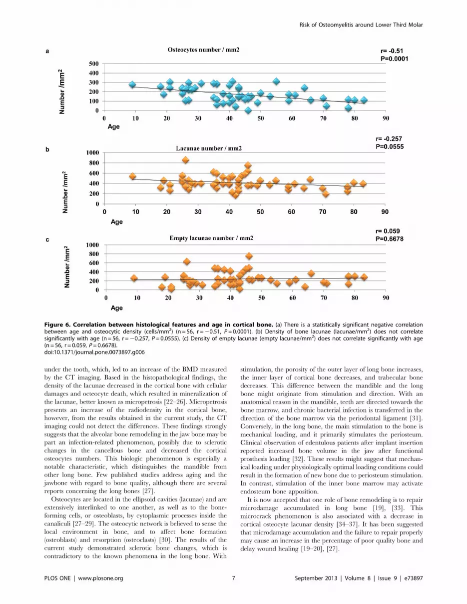
HistomorphometryHistomorphometric analysis of the osteocytic density in the
cortical bone surrounding the third molar tooth revealed age-
dependent relations. The mean osteocytic density was 170.7682.2
cells/mm2. The correlation between age and osteocytic density in
mandibular cortical bone was statistically significant (r = 20.51,
P,0.0001; Figure 6a). The mean density of lacunae was
413.16130 lacunae/mm2. Although there was a weak negative
correlation between age and the density of lacunae, it was not
statistically significant (r = 20.257, P = 0.056; Figure 6b). The
mean density of the empty lacunae was 242.56146 empty
lacunae/mm2. There was no correlation between age and the
density of the empty lacunae (r = 0.059, P = 0.668; Figure 6c).
The osteocyte numbers for all age categories are shown in
Figure 7a. In brief, the young generation had 223.4657.2 cells/
mm2, the middle age group had 163.1683.1 cells/mm, and the
old generation was 109.0665.2 cells/mm2. There were statistically
significant differences between each cancellous bone groups
(ANOVA and Bonferroni correction, *P,0.05, **P,0.01;
Figure 7a). The mean lacunae densities for the three age categories
are shown in Figure 7b. The young generation had 433.06140
lacunae/mm2, the middle age group had 427.16131 lacunae/
mm2, and the old generation had 339.5683.0 lacunae/mm2.
There were no statistically significant differences (P = 0.139). The
mean density of the empty lacunae for the three age categories are
shown in Figure 7c. The young generation had 209.66140 empty
lacunae/mm2, the middle age group had 264.06161 empty
lacunae/mm2, and the old generation had 230.5696.4 empty
lacunae/mm2. There were no statistically significant differences
(P = 0.470).
Discussion
In the present study, the characteristics of the bone surrounding
the third molar tooth were investigated quantitatively with both
CT imaging and histopathology. In the outermost layer of the
cortical bone, the osteocyte density and the density of the bone
lacunae had decreased. In contrast, it seemed that the endosteum
formed new bone into the bone marrow for the cancellous bone
Figure 5. The distribution of three age categories of pathological features. The distribution of three age categories of pathological features.Old generation shows 100% focal sclerotic osteomyelitis histopathologically, whereas young generation shows relatively few osteomyelitis.doi:10.1371/journal.pone.0073897.g005
Risk of Osteomyelitis around Lower Third Molar
PLOS ONE | www.plosone.org 6 September 2013 | Volume 8 | Issue 9 | e73897
under the tooth, which, led to an increase of the BMD measured
by the CT imaging. Based in the histopathological findings, the
density of the lacunae decreased in the cortical bone with cellular
damages and octeocyte death, which resulted in mineralization of
the lacunae, better known as micropetrosis [22–26]. Micropetrosis
presents an increase of the radiodensity in the cortical bone,
however, from the results obtained in the current study, the CT
imaging could not detect the differences. These findings strongly
suggests that the alveolar bone remodeling in the jaw bone may be
part an infection-related phenomenon, possibly due to sclerotic
changes in the cancellous bone and decreased the cortical
osteocytes numbers. This biologic phenomenon is especially a
notable characteristic, which distinguishes the mandible from
other long bone. Few published studies address aging and the
jawbone with regard to bone quality, although there are several
reports concerning the long bones [27].
Osteocytes are located in the ellipsoid cavities (lacunae) and are
extensively interlinked to one another, as well as to the bone-
forming cells, or osteoblasts, by cytoplasmic processes inside the
canaliculi [27–29]. The osteocytic network is believed to sense the
local environment in bone, and to affect bone formation
(osteoblasts) and resorption (osteoclasts) [30]. The results of the
current study demonstrated sclerotic bone changes, which is
contradictory to the known phenomena in the long bone. With
stimulation, the porosity of the outer layer of long bone increases,
the inner layer of cortical bone decreases, and trabecular bone
decreases. This difference between the mandible and the long
bone might originate from stimulation and direction. With an
anatomical reason in the mandible, teeth are directed towards the
bone marrow, and chronic bacterial infection is transferred in the
direction of the bone marrow via the periodontal ligament [31].
Conversely, in the long bone, the main stimulation to the bone is
mechanical loading, and it primarily stimulates the periosteum.
Clinical observation of edentulous patients after implant insertion
reported increased bone volume in the jaw after functional
prosthesis loading [32]. These results might suggest that mechan-
ical loading under physiologically optimal loading conditions could
result in the formation of new bone due to periosteum stimulation.
In contrast, stimulation of the inner bone marrow may activate
endosteum bone apposition.
It is now accepted that one role of bone remodeling is to repair
microdamage accumulated in long bone [19], [33]. This
microcrack phenomenon is also associated with a decrease in
cortical osteocyte lacunar density [34–37]. It has been suggested
that microdamage accumulation and the failure to repair properly
may cause an increase in the percentage of poor quality bone and
delay wound healing [19–20], [27].
Figure 6. Correlation between histological features and age in cortical bone. (a) There is a statistically significant negative correlationbetween age and osteocytic density (cells/mm2) (n = 56, r = 20.51, P = 0.0001). (b) Density of bone lacunae (lacunae/mm2) does not correlatesignificantly with age (n = 56, r = 20.257, P = 0.0555). (c) Density of empty lacunae (empty lacunae/mm2) does not correlate significantly with age(n = 56, r = 0.059, P = 0.6678).doi:10.1371/journal.pone.0073897.g006
Risk of Osteomyelitis around Lower Third Molar
PLOS ONE | www.plosone.org 7 September 2013 | Volume 8 | Issue 9 | e73897

In the jaw bone, and particularly in the third molar area, the
decrease in the number of osteocytes and lacunae may be a result
of the damage response against bacterial infection (from intraoral
microorganisms to the outer cortical bone) than microdamage,
which is related to load transmission. In this study, there was no
clear evidence of microcracks in these decalcified specimens.
Future investigation of undecalcified specimens is needed to detect
microdamage in the mandible.
A number of authors have reported that age is a consistent
factor in the determination of surgical difficulty at the removal of
third molar teeth, considering the differences in bone density
associated with age [1–4]. The positive correlation may be related
to an increase in bone density, which may require more handling
during the operation. It can be easily speculated that the sclerotic
bone might make removal of the third molar teeth more difficult.
Moreover, the low density of osteocytes and reduced vascularity
might negatively influence postoperative bone healing. From the
obtained results, it can be suggested that considerable patholog-
ically osteomyelitis condition around impacted tooth exist in the
aged patients. Osteomyelitis of the jaw could be considered as an
inflammatory condition of the bone, beginning in the medullar
cavity and harvasian systems and expanding to involve the
periosteum of the affected area [38]. It has been known that acute
and chronic osteomyelitis of the jaw is caused mostly by a bacterial
focus (odontgenic disease, pulpal and periodontal infection,
extraction wounds, foreign bodies, and infected fractures) [38].
In this study, these clinical conditions were representing chronic
non-suppurative inflammation, which were pathologically chronic
focal sclerotic osteomyelitis of the jaw bone.
Possible mechanisms of these pathological changes are that
viable bone would be damaged and transform to a non-viable
bone condition with chronic bacterial infection. Osteocyte death
could induce micropetrosis in the lacunae and these tissue reaction
cascades induce micro bone consolidation. However, the density
of the empty lacunae did not reflect its numbers particularly in
osteomyelitis samples as shown in Figure 6c. This reflects reactive
osteogenesis due to osteomyelitis induced by bacterial infection
and these pathological conditions might increase apparent bone
lacunae with inflammatory cell infiltration. Further, bacterial
infection could result in sclerotic osteomyelitis with bone
consolidation and reducing osteocytes cell numbers.
Conclusively, with an apparent low-grade bacterial infection,
the alveolar bone around third molar tooth demonstrated sclerotic
change of the cancellous bone and considerable death of the
osteocyte in the cortical bone with micropetrosis evaluated by CT
imaging and histopathology (Figure 8). It can be suggested that the
bone quality is not solely indicated by refraction of the mineralized
tissue, but might be related to the viable cellular activity and
influence the healing process of bony wounds. The bone which
contains enough viable cells is thought to present good bone
quality and the bone with reduced cell numbers might be
considered ‘bad’.
Within the limitation of the current study, it can be suggested
that at the time of third molar tooth extraction, if the alveolar bone
Figure 7. Three age categories of histological features of cortical bone. (a) Three age categories of osteocytic density (cells/mm2). There arestatistically significant differences (ANOVA and Bonferroni correction, *P,0.05, **P,0.01). (b) Three age categories of bone lacunae density (lacunae/mm2). There are no statistically significant differences (ANOVA and Bonferroni correction, P = 0.139). (c) Three age categories of empty bone lacunaedensity (empty lacunae/mm2). There are no statistically significant differences (ANOVA and Bonferroni correction, P = 0.470).doi:10.1371/journal.pone.0073897.g007
Risk of Osteomyelitis around Lower Third Molar
PLOS ONE | www.plosone.org 8 September 2013 | Volume 8 | Issue 9 | e73897

shows sclerotic change in an aged population, the removal of
impacted teeth should not be considered as simple tooth
extraction, but it must be regarded as treatment of sclerotic
osteomyelitis around the teeth.
Acknowledgments
We thank Professor Tatsuro Tanaka for help with CT analysis.
Author Contributions
Conceived and designed the experiments: IM TT. Performed the
experiments: IM. Analyzed the data: AI YM. Contributed reagents/
materials/analysis tools: IM. Wrote the paper: IM.
References
1. Queral-Godoy E, Valmaseda-Castellon E, Berini-Aytes L, Gay-Escoda C (2005)
Incidence and evolution of inferior alveolar nerve lesions following lower third
molar extraction. Oral Surg Oral Med Oral Pathol Oral Radiol Endod. 99:259–264.
2. Valmaseda-Castellon E, Berini-Aytes L, Gay-Escoda C (2001) Inferior alveolar
nerve damage after lower third molar surgical extraction: a prospective study of
1117 surgical extractions. Oral Surg Oral Med Oral Pathol Oral Radiol Endod.92: 377–383.
3. Akadiri OA, Obiechina AE (2009) Assessment of difficulty in third molar
surgery–a systematic review. J Oral Maxillofac Surg. 67: 771–774.
4. Peterson LJ (2003) Principles of management of impacted teeth. In: Ellis E,
Hupp JR, Tucker MR, editors. Contemporary oral and maxillofacial surgery.
4th edition. St. Louis, Missouri: Mosby 184–213.
5. Albrektsson T, Branemark PI, Hansson HA, Lindstrom J (1981) Osseointegratedtitanium implants. Requirements for ensuring a long-lasting, direct bone-to-
implant anchorage in man. Acta Orthop Scand 52: 155–170.
6. Albrektsson T (2002) Biologic and bioengineering considerations for prescribing
prosthetic implants. In: Zarb GA, Lekholm U, Albrektsson T, Tennenbaum H,editors. Aging, Osteoporosis, and Dental Implants. Chicago: Quintessence books
15–16.
7. Lekholm U, Zarb G (1987) Patient selection and preparation. In: Branemark PI,
Zarb G, Albrektsson T, editors. Tissue-integrated prostheses. Chicago:
Quintessence books 199–209.
8. Watzek G, Ulm C (2002) Compromised alveolar bone quality in edentulous
jaws. In: Zarb GA, Lekholm U, Albrektsson T, Tennenbaum H, editors. Aging,Osteoporosis, and Dental Implants. Chicago: Quintessence books 67–84.
9. Quirynen M, Mraiwa N, Van Steenberghe D, Jacobs R (2003) Morphology anddimensions of the mandibular jaw bone in the interforaminal region in patients
requiring implants in the distal areas. Clin Oral Implants Res 14: 280–285.
10. Shapurian T, Damoulis PD, Reiser GM, Griffin TJ, Rand WM (2006)Quantitative evaluation of bone density using the Hounsfield index. Int J Oral
Maxillofac Implants 21: 290–97.
11. Shahlaie M, Gantes B, Schulz E, Riggs M, Crigger M (2003) Bone densityassessments of dental implant sites: 1. Quantitative computed tomography.
Int J Oral Maxillofac Implants 18: 224–231.
12. Friberg B, Sennerby L, Roos J, Lekholm U (1995) Identification of bone quality
in conjunction with insertion of titanium implants. A pilot study in jaw autopsy
specimens. Clin Oral Implants Res 6: 213–219.
13. Miyamoto I, Tsuboi Y, Wada E, Suwa H, Iizuka T (2005) Influence of cortical
bone thickness and implant length on implant stability at the time of surgery–clinical, prospective, biomechanical, and imaging study. Bone 37: 776–780.
14. Osteoporosis prevention, diagnosis, and therapy. (2001) NIH Consensus
Development Panel on Osteoporosis Prevention, Diagnosis, and Therapy.JAMA 285: 785–795.
15. Currey JD (2002) Bones: structure and mechanics. Princeton: Princeton
University Press. 54–122.
Figure 8. Schematic illustration of the formation mechanisms of the chronic focal bone consolidation. Schematic illustration of theformation mechanisms of the chronic focal osteomyelitis. The chronic bacterial infection around tooth would induce tissue damage and it resulted inosteocytes death and resulted in micropetrosis. These accumulation of micropetrosis might induce bone sclerosis. Red arrow head shows cancellousbone consolidation.doi:10.1371/journal.pone.0073897.g008
Risk of Osteomyelitis around Lower Third Molar
PLOS ONE | www.plosone.org 9 September 2013 | Volume 8 | Issue 9 | e73897

16. Lettry S, Seedhom BB, Berry E, Cuppone M (2003) Quality assessment of the
cortical bone of the human mandible. Bone 32: 35–44.17. ASA Physical Status Classification System. (2009) Available: http://www.asahq.
org/for-members/clinical-information/asa-physical-status-classification-system.
aspx.18. Kassolis JD, Scheper M, Jham B, Reynolds MA (2010) Histopathologic findings
in bone from edentulous alveolar ridges: a role in osteonecrosis of the jaws? Bone47: 127–130.
19. Mullender MG, Tan SD, Vico L, Alexandre C, Klein-Nulend J (2005)
Differences in osteocyte density and bone histomorphometry between men andwomen and between healthy and osteoporotic subjects. Calcif Tissue Int 77:
291–296.20. Qiu S, Rao DS, Fyhrie DP, Palnitkar S, Parfitt AM (2005) The morphological
association between microcracks and osteocyte lacunae in human cortical bone.Bone 37: 10–15.
21. McGraw KO, Wong SP (1996) Forming inferences about some intraclass
correlations coefficients: correction. Psychol Methods 1: 390.22. Frost HM (1960) Micropetrosis. J Bone Joint Surg Am 42-A: 144–150.
23. Boyde A, Maconnachie E, Reid S, Delling G, Mundy GR (1986) Scanningelectron microscopy in bone pathology: review of methods, potential and
applications. Scan Electron Microsc (Pt 4), 4: 1537–1554.
24. Boyde A, Hendel P, Hendel R, Maconnachie E, Jones SJ (1990) Human cranialbone structure and the healing of cranial bone grafts: a study using backscattered
electron imaging and confocal microscopy. Anat Embryol 181: 235–251.25. Vashishth D (2004) Rising crack-growth-resistance behavior in cortical bone:
implications for toughness measurements. J Biomech 37: 943–946.26. Noble BS (2008) The osteocyte lineage. Arch Biochem Biophys 473: 106–111.
27. Busse B, Djonic D, Milovanovic P, Hahn M, Puschel K, Ritchie RO, Djuric M,
Amling M (2010) Decrease in the osteocyte lacunar density accompanied byhypermineralized lacunar occlusion reveals failure and delay of remodeling in
aged human bone. Aging Cell 9: 1065–1075.
28. Doty SB (1981) Morphological evidence of gap junctions between bone cells,
Calcif Tissue Int 33: 509–512.
29. Donahue HJ (2000) Gap junctions and biophysical regulation of bone cell
differentiation. Bone 26: 417–422.
30. Seeman E, Delmas PD (2006) Bone quality–the material and structural basis of
bone strength and fragility. N Engl J Med 354: 2250–2261.
31. Krakowiak PA (2011) Alveolar osteitis and osteomyelitis of the jaws. Oral
Maxillofac Surg Clin North Am 23: 401–413.
32. Sennerby L, Carlsson GE, Bergman B, Warfvinge J (1988) Mandibular bone
resorption in patients with tissue-integrated prostheses and in complete-denture
wearers. Acta Odontol Scand 46: 135–140.
33. Bartold PM, Kuliwaba JS, Lee V, Shah S, Marino V, Fazzalari NL (2011)
Influence of surface roughness and shape on microdamage of the osseous surface
adjacent to titanium dental implants. Clin Oral Implants Res 22: 613–618.
34. Grynpas MC (2002) The concept of bone quality in osteoporosis. In: Zarb GA,
Lekholm U, Albrektsson T, Tennenbaum H, editors. Aging, Osteoporosis, and
Dental Implants. Chicago: Quintessence books. 25–34.
35. Vashishth D, Verborgt O, Divine G, Schaffler MB, Fyhrie DP (2000) Decline in
osteocyte lacunar density in human cortical bone is associated with accumulation
of microcracks with age. Bone 26: 375–380.
36. O’Brien FJ, Taylor D, Lee TC (2003) Microcrack accumulation at different
intervals during fatigue testing of compact bone. J Biomech 36: 973–980.
37. Najafi AR, Arshi AR, Eslami MR, Fariborz S, Moeinzadeh MH (2007)
Micromechanics fracture in osteonal cortical bone: a study of the interactions
between microcrack propagation, microstructure and the material properties.
J Biomech 40: 2788–2795.
38. Baltensperger MM, Eyrich G (2010) Osteomyelitis of the jaws: Definition and
classification. In: Baltensperger MM, Eyrich GK, editors. Osteomyelitis of the
jaws. Berlin Heidelberg: Springer-Verlag 5–56.
Risk of Osteomyelitis around Lower Third Molar
PLOS ONE | www.plosone.org 10 September 2013 | Volume 8 | Issue 9 | e73897


