Embed Size (px)
Citation preview
Pre-operative Evaluations for DBS in Dystonia
Stephane Thobois, MD, PhD,1* Takaomi Taira, MD,2 Cynthia Comella, MD,3 Elena Moro, MD, PhD,4
Susan Bressman, MD, PhD,5 and Alberto Albanese, MD, PhD6
1Universite Lyon I, Hospices Civils de Lyon, Hopital Neurologique Pierre Wertheimer and CNRS, UMR 5229, Lyon, France2Department of Neurosurgery, Tokyo Women’s Medical University, Tokyo, Japan
3Department of Neurological Sciences, Rush University Medical Center, Chicago, Illinois, USA4Movement Disorders Center, TWH, UHN, Division of Neurology, University of Toronto, Toronto, Ontario, Canada
5Department of Neurology, Beth Israel Medical Center, New York, New York, USA6Fondazione IRCCS Istituto Neurologico Carlo Besta and Universita Cattolica del Sacro Cuore, Milan, Italy
ABSTRACT:Background: The preoperative evaluation in dystoniaaims at characterizing the severity and topography ofmotor symptoms in patients, who have previously beenselected for deep brain stimulation (DBS). Methods:The literature search was performed using PubMed,CINAHL, and the Cochrane Collaborative databases.Results: Commonly used scales for clinical assessmentare the Burke-Fahn-Marsden dystonia rating scale forgeneralized dystonia and the Toronto Western Spas-modic Torticollis Scale for cervical dystonia. Motorassessment is completed by quality of life and func-tional scales, such as the Short-Form Health Survey(SF-36) or the Parkinson’s Disease Questionnaire 39.
Validated rating scales for cranial or upper limb dysto-nia are lacking. Discussion: In common clinical prac-tice, these outcome measures can be administered inan open-label fashion because double blind assess-ment is only required for ascertaining new treatmentindications or research purposes. The same measuresare to be used postoperatively to revaluate outcome af-ter DBS. Brain MRI is required to confirm diagnosis andassess structural abnormalities. Other imaging techni-ques, particularly functional imaging, are used forresearch purposes. VC 2011 Movement Disorder Society
Key Words: DBS; dystonia
Introduction
Preoperative evaluation is a crucial step in the man-agement of patients with dystonia who are candidatefor deep brain stimulation (DBS). Issues related to inclu-sion and exclusion criteria for DBS surgery have beendetailed in a previous article1 of this supplement. Beforeentering preoperative workup, each patient should beclassified along with the three axes of aetiology, age ofonset, and spread of dystonia;2 this will allow identify-ing the most appropriate tools for assessment. Preopera-tive evaluation aims at characterizing the severity andtopography of motor symptoms and their impact on
activities of daily living (ADLs) and social activities andprovides a baseline reference for mid- and long-termpostoperative evaluations. The quality and accuracy ofthe preoperative assessment and the choice of assess-ment tools is crucial as will affect all subsequent post-operative comparisons. The preoperative phase alsoincludes a number of steps related to the assessment ofthe surgical risk and the determination of the surgicaltrajectory. This article will review the evidence on theapplication and evaluation of clinical scales to be usedfor preoperative and postoperative evaluation ofpatients undergoing DBS for dystonia.
Methods
Search Strategy
The literature search was performed using PubMed,CINAHL, and the Cochrane Collaborative databases ini-tially from 1980 to January 2008 using the terms: dysto-nia and DBS; pallidal stimulation and dystonia;subthalamic stimulation and dystonia; thalamic stimula-tion and dystonia; secondary dystonia and DBS;
------------------------------------------------------------* Correspondence to: Stephane Thobois, Hopital Neurologique PierreWertheimer, Neurologie C, 59 Bd Pinel, 69677 Lyon, France; [email protected]
Potential conflict of interest: Nothing to report.
Received: 24 April 2010; Revised: 21 August 2010; Accepted: 21September 2010Published online in Wiley Online Library (wileyonlinelibrary.com).DOI: 10.1002/mds.23481
S U P P L E M E N T
Movement Disorders, Vol. 26, No. S1, 2011 S17
neurodegenerative diseases and DBS. The search wascombined with the one used for neuropsychology, neuro-psychiatry, microelectrode recording, neuroimaging, elec-trophysiology, surgical techniques, complications, andtargeting. Only English-language publications involvinghuman subjects’ were considered. A total of 235 articleswere retrieved. To facilitate the committees’ work, thearticles were divided in three groups, which often over-lapped: preoperative, intraoperative, and postoperative.A PDF file was created for each article obtained from thesearch and put in a CD that was mailed to the members.During the writing phase, additional 71 articles wereadded to update the search, covering the period from Jan-uary 2008 to September 2009.
Process of Generating ClinicalRecommendations
The Consensus Committee members of the Task Forceincluded neurologists, neurosurgeons, neurophysiolo-gists, psychiatrists, neuropsychologists, nurses, and mid-level practitioners with expertise and experience in DBS.The experts were also chosen from different countries inAsia, Europe, North and South America, to provide amore comprehensive contribution to the Task Force. Theauthors of each article were selected taking into accounttheir specific expertise in the field. The steering commit-tee prepared a list of questions related to preoperative,intraoperative, and postoperative issues and establishedtwo chairs responsible for each of these three areas (sub-committees). These chairs then assigned a few questionsto be addressed by each member of the subcommittees.The answers to the questions had to be formulated afterreviewing the available literature (provided on CD) andcombining their expertise. As the level of evidence formost of the DBS studies was low, the responses wereorganized following the template previously used for theSpecial Supplement on DBS for Parkinson’s disease (PD):(1) available data, (2) conclusions, (3) pragmatic recom-mendations, and (4) points to be addressed.3 A first docu-ment was prepared from this initial work and wasreviewed and discussed by the entire Task Force groupduring a one-day meeting. During this meeting, the TaskForce members provided further feedback and agreed onadditional refinements of the whole document adding thecomments and remarks collected during the meeting. Spe-cial attention was paid to formulate pragmatic recommen-dations in absence of available studies. A second version ofthe project was sent to the entire working committee forfinal approval. The Executive Committee then met againto refine the Special Issue document before submission.
Methods of Assessments
Descriptions and Interest of the DifferentScales for Dystonia
Motor Scales. Motor scales for dystonia have been theobject of a number of publications, encompassing
descriptions of rating instruments and validation stud-ies.4–11 However, none of the scales fulfils all the rec-ommended criteria for health measurement ratingscales defined by the Scientific Advisory Group of theMedical Outcomes Trust (SAC).12 These criteriainclude: conceptual and measurement model; reliabil-ity; validity; responsiveness; interpretability; respond-ent and administrative burden; alternate forms;cultural and language adaptations. In particular, noneof the scales that will be described below has beenspecifically designed to assess responsiveness to atreatment.13 Nevertheless, several controlled studieshave demonstrated that these scales are able to detectsignificant improvement in dystonic patients under-going different treatments. This is the case, for exam-ple, for botulinum toxin type A in cervical dystonia(CD), whose efficacy was demonstrated using specificscales in numerous class I studies14 (using the classifi-cation proposed by the American Academy of Neurol-ogy).15 In addition, the efficacy of DBS in dystoniacould also be assessed in one class I study16 and infive class III studies.17–21 These trials provide a cleardemonstration that dystonia can be assessed usingobjective measures.
Generalized/Segmental Dystonia. The Burke–Fahn–Marsden dystonia rating scale (BFMDRS)4 was intro-duced to assess generalized dystonia patients. It iscomposed of a motor part assessing dystonia and apart assessing the resulting disability. The motor sub-scale evaluates two clinical features of dystonia (sever-ity and provoking factors) in eight body regions (eyes,mouth, neck, and the four limbs) and one functionalarea (speech and swallowing). Severity ranges from 0(no dystonia) to 4 (severe dystonia). The provokingfactors assess the situation under which dystoniaoccurs and range from 0 (no dystonia) to 4 (dystoniaat rest). These two features, severity and provokingfactors, are multiplied and then scores are summed,except for the eyes mouth and neck which are halvedbefore summing as they are considered regions of‘‘lower weight.’’ The resulting maximum total scoreon the BFM severity is 120.4 The BFMDRS wasclearly designed to assess patients with severe general-ized dystonia and has limitations when applied tomilder or nongeneralized cases. These include the factthat arms and legs are given one rating each, withoutdistinguishing proximal and distal components, thecombination of functional features (such as speechand swallowing) with the inspection of dystonia inother body regions, and the arbitrary reduction of theweight in the cranial/cervical region.The BMFDRS clinimetric properties were assessed
in a study of 10 patients with dystonia rated by fourdifferent examiners: the overall reliability, inter-rateragreement, and concurrent validity were demonstrated
T H O B O I S E T A L .
S18 Movement Disorders, Vol. 26, No. S1, 2011
for the BMFDRS total score but not analyzed for eachdifferent body regions and area of function.4 After thefirst encouraging effort, the BFMDRS was not furthersystematically developed and tested as a multicenterinstrument.The BFMDRS section on disability assesses the
effects of dystonia on ADL (speech, handwriting, feed-ing, eating/swallowing, hygiene, dressing, and walk-ing), and the total maximum score is 30.The unified dystonia rating scale (UDRS) was
designed to overcome limitations of the BFMDRS. Itincludes a more detailed assessment of separate bodyareas with specific ratings for proximal and distallimbs, and does not mix bodily inspection with func-tional variables, such as speech and swallowing.9 Inaddition, the UDRS rates duration similarly to the du-ration factor previously validated for the TorontoWestern Spasmodic Torticollis Rating Scale(TWSTRS).9 Furthermore, the UDRS weights the dif-ferent body regions equally. Fourteen body areas areevaluated: eyes and upper face, lower face, jaw andtongue, larynx, neck, trunk, shoulder/proximal arm(right and left), distal arm/hand (right and left), proxi-mal leg (right and left), and distal leg/foot (right andleft). For each of these, the UDRS requires rating theseverity and duration. Severity rating is specific foreach body region and varies from 0 (no dystonia) to 4(extreme dystonia); duration also ranges from 0 to 4and assesses whether dystonia occurs at rest or withaction, and whether it is predominantly of maximal orsub maximal intensity. The total UDRS score is thesum of the severity and duration factors, with a maxi-mum total of 112. The severity score is expressed as apercentage of the maximum amplitude of the physio-logical movement, which indicates that this, as allother dystonia scales, is more appropriate to rate mo-bile dystonia versus fixed posturing.The global dystonia rating scale (GDS) evaluates the
severity of dystonia in the same 14 body areas as theUDRS.9 The GDS is a Likert-type scale with ratings of0–10 (from 0, no dystonia, to 10, severe dystonia).There are no modifying factors in the GDS, and thetotal score is the sum of all the body area scores witha maximum of 140. The GDS is a very simple scalethat allows a quick rating of dystonia but does notgive precise indications about its clinical aspects (mo-bile vs. fixed; disability. . .). On the other hand eachbody part has a similar weight, which has the advant-age not to minimize any features of dystonia. Theother advantage of this scale is its ease of use.A comparison of the internal consistency and reli-
ability of the BFMDRS, UDRS, and GDS was per-formed by 25 dystonia experts using a standardizedvideotape protocol.9 All three scales showed excellentinternal consistency and good correlation amongraters. The inter-rater agreement was excellent being
lowest for eyes, jaw, face, and larynx. There washigher inter-rater consistency for motor severity thanfor the ratings of modifying factors (duration in theUDRS and provoking factors in the BFM). Seventy-four percent of the raters found the GDS the easiest toapply against 38% for the BFM and only 5% for theUDRS.9 A recent study showed that the UDRS andBFM scales provide similar accuracy and reliability toassess the consequences of DBS in dystonia.22
The global outcome scale (GOS) scores the globalimprovement of the dystonia after a therapeutic inter-vention. The improvement is rated from 4 (marked) to0 (no effect).23 The GOS is a very simple but impre-cise scale that does not differentiate the improvementof each body part. Because of these limitations thescale is rarely used.23
For tardive dyskinesia, which encompasses dystoniaand other movement disorders (particularly chorea,myoclonus, and tremor), composite scales are moreappropriate, such as the abnormal involuntary move-ment scale (AIMS) or the extrapyramidal symptomsrating scale (ESRS).5–6 The ESRS is divided into foursubscales and four clinical global impression severitysubscales. These consist in a questionnaire of drug-induced extrapyramidal symptoms, an examination ofparkinsonism and akathisia, an examination of dysto-nia, an examination of dyskinesia, and a clinicalglobal impression severity scales for tardive dyskinesia,parkinsonism, dystonia, and akathisia.6 The AIMScontains seven items assessing the severity of abnormalmovements in different body locations. This scale alsoincludes a global judgment of the severity, consequen-ces, and patient’s awareness of abnormal movements.It has been observed that the ESRS and the AIMShave a high degree of concordance.10
Cervical Dystonia. The Tsui Torticollis Rating Scalewas the first rating scale specifically designed for CD.7
It contains six items and is designed for video assess-ment. This scale evaluates the amplitude and durationof neck involuntary movements in the neck, elevationof shoulder, and head tremor.The TWSTRS9 was developed to provide clinical
investigators with a better instrument to assess the se-verity and disability of CD, which is the most com-mon form of focal or segmental dystonia. TheTWSTRS was developed in 1990 and consists of 22items. The total TWSTRS is comprised of three sepa-rate subscales: motor severity, disability, and pain dueto CD. The motor severity scale consists of 10 itemsassessing the severity of head posture in several axesof movement (turning, tilting, anterocollis, retrocollis,and shoulder elevation), the effect of sensory tricks,range of motion, and duration of dystonia. The scorefor motor severity subscale ranges from 0 (no symp-toms) to 35 (severe CD). The TWSTRS subsection for
P R E O P E R A T I V E E V A L U A T I O N S
Movement Disorders, Vol. 26, No. S1, 2011 S19
motor severity has been validated for inter-rater reli-ability and validity and a teaching tape has beendeveloped to ensure consistency across raters for mul-ticenter trials.8,24 The disability subscale consists ofseven items assessing the effect of CD on work per-formance, ADLs, driving, reading, watching television,conducting activities outside home, and social embar-rassment. The maximal score for the disability sub-scale is 32. The pain subscale consists of five items toassess CD related pain at its maximal, minimal andusual level, and to indicate the duration of pain duringa day, and disability due to pain. The maximum scorefor the pain subscale is 20. The total TWSTRS is thesum of the three subscale scores, with a maximumvalue of 87. The total TWSTRS has been used exten-sively as an outcome variable in clinical trials of phar-macological and surgical interventions.25–32
It has been shown that there is a good correlationbetween the scores obtained with the TWSTRS andthe Tsui scale.33 The metric properties of the totalTWSTRS and of severity subscales were investigated.Factor analysis showed that 18 of the 22 items of thetotal TWSTRS fall into three clinically distinct and rel-evant factors: (1) motor severity, (2) disability, and (3)pain.8 These domains correspond to the three sub-scales of the total TWSTRS, and each measures a sep-arate aspect of CD. The item for social embarrassmentdid fall in any factor as well as three additional items(sensory trick, lateral and sagittal shift).8 There aretwo possible explanations for this inconsistency. First,the range of scores available for these items is limitedto absence/presence (lateral and sagittal shift) or to 0–2 (sensory tricks). Second, it has been observed thatthe observation of sensory tricks is a clinical featurerelevant to the diagnosis rather than to clinical signs.Furthermore, the TWSTRS does not clearly assess dys-tonic tremor, as well as complex combination of pha-sic and tonic dystonic features.
Focal Dystonias. The clinical evaluation of focal dys-tonias is often difficult.A scale of 0 (normal) to 4 (worst) has been
proposed to rate the severity of blepharospasm andoromandibular dystonia, but the inter-rater reproduci-bility was poor.34,35 In a recent study, the metricproperties of the Jankovic Rating Scale (JRS) and aself-rating patient response outcome scale (the Ble-pharospasm Disability Index, BSDI) have been com-pared in blepharospasm patients.36 The internalconsistency and retest reliability of the BSDI weregood and the scores obtained using both scales werewell correlated. Therefore, these authors suggest thatJRS and BSDI can both be used to reliably assess ble-pharospasm in treatment trials.For task-specific dystonias, the writer’s cramp rating
scale (WCRS) was developed for patients with writer’s
cramp.37 The WCRS is divided into three subscales,respectively studying the dystonic posture, the latencyfor dystonia to occur, and the presence of writingtremor.26 Although this scale is easy to implement andhas sufficient inter-rater reliability it remains largelyunused.The main characteristics of the above mentioned
scales have been summarized in Table 1.
Quality of Life Scales. The assessment of quality oflife (QoL) is crucial to determine the impact of thesurgery on ADL. Most studies assessing this outcomemeasure have used the Short-Form Health Survey (SF-36) or the PD Questionnaire 39 (PDQ-39).38–41 TheSF-36 scale assesses the general and mental health, thephysical and social functioning, the physical and emo-tional roles, and the pain and vitality.42 The scores oneach subscale are comprised between 0 (worst) to 100(best). The PDQ-39 scale was originally designed forPD43 but has also been used for dystonia. It is dividedinto seven sections: mobility, ADL, emotional well-being, stigma, cognition, communication, and bodilydiscomfort.The CD Impact Profile (CDIP-58) has been devel-
oped for CD. It measures the health impact of the dis-ease from patient’s perceptions.44 This scale is dividedinto eight sections (head and neck symptoms, painand discomfort, upper limb activities, walking, sleep,annoyance, mood, and psychosocial functioning). Thiscomposite scale is more sensitive in measuring thefunctional outcome of a treatment, such as botulinumtoxin, than the SF-36 or TWSTRS.45 However, its usehas not gained wide diffusion.
Conclusions
For generalized and CD, the two most accepted andused rating scales are the BFMDRS and TWSTRS,respectively. For other focal dystonias, there are nogenerally agreed upon scales. The currently availablerating scales have several limitations. The BFMDRSscale uses weighting factors that can minimize the realimpact of eyes, mouth, and neck dystonia. In addition,other associated movement disorders, such as tremoror myoclonus, are not considered in most of the avail-able dystonia scales. Moreover, the available currentscales do not sufficiently discriminate mobile (phasic)dystonic movements from fixed (tonic) dystonicpostures.
Pragmatic Recommendations
The features of dystonia should be monitored beforeDBS using the most appropriate among the availabledystonia scales. The choice of which scale to useshould depend upon the type of dystonia, according totopography rather than aetiology. For generalized dys-tonia, it is recommended to use the total BFMDRS,
T H O B O I S E T A L .
S20 Movement Disorders, Vol. 26, No. S1, 2011
which may not always be appropriate for focal dysto-nias. As an alternative, the GDS provides a rapidassessment and is easily applied, although it has beenused less frequently than the BFMDRS. The UDRSmay also be used, although its implementation is moredifficult. For CD, the TWSTRS, including subscalesfor severity, disability, and pain, is recommended.These scales have been designed to assess patientswith primary dystonia and do not always capturecomplex dystonia phenotypes, such as those observedin dystonia-plus or in secondary dystonias.Given these limitations, it is recommended that a
limited number of expert evaluators be charged to ratepatients with dystonia and that standardized videosare performed during each assessment.4
The impact of surgery on QoL is a crucial issue thatmay provide outcome results divergent from the motorassessment.
Points to Be Addressed
New more comprehensive scales for dystonia shouldbe developed: they should also accurately measuretonic postures and phasic movements. Finally, there isa need for uniform training for the BFMDRS andUDRS. Uniform training is available for the TWSTRS,
although it has not been shown whether such trainingimproves inter-rater reliability. For other focal dysto-nias, although several scales exist, their internal con-sistency and reliability have been poorly studied andtheir use remains incidental. Thus, there is a clearneed for specific scales that objectively quantify theeffect of DBS in focal dystonia.
Clinical Use of the Scales for Dystonia
Should Standardized Evaluation Be PerformedPreoperatively and Postoperatively? How?When?
Motor Assessment. Postoperative objective and subjec-tive assessments have been compared with the preop-erative condition in a number of publications, en-compassing clinical series, case control studies, cohortstudies, and single case reports.16–20,32,38,41,46–82 Thereare only six controlled trials that evaluate the effectsof GPi DBS in a blinded fashion (one class I levelstudy16 and five class III studies17–21). These trials pro-vide a clear demonstration of the benefit of DBS forthe primary generalized and tardive dystonias andalso for CD.16–20 Favorable outcome has also beenreported for PKAN.46 In these studies a videotapedassessments scored by independent blinded raters
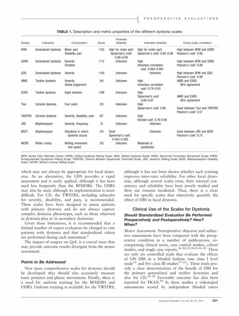
TABLE 1. Description and metric properties of the different dystonia scales
Scales Indication Composition Score
Intrarater
reliability Interrater reliability Cross scale correlation
BFM Generalized dystonia Motor part /150 High for motor part: High for motor part: High between BFM and UDRSDisability part Spearman’s coef:
0.98–0.99Spearman’s coef: 0.85–0.96 Pearson’s coef: 0.98
UDRS Generalized dystonia Severity /112 Unknown High High between BFM and UDRSDuration Intraclass correlation
coef: 0.994–0.997Pearson’s coef: 0.98
GDS Generalized dystonia Severity /140 Unknown Unknown High between BFM and GDSPearson’s coef: 0.98
AIMS Tardive dystonia Severity /40 Unknown High AIMS and ESRS:96% agreementGlobal judgement Intraclass correlation
coef: 0.79–0.93ESRS Tardive dystonia Eight sections /198 Unknown High
Spearman’s coef:0.80–0.97
AIMS and ESRS:96% agreement
Tsui Cervical dystonia Four parts /25 Unknown HighSpearman’s coef: 0.86 Good between Tsui and TWSTRS
Pearson’s coef: 0.57TWSTRS Cervival dystonia Severity, disability, pain /87 Unknown High
Kendall coef: 0.76-0.98JRS Blepharospam Severity, frequency /8 Unknown Unknown
BSDT Blepharospam Situations in whichdystonia occurs
/24 Good Unknown Good between JRS and BSDISpearman’s coef:0.453–0.595
Pearson’s coef: 0.73
WCRS Writer cramp Writing movementand speed
/30 Unknown Moderate tosubstantial
BFM, Burke Fahn Marsden Scale; UDRS, Unified Dystonia Rating Scale; GDS, Global Dystonia Scale; AIMS, Abnormal Involuntary Movement Scale; ESRS,Extrapyramidal Symptoms Rating Scale; TWSTRS, Toronto Western Spasmodic Torticollis Scale; JRS, Jankovic Rating Scale; BSDI, Blepharospasm DsabilityIndex; WCRS, Writer’s Cramp Rating Scale.
P R E O P E R A T I V E E V A L U A T I O N S
Movement Disorders, Vol. 26, No. S1, 2011 S21
allowed controlled evaluations of the effects of thesurgery.16–20 It is notable that data on the benefit ofDBS in dystonia reported by open studies are in keep-ing with the findings reported by controlled studies.A number of practical issues have been addressed by
the available studies. Preoperatively the assessment ismost often performed between the last month andthe last week preceding surgery.16–20,32,38,41,46–82 Thetime interval between surgery and the first post-operative evaluation is usually between 3 and 12months.16–20,32,38,46–82 Patient management of thedoes not require more frequent controls and the firstpreoperative evaluation is aimed at assessing the acuteeffects of stimulation on dystonia and the thresholdsfor stimulation-induced side effects. Most of the stud-ies have clearly shown that the improvement startswithin the first hours or days after beginning the stim-ulation, and then progresses. Most of the benefit isusually obtained after 3–6 months.16–20,32,38,46–82 Theimprovement first affects phasic dystonic movementsand later tonic postures.16 Some additional improve-ment can occur later but, usually, to a less extent andslower. Some studies, however, have shown an addi-tional 10–30% improvement of the dystonia between1 and 1.5 years.48,80–81 The postoperative outcomeswill be discussed in detail in another article on thissame issue.83
Quality of Life Assessment. The QoL assessment isusually performed when the patients have the preoper-ative motor assessment, that is, from 1 month to 1week before surgery.16–19,38–39,41 The interval betweensurgery and the postoperative evaluation of QoL isgenerally between 3 and 18 months.16–19,38–39,41 QoLusually improves significantly after GPi DBS in gener-alized and segmental dystonia and CD.16–19,38–39,41
Conclusions
Validated motor and disability scales are widelyused to assess patients before surgery in all the pub-lished studies. Most of the time evaluations have beendone in open label fashion.
Pragmatic Recommendations
Validated scales (see previous section) should beused to assess patients with dystonia within few weeksbefore surgery. The benefit should be evaluated at 3–6months after surgery and further evaluations shouldbe scheduled at yearly intervals. Videotaped assess-ments are recommended.
Points to Be Addressed
The ideal time-frame to assess the efficacy of DBS indifferent forms of dystonia needs to be better defined.It remains also to be specified if this should differ for
primary generalized or focal forms or for secondarydystonias.
Should Evaluation in the OFF StimulationCondition Be Performed in Routine orResearch Protocol? How Long and When?
Evaluations are rarely performed in OFF stimulationcondition. OFF stimulation condition has only beenassessed in three class III and in five class IV stud-ies.17–18,20,56,58,66,71,82 However, assessments withoutstimulation may provide important information on theimmediate effect of stimulation, the delay of reoccur-rence of the clinical signs and possibly further worsen-ing of preoperative motor conditions. OFF stimulationstudies thus allow better comparison with the preoper-ative motor condition and may show evidence ofunderlying disease progression.The duration of the stimulation wash-out period
preceding assessment may be variable. This has beenspecifically studied by Grips et al.,58 who showed thatmost of the phasic motor symptoms in patients withsegmental dystonia reoccurred within 4 hours afterswitching off bilateral GPi DBS, while the tonic signsmay take much longer to worsen. In the study ofVidailhet et al.17 on generalized dystonia, the maxi-mum tolerated duration of the OFF stimulation periodwas 7 hours. In a single case study in Lesch–Nyhandystonia, the stimulator could be switched off for 1months.71 By contrast, tardive dystonia and CD mayworsen very quickly after the stimulator is switchedoff.20,82 This indicates that the effects observed afterswitching off stimulation may depend on the etiologyof dystonia. Furthermore, it has to be taken intoaccount that severe worsening of dystonia may be lifethreatening in severe generalized cases; this can be pre-vented by careful observation of patients during thisperiod.
Conclusions
Evaluations in the OFF stimulation condition havebeen performed in few studies, which provide interest-ing data concerning the posteffect duration of DBS indystonia.
Pragmatic Recommendations
A reasonable duration of the OFF period may be ofaround 3–4 hr although this does not lead to theworst off condition. In routine clinical setting, OFFstimulation evaluation is not acceptable because of therisk of reoccurrence of severe dystonia manifestations.
Points to Be Addressed
It is unclear whether the time course of motor signsreoccurrence after DBS switch-off depends on the etiol-ogy of dystonia. This needs to be addressed by specificstudies.
T H O B O I S E T A L .
S22 Movement Disorders, Vol. 26, No. S1, 2011
Role of Imaging
Is There Any Role for Preoperative Imaging(Brain MRI, PET)?
Morphological Imaging: Conventional MR Imaging.Brain imaging is mandatory to determine the aetiologyof dystonia and should be performed before consider-ing any patient for surgical treatment.1 In primarydystonia, there are no major structural abnormalities,as seen with brain CT or MRI. However, somedetailed MRI studies indicate changes of gray materdensity in the motor circuit or changes of basal gan-glia volume.2,84–86 One study with conventional MRIshowed T2 bilateral abnormalities in the lentiform nu-cleus in primary CD.87 However, the abnormalitieswere only detected on calculated T2 values; noobvious signal changes could be recognized on visualinspection of T2-weighted images.87 Recently, struc-tural abnormalities were shown in the cerebellum andsensorimotor circuit in writer’s cramp.88 Using voxel-based morphometry, gray matter density decrease wasfound in the hand area of the left primary sensorimo-tor cortex, bilateral thalamus, and cerebellum.88 How-ever, other studies rather found grey-matter increasein motor and prefrontal cortex and basal ganglia.89–90
Differences in the genetic status of these patients mayexplain these discrepancies.91 However, such changeswere not visualized on conventional images. The mainaim of conventional structural MRI brain images insurgical candidates is to determine the feasibility ofsurgical implantation and the technical approach inde-pendently of the search for the cause of the dystonia.Surgeons will use this brain MRI to rule out majorsurgical contraindications such as brain tumors, severevascular changes, or malformations and to visualizethe target structures. Some secondary dystonias suchas PKAN, poststroke dystonia, neuroacanthocytosis,or inborn errors of metabolism are associated withsevere basal ganglia damage that can have an impacton the choice of the target of implantation and on theexpected results.92–95 In most of the published series,the brain MRI sequences are not described.
Nonconventional MR Imaging. Brain MR spectros-copy revealed no abnormal N-acetylaspartate/creatine(NAA/Cr) and lactate/creatine ratios in patients withfocal hand dystonia, whereas it has been shown thatNAA/Cho and NAA/Cr were significantly lower inpatients with spasmodic torticollis.96–97
There are some reports on diffusion tensor images(DTI) indicating abnormal fractional anisotropy andmean diffusivity in CD and idiopathic dystonia.98,99
Conclusions
Brain MRI is required for the aetiological diagnosisof dystonia. At the preoperative evaluation stage brain
MRI is used to ensure that no focal lesions may inter-fere with the implant. Other imaging modalities suchas fMRI, MR spectroscopy, and DTI are used only forresearch purpose and, thus, not useful for routine pre-operative evaluation.
Pragmatic Recommendations
Brain MRI should be performed in every patientconsidered for DBS to ascertain if there are structurallesions that may be causative of dystonia or interferewith the surgical procedure. Functional MRI, MRspectroscopy, and DTI are not necessary in generalclinical practice of DBS and do not influence surgicalprocedure or outcome. Therefore, they should be donein specialized centers for research on movementdisorders.
Points to Be Addressed
Morphological brain MRI is required before DBS indystonia for every patient. However, the sequences tobe used may differ from a center to another. It wouldbe useful to define a common protocol that could beapplied in every center aiming at implanting patientswith dystonia. The contribution of new MRI sequen-ces also needs to be clarified.
Functional Imaging
The pathophysiology of dystonia is complex and notfully understood. Electrophysiological and functionalimaging studies have shown an excess of brain activa-tion, a loss of cortico-cortical inhibition, and a lack ofthe selectivity of brain activation.100 More precisely,functional imaging studies have shown overactivity ofthe dorsolateral prefrontal cortex, premotor and ante-rior cingulate cortex, cerebellum, and putamen inpatients with primary and secondary dystonia.100–103
In primary dystonia (generalized or focal) a decreaseof rCBF is usually seen in the primary motor cor-tex.101–105 On the other hand, in secondary dystoniarCBF is often increased in the primary motor cor-tex.106 fMRI studies performed in writer’s cramp andMeige’s syndrome have demonstrated an altered soma-totopic representation, which contributes to the loss offunctional selectivity of muscle activity.107 In tardivedystonia, an increase in regional cerebral blood flowhas been found in the prefrontal cortex (areas 8 and11), the anterior cingulated, and the lateral premotorcortex.108 Other PET or SPECT studies in tardive dys-tonia patients have looked at the modifications of thepostsynaptic dopaminergic system. In patients studiedafter long-term neuroleptic treatment withdrawal, anupregulation of dopaminergic D2 receptors has beenobserved using PET and [11C]-Raclopride, a D2 re-ceptor ligand.109 Notably, these studies concernedpatients with severe tardive dystonia, and they are inagreement with the suspected role of dopamine
P R E O P E R A T I V E E V A L U A T I O N S
Movement Disorders, Vol. 26, No. S1, 2011 S23
receptor trafficking in the occurrence of this pathol-ogy.109 In contrast, other studies showed normal do-pamine D2 receptor density and/or affinity in TD.110
Conclusions
PET functional imaging has clearly demonstratedthat the abnormal movements and postures in dysto-nia are related to a widespread excess of brain activa-tion, whatever the cause of dystonia.
Pragmatic Recommendations
Despite their potential applicability to elucidate thepathophysiology of dystonia, functional imaging studieshave no clear role at present in routine clinical practice.
Acknowledgments: The authors thank Dr. R. Kaji for his initial par-ticipation in the Task Force.
Financial Disclosures: S. Thobois has received honoraria for consult-ing services from Boehringer Ingelheim and has received research grantsupport from Eutherapie. T. Taira has nothing to disclose. C. Comellahas received honoraria for consulting service from Allergan, Merz, Ipsen,Esai, UBC, and Boehringer. She has received research support to herinstitution from Allergan, Merz, Ipsen, and Boehringer, Dystonia StudyGroup and NIH. She has received book royalties from Cambridge Pressand Walters-Kluver. E. Moro has received honoraria for consulting serv-ices and lecturing from Medtronic. She has also received research grantsupport from CurePSP, St. Jude Medical, and Canadian Institute ofHealth Research. S. Bressman has nothing to disclose. A. Albanese hasreceived honoraria for consulting services and lecturing from Lundbeck,Merz, Ispen, and Eisai.
Author Roles: S. Thobois was involved in conception, organization,and execution of the research project and writing of the first draft andreview and critique of the manuscript; T. Taira was involved in execu-tion of the research project and review and critique of the manuscript;C. Comella wasinvolved in execution of the research project and reviewand critique of the manuscript; E. Moro: was involved in conception, or-ganization, and execution of the research project and review and critiqueof the manuscript; S. Bressman was involved in execution of the researchproject and review and critique of the manuscript; A. Albanese wasinvolved in conception, organization, and execution of the research pro-ject and writing of the first draft and review and critique of themanuscript.
References1. Bronte-Stewart H, Valldeoriola F, Merello M, et al. Inclusion
and exclusion criteria for DBS in Dystonia. In this Mov DisordSuppl 2010 issue.
2. Albanese A, Asmus F, Bhatia KP et al. EFNS guidelines on diag-nosis and treatment of primary dystonias. Eur J Neurol 2010May 5. [Epub ahead of print].
3. Benabid AL, Deuschl G, Lang AE, Lyons KE, Rezai AR. DeepBrain Stimulation for Parkinson’s Disease. Mov Disord 2006;21(Suppl 14):S168–S170.
4. Burke RE, Fahn S, Marsden CD, Bressman SB, Moskowitz C,Friedman J. Validity and reliability of a rating scale for the pri-mary torsion dystonias. Neurology 1985;35:73–77.
5. Guy E.Abnormal Involuntary Movement Scale. 1976. ECDEUAssessment of Manual for Psychopharmacology.Rockville,MD:National Institute of Mental Health. Revised 1976.
6. Chouinard G, Margolese HC. Manual for the ExtrapyramidalSymptom Rating Scale (ESRS). Schizophr Res. 2005 Jul 15; 76(2-3):247–265.
7. Tsui JK, Eisen A, Stoessl AJ, Calne S, Calne DB. Double-blindstudy of botulinum toxin in spasmodic torticollis. Lancet 1986;2:245–247.
8. Consky E, Lang A.Clinical assessment of patients with cervicaldystonia. In:Jankovic J, Hallett M, editors. Therapy with botuli-num toxin. New York: Marcel Dekker; 1994. p 211–237.
9. Comella CL, Leurgans S, Wuu J, Stebbins GT, Chmura T. Dysto-nia Study Group. Rating scales for dystonia: a multicenter assess-ment. Mov Disord 2003;18:303–312.
10. Gharabawi GM, Bossie CA, Lasser RA, Turkoz I, Rodriguez S,Chouinard G. Abnormal involuntary movement scale (AIMS) andextrapyramidal symptom rating scale (ESRS): cross-scale compar-ison in assessing tardive dyskinesia. Schizophr Res 2005;77:119–128.
11. Monbaliu E, Ortibus E, Roelens F, et al. Rating scales for dysto-nia in cerebral palsy: reliability and validity. Dev Med ChildNeurol 2010;52:570–575.
12. Scientific Advisory Group of the Medical Outcomes Trust.Assessing health status and quality of life instruments: attributesand review criteria. Qual Life Res 2002;11:193–205.
13. Cano SJ, Hobart JC, Fitzpatrick R, Bhatia K, Thompson AJ,Warner TT. Patient-based outcomes of cervical dystonia: a reviewof rating scales. Mov Disord 2004;19:1054–1059.
14. Costa J, Espırito-Santo C, Borges A, et al. Botulinum toxin typeA therapy for cervical dystonia. Cochrane Database Syst Rev2005;(1):CD003633.
15. Gronseth G, French J. Practice parameters and technology assess-ments: what they are, what they are not, and why you shouldcare. Neurology 2008;71:1639–1643.
16. Kupsch A, Benecke R, Muller J, et al. Deep-Brain Stimulation forDystonia Study Group. Pallidal deep-brain stimulation in primarygeneralized or segmental dystonia. N Engl J Med 2006;355:1978–1990.
17. Vidailhet M, Vercueil L, Houeto JL, et al. Bilateral deep-brainstimulation of the globus pallidus in primary generalized dysto-nia. N Engl J Med 2005;352:459–467.
18. Vidailhet M, Vercueil L, Houeto JL, et al. Bilateral, pallidal,deep-brain stimulation in primary generalised dystonia: a pro-spective 3 year follow-up study. Lancet Neurol 2007;6:223–229.
19. Kiss ZH, Doig-Beyaert K, Eliasziw M, Tsui J, Haffenden A,Suchowersky O. Functional and Stereotactic Section of the Cana-dian Neurosurgical Society; Canadian Movement DisordersGroup. The Canadian multicentre study of deep brain stimulationfor cervical dystonia. Brain 2007;130:2879–2886.
20. Damier P, Thobois S, Witjas T, et al. French Stimulation for Tar-dive Dyskinesia (STARDYS) Study Group. Bilateral deep brainstimulation of the globus pallidus to treat tardive dyskinesia.Arch Gen Psychiatry 2007;64:170–176.
21. Vidailhet M, Yelnik J, Lagrange C, et al. Bilateral pallidal deepbrain stimulation for the treatment of patients with dystonia-choreoathetosis cerebral palsy: a prospective pilot study. LancetNeurol 2009;8:709–717.
22. Susatia F, Malaty IA, Foote KD, et al. An evaluation of ratingscales utilized for deep brain stimulation for dystonia. J Neurol2010;257:44–58.
23. Eltahawy HA, Saint-Cyr J, Poon YY, Moro E, Lang AE, LozanoAM. Pallidal deep brain stimulation in cervical dystonia: clinicaloutcome in four cases. Can J Neurol Sci 2004;31:328–332.
24. Comella CL, Stebbins GT, Goetz CG, Chmura TA, Bressman SB,Lang AE. Teaching tape for the motor section of the Toronto West-ern Spasmodic Torticollis Scale. Mov Disord 1997;12:570–575.
25. Ford B, Louis ED, Greene P, Fahn S. Outcome of selective rami-sectomy for botulinum toxin resistant torticollis. J Neurol Neuro-surg Psychiatry 1998;65:472–478.
26. Brashear A, Lew MF, Dykstra DD, et al. Safety and efficacy ofNeuroBloc (botulinum toxin type B) in type A-responsive cervicaldystonia. Neurology 1999;53:1439–1446.
27. Brin MF, Lew MF, Adler CH, et al. Safety and efficacy of Neuro-Bloc (botulinum toxin type B) in type A-resistant cervical dysto-nia. Neurology 1999;53:1431–1438.
28. Kiss ZH, Doig K, Eliasziw M, Ranawaya R, Suchowersky O.The Canadian multicenter trial of pallidal deep brain stimulationfor cervical dystonia: preliminary results in three patients. Neuro-surg Focus 2004 Jul 15;17(1):E5.
29. Comella CL, Jankovic J, Shannon KM, et al. Dystonia StudyGroup. Comparison of botulinum toxin serotypes A and B forthe treatment of cervical dystonia. Neurology 2005;65:1423–1429.
30. Truong D, Duane DD, Jankovic J, et al. Efficacy and safety ofbotulinum type A toxin (Dysport) in cervical dystonia: results of
T H O B O I S E T A L .
S24 Movement Disorders, Vol. 26, No. S1, 2011
the first US randomized, double-blind, placebo-controlled study.Mov Disord 2005;20:783–791.
31. Meyer C. Outcome of selective peripheral denervation for cervi-cal dystonia. Stereotact Funct Neurosurg 2001;77:44–47.
32. Bittar RG, Yianni J, Wang S, et al. Deep brain stimulation forgeneralised dystonia and spasmodic torticollis. J Clin Neurosci2005;12:12–16.
33. Tarsy D. Comparison of clinical rating scales in treatment of cervi-cal dystonia with botulinum toxin. Mov Disord 1997;12:100–112.
34. Jankovic J, Schwartz K, Donovan DT. Botulinum toxin treatmentof cranial-cervical dystonia, spasmodic dysphonia, other focaldystonias and hemifacial spasm. J Neurol Neurosurg Psychiatry1990;53:633–639.
35. Defazio G, Lepore V, Abbruzzese G, et al. Reliability among neu-rologists in the severity assessment of blepharospasm and oro-mandibular dystonia: a multicenter study. Mov Disord 1994;9:616–621.
36. Jankovic J, Kenney C, Grafe S, Goertelmeyer R, Comes G. Rela-tionship between various clinical outcome assessments in patientswith blepharospasm. Mov Disord 2009;24:407–413.
37. Wissel J, Kabus C, Wenzel R, et al. Botulinum toxin in writer’scramp: objective response evaluation in 31 patients. J NeurolNeurosurg Psychiatry 1999;61:172–175.
38. Bereznai B, Steude U, Seelos K, Botzel K. Chronic high-frequencyglobus pallidus internus stimulation in different types of dystonia:a clinical, video, and MRI report of six patients presenting withsegmental, cervical, and generalized dystonia. Mov Disord 2002;17:138–144.
39. Diamond A, Jankovic J. The effect of deep brain stimulation onquality of life in movement disorders. J Neurol Neurosurg Psychi-atry 2005;76:1188–1193.
40. Halbig TD, Gruber D, Kopp UA, Schneider GH, Trottenberg T,Kupsch A. Pallidal stimulation in dystonia: effects on cognition,mood, and quality of life. J Neurol Neurosurg Psychiatry 2005;76:1713–1716.
41. Blahak C, Wohrle JC, Capelle HH, et al. Health-related qualityof life in segmental dystonia is improved by bilateral pallidalstimulation. J Neurol 2008;255:178–182.
42. Ware JE,Jr, Sherbourne CD. The MOS 36-item short-form healthsurvey (SF-36). I. Conceptual framework and item selection. MedCare 1992;30:473–483.
43. Peto V, Jenkinson C, Fitzpatrick R. PDQ-39: a review of the de-velopment, validation and application of a Parkinson’s diseasequality of life questionnaire and its associated measures. J Neurol1998;245 (Suppl 1):S10–S14.
44. Cano SJ, Warner TT, Linacre JM, et al. Capturing the true bur-den of dystonia on patients: the Cervical Dystonia Impact Profile(CDIP-58). Neurology 2004;63:1629–1633.
45. Cano SJ, Hobart JC, Edwards M, et al. CDIP-58 can measure theimpact of botulinum toxin treatment in cervical dystonia. Neurol-ogy 2006;67:2230–2232.
46. Castelnau P, Cif L, Valente EM, et al. Pallidal stimulationimproves pantothenate kinase-associated neurodegeneration. AnnNeurol 2005;57:738–741.
47. Diamond A, Shahed J, Azher S, Dat-Vuong K, Jankovic J. Globuspallidus deep brain stimulation in dystonia. Mov Disord 2006;21:692–695.
48. Krauss JK, Pohle T, Weber S, Ozdoba C, Burgunder JM. Bilateralstimulation of globus pallidus internus for treatment of cervicaldystonia. Lancet 1999;354:837–838.
49. Krauss JK, Loher TJ, Pohle T, et al. Pallidal deep brain stimula-tion in patients with cervical dystonia and severe cervical dyski-nesias with cervical myelopathy. J Neurol Neurosurg Psychiatry2002;72:249–256.
50. Krauss JK, Loher TJ, Weigel R, Capelle HH, Weber S, BurgunderJM. Chronic stimulation of the globus pallidus internus for treat-ment of non-dYT1 generalized dystonia and choreoathetosis: 2-year follow up. J Neurosurg 2003;98:785–792.
51. Tronnier VM, Fogel W. Pallidal stimulation for generalized dys-tonia. Report of three cases. J Neurosurg 2000;92:453–456.
52. Coubes P, Roubertie A, Vayssiere N, Hemm S, Echenne B. Treat-ment of DYT1-generalised dystonia by stimulation of the internalglobus pallidus. Lancet 2000;355:2220–2221.
53. Coubes P, Cif L, El Fertit H, et al. Electrical stimulation of theglobus pallidus internus in patients with primary generalized dys-tonia: long-term results. J Neurosurg 2004;101:189–194.
54. Alterman RL, Miravite J, Weisz D, Shils JL, Bressman SB,Tagliati M. Sixty hertz pallidal deep brain stimulation for pri-mary torsion dystonia. Neurology 2007;69:681–688.
55. Cersosimo MG, Raina GB, Piedimonte F, Antico J, Graff P,Micheli FE. Pallidal surgery for the treatment of primary general-ized dystonia: long-term follow-up. Clin Neurol Neurosurg 2008;110:145–150.
56. Goto S, Yamada K. Long term continuous bilateral pallidal stim-ulation produces stimulation independent relief of cervical dysto-nia. J Neurol Neurosurg Psychiatry 2004;75:1506–1507.
57. Goto S, Yamada K, Shimazu H, et al. Impact of bilateral pallidalstimulation on DYT1-generalized dystonia in Japanese patients.Mov Disord 2006;21:1785–1787.
58. Grips E, Blahak C, Capelle HH, et al. Patterns of reoccurrence ofsegmental dystonia after discontinuation of deep brain stimula-tion. J Neurol Neurosurg Psychiatry 2007;78:318–320.
59. Ostrem JL, Marks WJ, Jr, Volz MM, Heath SL, Starr PA. Pallidaldeep brain stimulation in patients with cranial-cervical dystonia(Meige syndrome). Mov Disord 2007;22:1885–1891.
60. Starr PA, Turner RS, Rau G, et al. Microelectrode-guided im-plantation of deep brain stimulators into the globus pallidusinternus for dystonia: techniques, electrode locations, and out-comes. J Neurosurg 2006;104:488–501.
61. Opherk C, Gruber C, Steude U, Dichgans M, Botzel K. Successfulbilateral pallidal stimulation for Meige syndrome and spasmodictorticollis. Neurology 2006 Feb 28;66(4):E14.
62. Tisch S, Zrinzo L, Limousin P, et al. Effect of electrode contactlocation on clinical efficacy of pallidal deep brain stimulation inprimary generalised dystonia. J Neurol Neurosurg Psychiatry2007;78:1314–1319.
63. Vercueil L, Pollak P, Fraix V, et al. Deep brain stimulation in thetreatment of severe dystonia. J Neurol 2001;248:695–700.
64. Yianni J, Bain PG, Gregory RP, et al. Post-operative progress ofdystonia patients following globus pallidus internus deep brainstimulation. Eur J Neurol 2003;10:239–247.
65. Zorzi G, Marras C, Nardocci N, et al. Stimulation of the globuspallidus internus for childhood-onset dystonia. Mov Disord 2005;20:1194–1200.
66. Muta D, Goto S, Nishikawa S, et al. Bilateral pallidal stimulationfor idiopathic segmental axial dystonia advanced from Meigesyndrome refractory to bilateral thalamotomy. Mov Disord 2001;16:774–777.
67. Magarinos-Ascone CM, Regidor I, Martınez-Castrillo JC,Gomez-Galan M, Figueiras-Mendez R. Pallidal stimulationrelieves myoclonus-dystonia syndrome. J Neurol Neurosurg Psy-chiatry 2005;76:989–991.
68. Capelle HH, Weigel R, Krauss JK. Bilateral pallidal stimulationfor blepharospasm-oromandibular dystonia (Meige syndrome).Neurology 2003;60:2017–2018.
69. Chuang C, Fahn S, Frucht SJ. The natural history andtreatment of acquired hemidystonia: report of 33 cases andreview of the literature. J Neurol Neurosurg Psychiatry 2002;72:59–67.
70. Cif L, Valente EM, Hemm S, et al. Deep brain stimulation inmyoclonus-dystonia syndrome. Mov Disord 2004;19:724–727.
71. Cif L, Biolsi B, Gavarini S, et al. Antero-ventral internal pallidumstimulation improves behavioral disorders in Lesch-Nyhan dis-ease. Mov Disord 2007;22:2126–2129.
72. Krause M, Fogel W, Kloss M, Rasche D, Volkmann J, TronnierV. Pallidal stimulation for dystonia. Neurosurgery 2004;55:1361–1368.
73. Trottenberg T, Paul G, Meissner W, et al. Pallidal and thalamicneurostimulation in severe tardive dystonia. J Neurol NeurosurgPsychiatry 2001;70:557–559.
74. Trottenberg T, Volkmann J, Deuschl G, et al. Treatment ofsevere tardive dystonia with pallidal deep brain stimulation. Neu-rology 2005;64:344–346.
75. Zhang JG, Zhang K, Wang ZC, Ge M, Ma Y. Deep brain stimu-lation in the treatment of secondary dystonia. Chin Med J (Engl)2006a;119:2069–2074.
P R E O P E R A T I V E E V A L U A T I O N S
Movement Disorders, Vol. 26, No. S1, 2011 S25
76. Zhang JG, Zhang K, Wang ZC. Deep brain stimulation in thetreatment of tardive dystonia. Chin Med J (Engl) 2006b;119:789–792.
77. Umemura A, Jaggi JL, Dolinskas CA, Stern MB, Baltuch GH.Pallidal deep brain stimulation for longstanding severe general-ized dystonia in Hallervorden-Spatz syndrome. Case report.J Neurosurg 2004;100:706–709.
78. Franzini A, Marras C, Ferroli P, et al. Long-term high-frequencybilateral pallidal stimulation for neuroleptic-induced tardive dys-tonia. Report of two cases. J Neurosurg 2005;102:721–725.
79. Angelini L, Nardocci N, Estienne M, Conti C, Dones I, BroggiG. Life-threatening dystonia-dyskinesias in a child: successfultreatment with bilateral pallidal stimulation. Mov Disord 2000;15:1010–1102.
80. Isaias IU, Alterman RL, Tagliati M. Outcome predictors of pal-lidal stimulation in patients with primary dystonia: the role ofdisease duration. Brain 2008;131:1895–1902.
81. Magarinos-Ascone CM, Regidor I, Gomez-Galan M, Cabanes-Martınez L, Figueiras-Mendez R. Deep brain stimulation in theglobus pallidus to treat dystonia: electrophysiological characteris-tics and 2 years’ follow-up in 10 patients. Neuroscience 2008;152:558–571.
82. Moro E, Piboolnurak P, Arenovich T, Hung SW, Poon YY, Loz-ano AM. Pallidal stimulation in cervical dystonia: clinical impli-cations of acute changes in stimulation parameters. Eur J Neurol2009;16:506–512.
83. Tagliati M, Krack P, Volkmann J, et al. Long-term management(includes adverse events, battery change, imaging, trouble shoot-ing, special considerations). In this Mov Disord Suppl 2010 issue.
84. Rutledge JN, Hilal SK, Silver AJ, Defendini R, Fahn S. Magneticresonance imaging of dystonic states. Adv Neurol 1988;50:265–275.
85. Draganski B, Thun-Hohenstein C, Bogdahn U, Winkler J, MayA. ‘‘Motor circuit’’ gray matter changes in idiopathic cervicaldystonia. Neurology 2003;61:1228–1231.
86. Obermann M, Yaldizli O, De Greiff A, et al. Morphometricchanges of sensorimotor structures in focal dystonia. Mov Disord2007;22:1117–1123.
87. Schneider S, Feifel E, Ott D, Schumacher M, Lucking CH,Deuschl G. Prolonged MRI T2 times of the lentiform nucleus inidiopathic spasmodic torticollis. Neurology 1994;44:846–850.
88. Delmaire C, Vidailhet M, Elbaz A, et al. Structural abnormalitiesin the cerebellum and sensorimotor circuit in writer’s cramp.Neurology 200769:376–380.
89. Garraux G, Bauer A, Hanakawa T, Wu T, Kansaku K, HallettM. Changes in brain anatomy in focal hand dystonia. Ann Neu-rol 2004;55:736–739.
90. Egger K, Mueller J, Schocke M, et al. Voxel based morphometryreveals specific gray matter changes in primary dystonia. MovDisord 2007;22:1538–1542.
91. Draganski B, Schneider SA, Fiorio M, et al. Genotype-phenotypeinteractions in primary dystonias revealed by differential changesin brain structure. Neuroimage 2009;47:1141–1147.
92. Lehericy S, Grand S, Pollak P, et al. Clinical characteristics andtopography of lesions in movement disorders due to thalamiclesions. Neurology 2001;57:1055–1066.
93. Sedel F, Saudubray JM, Roze E, Agid Y, Vidailhet M. Movementdisorders and inborn errors of metabolism in adults: a diagnosticapproach. J Inherit Metab Dis 2008;31:308–318.
94. McNeill A, Birchall D, Hayflick SJ, et al. T2* and FSE MRI dis-tinguishes four subtypes of neurodegeneration with brain ironaccumulation. Neurology 2008;70:1614–1619.
95. Woimant F, Chaine P, Favrole P, Mikol J, Chappuis P. Wilsondisease. Rev Neurol (Paris) 2006;162:773–781.
96. Naumann M, Warmuth-Metz M, Hillerer C, Solymosi L, ReinersK. 1H magnetic resonance spectroscopy of the lentiform nucleusin primary focal hand dystonia. Mov Disord 1998;13:929–933.
97. Federico F, Lucivero V, Simone IL, et al. Proton MR spectros-copy in idiopathic spasmodic torticollis. Neuroradiology 2001;43:532–536.
98. Fabbrini G, Pantano P, Totaro P, et al. Diffusion tensor imagingin patients with primary cervical dystonia and in patients withblepharospasm. Eur J Neurol 2008;15:185–189.
99. Bonilha L, de Vries PM, Vincent DJ, et al. Structural white mat-ter abnormalities in patients with idiopathic dystonia. Mov Dis-ord 2007;22:1110–1116.
100. Hallett M. Dystonia: abnormal movements result from loss of in-hibition. Adv Neurol 2004;94:1–9.
101. Ceballos-Baumann AO, Passingham RE, Warner T, Playford ED,Marsden CD, Brooks DJ. Overactive prefrontal and underactivemotor cortical areas in idiopathic dystonia. Ann Neurol 1995;37:363–372.
102. Playford ED, Passingham RE, Marsden CD, Brooks DJ. Increasedactivation of frontal areas during arm movement in idiopathictorsion dystonia. Mov Disord 1998;13:309–318.
103. Kumar R, Dagher A, Hutchison WD, Lang AE, Lozano AM.Globus pallidus deep brain stimulation for generalized dystonia:clinical and PET investigation. Neurology 1999;53:871–874.
104. Detante O, Vercueil L, Thobois S, et al. Globus pallidus internusstimulation in primary generalized dystonia: a H215O PET study.Brain 2004;127:1899–1908.
105. Ibanez V, Sadato N, Karp B, Deiber MP, Hallett M. Deficientactivation of the motor cortical network in patients with writer’scramp. Neurology 1999;53:96–105.
106. Ceballos-Baumann AO, Passingham RE, Marsden CD, BrooksDJ. Motor reorganization in acquired hemidystonia. Ann Neurol1995;37:746–757.
107. Delmaire C, Krainik A, Tezenas du Montcel S, et al. Disorgan-ized somatotopy in the putamen of patients with focal hand dys-tonia. Neurology 2005;64:1391–1396.
108. Thobois S, Ballanger B, Xie-Brustolin J, et al. for the FrenchStimulation for Tardive dystonia (STARDYS) Study Group.Globus pallidus stimulation reduces frontal hyperactivity in tar-dive dystonia. J Cereb Blood Flow Metab 2008;28:1127–1138.
109. Silvestri S, Seeman MV, Negrete JC, et al. Increased dopamineD2 receptor binding after long-term treatment with antipsy-chotics in humans: a clinical PET study. Psychopharmacology(Berl) 2000;152:174–180.
110. Blin J, Baron JC, Cambon H, et al. Striatal dopamine D2 recep-tors in tardive dyskinesia: PET study. J Neurol Neurosurg Psychi-atry 1989;52:1248–1252.
T H O B O I S E T A L .
S26 Movement Disorders, Vol. 26, No. S1, 2011