Embed Size (px)
Citation preview

608
Preeclampsia is a severe, pregnancy-specific syndrome that is characterized by endothelial dysfunction and presents
with hypertension and proteinuria after the 20th week of ges-tation. Therapeutic options are limited beyond delivery of the fetus and placenta and, therefore, preeclampsia remains one of the major causes of fetal and maternal morbidity and mor-tality worldwide, and particularly in developing countries.1
The widespread endothelial dysfunction that character-izes preeclampsia is believed to be because of an imbalance between pro- and antiangiogenic factors.2,3 The placenta is a major source of circulating antiangiogenic factors during both normal and preeclamptic pregnancies.3–6 In preeclampsia, in particular, the outermost layer of the placenta, the syncy-tiotrophoblast, forms knots that contain high amounts of the
antiangiogenic protein soluble fms-like tyrosine kinase-1 (sFlt-1).7 These syncytial knots are released into the maternal circu-lation, thereby becoming syncytial aggregates that can become lodged in maternal organs.8–10 Importantly, a recent study showed that on their release, circulating syncytial aggregates remain transcriptionally active and likely serve as an autono-mous source of sFlt-1 protein within the maternal circulation.7
We hypothesized that in preeclampsia, syncytial knots are the primary placental site of sFlt-1 production and that increased numbers of sFlt-1-containing syncytial aggregates are retained in the maternal lungs. To test this hypothesis, we first studied the expression of sFlt-1 in both normal and pre-eclamptic placentas. Next, we used placenta- and fetus-spe-cific markers to investigate the presence of sFlt-1–containing
Abstract—Preeclampsia is associated with increased levels of the circulating antiangiogenic factor sFlt-1 and with an excessive shedding of placenta-derived multinucleated syncytial aggregates into the maternal circulation. However, it remains unclear whether these aggregates are transcriptionally active in the maternal organs and can, therefore, contribute to the systemic manifestations of preeclampsia. In this study, we measured placental soluble fms-like tyrosine kinase-1 (sFlt-1) mRNA levels in preeclamptic- and control placentas and performed RNA in situ hybridization to localize the main placental expression site of sFlt-1 mRNA. Because the maternal lung is the first capillary bed that circulating syncytial aggregates traverse, we studied the presence and persistence of placental material in lungs of preeclamptic and control subjects. To confirm the placental origin of these aggregates in maternal lungs, immunohistochemistry for the placenta-specific marker hCG (human chorionic ghonadotropin) and Y chromosome in situ hybridization were performed. Using human placental tissue, we found that syncytial knots are the principal site of expression of the antiangiogenic factor sFlt-1. In addition, autopsy material obtained from women with preeclampsia (n=9) showed significantly more placenta-derived syncytial aggregates in the lungs than in control subjects (n=26). Importantly, these aggregates still contained the antiangiogenic factor sFlt-1 after their entrapment in the maternal lungs. The current study confirms the important role of syncytial knots in placental sFlt-1 mRNA production. In addition, it shows a significant association between preeclampsia and larger quantities of sFlt-1 containing syncytial aggregates in maternal lungs, suggesting that the transfer of syncytial aggregates to the maternal compartment may contribute to the systemic endothelial dysfunction that characterizes preeclampsia. (Hypertension. 2013;62:608-613.) • Online Data Supplement
Key Words: endothelial cells ■ hypertension, pregnancy-induced ■ placental circulation ■ preeclampsia ■ sFlt-1 protein, human
Received April 4, 2013; first decision April 16, 2013; revision accepted June 10, 2013.From the Department of Pathology (A.J.B., M.E.P., F.P., J.A.B., S.W., H.J.B.), and Department of Obstetrics (K.W.M.B.), Leiden University Medical
Center, Leiden, The Netherlands; Department of Obstetrics and Gynecology, Isala Clinics, Zwolle, The Netherlands (J.M.S.); and Howard Hughes Medical Institute and Department of Medicine, Beth Israel Deaconess Medical Center and Harvard Medical School, Boston, MA (A.R., S.A.K.).
*A.J.B. and M.E.P. contributed equally to this article.The online-only Data Supplement is available with this article at http://hyper.ahajournals.org/lookup/suppl/doi:10.1161/HYPERTENSIONAHA.
113.01505/-/DC1.Correspondence to Marlies Penning, Leiden University Medical Center, Department of Pathology, L1 Q, PO Box 9600, P0-107, 2300 RC Leiden, The
Netherlands. E-mail [email protected]
Preeclampsia Is Associated With the Presence of Transcriptionally Active Placental Fragments
in the Maternal LungAletta J. Buurma,* Marlies E. Penning,* Frans Prins, Joke M. Schutte, Jan Anthonie Bruijn,
Suzanne Wilhelmus, Augustine Rajakumar, Kitty W.M. Bloemenkamp, S. Ananth Karumanchi, Hans J. Baelde
© 2013 American Heart Association, Inc.
Hypertension is available at http://hyper.ahajournals.org DOI: 10.1161/HYPERTENSIONAHA.113.01505
by guest on August 28, 2016
http://hyper.ahajournals.org/D
ownloaded from
by guest on A
ugust 28, 2016http://hyper.ahajournals.org/
Dow
nloaded from
by guest on August 28, 2016
http://hyper.ahajournals.org/D
ownloaded from
by guest on A
ugust 28, 2016http://hyper.ahajournals.org/
Dow
nloaded from
by guest on August 28, 2016
http://hyper.ahajournals.org/D
ownloaded from

Buurma et al Syncytial Aggregates in Preeclampsia 609
syncytial aggregates in the lungs of women with preeclampsia and control subjects.
MethodsPatient Selection and Tissue CollectionPlacentas were obtained from preeclamptic11 (n=32) and control (n=37) subjects who delivered at the Leiden University Medical Center (LUMC), The Netherlands from 2007 through 2010. All women gave written informed consent. Parallel to the collection of placenta mate-rial, autopsy samples from women who died during pregnancy were obtained via a nationwide search using the Dutch PALGA system, a histopathology and cytopathology network and archive that includes all pathology laboratories within The Netherlands.12 The paraffin-embedded lung samples obtained from 9 preeclampsia patients and 26 pregnant control subjects were provided by collaborating labo-ratories. The control subjects were women who died because of a cause other than a hypertensive disorder of pregnancy. The cause of death in each case was confirmed using the records of the National Maternal Mortality Committee of the Dutch Society of Obstetrics and Gynecology. To investigate the effect of pregnancy on maternal aggregates, an additional control group (n=11) comprising nonpreg-nant, nonhypertensive women was included. All tissues were coded and handled anonymously in accordance with the Dutch National Ethics Guidelines (Code for Proper Secondary Use of Human Tissue, Dutch Federation of Medical Scientific Societies). This study was approved by the ethics committee of the LUMC.
Placental sFlt-1 mRNA ExpressionSYBR Green quantitative polymerase chain reaction was performed to quantify the placental sFlt-1 mRNA levels. The expression of sFlt-1 was normalized to the expression of hypoxanthine phospho-ribosyltransferase and glyceraldehyde-3-phosphate dehydrogenase. All cDNA samples were measured in duplicate. In addition, in situ hybridization was performed to identify the cells in the placenta that synthesized sFlt-1 mRNA. Accordingly, an RNA probe was prepared to specifically recognize sFlt-1 but not Flt-1 mRNA. Four placentas per group were examined.
ImmunohistochemistryTo test for the presence of placental material in the maternal lungs, lung tissues from women with preeclampsia were stained immuno-histochemically for the trophoblast-specific marker human chori-onic gonadotropin (hCG). If hCG-positive syncytial aggregates were observed, sequential sections were stained for Flt-1 protein to deter-mine whether these syncytial knots still contained this antiangiogenic protein.7 The control group was also screened using hCG staining to determine the specificity of these syncytial aggregates to preeclamp-sia. Sections were incubated with an antihuman β-hCG antibody (1:1600; DakoCytomation) or an antihuman Flt-1 antibody (1:100; R&D Systems). Binding of the primary antibody was visualized using the appropriate secondary antibodies with diaminobenzidine as the chromogen. Placental tissue served as a positive control.
Y Chromosome In Situ HybridizationA digoxigenin (DIG)-labeled DNA probe that specifically recog-nizes the Y chromosome13 was used to determine whether the puta-tive syncytiotrophoblast aggregates in the maternal lungs were of fetal origin. Sections of lungs from women who had carried a male fetus were incubated with the DIG-labeled probe. To visualize the probe, the sections were incubated first with a mouse anti–DIG monoclonal antibody (Sigma-Aldrich) followed by goat anti–mouse IgG Alexa-647 (Invitrogen).
Quantification of StainingThe number of sFlt-1 mRNA-positive syncytial knots was counted by 2 independent observers who were blind with respect to the groups. Two observers also scored the lung sections for the absence or
presence of hCG. When hCG-positive multinucleate aggregates were present, the sequential sections were tested for the colocalization of hCG with Flt-1 protein and the Y chromosome.
ResultsClinical DataPlacentas were investigated from women with preeclampsia (n=32) and pregnant controls (n=37). Gestational age was sig-nificantly lower in the women with preeclampsia (mean, 30.6 weeks; SD, 1.3 weeks) than in the control subjects (mean, 39.6 weeks; SD, 1.7 weeks; P<0.05). Clinical data of the women whose lungs were investigated are provided in the Table. Furthermore, in these women, the presence of pulmonary edema was investigated at the clinical, gross, and microscopic levels. Neither the presence of clinical symptoms of pulmonary edema nor the evidence of pulmonary edema on either gross or micro-scopic examination differed significantly between the groups.
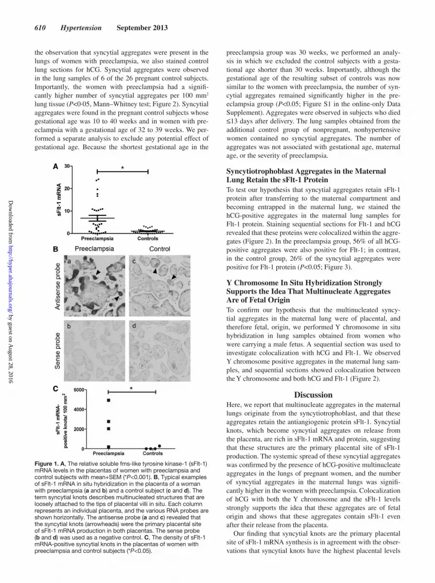
Increased Placental sFlt-1 mRNA Expression in PreeclampsiaTo compare the levels of sFlt-1 mRNA in the preeclamptic and control placentas, quantitative polymerase chain reaction was used to measure sFlt-1 mRNA. On average, the placental sFlt-1 mRNA levels were 6-fold higher in the preeclamptic placentas than in the placentas obtained from control subjects (P<0·001, Mann–Whitney test). The preeclamptic placentas had more intense sFlt-1 staining (measured using in situ hybridization) than control placentas, particularly in the syncytial knots (Figure 1). In addition, the number of syncytial knots was sig-nificantly higher in the women with preeclampsia than in the control subjects (P<0·05; Figure 1). As expected, the sense control probe was negative in all samples (Figure 1).
Presence of hCG-Positive Aggregates in Maternal Lungs Is Significantly Associated With PreeclampsiaBecause hCG was highly expressed within the syncytial knots, we considered hCG to be a suitable specific marker to study the presence of syncytiotrophoblast aggregates in maternal lungs. hCG-positive multinucleate aggregates were observed in the lungs of 6 of the 9 women with preeclampsia. After
Table. Clinical Data
Characteristics PE (n=9) Controls (n=26)
Maternal age at death, y 33.3 (4.6) 32.0 (5.2)
Death postpartum, %† 100 42.3
GA at birth, wk* 35.2 (3.0) 38.5 (3.2)
Death postpartum, h 107.4 (157.2) 96.8 (180.0)
Death during pregnancy, %† 0 57.5
GA at death, wk … 22.3 (10.6)
Death-autopsy time, h 19.7 (13.8) 25.7 (14.7)
Sex offspring
Male, % 57.1 47.8
Female, % 42.9 52.2
Parity 0.7 (1.1) 1.0 (1.2)
Data are given as mean±SD. GA indicates gestational age; and PE, preeclampsia.*P<0.05; † P<0.01.
by guest on August 28, 2016
http://hyper.ahajournals.org/D
ownloaded from
610 Hypertension September 2013
the observation that syncytial aggregates were present in the lungs of women with preeclampsia, we also stained control lung sections for hCG. Syncytial aggregates were observed in the lung samples of 6 of the 26 pregnant control subjects. Importantly, the women with preeclampsia had a signifi-cantly higher number of syncytial aggregates per 100 mm2 lung tissue (P<0·05, Mann–Whitney test; Figure 2). Syncytial aggregates were found in the pregnant control subjects whose gestational age was 10 to 40 weeks and in women with pre-eclampsia with a gestational age of 32 to 39 weeks. We per-formed a separate analysis to exclude any potential effect of gestational age. Because the shortest gestational age in the
preeclampsia group was 30 weeks, we performed an analy-sis in which we excluded the control subjects with a gesta-tional age shorter than 30 weeks. Importantly, although the gestational age of the resulting subset of controls was now similar to the women with preeclampsia, the number of syn-cytial aggregates remained significantly higher in the pre-eclampsia group (P<0.05; Figure S1 in the online-only Data Supplement). Aggregates were observed in subjects who died ≤13 days after delivery. The lung samples obtained from the additional control group of nonpregnant, nonhypertensive women contained no syncytial aggregates. The number of aggregates was not associated with gestational age, maternal age, or the severity of preeclampsia.
Syncytiotrophoblast Aggregates in the Maternal Lung Retain the sFlt-1 ProteinTo test our hypothesis that syncytial aggregates retain sFlt-1 protein after transferring to the maternal compartment and becoming entrapped in the maternal lung, we stained the hCG-positive aggregates in the maternal lung samples for Flt-1 protein. Staining sequential sections for Flt-1 and hCG revealed that these proteins were colocalized within the aggre-gates (Figure 2). In the preeclampsia group, 56% of all hCG-positive aggregates were also positive for Flt-1; in contrast, in the control group, 26% of the syncytial aggregates were positive for Flt-1 protein (P<0.05; Figure 3).
Y Chromosome In Situ Hybridization Strongly Supports the Idea That Multinucleate Aggregates Are of Fetal OriginTo confirm our hypothesis that the multinucleated syncy-tial aggregates in the maternal lung were of placental, and therefore fetal, origin, we performed Y chromosome in situ hybridization in lung samples obtained from women who were carrying a male fetus. A sequential section was used to investigate colocalization with hCG and Flt-1. We observed Y chromosome positive aggregates in the maternal lung sam-ples, and sequential sections showed colocalization between the Y chromosome and both hCG and Flt-1 (Figure 2).
DiscussionHere, we report that multinucleate aggregates in the maternal lungs originate from the syncytiotrophoblast, and that these aggregates retain the antiangiogenic protein sFlt-1. Syncytial knots, which become syncytial aggregates on release from the placenta, are rich in sFlt-1 mRNA and protein, suggesting that these structures are the primary placental site of sFlt-1 production. The systemic spread of these syncytial aggregates was confirmed by the presence of hCG-positive multinucleate aggregates in the lungs of pregnant women, and the number of syncytial aggregates in the maternal lungs was signifi-cantly higher in the women with preeclampsia. Colocalization of hCG with both the Y chromosome and the sFlt-1 levels strongly supports the idea that these aggregates are of fetal origin and shows that these aggregates contain sFlt-1 even after their release from the placenta.
Our finding that syncytial knots are the primary placental site of sFlt-1 mRNA synthesis is in agreement with the obser-vations that syncytial knots have the highest placental levels
Figure 1. A, The relative soluble fms-like tyrosine kinase-1 (sFlt-1) mRNA levels in the placentas of women with preeclampsia and control subjects with mean+SEM (*P<0.001). B, Typical examples of sFlt-1 mRNA in situ hybridization in the placenta of a woman with preeclampsia (a and b) and a control subject (c and d). The term syncytial knots describes multinucleated structures that are loosely attached to the tips of placental villi in situ. Each column represents an individual placenta, and the various RNA probes are shown horizontally. The antisense probe (a and c) revealed that the syncytial knots (arrowheads) were the primary placental site of sFlt-1 mRNA production in both placentas. The sense probe (b and d) was used as a negative control. C, The density of sFlt-1 mRNA-positive syncytial knots in the placentas of women with preeclampsia and control subjects (*P<0.05).
by guest on August 28, 2016
http://hyper.ahajournals.org/D
ownloaded from

Buurma et al Syncytial Aggregates in Preeclampsia 611
of sFlt-1 protein and that these knots are more numerous in the setting of preeclampsia.7,14 Syncytial knots detach readily from the placenta, becoming syncytial aggregates that circu-late in the maternal blood.7 It has long been known that cir-culating placental material, most likely trophoblast cells, can reach maternal organs, particularly the lungs.10 Using colo-calization of hCG with the Y chromosome, we show that the placental multinucleate aggregates in the maternal lung were derived from the syncytiotrophoblast. Interestingly, these pla-centa-derived aggregates in the maternal lung still contained sFlt-1 protein. This observation supports the idea of circu-lating syncytial aggregates as a mechanism of sFlt-1 release into the maternal circulation. Importantly, we also found that preeclampsia was associated with a significantly higher number of syncytial aggregates within the maternal lung tis-sue. During both preeclamptic and normal pregnancies, the
placenta is the principal source of sFlt-1. However, previous research has shown that ≈25% of all sFlt-1 is derived from shed syncytial aggregates.7 It has been estimated that near the end of pregnancy, ≈3 g of this placental material is shed into the maternal circulation (ie, into the maternal lungs) daily.15 Therefore, the cumulative quantity of these circulating aggre-gates, and their relative contribution to total sFlt-1 production, should not be underestimated.
The lungs of the women with preeclampsia contained a significantly higher percentage of sFlt-1–positive syncytial aggregates than the control samples. This observation further supports the idea that preeclampsia is associated both with an increased number of circulating syncytial aggregates and with increased sFlt-1 expression within these aggregates. By releas-ing sFlt-1, these aggregates may contribute to the systemic endothelial dysfunction that is characteristic of preeclampsia.
Figure 2. A, Sections of placenta and sequential sections of maternal lungs that were stained for hCG or Flt-1 (using immunohistochemistry) or the Y chromosome (using in situ hybridization). To simplify the terminology, we use syncytial knots to describe multinucleated structures that are loosely attached to the tips of placental villi in situ and syncytial aggregates to describe detached multinuclear structures within the maternal lungs. The left column shows a placenta obtained from a preeclamptic patient, and the middle and right columns represent the lungs obtained from 2 women who were pregnant with a boy and died because of preeclampsia. The various stains are shown horizontally. Within the preeclamptic placenta, the hCG (a) and Flt-1 (b) proteins are most abundantly present within the syncytial knots (arrowheads). In addition, Y chromosome in situ hybridization (c) shows the presence of the Y chromosome in the nucleus (visible as red puncta). d and g, The presence of hCG-positive aggregates (arrowheads) in the lungs of 2 women who died because of preeclampsia. e and h, The Flt-1 staining patterns in sections that were sequential to the sections shown in d and g, respectively. These images demonstrate that within the maternal lungs, hCG-positive aggregates also contain Flt-1 protein. The next sequential sections were used to perform Y chromosome in
situ hybridization (f and i). These images show that the multinucleate aggregates contain Y chromosomes (arrowheads), indicating that it is very unlikely that these aggregates are not of fetal origin. Altogether, the figure demonstrates colocalization of hCG, Flt-1, and the Y chromosome in the multinucleate aggregates (arrowheads). B, Additional examples of multinucleate aggregates in maternal lungs, with colocalization of hCG (a–d) and Flt-1 (e–h). Each row shows matched sequential sections. C, The number of hCG-positive syncytial aggregates within the maternal lungs of women with preeclampsia (n=9) and control subjects (n=26; *P<0.05).
Figure 3. A, The presence of a hCG-positive aggregate in the lung of a control subject. B, Negative Flt-1 staining in a section that was sequential to the section shown in A. These images demonstrate that within the lungs of control subjects, the minority of the hCG-positive aggregates also contain Flt-1 protein. C, Within the preeclampsia group, 56% of all hCG-positive aggregates were also Flt-1 positive, whereas in the control group 26% of the syncytial aggregates showed positivity for Flt-1 protein (*P<0.05).
by guest on August 28, 2016
http://hyper.ahajournals.org/D
ownloaded from

612 Hypertension September 2013
This finding is also consistent with the observation that pre-eclamptic placentas contain more syncytial aggregates that are heavily loaded with sFlt-1 than placentas obtained after uneventful pregnancies.
In addition, the presence of syncytial aggregates in maternal organs, particularly in the early stages of pregnancy, may play a key role in the development of immune tolerance. As early as gestational week 10, we observed syncytial aggregates in maternal lungs. Because preeclampsia rarely presents before 20 weeks of gestation,11 we could not investigate the presence of syncytial aggregates in the lungs of women with preeclampsia early in pregnancy. We did, however, observe syncytial aggre-gates in the lungs of women with preeclampsia at gestational week 32 and later, and other groups have reported the presence of trophoblast fragments in maternal blood in earlier stages of preeclamptic pregnancy.15 Altogether, circulating syncytial aggregates are present early in pregnancy, and we and others16 have found a strong association between increased shedding of syncytial aggregates and preeclampsia. Thus, one may specu-late that the release and transfer of syncytial aggregates to the maternal compartment is an early event in the pathogenesis of preeclampsia. However, the relative contribution of sFlt-1 expression in these aggregates in early pregnancy is unclear.
The presence and persistence of fetal cells in maternal organs may also have both short-term and long-term impli-cations for postpartum maternal health. Syncytial aggregates that remain in the maternal lungs may undergo further dis-aggregation, forming smaller microparticles. These sFlt-1–loaded microparticles may, via their release into the systemic maternal circulation, contribute to endothelial dysfunction in maternal organs other than the lungs. We found that even 13 days after delivery, hCG-positive syncytial aggregates can be detected within the maternal lungs. This finding supports the idea that placenta-derived syncytial aggregates may be involved in the postpartum complications that are associated with preeclampsia. Preeclampsia usually resolves rapidly after delivery, and its resolution is reflected by a parallel decrease in sFlt-1 levels.17 However, in a subset of women, the symp-toms and complications of preeclampsia can persist or pres-ent several days after delivery. Syncytial aggregates remain transcriptionally active ≤48 hours after delivery, and estimates suggest that during pregnancy, 25% of all circulating sFlt-1 is derived from circulating syncytial aggregates.7 Therefore, we propose that these aggregates may play an important role in postpartum (pre)eclampsia.
It must be acknowledged that the placentas in our study were not obtained from the same women from whom we obtained the lung tissues. Therefore, the preeclampsia phenotype of the women whose lung tissues were investigated might have been more severe than the phenotype of the women who provided the placentas. As a consequence of this potential mismatch between phenotypes, we were unable to correlate placental sFlt-1 production to the portion of sFlt-1–loaded syncytial aggregates in the maternal lungs. To overcome this complica-tion, an animal model could be used to study the association between placental sFlt-1 production and lung pathology.
Trophoblast cells are likely not the only fetal cell popu-lation that is present in the maternal lung. A previous study using mice suggested that fetal cells in the maternal lung
comprised a mixture of cell types that includes trophoblasts, mesenchymal stem cells, and cells from the immune system.18 We have now confirmed the presence of trophoblast cells in the human maternal lung. In the long run, the release of vital cells from the placenta may result in chimerism, as fetal cells can be retained in the maternal blood and organs for decades after delivery.19,20 Because retained fetal cells have stem cell–like properties,21 it can be speculated that these cells provide a mechanism through which maternal health can be affected for decades after pregnancy.
PerspectivesIn conclusion, we have demonstrated that multinucleate aggregates in the maternal lungs originate from the syncytio-trophoblast, that their presence is significantly associated with preeclampsia, and that these aggregates retain the antiangio-genic protein sFlt-1. Further studies are needed to determine the relevance and relative contribution of trophoblast cells, and other cell types, to maternal health. Likewise, understand-ing what drives the formation, detachment, and transfer of syncytial knots to the maternal compartment, and why these knots produce sFlt-1, are important questions to be investi-gated. Nevertheless, this report highlights the importance of investigating further the role that syncytial aggregates play in preeclampsia and its complications.
AcknowledgmentsWe thank Kimberley Veraar and Malu Zandbergen for their excellent technical support.
DisclosuresS.A. Karumanchi is a coinventor of multiple patents related to angio-genic proteins for the diagnosis and therapy of preeclampsia. These patents have been licensed to multiple companies. S.A. Karumanchi reports having served as a consultant to Roche, Siemens, and Beckman Coulter and has financial interest in Aggamin LLC. The other authors report no conflicts.
References 1. Steegers EA, von Dadelszen P, Duvekot JJ, Pijnenborg R. Pre-eclampsia.
Lancet. 2010;376:631–644. 2. Levine RJ, Maynard SE, Qian C, Lim KH, England LJ, Yu KF,
Schisterman EF, Thadhani R, Sachs BP, Epstein FH, Sibai BM, Sukhatme VP, Karumanchi SA. Circulating angiogenic factors and the risk of pre-eclampsia. N Engl J Med. 2004;350:672–683.
3. Maynard SE, Min JY, Merchan J, Lim KH, Li J, Mondal S, Libermann TA, Morgan JP, Sellke FW, Stillman IE, Epstein FH, Sukhatme VP, Karumanchi SA. Excess placental soluble fms-like tyrosine kinase 1 (sFlt1) may contribute to endothelial dysfunction, hypertension, and pro-teinuria in preeclampsia. J Clin Invest. 2003;111:649–658.
4. Ahmad S, Ahmed A. Elevated placental soluble vascular endothelial growth factor receptor-1 inhibits angiogenesis in preeclampsia. Circ Res. 2004;95:884–891.
5. Bujold E, Romero R, Chaiworapongsa T, Kim YM, Kim GJ, Kim MR, Espinoza J, Gonçalves LF, Edwin S, Mazor M. Evidence supporting that the excess of the sVEGFR-1 concentration in maternal plasma in preeclampsia has a uterine origin. J Matern Fetal Neonatal Med. 2005;18:9–16.
6. Clark DE, Smith SK, He Y, Day KA, Licence DR, Corps AN, Lammoglia R, Charnock-Jones DS. A vascular endothelial growth factor antagonist is produced by the human placenta and released into the maternal circula-tion. Biol Reprod. 1998;59:1540–1548.
7. Rajakumar A, Cerdeira AS, Rana S, Zsengeller Z, Edmunds L, Jeyabalan A, Hubel CA, Stillman IE, Parikh SM, Karumanchi SA. Transcriptionally active syncytial aggregates in the maternal circulation may contribute to cir-culating soluble fms-like tyrosine kinase 1 in preeclampsia. Hypertension. 2012;59:256–264.
by guest on August 28, 2016
http://hyper.ahajournals.org/D
ownloaded from

Buurma et al Syncytial Aggregates in Preeclampsia 613
8. Chua S, Wilkins T, Sargent I, Redman C. Trophoblast deportation in pre-eclamptic pregnancy. Br J Obstet Gynaecol. 1991;98:973–979.
9. Redman CW, Sargent IL. Circulating microparticles in normal pregnancy and pre-eclampsia. Placenta. 2008;29 suppl A:S73–S77.
10. Schmorl G. Pathologisch-Anatomische Untersuchungen Über Puerperal-Eklampsie. Leipzig: Verlag FCW Vogel; 1893.
11. Brown MA, Lindheimer MD, de Swiet M, Van Assche A, Moutquin JM. The classification and diagnosis of the hypertensive disorders of pregnancy: statement from the International Society for the Study of Hypertension in Pregnancy (ISSHP). Hypertens Pregnancy. 2001;20:IX–XIV.
12. Casparie M, Tiebosch AT, Burger G, Blauwgeers H, van de Pol A, van Krieken JH, Meijer GA. Pathology databanking and biobanking in The Netherlands, a central role for PALGA, the nationwide histopathology and cytopathology data network and archive. Cell Oncol. 2007;29:19–24.
13. Lau YF. Detection of Y-specific repeat sequences in normal and variant human chromosomes using in situ hybridization with biotinylated probes. Cytogenet Cell Genet. 1985;39:184–187.
14. Nevo O, Soleymanlou N, Wu Y, Xu J, Kingdom J, Many A, Zamudio S, Caniggia I. Increased expression of sFlt-1 in in vivo and in vitro models of human placental hypoxia is mediated by HIF-1. Am J Physiol Regul Integr Comp Physiol. 2006;291:R1085–R1093.
15. Johansen M, Redman CW, Wilkins T, Sargent IL. Trophoblast deportation in human pregnancy–its relevance for pre-eclampsia. Placenta. 1999;20:531–539.
16. Askelund KJ, Chamley LW. Trophoblast deportation part I: review of the evidence demonstrating trophoblast shedding and deportation during human pregnancy. Placenta. 2011;32:716–723.
17. Reddy A, Suri S, Sargent IL, Redman CW, Muttukrishna S. Maternal cir-culating levels of activin A, inhibin A, sFlt-1 and endoglin at parturition in normal pregnancy and pre-eclampsia. PLoS One. 2009;4:e4453.
18. Pritchard S, Wick HC, Slonim DK, Johnson KL, Bianchi DW. Comprehensive analysis of genes expressed by rare microchimeric fetal cells in the maternal mouse lung. Biol Reprod. 2012;87:42.
19. Bianchi DW, Zickwolf GK, Weil GJ, Sylvester S, DeMaria MA. Male fetal progenitor cells persist in maternal blood for as long as 27 years postpartum. Proc Natl Acad Sci U S A. 1996;93:705–708.
20. O’Donoghue K, Chan J, de la Fuente J, Kennea N, Sandison A, Anderson JR, Roberts IA, Fisk NM. Microchimerism in female bone marrow and bone decades after fetal mesenchymal stem-cell trafficking in pregnancy. Lancet. 2004;364:179–182.
21. Khosrotehrani K, Johnson KL, Cha DH, Salomon RN, Bianchi DW. Transfer of fetal cells with multilineage potential to maternal tissue. JAMA. 2004;292:75–80.
What Is New?•Within the placenta, syncytial knots are the principal site of expression
of the antiangiogenic factor soluble fms-like tyrosine kinase-1 (sFlt-1).
•Placenta-derived syncytial aggregates that become lodged in the mater-nal lungs retain the antiangiogenic factor sFlt-1.
•Preeclampsia is associated with significantly higher quantities of sFlt-1–loaded syncytial aggregates within the maternal lung.
What Is Relevant?•Although the precise cause of preeclampsia and its complications re-
mains unknown, the condition is associated with excessive shedding of placental material into the maternal circulation.
•Placenta-derived syncytial aggregates within the maternal lungs contain sFlt-1 and may contribute to the systemic endothelial dysfunction that characterizes preeclampsia.
• Furthermore, retained fetal cells within the mother may have stem cell–like properties, thereby providing a mechanism through which maternal health can be affected for decades after pregnancy.
Summary
The current study confirms the important role of syncytial knots in placental sFlt-1 mRNA production. In addition, it demonstrates a significant association between preeclampsia and the presence of increased quantities of sFlt-1 containing syncytial aggregates in maternal lungs. These observations suggest that the transfer of syncytial aggregates into the maternal compartment likely con-tributes to the systemic endothelial dysfunction that characterizes preeclampsia.
Novelty and Significance
by guest on August 28, 2016
http://hyper.ahajournals.org/D
ownloaded from

and Hans J. BaeldeSuzanne Wilhelmus, Augustine Rajakumar, Kitty W.M. Bloemenkamp, S. Ananth Karumanchi
Aletta J. Buurma, Marlies E. Penning, Frans Prins, Joke M. Schutte, Jan Anthonie Bruijn,Fragments in the Maternal Lung
Preeclampsia Is Associated With the Presence of Transcriptionally Active Placental
Print ISSN: 0194-911X. Online ISSN: 1524-4563 Copyright © 2013 American Heart Association, Inc. All rights reserved.
is published by the American Heart Association, 7272 Greenville Avenue, Dallas, TX 75231Hypertension doi: 10.1161/HYPERTENSIONAHA.113.01505
2013;62:608-613; originally published online July 1, 2013;Hypertension.
http://hyper.ahajournals.org/content/62/3/608World Wide Web at:
The online version of this article, along with updated information and services, is located on the
http://hyper.ahajournals.org/content/suppl/2013/07/01/HYPERTENSIONAHA.113.01505.DC1.htmlData Supplement (unedited) at:
http://hyper.ahajournals.org//subscriptions/
is online at: Hypertension Information about subscribing to Subscriptions:
http://www.lww.com/reprints Information about reprints can be found online at: Reprints:
document. Permissions and Rights Question and Answer this process is available in the
click Request Permissions in the middle column of the Web page under Services. Further information aboutOffice. Once the online version of the published article for which permission is being requested is located,
can be obtained via RightsLink, a service of the Copyright Clearance Center, not the EditorialHypertensionin Requests for permissions to reproduce figures, tables, or portions of articles originally publishedPermissions:
by guest on August 28, 2016
http://hyper.ahajournals.org/D
ownloaded from

ONLINE SUPPLEMENT
PREECLAMPSIA IS ASSOCIATED WITH THE PRESENCE OF
TRANSCRIPTIONALLY ACTIVE PLACENTAL FRAGMENTS IN THE
MATERNAL LUNG
Aletta Buurma (BSc)1*, Marlies Penning (MD)1*, Frans Prins1, Joke Schutte (MD
PhD)2, Jan Anthonie Bruijn (MD PhD)1, Suzanne Wilhelmus (MD) 1, Augustine
Rajakumar (PhD)3, Kitty Bloemenkamp (MD PhD)4, S. Ananth Karumanchi
(MD)3, Hans Baelde (PhD)1
1. Department of Pathology, Leiden University Medical Center, Albinusdreef 2,
2300 RC Leiden, the Netherlands
2. Department of Obstetrics & Gynecology, Isala Clinics, Dokter van Heesweg 2,
8025 AB Zwolle, the Netherlands
3. Howard Hughes Medical Institute and Department of Medicine, Beth Israel
Deaconess Medical Center and Harvard Medical School, Boston, MA, USA
4. Department of Obstetrics, Leiden University Medical Center, Albinusdreef 2,
2300 RC Leiden, the Netherlands
*These two authors contributed equally
Figure S1. Number of syncytial aggregates in preeclamptic women and
control subjects whose gestational age was over 30 weeks.
Figure S2. Number of syncytial aggregates in preeclamptic women and
control subjects who died postpartum.

Figure S1: Number of syncytial aggregates in preeclamptic women and
control subjects whose gestational age was over 30 weeks.
When comparing this subset of patients, the mean gestational age of the
preeclamptic women (n=9) was 35.1 weeks, whereas the gestational age of the
control subjects (n=14) was 38.0 weeks. The number of aggregates was still
higher in the women with preeclampsia (p<0.05).

Figure S2: Number of syncytial aggregates in preeclamptic women and
control subjects who died postpartum.
In this subset of patients, we compared the number of aggregates of the women
with preeclampsia (n=9) and control subjects (n=11). The number of aggregates
was still higher in the women with preeclampsia (p<0.05).


