Embed Size (px)
Citation preview

Seediscussions,stats,andauthorprofilesforthispublicationat:https://www.researchgate.net/publication/221869453
PrenataldiagnosisofWolf-Hirschhornsyndromeconfirmedbycomparativegenomichybridizationarray:Reportoftwocasesandreviewoftheliterature
ARTICLEinMOLECULARCYTOGENETICS·FEBRUARY2012
ImpactFactor:2.14·DOI:10.1186/1755-8166-5-12·Source:PubMed
CITATIONS
5
READS
35
12AUTHORS,INCLUDING:
StavrosSifakis
UniversityofCrete
160PUBLICATIONS1,405CITATIONS
SEEPROFILE
AnnalisaVetro
UniversityofPavia
48PUBLICATIONS744CITATIONS
SEEPROFILE
PanagiotisPeitsidis
HelenaVenizelou
46PUBLICATIONS324CITATIONS
SEEPROFILE
OrsettaZuffardi
UniversityofPavia
345PUBLICATIONS10,690CITATIONS
SEEPROFILE
Availablefrom:AnnalisaVetro
Retrievedon:03February2016

CASE REPORT Open Access
Prenatal diagnosis of Wolf-Hirschhorn syndromeconfirmed by comparative genomic hybridizationarray: report of two cases and review of theliteratureStavros Sifakis1, Emmanouil Manolakos2,8*, Annalisa Vetro3, Dimitra Kappou1, Panagiotis Peitsidis4,Maria Kontodiou2, Antonios Garas5, Nikolaos Vrachnis6, Anastasia Konstandinidou7, Orsetta Zuffardi3, Sandro Orru8
and Ioannis Papoulidis2
Abstract
Wolf-Hirschhorn syndrome (WHS) is a well known genetic condition caused by a partial deletion of the short armof chromosome 4. The great variability in the extent of the 4p deletion and the possible contribution of additionalgenetic rearrangements lead to a wide spectrum of clinical manifestations. The majority of the reports of prenatallydiagnosed WHS cases are associated with large 4p deletions identified by conventional chromosome analysis;however, the widespread clinical use of novel molecular techniques such as array comparative genomichybridization (a-CGH) has increased the detection rate of submicroscopic chromosomal aberrations associated withWHS phenotype. We provide a report of two fetuses with WHS presenting with intrauterine growth restriction asan isolated finding or combined with oligohydramnios and abnormal Doppler waveform in umbilical artery anduterine arteries. Standard karyotyping demonstrated a deletion on chromosome 4 in both cases [del(4)(p15.33) anddel(4)(p15.31), respectively] and further application of a-CGH confirmed the diagnosis and offered a precisecharacterization of the genetic defect. A detailed review of the currently available literature on the prenataldiagnostic approach of WHS in terms of fetal sonographic assessment and molecular cytogenetic investigation isalso provided.
Keywords: 4p- syndrome, Comparative genomic hybridization array, “Greek warrior” helmet profile, Fluorescent situhybridization, Prenatal diagnosis, Wolf-Hirschhorn syndrome
BackgroundWolf-Hirschhorn syndrome (WHS; OMIM 194190) [1],also known as deletion 4p and 4p-syndrome, is a wellknown clinical condition caused by a partial deletion ofthe short arm of chromosome 4. WHS was first (andindependently) described by Wolf et al. (1965) andHirschhorn et al. (1965) [2,3]; thereafter, more than 180documented cases have been published in the literature,most of them diagnosed postnatally. The prevalence ofWHS is reported to be around 1/50.000 live births witha 2:1 female/male ratio; however, this is likely
underestimated because of under-recognition or mis-diagnosis of affected individuals [4,5].In the majority of cases, WHS is caused by a “pure”
deletion of 4p16 with no other cytogenetic abnormalitywhile in the remaining cases, there could be a morecomplicated cytogenetic finding such as chromosome 4ring, 4p- mosaicism, or a derivative chromosome 4resulting from either a de novo or inherited unbalancedtranslocation [5,6]. The complexity of the WHS-asso-ciated basic genomic changes is an important factorexplaining phenotypic variability; though the typicalclinical features include growth restriction of prenatalonset, profound psychomotor retardation, seizures, ske-letal abnormalities, and a distinctive facial appearance[7]. Associated major malformations with variable
* Correspondence: [email protected] S.A., Laboratory of Genetics, Athens-Thessaloniki, GreeceFull list of author information is available at the end of the article
Sifakis et al. Molecular Cytogenetics 2012, 5:12http://www.molecularcytogenetics.org/content/5/1/12
© 2012 Sifakis et al; licensee BioMed Central Ltd. This is an Open Access article distributed under the terms of the Creative CommonsAttribution License (http://creativecommons.org/licenses/by/2.0), which permits unrestricted use, distribution, and reproduction inany medium, provided the original work is properly cited.

incidence (30-70%) are mainly related to midline fusiondefects such as midline scalp defects, agenesis of corpuscallosum, cleft lip/palate, heart defects, and urinary tractmalformations [7,8].Most prenatally diagnosed cases of WHS are asso-
ciated with large 4p deletions identified by conventionalchromosome analysis while the widespread clinical useof novel high-resolution molecular techniques such asarray comparative genomic hybridization (a-CGH)increased the detection rate of submicroscopic chromo-somal aberrations that could also lead to a WHS pheno-type. Herein, we present two WHS cases suspectedupon abnormal signs in prenatal ultrasonography, diag-nosed with conventional cytogenetics and further char-acterized through a-CGH. A detailed review of thecurrent literature on prenatal diagnosis of WHS is alsoprovided.
Cases presentationCase 1A 25-year-old primigravida was referred to our clinic at23 weeks of gestation due to fetal intrauterine growthrestriction (IUGR). The family history was unremarkableand first-trimester screening test for chromosomal aneu-ploidies was normal. Ultrasound examination showedfetal measurements (BPD, HC, AC, FL) below the 5th
centile, consistent with severe symmetrical IUGR. Umbi-lical artery Doppler flow velocimetry exhibited reverseend-diastolic flow and pulsatility index (PI) was 1.83(>95th centile); in addition, uterine artery PI were bilat-erally increased (>95th centile) measuring 2.25 and 1.78respectively and notches were present as well. No fetalmalformation was present. As the amniotic fluid volumewas reduced (AFI < 5), the ultrasound imaging of fetalfacial anomalies was hampered. Upon abnormal ultra-sound findings, an amniocentesis was performed andkaryotype analysis led to the diagnosis of WHS whichwas further confirmed by a-CGH and FISH. Aftergenetic counseling, termination of pregnancy was per-formed at parents’ request at 25 weeks of gestation. Amale neonate was delivered vaginally after medicalinduction with prostaglandins. Detailed pathologicalexamination of the proband was denied by the parents.
Case 2A 37-year-old primigravida was referred to our clinic forgenetic counseling at 23 weeks of gestation due to pre-sence of growth restriction in serial obstetric scans sincethe 13th week of gestation. The couple was healthy, noconsanguineous, with unremarkable medical history. Anamniocentesis was performed at 23 weeks of pregnancy,and the fetal karyotype was compatible with the diagno-sis of WHS. a-CGH analysis showed with high precisiona 19.3 Mb terminal 4p deletion, in the area 4p15.3-pter.
After extensive counseling, the family decided to termi-nate the pregnancy and agreed to an autopsy for thefetus. A female fetus was delivered at 24 weeks aftermedical induction. Fetal autopsy showed external fea-tures of facial dysmorphism with bilateral cleft lip,hypertelorism, broad and high nasal bridge, small filterand large ears (Figure 1). The skull was oval shaped,consistent with the helmet-like typical description ofWHS related facial appearance. The somatometric para-meters indicated a symmetric restriction of fetal growth.Organ dissection showed a small cerebellum with neu-roglial heterotopias, a cardiac defect (patent foramenovale), intestinal malrotation, hypoplastic kidneys, acces-sory spleen and enlarged ovaries. Placenta was hypo-trophic with a weight of 170 g without any significantmacroscopic or histological abnormalities; the umbilicalcord presented three vessels. Growth velocities wereequivalent to 20 weeks of pregnancy.
Cytogenetic and molecular cytogenetics analysisAmniotic fluid was collected from case 1 and case 2 at23 weeks of gestation. Cytogenetic analysis was per-formed on cultured amniocytes by G-banding accordingto standard procedures. At least 20 metaphases wereanalyzed per case, revealing a male karyotype with term-inal deletion of the short arm of one of chromosome 4[46, XY, del(4)(p15.33)] in case 1, and a female karyo-type with terminal deletion of the short arm of one ofchromosome 4 [46, XY, del(4)(p15.31)] in case 2. a-CGH was done on DNA from cultured amniocytes tocharacterize the extent of the deletions using a 100 kbresolution array (kit 44 K) in case 1 and a 40 kb resolu-tion (kit 180 K) in case 2. Molecular karyotyping wascarried out through oligonucleotide array-CGH plat-forms (Agilent Technologies, Santa Clara, CA) asdescribed elsewhere [9].
Figure 1 Autopsy of a 24 weeks’ gestation female fetus afterpregnancy termination (Case 2) that showed external featuresof facial dysmorphism with bilateral cleft lip, hypertelorism,broad and high nasal bridge, small filter and large ears.
Sifakis et al. Molecular Cytogenetics 2012, 5:12http://www.molecularcytogenetics.org/content/5/1/12
Page 2 of 7
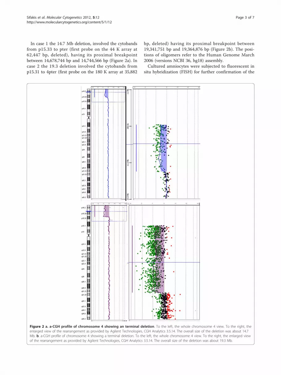
In case 1 the 14.7 Mb deletion, involved the cytobandsfrom p15.33 to pter (first probe on the 44 K array at62,447 bp, deleted), having its proximal breakpointbetween 14,678,744 bp and 14,744,566 bp (Figure 2a). Incase 2 the 19.3 deletion involved the cytobands fromp15.31 to 4pter (first probe on the 180 K array at 35,882
bp, deleted) having its proximal breakpoint between19,341,751 bp and 19,364,876 bp (Figure 2b). The posi-tions of oligomers refer to the Human Genome March2006 (versions NCBI 36, hg18) assembly.Cultured amniocytes were subjected to fluorescent in
situ hybridization (FISH) for further confirmation of the
Figure 2 a. a-CGH profile of chromosome 4 showing an terminal deletion. To the left, the whole chromosome 4 view. To the right, theenlarged view of the rearrangement as provided by Agilent Technologies, CGH Analytics 3.5.14. The overall size of the deletion was about 14.7Mb. b. a-CGH profile of chromosome 4 showing a terminal deletion. To the left, the whole chromosome 4 view. To the right, the enlarged viewof the rearrangement as provided by Agilent Technologies, CGH Analytics 3.5.14. The overall size of the deletion was about 19.3 Mb.
Sifakis et al. Molecular Cytogenetics 2012, 5:12http://www.molecularcytogenetics.org/content/5/1/12
Page 3 of 7

diagnosis. The subtelomeric FISH was performed byusing the commercially available TelVysion 4p SpectrumGreen probe following the manufacturer’s instructions(Vysis Inc, Downers Grove, Ill, USA). In both cases theprobe showed a signal on only one homologue. Parentalkaryotyping was found to be normal. Hybridization withthe probe on metaphase chromosomes of the parentsshowed normal signal on both chromosomes 4 andneither parent was found to carry a translocation of the4pter region (data not shown).
DiscussionWe report two cases of WHS presented with IUGR asan isolated finding or combined with fetal and uterinearteries Doppler abnormalities and oligohydramnios.After invasive testing, conventional cytogenetic investi-gation led to a diagnosis of WHS; in addition, molecularanalysis of the cultured amniocytes with a-CGH andFISH further defined the precise breakpoints of the twodeletions.WHS is a well-described multiple congenital anomaly
and mental retardation syndrome caused by partial dele-tion of the short arm of chromosome 4 involving atleast a 165 kb segment of 4p16.3 [7,10,11]. Prenataldiagnosis of WHS is usually confirmed by detection of acytogenetically visible 4p- deletion discovered after inva-sive testing performed because of advanced maternalage, severe IUGR (which is the most frequent ultra-sound finding, associated or not with other fetalabnormalities), or known parental balanced chromoso-mal rearrangement [12-19]. In case 1, IUGR was furthercomplicated by Doppler abnormalities in the umbilicalartery, bilaterally increased uterine artery PI and oligo-hydramnios, whereas in case 2, early onset growthrestriction was not accompanied with abnormal Doppleror decreased amniotic fluid volume. The ultrasono-graphic presentation of WHS with IUGR and a notchon the uterine artery also overlap with previouslydescribed case by Levaillant et al. [15], while oligohy-dramnios as a unique finding or associated with otherfetal malformations has also been reported in fetuseswith WHS [20-24]. A wide range of other anatomicalabnormalitites as renal hypoplasia, skeletal dysplasias,hypospadias, diaphragmatic hernia, single umbilicalartery also complicates these cases with variable inci-dence [7,25-31]. In addition, craniofacial dysmorphicfeatures such as microcephaly, “Greek warrior helmet”profile (the broad high nasal bridge continuing to theforehead), prominent glabella, high arched eyebrows andhypertelorism are strongly evocative of WHS[7,26,32,33].Unfortunately, minor anatomical defects indicative of
facial dysmorphism in our case 2 were missed by serialultrasound scans between 13 and 22 weeks. Several
reports of concomitant WHS and other structural chro-mosomal aberrations as a result of an unbalanced trans-location display complex phenotypes and confuse someof the correlations [33-35]. A brief overview of the ultra-sound features, the mode of the cytogenetic analysisapplied and the perinatal outcome in 36 WHS cases,including our 2 cases and 34 other published cases, ispresented in Additional file 1: Table S1. In the contextof growth retardation, a reference ultrasonography with2-D and 3-D fetal facial imaging and/or a detailed pre-natal fetal brain evaluation with CT/MRI analysis andfetal echocardiography could be helpful in adding cluestowards diagnosis and orientate karyotype analysis on4p- extremities [15,36,37].Regarding the molecular basis of WHS, in about 55%
of cases, WHS results from an isolated 4p deletion (aso-called “’pure deletion”); about 40-45% of affectedindividuals have an unbalanced translocation (de novoor inherited from a familial balanced rearrangement)characterized by both a deletion of 4p and a partial tris-omy of a different chromosome arm; in the remainingcases, there is a complex rearrangement leading to a4p16.3 deletion (e.g., chromosome 4 ring) [38,39]. Unba-lanced translocations involving the short arms of chro-mosomes 4 and 8 appear with high frequency in severallarge series of WHS patients [40-43]. These rearrange-ments usually arise as a result of a) a homologous non-allelic recombination mediated by olfactory receptors(OR)-gene clusters in both 4p and 8p, or b) a parentalinversion polymorphism on 4p16 [44,45]. Recent studiespoint to a multigenic profile of WHS that contributes tothe complex phenotype though two critical regions(WHSCR1 and -2) have been implicated in the patho-genesis of the syndrome [8,46,47]. WHSCR-1 is a 165kb area approximately 2 Mb from the telomere of 4pand includes the WHS candidate gene 2 (WHSC2) andpart of WSHC1 which is implicated in growth delay andfacial characteristics [48-50]. WHSCR-2, that contributesto the basic phenotype (typical craniofacial pattern, mildmental retardation, growth delay and seizures) resides ina 300-600 kb interval positioned between 1.9 and 1.6-1.3Mb from the telomere and is contiguous and telomericto WHSCR-1. This genomic region includes a third criti-cal gene called LETM1 (leucine zipper/EF-hand-contain-ing transmembrane) associated with the neuromuscularfeatures of WHS patients such as seizures disorders[5,51], and partially the WHSC1. Moreover, recent stu-dies suggest that the fibroblast growth factor receptor-like 1 (FGFRL1) represents a plausible candidate genefor part of the craniofacial phenotype of WHS [47,52].An increasing number of genotype-phenotype correla-
tion studies compare specific clinical features of patientswith different sized 4p deletions in order to refine the4p phenotypic map but the variable expressivity or
Sifakis et al. Molecular Cytogenetics 2012, 5:12http://www.molecularcytogenetics.org/content/5/1/12
Page 4 of 7

penetrance of the clinical features and the fact thatWHS is likely to be a contiguous gene syndrome, makesit a challenging task. According to a recent study WHScases can be divided into three clinical categories: thefirst one comprises a microdeletion not exceeding 3.5Mb at 4p16-4pter results in a mild phenotype and islikely to be under diagnosed, the second one is asso-ciated to deletions between 5 and 18 Mb that presentwith severe psychomotor delay and typical abnormalitieswhereas those greater than 22 Mb at 4p15-4pter consistthe third category associated with major malformations[8]. However, Battaglia et al. (2001) demonstrated that asubmicroscopic deletion that was detected only by FISHmay account for the severe WHS phenotype and con-cluded that there is no such a strict correlation [53].Alternative mechanisms that can lead to complex phe-notypes include: a) unbalanced translocation mutationsresulting in 4p deletion and partial trisomy affecting thefinal phenotype; b) allelic variation in the homologous4p region; c) mutations in modifier genes located out-side the deleted regions; d) position effects and telomeresilencing; e) different genetic background and post-zygo-tic mutational events [43,46]. Differential diagnosis ofWHS should include the proximal interstitial 4p dele-tion which is a discrete syndrome that usually involvesbands 4p12-p16 that are proximal to and exclude theWHS critical region [54].The majority of prenatally diagnosed cases of WHS
reported in the medical literature are delineated by con-ventional cytogenetic analysis, but during the last decadethe availability of new technologies especially a-CGH haveenabled a more precise description of the molecularmechanisms that can account for the WHS phenotype[23,55]. Indeed, few published reports refer to cases that astandard karyotype was interpreted as normal and arequired subsequent molecular analysis by FISH or/and a-CGH upon prenatally or postnatally identified fetal mal-formations allowed the final diagnosis [11,33,37,56]. A pre-natal misdiagnosis of a WHS case is more likely when thefetus presents only with fetal growth restriction or othernon-specific or minor features and the standard karyotyperesults to be balanced [15,23,36,37,57]. Conventional G-banded cytogenetic analysis seems to detect approximately50-60% of WHS cases while application of FISH analysisusing a WHSCR probe detects more that 95% of deletionsin WHS [39,53]. In addition, a-CGH can detect all cur-rently known deletions of the WHSCR and determine ifthe deletion is “pure” or part of a more complex imbalancemore accurately than either FISH or conventional G-bandanalysis alone [39]. A comprehensive analysis of the roleof a-CGH in the evaluation of WHS patients demon-strated that the true prevalence of unbalanced transloca-tions is certainly higher than reported previously and isapproximately 45% as both karyotype and routine FISH
analysis of the region may not detect these cases; also a-CGH adds information on approximate size of both thedeletion and duplication compared to a subtelomericFISH assay [43]. Although both of our cases were asso-ciated with cytogenetically visible deletions, we applied a-CGH analysis to confirm that they were pure distal dele-tions, to define their extent at molecular level and toestablish a firm diagnosis. We also applied FISH analysisto further confirm our findings, to extend the investigationto both couples and define the potential presence of abalanced rearrangement involving 4p16.3 in the parents ofa proband so as to provide a thorough genetic counseling.In conclusion, growth restriction as an isolated finding orassociated with facial dysmorphism and/or other majormalformations such as renal or skeletal abnormalities andmidline fusion defects may be indicative of a WHS caseand should trigger cytogenetic investigation. A combineddiagnostic approach based on conventional karyotypingand molecular analysis, would offer a definitive resultwithin the time frame required for management of theaffected pregnancy and for a prompt genetic counselingabout the long term complications and poor prognosis ofthese cases. This is crucial as, according to the data pre-sented in the Additional file 1: Table S1, most of the par-ents opt for pregnancy termination. Furthermore, as partof the genetic counseling prenatal testing should beoffered to families in which one parent is known to be acarrier of a chromosome rearrangement involving 4p16.3Additional investigation with high-resolution techniquessuch as a-CGH is nowadays strongly recommended parti-cularly in case of discordance between prenatal ultrasoundfindings and normal karyotype. In the future, the imple-mentation of this technique in the routine practice of pre-natal diagnosis will improve the diagnostic yield inpregnancies with abnormal ultrasound findings and parti-cularly to WHS, it will enable a more precise estimation ofthe true incidence of the syndrome and will advance ourknowledge regarding the genotype-phenotype correlations.
ConsentWritten informed consent was obtained from the parentsfor publication of these Case reports and any accompany-ing images. A copy of the written consent is available forreview by the Editor-in-Chief of this journal.
Additional material
Additional file 1: Table S1. Reported cases of prenatal diagnosis ofWHS: sonographic findings, karyotype, and pregnancy outcome.
Author details1Department of Obstetrics & Gynecology, University of Crete, Heraklion,Greece. 2Eurogenetica S.A., Laboratory of Genetics, Athens-Thessaloniki,
Sifakis et al. Molecular Cytogenetics 2012, 5:12http://www.molecularcytogenetics.org/content/5/1/12
Page 5 of 7

Greece. 3Dipartimento di Patologia Umana ed Ereditaria, Universita di Pavia,Pavia, Italia. 4Helena Venizelou Hospital, Athens, Greece. 5Department ofObstetrics & Gynecology, University of Thessalia, Larissa, Greece. 62ndDepartment of Obstetrics and Gynecology, Aretaieion Hospital, Univeristy ofAthens, Athens, Greece. 71st Department of Pathology, Univeristy of Athens,Athens, Greece. 8Cattedra di Genetica Medica, Universita di Cagliari, Cagliari,Italia.
Authors’ contributionsSS and EM drafted the manuscript and coordinate the whole project. EM,AV, MK, OZ, SO, IP performed cytogenetic analysis, a-CGH, and FISH. SS, AG,NV performed the clinical evaluation of the pregnancies. DK and PPparticipated in the coordination and helped to draft the manuscript. AKperformed the pathological examination and helped to draft the manuscript.All authors have read and approved the manuscript.
Competing interestsThe authors declare that they have no competing interests.
Received: 26 December 2011 Accepted: 28 February 2012Published: 28 February 2012
References1. On-Line Mendelian Inheritance in Man, OMIM. [http://www.ncbi.nlm.nih.
gov/omim?term=WHS].2. Wolf U, Reinwein H, Porsch R, Schröter R, Baitsch H: Deficiency on the
short arms of a chromosome No. 4. Humangenetik 1965, 1:397-413.3. Hirschhorn K, Cooper HL, Firschein IL: Deletion of short arms of
chromosome G-5 in a child with defects of midline fusion. Hum Genet1965, 1:679-682.
4. Wieczorek D, Krause M, Majewski F, Albrecht B, Horn D, Riess O, Gillessen-Kaesbach G: Effect of the size of the deletion and clinical manifestationin Wolf-Hirschhorn syndrome: analysis of 13 patients with a de novodeletion. Eur J Hum Genet 2000, 8:519-526.
5. Zollino M, Lecce R, Fischetto R, Murdolo M, Faravelli F, Selicorni A, Buttè C,Memo L, Capovilla G, Neri G: Mapping the Wolf-Hirschhorn syndromephenotype outside the currently accepted WHS critical region anddefining a new critical region, WHSCR-2. Am J Hum Genet 2003,75:590-597.
6. Dallapiccola B, Mandich P, Bellone E, Selicorni A, Mokin V, Ajmar F,Novelli G: Parental origin of chromosome 4p deletion in Wolf-Hirschhornsyndrome. Am J Med Genet 1993, 47:921-924.
7. Battaglia A, Filippi T, Carey JC: Update on the clinical features and naturalhistory of Wolf-Hirschhorn (4p-) syndrome: experience with 87 patientsand recommendations for routine health supervision. Am J Med Genet CSemin Med Genet 2008, 148C:246-251.
8. Zollino M, Murdolo M, Marangi G, Pecile V, Galasso C, Mazzanti L, Neri G:On the nosology and pathogenesis of Wolf-Hirschhorn syndrome:genotype-phenotype correlation analysis of 80 patients and literaturereview. Am J Med Genet C Semin Med Genet 2008, 148C:257-269.
9. Manolakos E, Vetro A, Kefalas K, Thomaidis L, Aperis G, Sotiriou S, Kitsos G,Merkas M, Sifakis S, Papoulidis I, Liehr T, Zuffardi O, Petersen MB: Deletion2q31.2-q31.3 in a 4-year-old girl with microcephaly and severe mentalretardation. Am J Med Genet A 2011, 155A:1476-1482.
10. Estabrooks LL, Rao KW, Driscoll DA: Preliminary phenotypic map ofchromosome 4p16 based on 4p deletions. Am J Med Genet 1995,57:581-586.
11. Zollino M, Di Stefano C, Zampino G, Mastroiacovo P, Wright TJ, Sorge G,Selicorni A, Tenconi R, Zappalà A, Battaglia A, Di Rocco M, Palka G,Pallotta R, Altherr MR, Neri G: Genotype-phenotype correlations andclinical diagnostic criteria in Wolf-Hirschhorn syndrome. Am J Med Genet2000, 94:254-261.
12. Blunt S, Berry AC, Seller MJ, Williams CA: Prenatal recognition of 4p-syndrome. J Med Genet 1977, 14:232-233.
13. Kohlschmidt N, Zielinski J, Brude E, Schäfer D, Olert J, Hallermann C,Coerdt W, Arnemann J: Prenatal diagnosis of a fetus with a cryptictranslocation 4p;18p and Wolf-Hirschhorn syndrome (WHS). Prenat Diagn2000, 20:152-155.
14. Aslan H, Karaca N, Basaran S, Ermis H, Ceylan Y: Prenatal diagnosis of Wolf-Hirschhorn syndrome (4p-) in association with congenital hypospadiasand foot deformity. BMC Pregnancy Childbirth 2003, 3:1.
15. Levaillant JM, Touboul C, Sinico M, Vergnaud A, Serero S, Druart L,Blondeau JR, Abd Alsamad I, Haddad B, Gérard-Blanluet M: Prenatalforehead edema in 4p- deletion: the ‘Greek warrior helmet’ profilerevisited. Prenat Diagn 2005, 25:1150-1155.
16. Eiben B, Leipoldt M, Schübbe I, Ulbrich R, Hansmann I: Partial deletion of4p in fetal cells not present in chorionic villi. Clin Genet 1988, 33:49-52.
17. Verloes A, Schaaps JP, Herens C, Soyeur D, Hustin J, Dodinval P: Prenataldiagnosis of cystic hygroma and chorioangioma in the Wolf-Hirschhornsyndrome. Prenat Diagn 1991, 11:129-132.
18. Tachdjian G, Fondacci C, Tapia S, Huten Y, Blot P, Nessmann C: The Wolf-Hirschhorn syndrome in fetuses. Clin Genet 1992, 42:281-287.
19. Chen SR, Lee CC, Chen WL, Chen MH, Chang KM: De novo unbalancedtranslocation resulting in monosomy for proximal 14q and distal 4p in afetus with intrauterine growth retardation, Wolf-Hirschhorn syndrome,hypertrophic cardiomyopathy, and partial hemihypoplasia. J Med Genet1998, 35:1050-1053.
20. Vamos E, Pratola D, Van Regemorter N, Freund M, Flament-Durand J,Rodesch F: Prenatal diagnosis and fetal pathology of partial trisomy 20P-monosomy 4P resulting from paternal translocation. Prenat Diagn 1985,5(Suppl 3):209-214.
21. Petek E, Wagner K, Steiner H, Schaffer H, Kroisel PM: Prenatal diagnosis ofpartial trisomy 4q26-qter and monosomy for the Wolf-Hirschhorn criticalregion in a fetus with split hand malformation. Prenat Diagn 2000,20:349-352.
22. Witters I, Van Schoubroeck D, Fryns JP: Choroid plexus cysts andoligohydramnios: presenting echographic signs in a female fetus withdeletion of the Wolf-Hirschhorn syndrome region (4p16.3). Genet Couns2001, 12:387-388.
23. Chao A, Lee YS, Chao AS, Wang TH, Chang SD: Microarray-basedcomparative genomic hybridization analysis of Wolf-Hirschhornsyndrome in a fetus with deletion of 4p15.3 to 4pter. Birth Defects Res AClin Mol Teratol 2006, 76:739-743.
24. Basgul A, Kavak ZN, Akman I, Basgul A, Gokaslan H, Elcioglu N: Prenataldiagnosis of Wolf-Hirschhorn syndrome (4p-) in association withcongenital diaphragmatic hernia, cystic hygroma and IUGR. Clin ExpObstet Gynecol 2006, 33(Suppl 2):105-106.
25. Vinals F, Sepulveda W, Selman E: Prenatal detection of congenitalhypospadias in the Wolf-Hirschhorn (4p-) syndrome. Prenat Diagn 1994,14:1166-1169.
26. Dietze I, Fritz B, Huhle D, Simoens W, Piecha E, Rehder H: Clinical,cytogenetic and molecular investigation in a fetus with Wolf-Hirschhornsyndrome with paternally derived 4p deletion. Case report and reviewof the literature. Fetal Diagn Ther 2004, 19:251-260.
27. Beaujard MP, Jouannic JM, Bessières B, Borie C, Martin-Luis I, Fallet-Bianco C,Portnoï MF: Prenatal detection of a de novo terminal invertedduplication 4p in a fetus with the Wolf-Hirschhorn syndromephenotype. Prenat Diagn 2005, 25:451-455.
28. Casaccia G, Mobili L, Braguglia A, Santoro F, Bagolan P: Distal 4pmicrodeletion in a case of Wolf-Hirschhorn syndrome with congenitaldiaphragmatic hernia. Birth Defects Res A Clin Mol Teratol 2006, 76:210-213.
29. Sepulveda W: Prenatal 3-dimensional sonographic depiction of the Wolf-Hirschhorn phenotype: the “Greek warrior helmet” and “tulip” signs. JUltrasound Med 2007, 26:407-410.
30. Sergi C, Schulze BR, Hager HD, Beedgen B, Zilow E, Linderkamp O, Otto HF,Tariverdian G: Wolf-Hirschhorn syndrome: case report and review of thechromosomal aberrations associated with diaphragmatic defects.Pathologica 1998, 90(Suppl 3):285-293.
31. Schinzel A: Discrepancies in cytogenetic results between different tissuesin two fetuses with Wolf- Hirschhorn syndrome. Cytogenet Cell Genet2000, 91(Suppl 1-4):231-233.
32. Sase M, Hasegawa K, Honda R, Sumie M, Nakata M, Sugino N, Furukawa S:Ultrasonographic findings of facial dysmorphism in Wolf-Hirschhornsyndrome. Am J Perinatol 2005, 22:99-102.
33. Chen CP, Chen YJ, Chern SR, Tsai FJ, Chang TY, Lee CC, Town DD, Lee MS,Wang W: Prenatal diagnosis of concomitant Wolf-Hirschhorn syndromeand split hand-foot malformation associated with partial monosomy 4p(4p16.1- > pter) and partial trisomy 10q (10q25.1- > qter). Prenat Diagn2008, 28:450-453.
34. Phelan MC, Saul RA, Gailey TA Jr, Skinner SA: Prenatal diagnosis of mosaic4p- in a fetus with trisomy 21. Prenat Diagn 1995, 15:274-277.
Sifakis et al. Molecular Cytogenetics 2012, 5:12http://www.molecularcytogenetics.org/content/5/1/12
Page 6 of 7

35. Tapper JK, Zhang S, Harirah H, Panova NI, Merryman LS, Hawkins JC,Lockhart LH, Gei AB, Velagaleti GV: Prenatal diagnosis of a fetus withunbalanced translocation (4;13)(p16;q32) with overlapping features ofPatau and Wolf-Hirschhorn syndromes. Fetal Diagn Ther 2002, 17:347-351.
36. De Keersmaecker B, Albert M, Hillion Y, Ville Y: Prenatal diagnosis of brainabnormalitiesin Wolf-Hirschhorn (4p-) syndrome. Prenat Diagn 2002,22:366-370.
37. Boog G, Le Vaillant C, Collet M, Dupré PF, Parent P, Bongain A, Benoit B,Trastour C: Prenatal sonographic patterns in six cases of Wolf-Hirschhorn(4p) syndrome. Fetal Diagn Ther 2004, 19:421-430.
38. South ST, Hannes F, Fisch GS, Vermeesch JR, Zollino M: Pathogenicsignificance of deletions distal to the currently described Wolf-Hirschhorn syndrome critical regions on 4p16.3. Am J Med Genet C SeminMed Genet 2008, 148C:270-274.
39. Battaglia A, Carey JC, South ST, Wright TJ: Wolf-Hirschhorn syndrome. InGeneReviews [Internet]. Edited by: Pagon RA, Bird TD, Dolan CR, Stephens K.Seattle: University of Washington, Seattle; , 1993-2002 Apr 29 [updated 2010Jun 17].
40. Tranebjaerg L, Petersen A, Hove K, Rehder H, Mikkelsen M: Clinical andcytogenetic studies in a large (4;8) translocation family with pre- andpostnatal Wolf syndrome. Ann Genet 1984, 27(Suppl 4):224-229.
41. Müller-Navia J, Nebel A, Oehler D, Theile U, Zabel B, Schleiermacher E:Microdissection and DOP-PCR-based reverse chromosome painting as afast and reliable strategy in the analysis of various structuralchromosome abnormalities. Prenat Diagn 1996, 16(Suppl 10):915-922.
42. Wieczorek D, Krause M, Majewski F, Albrecht B, Meinecke P, Riess O,Gillessen-Kaesbach G: Unexpected high frequency of de novounbalanced translocations in patients with Wolf-Hirschhorn syndrome(WHS). J Med Genet 2000, 37(Suppl 10):798-804.
43. South ST, Whitby H, Battaglia A, Carey JC, Brothman AR: Comprehensiveanalysis of Wolf-Hirschhorn syndrome using array CGH indicates a highprevalence of translocations. Eur J Hum Genet 2008, 16:45-52.
44. Giglio S, Calvari V, Gregato G, Gimelli G, Camanini S, Giorda R, Ragusa A,Guerneri S, Selicorni A, Stumm M, Tonnies H, Ventura M, Zollino M, Neri G,Barber J, Wieczorek D, Rocchi M, Zuffardi O: Heterozygous submicroscopicinversions involving olfactory receptor-gene clusters mediate therecurrent t(4;8)(p16;p23) translocation. Am J Hum Genet 2002, 71:276-285.
45. Zollino M, Lecce R, Murdolo M, Orteschi D, Marangi G, Selicorni A, Midro A,Sorge G, Zampino G, Memo L, Battaglia D, Petersen M, Pandelia E,Gyftodimou Y, Faravelli F, Tenconi R, Garavelli L, Mazzanti L, Fischetto R,Cavalli P, Savasta S, Rodriguez L, Neri G: Wolf-Hirschhorn syndrome-associated chromosome changes are not mediated by olfactory receptorgene clusters nor by inversion polymorphism on 4p16. Hum Genet 2007,122:423-430.
46. Bergemann AD, Cole F, Hirschhorn K: The etiology of Wolf-Hirschhornsyndrome. Trends Genet 2005, 21:188-195.
47. Hammond P, Hannes F, Suttie M, Devriendt K, Vermeesch JR, Faravelli F,Forzano F, Parekh S, Williams S, McMullan D, South ST, Carey JC, Quarrell O:Fine-grained facial phenotype-genotype analysis in Wolf-Hirschhornsyndrome. Eur J Hum Genet 2012, 20:33-40.
48. Wright TJ, Ricke DO, Denison K, Abmayr S, Cotter PD, Hirschhorn K,Keinänen M, McDonald-McGinn D, Somer M, Spinner N, Yang-Feng T,Zackai E, Altherr MR: A transcriptmap of the newly defined 165 kb Wolf-Hirschhorn syndrome critical region. Hum Mol Genet 1997, 6:317-324.
49. Wright TJ, Costa JL, Naranjo C, Francis-West P, Altherr MR: Comparativeanalysis of a novel gene from the Wolf-Hirschhorn/Pitt-Rogers- Dankssyndrome critical region. Genomics 1999, 59:203-212.
50. Rauch A, Schellmoser S, Kraus C, Dörr HG, Trautmann U, Altherr MR,Pfeiffer RA, Reis A: First known microdeletion within the Wolf-Hirschhornsyndrome critical region refines genotype -phenotype correlation. Am JMed Genet 2001, 99:338-342.
51. Endele S, Fuhry M, Pak SJ, Zabel BU, Winterpacht A: LETM1, a novel geneencoding a putative EF-hand ca2+ banding protein, flanks the Wolf-Hirschhorn syndrome (WHS) critical region and is deleted in most WHSpatients. Genomics 1999, 60:218-225.
52. Catela C, Bilbao-Cortes D, Slonimsky E, Kratsios P, Rosenthal N, TeWelscher P: Multiple congenital malformations of Wolf-Hirschhornsyndrome are recapitulated in Fgfrl1 null mice. Dis Model Mech 2009,2:283-294.
53. Battaglia A, Carey JC, Wright TJ: Wolf-Hirschhorn (4p-) syndrome. AdvPediatr 2001, 48:75-113.
54. Bailey NG, South ST, Hummel M, Wenger SL: Case report: cytogenetic andmolecular analysis of proximal interstitial deletion of 4p, review of theliterature and comparison with Wolf-Hirschhorn syndrome. J Assoc GenetTechnol 2010, 36:5-10.
55. Maas NM, Van Buggenhout G, Hannes F, Thienpont B, Sanlaville D, Kok K,Midro A, Andrieux J, Anderlid BM, Schoumans J, Hordijk R, Devriendt K,Fryns JP, Vermeesch JR: Genotype-phenotype correlation in 21 patientswith Wolf-Hirschhorn syndrome using high resolution array comparativegenome hybridisation (CGH). J Med Genet 2008, 45:71-80.
56. Goodship J, Curtis A, Cross I, Brown J, Emslie J, Wolstenholme J: Asubmicroscopic translocation, t(4;10), responsible for recurrent Wolf-Hirschhorn syndrome identified by allele loss and fluorescent in situhybridisation. J Med Genet 1992, 29:451-454.
57. Zuffardi O, Vetro A, Brady P, Vermeesch J: Array technology in prenataldiagnosis. Semin Fetal Neonatal Med 2011, 16:94-98.
doi:10.1186/1755-8166-5-12Cite this article as: Sifakis et al.: Prenatal diagnosis of Wolf-Hirschhornsyndrome confirmed by comparative genomic hybridization array:report of two cases and review of the literature. Molecular Cytogenetics2012 5:12.
Submit your next manuscript to BioMed Centraland take full advantage of:
• Convenient online submission
• Thorough peer review
• No space constraints or color figure charges
• Immediate publication on acceptance
• Inclusion in PubMed, CAS, Scopus and Google Scholar
• Research which is freely available for redistribution
Submit your manuscript at www.biomedcentral.com/submit
Sifakis et al. Molecular Cytogenetics 2012, 5:12http://www.molecularcytogenetics.org/content/5/1/12
Page 7 of 7


