Embed Size (px)
Citation preview
ENVIRONMENTALHEALTH PERSPECTIVES
This article will be available in its final, 508-conformant form 2–4 months after Advance Publication. If you require assistance accessing this article before then, please contact Dorothy L. Ritter, EHP Web Editor. EHP will provide an accessible version within 3 working days of request.
http://www.ehponline.org
ehpPrenatal Exposure to Maternal Cigarette Smoking and DNA Methylation: Epigenome-Wide Association in a
Discovery Sample of Adolescents and Replication in an Independent Cohort at Birth through 17 Years of Age
Ken W. K. Lee, Rebecca Richmond, Pingzhao Hu, Leon French, Jean Shin, Celine Bourdon, Eva Reischl, Melanie Waldenberger,
Sonja Zeilinger, Tom Gaunt, Wendy McArdle, Susan Ring, Geoff Woodward, Luigi Bouchard, Daniel Gaudet, George Davey-Smith, Caroline Relton, Tomas Paus,
and Zdenka Pausova
http://dx.doi.org/10.1289/ehp.1408614
Received: 27 April 2014Accepted: 16 October 2014
Advance Publication: 17 October 2014
1
Prenatal Exposure to Maternal Cigarette Smoking and DNA
Methylation: Epigenome-Wide Association in a Discovery Sample of
Adolescents and Replication in an Independent Cohort at Birth
through 17 Years of Age
Ken W. K. Lee,1 Rebecca Richmond,2 Pingzhao Hu,1,3 Leon French,4 Jean Shin,1 Celine
Bourdon,1 Eva Reischl,5 Melanie Waldenberger,5 Sonja Zeilinger,5 Tom Gaunt,2 Wendy
McArdle,2 Susan Ring,2 Geoff Woodward,2 Luigi Bouchard,6, 7 Daniel Gaudet,7, 8 George Davey-
Smith,2 Caroline Relton,9 Tomas Paus,4 and Zdenka Pausova1
1The Hospital for Sick Children, University of Toronto, Toronto, Canada; 2School of Social and
Community Medicine, University of Bristol, Bristol, UK; 3Department of Biochemistry and
Medical Genetics, University of Manitoba, Winnipeg, Canada; 4Rotman Research Institute,
University of Toronto, Toronto, Canada; 5Research Unit of Molecular Epidemiology, Helmholtz
Zentrum Muenchen, Munich, Germany and Institute of Epidemiology II, Helmholtz Zentrum
Muenchen, German Research Center for Environmental Health, Neuherberg, Germany;
6Department of Biochemistry, Université de Sherbrooke, Sherbrooke, Canada; 7ECOGENE-21
and Lipid Clinic, Chicoutimi Hospital, Chicoutimi, Canada; 8Department of Medicine,
Université de Montréal, Montréal, Canada; 9Institute of Genetic Medicine, Newcastle University,
Newcastle upon Tyne, UK
Address correspondence to Zdenka Pausova, The Hospital for Sick Children, Associate
Professor, Departments of Physiology and Nutritional Sciences, University of Toronto, Toronto,
2
Canada. Telephone: (416) 813-7654/304340. Fax: (416) 813-5771. E-mail:
Short title: Prenatal smoke exposure and DNA methylation
Acknowledgments: We thank the following individuals for their contributions in acquiring data
for the SYS: Manon Bernard (database architect, The Hospital for Sick Children) and Helene
Simard, MA, and her team of research assistants (Cégep de Jonquière). We also thank Dr.
Mathieu Lemire (Ontario Institute for Cancer Research) for his assistance with data analysis. We
also thank The Centre for Applied Genomics, The Hospital for Sick Children for assistance with
EWAS statistical analyses. In addition, we thank the entire ALSPAC team, which includes
interviewers, computer and laboratory technicians, clerical workers, research scientists,
volunteers, managers, receptionists, and nurses. Finally, we would like to thank Dr. Thomas
Hudson (Ontario Institute for Cancer Research) and Dr. Andrew Patterson (The Hospital for Sick
Children) for critical review of the manuscript. Luigi Bouchard is a junior research scholar from
the FRQS and a member of the FRQS-funded Centre de recherche clinique Étienne-Le Bel.
Funding information: The Canadian Institutes of Health Research and the Heart and Stroke
Foundation of Canada fund the SYS. The McLaughlin Centre at the University of Toronto
provided supplementary funds for the DNA methylation studies in the SYS. The UK Medical
Research Council and the Wellcome Trust (Grant ref: 092731), and the University of Bristol
provide core support for ALSPAC.
Competing financial interests: The authors declare they have no actual or potential competing
financial interests.
3
Abstract
Background: Prenatal exposure to maternal cigarette smoking (prenatal smoke exposure) had
been associated with altered DNA methylation (DNAm) at birth.
Objective: We examined whether such alterations are present from birth through adolescence.
Methods: We used the Infinium HumanMethylation450K BeadChip to search across 473,395
CpGs for differential DNAm associated with prenatal smoke exposure during adolescence in a
discovery cohort (n=132) and at birth, during childhood, and during adolescence in a replication
cohort (n=447).
Results: In the discovery cohort, we found five CpGs in MYO1G (top-ranking CpG:
cg12803068, p=3.3x10⁻¹¹) and CNTNAP2 (cg25949550, p=4.0x10⁻⁹) to be differentially
methylated between exposed and non-exposed individuals during adolescence. The CpGs in
MYO1G and CNTNAP2 were associated, respectively, with higher and lower DNAm in exposed
vs. non-exposed adolescents. The same CpGs were differentially methylated at birth, during
childhood, and during adolescence in the replication cohort. In both cohorts and at all
developmental time-points, the differential DNAm was in the same direction and of a similar
magnitude, and was not altered appreciably by adjustment for current smoking by the
participants or their parents. In addition, four of the five EWAS-significant CpGs in the
adolescent discovery cohort were also among the top sites of differential methylation in a
previous birth cohort, and differential methylation of CpGs in CYP1A1, AHRR and GFI1
observed in that study was also evident in our discovery cohort.
Conclusions: Our findings suggest that modifications of DNAm associated with prenatal
maternal smoking may persist in exposed offspring for many years - at least until adolescence.
4
Introduction
Maternal cigarette smoking during pregnancy was and still is common in the industrialized
world. In the US, for example, 30-40% of pregnant women smoked in the 1960s and 1970s and
16% of pregnant women smoke today (2012 US National Survey on Drug Use and Health).
Prenatal exposure to maternal cigarette smoking (prenatal smoke exposure) has been associated
with a number of health problems in the exposed offspring (Oken et al. 2008; Power et al. 2010;
Syme et al. 2010). The underlying mechanisms of these associations are not clear but may
involve lasting modulations of DNA methylation (DNAm).
DNAm is a chemical modification constituted most commonly by the addition of a methyl group
to cytosines in CpG dinucleotides (CpGs) (Relton and Davey Smith 2010). It is catalyzed by two
main types of DNA methyltransferases (DNMTs): de novo DNMTs, which play a key role in
establishing new DNAm patterns during cell differentiation and embryogenesis, and
maintenance DNMTs, which are responsible for copying these patterns from cell to cell during
successive mitotic divisions (Jeltsch 2006; Tang et al. 2009). The main function of DNAm is the
regulation of gene expression and genomic architecture (Levin and Moran 2011; Tsukahara et al.
2009).
Current research suggests that cigarette smoke is a powerful environmental modifier of DNAm
(reviewed in (Lee and Pausova 2013)). Cigarette smoke contains a large number of chemicals,
such as carcinogens, nicotine and carbon monoxide, that have been shown to modify DNAm in
differentiating and dividing cells (Cuozzo et al. 2007; Di et al. 2012; Han et al. 2001; Huang et
al. 2012; Mercer et al. 2009; Mortusewicz et al. 2005; Satta et al. 2008; Shahrzad et al. 2007).
Consistent with these possible effects, reproducible differences in DNAm have been reported in
5
peripheral blood cells of smokers vs. non-smokers (Breitling et al. 2011; Philibert et al. 2012;
Shenker et al. 2013; Zeilinger et al. 2013).
Many chemicals contained in cigarette smoke can easily pass from a smoking pregnant woman
to the developing embryo/fetus (Lambers and Clark 1996). Accordingly, differences in DNAm
have been reported in cord blood-cells of babies born to smoking vs. non-smoking mothers
(Joubert et al. 2012). Whether these DNAm differences persist postnatally in peripheral blood
cells of the exposed offspring has not been studied; this is the main subject of the present
investigation. Prenatal exposure to cigarette smoke may induce lasting and soma-wide (present
in all somatic cells) modifications of DNAm in the exposed offspring, particularly if occurring
during early stages of embryogenesis when global erasure and re-establishment of DNAm
patterns take place in yet-undifferentiated stem cells (Feng et al. 2010; Smith et al. 2012). These
DNAm modifications are then propagated during embryonic development to all somatic cell
lineages (including precursors of peripheral blood cells) and maintained throughout life by the
action of maintenance DNMTs, which copy these modifications from cell to cell during
successive cell divisions (Davies et al. 2012; Faulk and Dolinoy 2011; Jurkowska et al. 2011;
Lee and Pausova 2013; Petronis 2010).
To investigate whether DNAm modifications induced by prenatal smoke exposure persist
postnatally (at least until adolescence), we first conducted an epigenome-wide association study
(EWAS) to search for CpGs methylated differentially between exposed and non-exposed
adolescents in one cohort, the Saguenay Youth Study (SYS) (Pausova et al. 2007). Subsequently,
we tested whether the identified CpGs were also methylated differentially between exposed and
non-exposed newborns, children, and adolescents in another cohort, the Avon Longitudinal
Study of Parents and Children (ALSPAC) (Boyd et al. 2013).
6
Methods
Discovery cohort: The Saguenay Youth Study (SYS)
Recruitment and assessment of prenatal smoke exposure
The SYS (n=1,028, 12- to 18-year old) is a population-based cross-sectional study of
cardiovascular, metabolic, brain and mental health in adolescents carried out in the genetic
founder population of the Saguenay Lac St. Jean region of Quebec, Canada (Pausova et al.
2007). All participants were White Caucasians. Adolescents exposed (n=490) and non-exposed
(n=538) to maternal cigarette smoking during pregnancy were matched at recruitment by
maternal education and school attended to reduce potential confounding related to
socioeconomic status (Pausova et al. 2007). They were recruited via high schools. Exposure
status was defined as having a mother who smoked at least 1 cigarette per day during the second
trimester of pregnancy. Being non-exposed was defined as having a mother who did not smoke
at least 1 year before and throughout the pregnancy. The information on exposure was obtained
from the mother using a structured telephone interview at the time of recruitment of her
adolescent offspring, and was validated subsequently against medical records from the time of
pregnancy for a subset of adolescents; Kappa statistics with a mean (standard deviation) value of
0.69 (0.04) indicated a good strength of agreement in this subset (good agreement, 0.6-0.8)
(Landis and Koch 1977). The main exclusion criteria were as follows: premature birth (<35
weeks) or detached placenta; maternal alcohol abuse during pregnancy (>210 ml of
alcohol/week); positive medical history of type 1 diabetes, or of heart disease requiring surgery
or sustained medication; and contraindications for magnetic resonance imaging. Additional
recruitment details and selection criteria have been described previously (Pausova et al. 2007).
7
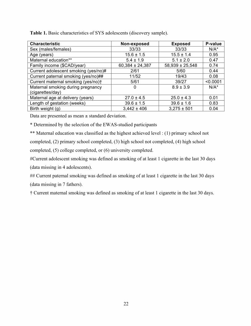
In the present study, we selected 66 exposed adolescents matched to 66 non-exposed by sex, age,
and maternal education. The exposed adolescents were selected within each sex separately as
those with the exposure being close to the sex-specific median (10 cigarettes/day in both sexes).
This process resulted in a selection of 66 adolescents (33 males and 33 females) who were
exposed to maternal cigarette smoking throughout pregnancy (all trimesters) at a level of 9±4
cigarettes/day (range 5-20 cigarettes/day). The studied adolescents were 12- to 18-year old
(Table 1). Written consent of the parents and assent of the adolescents were obtained. The
research ethics committees of the Chicoutimi Hospital and the Hospital for Sick Children
approved the study.
Adolescent and parent cigarette smoking
Adolescents self-reported their mental health and substance use (including cigarette smoking) by
completing a questionnaire developed for the SYS by J. Séguin from the University of Montreal
(Groupe de Recherche sur l’Inadaptation Psychosociale); this questionnaire is based on validated
National Longitudinal Survey of Children and Youth (NLSCY) and Quebec Longitudinal Study
of Child Development (QLSCD) protocols (Pausova et al. 2007). Blood collection and
adolescent smoking assessment occurred on the same day. Parents self-reported their own mental
health and substance use (including cigarette smoking) using a similar questionnaire, as adapted
by colleagues at the Groupe de Recherche sur l’Inadaptation Psychosociale of the University of
Montreal (Pausova et al. 2007).
Epityping with the Infinium HumanMethylation450K BeadChip
DNA samples were extracted from the buffy coat (peripheral blood cells) using the Qiagen
Autopure LS (Qiagen, Venlo, Netherlands). Bisulfite conversion was performed on 800 ng of
8
DNA from each sample with the EZ-96 DNA Methylation Kit (Zymo Research, Irvine, CA).
DNA was subsequently used for hybridization on the Infinium HumanMethylation450K
BeadChip (Illumina, San Diego, CA). Bisulfite conversion and methylation array hybridization
were performed at the Helmholtz Zentrum München German Research Center for Environmental
Health (Neuherberg, Germany). The Infinium HumanMethylation450K BeadChip interrogates
methylation at >485,000 CpGs and provides coverage of > 99% of RefSeq genes (Sandoval et al.
2011). The DNAm score at each CpG, referred to as the DNAm β value, is derived from the
fluorescent intensity ratio [β = intensity of the methylated allele / (intensity of the unmethylated
allele + intensity of the methylated allele + 100)] (Bibikova et al. 2011). All samples were
randomly loaded onto 11 arrays (12 samples per array) and processed by the same technician at
the same time to minimize batch effects. Methylation values were normalized using the pre-
process Illumina algorithm implemented in Minfi R package (Aryee et al. 2014). Parameters
were set to mimic the Illumina Genome Studio normalization procedure (Illumina, San Diego,
CA). Quality control was performed by excluding CpGs with detection p ≥ 0.05 in more than
20% of samples (764 CpGs). After excluding these probes, as well as control probes and probes
on sex chromosomes, a total of 473,395 CpGs were analyzed. All 132 samples had > 98% sites
with detection p < 0.05.
Statistical methods (EWAS)
Methylation β values at each of the 473,395 CpGs were transformed to obtain M-values, defined
as log2 [β / (1 – β)], which have greater statistical power than β values to detect differential
methylation at highly methylated and unmethylated CpGs (Du et al. 2010). Multivariable linear
regression was used to perform association tests between the M value at each CpG (n=475,395)
as the dependent variable and prenatal smoke exposure (exposed vs. non-exposed) as the
9
independent variable. These analyses were adjusted for potential confounding by age, sex, batch,
and blood cell fractions (Model A). Blood cell fractions (granulocytes, B cells, CD8+ T cells,
CD4+ T-cells, NK cells, monocytes) were determined by the method of Houseman (Houseman
et al. 2012); the fractions were analyzed with principal component analysis and the first 3
components capturing 95% of variance were included in the regression model. The Manhattan
and Q-Q plots for this Model A are shown in Supplemental Material, Figure S1. Note that none
of the estimated blood cell fractions differed between exposed and non-exposed individuals
(p=0.31-0.89). In additional analyses, we also adjusted for current smoking by adolescents
(Model B) or for both current smoking by adolescents and second-hand smoking by parents
(Model C). Further, we used the above-described basic Model A to analyze subsets of
adolescents (a) who did not smoke themselves (61 exposed and 64 non-exposed, Model D) or (b)
who did not smoke themselves and their parents did not smoke at present (19 exposed and 49
non-exposed, Model E). Finally, we ran an analysis that was not corrected for any potential
confounders (Model F). CpGs achieving Bonferroni-corrected statistical significance of p<0.05
(uncorrected p<1.1x10⁻⁷) were considered EWAS significant. False discovery rate (FDR)-
corrected p-values were also determined according to the method of Benjamini and Hochberg
(Benjamini and Hochberg 1995). These statistical analyses were performed using the R software
(http://www.r-project.org/).
Replication cohort: Avon Longitudinal Study of Parents and Children (ALSPAC)
Recruitment and assessment of prenatal smoke exposure
ALSPAC is a population-based longitudinal birth cohort ascertained in the former Avon Health
Authority in southwest England between April 1st 1991 and December 31st 1992; the initial
cohort included 14,541 pregnancies and 13,971 children alive at 12 months of age (Boyd et al.
10
2013). From this cohort, approximately 500 child participants have been epityped with the
Infinium HumanMethylation450K BeadChip (Illumina, San Diego, CA) as part of a larger
methylation project, ARIES, which is generating epigenetic information for 1,000 mother-
offspring pairs at multiple time points across the life-course
(http://www.ariesepigenomics.org.uk/). The individuals included in the present study are a subset
of the offspring participants in ARIES for whom 450K data were available at the time of
submission of this manuscript. Of these individuals, 82% (430/526) had methylation data
available for at least two of the following 3 time points: at birth, 7 years of age, and 17 years of
age. DNA samples were extracted from cord blood cells (birth) and peripheral blood cells (at 7
and 17 years of age). From these, we selected exposed individuals as child participants whose
mothers smoked ≥ 5 cigarettes/day throughout their gestation and non-exposed individuals as
child participants whose mothers did not smoke ~4/5 months before pregnancy and throughout
all trimesters of their gestation (Supplemental Material, Table S1). Prenatal exposure to maternal
cigarette smoking was assessed during pregnancy. Methylation values were normalized using the
approach of Touleimat and Tost (Touleimat and Tost 2012).
Statistical methods
Associations between prenatal exposure to maternal cigarette smoking and M values at the 5
EWAS-significant CpGs identified in the SYS were tested using linear regression models
adjusted for age, sex, maternal education, household social class, and batch. We included
maternal education and household social class in the model, as unlike in the SYS exposed and
non-exposed participants were not matched for any indices of socioeconomic status at
recruitment. In additional analyses, we also adjusted for parent (second-hand) smoking at 7 years
11
of age, and for both participant smoking and parent (second-hand) smoking at 17 years of age.
Statistical analyses were performed using the R software (http://www.r-project.org/).
Results
EWAS search for differential DNAm associated with prenatal smoke exposure in the SYS
adolescents
The EWAS search was carried out in the SYS, which is a population-based cross-sectional study
of 12- to 18-year old adolescents aimed at investigating long-term health outcomes associated
with prenatal exposure to maternal cigarette smoking (Table 1). All participants are from the
genetic founder population of the Saguenay Lac St. Jean region of Quebec, Canada (Pausova et
al. 2007).
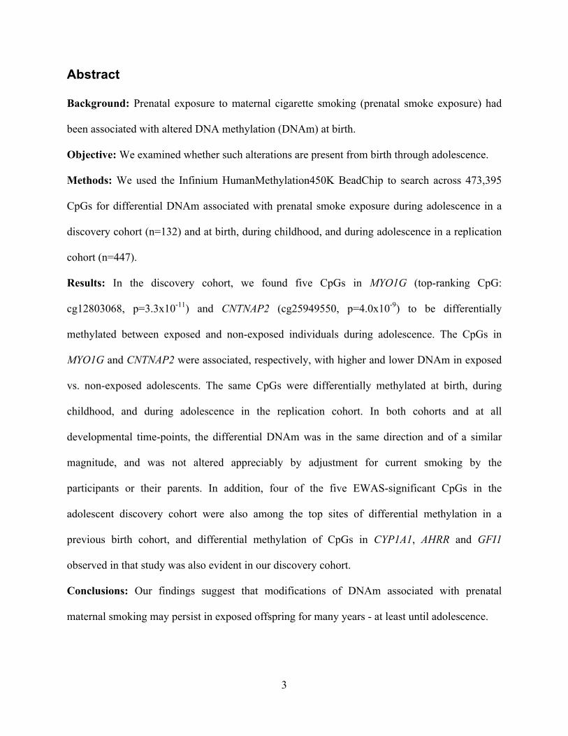
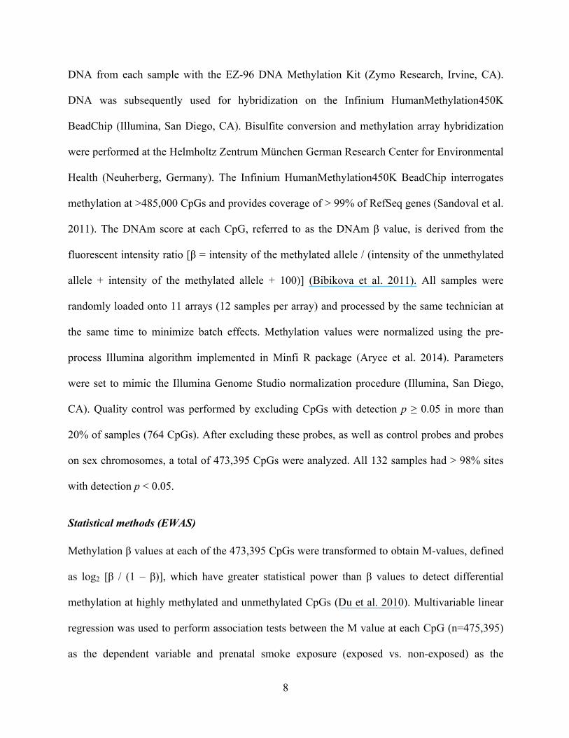
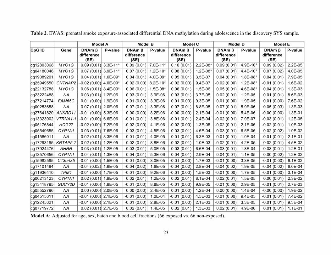
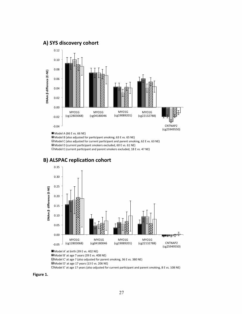
The EWAS search in the SYS identified 5 EWAS-significant (p < 1.1x10⁻⁷) CpGs (Figure 1A
and Table 2, Model A). Four of them, including the most significant one (cg12803068, p = 3.3 x
10-11), were located within a 1-kb segment of the myosin IG gene (MYO1G); they were all
associated with higher DNAm in exposed than non-exposed adolescents (Figure 1A and Table 2,
Model A). DNAm at all 4 CpGs was correlated (r=0.83-0.92, p<0.0001) and as such the 4 CpGs
are not independent signals. The other significant CpG was found in the contactin associated
protein-like 2 gene (CNTNAP2, cg25949550, p = 4.0 x 10⁻⁹). In contrast with the MYO1G CpGs,
it was associated with lower DNAm in exposed than non-exposed adolescents (Figure 1A and
Table 2, Model A). None of the above CpGs were cross-reactive or polymorphic as listed in
(Chen et al. 2013; Price et al. 2013). These results were obtained while adjusting for potential
confounders, namely age, sex, batch, and blood cell fractions (Model A). They remained
virtually unchanged when not adjusted for any of these potential confounders (Model F,
12
Supplemental Material, Table S2). In both adjusted and unadjusted analyses, differential DNAm
(DNAm β difference between exposed and non-exposed) ranged from +0.04 to +0.09 in MYO1G
and it was -0.02 in CNTNAP2.
Next, we examined whether current cigarette smoking by the adolescent participants or by their
parents confounded these associations. In our sample, only 5 of 66 exposed and 2 of 66 non-
exposed (Table 1) adolescents reported “smoking at least 1 cigarette in the last 30 days”.
Associations of prenatal maternal smoking and differential DNAm were comparable in models
that also adjusted for participant smoking (Figure 1A, Model B) or that excluded current
participant smokers (Figure 1A, Model D).” Adjusting for current smoking by parents as well as
by the participants had little influence on point estimates, though p-values were increased
somewhat: 2 of the 5 CpGs remained EWAS-significant, while the remaining 3 showed p <
1.5x10⁻⁶ (Figure 1A and Table 2, Model C). Excluding adolescents who were current smokers or
whose parents were current smokers reduced the sample size from 66 exposed and 66 non-
exposed to 18 exposed and 47 non-exposed. Nevertheless, all 5 CpGs remained significant (at p
< 0.05) even in this smaller subsample (Figure 1A and Table 2, Model E).
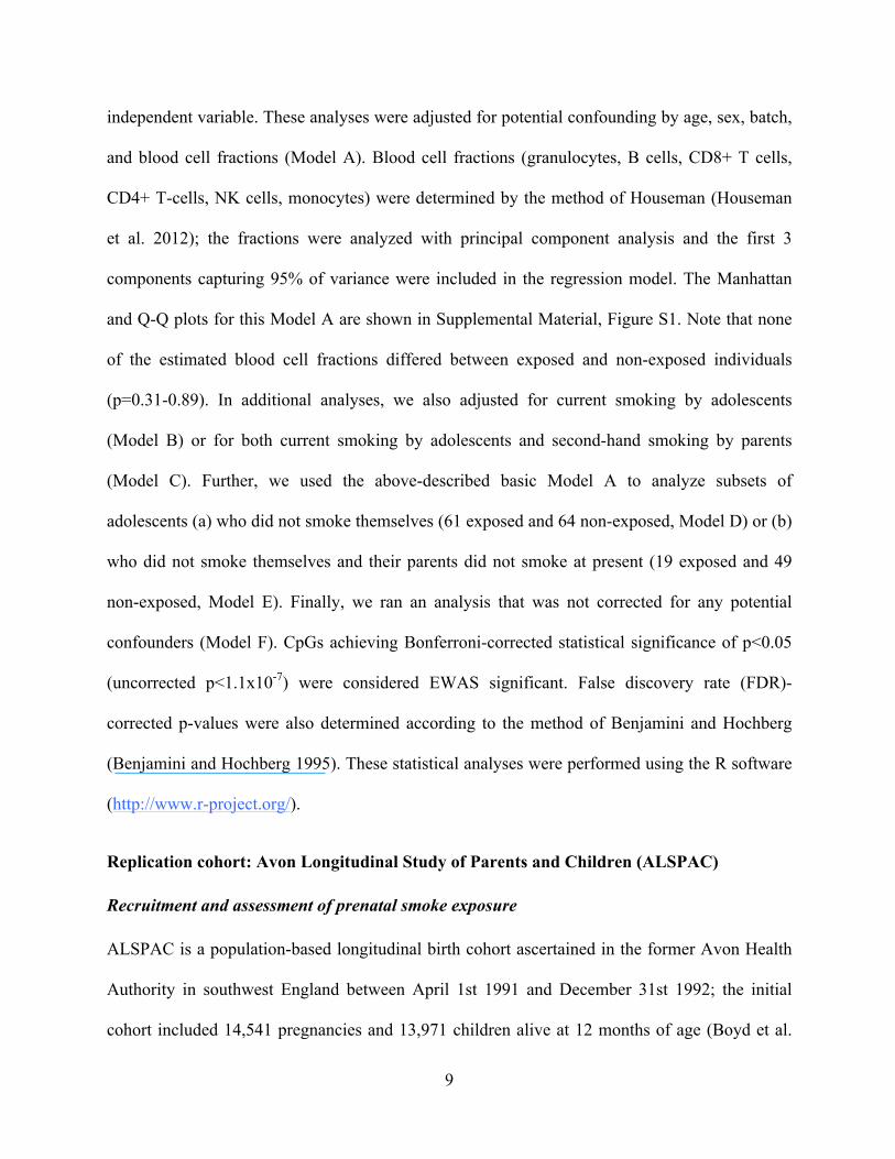
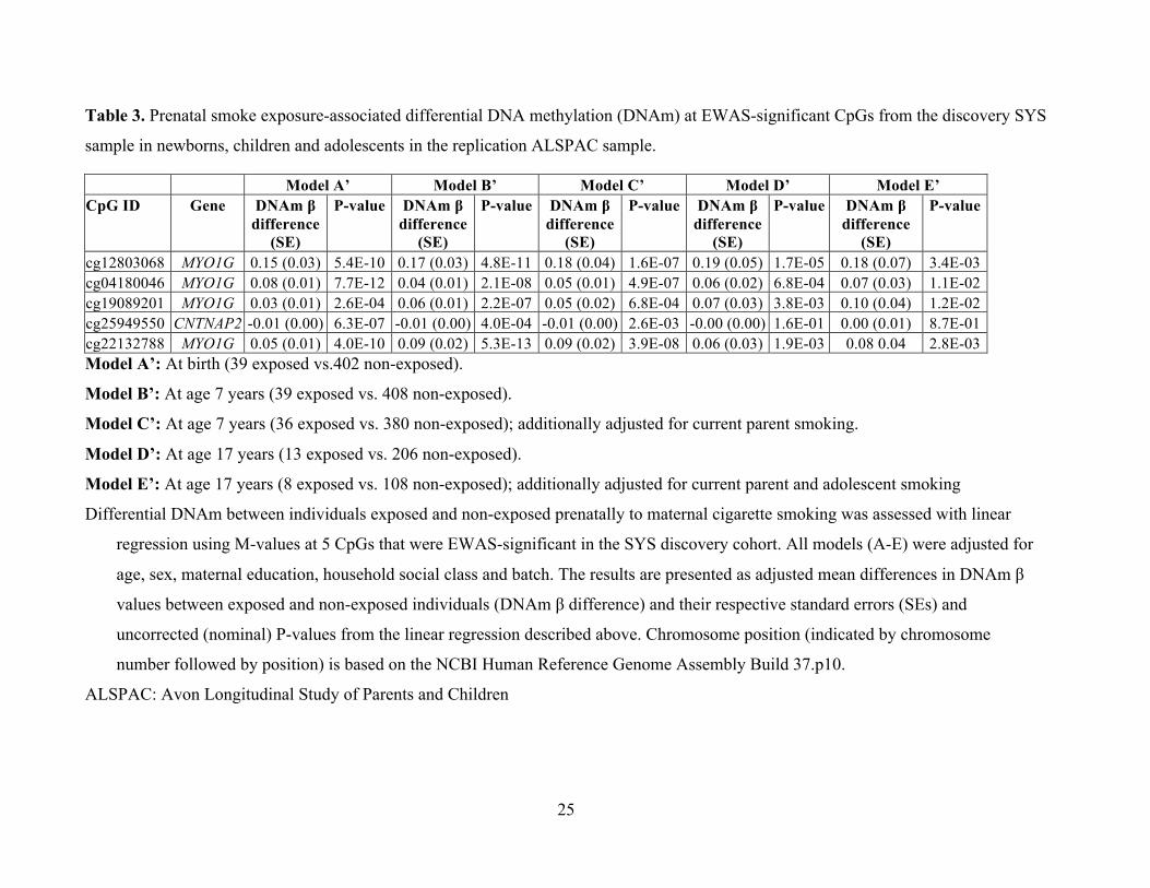
Replication and persistence of differential DNAm associated with prenatal smoke exposure
in the ALSPAC newborns, children and adolescents
The replication and persistence studies were carried out an independent sample, the ALSPAC
cohort, which is a population-based longitudinal birth cohort ascertained in the former Avon
Health Authority in southwest England between April 1st 1991 and December 31st 1992
(Supplemental Material, Table S1) (Boyd et al. 2013). Specifically, we tested whether the 5
EWAS-significant CpGs identified in the discovery SYS sample of adolescents, were also
13
differentially methylated in exposed vs. non-exposed newborns (39 vs. 402), children (7 years;
39 vs. 408), and adolescents (17 years, 13 vs. 206) from the ALSPAC cohort. We found that all 5
CpGs were associated with prenatal smoke exposure at birth and during childhood (Table 3,
Models A’ and B’, respectively) – and 4 out of the 5 CpGs were associated with prenatal smoke
exposure during adolescence (Table 3 Model D’). The direction and magnitude of differential
DNAm at all 3 developmental time points for all 5 CpGs (Figure 1B) was consistent with the
findings in the discovery cohort (Figure 1A). These results remained significant (p < 0.05) after
adjusting for exposure to second-hand smoke (i.e., current smoking by parents) during childhood
(Table 3, Model C’) and for current participant smoking and parent smoking during adolescence
(Table 3, Model E’), despite substantially reduced sample sizes (Figure 1B).
Discussion
The results of the present study suggest that prenatal smoke exposure is associated with
modifications of DNAm that persist in the exposed offspring for years – from birth until at least
adolescence – and that these associations remain after adjusting for blood-cell fractions, current
smoking by adolescent participant, and current smoking by parents.
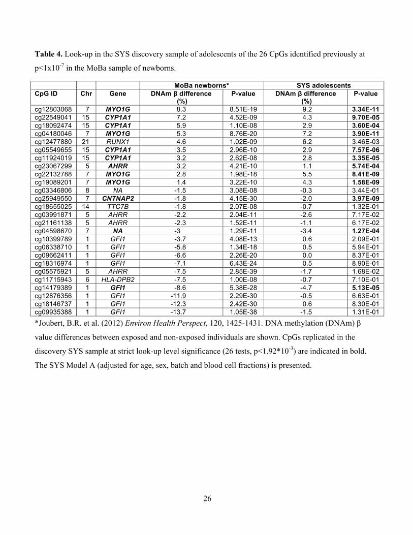
To our knowledge, our study is the first to report that prenatal smoke exposure-associated
differential DNAm may persist in the exposed offspring for years after their birth. The MoBa
study reported that prenatal smoke exposure was associated with differential DNAm at birth
(Joubert et al. 2012). Importantly, all 5 CpGs we found to be differentially methylated in the
present study at birth and during childhood (in the ALSPAC replication cohort), and during
adolescence (in the SYS discovery sample and in the ALSPAC cohort) were also differentially
methylated in the MoBa study at birth (Joubert et al. 2012) with the differences between exposed
14
and non-exposed being of the same direction and similar magnitude (Table 4). Other CpGs with
prenatal smoke exposure-associated differential DNAm in the MoBa study were also significant
at the strict look-up level (26 CpGs, p < 1.92*10-3) in the SYS adolescents, including CpGs in
CYP1A1, AHRR, and GFI1, and the gene-unmapped CpG 04598670 (Table 4). Differential
DNAm across the 26 MoBa CpGs was correlated between the SYS and MoBa samples (r2 =
0.53, p < 0.0001) (Supplemental Material, Figure S2).” Taken together, these studies indicate
that prenatal smoke exposure is associated with specific DNAm modifications observed at birth
(MoBa, ALSPAC at birth) and that some of these modifications may persist in the exposed
offspring until at least adolescence (SYS, ALSPAC).
The EWAS-significant CpGs were located in MYO1G and CNTNAP2. MYO1G encodes a plasma
membrane-associated class I myosin that is expressed abundantly in hematopoietic cells; it
regulates cell elasticity and migration (Olety et al. 2010; Patino-Lopez et al. 2010). CNTNAP2
encodes a member of the neurexin family of cell-adhesion molecules that plays a critical role in
brain development (Anderson et al. 2012), and has been implicated in a number of
neurodevelopmental disorders, including autism (Anney et al. 2012). One of the main functions
of DNAm is the regulation of gene expression. The 4 EWAS-significant CpGs in MYO1G (but
not the one in CNTNAP2) were located in a region containing binding sites for a number of
transcription factors and enhancers (http://genome.ucsc.edu/ENCODE) and thus might alter
mRNA expression of the gene. This possibility remains to be studied, however.
In addition to prenatal exposure to cigarette smoke, active smoking has been associated with
differential DNAm in peripheral blood cells (Breitling et al. 2011; Philibert et al. 2012; Shenker
et al. 2013; Zeilinger et al. 2013). In our discovery sample, only 5% of adolescents reported
current cigarette smoking, and excluding these participants or adjusting for their current smoking
15
had little influence on model estimates. Further, we considered that postnatal exposure to
second-hand smoke might be a confounder of the associations between prenatal smoke exposure
and DNAm during childhood and adolescence. Although second-hand smoke exposure has not
been reported to impact DNAm in peripheral blood cells, some evidence exists it is associated
with differential DNAm in cancer tissues (though to a lesser degree than variations observed in
active smokers) (Scesnaite et al. 2012; Wilhelm-Benartzi et al. 2011). Our results showed,
however, that the differential DNAm associated with prenatal smoke exposure seen in the SYS
adolescents was present (albeit somewhat attenuated) even after additional adjusting for second-
hand smoke exposure to parental smoking. Importantly, the prenatal smoke exposure-associated
differential DNAm we observed in the ALSPAC replication sample also remained significant
after additional adjusting for adolescents’ smoking and their exposure to second-hand smoke
generated by the parents. Across both SYS (adolescence) and ALSPAC (birth, childhood,
adolescence) samples and all statistical models we tested (with varying numbers of available
participants), the magnitude of DNAm differences between exposed and non-exposed
individuals was generally consistent. Collectively, these results support the postnatal persistence
of prenatal smoke exposure-associated DNAm modifications in the exposed offspring.
In conclusion, our results suggest that prenatal smoke exposure may induce reproducible
alterations of DNAm in the offspring, and that some of these alterations may persist for many
years – well into adolescence. The potential implications of such epigenetic modifications for the
“programming” of health require further study. Likewise, their possible utility as blood
biomarkers of prenatal environmental exposures, such as maternal cigarette smoking, needs
further exploration. In closing, promotion of smoking cessation remains imperative (Ng et al.

16
2014); gaining knowledge about molecular mechanisms capable of influencing health trajectories
in the exposed population is critical for the prevention and treatment of associated disorders.
17
References
Anderson GR, Galfin T, Xu W, Aoto J, Malenka RC, Sudhof TC. 2012. Candidate autism gene
screen identifies critical role for cell-adhesion molecule CASPR2 in dendritic arborization
and spine development. Proc Natl Acad Sci U S A 109:18120-18125.
Anney R, Klei L, Pinto D, Almeida J, Bacchelli E, Baird G, et al. 2012. Individual common
variants exert weak effects on the risk for autism spectrum disorders. Hum Mol Genet
21:4781-4792.
Aryee MJ, Jaffe AE, Corrada-Bravo H, Ladd-Acosta C, Feinberg AP, Hansen KD, et al. 2014.
Minfi: A flexible and comprehensive bioconductor package for the analysis of Infinium
DNA Methylation microarrays. Bioinformatics 30:1363-1369.
Benjamini Y, Hochberg Y. 1995. Controlling the false discovery rate: A practical and powerful
approach to multiple testing. Journal of the Royal Statistical Society B 85:289-300.
Bibikova M, Barnes B, Tsan C, Ho V, Klotzle B, Le JM, et al. 2011. High density DNA
methylation array with single CpG site resolution. Genomics 98:288-295.
Boyd A, Golding J, Macleod J, Lawlor DA, Fraser A, Henderson J, et al. 2013. Cohort profile:
The 'children of the 90s'--the index offspring of the Avon Longitudinal Study of Parents and
Children. Int J Epidemiol 42:111-127.
Breitling LP, Yang R, Korn B, Burwinkel B, Brenner H. 2011. Tobacco-smoking-related
differential DNA methylation: 27k discovery and replication. Am J Hum Genet 88:450-457.
Chen YA, Lemire M, Choufani S, Butcher DT, Grafodatskaya D, Zanke BW, et al. 2013.
Discovery of cross-reactive probes and polymorphic cpgs in the Illumina Infinium Human
Methylation450 microarray. Epigenetics 8:203-209.
Cuozzo C, Porcellini A, Angrisano T, Morano A, Lee B, Di Pardo A, et al. 2007. DNA damage,
homology-directed repair, and DNA methylation. PLoS Genet 3:e110.
Davies MN, Volta M, Pidsley R, Lunnon K, Dixit A, Lovestone S, et al. 2012. Functional
annotation of the human brain methylome identifies tissue-specific epigenetic variation
across brain and blood. Genome Biol 13:R43.
Di YP, Zhao J, Harper R. 2012. Cigarette smoke induces MUC5AC protein expression through
the activation of SP1. J Biol Chem 287:27948-27958.
18
Du P, Zhang X, Huang CC, Jafari N, Kibbe WA, Hou L, et al. 2010. Comparison of beta-value
and M-value methods for quantifying methylation levels by microarray analysis. BMC
Bioinformatics 11:587.
Faulk C, Dolinoy DC. 2011. Timing is everything: The when and how of environmentally
induced changes in the epigenome of animals. Epigenetics 6:791-797.
Feng S, Jacobsen SE, Reik W. 2010. Epigenetic reprogramming in plant and animal
development. Science 330:622-627.
Han L, Lin IG, Hsieh CL. 2001. Protein binding protects sites on stable episomes and in the
chromosome from de novo methylation. Mol Cell Biol 21:3416-3424.
Houseman EA, Accomando WP, Koestler DC, Christensen BC, Marsit CJ, Nelson HH, et al.
2012. DNA methylation arrays as surrogate measures of cell mixture distribution. BMC
Bioinformatics 13:86.
Huang J, Okuka M, Lu W, Tsibris JC, McLean MP, Keefe DL, et al. 2012. Telomere shortening
and DNA damage of embryonic stem cells induced by cigarette smoke. Reprod Toxicol.
Jeltsch A. 2006. Molecular enzymology of mammalian DNA methyltransferases. Curr Top
Microbiol Immunol 301:203-225.
Joubert BR, Haberg SE, Nilsen RM, Wang X, Vollset SE, Murphy SK, et al. 2012. 450k
epigenome-wide scan identifies differential DNA methylation in newborns related to
maternal smoking during pregnancy. Environ Health Perspect 120:1425-1431.
Jurkowska RZ, Jurkowski TP, Jeltsch A. 2011. Structure and function of mammalian DNA
methyltransferases. Chembiochem 12:206-222.
Lambers DS, Clark KE. 1996. The maternal and fetal physiologic effects of nicotine. Semin
Perinatol 20:115-126.
Landis JR, Koch GG. 1977. The measurement of observer agreement for categorical data.
Biometrics 33:159-174.
Lee KW, Pausova Z. 2013. Cigarette smoking and DNA methylation. Front Genet 4:132.
Levin HL, Moran JV. 2011. Dynamic interactions between transposable elements and their hosts.
Nat Rev Genet 12:615-627.
Mercer BA, Wallace AM, Brinckerhoff CE, D'Armiento JM. 2009. Identification of a cigarette
smoke-responsive region in the distal MMP-1 promoter. Am J Respir Cell Mol Biol 40:4-12.
19
Mortusewicz O, Schermelleh L, Walter J, Cardoso MC, Leonhardt H. 2005. Recruitment of
DNA methyltransferase into DNA repair sites. Proc Natl Acad Sci U S A 102:8905-8909.
Ng M, Freeman MK, Fleming TD, Robinson M, Dwyer-Lindgren L, Thomson B, et al. 2014.
Smoking prevalence and cigarette consumption in 187 countries, 1980-2012. JAMA
311:183-192.
Oken E, Levitan EB, Gillman MW. 2008. Maternal smoking during pregnancy and child
overweight: Systematic review and meta-analysis. Int J Obes 32:201-210.
Olety B, Walte M, Honnert U, Schillers H, Bahler M. 2010. Myosin 1G (MYO1G) is a
haematopoietic specific myosin that localises to the plasma membrane and regulates cell
elasticity. FEBS Lett 584:493-499.
Patino-Lopez G, Aravind L, Dong X, Kruhlak MJ, Ostap EM, Shaw S. 2010. Myosin 1G is an
abundant class I myosin in lymphocytes whose localization at the plasma membrane
depends on its ancient divergent pleckstrin homology (PH) domain (Myo1PH). J Biol Chem
285:8675-8686.
Pausova Z, Paus T, Abrahamowicz M, Almerigi J, Arbour N, Bernard M, et al. 2007. Genes,
maternal smoking, and the offspring brain and body during adolescence: Design of the
Saguenay Youth Study. Hum Brain Mapp 28:502-518.
Petronis A. 2010. Epigenetics as a unifying principle in the aetiology of complex traits and
diseases. Nature 465:721-727.
Philibert RA, Beach SR, Brody GH. 2012. Demethylation of the aryl hydrocarbon receptor
repressor as a biomarker for nascent smokers. Epigenetics 7:1331-1338.
Power C, Atherton K, Thomas C. 2010. Maternal smoking in pregnancy, adult adiposity and
other risk factors for cardiovascular disease. Atherosclerosis 211:643-648.
Price ME, Cotton AM, Lam LL, Farre P, Emberly E, Brown CJ, et al. 2013. Additional
annotation enhances potential for biologically-relevant analysis of the Illumina Infinium
Humanmethylation450 Beadchip array. Epigenetics Chromatin 6:4.
Relton CL, Davey Smith G. 2010. Epigenetic epidemiology of common complex disease:
Prospects for prediction, prevention, and treatment. PLoS Med 7:e1000356.
Sandoval J, Heyn H, Moran S, Serra-Musach J, Pujana MA, Bibikova M, et al. 2011. Validation
of a DNA methylation microarray for 450,000 CpG sites in the human genome. Epigenetics
6:692-702.
20
Satta R, Maloku E, Zhubi A, Pibiri F, Hajos M, Costa E, et al. 2008. Nicotine decreases DNA
methyltransferase 1 expression and glutamic acid decarboxylase 67 promoter methylation in
GABAergic interneurons. Proc Natl Acad Sci U S A 105:16356-16361.
Scesnaite A, Jarmalaite S, Mutanen P, Anttila S, Nyberg F, Benhamou S, et al. 2012. Similar
DNA methylation pattern in lung tumours from smokers and never-smokers with second-
hand tobacco smoke exposure. Mutagenesis 27:423-429.
Shahrzad S, Bertrand K, Minhas K, Coomber BL. 2007. Induction of DNA hypomethylation by
tumor hypoxia. Epigenetics 2:119-125.
Shenker NS, Polidoro S, van Veldhoven K, Sacerdote C, Ricceri F, Birrell MA, et al. 2013.
Epigenome-wide association study in the European Prospective Investigation into Cancer
and Nutrition (EPIC-Turin) identifies novel genetic loci associated with smoking. Hum Mol
Genet 22:843-851.
Smith ZD, Chan MM, Mikkelsen TS, Gu H, Gnirke A, Regev A, et al. 2012. A unique regulatory
phase of DNA methylation in the early mammalian embryo. Nature 484:339-344.
Syme C, Abrahamowicz M, Mahboubi A, Leonard GT, Perron M, Richer L, et al. 2010. Prenatal
exposure to maternal cigarette smoking and accumulation of intra-abdominal fat during
adolescence. Obesity (Silver Spring) 18:1021-1025.
Tang M, Xu W, Wang Q, Xiao W, Xu R. 2009. Potential of DNMT and its epigenetic regulation
for lung cancer therapy. Curr Genomics 10:336-352.
Touleimat N, Tost J. 2012. Complete pipeline for Infinium Human Methylation 450k Beadchip
data processing using subset quantile normalization for accurate DNA methylation
estimation. Epigenomics 4:325-341.
Tsukahara S, Kobayashi A, Kawabe A, Mathieu O, Miura A, Kakutani T. 2009. Bursts of
retrotransposition reproduced in arabidopsis. Nature 461:423-426.
Wilhelm-Benartzi CS, Christensen BC, Koestler DC, Houseman EA, Schned AR, Karagas MR,
et al. 2011. Association of secondhand smoke exposures with DNA methylation in bladder
carcinomas. Cancer Causes Control 22:1205-1213.
Zeilinger S, Kuhnel B, Klopp N, Baurecht H, Kleinschmidt A, Gieger C, et al. 2013. Tobacco
smoking leads to extensive genome-wide changes in DNA methylation. PLoS One
8:e63812.
21
Figure Legend
Figure 1. EWAS-significant CpGs in the discovery SYS and replication ALSPAC cohorts.
Mean differences in DNAm β values (and their 95% confidence intervals) between exposed (E)
and non-exposed (NE) individuals during adolescence in the discovery SYS cohort and at birth,
during childhood and during adolescence in the replication ALSPAC cohort are presented. In all
presented models, the differences in the SYS cohort were adjusted for age, sex, batch and blood
cell fractions, and the differences in the ALSPAC cohort were adjusted for age, sex, batch,
maternal education and parental social class. SYS: Saguenay Youth Study; ALSPAC: Avon
Longitudinal Study of Parents and Children. P-values of the associations are presented in Tables
2 and 3.
22
Table 1. Basic characteristics of SYS adolescents (discovery sample).
Characteristic Non-exposed Exposed P-value Sex (males/females) 33/33 33/33 N/A*
Age (years) 15.6 ± 1.5 15.5 ± 1.4 0.95 Maternal education** 5.4 ± 1.9 5.1 ± 2.0 0.47 Family income ($CAD/year) 60,384 ± 24,387 58,939 ± 25,548 0.74 Current adolescent smoking (yes/no)# 2/61 5/60 0.44 Current paternal smoking (yes/no)## 11/52 19/43 0.08 Current maternal smoking (yes/no)† 5/61 39/27 <0.0001 Maternal smoking during pregnancy (cigarettes/day)
0 8.9 ± 3.9 N/A*
Maternal age at delivery (years) 27.0 ± 4.5 25.0 ± 4.3 0.01 Length of gestation (weeks) 39.6 ± 1.5 39.6 ± 1.6 0.83 Birth weight (g) 3,442 ± 406 3,275 ± 501 0.04 Data are presented as mean ± standard deviation.
* Determined by the selection of the EWAS-studied participants
** Maternal education was classified as the highest achieved level : (1) primary school not
completed, (2) primary school completed, (3) high school not completed, (4) high school
completed, (5) college completed, or (6) university completed.
#Current adolescent smoking was defined as smoking of at least 1 cigarette in the last 30 days
(data missing in 4 adolescents).
## Current paternal smoking was defined as smoking of at least 1 cigarette in the last 30 days
(data missing in 7 fathers).
† Current maternal smoking was defined as smoking of at least 1 cigarette in the last 30 days.
23
Table 2. EWAS: prenatal smoke exposure-associated differential DNA methylation during adolescence in the discovery SYS sample.
Model A Model B Model C Model D Model E CpG ID Gene DNAm β
difference (SE)
P-value DNAm β difference
(SE)
P-value DNAm β difference
(SE)
P-value DNAm β difference
(SE)
P-value DNAm β difference
(SE)
P-value
cg12803068 MYO1G 0.09 (0.01) 3.3E-11* 0.09 (0.01) 7.0E-11* 0.10 (0.01) 2.2E-08* 0.09 (0.01) 4.9E-10* 0.09 (0.02) 2.2E-05 cg04180046 MYO1G 0.07 (0.01) 3.9E-11* 0.07 (0.01) 1.2E-10* 0.08 (0.01) 1.2E-08* 0.07 (0.01) 4.4E-10* 0.07 (0.02) 4.0E-05 cg19089201 MYO1G 0.04 (0.01) 1.6E-09* 0.04 (0.01) 4.0E-09* 0.05 (0.01) 3.5E-07 0.04 (0.01) 1.8E-08* 0.04 (0.01) 7.9E-05 cg25949550 CNTNAP2 -0.02 (0.00) 4.0E-09* -0.02 (0.00) 8.2E-10* -0.02 (0.00) 9.4E-07 -0.02 (0.00) 1.2E-08* -0.01 (0.01) 1.6E-02 cg22132788 MYO1G 0.06 (0.01) 8.4E-09* 0.06 (0.01) 1.5E-08* 0.06 (0.01) 1.5E-06 0.05 (0.01) 4.6E-08* 0.04 (0.01) 1.3E-03 cg23222488 NA 0.03 (0.01) 1.2E-06 0.03 (0.01) 3.9E-06 0.03 (0.01) 3.7E-05 0.02 (0.01) 1.2E-05 0.01 (0.01) 8.6E-03 cg27214774 FAM65C 0.01 (0.00) 1.9E-06 0.01 (0.00) 3.3E-06 0.01 (0.00) 9.3E-05 0.01 (0.00) 1.9E-05 0.01 (0.00) 7.6E-02 cg00253658 NA 0.07 (0.01) 2.0E-06 0.07 (0.01) 3.3E-06 0.07 (0.01) 8.8E-05 0.07 (0.01) 5.9E-06 0.05 (0.03) 1.3E-03 cg27641820 ANKRD11 -0.01 (0.00) 5.3E-06 0.00 (0.00) 8.2E-06 -0.00 (0.00) 2.1E-04 -0.01 (0.00) 5.4E-06 -0.00 (0.00) 1.3E-01 cg13323902 VTRNA1-1 -0.01 (0.00) 6.6E-06 -0.01 (0.01) 3.8E-06 -0.01 (0.01) 2.4E-04 -0.02 (0.01) 7.9E-07 -0.03 (0.01) 1.2E-05 cg05176844 HCG27 -0.02 (0.00) 7.2E-06 -0.02 (0.00) 4.1E-06 -0.02 (0.00) 1.3E-05 -0.02 (0.01) 2.1E-06 -0.02 (0.01) 1.0E-03 cg05549655 CYP1A1 0.03 (0.01) 7.6E-06 0.03 (0.01) 4.5E-06 0.03 (0.01) 4.6E-04 0.03 (0.01) 6.5E-06 0.02 (0.02) 1.9E-02 cg01586011 NA 0.02 (0.01) 8.3E-06 0.01 (0.01) 4.0E-05 0.01 (0.01) 6.3E-03 0.01 (0.01) 1.0E-04 -0.01 (0.01) 2.1E-01 cg17293195 KRTAP5-7 -0.02 (0.01) 1.2E-05 -0.02 (0.01) 8.8E-06 -0.02 (0.01) 1.0E-03 -0.02 (0.01) 4.2E-05 -0.02 (0.01) 4.5E-02 cg17924476 AHRR 0.03 (0.01) 1.2E-05 0.03 (0.01) 5.0E-05 0.03 (0.01) 6.6E-04 0.03 (0.01) 1.8E-04 0.03 (0.01) 1.2E-01 cg13570656 CYP1A1 0.04 (0.01) 1.3E-05 0.04 (0.01) 5.3E-06 0.04 (0.01) 2.9E-04 0.04 (0.01) 1.1E-05 0.00 (0.02) 1.2E-02 cg15982595 C13orf35 -0.01 (0.00) 1.5E-05 -0.01 (0.00) 3.0E-05 -0.01 (0.00) 1.7E-03 -0.01 (0.00) 3.3E-05 -0.01 (0.00) 6.1E-02 cg17101494 NA -0.04 (0.02) 1.6E-05 -0.04 (0.02) 1.6E-05 -0.04 (0.02) 2.8E-04 -0.04 (0.02) 1.9E-05 -0.04 (0.02) 6.0E-04 cg11936410 TPM1 -0.01 (0.00) 1.7E-05 -0.01 (0.00) 9.2E-06 -0.01 (0.00) 1.5E-03 -0.01 (0.00) 1.7E-05 -0.01 (0.00) 3.1E-04 cg00213123 CYP1A1 0.02 (0.01) 1.9E-05 0.02 (0.01) 1.2E-05 0.02 (0.01) 8.1E-04 0.02 (0.01) 1.5E-05 0.00 (0.01) 2.3E-02 cg13418795 GUCY2D -0.01 (0.00) 1.9E-05 -0.01 (0.00) 8.8E-05 -0.01 (0.00) 9.9E-05 -0.01 (0.00) 2.9E-05 -0.01 (0.01) 2.7E-03 cg05552796 NA 0.00 (0.00) 2.0E-05 0.00 (0.00) 2.4E-05 0.01 (0.00) 1.2E-04 0.00 (0.00) 1.4E-04 -0.00 (0.00) 1.9E-02 cg04515311 NA -0.01 (0.00) 2.1E-05 -0.01 (0.00) 1.0E-04 -0.01 (0.00) 4.5E-03 -0.01 (0.00) 9.4E-05 -0.01 (0.01) 7.4E-02 cg12245321 NA -0.01 (0.00) 2.1E-05 -0.01 (0.00) 2.8E-05 -0.01 (0.00) 2.1E-03 -0.01 (0.00) 3.3E-05 -0.01 (0.01) 9.3E-04 cg07719772 NA 0.02 (0.01) 2.7E-05 0.02 (0.01) 1.4E-05 0.02 (0.01) 1.3E-03 0.02 (0.01) 4.9E-06 0.01 (0.01) 1.1E-01
Model A: Adjusted for age, sex, batch and blood cell fractions (66 exposed vs. 66 non-exposed).
24
Model B: Adjusted for current participant smoking in addition to age, sex, batch and blood cell fractions (63 exposed vs. 65 non-
exposed).
Model C: Adjusted for current participant and parent smoking in addition to age, sex, batch and blood cell fractions (62 exposed vs.
63 non-exposed).
Model D: Current participant smokers excluded; adjusted for age, sex, batch and blood cell fractions (60 exposed vs. 61 non-
exposed).
Model E: Current participant and/or parent smokers excluded; adjusted for age, sex, batch and blood cell fractions (18 exposed vs. 47
non-exposed).
Differential DNA methylation (DNAm) between adolescents exposed and non-exposed prenatally to maternal cigarette smoking was
assessed with linear regression using M-values at 473,395 CpGs. The results are presented as adjusted mean differences in DNAm β
values between exposed and non-exposed individuals (DNAm β difference) and their respective standard errors (SEs) and uncorrected
(nominal) P-values from the linear regression described above. *Indicates FDR-significance (p<0.05). Chromosome position
(indicated by chromosome number followed by position) is based on the NCBI Human Reference Genome Assembly Build 37.p10.
25
Table 3. Prenatal smoke exposure-associated differential DNA methylation (DNAm) at EWAS-significant CpGs from the discovery SYS
sample in newborns, children and adolescents in the replication ALSPAC sample.
Model A’ Model B’ Model C’ Model D’ Model E’ CpG ID Gene DNAm β
difference (SE)
P-value DNAm β difference
(SE)
P-value DNAm β difference
(SE)
P-value DNAm β difference
(SE)
P-value DNAm β difference
(SE)
P-value
cg12803068 MYO1G 0.15 (0.03) 5.4E-10 0.17 (0.03) 4.8E-11 0.18 (0.04) 1.6E-07 0.19 (0.05) 1.7E-05 0.18 (0.07) 3.4E-03 cg04180046 MYO1G 0.08 (0.01) 7.7E-12 0.04 (0.01) 2.1E-08 0.05 (0.01) 4.9E-07 0.06 (0.02) 6.8E-04 0.07 (0.03) 1.1E-02 cg19089201 MYO1G 0.03 (0.01) 2.6E-04 0.06 (0.01) 2.2E-07 0.05 (0.02) 6.8E-04 0.07 (0.03) 3.8E-03 0.10 (0.04) 1.2E-02 cg25949550 CNTNAP2 -0.01 (0.00) 6.3E-07 -0.01 (0.00) 4.0E-04 -0.01 (0.00) 2.6E-03 -0.00 (0.00) 1.6E-01 0.00 (0.01) 8.7E-01 cg22132788 MYO1G 0.05 (0.01) 4.0E-10 0.09 (0.02) 5.3E-13 0.09 (0.02) 3.9E-08 0.06 (0.03) 1.9E-03 0.08 0.04 2.8E-03 Model A’: At birth (39 exposed vs.402 non-exposed).
Model B’: At age 7 years (39 exposed vs. 408 non-exposed).
Model C’: At age 7 years (36 exposed vs. 380 non-exposed); additionally adjusted for current parent smoking.
Model D’: At age 17 years (13 exposed vs. 206 non-exposed).
Model E’: At age 17 years (8 exposed vs. 108 non-exposed); additionally adjusted for current parent and adolescent smoking
Differential DNAm between individuals exposed and non-exposed prenatally to maternal cigarette smoking was assessed with linear
regression using M-values at 5 CpGs that were EWAS-significant in the SYS discovery cohort. All models (A-E) were adjusted for
age, sex, maternal education, household social class and batch. The results are presented as adjusted mean differences in DNAm β
values between exposed and non-exposed individuals (DNAm β difference) and their respective standard errors (SEs) and
uncorrected (nominal) P-values from the linear regression described above. Chromosome position (indicated by chromosome
number followed by position) is based on the NCBI Human Reference Genome Assembly Build 37.p10.
ALSPAC: Avon Longitudinal Study of Parents and Children
26
Table 4. Look-up in the SYS discovery sample of adolescents of the 26 CpGs identified previously at
p<1x10⁻⁷ in the MoBa sample of newborns.
MoBa newborns* SYS adolescents CpG ID Chr Gene DNAm β difference
(%) P-value DNAm β difference
(%) P-value
cg12803068 7 MYO1G 8.3 8.51E-19 9.2 3.34E-11 cg22549041 15 CYP1A1 7.2 4.52E-09 4.3 9.70E-05 cg18092474 15 CYP1A1 5.9 1.10E-08 2.9 3.60E-04 cg04180046 7 MYO1G 5.3 8.76E-20 7.2 3.90E-11 cg12477880 21 RUNX1 4.6 1.02E-09 6.2 3.46E-03 cg05549655 15 CYP1A1 3.5 2.96E-10 2.9 7.57E-06 cg11924019 15 CYP1A1 3.2 2.62E-08 2.8 3.35E-05 cg23067299 5 AHRR 3.2 4.21E-10 1.1 5.74E-04 cg22132788 7 MYO1G 2.8 1.98E-18 5.5 8.41E-09 cg19089201 7 MYO1G 1.4 3.22E-10 4.3 1.58E-09 cg03346806 8 NA -1.5 3.08E-08 -0.3 3.44E-01 cg25949550 7 CNTNAP2 -1.8 4.15E-30 -2.0 3.97E-09 cg18655025 14 TTC7B -1.8 2.07E-08 -0.7 1.32E-01 cg03991871 5 AHRR -2.2 2.04E-11 -2.6 7.17E-02 cg21161138 5 AHRR -2.3 1.52E-11 -1.1 6.17E-02 cg04598670 7 NA -3 1.29E-11 -3.4 1.27E-04 cg10399789 1 GFI1 -3.7 4.08E-13 0.6 2.09E-01 cg06338710 1 GFI1 -5.8 1.34E-18 0.5 5.94E-01 cg09662411 1 GFI1 -6.6 2.26E-20 0.0 8.37E-01 cg18316974 1 GFI1 -7.1 6.43E-24 0.5 8.90E-01 cg05575921 5 AHRR -7.5 2.85E-39 -1.7 1.68E-02 cg11715943 6 HLA-DPB2 -7.5 1.00E-08 -0.7 7.10E-01 cg14179389 1 GFI1 -8.6 5.38E-28 -4.7 5.13E-05 cg12876356 1 GFI1 -11.9 2.29E-30 -0.5 6.63E-01 cg18146737 1 GFI1 -12.3 2.42E-30 0.6 8.30E-01 cg09935388 1 GFI1 -13.7 1.05E-38 -1.5 1.31E-01
*Joubert, B.R. et al. (2012) Environ Health Perspect, 120, 1425-1431. DNA methylation (DNAm) β
value differences between exposed and non-exposed individuals are shown. CpGs replicated in the
discovery SYS sample at strict look-up level significance (26 tests, p<1.92*10-3) are indicated in bold.
The SYS Model A (adjusted for age, sex, batch and blood cell fractions) is presented.
27
!0.05%
0.00%
0.05%
0.10%
0.15%
0.20%
0.25%
0.30%
0.35%
DNAm
%β%%differen
ce%(E
0NE)%
Model%A'%at%birth%(39%E%vs.%402%NE)%Model%B'%at%age%7%years%(39%E%vs.%408%NE)%Model%C'%at%age%7%(also%adjusted%for%parent%smoking,%36%E%vs.%380%NE)%Model%D'%at%age%17%years%(13%E%vs.%206%NE)%Model%E'%at%age%17%years%(also%adjusted%for%current%parOcipant%and%parent%smoking,%8%E%vs.%108%NE)%
!0.04%
!0.02%
0.00%
0.02%
0.04%
0.06%
0.08%
0.10%
0.12%DN
Am%β%differen
ce%(E
0NE)%
Model%A%(66%E%vs.%66%NE)%%Model%B%(also%adjusted%for%par>cipant%smoking;%63%E%vs.%65%NE)%Model%C%(also%adjusted%for%current%par>cipant%and%parent%smoking,%62%E%vs.%63%NE)%Model%D%(current%par>cipant%smokers%excluded,%60%E%vs.%61%NE)%Model%E%(current%par>cipant%and%parent%smokers%excluded,%18%E%vs.%47%NE)%
Figure'1.'
A)'SYS'discovery'cohort'
B)'ALSPAC'replica=on'cohort'
MYO1G&&(cg12803068)&&
MYO1G&&(cg04180046&
MYO1G&&(cg22132788)&
MYO1G&&(cg19089201)&
CNTNAP2&&(cg25949550)&&
MYO1G&&(cg12803068)&&
MYO1G&&(cg04180046&
MYO1G&&(cg22132788)&
MYO1G&&(cg19089201)& CNTNAP2&&
(cg25949550)&&


