Embed Size (px)
Citation preview
RESEARCH ARTICLE Open Access
Prevalence of cystic echinococcosis inslaughtered livestock in Iran: a systematicreview and meta-analysisAliakbar Vaisi-Raygani1, Masoud Mohammadi1* , Rostam Jalali1, Nader Salari2 and Melika Hosseinian-Far3
Abstract
Background: Hydatidosis is a zoonotic disease and has a great general and economic health importance in bothdeveloped and developing countries. Therefore, this systematic and meta-analytic study was conducted todetermine the prevalence of cystic echinococcosis in slaughtered livestock in Iran.
Methods: The present study was conducted as a systematic review and meta-analysis. The SID & Magiran, MEDLINE(PubMed), ScienceDirect, Scopus, and Google Scholar databases were searched with a view to selecting relevantresearch works. As a result, 31 articles published from April 1970 to April 2020 were selected. The heterogeneity ofthe studies was assessed using the I2 index. Data analysis was conducted within the Comprehensive Meta-Analysissoftware (CMA) v.3.0 (Biostat, Englewood, NJ, USA) and Arc map (ArcGIS 10.3) software.
Results: The heterogeneity of the studies was evaluated using the I2 test which value was 99% showing a highheterogeneity in the studies. The results of publication bias in studies were evaluated by the Egger test, which werenot statistically significant (P = 0.144). The overall prevalence of cystic echinococcosis in slaughtered livestock in Iranis 13.9% (95%CI: 10.7–17.7%). The results of the meta-regression analysis indicate the increasing trend of the hydatidcyst prevalence with the increase of sample size and publication year (P < 0.05).
Conclusion: According to the results of this study and the relatively high prevalence of cystic echinococcosis inslaughtered livestock in Iran, health policy makers should make effective decisions in this regard, and implementcareful inspections and interventions by experts and health authorities.
Keywords: Hydatid cyst, Echinococcus granulosus, Livestock, Cystic echinococcosis, Meta-analysis
BackgroundHydatid cyst is the larval stage of Echinococcus granulo-sus, a 3-7 mm worm in dog’s intestine, where the worm’seggs are dispersed in the environment by the infecteddog’s stool. E. granulosus is a cyclophyllid cestode andbelongs to the Echinococcus genus; it includes 10 maingenotypes (G1-G10), Sheep strain (G1), Tasmaniansheep strain (G2), Buffalo strain (G3), Horse strain (G4),
Cattle strain (G5), Camel strain (G6), Pig strain (G7) andCervid strain (G8), human polish strain (G9), andFennoscanadian cervid strains (G10) [1, 2].In the evolutionary cycle of this parasite, wild and
domestic carnivores especially dogs are the final host,with herbivores being the intermediate hosts of thisparasite and humans are accidental intermediate hosts[2]. Livestock are infected by eating these eggs throughwater, food, and vegetables, after which the hydatid cystsform in their bodies [3].Although the infection of carnivores with the mature
stage of the worm does not cause a particular problem,
© The Author(s). 2021 Open Access This article is licensed under a Creative Commons Attribution 4.0 International License,which permits use, sharing, adaptation, distribution and reproduction in any medium or format, as long as you giveappropriate credit to the original author(s) and the source, provide a link to the Creative Commons licence, and indicate ifchanges were made. The images or other third party material in this article are included in the article's Creative Commonslicence, unless indicated otherwise in a credit line to the material. If material is not included in the article's Creative Commonslicence and your intended use is not permitted by statutory regulation or exceeds the permitted use, you will need to obtainpermission directly from the copyright holder. To view a copy of this licence, visit http://creativecommons.org/licenses/by/4.0/.The Creative Commons Public Domain Dedication waiver (http://creativecommons.org/publicdomain/zero/1.0/) applies to thedata made available in this article, unless otherwise stated in a credit line to the data.
* Correspondence: [email protected] of Nursing, School of Nursing and Midwifery, KermanshahUniversity of Medical Sciences, Kermanshah, IranFull list of author information is available at the end of the article
Vaisi-Raygani et al. BMC Infectious Diseases (2021) 21:429 https://doi.org/10.1186/s12879-021-06127-2
the establishment of larvae (cyst) in various organs, espe-cially the liver and lungs, and sometimes brain, heartand spinal cord of the intermediate host, like humans,cause hydatidosis, However, rupture of a cyst results intrauma and physical internal injury, and can also causemore severe complications [3].This, in turn, causes its components to reach other tis-
sues through bloodstream, causing severe and even fataldiseases [4, 5]. The clinical symptoms of hydatidosis inhumans and livestock depend on the number, size, andlocation of the formed cysts. The importance of the dis-ease in humans is due to the involvement of vital organssuch as the liver, lungs, while in domestic livestock andcattle, it is due to the significant economic loss [6, 7].Given the considerable economic losses due to hydatido-sis in the public health and livestock sector, this emer-ging and re-emerging disease is considered as one of themajor health and economic concerns [8].Hydatidosis has a worldwide distribution and is en-
demic in some parts of the world such as Australia,North Africa, and the Middle East. It is also reported tobe widespread in most parts of Iran [9–11]. Stray dogsand herds are key disseminators of the infection acrossIran, nevertheless, wild carnivores such as yellow jackalsand red foxes also maintain the parasite life cycle insome parts of the Country [12].The rate of animal contamination in the Country has
been reported to be between 1.5 and 64% in sheep, goat,cattle, buffalo, and camel. Due to the difficulty in diag-nosis and treatment of hydatid cyst and the risks of thisdisease for humans, disease control and prevention arevital throughout the world [12, 13].Moreover, due to the zoonotic nature of the disease,
as well as its health, medical, and economic importance,conducting a study on the prevalence of disease in live-stock populations and having an effective preventionand control plan for the disease is required [14]. Fur-thermore, the overall prevalence of cystic echinococcosisin slaughtered livestock in Iran is still unknown. Accord-ingly, this piece of research intends to answer the follow-ing research question: ‘what is the overall Prevalence ofcystic echinococcosis in slaughtered livestock in Iran?’Since there are inconsistent reports on the prevalence ofthe disease in different regions of Iran, this study aimedto conduct a systematic review and a meta-analysis tooverall the prevalence of cystic echinococcosis in slaugh-tered livestock in Iran.
MethodsThis study was conducted in accordance with the cri-teria of the Preferred Reporting Items for SystematicReviews and Meta-Analyzes (PRISMA) and Cochraneseven-step approach. Based on which, selection of re-search questions, systematic search of databases,
organization of documents for review, selection of stud-ies in accordance with the criteria defined by the au-thors, information extraction, analysis and finally thepresentation of the final report were implemented.
Research question and determining the keywordsSystematic search of articles was performed in Iraniandatabases including (SID, Magiran) as well as the inter-national databases of Google scholar, MEDLINE(PubMed), Scopus, ScienceDirect.The keywords used for the search in this study were
selected based on published preliminary studies and alsoMedical Subject Headings (MESH Terms) in thereviewed database. Also, a detailed study of the ques-tions in this study and the keywords were selectedaccording to PECO criteria.PECO criteria included: Participants: In this study,
total livestock studied in Iran, Exposure: cystic echino-coccosis, Comparison: cystic echinococcosis was consid-ered in the total livestock studied in Iran, Outcomes:The overall prevalence of cystic echinococcosis inslaughtered livestock in Iran was reported by Species oflivestock and Regions of Iran and sample size. Thesearch process in Persian databases was done usingPersian keywords, and English equivalent words wereused in the English databases including livestock, slaugh-terhouse, hydatid cyst, Echinococcosis, cystic echinococ-cosis. Also, in This study the AND/OR operators, wereused to provide more comprehensive access to allarticles. Therefore, the AND/OR operator was used tocheck the common names for the disorder like bymatching words in the MeSH browser. The search wasconducted in various databases April 1970 to April 2020.References to past related studies and the GoogleScholar search engine were also further explored to findrelevant empirical studies.
Inclusion and exclusion criteriaInclusion criteria included cross-sectional studies thatfocused on the prevalence of cystic echinococcosis inslaughtered livestock in Iran, studies that have the fulltext available and the information in the present study,including the study sample and the number of slaugh-tered livestock with cystic echinococcosis and exclusioncriteria included observational studies such as controlcase and cohort studies, case report studies, case series,review studies, intervention and clinical trial studies.
Selection of studiesAfter collecting the studies researched in EndNotereference management software version X7, forWindows, Thomson Reuters), the studies were startedby the authors. Evaluations in this study were performedindependently and blinded. Initially, two researchers (NS
Vaisi-Raygani et al. BMC Infectious Diseases (2021) 21:429 Page 2 of 10
and AVR) reviewed the titles and abstracts of articles. Incase of disagreement among the researchers regardingeach of the articles, the third party (MM) reviewed andprovided the final opinion regarding that study. Then,the full text of the studies confirmed in the initial evalu-ation was reviewed by the same researchers in terms ofcriteria defined according to the PECO criterion.
Qualitative evaluation of studiesThe quality of confirmatory studies in the previousstages was measured by the methodological quality as-sessment tool of observational studies. The STROBEchecklist was used in this study. This checklist examinesvarious aspects of writing a study, including title, studyobjectives, study type, population, sample size, studydata collection tools, statistical analysis. A score wasassigned in the range of 0–32 to the studies. Due to thefact that in this systematic review, studies with good oraverage quality were included in the analysis, articlesthat received a score of 12 and above were selected bythe authors, and studies with a score of less than 12were considered to be of poor quality and excluded.
Statistical analysisData was extracted through pre-designed forms. Variouscriteria such as demographic information (first author,year of publication, Kind of animal checked, Area, Sam-ple size and prevalence), were extracted and entered intothe relevant forms and Comprehensive Meta-analysis(Biostat, Englewood, NJ, USA version 3) was used toanalyze the data. The Egger test and the correspondingFunnel plot were used to investigate the publication bias.The I2 index was used to assess the heterogeneity of theselected research works.
Geographical study of the prevalence of cysticechinococcosisFor this purpose, the information extracted from themeta-analysis was entered into Arc map software (Arc-GIS 10.3) software and the cystic echinococcosis preva-lence was reported using maps drawn by the software.
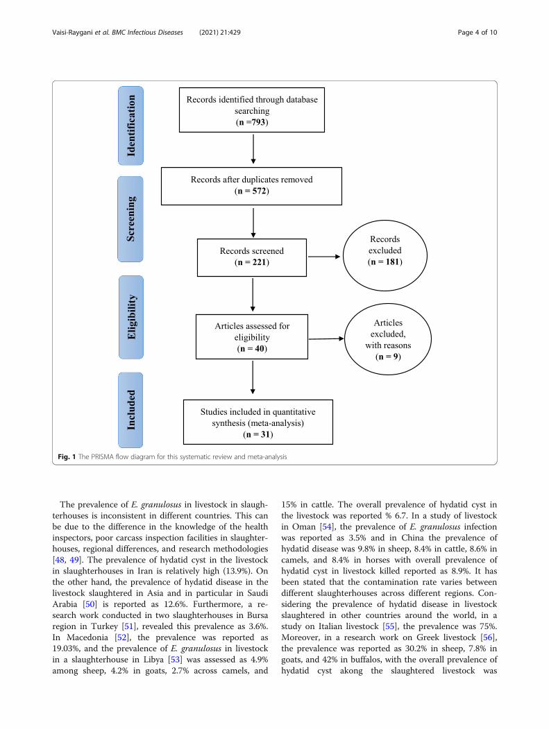
ResultsSearch outputIn the present study, all studies performed on the preva-lence of cystic echinococcosis among slaughtered live-stock in Iran were examined systematically based on thePRISMA guidelines. In the initial search, 724 studieswere identified, from which 31 studies published be-tween April 1970 to April 2020 entered the final analysis[15–45] (Fig. 1).Data of from all the final studies were extracted using
a different pre-prepared checklist. The items on thechecklist included: author’s name, article title, year and
location of the study, the domestic animal studied, sam-ple size, and the prevalence of cystic echinococcosisamong the slaughtered livestock in Iran [15–45](Table 1).
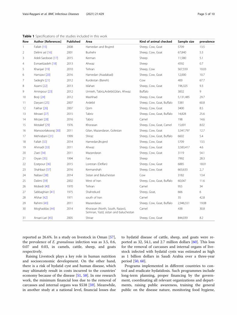
Heterogeneity and publication biasThe results of publication bias in studies were evaluatedby the Egger test, which were not statistically significant(P = 0.144) (Fig. 2). Also, the heterogeneity of the studieswas evaluated using the I2 index which value was 99%,Therefore, the random effects model was used to com-bine the results of the studies.The highest prevalence of cystic echinococcosis in
slaughtered livestock was reported in Baneh with 67.7%(95% CI: 63–72.2) [21], while the lowest cystic echino-coccosis in slaughtered livestock was observed in Ahwazslaughterhouses with 0.7% (95% CI: 0.5–0.9) [18] (Fig. 3).The total number of livestock included in this systematicreview and meta-analysis was 17,510,307 consisting ofsheep, cattle, goats, buffalos, and camels [15–45]. Theoverall prevalence of cystic echinococcosis among theslaughtered livestock in Iran based on the random ef-fects model was found to be 13.9% (95% CI: 10.7–17.7).
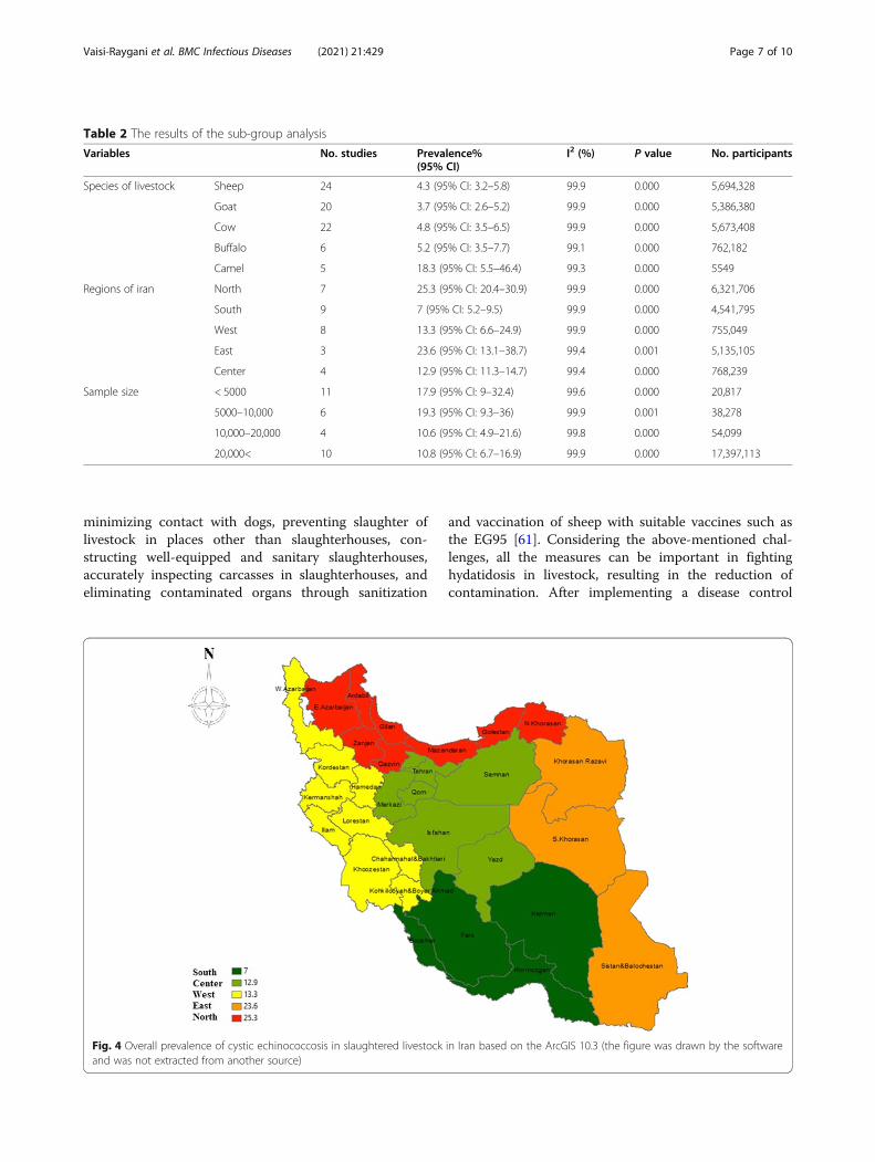
Sub-group analysisTable 2 presents an analysis of different sub-groups ac-cording to Specie of livestock, Regions of Iran, and Sam-ple size (Table 2) prevalence of cystic echinococcosis inslaughtered livestock in Iran were reported based on dif-ferent geographical areas in Iran and according to theGeographical Information System (GIS) (Fig. 4).
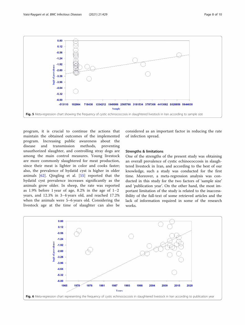
Meta-regression analysisAccordingly, the results of the meta-regression revealedthat any increase in the sample size is associated with astatistically significant growth in cystic echinococcosisprevalence. In other words, studies with larger samplesreported significantly higher prevalence of cystic echino-coccosis (P < 0.05) (Fig. 5). In addition, the increase ofthe publication year of a study was associated with a sig-nificant increase in cystic echinococcosis prevalence,such that the studies published in more recent years hadreported significantly higher cystic echinococcosis preva-lence compared to the older studies (P < 0.05) (Fig. 6).
DiscussionE. granulosus is known as a parasite and veterinary prob-lem in the Middle East. Its intermediate hosts includecamels, cows, sheep, and goats and the source for hu-man cystic echinococcosis (CE) is contaminated foodand water in which the parasite eggs and livestock areconsidered as reservoir hosts [46, 47]. The results of ourstudy suggest that the overall prevalence of cystic echi-nococcosis in slaughtered livestock in Iran is 13.9%.
Vaisi-Raygani et al. BMC Infectious Diseases (2021) 21:429 Page 3 of 10
The prevalence of E. granulosus in livestock in slaugh-terhouses is inconsistent in different countries. This canbe due to the difference in the knowledge of the healthinspectors, poor carcass inspection facilities in slaughter-houses, regional differences, and research methodologies[48, 49]. The prevalence of hydatid cyst in the livestockin slaughterhouses in Iran is relatively high (13.9%). Onthe other hand, the prevalence of hydatid disease in thelivestock slaughtered in Asia and in particular in SaudiArabia [50] is reported as 12.6%. Furthermore, a re-search work conducted in two slaughterhouses in Bursaregion in Turkey [51], revealed this prevalence as 3.6%.In Macedonia [52], the prevalence was reported as19.03%, and the prevalence of E. granulosus in livestockin a slaughterhouse in Libya [53] was assessed as 4.9%among sheep, 4.2% in goats, 2.7% across camels, and
15% in cattle. The overall prevalence of hydatid cyst inthe livestock was reported % 6.7. In a study of livestockin Oman [54], the prevalence of E. granulosus infectionwas reported as 3.5% and in China the prevalence ofhydatid disease was 9.8% in sheep, 8.4% in cattle, 8.6% incamels, and 8.4% in horses with overall prevalence ofhydatid cyst in livestock killed reported as 8.9%. It hasbeen stated that the contamination rate varies betweendifferent slaughterhouses across different regions. Con-sidering the prevalence of hydatid disease in livestockslaughtered in other countries around the world, in astudy on Italian livestock [55], the prevalence was 75%.Moreover, in a research work on Greek livestock [56],the prevalence was reported as 30.2% in sheep, 7.8% ingoats, and 42% in buffalos, with the overall prevalence ofhydatid cyst akong the slaughtered livestock was
Fig. 1 The PRISMA flow diagram for this systematic review and meta-analysis
Vaisi-Raygani et al. BMC Infectious Diseases (2021) 21:429 Page 4 of 10
reported as 26.6%. In a study on livestock in Oman [57],the prevalence of E. granulosus infection was as 3.5, 0.6,0.07 and 0.03, in camels, cattle, sheep, and goatsrespectively.Raising Livestock plays a key role in human nutrition
and socioeconomic development. On the other hand,there is a risk of hydatid cyst and human disease, whichmay ultimately result in costs incurred to the countries’economy because of the disease [51, 58]. In one researchwork, the minimum financial loss due to the removal ofcarcasses and internal organs was $538 [59]. Meanwhile,in another study at a national level, financial losses due
to hydatid disease of cattle, sheep, and goats were re-ported as 32, 54.1, and 2.7 million dollars [60]. This lossfor the removal of carcasses and internal organs of live-stock infected with hydatid cysts was estimated as highas 1 billion dollars in Saudi Arabia over a three-yearperiod [50, 60].Programs implemented in different countries to con-
trol and eradicate hydatidosis. Such programmes includelong-term planning, proper financing by the govern-ment, coordinating all relevant organizations and depart-ments, raising public awareness, training the generalpublic on the disease nature, monitoring food hygiene,
Table 1 Specifications of the studies included in this work
Row Author [References] Published Area Kind of animal checked Sample size prevalence
1 Fallah [15] 2008 Hamedan and Brujerd Sheep, Cow, Goat 5709 13.5
2 Delimi asl [16] 2001 Bushehr Sheep, Cow, Goat 67,840 5.3
3 Adeli-Sardooei [17] 2015 Kerman Sheep 11,580 5.1
4 Esmaeilzadeh [18] 2013 Ahwaz Sheep 4592 0.7
5 Khanjari [19] 2010 Tehran Sheep, Cow 567,559 10.03
6 Hamzavi [20] 2016 Hamedan (Asadabad) Sheep, Cow, Goat 12,000 10.7
7 Sadeghi [21] 2012 Kurdestan (Baneh) Cow 400 67.7
8 Azami [22] 2013 Isfahan Sheep, Cow, Goat 196,325 9.3
9 Aminpour [23] 2012 Urmieh, Tabriz,Ardebil,Gilan, Ahwaz Buffalo 3832 9
10 Borji [24] 2012 Mashhad Sheep, Cow, Goat 5,131,485 29.7
11 Daryani [25] 2007 Ardebil Sheep, Cow, Goat, Buffalo 5381 60.8
12 Fakhar [26] 2007 Qom Sheep, Cow, Goat 3400 8.5
13 Mirzaei [27] 2015 Tabriz Sheep, Cow, Goat, Buffalo 14,828 25.6
14 Mirzaei [28] 2016 Tabriz Camel 198 14.6
15 Motakef [29] 1976 Khorasan Sheep, Cow, Goat, Camel 15,691 8.02
16 Mansoorlakooraj [30] 2011 Gilan, Mazandaran, Golestan Sheep, Cow, Goat 3,347,797 12.7
17 Mehrabani [31] 1999 Shiraz Sheep, Cow, Goat, Buffalo 6602 5.4
18 Fallah [32] 2014 Hamedan,Brujerd Sheep, Cow, Goat 5709 13.5
19 Ahmadi [33] 2011 Ahwaz Sheep, Cow, Goat 3,583,417 4.6
20 Ziaei [34] 2011 Mazandaran Sheep, Cow, Goat 3119 54.1
21 Oryan [35] 1994 Fars Sheep 7992 28.3
22 Ezatpour [36] 2015 Lorestan (Delfan) Sheep, Cow, Goat 6885 18.01
23 Shahbazi [37] 2016 Kermanshah Sheep, Cow, Goat 663,633 2,.7
24 Nabavi [38] 2014 Sistan and Baluchestan Cow 3182 13.4
25 Dalimi [39] 2002 West of Iran Sheep, Cow, Goat, Buffalo 60,047 11.6
26 Mobedi [40] 1970 Tehran Camel 955 34
27 Sabbaghian [41] 1975 Shahrekurd Sheep, Goat, 666 6
28 Afshar [42] 1971 south of Iran Camel 35 42.8
29 Rahimi [43] 2011 Mazandaran Sheep, Cow, Goat, Buffalo 2,946,551 19.08
30 Moghaddas [44] 2014 Khorasan (North, South, Razavi),Semnan, Yazd, sistan and baluchestan
Camel 438 30.8
31 Ansari-Lari [45] 2005 Shiraz Sheep, Cow, Goat 844,039 8.2
Vaisi-Raygani et al. BMC Infectious Diseases (2021) 21:429 Page 5 of 10
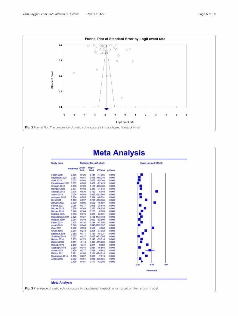
Fig. 2 Funnel Plot: The prevalence of cystic echinococcosis in slaughtered livestock in Iran
Fig. 3 Prevalence of cystic echinococcosis in slaughtered livestock in Iran based on the random model
Vaisi-Raygani et al. BMC Infectious Diseases (2021) 21:429 Page 6 of 10
minimizing contact with dogs, preventing slaughter oflivestock in places other than slaughterhouses, con-structing well-equipped and sanitary slaughterhouses,accurately inspecting carcasses in slaughterhouses, andeliminating contaminated organs through sanitization
and vaccination of sheep with suitable vaccines such asthe EG95 [61]. Considering the above-mentioned chal-lenges, all the measures can be important in fightinghydatidosis in livestock, resulting in the reduction ofcontamination. After implementing a disease control
Table 2 The results of the sub-group analysis
Variables No. studies Prevalence%(95% CI)
I2 (%) P value No. participants
Species of livestock Sheep 24 4.3 (95% CI: 3.2–5.8) 99.9 0.000 5,694,328
Goat 20 3.7 (95% CI: 2.6–5.2) 99.9 0.000 5,386,380
Cow 22 4.8 (95% CI: 3.5–6.5) 99.9 0.000 5,673,408
Buffalo 6 5.2 (95% CI: 3.5–7.7) 99.1 0.000 762,182
Camel 5 18.3 (95% CI: 5.5–46.4) 99.3 0.000 5549
Regions of iran North 7 25.3 (95% CI: 20.4–30.9) 99.9 0.000 6,321,706
South 9 7 (95% CI: 5.2–9.5) 99.9 0.000 4,541,795
West 8 13.3 (95% CI: 6.6–24.9) 99.9 0.000 755,049
East 3 23.6 (95% CI: 13.1–38.7) 99.4 0.001 5,135,105
Center 4 12.9 (95% CI: 11.3–14.7) 99.4 0.000 768,239
Sample size < 5000 11 17.9 (95% CI: 9–32.4) 99.6 0.000 20,817
5000–10,000 6 19.3 (95% CI: 9.3–36) 99.9 0.001 38,278
10,000–20,000 4 10.6 (95% CI: 4.9–21.6) 99.8 0.000 54,099
20,000< 10 10.8 (95% CI: 6.7–16.9) 99.9 0.000 17,397,113
Fig. 4 Overall prevalence of cystic echinococcosis in slaughtered livestock in Iran based on the ArcGIS 10.3 (the figure was drawn by the softwareand was not extracted from another source)
Vaisi-Raygani et al. BMC Infectious Diseases (2021) 21:429 Page 7 of 10
program, it is crucial to continue the actions thatmaintain the obtained outcomes of the implementedprogram. Increasing public awareness about thedisease and transmission methods, preventingunauthorized slaughter, and controlling stray dogs areamong the main control measures. Young livestockare more commonly slaughtered for meat production,since their meat is lighter in color and cooks faster;also, the prevalence of hydatid cyst is higher in olderanimals [62]. Qingling et al. [55] reported that thehydatid cyst prevalence increases significantly as theanimals grow older. In sheep, the rate was reportedas 1.9% before 1 year of age, 8.2% in the age of 1–2years, and 12.3% in 3–4 years old, and reached 17.2%when the animals were 5–6 years old. Considering thelivestock age at the time of slaughter can also be
considered as an important factor in reducing the rateof infection spread.
Strengths & limitationsOne of the strengths of the present study was obtainingan overall prevalence of cystic echinococcosis in slaugh-tered livestock in Iran, and according to the best of ourknowledge, such a study was conducted for the firsttime. Moreover, a meta-regression analysis was con-ducted in this study for the two factors of ‘sample size’and ‘publication year’. On the other hand, the most im-portant limitation of the study is related to the inaccess-ibility of the full-text of some retrieved articles and thelack of information required in some of the researchworks.
Fig. 5 Meta-regression chart showing the frequency of cystic echinococcosis in slaughtered livestock in Iran according to sample size
Fig. 6 Meta-regression chart representing the frequency of cystic echinococcosis in slaughtered livestock in Iran according to publication year
Vaisi-Raygani et al. BMC Infectious Diseases (2021) 21:429 Page 8 of 10
ConclusionConsidering the results of this research, there is rela-tively high prevalence of hydatid cyst in livestock inslaughterhouses. Moreover, since hydatid cyst is a riskfactor for human health, it is necessary that health policymakers make effective decisions in relation to this dis-ease and implement accurate inspections by health ex-perts and authorities.
AbbreviationsSID: Scientific Information Database; GIS: Geographic Information System;STROBE: Strengthening the Reporting of Observational Studies inEpidemiology for cross- sectional Study; PRISMA: Preferred reporting itemsfor Systematic Reviews and Meta-Analysis
AcknowledgementsThe authors would like to thank the members of the Faculty of Nursing andMidwifery, Kermanshah University of Medical Sciences.
Authors’ contributionsMM contributed to the design, statistical analysis, participated in most of thestudy steps. MM and AVR prepared the manuscript. MM and RJ and NS andMHF assisted in designing the study, and helped in the, interpretation of thestudy. All authors read and approved the final version of the manuscript.
FundingFunding for this research was provided by the deputy of research andtechnology –Kermanshah University of Medical Sciences,(980824), the deputyof research and technology –Kermanshah University of Medical Sciences hadno role in the design of the study and collection, analysis, and interpretationof data and in writing of the manuscript.
Availability of data and materialsDatasets are available through the corresponding author upon reasonablerequest.
Declarations
Ethics approval and consent to participateEthics approval was received from the ethics committee of deputy ofresearch and technology, Kermanshah University of Medical Sciences.Reference Number: IR.KUMS.REC.1398.482.
Consent for publicationNot applicable.
Competing interestsThe authors declare that they have no conflict of interest.
Author details1Department of Nursing, School of Nursing and Midwifery, KermanshahUniversity of Medical Sciences, Kermanshah, Iran. 2Department ofBiostatistics, School of Health, Kermanshah University of Medical Sciences,Kermanshah, Iran. 3Department of Food Science & Technology, Faculty ofAgriculture, Ferdowsi University of Mashhad (FUM), Mashhad, Iran.
Received: 8 January 2020 Accepted: 29 April 2021
References1. Grosso G, Gruttadauria S, Biondi A, Marventano S. Mistretta worldwide
epidemiology of liver hydatidosis including the Mediterranean area. World JGastroenterol. 2012;18(13):1425–37. https://doi.org/10.3748/wjg.v18.i13.1425.
2. Hosseini-Safa A, Mohaghegh MA, Pestechian N, Ganji M, Mohammadi R,Lamouki M, et al. First report of Tasmanian sheep strain (G2) genotypeisolated from Iranian goat using the high-resolution melting (HRM) analysis.Gastroenterol Hepatol Bed Bench. 2016;9(Suppl1):S70–4.
3. Jahangir A, Taherikalani M, Asadolahi K, Emaneini M. Echinococcosis/hydatidosis in Ilam province, western Iran. Iran J Parasitol. 2013;8(3):417–22.
4. Mandal S, Mandal MD. Human cystic echinococcosis: epidemiologic,zoonotic, clinical, diagnostic and therapeutic aspects. Asian Pac J Trop Dis.2012;5(4):253–60. https://doi.org/10.1016/S1995-7645(12)60035-2.
5. Karimi A, Asadi K, Mohseni F, Akbar MH. Hydatid cyst of the biceps femorismuscle (a rare case in orthopedic surgery). Shiraz E Med J. 2011;12(3):150–4.
6. Salem COA, Schneegans F, Chollet J, Jemli M. Epidemiological studies onechinococcosis and characterization of human and livestock hydatid cystsin Mauritania. Iran J Parasitol. 2011;6(1):49–57.
7. Berenji F, Jamshidi M. A case report of muscle hydatidosis from Iran. Iran JParasitol. 2014;10(1):132–5.
8. Dakkak A. Echinococcosis/hydatidosis: a severe threat in Mediterraneancountries. Vet Parasitol. 2010;174(1):2–11. https://doi.org/10.1016/j.vetpar.2010.08.009.
9. Yousefi H. SituationI of HydatidY cyst infection during last two decades(1985-2005) in Iran (review of articles). J Shahrekord Univ Med Sci. 2008;10(1):78–88.
10. Yousofi H, Mahmoudi T, Zebardast N, Ganji F. Survey of the risk factors ofhydatid cyst infection in Lordegan area of Chaharmahal and Bakhtiariprovince of Iran, 2004. J Shahrekord Univ Med Sci. 2007;8(4):63–7.
11. Yahya AI, Bhatnagar R. Surgical techniques performed in hepaticHydatidosis in Libya: an endemic area. Ind J Pathol. 2014;3(2):29–59.
12. Khalkhali H, Foroutan M, Khademvatan S, Majidiani H, Aryamand S, Khezri P,et al. Prevalence of cystic echinococcosis in Iran: a systematic review andmeta-analysis. J Helminthol. 2018;92(3):260–68.
13. Moazeni M. Hydatid cyst control: a glance at the experiences of othercountries. Payavard Salamat. 2008;1(2):9–11.
14. Colon G. Editor carcass elimination as a measure to prevent Hydatidosis. VetAspects Echinococcosis Congress. 2002;5(2):63–9.
15. Fallah M, Kavand A, Yossefimashof R. Evaluation of bacterial contaminationof hydatid cysts and determination of the type of bacteria causing infectionin slaughtered animals in Hamadan and Borujerd slaughterhouses 2006.Yafteh. 2008;10(3):29–37.
16. Delimi Asl AH, Zariffard MR, Pour Ebrahim MR. Echinococcosis (hydatidiosis)study in Bushehr province. Pajouhesh and Sazandegi. 2001;50:86–7.
17. Adeli-Sardooei M, Hayati B, Badakhshan Y. Financial loss estimation of cysticechinococcosis of sheep in south of Kerman province abattoirs (2011-2014).J Anim Res. 2015;25(4):158–68.
18. Esmaeilzadeh S, Sabbagh A, Mohammadian B, Alborzi AR, Ghorbanpoor M,Pourmehdi Borujeni M. Frequency of ovine pulmonary lesions in Ahvazslaughterhouse: pathological, bacteriological and parasitological study. IranVet J. 2014;9(4):14–25.
19. Khanjari A, Bokaei S, Abbaszadeh S, Nemati GH, Akhondzade Bastani A, MisaghiA, et al. Evaluation of hydatid cyst prevalence in slaughtered animals inMeysam slaughterhouse (Southwest of Tehran province). Vet J. 2010;88:40–4.
20. Hamzavi Y, Nazari N, Mikaeili A, Parandin F, Faizi F, Sardari M.Prevalence of Hydatid Cystin slaughtered livestock in Asadabadslaughterhouse during 2014-2015. Pajouhan Sci J. 2016;14(3):58–66.https://doi.org/10.21859/psj-140358.
21. Sadeghi Dehkurdi Z, Yousefi M. Prevalence of hydatid cyst in Banehslaughterhouse cattle. J Vet Lab Res. 2012;4(1):129.
22. Azami M, Anvarinejad M, Ezatpour B, Alirezae M. Prevalence of Hydatidosisin slaughtered animals in Iran. Turkiye Parazitol Derg. 2013;37(2):102–6.https://doi.org/10.5152/tpd.2013.24.
23. AminPour A, Hosseini SH, Shayan P. The prevalence and fertility of hydatidcysts in buffaloes from Iran. J Helminthol. 2012;86(3):373–7. https://doi.org/10.1017/S0022149X11000514.
24. Borji H, Azizzadeh M, Afsai A. An abattoir-based study on the prevalence ofhydatidosis in livestock in Mashhad, Iran. J Helminthol. 2012;86(2):233–6.https://doi.org/10.1017/S0022149X11000228.
25. Daryani A, Alaei R, Arab R, Sharif M, Dehghan MH, Ziaei H. The prevalence,intensity and viability of hydatid cysts in slaughtered animals in the Ardabilprovince of Northwest Iran. J Helminthol. 2007;81(1):13–7. https://doi.org/10.1017/S0022149X0720731X.
26. Fakhar M, Sadjjadi SM. Prevalence of hydatidosis in slaughtered herbivoresin Qom province, central part of Iran. Vet Res Commun. 2007;31(8):993–7.https://doi.org/10.1007/s11259-007-0017-4.
27. Mirzaei M, Rezaei H, Nematollahi A. Role of ruminants in the epidemiologyof Echinococcus granulosus in Tabriz area, Northwest of Iran. Trop Biomed.2015;32(2):269–75.
28. Mirzaei M, Rezaei H, Nematollahi A, Ashrafihelan J. Survey of hydatidosisinfection in slaughtered camel (Camelus dromedarius) in Tabriz area,
Vaisi-Raygani et al. BMC Infectious Diseases (2021) 21:429 Page 9 of 10
Nortwest of Iran. J Parasit Dis. 2016;40(2):444–7. https://doi.org/10.1007/s12639-014-0523-6.
29. Motakef M, Minou AA, Lari MM. An epidemiological approach to the studyof Echinococcosis in north-east region of Iran (Khorassan). Pahlavi Med J.1976;7(4):503–15.
30. Mansoorlakooraj H, Saadati D, Javadi R, Heydari S, Torki E, Gholami H, et al.A survey on hydatidosis in livestock in northern Iran based on datacollected from slaughterhouses from 2004 to 2008. Vet Parasitol. 2011;182(2–4):364–7. https://doi.org/10.1016/j.vetpar.2011.05.013.
31. Mehrabani D, Oryan A, Sadjjadi SM. Prevalence of Echinococcus granulosusinfection in stray dogs and herbivores in Shiraz, Iran. Vet Parasitol. 1999;86(3):217–20. https://doi.org/10.1016/S0304-4017(99)00151-X.
32. Fallah M, Kavand A, Mashouf RY. Infected hydatid cysts bacteria inslaughtered livestock and their effects on protoscoleces degeneration.Jundishapur J Microbiol. 2014;7(6):e10135. https://doi.org/10.5812/jjm.10135.
33. Ahmadi NA, Meshkehkar M. An abattoir-based study on the prevalence andeconomic losses due to cystic echinococcosis in slaughtered herbivores inAhwaz, South-Western Iran. J Helminthol. 2011;85(1):33–9. https://doi.org/10.1017/S0022149X10000234.
34. Ziaei H, Fakhar M, Armat S. Epidemiological aspects of cystic echinococcosisin slaughtered herbivores in sari abattoir, North of Iran. J Parasit Dis. 2011;35(2):215–8. https://doi.org/10.1007/s12639-011-0051-6.
35. Oryan A, Moghaddar N, Gaur SN. Metacestodes of sheep with specialreference to their epidemiological status, pathogenesis and economicimplications in Fars Province, Iran. Vet Parasitol. 1994;51(3–4):231–40. https://doi.org/10.1016/0304-4017(94)90160-0.
36. Ezatpour B, Farhadi SJ, Azami M, Alirezaei M, Ebrahimzadeh F. Importance ofcystic echinococcosis in slaughtered herbivores from Iran. J Parasit Dis.2015;39(2):234–7. https://doi.org/10.1007/s12639-013-0328-z.
37. Shahbazi Y, Hashemnia M, Afshari Safavi EA. A retrospective survey ofhydatidosis based on abattoir data in Kermanshah, Iran from 2008 to 2013. JParasit Dis. 2016;40(2):459–63. https://doi.org/10.1007/s12639-014-0526-3.
38. Nabavi R, Khedri J, Saadati D. Comparative prevalence of hepato-pulmonaryhydatidosis among native and imported cattle in north of Sistan andBaluchestan: Iran. J Parasit Dis. 2014;38(4):371–3. https://doi.org/10.1007/s12639-013-0262-0.
39. Dalimi A, Motamedi G, Hosseini M, Mohammadian B, Malaki H, Ghamari Z,et al. Echinococcosis/hydatidosis in western Iran. Vet Parasitol. 2002;105(2):161–71. https://doi.org/10.1016/S0304-4017(02)00005-5.
40. Mobedi I, Madadi H, Arfaa F. Camel, Camelus dromedarius, as intermediatehost of Echinococcus granulosus in Iran. J Parasitol. 1970;56(6):1255. https://doi.org/10.2307/3277581.
41. Sabbaghian H, Hoghochi N, Ghadirian E. A survey on the prevalence ofechinococcosis in Shahre-Kord, Iran. Bull Soc Pathol Exot Filiales. 1975;68(6):574–8.
42. Afshar A, Nazarian I, Baghban-Baseer B. A survey of the incidence of hydatidcyst in camels in South Iran. Br Vet J. 1971;127(11):544–6. https://doi.org/10.1016/S0007-1935(17)37288-3.
43. Rahimi MT, Sharifdini M, Ahmadi A, Laktarashi B, Mahdavi SA, BeigomKia E.Hydatidosis in human and slaughtered herbivores in Mazandaran province,northern Iran. Asian Pac J Trop Dis. 2011;1(3):212–5. https://doi.org/10.1016/S2222-1808(11)60031-5.
44. Moghaddas E, Borji H, Naghibi AG, Razmi GH, Shayan P. Epidemiologicalstudy of hydatidosis in the dromedaries (Camelus dromedarius) of differentregions of Iran. Asian Pac J Trop Biomed. 2014;4(1):148–51.
45. Ansari-Lari M. A retrospective survey of hydatidosis in livestock in shiraz,Iran, based on abattoir data during 1999–2004. Vet Parasitol. 2005;133(1):119–23. https://doi.org/10.1016/j.vetpar.2005.05.031.
46. Benito A, Carmena JL, Martinez J, Guisantes JA. Dog echinococcosis innorthern Spain: comparison of coproantigen and serum antibody assayswith coprological exam. Vet Parasitol. 2006;142(1-2):102–11. https://doi.org/10.1016/j.vetpar.2006.06.011.
47. Demir P, Mor N. Seasonal distribution and economic importance of cysticechinococcosis in cattle slaughtered at Kars municipal abattoir, Turkey.Turkiye Parazitol Derg. 2011;35(4):185–8. https://doi.org/10.5152/tpd.2011.48.
48. Randolph TF, Schelling E, Grace D, Nicholson CF, Leroy JL, Cole DC, et al.Role of livestock in human nutrition and health for poverty reduction indeveloping countries. J Anim Sci. 2007;85(11):2788–800. https://doi.org/10.2527/jas.2007-0467.
49. McManus DP. Molecular discrimination of taeniid cestodes. Parasitol Int.2006;55(Suppl):31–7.
50. Ibrahim MM. Study of cystic echinococcosis in slaughtered animals in AlBaha region, Saudi Arabia: Interaction between some biotic and abioticfactors. Acta Trop. 2010;113:26–33.
51. Sariozkan S, Yalcin C. Estimating the production losses due tocysticechinococcosis in ruminants in Turkey. Vet Parasitol. 2009;163(4):330–4.https://doi.org/10.1016/j.vetpar.2009.04.032.
52. Nayar S. Hypertrophy of the liver due to hydatid cyst--a probable cause forrecurrent tympany in a cross-bred bull. Ind Vet J. 1974;51(2):161–3.
53. Kassem HH, Abdel-Kader AK, Nass SA. Prevalence of hydatid cysts inslaughtered animals in Sirte, Libya. J Egypt Soc Parasitol. 2013;43(1):33–40.https://doi.org/10.12816/0006365.
54. Al Kitani FA, Al Riyami S, Al Yahyai S, Al Rawahi AH, Al Maawali M, HussainMH. Abattoir based surveillance of cystic echinococcosis (CE) in theSultanate of Oman during 2010-2013. Vet Parasitol. 2015;211(3–4):208–15.https://doi.org/10.1016/j.vetpar.2015.06.011.
55. Qingling M, Guanglei W, Jun Q, Xinquan Z, Tianli L, Xuemei S, et al.Prevalence of hydatid cysts in livestock animals in Xinjiang, China. Korean JParasitol. 2014;52(3):331–4. https://doi.org/10.3347/kjp.2014.52.3.331.
56. Scala A, Garippa G, Varcasia A, Tranquillo V, Genchi C. Cystic Echinococcosisin slaughtered sheep in Sardinia (Italy). Vet Parasitol. 2006;135(1):33–8.https://doi.org/10.1016/j.vetpar.2005.08.006.
57. Chaligiannis I, Maillard S, Boubaker G, Spiliotis M, Saratsis A, Gottstein B,et al. Echinococcus granulosus infection dynamics in livestock of Greece.Acta Trop. 2015;150:64–70. https://doi.org/10.1016/j.actatropica.2015.06.021.
58. Saeed I, Kapel C, Saida LA, Willingham L, Nansen P. Epidemiology ofEchinococcus granulosus in Arbil province, northern Iraq, 1990–1998. JHelminthol. 2000;74(1):83–8. https://doi.org/10.1017/S0022149X00000111.
59. Kebede N, Gebre-Egziabher Z, Tilahun G, Wossene A. Prevalence andfinancial effects of hydatidosis in cattle slaughtered in Birre- Sheleko andDangila abattoirs, Nortwestern Ethiopa. Zoonoses Public Health. 2011;58(1):41–6. https://doi.org/10.1111/j.1863-2378.2009.01250.x.
60. Razi Jalali MH, Ghorbanpor Najafabadi M, Hoghoghi Rad N. Study ofexperimental Hydatid disease effects on live weight of sheep. Iran J AnimVet Sci. 2005;11:81–7.
61. Moazeni M. Hydatid Cyst Control: A glance at the experiences of othercountries. Payavard. 2008;1(2):11–9.
62. Torgerson PR, Heath DD. Transmission dynamics and control options forEchinococcus granulosus. Parasitology. 2003;127(Suppl):143–58.
Publisher’s NoteSpringer Nature remains neutral with regard to jurisdictional claims inpublished maps and institutional affiliations.
Vaisi-Raygani et al. BMC Infectious Diseases (2021) 21:429 Page 10 of 10


