Embed Size (px)
Citation preview
RESEARCH Open Access
Pro-inflammatory cytokine/chemokine productionby reovirus treated melanoma cells is PKR/NF-�Bmediated and supports innate and adaptiveanti-tumour immune primingLynette Steele1, Fiona Errington1, Robin Prestwich1, Elizabeth Ilett1, Kevin Harrington2, Hardev Pandha3,Matt Coffey4, Peter Selby1, Richard Vile1,5, Alan Melcher1*
Abstract
Background: As well as inducing direct oncolysis, reovirus treatment of melanoma is associated with activation ofinnate and adaptive anti-tumour immune responses.
Results: Here we characterise the effects of conditioned media from reovirus-infected, dying human melanomacells (reoTCM), in the absence of live virus, to address the immune bystander potential of reovirus therapy. Inaddition to RANTES, IL-8, MIP-1a and MIP-1b, reovirus-infected melanoma cells secreted eotaxin, IP-10 and the type1 interferon IFN-b. To address the mechanisms responsible for the inflammatory composition of reoTCM, we showthat IL-8 and IFN-b secretion by reovirus-infected melanoma cells was associated with activation of NF-�B anddecreased by pre-treatment with small molecule inhibitors of NF-�B and PKR; specific siRNA-mediated knockdownfurther confirmed a role for PKR. This pro-inflammatory milieu induced a chemotactic response in isolated naturalkiller (NK) cells, dendritic cells (DC) and anti-melanoma cytotoxic T cells (CTL). Following culture in reoTCM, NK cellsupregulated CD69 expression and acquired greater lytic potential against tumour targets. Furthermore, melanomacell-loaded DC cultured in reoTCM were more effective at priming adaptive anti-tumour immunity.
Conclusions: These data demonstrate that the PKR- and NF-�B-dependent induction of pro-inflammatorymolecules that accompanies reovirus-mediated killing can recruit and activate innate and adaptive effector cells,thus potentially altering the tumour microenvironment to support bystander immune-mediated therapy as well asdirect viral oncolysis.
BackgroundReovirus is a nonenveloped dsRNA virus which is highlyprevalent in the human population but produces fewclinical symptoms. Great interest has surrounded theuse of reovirus as an oncolytic agent due to its ability toinfect and induce death in a range of human malignan-cies whilst sparing normal cells. Furthermore reovirushas completed a number of early clinical trials and isnow being tested in the phase III setting [1-3]. Initialstudies indicated that the tumour specific oncolyticactivity was dependent upon the presence of an
activated Ras signalling pathway [4], although recentdata has indicated that susceptibility to reovirus infec-tion may be influenced by additional complex mechan-isms [5,6].Previous work in our laboratory has indicated that
human melanoma cell lines, as well as freshly resectedtumour, undergo reovirus-induced apoptotic death in aRas/RalGEF/p38 dependent manner, and that this deathis accompanied by the release of inflammatory chemo-kines and cytokines [7]. The release of pro-inflammatorymediators following viral infection of tumour cells hasbeen observed with other oncolytic viruses such asHerpes Simplex Virus (HSV) [8] and Newcastle diseasevirus (NDV) [9].* Correspondence: [email protected]
1Leeds Institute of Molecular Medicine, University of Leeds, Leeds, UKFull list of author information is available at the end of the article
Steele et al. Molecular Cancer 2011, 10:20http://www.molecular-cancer.com/content/10/1/20
© 2011 Steele et al; licensee BioMed Central Ltd. This is an Open Access article distributed under the terms of the Creative CommonsAttribution License (http://creativecommons.org/licenses/by/2.0), which permits unrestricted use, distribution, and reproduction inany medium, provided the original work is properly cited.
As well as inducing direct oncolysis, several viruses,either naturally or via insertion of immune-activatinggenes, have been shown to stimulate anti-tumourimmune responses, indicating their potential as immu-notherapeutic as well as cytotoxic agents [10]. We havepreviously shown that reovirus can exert immunogeniceffects against tumour cells by directly activating DC tostimulate innate NK/T cell cytotoxicity [11], and by reo-virus-induced tumour cell death facilitating the primingof innate and adaptive anti-tumour responses in mouseand human model systems [12-14]. However, the immu-nogenicity of the pro-inflammatory milieu produced byreovirus-infected melanoma cells (independent of theeffects of the virus itself which may be cleared rapidly invivo), and the signalling pathways involved in initiatingcytokine/chemokine production in tumour cells, havenot been addressed.Chemokines can participate in the host response dur-
ing infection and inflammation by directing immuneeffector cell migration. Four families of chemokineshave been described based on the position of conservedcysteine residues [15]. Multiple chemokines can shareone common receptor, and each chemokine can poten-tially bind to several different receptors, thereby allow-ing multiple biological outcomes depending upon thecomposition of the chemokine milieu and the cellswithin the environment [16,17]. Furthermore, at sites ofinflammation, chemokines can form heteromers, poten-tially inducing synergistic actions and enhancing leuko-cyte migration and activation [18]. Hence, the inductionof multiple chemokines within an immunosuppressivetumour microenvironment has the potential to inducepotent effects on immune effector cells to enhance ther-apy. For example, in a murine B16 melanoma model,ectopic expression and secretion of IP-10 by tumourcells increased the number of NK cells at the tumoursite and prolonged NK cell dependent survival [19].Data have also indicated a good correlation betweenCXCR3 expression on T cells and an improved clinicaloutcome in stage III melanoma patients [20].The current study further investigates the chemokines
and cytokines (including type I IFNs) induced by reo-viral oncolysis and the signaling pathways responsiblefor the production of these pro-inflammatory mediators.We also determine the effects of reoTCM, specifically inthe absence of active virus to exclude the direct conse-quences of viral immune activation, on chemotaxis, acti-vation and effector functions of NK cells, DC and CTL.
Materials and methodsCell Culture and ReovirusSkmel-28, Mel-624, Mel-888 and MeWo cells weregrown in Dulbecco’s Modified Eagle Medium (DMEM)(Invitrogen, Paisley, UK) supplemented with 10% (v/v)
foetal calf serum (FCS) (BioSera, Crawley Down, UK)and 1% (v/v) L-glutamine (Sigma Aldrich). Peripheralblood mononuclear cells (PBMC) were obtained, withlocal ethics approval, from buffy coats of healthy blooddonors by Ficoll-Hypaque density centrifugation.Human myeloid immature dendritic cells (DC) weregenerated from human PBMC by MACS CD14+ selec-tion (Miltenyi Biotech) or monocyte adherence as pre-viously described [21]. Monocytes were cultured inRPMI 1640 (Invitrogen Life Technologies) supplementedwith 10% FCS, 1% L-glutamine (complete media), 800U/ml GM-CSF (Schering-Plough) and 500 U/ml IL-4(R&D systems) for 5 days. NK cells were isolated fromhuman PBMC using MACS negative depletion kits fol-lowing standard protocols as previously described (>90% purity) [11]. NK cells were routinely cultured inDMEM supplemented with 10% (v/v) human AB serum(Sera Laboratories International Limited), 5% (v/v) foetalcalf serum and 1% (v/v) L-glutamine. Cytotoxic T lym-phocytes (CTL) primed using reovirus infected mel-888cells (moi 0.1) were generated in a 14 day priming assayas detailed below. For chemotaxis assays, NK, DC andCTL were resuspended in RPMI/0.5%Human AB/1%L-Glutamine. All cell lines were grown at 37°C in anatmosphere containing 5% CO2 and were routinelytested for, and confirmed free of, Mycoplasma. Whereindicated melanoma cells were treated with 2.5 mM2-aminopurine (2-AP) (Sigma-Aldrich) or 50 μM caffeicacid phenethyl ester (CAPE) (Axxora, Nottingham, UK)for 1 hour prior to, and during, treatment with reovirus.Neither 2-AP nor CAPE at the doses used were directlytoxic to melanoma cells (data not shown). Reovirus type3 Dearing strain was provided by Oncolytics BiotechInc. Virus titer was determined by a standard plaqueassay using L929 cells.
ELISA, Luminex and Flow CytometryIL-8 was detected using matched pair antibodies (BDPharmingen). IFN-b was detected using an IFN-b kit(PBL Laboratories) as per the manufacturer’s instruc-tions. Eotaxin and IP-10 were detected using Luminextechnology (Biosource) according to the manufacturer’sinstructions. To assess NK cell phenotypic activation,NK cells were labelled with CD69-APC/CD56-PE/CD3PerCP antibodies (BD Pharmingen, R and D Sys-tems). CD69 expression was determined on gatedCD56-PE+ve/CD3-PerCP-ve populations using a FACS-Calibur instrument (BD Biosciences).
Western BlottingMelanoma cell lines were seeded in 10 cm dishes andleft untreated or infected with 10pfu/cell reovirus for 4,8, 12, 16, 20 and 24 hours prior to preparation of wholecell lysates. Nuclear fractions were prepared using the
Steele et al. Molecular Cancer 2011, 10:20http://www.molecular-cancer.com/content/10/1/20
Page 2 of 13
Active Motif Nuclear Extract Kit according to the man-ufacturers’ instructions. Whole cell lysates were pre-pared using RIPA buffer supplemented with a proteaseinhibitor cocktail (25 μl/ml, Sigma). 10 μg of total pro-tein was loaded per lane for whole cell lysates. 20 μg ofprotein was loaded per lane for nuclear fractions. Pro-teins were separated on a 10% SDS polyacrylamide geland transferred to a nitrocellulose membrane. Mem-branes were probed with antibodies against total PKR(BD Transduction Laboratories), I-�B (Santa Cruz) andp65 NF-�B (Upstate). Antibody binding was detectedusing the Odyssey system (LI-COR Biosciences UK,Cambridge, UK). Equal lane loading was confirmedusing monoclonal antibody against b-actin (Sigma).
RNA Interference (RNAi) StudiessiRNAs were purchased from Ambion and reconstitutedto a concentration of 20 μM according to the manufac-turers’ instructions. An siRNA oligonucleotide againstPKR (PKRV) and an irrelevant (green fluorescent pro-tein-specific) control siRNA were used. The sequenceswere, PKRV 5’ AAGGUGAAGGUAGAUCAAAGA-3’,Control 5’ AAGGACGACGGAAACUACAAG-3’. Mela-noma cells in 24 well plates and 10 cm dishes weretransfected with 100 nM (this concentration was opti-mized by initial titration experiments) of siRNA. ControlsiRNA transfections using equivalent concentrationswere included in each experiment. Transfections werecarried out in serum-free media using OligofectAMINE(Invitrogen) for 6 hours. Media were replenished withserum and supplements and cells were cultured in thecontinued presence of siRNA overnight. After the over-night incubation pre-existing media was removed fromthe 24 well plates and replaced with 1 ml normalgrowth media, and cells were left untreated or infectedwith reovirus (0-10pfu/cell). Supernatants from thesecells were collected at 24 and 48 hours post reovirusinfection. For 10 cm dishes the pre-existing media wasremoved and whole cell lysates were prepared and sepa-rated by SDS-PAGE prior to probing with total PKRantibody. Equal lane loading was confirmed using amonoclonal antibody against b-actin.
Priming of Mel-888 Cytotoxic T Lymphocytes (CTL)Mel-888 cells were seeded into tissue culture flasks andallowed to adhere. DC, resuspended in a 50:50 mix ofDC media/reoTCM (from Mel-888), non-reoTCM, orDC media/DMEM complete, were added to the Mel-888cells at a 1:3 ratio (DC:tumour cell) overnight. Suspen-sion cells were aspirated, leaving the tumour monolayerintact, and cells were pelleted as previously described[13]. Tumour loaded DC were then resuspended in CTLmedia [RPMI supplemented with 7.5% (v/v) human ABserum (Sigma), 1% (v/v) L-glutamine, 1% (v/v) sodium
pyruvate (Life Technologies), 1% (v/v) non-essentialamino acids (Life Technologies), 1% (v/v) HEPES (LifeTechnologies), 20 μmol/L 2 -mercaptoethanol (Sigma),and mixed with autologous PBMC at a ratio of 1:10 to1:30. Cultures were supplemented with 5 ng/ml IL-7(R&D systems) from day 1 and 30 U/ml IL-2 (R&D sys-tems) on day 4 only. Cultures were re-stimulated usingthe same protocol at day 7. Cells were harvested at day14 and chromium release, CD107 degranulation andintracellular IFN-g assessed as detailed below.
NK, DC and CTL chemotaxis1.5 × 105 melanoma cells were plated in 6 well platesand left overnight. Pre-existing media was removed andreplaced with DMEM/2%FCS/1% L-Glutamine and cellswere infected with 1 or 10pfu/cell reovirus for 48 hours.Supernatants were collected and passed through a0.2 μm Acrodisc syringe filter (Pall Life Sciences) andthen through a ViresolveNFR filter (Millipore) to removevirus. Chemotaxis was assessed using a transwell system.650 μl filtered TCM from each of the four melanomacell lines was added to the lower chamber of triplicatewells in a 24-well plate and a 5 μm (NK cells and CTL)or 8 μm (DC) Thincert™ membrane (Greiner Bio-One)placed in the well. 5 × 10⁵ NK, DC or CTL (the anti-Mel-888 CTL used in these chemotaxis experiments hadbeen previously primed using reovirus-infected Mel-888-loaded DC as described [12]), resuspended in 100 μlof low serum media were placed in the upper chamberand plates were incubated at 37°C for 3 hours. Cells inthe lower wells were harvested, washed in FACS bufferand labelled with 3 μl of CD56-PE (Serotec) and 3 μlCD3-FITC (BD Biosciences) (NK cells), CD11c-PE (BDBiosciences) (DC) or 3 μl CD3-FITC and CD8-PerCP(CTL) for 30 minutes at 4°C. Cells were washed, resus-pended in 300 μl FACS buffer and transferred to a Tru-Count™ tube (BD Biosciences) containing a knownnumber of fluorescent beads to provide the internalcounting control. A cell to bead ratio was determinedfor each tube and a migration index was calculated bynormalising this ratio to those of controls in whichTCM harvested from uninfected tumour cells (‘non-reoTCM’) was used.
CD107 degranulation and intracellular IFN-g stainingCD107 is a marker of lysosomal granule exocytosis andwas used as a marker of NK cell and CTL degranulationas previously described [14]. NK cells and CTL wereincubated at a 1:1 ratio with tumour targets (K562, Mel-888 or SKOV-3), in the presence of CD107a-FITC andCD107b-FITC antibodies (BD Bioscience). Brefeldin A(10 μg/ml) was added after 1 hour and cells were incu-bated for a further 4 hours. Cells were then labelledwith CD8 PerCP (for CTL) or CD56-PE, CD3-PerCP
Steele et al. Molecular Cancer 2011, 10:20http://www.molecular-cancer.com/content/10/1/20
Page 3 of 13
and Dead Cell Discriminator (Miltenyi Biotech) for NKcell cultures, and fixed in 1% PFA. Analysis was per-formed by gating on CD8 populations (CTL) or onCD56+ve populations, and excluding cells labelled withDead Cell Discriminator and CD3 in the FL3 channel(NK cells). For determination of intracellular IFN-g, cellswere treated as above, permeabilised with 0.3% saponinand labelled with IFN-g-FITC (BD Pharmingen) prior toflow cytometric analysis.
51Chromium cytotoxicity assayCytotoxicity of CTL was measured using a standard4 hour 51Cr assay [21] against Mel-888 or irrelevantSKOV-3 targets. Percent lysis was calculated using the for-mula: % lysis = 100 × (cpm experiment - cpm spontaneousrelease)/cpm maximum release - cpm spontaneous release.
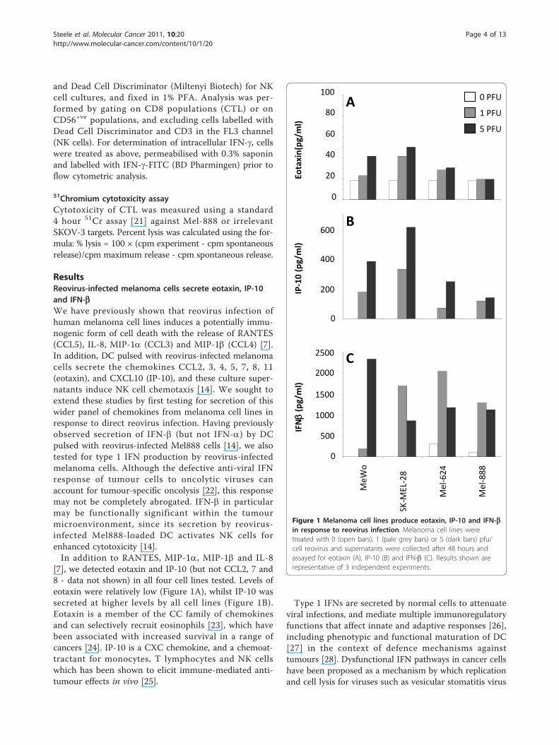
ResultsReovirus-infected melanoma cells secrete eotaxin, IP-10and IFN-bWe have previously shown that reovirus infection ofhuman melanoma cell lines induces a potentially immu-nogenic form of cell death with the release of RANTES(CCL5), IL-8, MIP-1a (CCL3) and MIP-1b (CCL4) [7].In addition, DC pulsed with reovirus-infected melanomacells secrete the chemokines CCL2, 3, 4, 5, 7, 8, 11(eotaxin), and CXCL10 (IP-10), and these culture super-natants induce NK cell chemotaxis [14]. We sought toextend these studies by first testing for secretion of thiswider panel of chemokines from melanoma cell lines inresponse to direct reovirus infection. Having previouslyobserved secretion of IFN-b (but not IFN-a) by DCpulsed with reovirus-infected Mel888 cells [14], we alsotested for type 1 IFN production by reovirus-infectedmelanoma cells. Although the defective anti-viral IFNresponse of tumour cells to oncolytic viruses canaccount for tumour-specific oncolysis [22], this responsemay not be completely abrogated. IFN-b in particularmay be functionally significant within the tumourmicroenvironment, since its secretion by reovirus-infected Mel888-loaded DC activates NK cells forenhanced cytotoxicity [14].In addition to RANTES, MIP-1a, MIP-1b and IL-8
[7], we detected eotaxin and IP-10 (but not CCL2, 7 and8 - data not shown) in all four cell lines tested. Levels ofeotaxin were relatively low (Figure 1A), whilst IP-10 wassecreted at higher levels by all cell lines (Figure 1B).Eotaxin is a member of the CC family of chemokinesand can selectively recruit eosinophils [23], which havebeen associated with increased survival in a range ofcancers [24]. IP-10 is a CXC chemokine, and a chemoat-tractant for monocytes, T lymphocytes and NK cellswhich has been shown to elicit immune-mediated anti-tumour effects in vivo [25].
Type 1 IFNs are secreted by normal cells to attenuateviral infections, and mediate multiple immunoregulatoryfunctions that affect innate and adaptive responses [26],including phenotypic and functional maturation of DC[27] in the context of defence mechanisms againsttumours [28]. Dysfunctional IFN pathways in cancer cellshave been proposed as a mechanism by which replicationand cell lysis for viruses such as vesicular stomatitis virus
0
60
80
100
Eota
xin(
pg/m
l)
40
20
A
200
600
400
0
B
IP-1
0 (p
g/m
l)
MeW
o
SK-M
EL-2
8
Mel
-624
Mel
-888
500
1500
1000
2000
2500
IFN
(pg/
ml)
C
0
0 PFU
1 PFU
5 PFU
Figure 1 Melanoma cell lines produce eotaxin, IP-10 and IFN-bin response to reovirus infection. Melanoma cell lines weretreated with 0 (open bars), 1 (pale grey bars) or 5 (dark bars) pfu/cell reovirus and supernatants were collected after 48 hours andassayed for eotaxin (A), IP-10 (B) and IFN-b (C). Results shown arerepresentative of 3 independent experiments.
Steele et al. Molecular Cancer 2011, 10:20http://www.molecular-cancer.com/content/10/1/20
Page 4 of 13
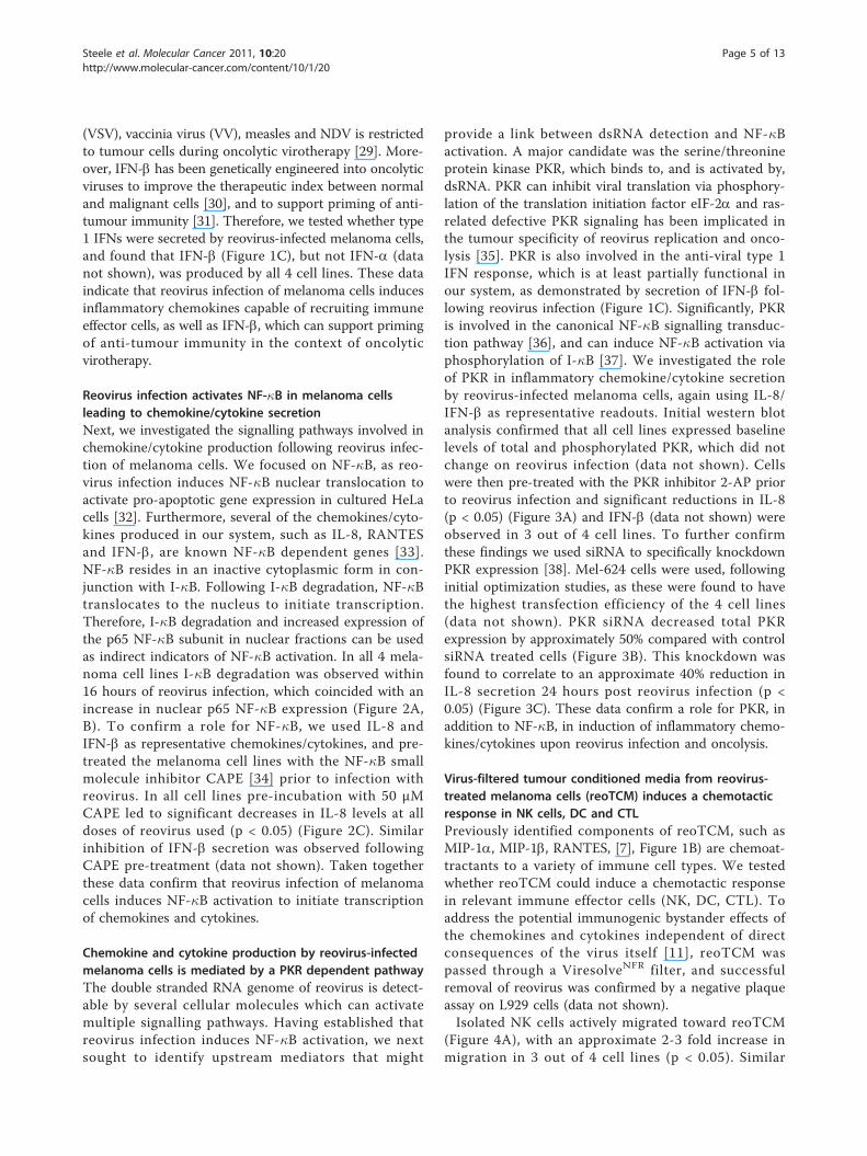
(VSV), vaccinia virus (VV), measles and NDV is restrictedto tumour cells during oncolytic virotherapy [29]. More-over, IFN-b has been genetically engineered into oncolyticviruses to improve the therapeutic index between normaland malignant cells [30], and to support priming of anti-tumour immunity [31]. Therefore, we tested whether type1 IFNs were secreted by reovirus-infected melanoma cells,and found that IFN-b (Figure 1C), but not IFN-a (datanot shown), was produced by all 4 cell lines. These dataindicate that reovirus infection of melanoma cells inducesinflammatory chemokines capable of recruiting immuneeffector cells, as well as IFN-b, which can support primingof anti-tumour immunity in the context of oncolyticvirotherapy.
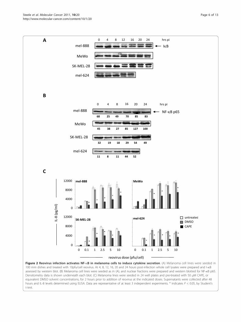
Reovirus infection activates NF-�B in melanoma cellsleading to chemokine/cytokine secretionNext, we investigated the signalling pathways involved inchemokine/cytokine production following reovirus infec-tion of melanoma cells. We focused on NF-�B, as reo-virus infection induces NF-�B nuclear translocation toactivate pro-apoptotic gene expression in cultured HeLacells [32]. Furthermore, several of the chemokines/cyto-kines produced in our system, such as IL-8, RANTESand IFN-b, are known NF-�B dependent genes [33].NF-�B resides in an inactive cytoplasmic form in con-junction with I-�B. Following I-�B degradation, NF-�Btranslocates to the nucleus to initiate transcription.Therefore, I-�B degradation and increased expression ofthe p65 NF-�B subunit in nuclear fractions can be usedas indirect indicators of NF-�B activation. In all 4 mela-noma cell lines I-�B degradation was observed within16 hours of reovirus infection, which coincided with anincrease in nuclear p65 NF-�B expression (Figure 2A,B). To confirm a role for NF-�B, we used IL-8 andIFN-b as representative chemokines/cytokines, and pre-treated the melanoma cell lines with the NF-�B smallmolecule inhibitor CAPE [34] prior to infection withreovirus. In all cell lines pre-incubation with 50 μMCAPE led to significant decreases in IL-8 levels at alldoses of reovirus used (p < 0.05) (Figure 2C). Similarinhibition of IFN-b secretion was observed followingCAPE pre-treatment (data not shown). Taken togetherthese data confirm that reovirus infection of melanomacells induces NF-�B activation to initiate transcriptionof chemokines and cytokines.
Chemokine and cytokine production by reovirus-infectedmelanoma cells is mediated by a PKR dependent pathwayThe double stranded RNA genome of reovirus is detect-able by several cellular molecules which can activatemultiple signalling pathways. Having established thatreovirus infection induces NF-�B activation, we nextsought to identify upstream mediators that might
provide a link between dsRNA detection and NF-�Bactivation. A major candidate was the serine/threonineprotein kinase PKR, which binds to, and is activated by,dsRNA. PKR can inhibit viral translation via phosphory-lation of the translation initiation factor eIF-2a and ras-related defective PKR signaling has been implicated inthe tumour specificity of reovirus replication and onco-lysis [35]. PKR is also involved in the anti-viral type 1IFN response, which is at least partially functional inour system, as demonstrated by secretion of IFN-b fol-lowing reovirus infection (Figure 1C). Significantly, PKRis involved in the canonical NF-�B signalling transduc-tion pathway [36], and can induce NF-�B activation viaphosphorylation of I-�B [37]. We investigated the roleof PKR in inflammatory chemokine/cytokine secretionby reovirus-infected melanoma cells, again using IL-8/IFN-b as representative readouts. Initial western blotanalysis confirmed that all cell lines expressed baselinelevels of total and phosphorylated PKR, which did notchange on reovirus infection (data not shown). Cellswere then pre-treated with the PKR inhibitor 2-AP priorto reovirus infection and significant reductions in IL-8(p < 0.05) (Figure 3A) and IFN-b (data not shown) wereobserved in 3 out of 4 cell lines. To further confirmthese findings we used siRNA to specifically knockdownPKR expression [38]. Mel-624 cells were used, followinginitial optimization studies, as these were found to havethe highest transfection efficiency of the 4 cell lines(data not shown). PKR siRNA decreased total PKRexpression by approximately 50% compared with controlsiRNA treated cells (Figure 3B). This knockdown wasfound to correlate to an approximate 40% reduction inIL-8 secretion 24 hours post reovirus infection (p <0.05) (Figure 3C). These data confirm a role for PKR, inaddition to NF-�B, in induction of inflammatory chemo-kines/cytokines upon reovirus infection and oncolysis.
Virus-filtered tumour conditioned media from reovirus-treated melanoma cells (reoTCM) induces a chemotacticresponse in NK cells, DC and CTLPreviously identified components of reoTCM, such asMIP-1a, MIP-1b, RANTES, [7], Figure 1B) are chemoat-tractants to a variety of immune cell types. We testedwhether reoTCM could induce a chemotactic responsein relevant immune effector cells (NK, DC, CTL). Toaddress the potential immunogenic bystander effects ofthe chemokines and cytokines independent of directconsequences of the virus itself [11], reoTCM waspassed through a ViresolveNFR filter, and successfulremoval of reovirus was confirmed by a negative plaqueassay on L929 cells (data not shown).Isolated NK cells actively migrated toward reoTCM
(Figure 4A), with an approximate 2-3 fold increase inmigration in 3 out of 4 cell lines (p < 0.05). Similar
Steele et al. Molecular Cancer 2011, 10:20http://www.molecular-cancer.com/content/10/1/20
Page 5 of 13
mel-888 MeWo
mel-624SK-MEL-28
0
4000
8000
12000
0
4000
8000
12000
0 0.1 1 2.5 5 100 0.1 1 2.5 5 10
IL-8
(pg/
ml)
reovirus dose (pfu/cell)
untreatedDMSOCAPE
C
SK-MEL-28
mel-888
MeWo
mel-624
0 4 8 12 16 20 24 hrs pi
I BA
NF- B p65mel-888
0 4 8 16 20 24 hrs pi
MeWo
mel-624
SK-MEL-28
B
68 25 49 70 85 83
45 38 27 85 127 100
32 19 18 29 54 49
11 8 11 44 52
Figure 2 Reovirus infection activates NF-�B in melanoma cells to induce cytokine secretion. (A) Melanoma cell lines were seeded in100 mm dishes and treated with 10pfu/cell reovirus. At 4, 8, 12, 16, 20 and 24 hours post-infection whole cell lysates were prepared and I-�Bassessed by western blot. (B) Melanoma cell lines were seeded as in (A), and nuclear fractions were prepared and western blotted for NF-�B p65.Densitometry data is shown underneath each blot. (C) Melanoma lines were seeded in 24 well plates and pre-treated with 50 μM CAPE, orequivalent DMSO solvent concentrations, for 2 hours prior to addition of reovirus at the indicated doses. Supernatants were collected after 48hours and IL-8 levels determined using ELISA. Data are representative of at least 3 independent experiments. * indicates P < 0.05, by Student’st-test.
Steele et al. Molecular Cancer 2011, 10:20http://www.molecular-cancer.com/content/10/1/20
Page 6 of 13
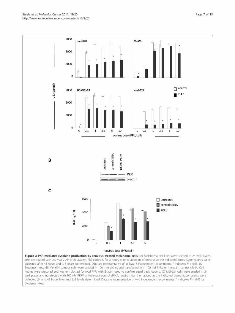
Figure 3 PKR mediates cytokine production by reovirus treated melanoma cells. (A) Melanoma cell lines were seeded in 24 well platesand pre-treated with 2.5 mM 2-AP or equivalent PBS controls, for 2 hours prior to addition of reovirus at the indicated doses. Supernatants werecollected after 48 hours and IL-8 levels determined. Data are representative of at least 3 independent experiments. * indicates P < 0.05, byStudent’s t-test. (B) Mel-624 tumour cells were seeded in 100 mm dishes and transfected with 100 nM PKRV or irrelevant control siRNA. Celllysates were prepared and western blotted for total PKR, with b-actin used to confirm equal track loading. (C) Mel-624 cells were seeded in 24well plates and transfected with 100 nM PKRV or irrelevant control siRNA; reovirus was then added at the indicated doses. Supernatants werecollected 24 and 48 hours later and IL-8 levels determined. Data are representative of two independent experiments. * indicates P < 0.05 byStudent’s t-test.
Steele et al. Molecular Cancer 2011, 10:20http://www.molecular-cancer.com/content/10/1/20
Page 7 of 13
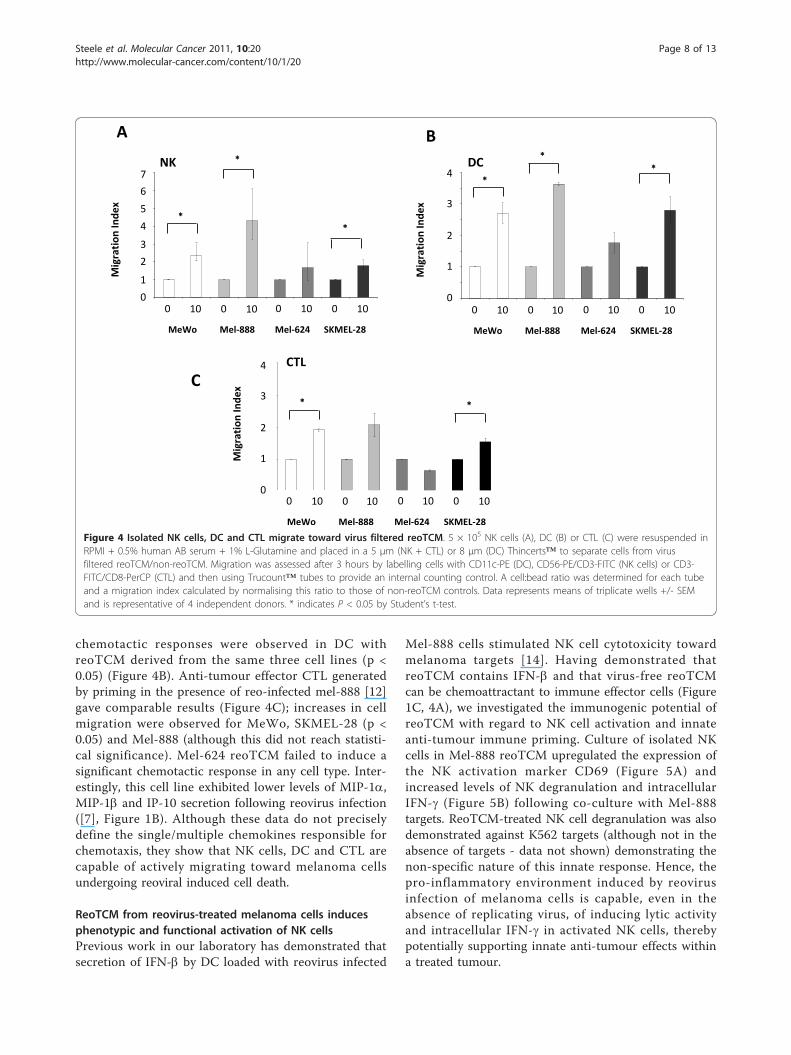
chemotactic responses were observed in DC withreoTCM derived from the same three cell lines (p <0.05) (Figure 4B). Anti-tumour effector CTL generatedby priming in the presence of reo-infected mel-888 [12]gave comparable results (Figure 4C); increases in cellmigration were observed for MeWo, SKMEL-28 (p <0.05) and Mel-888 (although this did not reach statisti-cal significance). Mel-624 reoTCM failed to induce asignificant chemotactic response in any cell type. Inter-estingly, this cell line exhibited lower levels of MIP-1a,MIP-1b and IP-10 secretion following reovirus infection([7], Figure 1B). Although these data do not preciselydefine the single/multiple chemokines responsible forchemotaxis, they show that NK cells, DC and CTL arecapable of actively migrating toward melanoma cellsundergoing reoviral induced cell death.
ReoTCM from reovirus-treated melanoma cells inducesphenotypic and functional activation of NK cellsPrevious work in our laboratory has demonstrated thatsecretion of IFN-b by DC loaded with reovirus infected
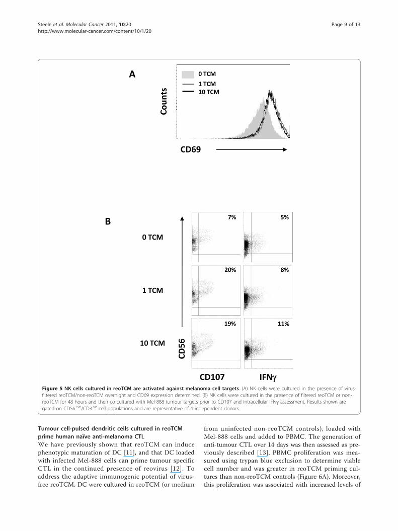
Mel-888 cells stimulated NK cell cytotoxicity towardmelanoma targets [14]. Having demonstrated thatreoTCM contains IFN-b and that virus-free reoTCMcan be chemoattractant to immune effector cells (Figure1C, 4A), we investigated the immunogenic potential ofreoTCM with regard to NK cell activation and innateanti-tumour immune priming. Culture of isolated NKcells in Mel-888 reoTCM upregulated the expression ofthe NK activation marker CD69 (Figure 5A) andincreased levels of NK degranulation and intracellularIFN-g (Figure 5B) following co-culture with Mel-888targets. ReoTCM-treated NK cell degranulation was alsodemonstrated against K562 targets (although not in theabsence of targets - data not shown) demonstrating thenon-specific nature of this innate response. Hence, thepro-inflammatory environment induced by reovirusinfection of melanoma cells is capable, even in theabsence of replicating virus, of inducing lytic activityand intracellular IFN-g in activated NK cells, therebypotentially supporting innate anti-tumour effects withina treated tumour.
0
7
6
5
4
3
2
1
0 10 0 10 0 10 0 10
MeWo Mel-888 Mel-624 SKMEL-28
NK
Mig
rati
on In
dex
0
0.5
1
1.5
2
2.5
3
3.5
4
mewo 0 TCM mewo 10TCM mel-888 0TCM mel888 10TCM mel624 0TCM mel624 10TCM skmel 0TCM skmel 10TCM
0 10 0 10 0 10 0 10
MeWo Mel-888 Mel-624 SKMEL-28
0
4
3
2
1
Mig
rati
on In
dex
DC
0
1
2
3
4
0 10 0 10 0 10 0 10
MeWo Mel-888 Mel-624 SKMEL-28
0
4
3
2
1Mig
rati
on In
dex
CTL
A B
C
Figure 4 Isolated NK cells, DC and CTL migrate toward virus filtered reoTCM. 5 × 10⁵ NK cells (A), DC (B) or CTL (C) were resuspended inRPMI + 0.5% human AB serum + 1% L-Glutamine and placed in a 5 μm (NK + CTL) or 8 μm (DC) Thincerts™ to separate cells from virusfiltered reoTCM/non-reoTCM. Migration was assessed after 3 hours by labelling cells with CD11c-PE (DC), CD56-PE/CD3-FITC (NK cells) or CD3-FITC/CD8-PerCP (CTL) and then using Trucount™ tubes to provide an internal counting control. A cell:bead ratio was determined for each tubeand a migration index calculated by normalising this ratio to those of non-reoTCM controls. Data represents means of triplicate wells +/- SEMand is representative of 4 independent donors. * indicates P < 0.05 by Student’s t-test.
Steele et al. Molecular Cancer 2011, 10:20http://www.molecular-cancer.com/content/10/1/20
Page 8 of 13
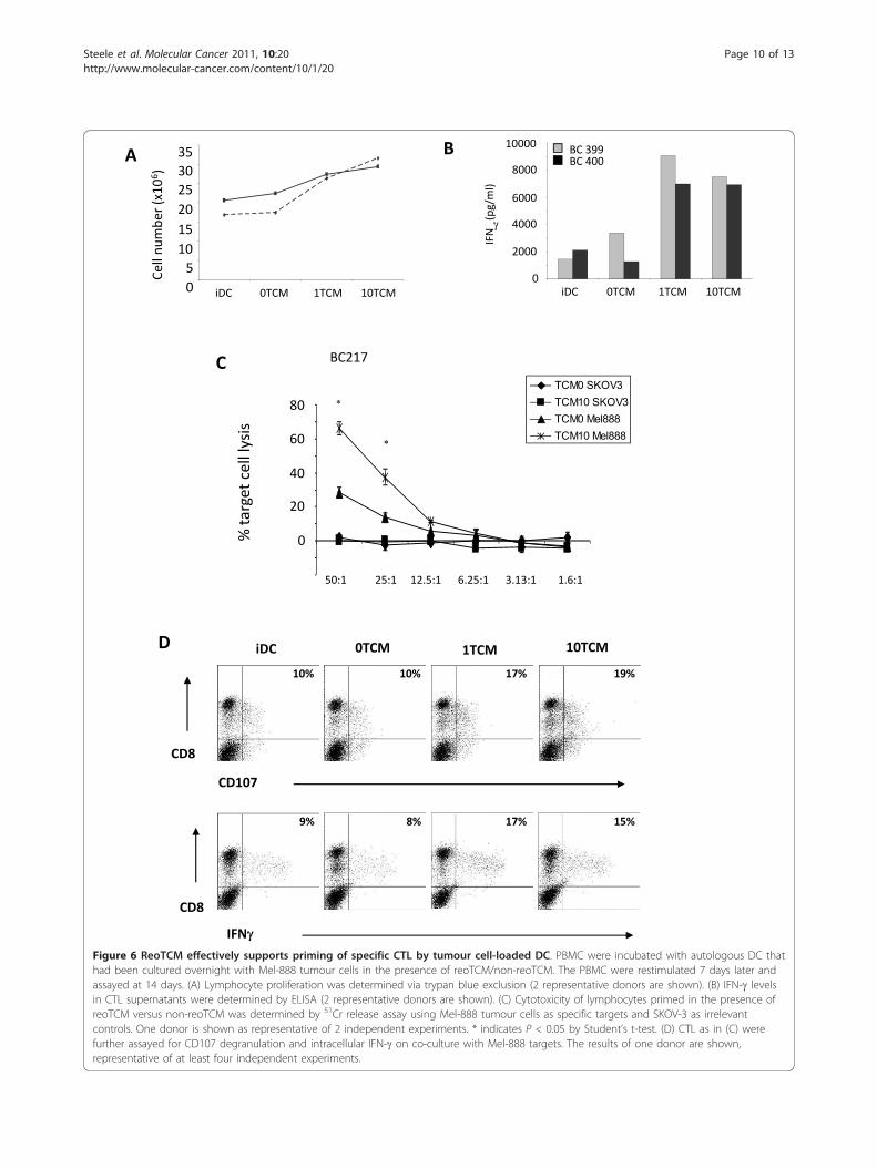
Tumour cell-pulsed dendritic cells cultured in reoTCMprime human naïve anti-melanoma CTLWe have previously shown that reoTCM can inducephenotypic maturation of DC [11], and that DC loadedwith infected Mel-888 cells can prime tumour specificCTL in the continued presence of reovirus [12]. Toaddress the adaptive immunogenic potential of virus-free reoTCM, DC were cultured in reoTCM (or medium
from uninfected non-reoTCM controls), loaded withMel-888 cells and added to PBMC. The generation ofanti-tumour CTL over 14 days was then assessed as pre-viously described [13]. PBMC proliferation was mea-sured using trypan blue exclusion to determine viablecell number and was greater in reoTCM priming cul-tures than non-reoTCM controls (Figure 6A). Moreover,this proliferation was associated with increased levels of
0 TCM
1 TCM
10 TCM
CD69Co
unts
1 TCM10 TCM
0 TCMA
B
CD107
CD56
7%
20%
19%
5%
8%
11%
IFNFigure 5 NK cells cultured in reoTCM are activated against melanoma cell targets. (A) NK cells were cultured in the presence of virus-filtered reoTCM/non-reoTCM overnight and CD69 expression determined. (B) NK cells were cultured in the presence of filtered reoTCM or non-reoTCM for 48 hours and then co-cultured with Mel-888 tumour targets prior to CD107 and intracellular IFNg assessment. Results shown aregated on CD56+ve/CD3-ve cell populations and are representative of 4 independent donors.
Steele et al. Molecular Cancer 2011, 10:20http://www.molecular-cancer.com/content/10/1/20
Page 9 of 13
0
253035
5101520
Cell
num
ber (
x106 )
iDC 1TCM 10TCM0TCM iDC 1TCM 10TCM0TCM0
2000
4000
6000
8000
10000
IFN
(pg/
ml)
BC 399BC 400A B
0
20
40
60
80
% ta
rget
cel
l lys
is
BC217
TCM0 SKOV3TCM10 SKOV3TCM0 Mel888TCM10 Mel888
50:1 25:1 12.5:1 6.25:1 3.13:1 1.6:1
C
D
CD107
CD8
IFN
CD8
9% 8% 17% 15%
10% 10% 17% 19%
iDC 0TCM 1TCM 10TCM
Figure 6 ReoTCM effectively supports priming of specific CTL by tumour cell-loaded DC. PBMC were incubated with autologous DC thathad been cultured overnight with Mel-888 tumour cells in the presence of reoTCM/non-reoTCM. The PBMC were restimulated 7 days later andassayed at 14 days. (A) Lymphocyte proliferation was determined via trypan blue exclusion (2 representative donors are shown). (B) IFN-g levelsin CTL supernatants were determined by ELISA (2 representative donors are shown). (C) Cytotoxicity of lymphocytes primed in the presence ofreoTCM versus non-reoTCM was determined by 51Cr release assay using Mel-888 tumour cells as specific targets and SKOV-3 as irrelevantcontrols. One donor is shown as representative of 2 independent experiments. * indicates P < 0.05 by Student’s t-test. (D) CTL as in (C) werefurther assayed for CD107 degranulation and intracellular IFN-g on co-culture with Mel-888 targets. The results of one donor are shown,representative of at least four independent experiments.
Steele et al. Molecular Cancer 2011, 10:20http://www.molecular-cancer.com/content/10/1/20
Page 10 of 13
IFN-g in the priming culture supernatants, consistentwith an evolving Th1 adaptive T cell response (Figure6B). A chromium cytotoxicity assay was used to deter-mine the lytic ability of CTL generated by reoTCM andnon-reoTCM-treated tumour-loaded DC. Whilst somespecific anti-Mel888 CTL activity was seen under non-reoTCM DC conditions, levels of killing were signifi-cantly higher when reoTCM-conditioned DC were usedfor CTL priming (approximately 70% lysis comparedwith 30%) (p < 0.05) (Figure 6C). No killing of irrelevantSKOV-3 tumour targets was observed. In addition, CTLCD107 degranulation and intracellular IFN-g, in the pre-sence of Mel888 targets (but not SKOV-3, data notshown), was also higher after reoTCM priming com-pared with their non-reoTCM counterparts (Figure 6D).Although these cytotoxicity assays do not address theMHC class I restriction or antigen specificity of killing(which are potential confounding factors when using anallogeneic tumour cell line as an antigen source forhuman CTL priming), we have previously shown in thissystem that specific anti-Mel888 CTL include T cellswhich recognize the tumour-associated antigen MART-1 [12,13], demonstrating that these responses includetargeting of antigens relevant to anti-tumour therapy.Hence, in addition to activation of innate NK cell anti-melanoma activity, virus-free reoTCM is able to supporteffective priming of a specific adaptive CTL response.
DiscussionReovirus is a tumour-specific oncolytic virus currentlyunder clinical investigation [39,40]. We, and others,have shown that reovirus is one of several therapeuticviruses whose activity can be mediated via activation ofan anti-tumour immune response, as well as the directoncolytic effect of viral replication in tumour cells [10].Whether the immune response to viral therapy is pro-blematic, due to rapid systemic inactivation of the agent,or actively therapeutic, via provision of a ‘danger’ signalwithin an otherwise immunosuppressive tumour micro-environment, likely depends on multiple factors. Theseinclude route of virus delivery (intratumoural versusintravenous), the pre-existing immune status of thepatient and the mechanisms by which the virus natu-rally, or via genetic modification, targets tumour cells.Consequently, various immunomodulatory strategieshave been employed to improve oncolytic viral therapy,ranging from immunosuppression to improve viral per-sistence in the circulation [41], through to enhancementof immune activation via insertion of transgenes, suchas GMCSF, into the viral genome [42].We have previously shown that i) reovirus induces
apoptotic death in human melanoma cells and that thisdeath is associated with secretion of inflammatory che-mokines/cytokines [7], ii) reovirus directly activates DC
in the absence of tumour cells [11], and iii) reovirus-infected melanoma cells can activate innate and adaptivearms of the anti-tumour immune response [12-14].However, these data were generated in the continuedpresence of active virus. Since reovirus itself is directlyimmunostimulatory [11], removal of the virus fromreoTCM via filtration allowed us to specifically investi-gate the additional, bystander immunogenic effects ofthe inflammatory environment potentially generated intreated tumours. The immunogenic component ofhuman reoviral therapy may have particular clinical rele-vance, since levels of reovirus replication in freshlyresected melanoma cells may be low [7]. Furthermore,the switch from a suppressive to an inflammatorytumour milieu may persist even after the virus has beencleared [43].We extended our previous analysis of the chemokines
and cytokines produced by reovirus-infected humanmelanoma lines, and showed that eotaxin and IP-10were also secreted (Figure 1). Interestingly, we alsodetected IFN-b (but not IFN-a) under these conditions,illustrating that an anti-viral type 1 IFN response is par-tially functional in these tumour cells. This is particu-larly important as IFN-b is involved in innate immuneactivation by DC loaded with reovirus-infected cells[14]. Moreover, IFN-b has been engineered into otheroncolytic viruses to increase the therapeutic indexbetween malignant and normal cells, and to enhanceanti-tumour immune activation [30].Although the mechanisms responsible for the inflam-
matory response of tumour cells to reovirus infectionhave not been addressed to date, previous studies haveimplicated a role for NF-�B since i) reovirus infectioninitiates translocation of the p50/p65 NF-�B subunits tothe nucleus and activates pro-apoptotic gene expression[32,44], ii) reovirus induces apoptosis in melanoma cells[7], and iii) NF-�B is involved in the production of che-mokines and cytokines such as IL-8 and IFN-b [33].This study confirms that reovirus infection of melanomacells activates NF-�B, as assessed by I-�B degradationand accumulation of nuclear p65, and that blocking NF-�B with the small molecule inhibitor CAPE significantlydecreases production of IL-8 and IFN-b (Figure 2).Importantly, this effect was seen across all 4 cell lines,suggesting that common signalling pathways are acti-vated following reovirus infection of melanoma. Toaddress viral sensing and signaling molecules that maylie upstream of NF-�B, we investigated the dependenceof IL-8 and IFN-b production on PKR, as one of a num-ber of candidate dsRNA sensors. PKR is involved in thetumour specificity of reoviral oncolysis (although theprecise mechanism remains to be fully elucidated), andthe anti-viral type 1 IFN response [35]. We found, viasmall molecule blockade and siRNA knockdown, that
Steele et al. Molecular Cancer 2011, 10:20http://www.molecular-cancer.com/content/10/1/20
Page 11 of 13
PKR is involved in the inflammatory response of mela-noma cells following reovirus infection (Figure 3).Although we have been unable to detect any significantchange in total or phosphorylated PKR following reo-virus infection of melanoma cell lines (data not shown),these data suggest that dsRNA detection by PKR initi-ates activation of NF-�B-dependent chemokines andcytokines in tumour cells. These findings are in agree-ment with previous observations following direct infec-tion of DC [11]. However, further work is required tofully characterize the signaling pathways responsible forthe immunogenic nature of reovirus-induced tumourcell oncolysis.Filtered reoTCM induced a chemotactic response in
NK cells, DC and anti-tumour CTL (previously primedusing reovirus-infected tumour cells) (Figure 4), suggest-ing that the immunogenic milieu in treated tumours hasthe potential to recruit a range of immune cells capableof viral detection and innate/adaptive effector functions.With regard to improving access of primed CTL totumours, this finding is consistent with previous murinedata showing greater persistence of adoptively transferredantigen specific T cells within tumours undergoing VSV-mediated oncolysis [45]. To date we have not specificallyidentified which of the secreted chemokine(s) are respon-sible for NK cell, DC and CTL migration. It is possiblethat multiple chemokines may act in combination toengage a variety of receptors to induce a particular phy-siological response [16]. The ability of reoTCM to sup-port activation of innate (Figure 5) and adaptive (Figure6) immune responses against human melanoma cellsshows that the immunogenic effects of reovirus inducedcell death are not dependent on the continued presenceof virus once an initiating danger signal has been deliv-ered. Therefore, even if viral replication in patients is lim-ited by neutralization, for example following repeatedadministration [46], the immunogenic response oftumour cells to reovirus infection may be sufficient toinduce continuing anti-tumour effects.Overall, the present study shows that reovirus infection
of human melanoma cells induces a range of chemokinesand cytokines capable of inducing a chemotactic responsein NK cells, DC and primed CTL. This inflammatoryresponse is dependent upon NF-�B and PKR and is suffi-cient, in the absence of live virus, to support priming ofinnate and adaptive anti-tumour immunity. This data sup-ports the potential of bystander activation of human anti-tumour immunity by reovirus killing of tumour cells, evenif persistent viral replication is limited by the anti-viralimmune response.
AbbreviationsTCM: Tumour conditioned media; RANTES: regulated on activation normal Texpressed and secreted; MIP: macrophage inflammatory protein; IFN:Interferon; PKR: Protein kinase R; NK: Natural Killer; DC: dendritic cells; CTL:
cytotoxic T lymphocytes; NDV: Newcastle Disease Virus; HSV: Herpes SimplexVirus; DMEM: Dulbecco Modified Eagle Medium; FCS: Foetal Calf Serum;PBMC: peripleral blood mononuclear cells; CAPE: caffeic acid phenethyl ester;2-AP: 2-aminopurine; FACS: fluorescent activated cell sorting; VSV: vesicularstomatitis virus; GMCSF: granulocyte macrophage colony-stimulating factor;siRNA: small interfering RNA; SDS-PAGE: sodium dodecyl sulphatepolyacrylamide gel electrophoresis;
Author details1Leeds Institute of Molecular Medicine, University of Leeds, Leeds, UK.2Institute of Cancer Research, Centre for Cell and Molecular Biology, ChesterBeatty Laboratories, London, UK. 3Postgraduate Medical School, University ofSurrey, Guildford, UK. 4Oncolytics Biotech Inc, Calgary, Alberta, Canada.5Molecular Medicine Program and Department of Immunology, Mayo Clinic,Rochester, Minnesota, USA.
Authors’ contributionsLS contributed to conception and design, acquisition of data, analysis andinterpretation of data and wrote the manuscript. FE, RP, HP, KH, PS, RV andEI contributed to conception, analysis and interpretation of data (as did MCwho also provided clinical grade reovirus (Reolysin). AM conceived thestudy, participated in its design and coordination and co-wrote themanuscript. All authors read and approved the final manuscript.
Competing interestsOncolytics Biotech Inc: KH/RV/HP/AM, commercial research grant. MC,employee.
Received: 7 September 2010 Accepted: 21 February 2011Published: 21 February 2011
References1. Vidal L, Pandha HS, Yap TA, White CL, Twigger K, Vile RG, Melcher A,
Coffey M, Harrington KJ, Debono JS: A phase I study of intravenousoncolytic reovirus type 3 dearing in patients with advanced cancer. ClinCancer Res 2008, 14:7127-7137.
2. Harrington KJ, Karapanagiotou EM, Roulstone V, Twigger KR, White CL,Vidal L, Beirne D, Prestwich R, Newbold K, Ahmed M, et al: Two-StagePhase I Dose-Escalation Study of Intratumoral Reovirus Type 3 Dearingand Palliative Radiotherapy in Patients with Advanced Cancers. ClinCancer Res 2010.
3. Thirukkumaran CM, Nodwell MJ, Hirasawa K, Shi ZQ, Diaz R, Luider J,Johnston RN, Forsyth PA, Magliocco AM, Lee P, et al: Oncolytic viraltherapy for prostate cancer: efficacy of reovirus as a biologicaltherapeutic. Cancer Res 2010, 70:2435-2444.
4. Coffey MC, Strong JE, Forsyth PA, Lee PW: Reovirus therapy of tumorswith activated Ras pathway. Science 1998, 282:1332-1334.
5. Song L, Ohnuma T, Gelman IH, Holland JF: Reovirus infection of cancercells is not due to activated Ras pathway. Cancer Gene Ther 2008.
6. van Houdt WJ, Smakman N, van den Wollenberg DJ, Emmink BL,Veenendaal LM, van Diest PJ, Hoeben RC, Borel Rinkes IH, Kranenburg O:Transient infection of freshly isolated human colorectal tumor cells byreovirus T3D intermediate subviral particles. Cancer Gene Ther 2008.
7. Errington F, White CL, Twigger KR, Rose A, Scott K, Steele L, Ilett LJ,Prestwich R, Pandha HS, Coffey M, et al: Inflammatory tumour cell killingby oncolytic reovirus for the treatment of melanoma. Gene Ther 2008.
8. Benencia F, Courreges MC, Conejo-Garcia JR, Mohamed-Hadley A, Zhang L,Buckanovich RJ, Carroll R, Fraser N, Coukos G: HSV oncolytic therapyupregulates interferon-inducible chemokines and recruits immuneeffector cells in ovarian cancer. Mol Ther 2005.
9. Washburn B, Schirrmacher V: Human tumor cell infection by NewcastleDisease Virus leads to upregulation of HLA and cell adhesion moleculesand to induction of interferons, chemokines and finally apoptosis. Int JOncol 2002, 21:85-93.
10. Prestwich RJ, Harrington KJ, Pandha HS, Vile RG, Melcher AA, Errington F:Oncolytic viruses: a novel form of immunotherapy. Expert Rev AnticancerTher 2008, 8:1581-1588.
11. Errington F, Steele L, Prestwich R, Harrington KJ, Pandha HS, Vidal L, deBono J, Selby P, Coffey M, Vile R, Melcher A: Reovirus Activates HumanDendritic Cells to Promote Innate Antitumor Immunity. J Immunol 2008,180:6018-6026.
Steele et al. Molecular Cancer 2011, 10:20http://www.molecular-cancer.com/content/10/1/20
Page 12 of 13
12. Prestwich RJ, Errington F, Ilett EJ, Morgan RS, Scott KJ, Kottke T,Thompson J, Morrison EE, Harrington KJ, Pandha HS, et al: Tumor infectionby oncolytic reovirus primes adaptive antitumor immunity. Clin CancerRes 2008, 14:7358-7366.
13. Prestwich RJ, Ilett EJ, Errington F, Diaz RM, Steele LP, Kottke T, Thompson J,Galivo F, Harrington KJ, Pandha HS, et al: Immune-Mediated AntitumorActivity of Reovirus Is Required for Therapy and Is Independent ofDirect Viral Oncolysis and Replication. Clin Cancer Res 2009.
14. Prestwich RJ, Errington F, Steele LP, Ilett EJ, Morgan RS, Harrington KJ,Pandha HS, Selby PJ, Vile RG, Melcher AA: Reciprocal human dendritic cell-natural killer cell interactions induce antitumor activity following tumorcell infection by oncolytic reovirus. J Immunol 2009, 183:4312-4321.
15. Rossi D, Zlotnik A: The biology of chemokines and their receptors. AnnuRev Immunol 2000, 18:217-242.
16. Mantovani A: Chemokines. Introduction and overview. Chem Immunol1999, 72:1-6.
17. Vicari AP, Caux C: Chemokines in cancer. Cytokine Growth Factor Rev 2002,13:143-154.
18. Paoletti S, Petkovic V, Sebastiani S, Danelon MG, Uguccioni M, Gerber BO: Arich chemokine environment strongly enhances leukocyte migrationand activities. Blood 2005, 105:3405-3412.
19. Wendel M, Galani IE, Suri-Payer E, Cerwenka A: Natural killer cellaccumulation in tumors is dependent on IFN-gamma and CXCR3ligands. Cancer Res 2008, 68:8437-8445.
20. Mullins IM, Slingluff CL, Lee JK, Garbee CF, Shu J, Anderson SG, Mayer ME,Knaus WA, Mullins DW: CXC chemokine receptor 3 expression byactivated CD8+ T cells is associated with survival in melanoma patientswith stage III disease. Cancer Res 2004, 64:7697-7701.
21. Errington F, Jones J, Merrick A, Bateman A, Harrington K, Gough M,O’Donnell D, Selby P, Vile R, Melcher A: Fusogenic membraneglycoprotein-mediated tumour cell fusion activates human dendriticcells for enhanced IL-12 production and T-cell priming. Gene Ther 2006,13:138-149.
22. Randall RE, Goodbourn S: Interferons and viruses: an interplay betweeninduction, signalling, antiviral responses and virus countermeasures.J Gen Virol 2008, 89:1-47.
23. Hogan SP: Recent advances in eosinophil biology. Int Arch AllergyImmunol 2007, 143(Suppl 1):3-14.
24. Fernandez-Acenero MJ, Galindo-Gallego M, Sanz J, Aljama A: Prognosticinfluence of tumor-associated eosinophilic infiltrate in colorectalcarcinoma. Cancer 2000, 88:1544-1548.
25. Luster AD, Leder P: IP-10, a -C-X-C- chemokine, elicits a potent thymus-dependent antitumor response in vivo. J Exp Med 1993, 178:1057-1065.
26. Biron CA, Nguyen KB, Pien GC, Cousens LP, Salazar-Mather TP: Natural killercells in antiviral defense: function and regulation by innate cytokines.Annu Rev Immunol 1999, 17:189-220.
27. Le Bon A, Tough DF: Links between innate and adaptive immunity viatype I interferon. Curr Opin Immunol 2002, 14:432-436.
28. Ullrich E, Menard C, Flament C, Terme M, Mignot G, Bonmort M, Plumas J,Chaperot L, Chaput N, Zitvogel L: Dendritic cells and innate defenseagainst tumor cells. Cytokine Growth Factor Rev 2008, 19:79-92.
29. Russell SJ: RNA viruses as virotherapy agents. Cancer Gene Ther 2002,9:961-966.
30. Kirn DH, Wang Y, Le Boeuf F, Bell J, Thorne SH: Targeting of interferon-beta to produce a specific, multi-mechanistic oncolytic vaccinia virus.PLoS Med 2007, 4:e353.
31. Willmon CL, Saloura V, Fridlender ZG, Wongthida P, Diaz RM, Thompson J,Kottke T, Federspiel M, Barber G, Albelda SM, Vile RG: Expression of IFN-beta enhances both efficacy and safety of oncolytic vesicular stomatitisvirus for therapy of mesothelioma. Cancer Res 2009, 69:7713-7720.
32. Connolly JL, Rodgers SE, Clarke P, Ballard DW, Kerr LD, Tyler KL,Dermody TS: Reovirus-induced apoptosis requires activation oftranscription factor NF-kappaB. J Virol 2000, 74:2981-2989.
33. Siebenlist U, Franzoso G, Brown K: Structure, regulation and function ofNF-kappa B. Annu Rev Cell Biol 1994, 10:405-455.
34. Natarajan K, Singh S, Burke TR Jr, Grunberger D, Aggarwal BB: Caffeic acidphenethyl ester is a potent and specific inhibitor of activation of nucleartranscription factor NF-kappa B. Proc Natl Acad Sci USA 1996,93:9090-9095.
35. Marcato PSM, Lee Patrick: Connecting Reovirus oncolysis and Rassignalling. Cell cycle 2005, 4.
36. Garcia MA, Meurs EF, Esteban M: The dsRNA protein kinase PKR: virus andcell control. Biochimie 2007, 89:799-811.
37. Kumar A, Haque J, Lacoste J, Hiscott J, Williams BR: Double-stranded RNA-dependent protein kinase activates transcription factor NF-kappa B byphosphorylating I kappa B. Proc Natl Acad Sci USA 1994, 91:6288-6292.
38. Zhang P, Samuel CE: Protein kinase PKR plays a stimulus- and virus-dependent role in apoptotic death and virus multiplication in humancells. J Virol 2007, 81:8192-8200.
39. Comins C, Heinemann L, Harrington K, Melcher A, De Bono J, Pandha H:Reovirus: Viral Therapy for Cancer ‘as Nature Intended’. Clin Oncol (R CollRadiol) 2008.
40. Harrington KJ, Vile RG, Melcher A, Chester J, Pandha HS: Clinical trials withoncolytic reovirus: Moving beyond phase I into combinations withstandard therapeutics. Cytokine Growth Factor Rev 2010.
41. Hirasawa K, Nishikawa SG, Norman KL, Coffey MC, Thompson BG, Yoon CS,Waisman DM, Lee PW: Systemic reovirus therapy of metastatic cancer inimmune-competent mice. Cancer Res 2003, 63:348-353.
42. Kim JH, Oh JY, Park BH, Lee DE, Kim JS, Park HE, Roh MS, Je JE, Yoon JH,Thorne SH, et al: Systemic armed oncolytic and immunologic therapy forcancer with JX-594, a targeted poxvirus expressing GM-CSF. Mol Ther2006, 14:361-370.
43. Galivo F, Diaz RM, Wongthida P, Thompson J, Kottke T, Barber G, Melcher A,Vile R: Single-cycle viral gene expression, rather than progressivereplication and oncolysis, is required for VSV therapy of B16 melanoma.Gene Ther 2010, 17:158-170.
44. O’Donnell SM, Hansberger MW, Connolly JL, Chappell JD, Watson MJ,Pierce JM, Wetzel JD, Han W, Barton ES, Forrest JC, et al: Organ-specificroles for transcription factor NF-kappaB in reovirus-induced apoptosisand disease. J Clin Invest 2005, 115:2341-2350.
45. Kottke T, Diaz RM, Kaluza K, Pulido J, Galivo F, Wongthida P, Thompson J,Willmon C, Barber GN, Chester J, et al: Use of Biological Therapy toEnhance Both Virotherapy and Adoptive T-Cell Therapy for Cancer. MolTher 2008.
46. White CL, Twigger KR, Vidal L, De Bono JS, Coffey M, Heinemann L,Morgan R, Merrick A, Errington F, Vile RG, et al: Characterization of theadaptive and innate immune response to intravenous oncolytic reovirus(Dearing type 3) during a phase I clinical trial. Gene Ther 2008.
doi:10.1186/1476-4598-10-20Cite this article as: Steele et al.: Pro-inflammatory cytokine/chemokineproduction by reovirus treated melanoma cells is PKR/NF-�B mediatedand supports innate and adaptive anti-tumour immune priming.Molecular Cancer 2011 10:20.
Submit your next manuscript to BioMed Centraland take full advantage of:
• Convenient online submission
• Thorough peer review
• No space constraints or color figure charges
• Immediate publication on acceptance
• Inclusion in PubMed, CAS, Scopus and Google Scholar
• Research which is freely available for redistribution
Submit your manuscript at www.biomedcentral.com/submit
Steele et al. Molecular Cancer 2011, 10:20http://www.molecular-cancer.com/content/10/1/20
Page 13 of 13


