Embed Size (px)
Citation preview
Propeptide-Mediated Inhibition of Cognate GingipainProteinasesN. Laila Huq, Christine A. Seers, Elena C. Y. Toh, Stuart G. Dashper, Nada Slakeski, Lianyi Zhang,
Brent R. Ward, Vincent Meuric, Dina Chen, Keith J. Cross, Eric C. Reynolds*
Oral Health Cooperative Research Centre, Melbourne Dental School, Bio21 Institute of Molecular Science and Biotechnology, The University of Melbourne, Victoria,
Australia
Abstract
Porphyromonas gingivalis is a major pathogen associated with chronic periodontitis. The organism’s cell-surface cysteineproteinases, the Arg-specific proteinases (RgpA, RgpB) and the Lys-specific proteinase (Kgp), which are known as gingipainshave been implicated as major virulence factors. All three gingipain precursors contain a propeptide of around 200 aminoacids in length that is removed during maturation. The aim of this study was to characterize the inhibitory potential of theKgp and RgpB propeptides against the mature cognate enzymes. Mature Kgp was obtained from P. gingivalis mutantECR368, which produces a recombinant Kgp with an ABM1 motif deleted from the catalytic domain (rKgp) that enables theotherwise membrane bound enzyme to dissociate from adhesins and be released. Mature RgpB was obtained from P.gingivalis HG66. Recombinant propeptides of Kgp and RgpB were produced in Escherichia coli and purified using nickel-affinity chromatography. The Kgp and RgpB propeptides displayed non-competitive inhibition kinetics with Ki values of2.04 mM and 12 nM, respectively. Both propeptides exhibited selectivity towards their cognate proteinase. The specificity ofboth propeptides was demonstrated by their inability to inhibit caspase-3, a closely related cysteine protease, and papainthat also has a relatively long propeptide. Both propeptides at 100 mg/L caused a 50% reduction of P. gingivalis growth in aprotein-based medium. In summary, this study demonstrates that gingipain propeptides are capable of inhibiting theirmature cognate proteinases.
Citation: Huq NL, Seers CA, Toh ECY, Dashper SG, Slakeski N, et al. (2013) Propeptide-Mediated Inhibition of Cognate Gingipain Proteinases. PLoS ONE 8(6):e65447. doi:10.1371/journal.pone.0065447
Editor: Eugene A. Permyakov, Russian Academy of Sciences, Institute for Biological Instrumentation, Russian Federation
Received December 20, 2012; Accepted April 24, 2013; Published June 10, 2013
Copyright: � 2013 Huq et al. This is an open-access article distributed under the terms of the Creative Commons Attribution License, which permits unrestricteduse, distribution, and reproduction in any medium, provided the original author and source are credited.
Funding: This work was supported by the Australian National Health and Medical Research Council. The funder had no role in study design, data collection andanalysis, decision to publish, or preparation of the manuscript.
Competing Interests: The authors have declared that no competing interests exist.
* E-mail: [email protected]
Introduction
Porphyromonas gingivalis is a major pathogen associated with
chronic periodontitis. The organism’s cell surface cysteine
proteinases, the Arg- and Lys-specific gingipains [1–2] have been
implicated as major virulence factors that play an important role in
colonisation and establishment of the bacterium as well as in the
evasion of host defences [3–5].
Recent studies have demonstrated associations between peri-
odontitis and systemic morbidities such as diabetes and cardio-
vascular disease [6], pre-term and low weight births [7],
Alzheimer’s disease [8], cancers [9], respiratory diseases [10]
and rheumatoid arthritis [11]. The correlation between these
systemic diseases and the entry of the bacterium and its gingipains
into the circulation system are currently under investigation [12].
The gingipains RgpA, RgpB, and Kgp are encoded by three
genes, rgpA, rgpB, and kgp respectively [13–15]. The gene rgpB
encodes a single chain proteinase with a short 24 amino acid (aa)
leader sequence, 205 aa propeptide, and a ,500 aa catalytic
domain [16]. In contrast, the longer rgpA and kgp genes each
encode a leader sequence, propeptide, catalytic domain plus
additional haemagglutinin-adhesin (HA) domains. Due to the
importance of the gingipains in virulence [3–5] there is interest in
the development of specific and safe inhibitors of the proteinases.
Examination of the reported peptide-derived and non-peptide
inhibitors of the gingipains in the literature reveals a surprising
diversity of affinity, specificity and structural features. The
inhibitors also display various modes of inhibition: competitive,
non-competitive and uncompetitive [17–21]. To describe the
specificity of proteases, a model of an active site composed of
contiguous pockets termed subsites S1, S2 … etc is used with
substrate residues P1, P2…etc occupying the corresponding
subsites [22]. The residues in the substrate sequence are numbered
consecutively outward from the cleavage site
2P42P32P22P1+P1’2P2’2P3’2P4’2, 2S42S32S22S1*S1’
2S2’2S3’2S4’2. The scissile bond represented by the symbol+is
located between the P1 and P1’ positions, while the catalytic site is
represented by the symbol *.
Bioinformatic analysis of known proteins and synthetic
substrates cleaved by the gingipains reveals that although
hydrophobic residues are frequently found at positions P4-P2
and P1’-P4’, overall the size, charge, and shape preferences of
substrates are not clear (unpublished, [23]). This may reflect the
ability of the gingipain active site to accommodate various
substrates with only a strong specificity for an Arg or Lys residue
in the P1 position.
Recent studies have highlighted that protease propeptides are a
promising source of inhibitors for the cognate protease [24–25].
PLOS ONE | www.plosone.org 1 June 2013 | Volume 8 | Issue 6 | e65447

Many cysteine proteases are synthesized as inactive forms or
zymogens with N-terminal propeptide regions. These propeptides
may have multiple functions including inhibiting the proteolytic
activity of the mature enzyme, folding of the precursor enzyme,
protecting the enzyme against denaturation in extreme pH
conditions, transporting the precursor enzyme to lysosomes, and
mediating membrane association [26]. Typically the enzyme
becomes activated upon removal of the propeptide by intra- or
intermolecular proteolysis or in other cases by Ca2+ binding or
acidification [26]. Although cysteine protease propeptides range
from 30–250 aa, most are less than 100 aa residues [26–28]. The
gingipain catalytic-domain propeptides are unusually long, being
,200 residues suggesting that the gingipain propeptides may have
a more complex function than the shorter propeptides of other
proteases.
The aim of this study was to characterize the inhibitory
potential of recombinant Kgp and RgpB propeptides against their
cognate catalytic domains purified from P. gingivalis. The specificity
of recombinantly expressed RgpB and Kgp propeptides for
protease inhibition was determined as well as the interaction of
the propeptides with both cognate and heterologous proteases and
their effect on the growth of the bacterium.
Methods
Production of Recombinant Kgp Catalytic DomainPlasmids and oligonucleotides used in the course of this work
are listed in Table 1 and Table 2 respectively. Plasmids used were
propagated in Escherichia coli a–Gold Select (Bioline Australia) or
BL-21 (DE3) cells (Novagen). Allele exchange suicide plasmids
(described below) were all linearised using XbaI restriction
endonuclease (RE) digestion and transformed into electropora-
tion-competent P. gingivalis cells [29] with transformants selected
after anaerobic incubation at 37uC for up to ten days. EcoRV and
ApaI recognition sequences were engineered into plasmid pNS1
[30] upstream of the kgp promoter [31] using oligonucleotide
primers EA-For and EA-Rev (Table 1) and the QuikChange II
Site-directed Mutagenesis Kit (Stratagene) following manufactur-
er’s instructions, generating pNS2. The Bacteroides fragilis cepha-
losporinase-coding gene cepA was amplified from a pEC474
template DNA [32] using oligonucleotides CepAf and CepAr
and ligated into pGEM-T Easy (Promega) to generate pCS19. cepA
was excised from pCS19 using EcoICRI/ApaI RE digestion and
ligated into pNS2 that had been digested with BstEII (BII), end-
filled then digested with ApaI. The resultant plasmid pPC1 has
cepA that is transcribed from its own promoter and replaces
nucleotides (nt) of pNS2 that include the kgp promoter and kgp nt
coding from Met1-Tyr748. P. gingivalis W50 was transformed with
pPC1 to produce the Kgp-null strain ECR364. Plasmid pPC2 was
produced by ligating ermF excised from pAL30 [33] using ApaI
and EcoRICRI RE digestion into pNS2 digested with ApaI and
EcoRV. The nt coding ABM1 at the C-terminus of the Kgp
catalytic domain (Gly681-A710, GEPSPYQPVSNL-
TATTQGQKVTLKWEAPSA) were then deleted from pPC2
using a combination of splicing by overlap extension (SOE) PCR,
RE digestion and ligation as follows. Primer pairs ABM1del_For1
plus ABM1del_Rev1 and ABM1del_For2 plus ABM1del_Rev2
were used to generate two PCR amplicons which were annealed,
extended and amplified using ABM1del_For1 and ABM1del_-
Rev2 as primers. The SOEn amplicon was digested with SnaBI
and BstEII and ligated to SnaBI-BstEII digested pPC2 to generate
pPC3 (Table 1) that was linearised and electroporated into
Table 1. Plasmids used in the course of this study.
Plasmids Descriptiona Reference
pEC474 pBR322: : cepA [32]
pCS19 pGEM-T Easy: : cepA This study
pNS1 pUC18: : 3521 nt BamHI fragment of P. gingivalis W50 encompassing the 39 of PG1842 and the 59 of kgp [30]
pNS2 pNS1 with nucleotide T405C, A414G, T418C, A419C mutations to produce ApaI and EcoRV recognition sites. This study
pPC1 pNS2: : cepA. cepA replaces nt that include the kgp promoter and kgp nt coding from Met1-Tyr748 This study
pPC2 ermF ligated between the ApaI and EcoRV sites of pNS2. ermF upstream of the kgp promoter.
pPC3 pPC2 excluding kgp codons 681–710 This study
pET-28b Expression vector Novagen
pKgpPP1 Insert in pGEM-T Easy codes Kgp propeptide, residues 20–228 This study
pRgpPP1 Insert in pGEM-T Easy codes Rgp propeptide, residues 25–222 This study
pKgpPP2 Insert from pKgpPP1 in pET28b codes Kgp propeptide, residues 20–228 This study
pRgpPP2 Insert from pRgpPP1 in pET28b codes Kgp propeptide, residues 20–228 This study
doi:10.1371/journal.pone.0065447.t001
Table 2. Oligonucleotides used in the course of this study.
Oligonucleotide Sequencea
EA-For GATTACAGTCGATATCTTGGCAAAGGGCCCATTGACAGCC
EA-Rev GGCTGTCAATGGGCCCTTTGCCAAGATATCGACTGTAATC
CepAf CGGATATAGGGACGTCAAAAGAG
CepAr GGCTACAGATACTGGACGTCTCAA
ABM1del_For1 GCTTCTGCCGGTTCTTACGTAGC
ABM1del_Rev2 ACAAGAACTGGTAACCCGTATTGTCTC
ABM1del_Rev1 CTGCCTTCTTTACCTGAATTTGCTTGATCA
ABM1del_For2 AATTCAGGTAAAGAAGGCAGAAGGTTCCCG
Kgp-PP-for ACGCAGCATATGCAAAGCGCCAAGATTAAGCTTGAT
Kgp-PP rev ACGCAGCTCGAGtcaTCTATTGAAGAGCTGTTTATAAGC
Rgp-PP-for ACGCAGCATATGCAGCCGGCAGAGCGCGGTCGCAAC
Rgp-PP-rev ACGCAGCTCGAGtcaGCGCGTAGCTTCATAATTCATGAA
aRestriction endonuclease sites are underlined and stop codons in lowercase.doi:10.1371/journal.pone.0065447.t002
Propeptide Inhibition of Cognate Gingipains
PLOS ONE | www.plosone.org 2 June 2013 | Volume 8 | Issue 6 | e65447

ECR364 to replace cepA generating P. gingivalis ECR368 that
produces rKgp with the ABM1 (Gly681– Ala710) deletion.
Bacterial Strains and Growth ConditionsP. gingivalis W50, ECR368 producing rKgp, and strain HG66
[16] were grown at 37uC in a MACS MG500 anaerobe
workstation (Don Whitely Scientific) with an atmosphere of 10%
CO2, 5% H2, 85% N2, on 10% horse blood agar (HBA; Oxoid),
with erythromycin supplementation (10 mg/mL) for ECR368. P.
gingivalis was grown in batch planktonic culture in Brain Heart
Infusion broth (BHI, 37 g/L), supplemented with haemin (5 mg/
L), cysteine (0.5 g/L), and erythromycin (10 mg/mL) for ECR368.
Culture purity was routinely assessed by Gram stain and
observation of colony morphology on HBA plates.
P. gingivalis W50 was grown in a minimal medium [34–35] for at
least 6 passages and then stored at 280uC for subsequent growth
experiments. The minimal medium was prepared as follows: basal
buffer (10 mM NaH2PO4, 10 mM KCl, and 10 mM MgCl2) was
supplemented with haemoglobin (50 nM) and BSA (3% A-7906;
Sigma-Aldrich Co.), pH 7.4, and filter sterilized (0.1 mm mem-
brane filter Filtropur BT50, Sarstedt). The cells (108 in 200 mL)
were inoculated into each well of a 96-well microtitre plate
(Greiner Bio-One 96-Well Cell Culture Plates) with 100 mg/L of
rKgp-propeptide (Kgp-PP), rRgpB-propeptide (RgpB-PP) or Kgp-
PP plus RgpB-PP. The plate was sealed with a plateseal microtitre
plate sealer (Perkin Elmer Life Sciences, Rowville, VIC, Australia)
and incubated overnight at 37uC in the anaerobic chamber. The
cell density of the culture was monitored at 620 nm for 50 h at
37uC, using a Multiskan Ascent microplate reader (Thermo
Electron Corporation). The P. gingivalis W50 isogenic triple mutant
lacking RgpA, RgpB, and Kgp W50ABK [36] was used as a
negative control of growth in the minimal medium.
Purification of Kgp and RgpBA procedure for the large scale purification of rKgp from the P.
gingivalis strain ECR368 and RgpB from P. gingivalis HG66 was
developed. Briefly, the bacteria were subcultured using a 1/100 v/
v inoculum into 5–6 L BHI broth without additional haemin and
incubated at 37uC for three days. The cells were pelleted by
centrifugation (17,700 g, 60 min, 4uC) then the pH of the collected
supernatant was lowered to pH 5.3 using acetic acid prior to
filtration. The filtrate was concentrated using tangential flow
filtration on a Sartorius Sartoflow alpha system with a 10,000 Da
Molecular Weight Cut Off (MWCO) membrane, followed by
diafiltration with 1 L 50 mM Na-acetate pH 5.3. The proteins
were precipitated with chilled acetone added slowly to a final ratio
of supernatant: acetone of 1:1.5, and separated by centrifugation
(17,700 g, 30 min, 210uC). The precipitate was solubilised in
50 mM Na-acetate pH 5.3 and centrifuged (17,700 g, 30 min,
210uC). The resultant supernatant was filtered through a 0.22 mm
filter and desalted using Sephadex G-25 (200 mL) in 50 mM Na-
acetate pH 5.3. The void volume was collected and then subjected
to ion exchange chromatography using Q-sepharose (200 mL)
equilibrated in 50 mM Na-acetate pH 5.3. After elution of the
unbound fraction, a gradient of 0–1 M NaCl in 50 mM Na-
acetate pH 5.3 was applied to elute the proteins containing Arg-
protease activity and then remove the haemin.
The unbound fraction from the Q-sepharose, containing rKgp
was diluted in 10 volumes of 50 mM Na-acetate pH 5.3 to reduce
the ionic strength and loaded onto a 50 mL SP-sepharose column
equilibrated in 10 mM Na-acetate pH 5.3. A gradient of 0–1 M
NaCl in 50 mM Na-acetate pH 5.3, enabled the elution of the
bound proteins that contained Lys-specific activity. The fractions
were pooled, concentrated using 3,000 Da MWCO filters and
subjected to size-exclusion chromatography using a 300 mL
Superdex G75 column and the fraction containing rKgp was
collected and stored at 270uC. Samples collected at each
purification step were analysed for Lys- and Arg-protease activity,
purity using SDS-PAGE, and protein estimation by absorbance at
280 nm, bicinchoninic acid (BCA) assay (Pierce, USA) and 2D
Quant assay (GE Healthcare, Australia). The same protocol was
used to purify RgpB from P. gingivalis HG66 culture supernatants.
The concentrations of the RgpB (MW 55,636, 507 aa,
e= 546103 M21 cm21) and rKgp (MW 50,114 Da, 454 aa,
e= 1056103 M21 cm21) were determined by measuring the
absorbance at 280 nm using a 96 well UV plate and a
PerkinElmer 1420 Multilabel Counter VICTOR3TM reader.
The purified rKgp and RgpB proteins were subjected to trypsin
hydrolysis and then LC-MS/MS analysis. The tryptic peptides
were derived solely from the respective proteinase with no
Figure 1. Cluster W 2.0.8 multiple sequence alignment of the Kgp, RgpB and RgpA propeptides. The distributions of the lysines (K) andarginines (R) are shown in pink and red respectively.doi:10.1371/journal.pone.0065447.g001
Propeptide Inhibition of Cognate Gingipains
PLOS ONE | www.plosone.org 3 June 2013 | Volume 8 | Issue 6 | e65447

contamination by other proteins. The purified rKgp (0.66 U/mg)
exhibited no Arg-X proteolytic activity and the purified RgpB
(5 U/mg) exhibited no Lys-X proteolytic activity.
Production and Purification of Recombinant Kgp andRgpB Propeptides
Recombinant Kgp and RgpB propeptides were produced with
an N-terminal hexahistidine tag followed by the thrombin
cleavage sequence to enable the binding to Ni-affinity resin with
release following thrombin cleavage. DNA encoding the propep-
tide of P. gingivalis W50 Kgp (aa 20–228; O07442_PORGI) [30] or
P. gingivalis W50 RgpB (aa 25–222; PG0506, CPG2_PORGI) [14]
was amplified by PCR using the genomic DNA of strain W50 as a
template and BIOTAQ DNA polymerase. Primer pair Kgp-PP-
for and Kgp-PP-rev and primer pair Rgp-PP-for and Rgp-PP-rev,
containing NdeI and XhoI RE sites and a stop codon in the
antisense oligonucleotide were used for PCR of Kgp and Rgp
propeptide coding DNAs respectively. The PCR products were
ligated into pGEM-T Easy vector and the inserts sequenced. The
plasmid inserts were then excised using NdeI and XhoI cleavage
then ligated into NdeI/XhoI cleaved pET-28b expression vector
(Novagen) and used to transform E. coli a-Gold Select cells. The
recombinant plasmids were isolated and the insert was sequenced
to verify correct amplification and ligation.
The recombinant pET-28b vectors were then transformed into
E. coli BL-21 (DE3) (Novagen) and gene expression induced by
addition of 1 mM isopropyl b-D-1-thiogalactopyranoside to
cultures (OD600 nm ,0.5–0.7) growing in Luria-Bertani medium
[37]. After 4 h of induced expression the cells were harvested by
centrifugation (8,000 g, 20 min, 4uC), suspended in lysis buffer
(50 mM Na2HPO4, 300 mM NaCl, 10 mM imidazole, pH 8.0)
and disrupted by sonication (4 s on, 8 s off, 32% amplitude, for
15 min with a tapered 6.5 mm microtip) and stirring (30 min,
4uC). The lysate was centrifuged at 15,000 g for 15 min and the
recombinant propeptides purified from the supernatant using Ni
affinity chromatography with a modification of the procedure of
Hondoh et al. (2006) [38]. Briefly, a 50% Ni-NTA (Qiagen) slurry
(4 mL) was added to the supernatant, which was then stirred for
15 min at 4uC. The mixture was loaded on an open column with a
volume of 20 mL and the flow through was removed. The resin
was washed twice with 10 mL of purification buffer (50 mM
Na2HPO4, 300 mM NaCl, 20 mM imidazole, pH 8.0). The
column was stoppered and purification buffer (2 mL) containing
25 NIH units of thrombin (Sigma) was added to the slurry and
incubated for 2 h at room temperature to cleave the propeptide
His-tag and release propeptide from the nickel resin. The released
propeptide and thrombin protease were then washed from the
column using 15 mL of purification buffer and this solution was
loaded onto a stoppered column containing 1 mL of Benzamidine
Sepharose resin (Pharmacia). The solution was left to incubate for
15 min at room temperature to enable the thrombin protease to
bind to the Benzamidine Sepharose resin. Once the flow through
fraction was collected, the resin was washed twice with 2.5 mL of
wash buffer (5 mM Na2HPO4, 50 mM NaCl, at pH 8.0) and each
of the washes collected. The flow through fraction was then
combined with the two wash fractions, resulting in a 20 mL
solution. The extract was concentrated through a 3 kDa MWCO
filter (Amicon) and applied to a gel filtration column (HiLoad 26/
600 Superdex 75) attached to an AKTA-Basic FPLC system and
eluted with 50 mM Tris-HCl, 150 mM NaCl, at pH 8.0 at a flow
rate of 2 mL/min. The eluate was monitored at 280 and 215 nm.
The eluate was collected, concentrated with a 3,000 MWCO
Amicon centrifugal filter unit and the concentrations of the rKgp
propeptide (MW 23,403 Da, 213 aa, e= 11,920 M21 cm21) and
Figure 2. Purification of Kgp propeptide. (A) Non-reducing SDS-PAGE of recombinant Kgp propeptide expressed in E. coli. Lane 1: See-BlueH Pre-stained standard, where sizes in kDa are indicated; Lane 2:Flow through the Ni Column; Lane 3: Wash from Ni column; Lane 4:Thrombin cleaved product; Lane 5: Total thrombin-free Kgp propeptideextract prior to size exclusion chromatography. The gel was stainedwith CoomassieH Brilliant Blue (G250). (B) Size exclusion chromatogram(Superdex G75) of Kgp propeptide revealing dimerisation. (C) SDS-PAGEof the purified Kgp-propeptide monomer and dimer forms incubatedwith and without 5 mM DTT but without boiling revealing thedisruption of the dimer with 5 mM DTT.doi:10.1371/journal.pone.0065447.g002
Propeptide Inhibition of Cognate Gingipains
PLOS ONE | www.plosone.org 4 June 2013 | Volume 8 | Issue 6 | e65447

the rRgpB propeptide (MW 23,204 Da, 209aa,
e= 10,430 M21 cm21) determined using absorbance at 280 nm.
MALDI-TOF MS AnalysisPeptides and proteins were identified using an Ultraflex MALDI
TOF/TOF Mass Spectrometer (MS) (Bruker, Bremen, Germany)
and LC-MS. The samples were co-crystallized (1:1 v/v) on an
MTP AnchorchipTM 800/384 TF plate with saturated 4-hydroxy-
a-cyanocinnamic acid matrix in standard buffer (97% acetone, 3%
0.1% TFA). The samples were analysed using Bruker Daltonics
FlexAnalysis 2.4 and Bruker Daltonics BioTools 3.0 software with
fragmentation spectra matched to an in-house P. gingivalis database
installed on a local MASCOT server.
In-gel Digestion and LC-MS AnalysisProtein bands were excised from the CoomassieH blue-stained
SDS-PAGE gel, and analysed by LC-MS/MS as published
previously [39]. The tryptic digests were acidified with trifluor-
oacetic acid (TFA) to 0.1% before online LC-MS/MS (UltiMate
3000 system, Dionex) with a precolumn of PepMap C18, 300 mm
(inner diameter)65 mm (Dionex) and an analytical column of
PepMap C18, 180 mm (inner diameter) 615 cm (Dionex). Buffer
A was 2% (v/v) acetonitrile and 0.1% (v/v) formic acid in water
and buffer B was 98% (v/v) acetonitrile and 0.1% (v/v) formic
acid in water. Digested peptides (5 mL) were initially loaded and
desalted on the precolumn in buffer A at a flow rate of 30 mL/min
for 5 min. The peptides were eluted using a linear gradient of 0–
40% buffer B for 35 min, followed by 40–100% buffer B for 5 min
at a flow rate of 2 mL/min directly into the HCTultra ion trap
mass spectrometer via a 50 mm ESI needle (Bruker Daltonics).
The ion trap was operated in the positive ion mode at an MS scan
speed of 8100 m/z/s over an m/z range of 200–2500 and a fast
Table 3. Summary of the proteolytic activities of various purified proteases and P. gingivalis whole cell preparations in thepresence of RgpB and Kgp propeptides.
Protease [Protease] Inhibitor [Inhibitor] (mg/L) Substrate % Proteolytic Activity
RgpB 0.0085 mg/mL Kgp-PP 50 BapNA 10563
Kgp 0.0075 mg/mL RgpB-PP 50 GPKNA 118611
Caspase 3 60 Units Kgp-PP 100 Ac-DEVD-pNa (200 mM) 117617
Caspase 3 60 Units RgpB-PP 100 Ac-DEVD-pNa (200 mM) 133615
Papain 2.75 mg/L Kgp-PP 100 BapNA 102610
Papain 2.75 mg/L RgpB-PP 100 BapNA 103617
Whole cell W50 3.26107 cells Kgp-PP 40 80 GPKNA 92623 65629
Whole cell W50 3.26107 cells RgpB-PP 40 80 BapNA 68632 59638
doi:10.1371/journal.pone.0065447.t003
Figure 3. Proteolytic activity time course profiles of rKgp andRgpB using chromogenic and fluorescent substrates. (A) Timecourse of rKgp measured as change in absorbance (405 nm) without aninhibitor (N) and with 40 mg/L Kgp propeptide (Kgp-PP) with (%) andwithout the hexahistidine tag (n) at 1 mM cysteine in the assay withthe Lys-specific chromogenic substrate (GPKNA). The final concentra-tion of rKgp per well is 1.16 mg/L. (B) Time course of RgpB using thefluorescent natural substrate DQ-BSA without RgpB-PP (&), with10 mg/L RgpB-PP (e) and 1 mM TLCK (%).doi:10.1371/journal.pone.0065447.g003
Figure 4. Inhibition of rKgp and RgpB by their propeptides.Assays were performed using the chromogenic substrates GPKNA (%)and BapNA (#). The % proteolytic activity was also determined usingthe fluorescent substrate DQ-BSA with rKgp (&) and RgpB (N).doi:10.1371/journal.pone.0065447.g004
Propeptide Inhibition of Cognate Gingipains
PLOS ONE | www.plosone.org 5 June 2013 | Volume 8 | Issue 6 | e65447

Ultra Scan of 26000 m/z/s for MS/MS analysis over an m/z
range of 100–2800. The drying gas (N2) was set to 8–10 L/min
and 300uC. The peptides were fragmented using auto-MS/MS
with the SmartFrag option on up to five precursor ions between
m/z 400–1200 for each MS scan. Proteins were identified by MS/
MS ion search using Mascot v 2.2 (Matrix Science) queried against
the P. gingivalis database obtained from J. Craig Venter Institute
(JCVI.ORG).
Intact Protein AnalysisAn accurate molecular weight mass of the protein was
determined using an Agilent 6220 Q-TOF by direct infusion
Electrospray Ionization (ESI Q-TOF). The mass spectrometer was
operated in positive MS only mode and data were collected from
100 to 2500 m/z. Internal, reference masses of 121.0508 and
922.0097 were used throughout. Deconvolution of the mass
spectra was carried out using the Agilent Mass Hunter Qualitative
Analysis software (B.05) and protein masses were obtained using
maximum entropy deconvolution.
Protease Inhibition AssaysLys- and Arg-specific proteolytic activity was determined using
the synthetic chromogenic substrates N-(p-tosyl)-Gly-Pro-Lys 4-
nitroanilide acetate salt (GPKNA) and N-benzoyl-DL-arginine-4-
nitroanilide hydrochloride (BapNA) (Sigma Aldrich), respectively.
The protease assays were conducted as described previously [18].
Figure 5. Characterisation of propeptide inhibition. Secondary plot of the reciprocal of Vmax against inhibitor concentration X RgpB and &
rKgp. The Ki’ values were obtained from the x-intercept.doi:10.1371/journal.pone.0065447.g005
Figure 6. Processing of rKgp in vivo. (A) Reduced SDS-PAGE analysis of incompletely processed precursors observed in Day 1 and 3 culturesupernatants of P. gingivalis ECR368 that releases rKgp. (B) SDS-PAGE analysis of the ,60 kDa precursor form of rKgp with and without DTT revealinga single band under reducing conditions and two bands under non-reducing conditions, highlighting the disulphide bridge (CYS–CYS) that formsbetween the N-terminal half of the propeptide and the mature protease as summarized in (C) Processing steps: The N-terminal half of the propeptideis represented by the white rectangle, the C-terminal half by the black rectangle and the mature Kgp by the hatched rectangle.doi:10.1371/journal.pone.0065447.g006
Propeptide Inhibition of Cognate Gingipains
PLOS ONE | www.plosone.org 6 June 2013 | Volume 8 | Issue 6 | e65447
The assay mixture contained P. gingivalis W50 whole cells (final cell
density of ,36107 cells/mL) or either the RgpB or rKgp (3.3–
8.5 mg/L) proteases, recombinant propeptides at various concen-
trations, 1–10 mM cysteine pH 8.0, 5 mM dithiothreitol (DTT)
and 1 mM chromogenic substrate made up to a final volume of
200 mL with TC150 buffer (50 mM Tris-HCl, 150 mM NaCl,
5 mM CaCl2, pH 8.0). Protease inhibitor, Na-p-tosyl-l-lysine
chloromethylketone (TLCK) (1 mM) treated rKgp and RgpB
proteases and blank wells with no proteases were used as negative
controls. The caspase inhibitor Z-VAD-FMK (carbobenzoxy-
valyl-alanyl-aspartyl-[O-methyl]- fluoromethylketone; Sigma
USA) was used to selectively inhibit rKgp. Substrate cleavage
was determined by measuring the absorbance at 405 nm at 10 s
intervals for ,20–30 min at 37uC using a PerkinElmer 1420
Multilabel Counter VICTOR3TM. The proteolytic activity of the
W50 whole cells and the RgpB/rKgp enriched fraction was
determined as Units/1011 cells and Units/mg respectively, where
1 unit is equivalent to 1 mmole p-nitroanilide released/min.
RgpB and rKgp protease activity was also determined using
DQTM Green bovine serum albumin (DQ-BSA) (Molecular
Probes, USA) [18,40–41]. The assay mixture contained rKgp or
RgpB (3.3–8.5 mg/L), recombinant propeptides (various concen-
trations), 1–10 mM cysteine, 5 mM DTT and DQ BSA (10 mL;
0.1 g/L), made up to a final volume of 200 mL with TC150 buffer.
Negative controls were prepared as described earlier. The assay
mixtures were incubated in the dark for 3 h at 37uC prior to
measuring fluorescence using a fluorometer (PerkinElmer 1420
Multilabel Counter VICTOR3TM).
Samples from each well were analysed for propeptide and
protease hydrolysis using SDS-PAGE. Each sample (36200 mL)
was concentrated using a 3 kDa MWCO Amicon centrifugal filter
unit at 14,000 g for 5 min. The concentrate was denatured using
5% (v/v) 1 M DTT and 25% (v/v) 64 reducing sample buffer
with heating for 10 min at 70uC unless otherwise stated. After
microcentrifugation, 20–30 mL was loaded onto a precast 8–12%
gradient Bis-Tris gel. SeeBlueH Pre-Stained standard was used as a
molecular marker and a potential difference of 140 V and MES
buffer (Life Technologies, Australia) were used to run the gel. The
gel was stained with CoomassieH Brilliant Blue (G250) overnight
and destained in deionised water.
Propeptide SpecificityThe specificity of each propeptide for its cognate enzyme, was
examined by incubating the propeptide (50 mg/L) with the other
gingipain at 7.5–8.5 mg/L concentration. The cross-reactivity
with papain (P3375, Sigma) (2.75 g/L) was examined using
BapNA as a substrate. The cross-reactivity with 60 Units of
caspase (BML-SE169-5000, Enzo LifeSciences) was examined
using (200 mM) Ac-DEVD-pNa (ALX-260-048-M005, Enzo Life-
Sciences) substrate.
Determination of Type of Inhibition and InhibitionConstants
Inhibition kinetics were determined using purified rKgp
(7.5 mg/L) and RgpB (8.5 mg/L) in the chromogenic substrate
assay as described above. Initial reaction rates were obtained at
substrate (GPKNA/BapNA) concentrations of 0.125, 0.25, 0.5,
0.75, and 1 mM and inhibitor (DTT-stabilised monomer of
rKgp/rRgpB propeptide) concentrations of 0 to 200 mg/L. The
proteolysis by rKgp (3.3 mg/L) was also examined using the
fluorescent BSA substrate with rKgp propeptide concentrations of
2.5–50 mg/L. The initial rates of reaction were plotted against
substrate concentrations. The curves were fitted individually by non-
linear regression analysis to the Michaelis-Menten expression:
v = d[P]]/dt = Vmax[S]/(Km+[S]) using the program Kaleidagraph
(Synergy Software). The calculated Km and Vmax parameters of the
proteolytic assays with increasing inhibitor concentrations were not
consistent with competitive inhibition. Subsequently, the Km value
derived from the control experiment without inhibitor was fixed and
used for all subsequent fitting of the data sets with increasing inhibitor
concentrations. The reciprocal of the Vmax values derived from the
fitted curves were plotted against the inhibitor concentrations. Ki was
obtained from the x-intercept value.
Statistical AnalysisProtease activity data were subjected to a single factor analysis
of variance (ANOVA). When the ANOVA indicated statistical
significant difference (p,0.05) between the means of tested
inhibitors, a modified Tukey test was performed on the data
[42–43].
Results
Analysis of Proteinase Stability and Enzyme KineticsBoth RgpB and rKgp were stable at 4uC at pH 5.3 for several
months without loss of activity. The Km for RgpB with the
substrate BapNA was 64 mM and activation was dependent on the
cysteine concentration in the proteolytic assay. Similar to RgpB
the level of rKgp activation was dependent on cysteine concen-
tration in the proteolytic assay and glycyl-glycine at 10 mM
enhanced rKgp hydrolysis of the substrate GPKNA two-fold. The
Km value for rKgp was 46 mM consistent with the Km value of
50 mM using the same substrate GPKNA, reported for Kgp
isolated from P. gingivalis HG66 that releases the Lys-gingipain
with associated adhesins into the culture fluid [44]. The Kcat was
4.5 s21 and the Kcat/Km parameter representing the catalytic
efficiency was 6.36104 M21 s21.
Since the synthetic small molecule chromogenic substrates are
not the natural substrates in vivo, a fluorescently-labelled protein
substrate, DQ-BSA with 23 arginines and 59 lysines was also used
as a substrate to measure the proteolytic activity. Trypsin-like
proteases cleave the self-quenched DQ-BSA releasing peptides
with an average length of less than 8 amino acids [45]. Since the
DQ-BSA is a multisite substrate the observed Km is an average
over all sites. Based on the equation below, the time course data
were fitted to the expression with the assumption that the total
product formed P‘ exactly equals S0 and where S,Km.
Pt~S0 1{ exp {Kcat=Km)E0tð Þ½ �
Using this assay with DQ-BSA as substrate the catalytic
efficiency Kcat/Km for rKgp was 5.006103 M1 s21 and for RgpB
was 7.756103 M21 s21.
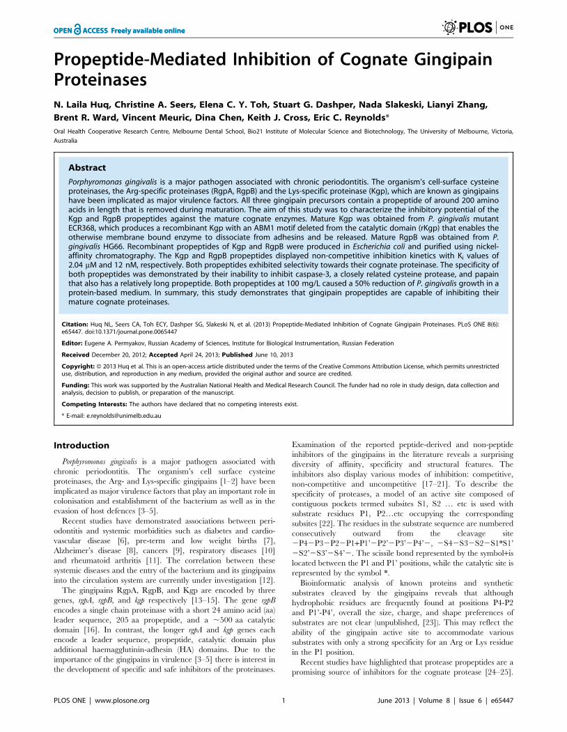
Table 4. Relative growth inhibition of P. gingivalis in aprotein-based minimal medium (MM) by RgB-propeptide (PP)and Kgp-propeptide (PP).
Percentage of growth
MM 100%
MM+RgpB-PP 100 mg/L 55619%
MM+Kgp-PP 100 mg/L 45622%
MM+RgpB-PP 100 mg/L+ rKgp-PP 100 mg/L 60612%
doi:10.1371/journal.pone.0065447.t004
Propeptide Inhibition of Cognate Gingipains
PLOS ONE | www.plosone.org 7 June 2013 | Volume 8 | Issue 6 | e65447
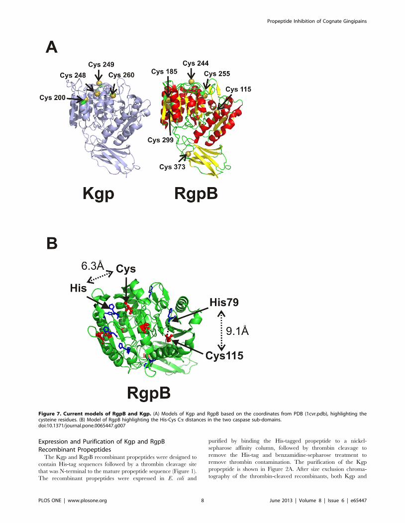
Expression and Purification of Kgp and RgpBRecombinant Propeptides
The Kgp and RgpB recombinant propeptides were designed to
contain His-tag sequences followed by a thrombin cleavage site
that was N-terminal to the mature propeptide sequence (Figure 1).
The recombinant propeptides were expressed in E. coli and
purified by binding the His-tagged propeptide to a nickel-
sepharose affinity column, followed by thrombin cleavage to
remove the His-tag and benzamidine-sepharose treatment to
remove thrombin contamination. The purification of the Kgp
propeptide is shown in Figure 2A. After size exclusion chroma-
tography of the thrombin-cleaved recombinants, both Kgp and
Figure 7. Current models of RgpB and Kgp. (A) Models of Kgp and RgpB based on the coordinates from PDB (1cvr.pdb), highlighting thecysteine residues. (B) Model of RgpB highlighting the His-Cys Ca distances in the two caspase sub-domains.doi:10.1371/journal.pone.0065447.g007
Propeptide Inhibition of Cognate Gingipains
PLOS ONE | www.plosone.org 8 June 2013 | Volume 8 | Issue 6 | e65447

Rgp recombinant propeptides were deemed pure by SDS-PAGE
and MS, with yields of 10–13 mg/L culture fluid. LC-MS analysis
of tryptic peptides also confirmed the expected sequences of both
propeptides. MS analysis using ESI Q-TOF showed a deconvo-
luted protein mass of 23,407.8, corresponding to a molecular mass
of 23,403 Da for the thrombin cleaved Kgp propeptide and a
deconvoluted protein mass of 23,205.3, corresponding to a
molecular mass of 23,204 Da for the thrombin cleaved RgpB
propeptide.
Dimerisation of Kgp PropeptideInitial studies with the Kgp-propeptide yielded inconsistent
inhibition results. The Kgp propeptide exhibited the propensity to
dimerize at higher concentrations as found in the cell lysate, Ni-
affinity column-bound and thrombin-free products (Figure 2) and
as detected by the relative Kav during size exclusion chromatog-
raphy (Figure 2B). The monomer-dimer equilibrium at room
temperature was evident for both Kgp propeptide monomer and
dimer fractions as observed from the SDS gel within 1 h of
separation by chromatography. The involvement of the single
cysteine residue within the propeptide amino acid sequence in this
dimerisation was investigated. Size-exclusion chromatography of
the eluted dimer fractions incubated with 5 mM DTT demon-
strated release of monomer. SDS-PAGE of the dimer and
monomer fractions with and without 5 mM DTT confirmed the
involvement of the cysteine residue (Figure 2C). Following the
DQ-BSA substrate assay with the Kgp propeptide monomer and
dimer in equilibrium, post-assay contents revealed that precipita-
Figure 8. Interaction of gingipain catalytic domains with their propeptides. (A) Model of RgpB highlighting the N-terminus, catalytic Cysand His residues, and residues that differ between strains in red. The residues that form a surface-exposed conserved patch are predicted to interactwith the propeptide. (B) Schematic representation of the inhibition of Kgp by its propeptide. Kgp was modelled using Orchestrar from within Sybyl-8.1 [55] and based on the X-ray crystal structure of RgpB 1cvr.pdb [56]. The propeptide is based on the A chain of the X-ray crystal structure of RgpBinteracting with its propeptide 4ief.pdb [49].doi:10.1371/journal.pone.0065447.g008
Propeptide Inhibition of Cognate Gingipains
PLOS ONE | www.plosone.org 9 June 2013 | Volume 8 | Issue 6 | e65447

tion occurred on standing, suggestive of enzyme propeptide
interactions. However the precipitation was not observed in the
assays with added 5 mM DTT using the Kgp propeptide DTT-
stabilized monomer. Iodoacetylation of the Kgp propeptide after
DTT treatment prevented dimer formation based on Superdex
G75 size-exclusion chromatography and non-reducing PAGE
analysis.
Reproducible inhibitory activity was achieved with the mono-
mer purified in the presence of 5 mM DTT using size-exclusion
chromatography, with additional 5 mM DTT plus 10 mM
cysteine in the proteolytic assays. These assay conditions ensured
that the protease rKgp was fully reduced thus producing higher
activity of the mature enzyme and a reproducible dose inhibitory
response in both assays using DTT-stabilized monomer Kgp-
propeptide. In the proteolytic assay with the chromogenic
substrate, activity of rKgp (0.15 mM) increased by 4961% with
the addition of 5 mM DTT. In the DQ-BSA assay, addition of
5 mM DTT produced a 2564% enhancement of activity.
Propeptide Inhibition of Cognate ProteasesThe inhibition of P. gingivalis W50 whole cell proteolytic activity
by the Kgp and RgpB recombinant propeptides was determined
using chromogenic substrates. The rate of substrate hydrolysis was
monitored for linearity, to ensure there was no sharp increase in
absorbance during the assay which would indicate that the
inhibitory peptides were being used as a preferred substrate. The
Kgp propeptide exhibited ,35% inhibition of P. gingivalis W50
whole cell Lys-protease activity at 80 mg/L, while the RgpB
propeptide exhibited 41% inhibition of W50 whole cell Arg-
protease activity at 80 mg/L (Table 3).
To establish targeted inhibition of the catalytic domain of the
proteases, the propeptide was incubated with purified RgpB or
rKgp. Using both chromogenic and fluorescent DQ-BSA assays,
rKgp and RgpB were inhibited by their propeptides in a dose-
dependent manner (Figures 3 and 4). The DTT-stabilized
monomer at 100 mg/L (4 mM) demonstrated 68% inhibition of
0.15 mM rKgp compared to negligible 0–5% inhibition by the
dimer with GPKNA as substrate. Similarly, in the DQ-BSA assay
the DTT-stabilized monomer at 100 mg/L (4 mM) demonstrated
57% inhibition of 0.15 mM rKgp compared to negligible 0–4%
inhibition by the equivalent dimer. The iodoacetylated monomer
(100 mg/L) demonstrated 2865% inhibition in the proteolytic
assay using DQ-BSA as substrate. The RgpB recombinant
propeptide at a concentration of 10 mg/mL inhibited , 95% of
RgpB activity.
The thrombin-like capability of the proteinases to cleave small
molecule substrates while bound to inhibitors [46] was examined.
The proteolysis assays with increasing concentrations of inhibitor
were conducted with excess substrate. Fluorescence analysis of the
96-well plates 6–12 h after the proteolysis assay with DQ-BSA was
consistent with the original inhibitor dose-response observed
during the assay. In contrast the proteolysis assay using the small
chromogenic substrates revealed that substrate consumption
continued for a further 6–12 h irrespective of the presence and
level of propeptide inhibitor. One interpretation for this observa-
tion is that the propeptide–protease interaction allowed small
molecules to still have access to the active site, however larger
substrates were blocked.
Propeptide Selectivity and SpecificityBoth RgpB and Kgp propeptides demonstrated selectivity for
their own cognate protease with no inhibition observed when Kgp
propeptides were incubated with RgpB and vice versa (Table 3).
The specificity of the propeptides was further examined using two
examples of cysteine proteases. The cysteine protease papain
(2.75 mg/mL), with a propeptide of 115 residues, was not
significantly inhibited by Kgp nor RgpB propeptides at 50 mg/
L concentrations (Table 3). The cysteine protease caspase 3 that
has structural homology with the RgpB and Kgp catalytic domains
also was not inhibited by either Kgp or RgpB propeptides.
Determination of Type of Inhibition and InhibitionConstants
In order to determine the inhibition constant of Kgp and RgpB
propeptides and characterize inhibition mechanism, a kinetics
analysis was performed with purified rKgp and RgpB. The
dissociation constant Ki’, for non-competitive binding of the
inhibitor Kgp propeptide to the enzyme rKgp, was 2.01 mM for
the monomer. The inhibition kinetics were also analysed for the
fluorescent multi-site substrate DQ-BSA and the derived Ki’
parameter was 2.04 mM. The RgpB propeptide also displayed
non-competitive inhibition kinetics against RgpB with a Ki’of
12 nM (Figure 5).
Analysis of Propeptide StabilityThe Kgp propeptide contains 13 Lys residues which could make
the propeptide a potential substrate for Kgp proteolytic activity.
To examine the fate of the Kgp recombinant propeptide in the
presence of the proteases, the post-assay contents were analysed
using SDS-PAGE and HPLC. The SDS-PAGE gels and HPLC
chromatograms revealed intact Kgp and RgpB propeptides as well
as degradation products that were then further analysed by LC-
MS. Identification of the tryptic peptides coupled with the
expected sizes of the Kgp propeptide fragments enabled a
fragmentation pattern to be derived. Lys110 was the most
susceptible to cleavage by the proteinase. Lys residues 4, 41, 69,
100, 129, 168 and 204 were also found to be susceptible to
cleavage. In contrast, Lys residues 6, 22, 37, 84, and 116 were
relatively resistant to proteolytic cleavage by Kgp. Kgp propeptide
Arg residues at position 13, 146, and 149 were also observed to be
relatively resistant to proteolysis by RgpB. The observation of Lys
and Arg residues that are relatively proteolytically resistant to
cleavage by Kgp and RgpB is indicative that the long propeptides
have conformational preferences.
In vivo Processing of Secreted rKgp Precursor FormsA culture of P. gingivalis ECR368 was examined at Days 1
(exponential growth) and 3 (stationary phase) after inoculation. A
reducing SDS-gel of the cell free culture fluid revealed the
presence of precursors with estimated sizes of ,70 and 60 kDa
designated Full-ProKgp and Half-ProKgp respectively (Figure 6).
These are consistent with precursor forms of the gingipains
reported previously [47–48]. The ,60 kDa intermediate present
at equivalent or greater abundance indicates that the sequential
cleavage rates k2,k1. The presence of an intra-molecular
disulphide bond within the ,60 kDa precursor form was
investigated. A non-reducing SDS-gel (Figure 6) of the 60 kDa
precursor revealed the presence of a higher molecular weight
,70 kDa form, indicating that in a small population the 1st half of
the propeptide although cleaved was still covalently attached to the
Kgp catalytic domain through a disulphide bridge. The stable
intermediate precursors with extra 10 kDa or 20 kDa propeptide
regions eluted earlier than the mature Kgp as expected, from
Superose 12 in 50 mM phosphate, 150 mM NaCl, pH 6.
Propeptide Inhibition of Cognate Gingipains
PLOS ONE | www.plosone.org 10 June 2013 | Volume 8 | Issue 6 | e65447

Propeptide-mediated Inhibition of P. gingivalis GrowthP. gingivalis W50 was grown in a protein-based minimal medium
and reached a maximum cell density equivalent to an OD 620 nm
of 0.32 after 40 h of incubation. The P. gingivalis triple gingipain
mutant lacking RgpA, RgpB and Kgp does not grow in this
defined protein-based minimal medium confirming that gingipain
proteolytic activity is essential for the breakdown of the proteins
(BSA and haemoglobin) in this medium. Both Kgp and RgpB
propeptides demonstrated a significant inhibitory effect on P.
gingivalis W50 growth in this protein-based minimal medium
(Table 4).
Discussion
Despite recognition that the traversal of the Arg- and Lys-
gingipains from the cytosol to the final cell surface destination is
accomplished without premature activation, the role of the
gingipain propeptides has not been extensively investigated. This
current study has demonstrated that Kgp and RgpB propeptides
inhibit the proteolytic activity of the membrane bound proteinases
of P. gingivalis W50 in whole cell assays. To demonstrate targeted
inhibition, characterise the mode of inhibition, and investigate the
inter-molecular proteinase-propeptide interaction, cognate cata-
lytic domains were purified from strains HG66 (RgpB) and
ECR368 (rKgp).
In contrast to the nanomolar Ki estimated for the RgpB
recombinant propeptide, a micromolar Ki was calculated for the
Kgp propeptide. This has been attributed to the tendency of the
Kgp propeptide to form covalent dimers through a single cysteine
residue. The inhibitory capability of the mixture of monomer/
dimer rKgp propeptides added to the proteolytic assay was
inconsistent. This was resolved after separation of the DTT
stabilized monomers from the non-inhibitory dimers using size-
exclusion chromatography in 5 mM DTT.
The recent report of the RgpB propeptide co-crystallized with
the cognate RgpB catalytic domain indicates that the propeptide
attaches laterally to the RgpB catalytic domain through a large
concave surface. The RgpB propeptide adopts an overall
‘‘croissant ‘‘ shape with a projecting ‘‘ inhibitory’’ loop consisting
of sixteen residues (Lys113– Glu128) that approaches the active-
site cleft of RgpB on its non-primed side in a substrate-like manner
[49].
Observation of the precursor ProKgp (,70 kDa) with the
intermediate half-ProKgp (,60 kDa) by reducing SDS-PAGE, at
equivalent or greater abundance in the culture fluid during
exponential growth of the P. gingivalis mutant ECR 368 indicates
that the second cleavage step is slower than the first cleavage step.
Although precursor forms have been observed for both RgpB and
Kgp [47–48], the presence of the disulphide bridge between the
Kgp propeptide and catalytic domain in the precursor form has
not been reported previously and may have resulted from
oxidation during extraction. This observation can not be explained
by the reported structure of the RgpB propeptide interacting with
the RgpB catalytic domain [49]. The catalytic domain of Kgp has
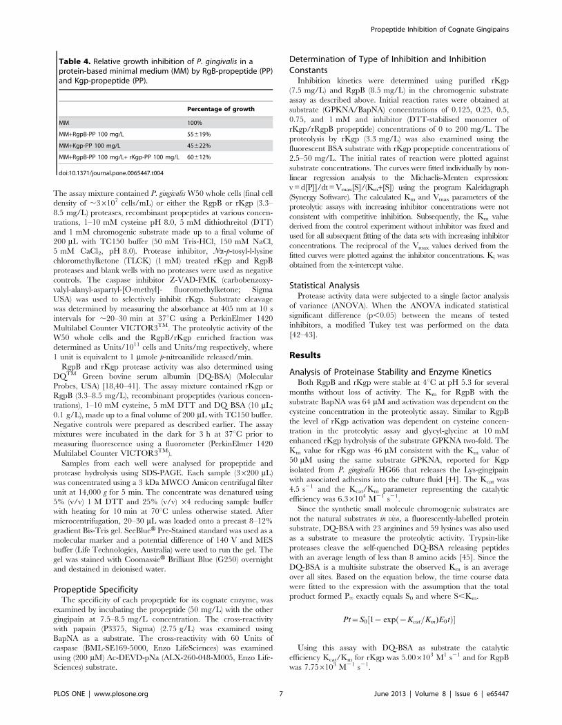
four cysteines: Cys200, Cys248, Cys249 and Cys260 (Figure 7A). The
observed in vitro inhibition by the discrete Kgp propeptide is not
dependent on the formation of a disulphide bridge between the
propeptide and catalytic domain as the inhibition is retained both
in a reducing environment and by the iodoactylated Kgp
propeptide. However disulphide bridge formation within the
precursor form does occur in a non-reducing environment.
From a model of Kgp (Figure 7A) based on the RgpB structure,
the catalytic cysteine is the most exposed and hence most likely to
form a disulphide bond. The effect of the propeptide cysteine
forming a disulphide bridge with either the catalytic cysteine
Cys249 or the neighbouring Cys248 would have the effect of
abolishing Lys-protease activity in the 70 kDa precursor form.
This would be consistent with the recent report that an active site
probe, a biotinylated irreversible Kgp-specific inhibitor [50] did
not bind to the active site of the 70 kDa precursor form under
non-reducing conditions [48]. However it is also plausible that the
cleaved N-terminal half of the Kgp propeptide forms a disulphide
bridge with one of the other two cysteines within the mature Kgp
protease: Cys200 found only in Kgp, or Cys260, common to both
RgpB and Kgp (Figure 7A). In the model of the mature proteinase,
both these cysteines are less accessible for bridge formation;
however, accessibility may be altered in the precursor form.
To understand the observed strong selectivity of the propeptides
for the cognate proteases, the sequence variation of the RgpA/B
and Kgp propeptides and the catalytic domains from the P.
gingivalis strains W50, W83, ATCC 33277, TDC60, 381, W12 was
examined. The RgpA/B and Kgp propeptides from the known P.
gingivalis strains are all highly conserved with a calculated
percentage identity (%ID) of 98–100% between the propeptide
homologs. However sequence conservation is less between the
RgpA and RgpB propeptide paralogs (75–76% ID ) and between
the RgpA/B and Kgp propeptide paralogs (20–22% ID). Similarly
the sequences of the catalytic domains of RgpA/B and Kgp are
also highly conserved (94–100% ID) between the homologs with
less conservation between the paralogs. This is consistent with the
observed selectivity.
The specificity of the propeptides for the gingipains was
examined using two examples of cysteine proteases. Since, the
three gingipain propeptides range from 203 to 209 residues,
significantly larger than the average propeptide lengths of ,40
residues observed in most cysteine proteases [26], the 212 residue
papain that is inhibited by its own 115 residue propeptide was
selected. Neither Kgp nor RgpB propeptides demonstrated any
inhibition for papain consistent with the differences between the
papain and gingipain catalytic domains and active site configura-
tions.
The second example was selected based on the structural
similarities of the catalytic domains. Caspase 3 (pdb1pau) and
RgpB structures (pdb1cvr) [51–53] share a common ‘‘caspase–
hemoglobinase’’ fold with similar active site pockets despite limited
sequence similarity [54]. The mature caspase 3 enzyme and
zymogen backbone structures can be superimposed to within
3.8 A over 106 residues. The current understanding of caspase
activation and the caspase structure, presented a compelling
argument to examine the effects of Kgp and RgpB propeptides on
caspase activity. The absence of inhibition exhibited by both
propeptides against caspase 3, highlights the specificities of the 200
residue propeptides.
Both RgpB and, by homology, Kgp catalytic domains have the
appearance of two adjacent caspase sub-domains plus the C-
terminal Ig-fold [54]. The RgpB active site cysteine and histidine
occur in the second caspase sub-domain and their respective Caatoms are within 6.3 A. In the first caspase sub-domain the RgpA/
RgpB sequences have a cysteine (Cys115) and histidine (His79) at
topologically analogous positions (Figure 7B). The catalytic
potential of these two residues in RgpB and RgpA has not been
explored. However, this difference between Kgp and RgpB/RgpA
may also account for the selectivity exhibited by the cognate
propeptides.
To further understand the interaction between the conserved
propeptides and the cognate proteases, the residues within the
catalytic domains of RgpA, RgpB and Kgp that differ between the
different strains of P. gingivalis were identified. These point mutated
Propeptide Inhibition of Cognate Gingipains
PLOS ONE | www.plosone.org 11 June 2013 | Volume 8 | Issue 6 | e65447

residues found within RgpA and RgpB were mapped against the
crystal structure of RgpB. This revealed that the residues located
on the first a-helix immediately C-terminal of the fifth b-strand
and the N-terminal portion of the next a-helix are conserved. This
surface-exposed, conserved patch is depicted between the position
of the known N-terminal residue of the catalytic domain and the
active site (Figure 8A). In the case of Kgp, 28 residues that differ
between different strains of P. gingivalis were identified. Three
residues within 10 A of the catalytic site were changed: A449S,
L454S, and I478V. Interestingly, the A449S and L454S point
mutations are found together in F5XB86 (TDC60), Q51817
(W83), and Q6Q4T4 (an un-named strain) making a small region
close to the catalytic site of Kgp more hydrophilic in those strains.
Mapping all the 28 point-mutated residues to the model of Kgp
revealed an analogous surface-exposed, structurally identical,
conserved patch in Kgp. The surface-exposed, conserved patches
in RgpB and Kgp are predicted to be covered by the propeptide in
the respective zymogens.
Models of both Kgp and the Kgp propeptide were produced
using Orchestrar from within Sybyl-8.1 [55] and based on the X-
ray crystal structure of RgpB (1cvr.pdb) [56] and chain A from the
crystal structure of RgpB co-crystallized with its propeptide
(4ief.pdb) [49] respectively. The Kgp propeptide model was
validated by calculating the ‘Fugue alignment’ [57] between the
Kgp and RgpB propeptides, which gave a Z-score of 10.72
classified as ‘certain’ with greater than 99% confidence. The
model of the propeptide had an rms deviation of 1.28 A from the
crystal coordinates after energy minimization to a maximum
gradient of 0.5 kcal mol21 A21 using the AMBER force-field. A
model of the Kgp propeptide docked with Kgp was then produced
by independently aligning by least-squares the model of Kgp and
the model Kgp propeptide against the B and A-chains respectively
of the co-crystallized RgpB/RgpB propeptide (4ief.pdb). This
alignment predicts that the Lys110 of the inhibitory-loop of the
Kgp propeptide will insert into the catalytic pocket of Kgp.
A schematic representation of the inhibition of Kgp by its
propeptide based on this model is shown in Figure 8B. From the
model structure the cleavage of the propeptide at Lys110 will leave
a substantial protein domain still capable of allosterically blocking
access to the catalytic site by large, substrate proteins. The bound
orientation of the propeptides with their proteases is consistent
with an interaction between the identified conserved patch
(Fig. 8A) and the propeptide. The schematic (Fig. 8B) is also
consistent with possible exosite binding that could explain the
selectivity and specificity of the propeptides. Experimentally, the
peptide bond C-terminal to Lys110 was found to be susceptible to
cleavage by Kgp. This is consistent with the location of Lys110
being in a loop; the peptide bond is only protected from cleavage
when the propeptide is bound to Kgp with the appropriate
orientation.
It was interesting to examine the effects of the propeptides on
growth of P. gingivalis. The requirement of cell surface located
proteinases for nutrient acquisition, tested using the triple mutant
without the RgpA, RgpB and Kgp gingipains in a protein-based
minimal medium was consistent with previous reports [41,58].
The observed retardation of the planktonic growth of P. gingivalis
by the added propeptides highlights their potential for inhibition of
P. gingivalis growth and virulence.
In summary the P. gingivalis cell surface gingipains are carefully
regulated prior to activation by high-selectivity propeptides that
are tailored to each proteinase. It is possible that the long
propeptide has a role in propeptide-mediated folding as well as
preventing proteinase premature activation throughout the
multiple processing, propeptide detachment, and rearrangement
events that occur to enable the cell surface assembly of the
gingipain complexes.
Acknowledgments
The technical assistance of Huiling He, Pearlyn Zhi Rong Chiew and
Ching-Seng Ang is gratefully acknowledged.
Author Contributions
Conceived and designed the experiments: NLH CAS ECR SGD KJC.
Performed the experiments: ECYT NS LZ BRW VM DC. Analyzed the
data: NLH CAS KJC ECR. Contributed reagents/materials/analysis tools:
CAS NS DC. Wrote the paper: NLH KJC ECR.
References
1. O’Brien-Simpson N, Veith PD, Dashper SG, Reynolds EC (2003) Porphyromonas
gingivalis gingipains: the molecular teeth of a microbial vampire. Curr Protein
Pept Sci 4: 409–426.
2. Guo Y, Nguyen KA, Potempa J (2010) Dichotomy of gingipains action as
virulence factors: from cleaving substrates with the precision of a surgeon’s knife
to a meat chopper-like brutal degradation of proteins. Periodontol 2000 54: 15–
44.
3. Shi Y, Ratnayake DB, Okamoto K, Abe N, Yamamoto K, et al. (1999) Genetic
analyses of proteolysis, hemoglobin binding, and hemagglutination of Porphyr-
omonas gingivalis. Construction of mutants with a combination of rgpA, rgpB, kgp,
and hagA. J Biol Chem 274: 17955–17960.
4. Lamont RJ, Jenkinson HF (1998) Life below the gum line: pathogenic
mechanisms of Porphyromonas gingivalis. Microbiol Mol Biol Rev 62: 1244–1263.
5. Nakayama K, Yoshimura F, Kadowaki T, Yamamoto K (1996) Involvement of
arginine-specific cysteine proteinase (Arg-gingipain) in fimbriation of Porphyr-
omonas gingivalis. J Bacteriol 178: 2818–2824.
6. Humphrey LL, Fu R, Buckley DI, Freeman M, Helfand M (2008) Periodontal
disease and coronary heart disease incidence: a systematic review and meta-
analysis. J Gen Intern Med 23: 2079–2086.
7. Dasanayake AP, Gennaro S, Hendricks-Munoz KD, Chhun N (2008) Maternal
periodontal disease, pregnancy, and neonatal outcomes. MCN Am J Matern
Child Nurs 33: 45–49.
8. Kamer AR, Craig RG, Dasanayake AP, Brys M, Glodzik-Sobanska L, et al.
(2008) Inflammation and Alzheimer’s disease: possible role of periodontal
diseases. Alzheimers Dement 4: 242–250.
9. Hujoel PP, Drangsholt M, Spiekerman C, Weiss NS (2003) An exploration of the
periodontitis-cancer association. Ann Epidemiol 13: 312–316.
10. Renvert S (2003) Destructive periodontal disease in relation to diabetes mellitus,
cardiovascular diseases, osteoporosis and respiratory diseases. Oral Health Prev
Dent 1 Suppl 1: 341–357; discussison 358–349.
11. Detert J, Pischon N, Burmester GR, Buttgereit F (2010) The association between
rheumatoid arthritis and periodontal disease. Arthritis Res Ther 12: 218.
12. Kadowaki T, Takii R, Yamatake K, Kawakubo T, Tsukuba T, et al. (2007) A
role for gingipains in cellular responses and bacterial survival in Porphyromonas
gingivalis-infected cells. Front Biosci 12: 4800–4809.
13. Curtis MA, Kuramitsu HK, Lantz M, Macrina FL, Nakayama K, et al. (1999)
Molecular genetics and nomenclature of proteases of Porphyromonas gingivalis.
J Periodontal Res 34: 464–472.
14. Slakeski N, Bhogal PS, O’Brien-Simpson NM, Reynolds EC (1998) Character-
ization of a second cell-associated Arg-specific cysteine proteinase of Porphyr-
omonas gingivalis and identification of an adhesin-binding motif involved in
association of the prtR and prtK proteinases and adhesins into large complexes.
Microbiology 144: 1583–1592.
15. Bhogal PS, Slakeski N, Reynolds EC (1997) A cell-associated protein complex of
Porphyromonas gingivalis W50 composed of Arg- and Lys-specific cysteine
proteinases and adhesins. Microbiology 143: 2485–2495.
16. Mikolajczyk-Pawlinska J, Kordula T, Pavloff N, Pemberton PA, Chen WC, et al.
(1998) Genetic variation of Porphyromonas gingivalis genes encoding gingipains,
cysteine proteinases with arginine or lysine specificity. Biol Chem 379: 205–211.
17. Rangarajan M, Smith SJ, U S, Curtis MA (1997) Biochemical characterization
of the arginine-specific proteases of Porphyromonas gingivalis W50 suggests a
common precursor. Biochem J 323: 701–709.
18. Toh EC, Dashper SG, Huq NL, Attard TJ, O’Brien-Simpson NM, et al. (2011)
Porphyromonas gingivalis cysteine proteinase inhibition by k-casein peptides.
Antimicrob Agents Chemother 55: 1155–1161.
Propeptide Inhibition of Cognate Gingipains
PLOS ONE | www.plosone.org 12 June 2013 | Volume 8 | Issue 6 | e65447

19. Kontani M, Amano A, Nakamura T, Nakagawa I, Kawabata S, et al. (1999)
Inhibitory effects of protamines on proteolytic and adhesive activities ofPorphyromonas gingivalis. Infect Immun 67: 4917–4920.
20. Blat Y (2010) Non-competitive inhibition by active site binders. Chem Biol Drug
Des 75: 535–540.21. Dashper SG, Pan Y, Veith PD, Chen YY, Toh EC, et al. (2012) Lactoferrin
inhibits Porphyromonas gingivalis proteinases and has sustained biofilm inhibitoryactivity. Antimicrob Agents Chemother 56: 1548–1556.
22. Schechter I, Berger A (1967) On the size of the active site in proteases. I. Papain.
Biochem Biophys Res Commun 27: 157–162.23. Ally N, Whisstock JC, Sieprawska-Lupa M, Potempa J, Le Bonniec BF, et al.
(2003) Characterization of the specificity of arginine-specific gingipains fromPorphyromonas gingivalis reveals active site differences between different forms of
the enzymes. Biochemistry 42: 11693–11700.24. Guay J, Falgueyret JP, Ducret A, Percival MD, Mancini JA (2000) Potency and
selectivity of inhibition of cathepsin K, L and S by their respective propeptides.
Eur J Biochem 267: 6311–6318.25. Demidyuk IV, Shubin AV, Gasanov EV, Kostrov SV (2010) Propeptides as
modulators of functional activity of proteases. BioMolecular Concepts 1: 305–322.
26. Wiederanders B, Kaulmann G, Schilling K (2003) Functions of propeptide parts
in cysteine proteases. Curr Protein Pept Sci 4: 309–326.27. Lecaille F, Kaleta J, Bromme D (2002) Human and parasitic papain-like cysteine
roteases: Their role in physiology and pathology and recent developments ininhibitor design. Chemical Rev 102: 4459–4488.
28. Rawlings ND, Tolle DP, Barrett AJ (2004) MEROPS: the peptidase database.Nucleic Acids Res 32: D160–164.
29. Slakeski N, Seers CA, Ng K, Moore C, Cleal SM, et al. (2011) C-terminal
domain residues important for secretion and attachment of RgpB inPorphyromonas gingivalis. J Bacteriol 193: 132–142.
30. Slakeski N, Cleal SM, Bhogal PS, Reynolds EC (1999) Characterization of aPorphyromonas gene prtK that encodes a lysine-specific cysteine proteinase and
three sequence-related adhesins. Oral Microbiol Immunol 14: 92–97.
31. Jackson CA, Hoffmann B, Slakeski N, Cleal S, Hendtlass AJ, et al. (2000) Aconsensus Porphyromonas gingivalis promoter sequence. FEMS Microbiol Lett 186:
133–138.32. Seers CA, Slakeski N, Veith PD, Nikolof T, Chen Y-Y, et al. (2006) The RgpB
C-terminal domain sas a role in attachment of RgpB to the outer membrane andbelongs to a novel C-terminal-domain family found in Porphyromonas gingivalis.
J Bacteriol 188: 6376–6386.
33. Dashper SG, Ang CS, Veith PD, Mitchell HL, Lo AWH, et al. (2009) Responseof Porphyromonas gingivalis to heme limitation in continuous culture. J Bacteriol
191: 1044–1055.34. Oda H, Saiki K, Numabe Y, Konishi K (2007) Effect of gamma-
immunoglobulin on the asaccharolytic growth of Porphyromonas gingivalis.
J Periodontal Res 42: 438–442.35. Shi Y, Kong W, Nakayama K (2000) Human lactoferrin binds and removes the
hemoglobin receptor protein of the periodontopathogen Porphyromonas gingivalis.J Biol Chem 275: 30002–30008.
36. Pathirana RD, O’Brien-Simpson NM, Brammar GC, Slakeski N, Reynolds EC(2007) Kgp and RgpB, but not RgpA, are important for Porphyromonas gingivalis
virulence in the murine periodontitis model. Infect Immun 75.
37. Sambrook J, Fritsch EF, Maniatis T, editors (1989) Molecular Cloning: aLaboratory Manual,. 2nd ed: Cold Spring Harbor Laboratory.
38. Hondoh T, Kato A, Yokoyama S, Kuroda Y (2006) Computer-aided NMRassay for detecting natively folded structural domains. Protein Sci 15: 871–883.
39. O’Brien-Simpson NM, Pathirana RD, Paolini RA, Chen YY, Veith PD, et al.
(2005) An immune response directed to proteinase and adhesin functionalepitopes protects against Porphyromonas gingivalis-induced periodontal bone loss.
J Immunol 175: 3980–3989.
40. Yoshioka M, Grenier D, Mayrand D (2003) Monitoring the uptake of protein-derived peptides by Porphyromonas gingivalis with fluorophore-labeled substrates.
Curr Microbiol 47: 1–4.41. Grenier D, Imbeault S, Plamondon P, Grenier G, Nakayama K, et al. (2001)
Role of gingipains in growth of Porphyromonas gingivalis in the presence of human
serum albumin. Infect Immun 69: 5166–5172.42. Zar JH (1984) Biostatistical analysis. Englewood Cliffs, N.J.: Prentice-Hall.
43. Fowler J, Cohen L (1997) Practical statistics for field biology. Brisbane, Australia:John Wiley and Sons.
44. Pike R, McGraw W, Potempa J, Travis J (1994) Lysine- and arginine-specificproteinases from Porphyromonas gingivalis. Isolation, characterization, and evidence
for the existence of complexes with hemagglutinins. J Biol Chem 269: 406–411.
45. Cruz LJ, Tacken PJ, Fokkink R, Joosten B, Stuart MC, et al. (2010) TargetedPLGA nano- but not microparticles specifically deliver antigen to human
dendritic cells via DC-SIGN in vitro. J Control Release 144: 118–126.46. Wilkens M, Krishnaswamy S (2002) The contribution of factor Xa to exosite-
dependent substrate recognition by prothrombinase. J Biol Chem 277: 9366–
9374.47. Mikolajczyk J, Boatright KM, Stennicke HR, Nazif T, Potempa J, et al. (2003)
Sequential autolytic processing activates the zymogen of Arg-gingipain. J BiolChem 278: 10458–10464.
48. Sztukowska M, Veillard F, Potempa B, Bogyo M, Enghild JJ, et al. (2012)Disruption of gingipain oligomerization into non-covalent cell-surface attached
complexes. Biol Chem 393: 971–977.
49. de Diego I, Veillard FT, Guevara T, Potempa B, Sztukowska M, et al. (2013)Porphyromonas gingivalis virulence factor gingipain RgpB shows a unique
zymogenic mechanism for cysteine peptidases. J Biol Chem.50. Kato D, Boatright KM, Berger AB, Nazif T, Blum G, et al. (2005) Activity-based
probes that target diverse cysteine protease families. Nat Chem Biol 1: 33–38.
51. Sheets SM, Potempa J, Travis J, Fletcher HM, Casiano CA (2006) Gingipainsfrom Porphyromonas gingivalis W83 synergistically disrupt endothelial cell adhesion
and can induce caspase-independent apoptosis. Infect Immun 74: 5667–5678.52. Eichinger A, Beisel HG, Jacob U, Huber R, Medrano FJ, et al. (1999) Crystal
structure of gingipain R: an Arg-specific bacterial cysteine proteinase with acaspase-like fold. Embo J 18: 5453–5462.
53. Chen JM, Rawlings ND, Stevens RA, Barrett AJ (1998) Identification of the
active site of legumain links it to caspases, clostripain and gingipains in a newclan of cysteine endopeptidases. FEBS Lett 441: 361–365.
54. Aravind L, Koonin EV (2002) Classification of the caspase-hemoglobinase fold:detection of new families and implications for the origin of the eukaryotic
separins. Proteins 46: 355–367.
55. Tripos (2011) SYBYL-X 1.2: Tripos International, 1699 South Hanley Rd., St.Louis, Missouri, 63144, USA.
56. Eichinger A, Beisel H, Jacob U, Huber R, Medrano F, et al. (1999) Crystalstructure of gingipain R: an Arg-specific bacterial cysteine proteinase with a
caspase-like fold. Embo J 18: 5453–5462.57. Shi J, Blundell TL, Mizuguchi K (2001) FUGUE: sequence-structure homology
recognition using environment-specific substitution tables and structure-
dependent gap penalties. J Mol Biol 310: 243–257.58. Grenier D, Roy S, Chandad F, Plamondon P, Yoshioka M, et al. (2003) Effect of
inactivation of the Arg- and/or Lys-gingipain gene on selected virulence andphysiological properties of Porphyromonas gingivalis. Infect Immun 71: 4742–4748.
Propeptide Inhibition of Cognate Gingipains
PLOS ONE | www.plosone.org 13 June 2013 | Volume 8 | Issue 6 | e65447


