Embed Size (px)
Citation preview
American Cancer Society Second National Conference onCancer Genetics
Supplement to Cancer
Prophylactic Mastectomy and Inherited Predispositionto Breast Carcinoma
Kevin S. Hughes, M.D.1
Moshe Z. Papa, M.D.2
Timothy Whitney, M.D.1
Robert McLellan, M.D.1
1 The Risk Assessment Clinic, The Lahey Clinic,Peabody, Massachusetts.
2 The Chaim Sheba Medical Center, Sackler Schoolof Medicine, Tel Aviv University, Tel Hashomer,Israel.
Presented at the American Cancer Society SecondNational Conference on Cancer Genetics, SanFrancisco, California, June 2, 1998.
Address for reprints: Kevin S. Hughes, M.D., TheRisk Assessment Clinic, The Lahey Clinic, 1 EssexCenter Drive, Peabody, MA 01960.
Received May 25, 1999; accepted June 3, 1999.
Relative to her risk of breast carcinoma, the woman with a BRCA1 or BRCA2 gene
mutation can be managed either by intensive screening (with or without chemo-
prevention) or by prophylactic mastectomy. Although it would be preferable to
avoid prophylactic surgery, the current level of screening technology and the
rudimentary state of chemoprevention do not guarantee a good outcome with
intensive surveillance. A review of the currently available data was undertaken to
determine the efficacy of prophylactic surgery, intensive screening, and chemo-
prevention. An attempt then was made to extrapolate the efficacy of the various
approaches to the management of women who carry BRCA1 or BRCA2 gene
mutations. Intensive surveillance may not detect breast carcinoma at an early,
curable stage in young women with BRCA1 or BRCA2 gene mutations because the
growth rate of the tumors in these women most likely will be rapid and the density
of the breast tissue may compromise detection. Chemoprevention is in its infancy,
and its efficacy in this population is unknown. Conversely, prophylactic surgery
may not be completely effective in preventing breast carcinoma. The authors are
hopeful that sometime in the next decade advances in chemoprevention, screening
technology, or breast carcinoma treatment will make mastectomy obsolete. How-
ever, for the time being prophylactic mastectomy has attributes that make it an
alternative for this population that must be considered. Careful discussion of all
options is essential in the management of these women. Cancer 1999;86:2502–16.
© 1999 American Cancer Society.
KEYWORDS: prophylactic mastectomy, BRCA1, BRCA2, breast carcinoma, breastreconstruction.
The cloning of the BRCA1 and BRCA2 genes has opened the door tothe identification of a subset of women at a very high risk of
developing breast or ovarian carcinoma.1 To develop clinical man-agement strategies, it is important to understand the genetic issues,including the frequency and penetrance of the gene mutation and theexpected age at which that malignancy will develop. In general,women with a BRCA1 mutation are believed to have a 19.1% risk ofdeveloping breast carcinoma by the age of 40 years, an 85% risk ofdeveloping breast carcinoma by the age of 70 years, and between a26 – 85% risk of developing ovarian carcinoma by the age of 70years.1-4 Women with a BRCA2 mutation are believed to have a risk ofdeveloping breast carcinoma similar to that of women with a BRCA1mutation, although perhaps beginning at a later age,2,5 and a 10 –16%risk of developing ovarian carcinoma by the age of 70 years.2,6
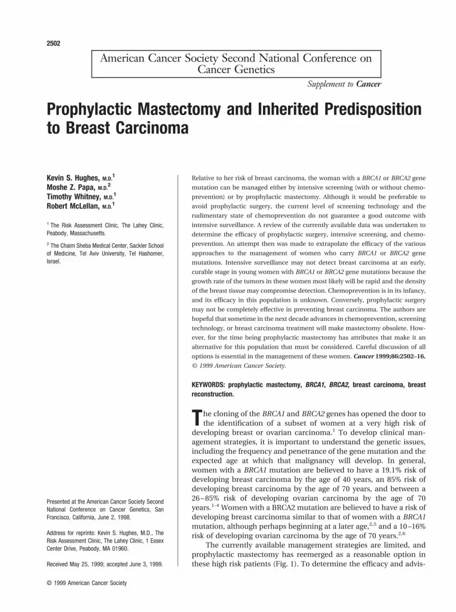
The currently available management strategies are limited, andprophylactic mastectomy has reemerged as a reasonable option inthese high risk patients (Fig. 1). To determine the efficacy and advis-
2502
© 1999 American Cancer Society

ability of prophylactic surgery, that option must becompared with the efficacy of the currently availablealternatives: screening and chemoprevention.
In the past, prophylactic mastectomy frequently wasused for patients who would not be considered at highrisk today. Given the fact that surgeons tended to usewhat currently would be considered an inadequate pro-cedure (subcutaneous mastectomy) and the fact thatreconstruction was rudimentary, it is not surprising thatthis procedure fell out of favor. Current-day prophylacticmastectomy involves using the appropriate procedure(total mastectomy) and continuously improving recon-struction methods to deal with a group of patients atproven high risk (proven carriers of BRCA1 and BRCA2gene mutations). These current techniques and indica-tions must be compared objectively with the currentefficacy of screening and the emerging field of chemo-prevention. In this comparison, prophylactic mastec-tomy appears at least as reasonable, and at times moreappropriate, than the alternatives. In the future, as che-moprevention becomes more effective and as screeningtechnologies improve, the need for prophylactic mastec-tomy may disappear.
Breast carcinoma screening is justified in the gen-eral population, most likely after the age of 40 years,and certainly after the age of 50 years.7 Intensivescreening of carriers of BRCA1 and BRCA2 gene mu-tations is likely to be indicated and effective if initiatedat an early age,5 but even with the current technologyadvanced breast carcinoma still will develop in a cer-tain number of women who will die despite intensivescreening. Chemoprevention of breast carcinoma is inits infancy and although the Breast Cancer Prevention
Trial has shown promise by suggesting that tamoxifenmay decrease the risk of breast carcinoma, to date nodata have proven that tamoxifen will be effective forcarriers of BRCA1 and BRCA2 gene mutations. There-fore, both screening and chemoprevention must becompared with removal of breast tissue by prophylac-tic mastectomy as options for treatment, given thatprophylactic mastectomy appears to reduce the oc-currence of breast carcinoma without providing abso-lute control.
To evaluate each approach with respect to carriersof BRCA1 and BRCA2 gene mutations, our method hasbeen to assess the risk and benefits through a series ofquestions, including: Is effective chemopreventionavailable? What is the efficacy of screening? Withscreening, how often will breast carcinoma be identi-fied at an early, curable stage? What is the efficacy ofprophylactic mastectomy? How often will breast car-cinoma develop in women who have undergone pro-phylactic mastectomy, and how many of those womenwill die? What type of mastectomy should be per-formed? When should this surgery be performed?Should the patient undergo reconstruction, and if so,what type? What will be the psychosocial effect of thisprocedure on the patient and her family?
We will attempt to summarize the state of the artas it relates to these questions for chemoprevention,screening, and prophylactic mastectomy. Many ofthese issues cannot be answered at our current level ofknowledge, particularly with respect to the value ofchemoprevention, and continued research is essen-tial.
FIGURE 1. Options for a mutation car-
rier. Relative to her risk for the develop-
ment of breast carcinoma, a woman
found to harbor a mutation in the BRCA1
and/or BRCA2 genes can be managed
by intensive observation (with or without
chemoprevention) or by prophylactic
mastectomy, with or without reconstruc-
tion.
Prophylactic Mastectomy/Hughes et al. 2503

ChemopreventionChemoprevention is defined as the utilization of adrug or a chemical to prevent the development ofcancer. Whereas this approach may be ideal in thefuture, to our knowledge no proven agent currently isavailable. The Breast Cancer Prevention Trial has sug-gested that tamoxifen is effective in preventing breastcarcinoma. However, it is unclear whether tamoxifenwill be effective in the population carrying a BRCA1 orBRCA2 gene mutation or at what age tamoxifen treat-ment should be initiated, how long it should be con-tinued, or what the long term effects will be. It will bemany years before the extended use of this or anyagent has been tested in a young population.
ScreeningIntensive screening programs are predicated on thecurrent ability to provide adequate treatment whenbreast carcinoma is detected. Effective surveillancetherefore is dependent on an adequate incidence ofbreast carcinoma, a silent early phase, adequate treat-ment, and adequate technology.8 Because screeningwould appear to be a much less distressing and mu-tilating approach, it is tempting to recommend inten-sive screening rather than prophylactic mastectomy tothe woman who carries a BRCA1 or a BRCA2 genemutation. However, it is important to remember thatintensive screening does not work by preventingbreast carcinoma (primary prevention), but ratherworks by detecting breast carcinoma at an earlier,more treatable stage (secondary prevention). The ef-ficacy of intensive screening is dependent on the abil-ity to detect breast carcinoma early enough so thatsurgery, chemotherapy, or radiation (or a combinationof all three) may be used to eradicate the tumor. Ifcure rates were not stage-dependent, then early de-tection would not be necessary.
To our knowledge the efficacy of screening for awoman who carries a BRCA1 or BRCA2 gene mutationhas not been tested. Although to our knowledge nodata specific to BRCA1 and BRCA2 carriers are avail-able, it is possible to make some broad predictionsrelative to this group using the findings in the generalpopulation as a baseline.
Screening is effective in women age .50 yearsbecause they have an adequate incidence of breastcarcinoma development,9 because of the existence ofa silent, early phase of breast carcinoma that is detect-able by mammography,9 because adequate treatmentis available for women with breast carcinoma (thedecreased mortality found with screening in the ran-domized trials of women age .50 years is dependenton the ability to cure this disease, and the cure is
greater at a lower stage), and because the currenttechnology is adequate to detect breast carcinoma inwomen age .50 years.
The silent, early phase of breast carcinoma pre-sents a window of opportunity in the postmenopausalwoman because it is at this stage that a tumor can beidentified on mammography even before it reaches apalpable size. This time can range from 1–3 years, inpart due to the doubling time of tumors in this agegroup. Assuming that a single tumor begins the pro-cess of carcinogenesis, volume calculations show that20 doublings will need to take place for the tumor toreach a greatest dimension of 1 mm, and 10 moredoublings will need to take place for the tumor toprogress to a greatest dimension of 1 cm.10,11
The threshold size for detecting breast tumors bymammography is approximately 1–2 mm (approxi-mately 20 doublings), whereas the threshold size fordetecting breast tumors by palpation is approximately1 cm (approximately 30 doublings). Spratt et al.10,11
have estimated a doubling time of 103–191 days forlymph node negative invasive tumors in women age.50 years. Using an estimate of 125 days and usingvolume calculations, the time from inception to mam-mographic detection would be approximately 2500days (6.8 years), and the time from mammographicdetection to clinical detection by palpation would bean additional 1250 days (3.4 years) (Fig. 2). This pro-duces a window of opportunity of .3 years duringwhich time breast tumors will be detectable by mam-mography but not by palpation. In general, the longerthis window is, the more effective screening mam-mography will be. In addition, the majority of womengo through a phase of ductal carcinoma in situ beforethe development of invasive breast carcinoma, whichadds to the window of opportunity.
Efficacy of Surveillance in Patients with HereditaryBreast CarcinomaBreast carcinoma will develop in as many as 19.1% offemale carriers of BRCA1 and BRCA2 gene mutationsby the age of 40 years, and in as many as 50% of thesewomen by the age of 50 years.1-4 Two factors will havea negative effect on the length of the mammographicdetection window in this population. First, the rate ofgrowth of breast carcinoma is faster in youngerwomen and second, the breast parenchyma is denserin younger women, making mammographic detectionmore difficult.
Spratt et al.10,11 have estimated a doubling time of50 – 64 days for lymph node negative invasive tumorsin women in their 30s. Using 50 days, and assumingthe same mammographic and palpation thresholdsdiscussed earlier, the time from inception to mammo-
2504 CANCER Supplement December 1, 1999 / Volume 86 / Number 11

graphic detection would be approximately 1000 days(2.7 years), and the time from mammographic detec-tion to clinical detection by palpation would be anadditional 500 days (1.35 years) (Fig. 2). This producesa window of slightly greater than 1 year during whichbreast tumors will be detectable by mammography,but not by palpation.
However, another problem is that young womentend to have denser breast tissue and therefore theability to visualize tumors on mammograms is dimin-ished and the ability to palpate tumors is compro-mised (Fig. 3). In fact, Dr. Barbara Smith found nega-tive results on mammography in .100 women who
were age ,40 years and underwent treatment forbreast carcinoma at the Massachusetts General Hos-pital (personal communication). This finding meansthat the mammographic and palpation thresholdsmost likely are larger in the younger population andthat as many as 33% of the tumors in this group maynot be detectable by mammography.
Carriers of the BRCA1 and BRCA2 gene mutationsmay or may not have denser breast tissue on mam-mography than noncarriers, but it would appear rea-sonable to assume that the density of breast tissue iscommensurate with age.
With regard to the rate of tumor growth, it is likely
FIGURE 2. Doubling time of 50 days
versus 125 days. In postmenopausal
women, the growth rate of tumors is
slower, depicted here by a doubling time
of 125 days, and the window of time for
mammographic detection (mammo) is
relatively long. In premenopausal
women the growth rate of tumors is
faster, and the window of time for mam-
mographic detection is shortened.
FIGURE 3. Dense breast tissue. In
premenopausal women, the density of
breast tissue increases the mammo-
graphic threshold and also can shorten
the window of time for mammographic
detection (mammo).
Prophylactic Mastectomy/Hughes et al. 2505

that tumors in carriers of the BRCA1 gene mutationwill grow rapidly both because of younger age and thegene mutation. Foulkes et al.12 found that AshkenaziJewish women who carry one of the two establishedmutations (185 delAG and 5382insC) were more likelyto have Grade 3 tumors (11 of 12 tumors were Grade 3in carriers vs. 29 of 100 tumors in noncarriers), weremore likely to be lymph node positive (45.5% in carri-ers vs. 31.1% in noncarriers), and were more likely tohave tumors that were larger in greatest dimension(mean size 2.48 cm in carriers vs. 1.71 cm in noncar-riers). Other studies found that tumors in carriers weremore likely to be estrogen receptor negative13,14 andp53 positive.15 Therefore, carriers of the BRCA1 muta-tion will likely have faster-growing tumors that willpass through the mammographic window rapidly.BRCA2 breast tumors also appear to be of higher gradethan sporadic tumor and are also likely to grow fasterand be less easily identified when small.16
Therefore, we would expect that the carriers ofBRCA1 and BRCA2 gene mutations will have rapidlygrowing tumors that will be difficult to identify bymammography or palpation, especially when the pa-tients are age ,40 years. Despite intensive screeningwith our current technology, we can expect to detectthese tumors later and at a larger size. It is reasonableto assume that a certain number of tumors will havemetastasized by the time they are detected.
Regardless of growth rate, screening will not beeffective if there is poor compliance with screeningrecommendations. Lermann found that amongwomen who carried a BRCA1 or BRCA2 gene muta-tion, ,50% followed the recommendations for mam-mography when evaluated 6 months after testing.17 Inthe absence of compliance, screening will be ineffec-tive.
Possible Outcomes of ScreeningThe possible outcomes of screening are depicted inFigure 4. If all women carrying gene mutations inBRCA1 and BRCA2 receive a recommendation for in-tensive screening, the current technology will permitdetection of tumors at a still curable stage in themajority of these women. Approximately 80 –90% ofthe screened patients can be cured18 in the postmeno-pausal stage, whereas the number of tumors detectedat a still curable stage most likely will be much lowerin the premenopausal group, who have dense breasttissue, fast-growing tumors, and possibly poor com-pliance. The size of the arrows shown in Figure 4 areroughly proportional to the number of women pre-dicted to go along each path, and the proportions willchange as we increase our understanding of the nat-ural history and treatment of those patients (Fig. 4).The number of women who develop breast carcinomawill be decreased when adequate chemopreventionbecomes available. The number of women who arecured of breast carcinoma will increase with betterscreening technology (detecting tumors at an earlierstage) and improved treatment (more breast carci-noma cured stage for stage).
PROPHYLACTIC SURGERY TO REDUCE THE RISKOF BREAST CARCINOMAThe promise of chemoprevention not withstanding,prophylactic mastectomy is the only currently avail-able method that appears to decrease the lifetime riskof breast carcinoma to a significant degree, but mas-tectomy also is clearly the most invasive of the treat-ment options. Efficacy is dependent on the ability toremove nearly all breast tissue and on the suppositionthat the risk of tumor is proportional to the amount ofresidual breast tissue. Although prophylactic mastec-tomy is a disfiguring approach that many in the med-
FIGURE 4. Possible outcomes of
breast screening. If all women carrying
mutations in the BRCA1 and BRCA2
genes are screened, the level of the
current technology will allow the major-
ity of tumors to be detected at a stage
when the carcinoma (ca) still is curable.
In the postmenopausal patient, approxi-
mately 80–90% of patients screened
can be cured. In this premenopausal
group, with dense breast tissue and
fast-growing tumors, the number of tu-
mors detected when curable most likelywill be ,80%. This fact must be taken into account when recommending screening to this group. The size of the arrows is a visual representation of the number
of patients expected to move down each pathway. The exact size of the arrows will be determined by future research.
2506 CANCER Supplement December 1, 1999 / Volume 86 / Number 11

ical profession would prefer to avoid, it must becompared objectively with screening and chemopre-vention in terms of the ultimate result.
The perception exists that the literature has calledinto question the efficacy of prophylactic mastectomy(based primarily on reports suggesting failure of the pro-cedure to eliminate the risk of breast carcinoma19-32).Although at first glance the literature19-32 (Table 1) wouldappear to suggest that prophylactic mastectomy is notcompletely effective, in actuality the efficacy of prophy-lactic mastectomy has not been studied adequately. Al-though several series and case reports appear in theliterature, the combined data do not provide a compel-ling argument for or against this procedure. In addition,the method of resecting breast tissue and the indicationsfor prophylactic mastectomy have been quite variableand might lead to a false assessment of its efficacy.Confounding factors within these reports include: 1) thetype of mastectomy performed, 2) technical thorough-ness of the mastectomy, 3) the indications for prophy-lactic mastectomy, 4) limited patient follow-up, and 5)lack of a denominator for adequate comparison with apopulation not undergoing this procedure.
To understand the important association betweenthe type of mastectomy performed and the efficacy ofprophylactic mastectomy, it is essential that the tech-nical differences and resulting residual tissue be dis-
cussed. Removal of all breast tissue most likely is notpossible, but the extent of residual tissue is dependenton the procedure performed and the meticulousnessof the technique.
The most extensive mastectomy is the radicalmastectomy, which involves removal of the nipple-areolar complex and surrounding skin, “all” breasttissue, the pectoralis major and minor muscles, andthe axillary lymph nodes. Even with a radical mastec-tomy, some breast tissue remains, and new primarybreast tumors can develop.33 The radical mastectomyis a deforming procedure that makes reconstructiondifficult, and it has no role in prophylactic surgery.
The modified radical mastectomy involves re-moval of the nipple-areolar complex and surroundingskin, “all” breast tissue, and the axillary lymph nodesbut does not include removal of the pectoralis majormuscle. The modified radical mastectomy is really notnecessary for prophylactic surgery because formal dis-section of the lymph nodes adds morbidity withoutincreasing the efficacy of the procedure. Some sur-geons believe that removing the lymph nodes is nec-essary to ensure total removal of the breast tissuecontained in the tail of Spence, but more likely thannot this area is removed using a technical total mas-tectomy.
The total or simple mastectomy removes the nip-
TABLE 1Series and Case Reports of Prophylactic Mastectomy in the Literaturea
Author Cases Denominator MastectomyTime to breastcarcinoma Mean follow-up
Breast carcinomadeveloped Reference
Case reportsHolleb et al. 2 ? Total 10 years, 12 years Flap, flap 19Ziegler and Kroll. 1 ? Total 18 years Flap 20Goodnight et al. 1b ? Subcutaneous 3 years Flap 21Bowers and Radlauer 2 ? Subcutaneous 3 years, 10 years NA Flap, flap 22Mendez-Fernandez et al. 1 ? Subcutaneous 8 years NA Nipple 23Eldar et al. 1 ? Subcutaneous 6 years NA Flap 24Jameson et al. 1 ? Subcutaneous 42 years NA Under nipple 25
SeriesHumphrey 3 16 Subcutaneous NA NA Flap 26Pennisi and Capozzi 6 1232c Subcutaneous NA NA Unknown 27Woods and Meland 5 1500 Subcutaneous NA 22 years Unknown 28Slade 1 83d Subcutaneous 10 years NA Under nipple 29Fredericks 1 39e Subcutaneous 5 years NA Flap 30Amaaki et al. 1 9f Subcutaneous NA NA NA 31Hartmann et al. 7 950 89% subcutaneous 17 years NA NA 32
NA: not available.a Patients were excluded as not truly prophylactic if they had contralateral or ipsilateral breast carcinoma at the time of the initial procedure.b Three patients were excluded because of breast carcinoma at the time of the initial procedure.c Two hundred sixty-eight patients were excluded because of breast carcinoma at the time of the initial procedure.d Five patients were excluded because of breast carcinoma at the time of the initial procedure.e One patient was excluded because of breast carcinoma at the time of the initial procedure.f Eight patients were excluded because of breast carcinoma at the time of the initial procedure.
Prophylactic Mastectomy/Hughes et al. 2507

ple-areolar complex and surrounding skin and “all”breast tissue, but does not include removal of theaxillary lymph nodes or the pectoralis major and mi-nor muscles. This is the surgery most often performedfor prophylactic surgery today.
The subcutaneous mastectomy removes only thebreast tissue that is separate from the nipple, but doesnot include removal of the nipple-areolar complex,the skin of the breast, the axillary lymph nodes, or thepectoralis major and minor muscles. The subcutane-ous mastectomy is performed through an incisionmade in the inframammary crease. Dissection then isperformed by separation of the skin and nipple fromthe breast tissue. It technically is difficult to producethin flaps and to remove the breast tissue found in thetail of Spence, thereby allowing breast tissue to be leftbehind in this area. In addition, by definition, thenipple and its underlying breast tissue are left behindroutinely.
In subcutaneous mastectomy, a large amount ofbreast tissue is left beneath the nipple. In subcutane-ous and total mastectomy, breast tissue can be leftadherent to the skin flaps and on the pectoralis fascia.The amount of residual tissue on the skin flaps isdependent on technique. The thinner the flap made(the less subcutaneous fat left on the flap), the lessbreast tissue left, but the less viable the flap. A thickerflap (more subcutaneous fat left on the flap) will havea better blood supply and be less subject to ischemiaor necrosis but will have more residual breast tissue.In addition, leaving behind the pectoralis fascia makesthe surgery easier and decreases blood loss, but breasttissue is left behind on and in the fascia. Although it istempting to perform lesser surgery in these patientswho do not have breast carcinoma, meticulous tech-nique will limit the amount of residual tissue andtherefore hopefully decrease the future risk.
If the assumption is made that the efficacy ofprophylactic mastectomy is inversely proportional tothe amount of breast tissue left behind, then it isobvious that a total mastectomy with thin skin flapsand removal of the pectoralis fascia provides the max-imum acceptable surgery for prophylaxis while mini-mizing residual tissue.
The majority of series in the literature report onsubcutaneous mastectomy and therefore their re-ported rate of the development of new tumors is re-lated to this procedure and not to total mastectomy. Instudies in which the site of recurrence is recorded forsubcutaneous mastectomy, breast carcinoma devel-oped in the residual tissue below the nipple in 3 of the11 patients and under the flap in the other 8 pa-tients.19-26,29 The large amount of tissue left beneaththe nipple accounts for the recurrence in that area,
whereas recurrence beneath the flap is explained byresidual breast tissue left on the flap. Because it tech-nically is more difficult to remove the axillary tail ofSpence and harder to make thin flaps during a subcu-taneous mastectomy performed through an infra-mammary crease incision, it is reasonable to assumethat subcutaneous mastectomies will lead to a greaterincidence of breast carcinoma under the flap than atotal mastectomy.
The length of follow-up and the age of the patientare critical factors in determining the number ofbreast carcinoma cases expected in patients undergo-ing prophylactic mastectomies, and thus to quantitatethe efficacy of the procedure. The number of tumorsthat would have developed in this population is de-pendent on the age of the patient (older women areexpected to develop more tumors in a given year thanyounger women) and on the length of time each pa-tient is followed. Only the series by Hartmann et al.32
takes these 2 factors into account; that author found a91% reduction in breast carcinoma risk despite thefact that 90% of the mastectomies were subcutaneous.The other series do not provide individual informationregarding age, risk, and follow-up and in 1 series, 30%of the patients were lost to follow-up.27 The case re-ports19-25 are unable to provide a denominator, so thepercent failing cannot be ascertained.
Factors that might lead to an overestimate of theefficacy of prophylactic mastectomy include inade-quate risk assessment and marginal indications forsurgery (by today’s standards). Today we considerwomen with lobular carcinoma in situ or a very strongfamily history suggestive of BRCA1 or BRCA2 genemutations to be at high risk of developing breast car-cinoma. Women with atypical hyperplasia are be-lieved to be at moderate risk. In the reports in theliterature, the indication for prophylactic mastectomyoften was something that one would believe to havelittle or no impact on risk, such as persistent breastnodules, ductal hyperplasia, papillomatosis, signifi-cant macrocystic disease, severe dysplasia on mam-mogram, or multiple biopsies.27 Although some pro-phylactic mastectomies were performed because of apositive family history, the definition of a strong fam-ily history was marginal by today’s standards. Woodsand Meland defined a positive family history as “amaternal history of breast cancer in one or more pri-mary relatives.”28 In the series by Hartmann et al.32 of950 patients who underwent bilateral prophylacticmastectomy, 35% did not have a family history, 34%had a family history that was not considered signifi-cant by today’s standards, and 31% had a strong fam-ily history as suggested by multiple relatives, youngage at the time of diagnosis, or the presence of ovarian
2508 CANCER Supplement December 1, 1999 / Volume 86 / Number 11

carcinoma. Judging by this series, ,50% of the womenundergoing prophylactic mastectomy would have hada significant family history by today’s standards and,in the absence of genetic testing, ,50% would havebeen carriers of a gene mutation.
TYPES OF MASTECTOMYIn summary, the literature provides little informationthat would address the efficacy of prophylactic mas-tectomy adequately. In this section the techniques ofprophylactic mastectomy and reconstruction are dis-cussed.
Subcutaneous MastectomySubcutaneous mastectomy is nearly uniformly fol-lowed by submuscular insertion of implants, and theprocedure requires approximately 3–5 hours of oper-ative time. In general, the cosmetic result is excellent,but sensation in the nipple usually is diminished orabsent. Assuming efficacy is proportional to theamount of breast tissue removed, this procedure islikely to be inadequate. The mammogram shown inFigure 5 gives an idea of how much breast tissueremains after this procedure.
The initial morbidity of the procedure is low and isproportional to the amount of breast tissue removed.The most serious complication is slough or loss of thenipple-areolar complex. This occurs if the nipple-are-olar complex is devascularized by removal of thebreast tissue below the nipple, and can be preventedby leaving larger amounts of residual breast tissue.This means that the more breast tissue removed, themore effective the procedure will be in preventingbreast carcinoma but the greater the risk of loss of thenipple-areolar complex. Conversely, leaving more re-sidual breast tissue will minimize the risk of loss of thenipple-areolar complex but will increase the risk ofbreast carcinoma in the future.
Long term morbidity is dependent on the methodof reconstruction. The use of submuscular implants,which have a limited life expectancy, will necessitatereplacement every 5–15 years.34,35 Because patientsundergoing this procedure often are in their 30s, theycan expect to undergo 4 – 8 future surgeries on eachside for exchange of implants over a lifetime. Althoughmuch concern has been voiced regarding the risk ofsilicone implants, recent studies do not corroboratethe initial concerns. Despite this evidence, the ten-dency currently is to use saline-filled prostheses,which (although having less theoretic risk) do not havethe same consistency or natural feel as silicone andthus result in a less pleasing cosmetic appearance inthis situation. Saline implants are adequate for breastaugmentation because they are placed beneath nor-
mal skin, breast tissue, and muscle, which togetherhide the texture of the implant. After subcutaneousmastectomy, the implant is only covered by muscleand skin, and its consistency is obvious to the touch.
Due to the large amount of residual breast tissue,these patients require routine mammography and fre-quent physical examinations at the schedule appro-priate to their risk category. The efficacy of screeningto detect breast tumors early will be compromised bythe presence of an implant. In performing mammog-raphy, special distraction views must be undertaken to
FIGURE 5. This mammogram demonstrates the amount of breast tissue left
behind the nipple after a subcutaneous mastectomy.
Prophylactic Mastectomy/Hughes et al. 2509

assure adequate visualization of all breast tissue. Phys-ical examination may be aided because the implantbelow the muscle pushes the breast tissue forwardwhere it may be more accessible; however, the softimplant provides a poor base for examination, and abreast mass could be obscured as it is pressed into thesoft background.
The false-positive rate (positive findings sugges-tive of breast carcinoma) for physical examination andmammography most likely will be increased due tothe scarring and calcification caused by the initialsurgery. This likely will generate additional biopsies inthis high risk population. In addition, whereas theaverage woman can undergo a breast biopsy underlocal anesthesia with sedation, it will be necessary touse general anesthesia for a woman with an implant toavoid potentially damaging the implant with the nee-dle used to inject local anesthetic.
Due to the large amount of residual breast tissue,the risk of breast carcinoma after subcutaneous mas-tectomy appears too high to endorse this procedure.Women who already have undergone subcutaneousmastectomy need intensive monitoring with mam-mography and physical examination, not unlike anywoman in her risk category.
Total MastectomyTotal mastectomy, when performed without recon-struction, requires approximately 2– 4 hours of surgi-cal time. In general, the cosmetic result is poor be-cause there is no breast form present and an externalprosthesis must be used. Assuming efficacy is in-versely proportional to the amount of residual breasttissue, this procedure is likely to be very effectivebecause 90 –95% of the breast tissue is removed.
The initial morbidity of the procedure is low but isdependent on the amount of breast tissue removed.The most troubling complication is slough of the skinflap. This occurs if the skin flap is made too thin,causing devascularization. This problem tends to beself-limiting and can be prevented by leaving largeramounts of subcutaneous fat with the flap. Thismeans that the more breast tissue removed, the moreeffective the procedure will be in preventing breastcarcinoma but the greater the risk of skin loss. Con-versely, leaving more residual breast tissue will mini-mize the risk of skin loss but will increase the risk ofbreast carcinoma in the future. In follow-up, mam-mography is not necessary.
In the event of the development of breast carci-noma, the efficacy of screening to detect tumors earlywill be quite good. Physical examination may be aidedbecause the residual breast tissue lies directly againstthe pectoral muscle, with minimal intervening skin or
fat. The chest wall provides a firm surface that makespalpation easy.
The false-positive rate for physical examinationwill be quite low if the majority of breast tissue hasbeen removed. A suboptimal procedure will leave be-hind residual breast tissue that may cause incorrectdiagnosis of masses in the future and lead to unnec-essary biopsies.
BREAST RECONSTRUCTION AFTER TOTALMASTECTOMYIn the last 20 years, the state of the art in reconstruc-tive surgery has shifted away from implant reconstruc-tion and toward autogenous tissue replacement of thebreast mound. These procedures are more compli-cated and take more surgical time, and their utiliza-tion will be dependent on the health of the patient.Because carriers of BRCA1 and BRCA2 gene mutationswho wish to undergo prophylactic mastectomy arelikely to be younger patients in good cardiovascularhealth, all available options can be used when consid-ering reconstruction. To make an informed choice,patients who are planning to undergo prophylacticmastectomy should be aware of all of the currentmethods available.
When a patient has chosen to undergo recon-struction, the patient and her surgeon should considera series of personal choices with respect to: 1) timingof reconstruction (immediate vs. delayed), 2) treat-ment method (expander/implant, her own tissue, orsome combination), and 3) the site of tissue donationif the patient chooses autogenous tissue reconstruc-tion. Preoperative discussion must include a review ofthe risks and benefits for each method, relative to thepatient’s age and general health (Figs. 6 and 7).
Regardless of reconstructive method, nipple-are-olar reconstruction or tattooing will require a secondprocedure several months later.
Total Mastectomy with Staged Expander-ImplantReconstruction versus Immediate Implant ReconstructionStaged Expander-Implant ReconstructionExpander-implant reconstruction is a staged methodof reconstruction utilizing tissue-stretching balloonexpanders to recreate the breast mound before place-ment of a permanent breast prosthesis in a secondshort procedure. The tissue expander is placed into asubmuscular position at the initial mastectomy. In thepast, implants were placed into the subcutaneous po-sition, leading to a high complication rate of 25–50%,including exposure, infection, and capsular contrac-ture.29,36 This subcutaneous technique largely hasbeen abandoned and the current, preferred approachis to place the implant into a submuscular pocket
2510 CANCER Supplement December 1, 1999 / Volume 86 / Number 11

behind the pectoralis muscle, utilizing rectus fasciaand serratus muscles to cover the inferolateral posi-tions of the implant completely.37,38 The use of bal-loon tissue expanders allows the recreation of thepreoperative breast size but requires serial outpatientexpansions in which saline is instilled into the implantthrough a buried port. In general, the expander isfilled beyond the volume required to restore the pre-operative cup size to create enough residual skin torecreate the inframammary fold. Although the initialmorbidity of the procedure is low, complications canoccur, including infection of the expander, hematoma,skin necrosis, and exposure of the prosthesis, some ofwhich require the expander to be removed completelyand another reconstructive approach considered.29
Long term morbidity is similar to that of patientswho undergo immediate final placement of a prosthe-sis or cosmetic augmentation, and it is related primar-ily to the implant. Complications include wrinkling,scarring, and capsular contracture, which can lead toa hard mound, a poor aesthetic appearance, or rup-ture of the implant. The implants are likely to requirereplacement every 5–15 years.34,35 In a young patientin her 30s with bilateral prophylactic mastectomiesand implant reconstruction, the normal life expect-ancy of the prosthesis may result in the need for 4-8replacement procedures for each breast during herlifetime. The need for serial implant replacement mustbe discussed as one of the factors influencing the
choice of reconstructive method before prophylacticmastectomy is performed.
Immediate Implant ReconstructionOn occasion, implant reconstruction can be per-formed in a single step, by placing the appropriateimplant initially at the time of mastectomy. In general,this procedure is reserved for patients with smallerbreasts without significant ptosis. It is difficult to avoidpostoperative skin slough if the native breast skin flapsare closed with tension over a large implant.
Immediate placement of the permanent prosthe-sis requires accepting a smaller breast size because thenipple-areolar complex and some adjoining skin willhave been removed, decreasing the size of the breastmound. Immediate submuscular implants can pro-vide reasonable results if bilateral reconstruction isrequired because both breasts then can be reduced insize symmetrically. In general, this reconstructivemethod adds only 1–2 hours to surgical time whenperformed immediately after mastectomy.
Long term follow-up after implant reconstructionis straightforward; it should be easy to palpate the rareareas of residual breast tissue within the skin flaps orpectoralis fascia over the submuscular implant by self-examination or by clinical examination, but a softimplant may obscure palpable masses. Palpablemasses may require biopsy, which should be per-formed under general anesthesia to avoid puncturing
FIGURE 6. The most commonly used
reconstruction methods after total mas-
tectomy include (A) submuscular im-
plant, (B) pedicle transverse rectus ab-
dominis myocutaneous (TRAM) flap, and
(C) free TRAM flap.
FIGURE 7. Cosmetic results after total
mastectomy and reconstruction. (A)
Mastectomy and implants, (B) mastec-
tomy and rotation transverse rectus ab-
dominis myocutaneous (TRAM), and (C)
mastectomy and free TRAM. All patients
underwent delayed reconstruction of the
nipple. The arrows shown in the rotation
(pedicle) TRAM (panel B) depict a mac-
roscopically visible area of fat necrosis.
Prophylactic Mastectomy/Hughes et al. 2511

the implant with a needle. Mammography of the re-constructed breast is not necessary.
Autogenous Tissue ReconstructionAdvances in surgical technique and a dissatisfactionwith the complications and results of implant recon-struction have led to the increasing popularity of au-togenous tissue methods of reconstruction.39 This isparticularly true among younger patients who do notwish to face repeated surgeries for the management ofruptured implants through their lifetime. Autogenoustissue reconstruction provides the ability to create amore mature, ptotic breast mound and the ability torestore lost skin to the mastectomy wound while elim-inating the implant-associated problems of capsularcontracture and rupture.
All methods of tissue reconstruction add signifi-cantly to the duration of surgery, a risk that must beweighed against the patient’s physiologic status. If apatient chooses her own tissue for reconstruction, avariety of reconstructive methods, including rotationor pedicle flaps and microsurgical transplantation, areavailable. These procedures can harvest tissue from anarsenal of donor sites, including the abdomen, back,buttock, and thigh.40 Choice of procedure and site oftissue donation will be discussed at this time.
Pedicle Transverse Rectus Abdominis Myocutaneous FlapReconstructionPedicle transverse rectus abdominis myocutaneous(TRAM) flap breast reconstruction currently is the
most popular method of autogenous tissue recon-struction, and was introduced by Hartrampf et al. in1982.41 The flap uses the skin and subcutaneous fat ofthe lower abdomen, rotated to the breast defect basedon the vascular pedicle of the superior epigastric ar-tery contained within the rectus abdominis muscle(Fig. 8).42 The breast mound is reconstructed aftermastectomy within the native breast skin envelope,replacing resected skin as needed. Although the pro-cedure sounds extensive, the harvesting of the donorfat and skin is the same procedure used in a cosmeticabdominoplasty (“tummy tuck”). Therefore the proce-dure also improves abdominal contour and firmness,a benefit universally popular among patients.39 In thisprocedure, the rectus abdominis muscle is transectedat or near the pubis, capturing the periumbilical per-forators to the skin coming through the rectus muscle.The abdominal tissue then is tunneled to the mastec-tomy wound to provide tissue for the breast mound.
Compared with implant reconstructions, the ex-tent of surgery prolongs both hospitalization (4 –7days) and perioperative recovery (4 – 6 weeks). Flap-associated complications, particularly partial flap lossand fat necrosis, can affect up to 30% of pedicle TRAMpatients and are increased by preoperative obesityand smoking.43,44 These complications often lead toadditional procedures to remove fat necrosis or scartissue, both for cosmetic result and to rule out thepossibility that these areas represent malignancy.These secondary revision procedures usually are per-
FIGURE 8. Pedicle transverse rectus
abdominis myocutaneous flap recon-
struction of the breast uses the skin and
subcutaneous fat of the lower abdomen
to replace the breast mound. The skin
and fat are rotated to the breast defect
based on the vascular pedicle of the
superior epigastric artery contained
within the rectus abdominis muscle. The
inferior epigastric artery is transected
and ligated.
2512 CANCER Supplement December 1, 1999 / Volume 86 / Number 11

formed in an outpatient setting and occasionally canbe combined with nipple-areolar reconstruction.
In the TRAM procedure for bilateral reconstruc-tion, restoration of the breast mound is limited by theavailability of abdominal pannus or fat, which must besplit in the midline to provide symmetry. Therefore,the final breast size may be smaller than the preoper-ative size.
Long term follow-up of patients who have under-gone TRAM flap reconstruction requires serial exam-inations by experienced observers. Mammography ofthe TRAM flap is not necessary and even may beconfusing because fat necrosis causes scarring andcalcifications that may be mistaken for tumor and leadto unnecessary biopsies. As more experience is gained,mammography of the reconstructed breast may helpto reduce anxiety if typical features of fat necrosis areobserved.45 The presence of the flap does not permiteasy examination of the pectoral fascia, which shouldbe removed with the mastectomy specimen. This pro-cedure has been proven to be oncologically safe in thetreatment of breast carcinoma patients and does notobscure recurrence of disease.46,47
Microsurgical TRAM Flap ReconstructionIn pedicle TRAM reconstruction, as described previ-ously, the volume of the breast that can be recon-structed is limited by several factors, including theblood supply, the patient’s body habitus, and the pres-ence of previous abdominal surgical scars. The bloodsupply is of particular significance because the proce-dure depends on the adequacy of the secondary cir-culation to the rectus muscle through the superiorepigastric artery. Reconstructive surgeons have at-tempted to improve flap circulation by either “super-charging” the TRAM flap by augmenting the superiorepigastric blood flow with microsurgical anastomosesof the primary blood supply through the deep inferiorepigastric system to axillary vessels48 or by completemicrosurgical transplantation of the abdominal tissuebased on the deep inferior (dominant) epigastric pedi-cle, the so-called “free” TRAM.49 Microsurgical freeTRAM flap reconstruction has several advantages overconventional pedicle techniques, despite the in-creased duration and complexity of the procedure.The extent of abdominal surgery is less, the need forfull muscle harvest and tunneling is abrogated, theamount of muscle harvested can be greatly reduced,and restoration of the primary blood supply of the flapthrough the deep inferior epigastric artery has beenfound to result in a reduction in fat necrosis.50,51 Al-though not without risks,50 the frequency of flap loss,partial skin loss, fat necrosis, and abdominal hernia-
tion can be reduced to between 1–5%, well below thatobserved in conventional pedicle TRAM flaps.44,51,52
Microsurgical breast reconstruction adds addi-tional surgical time to the mastectomy (6 –12 hours)and carries with it the risk of vessel thrombosis leadingto complete loss of the reconstruction. Despite thisrisk, flap loss is uncommon43,51 and cosmetic resultsare excellent. The free mobility of the flap allows su-perior shaping and the inframammary fold remainsintact because tunneling is unnecessary. Follow-uprequires a similar approach to pedicle TRAM, withserial physical examination. Because of the apprecia-ble decrease in fat necrosis, the rate of false-positivefindings on physical examination and the rate of sec-ondary biopsies should be decreased considerably.
Other Reconstructive ApproachesFor patients who lack sufficient abdominal tissue toprovide for pedicle or microsurgical TRAM flap recon-struction, other donor sites for free tissue transplan-tation to the breast mound have been utilized, includ-ing thigh, buttock, and pelvic rim.40
Latissimus Flap ReconstructionIn selected patients, the latissimus flap can be used forreconstruction of the breast. If adequate tissue is avail-able for a smaller breast mound, the latissimus flapmay suffice for reconstruction. However, the com-bined use of latissimus flap and implant reconstruc-tion often is required to restore adequate volume tothe breast mound.35 This method harvests the latissi-mus muscle and the overlying soft tissue from theback based on the thoracodorsal artery and vein,which is transposed anteriorly to recreate the breastmound. Bilateral reconstruction, although possible, ismore difficult due to the required repositioning of thepatient intraoperatively from one side to the other topermit exposure of the back, axilla, and breast. Uni-lateral reconstruction can be quite effective, allowingpatients who do not wish to undergo serial expansionand subsequent second-stage placement of an im-plant to complete their reconstruction in one stage.Unfortunately, although quite reliable, this method ofreconstruction combines the risks of a flap procedure(longer surgery, donor site harvest, and hematoma)with those related to implants, such as capsular con-tracture and rupture of the implant.
Long term follow-up is similar to that of TRAMreconstruction, primarily through serial physical ex-amination. With the use of an implant, the implantremains deep to the muscle layer, permitting detec-tion of developing tumors over the implant by palpa-tion.
Prophylactic Mastectomy/Hughes et al. 2513

SUMMARY OF RECONSTRUCTION OPTIONSWomen carrying gene mutations in BRCA1 and BRCA2who undergo prophylactic mastectomy should un-dergo reconstruction at the same time as that proce-dure. Immediate reconstruction allows conservationof the maximum amount of native breast skin, andtherefore provides a vastly superior cosmetic resultwhen compared with delayed reconstruction. The at-tributes of the major types of reconstruction are sum-marized in Table 2. Although reconstruction theoret-ically will mask the occurrence of new breast tumorsarising in residual breast tissue, this risk is most likelysmall, and the psychosocial benefits of reconstructionare great. In terms of reconstruction, our preference isfor autogenous tissue, ideally the free TRAM proce-dure.
OUTCOMES OF PROPHYLACTIC MASTECTOMYThe efficacy of prophylactic mastectomy in womenwho carry a BRCA1 or BRCA2 gene mutation has not
been tested. Women with mutations need to be fol-lowed intensively, regardless of whether they chooseprophylactic mastectomy or intensive surveillance,and the rate of development of breast carcinoma, themorbidity of treatment, and the rate of death must bemonitored. Although instances of new, primary breastcarcinoma after prophylactic mastectomy do exist,overall the rate of breast carcinoma does appear to bedecreased by this procedure. Although it is not possi-ble to remove all breast tissue, the literature and ex-pert opinion suggest that removal of 90 –95% of breasttissue removes 90 –95% of the risk. Whether the resultsin patients at low risk can be extrapolated to BRCA1and BRCA2 gene mutation carriers needs to be exam-ined.
The possible outcomes of prophylactic mastec-tomy are depicted in Figure 9. If all women carryinggene mutations in BRCA1 and BRCA2 undergo pro-phylactic mastectomy, theoretically a large numberwill not develop breast carcinoma and therefore will
FIGURE 9. Possible outcomes of pro-
phylactic mastectomy. If all women car-
rying mutations in the BRCA1 and
BRCA2 genes undergo prophylactic
mastectomy, theoretically a large num-
ber will not develop breast carcinoma
(ca) and therefore will not die of breast
carcinoma. However, an undetermined
number of women will develop breast
carcinoma in the flap, will be treated by
excision plus radiation, and will either be
cured or die. The size of the arrows is a
visual representation of the number of
patients expected to move down each
pathway. The exact size of the arrows
will be determined by future research.
TABLE 2Types of Reconstruction
Total mastectomy and implants Total mastectomy and rotation TRAM Total mastectomy and free TRAM
Surgery 4–6 hours 6–8 hours 6–12 hoursCosmetic result Good Very good ExcellentEfficacy Removes 90–95% of tissue Removes 90–95% of tissue Removes 90–95% of tissueFollow-up Physical examination Physical examination Physical examination
False-negative rate Low High ModerateFalse-positive rate Low Highest Moderate
MorbidityShort term Moderate High Very highLong term High Moderate Small
TRAM: transverse rectus abdominis myocutaneous.
2514 CANCER Supplement December 1, 1999 / Volume 86 / Number 11

not die of breast carcinoma. However, some undeter-mined number of women will develop breast carci-noma under the flap, will be treated by excision plusradiation, and either will be cured or die. The size ofthe arrows in Figure 9 are roughly proportional to thenumber of women predicted to progress along eachpath and will change as our understanding of thenatural history and treatment of these patients in-creases.
GENETIC TESTING AND PROPHYLACTIC SURGERYIt is our opinion that a woman who is consideringprophylactic surgery should be encouraged to un-dergo genetic testing for hereditary susceptibility tobreast carcinoma. If the testing conclusively shows shedoes not carry a deleterious mutation, an attemptshould be made to dissuade her from her decision.She most likely is at no more risk than the generalpopulation, and prophylactic surgery is not indicatedat that risk level. If the testing conclusively shows shedoes carry a deleterious mutation, then prophylacticsurgery should be considered and discussed in thecontext of the information presented in the currentstudy. If the testing is inconclusive, the risk estimatebased on pedigree analysis combined with the likeli-hood of a false-negative test should be used to deter-mine the appropriate course of action.
CONCLUSIONSWomen with a BRCA1 or BRCA2 gene mutation mustchoose between prophylactic mastectomy or intensivesurveillance (with or without chemoprevention). Inmaking this choice, the advantages and limitations ofeach approach must be compared objectively. Cur-rently, we believe that the relative efficacy of screeningand the impending improvements in screening, che-moprevention, and treatment make intensive screen-ing a close alternative to prophylactic mastectomy.When advising a woman who carries a BRCA1 orBRCA2 gene mutation, we discuss the pros and cons ofscreening, chemoprevention, and prophylactic sur-gery. In the future, we can look forward to consider-able improvements in screening, chemoprevention,and treatment, making prophylactic mastectomy ob-solete. Until then, it remains a useful part of ourarmamentarium.
REFERENCES1. Easton DF, Ford D, Bishop DT. Breast and ovarian cancer
incidence in BRCA1-mutation carriers. Breast Cancer Link-age Consortium. Am J Hum Genet 1995;56:265–71.
2. Ford D, Easton DF, Bishop DT, Narod SA, Goldgar DE. Risksof cancer in BRCA1-mutation carriers. Breast Cancer Link-age Consortium. Lancet 1994;343:692–5.
3. Ford D, Easton DF. The genetics of breast and ovariancancer. Br J Cancer 1995;72:805–12.
4. Easton DF, Bishop DT, Ford D, Crockford GP. Genetic link-age analysis in familial breast and ovarian cancer: resultsfrom 214 families. The Breast Cancer Linkage Consortium.Am J Hum Genet 1993;52:678 –701.
5. Burke W, Daly M, Garber J, Botkin J, Kahn MJ, Lynch P, et al.Recommendations for follow-up care of individuals with aninherited predisposition to cancer. II. BRCA1 and BRCA2. Can-cer Genetics Study Consortium. JAMA 1997;277:997–1003.
6. Struewing JP, Hartge P, Wacholder S, Baker SM, Berlin M,McAdams M, et al. The risk of cancer associated with spe-cific mutations of BRCA1 and BRCA2 among AshkenaziJews. N Engl J Med 1997;336:1401– 8.
7. National Institutes of Health Consensus Development Con-ference Statement: Breast Cancer Screening for WomenAges 40-49, January 21-23, 1997. National Institutes ofHealth Consensus Development Panel. J Natl Cancer Inst1997;89(14):1015–26.
8. Iselius L, Wilking N. Screening for familial cancer. In: WeberW, Mulvihill JJ, Narod S, editors. Familial cancer manage-ment. New York: CRC Press, 19XX:75– 6.
9. Landis SH, Murray T, Bolden S, Wingo PA. Cancer statistics,1998. CA Cancer J Clin 1998;48:6 –29.
10. Spratt JS, Meyer JS, Spratt JA. Rates of growth of human solidneoplasms: part I. J Surg Oncol 1995;60:137– 46.
11. Spratt JS, Meyer JS, Spratt JA. Rates of growth of humanneoplasms: Part II. J Surg Oncol 1996;61:68 – 83.
12. Foulkes WD, Wong N, Brunet JS, Begin LR, Zhang JC, Mar-tinez JJ, et al. Germ-line mutation is an adverse prognosticfactor in Ashkenazi Jewish women with breast cancer. ClinCancer Res 1997;3:2465–9.
13. Verhoog LC, Brekelmans CTM, Seynaeve C, van den BoschLMC, Dahmen G, van Geel AN, et al. Survival and tumourcharacteristics of breast-cancer patients with germline mu-tations of BRCA1. Lancet 1998;351:316 –21.
14. Johannsson OT, Ranstam J, Borg A, Olsson H. Survival ofBRCA1 breast and ovarian cancer patients: a population-based study from southern Sweden. J Clin Oncol 1998;16:397– 404.
15. Crook T, Crossland S, Crompton MR, Osin P, Gusterson BA.p53 mutations in BRCA1-associated familial breast cancer[letter]. Lancet 1997;350(9078):638 –9.
16. Breast Cancer Linkage Consortium. Pathology of familialbreast cancer: differences between breast cancers in carriersof BRCA1 or BRCA2 mutations and sporadic cases. BreastCancer Linkage Consortium. Lancet 1997;349(9064):1505–10.
17. Lerman C. Few BRCA1 carriers take recommended precau-tions. Oncol News 1998;7(2):1.
18. Smart CR, Byrne C, Smith RA, Garfinkel L, Letton AH, DoddGD, et al. Twenty-year follow-up of the breast cancers di-agnosed during the Breast Cancer Detection DemonstrationProject. CA Cancer J Clin 1997;47(3):134 – 49.
19. Holleb AI, Montgomery R, Farrow JH. The hazard of incom-plete simple mastectomy. Surg Gynecol Obstet 1965;121:819–22.
20. Ziegler LD, Kroll SS. Primary breast cancer after prophylac-tic mastectomy. Am J Clin Oncol 1991;14:451– 4.
21. Goodnight JE Jr., Quagliana JM, Morton DL. Failure of sub-cutaneous mastectomy to prevent the development ofbreast cancer. J Surg Oncol 1984;26:198 –201.
Prophylactic Mastectomy/Hughes et al. 2515

22. Bowers DG Jr., Radlauer CB. Breast cancer after prophylacticsubcutaneous mastectomies and reconstruction with silas-tic prostheses. Plast Reconstr Surg 1969;44:541– 4.
23. Mendez-Fernandez MA, Henly WS, Geis RC, Schoen FJ,Hausner RJ. Paget’s disease of the breast after subcutaneousmastectomy and reconstruction with a silicone prosthesis.Plast Reconstr Surg 1980;65(5):683–5.
24. Eldar S, Meguid MM, Beatty JD. Cancer of the breast afterprophylactic subcutaneous mastectomy. Am J Surg 1984;148:692–3.
25. Jameson MB, Roberts E, Nixon J, Probert JC, Braatvedt GD.Metastatic breast cancer 42 years after bilateral subcutane-ous mastectomies. Clin Oncol (R Coll Radiol) 1997;9(2):119 –21.
26. Humphrey LJ. Subcutaneous mastectomy is not a prophy-laxis against carcinoma of the breast: opinion or knowledge?Am J Surg 1983;145:311–21.
27. Pennisi VR, Capozzi A. Subcutaneous mastectomy data: afinal statistical analysis of 1500 patients. Aesthetic Plast Surg1989;13:15–21.
28. Woods JE, Meland NB. Conservative management in full-thickness nipple-areolar necrosis after subcutaneous mas-tectomy. Plast Reconstr Surg 1989;84:258 – 66.
29. Slade CL. Subcutaneous mastectomy: acute complicationsand long-term follow-up. Plast Reconstr Surg 1984;73(1):84 –90.
30. Fredericks S. A 10-year experience with subcutaneous mas-tectomy. Clin Plast Surg 1975;2:347–57.
31. Amaaki T, Yasumura K, Kami T, Tahara H. Prophylacticsubcutaneous total glandectomy for mammary cystic dis-ease, with immediate primary breast reconstruction. AnnPlast Surg 1979;3:420 – 4.
32. Hartmann L, Jenkins R, Schaid D, et al. Prophylactic mas-tectomy (PM): preliminary retrospective cohort analysis.Proc Am Assoc Cancer Res 1997;38:168.
33. Mackarem G, Roche CA, Silverman ML, Hughes KS. Briefclinical report: the development of new, primary noninva-sive carcinoma of the breast 29 years after bilateral radicalmastectomy. Breast J. 1998;4:51– 4.
34. Gabriel SE, Woods JE, O’Fallon WM, Beard CM, Kurland LT,Melton LJ III. Complications leading to surgery after breastimplantation. N Engl J Med 1997;336:677– 82.
35. Bostwick J III. Breast reconstruction following mastectomy.CA Cancer J Clin 1995;45:289 –304.
36. Horton CE, Dascombe WH. Total mastectomy: indicationsand techniques. Clin Plast Surg 1988;15:677– 87.
37. Jarrett JR. Prophylactic mastectomy. In: Marsh JL, DeckerBC, editors. Current therapy in plastic and reconstructivesurgery. Toronto:,1989:64 –70.
38. Jarrett JR, Cutler RG, Teal DF. Aesthetic refinements in pro-phylactic subcutaneous mastectomy with submuscular re-construction. Plast Reconstr Surg 1982;69:624 –31.
39. Bostwick J III, Jones G. Why I choose autogenous tissue inbreast reconstruction. Clin Plast Surg 1994;21:165–75.
40. Elliott LF. Options for donor sites for autogenous tissuebreast reconstruction. Clin Plast Surg 1994;21:177– 89.
41. Hartrampf CR, Scheflan M, Black PW. Breast reconstructionwith a transverse abdominal island flap. Plast Reconstr Surg1982;69:216 –25.
42. Beasley ME. The pedicled TRAM as preference for immedi-ate autogenous tissue breast reconstruction. Clin Plast Surg1994;21:191–205.
43. Schusterman MA, Kroll SS, Miller MJ, Reece GP, Baldwin BJ,Robb GL, et al. The free transverse rectus abdominis mus-culocutaneous flap for breast reconstruction: one center’sexperience with 211 consecutive cases. Ann Plast Surg 1994;32:234 – 41.
44. Watterson PA, Bostwick J III, Hester TR Jr., Bried JT, TaylorGI. TRAM flap anatomy correlated with a 10-year clinicalexperience with 556 patients. Plast Reconstr Surg 1995;95:1185–94.
45. Leibman AJ, Styblo TM, Bostwick J III. Mammography of thepostreconstruction breast. Plast Reconstr Surg 1997;99:698 –704.
46. Noone RB, Frazier TG, Noone GC, Blanchet NP, Murphy JB,Rose D. Recurrence of breast carcinoma following immedi-ate reconstruction: a 13-year review. Plast Reconstr Surg1994;93:96 –106.
47. Slavin SA, Love SM, Goldwyn RM. Recurrent breast cancerfollowing immediate reconstruction with myocutaneousflaps. Plast Reconstr Surg 1994;93:1191–204.
48. Yamamoto Y, Nohira K, Sugihara T, Shintomi Y, Ohura T.Superiority of the microvascularly augmented flap: analysisof 50 transverse rectus abdominis myocutaneous flaps forbreast reconstruction. Plast Reconstr Surg 1996;97:79 – 83.
49. Grotting JC, Urist MM, Maddox WA, Vasconez LO. Conven-tional TRAM flap versus free microsurgical TRAM flap forimmediate breast reconstruction. Plast Reconstr Surg 1989;83:828 – 41.
50. Banic A, Boeckx W, Greulich M, Guelickx P, Marchi A, RigottiG. Late results of breast reconstruction with free TRAMflaps: a prospective multicentric study. Plast Reconstr Surg1995;95:1195–204.
51. Kroll SS. Invited discussion [discussion]. Plast Reconstr Surg1995;95:1205– 6. Discussion on: Plast Reconstr Surg 1995;95:195–204.
52. Shaw W. Invited discussion [discussion]. Ann Plast Surg 1994;32:241–2. Discussion on: Ann Plast Surg 1994;32:234–41.
2516 CANCER Supplement December 1, 1999 / Volume 86 / Number 11


