Embed Size (px)
Citation preview
EFFECTS OF HYPOXIA ON THE CONTROL OF BREATHING IN THE NEWBORN
A thesis submitted for the degree
Doctor of Philosophy
by
Gareth Lewis Ackland
University of London
Departments of Physiology and Obstetrics & GynaecologyFaculty of Science
University College London
Submitted September 1994

ProQuest Number: 10105222
All rights reserved
INFORMATION TO ALL USERS The quality of this reproduction is dependent upon the quality of the copy submitted.
In the unlikely event that the author did not send a complete manuscript and there are missing pages, these will be noted. Also, if material had to be removed,
a note will indicate the deletion.
uest.
ProQuest 10105222
Published by ProQuest LLC(2016). Copyright of the Dissertation is held by the Author.
All rights reserved.This work is protected against unauthorized copying under Title 17, United States Code.
Microform Edition © ProQuest LLC.
ProQuest LLC 789 East Eisenhower Parkway
P.O. Box 1346 Ann Arbor, Ml 48106-1346
__________________________________________________________Abstract
Abstract
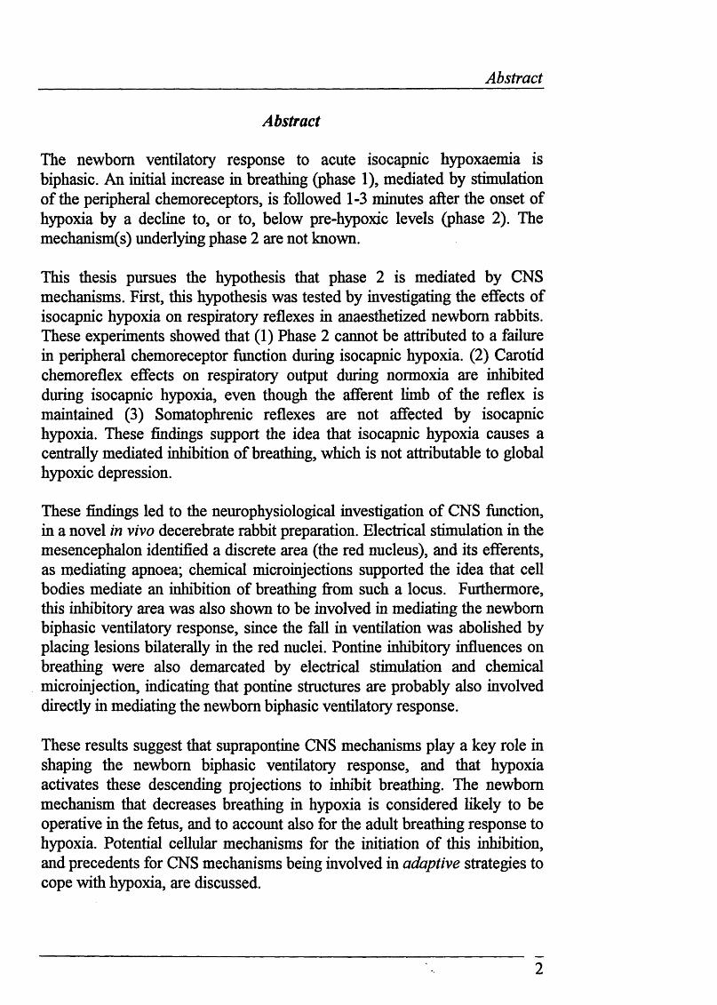
The newborn ventilatory response to acute isocapnic hypoxaemia is biphasic. An initial increase in breathing (phase 1), mediated by stimulation of the peripheral chemoreceptors, is followed 1-3 minutes after the onset of hypoxia by a decline to, or to, below pre-hypoxxc levels (phase 2). The mechanism(s) underlying phase 2 are not known.
This thesis pursues the hypothesis that phase 2 is mediated by CNS mechanisms. First, this hypothesis was tested by investigating the effects of isocapnic hypoxia on respiratory reflexes in anaesthetized newborn rabbits. These experiments showed that (1) Phase 2 cannot be attributed to a failure in peripheral chemoreceptor function during isocapnic hypoxia. (2) Carotid chemorefiex effects on respiratory output during normoxia are inhibited during isocapnic hypoxia, even though the afferent limb of the reflex is maintained (3) Somatophrenic reflexes are not affected by isocapnic hypoxia. These findings support the idea that isocapnic hypoxia causes a centrally mediated inhibition of breathing, which is not attributable to global hypoxic depression.
These findings led to the neurophysiological investigation o f CNS function, in a novel in vivo decerebrate rabbit preparation. Electrical stimulation in the mesencephalon identified a discrete area (the red nucleus), and its efferents, as mediating apnoea; chemical microinjections supported the idea that cell bodies mediate an inhibition of breathing fi’om such a locus. Furthermore, this inhibitory area was also shown to be involved in mediating the newborn biphasic ventilatory response, since the fall in ventilation was abolished by placing lesions bilaterally in the red nuclei. Pontine inhibitory influences on breathing were also demarcated by electrical stimulation and chemical microinjection, indicating that pontine structures are probably also involved directly in mediating the newborn biphasic ventilatory response.
These results suggest that suprapontine CNS mechanisms play a key role in shaping the newborn biphasic ventilatory response, and that hypoxia activates these descending projections to inhibit breathing. The newborn mechanism that decreases breathing in hypoxia is considered likely to be operative in the fetus, and to account also for the adult breathing response to hypoxia. Potential cellular mechanisms for the initiation of this inhibition, and precedents for CNS mechanisms being involved in adaptive strategies to cope with hypoxia, are discussed.
Personal statement
Personal statement
Except as acknowledged on page 92, the work presented in this thesis was performed solely by the candidate and is original.
Gareth L. Ackland
■ *v
Certified by supervisor Professor Mark A. Hanson
Acknowledgments
AcknowledgmentsThroughout the course of the last three years I have begun to understand the excitement, challenge and intellectual freedom that pursuing research offers. This is not solely the result of working in such a young (!) and demanding area of physiology, but is inspired by being in an enthusiastic academic environment which has encouraged questions to be asked, hypotheses to be addressed and to stimulate wide discussion on the topics in question. Furthermore, the value of conducting basic medical science within the Department of Obstetrics & Gynaecology has stressed the mutual importance of clinical medicine and basic science. I thank Professor Charles Rodeck (Obstetrics & Gynaecology) and Professors Roger Woledge and Michael Spyer (Physiology) for the opportunity to conduct my studies in both Departments.
In particular, I am indebted to the following:
Professor Mark Hanson: For his support throughout the three years, strong encouragement for promoting the generation of independent ideas and for the great times when discussing matters both scientific and not.
Dr. Ray Noble: For the many (late night) discussions, visionaryapproach, sharing his knowledge of neurophysiology and the problems associated with investigating the newborn brain stem, and also for discussing general scientific and non-scientific matters.
Dr. Peter Moore: For his teaching, consideration, support and patience.
Dr. Bridget Waites: For her timely arrival and infectious enthusiasm.
In addition I am also grateful to the other colleagues who have made the period of study possible:
Nicole Calder Dr. Dino Giussani Dr. Takanori WatanabeClare Crowe Lucy Green Dr. Keith CaddyLiUian Patterson Fei Li Dr. Juhe SmithDr. Laura Bennet Dr. Shiro Kozuma
Finally, I acknowledge gratefiiUy the support of the Wellcome Trust Prize Studentship scheme, and the tremendous value of the Communication Conference and Presentation events organized by The Wellcome Trust that I attended during the course of my Ph.D. course. These courses provided excellent opportunities to learn vital skills not, as yet, taught routinely during a Ph.D. training.
Table o f contents
Table of contents
INTRODUCTIONCHAPTER 1 - EFFECTS OF HYPOXIA ON CELLULAR AND SYSTEMIC FUNCTION: A REVIEW General overviewNewborns are particularly susceptible to the occurrence of hypoxia............................ 32Why study solely the effects of hypoxia on respiratory control in the newborn? 32Structure of Chapter 1........................................................ 33
SECTION A: EFFECTS OF HYPOXIA ON CELLULAR PHYSIOLOGY
Understanding the effects of hypoxia on cellular function is essential for the interpretation of systemic physiological changes....................................... 35
What is hypoxia? ....................................................................................................3 5
Several experimental models are used to produce hypoxia.................................36
The consequences of hypoxia at the cellular level....................................................37
Ionic changes during hypoxia/anoxia................................................................. 37Membrane potential changes during hypoxia/anoxia..........................................38Synaptic activity during hypoxia/anoxia.............................................................38Prolonged anoxia leads to anoxic brain damage................................................ 39Differential sensitivity to hypoxia throughout the nervous system.....................40Maturational differences in cellular responses to hypoxia..................................40
SECTION B: EFFECT OF HYPOXIA ON CARDIORESPIRATORY CONTROL The NEWBORN VENTILATORY RESPONSE TO ACUTE HYPOXAEMIA IS BIPHASIC...43
Stimulation of the peripheral chemoreceptors causes phase 1- THE INCREASE IN VENTILATION...............................................................................44
Cardiovascular response to acute hypoxaemia in newborns............................... 45
The fetal cardiorespiratory response to acute hypoxaemia...................................46
The relevance of adult and fetal respiratory control to the study ofthe effects of hypoxia on breathing in newborns............................................... 46Acute hypoxaemia abolishes ‘fetal breathing’ ................................................... 46Fetal chemoreceptors respond to hypoxaemia but normally do notcontribute to the control of FBM........................................................................46Peripheral chemoreceptor sensitivity to Pao^ but not PacO],increases postnatally............................................................................................47Fetal cardiovascular responses to acute hypoxaemia are mediated bythe peripheral chemoreceptors............................................................................ 48
The adult cardiorespiratory response to acute isocapnic hypoxaemia.................49
Adults also show BVR to acute isocapnic hypoxaemia......................................49Adult cardiovascular responses to hypoxaemia...................................................50
Table o f contents
THE MECHANISM UNDERLYING PHASE 2 OF BVRIS UNKNOWN............................................................................................................ 50
Hypothesis 1; BVR is due to peripheral chemoreceptor adaptationTO ACUTE HYPOXAEMIA..........................................................................................50
Direct recordings from carotid chemoreceptor fibres in newborns................... 51Indirect tests of peripheral chemoreceptor function in newborns...................... 52Studies of peripheral chemoreceptor fiinction during hypoxaemia in the adult. 54
Hypothesis 2; Central INHIBITION of respiratory output by a (HYPOXIA-SENSITIVE) NEURAL PATHWAY LOCATED IN THE BRAIN CAUSES BVR. .56
Fetal inhibitory brain stem influences on FBM during hypoxaemia.....................56Inhibitory brain stem influences on breathing in the newborn during BVR........58Evidence that suggests newborn/ fetal inhibitory brain stem mechanismsare also functional in the adult during hypoxaemia............................................. 59
Hypothesis 3: Direct hypoxic depression of respiratory neuronesCAUSES THE FALL IN VENTILATION OBSERVED IN BVR..... .... ............. ...............62
Hypothesis 4: BVR is caused by the drop in metabolic rateOBSERVED DURING HYPOXIA..................................................................... ................64
Metabolism falls during acute hypoxaemia......................................................... 64How much oxygen is essential for aerobic metabolism?.....................................65Critical effects of environmental temperature on metabolic responses tohypoxaemia.........................................................................................................65Brain stem control of thermoregulation..............................................................66Brown adipose tissue is of particular importance in the newborn..................... 66Phase 2 of BVR cannot be explained fully by a fall in metabolism......................67
H ypothesis 5: BVR is th e r e s u l t o f CO2 w ash o u t caused by an in c reaseIN CEREBRAL BLOOD FLOW DURING HYPOXAEMIA................................................... 67
CBF increases in the fetus and newborn during isocapnic hypoxaemia.............68CBF also increases in adults during isocapnic hypoxaemia.................................69
Hypothesis 6: BVR is caused by respiratory muscle failureOR FATIGUE................................................................................................................. 70
Hypothesis 7: Changes in lung mechanics cause BVR .................................71
Table o f contents
Hypothesis 8; Phase 2 of BVR is attributable to the accumulationOF INfflBITORY NEUROTRANSMITTERS/NEUROMODULATORS DURING HYPOXIA. ...72
Adenosine and GABA are the most likely neurochemical mediators of BVR ....72Adenosine..........................................................................................................73Cellular actions of adenosine.............................................................................. 73Adenosine is released during hypoxaemia...........................................................73Adenosine inhibits neurotransmitter release.......................................................73Systemic actions of adenosine............................................................................ 74Adenosine & adenosine analogue can inhibit breathing...................................... 74Effects of adenosine receptor antagonists...........................................................75GABA ........ 76Brain GABA concentration increases during hypoxia and inhibits breathing......76Other candidates..............................................................................................77Dopamine........................................................................................................... 77Acetylcholine......................................................................................................77Opioids............................................................................................................... 77
SECTION C: SUMMARY OF INTRODUCTION AND AIMS OF THE PROJECT Summary of introduction.............................................................................................80
AIMS OF THE PROJECT..................................................................................................81Aim 1. To develop an anaesthetized newborn animal preparation suitablefor a range of neurophysiological experiments....................................................81Aim 2. Is adaptation of carotid chemoreceptor fibres during acute
• hypoxaemia responsible for causing the biphasic ventilatory response?............81
Table o f contents
RESULTS: SECTION!- EFFECTS OF HYPOXIA ON RESPIRATORY REFLEXESOverview,......................................................................................................................84
CHAPTER 2: CAROTID CHEMOREFLEXES ARE INHIBITED BY A CNS MECHANISM DURING ISOCAPNIC HYPOXAEMIA
2.2 Hypotheses............................................................ .i ...............................87
(1) Afferent carotid chemoreceptor discharge is stimulated but does not decline during acute hypoxaemia in anaesthetized newborn rabbits....................87(2) The effect of transient carotid chemoreceptor stimulation on breathing is modulated during hypoxaemia by a central mechanism that inhibits respiratory output in anaesthetized newborn rabbits.......................................... 87
2.3 Methods............................................................................................................. 88
Animal delivery and care....................................................................................88Sedation and anaesthesia....................................................................................88Monitoring anaesthesia..................................................................................... 88Ventilation......................................................................................................... 89Monitoring of pH, blood gases and body temperature.......................................89Carotid chemoreceptor stimulation....................................................................90
2.4 Experimental PROTOCOLS.............................................................................92
Recording multifibre carotid chemoreceptor afferent activity (n=8 rabbits).....92Carotid chemoreceptor - phrenic chemorefiex (n= 11 rabbits)......................... 92Other preliminary carotid chemorefiex experiments......................................... 93
2.5 Signal processing and data collection ..................................................94
Blood pressure, heart rate and stimulation marker............................................ 94Carotid chemoreceptor activity......................................................................... 96Phrenic nerve activity.........................................................................................94Data display and storage................................................................................... 94
2.6 Analysis.............................................................................................................. 97
Cardiovascular parameters................................................................................ 97Recordings fi'om carotid chemoreceptor multifibres......................................... 97Carotid chemorefiex test - respiratory parameters............................................. 97
2.7 RESULTS............................................................................................................... 99Carotid chemoreceptor recordings....................................... ................... 99
Blood gas/pH status and cardiovascular parameters........................................... 99Carotid chemoreceptor fibres do not adapt to acute isocapnic hypoxaemia.......99COj boli increase carotid chemoreceptor discharge during normoxaemia...... 100CO; injections during isocapnic hypoxaemia also increased carotidchemoreceptor discharge..................................................................................102CÜ2 boli failed to increase carotid chemoreceptor discharge onlyduring severe hypoxaemia.................................................................................104
8
Table o f contents
Carotid chemoreflex TEST.................................................................... ....... 105
Blood gas/pH status and cardiovascular parameters......................................... 105Isocapnic hypoxaemia elicited BVR................................................................. 105Carotid chemorefiex effects during normoxaemia............................................ 107Effects of carotid chemoreceptor stimulation on respiratory output wereeasier to achieve consistently during expiration............................................... 108Lack of effect of injections on cardiovascular reflexes during normoxaemia.... 108 Carotid chemorefiex effects during isocapnic hypoxaemia -levels of hypoxaemia................................................ 108Carotid chemorefiex effects seen during normoxaemia disappeared duringisocapnic hypoxaemia....................................................................................... 108Transient carotid chemoreceptor stimulation occurred at similar timesduring normoxaemia and hypoxaemia..............................................................112Lack of correlation between the phase of BVR and presenceof chemorefiex effect.......................................................................................113
Effect of carotid chemoreceptor stimulation during expiration on othercomponents of the respiratory cycle..................................................................118Inherent variability of controls was not significant ,................................119Post-stimulus effects on respiratory output.......................................................119Carotid chemorefiex during apnoea.................................................................. 120Carotid chemorefiex during severe hypoxaemia................................................121Other preliminary carotid chemorefiex experiments........................................ 121Dithionite injections (n=2 rabbits) support findings of C02-saturated salinechemorefiex experiments...................................................................................121Decerebrate rabbit carotid chemorefiex experiments........................................ 123Lamb carotid chemorefiex experiments............................................................123
2.8 DISCUSSION....................................................................................................... 125
Methods.....................................................................................................................125Measurement and control of blood gases, and a direct index of centralrespiratory output were essential for addressing chemorefiex hypothesis 125Development of a suitable newborn rabbit preparation.................................... 125Implementation of a suitable anaesthetic regime.............................................. 126
Carotid chemoreceptor recordings..............................................................127Methodological drawbacks...............................................................................127Lack of baroreceptor effects............................................................................ 129Severe hypoxaemia/asphyxia............................................................................ 130
Carotid chemoreflex test.................................................................................130Variable magnitude of BVR............................................................................ 130Chemical carotid chemoreceptor stimulation is more appropriatethan electrical CSN stimulation in the newborn rabbit preparation..................130
Reliability of the transient carotid chemoreceptor stimulus test........................131Perturbation of the respiratory rhythm produces effectson subsequent breaths.......................................................................................132Dithionite experiments support findings using CO2- saline experiments........... 132Possible interpretations of results fi'om carotid chemorefiex and carotid chemoreceptor recording experiments: implications for BVR.........................133
Table o f contents
Lack of correlation between peak in central respiratory outputand type of carotid chemoreflex effect..............................................................135CNS studies show that hypoxia inhbits expiratory respiratory neurones.......... 136Hypoxia also inhibits expiratory motor activity.................................................137The application of oscillatory theories to the carotidchemoreflex experiments.................................................................................. 138Carotid chemoreflex experiments are supported by similar studiesin the newborn rabbit.......................................................................... 139The findings of carotid chemoreflex experiments are also supported by similar studies in adults.....................................................................................139
2.9 Summary.......................................................................................................... 140
CHAPTER 3 - ISOCAPNIC HYPOXAEMIA DOES NOT INHIBITSOMATOPHRENIC REFLEXES
3.1 INTRODUCTION................................................................................................142
3.2 Hypothesis....................................................................................................... 143
Phase 2 of BVR is caused by a brain stem mechanism that inhibits respiratory reflexes globally................................................................................................ 143
3.3 Methods...........................................................................................................143
Surgical preparation.........................................................................................143Monitoring anaesthesia....................................................................................144Ventilation....................................................................................................... 145Control of pH, blood gases and body temperature..........................................145Phrenic nerve activity.......................................................................................146Peripheral nerve stimulation............................................................................ 146
3.4 Signal processing and data collection...................................................147
Cardiovascular data.........................................................................................147Respiratory data..............................................................................................147Data collection and storage..............................................................................147
3.5 Experimental protocols..............................................................................147
3.6 Analysis.............................................................................................................149
Cardiovascular parameters.............................................................................. 149Respiratory parameters....................................................................................149
10
Table o f contents
3.7 Results..............................................................................................................150
Blood gas/pH and cardiovascular data............................................................. 150BVR was present in all preparations................................................................ 151Stimulation of somatic afferents in normoxaemia increases respiratory output. 152 No clear effect on timing of punctate somatic afferent stimuli onrespiratory output during normoxaemia........................................................... 154
Lack of effect of somatic afferent stimulation on blood pressure/heart rate.... 155 Stimulation of somatic afferents caused respiratory output to increasein isocapnic hypoxaemia............................................ 155Somatic afferent stimulation also increases respiratory output in recovery...... 156Severe hypoxaemia/asphyxia did not abolish effect of stimulatingsomatic afferents on respiratory output............................................................ 156
3.8 DISCUSSION......................................................................................................... 157
M ethods.......................................................................................................... 157Decerebration................................................................................................... 157Results..............................................................................................................157The biphasic ventilatory response is present in both anaesthetized anddecerebrate, decerebellate preparations.............................................................157No clear effects of somatic afferent stimulation on respiratory timing.............. 158Comparable results from a recent similar study.................................................158The effect of changing stimulation intensity......................................................159Group I and II fibres are stimulated at the threshold used forthe somatophrenic reflex...................................................................................160Evoking somatophrenic reflexes at threshold does not elicitstrong cardiovascular reflexes due to stimulation of group I and II fibres...... 161Stimulation of hindlimb afferents is unlikely to affect carotid chemoreceptordischarge...........................................................................................................162Hypoxia and hypoxia-related substances do not affect somatic afferents......... 162
3.9 Summary..........................................................................................................163
11
Table o f contents
RESULTS: SECTION 2 - THE ROLE OF THE BRAIN STEM AND MID-BRAIN IN MEDIATING THE EFFECTS OF HYPOXIA ON BREATHING IN NEWBORNS
Overview o f Results: Section 2 ................................................................................. 167
CHAPTER 4: THE DEVELOPMENT OF A NEWBORN RABBH PREPARATION FOR NEUROPHYSIOLOGICAL STUDIES INVESTIGATING THE ROLE OF THE BRAIN STEM AND MESENCEPHALON IN MEDIATING BVR
4.1 Introduction................................................................................................... 168
4.2 AIM: TO DEVELOP NEWBORN DECEREBRATE PREPARATION ..........................169
4.3 Methods............................................................................................................169General methods.............................................................................................. 169Decerebration.................................................................................................. 171Electrical stimulation.......................................................................................173Chemical stimulation........................................................................................173Reversible focal cooling...................................................................................175Histological methods.......................................................................................176
4.4 Signal processing and data collection ................................................. 178
4.5 Analysis __________________________________ ........178Central respiratory output................................................................................178Electrical stimulation.......................................................................................178Microinjectate stimulation......................................................................... 179Cardiovascular parameters:..............................................................................179
4.6 Experimental PROTOCOL.............................................................................179Respiratory response to isocapnic hypoxaemia................................................179
4.7 Results ...___ ............____ ........................................................................... 180Decerebrate newborn rabbits exhibit BVR (n=32 rabbits)................................ 180Sham rage occurred in one rabbit during hypoxaemia...................................... 182Halothane depresses cardiorespiratory output markedly innewborn decerebrate rabbits (n=2) and lambs (n=2)....................................... 183Histological techniques.....................................................................................183
4.8 Discussion..........................................................................................................184
M ethods...........................................................................................................184Decerebration conferred stability...................................................................... 184Histological techniques proved suitable............................................................ 184Results.............................................................. 185Decerebrate newborn rabbits exhibit BVR........................................................185Sham rage occurred in only one rabbit.............................................................185Arterial blood pressure showed “biphasic response”toisocapnic hypoxaemia........................................................................................185
4.9 Summary............................................................................................................186
12
Table o f contents
CHAPTER 5 - BVR IS MEDIATED BY STRUCTURES IN THE MESENCEPHALON
5.1 DfmODUCTION............ ................................................................. . ... .. ....... 188
5.2 Hypotheses ................................................................................................... 189
(1) Phase 2 of BVR is abolished by the destruction of a group of mesencephalic cell bodies, demarcated by electrical and chemicalstimulation, in newborn ‘decerebrate rabbits ........................................ 189
' (2) Any putative mesencephalic inhibitory mechanism is only functionalduring hypoxia................................................................................................ 189
5.3 Methods ........................................................................................................190
5.4 Protocols.................................................................................. ..................... 190
Identification of areas that mediate inhibition ofrespiratory output...........................................................................................190The respiratory response to isocapnic hypoxaemiaafter areas that had been demonstrated to inhibit respiratory outputare destroyed by electrolytic lesions..................................................................190Does inhibition of a inhibitory mesencephalic area increaserespiratory output?...........................................................................................191
5.5 Analysis............................................................................................................ 191
5.6 Results ...................................................................__.................................. 192
Identification of inhibitory mesencephalic areas by electrical stimulation(n=38 rabbits).................................................................................................. 192
hficroinjection expèriments (n=6 rabbits).........................................................197Lesioning (n=14 rabbits).................................................................................. 203Lesioning mesencephalic areas that produced an inhibition of respiratoryoutput during normoxaemia abolished phase 2 of BVR (n=5 rabbits).............203Abolition of BVR attributable to maintenance of respiratory fi*equency..........204Bilateral lesions in areas where apnoea was elicited abolished BVR(n=3 rabbits)......................................................................................................206Kainic acid injection (n=l rabbit).....................................................................211Midline electrolytic lesion (n=l rabbit).............................................................212Phase 2 of BVR persisted when lesions were made in mesencephalic areas that failed to produce an inhibition of respiratory output during normoxaemia(n=9 rabbits).....................................................................................................213Decline in respiratory fi’equency accounted for post-lesion BVR inanimals where bilateral/midline apnoea could not be elicited........................... 216Microinjection of GABA increased respiratory output duringisocapnic hypoxaemia (n=3 rabbits) but not normoxaemia (n=4 rabbits).........217
5.7 Discussion......................................................................................................... 219
Electrical stimulation....................................................................................... 219Chemical stimulation........................................................................................ 219Identification of inhibitory mesencephalic areas............................................... 220
13
Table o f contents
EAA microinjection......................................................................................... 221Extensive bilateral electrical stimulation in the mesencephalon could causedeleterious cardiovascular changes...................................................................222Bilateral electrolytic lesions made only in the red nucleus did notaffect subsequent respiratory output during normoxaemia...............................222Bilateral electrolytic lesions made only in the red nucleus abolishedphase 2 of BVR (lesioned(non BVR animals).................................................. 223The failure of other lesions to abolish phase 2 of BVR (lesioned(BVR)) animals was due to placement of the stimulating electrode outside the rednucleus/rubrospinal tract........................................ Z....................................... 223Higher incidence of sham rage during hypoxaemia in lesion (BVR) andlesion (non-BVR) rabbits compared to controls.............................................. 224Lesioning had no clear effect on phase 1 increase............................................ 224Biphasic arterial blood pressure response not affected by lesions.................... 224GABA microinjections indicate that an inhibitory mesencephalicinhibitory area is only active during isocapnic hypoxaemia...............................225Abolition of BVR by lesions in the red nucleus reconciles apparentlycontradictory supra- and rostral pontine lesioning/transection studies............ 226The structure of the red nucleus.......................................................................227Nucleus ruber magnocellularis.........................................................................227Nucleus ruber parvicellularis............................................................................ 228The red nucleus controls motor output and spinal cord processing................ 228Red nucleus inhibitory efferents have been identified in adult cats................... 229Respiratory related afferents to the mesencephalon and red nucleus............... 230
5.8 Summary. ..............___ ......___ ......................................................232
14
Table o f contents
CHAPTER 6 - THE ROLE OF THE PONS m MEDIATING INHIBITION OF RESPIRATORY OUTPUT IN NEWBORN DECEREBRATE RABBITS
6.1 Introduction...................................................................................................234
6.2 Hypotheses ......................... ......................................................................234
(1) Electrical stimulation of areas in the pons causes inhibitionof respiratory output...................................................................................234
(2) Microinjection of EAA into dorsal pons at the level of the middle cerebellar peduncle causes an inhibition of respiratory output.........................234
(3) Cooling of upper rostral pontine regions reverses phase 2 of BVR............234
6.3 Methods........................................................................................................... 235
6.4 Analysis............................................................................................................ 235
6.5 Results............................................................................................................. 236
Experiment 1: Electrical stimulation of pontine structures (n=20 rabbits) 236Loci where respiratory output was inhibited.................................................... 237
Locus coeruleus (n=5 rabbits ) ...............................................................237Reticular formation (n=10 rabbits)......................................................... 238Raphe nuclei (n=3 rabbits)......................................................................239
Experiment 2: Microinjections into the dorsal pons, including locus coeruleus(n=8 rabbits).....................................................................................................241Experiment 3: Effect of cooling at level of middle cerebellar peduncle onrespiratory output during isocapnic hypoxaemia (n=l rabbit).................... 244
Cooling of dorsolateral pons reversed phase 2 by increasingpeak phrenic activity............................................................................... 244
6.6 Discussion......................................................................................................... 247
Pontine reticular formation.............................................................................. 247Raphe nuclei.....................................................................................................248Locus coeruleus................................................................................................249Microinjections.................................................................................................250Confirmation that cooling the upper rostral pons reverses phase 2of BVR in the rabbit........................................................................................ 251
6.7 Summary......................................................................................................... 252
15
Table o f contents
CHAPTER 7 - FINAL DISCUSSION: HYPOXIA AND BVR FROM AN EVOLUTIONARY PERSPECTIVE
7.1 Main findings of the thesis .......................................................................254
Results: Section A ........................................................................................... 255Results: Section B ........................................................................................... 256
7.2 Hypoxia and BVR from an evolutionary perspective.......... .........257
Numerous strategies exist across species for countering theeffects of hypoxic environments.......................................................................257The evolution of an oxygenated atmosphere.................................................... 257Diversity of mechanisms for counteracting hypoxia..................... 258Aquatic breathers..............................................................................................258Air-breathers/air-aquatic breathers...................................................................258Intermittent air-breathers................................................................................. 259Metabolic considerations................................................................................. 260Evidence for a hypoxia-sensitive mechanism at the molecular/genetic level....261Evidence from cellular_m vivo studies for hypoxia-sensitive mechanism 262Potential role of the mesencephalon, red nucleus and rostral brain stem 263Clinical and pathophysiological implications.................................................... 264In conclusion....................................................................................................264
7.3 Final Summary ..........___ .....___ .......__......................__..................265
REFERENCES,.................................................................................................... 266
APPENDICESAppendix 1 - Calibrations..........................................................................................308Appendix 2 - Solutions.............................................................................................. 312Appendix 3 - Histological atlas of newborn rabbit brainstem/mesencephalon 314Appendix 4 - Individualized carotid chemoreceptor recording results (Chapter 2)... 329 Appendix 5 - Map of mesencephalic area, from electrical stimulation experiments,
that elicit inhibition of respiratory output............................................. 332Appendix 6 - Method of ventilation........................................................................... 334
16
Table o f figures
Table of figures
CHAPTER 1 - INTRODUCTION
Figure 1.1 Schematic diagram illustrating the potential development ofthe project, depending on the outcome of the initial experimentsinvestigating carotid chemoreflexes in newborn anaesthetised rabbits.................. 82
CHAPTER 2 - CAROTID CHEMOREFLEXES ARE INHIBITED BY A CNS MECHANISM DURING ISOCAPNIC HYPOXAEMIA
Figure 2.1 Signal collection and processing set-up used for carotid chemoreceptor recordings..................................................................................... 95
Figure 2.2 Signal collection and processing set-up forcarotid chemoreflex experiments.......................................................................96
Figure 2.3 Method of analysis for carotid chemoreflex test.................................. 98
Figure 2.4 Carotid chemoreceptor discharge increased and remainedelevated throughout hypoxaemia...........................................................................99
Figure 2.5 CO2 -saturated saline bolus increased carotid chemoreceptor discharge during normoxaemia. After 10 minutes of isocapnic hypoxaemia, there was a similar increase in carotid chemoreceptor discharge on CO2 -saturated saline bolus injection. Also note increase in baseline carotid chemoreceptor discharge..................................................................................... 100
Figure 2.6 Group results showing of effect of C02-saturated saline on multifibre carotid chemoreceptor discharge during normoxaemia
and isocapnic hypoxaemia....................................................................................101
Figure 2.7 Effect of Ringer’s saline (pH 7.38) on multifibre carotid chemoreceptor discharge during normoxaemia................................................... 101
Figure 2.8 C02-saturated saline injections increased carotid chemoreceptor discharge 1 min before the onset of isocapnic hypoxaemia................................. 102
Figure 2.9 C02-saturated saline bolus increased carotid chemoreceptor discharge 1 min after the onset of isocapnic hypoxaemia. Note increased basal carotid chemoreceptor discharge due to fall in Paoj...................................103
Figure 2.10 C02-saturated saline injection continued to increase carotid chemoreceptor discharge even 10 min after the onset of isocapnic hypoxaemia. Note basal carotid chemoreceptor discharge remains elevated............................103
17
Table o f figures
Figure 2.11 COz-saturated saline increased carotid chemoreceptor discharge 1 min after the end of isocapnic hypoxaemia. Note increased basal carotid chemoreceptor discharge has declined due to the return to normpxaemic Paoj values..........................................................................................................104
Figure 2.12 Isocapnic hypoxaemia elicited BVR in anaesthetized, paralysed and vagotomized newborn rabbits....................................................................... 105
Figure 2.13a PPA during control (normoxaemia), and phase 1 andphase 2 of BVR................................................................................................... 106
Figure 2.13b Respiratory frequency during control (normoxaemia), andphase 1 and phase 2 of BVR...............................................................................106
Figure 2.14 Transient carotid chemoreceptor stimulation (using COz-saturated saline) during expiration prolonged expiration during normoxaemia...................107
Figure 2.15 Transient carotid chemoreceptor stimulation (using C02-saturated saline) during early inspiration increased PPA during normoxaemia................... 107
Figure 2.16a The effect of prolongation of Te produced by carotid chemoreceptor stimulation during expiration disappeared during isocapnic hypoxaemia, as shown by the % change in Te for the stimulation breath as compared with control breaths........................................................................109
Figure 2.16b The effect of prolongation of Te produced by carotid chemoreceptor stimulation during expiration disappeared during isocapnic hypoxaemia, as shown by the difference in Te between stimulation and control breaths.....................................................................................................109
Figure 2.17 Transient carotid chemoreceptor stimulation (using C02-saturated saline) made during expiration prolonged the duration of expiration during normoxaemia....................................................................................................... 110
Figure 2.18 Transient carotid chemoreceptor stimulation fails to pertub respiratory output during isocapnic hypoxaemia in the same rabbit.................... I l l
Figure 2.19 Prolongation of Te returns during recovery to normoxaemia.......... 112
Figure 2.20a Individual rabbit data for transient carotid chemoreceptor stimulation in amnaesthetized, paralyzed and vagotomized rabbits..................... 114
Figure2.20b Individual runs for transient carotid chemoreceptor stimulation in a decerebrate lamb and decerebrate rabbit......................................................117
Figure 2.21 Effect of transient carotid chemoreceptor stimulation during normoxia and hypoxia on (1) Te, (2) comparison between Ti of pre-stimulation breath (PSC) and Ti (poststimulation breath) (3) PPA of precontrol breath and poststimulation breath...................................................................................118
18
Table o f figures
Figure 2.22 Comparison of the pre-stimulation breath and pre-control during normoxaemia and isocapnic hypoxaemia showed that there was no tendency for breaths to be prolonged spontaneously..........................................................119
Figure 2.23 Following perturbation of Te by transient carotid chemoreceptor stimulation, there was a compensatory increase in respiratory frequency for the next breath during normoxaemia and early hypoxaemia only........................119
Figure 2.24a, b Carotid chemoreceptor stimulation could elicit phrenic nerve activity during apnoea, but effect disappeared with prolonged apnoea............... 120
Figure 2.25 Transient carotid chemoreceptor stimulation shortened Ttot during gasping/very slow breathing..................................................................... 120
Figure 2.26 Sodium dithionite injections stimulated respiratory output in normoxaemia. ABP also fell during stimulation of respiratory output................ 121
Figure 2.27 Stimulatory effect of sodium dithionite on respiratory outputand ABP persisted at 180s of isocapnic hypoxaemia...........................................121
Figure 2.28. Stimulatory effect of sodium dithionite on respiratory outputand ABP persisted after 240s of isocapnic hypoxaemia...................................... 122
Figure 2.29 On recovery to normoxaemia, sodium dithionite produced hyperpnoea again. ABP is not shown.................................................................. 122
Figure 2.30 Three possible interpretations for the carotidchemoreflex experiments..................................................................................... 133
CHAPTER 3 - ISOCAPNIC HYPOXAEMIA DOES NOT INHIBIT SOMA TOPHRENIC REFLEXES,Figure 3.1 Diagram showing electronic set-up for signal processing and data collection for somatophrenic reflex experiments................................................. 148
Figure 3.2 BVR was elicited in anaesthetised rabbit experiments (n=7 rabbits),(peak and nadir of respiratory output during isocapnic hypoxaemia shown)...... 151
Figure 3.3 BVR in decerebrate lamb experiments (n=4 lambs), showingpeak and nadir .of respiratory output (during isocapnic hypoxaemiacompared to normoxaemic controls.................................................................. 151
Figure 3.4 Stimulating sciatic nerve both during normoxaemia and after 5 min isocapnic hypoxaemia increased respiratory output (decerebrate lamb response)............................................................................................................. 152
Figure 3.5 Somatic afferent stimulated respiratory output during normoxaemia and after 5 min isocapnic hypoxaemia in anaesthetised rabbits. Similar results were seen for decerebrate rabbit experiments......................................................153
Figure 3.6 Somatic afferent stimulation increased respiratory output during normoxaemia and after 5 min isocapnic hypoxaemia in decerebrate lambs......... 153
Figure 3.7 Sustained or transient stimulation of somatic afferents caused phrenic activity to return during severe hypoxaemia/asphyxia............................ 156
19
Table o f figures
CHAPTER 4 - THE DEVELOPMENT OF A NEWBORN RABBIT PREPARATION FOR NEUROPHYSIOLOGICAL STUDIES INVESTIGATING THE ROLE OF THE BRAIN STEM AND MESENCEPHALON IN MEDIATING BVR
Figure 4.1 Cooling probe used to cool the dorsorostral pons............................. 175
Figure 4.2 Newborn decerebrate rabbits show a biphasic response in respiratory output to 8 min of isocapnic hypoxaemia (n=32 rabbits).................. 181
Figure 4.3 PPA during control, phase 1 and phase 2 of BVR (n=32 rabbits).... 181
Figure 4.4 Respiratory frequency during control, phase 1 and phase 2of BVR (n=3 2 rabbits)........................................................................................182
Figure 4.5 Sham rage in one decerebrate rabbit was characterized by ABP increase (70% over control ABP), plus an initial augmentation of PPA,
followed by apnoea..............................................................................................182
CHAPTER 5 - BVR IS MEDIATED BY STRUCTURES IN THE MESENCEPHALON
Figure 5.1 Electrical stimulation in the mesencephalon at the level of thesuperior coUiculus could produce rapid increases in respiratory output,due to either increased frequency and/or PPA....................................................192
Figure 5.2 Electrical stimulation, within a discrete loci 7-8mm from the surface of the mesencephalon at the level of the superior colliculi, inhibited
respiratory output. Apnoea was observed usually................................................193
Figure 5.3 \fixed effects on respiratory output could also be elicited by electrical stimulation of mesencephalic areas at the level of the superior/inferior colliculi......................................................................................193
Figure 5.4 Histology confirmed that electrical stimulation of the rubrospinal tract, as well as the red nucleus, inhibited respiratory output............ 194
Figure 5.5 Discrete inhibitory area illustrated by showing the effects of electrical stimulation on respiratory output in one rabbit. Stimulatory effect of electrical stimulation on respiratory output at 6mm depth from surface is followed 1mm below, at depth 7mm, by profound inhibition of respiratory output. Partial inhibition (mixed effect) occurs at a depth of 7.3mm, but having driven the electrode through the area of inhibition, electrical stimulation again causes rapid increases in respiratory output....................................................................196
Figure 5.6 Stimulation of respiratory output due to microinjection of glutamate in the periaqueductal grey................................................................... 198
20
Table o f figures
Figure 5.7a Transient inhibition of two breaths (fall in PPA) due to microinjection of glutamate at depth where electrical stimulation inhibited respiratory output at the level of the superior coUiculus..................................... 200
Figure 5.7b Transient prolongation of Te foUowing microinjection of glutamate in area of mesencephalon at level of superior coUiculus where electrical stimulation had inhibited respiratory output.........................................200
Figure 5.7c In another rabbit, transient inhibition of breath (PPA decreased) followed microinjection of glutamate in the red nucleus..................................... 201
Figure 5.7d Histological trace showing site of microinjection in red nucleus (lateral border) for figure 5.7c. Two microinjections were made at this site, both causing transient inhibition of respiratory output (decreased PPA)........... 201
Figure 5.8 BUateral electrolytic/chemical lesions in the red nucleus or its efferents abolished phase 2 of BVR (n=5 rabbits)...............................................203
Figure 5.9 For non-lesion experiments PPA increased during phase 1 ofBVR but did not alter during phase 2 (n=5 rabbits).........................................205
Figure 5.10 Respiratory fi*equency did not contribute to phase 1 of BVR but declined sharply during phase 2 in non-lesioned experiments. In marked contrast, respiratory frequency was maintained throughout isocapnic hypoxaemia in lesioned rabbits where (bilateral) apnoea had been elicited previously by electrical stimulation (n=5 rabbits)...................................................................... 205
Figure 5.11a Rabbit 090394. Isocapnic hypoxaemia elicited BVR in the intact rabbit. Apnoea was elicited subsequently on both sides of the midline of the mesencephalon at the level of the superior coUiculus, by electrical stimulation. Histological trace shows that lesions were made in these apnoeogenic areas in both the red nucleus and in the rubrospinal tract, at the level of the inferior coUiculus. Here lesioning increased the relative magnitude of phase 1, but abolished phase 2.......................................................................................... 208
Figure 5.11b Rabbit 040594. Isocapnic hypoxaemia elicited BVR in the intact rabbit. Apnoea was eUcited subsequently on both sides of the midUne of the mesencephalon at the level of the superior coUiculus, by electrical stimulation. Histological trace shows that were made in these apnoeogenic areas in both the red nucleus and in the rubrospinal tract, at the level of the inferior coUiculus. ...209
Figure 5.11c Rabbit 210694. Isocapnic hypoxaemia eUcited BVR in the intact rabbit. Apnoea was elicited subsequently on both sides of the midUne of the mesencephalon at the level of the superior coUiculus, by electrical stimulation. Histological trace shows that lesions were made in these apnoeogenic areas in both the red nucleus but predominantly in the rubrospinal tract, at the level of the inferior coUiculus. Lesioning did not alter the magnitude of phase 1 in this rabbit, compared to the intact hypoxaemic run................................................... 210
21
Table o f figures
Figure 5.12 Unilateral electrical stimulation demarcated an area in the red nucleus which produced apnoea. Kainic acid was injected subsequently into this area, but spread medially to the other side of the mesencephalon at the level of the superior coUiculus. Thus bilateral destruction at the depth of the red nucleus in the mesencephalon at the level of the red nucleus abolished phase 2 of BVR...................................................................................................211
Figure 5.13 A midline electrolytic lesion abolished phase 2 of BVR. Lesioning increased the magnitude of phase 1, in comparison to the peak respiratory output during hypoxaemia in the intact rabbit. Histology showed that the midline lesion had been made in the region of the decussation of rubrospinal fibres.................................................................................................212
Figure 5.14a BVR persisted in all rabbits where bilateral respiratoryinhibition was not found. (n=9 rabbits)...............................................................214
Figure 5.14b Example of rabbit where bilateral lesions were made, but where BVR persisted on exposure to isocapnic hypoxaemia after lesioning/electrical stimulation. In this case, apnoea was only elicited unilaterally fi'om red nucleus. Apnoea was only found on the left side: the stimulating electrode was placed more laterally. Hence, the inhibitory region in the red nucleus was not lesioned on the left side..................................214
Figure 5.15 Respiratory frequency did not contribute to the rise in respiratory output during hypoxaemia, but fell sharply thereby causing phase 2 for lesion (BVR) rabbits (n=9 rabbits)................................................................................ 216
Figure 5.16 An increase in PPA caused the peak in respiratory output during hypoxaemia in lesion (BVR) rabbits (n=9 rabbits). This increase was greater in pre-lesion than post-lesion rabbits. PPA did not fall during phase 2...............216
Figure 5.17 Microinjection of GABA, into an area at the level of the superior coUiculus where electrical stimulation made during normoxaemia had eUcited inhibition of respiratory output, reversed apnoea, by initiating gasping.............. 217
Figure 5.18 GABA, microinjected into an area at the level of the superior coUiculus where electrical stimulation made during normoxaemia had eUcited inhibition of respiratory output, could increase respiratory output transiently during hypoxaemia.............................................................................218
22
Table o f figures
CHAPTER 6 - THE ROLE OF THE PONS IN MEDIATING INHIBITION OF RESPIRATORY OUTPUT IN NEWBORN DECEREBRATE RABBITS
Figure 6.1 Diagram showing rostro-caudal positions where tracks weremade with the electrical stimulating electrode....................................................236Figure 6.2 Stimulation of locus coeruleus could cause inhibitionof respiratory output........................................................................................... 237
Figure 6.3 Electrical stimulation of the reticular formation produced apnoea ....238
Figure 6.4 Electrical stimulation produced inhibition in the regionof the raphe/midline nuclei.................................................................................. 239
Figure 6.5 Compilation of sites where electrical stimulation produced inhibition of breathing or apnoea.........................................................................240
Figure 6.6 Inhibition of respiratory output due to microinjection ofglutamate in region of locus coeruleus................................................................242
Figure 6.7 Stimulation of respiratory output in dorsorostral pons, again at level of locus coeruleus, stimulated respiratory output during normoxaemia and isocapnic hypoxaemia. The example shown illustrates the effect of glutamate injected during hypoxaemic apnoea.................................................... 242
Figure 6.8 Microinjection of glutamate into parabrachial regionproduced apneusis...............................................................................................243
Figure 6.9 Phase 2 of BVR was reversed by cooling the floor of the IV ventricle at the level of the middle cerebellar peduncle..................................... 243
Figure 6.10 The restoration of phase 1 levels of respiratory output oncooling (probe temperature (°C) indicated by dotted line) was attributablesolely to an increase in PPA, with no effect on respiratory fi-equency................ 245
CHAPTER 7- FINAL DISCUSSION: HYPOXIA AND BVR FROM AN EVOLUTIONARY PERSPECTIVE
Figure 7.1 Summary of the development of the project...................................... 254
23
Table o f tables
Table of tables
Table 1.1 The biphasic ventilatory response has been reported in all mammalsstudied, in both the anaesthetized and conscious states.......................43
Table 2.1 Blood gas, pH, HR and ABP values for carotid chemoreceptorrecordings (n=8 rabbits)....................................................................... 99
Table 2.2 Blood gas, pH, HR and ABP values for anaesthetized rabbit carotidchemoreflex experiments (n=10 rabbits)........................................... 105
Table 3.1 Blood gas, pH, HR and ABP values for anaesthetized rabbitsexposed to isocapnic hypoxaemia (n=7 rabbits)................................. 150
Table 3.2 Blood gas, pH, HR and ABP values for decerebrate rabbitsexposed to isocapnic hypoxaemia (n=4 rabbits) .....................150
Table 3.3 Blood gas, pH, HR and ABP values for decerebrate lambsexposed to isocapnic hypoxaemia (n=4 lambs).................................. 150
Table 3.4 In preliminary experiments, no consistent effects were seenwith punctate stimuli..........................................................................154
Table 4.1 Blood gas, pH, HR and ABP values for decerebrate rabbitsexposed to isocapnic hypoxaemia (n=32 rabbits).............................. 180
Table 5.1 Blood gas, pH, HR and ABP values for lesion (non-BVR)rabbits exposed to isocapnic hypoxaemia experiments (n=5 rabbits).......................................................................................204
Table 5.2 Blood gas, pH, HR and ABP values for lesion (BVR) rabbitsexposed to isocapnic hypoxaemia experiments (n=9 rabbits)......................................................................................213
24
Table o f plates
Table of plates
Plate 2.1 Photograph showing arrangement of catheters for injection of COj- saturated saline to stimulate carotid body chemoreceptors................................... 91
Plate 2.2 Photograph showing recording electrode used for both carotid chemoreceptor fibre and phrenic nerve recordings................................................91
Plate 3.1 Photograph showing custom-made stimulating electrode for somatophrenic experiments................................................................................. 146
Plate 4.1 Photograph showing recording electrode used for most experiments reported in Results: Section 2 .............................................................................171
Plate 5.1 Photograph of Cresyl Violet stained slice showing lesions made in rubrospinal tract and adjacent reticular formation, at level of inferior coUiculus. Electrical stimulation of the the two most lateral tracks elicited apnoea/inhibition of respiratory output............................................................................................195
Plate 5.2 Photograph of Cresyl Violet stained slice showing site of microinjection of glutamate into PAG, that stimulated respiratory output 199
Plate 5.3 Photograph of Cresyl Violet stained slice showing site where microinjection of utamate elicited transient inhibition of respiratory output.This was located on the lateral border of the red nucleus..................... 202
Plate 5.4 Photograph showing bilateral lesions placed in red nucleus, which abolished phase 2 of BVR (rabbit 090394). The lesions were made at sites where respiratory output had been inhibited/abolished by electrical stimulation............207
Plate 5.5 Photograph showing slice illustrating start of stimulating electrode tracks made in one rabbit where BVR was not abolished..............................................215
Plate 6.1 Photograph showing site of cooling probe (locus coreuleus) where ventilatory fall during isocapnic hypoxia was reversed........................................246
Plates Al-Al 1 Histological atlas: Transverse sections of the brain stem and mesencephalon in the newborn rabbit............................318-328
25
Abbreviations
Abbreviations
The following list includes the symbols and abbreviations used in respiratoiy physiology. These comply with the standards approved by the Commission on Respiratory Physiology (1980). For main histological abbreviations, refer to Appendix 3.
[ li Intracellular concentration of ion
[ ]o Extracellular concentration of ion
P fla Hydrogen ion concentration in arterial blood
% Percentage
°c Degrees Celsius
a Alpha
5-HT Serotonin
ABP Arterial blood pressure
ACh Acetylcholine
ATP Adenosine triphosphate
BAT Brown adipose tissue
bpm Beats per minute
BVR Biphasic ventilatory response
C {number} Cervical nerve root
ca. Circa
Calcium ion
cr Chloride ion
Co. Company
CO2 Carbon dioxide
CBF Cerebral blood flow
26
Abbreviations
cm
CNS
CSF
CSN
DA
EAA
ECG
EMG
EPSP
f
FRC
FBM
F 1 0 2
GA
HR
lAA
i.m.
IPSP
IQR
i.v.
K+
L {number}
Ltd
MAP
n
Centimetre
Central nervous system
Cerebrospinal fluid
Carotid sinus nerve
Dopamine
Excitatory amino acid
Electrocardiogram
Electromyogram
Excitatory postsynaptic potential
Frequency
Functional residual capacity
Fetal breathing movements
Inspired oxygen fraction
Gestational age
Heart rate
Inhibitory amino acid
Intramuscular
Inhibitory postsynaptic potential
Interquartile range
Intravenous
Potassium ion
Lumbar nerve root
Limited
Mean arterial pressure
Number
27
Abbreviations
n Number
N2 Nitrogen
Na^ Sodium ion
NaCN Sodium cyanide
NE Noradrenaline
NTS Nucleus tractus solitarius
O2 Oxygen
P Probability
P0 2 Partial pressure of oxygen
Pc02 Partial pressure of carbon dioxide
PAG Periaqueductal grey
PCr Phosphocreatine
Paco2 Partial pressure of carbon dioxide in arterial blood
Pao2 Partial pressure of oxygen in arterial blood
pH Logjo hydrogen ion concentration in a solution
pHa Logio hydrogen ion concentration in arterial blood
PPA Peak phrenic activity
RVLM Rostro-ventrolateral medulla
S {number} Sacral nerve root
s.c. Subcutaneously
SD Standard deviation
SEM Standard error of the mean
Te Expiratory time
Ti Inspiratory time
28
Abbreviations
UK United Kingdom
USA United States of America
Vco2 Carbon dioxide production
V e Ventilation
VLM Ventrolateral medulla
V0 2 Oxygen consumption
V t Tidal volume
w/v Weight per unit volume
29
SI units
SI units
Length
m
cm
mm
pm
Metre
Centimetre
Millimetre
Micrometres
Mass
kg
gmg
lig
Kilograms
Grams
Milligrams
Micrograms
Time
hr
min
s
ms
Hz
Hour
Minute
Second
Millisecond
Cycles per second
Volume
1
ml
pini
Litre
Millilitre
Microlitres
Nanolitres
Electrical
mA
pA
mV
Q
Milliamps
Microamps
Millivolts
Resistance
Chemical
M Moles
mM Millimoles
pM Micromoles
Pressure is expressed as mmHg (millimetres of mercury) throughout the thesis, rather than in SI pressure units (Pascals)
30
Introduction
CHAPTER 1
EFFECTS OF HYPOXIA ON CELLULAR AND SYSTEMIC PHYSIOLOGICAL FUNCTION: A REVIEW
31
Introduction
General overview o f introduction
Newborns are particularly susceptible to the occurrence of hypoxiaNewborn babies, especially those bom prematurely, are particularly prone to apnoea (Miller & Smull, 1955; Henderson-Smart, 1981). Apnoea results in asphyxia - a fall in Paoj and increased Paco2 * Apnoeas may be central or obstmctive. Central apnoea, the cessation of airflow and inspiratory effort, is particularly common in premature babies (Milner & Greenough, 1988). Immaturity of sleep state patterns and inhibitory laryngeal respiratoiy reflexes have been imphcated in central apnoea (Marchai, Bairam & Vert, 1987). Obstructive apnoea, the cessation of airflow despite continued respiratory efforts, is the result of poor co-ordination of the upper airway musculature. This occurs during early infancy in both term and pre-term infants, thereby increasing the risk of hypoxic spells. This occurs during sleep (Henderson-Smart, 1980). About 50% of apnoeas in premature babies are mixed i.e. both central and obstructive (Brazy, Kinney & Oakes, 1987).
Although it is not clear whether SIDS victims have a higher incidence of disturbed respiratory patterns, such as increased incidence and severity of apnoeic attacks, clearly respiratory arrest must occur at some stage. Whether hypoxaemia ehcits inappropriate physiological responses as a result of immature development of respiratory control in SIDS victims compared to age-matched babies is not known.
Why study solely the effects o f hypoxia on respiratory control in the newborn?To understand the basic physiological mechanisms that underhe the newborn response to asphyxia requires separating out the effects of Paoj and Pacoz- The consequences of hypoxia in the newborn become more clearly hazardous when the cardiorespiratory responses are considered. Although hypoxia ehcits a number of cellular and systemic physiological effects, this thesis concentrates on the apparently paradoxical ventilatory response o f newborn mammals that is observed in hypoxia.
The terms "newborn" and "neonate" are often used loosely in the literature: in clinical medicine neonate refers to infants less than one month old. For clarity the term newborn is used throughout this thesis to indicate that the postnatal age under investigation is around one month old, and not yet an adult.
32
Introduction
Structure of Chapter 1
Section A of this introduction considers the effects of hypoxia on cellular function. Because the integration of the factors that control respiratory output occurs in the central nervous system, section A focuses on the responses of brain tissue to hypoxia, including how the cellular properties of the newborn central nervous system indicate why newborns may tolerate systemic hypoxia better than adults.
Section B examines a variety of hypotheses that have been proposed to explain the effect of hypoxia on the control of breathing in the newborn, adult and fetus. This section also considers how hypoxia affects neurotransmitter and neuromodulator release and hence cellular processes, and the consequences of these changes for systemic physiological fimction.
Section C considers the initial aims of the project, and discusses broadly the possible imphcations of the first experiments undertaken. The type of experiments predicted to follow from these initial studies are discussed.
33
Introduction
SECTION A
EFFECTS OF HYPOXIA ON CELLULAR PHYSIOLOGY
34
Introduction
1.1 Understanding the effects of hypoxia on cellular function is essential for the interpretation of systemic physiological changesWhile the experiments conducted in this thesis can be classified broadly as “systems” or “whole animal” physiology, it is important to note that the cellular mechanisms that are involved in the response to hypoxia are also considered. Hypoxia is an excellent example of how the study of cellular and systemic physiology are interdependent. Experiments investigating cellular mechanisms can help elucidate systemic physiological processes. For example, this is particularly clear when considering the role of neurotransmitters/neuromodulators on cellular fimction and systemic hypoxic responses (see section B). Conversely, the systemic effects of hypoxia provide important markers for future work conducted at the cellular level.
1.2 What is hypoxia?Hypoxia is an insufhcient supply of oxygen to match the O2 demand of a cell, tissue or organ. The critical Pqj is that P0 2 at which the demand for oxygen uptake falls below its normoxic level. A reduction in Poj of arterial blood (Paoj), which will be referred to mostly throughout this thesis, is termed hypoxaemia. Unmeasured or unknown Paoj levels in experiments where inspired oxygen is reduced are referred to as hypoxia.
Oxidative metabohsm may be impaired, but not abohshed, under hypoxic conditions. In contrast, anoxia, where the oxygen supply is completely cut off, results in the cessation of oxidative phosphorylation once mitochondrial P0 2 falls below a critical level. When oxygen levels fall below critical levels, but not necessarily under anoxic conditions, the process of glycolysis occurs solely. This is the anaerobic degradation of glucose to lactate and pyruvate. Anaerobic metabohsm produces one-nineteenth of ATP per mole of glucose as compared to aerobic metabolism.
Despite the striking quahtative and quantitative differences between hypoxia and anoxia, these terms have often been used interchangeably. This is true both of many studies investigating the effects o f reduced Pao2 on cellular and systemic physiological processes. Therefore, this introduction will set out to discuss the effects of hypoxia on cellular
35
Introduction
and systemic physiological processes paying particular attention to the levels of hypoxia used. Anoxia has been commonly used in cellular studies, presumably because it should ehcit maximal changes in cellular function. Caution must therefore be exercised in relating work at the cellular level to aspects of systems physiology.
Several experimental models are used to produce hypoxia.
Hypoxic hypoxia occurs when the Pqj of the inspired gas mixture supplied is reduced, usually by replacing O2 with N 2 .
Anaemic hypoxia occurs when the oxygen-carrying capacity of the blood is reduced. This form of hypoxia can be achieved by:a) Removal of red blood cells, (e.g. Koos, Murray & Doany, 1992)b) Dietary iron deficiency (e.g. Koos & Doany, 1990)c) Poisoning the haemoglobin of red cells with carbon monoxide,
which displaces oxygen and shifts the oxygen dissociation curve to the left (see Haab, 1990 )
d) Oxidizing haemoglobin to methaemoglobin using NaN02 (Hudak, Koehler, Rosenberg, Traystman & Douglas-Jones,1986)
Histotoxic hypoxia occurs when oxygen utilization is inhibited, for example by cyanide, which inhibits cytochrome oxidase, (e.g. Mitra, Dev, Romaniuk, Trivedi, Prabhakar, & Chemiack, 1992)
Stagnant hypoxia occurs when the supply of blood is inadequate (Barcroft, 1920a,b). Severe congestive heart failure results in circulatory hypoxia, while shock or infusion of a drug that dramatically lowers blood pressure can cause hypotensive hypoxia.
36
Introduction
1.3 The consequences of hypoxia at the cellular level.The severe impairment of oxidative phosphorylation produces hydrogen and lactate ions. These waste products of anaerobic metabohsm are retained within the brain unlike CO2 and H2 O, the end products of aerobic metabohsm. This is due to the relative impermeability of the blood-brain barrier to charged ions. Consequently, an intraceUular acidosis ensues. Outside the brain, both hydrogen and lactate ions escape into the circulation, causing "lactacidosis". Because tissue P0 2 is extremely difficult to measure, metabohtes such as lactate can be useful in determining the state of oxygenation of an organ.
Ionic changes during hypoxia/anoxia.
Normal brain fimction depends on the maintenance, by ATP hydrolysis, of constant ion concentrations in electrochemical equihbrium across the nerve membranes. If the mean brain tissue P0 2 faUs below 20 mmHg (Lubbers, 1973) brain energy metabohsm is disrupted, leading to inadequate ATP production. The brain has very smaU oxygen stores. An initiaUy moderate increase in [K+]o increases ceh excitabihty (Kmjevic & Morris, 1981), but also inhibits synaptic transmission presynapticaUy (BCmjevic & Morris, 1976). At first, [Na+]o, [Cl’]o and [Ca2+]o are only affected shghtly (Siesjo, 1978) but later during the anoxic insult these ion concentrations show a rapid and pronounced faU (Zeuthen, Dora, Silver, Chance & Kovach, 1979). After a few minutes, due to the opening of ATP-dependent channels, [K+]o rises very steeply. When brain ATP levels are kept high during anoxia (ischaemia), either by elevating glucose stores, or by hypothermia to slow down metabohsm (Astrup, Rehncrona & Siesjo, 1980), ionic disequihbrium is delayed.
37
Introduction
Membrane potential changes during hypoxia/anoxia.
Initially the membrane potential of nerve cells undergoes a transient depolarization in hypoxic/anoxic conditions. This is followed by hyperpolarization, accompanied by a reduction in input (membrane) resistance, before a fast depolarization occurs due to the rapid extracellular changes described above (Hansen, Hounsgaard & Jahnsen, 1982; Leblond & Kmjevic, 1989). Ghal cell membrane potentials also fall during hypoxia (KufQer, Nicholls & Orkand, 1966).
The early hyperpolarization associated with a reduction in input resistance is due to the activation of K+ channels in the postsynaptic membrane. The dependence of intracellular Ca + homeostasis on ATP may be involved, since anoxia dismpts the sequestration of calcium by internal stores. This [Ca +Ji rise increases membrane K+ conductance leading to decreased excitabihty (Kmjevic, 1975; Brinley, Tiffert, Scarpa & Mullins, 1977). However, since ATP channel blockers have httle effect on the anoxic hyperpolarization, it seems unlikely that K+ ATP channels are involved. A rise in Ca , or cychc AMP that is triggered by anoxia (Schmidt, Scmidt & Robinson, 1973) or a dechne in locahsed ATP could also result in the phosphorylation or dephosphorylation of membrane proteins, thereby increasing membrane permeabihty. The rapid depolarization is mediated in part by release of glutamate from synaptic terminals, which is modulated by presynaptic ATP-sensitive K+ channels (Ben-Ari, Kmjevic & Crepel, 1990).
Synaptic activity during hypoxia/anoxia.
Neuronal cell discharge briefly increases at the start o f anoxia (Grossman & Williams, 1971), even though postsynaptic potentials can still be ehcited (Speckmann & Caspers, 1974; Hansen, Hounsgaard & Jahnsen, 1982). Anoxia rapidly abohshes IPSPs, which precede the depression of EPSPs. For example, when hippocampal neurones are hyperpolarized under anoxic or hypoxic conditions evoked excitatory post-synaptic potentials are affected less than evoked inhibitory potentials (Fujiwara, Higashi, Shimoji & Yoshimura, 1987; Rosen & Morris, 1993).
38
Introduction
Even though the synthesis rate of neurotransmitters dependent on glucose is reduced even during mild hypoxia (Gibson & Blass, 1983), it is unlikely that neurotransmitter synthesis plays the key role in the blockade of synaptic transmission. Rather the blockade of synaptic responses is due to the impairment of presynaptic Ca + currents required for synaptic transmission (Kmjevic, Chembini & Ben Ari, 1989; Kmjevic & Leblond, 1989).
Prolonged anoxia leads to anoxic brain damage.
Recovery from anoxia is characterised by a post anoxic hyperpolarization, which starts after the retum to normoxia, due to the reoxygenation of a Na+/K+ ATPase (Leblond & Kmjevic, 1989). This coincides with the gradual recovery of potentials, resistance, excitability and IPSP/EPSP to control values. Often a temporary rebound effect can also be observed.
If anoxia is prolonged (>10 minutes) an irreversible depolarization ensues (Hansen, Hounsgaard & Jahnsen, 1982; Davis, Janigro & Schwartdo*oin, 1986; Fujiwara, Higashi, Shimoji & Yoshimura, 1987). Prolonged anoxic exposure leads to brain damage: this is due to excessive release of excitatory amino acid transmitters (Choi & Rothman, 1990) leading to increased [Ca^+ji caused by the entry of Ca2+ through channels depolarised due to Na+ influx. Ca + activates phosphoHpases, thereby generating arachidonic acid which in tum is converted to potentially harmful prostaglandins and leukotrienes (for reviews see Siesjo & Bengtsson, 1989; Palmer & Vannucci, 1993).
39
Introduction
Differential sensitivity to hypoxia throughout the nervous system.
Peripheral neurones do not seem to be as vulnerable to hypoxia or anoxia (Gerard, 1930; Adrian, 1933; Lehmann, 1937) as are areas of the central nervous system. In the brain, there is a differential sensitivity to anoxia, hypoxia or ischaemia. For example, from histological studies in the cerebellum, the Purkinje cells are highly vulnerable to anoxia, the neighbouring granular cells show less sensitivity while the interspersed Golgi cells are resistant to the insult (Scholz, 1963). Electrophysiological studies also show that the cells of the hippocampal shce preparation are differentially sensitive to anoxia (Kmjevic & Leblond, 1989). Differential sensitivity to hypoxia has been attributed to different metabolic and biochemical properties of particular brain regions. For example, brain structures that show the highest rates of blood flow and glucose utihsation usually seem to be the most vulnerable to anoxic insults (Myers, 1977). Differential lactic acid accumulation during hypoxia or anoxia may also contribute to making some regions more susceptible than others (Myers, 1979).
Maturational differences in cellular responses to hypoxia.
There are marked differences between adult and neonatal cellular responses to hypoxia. In vitro intracellular recordings from adult and neonatal rat hypoglossal motor neurones reveal that adult neurones exhibit a threefold greater depolarization and an increased input resistance compared to the neonate on exposure to tissue P0 2 o f 10-15 mmHg (Haddad & Donnelly, 1990). A much slower rate o f [K^]o accumulation has been reported in the newborn cerebral cortex during anoxia (Mares, Kriz, Brozek and Bures, 1976; Hansen, 1977). Moreover, in the young rat hippocampus, smaller changes in synaptic transmission, membrane potential and input resistance are observed in comparison to the adult (Cherubini, Ben-Ari & Kmjevic, 1989).
40
Introduction
A number of factors have been proposed to account for the abihty of young/immature neurones to tolerate a fall in P0 2 : to a greater extent than the adult.
(a) Different membrane properties (Hochachaka, 1986),
(b) Slower rise in [K^q accumulation, and higher [K^]g levels attained, in younger animals in comparison to adults (Haddad & Donnelly, 1990),
(c) Smaller demands for ion pumping in the immature brain. The neurones of young mammals are smaller, less branched (Aghajanian & Bloom, 1967) and also have larger interstitial spaces (Bondareflf & Psyh, 1968).
(d) The rate of energy metabolism (per unit weight) in newborns is reported to be 5-20% of that in the adult and glycolytic capacity is sufficient to generate twice the amount of ATP at the normal rate. Global ischaemia depletes ATP and PCr more rapidly in older animals. (Dufiy, Kohle & Vannucci, 1975; Hansen & Nordstrom, 1979).
These findings at the cellular level support the fi*equently cited and long held observation that newborn mammals are more tolerant to hypoxia/anoxia than adults (Boyle, 1670; Le Gallois, 1812; Bert, 1870; Kabat, 1940; Fazekas, Alexander & Himwich, 1941). The use of newborn CNS tissue for some in vitro preparations illustrates further this greater tolerance to hypoxic/anoxic conditions (see Mitchell, 1993).
41
Introduction
SECTION B
EFFECT OF HYPOXIA ON CARDIORESPIRATORY CONTROL
42
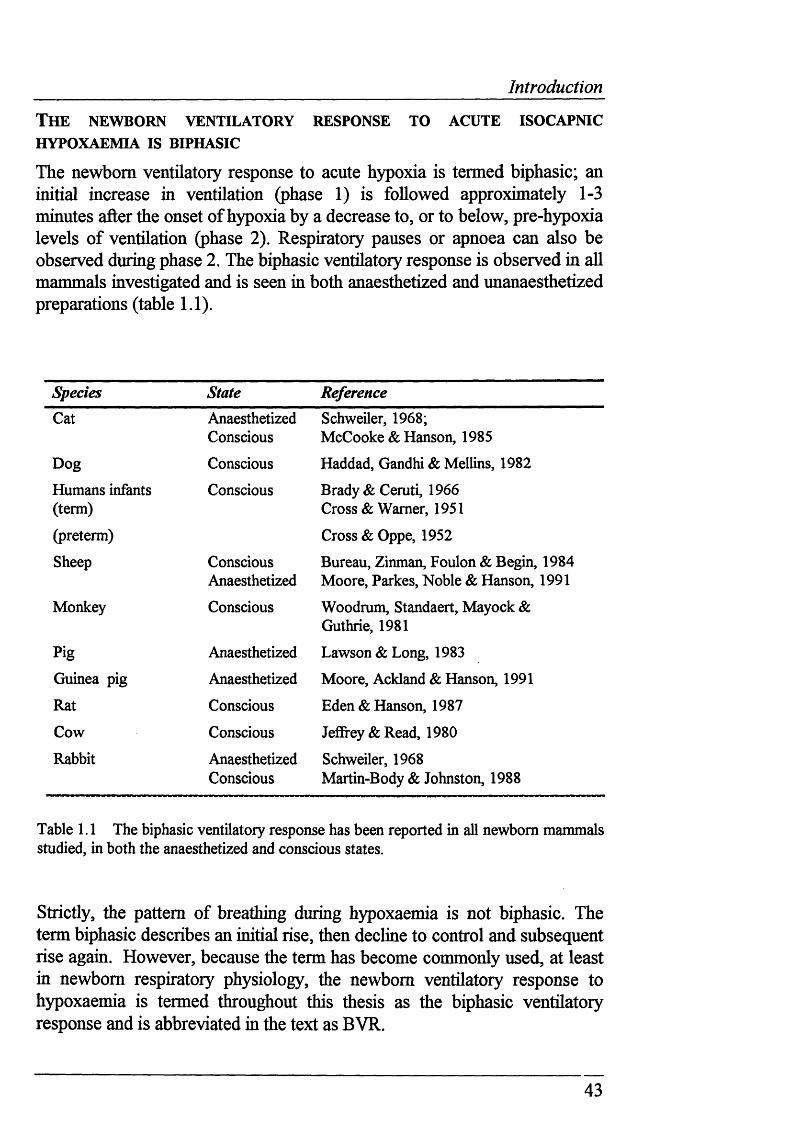
Introduction
T h e n ew b o r n v e n tila to r y r espo n se t o a c u t e is o c a p n ic
HYPOXAEMIA IS BIPHASIC
The newborn ventilatory response to acute hypoxia is termed biphasic; an initial increase in ventilation (phase 1) is followed approximately 1-3 minutes after the onset of hypoxia by a decrease to, or to below, pre-hypoxia levels of ventilation (phase 2). Respiratory pauses or apnoea can also be observed during phase 2. The biphasic ventilatory response is observed in all mammals investigated and is seen in both anaesthetized and unanaesthetized preparations (table 1.1).
Species State ReferenceCat Anaesthetized
ConsciousSchweiler, 1968;McCooke & Hanson, 1985
Dog Conscious Haddad, Gandhi & Mellins, 1982
Humans infants (term)
Conscious Brady & Ceruti, 1966 Cross & Warner, 1951
(preterm) Cross & Oppe, 1952
Sheep ConsciousAnaesthetized
Bureau, Zinman, Foulon & Begin, 1984 Moore, Parkes, Noble & Hanson, 1991
Monkey Conscious Woodrum, Standaert, Mayock & Guthrie, 1981
Pig Anaesthetized Lawson & Long, 1983
Guinea pig Anaesthetized Moore, Ackland & Hanson, 1991
Rat Conscious Eden & Hanson, 1987
Cow Conscious Jeffrey & Read, 1980
Rabbit AnaesthetizedConscious
Schweiler, 1968 Martin-Body & Johnston, 1988
Table 1.1 The biphasic ventilatory response has been reported in all newborn mammals studied, in both the anaesthetized and conscious states.
Strictly, the pattern of breathing during hypoxaemia is not biphasic. The term biphasic describes an initial rise, then decline to control and subsequent rise again. However, because the term has become commonly used, at least in newborn respiratory physiology, the newborn ventilatory response to hypoxaemia is termed throughout this thesis as the biphasic ventilatory response and is abbreviated in the text as BVR.
43
Introduction
W h a t a r e t h e m ec h a n ism s t h a t m e d ia t e BV R?
St im u l a t io n o f t h e p e r ip h e r a l c h e m o r e c e p t o r s ca u ses ph a se 1 - THE in c r e a se IN VENTILATION.
The principal peripheral arterial chemoreceptors, the carotid and aortic bodies, transduce changes in pH , Pacoj and Paoj (Heymans & Bouckaert, 1930; Heymans, Bouckaert & Dautrebande, 1930). The carotid body is composed of two parenchymatous cell types. Type 1 cells, or glomus cells, are chemoreceptive and are surrounded by sustentacular (type 2) cells forming clusters that are richly supphed by fenestrated capillaries (Verna, 1979). Preganghonic sympathetic fibres from the superior cervical ganghon synapse onto the type 1 cells (Floyd & Neil, 1952; Eyzaguirre & Lewin, 1961). Chemoreceptor discharge may also be modulated by other efferent fibres, apart from preganghonic sympathetic efferents (Fidone & Sato, 1970; Neil & ORegan, 1971a,b). Blood flow through the carotid body is the highest per unit mass in the body, with a perfiision rate ten times higher than would be predicted from its metabohc requirements.
Decreases in Paoj, or increases in Paco2 or are transduced intoincreased action potential discharge. Conversely, alkah or high Paoj levels diminish carotid chemoreceptor discharge. The mechanism of chemotransduction is not fully understood. The two most likely, but conflicting, theories both agree that an elevation in intracellular calcium is necessary for the release of neurotransmitter vesicles and subsequent generation of action potentials in the carotid sinus nerve. However, the mechanism that triggers this increase in cytosohc calcium remains elusive. The key event in promoting synaptic vesicle release could be due to the closing of O2 sensitive K'*' channels, thereby allowing calcium influx which in tum would further release internal calcium stores (Gonzalez, Almaraz, Obeso & Rigual, 1992). Alternatively, the hypoxic sensing mechanism could hinge on the dismption of ATP production in mitochondria which would initiate release of calcium from mitochondria, and internal calcium stores (Biscoe & Duchen, 1990; Duchen & Biscoe, 1992). However, more recently, data has emerged that supports strongly the idea for voltage-gated calcium entry (Buckler & Vaughan-Jones, 1994).
Carotid chemoreceptor afferents, along with baroreceptor afferents from the carotid sinus, are carried by the carotid sinus nerve, a branch of the glossopharyngeal nerve (De Castro, 1928). Stimulation of carotid baroreceptor afferents, by increasing the pressure in the carotid sinus, inhibits breathing (Grunstein, Derenne, & Müic-Emh, 1975). Both baro- and chemoreceptor afferents terminate in the NTS (Donoghue, Felder, Jordan &
44

Introduction
Spyer, 1984). Aortic and carotid chemoreceptors perform a similar role (Comroe, 1939), although it seems that carotid chemoreceptors make a larger contribution to the hypoxic ventilatory response (Martin-Body, Robson & Sinclair, 1985). Aortic afferents travel in the aortic branch of the vagus nerve (Nonidez, 1935). Other chemoreceptors sensitive to changes in blood gas status have been located but mostly they play a minor or undefined role in the regulation of respiration; it is possible that they perform local regulatory functions. Peri-adventitial chemoreceptor tissue has been found outside the carotid body region (Clarke & Daly, 1985). Chemoreceptive activity has also been recorded firom the abdominal vagal paraganglia in adult rats (Howe, Pack & Wise, 1981) and fi*om the aorticopulmonary region in young kittens (Coleridge, Coleridge & Howe, 1967). Pulmonary neuroepithehal cells (Lauweryns, de Bock, Guelinckx & Decramer, 1983; Lauweryns, Van Lommel & Dom, 1985), cultured firom fetal rabbit lungs also exhibit, perhaps hke type 1 carotid body cells (Gonzalez, Almaraz, Obeso & Rigual, 1992), 02-sensitive potassium channels (Youngson, Nurse, Yeger & Cutz, 1993).
A sudden reduction in Fiqj initially causes increased ventilation due to peripheral chemoreceptor stimulation (Schweiler, 1968; Blanco, Hanson, Johnson & Rigatto, 1984; Lawson & Long, 1983). Peripheral chemodenervation abohshes the increase in ventilation (Schweiler, 1968). Indeed, the peripheral chemoreceptors play a vital role in ensuring normal respiratory control during early development under normoxic conditons because peripheral chemodenervation markedly decreases the survival rate, and causes prolonged apnoea, in newborn piglets (Donnelly & Haddad, 1990), lambs (Bureau, Lamarche, Foulon & Dalle, 1985) kittens and rabbits (Schweiler, 1968).
Cardiovascular response to acute hypoxaemia in newborns.
In human infants hypoxaemia causes bradycardia (Poets, Stebbens, Samuels & Southall, 1993) and results in a fall in cardiac output. An increase in systemic vascular resistance ensures that arterial blood pressure is maintained or sometimes elevated. However, the effects of hypoxaemia on cardiovascular variables are less clear in anaesthetized animal preparations, since the varying effects of different anaesthetics and the degree of hypoxaemia can both produce markedly dissimilar effects (for example, see Gootman, Buckley & Gootman, 1979; Gootman, Gootman, Buckley, Peterson, Steele, Sica & Gandhi, 1990).
45
Introduction
The fetal cardiorespiratory response to acute hypoxaemia
The relevance of adult andfetal respiratory control to the study o f the effects o f hypoxia on breathing in newborns
Although this introduction is concerned primarily with the effects of hypoxia on respiratoiy control in the newborn, it will also consider adult and fetal work. This approach aims to draw on the considerable wealth of knowledge from adult respiratory physiology which thus provides the background on which the relatively new area of research into newborn respiratory physiology can draw. Fetal responses to hypoxia are discussed in order to consider the idea that newborn respiratory responses to hypoxia are a transitional stage during the maturational process from fetus to adult.
Acute hypoxaemia abolishes ffetal breathing**
The fetus exhibits breathing like movements, termed "fetal breathing movements" (FBM), which in early gestation are continuous, being only interrupted by short periods of apnoea, and then later become episodic along with trunk and limb movements (Merlet, Hoerter, Devilleneuve & Tchobroutsky, 1970; Dawes, Fox, Leduc, Liggins & Richards, 1970). Of course, FBM have no role in gaseous exchange but seem to be an important stimulus for the growth and structural maturation of the fetal lung (Harding, Hooper & Han, 1993). However, FBM increase under conditions of hypercapnia (Dawes, Gardner, Johnston & Walker, 1982) or perfusion of acidic cerebrospinal fluid (Bissonnette, Hohimer & Richardson, 1981) and are abolished in acute isocapnic hypoxaemia, where Paoj falls from the fetal normoxaemic value of 25 mmHg to ca. 15 mmHg (Boddy, Dawes, Fisher, Pinter & Robinson, 1974).
Fetal chemoreceptors respond to hypoxaemia but normally do not contribute to the control o f FBM.
Indirect methods suggested that chemoreflexes were present in the fetus (Barcroft & Karvonen, 1948), and Cross & Malcolm (1952) first showed that the carotid chemoreceptors were active in anaesthetized fetal lambs (139-140 days gestional age) when carotid sinus nerve discharge was silenced by hyperoxia after hypoxaemic chemoreceptor stimulation. Although single fibre recordings from the fetal lamb carotid sinus nerve indicated that the carotid chemoreceptors were relatively insensitive to a range of chemostimulants (Biscoe & Purves, 1967), Blanco, Dawes, Hanson & McCooke (1984) showed clearly that carotid chemoreceptors are active in utero and their hypoxic sensitivity lies to the left of the adult's. Aortic chemoreceptor activity is also present in the fetus (Ponte & Purves, 1973).
_
_______________________________________________________________________ Introductipn
Despite increased afferent chemoreceptor activity under hypoxaemic conditions, FBM are still abohshed during hypoxaemia in vagotomised (Boddy, Dawes, Fisher, Pinter & Robinson, 1974) and/or carotid denervated (Jansen, Ioffe, Russell & Chemick, 1981; Moore, Parkes, Nijhuis & Hanson, 1988) fetal lambs. Therefore, it seems that respiratory-related peripheral chemoreflexes are inhibited in utero. The significance of this will be discussed further when phase 2 of the newborn biphasic ventilatory response is considered in detail.
Peripheral chemoreceptor sensitivity to Pao2 , but not Paco2 ,f increases postnatally.
From birth, single fibre recordings from the carotid chemoreceptors in anaesthetized lambs show that P0 2 sensitivity is initially weak, being absent in normoxaemia on the day of birth (Blanco, Dawes, Hanson & McCooke, 1984). Carotid chemoreceptor sensitivity subsequently "resets”, as it gains in magnitude at a given Paoj with increasing postnatal age (Blanco, Dawes, Hanson & McCooke, 1984). This means that the response curve relating carotid chemoreceptor discharge to Paoj shifts upwards and to the right from the fetal Poj range towards the adult Pqj range over the first two postnatal weeks. Similar recording techniques indicate that aortic chemoreceptors also reset (Kumar & Hanson, 1989). Postnatal resetting has also been demonstrated by recording from single chemoreceptor afferents in an in vitro, superfiised newborn rat carotid body (Kholwadwala & Donnelly, 1992).
Indirect assessment of chemoreceptor function, using an alternate breath test, supports this finding in human infants (WiUiams, Smyth, Boon, Hanson, Kumar & Blanco, 1991). In this alternate breath test, the subject alternately breathes one breath of normoxia and then one breath of hypoxia or hypercapnia. Sudden changes in peripheral chemoreceptor discharge affect ongoing ventilation, with the type of ventilatory change ehcited being dependent on the timing of the peripheral chemoreceptor stimulus in relation to respiratory output (Black & Torrance, 1967). Hie alternate breath test assesses peripheral chemoreceptor fimction by measuring how ongoing ventilation is affected by altering inspired gas mixtures on a breath to breath basis.
This chemoreflex test in conscious kittens shows that carotid chemoreceptor dynamic sensitivity to CO2 is strong from birth even though the response to hypoxia is weak (Watanabe, Kumar & Hanson, 1992). This differential
47
Introduction
sensitivity to Paoj and Paco2 at birth may have important imphcations for understanding how the carotid chemoreceptors function (Torrance, 1992). However, other studies employing different methodology suggest that the peripheral chemoreceptor steady-state sensitivity to CO2 gains in magnitude with increasing postnatal age (Carroll, Bamford & Fitzgerald, 1994; Wolsink, Berkenbosch, De Goede & Ohevier, 1993).
Fetal cardiovascular responses to acute hypoxaemia are mediated by the peripheral chemoreceptors.
Acute hypoxaemia produces a rapid initial bradycardia, a slow increase in arterial blood pressure (Boddy, Dawes, Fisher, Pinter & Robinson, 1974) and a redistribution of cardiac output from the carcass to more vital organs: i.e. the brain, heart and adrenals (Cohn, Sacks, Heymann & Rudolph, 1974). While the peripheral chemoreceptors play no role in controlling FBM, the chemoreflex effects on cardiovascular control in hypoxaemia are profound. Carotid chemoreflexes mediate the bradycardia, the increase in peripheral (femoral) resistance via adrenergic receptors and the release of some vasoconstrictor humoral substances (Giussani, 1992; Giussani, Spencer, Moore, Bennet & Hanson, 1993).
48
Introduction
The adult cardiorespiratory response to acute isocapnic hypoxaemia.
Adults also show BVR to acute isocapnic hypoxaemia
Even though phase 2 of the newborn BVR diminishes greatly with increasing post-natal age (Eden & Hanson, 1987), remnants of the BVR seem to persist into adulthood. However, in adults the time for the decline in breathing to begin takes longer (i.e. the period of hypoxaemia has to be longer for breathing to decline). Also, unlike the newborn, ventilation falls but remains above pre-hypoxaemic ventilatory levels (Weiskopf & Gabel, 1975; Easton, Slykerman & Anthonisen, 1986; van Beek, Berkenbosch, DeGoede & Ohevier, 1986; Vizek, Pickett & Weil, 1987; Marshall & Metcalfe, 1988). As in newborns, the peripheral chemoreceptors stimulate the increase in ventilation (Vizek, Pickett & Weil, 1987) and peripheral chemodenervation prevents the initial ventilatory rise (Lahiri & Delaney, 1975).
Adult cardiovascular responses to hypoxaemia
In spontaneously breathing animals, an increase in heart rate and a fall in arterial blood pressure is seen, with cardiac output and regional blood flow increasing, especially to the brain (Daly & Scott, 1963; Marshall & Metcalfe, 1988). When an animal is artificially ventilated during chemoreceptor stimulation, bradycardia, decreased cardiac output and increased peripheral vascular resistance occur (Daly & Scott, 1962). This shows that cardiovascular effects in spontaneously breathing animals are due to peripheral chemoreceptor-mediated hyperventilation, which produces a fall in Paco2 and inhibits vagal reflexes (see Daly, 1986 for review). The distribution of blood flow within the lungs improves during hypoxaemia, due to an increase in pulmonary wedge pressure, mediated locally by Paqj (Marshall & Marshall, 1983).
49
Introduction
T h e MECHANISM UNDERLYING PHASE 2 OF BVR IS UNKNOWN
This section considers the possible mechanisms that may cause the decline in ventilation seen in newborns during acute hypoxaemia. The emphasis in this section is placed firmly on studies in the newborn. Reference to the larger hterature on adult and fetal breathing responses to hypoxaemia is made only when it is appropriate for interpreting newborn studies.
H y p o th e s is h BVRis due to peripheral chemoreceptor adaptation to acute hypoxaemia.
Since the increase in ventilation (phase 1 of the BVR) during hypoxaemia in the newborn is caused by peripheral chemoreceptor stimulation, the decline in ventilation (phase 2) could simply be due to the inabihty o f the peripheral chemoreceptors to maintain their initial elevated level o f discharge throughout the hypoxaemic challenge. This hypothesis assumes that respiratory output is directly proportional to the level of carotid chemoreceptor discharge throughout hypoxaemia. One assumption that has been made in considering the peripheral chemoreceptor adaptation hypothesis is that the carotid chemoreceptors play the predominant, if not exclusive, role in mediating peripheral chemoreceptor control in breathing during hypoxaemia. However, as will be discussed in greater detail (see central inhibition of respiratory output hypothesis) phase 2 may not necessarily be due to a fall in afferent fibre discharge. Rather, carotid chemoreceptor input may be modulated once it has arrived in the respiratory centres. This possibihty is referred to throughout this subsection but only as a general concept, rather than being detailed in great depth as a possible mechanism.
The peripheral chemoreceptor adaptation hypothesis has been tested in two different types of experiment. First, direct recordings fi'om either single/few carotid chemoreceptor fibres or the whole carotid sinus nerve have been made. All these experiments have been conducted in anaesthetized kittens. Second, indirect methods of testing the fimction of the carotid chemoreceptors during hypoxaemia have been employed in unanaesthetized, conscious preparations in many species.
50
Introduction
Direct recordings from carotid chemoreceptor fibres in newborns
Schweiler (1968) first tested the peripheral chemoreceptor adaptation hypothesis, when he recorded phrenic nerve and single fibre carotid chemoreceptor activity simultaneously in nine kittens aged less than 10 days old and a further 3 kittens >13 days old. Chemoreceptor discharge was maintained throughout the hypoxaemic period (Paoj unreported; Fiqj 0.1), even though phrenic nerve activity fell. In peripheral chemodenervated animals subjected to hypoxaemia, ventilation fell even further, often to apnoea.
These results were supported by Blanco, Hanson, Johnson & Rigatto, (1984) who also showed in anaesthetized kittens (5-34 days old) that the discharge of single or few carotid chemoreceptor fibres increased quickly and was maintained above pre-hypoxic levels throughout hypoxaemia (Paoj 35 mmHg) even though the animals exhibited BVR. However similar recordings fi'om single/few carotid chemoreceptor fibres in 0-19 day old anaesthetized newborn kittens, showed that the initial increase in activity on exposure to hypoxaemia (Paoj 55 mmHg) was only sustained in 54% of fibres tested (Marchai, Bairam, Haouzi, Crance, Di Giuiho, Vert & Lahiri, 1992). This adaptation was observed more in veiy young kittens (<10 days old). The decline in chemoreceptor discharge occurred within 30 seconds of the onset of hypoxaemia. Multifibre carotid sinus nerve recordings fi'om 1 week old kittens have also showed that some kittens failed to maintain the initial elevation in discharge during hypoxaemia (Paoj 40 mmHg) (Carroll, Bamford & Fitzgerald, 1994) whereas older kittens did not exhibit this response. In a preliminary study. Mulligan & Bhide (1989) report that single fibre carotid chemoreceptor activity, as recorded extracellularly firom the petrosal ganghon in the piglet, f^ s in sustained hypoxia (Paoj unreported).
Clearly, there is controversy over whether carotid chemoreceptor fibres adapt to hypoxaemia. Interpretation of single or few chemoreceptor fibre recordings firom the carotid sinus nerve hinges on the assumption that the fibres recorded are representative of afferent nerve input as a whole. It is likely that the population of fibres recorded is biased towards the myelinated type, since these units have the largest action potentials. Nonetheless, the interpretation of whole nerve recording studies is hampered by the recording of baroreceptor activity in the whole CSN.
51
Introduction
Indirect tests ofperipheral chemoreceptor function in newborns
Several studies, using unanaesthetized lambs, have supported the peripheral chemoreceptor adaptation hypothesis on the strength of indirect methods. There are many problems in measuring physiological changes in this type of experiment. Surgery, prior to the experiment, for implanting catheters and electrodes requires the animals to recover. This is often hazardous in newborn animals. Thus control and measurement of pH and blood gases is difBcult. Also, quantification of changes in various parameters or stimuh that are used to test the hypothesis, such as hypoxaemia, is often impossible.
A series o f studies have been undertaken in unanaesthetized lambs to test the hypothesis that before maturation of the peripheral chemoreceptors is complete, the chemorefiex response would adapt to sustained steady-state hypoxaemia. This hypothesis introduces a further comphcation in considering the mechanisms underlying phase 2 of BVR. The postnatal period over which the newborn BVR occurs may not be the same as the time over which the peripheral chemoreceptors "reset". It is possible therefore that the relative importance of the mechanisms that contribute to the newborn BVR may shift with postnatal age.
Unanaesthetized 1-2 day old newborn lambs were exposed to steady-state hypoxia (Fiqj 0.08) or progressive hypoxaemia (from F1 0 2 0.21 to Fiqj 0.08) (Bureau, Cote, Blanchard, Hobbs, Foulon & Dalle, 1986). In steady-state hypoxaemia the lambs exhibited BVR. In progressive hypoxaemia, a similar maximal increase in Ve was reached to that in steady-state hypoxaemia. Transient administration of 100% oxygen was used to silence the peripheral chemoreceptors (the Dejours test). This test produced a smaller fall in Ve for the steady-state response than for the progressive hypoxaemic test. Because there was less of a fall in Ve in the steady-state situation, the authors concluded that in this test chemoreceptor discharge adapted: hence during the steady state exposure to hypoxaemia, peripheral chemoreceptor activity would be close to baseline discharge.
This conclusion fails to explain why progressive hypoxia should continually produce an increase in Ve. Even though the same final degree of Fiqj was achieved, there is no way of knowing what level of hypoxaemia was produced in each type of hypoxic exposure in this study. Furthermore, repeated, sustained chemoreceptor stimulation (as in the progressive hypoxia protocol) has been shown to produce long-lasting effects on breathing. Prolonged peripheral chemoreceptor stimulation can also continue to stimulate ventilation after the period of stimulation has ceased (Cao, ZwiUich, Berthon-Jones & Sulhvan, 1992). The fact that a sustained peripheral chemoreceptor stimulus, in the form of progressive hypoxia,
52
Introduction
could overcome any central effects of hypoxia suggests that the balance between excitatory peripheral chemoreceptor stimulation and a putative central modulation of chemoreflexes can be shifted towards the excitatory limb of the reflex provided it is strong enough. Thus, the study failed to show that the ventilatory adaptation to hypoxia was mediated by the peripheral chemoreceptors.
In a similar study the Dejours test was apphed at the beginning, middle and end of 18 minutes of exposure to hypoxia in newborn lambs (Carroll & Bureau, 1987). No blood gas values or cardiovascular data were recorded in this experiment. In two day old lambs, the fall in breathing produced by the Dejours test 18 minutes into the hypoxic period was smaller than that produced at 3 minutes. This does not explain the fall in ventilation observed before 18 minutes into hypoxia. Furthermore, the Dejours test apphed 3 minutes into hypoxia did not show a larger fall in breathing than that observed in normoxia, even though breathing had already fallen (but remained above control). This would argue for the BVR being produced by some other mechanism, perhaps central modulation o f peripheral chemoreflexes.
The difference between the peak Ve and that during phase 2 indicates that this difference becomes progressively larger during the hypoxic period, which would also indicate that a central inhibitory effect slowly but progressively reduced the effects of sustained excitatory input from the peripheral chemoreceptors. In older lambs, BVR was again seen but the Dejours test did not have any effect on the magnitude of the difference seen in the resultant Ve drop at any point in the hypoxaemic period. Thus another conclusion from the study of Carroll & Bureau (1987) is that the BVR could not have been caused by the peripheral chemoreceptors adapting but rather by a centrally mediated, inhibitory effect. In essence, the authors illustrated the process of "resetting" from ages 2 to 10 days in newborn lambs. In addition, the methodology used in this study, the Dejours test, can invoke other changes that affect ventilation. Decreases in [pH]a due to increased HbOi, cerebral blood flow, bronchoconstriction (impeding a decrease in Ve) and changes in Paco2 may also occur (Dejours, 1962). Thesefactors may well have played a role in this study, since ventilation declined over at least 5 breaths on administration of 2 breaths of O2 . Clearly, the decrease in Ve on 100% O2 inhalation does not necessarily indicate the magnitude of the hypoxic drive.
Kumar, Thomas & Elnazir (1992) showed that an alternating PO2 stimulus to the peripheral chemoreceptors, induced by switching between Fiqj 0.21 and 0.12, produces BVR, similar to the BVR produced by a non-alternating
53
Introduction
stimulus of F1 0 2 0.12 in conscious newborn rats. The assumption is that the central levels of tissue P0 2 do not fluctuate with the alternating peripheral chemoreceptor stimulus. With an alternating stimulus, the peripheral chemoreceptors should continually respond to alternate low and normoxic levels of Pao2 aud hence not adapt to the O2 stimulus. Hence the pattern of V e was driven by the peripheral chemoreceptors throughout the hypoxic period. Assuming central tissue P0 2 stayed constant during hypoxia, this suggests that at greater levels of chemoreceptor stimulation (i.e. F1 0 2 0.12) any central inhibitory mechanisms can be overridden because V e responds to the increased chemoreceptor activity. However, once peripheral chemoreceptor discharge falls again (F1 0 2 0.21) this input is not sufficient to drive ventilation. Therefore, this study provides further evidence to support the idea that central mechanisms mediate BVR.
Studies o f peripheral chemoreceptor function during hypoxaemia in the adult
The mechanism underlying phase 2 of the adult BVR has attracted similar controversy to that in the newborn.
Whole carotid sinus nerve activity (with baroreceptor activity removed by stripping the adventitia over the surface of the carotid sinus) increases and is maintained during hypoxaemia in anaesthetized adult cats, even during ventilatory decline (Vizek, Pickett & Weil, 1987; Andronikou, Shirahata, Mokashi & Lahiri, 1988). However, carotid chemoreceptors adapt to prolonged (60-90 min) hypoxia in adult rabbits (Li, Ponte & Sadler, 1990). Chemical silencing of the peripheral chemoreceptors using 100% O2 during isocapnic hypoxia in adult humans also unmasks an inhibition of ventilation of central origin, since ventilation falls to below control values (Holtby, Berezanski & Anthonisen, 1988)
However, on the basis of reflex ventilatory studies in adult man, the decline in ventilation seen during the adult BVR has also been attributed, at least in part, to peripheral chemoreceptor adaptation. Furthermore, the fall in breathing has been linked to die magnitude of the the initial ventilatory increase (Easton, Slykerman & Anthonisen, 1986). The increase in ventilation due to a period of hypoxia after previous exposure to hypoxia is reported to be smaller than that seen for the initial hypoxic period (Easton, Slykerman & Anthonisen, 1988). Thus the chemorefiex has been imphed to be altered at the level of the carotid body. The rationale here is that the54
Introduction
peripheral chemoreceptors, for the same test stimulus level of hypoxia, do not effect the same increase in Ve post-hypoxia because their discharge has fallen due to adaptation.
Further studies in man support these ideas. 1 min hypoxic and hypercapnie pulses were apphed to awake human adults to ascertain the gain in ventilation produced by these tests during hypoxia (Bascom, Clement, Cunningham, Painter, & Robbins, 1991). Although both hypoxic and hypercapnie senstivity decreased during hypoxia, the drop in CO2 sensitivity was less than that for hypoxia pulses. Since there is no discrimination between CO2 and O2 at the peripheral chemoreceptor fibre level, and hypoxia is detected solely by the peripheral chemoreceptors, attenuation of the peripheral chemoreceptor drive was proposed as the mechanism underlying BVR in man. However, it is difficult to interpret these studies because the magnitude of each test pulse cannot be measured as afferent chemoreceptor discharge ; i.e. was the magnitude of these stimuh neurally equivalent?
55
Introduction
H ypothesis 2\ Central inhibition of respiratory output by a (hypoxia- sensitive) neural pathway located in the brain causes BVR.
Fetal inhibitory brain stem influences on FBM during hypoxaemia.
The concept of inhibition of respiratory output was first raised by Barcroft (1946). He described the development of neuromuscular movement in the sheep fetus, which he first observed to start at around GA35. The fetus responded to ‘tapping sharply between the eye and moutF’ by moving its head. With increasing gestational age, the area of stimulation which evoked a reflex response increased. By about 40 days, this trigeminal reflex resulted in a rhythmic spasm, which included intercostal muscle movement. Afl;er this stage, ca. GA42 days and beyond, the stimulus caused diaphragmatic movement to outlast that of other somatic movements; respiratory movements became easier to eflcit. However, by GA60, the fetal lamb had become:
inert. Only slight and localized responses follow quite considerable stimuli. Ascompared with its condition 10 days earlier it gives the impression o f being moribund. That it is not so may be proved by subjecting it to asphyxia or even to exposure. ”
Barcrofl: (1946) investigated further the disappearance of thesomatic/respiratory responses by transecting the fetal lamb brain at four levels. Transections at the level of the hypoglossal nucleus only allowed a few diaphragmatic movements to persist. Respiratory movements only occurred after somatic stimulation when transections were made just caudal to the pons. However, transections made at the upper border of the pons resulted in continuous FBM. Although only two fetal brains were transected in this manner, these findings are compelling since one of the transected fetal lambs was a twin: in the other, non-transected twin the absence o f FBM continued.
Dawes, Gardner, Johnston & Walker (1983) pursued Barcroft’s idea further, and demonstrated that midcolhcular transections prevented the abohtion of FBM during acute isocapnic hypoxaemia in the chronically instrumented fetal lamb. This study strongly suggested that an inhibitory mechanism that abohshes FBM is activated during hypoxaemia. However, transections are crude perturbations of tissue, since there is extensive necrosis o f tissue beyond the transection site. This renders any accurate appraisal o f which brain structures were affected by a transection extremely difftcult (as Barcroft conceded originally). Thus whether the origin of the inhibitoiy process was rostral or caudal to the transection was debatable, although Dawes, Gardner, Johnston & Walker (1983) attributed the site to being suprapontine.
56
Introduction
Gluckman & Johnston (1987) used a more refined technique, by extensive electrolytic lesions of areas throughout the upper pons in the fetal lamb. However, in this study the area demarcated (which included the parabrachial and Kolliker-Fuse complex) that prevented the abolition of FBM during hypoxaemia is a compilation of many electrolytic lesions in numerous fetal lambs. It is not clear how repeatable the effect on FBM of lesioning this area was. For this reason reservations must be held as to the exact location of the site that prevented the abolition of FBM in the fetuses studied. However, it is interesting to note that chemical and electrical stimulation in the medial parabrachial and Kolliker-Fuse region, demarcated in the study of Johnston & Gluckman (1987), elicits profound inhibition of breathing in adult rats (Lara, Parkes, Silva-Carvhalo, Izzo, Dawid-Milner & Spyer, 1994)
Carotid (Dawes, Gardner, Johnston & Walker, 1983) or complete peripheral chemodenervation (Moore, Parkes, Nijhuis & Hanson, 1989) seemed to have no contribution to the stimulation of FBM in midbrain transected fetal lambs when denervation was performed at the same time as brain stem transection (although Murai, Lee, Wallen & Kitterman (1985) reported that peripheral chemodenervation reduced the incidence of FBM by ca. 10% in intact fetuses). Instead of transecting and also denervating at the same time, Johnston & Gluckman (1992) performed lesions of the rostrolateral pons prior to subsequent peripheral chemodenervation. This approach resulted in FBM being reduced after denervation in those fetuses which had exhibited a stimulatory response to hypoxaemia after the placement of lesions. Thus carotid chemoreceptors can play a role in the control of FBM, but only when inhibitory brain stem projections are removed. However, carotid chemoreceptors do play other important roles under normal conditions. Carotid denervation first revealed an important role for the carotid chemoreceptors in mediating the control of fetal cardiovascular responses to hypoxaemia. Thus their role in the fetus is to initiate appropriate cardiovascular responses to maintain adequate oxygenation to essential organs (Giussani, Spencer, Moore, Bennet & Hanson, 1992).
Hence a balance between excitatory (peripheral chemoreceptor) inputs and inhibitory (brain stem) influences seems to shape the response o f FBM to hypoxaemia - in the fetus this balance is heavily weighted towards the inhibitory (brain stem) processes.
57
Introduction
Inhibitory brain stem influences on breathing in the newborn during BVR.
Stimulation of carotid chemoreceptor activity by close intra-carotid injection of cyanide produces an inhibition of breathing during hypoxia, rather than the stimulatory effect observed in normoxia (Schramm & Grunstein, 1987). The dose-response relationship of the cyanide-induced inhibition during hypoxia suggested that the peripheral chemoreceptors may activate a central mechanism to cause BVR, because increasing the dose of cyanide produced more profound decreases in Ve (but see Chapter 2.8). However, continuous electrical stimulation of the CSN in normoxaemia produces a sustained, elevated level of phrenic activity (Lawson & Long, 1984). The latter study shows that the newborn brain stem is able to maintain the integration of increased afferent CSN activity over a long period of time, and that peripheral chemoreceptor stimulation itself is unlikely to initiate some central mechanism to cause BVR. Thus, it would seem very likely that the brain stem must modulate peripheral chemoreflexes, rather than be modulated by the peripheral chemoreceptors to produce phase 2 o f BVR. Indeed, perturbation of the brain stem itself, by transection of the rostral pons, at or below the intercoUicular level, in newborn rabbits (Martin-Body & Johnston, 1988) or rats (Hanson & WiUiams, 1989) abohshes phase 2 of the BVR. These latter findings resemble those in the transected fetus.
Electrical stimulation showed that there are areas in the ventrolateral pons of newborn lambs that mediate a fall in respiratory output (Coles, Kumar & Noble, 1989). These are similar areas to Ihose that have been imphcated in the fetal responses to hypoxaemia after lesioning. Apnoea was ehcited at the level of the trigeminal motor nucleus, although whether electrical stimulation of cell bodies or axons of passage in this area was responsible for the respiratory effects seen remains unresolved. Extracellular recordings fi'om this region showed that action potential discharge increased during hypoxaemia (Noble & Wilhams, 1989), and that this was not due to carotid chemoreceptor stimulation (Noble, WiUiams, Hanson & Smith, 1990).
Unilateral focal cooling of the rostral pons, at the level of the facial coUiculus abolishes phase 2 of BVR during hypoxaemia in newborn lambs. However, rewarming the cooling probe returns breathing to its phase 2 level (Moore, Parkes, Noble & Hanson, 1991). Since cooling the probe during normoxaemia and phase 1 has no effect on breathing, this technique shows for the first time that inhibitory mechanisms are active during hypoxaemia. Furthermore, these inhibitory mechanisms seem to be activated only during isocapnic hypoxaemia. However, this technique cannot identify areas containing ceU bodies that mediate BVR. Whether cooling to the levels used in this study (20°C) reduces axonal transmission, or inhibits ceU body activity remains unclear. Certainly local blood flow around the tip o f the
58
Introduction
cooling probe is likely to be affected. Also no other sites in the upper pons, or the response to other factors that drive respiratory output such as hypercapnia, were investigated using this technique. Thus no further information was provided about any specific sites mediating the inhibition of respiratory output during phase 2.
Clearly, a variety of techniques have imphcated brain stem inhibitory processes in the newborn BVR, although fiirther work is required to demarcate specific areas.
Evidence that suggests newborn/ fetal inhibitory brain stem mechanisms are also functional in the adult during hypoxaemia.
Ventilatory decline during hypoxaemia has long been held to be due to direct hypoxic depression of CNS function in adults (Gesell, 1939). Hypoxic depression is only likely to occur at very severe levels of hypoxia. However, there are advocates of the idea that a general hypoxic depression causes a decline in cardiorespiratory efferent activity (see Hypothesis 3). In recent times, this concept has been challenged in the adult, as well as the fetus and newborn: rather BVR in the adult has been attributed to hypoxaemia initiating neurophysiological changes, at either the peripheral chemoreceptors or centrally. Clearly hypotheses on the adult ventilatory response to hypoxaemia have important implications for similar work in the newborn.
Centrally mediated ventilatory decline during acute hypoxaemia was first indicated in anaesthetized (Selladuri & Wright, 1932; Von Euler & Liljestrand, 1936; Chemiack, Edelman & Lahiri, 1970/71) and conscious (Watt, Dunke & Comroe, 1943) animals that were peripherally chemodenervated. Since these studies, the development of two refined methods of controlling brain Pqj separately from the peripheral circulation have provided further evidence that BVR in the adult is of a central origin. First, the isolated perfused brain stem technique permits only the brain stem to be exposed to hypoxaemia (Van Beek, Berkenbosch, De Goede & Ohevier, 1984). Second, carbon monoxide causes brain hypoxia (Gauthier, 1986; Melton, Neubauer & Edelman, 1988) - the peripheral chemoreceptors are unable to detect the effect of CO administration since the critical factor in chemoreception is Pqj rather than O2 content (Lahiri, Mulhgan, Nishino, Mokashi & Davies, 1981). Both of these methods cause respiratory output to decline during central (or brain) hypoxaemia.
59
Introduction
Other experiments have also controlled afferent carotid chemoreceptor drive independently of central hypoxia. Lahiri & Delaney (1974) and Lahiri (1976) showed in adult cats that electrical CSN stimulation did not produce an increase in ventilation in hypoxaemia (Paoj 33mHg) yet CSN stimulation did stimulate ventilation in subsequent normoxaemia or hyperoxaemia. However, a similar study using this methodology did not find any attennuation of the CSN mediated reflex on breathing during hypoxia in adult rabbbits (Wiemer, Schone & Kiwull, 1973).
Lee and Millhom (1975) altered afferent carotid chemoreceptor input by a cross perfusion technique. Here the Paoj fi'om a donor dog was varied by changing Fiqj. The blood of the donor dog was used to perfuse the carotid bodies of in a recipient dog where the circulation of each carotid body was isolated fi'om the systemic circulation. In this way, carotid chemoreceptor discharge in the recipient dog could be manipulated by changing the donor dog’s Paoj. Three donor Paoj levels were used: hypoxia (38mmHg) normoxia (90mmHg) and hyperoxia (516mmHg). The decline in ventilation caused by central hypoxia was not affected by the level of carotid chemoreceptor drive.
The clearest evidence to suggest that phase 2 of the adult BVR may be caused by a similar mechanism to that of the newborn is provided by transection studies. Transection at or below the intercoUicular level in unanaesthetized adult rats converts the adult BVR into a stimulation of breathing during hypoxaemia. Most importantly, the effect of mid-coUicular transection was not dependent on the presence of afferent peripheral chemoreceptor input (Martin-Body, 1988). These studies strongly suggest that central mechanisms cause the inhibition in ventilation in adults.
Breathing remains inhibited on recovery to hyperoxia for up to 1 hr after an isocapnic hypoxaemic episode in adult cats (Millhom, Eldridge, Kiley & Waldrop, 1984). This prolonged inhibition of breathing can be partiaUy reversed by theophylline, the adenosine antagonist, but abohshed by removal of the mesencephalon (Gallman & MilUiom, 1988). Electrical and chemical stimulation studies revealed that there were ceU bodies located in the ventromedial region in the mesencephalon, that inhibited breathing (Gallman & Millhom, 1991). However the fi'equency of prolonged inhibitory responses due to chemical stimulation was unreported; in addition, the latency of some effects of chemical stimulation on phrenic nerve activity was rather long, thereby bringing into question the location of the site that mediated the inhibition.
60
Introduction
However, the inhibition of respiratory output in isocapnic hypoxaemia is also dependent on the degree of hypoxaemia: inhibition is more likely to occur in “severe” hypoxaemia (<26mmHg). At Paoj >35mmHg, a long lasting facihtation of breathing is produced. This supports previous findings where awake, peripherally chemodenervated cats (Miller & Tenney, 1975; Long, Giesbrecht & Anthonisen, 1993), goats (Smith, Engwall, Dempsey & Bisgard, 1993) and rats (Martin-Body, Robson & Sinclair, 1986) exhibited a sustained Ve increase to hypoxia. Mid-coUicular decerebration abohshed the tachypnoea caused by hypoxia in decerebrate cats (Tenney & Ou, 1977) and removal of the diencephalon abohshes this facihtatory effect (Gallman & Millhom, 1988). Higher cortical fimction, which has an inhibitory influence on the diencephalon (von Euler, Herrers & Wexler, 1970), is particularly vulnerable to hypoxaemia. Therefore, in anaesthetized adults exposed to hypoxia, the inhibitory influence of the cortex on the diencephalon is abohshed by anaesthesia and so a fall in ventilation occurs. In conscious, denervated adults exposed to moderate Paoj, tachypnoea occurs because cortical influences are depressed and this unmasks the inhibition of the diencephalon. Thus there may be a differential sensitivity to hypoxaemia in the brain, in a caudal to rostral direction, as suggested by Neubauer, Melton & Edelman (1990). In summaiy, these studies suggest that there are two CNS mechanisms responsive to hypoxaemia, and that the inhibitory mechanism was probably mediated by cell bodies located in the mesencephalon.
BVR can also be altered pharmacologically by the apphcation of neurotransmitters and neurotoxins to discrete brain stem regions, which again suggests that central mechanisms are essential in mediating the responses seen. In particular, ventrolateral medullary regions are involved in mediating the respiratory response to hypoxaemia in adults. Focal cooling (Schlaefke, See, Herker-See & Loeschcke, 1979), topical apphcation of kainic acid (Martin & Sinclair, 1990) or bilateral coagulation of the intermediate area (Schlaefke, See, Herker-See & Loeschcke, 1979) of the surface of the VLM all attenuate the ventilatory response to hypoxaemic stimulation of the peripheral chemoreceptors. VLM neuronal discharge frequency also alters during carotid chemoreceptor stimulation (Caverson, Ciriello & Calaresu, 1984). Single unit extracellular recordings in intact, paralyzed, decerebrate cats reveal that isocapnic hypoxaemia causes both excitation and inhibition of medullary respiratory neurons, unlike the consistent excitation observed under hypercapnie conditions (St. John & Wang, 1977) The hypoxaemic inhibition occurred even though phrenic discharge was elevated. However, in peripherally chemodenervated preparations hypoxaemia depressed both respiratory neuronal and phrenic
61
Introduction
activities, despite hypercapnia producing similar changes to those seen in intact preparations.
Microinjection of an excitatory amino acid antagonist (kyneuric acid) into rostral VLM sites accentuates the respiratory response to hypoxaemia (Dillon, Welsh & Waldrop, 1991) but ^utamate (Gatti, Norman, Taviera Dasilva & Gillis, 1990), or focal cooling (Budzinska, von Euler, Kao, Pantaleo & Yamamoto, 1985) tonically depress ventilation in a similar area, the nucleus paragigantocellularis lateralis. Conversely, microinjections of EAA antagonists into more caudal areas of VLM produce apnoea (Dillon, Welsh & Waldrop, 1991). This suggests that EAA exert an inhibitory tonic control on respiratory output in the VLM region.
H ypothesis 3: Direct hypoxic depression of respiratory neurones causes the fall in ventilation observed in BVR
Proponents of the hypothesis that ventilatory decline occurs during hypoxia because respiratory neurones are depressed by lack of Pqj have based their conclusions generally from observing the effects of severe levels o f hypoxia or even ischaemia on respiratory neurones. Clearly the levels of hypoxia applied to an in vivo or in vitro preparation could produce radically different neuronal responses. However, at more severe levels of hypoxia neurones protect themselves against this extreme metaboHc challenge. Indeed, newborn respiratory neurones are more tolerant to severe hypoxia than those of the adult (Ballanyi, Kuwana, Volker, Morawietz & Richter, 1992). Presumably this is an important maturational adaptation because newborns are frequently exposed to hypoxic episodes during early development.
However, neurones respond directly to hypoxia even at mild levels. For example, intra- and extracellular recordings from hypoglossal neurones in newborn kittens show that discharge frequency increases even at Fiqj 0.18. Most interestingly, in the context of BVR, is that ca. 50% of these hypoglossal neurones failed to sustain an initial increase in activity, with some failing to maintain discharge frequency above prehypoxaemic levels (Smith, Li & Noble, 1993a). Both in vitro (Haddad & Donnelly, 1990) and in vivo recordings (Smith, Li & Noble, 1993b) from hypoglossal motoneurones reveal that input resistance initially rises during hypoxia, with
62
Introduction
a subsequent decrease after a few minutes. Excitability of hypoglossal neurones is also enhanced during early hypoxia (tissue Pqj 15-20mmHg), with a fall in spike fi'equency and even depolarization blockade in some motoneurones occurring afterwards (Haddad & Donnelly, 1990; Donnelly, Jiang & Haddad, 1992). Unlike the in vivo newborn kitten recordings, no repolarization or hyperpolarization occurs in the in vitro preparation. Such differences are probably dependent on the integrity of other afiferents to the hypoglossal motoneurones, and the presence of cardiorespiratory reflexes during hypoxia. However, there are obvious parallels between the time course o f the depolarization response to hypoxia in both in vitro and in vivo hypoglossal motoneurones and BVR.
Regardless of how respiratory neurones behave under severe hypoxia, the fact remains that BVR is ehcited at relatively mild levels of hypoxia (Pao; 35-60mmHg). At these mild levels of hypoxia, there is still oxygen available, so neurones probably adapt to this lower Paoj level by decreasing metabohsm. Neurones exposed to mild levels of hypoxia are certainly unlikely to shut down fimction altogether i.e. undergo depolarization blockade. Indeed, neurones are still excitable at severe levels o f hypoxia (Richter, Bischofif, Anders, Bellingham & Windhorst, 1993). Furthermore, die abohtion of phase 2 of BVR or the persistence of FBM during hypoxia in mid-cohicular transected animals strongly argues against the faU in breathing being due entirely to direct hypoxic depression of (cardio)respiratoiy fimction. Thus, at relatively mild levels of hypoxia, the inhibition of breathing cannot be attributed solely to the direct action of hypoxia on ceUular fimction of cardiorespiratory neurones.
63
Introduction
H ypothesis 4: BVR is caused by the drop in metabolic rate observed during hypoxia.
Metabolism falls during acute hypoxaemia.
Metabolic rate (i.e. oxygen consumption (Voj) and CO2 production (Vco;)) decrease during acute hypoxia in newborn infants and unanesthetized newborns, including lambs, kittens, puppies and rabbit pups (Acheson, Dawes & Mott, 1957; Cross, Dawes & Mott, 1959; Dawes & Mott, 1959; Moore, 1956; Moore, 1959). Accordingly, Ve also declines, since it matches metabohc requirements (Mortola, Rezzonico & Lanthier, 1989). Indeed, the slope of the end-tidal PETCO2 plateau increases in direct proportion to the production of metabohc CO2 in adult man (Cochrane, Newstead, NoweU, Openshaw & Wolff, 1982). That ventilation is dependent on metabohc CO2
is demonstrated further by Philhpson, Duffin & Cooper, (1981). When the rate of CO2 removal was matched to the metabohc production of CO2 , by extracorporeal veno-venous exchange in dogs, ventilation ceased even though mean pHa, Paco2 Pao; were within the normal physiological range.
The faU in Vqj in human infants (Brady & Ceruti, 1966) and unanaesthetized puppies (Haddad, Gandhi & Mellins, 1982) and kittens (Frappeh, Saiki & Mortola, 1991) occurs very rapidly, within 2 min, even at mild levels of hypoxaemia. The link between phase 2 and metabohsm is further reinforced by evidence that suggests faUs in Vqj during hypoxaemia are less pronounced in older rats (Taylor, 1960) and lambs (Cross, Dawes & Mott, 1959) than at birth. Thus die possibihty arises that the metabohc response to hypoxaemia gains in magnitude with increasing postnatal age, in a similar way to that of peripheral chemoreceptor resetting.
The decline in V0 2 and Vco2 could be due to lower substrate utilization, effected by local and circulating factors possibly under the direct control of a neurohumoral reflex. However, peripheral chemodenervation does not affect the fall in metabohsm in cold newborn rabbits caused by hypoxia (Blatteis, 1964). Alternatively, the availabihty of O2 itself could control the rate of aerobic metabohsm. Acute hyperoxia increases V0 2 m mice (Mortola & Tenney, 1986) which imphes that metabohc rates are limited essentiaUy by O2 availabihty. Hence even during mild acute hypoxaemia, metabohsm would fall. Body (rectal) temperature falls also, but at a much slower rate than the fah in aerobic metabohsm (Hill, 1959; Moore, 1956; Moore, 1959; Bonora & Gautier, 1987; Mortola & Rezzonico, 1988).
64
Introduction
Anaerobic metabolism is initiated once Vqj drops below a critical level (Moss, Moreau & Lister, 1987). Monitoring the degree and onset of anaerobic metabolism, using lactate and pyruvate levels, can often be difficult to interpret because of factors such as tissue release ^ d washout which is dependent on hepatic and renal removal rates. Thus V 0 2 can fall without a rise in the products of anaerobic metabhsm (Sidi, Kuipers, Heymann & Rudolph, 1983).
How much oxygen is essential fo r aerobic metabolism?
Post-hypoxic metabohc depression persists after recovery from relatively mild levels of hypoxia (Frappell, Saiki & Mortola, 1991). Adams & Cain (1983) have thus proposed that this persistent fall in metabohsm means that under prehypoxic conditions there is an excess of non-essential O2 that is surplus to essential aerobic metabohsm. Hence anaerobic metabohsm may not be required for energy production until 0% is considerably lowered.
Critical effects o f environmental temperature on metabolic responses to hypoxaemia.
The fah in V e and the reduction in V qj are criticahy dependent on the environmental temperature. Within the thermoneutral range, V0 2 is maintained during hypoxia (Hih, 1959), but BVR stih occurs (Brady & Ceruti, 1966). However, at lower^ ambient temperatures below the thermoneutral range, the decline in V0 2 is more pronounced (Adamson, 1959; Dawes & Mott, 1959; Hih, 1959; Blatteis, 1964). Under hypothermic conditions, hypoxia produces a fah in venthation in adult rats (Maskrey,1990). However, the fah in V e is abohshed when the posterior hypothalamus is lesioned (Maskrey & Hinrichsen, 1994).
The gain of the hypoxic chemorefiex, measured using the alternate breath test in unanaesthetized kittens is greater in the cold tham warmer conditions. Since the gain of the hypoxic chemorefiex was not increased by a pyrogen, which increases metabohsm, thermoreceptor inputs may mediate the decreased gain in warm conditions (Watanabe, Kumar & Hanson, 1992). Moreover, a nhld increase in the body temperature of premature babies also produces more periodic breathing during active sleep (Berterottiere, D'Ahest, Dehan & Gaultier, 1990).
65
Introduction
Brain stem control o f thermoregulation
IntercoUicular decerebration in adult rats causes body temperature to faU and an absence of shivering. Although decerebration at the level of the lower pons or meduUa fails to return core temperature to control, vasoconstriction, piloerection and shivering are restored (Amini-Sereshki & Zarrindast, 1984). Furthermore, these latter thermoregulatory responses are induced by microinjection of local anaesthetic into the pontine tegmentum. This work suggests that the mid-brain and upper pons exert a tonic inhibition on thermoregulatory areas caudal to the mid-brain.
Brown adipose tissue is o f particular importance in the newborn
Heat production in newborns is initiaUy dependent on brown adipose tissue (BAT) (Dawkins & Hull, 1964), until shivering thermogenesis develops. BAT is under control of the sympathetic nervous system. Bulbs of Krause (cold receptors) in the skin, and projections from the supraoptic nucleus of the hypothalamus (Amir & De-Blasio, 1991) cause noradrenaline mediated hpolysis. The free fatty acids released are utilized within mitochondria that are unique to BAT adipocytes. Here oxidative phosphorylation is uncoupled, which results in greater heat production (Nedergaard, Connolly & Cannon, 1986).
BAT plays a role at lower than thermoneutral temperatures but does not contribute to the BVR within the thermonetural range. Zucker rat pups, with genetic defects in BAT biochemistry, do not exhibit a phase 2 response in colder temperatures, compared to rats with a normal genotype, although both genotypes show similar Ve responses at the thermoneutral temperature (Hanson & WiUiams, 1987).
66
Introduction
Phase 2 o f BVR cannot be explainedfully by a fall in metabolism.
Clearly the fall in V e can be largely driven by the decline in metabohsm during hypoxaemia. However, since Vqj is maintained during hypoxia within the thermoneutral range, (Hill, 1959) it does not seem likely that phase 2 is necessarily critically dependent on metabohc changes. The time course of decreases in Paco2 in newborn babies (Brady & Ceruti, 1966) and PacO: in puppies (Haddad, Gandhi & Mellins, 1982) and kittens (Frappeh, Saiki & Mortola, 1991) is rapid, but the onset of phase 2, and the initiation of the fah in metabohsm do not seem to coincide: the fah in metabohsm precedes the venthatory decline. Furthermore, post-hypoxic metabohc depression persists after recoveiy from relatively nhld levels of hypoxia (Frappeh, Saiki & Mortola, 1991), yet breathing returns to above phase 2 levels. Thus it is difBcult to envisage a rigorously tight coupling between V e and metabohsm.
On balance, therefore, another factor such as central inhibition of respiratory output during hypoxaemia is more likely to contribute to phase 2.
H ypothesis 5: BVR is the result o f CO2 washout caused by an increase in cerebral blood flow during hypoxaemia.
Both hypoxaemia (Jones & Traytsman, 1984) and hypercapnia (Reivich, 1964) increase CBF in the adult, newborn and fetus. As P0 2 fahs, pial arterioles dhate (Wolff & Lennox, 1930; Kontos, Wei, Raper, Rosenblum, Navari & Patterson, 1978). The relationship between cerebral aerobic metabohsm and CBF is tightly linked. Cerebral oxygen consumption is operative above Paoj of «25mmHg (Johannsson & Siesjo, 1975). In conscious man cerebral energy production remains unaltered at Paoj 35mmHg (Cohen, Alexander, Smith, Reivich & Wohman, 1967). The main factor that upholds this relationship is CBF. Paoj and CBF are related in a curvilinear or semilogarithmic manner which is predictable from the shape of the oxygen-haemoglobin dissocation curve. Under non-isocapnic hypoxaemic conditions, CBF may change very httle because the fah in PacO] causes vasoconstriction of pial arterioles and hence counteracts the vasodilatation seen when only Paoj fahs (Shapiro, Wasserman, Baker & Patterson, 1970). Clearly, the role o f CBF in BVR must be assessed under isocapnic conditions.
67
Introduction
CBF increases in the fetus and newborn during isocapnic hypoxaemia
CBF increases over the first few postnatal weeks, before declining to adult values in puppies (Kennedy, Grave, Juhle & Sokoloff, 1972). Although not necessarily using the same methodolgy, similar patterns have emerged firom other studies in sheep (Rosenberg, Jones, Traystman, Simmons & Molteni, 1982), monkeys (Kennedy, Sakurada, Shinohara & Miyaoka, 1982) and both premature (Griesen, 1986) and full-term human infants (Kennedy & Sokoloff, 1957). In puppies, the transition from a homogenous CBF pattern to a highly differentiated adult pattern occurs most rapidly in the brain stem and more gradually in the cortex (Kennedy, Grave, Juhle & Sokoloff, 1972). This transitional stage in CBF can partly be accounted for by a postnatal fall in haemoglobin concentration in the puppy, but this change does not account for temporally different regional development during the postnatal period.
The greatest regional increase in blood flow that occurs during isocapnic hypoxaemia and hypercapnia in both the fetal and newborn sheep is in the brain stem (Rosenberg, Jones, Traystman, Simmons & Molteni, 1982; Koehler, Traystman, Zeger, Rogers & Jones, 1984). However, regionally different increases in blood flow cannot necessarily be explained by cerebral metabohc rate, since there is a negative correlation between glucose uptake and blood flow increases in newborn puppies (Cavazzuti & Duffy, 1982).
Arterial hypoxaemia produces a fah in Paco2 hi unanaesthetized spontaneously breathing newborn piglets (Lawson & Long, 1983) and kittens (Blanco, Hanson, Johnson & Rigatto, 1984). If PacO] is maintained throughout the hypoxaemic period, BVR is stih present which may suggest that another mechanism, rather than an increase in CBF, is involved. However, caution must be exercised in pursuing this idea since Pacoj is probably not indicative of tissue Pco2 - A local decline in tissue Pco^ may occur due to increased blood flow during isocapnic hypoxaemia or the Haldane effect. Only small amounts of CO2 are carried in plasma protein carbamino compounds. The majority of carbamino linked CO2 is carried by haemoglobin. The carriage of CO2 carbamino groups is strongly influenced by the degree of haemoglobin oxygenation. Thus in hypoxaemia haemoglobin is reduced, so becoming more basic. This results in the increased carriage of CO2 as bicarbonate, and increased carbamino CO2
carriage - the Haldane effect (Christiansen, Douglas & Haldane, 1914).
68
Introduction
pH measurements of brain stem extracellular fluid in newborn piglets during isocapnic hypoxaemia (Paoj 27mmHg) show that a transient alkalosis initially occurs but this is rapidly followed by acidosis (Brown & Lawson, 1988). Acidosis should stimulate Ve, but in this study BVR was still observed in newborn piglets. This refutes the idea that an alkalotic shift due to CO2 washout causes BVR. Acute (metabohc) acidosis has no effect on CBF in newborn puppies (Hermansen, Kotagal, Kleinman, 1984). The increase in CBF observed in newborn piglets (Suguihara, Bancalari & Hehre, 1990) can be reversed by the blockade of a adrenergic receptors. Adrenergic antagonism fails to alter the hyperventilation that occurs in hypoxic newborn piglets, in the presence of decreases in MAP, cardiac output and CBF (Suguihara, Bancalari, Hehre & Osiovich, 1991) which again argues against CBF being instrumental in causing BVR.
Carotid denervation has no effect on the CBF response to hypoxaemia (Miyabe, Jones, Koehler & Traystman, 1989). This indicates that the increase in CBF is not mediated by a chemorefiex, but rather is locally controlled. However, unilateral sympathetic stimulation in newborn lambs during hypoxaemia decreases CBF by 25% (Wagerle, Heffeman, Sacks & Dehvoria-Papadopoulos, 1983).
CBF also increases in adults during isocapnic hypoxaemia
In contrast to the fetus and newborn, blood flow increases of equal magnitude occur throughout the forebrain, midbrain and brain stem during isocapnic hypoxaemia or hypercapnia in the adult. Studies in the adult suggest that an alkaline shift in brain extracellular fluid, as shown during hypocapnic hypoxia (Norberg & Siesjo, 1975), and at the surface of the medulla (Eldridge & Kiley, 1987) may cause a fall in ventilation. This decline may be due to increased blood flow (van Beek, Berkenbosch, DeGoede & Ohevier, 1984) resulting in washout of CO; (Ohevier, Berkenbosch, van Beek, DeGoede & Quanjer, 1982; Feustel, Vurek & Severinghaus, 1983) or the Haldane effect. However, the alkaline shift measured at the medullary surface is only transient, being quickly followed by acidosis and further falls in ventilation (Neubauer, Posner, Santiago & Edelman, 1987). Acidosis of the ventral medullary surface has also been reported during hypoxaemia in adult goats (Xu, Spellman, Sato, Baumgartner, Ciricillo & Severinghaus, 1991). In contrast, Javaheii & Teppema (1987) reported that meduUary extraceUular fluid pH and Pco; did not change in hypoxaemia, independently of a reduction in cerebrovascular resistance (Javaheri, 1986) and hence an increase in CBF.
69
Introduction
Artificial, constant flow perfusion of the adult cat brain stem allows central hypoxaemia to be controlled independently of peripheral Paoj (Van Beek, Berkenbosch, De Goede & Ohevier, 1984; Ward, Berkenbosch, DeGoede & Ohevier, 1990). This model showed that despite central hypoxaemia (« 40mmHg) depressing Ve, the ventilatory sensitivity to changing levels of central and peripheral Paco2 remained unaltered. It was therefore concluded that changes in cerebral blood flow must underly BVR in the adult.
However, the advent of new imaging techniques questions this latter idea. First, near infi*ared spectroscopy, which measures changes in the oxidative state of cytochromes, reveals that cerebral oxygenation and blood flow stay constant in adult humans, even though BVR occurs (Bacon, Sherif Aflfl, Griebel & Camporesi, 1990). Second, simultaneous optical imaging of ventrolateral meduhary neurons in adult cats shows not only that there is a topographically heterogenous response of VLM neurones to hypoxaemia but that the changes are due to altered neural activity rather than blood flow changes (Gozal, Dong, Rector & Harper, 1993).
Carotid chemoreceptor stimulation has no effect on CBF in anaesthetized dogs and monkeys (Heistad, Marcus, Ehrhardt & Abboud, 1976). Whether other neural mechanisms play a role in controlling CBF is unclear (Heistad & Marcus, 1978), although sympathetic pathways are involved. Intact sympathetic nerves reduce pial vasodilatation in awake rabbits by up to 20% during hypoxaemia (Busija, 1984).
H ypo th esis 6: BVR is caused by respiratory muscle failure or fatigue.
There is httle evidence to support this idea, although diaphragm EMG was depressed during hypoxaemia in one study in piglets (Watchko, LaFramboise, Standaert & Woodrum, 1986). Similar work does not support this latter observation (Blanco, Hanson, Johnson & Rigatto, 1984; Watchko, LaFramboise, Mayock Standaert & Woodrum, 1987; Rigatto, Wiebe, Rigatto, Lee & Cates, 1988) although the latter two studies still imphcate changes in lung mechanics in BVR. Furthermore, diaphragm EMG is still responsive to further chemostimulation after prolonged hypoxaemia (Bonora, Marlot, Gautier & Duron, 1984) and no pattern in the diaphragmatic frequency spectrum during phase 2 similar to that found in diaphragmatic failure (Gross, Grassino, Ross & Macklem, 1979) has been reported (Blanco, Hanson, Johnson & Rigatto, 1984).
70
Introduction
H yp o th esis 7: Changes in lung mechanics cause BVRStudies in unanesthetized, tracheostomized newborn monkeys have reported that during phase 2 of BVR (Fiqj 0.09-0.14), FRC increases, lung comphance decreases and pulmonaiy resistance stays constant (Woodrum, Standaert, Mayock, & Guthrie, 1981; LaFramboise, Guthrie, Standaert & Woodrum, 1983). Since a decrease in comphance makes the lungs less distensible, and assuming that the drive to breathe was maintained throughout hypoxaemia, these changes in lung function were proposed to account for the ventilatory fall. This idea gained more support from the sustained ventilatory response of older monkeys throughout hypoxia, where lung compliance did not change (Woodrum, Standaert, Mayock, & Guthrie,1981). Studies in unanaesthetized but non-tracheostomized kittens supports these findings (Rigatto, Wiebe, Rigatto, Lee & Cates, 1988). Here both respiratory frequency and integrated diaphragmatic EMG were elevated throughout hypoxia, but the decrease in V e was related to a fall in V t. Pulmonaiy bronchoconstriction or mechanical uncoupling o f the thoracic muscles were suggested to account for the fall in V t.
However, assessment of V t, V e and the pressure generated during an inspiratory effort against the closed airway at the end of an expiration in newborn, unanaesthetized lambs breathing through a facemask does not support the idea that lung comphance changes contribute to BVR (Cote, Yunis, Blanchard, Mortola & Bureau, 1988). Measurement of these three variables was used to determine the dynamic components of breathing: resistance and comphance. This showed that lung comphance did not change, and both inspiratory and expiratory resistance decreased during hypoxia, thereby increasing FRC (in comparison to normoxic values) during hypoxia. These changes help facihtate V e during hypoxia.
The most compelling evidence to refute the idea that changes in lung mechanics underly BVR is provided by studies where mechanical ventilation was used during the assessment of the respiratory response to hypoxaemia. In anaesthetized, vagotomized, paralysed piglets (Lawson & Long, 1983) and kittens (Blanco, Hanson, Johnson & Rigatto, 1984) phrenic nerve activity declines, independently of the mode of ventilation.
71
Introduction
H ypo th esis 8: Phase 2 o f BVR is attributable to the accumulation o f inhibitory neurotransmitters/neuromodulators during hypoxia.
An increase in some neuromodulators/neurotransmitters would be expected to occur during hypoxia because of the reduction in cellular metabohsm. However, it remains doubtful whether increases in neuromodulators/transmitters, such as adenosine and GAB A, can explain fully phase 2, since midcolhcular transections abohsh phase 2 despite the accumulation of such inhibitory substances. The release of such substances is more likely to account for the post-hypoxic inhibition of breathing reported in adult cats (Millhom, Eldridge, Kiley & Waldrop, 1984; Long, Lobchuk & Anthonisen, 1994).
In many cases, the experimental support for this hypothesis is weakened by the methodology employed. Many studies have administered inhibitory substances intravenously, rather than centrally, thus bringing into question whether the cardiorespiratoiy effects seen are due to peripheral or central actions. Also, the concentrations used have often been relatively high: under such circumstances it is far from surprising that the administration of large doses of inhibitory substances inhibit respiratory output. Thus conclusions based on these type of studies must be treated with some caution. However, it is important to stress that this hypothesis overlaps with hypothesis 2, since the release o f such inhibitory substances during hypoxia probably mediates the active (but site specific) inhibition of respiratory output.
Adenosine and GABA are the most likely neurochemical mediators o f BVR
Two prime candidates have been implicated in BVR, namely the neuromodulator adenosine and the inhibitory neurotransmitter GABA. These substances are reviewed in detail below. In addition, other substances have been imphcated. However, present evidence does not suggest such a major role for such substances in comparison to GABA and adenosine so the former are reviewed briefly.
72
Introduction
Adenosine
Adenosine, an endogenous purine, is a neuromodulator. It was first shown to be involved in regulation of the nervous system by stimulating the formation of cychc adenosine 3\5-monophosphate in brain shoes (Sattin & Rail, 1970). There are three receptor types A l, A2 (both extracellular) and P (intracellular) (see Dunwiddie, 1985 for review).
Cellular actions o f adenosine
Adenosine is released during hypoxaemia.
Increased adenosine production is caused by the inadequate supply of oxygen to cope with brain energy requirements. Adenosine levels in the brain have been shown to rise during hypoxaemia (Zetterstrom, Vemet, Ungerstedt, Tossman, Jonzon & Fredhohn, 1982) and transient ischaemia (Hagberg, Andersson, Lacarewicz, Jacobson, Butcher & Sandberg, 1987). In adult rats the increase has been shown to correlate directly with the degree of hypoxaemia (Winn, Rubio & Beme, 1981). Elevated CSF adenosine levels have also been measured during hypoxaemia in piglets (Laudignon, Farri, Beharry & Aranda, 1991) and in preductal arterial blood samples fi’om fetal lambs (Koos & Doany, 1991).
Adenosine inhibits neurotransmitter release.
Adenosine depresses neurotransmitter release in a heterologous manner; it acts at many synapses where it has no role as a neurotransmitter itself. Adenosine inhibits ACh release at neuromuscular junctions (Ginsborg & Hirst, 1972) and inhibits NE in the sympathetic nervous system (Fredhohn, 1976; Clanachan, Johns & Paton, 1977; Verhaege, Vanhoutte & Shephard, 1977). In the brain, adenosine inhibits release, or turnover, o f both excitatory and inhibitory neurotransmitters:- ACh (Vizi & Knoll, 1976; Harms, Wardeh & Mulder, 1979; Pedata, AntonieUi, Lambertini, Beani & Pepeu, 1983), 5- HT (Harms, Wardeh & Mulder, 1979), NE (Haas & Greene, 1988), dopamine (Michaehs, Michaehs & Myers, 1979; Harms, Wardeh & Mulder, 1979), GABA (Harms, Wardeh & Mulder, 1979; Hollins & Stone, 1980) and glutamate (Dolphin & Archer, 1983; Corradetti, Lo Conte, Moroni, Passania & Pepeu, 1984).
73
Introduction
This inhibition of neurotransmitter release can be presynaptic (Dunwiddie & Haas, 1985; Okada & Ozawa, 1980; Proctor & D unw idie, 1987) or postsynaptic, by increasing potassium conductance and thereby hyperpolarizing neurones (Segal, 1982; Haas & Greene, 1984; Trussell & Jackson, 1985; Gerber, Greene’ Haas & Stevens, 1989; Michaehs, Johne, Moghadam & Adams, 1990). Both pre- and postsynaptic effects are mediated via A l adenosine receptors (Deckert & Jorgensen, 1988; Dunwiddie & Fredhohn, 1989) by inhibiting adenylate cyclase.
Pretreatment of hippocampal shces with A l adenosine antagonists exarcebates neuronal damage from hypoxia (Boissard, Lindner, Gribkofif, 1992), while hypoxia-induced depression of hippocampal neuron activity is delayed by adenosine antagonists (Fowler, 1989; Gribkofif & Bauman, 1992) These findings suggest strongly that depression of neurotransmitter release by release of endogenous adenosine during hypoxaemia could reduce the vulnerabihty of neurones whose fimction is metabohcally compromised during this period. Therefore, adenosine may act as a neuroprotectant.
Systemic actions o f adenosine
Adenosine & adenosine analogue can inhibit breathing.
Exogenous adrninistration of adenosine, or adenosine analogues has been shown to produce ventilatory depression in humans (Fuller, Maxwell, Conradson, Dixon & Bames, 1987; Gleeson & Zwilhch, 1992), in adult (Hedner, Hedner, Wessberg & Jonason, 1982; Eldridge, Millhom, Waldrop & Kiley, 1983; Eldridge, Millhom & Kiley, 1985; Neylon & Marshall,1991) fetal (Bissonnette, Hohimer & Knopp, 1991) and newbom animals (Lagercrantz, Yamamoto, Fredhohn, Probhakas & Von Euler, 1984; Hedner, Hedner, Bergman, Mueller & Jonason, 1985; Runold, Lagercrantz, Fredhohn, 1986: Runold, Lagercrantz, Prabhakar & Fredhohn, 1989). Microinjections of an adenosine analogue into the nucleus tractus sohtarius produce respiratory depression in adult rats (Barraco & Janusz, 1989)
Koos, Chao & Doany (1992) and Moore, Bohm & Hanson (1993) have illustrated the strikingly similar effects of adenosine and hypoxia on breathing in both fetal and neonatal lambs. In the former study, the brain stem was transected in fetal lambs, thereby removing inhibitory influences on FBM. Infusion of adenosine produced a stimulation of FBM. In newbom lambs, Moore, Bohm & Hanson (1993) showed that both adenosine and hypoxia produce a phase 2 decline in phrenic activity and that both of these agents actions could be reversed by cooling locally an area of the rostrodorsal pons.
74
Introduction
Adenosine is a potent vasodilator in many tissues (heart, kidney, skeletal muscle, fat tissue and cerebral blood vessels) and also produces a fall in heart rate (Neylon & Marshall, 1991) and pulmonary vasoconstriction in adult rats (Thomas & Marshall, 1993). Whether such changes are due to centrally mediated puiinergic effects, peripheral effects or a mixture of both is difficult to interpret. Such changes in CBF or blood pressure could markedly influence neuronal responses. Microinjections of selective A1 and A2 adenosine agonists into the caudal nucleus tractus sohtarius produce converse responses in both blood pressure and heart rate. This work showed that both A1 and A2 receptor subtypes have differing roles in cardiovascular control (Barraco, el-Ridi, Ergene & PhiUis, 1991).
Effects o f adenosine receptor antagonists.
Systemic infusion of adenosine antagonists reverses phase 2 of the biphasic ventilatory response in newborns (Damall, 1985; Elnazir, Kumar & Marshall, 1992). Similar results have been reported in the adult (Eldridge, Millhom & Kiley, 1985; Nissley, Melton & Neubauer, 1986; Marshall & Thomas, 1990) and humans (Georgepoulous, Holtby, Berezanski & Anthonisen, 1989; Easton & Anthonisen, 1988). However, one study found that aminophylline pretreatment did not alter the biphasic ventilatory réponse to hypoxia in vagotomized piglets (Long & Lawson, 1984). Moreover, orally administered theophylline or dipyramidole had no effect on the ventilatory response to isocapnic hypoxaemia in humans (Parsons, Griffiths, Christie & Holgate, 1991). In the spinal cord, hypoxic depression of the monosynaptic reflex is reversed by an adenosine antagonist (Lloyd, Spence & Johnston, 1988).
75
Introduction
GABAGAB A, an inhibitory amino acid, is formed by the irreversible decarboxylation of glutamate (see Bowery, 1989 for review). The rate of GABA synthesis is controlled by the anaerobic catalyst glutamic acid decarboxylase. GABA is broken down into succinic semialdehyde and succinate, with a faster turnover rate than acetylcholine (xlO) and catecholamines (x>100). GABÀ acts via G A B A a (Cl" channel linked) or GAB As receptors (Ca and channels).
Brain GABA concentration increases during hypoxia and inhibits breathing
Hypoxia causes GABA to rise: this is particularly marked in severe hypoxia (Kazemi & Hoop, 1991). The greater release during severe hypoxia occurs because the breakdown of glutamate to GABA, continues by virtue of the anaerobic enzyme glutamic acid decarboxylase. Glutamate increases ventilation (Ang, Hoop & Kazemi, 1992). G A B A a receptors mediate decreases in VT, while G A B A b receptors mediate the decrease the frequency of respiratory output (Taveira da Silva, Hartley, Hamosh, Quest & Gilhs, 1987). The depression of ventilation is reversed by inhibiting GABA breakdown (Hedner, Hedner, Wessberg & Jonason, 1984). GABA also depresses cardiovascular function (Yamada, Norman, Hamosh & Gilhs,1982) and metabohc rate (Kneussel, Pappagianpoulos, Hoop & Kazemi, 1986). The combined cardiorespiratory depression is predictable from the GABAergic depression of NTS neurones receiving CSN (McWilham & Shepheard, 1988) and aortic or vagus nerve aSerents (Bennet, McWilham & Shepheard, 1987).
76
Introduction
Other candidates
A number of other neurotransmitters/neuromodulators have been imphcated in ventilatory inhibition or BVR. These include:-
Dopamine
Haloperidol, the dopamine antagonist, blocks the fall in ventilation on sustained hypoxia in awake adult cats (Tatsumi, Pickett & Weil, 1992). Dopaminergic pathways have also been imphcated in newborn BVR (Srinivasan, Lagercrantz & Yamamoto, 1989).
AcetylcholineCholinergic pontine mechanisms are imphcated strongly in the sleep state dependent inhibition of ventilation (Lydie & Baghdoyan, 1992).
Opioids
Opioid receptors mediate inhibition of breathing. Topical apphcation of morphine to the dorso-rostral surface of the pons (Hurle, MediaviUa & Florez, 1983), or intracistemal administration of heroin depresses breathing in adult cats (Taveira da Silva, Souza, Quest, Pagani, Moerschbaecher, BuUer, Hamosh & GiUis, 1983). Morphine microinjection into the medial pontine reticular formation produces apnoea (Keifer, Baghdoyan & Lydie,1992). Naloxone, the opioid antagonist, reduces the hypoxic ventilatory faU in adults (Neubauer, Posner, Santiago & Edelman, 1987) and reverses asphyxie apnoea in newborns (Chemick & Craig, 1982).
77
Aims
SECTION C
SUMMARY OF THE INTRODUCTION
AIMS OF THE PROJECT
78
___________________________________________ Aims
Su m m ary o f in tr o d u c tio n
Three hypotheses are most likely to account for BVR.
Peripheral chemoreceptor adaptation to hypoxaemia would seem to be the simplest explanation for BVR. Despite the controversy over direct recordings from single/few fibre preparations in the one species investigated, the data from other studies does not support this hypothesis.
Whilst central inhibition o f respiratory output is still based on largely circumstantial evidence, teleologically it is the most attractive hypothesis. The idea that the brain stem, or perhaps more rostral structures, modulates afferent respiratory-related information under hypoxaemic conditions provides a neurophysiological continuum from the fetus to the adult i.e. CNS function adapts to its environment.
A fa ll in metabolism during hypoxaemia is an attractive hypothesis because it provides a clear, direct link between oxygen utihsation and ventilation. However, it is unlikely to be a complete explanation for BVR, since the link between metabohsm and ventilation does not hold under all circumstances (namely within the thermoneutral range).
With these thoughts in mind, the aims of the thesis are discussed to consider how some of these hypotheses may be refined or perhaps rejected by further experimental investigation.
79
Aims
A im s o f t h e p r o je c t
The main aim of this project was to investigate further the mechanisms that are involved in phase 2 of the newborn biphasic ventüatory réponse to hypoxaemia. In order to do so, an experimental model had to be devised that would enable, depending on the results from my initial experiments, a range of different neurophysiological experiments to be conducted.
A i m L To develop an anaesthetized newborn animal preparation suitable fo r a range o f neurophysiological experiments.At the start of this project it was necessary to develop the above preparation for a number of reasons. The newborn preparations used in Professor Hanson's research group were kittens, lambs and guinea pigs. Although the cat is extensively used for neurophysiological research the cost of kittens was prohibitively expensive. The availabihty of lambs was obviously seasonal and also restricted by the amount of neuroanatomical data available should the project proceed to investigate the brain stem. Guinea pigs were found to be too small for conducting the sort of experiments I planned, although I did carry out some preliminary work investigating the respiratory response of the young anaesthetized guinea pig to acute hypoxaemia (Moore, Ackland & Hanson, 1992). Therefore, I chose to use frie rabbit (New Zealand white breed). A considerable number of respiratory physiology experiments had been conducted previously in adult rabbits. The ease of availability, relatively cheaper costs and anatomical size also made newborn rabbits a suitable first option.
Aim 2, Is adaptation o f carotid chemoreceptor fibres during acuteisocapnic hypoxaemia responsible fo r causing the biphasic ventilatory response?Firstly, I decided to investigate the hypothesis that carotid chemoreceptors adapt during hypoxaemia, thereby causing the biphasic ventilatory response. Recordings from carotid chemoreceptor fibres in young rabbits, of an age when they exhibit the biphasic ventilatory réponse, had not been reported. It was important to study the peripheral chemoreceptor response to isocapnic hypoxaemia in another species, particularly because o f the disagreement between the findings of Marchai, Bairam, Haouzi, Crance, Di Giuiho, Vert & Lahiri (1992), Blanco, Hanson, Johnson & Rigatto (1984) and Schweiler (1968) who had all recorded carotid chemoreceptor activity in kittens.
80
_________________________________________________________________________ Aims
Possible result (a): Carotid chemoreceptor fibres adapt during acute hypoxaemia in newborn rabbits.If recordings suggested that in young rabbits chemoreceptor fibres were unable to sustain an elevated level of discharge throughout an acute hypoxaemic episode, the concept of carotid chemoreceptor adaptation would gain more support.
Therefore a more detailed investigation into carotid body fimction in the newborn would be required. This would involve intracellular studies of dissociated type 1 carotid body cells removed from young rabbits, using techniques such as autofluorescence (Biscoe & Duchen, 1990)) and patch clamping. Little is known about the electrophysiological development of the carotid body in the newborn. Comparative studies investigating fetal and adult carotid body type 1 cells would also be of value in understanding how carotid body fimction may change developmentally. Other experiments could include assessing the effect of neuromodulators, such as adenosine, on newborn carotid body fimction. This type of experiment would demand further recordings from carotid chemoreceptor fibres.
Possible result (b): 1. Carotid chemoreceptor fibres do not adapt, but- 2. Effect o f isocapnic hypoxaemia on carotid chemorefiexes provides evidence for brain stem modulation of carotid chemoreceptor input during hypoxaemia in newborn rabbits.The results from these two separate sets of experiments would strongly suggest that, although carotid chemoreceptor activity remains elevated throughout hypoxaemia, some centrally acting mechanism prevents this increased chemoreceptor input from maintaining increased respiratory output.
This result would dictate two further types of approach. First, another well defined respiratory reflex could be employed to investigate the idea that mild and/or severe hypoxaemia may cause global depression of neural activity, thereby affecting the processing of all afferent information. In turn, if this experiment showed that the reflex respiratory effects caused by a range of peripheral inputs were affected by hypoxaemia a closer examination of the direct effects of hypoxaemia on cellular function in the newborn brain stem would be appropriate.
Secondly, an investigation into putative brain stem mechanisms involved in causing the decline in breathing would be pursued. Because of the limited
81
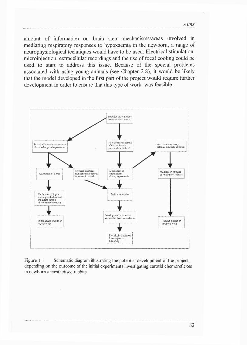
Aims
amount of information on brain stem mechanisms/areas involved in mediating respiratory responses to hypoxaemia in the newborn, a range of neurophysiological techniques would have to be used. Electrical stimulation, microinjection, extracellular recordings and the use of focal cooling could be used to start to address this issue. Because of the special problems associated with using young animals (see Chapter 2.8), it would be likely that the model developed in the first part of the project would require fiirther development in order to ensure that this type of work was feasible.
Establish anaesthetized newborn rabbit model
How does hypoxaemia affect respiratory carotid chetnoreflex?
Are other . reflexes similarly affected?
Reccrd afferent chemoreceptor fibre discharge in hypoxaemia
Increased discharge maintained throughout hypoxaemic period
Modulation of chemoreflex during hypoxaemia
Modulation o f range o f respiratory reflexesAdaptation o f fibres
Further recordings to investigate factors that modulate carotid chemoreceptor output
Drain stem studies
Develop new preparation suitable for brain stem studies
Cellular studies on newborn brain
Intracellular studies on carotid body
Electrical stimulationMicroinjcctionLesioning
Figure 1.1 Schematic diagram illustrating the potential development of the project, depending on the outcome of the initial experiments investigating carotid chemorefiexes in newborn anaesthetised rabbits.
82
Section I: respiratory reflexes
RESULTS: SECTION 1
EFFECTS OF HYPOXIA ON RESPIRATORY REFLEXES
83
Section 1: respiratory reflexes
Se c tio n 1: E f f e c t o f h ypoxia o n r e spir a to r y re f le x e s
Section 1 reports three sets of experiments conducted to investigate how isocapnic hypoxaemia affected a range of respiratory reflexes. The aim of this section was to investigate Anther the idea that BVR was caused by a central inhibitory mechanism that acted to reduce respiratory output during hypoxaemia.
Chapter 2Two sets of experiments are reported in this Chapter. First, carotid chemoreceptor discharge was recorded in newborn anaesthetised rabbits. Second, a carotid chemoreceptor-phrenic nerve reflex was used to assess how isocapnic hypoxaemia affected the carotid chemorefiexes. The results from these studies suggest that a central inhibitory influence inhibits carotid chemorefiexes during isocapnic hypoxaemia. This inhibitory influence acts to reduce breathing during isocapnic hypoxaemia in the newborn.
Chapter 3This Chapter reports the effects of isocapnic hypoxaemia on somatophrenic reflexes. The results from Chapter 3 support the idea that hypoxaemia may not necessarily affect all respiratory reflexes to the same extent. Considered with the findings of chapter 2, tiiis suggests that hypoxaemia does not depress CNS fimction globally.
84
Carotid chemorefiexes
CHAPTER 2
CAROTID RESPIRATORY CHEMOREFIEXES ARE INHIBITED BY A CNS MECHANISM DURING
ISOCAPNIC HYPOXAEMIA
85
Carotid chemorefiexes
2.1 I n tr o d u c tio n
This Chapter presents experiments that investigated the role of the carotid chemoreceptors in mediating BVR during hypoxaemia. This involved two approaches.
First, afferent carotid chemoreceptor discharge was recorded in newborn anaesthetised rabbits. I felt that this was necessary because of the controversy detailed in Chapter 1 over whether carotid chemoreceptor activity falls during acute hypoxaemia, thereby causing or contributing to BVR. Similar experiments had only been conducted in one species, namely the cat. Clearly BVR is a powerful and common response to hypoxia in newborn mammals. If BVR was indeed due solely to the failure of the carotid chemoreceptors to maintain the initial level of discharge in hypoxaemia, carotid chemoreceptor discharge would be expected to decline in all mammals at an age where they exhibit BVR. Therefore it was essential that carotid chemoreceptor recordings should also be made in another species.
Second, the demonstration of sustained carotid chemoreceptor afferent discharge throughout acute hypoxaemia would necessitate studying some central mechanism in causing BVR. In order to test further the hypothesis that some central mechanism caused BVR, I investigated the effect of hypoxaemia on a carotid chemoreflex, using a defined, quantifiable input. The input had to be quantified as accurately as possible under the same conditions as those under which the chemoreflex test would be performed. This approach demanded that the effects on breathing of the chosen input used to stimulate carotid chemoreceptor discharge be measured.
Both these approaches involve using the same afferent input (i.e. carotid chemoreceptor afferents), in addressing the same general question: do peripheral chemoreceptors adapt to hypoxaemia or is their discharge maintained, with phase 2 being caused by a central inhibition of respiratory output and hence peripheral chemorefiexes? For this reason both approaches are presented together in this Chapter.
The problems with the methodology of recording fi*om afferent carotid chemoreceptor fibres have been covered in Chapter 1. The remainder of the introduction to this Chapter presents the background upon which the chemoreflex test used in further experiments is based.
86
Carotid chemorefiexes
Black and Torrance (1967) showed that transient chemical stimulation of the carotid body with COj-equihbrated saline, or electrical stimulation of the carotid sinus nerve, caused specific changes in breathing. The effects on breathing depended on the point in the respiratory cycle at which the stimulus was dehvered. Stimuh dehvered in early inspiration produced an increase in tidal volume, while stimuh apphed in early to mid-expiration produced a prolongation of expiratory time, with no effect on the depth of that expiration. Furthermore, these effects on breathing were consistently reproducible (Black & Torrance, 1967; Black, McCloskey & Torrance, 1971; Eldridge, 1972a,b; Black, Goodman, Nail, Rao & Torrance, 1973). Thus transient stimulation of the carotid chemoreceptors produces a profound perturbation in the respiratory rhythm. The neuronal mechanisms underlying the generation of respiratory output have been reviewed extensively (Richter, 1982; Long & Puffin, 1986; Richter, Ballantyne & Remmers, 1986). In addition, recent work shows that respiratory rhythm may originate from pacemaker neurones located in the pre-Botzinger complex (Smith, Ellenberger, Ballanyi, Richter & Feldman, 1991).
The clearly defined "input-output" relationship between the carotid chemoreceptors and the pattern of breathing thus provides a pathway by which to test the integration of chemorefiexes in the brain stem during hypoxaemia. Therefore, I investigated whether hypoxaemia would alter the effect of this reproducible chemoreceptor mediated reflex on central respiratory output.
2.2 H y po t h e se s
(1) Afferent carotid chemoreceptor discharge is stimulated but does not decline during acute hypoxaemia in anaesthetized newborn rabbits.
(2) The effect of transient carotid chemoreceptor stimulation on breathing is modulated during hypoxaemia by a central mechanism that inhibits respiratory output in anaesthetized newborn rabbits.
87
Carotid chemorefiexes
2.3 M e t h o d s
The following procedures were conducted in accordance with the Animals (Scientific Procedures) Act 1986 (HMSO, 1986).
Animal delivery and care.Newborn New Zealand White rabbits (bred and supphed by Froxfield UK. Ltd.) were dehvered to Biological Services, University College London, on, or one day before, the experiment and housed in accordance with the Code of Practice for the Housing and Care of Animals used in Scientific Procedures (HMSO, 1986). Animals that were dehvered on the day before an experiment were injected s.c. with the broad spectrum antibiotic Bayotril (0.3 ml; Bayer, UK) to counter intestinal bacterial infection, such as mucoid enteropathy. This condition is one of the major causes of mortaht> in young rabbits.
Ages and weights: Mean age 28 days; range 26-31 days;Weight 670±19g (meantS.E.M.)
Sedation and anaesthesia.The rabbits were sedated with Ketalar (25 mg kg“i , ketamine hydrochloride; Parke-Davis, UK). Anaesthesia was induced with halothane (May & Baker Ltd., UK) 2-2.5% dehvered through a facemask (Arnolds, UK), via a Fluotec 3 halothane dispenser, in 4 1/min air (controUed using Platon Flowbits UK rotameters). Once surgery was complete, urethane (Sigma, UK; 1.3-1.6g/kg; 20% w/v in physiological saline solution) was administered via the lefl; femoral vein (iv cannula size 3FG, Portex UK). At this point, halothane anaesthesia was discontinued. The patency of ah catheters was maintained by regular flushes with heparin saline (Multiparin, 1 active unit/ml saline).
Monitoring anaesthesiaThe depth of anaesthesia was monitored by judging each of the fohowing criteria:-1) Arterial blood pressure
An increase in arterial blood pressure, on pinching the hind limb, was used as indicator for the administration of supplemental anaesthesia. The patency of all catheters was maintained with heparin saline (Multiparin, 1 active unit/ml saline).
2) Heart rateIn most preparations, electrocardiogram (ECG) electrodes were sewn through the thoracic skin either side of the heart, with a reference
88
Carotid chemorefiexes
electrode clipped onto skin of a hind leg. An increase in heart rate on pinching a hind limb was used as an indication for the administration of supplemental anaesthesia.
3) Phrenic nerve activityA rapid increase in phrenic nerve discharge on pinching a hind limb, during steady-state pH/blood gas status, was also used as an indicator of depth of anaesthesia.
4) Hind limb withdrawal reflexesLack of hind limb withdrawal reflex (in the non-paralysed anaesthetised state) indicated satisfactory anaesthesia.
Ventilation.The neck and left femoral region were shaved (in preparation for tracheotomy and femoral vessel cannulation. Once surgical anaesthesia was achieved, a tracheotomy was performed. An oral/nasal tube (Portex UK (size 2.5)) was introduced into the trachea and secured with braided silk suture (Pearsalls Sutures; size US 2/0). The upper airway was tied off vyith tightly secured silk suture. The rabbit was ventilated using a small animal ventilator (Harvard Apparatus 707 ventilator or Amsterdam Small Infant ventilator). Rabbits breathing from a free-ftow supply were assisted in ventilation, using the pressure controlled, small animal ventilator (Harvard Apparatus UK, model 707). This ventilator senses the pressure change when the animal initiates a breath and triggers the breathing cycle in a pressure controlled manner. Full details are given in Appendix 6.
Monitoring o f pH, blood gases and body temperature.Body temperature and blood gas status were carefully controlled to be within the normal physiological range throughout surgery and experimental period. Body temperature was servo-controlled at 38®C throughout the preparation, by means of a temperature probe inserted into the rectum and homeothermic blanket (CFP 8185, UK). 38°C is the temperature at which minimal oxygen consumption occurs in the newborn rabbit (Blatteis, 1964). Aortic arterial blood was obtained via a three way tap situated between a catheter introduced into the femoral artery and the blood pressure transducer (secured by luer lock connections). Post-mortem studies indicated the length (ca. 10cm) of catheter required to sample aortic blood. Blood gas and pH status was analysed by anaerobic removal of 0.2 ml blood using Instrumentation Laboratory pH/blood gas analysers (1L1302 or 1306 series, UK). Sample values were corrected for body temperature. Sodium bicarbonate (8.4% w/v in Ringer’s saline; Sigma, UK) was administered if metabolic acidosis occurred. In some experiments, Petcoj and halothane levels were monitored (Ohmeda 5250 respiratory monitor).
89
Carotid chemorefiexes
Carotid chemoreceptor stimulation.The carotid bifurcation was exposed on both sides of the neck. Each external carotid artery was cannulated (size 2FG catheter; Portex UK), positioned just above the carotid bifurcation to ensure that the carotid body was exposed to the desired experimental solution. The catheters were firmly secured with suture (Pearsalls Sutures; size US 5/0) and connected via a three way stop-cock and luer lock connector to a common injection catheter (plate 2.1).
The following solutions were injected manually:-CO2 - saturated saline: Saturated COi-saline was produced by bubbling CO2 (Medical Grade, British Oxygen Company, UK) through saline in a flask. The equihbrated solution was withdrawn into a glass syringe which served as the storage syringe. PCO2 was checked regularly in all experiments. Sodium dithionite (Sigma, UK): 0.25M in Ringer’s saline bubbled with N 2
Lobeline: 20pg/0.2ml Ringer’s saline (Sigma, UK)Tris buffer: 5.72g/l Tris HCl:1.66g/l Tris base (Sigma, UK); pH 7.4, 37°C.
90
Carotid chemorefiexes
i
Plate 2.1 Photograph showing arrangement of catheters for injection of CO2- saturated saline to stimulate carotid body chemoreceptors. The large glass syringe served as a reservoir for for COi-saturated saline, with the smaller (1ml) syringe used as the injector. The three-way tap enabled either uni- or bilateral chemical stimulation of the carotid chemoreceptors. Injections were registered by the pressure transducer, shown to the right of the storage syringe.
/
Plate 2.2 Photograph showing recording electrode used for both carotid chemoreceptor fibre and phrenic nerve recordings. The electrode comprises a Neurolog headstage (NLIOOAK), with stainless steel recording hooks, and an earthing lead.
91
Carotid chemoreflexes
2.4 E x pe r im e n t a l pr o t o c o l s .
Recording multifibre carotid chemoreceptor afferent activity (n=8 rabbits) All rabbits were paralysed (gallamine triethiodide, 8mg/kg), vagotomized and held supine in a stereotaxic frame. Multifibre carotid chemoreceptor activity was recorded using a pair of stainless steel hook electrodes (plate2.2). Chemoreceptor fibres from the cut left carotid sinus nerve were placed on one hook, with the other being earthed to the preparation. The carotid sinus nerve was sectioned at its junction with the glossopharyngeal nerve. The identity o f the nerve was confirmed after the end of the experiment by tracing it back to the carotid body. The sheath of the carotid sinus nerve was removed and the CSN was spht into filaments on a blackened steel plate, which acted as an earth. The fibre picking was performed by Professor Mark Hanson.
Chemoreceptor preparations were identified by their random discharge and response to COi-saturated saline. Baroreceptor activity was identified by the presence of cardiac rhythm. The nerve was bathed in mineral oil (Fisons UK (SG 0.830-0.860) or Sigma, UK). COi-saturated saline (0.1-0.3ml bolus) was injected retrogradely down one external carotid artery in order to stimulate the carotid body chemoreceptors, once every 60 seconds. These injections were performed in normoxia, for at least 8 minutes of hypoxaemia and during subsequent return to normoxia.
Carotid chemoreceptor - phrenic chemoreflex (n = ll rabbits)In 11 other rabbits, central respiratory output was recorded from the cut end of the right phrenic nerve, which was chssected from its C3 root to just above the sternal region. The dissection of this region was performed so as to make a deep pool in which the phrenic nerve could be bathed in paraffin hquid (SG 0.830-0.860; Fisons, UK) or mineral oil (Sigma, UK) to prevent desiccation. Every rabbit was paralysed (gallamine triethiodide, 8mg/kg), vagotomized and secured supine in a stereotaxic frame. The effect of transient carotid chemoreceptor stimulation on respiratory output was assessed by injecting C02-saturated saline (0.1-0.3ml bolus) retrogradely down both external carotid arteries in order to stimulate the carotid bodies, once every 60 seconds. These injections were performed in normoxia, up to 8 minutes of hypoxaemia and during subsequent return to normoxia.
92
Carotid chemoreflexes
Other preliminary carotid chemoreflex experimentsDuring the course of the project, the carotid chemoreflex hypothesis wasalso tested in two different types of preparation. Other peripheralchemostimulants were also used in the anaesthetized rabbit preparation, toinvestigate whether such agents ehcited similar responses to COi-saturatedsaline.
Anaesthetized rabbits - other chemostimulants (n=3 rabbits)A similar protocol for the carotid chemoreflex experiment was followed for three anaesthetized rabbits where the effects of sodium dithionite (n=2 rabbits) and lobeline (n=l rabbit) on the carotid chemoreflex were investigated.
Decerebrate rabbit (n=l rabbit)The carotid chemoreflex protocol was also conducted in 1 decerebrate rabbit. The methodology for the preparation is presented and discussed in Chapter 6.
Lambs (n=5)The carotid chemoreflex protocol was also undertaken in 5 lambs. Three of these lambs were anaesthetized with a-chloralose (70mg/kg i.v.. Sigma, UK), while the remainder were decerebrate. Full methods are detailed in Chapter 3.
93
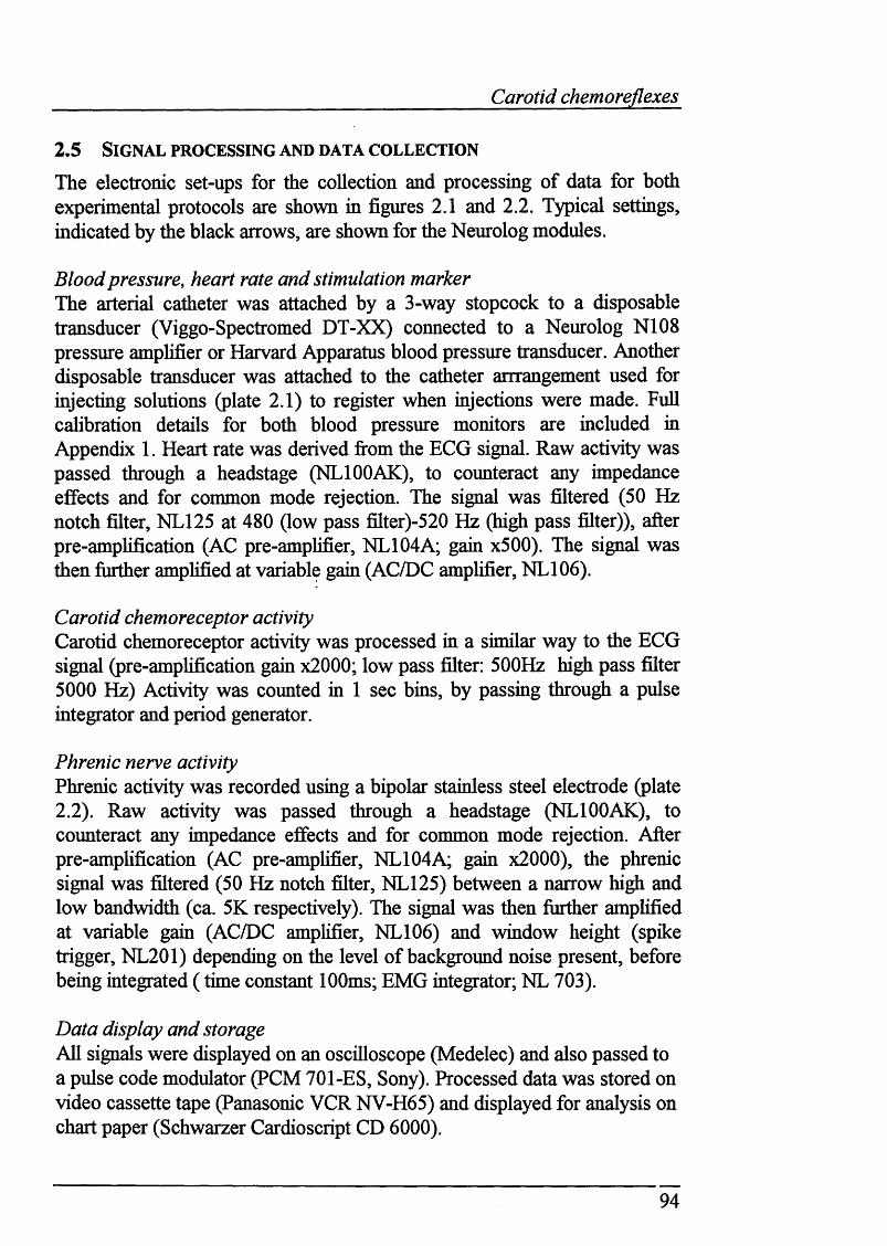
Carotid chemoreflexes
2 .5 Sig n a l pr o c e ssin g a n d d a ta c o l l e c t io n
The electronic set-ups for the collection and processing of data for both experimental protocols are shown in figures 2.1 and 2.2. Typical settings, indicated by the black arrows, are shown for the Neurolog modules.
Blood pressure, heart rate and stimulation marker The arterial catheter was attached by a 3-way stopcock to a disposable transducer (Viggo-Spectromed DT-XX) connected to a Neurolog N108 pressure amplifier or Harvard Apparatus blood pressure transducer. Another disposable transducer was attached to the catheter arrrangement used for injecting solutions (plate 2.1) to register when injections were made. Full cahbration details for both blood pressure monitors are included in Appendix 1. Heart rate was derived from the ECG signal. Raw activity was passed through a headstage (NLIOOAK), to counteract any impedance effects and for common mode rejection. The signal was filtered (50 Hz notch filter, NL125 at 480 (low pass filter)-520 Hz (high pass filter)), after pre-amplification (AC pre-amplifier, NL104A; gain x500). The signal was then further amplified at variable gain (AC/DC amplifier, NL106).
Carotid chemoreceptor activityCarotid chemoreceptor activity was processed in a similar way to the ECG signal (pre-amplification gain x2000; low pass filter: 500Hz high pass filter 5000 Hz) Activity was counted in 1 sec bins, by passing through a pulse integrator and period generator.
Phrenic nerve activityPhrenic activity was recorded using a bipolar stainless steel electrode (plate2.2). Raw activity was passed through a headstage (NLIOOAK), to counteract any impedance effects and for common mode rejection. After pre-amplification (AC pre-amphfier, NL104A; gain x2000), the phrenic signal was filtered (50 Hz notch filter, NL125) between a narrow high and low bandwidth (ca. 5K respectively). The signal was then further amplified at variable gain (AC/DC amplifier, NL106) and Avindow height (spike trigger, NL201) depending on die level of background noise present, before being integrated ( time constant 100ms; EMG integrator; NL 703).
Data display and storageAll signals were displayed on an oscilloscope (Medelec) and also passed to a pulse code modulator (PCM 701-ES, Sony). Processed data was stored on video cassette tape (Panasonic VCR NV-H65) and displayed for analysis on chart paper (Schwarzer Cardioscript CD 6000).
94
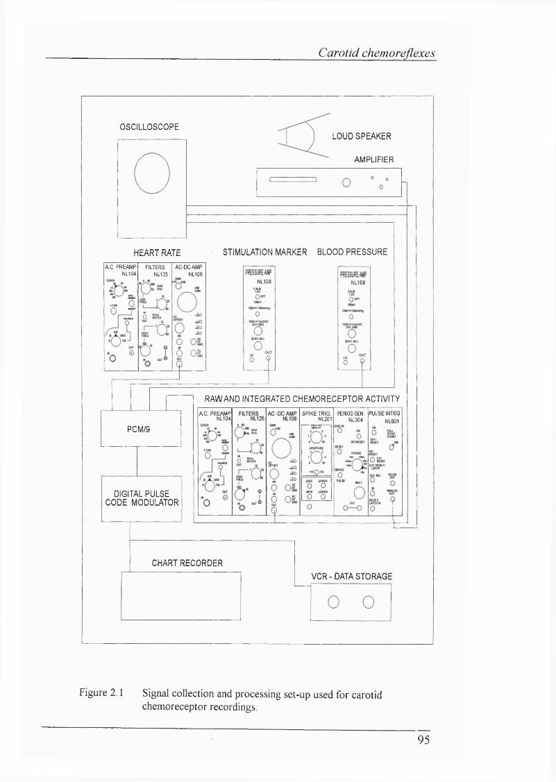
Carotid chemoreflexes
OSCILLOSCOPELOUD SPEAKER
AMPLIFIER
' STIMULATION MARKER BLOOD PRESSUREHEART RATE
A.C. PREAMP NL104
FILTERSNL125
AC-DCAMPNL106
NL108 NL108
6
O 0&
RAW AND INTEGRATED CHEMORECEPTOR ACTIVITY
PULSE INTEG.
NL601
A C . PREAMP NL104
SPIKE TRIG. NL201^ —
PERIOD GEN. NL304
PCM/9
o
DIGITAL PULSE CODE MODULATOR
CHART RECORDER
VCR-DATA STORAGE
Figure 2.1 Signal collection and processing set-up used for carotid chemoreceptor recordings.
95

Carotid chemoreflexes
OSCILLOSCOPE
LOUD SPEAKER
RAW AND INTEGRATED PHRENIC ACTIVITYHEART RATEEMGINTEGRATOR
NL703
SPIKE TRIG. NL201
A C . PREAMP NL104
AC-DCAMPNL106
A C . PREAMP NL104
A C-D CA M PNL106
FILTERSNL125
..So«0.so.,S0
..so«0.so.so
ornti
STIMULATION MARKER BLOOD PRESSUREPCM/9
NL108PRESSURE AMP
NL108
DIGITAL PULSE CODE MODULATOR
CHART RECORDER
VCR-DATA STORAGE
Figure 2.2 Signal collection and processing set-up for
carotid chemoreflex experiments.
96
Carotid chemoreflexes
2.6 A n a ly s is
Cardiovascular parametersArterial blood pressure and instantaneous heart rate were analysed at one minute intervals throughout the normoxaemic, hypoxaemic and recovery periods. Arterial blood pressure was expressed as mean arterial pressure (diastohc pressure + 1/3 (systohc pressure - diastohc pressure)). Heart rate was derived from the R-R interval as measured from the electrocardiograph record and expressed in beats per minute. Both ABP and HR were analyzed by comparing normoxaemic mean values with values at 5 min of hypoxaemia. Statistical analysis was performed using Student's paired t-test, vrith data presented as meantS.E.M.. P<0.05 was considered significant.
Recordings from carotid chemoreceptor multifibres Integrated chemoreceptor activity was analysed over the five seconds preceding an injection. The effect of an injection on chemoreceptor discharge was measured over the period of deflection o f the pressure marker. Carotid chemoreceptor activity at 1 min before the onset of isocapnic hypoxaemia was normalized to 100%. Carotid chemoreceptor activity at 1 min intervals during isocapnic hypoxaemia was expressed as a change relative to the pre-hypoxaemic control. The group data was compiled in this way to assess the relative changes in carotid chemoreceptor discharge, rather than absolute values. Statistical analysis was performed using Wilcoxon's signed rank test, due to non-uniform distribution of data. Thus data is presented as median and 95% confidence limits or interquartile range (IQR: 25th and 75th percentile ranks). P<0.05 was considered significant.
Carotid chemoreflex test - respiratory parameters.The analysis of fhe integrated phrenic neurogram is illustrated in figure 2.3. Peak phrenic activity (PPA), breath duration (T t ) and inspiratory time (Ti) were measured from the phrenic neurogram.
Since the absolute electrical activity measured from the phrenic nerve is dependent on the size of the nerve, integrity of its fibres and contact with the recording electrode, comparisons between animals are not appropriate unless this data is normahsed. To normalise the data, a value of 100 arbitrary units was assigned to the highest level of phrenic activity observed in an animal, with the remainder of the data being scaled accordingly.
97
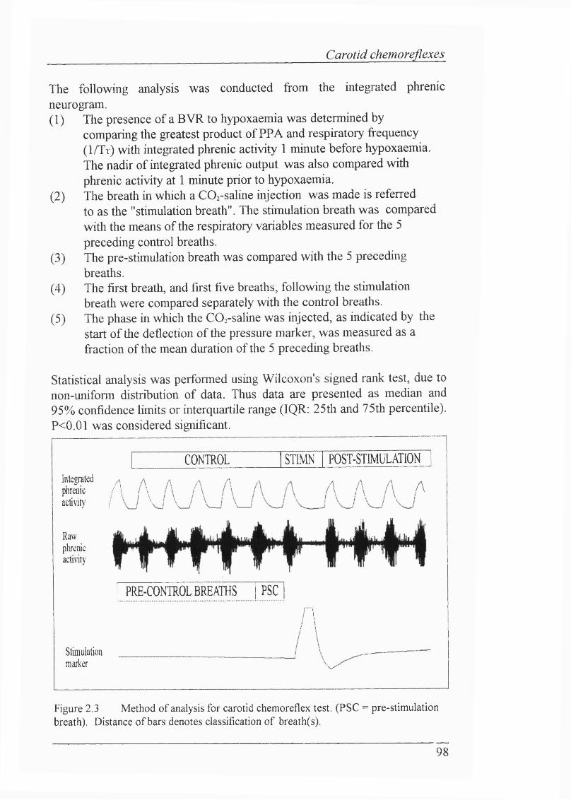
Carotid chemoreflexes
The following analysis was conducted from the integrated phrenicneurogram.(1) The presence of a BVR to hypoxaemia was determined by
comparing the greatest product of PPA and respiratory frequency ( 1 / T t ) with integrated phrenic activity 1 minute before hypoxaemia. The nadir of integrated phrenic output was also compared with phrenic activity at 1 minute prior to hypoxaemia.
(2) The breath in which a COz-saline injection was made is referredto as the "stimulation breath". The stimulation breath was compared with the means of the respiratory variables measured for the 5 preceding control breaths.
(3) The pre-stimulation breath was compared with the 5 preceding breaths.
(4) The first breath, and first five breaths, following the stimulation breath were compared separately with the control breaths.
(5) The phase in which the CO^-saline was injected, as indicated by the start of the deflection of the pressure marker, was measured as a fraction of the mean duration of the 5 preceding breaths.
Statistical analysis was performed using Wilcoxon's signed rank test, due tonon-uniform distribution of data. Thus data are presented as median and95% confidence limits or interquartile range (IQR: 25th and 75th percentile).P<0.01 was considered significant.
CONTROL STIMN POST-STMULATIONIntegratedphrenic / \ \ \
activit)' / ^
Rawplirenicactivity
PRE-CONTROL BREATHS PSC
n/ 1
Stimulationmarker
Figure 2.3 Method of analysis for carotid chemoreflex test. (PSC = pre-stimulationbreath). Distance of bars denotes classification of breath(s).
98
Carotid chemoreflexes
2 .7 R e su l t s
Multifibre carotid chemoreceptor recordings.Blood gas/pH status and cardiovascular parameters.The group blood gas/pH status and blood pressure/heart rate for carotid chemoreceptor recording experiments during normoxaemia, hypoxaemia and recovery are shown in table 2.1. Superscripts refer to P values obtained from Student’s paired t-test, comparing normoxaemia with hypoxaemia or recovery. NS denotes non-significance. All values are expressed as meant S.E.M..
NORMOXAEMIA HYPOXAEMIA RECOVERY
pHa 7.30±0.05 7.25±0.03 NS 7.24±0.06 NS
Pacoj (mmHg) 41.6±1.7 43.6±2.2 NS 47±4.1 NS
Paoj (mniHg) 102±9 35±3 ooi 88±10 NS
MAP (mmHg) 59.6±3.9 56.4±3.7NS 58.5±3.0 NS
Heart rate (bpm) 319±12 307±14 NS 304±10 NS
Table 2.1. Mean blood gas, pH, mean arterial blood pressure and heart rate for carotid chemoreceptor recording experiments (n=8 rabbits).
Carotid chemoreceptor fibres do not adapt to acute isocapnic hypoxaemia During isocapnic hypoxaemia, carotid chemoreceptor discharge increased (P=0.008; control vs. activity at 2 min; Wilcoxon’s signed rank test) and was sustained throughout the 8 minutes of hypoxaemia (figure 2.4: activity at 2 minutes of hypoxaemia vs. activity at 6 minutes of hypoxaemia; P=0.22; Wilcoxon signed rank test). Individual results are shown in Appendix 4.
200 T
180 --
160 --
140 --
120 -
100 - ■
80 --
40 --
20 - -
ime (min)
Figure 2.4 Carotid chemoreceptor discharge (expressed as median/IQR) increased and remained elevated throughout isocapnic hypoxaemia (n=8 rabbits).
99
Carotid chemo reflexes
CO2 boll increase carotid chemoreceptor discharge during normoxaemia. The Pco2 attained by bubbling CO2 through saline was typically >300mmHg (pH 5.5) as measured using a blood gas analyser (IL 1302 or 1306 series). Saline Pco did not decline during the course of the experiment. Indeed, Pcoz remained at these levels 30 minutes after bubbling the CO2 through saline.
C02-saturated saline injections produced an intense, transient increase in carotid chemoreceptor fibre discharge (Fig 2.5) during normoxaemia (P=0.004; Wilcoxon's signed rank test). The group results are shown in figure 2.6. The median increase in discharge due to C02-saturated saline injection over baseline chemoreceptor activity was 175% (interquartile range 153-336% increase). Baseline carotid chemoreceptor activity ranged from <1-5 spikes/s.
The latency from the onset of the injection, indicated by the pressure marker trace, to the initial increase in chemoreceptor activity was typically in the range of 200-400ms. Control injections of Ringer's saline solution or Tris buffered to pH 7.35 produced little or no significant effect (figure 2.7). In a few preparations baroreceptor effects were observed.
Integratedcarotidchemoreceptordischarge(spikes/s)
Raw carotidchemoreceptordischarge
Stiiniilationmarker n n
1 second
Figure 2.5 CO2 -saturated saline bolus increased carotid chemoreceptor discharge during normoxaemia. After 10 minutes of isocapnic hypoxaemia, there was a similar increase in carotid chemoreceptor discharge on CO2 -saturated saline bolus injection. Also note increase in baseline carotid chemoreceptor discharge
100
Carotid chemoreflexes
! 1
IT3CTjU
Ic3
500
450
4 00
350
300
2 50
200
150
1 00
50
0
P = 0 . 0 3 9
P < 0 . 0 5
N S
C o n t r o l S t i m u l a t i o n
N O R M O X I A
1—C o n t r o l S t i m u l a t i o n
H Y P O X I A
Figure 2.6 Group results (median and IQR) showing of effect of COz-saturated saline on multifibre carotid chemoreceptor discharge during normoxaemia and isocapnic hypoxaemia. P values refer to comparisons made between normoxic stimulation, and hypoxic control or hypoxic stimulation (Wilcoxon’s signed rank test).
Integrated
chemoreceptor discharge
Stimulationmarker
AnÂrJri.
Rawcarotidchemoreceptordischarge
Figure 2.7 Effect of Ringer’s saline (pH 7.38) on multifibre carotid chemoreceptordischarge during normoxaemia.
101
Carotid chemoreflexes
CO2 injections during isocapnic hypoxaemia also increased carotid chemoreceptor dischargeC02-saturated saline injections again produced an intense, transient increase in carotid chemoreceptor fibre discharge (figure 2.5) during isocapnic hypoxaemia (P=0.004; Wilcoxon's signed rank test). The median increase in discharge due to COi-saturated saline injection over baseline chemoreceptor activity was 150% (interquartile range 132-214% increase). Increases in carotid chemoreceptor fibre activity due to COz-saturated saline occurred throughout the hypoxaemic period. However, the increase in carotid chemoreceptor discharge produced by C02-saturated saline did not always reach the same level attained in normoxaemia: this occurred in 66% of experiments. Rather C02-saturated saline caused an increase in carotid chemoreceptor discharge that exceeded that seen for injections made in normoxaemia. Overall, however, the increase in carotid chemoreceptor discharge was greater in isocapnic hypoxaemia (P=0.039; Wilcoxon's signed rank test). Also, the increase in carotid chemoreceptor discharge produced by C02-saturated saline during normoxia was not different to the hypoxaemic control (P=0.24; Wilcoxon’s signed rank test).
Figures 2.8-11 show the changes in baseline discharge, and the effects of CO2 boli during normoxaemia and hypoxaemia in one rabbit where CO2
saline injections produced an increase in carotid chemoreceptor discharge that exceeded that seen for injections made in normoxaemia.
Raw carotidchemoreceptordischarge
3 0 1Integrated carotid chemoreceptor discharge (grikes/s) 0
Stimulationmarker
Arterialbloodpressure(nmHg)
70,
5 0 1
•11 I
1 second
Figure 2.8 C02-saturated saline injections increased carotid chemoreceptor discharge 1 min before the onset of isocapnic hypoxaemia.
102
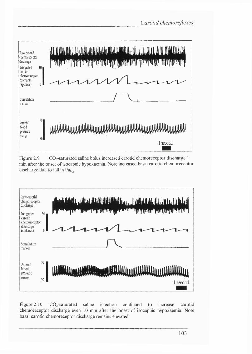
Carotid chemoreflexes
Raw carotid chemoreceptor dischargeIntegrated 3 0 1
carotidchemoreceptor (hscharge (spikes/s) 0
Stiiiiulationmariner
Arterialbloodpressure(ftwHg)
70
501 second
Figure 2.9 COa-saturated saline bolus increased carotid chemoreceptor discharge 1 min after the onset of isocapnic hypoxaemia. Note increased basal carotid chemoreceptor discharge due to fall in Pao2 -
Raw carotidchemoreceptordischarge
3 0 1In t^ a te d carotid chemorecepto- discharge (spikes/s) 0
Stimulationmarker
ArterialbloodpressureWHg)
70
501 second
Figure 2.10 C02-saturated saline injection continued to increase carotid chemoreceptor discharge even 10 min after the onset of isocapnic hypoxaemia. Note basal carotid chemoreceptor discharge remains elevated.
103
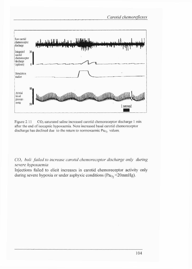
Carotid chemoreflexes
Raw carotid ckraoreceptor discharge
Integrated 3 0 1
carotidchemoreceptor discharge (spikes/s) 0
Stimulationmarker
Arterialbloodpressure m m
1 second
Figure 2.11 CO 2 -saturated saline increased carotid chem oreceptor discharge 1 min after the end o f isocapnic hypoxaemia. N ote increased basal carotid chem oreceptor discharge has declined due to the return to normoxaemic Pao2 values.
CO2 boli failed to increase carotid chemoreceptor discharge only during severe hypoxaemiaInjections failed to elicit increases in carotid chemoreceptor activity only during severe hypoxia or under asphyxie conditions (Pao2 <20mmHg).
104
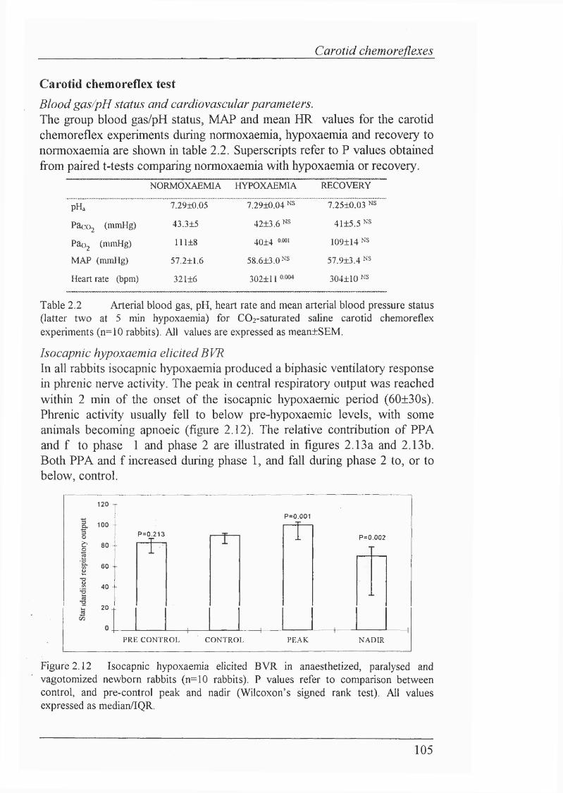
Carotid chemoreflexes
Carotid chemoreflex testBlood gas/pH status and cardiovascular parameters.The group blood gas/pH status, MAP and mean HR values for the carotid chemoreflex experiments during normoxaemia, hypoxaemia and recovery to normoxaemia are shown in table 2.2. Superscripts refer to P values obtained from paired t-tests comparing normoxaemia with hypoxaemia or recovery.
NORMOXAEMIA HYPOXAEMIA RECOVERY
pHa 7.29±0.05 7.29±0.04 NS 7.25±0.03 NS
Paco2 (mmHg) 43.3±5 42±3.6 NS 4I±5.5 NS
Pao2 (mmHg) 111±8 40±4 109±I4 NS
MAP (mmHg) 57.2±1.6 58.6±3.QNS 57.9±3.4 NS
Heart rate (bpm) 32I±6 302±11 304±I0 NS
Table 2.2 Arterial blood gas, pH, heart rate and mean arterial blood pressure status (latter two at 5 min hypoxaemia) for COz-saturated saline carotid chemoreflex experiments (n=10 rabbits). All values are expressed as mean±SEM.
Isocapnic hypoxaemia elicitedBVRIn all rabbits isocapnic hypoxaemia produced a biphasic ventilatory response in phrenic nerve activity. The peak in central respiratory output was reached within 2 min of the onset of the isocapnic hypoxaemic period (60±30s). Phrenic activity usually fell to below pre-hypoxaemic levels, with some animals becoming apnoeic (figure 2.12). The relative contribution of PPA and f to phase 1 and phase 2 are illustrated in figures 2.13a and 2.13b. Both PPA and f increased during phase 1, and fall during phase 2 to, or to below, control.
II
120
100 - -
80 -
60
40
20
P=0.001
P=0.213
Î
T P=0.002
PRE C ONT R OL CONTR OL PEAK NADI R
Figure 2.12 Isocapnic hypoxaemia elicited BVR in anaesthetized, paralysed and vagotomized newborn rabbits (n=10 rabbits). P values refer to comparison between control, and pre-control peak and nadir (Wilcoxon’s signed rank test). All values expressed as median/lQR.
105
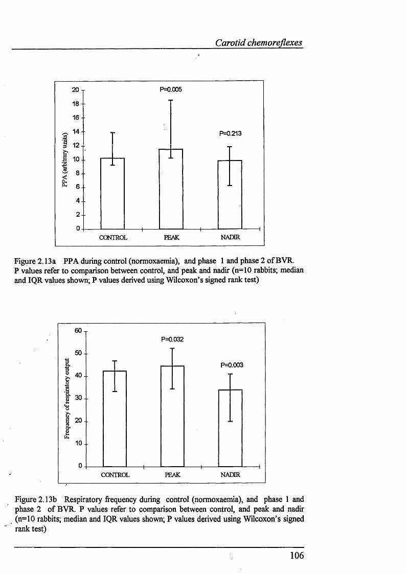
Carotid chemoreflexes
20 - r
18
1 6 -
1 14..I 12 +
S1 0 - -
8- 6 -
4 -
2 -
0
P=0.005
P=0.213
CONTROL PEAK NADIR
Figure 2.13a PPA during control (normoxaemia), and phase 1 and phase 2 of BVR.P values refer to comparison between control, and peak and nadir (n=10 rabbits; median and IQR values shown; P values derived using Wilcoxon's signed rank test)
6 0 -r
50 -
P=0.032
I
I 20I
1 0 -
P=0.003
CONTROL PEAK NADIR
Figure 2.13b Respiratory frequency during control (normoxaemia), and phase 1 and phase 2 of BVR. P values refer to comparison between control, and peak and nadir (n=10 rabbits; median and IQR values shown; P values derived using Wilcoxon’s signed rank test)
106
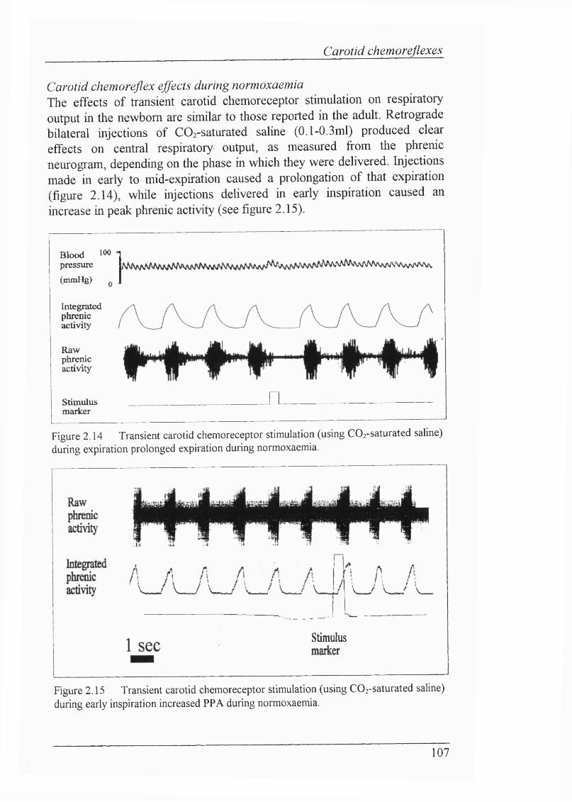
Carotid chemoreflexes
Carotid chemoreflex effects during normoxaemiaThe effects of transient carotid chemoreceptor stimulation on respiratory output in the newborn are similar to those reported in the adult. Retrograde bilateral injections of COi-saturated saline (0.1-0.3ml) produced clear effects on central respiratory output, as measured from the phrenic neurogram, depending on the phase in which they were delivered. Injections made in early to mid-expiration caused a prolongation of that expiration (figure 2.14), while injections delivered in early inspiration caused an increase in peak phrenic activity (see figure 2.15).
Blood 100 " pressure
(mmHg) 0
Integratedphrenicactivity
Rawphrenicactivity
Stimulusmarker
nFigure 2.14 Transient carotid chemoreceptor stimulation (using COz-saturated saline) during expiration prolonged expiration during normoxaemia.
Stimulus
Figure 2.15 Transient carotid chemoreceptor stimulation (using COz-saturated saline) during early inspiration increased PPA during normoxaemia.
107
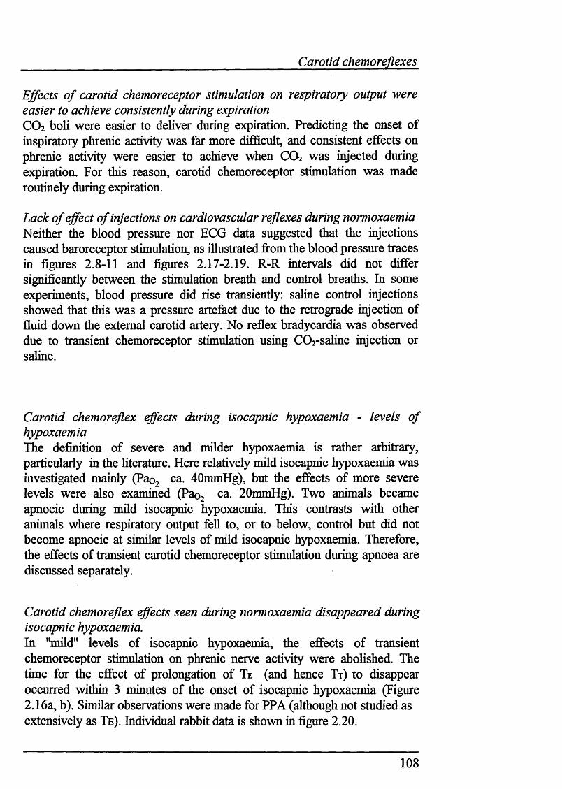
Carotid chemoreflexes
Effects o f carotid chemoreceptor stimulation on respiratory output were easier to achieve consistently during expirationCO2 boli were easier to deliver during expiration. Predicting the onset of inspiratory phrenic activity was far more difficult, and consistent effects on phrenic activity were easier to achieve when CO2 was injected during expiration. For this reason, carotid chemoreceptor stimulation was made routinely during expiration.
Lack o f effect o f injections on cardiovascular reflexes during normoxaemia Neither the blood pressure nor ECG data suggested that the injections caused baroreceptor stimulation, as illustrated from the blood pressure traces in figures 2.8-11 and figures 2.17-2.19. R-R intervals did not differ significantly between the stimulation breath and control breaths. In some experiments, blood pressure did rise transiently: saline control injections showed that this was a pressure artefact due to the retrograde injection of fluid down the external carotid artery. No reflex bradycardia was observed due to transient chemoreceptor stimulation using C02-saline injection or saline.
Carotid chemoreflex effects during isocapnic hypoxaemia - levels of hypoxaemiaThe definition of severe and milder hypoxaemia is rather arbitrary, particularly in the hterature. Here relatively mild isocapnic hypoxaemia was investigated mainly (Pao2 ca. 40mmHg), but the effects of more severe levels were also examined (Pao ca. 20mmHg). Two animals became apnoeic during mild isocapnic hypoxaemia. This contrasts with other animals where respiratory output fell to, or to below, control but did not become apnoeic at similar levels of mild isocapnic hypoxaemia. Therefore, the effects of transient carotid chemoreceptor stimulation during apnoea are discussed separately.
Carotid chemoreflex effects seen during normoxaemia disappeared during isocapnic hypoxaemia.In "mild" levels of isocapnic hypoxaemia, the effects o f transient chemoreceptor stimulation on phrenic nerve activity were abohshed. The time for the effect of prolongation of Te (and hence T t) to disappear occurred within 3 minutes of the onset of isocapnic hypoxaemia (Figure 2.16a, b). Similar observations were made for PPA (although not studied as extensively as Te). Individual rabbit data is shown in figure 2.20.
108
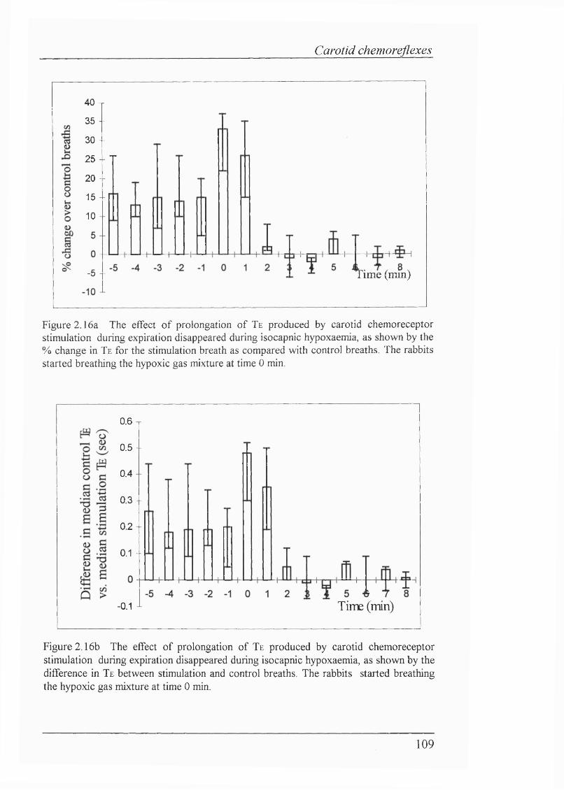
Carotid chemoreflexes
40 T35 -
30 -
25 - T20 -
15 -
10 -
Time (mm)-5 -
Figure 2.16a The effect of prolongation of Te produced by carotid chemoreceptor stimulation during expiration disappeared during isocapnic hypoxaemia, as shown by the % change in Te for the stimulation breath as compared with control breaths. The rabbits started breathing the hypoxic gas mixture at time 0 min.
0.6 T
0.5-
0.4
0.3 -
0.2 -
0.1 -
Time (min)- 0.1 —
Figure 2.16b The effect of prolongation of Te produced by carotid chemoreceptor stimulation during expiration disappeared during isocapnic hypoxaemia, as shown by the difference in Te between stimulation and control breaths. The rabbits started breathing the hypoxic gas mixture at time 0 min.
109
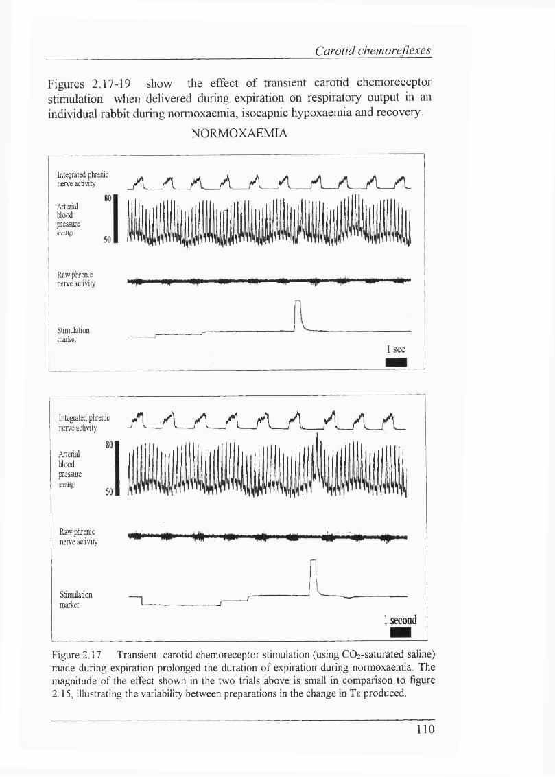
Carotid chemoreflexes
Figures 2.17-19 show the effect of transient carotid chemoreceptor stimulation when delivered during expiration on respiratory output in an individual rabbit during normoxaemia, isocapnic hypoxaemia and recovery.
NORMOXAEMIA
Integrated phrenic . _ ^ i A >nerve activity fA / I / V jA / I
Arterialbloodpressure(pinHg)
Raw phrenic nerve activity
Stimulationmarker
1 sec
a f " ' A_yi_/L_/i_/Lyi_yLA_A_nerve activity
Arterial blood pressure(pimHg)
Raw phrenic nerve activity
Stimulationmarker
50 P
1 second
Figure 2.17 Transient carotid chemoreceptor stimulation (using COz-saturated saline) made during expiration prolonged the duration o f expiration during normoxaemia. The magnitude o f the effect shown in the two trials above is small in comparison to figure 2.15, illustrating the variability between preparations in the change in Te produced.
110
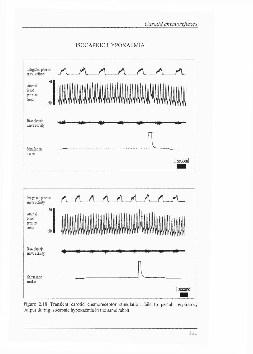
Carotid chemoreflexes
ISOCAPNIC HYPOXAEMIA
Integrated phrenic ^ nerve activity - T L
Arterialbloodpressure(jraniHg)
Raw phrenic nerve activity
Stimulationmarker
80
50
1 second
Integrated phrenic nerve activity
80Arterialbloodpressure(mmHg)
50
Raw phrenic nerve activity
Stimulationmarker
/ L _
III
1 second
Figure 2.18 Transient carotid chemoreceptor stimulation fails to pertub respiratory output during isocapnic hypoxaemia in the same rabbit.
I l l
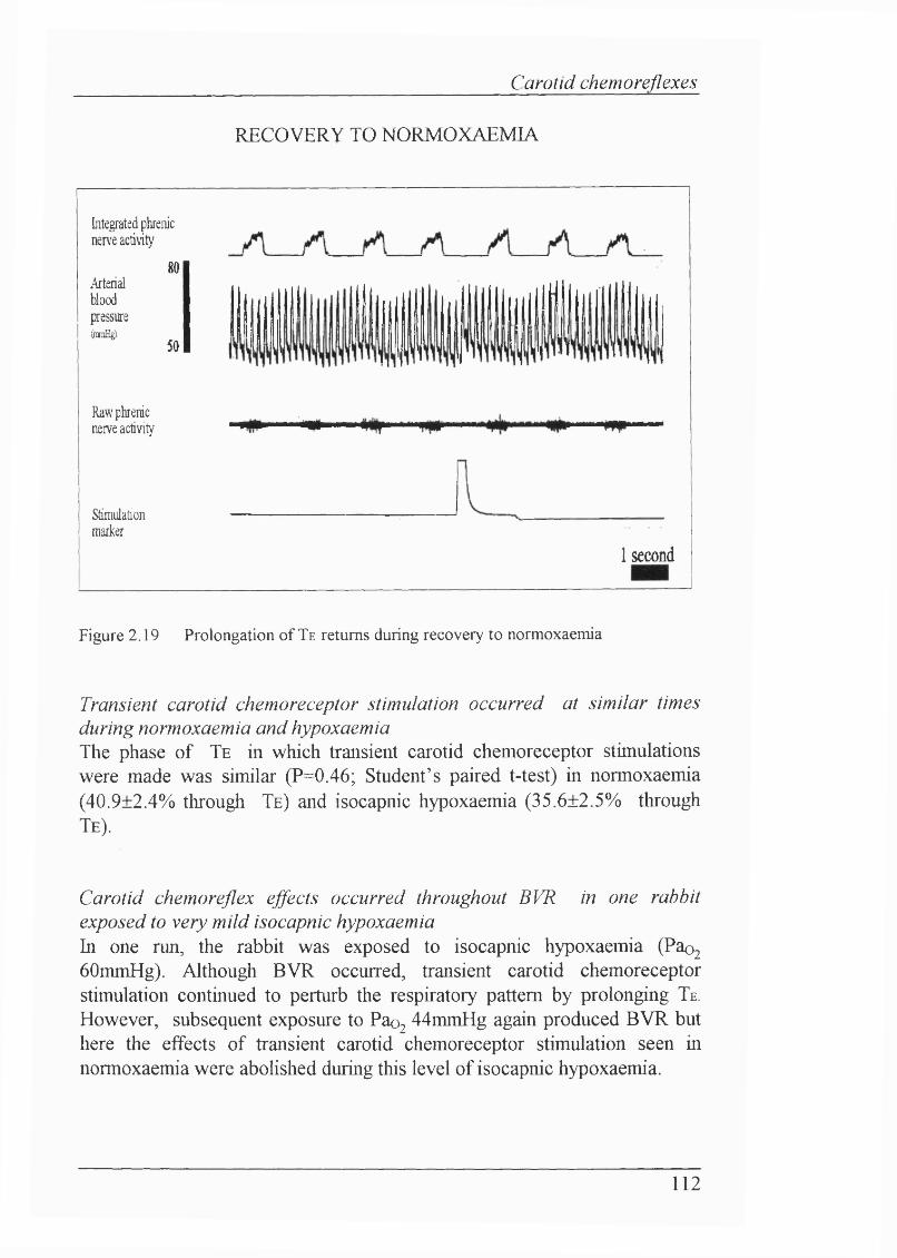
Carotid chemoreflexes
RECOVERY TO NORMOXAEMIA
Integrated phrenic nerve activity
80Arterialbloodpressure(jranH
50
Raw phrenic nerve activity
Stimulationmarker
1 second
Figure 2.19 Prolongation o f Te returns during recovery to normoxaemia
Transient carotid chemoreceptor stimulation occurred at similar times during normoxaemia and hypoxaemiaThe phase of T e in which transient carotid chemoreceptor stimulations were made was similar (P=0.46; Student’s paired t-test) in normoxaemia (40.9±2.4% through T e ) and isocapnic hypoxaemia (35.6±2.5% through T e ).
Carotid chemoreflex effects occurred throughout BVR in one rabbit exposed to very mild isocapnic hypoxaemiaIn one run, the rabbit was exposed to isocapnic hypoxaemia (Pao2
60mmHg). Although BVR occurred, transient carotid chemoreceptor stimulation continued to perturb the respiratory pattern by prolonging T e . However, subsequent exposure to Pao2 44mmHg again produced BVR but here the effects of transient carotid chemoreceptor stimulation seen in normoxaemia were abolished during this level of isocapnic hypoxaemia.
112
Carotid chemoreflexes
Lack o f correlation between the phase of BVR and presence o f chemoreflex effectIf ventilation declined because a central inhibitory mechanism inhibited peripheral chemoreflexes, the effect of chemoreceptor stimulation on breathing seen during normoxaemia would be expected to be abohshed only in phase 2. Therefore, a quahtative analysis was conducted to ascertain whether the inhibition of the effect of chemoreceptor stimulation was related to phase 2. Thus the following hypothesis was tested:-
"The (quahtative) effect of transient chemoreceptor stimulation on respiratory output during isocapnic hypoxaemia is dependent on which phase of BVR the stimulation is made
This quahtative analysis was conducted by comparing the effect of chemoreceptor stimulation on a breath with the mean+3 SD value of the preceding control breaths. Three standard deviations were chosen because then a type 1 error would occur only 1% of the time. If the magnitude of the stimulation breath parameter lay outside the mean+3 SD parameter value of the preceding control breaths, it was assigned as having an effect. If the value o f the stimulation breath parameter lay within the mean+3 SD value of the preceding control breaths, it was deemed to have no effect.
For each rabbit, the hypoxaemic period was divided into two periods, phase 1 and phase 2. Phase 1 was defined as the period fi*om the start of the hypoxaemic episode to the peak in respiratory output. Phase 2 was defined as the period fi'om the peak of respiratory output to the end of the hypoxaemic period (i.e. the phase of ventilatory decline). A group profile was compiled, assigning significant effects/no effects to the phase in which they occurred. In this way, the effect of stimulations made during hypoxaemia were not found to be significantly related to the phase BVR in which they occurred (one-way test; P>0.75).
Figure 2.20 shows the carotid chemoreflex data for each individual rabbit, illustrating the tendency for the carotid chemorefiex-mediated prolongation of expiration to disappear during isocapnic hypoxaemia. The graphs show normalized respiratory output (dashed line), plotted with the % change in Te over control (filled diamonds), against time (negative numbers indicate normoxaemia). Phase 1 is fi*om time 0 to peak normalized respiratory output. R denotes recovery value for % change in Te over control on return to normoxaemia. Figure 2.20a shows individual results for anaesthetized, paralyzed and vagotomized rabbits. Figure 2.20b shows results for a decerebrate rabbit.
113
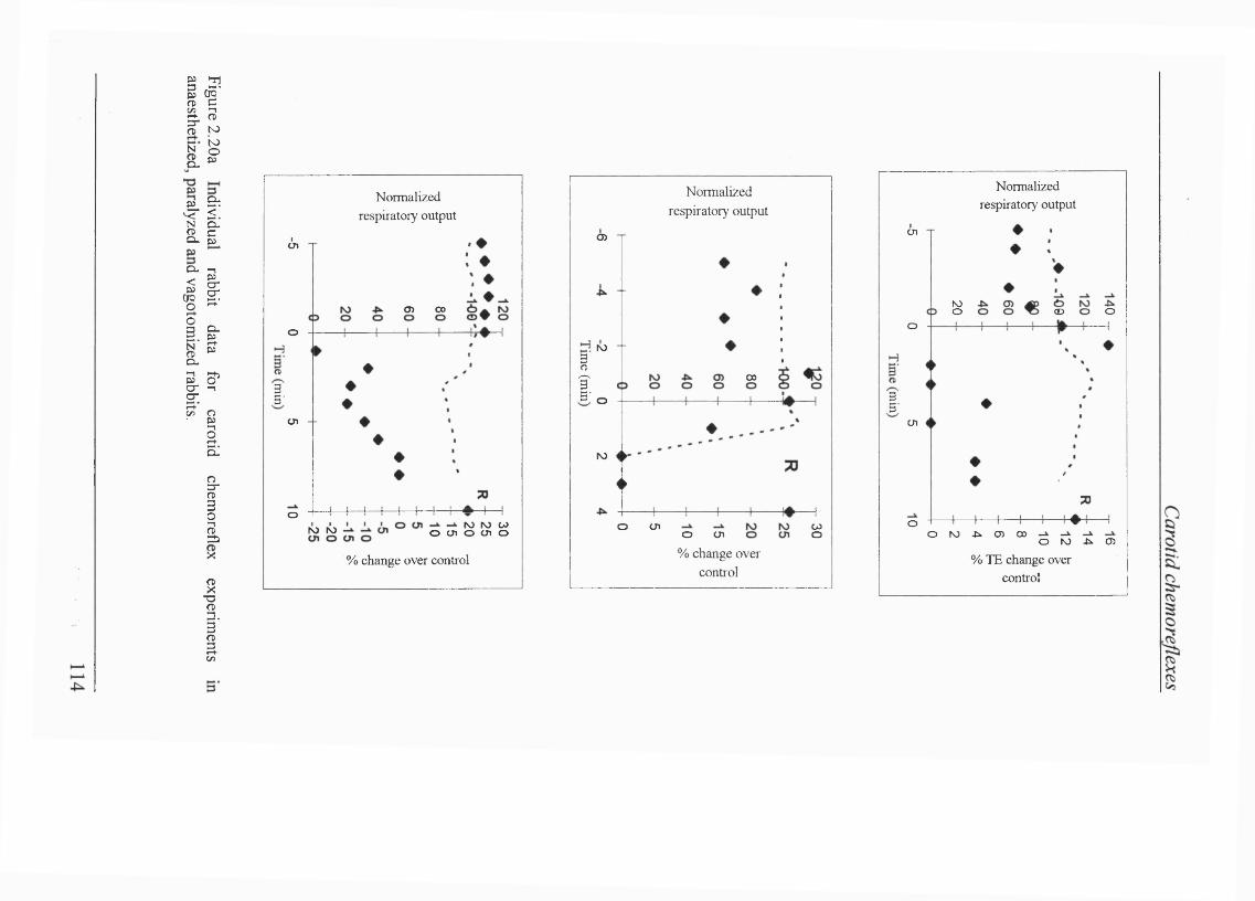
i l l so to
to§
a-<:CLc
N
a-
L|loi .
a i - ?cr5-(n O
■ I.CLIi%
CL%p:
IS.
I[/)
Normalized respiratory output
I-
(!n
Oi 00
o
ui
o N) N) CaIo cn o cn orb rb
% change over control
Normalized respiratory output
I?5-
o>
rb
CO
o
ro
i>
o cn woo cn% change over
control
Normalized respiratory output
B5'
cn
roo o(Do
cn
oo ro -li. O) CO o ro ^ cn
% TE change over control

à<:g-
§ a
1 1s
t.O. P) § ^
I îli - ïïHt
8
ICL
a-I
IIs
I3 .3
I
Normalized respiratory output
I
(In
O) 00
o
cn
o N) N) W o cn o cn o
% change over control
Normalized respiratory output
HI O) CO35
to
K)O WOO cn o cn% change over
control
Normalized respiratory output
cn
<Do
HftB5'
cn
o N) -tk <n CO o tv) ^ cn% TE change over
control
LA
?
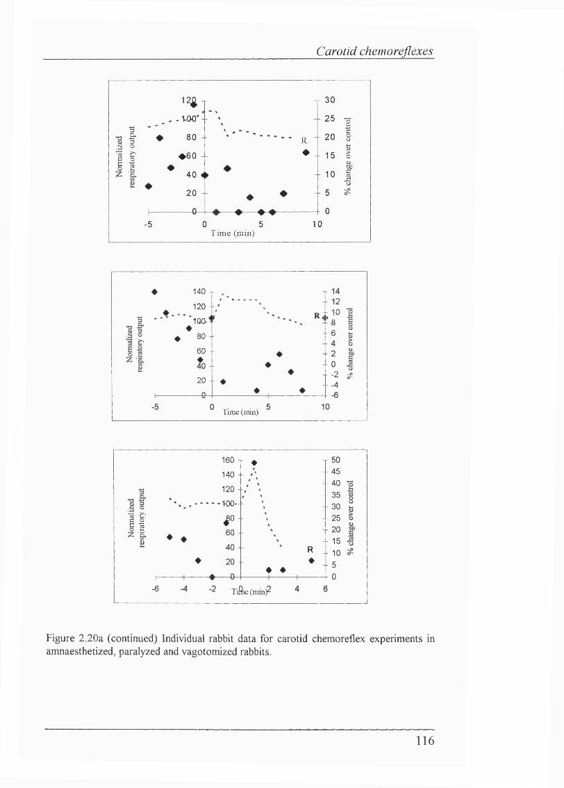
Carotid chemoreflexes
1 II
12 -r_ _ , . to o '- # 80 -
♦ 6 0
^ 40
20 + 0
-5
R♦
♦♦ ♦ ♦
0 5T ime (min)
y 30
- 251
- 20 8b
- 15 ë&
- 101
- 5
- 0
10
T - - 12
140 T
120 - -- - 10 4 ► „100-- - 680 --
60 -- 00
-2 -- -420 - - 4
T i m e ( m i n )
IIH
-6
160 n ♦140 -
120 -
100-
| 0 -
60 -
40 -
20 -
— 0 -♦ ♦
R♦
-2 Tifiie (min)
j 50 -- 45 -- 40 -- 35 -- 30 -- 25 - 20 -- 15 - - 10 -- 5
- - 0
Figure 2.20a (continued) Individual rabbit data for carotid chemoreflex experiments in amnaesthetized, paralyzed and vagotomized rabbits.
116
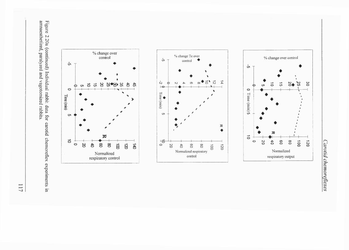
i î<T) <T»3-NO toCf. oN U>
IIg. =g.<pj Cu
CTQ C O B .o 3 crg . --1 o.I lv>
îs ,S
IIV3
% change over control
Ü1
^ ^ N Ê 6
Normalized respiratory control
ro
% c h a n g e T e o v e r c o n t r o l *
0 ro .t». O) 0 0 ^ 0 to ji.
H
! ♦î .3
4 -^ 4—M-
♦♦
♦ /
t ' 73♦
ro .tk o> 00 -Jko o o o ooN o r m a l i z e d r e s p i r a t o r y
c o n t r o l
roo
% change over control
ui
tn
.p»o O)o 00orooo roo
Normalized respiratory output
I
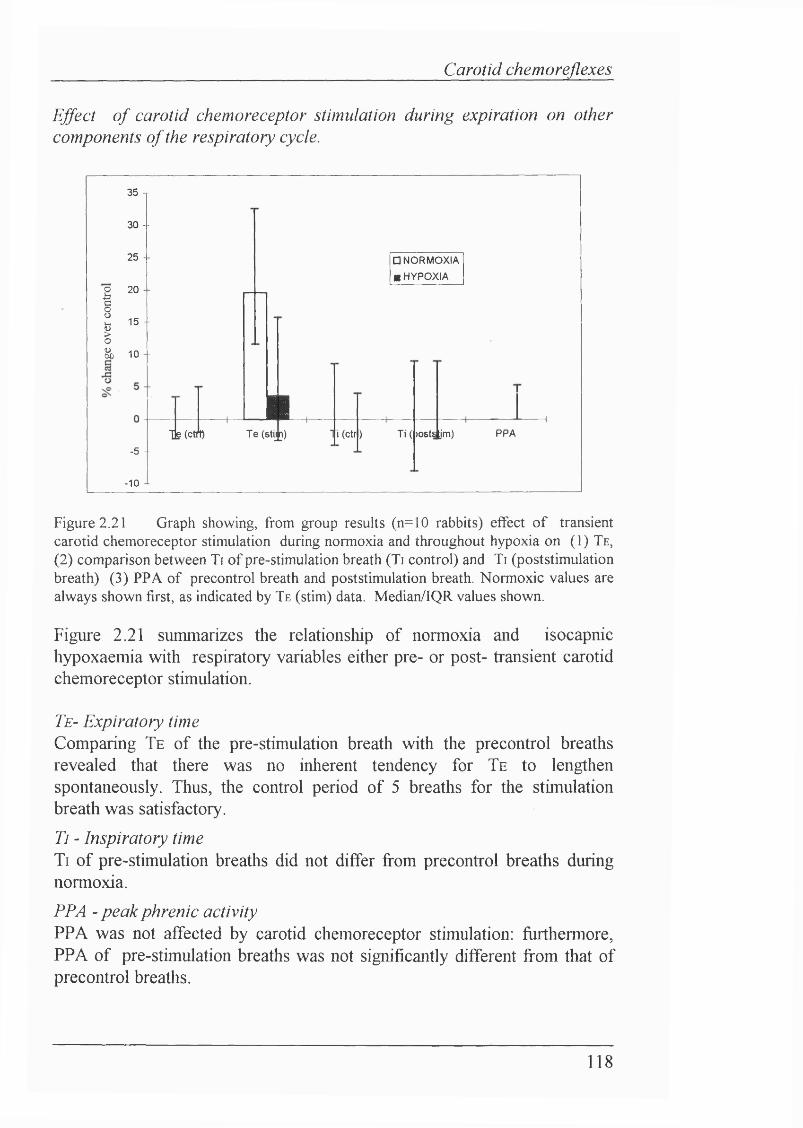
Carotid chemoreflexes
Effect o f carotid chemoreceptor stimulation during expiration on other components o f the respiratory cycle.
II&
35 T
30 --
25 -
20
15 --
10 -
-5 --
-1 0 -L
□ NORMOXIA
■ HYPOXIA
Tjg(ctmL l lT e (stijp)
!1 (ctr ) T i( )OStS jm) PPA
Figure 2.21 Graph showing, from group results (n=10 rabbits) effect of transient carotid chemoreceptor stimulation during normoxia and throughout hypoxia on (1) T e , (2) comparison between Ti of pre-stimulation breath (Ti control) and Ti (poststimulation breath) (3) PPA of precontrol breath and poststimulation breath. Normoxic values are always shown first, as indicated by T e (stim) data. Median/IQR values shown.
Figure 2.21 summarizes the relationship of normoxia and isocapnic hypoxaemia with respiratory variables either pre- or post- transient carotid chemoreceptor stimulation.
Te- Expiratory timeComparing Te o f the pre-stimulation breath with the precontrol breaths revealed that there was no inherent tendency for Te to lengthen spontaneously. Thus, the control period o f 5 breaths for the stimulation breath was satisfactory.
Ti - Inspiratory timeTi o f pre-stimulation breaths did not differ from precontrol breaths during normoxia.
PPA - peak phrenic activityPPA was not affected by carotid chemoreceptor stimulation: furthermore, PPA of pre-stimulation breaths was not significantly different from that of precontrol breaths.
118
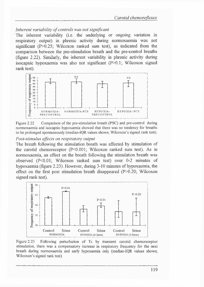
Carotid chemoreflexes
Inherent variability o f controls was not significantThe inherent variability (i.e. the underlying or ongoing variation in respiratory output) in phrenic activity during normoxaemia was not significant (P>0.25; Wilcoxon ranked sum test), as indicated from the comparison between the pre-stimulation breath and the pre-control breaths (figure 2.22). Similarly, the inherent variability in phrenic activity during isocapnic hypoxaemia was also not significant (P>0.1; Wilcoxon signed rank test).
0
1
NS
■1NS
N O R M O X I A .P R E C O N T R O L
N O R M O X I A -P C S H Y P O X I A .P R E C O N T R O L
H Y P O X I A - PCS
Figure 2.22 Comparison of the pre-stimulation breath (PSC) and pre-control during normoxaemia and isocapnic hypoxaemia showed that there was no tendency for breaths to be prolonged spontaneously (median-IQR values shown; Wilcoxon’s signed rank test).Post-stimulus effects on respiratory outputThe breath following the stimulation breath was affected by stimulation of the carotid chemoreceptor (P<0.001; Wilcoxon ranked sum test). As in normoxaemia, an effect on the breath following the stimulation breath was observed (P<0.01; Wilcoxon ranked sum test) over 0-2 minutes of hypoxaemia (figure 2.23). However, during 3-10 minutes of hypoxaemia, the effect on the first post stimulation breath disappeared (P>0.20; Wilcoxonsigned rank test).
P<0.01
b 4 5 -■ P >0.20
P<0.01cxX 4 0 -
b 3 5 -
Control Stimn Control StimnHYPOXIA (0.2min)
Control Stimn HYPOXIA (3.8min)NORMOXIA
Figure 2.23 Following perturbation of Te by transient carotid chemoreceptor stimulation, there was a compensatory increase in respiratory frequency for the next breath during normoxaemia and early hypoxaemia only (median-IQR values shown; Wilcoxon’s signed rank test).
119
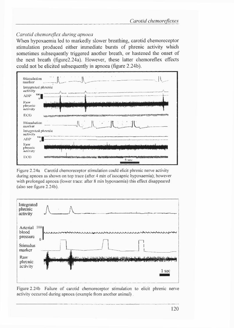
Carotid chemoreflexes
Carotid chemoreflex during apnoeaWlien hypoxaemia led to markedly slower breathing, carotid chemoreceptor stimulation produced either immediate bursts of phrenic activity which sometimes subsequently triggered another breath, or hastened the onset of the next breath (figure2.24a). However, these latter chemoreflex effects could not be elicited subsequently in apnoea (figure 2.24b).
stimulation m a r k e r ------Integrated phrenic activit>'ABP
0
Raw phrenic aclivitv
— J i .........
ECG
Stimulation marker I ntegni ted pJi reni c acfivilYABP ■ '“JRaw phrenic activity
ECO
......... ; _ J . , 1
5 sec
Figure 2.24a Carotid chemoreceptor stimulation could elicit phrenic nerve activity during apnoea as shown on top trace (after 4 min of isocapnic hypoxaemia); however with prolonged apnoea (lower trace: after 8 min hypoxaemia) this effect disappeared (also see figure 2.24b).
Arterial lOObloodpressure
StimulusmarkerRawphrenicactivity
1 sec
Figure 2.24b Failure of carotid chemoreceptor stimulation to elicit phrenic nerve activity occurred during apnoea (example from another animal).
120
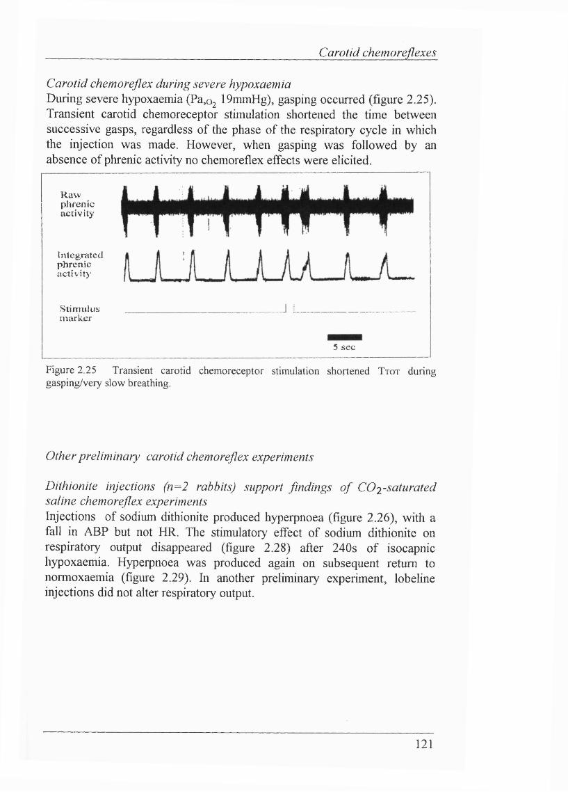
Carotid chemoreflexes
Carotid chemoreflex during severe hypoxaemiaDuring severe hypoxaemia (Pa, 0 2 19mmHg), gasping occurred (figure 2.25). Transient carotid chemoreceptor stimulation shortened the time between successive gasps, regardless of the phase of the respiratory cycle in which the injection was made. However, when gasping was followed by an absence of phrenic activity no chemoreflex effects were elicited.
Rawphrenicactivity
Integratedphrenicactiv ity
Stim ulusmarker
5 sec
Figure 2.25 Transient carotid chemoreceptor stimulation shortened T tot during gasping/very slow breathing.
Other preliminary carotid chemoreflex experiments
Dithionite injections (n= 2 rabbits) support findings o f CÜ2-saturated saline chemoreflex experimentsInjections of sodium dithionite produced hyperpnoea (figure 2.26), with a fall in ABP but not HR. The stimulatory effect of sodium dithionite on respiratory output disappeared (figure 2.28) after 240s of isocapnic hypoxaemia. Hyperpnoea was produced again on subsequent return to normoxaemia (figure 2.29). In another preliminary experiment, lobeline injections did not alter respiratory output.
121
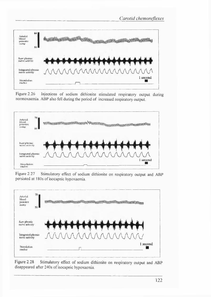
Carotid chemoreflexes
AiUHiiatbloodpicwura
8 0 1
5 0
Riiw ptuenic 1IWV0 activity
Stiinuiutioiinmtei1 second
Figure 2.26 Injections of sodium dithionite stimulated respiratory output during normoxaemia. ABP also fell during the period of increased respiratory output.
Aitciizdbloodpressure
7 0
5 0
Raw phrenic nerve activity
1 secondS t i n u i l a t i o ninaiker
Figure 2.27 Stimulatory effect of sodium dithionite on respiratory output and ABP persisted at 180s of isocapnic hypoxaemia.
A r t e r i a lbloodpresatre
70
50 IRaw phrenic nene activity
A / v.Aj V A A / vA A A . / \ A jW .A .^
Stinuilaticatiiwrkcx
I secondil
Figure 2.28 Stimulatory effect of sodium dithionite on respiratory output and ABP disappeared after 240s of isocapnic hypoxaemia.
122
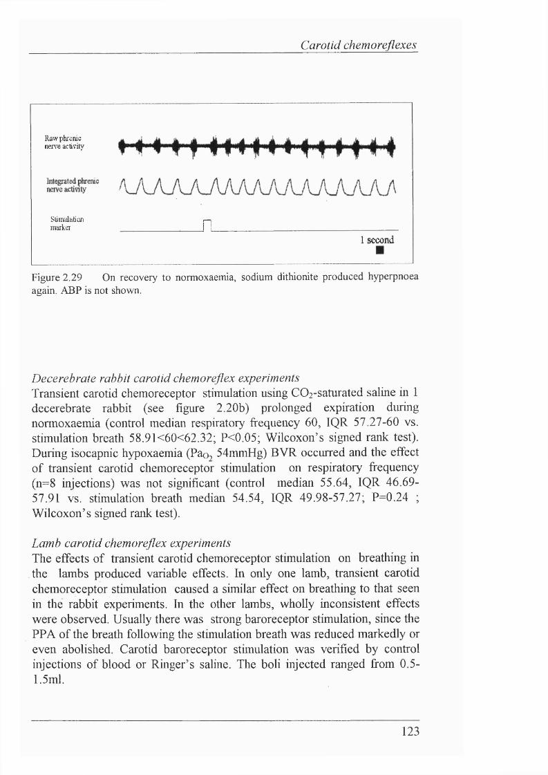
Carotid chemoreflexes
Raw phrenic nene activity
Stimidhtion r - .marker ______________ I L
1 second
Figure 2.29 On recovery to normoxaemia, sodium dithionite produced hyperpnoea again. ABP is not shown.
Decerebrate rabbit carotid chemoreflex experiments Transient carotid chemoreceptor stimulation using COi-saturated saline in 1 decerebrate rabbit (see figure 2.20b) prolonged expiration during normoxaemia (control median respiratory frequency 60, IQR 57.27-60 vs. stimulation breath 58.9K60<62.32; P<0.05; Wilcoxon’s signed rank test). During isocapnic hypoxaemia (Pao2 54mmHg) BVR occurred and the effect of transient carotid chemoreceptor stimulation on respiratory frequency (n=8 injections) was not significant (control median 55.64, IQR 46.69- 57.91 vs. stimulation breath median 54.54, IQR 49.98-57.27; P=0.24 ; Wilcoxon’s signed rank test).
Lamb carotid chemoreflex experimentsThe effects of transient carotid chemoreceptor stimulation on breathing in the lambs produced variable effects. In only one lamb, transient carotid chemoreceptor stimulation caused a similar effect on breathing to that seen in the rabbit experiments. In the other lambs, wholly inconsistent effects were observed. Usually there was strong baroreceptor stimulation, since the PPA of the breath following the stimulation breath was reduced markedly or even abolished. Carotid baroreceptor stimulation was verified by control injections of blood or Ringer’s saline. The boli injected ranged from 0.5- I.5ml.
123
Carotid chemoreflexes
In one decerebrate lamb (see figure 2.20b - injections shown at 1 minute intervals) transient CO2 injections prolonged expiration (i.e. decreased respiratory fi*equency) during normoxia (control median 16.67, IQR 15.79- 16.67 vs. stimulation median 18.53, IQR 18.31-18.75; P<0.01; Wilcoxon’s signed rank test). However, contrary to the anaesthetized rabbit experiments, during isocapnic hypoxaemia (F1 0 2 0.15) transient carotid chemoreceptor stimulation (7 injections) also prolonged expiration (control median respiratory fi’equency 16.67, IQR 16.25-16.92 vs. stimulation control 17.43, IQR 16.30-17.93; P<0.05; Wilcoxon’s signed rank test). Although this result seemed to contradict the previous experiments, analysis o f the % change over control due to transient carotid chemoreceptor stimulation revealed that the effect was greater during normoxia than isocapnic hypoxaemia (normoxaemia increase in respiratory frequency P(10.1<15.91<22.8%) >95% vs. hypoxaemia increase in respiratory frequency P(2.7<6.6<16.1%) >95%; P<0.05; Wilcoxon’s signed rank test). Furthermore, the % increase during recovery (median 28.6% increase in respiratory frequency, IQR 17.9- 29.95) was also greater than that seen during isocapnic hypoxaemia (P<0.05; Wilcoxon’s signed rank test).
In the other successfully completed carotid chemoreflex experiment in the lambs, transient carotid chemoreceptor stimulation shortened Te during normoxaemia and hence increased respiratory frequency during that breath only (control median respiratory frequency 36.25, IQR 35.62-36.88 vs. stimulation median 50, IQR 48.22-50; P<0.01; Wilcoxon’s signed rank test). However, during hypoxaemia transient CO2 carotid chemoreceptor stimulation did not cause such a significant event in respiratory output (control median respiratory frequency 31.65, IQR 23.86-40.02 vs. stimulation breath median 39.75, IQR 33.48-44; P>0.05; Wilcoxon’s signed rank test). During hypoxaemia (50% air:50% N^), respiratory output declined gradually to apnoea. Carotid chemoreceptor stimulation shortened interbreath intervals when respiratory output became progressively slower immediately before apnoea, and ehcited phrenic nerve activity during apnoea.
124
Carotid chemoreflexes
2.8 D isc u ssio n
MethodsMeasurement and control of blood gases, and a direct index o f central respiratory output were essential for addressing chemoreflex hypothesis In order to address the hypothesis it was necessary to use an acute, anaesthetised preparation, to gain a more detailed understanding of the neurophysiological pathways involved in the chemoreceptor-phrenic nerve reflex. Therefore, it was essential to measure phrenic activity as an index of central respiratory output and also to have a defined input. This avoids two comphcations. First, respiratory muscle fatigue has been imphcated in the biphasic ventilatory response (Chapter 1; hypothesis 6) thereby bringing into question the relative contribution of the respiratory centres to the ventilatory decline. The alternative to measuring nerve activity directly would be to perform non-invasive experiments and measure ventilation. This introduces the second comphcation in interpreting respiratory responses, since isocapnic hypoxaemia is very difficult to achieve under these conditions as non-invasive monitoring of blood gas status and blood pressure is difficult. Even though Paco2 undoubtedly falls during an acute hypoxaemic challenge, it was important to be able to assess the role of isocapnic hypoxaemia.
Development of a suitable newborn rabbit preparation Performing invasive surgery in the newborn presents a number of problems that are not encountered with mature animals. At birth the rabbit is immature and totally dependent on its mother. Rabbits only emerge fi*om their nest at around 3 weeks of age. but remain dependent on the doe until they are weaned at 6-8 weeks old (Adams, 1987). As defined by sexual maturity, rabbits become "adult" at around 3 months old. Because it was not possible to set up a rabbit breeding colony on site at UCL, the newborn rabbits used had to be dehvered on, or a day before, the experiment. Understandably, newborn rabbits of this age are potentially more vulnerable to being transported and the stress that is associated with leaving the doe. Newborn rabbits are also especially susceptible to a variety of infections. For example, one study showed that at least 50% of deaths in breeding colonies occurred before weaning (Seamer & Chesterman, 1967). The most common cause of death is intestinal infection, such as mucoid enteropathy (see Weisbroth, Flatt & Kraus, 1974). The range of weights of the newborn rabbits used varied considerably. This is presumably due to the large variation in fitter size of rabbits, which can range fi’om 3-12 per fitter. In addition, growth during the newborn period is rapid: a 3-week old rabbit can gain in excess of lOOg in just 7 days (Adams, 1987).
125
Carotid chemoreflexes
Implementation o f a suitable anaesthetic regimeDuring the course of this set of experiments a regime had to be implemented not only to address the hazards of infection and transport (see methods), but also to find a satisfactory anaesthetic regime to conduct the preparations. The main problem with using anaesthesia in newborn rabbits was the preservation of central respiratory output and/or chemoreflexes. For example, the animals would breathe under halothane but blood pressure was typically 40-50/30mmHg, body temperature could markedly fall and the respiratory response to hypoxaemia was often poor. In newborns, halothane is noted for causing large falls in blood pressure, depression of the baroreceptor reflex (Wear, Robinson & Gregory, 1982), impaired myocardial function (&ane & Su, 1987; McAulifle & Hickey, 1987) and depression of peripheral chemoreceptor activity (Ponte & Sadler, 1989). a- chloralose often abohshed respiratory output, although single doses did provide long stable periods (>6 hours) of anaesthesia.
The anaesthetic of choice, urethane, proved satisfactory as it maintained a reasonable blood pressure and respiratory reflexes were maintained. The levels of blood pressure and heart rate are in close agreement with previous workers who had employed this anaesthetic regime in young rabbits. Furthermore, blood pressure and heart rate levels are predictable from the levels observed in the greater number of experiments using adult rabbits under urethane anaesthesia. Typically the urethane anaesthetised adult rabbit has ABP of ca.lOOmmHg and HR of ca. 300 bpm (Ishikawa, Kallman & Sagawa, 1984; Bradley, Ghelarducci, Paton & Spyer, 1987). It would be expected that the newborn rabbits reported in this Chapter have lower ABP, as ABP increases with postnatal age (Mott, 1966). HR is related to body weight, with smaller animals having higher HR. Thus smaller rabbits (around 4 times lighter compared to adult rabbits used by Ishikawa, Kallman & Sagawa (1984) and Bradley, Ghelarducci, Paton & Spyer (1987) would be expected to exhibit higher heart rates.
Throughout these preparations it was imperative that careful attention was exercised over blood gas status. Due to the small blood volume of these animals (approximately 5.7ml/100g body weight) the frequency of blood sampling was kept to a minimum (ca. 8 samples per preparation).
126
Carotid chemoreflexes
C a r o t id c h e m o r e c e p t o r r e c o r d in g s
Methodological drawbacksThe technique of recording from multifibre chemoreceptor preparations presents considerable limitations chiefly because of the size of the carotid sinus nerve and the anatomical limitations of working with small, newborn animals. For this reason the sympathetic fibres of the superior cervical gamghon that innervate the carotid body via the ganghoglomerular body were not cut. Although sympathetic ganghomerular activity probably increased during isocapnic hypoxaemia (Lahiri, Matsumoto & Mokashi, 1986), it is most unlikely to have influenced the effect of transient carotid chemoreceptor stimulation on respiratory output (see O’Regan & Majcherczyk, 1983). Secondly, only a small proportion of fibres can be recorded from at any one time, and therefore the assumption has to be made that they are representative of the entire population of chemoreceptor fibres.
It was often difficult to maintain the multifibre preparations on the recording hooks in many preparations, without the activity recorded being disrupted by accumulation of blood. The preparation had to be treated with considerable dehcacy during the experimental run, to avoid disrupting the contact of the CSN with the recording hooks. Thus simultaneous recording of the phrenic nerve and one carotid sinus nerve (with the contralateral carotid chemoreceptors being stimulated chemically) was veiy difficult and unsuccessful.
Nonetheless, recordings from multifibre carotid chemoreceptor preparations clearly show that:(1) Carotid chemoreceptor fibres do not adapt to acute episodes of
isocapnic hypoxaemia.(2) Transient COz-equihbrated saline produces an increase in
chemoreceptor activity during isocapnic hypoxaemia.
That carotid chemoreceptor fibres do not adapt during isocapnic hypoxaemia supports the findings of previous studies in kittens (Schweiler, 1968; Blanco Hanson, Johnson & Rigatto, 1984). This suggests that some central process must be activated to cause ventilatory decline during hypoxaemia since carotid chemoreceptor discharge is sustained.
However there are considerable problems in interpreting data obtained from multi- or sub-populations of fibres of the whole CSN. Strictly, it is incorrect to assess the afferent carotid chemoreceptor activity for a group of animals, because clearly a different sub population of fibres must be recorded in every case even though the fibres that are recorded are mevitably biased
127
Carotid chemoreflexes
towards myelinated fibres which have the largest action potentials. Thus the discharge rate (spike/s) and firing threshold of each fibre, or fibre preparation, are likely to be different. It may also be incorrect to assume that during hypoxaemia the same type or number of fibres are active compared with those recorded in normoxaemia. In other words, more chemoreceptor fibres could be recruited during hypoxaemia. Another comphcation in this preparation is the role of C02-saturated saline injections made during isocapnic hypoxaemia. It need not necessarily be the case that the increases in the multifibre preparation discharge recorded in the experiments reported in this Chapter are representative o f the whole CSN output. In other words, this study may have been biased towards fibres that showed greater increases in discharge when stimulated by CO2 . The interpretation of transient carotid chemoreceptor stimulation, with regard to the carotid chemoreflex experiments, is further comphcated by the finding that in some experiments the maximum discharge attained using COz-saturated saline increased during isocapnic hypoxaemia, whereas in others the maximum level of carotid chemoreceptor discharge remained the same regardless of Pao2 levels.
Understanding the mechanism of carotid chemoreception is important for the interpretation of these results. A single chemoreceptor fibre is excited by both hypoxaemia and hypercapnia. For any fixed Paco2 there is a hyperbohc relation between chemoreceptor fibre discharge and Pao2 while there is a linear relationship between chemoreceptor fibre discharge and Paco2 at any fixed Pao2 • Thus at any level of hypoxaemia, there is a single steady-state response curve to CO2 . However, if Paco2 suddenly rises to a new level, the immediate response rises to a point on a steeper, but transient, response curve. The "bicarbonate hypothesis" (Torrance, 1974) provides the most complete and experimentally supported explanation to account for these properties of single carotid chemoreceptor fibres.
The bicarbonate hypothesis is an amendment of Winder's "acid receptor" hypothesis (1937), which proposed that the convergence of hypoxaemic and hypercapnie stimuh occurs at a stage before these stimuh are transduced into action potentials. Winder proposed that hypoxaemia, as well as hypercapnia, affected intracellular carotid body pH, by lactic acid formation. Torrance adapted this hypothesis by suggesting that hypoxaemia causes a failure in an active pH maintenance system in type 2 cells. The site of maintenance for pH was originally proposed to be an extracellular space and it was suggested that carbonic anhydrase mediated the rapid response of the carotid body (Black, McCloskey & Torrance, 1971). However, because an intracellular carbonic anhydrase inhibitor markedly slowed the rapid
128
Carotid chemoreflexes
response of chemoreceptor fibres (Hanson, Nye & Torrance, 1981), it became clear that carbonic anhydrase acted intracellularly and changes in intracellular pH determined the brisk excitatory response to sudden stimuh. Histological studies confirm that carbonic anhydrase is located intracellularly (Ridderstrale & Hanson, 1984).
Thus, at a sudden change of PcOj at the carotid body, the immediate change in intracellular pH in the type 1 cells is determined by the buffering power of the type 1 cell cytoplasm. Therefore transient changes in PcOj are independent of P0 2 , and the carotid chemoreceptor fibres response should be instantaneous, linear and without adaptation. In theory, this permits a comparison to be made between the effects of C02-stimulation at the carotid body in normoxaemia and hypoxaemia, because the maximum discharge reached due to C02-saline stimulation, should be the same, regardless of Pa,Oz. However, these assertions can only be used to support those experiments where the maximum discharge due to C02-saline stimulation during normoxaemia and isocapnic hypoxaemia remained the same. In contrast, these assertions do not hold for carotid chemoreceptor experiments where, in isocapnic hypoxaemia, C02-saline stimulation increased discharge above that seen in normoxaemia. Perhaps in this latter case, additional and /or different fibre types were recruited during hypoxaemia
The magnitude of increase of chemoreceptor fibre discharge on C02-saline stimulation over baseline activity during isocapnic hypoxaemia shows that COz-saline still produces a large increase in afferent information to the respiratory centres. This observation is clearly critical if the assumption that a similarly marked, transient increase in chemoreceptor drive is dehvered during hypoxaemia as in normoxaemia is to be satisfied. This permits a quantitative comparison between the effects of carotid chemoreceptor stimulation on central respiratory output in normoxaemia and hypoxaemia.
Lack o f baroreceptor effectsThere was httle evidence fi"om injection of control solutions in this set of experiments to suggest that this method of chemoreceptor stimulation also caused baroreceptor stimulation. This is strongly reinforced by the findings fi*om the carotid chemoreflex experiments. However, the placement of the stainless steel foot onto which the carotid sinus nerve fibres were laid occasionally occluded some of the vessels around the carotid bifurcation. Consequently, this caused a rapid injection of solution to distend the carotid sinus, thereby stimulating baroreceptor fibres. In order to overcome this occlusion problem, slower injections of solutions were made. Slower
129
Carotid chemoreflexes
injections of control solution had no significant effect on chemoreceptor fibre discharge, whereas COi-equihbrated saline injected over a comparable time course produced an increased discharge.
Severe hypoxaemia/asphyxiaIn severe hypoxaemia (Po2<20mmHg), chemoreceptor discharge rose and was maintained, at least until stagnant hypoxaemia (asphyxia) resulted. At this point COz-equilibrated saline failed to produce an increase in discharge over baseline discharge, because of the lack of carotid blood flow.
C a r o t id c h e m o r e f l e x t e st Variable magnitude o f BVRPhrenic nerve activity was chosen as the index of central respiratory output, as it has previously been shown to be a neural equivalent of ventilation (Eldridge, 1971). Each rabbit exhibited a biphasic respiratory response, as measured from the phrenic nerve. This is in agreement with previous studies, in both anaesthetised and conscious animals (Dawes & Mott, 1959; Schweiler, 1968; Wangsnes & Koos, 1991). However, the magnitude of the biphasic ventilatory response was variable. This could be due to different maturational stages in individual rabbits (perhaps indicated by the large range in body weight for animals of a similar age) or the varying effects, and metabohsm, of anaesthesia in individual animals.
Chemical carotid chemoreceptor stimulation is more appropriate than electrical CSN stimulation in the newborn rabbit preparation There are distinct advantages in stimulating the carotid chemoreceptor fibres chemically as opposed to electrically. The clear problem with electrical CSN stimulation in the newborn rabbit preparation is the physical limitation imposed by the small size, both in terms of maintaining the nerve on stimulating hooks and the risk of stimulus escape to surrounding tissues. In preliminary experiments the latter proved very difficult to control, resulting in the phrenic electrodes recording extraneous stimulation artefacts. The obvious advantage of chemical stimulation is that baroreceptor stimulation should be avoided. Grunstein, Derenne and Mihc-Emh (1975) showed that carotid baroreceptor stimulation in adult cats produced profound decreases in both tidal volume and frequency of breathing. Lawson & Long (1984) also noted that electrical CSN stimulation caused ABP to fall.
130
Carotid chemoreflexes
The disadvantage of chemical stimulation in the carotid chemoreflex experiments is that the timing of injections in relation to the phase o f the respiratory cycle is not precise. The method presented in this Chapter has been used for both chemical (Black & Torrance, 1967) and electrical CSN stimulation (Berger & Mitchell, 1976). A good estimation of the timing of the effect of a stimulation can be derived from the carotid chemoreceptor recording experiments, by measuring the latency of onset of the increase in carotid chemoreceptor discharge from the injection marker. From this analysis, the timing of injections in relation to the phase of the respiratory cycle can be ascertained accurately. A possible improvement in the methodology used in these experiments to increase the accuracy o f timing of stimuli would be to employ a mechanical pump, which would dehver boh at set periods after the onset of phrenic activity (i.e. inspiration) by using an electronic delay circuit (see Lipski, Trebski, Chodobska & Kruk, 1984).
Reliability o f the transient carotid chemoreceptor stimulus test The effects of transient chemoreceptor stimulation during normoxaemia were quite clear, depending on the phase in which the injections were made. However, there was considerable variabihty observed in the phrenic response to injections, and it was possible for no effect to be seen sometimes. This may be due to the phase in which the chemoreceptor stimulus reached the respiratory centres. This could not be accounted for by the effect of COz-saturated saline wearing off with time as reported in a similar study in the adult cat (Band, Cameron & Semple, 1970). Another possible explanation could be the unsynchronised detection of the CO2 - equilibrated saline by the carotid bodies (due to different catheter dead spaces or the relative positions of each catheter). The output from the integration o f right and left CSN input to NTS neurones may depend largely on the temporal relationship by which the afferent impulses arrive. Conditioning stimuh apphed to one CSN causes prolonged inhibition of the response to a subsequent stimulus, be it the same or contralateral CSN (Felder & Heesch, 1987). However this latter idea is less likely since unilateral injections also produced profound changes in ongoing phrenic activity, which were also variable in magnitude.
A more likely explanation for the variabihty may be due to the functional subdivision of expiration into stage 1 expiration (or post-inspiration) and stage 2 expiration. IntraceUular and extraceUular recordings from meduUary respiratory neurones reveal that stage 2 expiratory activity is shortened or eliminated either spontaneously or by hyperthermia, stimulation of the hypothalamic defence area or pulmonary C fibre afférents (Richter & Spyer,
131
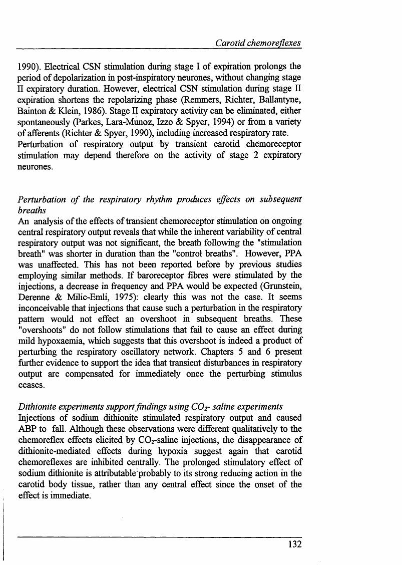
Carotid chemoreflexes
1990). Electrical CSN stimulation during stage I of expiration prolongs the period of depolarization in post-inspiratory neurones, without changing stage n expiratory duration. However, electrical CSN stimulation during stage II expiration shortens the repolarizing phase (Remmers, Richter, Ballantyne, Bainton & Klein, 1986). Stage H expiratory activity can be eliminated, either spontaneously (Parkes, Lara-Munoz, Izzo & Spyer, 1994) or from a variety of afferents (Richter & Spyer, 1990), including increased respiratory rate. Perturbation of respiratory output by transient carotid chemoreceptor stimulation may depend therefore on the activity of stage 2 expiratory neurones.
Perturbation o f the respiratory rhythm produces effects on subsequent breathsAn analysis o f the effects of transient chemoreceptor stimulation on ongoing central respiratory output reveals that while the inherent variability o f central respiratory output was not significant, the breath following the "stimulation breath" was shorter in duration than the "control breaths". However, PPA was unaffected. This has not been reported before by previous studies employing similar methods. If baroreceptor fibres were stimulated by the injections, a decrease in frequency and PPA would be expected (Grunstein, Derenne & Milic-Emh, 1975): clearly this was not the case. It seems inconceivable that injections that cause such a perturbation in the respiratory pattern would not effect an overshoot in subsequent breaths. These "overshoots" do not follow stimulations that fail to cause an effect during mild hypoxaemia, which suggests that this overshoot is indeed a product of perturbing the respiratory oscillatory network. Chapters 5 and 6 present further evidence to support the idea that transient disturbances in respiratory output are compensated for immediately once the perturbing stimulus ceases.
Dithionite experiments support findings using CO2 - saline experiments Injections of sodium dithionite stimulated respiratory output and caused ABP to fall. Although these observations were different quahtatively to the chemoreflex effects ehcited by COz-saline injections, the disappearance of dithionite-mediated effects during hypoxia suggest again that carotid chemoreflexes are inhibited centrally. The prolonged stimulatory effect of sodium dithionite is attributable probably to its strong reducing action in the carotid body tissue, rather than any central effect since the onset of the effect is immediate.
132
Carotid chemoreflexes
Possible interpretations o f results from carotid chemoreflex and carotid chemoreceptor recording experiments: implications fo r BVR Three possible outcomes could be predicted at the onset of these experiments to explain the lack of effect of carotid chemoreceptor stimulation on breathing during isocapnic hypoxaemia. These are summarised in figure 2.30.
Possibility 1
C e n t r a l I r e s p i r a t o r y 1 o u t p u t 1
MAX 1
C a r o t i d 1 c h c i T i o r c c c p l o r 1 d i s c h a r g e I
N O F t l v I O N I A t l Y I > C » L A d £ t V I I ^
T I TB lo c k s su perim p osed on carotid ch em orecep tor d ischarge represent p red icted increased d ischarge due to C O ;-sa lin e in jection
Possibility 2
C a r o t i dc h c m o r c c c p t o r
d i s c h a r g e
A d en o tes sm a llin c iea se in carotid ch em orecep tor discharge due to C O ;-sa lin e in jection
Possibility 3
C e r i t r r a lrespiiatoryo u t i i r i t
M A X IC a ro tidc h c n i o r c c c p t o r
d i s c h a r g e
N O R J v I O X I A .
1 I B I I
B d en o tes large in crease (rela tive to A ; see P o ss ib ility 2 ) in carotid ch em orecep tor d ischarge due to CO ^-saline
Figure 2.30 Three possible interpretations for the carotid chemoreflex experiments.
133
Carotid chemoreflexes
Possibility 1 : Carotid chemoreceptor fibres do adapt to an acute period of isocapnic hypoxaemia over a similar time course to that of the ventilatory decline (phase 2).
Possibility 2: Mild isocapnic hypoxaemia causes a large increase in carotid chemoreceptor discharge, near to maximum discharge fi*equency. Here CO2 - saline injections during isocapnic hypoxaemia would still increase carotid chemoreceptor activity to a maximum, but the percentage increase in activity over control baseline activity would be smaller than for Possibihty 3. (i.e. a relatively small increase in activity over hypoxaemic baseline activity would occur).
Possibility 3: Mild isocapnic hypoxaemia causes a sub-maximal increase in baseline carotid chemoreceptor discharge. COz-saline injections would therefore increase chemoreceptor discharge markedly, to the maximum discharge intensity (i.e. a relatively large increase in activity over hypoxaemic baseline activity would occur).
Clearly carotid chemoreceptor recordings strongly suggest that Possibility 1 is unlikely, as the chemoreceptor fibres recorded did not adapt to an acute hypoxaemic challenge. This conclusion is strengthened by considering Possibility 2. If baseline carotid chemoreceptor discharge increases to just below maximal discharge frequency, the effect of COz-saline stimulation on carotid chemoreceptor discharge would be relatively small. This relatively small change may have no effect on breathing. This could explain the failure to produce an effect on breathing during hypoxaemia. However, if this is the explanation for the latter, it again suggests strongly that carotid chemoreceptor fibres do not adapt to acute hypoxaemia. Thus for Possibility 2, ventilation is actively inhibited by some centrally acting mechanism, while the effect of C02-saline stimulation on carotid chemoreceptor discharge is not a large enough increase to ehcit any change in breathing regardless of any other inhibitory influences that become active during hypoxaemia.
Possibility 3 offers a sUghtly different explanation to Possibihty 2, in that it suggests that both the effects of C02-saline stimulation and ventilation are inhibited (although not necessarily by the same mechanism). Here baseline chemoreceptor discharge increases but to a sub-maximal level. C02-saline stimulation causes maximum chemoreceptor discharge, thereby producing a relatively large increase in afferent input, during both normoxaemia and mild hypoxaemia. This large increase produces a change in breathing during normoxaemia and for a period during hypoxaemia, but the effect of this large
134
Carotid chemoreflexes
increase in respiratory output is later inhibited by a centrally acting mechanism that is activated during exposure to lower PO2 levels.
Of the three options, the data from the carotid chemoreceptor recordings and carotid chemoreflex test experiments support Possibility 3. There are two reasons to favour Possibility 3. First, COz-saline injections cause dramatic increases in carotid chemoreceptor discharge during both normoxaemia and mild hypoxaemia. Second, COz-saline injections can produce marked effects on phrenic activity during gasping spells and also could cause changes in phrenic activity during apnoea. This suggests strongly that the large increase in transient carotid chemoreceptor discharge due to C02-saline injections is maintained even during severe hypoxaemia. Therefore, it would seem that relatively small increases in chemoreceptor discharge can produce effects on phrenic activity. This idea is reinforced by the lack of correlation between phrenic activity and the effect of the carotid chemoreflex.
Lack o f correlation between peak in central respiratory output and type o f carotid chemoreflex effectBecause the peak in central respiratory output is not clearly linked to the disappearance of the effect of transient chemoreceptor stimulation, it would seem that the relationship between the decline in ventilation and lack of effect of chemoreceptor stimulation is more complex than simply some central mechanism inhibiting chemoreceptor afferents to decrease breathing. Rather these findings support the idea that a central inhibitory mechanism inhibits respiratory output and coincidentally prevents carotid chemoreceptor stimulation affecting this inhibition (rather than inhibiting directly carotid chemoreflexes to cause respiratory output to fall). Thus breathing does not decline during phase 2 of BVR primarily because afferent carotid chemoreflexes are inhibited. This is supported by studies in the adult rabbit, which showed that the ventilatory decline seen during hypoxaemia occurs in the peripherally chemodenervated adult rabbits (Martin-Body, 1988). This also does not support the idea that the carotid chemoreceptors cause directly the activation of the central inhibitory mechanism that causes ventilation to fall, as suggested by Schramm & Grunstein (1987).
However, some caution must be placed on this interpretation. The timing of the stimulus at the carotid body is crucial in determining the magnitude of the change in respiratory output. The timing of stimuh undoubtedly varied during phase 2 and between experiments. Therefore the magnitude o f the effect on respiratory output measured during phase 2 may not be standardized. This makes a quantative comparison very difficult, and so the
135
Carotid chemoreflexes
interpretation presented here is based solely on qualitative changes in respiratory output. It is possible that precisely controlled timing of stimuh could reveal a tighter relationship between the magnitude of carotid chemoreflex effects and respiratory output.
CNS studies show that hypoxia inhbits expiratory respiratory neurones Expiratory neurones are located in two regions.(1) The expiratory neurones located in nucleus retroambiguahs project axons to the contralateral spinal cord to intercostal and other spinal expiratory motoneurones. These expiratory neurones have very few projections within the brain stem.(2) Expiratory neurones located in Botzinger complex (rostral portion of nucleus ambigualis) send inhibitory projections to medullary inspiratory neurones and also project to the contralateral spinal cord.
The effects of hypoxia on expiratory neuronal activity have been investigated by extracellular recordings in adult cats. Whereas hypercapnia causes a general excitation of medullary inspiratory and expiratory neurones, mediated by the central chemoreceptors, isocapnic hypoxia produces a heterogeneous response in dorsal and ventral medullary respiratory nuclei. Medullary respiratory neurones mediate these changes in three ways: by changing discharge frequency, changing the number of spikes per discharge (or burst) and also by recruitment.
Expiratory neurones in the caudal medulla are depressed by hypoxia (Fio 0.1), even though tidal volume increases. Furthermore, cessation of expiratory neurone discharge has also been reported, even though ventilation remained augmented (Nesland, Plum, Nelson & Siedler, 1966). Recordings from expiratory neurones in nucleus retroambiguahs also show that there is a reduction in burst duration and mean spikes per burst during hypoxia (Fio 0.1) concomitant with increased respiratory output. The mean firing rates o f these expiratory neurones did not alter during the initial change in ventilation due to hypoxia (Baker, Frazier, Hanley & Zechman, 1979). The hypoxic depression of expiratory neurones is not dependent on the maintenance of isocapnia (St. John & Wang, 1977). In carotid denervated animals, isocapnic hypoxia caused a general depression of neuronal activity. This indicated that when the peripheral chemoreceptors are intact, the differential effect on neuronal discharge during isocapnic hypoxia may be due to the unequal projection of peripheral chemoreceptor afferents. However, another possibility was that hypoxia alone depressed neuronal activity, rather than the depression being due to unequal distribution of peripheral and central chemoreceptor afferents.
136
Carotid chemoreflexes
St. John (1981) explored further this latter idea by recording respiratory units from the dorsal and ventral medullary respiratory nuclei and the pontine reticular formation during chemical stimulation of the carotid chemoreceptors and hypercapnia. Chemical stimulation of the carotid chemoreceptors eliminated the potential masking of effects by hypoxia. Both hypercapnia and carotid chemoreceptor stimulation increased dorsal nucleus inspiratory neurone activity. However, only hypercapnia augmented inspiratory and expiratory activity in ventral meduUary nuclei. Both types of stimuli caused a decline or did not affect respiratory units in the pontine reticular formation. St. John (1981) concluded that peripheral and central chemoreceptor excitation causes differential effects on respiratory neurones throughout the brain stem. This was also supported by recordings from bulbospinal and laryngeal respiratory neurones during hypercapnia and hypoxia (St. John & Bianchi, 1985). While this conclusion may hold for hypercapnia, it is difficult to be sure that this conclusion is appropriate for isocapnic hypoxia. Although lower Paoj does produce peripheral chemoreceptor stimulation central, neuraUy mediated changes can also occur (see Chapter 5), in addition to the undoubted local changes in neuronal function caused by hypoxia (such as changes in metabohsm and direct effects on neuronal excitabihty).
Hypoxia also inhibits expiratory motor activityEMG recordings from expiratory muscles also suggest strongly that hypoxia inhibits expiratory motor activity. Hypoxia increases upper airway resistance during expiration in anaesthetized, vagotomized adult cats, because the expiratory abduction of the posterior cricoaiytenoid muscle is inhibited (Bartlett, 1980). Similarly, the human glottis narrows in expiration during hypoxia (England, Bartlett & Knuth, 1982). Furthermore, isocapnic hypoxia inhibits abdominal expiratory motor activity (cranial ihohypogastric nerve) in decerebrate, vagotomized, paralyzed adult cats (Fregosi, Knuth, Ward & Bartlett, 1987). The reduction in abdominal expiratory motor activity occurred despite augmentation of phrenic nerve activity. Fregosi (1994) has also shown that electrical CSN stimulation augments abdominal expiratory EMG 20% more than systemic hypoxia, for the same level o f respiratory output. This suggests that systemic hypoxia attenuates the stimulatory effect of the carotid chemoreceptors on expiratory activity. However, neither electrical CSN stimulation or systemic hypoxia changed expiratory abdominal EMG activity after bilateral vagotomy, which implicates vagal afferents in expiratory EMG inhibition.
137
Carotid chemoreflexes
The application o f oscillatory theories to the carotid chemoreflex experimentsRespiratory rhythm is generated by an oscillatory process that can be perturbed briefly by the transient chemical stimulation of the carotid bodies; the underlying rhythm rapidly recovers under such circumstances In relation to Possibility 2, another study has shown that increasing respiratory drive (for example, by increasing PCO2 or electrical stimulation of the carotid sinus nerve) reduces the effectiveness of a perturbing stimulus in causing phase resetting of the respiratory rhythm (Eldridge, Paydarfar, Wagner & Dowell, 1989).
Understanding of the generation of respiratory rhythm has been advanced by focusing on the perturbation of this oscillatory system (Clark & von Euler, 1972; Cohen, 1971; Eldridge, 1972a, b; Eldridge, 1976; Iscoe & Polosa, 1976; Kawahara, Kumagai, Nakazono & Miyamoto, 1988; Kitano & Komatsu, 1988; Larrabee & Hodes, 1948; Paydarfar, Eldridge & Kiley, 1986; Shaimon, 1980; Younes & Polacheck, 1985; Zuperku & Hopp, 1985). The effect of the perturbation, as noted above, is phase dependent; this can be illustrated by plotting the cophase against the phase in which the perturbation is apphed. The cophase is defined as the time fi*om the end of the stimulus to the beginning of the next cycle after the stimulus, divided by the control cycle duration, for any number of cycles after the perturbed cycle. The phase resetting, or the return to control parameters, can either be strong or weak. Weak resetting infers that the perturbing stimulus shghtly affects cycle duration, while strong resetting suggests that the stimulus has a large effect on the oscillator. Although there are experimental difficulties in resolving some of the theoretical studies undertaken, a limit cycle oscillator has been proposed to be a suitable model for the respiratory rhythm generator ÇPaydarfar, Eldridge & Kiley, 1986). Limit cycle models have been shown to exhibit strong resetting at large stimulus amphtudes and weak resetting at low stimulus amphtudes (Winfree, 1980). In relation to the work presented in this Chapter, oscillatory studies lend support to the idea that the inabihty o f the phrenic to be able to respond to transient chemoreceptor stimulation during mild hypoxaemia is due to the reduced chance of perturbing the respiratory oscillatory network. Since carotid chemoreceptor drive is increased and sustained during mild hypoxaemia, this again points to some modulation of chemoreceptor discharge centrally.
In apnoea, COz-saline injections could produce an effect: these injections usually produced a breath which was often followed shortly afterwards by another inspiratory effort. This suggests that at some stage of reduced POj (e.g. severe levels of hypoxaemia) the mechanism that abohshes the effects
138
Carotid chemoreflexes
of C02-saline stimulation during milder levels of hypoxaemia is itself inhibited. In addition, it shows that the respiratory oscillator is still capable of generating a rhythm during apnoea, but that a large stimulus can overcome the depression caused by the lower POj once any neurally mediated inhibition of carotid chemoreflexes is removed. COz-saline injections also shortened inter-gasp intervals in severe hypoxaemia.
Carotid chemoreflex experiments are supported by similar studies in the newborn rabbitSchramm & Grunstein (1987) showed that the stimulatory effect of sodium cyanide on breathing during normoxaemia in young rabbits was modulated during hypoxaemia. Sodium cyanide produced dose-dependent ventilatory depression during hypoxaemia, and occasionally apnoea. Clearly the effects of COz-saturated saline and cyanide are different. The advantage of employing COz-saturated saline as a chemostimulant is that its effects are transient, as it is quickly buffered by the blood. Cyanide acts by inhibiting cytochrome-c oxidase and blocks oxidation reactions (Keilin & Hartree, 1939; Jones, Bickar, Wilson, Brunori, Colosimo & Sarti, 1984; Way, 1984) and thus its effects are longer lasting. It would therefore be difficult to assess the effects of cyanide on ventilation during phase 1 of the biphasic ventilatory response, because its action could outlast the duration of the phase I period rendering comparisons between the effects of chemoreceptor stimulation in phase 1 and 2 difficult. Cyanide is also hable to act centrally. For example, cyanide acting at the ventrolateral surface of the medulla causes respiratory depression and an increase in arterial blood pressure, presumably through increased sympathetic outflow (Haxhiu, Erokwu, Van Lunteren, Chemiack & Strohl, 1993). It is unfortunate that Schramm & Grunstein did not report cardiovascular data so the possibihty o f cyanide acting centrally (during hypoxia) could be excluded.
The findings o f carotid chemoreflex experiments are also supported by similar studies in adultsA number of experiments in adult animals suggest that the decline in ventilation due to hypoxia is independent of afferent carotid chemoreceptor input. The studies of Lee & Millhom (1972) and Lahiri (1974) and Lahiri & Delaney (1974) are discussed in Chapter 1. In addition to the important observations of these experiments, the ventilatory response to transient hypercapnia (single breath) is less enhanced during hypoxia than the ventilatory response to steady-state hypercapnia (Chemiack, Edehnan & Lahiri, 1971; Kronenberg, Hamilton, Gabel, Hickey, Read & Severinghaus, 1972). Easton & Anthonisen (1988b) have also shown that the decline in
139
Carotid chemoreflexes
ventilation seen in human adults during moderate (Sao 80%), sustained hypoxia is independent of the level of Petco2 *
2.9 Su m m a ry
♦ Carotid chemoreceptor fibres do not adapt to acute episodes of hypoxaemia.
♦ The effect of transient chemoreceptor stimulation on breathing is abohshed during hypoxaemia (Pao2«40mmHg).
♦ Phrenic nerve activity can still be elicited during apnoea, very slow (i.e. decreased) respiratory output or gasping by carotid chemoreceptor stimulation.
140
Somatophrenic reflexes
CHAPTER 3
ISOCAPNIC HYPOXAEMIA DOES NOT INHIBIT SOMATOPHRENIC REFLEXES IN ANAESTHETISED OR
DECEREBRATE NEWBORN RABBITS AND LAMBS
141
Somatophrenic reflexes
3.1 I n t r o d u c t io n
The data in Chapter 2 supports the idea that phase 2 of BVR is o f a central origin, with carotid chemoreflexes being inhibited during hypoxaemia. In this Chapter, I report experiments that were designed to test the hypothesis that phase 2 of BVR is caused by a brain stem mechanism that globally inhibits respiratory reflexes. The imphcation is that such a brain stem mechanism may only modulate selectively respiratory reflexes. Since BVR has been attributed (particularly from adult studies) merely to direct depression of neuronal ftmction, it was important to investigate whether there was a differential, centrally mediated effect on respiratory reflexes during hypoxaemia. If respiratory reflexes were not inhibited generally during hypoxaemia, the possibihty that BVR was caused solely by direct neuronal depression would be more unlikely. Although there is evidence to suggest that hypoxia has differential effects on respiratoiy outputs, such as upper airway and phrenic efferents (Hutt, Parisi, Santiago & Edelman, 1989; Cattarossi, Haxhiu-Poskurica, Haxhiu, & Carlo, 1993) these studies have not addressed the question of how a respiratory reflex, ehcited by a known quantified input, is affected by hypoxaemia. Therefore, I investigated how isocapnic hypoxaemia affected the output of the respiratory centre using another weU defined respiratory reflex, which was ehcited using a known, quantifiable input.
The respiratory reflex I chose to investigate was first demonstrated by Comroe & Schmidt (1943), who showed that the increase in ventilation seen on passive exercise could be blocked by either spinal anaesthesia or cordotomy. In addition, stimulation of hindlimb nerves, (Bessou, Dejours & Laporte, 1959), squeezing calf muscles (Eldridge & GiU- Kumar, 1980), electrical stimulation of spinal ventral roots L7-S1 (McCloskey & MitcheU, 1972) or electrical stimulation of hindlimb muscle groups (Neubauer, Strumpf & Edelman, 1983) ah increase ventilation. I chose to use the sciatic and median nerves as somatic afferent inputs, with phrenic nerve activity serving as a measure of central respiratory output.
Stimulation of somatic afferents at a threshold current determined to ehcit somatophrenic reflexes during normoxaemia permitted a comparison between the central respiratoiy response seen in normoxaemia and hypoxaemia. However, it is important to stress that these studies were not undertaken to compare directly somatophrenic reflexes with carotid chemoreceptor reflexes. Clearly, it is
142
Somatophrenic reflexes
inappropriate to compare between peripheral chemoreceptor input and somatic afferent input because it is impossible to measure whether these inputs are neurally equivalent at the level o f the brain stem. Therefore, each type of respiratory reflex must be considered individually.
3.2 H y po t h e sis
Phase 2 of BVR is caused by a brain stem mechanism that inhibits respiratory reflexes globally,
3.3 M e t h o d s
The following procedures were conducted in accordance with the Animals (Scientific Procedures) Act 1986.
Animal delivery and care.Two species were used in this study. 3-4 week old New Zealand White rabbits, bred and supphed by Froxfield UK Ltd.. The rabbits were dehvered to Biological Services, University College London, on, or one day before, the experiment. In addition four lambs were supphed by Turvihe Park Farm (Henley-on-Thames, UK). The lambs were dehvered to Biological Services, University CoUege London either on, or one day before, the experiment. Lambs were fed at regular intervals on overnight stays. The animals were housed in accordance with the Code of Practice for the Housing and Care of Animals used in Scientific Procedures (HMSO Pubhshing).
Ages and weightsRabbits: mean age 28 days; range 24-31 days; weight 669±40gLambs: mean age 7 days; range 5-10 days; weight 4.91±0.1kg.
Surgical preparationAnaesthesia: Both anaesthetized and decerebrate, unanaesthetized preparations were used in this set of experiments.
Rabbits The rabbits were sedated with ketalar (ketamine hydrochloride, 25 mg/kg"i; Parke-Davis, UK). Anaesthesia was induced with 2-2.5 % halothane (May & Baker Ltd.), dehvered through a facemask (Arnolds, UK) via a Fluotec 3 halothane dispenser in 4 1/min air (via Platon Flowbits rotameters, UK). Once extensive surgery was complete, urethane (1.3-1.6g/kg; 20% w/v in physiological saline
143
Somatophrenic reflexes
solution; Sigma, UK) was administered via the left femoral vein (Portex i.v cannula; size 3FG). At this point, halothane anaesthesia was discontinued. In 4 other rabbits, an occipital craniotomy was performed and decerebration was undertaken by aspiration to the superior colhcular level. The cerebellum was also removed by aspiration. At this point, halothane dehvery was discontinued. The development of the technique of decerebration is discussed fully in Chapter 4.
Lambs 4 lambs were sedated with sodium thiopentone (Intraval, 17mg/kg i.v.), administered through the external jugular vein. Anaesthesia was induced with 2-2.5 % halothane (May & Baker Ltd., UK), dehvered through a facemask (Arnolds, UK) via a Fluotec 3 halothane dispenser in 5 1/min air (via Platon Flowbits rotameters, UK).
After subsequent surgery (see below), occipital craniotomy was performed and decerebration was undertaken by aspiration. The cerebellum was also removed by aspiration.
Monitoring anaesthesiaDepth of anaesthesia was monitored by judging each of the following criteria;-(1) Arterial blood pressure
Arterial blood pressure was monitored via cathers placed in the right femoral arteiy in rabbits (Portex i.v cannula; size 3FG) or the right brachial artery in lambs (Portex i.v cannula; size 5FG). An increase in arterial blood pressure, on pinching the hindlimb, was used as indicator for the administration of supplemental anaesthesia. The patency of all catheters was maintained with heparin saline (1 active unit/ml saline; Multiparin, UK). In the decerebrate rabbit preparations heparin was not used, to reduce the risk of cerebral blood loss.
(2) Heart rateAn increase in heart rate on pinching a hindlimb was used as an indicator for the administration of supplemental anaesthesia. Heart rate could not always be monitored in the anaesthetized rabbit preparations because the lower blood pressures tended to dampen the heart rate oscillations in the arterial catheter.
144

Somatophrenic reflexes
(3) Phrenic nerve activityA rapid increase in phrenic nerve discharge during steady-state pH/blood gas status was also used as an indicator of depth of anaesthesia.
(4) Withdrawal reflexesLack of hind limb withdrawal reflex (in the non-paralysed anaesthetized state) indicated satisfactory anaesthesia.
VentilationOnce surgical anaesthesia was achieved, tracheostomy was performed. An oral/nasal tube (Portex, UK (rabbits-size 2.5; lambs size 5.0)) was introduced into the trachea and secured with braided silk suture (Pearsalls Sutures; size US 2/0). The upper airway was tied off with tightly secured silk suture. Both rabbits and lambs either breathed the gas mixture freely or were ventilated by a constant flow paediatric ventilator (Sheffield ventilator. East, UK). The decerebrate rabbits were ventilated by the Harvard Instruments small animal ventilator (see Chapter 2.3)
Control o f pH, blood gases and body temperature.All animals were paralysed (gallamine triethiodide, 4-8mg/kg, May & Baker Ltd., UK), to maintain isocapnic conditions during hypoxaemia, and vagotomized. Body temperature and blood gas status were carefully controlled to be within the normal physiological range throughout the preparations. Body temperature was servo-controlled at 38°C, by means of a temperature probe inserted into the rectum and homeothermic blanket (CFP 8185 homeothermic blanket control. Harvard Apparatus, UK). Blood gas and pH status was analysed using Instrumentation Laboratory pH/blood gas analysers (1302 or 1306 series, UK). 0.2 ml of aortic blood was anaerobically removed, via a three way tap inserted between the catheter and the blood pressure transducer (luer lock connections). If the animals became metabohcally acidotic, sodium bicarbonate (8.4% w/v in Ringer’s saline; Sigma, UK) was administered i.v.. Glucose (5% w/v; Sigma, UK) was also administered i.v. to maintain adequate metabohsm, as determined by CO2 production.
145
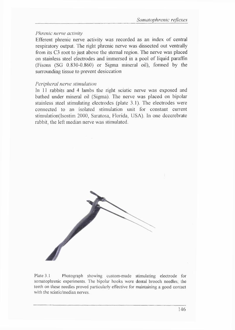
Somatophrenic reflexes
Phrenic nerve activityEfferent plirenic nerve activity was recorded as an index of central respiratory output. The right plirenic nerve was dissected out ventrally from its C3 root to just above tlie sternal region. The nerve was placed on stainless steel electrodes and immersed in a pool of liquid paraffin (Fisons (SG 0.830-0.860) or Sigma mineral oil), fonned by the surrounding tissue to prevent desiccation
Peripheral nerve stimulationIn 11 rabbits and 4 lambs tlie right sciatic nerve was exposed and bathed under mineral oil (Sigma). The nerve was placed on bipolar stainless steel stimulating electrodes (plate 3.1). The electrodes were comiected to an isolated stimulation unit for constant current stimulation(lsostim 2000, Saratosa, Florida, USA). In one decerebrate rabbit, the left median nerve was stimulated.
Plate 3.1 Photograph showing custom-made stimulating electrode for somatophrenic experiments. The bipolar hooks were dental brooch needles; the teeth on these needles proved particularly effective for maintaining a good contact with the sciatic/median nerves.
146
Somatophrenic reflexes
The electronic setup for the collection and processing of data is shown in figure 3.1. Typical settings are shown for the Neurolog modules. Cardiovascular dataBlood pressure was monitored fi*om a femoral artery (rabbit) or right brachial artery (lamb). The arterial catheter was attached by a 3-way stopcock to a disposable transducer (Viggo-Spectromed DT-XX) connected to a Neurolog N108 pressure amplifier or Harvard Apparatus blood pressure transducer. Full cahbration details for both blood pressure monitors are included in the Appendix. Heart rate was derived fi*om arterial blood pressure (Devices instantaneous ratemeter, type 2751).
Respiratory dataPhrenic activity was recorded using a bipolar stainless steel electrode (plate 2.2). Raw activity was passed through a headstage (NLIOOAK), to counteract any impedance effects and for common mode rejection. After pre-amphfication (AC pre amplifier, NL104A; gain x2000), the phrenic signal was filtered (50 Hz notch filter, NL125) between a narrow high and low bandwidth (5K respectively). The signal was then further amplified at variable gain (AC/DC amphfier, NL106) and window height (spike tigger, NL201) depending on the level of background noise present, before being integrated in 100ms bins (EMG integrator; NL 703).
Data collection and storageAll processed signals were passed to an oscilloscope (Gould (DSO) 1604), DAT tape deck (Biologic DTR-1800), chart recorder (Gould ES 1000) and CED 1401 (Cambridge Electronic Design). Each variable could be displayed and/or recorded using Spike 2 software, via the CED 1401, on an IBM-compatible computer (Nearward Computer Systems). Data was analysed from either Spike 2 software or chart paper.
3.5 E x pe r im e n t a l pr o t o c o l
The threshold parameters for altering respiratory output by electrical stimulation of a somatic afferent (50-100Hz; 0.1ms pulse width; current=threshold at which respiratory output was altered, for 5s) was determined during normoxaemia. After 5 minutes of isocapnic hypoxaemia, the somatic nerve was stimulated again at the threshold parameters determined during normoxaemia.
147
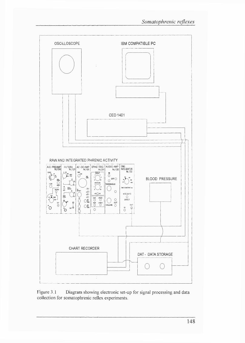
Somatophrenic reflexes
OSCILLOSCOPE IBM COMPATIBLE PC
C E D 1 4 0 1
RAW AND INTEGRATED PHRENIC ACTIVITY
E U 6INTEGRATOR
NL703
AUDIO AMP NL120
SPIKE TRIG. NL201
FILTERSNL125
AC-DC AMP NL106
BLOOD PRESSURE
INTEGRATE
W OUT VOLUME 0
CHART RECORDER
DAT- DATASTORAGE
Figure 3.1 Diagram showing electronic set-up for signal processing and data collection for somatophrenic reflex experiments.
148
Somatophrenic reflexes
3.6 A n a l y sis
Cardiovascular parametersABP is expressed as mean arterial pressure (diastolic pressure +1 / 3 (systolic pressure - diastolic pressure). HR is expressed in beats per minute. ABP and HR were analysed at one minute intervals throughout the two minutes of normoxaemia preceding hypoxaemia, and throughout the period of hypoxaemia. In addition, ABP and HR were analysed when threshold parameters were determined during normoxaemia and also during stimuh dehvered after 5 minutes of hypoxaemia had elapsed. Mean ABP and HR during in normoxaemia and after 5 minutes of hypoxaemia were compared. Statistical analysis was performed using Student's paired t- test. P<0.05 was considered significant.
Respiratory parametersCentral respiratory output was measured from the integrated phrenic neurogram. Since the absolute electrical activity measured from the phrenic nerve is dependent on the size of the nerve, integrity of its fibres and contact with the recording electrode, comparisons between animals are not appropriate unless this data is normalized (Eldridge, 1971). To normalize the data, a value of 100 units was assigned to the highest level of phrenic activity observed in an animal with the remainder of the data being scaled accordrugly.
The presence of a biphasic ventilatory response to hypoxaemia was determined by comparing the greatest product of PPA and respiratory frequency (1/Tt) with integrated phrenic activity 1 minute before hypoxaemia. The nadir of integrated phrenic output was also compared with phrenic activity at 1 minute prior to hypoxaemia. Phrenic activity during the period over which the sciatic nerve was stimulated were compared in terms of frequency (1/Tt) and PPA with the preceding control period. For 5 second stimuh, the control period would be the preceding equivalent number of breaths. For punctate stimuh, the analysis detailed in Chapter 2.6 was used.
Statistical analysis was performed using Wilcoxon’s signed rank test, due to non-uniform distribution of data. P<0.05 was considered significant.
149
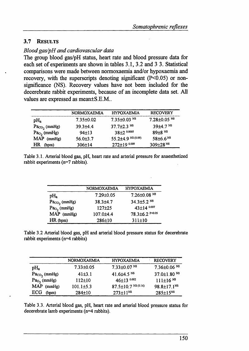
Somatophrenic reflexes
3.7 R e su l t s
Blood gas/pH and cardiovascular dataThe group blood gas/pH status, heart rate and blood pressure data for each set of experiments are shown in tables 3.1, 3.2 and 3 3. Statistical comparisons were made between normoxaemia and/or hypoxaemia and recovery, with the superscripts denoting significant (P<0.05) or nonsignificance (NS). Recovery values have not been included for the decerebrate rabbit experiments, because of an incomplete data set. All values are expressed as mean+S.E.M..
NORMOXAEMIA HYPOXAEMIA RECOVERY
pHa 7.35+0.02 7.35+0.03 7.28+0.05 NSPacoj (mmHg) 39.3+4.4 37.7+2.3 NS 39+4.7 NSPac>2 (mmHg) 94±13 38+2 89+8 NSMAP (mmHg) 56.0+3.7 55.2+4.9 NS (0-09) 58+6.6 NSH R (bpm) 306+14 272+19 309+28 NS
Table 3.1. Arterial blood gas, pH, heart rate and arterial pressure for anaesthetized rabbit experiments (n=7 rabbits).
NORMOXAEMIA HYPOXAEMIA
pHa 7.29+0.05 7.26+0.08 NSPacoj (mmHg) 38.3+4.7 34.3+5.2 NSPaoj (mmHg) 127+25 43+14MAP (mmHg) 107.0+4.4 78.3+6.2HR (bpm) 286+10 311+10
Table 3.2 Arterial blood gas, pH and arterial blood pressure status for decerebrate rabbit experiments (n=4 rabbits)
NORMOXAEMIA HYPOXAEMIA RECOVERY
pHa 7.33+0.05 7.33+0.07 NS 7.36+0.06 NSP acoj (mmHg) 41+3.1 41.6+4.5 NS 37.0+1.80 NSPaoj (mmHg) 112+10 46+13 0002 111+16 NSMAP (mmHg) 101.1+5.3 87.5+10.7 NS (016) 98.8+17. INSEG G (bpm) 284+10 273+1 INS 285+15NS
Table 3.3. Arterial blood gas, pH, heart rate and arterial blood pressure status for decerebrate lamb experiments (n=4 rabbits).
150
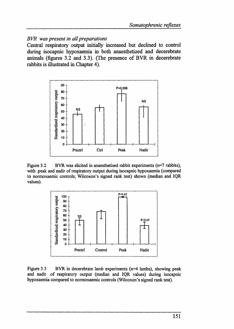
Somatophrenic reflexes
BVR was present in all preparationsCentral respiratory output initially increased but declined to control during isocapnic hypoxaemia in both anaesthetized and decerebrate animals (figures 3.2 and 3.3). (The presence of BVR in decerebrate rabbits is illustrated in Chapter 4).
90 TP<0.008
70 -■NS
NS
40 --
30 --
20 - -
NadirPrectrl Ctrl Peak
Figure 3.2 BVR was elicited in anaesthetised rabbit experiments (n=7 rabbits), with peak and nadir of respiratory output during isocapnic hypoxaemia (compared to normoxaemic controls; Wilcoxon’s signed rank test) shown (median and IQR values).
P<0.05"3 100 T f 9 0 -t 8 0 -3 7 0 -.§ 60 - § SO -
ê 30I 2 0 - I 10 -c/3 0 --
NSP<0.05
Prectrl Control Peak Nadir
Figure 3.3 BVR in decerebrate lamb experiments (n=4 lambs), showing peak and nadir of respiratory output (median and IQR values) during isocapnic hypoxaemia compared to normoxaemic controls (Wilcoxon’s signed rank test).
151
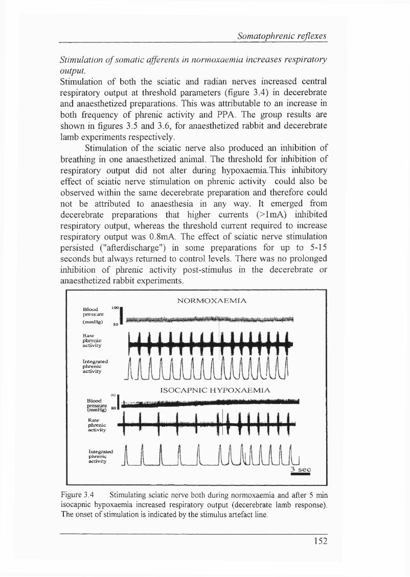
Somatophrenic reflexes
Stimulation o f somatic afférents in normoxaemia increases respiratory output.Stimulation of both the sciatic and radian nerves increased central respiratory output at threshold parameters (figure 3.4) in decerebrate and anaesthetized preparations. This was attributable to an increase in both fi-equency of phrenic activity and PPA. The group results are shown in figures 3.5 and 3.6, for anaesthetized rabbit and decerebrate lamb experiments respectively.
Stimulation of the sciatic nerve also produced an inhibition of breathing in one anaesthetized animal. The threshold for inhibition of respiratory output did not alter during hypoxaemia. This inhibitory effect of sciatic nerve stimulation on phrenic activity could also be observed within the same decerebrate preparation and therefore could not be attributed to anaesthesia in any way. It emerged fi*om decerebrate preparations that higher currents (>lmA) inhibited respiratory output, whereas the threshold current required to increase respiratory output was 0.8mA. The effect of sciatic nerve stimulation persisted ("afterdischarge") in some preparations for up to 5-15 seconds but always returned to control levels. There was no prolonged inhibition of phrenic activity post-stimulus in the decerebrate or anaesthetized rabbit experiments.
B loodpressure(mmHg) gQ IRawphrenicactivity
Integratedphrenicactivity
Blood
Rawphrenic I activity
N O R M O X A E M IA
IS O C A P N IC H Y P O X A E M IA
Integratedphrenicactivity
3 s e c
Figure 3.4 Stimulating sciatic nerve both during normoxaemia and after 5 min isocapnic hypoxaemia increased respiratory output (decerebrate lamb response). The onset of stimulation is indicated by the stimulus artefact line.
152
Somatophrenic reflexes
\
I-S
(/]
10090
80
70
60
50
40
30
20100
P=0.016
P=0.016P=NS
NS
NCmOCLAc o n l r d s d m u l u s
HYPOXIAomtid
HYPOXIAstimulus
Figure 3.5 Somatic afferent stimulated respiratory output during normoxaemia and after 5 min isocapnic hypoxaemia in anaesthetised rabbits. Similar results were seen for decerebrate rabbit experiments. P values refer to comparison with normoxia control; italicised P value refers to comparison of hypoxaemic stimulation with normoxaemic stimulation (median and IQR values; Wilcoxon’s signed rank test; n=7 rabbits).
I 80-r% 7 0 - “ 6 0 -
50 -55 40 -
3 0 --Q 2 0 -ia 1 0 - *0 -
p<0.05: NSP<0.05
NS
I
CO NORMOXIAcontrol
NORMOXIA stimulus
HYPOXIA control
HYPOXIA stimulus
Figure 3.6 Somatic afferent stimulation increased respiratory output during normoxaemia and after 5 min isocapnic hypoxaemia in decerebrate lambs. Median and IQR values shown. P values refer to comparison with normoxia control; italicised P value refers to normoxia vs. hypoxaemia stimulation comparison (Wilcoxon’s rigned rank test; n=4 rabbits).
153

Somatophrenic reflexes
No clear ejfect on timing of punctate somatic afferent stimuli on respiratory output during normoxaemiaPrevious work had shown that somatic afferent stimulation could affect the phase of the respiratory cycle (Iscoe & Polosa, 1976). However, punctate stimuh were not investigated extensively in the experiments presented in this Chapter, apart from in the decerebrate rabbit experiments. Punctate stimuh did not cause clear reproducible effects on phrenic activity in either anaesthetized or decerebrate rabbit experiments. In the decerebrate experiments punctate stimuh effected transient changes in phrenic activity but the relationship between the timing of the stimulus and the respiratory cycle did not emerge clearly from these preliminary experiments (table 3.4).
Table 3.4 shows the occurrence of an effect on respiratory output, caused by somatic afferent stimulation, in one decerebrate rabbit. There were no consistent effects produced at any stage of the respiratory cycle (denoted as % duration of Ti or Te). Furthermore, an effect could be different quahtatively even when the stimulus occurred at similar points during the respiratory cycle. This happened in ah these preliminary experiments investigating punctate stimuh, and is similar to the observations of inhibition (albeit limited) and stimulation in respiratory output in the same preparation.
Phase T to t
EFFECT NO EFFECT
PPAEFFECT NO
EFFECT
Ti(0-50) 2 9 2 7
Ti (51-100) 2 1 1 1
Te (0-50) 2 6 1 6
Te (51-100) 3 11 1 14
Table 3.4 In preliminary experiments, no consistent effects were seen with punctate stimuli. Table shows incidence of effects on PPA or TTOT when electrical stimulation was made during the first or second halves of inspiration and expiration.
154
Somatophrenic reflexes
Lack o f effect o f somatic afferent stimulation on blood pressure/heart rate.Clear cardiovascular changes were not observed in ABP or HR in any anaesthetized rabbit experiments during sciatic nerve stimulation at threshold. Similarly, in the decerebrate lamb experiments, small but variable cardiovascular changes were observed. At threshold, ABP increased by 6.4±1.9% in normoxaemia and by 6.3±2.5% in hypoxaemia. HR did not change.
Stimulation o f somatic afferents caused respiratory output to increase in isocapnic hypoxaemiaElectrical stimulation of the sciatic or radian nerve at the threshold values determined during normoxaemia again produced clear increases in phrenic activity after 5 minutes of isocapnic hypoxaemia (figure 3.4). The group hypoxaemic stimulation results are shown in figures 3.5 and 3.6, for anaesthetized and decerebrate experiments respectively. As in normoxaemia, afterdischarge was sometimes observed.
The peak in central respiratory output caused by somatic afferent stimulation during hypoxaemia was not different to that attained in normoxaemia (P>0.05 anaesthetized rabbits; P>0.05 lambs; Wilcoxon’s signed rank test). In some cases electrical sciatic nerve stimulation did increase respiratory output to a greater extent to that seen in normoxaemia. However, comparison of the % change in respiratory output over control confirmed that the peak in respiratory output attained by electrical stimulation did not change for both normoxaemia and hypoxaemia The anaesthetized rabbits showed an increase of 36% (IQR 31-49%) over control during normoxaemia vs. 25% increase (IQR 20-37%) during hypoxaemia (normoxaemic vs. hypoxaemic increase, P>0.05; Wilcoxon’s signed rank test). In the decerebrate lamb experiments, respiratory output increased by 33% over control (IQR 25-49%) on sciatic stimulation during normoxaemia, while during hypoxaemia a 38% increase (IQR 31-72%) was observed. Thus, overall, the group results revealed no consistently greater increase on hypoxaemic stimulation as compared to normoxaemic stimulation.
155
Somatophrenic reflexes
Somatic afferent stimulation also increases respiratory output in recoveryThe stimulation parameters determined during normoxaemia increased phrenic activity on recovery to normoxaemia.
Severe hypoxaemia/asphyxia did not abolish effect o f stimulating somatic afferents on respiratory outputDuring severe hypoxaemia/asphyxia (Pa,02 13mmHg; Pa,C02 80 mmHg) electrical stimulation of somatic afferents could still produce increases in respiratory output, even in apnoea.
Integratedphrenic
Rawphrenicaetivity
Stimulus offon
Figure 3.7 Sustained or transient stimulation of somatic afferents caused phrenic activity to return during severe hypoxaemia/asphyxia.
156
Somatophrenic reflexes
3.8 D isc u s s io n
MethodsDecerebrationDecerebrate, decerebellate preparations were used for two reasons. First, the effect of anaesthesia on somatophrenic reflexes could be assessed by comparing responses between the anaesthetized and decerebrate, unanaethetized rabbits. No difference was found between these two types of preparation in terms of their response to somatic afferent stimulation. Second, stimulation of receptors in hindlimb muscles or their afferent fibres causes a post-stimulatory depression in breathing, in addition to an increase in phrenic activity and subsequent short respiratory afterdischarge (Waldrop, Eldridge & Millhom, 1982). Both the cerebellum and suprapontine brain are required for this poststimulatory depression to occur. Therefore the decerebrate, decerebellate preparations provided a model which removed the potentially comphcating, inhibitory factors of the cerebellum and structures rostral to the superior coUicuh.
ResultsThe aim of these experiments was to assess how somatic afferent input is affected by hypoxaemia. That said, the data provides some interesting imphcations for interpretation of previous studies employing similar methodology.
The biphasic ventilatory response is present in both anaesthetized and decerebrate, decerebellate preparations.Both anaesthetized and decerebrate rabbit preparations exhibited a biphasic respiratory response, as measured fi’om the phrenic nerve. The magnitude of the biphasic ventilatory response was variable. Similarly, each decerebrate lamb (with the cerebellum removed) exhibited a biphasic ventilatory response. Therefore the mechanism underlying the biphasic ventilatory response is not dependent on the presence o f the cerebellum or structures rostral to the superior collicuh. The significance of this is discussed further in Chapter 4.
157
Somatophrenic reflexes
No clear effects o f somatic afferent stimulation on respiratory timing. No clear relationship emerged to suggest that sciatic or median nerve stimulation had phase-related effects on respiratory timing. The expiratory duration of the initial breath in anaesthetized rabbit preparations during a 5 second sciatic nerve stimulation was sometimes lengthened but there were no changes elicited on phrenic nerve activity that were consistent or comparable with chemical stimulation of the carotid chemoreceptors. Usually, transient stimuh apphed at any point in the decerebrate rabbit experiments induced either increases in PPA or Tt.
Comparable results from a recent similar study Trippenbach (1993) has reported that the stimulatory effect of electrical somatic afferent stimulation on breathing in newborn urethane anaesthetized newborn rabbits is age-dependent. In this study, the effect of F1 0 2 breathing during saphenous and vagus nervestimulation was assessed, using three arbitrarily chosen age-groups (1-3 days, 8-14 days and adults). During both normoxia and hypoxia, vagal stimulation shortened Te in all three groups. Saphenous nerve stimulation increased breathing in adults throughout hypoxia. In contrast, this stimulatory effect on breathing was abohshed in 8-14 day old rabbits. However, in 1-3 day old rabbits, the stimulatory effect of saphenous nerve stimulation was abohshed during phase 1.
From this work, it would appear that the results in this Chapter suggest that this somatophrenic reflex is unaffected in older rabbits (including the age used in this Chapter. However, some caution must be placed on this conclusion from the study of Trippenbach, since the Pao2 ranged from 24mmHg (youngest) to 37mmHg (oldest). Thus the effect of a similar degree of hypoxia for each age group was not investigated systematically. Furthermore, the threshold at which the saphenous- phrenic reflex was ehcited was not sought: rather, the lowest stimulus amphtude (voltage) that caused maximal stimulation of respiratory output was chosen. Clearly, this does not address the question of whether the threshold for ehciting the saphenous-phrenic reflex alters with age and/or hypoxia.
158
Somatophrenic reflexes
The effect o f changing stimulation intensityStimulation of the sciatic nerve also occasionally produced a depression in phrenic activity, usually at higher current intensities. These findings, in both anaesthetized and decerebrate experiments, support those of Senapti (1966) who showed that the ventilatory changes produced by stimulation of the lateral gastrocnemius-soleus nerve were dependent upon the strength of the electrical stimulus. In Senapti's study, increasing the stimulus strength could cause a depression in ventilation. At low stimulus strengths large fibres are activated; as the stimulus intensity increases smaller fibres are also recruited (Lloyd, 1943; Eccles & Lundberg, 1959). Senapti showed that the relationship between stimulus strength and ventilatoiy change was variable. In his study, increases in stimulus strength beyond that required to produce a maximal increase in breathing could cause breathing to fall. Other respiratory-related muscles have also been reported to show opposite effects on somatic afferent stimulation. Alae nasi activity shows either an increase or decrease on hind limb stimulation, although diaphragm activity always increased (Adams, van Lunteren, Chemiack & Haxhiu, 1985). Furthermore, stimulation of peripheral muscles inhibits phrenic motoneurones directly, in the spinal cord (Eldridge, Gill-Kumar, Millhom & Waldrop, 1981)
Morphometric analysis of the sciatic nerve in the adult rabbit (Germana, Mugha, Santoro, Abbate, Laura, Gughotta, Vita & Ciriaco, 1992) shows that the myelinated fibres with the largest diameter originating firom the sciatic nerve are distributed into the peroneal nerve, although there is no difference in the density of myelinated fibres between the sciatic nerve and its branches. The distribution of the myelinated fibre diameters is unimodal in the sciatic nerve, but bimodal in both the peroneal and tibial nerves. The axon diameter/fibre diameter ratio is different in the sciatic nerve firom its branches. With these morphometric differences, it is also possible that the depression in ventilation that was sometimes seen was due to damage of some fibres, thereby changing the balance of fibres recruited. The placement of the stimulating electrode also varied and this may mean that different populations of fibres were stimulated between preparations. In one experiment, moving the electrode progressively more centrally along the sciatic nerve, or its branches, suggested that placement of the electrode in different locations along the nerve may indeed affect the respiratory response seen. Both increases of variable magnitude and decreases were observed. However, this was not investigated systematically in each experiment.
159
Somatophrenic reflexes
Group I and II fibres are stimulated at the threshold used for the somatophrenic reflexBoth unmyelinated fibres (groups I and II) and myelinated fibres (types m and IV) mediate the cardiorespiratory reflex ehcited by somatic stimulation. The contribution of group I & II and group m & IV fibre types depends upon the intensity of electrical stimulation.
Senapti (1966) concluded that the reflex respiratory response ehcited by electrical stimulation of the lateral gastrocnemius-soleus nerve was mediated by group I and II type fibres. The fibre type stimulated was determined by relating the threshold required to increase ventilation to the threshold required, by stimulating the intact nerve, to cause contraction of the gastrocnemius muscle. This flexion reflex in cats is ehcited by group I and II fibres (Eccles & Lundberg, 1959). Similarly, other studies using electrical stimulation of stretch afferents (i.e. group I and n fibres) stimulate respiratory output (Bessou, Dejours & Laporte, 1959; Koizumi, Ushiyama & Brooks, 1961 ).Furthermore, Paintal (1960) found that the stimulus strength required to stimulate group m fibres varied fi'om 7 to 44 times the threshold of motor fibres. Group m afferent fibres are mostly connected to pressure-pain receptors. Electrical sciatic nerve stimulation at twice the threshold required to ehcit muscle twitch does not activate group HI and IV fibres (Kaufinan, Kozlowski & Rybicki, 1985; Kaufinan, Rybicki, Kozlowski & Iwamoto, 1986), but does increase ventilation in newborn kittens by ca. 35% over baseline.
Stimulation of group m afferents also causes respiratory output to increase, often outlasting the period o f stimulation (Senapti, 1966; Kaha, Senpati, Parida & Panda, 1972). Anodal block of group I and II fibres fails to abohsh the cardiovascular and respiratoiy responses to somatic afferent stimulation (McCloskey and MitcheU, 1972). In contrast, blocking unmyelinated and smah myelinated fibres (group m and IV fibres) by local anaesthesia abohshes the cardiovascular and respiratory responses to somatic afferent stimulation although group I and n fibre activity was often just reduced, and not eliminated, before cardiorespiratory responses were abohshed. Therefore high threshold afferents probably represent the most important afferent input to the somatophrenic reflex, with some contribution from group I and II fibres.
160
Somatophrenic reflexes
For the experiments reported in this Chapter, the fibre types stimulated were most likely to be group I and II. Stimulation of the intact nerve in some animals produced a small twitch at threshold levels, although this was not routinely determined. It is unlikely that the effects on respiratory output reported in this chapter are due to stimulation of nociceptive afferents because the threshold currents used were below those known to activate nociceptor afferent fibres (Burgess & Perl, 1967). The stimulation of group I and II fibre types in the experiments presented in this Chapter is also supported by the cardiovascular changes observed.
Evoking somatophrenic reflexes at threshold does not elicit strong cardiovascular reflexes due to stimulation of group I and II fibres Stimulation of group I and II fibres produce httle cardiovascular change (McCloskey & Mitchell, 1972; Sato & Schmidt, 1973), although activation of group IE and IV muscle afferents by increases in intramuscular pressure (Eldridge & Gill-Kumar, 1980), sciatic nerve stimulation (Kozelka, Christy & Wurster, 1982) and intramuscular tension causes ABP to rise (Mitchell, Kaufinan & Iwamoto, 1983; Stebbins, Brown, Levine & Longhurst, 1988). Heart rate increases during the early phase of muscle contraction due to vagal withdrawal (McMahon & McWiUiam, 1992); stimulation of group m and IV afferent fibres inhibit the cardiac vagal component of the barorefiex (McWiUiam & Yang, 1991).
In newborn kittens, muscle contraction and low intensity electrical sciatic nerve stimulation in normoxia causes smaU increases in ABP (<5mmHg) but HR remains unaltered (Parrish, HiU & Kaufinan, 1991). In contrast, ventilation increases by ca. 35% over baseline (a similar increase to that reported in the present Chapter for both anaesthetized rabbits and decerebrate lambs). In contrast, the same protocol in the adult cat caused considerably larger increases in ABP and HR, but not ventilation. Maximal electrical stimulation of the sciatic nerve caused a fourfold greater increase in systohc blood pressure compared to that seen with low intensity stimulation. Similarly, Trippenbach (1993) reported that electrical stimulation of the saphenous nerve only occasionaUy affected blood pressure (by decreasing MAP by <10% control), but not heart rate.
161
Somatophrenic reflexes
Therefore, the lack of effect on cardiovascular variables by sciatic nerve stimulation at the threshold parameters employed in the experiments presented in this Chapter suggest that group I and H fibres may mediate the observed effects.
Stimulation o f hindlimb afferents is unlikely to affect carotid chemoreceptor dischargeThe interpretation of these results may be complicated by the findings of Biscoe & Purves (1967), who demonstrated that carotid chemoreceptor activity increases and is maintained on passive limb movement. This increase in chemoreceptor activity persisted after cessation of hindlimb movements. The increase was essentially abohshed if the post-ganghonic branch fi'om the superior cervical ganghon, or both femoral and sciatic nerves, were ablated. This work disagrees with studies where squeezing calf muscles or stimulating muscle receptors in anaesthetized and paralyzed animals, with the vagi and carotid sinus nerves cut, still caused an increase in phrenic nerve activity (Parida, Senapti & Kalia, 1969; Eldridge & Gill-Kumar, 1980; Waldrop, Eldridge & Millhom, 1982). Also, hindlimb muscle contraction inhibits aortic chemoreceptor discharge (McCoy, Rotto & Kaufinan, 1987). Thus it is difficult to reconcile the Biscoe & Purves study with similar work in chemodenervated experiments.
Effects o f hypoxia and hypoxia-related substances on somatic afferentsHypoxia has very httle effect on the discharge of group in and IV muscle afferents (Hih, Pickar, Parrish & Kaufinan, 1992). Adenosine also has no effect, but lactic acid increases group HI and IV activity (Rotto & Kaufinan, 1988). Furthermore, chemical activation of group I and n has no effect on cardiorespiratory output (Waldrop, Rybicki & Kaufinan, 1984). Therefore, it seems reasonable to assume that the lack of effect of hypoxia on baseline somatic afferent discharge does not comphcate the interpretation of the experiments presented in this Chapter.
162

Somatophrenic reflexes
3.9 Su m m a ry
♦ Hypoxaemia does not cause global depression of respiratory- related reflexes.
♦ Removal of the cerebellum and brain structures rostral to the superior coUiculi does not alter the respiratory response to isocapnic hypoxaemia.
163

Section 2: CNS studies
RESULTS: SECTION 2
164
Section 2: CNS studies
RESULTS: SECTIO N!
THE ROLE OF THE BRAIN STEM AND MIDBRAIN IN MEDIATING THE EFFECTS OF
HYPOXIA ON BREATHING IN NEWBORNS
165
Section 2: CNS studies
RESULTS: SECTION 2 - THE ROLE OF CNS IN MEDIATING BVR
Overview o f Results: Section 2
Section 2 presents the results of an extensive series of experiments that I conducted to investigate the role of the brain stem in mediating the newborn respiratory response to hypoxaemia. Section 2 comprises three Chapters:-
Chapter 4To undertake Section 2 of the project a novel newborn rabbit preparation was developed to avoid the use of anaesthesia under experimental conditions. Because the newborn rabbit had not been extensively used for neurophysiological experiments before, a histological atlas of the newborn rabbit brain stem and midbrain also had to be constructed. The development of the surgery and monitoring of this decerebrate preparation is discussed, with the atlas of the brain stem of rabbits at the age used presented and discussed.
Chapter 5Data is presented that demonstrates for the first time that there is a specific neural pathway, and hence neuronal substrate, located in the mesencephalon which mediates the newborn BVR to hypoxaemia.
Chapter 6Extensive areas of the pons were explored to investigate whether more caudal regions of the brain stem may also be involved in mediating BVR in hypoxaemia.
166
Methods fo r CNS studies
CHAPTER 4
THE DEVELOPMENT OF A NEWBORN RABBIT PREPARATION FOR NEUROPHYSIOLOGICAL STUDIES INVESTIGATING THE ROLE OF THE BRAIN STEM AND
MESENCEPHALON IN MEDIATING BVR
167
Methods for CNS studies
4.1 In t r o d u c t io n
Investigating the role of the brain in the newborn presented some new problems. First, very little work had been undertaken to investigate the role of the brain in mediating BVR in the newborn. The methods used to investigate BVR in newborn rabbits would be to some extent new, as only lambs and kittens had been used previously for this type of study (Coles, Kumar & Noble, 1989; Smith, Li & Noble, 1993a,b). Since kittens are prohibitively expensive, and the availability of lambs is seasonal, it was necessary that a novel newborn preparation was developed. Newborn rabbits are relatively cheap, available throughout the year and clearly, from Chapters 2 and 3, were likely to be a suitable preparation for investigating the control of breathing. Therefore, I developed a newborn rabbit preparation to investigate the central mediation of BVR. In addition, because of the lack of a comprehensive stereotaxic atlas for the rabbit, be it adult or newborn, a histological atlas of the newborn rabbit brain stem had to be constructed.
The most important consideration was the stabihty o f the preparation. The type of studies that I envisaged undertaking would require the preparation to be stable for considerably longer than the experiments conducted in Chapters 2 and 3. Two factors were of particular importance. First, all anaesthetics affect breathing profoundly in the newborn. Second, I felt that administering additional doses: of anaesthesia in the course of a long experiment would be undesirable, because previous experience in the experiments of chapters 2 and 3 had shown that it was very difficult to estimate how much additional anaesthesia may be required without compromising respiratoiy output or cardiovascular stability. The advantages of using decerebrate preparations for neurophysiological work are widely acknowledged (e.g. see Lydie & Baghdoyan, 1992).
Therefore, I decided that a decerebrate preparation could be more suitable for studying the role of the brain stem in mediating BVR. This would obviate the need for anaesthesia. Furthermore, decerebration/transections at the level of the superior colHculus do not alter the adult (Tenny & Ou, 1977; Martin-Body, 1988) or newborn BVR (Hanson & WiUiams, 1989). As in intact fetuses, FBM are also inhibited during hypoxia in chronically decorticate fetal sheep (Ioffe, Janssen & Chemick, 1986).
168
Methods for CNS studies
This Chapter:(1) presents the development of a suitable newborn rabbit preparation for neurophysiological experiments(2) shows that the decerebrate rabbit preparation exhibits BVR(3) details the variety of methods used in Chapters 5 and 6 for investigating further CNS mechanisms that underly BVR.
4.2 A im : t o d e v e l o p n e w b o r n d e c e r e b r a t e pr e pa r a t io n
The initial aim of the CNS studies was to develop a decerebrate newborn rabbit preparation suitable for studying the role of the brain stem in hypoxaemia where:(1) BVR was still present in such preparations.(2) The absence of anaesthesia in such preparations promoted
cardiorespiratory stabihty.
4.3 M e t h o d s
The methods detailed in this Chapter were used subsequently throughout the experiments conducted in Chapters 5 and 6.
General methodsThe following procedures were conducted in accordance with the Animals (Scientific Procedures) Act 1986 (HMSO Publishing).
Animal delivery and careNewborn New Zealand White rabbits (bred and supphed by Froxfield UK. Ltd.) were dehvered to Biological Services, University College London, on, or one day before, the experiment and housed in accordance with the Code of Practice for the Housing and Care of Animals used in Scientific Procedures (HMSO Pubhshing, 1986). Animals that were dehvered on the day before an experiment were injected subcutaneously with the broad spectrum antibiotic Bayotril (2.5 %; 0.3 ml) to counter intestinal bacterial infection.
Age and weight 27 days (range 18-32); 564±12g
169
Methods fo r CNS studies
Sedation and anaesthesia.The rabbits were sedated with Ketalar (ketamine hydrochloride; Parke- Davis, 25 mg/kg). Anaesthesia was induced with halothane (May & Baker Ltd.) 2-2.5% dehvered through a facemask (Arnolds), via a Fluotec 3 halothane dispenser, in 4 1/min air (controlled using Platon Flowbits rotameters). Inspired halothane levels were monitored using a respiratoiy gas monitor (Ohmeda RGM5250).
Monitoring anaesthesiaWhen halothane was administered, the depth of anaesthesia was monitored by judging each of the following criteria:-(1) Arterial blood pressure
An increase in femoral arterial blood pressure, on pinching the hind limb, was used as indicator for the administration of supplemental anaesthesia. The patency of the catheter placed in the femoral artery was maintained by regular flushes with Ringer’s saline.
(2) Heart rateHeart rate was monitored from an instantaneous ratemeter (type 2751, Devices), which was derived fron the femoral pulse pressure. An increase in heart rate on pinching a hind limb was used as an indicator for the administration of supplemental anaesthesia.
(3) Hind limb withdrawal reflexes in the non-paralyzed stateLack of hind limb withdrawal reflex indicated satisfactory anaesthesia.
Ventilation.The neck and left femoral region were shaved (in preparation for tracheostomy and femoral vessel cannulation. Once surgical anaesthesia was achieved, a tracheostomy was performed. An oral/nasal tube (Portex (size 2.5)) was introduced into the trachea and secured with braided silk suture (Pearsalls Sutures; size US 2/0). The upper airway was tied off with tightly secured silk suture. The rabbit was ventilated using a small animal ventilator (Harvard Apparatus 707 ventilator).
Vagotomy and phrenic nerve activityOnce the rabbit’s head was held supine in the stereotaxic frame, mid cervical bilateral vagotomy was performed. Then both phrenic nerves were dissected, from the C3 root. Each phrenic nerve was tied off with braided silk suture just above its entrance into the chest. A strand of this tie was then attached to another silk suture tie which was secured onto branches of the brachial plexus. This enabled easy identification of the location of the nerve once the animal was secured in the stereotaxic frame in a prone position. When the animal was in this latter position the identifying suture
170
Methods for CNS studies
DecerebrationOnce each phrenic nerve was dissected and marked, the animal was placed prone in the stereotaxic frame. The frir overlying the occipital, frontal and parietal bones was shaved and the skin removed. A 1cm hole was then drilled in the frontal bone of the cranium; the bone was removed back from this area to ca. 1 cm from the junction of the parietal and occipital plates using Lemperts angular forceps (Downs Surgical, UK). These forceps enabled the soft bone to be cut, rather than tom, so preventing bleeding from small vessels located in the skull.
Following craniotomy, the dura mater was cut before the entire forebrain and midbrain, to the most rostral end of the mesencephalon at the level of the superior colHculus, was removed by aspiration (using a 1ml syringe attached to a suction pump).The cavity was filled with oxidised cellulose (Oxycel, Becton Dickinson, UK) to prevent execessive bleeding. Oxycel was also often placed around the remainder of the skull surrounding the cavity. Once decerebration was complete, halothane administration was decreased to half the dose previously required for maintenance of anaesthesia. After 10-15 minutes halothane dehvery was stopped. At this point, the animal was paralysed (Flaxedil 4-8 mg/kg i.v.; May & Baker, UK). On two occasions, Norcuron (Organon Teknika Ltd, UK) was used for paralysis.
Monitoring o f pH, blood gases and body temperature.Body temperature and blood gas status were carefully controlled to be within the normal physiological range throughout surgery and experimental period. Body temperature was servo-controlled throughout the preparation, by means of a temperature probe inserted into the rectum, a homeothermic blanket (CFP 8185 homeothermic blanket), heating lamps or a fan heater.
Blood gas and pH status was analysed by anaerobic removal of 0.2 ml of aortic arterial blood, via a three way tap inserted between the catheter and the blood pressure transducer, using a pH/blood gas analyser (1306 series Instrumentation Laboratory). Sample values were corrected for body temperature. End tidal gases were monitored using an Ohmeda 5250 respiratory gas monitor. This was essential for ensuring adequate oxygenation and monitoring the animals metaboHc state (assessed from PETCO2 ) without having to continually remove blood for blood gas sampling.
Glucose (5% w/v in Ringer’s saline; Sigma, UK) was usually administered i.v. via the femoral vein at ca. 3ml/min (model 11 infusion pump. Harvard
172
Methods for CNS studies
Apparatus, UK) to ensure adequate metabolism. Sodium bicarbonate (8.4% w/v in Ringer’s saline; Sigma, UK) was also infused i.v. via the femoral vein in the event of metabohc acidosis.
Electrical stimulationA biploar concentric stimulating electrode (Rhodes SNE-100, 50mm length) was firmly attached to a stereotaxic micromanipulator. The initial rostro- caudal and mediolateral placement of the electrode tip at the surface of the brain were noted (see Chapters 5 and 6 for zero/reference stereotaxic points. The depth of electrode placement was measured by. the distance from the surface of the brain. An isolated current stimulator (Isostim 2000, World Precision Instruments Inc., USA) was used to pass current through the electrode (100-350pA, pulse width 0.1ms, frequeuncy 50-100 Hz , duration 5-10 second). Cahbration details for the Isostim 2000 are included in the Appendix. Electrolytic lesions were made by passing higher currents (>3mA) for 20 seconds at high frequency (200Hz).
Chemical stimulationThree methods were tested to determine the most effective way of injecting small quantities of solutions into the brain.(a) Hamilton microhtre syringes (Hamilton Co., Canada): The micropipette was secured in a microelectrode holder (EH-2MS, Clark Electromedical Instruments, UK), which was connected by a side port to inelastic tubing leading to the tip of the Hamilton syringe. This set-up was unsatisfactory. It was very difficult to dehver accurate volumes because of the resistance of the dehvery tubing and the difficulty in building up enough pressure with the Hamilton syringe.(b) Micromanipulator dehvery system (Clark Electromedical Instruments, UK): The micropipette was held as described in (a). Although accurate volumes of solutions could be dehvered using this system, the volume of the injectâtes were usually far greater than intended because of the difficulty encountered with building enough pressure up to overcome the resistance of the microelectrode and tubing.(c) Pressurized picopump (PV820, World Precision Instruments Inc., USA).: This method was the only acceptable way of injecting solutions. Very smah volumes could be injected, even down to 3r|l. Furthermore, they could be measured accurately by the placement of a graticule behind the micropipette.
173
Methods for CNS studies
Solutions were microinjected via glass micropipettes (GClOOF-10; 1.0mm O.D. X 0.58mm I.D.; Clark Electromedical Instruments, UK) which were pulled to a fine tip diameter (Flaming/Brown micropipette puller, model P87; Sutter Instruments Co., USA) before being broken back to typically « 10pm. Each micropipette was filled using a stretched catheter (size 2FG, Portex, UK) with a range of solutions (see below). The electrodes were held securely in a micropipette holder; the holder was secured to a stereotaxic micromanipulator. 20 min elapsed between successive microinjections.
Microinjected solutions :L-glutamate (Sigma, UK): <0.5M in Ringer’s saline / NaOH (IM); pH 7.4 D,L-homocysteate (Sigma, UK): <0.2M in Ringer’s saline; pH 7.4 Kainic acid (Sigma, UK): Ipg/pl in Ringer’s saline Ringer’s saline: as described previously (Chapter 2)GAB A (Sigma, UK): <1M in Ringer’s saline
All solutions were coloured by the addition of Fast Green dye (pH 7.4; Sigma, UK) for histological verification of the location of microinjection sites. The pH of solutions was measured using a pH meter (model 3070; Jenway, UK).
Measurement o f microinjection volumesA graticule (1mm divided into 100 units) was placed behind the glass pipette. By adding a drop of mineral oil (Sigma, UK) to the glass pipette, a clear meniscus was visible between the mineral oil and Fast Green coloured solution. The volume of the microinjection was calculated by inserting the distance in d (m) moved by the meniscus into the equation:
Volume of microinjectate = d (htres)
(where r = internal radius (m) of glass pipette; tü = PI or 3.142)
Cahbration of the volumes ejected at rninimum pulse pressures and varying pulse pressure durations was carried out for each pipette before it was placed in the mesencephalon. In this way, damaged or blocked pipettes could be rejected.
174
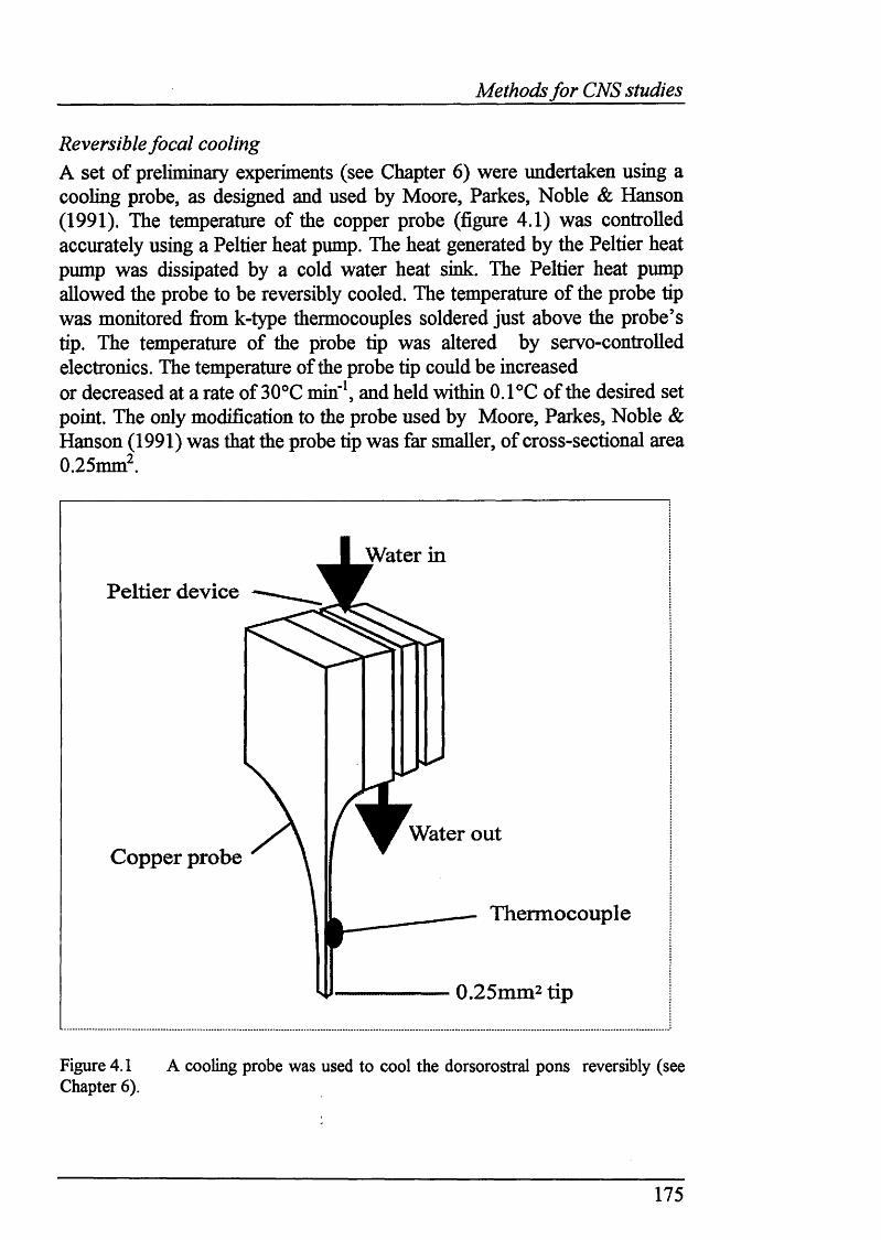
Methods fo r CNS studies
Reversible focal coolingA set o f preliminary experiments (see Chapter 6) were undertaken using a cooling probe, as designed and used by Moore, Parkes, Noble & Hanson (1991). The temperature of the copper probe (figure 4.1) was controlled accurately using a Peltier heat pump. The heat generated by the Peltier heat pump was dissipated by a cold water heat sink. The Peltier heat pump allowed the probe to be reversibly cooled. The temperature of the probe tip was monitored fi'om k-type thermocouples soldered just above the probe’s tip. The temperature of the probe tip was altered by servo-controlled electronics. The temperature of the probe tip could be increased or decreased at a rate of 30®C min"% and held within 0. PC of the desired set point. The only modification to the probe used by Moore, Parkes, Noble & Hanson (1991) was that the probe tip was far smaller, of cross-sectional area 0.25mm^.
Water inPeltier device
Copper probeWater out
Thermocouple
0.25mm2 tip
Figure 4.1 A cooling probe was used to cool the dorsorostral pons reversibly (see Chapter 6).
175
Methods for CNS studies
Histological methodsThe following methods were used for all brain tissue presented throughout Results: Section B. A complete atlas of the newborn rabbit brain stem is presented in Appendix 3.
Preparation o f slides for staining o f sectionsGlass microscope shdes (BDH, UK) were subbed in order to provide an adhesive surface for the placement of brain tissue shces for staining. Gelatin (Sigma,UK) was dissolved in water at 50°C. Chrome alum (chromic potassium sulphate; BDH, UK) was then added to the gelatin solution, with the resultant solution being filtered. The shdes were dipped in this warm filtered solution and diyed.
Preparation o f tissue prior to sectioningEach rabbit brain was usually perfused with Ringer’s saline injected intracardially. The brain was then removed by cutting the the rostral end of the spinal cord with a spatula and placed in 10% formalin (Sigma, UK). After 72 hours, the brain was removed from the formalin solution and usually placed in 10% sucrose solution (Sigma, UK) 24 hours before sectioning. Immediately prior to sectioning, the brain was bathed in distilled water.
Sectioning o f tissue100pm sections were cut on a freezing microtome (Leitz-Wetzlar, Germany). The brain was placed on a small area of paper tissue and frozen on top of a platform that could be reversibly cooled by a Peltier heating device (Pelcool, MSE Ltd, UK). Brain tissue shces were removed from the microtome blade with a wet paintbrush and placed in smaU dishes of distiUed water. Each shce was then mounted on a glass shde using a paintbrush that had been dipped in distilled water.
Photography o f tissue slicesNearly ah the brain tissue that was sectioned was photographed by exposing print paper (Multigrade 3 RC deluxe; Ilford, UK) beneath the shdes upon which the tissue was mounted. Once the photographic print paper had been exposed under a suitable filter/aperture (typicahy for 10-30s% it was placed in a bath containing Multigrade developer (1 part developer:9 parts H2 O) for the period o f time necessary for the image of the brain tissue to develop satisfactorily. After being washed in tap water, the developed print was
176
Methods for CNS studies
fixed in fix solution (Hypnonn H9; 1 part fix: 4 parts H2 O). After fixing for 2-3 min, the prints were washed again in tap water and left to dry overnight.
Histological staining of brain tissueAll tissue was stained as well as photographed. After being photographed, the shdes were assembled in a tray and initially bathed in buffer solution (9 parts 0.2M sodium acetate (S i^ a , UK): 0.2M glacial acetic acid (Sigma, UK), pH 4.5). After 5 min in buffer, the shdes were dipped in cresyl violet stain (0.2g in 100ml buffer; Sigma, UK) for 12-20 min. The shdes were then passed through a successive series of dishes containing 70%, 95% and 100% ethanol (Sigma, UK), for 3 min at a time. FinaUy, the shdes were dipped in Histocleanse (BDH, UK). Cover slips were mounted over each shde using DPX (BDH, UK).
177
Methods for CNS studies
4.4 Sig n a l pr o c e ssin g a n d d a ta c o l l e c t io n
The electronic set-up for signal processing and data analysis was identical to that shown in Chapter 3. Due to the large amount of data collected throughout these experiments. Spike! capture software was only used for experiements of short length, because of hard disk capacity restrictions on the IBM-compatible PC.
4.5 Ex pe r im e n t a l pr o to c o l
The decerebrate rabbits were exposed to isocapnic hypoxaemia for 8 minutes, with arterial blood gases taken after 4 minutes of hypoxaemia. If a rabbit went apnoeic, or respiratory output fell severely, the rabbit was ventilated immediately in normoxia again.
4.6 A n a l y sis
Central respiratory outputIn all cases, PPA, T t and Ti were measured from the integrated phrenic neurogram. The product of PPA and frequency of respiratory output (1/ T t)
was calculated, to give the neural equivalent of ventilation.
Electrical stimulationPhrenic nerve activity was assessed over the preceding five breaths before
the stimulus was apphed. The effect of electrical stimulation on central respiratory output was classified as stimulation (increase in PPA x f), inhibition (decrease in PPA x f) or mixed (combination of stimulation and inhibition). In the vast majority of stimulations, clear increases or decreases in respiratory output could be observed, even without measurement of respiratory variables.
178
Methods for CNS studies
Microinjectate stimulationPhrenic nerve activity was assessed over the five breaths before the onset of the microinjection (control) and during the period of the microinjection. If respiratory output changed outside this period, the response was considered not to have been mediated at the site of the micropipette tip and therefore disregarded. To ensure that any changes due to microinjections were not due to the inherent variablity of respiratory output, the first two breaths of the control period were compared with the five preceding breaths (precontrol).
Cardiovascular parameters:Arterial blood pressure is expressed as MAP (diastohc pressure + 1/3 (systolic pressure-diastolic pressure)). Heart rate is expressed in bpm. For the hypoxaemic experiments, both HR and ABP were measured at one min intervals 2 min before, and throughout, isocapnic hypoxaemia. ABP and HR at 5 min of isocapnic hypoxaemia were compared with control.
Respiratory response to isocapnic hypoxaemiaFive breaths were analyzed at one minute intervals 2 min. prior to hypoxaemia (pre-control) and throughout the isocapnic hypoxaemic period. The presence of BVR was determined by comparing the peak neural ventilation during isocapnic hypoxaemia with that at 1 min. before the onset of hypoxaemia (designated as control). The nadir of central respiratory output during isocapnic hypoxaemia was also compared with respiratory output at 1 min. prior to hypoxaemia and peak respiratory output during hypoxaemia. A similar analysis was used for the cooling experiment (Chapter 6).
179
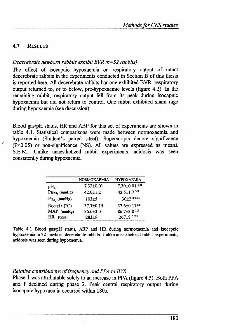
Methods for CNS studies
4.7 R e su l t s
Decerebrate newborn rabbits exhibit BVR (n=32 rabbits)The effect of isocapnic hypoxaemia on respiratory output o f intact decerebrate rabbits in the experiments conducted in Section B of this thesis is reported here. All decerebrate rabbits bar one exhibited BVR: respiratory output returned to, or to below, pre-hypoxaemic levels (figure 4.2). In the remaining rabbit, respiratory output fell fi*om its peak during isocapnic hypoxaemia but did not return to control. One rabbit exhibited sham rage during hypoxaemia (see discussion).
Blood gas/pH status, HR and ABP for this set of experiments are shown in table 4.1. Statistical comparisons were made between normoxaemia and hypoxaemia (Student’s paired t-test). Superscripts denote significance (?<0.05) or non-significance (NS). All values are expressed as meant S.E.M.. Unlike anaesthetized rabbit experiments, acidosis was seen consistently during hypoxaemia.
NORMOXAEMIA HYPOXAEMIA
pHa 7.32+0.01 7.3010.01Pacoj (mmHg) 42.0+1.2 42.511.7^"Paoj (mmHg) 103+5 3012Rectal t (°C) 37.7±0.15 37.6+0.17NSMAP (mmHg) 86.613.0 86.7±3.8°o8
HR (bpm) 283+9 26718 o
Table 4.1 Blood gas/pH status, ABP and HR during normoxaemia and isocapnic hypoxaemia in 32 newborn decerebrate rabbits. Unlike anaesthetized rabbit experiments, acidosis was seen during hypoxaemia.
Relative contributions o f frequency and PPA to BVR Phase 1 was attributable solely to an increase in PPA (figure 4.3). Both PPA and f declined during phase 2. Peak central respiratory output during isocapnic hypoxaemia occurred within 180s.
180
Methods for CNS studies
P « 0 .0 0 0 1
P = 0.10
e 300
PL, 150
Pl-ectrl
Figure 4.2 Newborn decerebrate rabbits show a biphasic response in respiratory output to 8 min of isocapnic hypoxaemia (n=32 rabbits). P values refer to statistical comparisons made between mean respiratory output at peak/nadir and precontrol with control (Student’s paired t-test).
I
14
12
10
P0.00008
PO.0G05
Î
Ε|
NORMOXIA FHASEl PHASE2
Figure 4.3 Graph showing PPA during control, phase 1 and phase 2 of BVR (n=32 rabbits). P values refer to statistical comparisons made between mean respiratory output at peak/nadir with control (Student’s paired t-test).
181
Methods for CNS studies
I
45 j
40 --
35
30
25 --
20 - -
15 --
10 -
5 --
P < 0 . 0 0 0 0 0 6
NORMOXIA P H A S E 1 P H A S E 2
Figure 4.4 Respiratory frequency during control, phase 1 and phase 2 o f BV R (n=32 rabbits). P values refer to statistical comparisons made between mean respiratory output at peak/nadir with control (Student’s paired t-test).
Sham rage occurred in one rabbit during isocapnic hypoxaemiaSham rage occurred in only 1/32 rabbits exposed to isocapnic hypoxaemia. This rabbit was exposed to Pao2 SOmmHg.
Arterial blood i30| pressure I(m m H g )
Integratedphrenicactivity
Rawphrenicactivity
50'
8 sec
Figure 4.5 Sham rage in one decerebrate rabbit was characterized by ABP increase (70% over control ABP), plus an initial augmentation o f PPA, follwed briefly by apnoea.
182
Methods for CNS studies
Halothane depresses cardiorespiratory output markedly in newborn decerebrate rabbits (n=2) and lambs (n=2)Subanaesthetic doses (0.5-1%) of halothane decreased profoundly ABP, HR and respiratory output in both decerebrate lambs and rabbits. The lambs were particularly susceptible to halothane administration, with just 0.5% concentration abohshing respiratory output.
Histological techniquesAnalysis of brain tissue was accomphshed using standard histological techniques. Shrinkage was calculated from some shces by measuring the % change between two tracks a known distance apart. In most shces, measurement of the distance between electrode tracks revealed shrinkage to be ca. 5-10 %.
183
Methods fo r CNS studies
4.8 D isc u ssio n
MethodsDecerebration conferred stabilityThe initial experiments revealed that decerebration could be successfiilly undertaken, as long as the craniotomy was completed carefully avoiding obvious venous sinuses and particularly vulnerable vessels located in the occipital plate of the skull. For this purpose, speciahsed forceps were essential. The most striking effect of decerebration was the disinhibition of phrenic activity and blood pressure once halothane anaesthesia had been discontinued. Typically MAP would increase by 40-50mmHg. Heart rate changes were more variable, but generally levels were similar to those seen under halothane anaesthesia. All the decerebrate animals breathed, and would do so spontaneously until paralysed. Furthermore, all decerebrate preparations were usually very stable in terms of blood gases/pH. Thus these preparations were viable for very long periods (often >14 hours). Most importantly, all decerebrate animals exhibited BVR as presented in Chapter 5. Therefore the decerebrate newborn rabbit preparation was employed in all experiments investigating the role of the brain stem in mediating BVR. A prospective comparison between decerebrate and anaesthetized rabbits to assess their relative stability was not conducted, although it was clear that the duration of the brain stem study experiments made repeated doses of anaesthesia impractical, due to the deleterious, and often fatal, effect of such additional anaesthetic administration found previously.
Histological techniques proved suitableThe methods used for histological analysis of electrode tracks and microinjection sites proved satisfactory. Both photographs and stained tissue shces helped identify electrode tracks, but tissue stained with cresyl violet demarcated cell bodies more clearly. The only drawback of the histological methods used was the use of a freezing microtome, because the tissue did not always cool uniformly. Occasionally shces of varying thickness were cut (i.e. ca. 125pm instead of 100pm). However, this had no effect on the interpretation of the position of electrode tracks.
184
Methods for CNS studies
ResultsDecerebrate newborn rabbits exhibit BVRPrecoUicular decerebration did not affect the presence of BVR during hypoxaemia. This agrees with similar work in newborn rabbits (Martin-Body & Johnston, 1988) and rats (Hanson & Wilhams, 1989). The differences between BVR in decerebrate and urethane anaesthetized rabbits was the lack of increase in f during phase 1, and persistence of elevated PPA in the decerebrate preparations. Phase 1 was attributable solely to an increase in PPA.
Sham rage occurred in only one rabbit.Decorticate or thalamic (i.e. prechiasmatic decerebration) adult animals exhibit sham rage, characterized by a rise in arterial blood pressure, increased respiratory output, pupillary dilatation, retraction o f nicitating membranes and phasic limb movement (Bizzi, Libretti, Malhani & Zanchetti, 1961). Sham rage is mediated by diencephalic mechanisms and evoked by stimulation of the carotid chemoreceptors, particularly during hypoxia (Bizzi, Libretti, Malliani & Zanchetti, 1961). Sham rage was observed in only one rabbit.
Arterial blood pressure showed “biphasic response** to isocapnic hypoxaemiaArterial blood pressure increased during isocapnic hypoxaemia, only to return to pre-hypoxaemic levels. Thus, blood pressure showed a “biphasic type” response. This is predictable from the similar pattern exhibited during hypoxia by decerebrate adult rabbits (Komer, 1971)
185
Methods fo r CNS studies
4.9 Su m m a r y
♦ Decerebrate newborn rabbits show BVR to isocapnic hypoxaemia
♦ The decerebrate newborn rabbit preparation is for extensive neurophysiologjcal studies in the brain stem
186
Mesencephalon
CHAPTER 5
BVR IS MEDIATED BY STRUCTURES IN THE MESENCEPHALON
187
Mesencephalon
5.1 I n t r o d u c t i o n
Although the visionary concept of CNS mediated inhibition of breathing was first raised nearly 50 years ago, by Sir Joseph Barcroft (1946), there is no compelling evidence to suggest any particular neuronal substrate may be involved. The background to the experiments investigating the inhibition of breathing during hypoxia is detailed in Chapter 1.
From previous studies, no obvious areas have emerged to suggest that they may mediate BVR. The outstanding paradox hes in the claim that areas in the upper pons mediate BVR, rather than suprapontine areas as suggested firom the effects of transections at the midcolhcular junction. It is unlikely that the lesion studies in the rostral pons conducted in the fetal lamb by Gluckman & Johnston (1987) may reconcile the two possibihties, because the reproducibihty of the lesions, and their effects on FBM, are not clear. None of the other studies in the upper pons have used the array of neurophysiological tools required to show that cell bodies mediate BVR. Clearly, many of these studies have been incomplete. That said, the nature of this work, which has been pursued almost exclusively in the fetus and newborn, is certainly very difficult.
The only evidence to suggest that a fall in breathing could be caused by a supra-pontineneuronal pathway comes fi’om the study of Gallman & Millhom (1991). However, this set of experiments is not entirely convincing. This study set out to determine whether cell bodies in the mesencephalon were responsible for the persistence of decreased breathing in hyperoxia alter hypoxia. Glutamate was microinjected to determine whether cell bodies mediated the inhibition of breathing produced by electrical stimulation in a particularly discrete region in the red nucleus. The injection volumes used were large (see Chapter 4; Lipski, Bellingham, West & Pilowsky, 1988) and thus the degree of microinjectate spread was not clear. Most notably, the latency of onset in changes in phrenic nerve activity due to microinjections was very long, which strongly suggests that the effects of the microinjections were not mediated at the tip of the microelectrode. This renders the authors’ conclusion that the inhibitory effects of chemical stimulation on phrenic nerve activity were due to depolarization of cell bodies in the immediate vicinity of the micropipette debatable.
I hypothesized that the results of transection studies were not due to the perturbation of structures in the upper pons, caudal to the transections: rather the influence of structures rostral to the midcolhcular junction
188
Mesencephalon
mediated phase 2 of BVR. Four types of experiments were undertaken totest this hypothesis.
(1) Electrical stimulation of the mesencephalon in normoxia would reveal whether there were sites which inhibited respiratory output, as shown by Gallman & Millhom (1991).
(2) At sites where electrical stimulation caused inhibition of respiratory output, microinjections of EAA such as glutamate (see Goodchild, Dampney & Handler, 1982) would be made in normoxia or hyperoxia. If microinjection of EAA caused respiratory output to fall, the source of inhibition would be attributable to cell bodies, rather than axons of passage.
(3) Bilateral electrolytic lesions would be made at sites shown to inhibit respiratory output to determine whether this would abohsh phase 2 of BVR.
(4) Pharmacological inhibition of cell bodies mediating an inhibitory effect on respiratory output, using lAA such as GAB A, should facihtate respiratory output. Facihtation of respiratory output only during hypoxia would suggest that the cell bodies involved do not exert a tonic inhibitory influence on respiratory output. Rather this would suggest that hypoxia effectively switches on an inhibitory mechanism, as suggested by Moore, Parkes, Noble & Hanson (1991).
5.2 H y po t h e se s
(1) Phase 2 of BVR is abohshed by the destruction of a group of mesencephahc cell bodies, demarcated by electrical and chemical stimulation, in newborn decerebrate rabbits.
(2) Any putative mesencephalic inhibitory mechanism is only functional during hypoxia.
189
Mesencephalon
5.3 M e t h o d s
The methods used in this Chapter are detailed in Chapter 4.
5.4 P r o t o c o l s
Three separate experiments were conducted to address the hypotheses.
Experiment 1: Identification o f areas that mediate inhibition o f respiratory outputBoth electrical stimulation and microinjection of EAA were used to identify areas that caused an inhibition of respiratory output. In each experiment, the length from the most rostral end of the mesencephalon (i.e. to the point of decerebration) to the junction of the inferior and superior colhcuh was measured by moving the tip of either type of electrode using a stereotaxic micromanipulator. Mediolateral coordinates were noted by the distance from the midline dividing the superior colhcuh.
Experiment 2: The respiratory response to isocapnic hypoxaemia after areas that had been demonstrated to inhibit respiratory output are destroyed by electrolytic lesions.Experiment 2 consisted of 3 steps:
Step 1: To show that each decerebrate newborn rabbit exhibited BVR to acute isocapnic hypoxaemia.After 2-3 min. recording respiratory output (as measured from phrenic nerve activity) in normoxaemia, each decerebrate rabbit was exposed to 8 min. of isocapnic hypoxaemia, before return to normoxaemia. Blood gas status was ascertained in normoxaemia and after 4 min. of isocapnic hypoxaemia.
Step 2: Bilateral electrolytic lesioning o f areas in the mesencephalon that cause an inhibition o f respiratory outputAfter at least 30 min. recovery from step 1, the electrical stimulation procedure described in Chapter 4 was carried out to search for areas that caused an inhibition of respiratory output (as measured from the phrenic nerve) in normoxaemia or hyperoxia. Once demarcated, these areas were electrolyticaUy lesioned. This was repeated on both sides o f the midline.
190
Mesencephalon
Step 3: Measure respiratory response to isocapnic hypoxaemia indecerebrate rabbit in which electrolytic lesions had been made bilaterally The following criteria had to be strictly satisfied to proceed fi*om step 2 to step 3:(a) Only areas that mediated profound inhibition of respiratory output (as determined by electrical stimulation), or apnoea, were lesioned.(b) Respiratory output and cardiovascular variables were regular and within normal (precontrol) parameters.
However, in the event of failing to satisfy the above criteria, step 3 was still undertaken. For example, if only mixed effects (i.e. initial stimulation followed by apnoea) were found in one side of the mid brain (or in a preparation) the region was lesioned. Although these experiments failed to satisfy the criteria (a) and (b), they served as useful controls.
The level of Fio, used during the hypoxic run for step 1 was repeated.
Experiment 3: Does inhibition of a inhibitory mesencephalic area increase respiratory output?The mesencephahc area that caused an inhibition of respiratory output was located by electrical stimulation on one side of the mesencephalon. Ipsilateral microinjections of GAB A were then made during normoxia and after 7 minutes of isocapnic hypoxaemia (or sooner if respiratory output had fallen to apnoea).
5.5 A n a l y sis
The analysis for electrical and microinjection experiments is detailed in Chapter 4. The presence of BVR was tested for as described in Chapter 4. However, in the event of a pattern of sham rage in phrenic activity, the period of respiratory output immediately before the onset of this behaviour was analyzed to ascertain peak respiratory output under regular conditions.
191
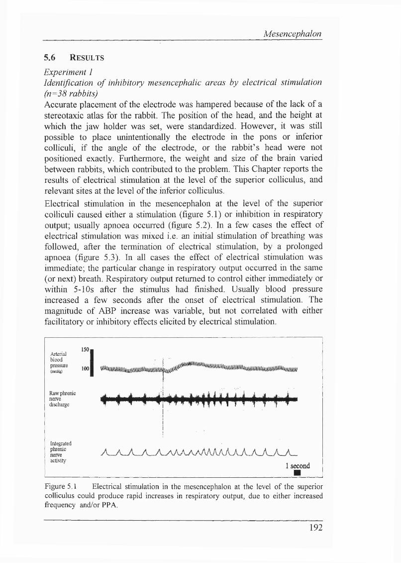
Mesencephalon
5.6 Results
Experiment 1Identification o f inhibitory mesencephalic areas by electrical stimulation (n=38 rabbits)Accurate placement of the electrode was hampered because of the lack of a stereotaxic atlas for the rabbit. The position of the head, and the height at which the jaw holder was set, were standardized. However, it was still possible to place unintentionally the electrode in the pons or inferior colliculi, if the angle of the electrode, or the rabbit’s head were not positioned exactly. Furthermore, the weight and size of the brain varied between rabbits, which contributed to the problem. This Chapter reports the results of electrical stimulation at the level of the superior colliculus, and relevant sites at the level of the inferior colliculus.Electrical stimulation in the mesencephalon at the level of the superior colliculi caused either a stimulation (figure 5.1) or inhibition in respiratory output; usually apnoea occurred (figure 5.2). In a few cases the effect of electrical stimulation was mixed i.e. an initial stimulation of breathing was followed, after the termination of electrical stimulation, by a prolonged apnoea (figure 5.3). In all cases the effect of electrical stimulation was immediate; the particular change in respiratory output occurred in the same (or next) breath. Respiratory output returned to control either immediately or within 5-10s after the stimulus had finished. Usually blood pressure increased a few seconds after the onset of electrical stimulation. The magnitude of ABP increase was variable, but not correlated with either facilitatory or inhibitory effects elicited by electrical stimulation.
Arterial ■ blood 1
discharge ^ f f ^ ^
11
Integrated
activity
' ^ ¥ 1 * * * "1 M l * T 7 1 7
1 second ■
Figure 5.1 Electrical stimulation in the mesencephalon at the level of the superior colliculus could produce rapid increases in respiratory output, due to either increased frequency and/or PPA.
192
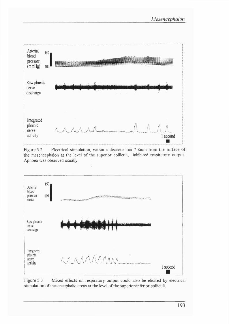
Mesencephalon
Arterial 150,blood pressure (mmHg) 100
iiiii
Raw phrenicnervedischarge
Integratedphrenicnerveactivit)'
I I I V / '--1 second
Figure 5.2 Electrical stimulation, within a discrete loci 7-8mm from the surface of the mesencephalon at the level of the superior colliculi, inhibited respiratory output. Apnoea was observed usually.
Arterialbloodpressure(mmHg)
Raw phrenicnervedischarge
Integratedphrenicnerve
150,
100
activity
;v\v .s\\V 'îY*^^vaw va va*’ '
aaajVVWWaa1 second
Figure 5.3 Mixed effects on respiratory output could also be elicited by electrical stimulation of mesencephalic areas at the level of the superior/inferior colliculi.
193
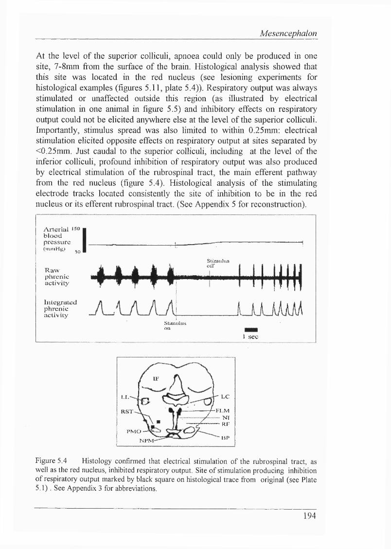
Mesencephalon
At the level of the superior colliculi, apnoea could only be produced in one site, 7-8mm from the surface of the brain. Histological analysis showed that this site was located in the red nucleus (see lesioning experiments for histological examples (figures 5.11, plate 5.4)). Respiratory output was always stimulated or unaffected outside this region (as illustrated by electrical stimulation in one animal in figure 5.5) and inhibitory effects on respiratory output could not be elicited anywhere else at the level of the superior colliculi. Importantly, stimulus spread was also limited to within 0.25mm: electrical stimulation elicited opposite effects on respiratory output at sites separated by <0.25mm. Just caudal to the superior colliculi, including at the level of the inferior colliculi, profound inhibition of respiratory output was also produced by electrical stimulation of the rubrospinal tract, the main efferent pathway from the red nucleus (figure 5.4). Histological analysis of the stimulating electrode tracks located consistently the site of inhibition to be in the red nucleus or its efferent rubrospinal tract. (See Appendix 5 for reconstruction).
Arterialb l o o dpressure(inm Hg) 50 IR a wphrenicactiv ity
I n t e g r a t e d p h r e n i c a c t i v i t y
Stim ulusoir 4m
1 _ U J iAàAÀStimuluson
J sec
4-rxM NT
RF
LL
P M OBJ*N P M
Figure 5.4 Histology confirmed that electrical stimulation of the rubrospinal tract, as well as the red nucleus, inhibited respiratory output. Site of stimulation producing inhibition of respiratory output marked by black square on histological trace from original (see Plate 5.1). See Appendix 3 for abbreviations.
194
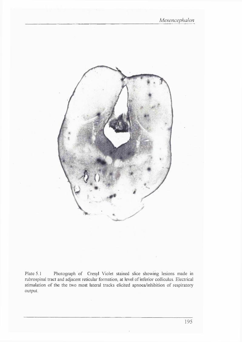
Mesencephalon
4
Plate 5.1 Photograph o f Cresyl Violet stained slice showing lesions made in rubrospinal tract and adjacent reticular formation, at level o f inferior colliculus. Electrical stimulation o f the the two most lateral tracks elicited apnoea/inhibition o f respiratory output.
195
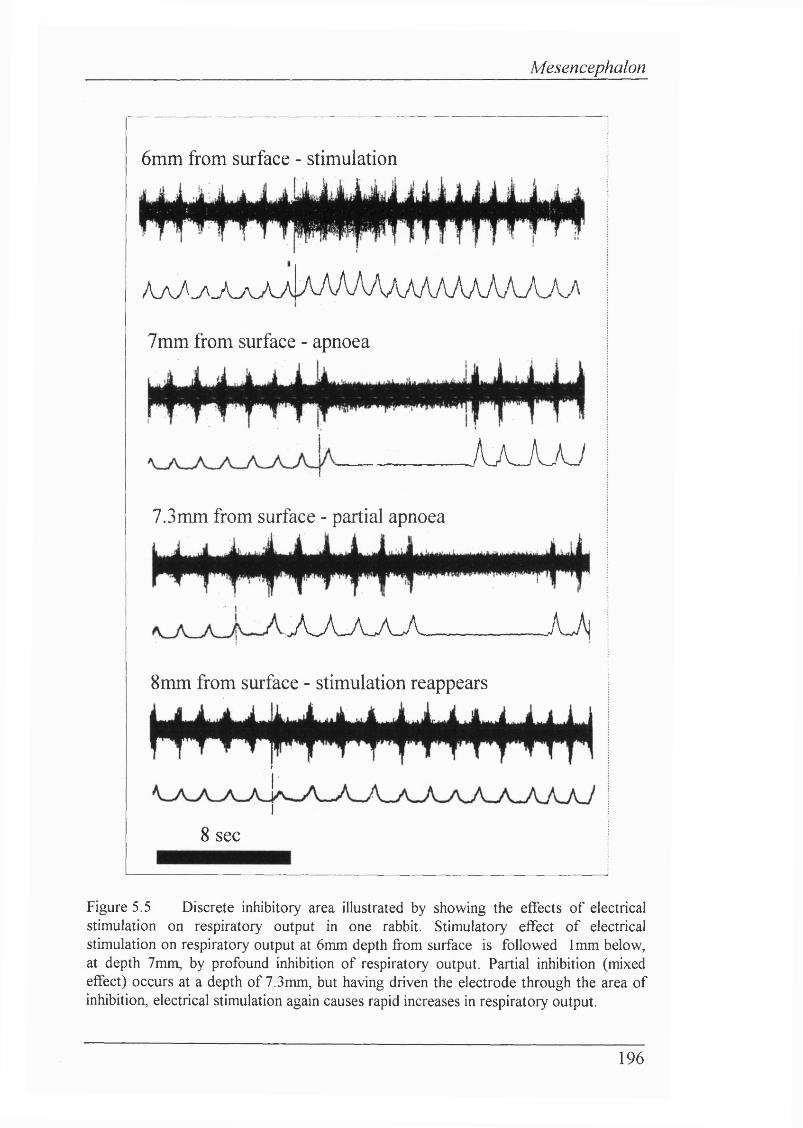
Mesencephalon
6mm from surface - stimulation
yu/v\AAA/lAAAAAAAA7V/UAAAA.A7mm from surface - apnoea
AaJL-W7.3mm from surface - partial apnoea
A A A A A A J \
8mm from surface - stimulation reappears
8 sec
Figure 5.5 Discrete inhibitory area illustrated by showing the effects of electrical stimulation on respiratory output in one rabbit. Stimulatory effect of electrical stimulation on respiratory output at 6mm depth from surface is followed 1mm below, at depth 7mm, by profound inhibition of respiratory output. Partial inhibition (mixed effect) occurs at a depth of 7.3mm, but having driven the electrode through the area of inhibition, electrical stimulation again causes rapid increases in respiratory output.
196
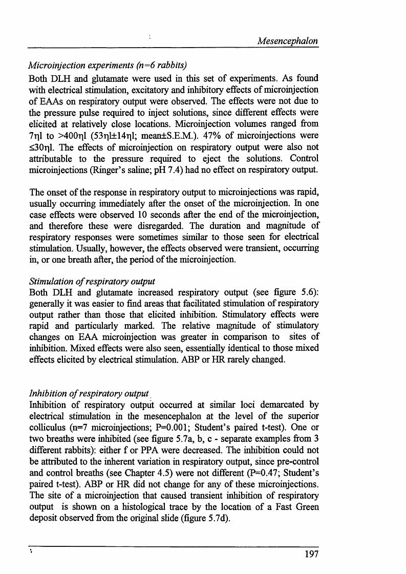
Mesencephalon
Microinjection experiments (n=6 rabbits)Both DLH and glutamate were used in this set of experiments. As found with electrical stimulation, excitatory and inhibitory effects of microinjection of EAAs on respiratory output were observed. The effects were not due to the pressure pulse required to inject solutions, since different effects were ehcited at relatively close locations. Microinjection volumes ranged from 7r|l to >400ql (53r|l±14ril; meantS.E.M.). 47% of microinjections were <30r|l. The effects of microinjection on respiratory output were also not attributable to the pressure required to eject the solutions. Control microinjections (Ringer’s saline; pH 7.4) had no effect on respiratory output.
The onset of the response in respiratory output to microinjections was rapid, usually occurring immediately after the onset of the microinjection. In one case effects were observed 10 seconds after the end of the microinjection, and therefore these were disregarded. The duration and magnitude of respiratory responses were sometimes similar to those seen for electrical stimulation. Usually, however, the effects observed were transient, occurring in, or one breath after, the period of the microinjection.
Stimulation o f respiratory outputBoth DLH and glutamate increased respiratory output (see figure 5.6): generally it was easier to find areas that facihtated stimulation of respiratory output rather than those that ehcited inhibition. Stimulatory effects were rapid and particularly marked. The relative magnitude of stimulatory changes on EAA microinjection was greater in comparison to sites of inhibition. Mixed effects were also seen, essentially identical to those mixed effects elicited by electrical stimulation. ABP or HR rarely changed.
Inhibition o f respiratory outputInhibition of respiratory output occurred at similar loci demarcated by electrical stimulation in the mesencephalon at the level o f the superior colhculus (n=7 microinjections; P=0.001; Student’s paired t-test). One or two breaths were inhibited (see figure 5.7a, b, c - separate examples from 3 different rabbits): either f or PPA were decreased. The inhibition could not be attributed to the inherent variation in respiratory output, since pre-control and control breaths (see Chapter 4.5) were not different (P=0.47; Student’s paired t-test). ABP or HR did not change for any of these microinjections. The site of a microinjection that caused transient inhibition of respiratory output is shown on a histological trace by the location of a Fast Green deposit observed from the original shde (figure 5.7d).
197
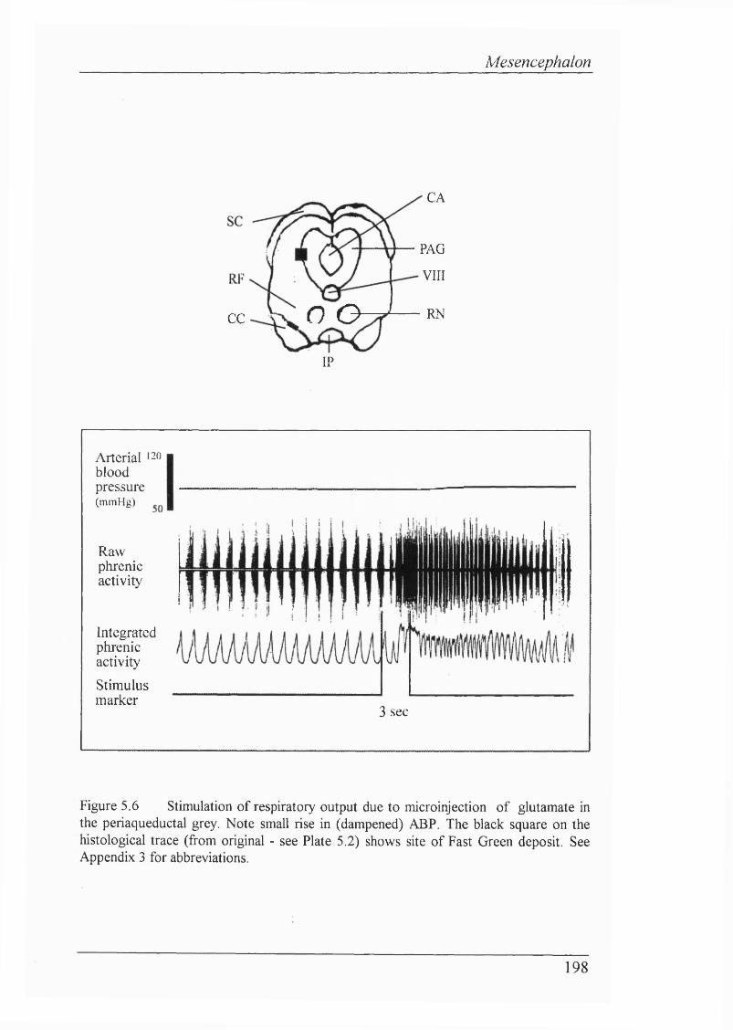
Mesencephalon
CA
PAG
VIII
O O RNCC
IP
Arterial ' 2 0
bloodpressureOnmHg)
Rawphrenicactivity
IntegratedphrenicactivityStimulusmarker
3 sec
Figure 5.6 Stimulation of respiratory output due to microinjection of glutamate in the periaqueductal grey. Note small rise in (dampened) ABP. The black square on the histological trace (from original - see Plate 5.2) shows site of Fast Green deposit. See Appendix 3 for abbreviations.
198
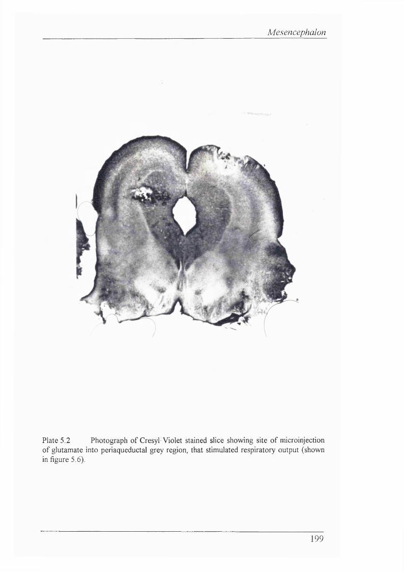
Mesencephalon
2%
Plate 5.2 Photograph of Cresyl Violet stained slice showing site of microinjection of glutamate into periaqueductal grey region, that stimulated respiratory output (shown in figure 5.6).
199
Mesencephalon
Arterialbloodpressure( m m H g )
Rawphrenicactivity
Integrated ,
Stimulusmarker
n1 sec
Figure 5.7a Transient inhibition of two breaths (fall in PPA) due to microinjection of glutamate at depth where electrical stimulation inhibited respiratory output at the level of the superior colliculus.
Stimulus
Figure 5.7b Transient prolongation of TE following microinjection of glutamate in area of mesencephalon at level of superior colliculus where electrical stimulation had inhibited respiratory output. Some caution, however, must be placed on the interpretation of this microinjection result since ongoing respiratory output tended to be irregular.
200
Mesencephalon
I
Stimulus
Figure 5.7c In another rabbit, transient inhibition of breath (PPA decreased) followed microinjection of glutamate in the red nucleus. See figure 5.7d for histological verification of microinjection site.
C AP A G
V I I I
R NR F
C C
L M
Figure 5.7d Histological trace showing site of microinjection (indicated by black square) in red nucleus (lateral border) for figure 5.7c. Two microinjections were made at this site, both of which caused transient inhibition of respiratory output (decreased PPA). See Plate 5.3 for histological slice showing site of Fast Green deposit: see Appendix 3 for abbreviations and scale.
201
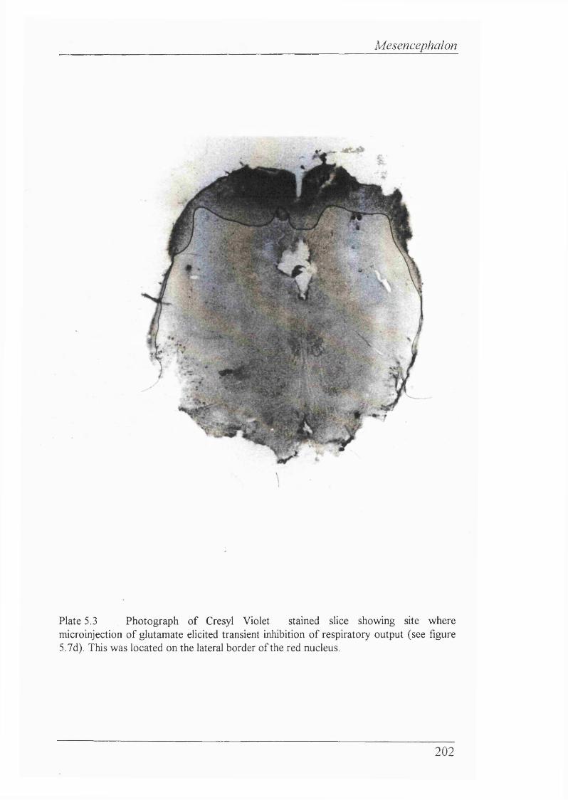
Mesencephalon
%
Plate 5.3 Photograph of Cresyl Violet stained slice showing site where microinjection of glutamate elicited transient inhibition of respiratory output (see figure 5.7d). This was located on the lateral border of the red nucleus.
202
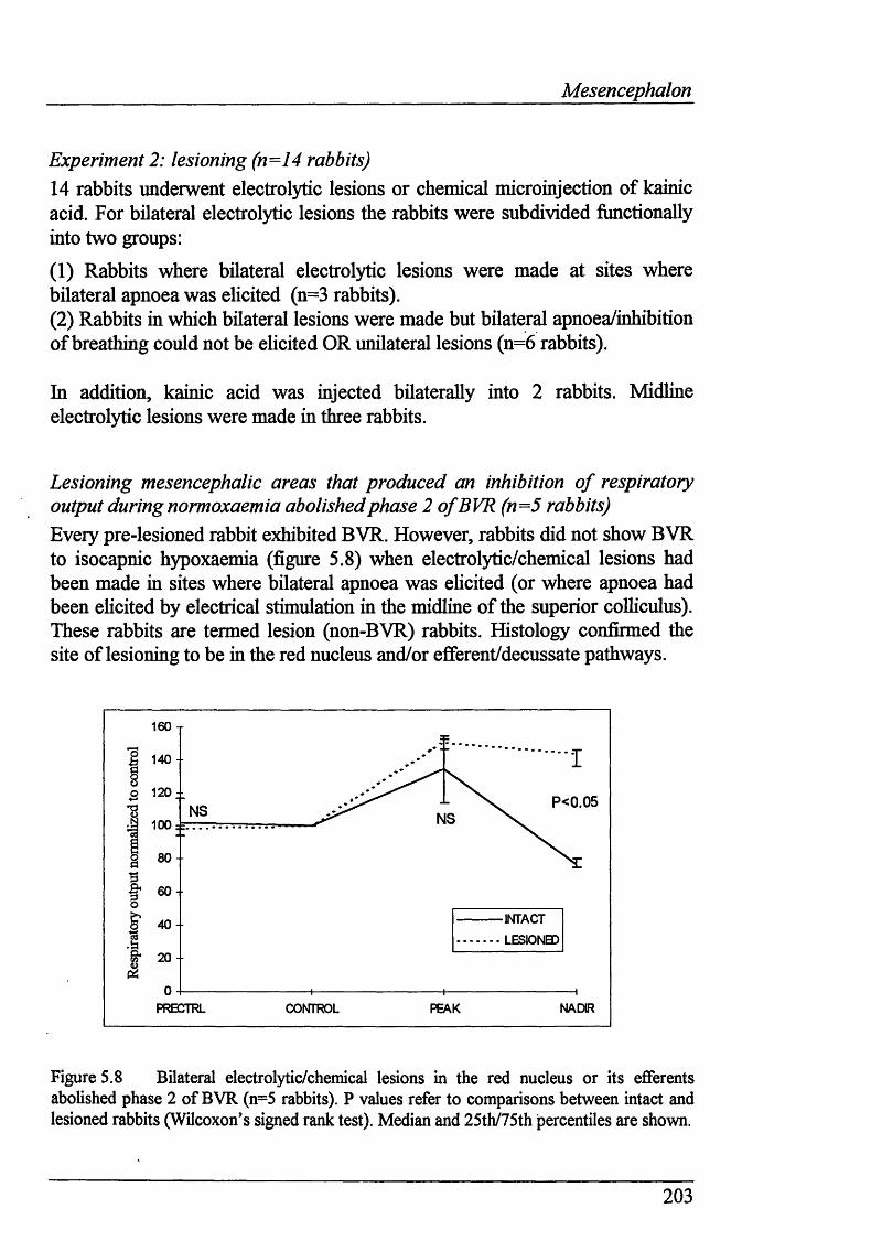
Mesencephalon
Experiment 2: lesioning (n=14 rabbits)14 rabbits underwent electrolytic lesions or chemical microinjection of kainic acid. For bilateral electrolytic lesions the rabbits were subdivided functionally into two groups:
(1) Rabbits where bilateral electrolytic lesions were made at sites where bilateral apnoea was elicited (n=3 rabbits).(2) Rabbits in which bilateral lesions were made but bilateral apnoea/inhibition of breathing could not be ehcited OR unilateral lesions (n=6 rabbits).
In addition, kainic acid was injected bilaterally into 2 rabbits. Midline electrolytic lesions were made in three rabbits.
Lesioning mesencephalic areas that produced an inhibition o f respiratory output during normoxaemia abolished phase 2 o f BVR(n=5 rabbits)Every pre-lesioned rabbit exhibited BVR. However, rabbits did not show BVR to isocapnic hypoxaemia (figure 5.8) when electrolytic/chemical lesions had been made in sites where bilateral apnoea was ehcited (or where apnoea had been ehcited by electrical stimulation in the midline of the superior colhculus). These rabbits are termed lesion (non-BVR) rabbits. Histology confirmed the site of lesioning to be in the red nucleus and/or efierent/decussate pathways.
160 T
P 120-- P < 0 . 0 5N S N S100 :=:
80
INTACTLESIONBD
40 --
PRBCTRL NADIRPEAK
Figure 5.8 Bilateral electrolytic/chemical lesions in the red nucleus or its efferents abolished phase 2 of BVR (n=5 rabbits). P values refer to comparisons between intact and lesioned rabbits (Wilcoxon’s signed rank test). Median and 25th/75th jpercentiles are shown.
203
Mesencephalon
Respiratory output did not fall from peak during isocapnic hypoxaemia for the lesioned experiments (peak vs. nadir; Wilcoxon’s signed rank test). Thus phase 2 was abolished completely, with no fall in ventilation occurring throughout isocapnic hypoxaemia. In one lesioned experiment, the rabbit was exposed to isocapnic hypoxaemia for 12 minutes, rather than 8 min, yet maintained respiratory output at peak levels throughout this period. Blood gas and pH status did not differ between intact and lesioned experiments (table 5.1). In contrast to their pre-lesion hypoxia/controls, 4/5 lesioned rabbits exhibited sham rage during isocapnic hypoxaemia.
INTACT LESIONEDNORMOXAEMIA HYPOXAEMIA NORMOXAEMIA HYPOXAEMIA
pHa 7.33± 0.00 7.31+0.02 NS 7.36+0.06 7.29+0.03 NSPacoj (mmHg) 43.3±2.5 4 2 .5 +2 .4 NS 40.8+3.1 45.0+1.3NS(p=o2)
Paog (mmHg) 108+19 31+1?<0.05 99+17 32+1*’ “Rectal t (°C) 37.8+0.4 3 7 .8 +0 .4 NS 38.4+0.2 38.5+0.2NSMAP (mmHg) 98.4±8.9 101.8+8.2NS 80.2+6.7 81.2+11.5 NSHR (bpm) 290+14 286+24 NS 310+7 333+14ns(oo8)
Table 5.1 Table showing blood gas/pH status, HR and ABP values during normoxaemia and isocapnic hypoxaemia for lesion (non-BVR) experiments (n=5 rabbits). Superscripts denote comparison between variables in normoxaemia and hypoxaemia. Pre and post lesion hypoxaemia levels were not different (P=0.28; Student’s paired t-test). In both intact and non-lesioned rabbits, ABP, but not HR, peaked during isocapnic hypoxaemia by ca. 10 mmHg (P<0.01; Student’s paired t-test) over control before falling back to 5 min levels.
Abolition o f BVR attributable to maintenance o f respiratory frequencyIn intact experiments, phase 1 was characterized by an increase in PPA with no concomitant change in respiratory frequency. Phase 2 was due to a profound fall in respiratory frequency.
Phase 1 in lesioned experiments was caused, as for intact experiments, by an increase in PPA solely. However, phase 2 was abohshed totally and this was attributable to the maintainence of respiratory frequency at peak levels during isocapnic hypoxaemia, in marked contrast to the fall observed in this variable in the same rabbits before lesioning.
The relative contributions of PPA and f to phase 1 and phase 2 for both intact and lesioned experiments are illustrated in figures 5.9 and 5.10 respectively.
204
Mesencephalon
180 T
160 -
140 -■
120 -
NS100
P<0.0580 -
60 --
PRBLESDN40-
POST LESION
20 - -
NADIRCONTROL
Figure 5,9 For non-lesion experiments PPA increased during phase 1 of BVR but did not alter during phase 2 (n=5 rabbits).
120 T
100
NS80 -
P<0.056 0 -
40 - PRELESION POST LESION
20 -
NADIRCONTROL PEAK
Figure 5.10 Respiratory frequency did not contribute to phase 1 of BVR but declined sharply during phase 2 in non-lesioned experiments. In marked contrast, respiratory frequency was maintained throughout isocapnic hypoxaemia in lesioned rabbits where (bilateral) apnoea had been elicited previously by electrical stimulation (n=5 rabbits).
205
Mesencephalon
Bilateral lesions in areas where apnoea was elicited abolished BVR (n=3 rabbits)In three rabbits the area where apnoea was ehcited was found bilaterally. Bilateral lesions were made in this area, which histology conhirmed to be within the red nucleus. Phase 2 of BVR was abohshed on subsequent exposure to isocapnic hypoxaemia.
Figures 5.11a, b, c show the respiratory response of each of these lesioned (non-BVR) rabbits to acute isocapnic hypoxaemia before electrolytic or bilateral lesions were made (denoted as “INTACT”) and post-lesion (denoted as “LESIONED”). Control values for respiratory output in intact and lesioned experiments are normalized to 100%, to permit relative comparisons. P values for comparison between respiratory output at precontrol, peak and nadir/8 min for intact vs. lesioned experiments are shown on each graph (Wilcoxon’s signed rank test) within a particular run (i.e. either in the intact or lesioned state). Itahcised P values refer to lesioned hypoxic runs.
For each rabbit, the histological traces showing where apnoea was ehcited, and hence where lesions were made, are ihustrated. These were compiled from the brain shces mounted on shdes, photographs, and from the depths at which apnoea occurred. In each case, the relative distances were calculated (as a fraction, e.g. the distance from the surface of the mesencephalon to the top of the red nucleus divided by the total ventrodorsal length) to compile the sites of lesions/apnoea as accurately as possible. One example of a histology shce showing bilateral lesions in the red nucleus which prevented phase 2 of BVR (from rabbit 090394) is shown in plate 5.4.
206
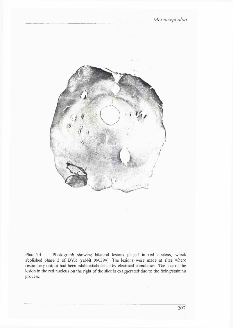
Mesencephalon
%
) i ' " # ■ .
7
/
Plate 5.4 Photograph showing bilateral lesions placed in red nucleus, whichabolished phase 2 o f BVR (rabbit 090394). The lesions were made at sites where respiratory output had been inhibited/abolished by electrical stimulation. The size o f the lesion in the red nucleus on the right o f the slice is exaggerated due to the fixing/staining process.
207
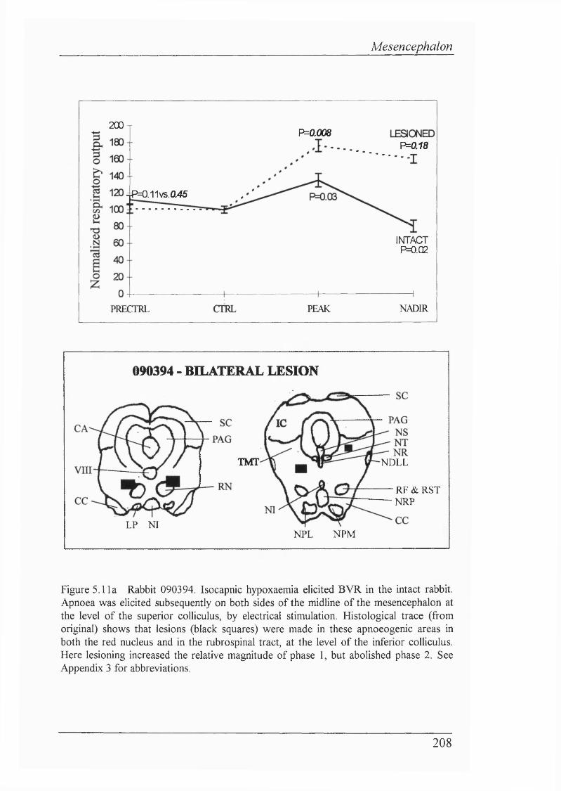
Mesencephalon
200xP=0.008
P=0.18
^P=0.11vs.a45
INTACTP=0.Q2
NADIRPEAKPRECTRL
090394 - BILATERAL LESION
IMT
LP NI
NDLL
R F & R S T-NRP
NPL NPM
Figure 5.11a Rabbit 090394. Isocapnic hypoxaemia elicited BVR in the intact rabbit. Apnoea was elicited subsequently on both sides of the midline of the mesencephalon at the level of the superior colliculus, by electrical stimulation. Histological trace (from original) shows that lesions (black squares) were made in these apnoeogenic areas in both the red nucleus and in the rubrospinal tract, at the level of the inferior colliculus. Here lesioning increased the relative magnitude of phase 1, but abolished phase 2. See Appendix 3 for abbreviations.
208
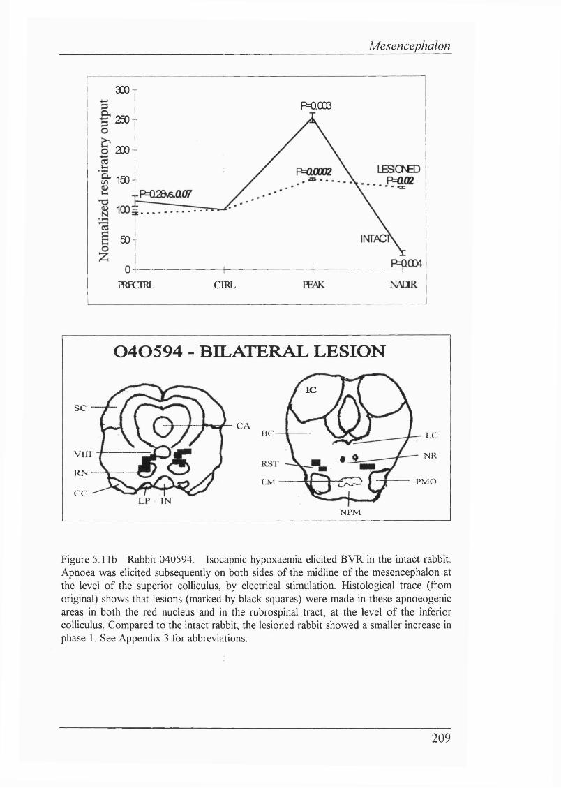
Mesencephalon
300t
P=QOOS250-
200 -
& 150 -
50 -
HRBCIRL CIRL
0 4 0 5 9 4 - BILATERAL LESION
SC
VIII
RN
CC
IC
CABC LC
N R
PMOLM
NPM
Figure 5.1 lb Rabbit 040594. Isocapnic hypoxaemia elicited BVR in the intact rabbit. Apnoea was elicited subsequently on both sides of the midline of the mesencephalon at the level of the superior colliculus, by electrical stimulation. Histological trace (from original) shows that lesions (marked by black squares) were made in these apnoeogenic areas in both the red nucleus and in the rubrospinal tract, at the level of the inferior colliculus. Compared to the intact rabbit, the lesioned rabbit showed a smaller increase in phase 1. See Appendix 3 for abbreviations.
209
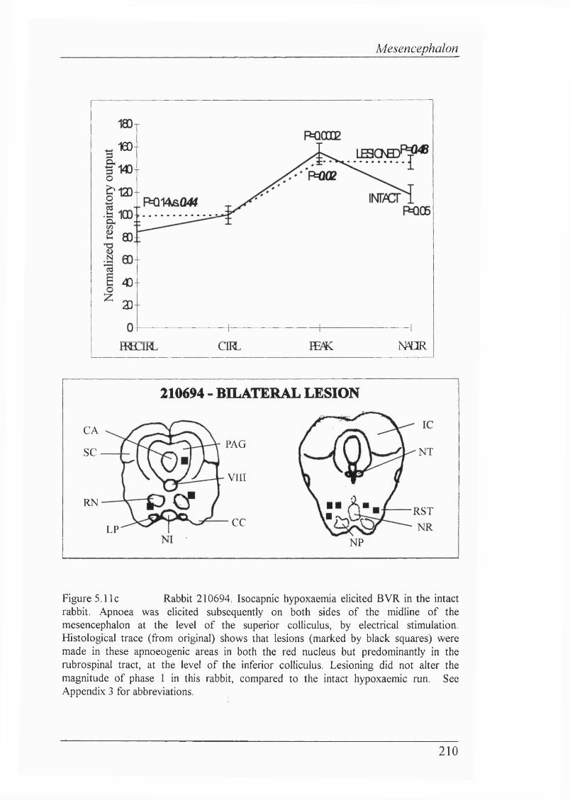
Mesencephalon
160 -
40
2D
NCIROEL
210694 - BILATERAL LESION
CAPAG
SC
v n i
RN
CCLP
NT
■ ■ RSTNR
NP
Figure 5.11c Rabbit 210694. Isocapnic hypoxaemia elicited BVR in the intactrabbit. Apnoea was elicited subsequently on both sides of the midline of the mesencephalon at the level of the superior colliculus, by electrical stimulation. Histological trace (from original) shows that lesions (marked by black squares) were made in these apnoeogenic areas in both the red nucleus but predominantly in the rubrospinal tract, at the level of the inferior colliculus. Lesioning did not alter the magnitude of phase 1 in this rabbit, compared to the intact hypoxaemic run. See Appendix 3 for abbreviations.
210
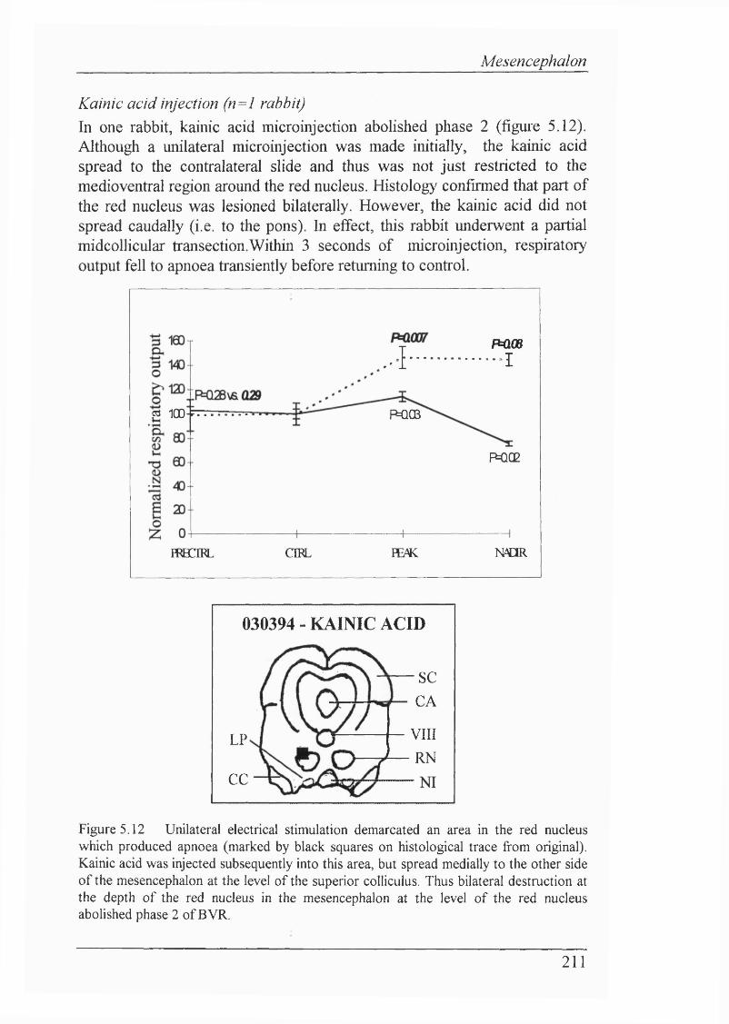
Mesencephalon
Kainic acid injection (n=l rabbit)In one rabbit, kainic acid microinjection abolished phase 2 (figure 5.12). Although a unilateral microinjection was made initially, the kainic acid spread to the contralateral slide and thus was not just restricted to the medio ventral region around the red nucleus. Histology confirmed that part o f the red nucleus was lesioned bilaterally. However, the kainic acid did not spread caudally (i.e. to the pons). In effect, this rabbit underwent a partial midcollicular transection.Within 3 seconds of microinjection, respiratory output fell to apnoea transiently before returning to control.
PCOBA I
çi 100- P=QC3
H BCIRL
POC2
CIRL ¥E/K N d R
030394 - KAINIC ACID
SCCA
VIIIRN
LP
CC
Figure 5.12 Unilateral electrical stimulation demarcated an area in the red nucleus which produced apnoea (marked by black squares on histological trace from original). Kainic acid was injected subsequently into this area, but spread medially to the other side of the mesencephalon at the level of the superior colliculus. Thus bilateral destruction at the depth of the red nucleus in the mesencephalon at the level of the red nucleus abolished phase 2 of BVR.
211
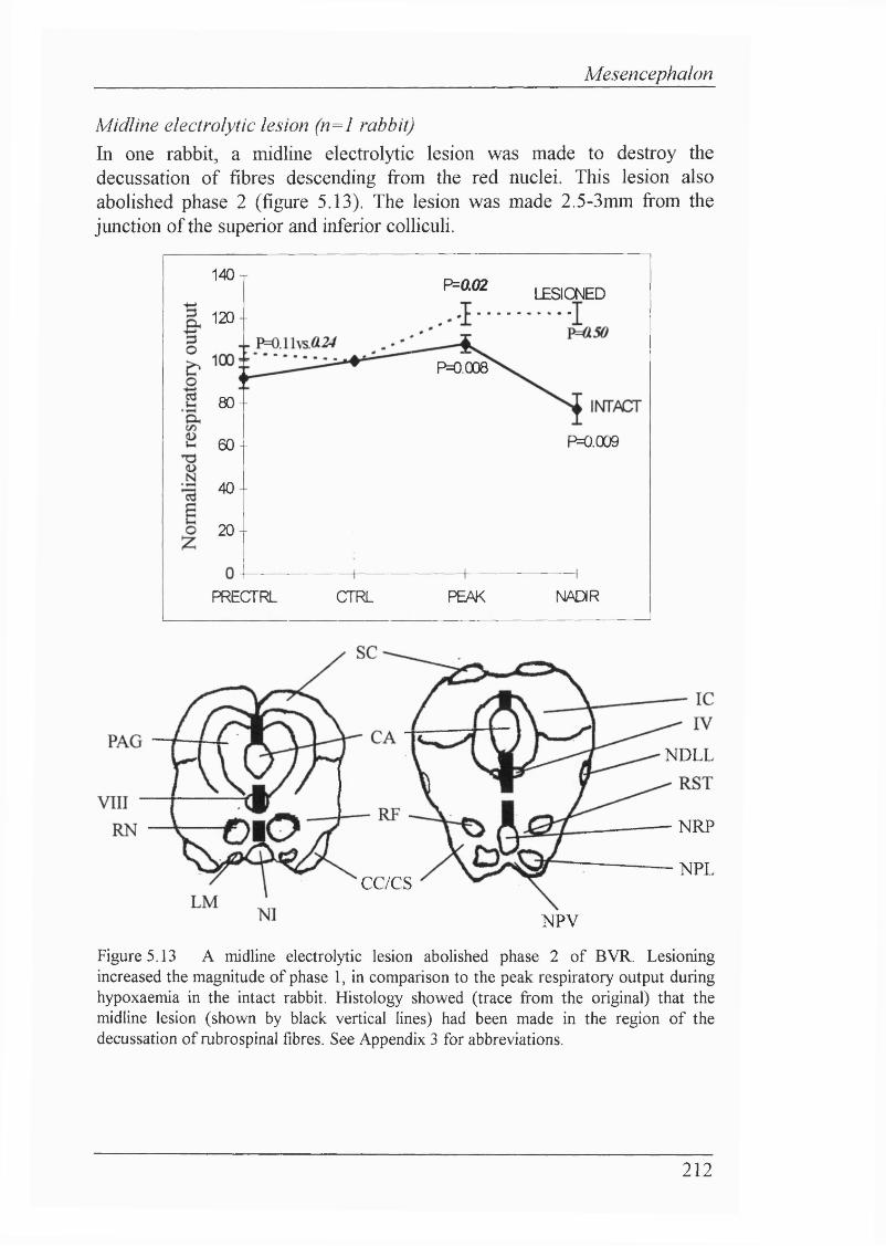
Mesencephalon
Midline electrolytic lesion (n=l rabbit)In one rabbit, a midline electrolytic lesion was made to destroy the decussation of fibres descending fi-om the red nuclei. This lesion also abolished phase 2 (figure 5.13). The lesion was made 2.5-3mm fi'om the junction of the superior and inferior colliculi.
140 T P=0.02 LESIONED120 -
P0.008
80-
P=0.0096 0 -
40-
2 0 -
NADIRPRECTRL CTRL PEAK
NDLL
CC/CS
NRP
NPL
NPV
Figure 5,13 A midline electrolytic lesion abolished phase 2 of BVR. Lesioning increased the magnitude of phase 1 , in comparison to the peak respiratory output during hypoxaemia in the intact rabbit. Histology showed (trace from the original) that the midline lesion (shown by black vertical lines) had been made in the region of the decussation of rubrospinal fibres. See Appendix 3 for abbreviations.
212
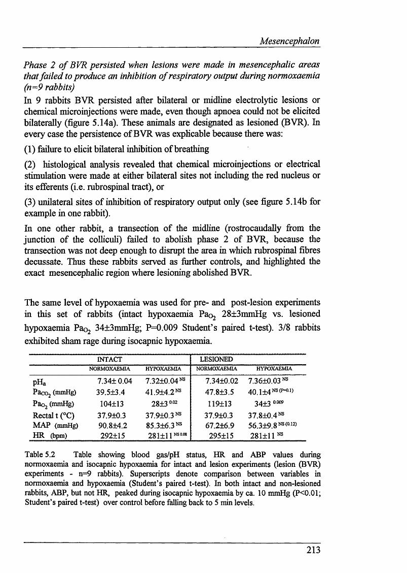
Mesencephalon
Phase 2 o f BVR persisted when lesions were made in mesencephalic areas that failed to produce an inhibition o f respiratory output during normoxaemia (n=9 rabbits)In 9 rabbits BVR persisted after bilateral or midline electrolytic lesions or chemical microinjections were made, even though apnoea could not be ehcited bilaterally (figure 5.14a). These animals are designated as lesioned (BVR). In every case the persistence of BVR was exphcable because there was:
(1) failure to ehcit bilateral inhibition o f breathing
(2) histological analysis revealed that chemical microinjections or electrical stimulation were made at either bilateral sites not including the red nucleus or its efferents (i.e. rubrospinal tract), or
(3) unilateral sites of inhibition of respiratory output only (see figure 5.14b for example in one rabbit).
In one other rabbit, a transection of the midline (rostrocaudally fi'om the junction of the colhcuh) failed to abohsh phase 2 of BVR, because the transection was not deep enough to disrupt the area in which rubrospinal fibres decussate. Thus these rabbits served as further controls, and highhghted the exact mesencephahc region where lesioning abohshed BVR.
The same level of hypoxaemia was used for pre- and post-lesion experiments in this set of rabbits (intact hypoxaemia Paoj 28±3mmHg vs. lesionedhypoxaemia Pao2 34±3mmHg; P=0.009 Student’s paired t-test). 3/8 rabbitsexhibited sham rage during isocapnic hypoxaemia.
INTACT LESIONEDNORMOXAEMIA HYPOXAEMIA NORMOXAEMIA HYPOXAEMIA
pHa 7.34+0.04 7.3210.04 NS 7.3410.02 7.3610.03 NSPacoj (mmHg) 39.5±3.4 41.914.2 NS 47.813.5 4 0 . 1 1 4 N S ( p=o.i )
Paoj (mmHg) 104±13 28130.02 119113 3413Rectal t (°C) 37.910.3 37.910.3 NS 37.910.3 3 7 .8 lO.4 NsMAP (mmHg) 90.814.2 85.316.3 NS 67.216.9 5 6 . 3 1 9 . 8 N S ( o-12)
HR (bpm) 292115 28111 INS 0.08 295115 281111 NS
Table 5.2 Table showing blood gas/pH status, HR and ABP values during normoxaemia and isocapnic hypoxaemia for intact and lesion experiments (lesion (BVR) experiments - n=9 rabbits). Superscripts denote comparison between variables in normoxaemia and hypoxaemia (Student’s paired t-test). In both intact and non-lesioned rabbits, ABP, but not HR, peaked during isocapnic hypoxaemia by ca. 10 mmHg (P<0.01; Student’s paired t-test) over control before falling back to 5 min levels.
213
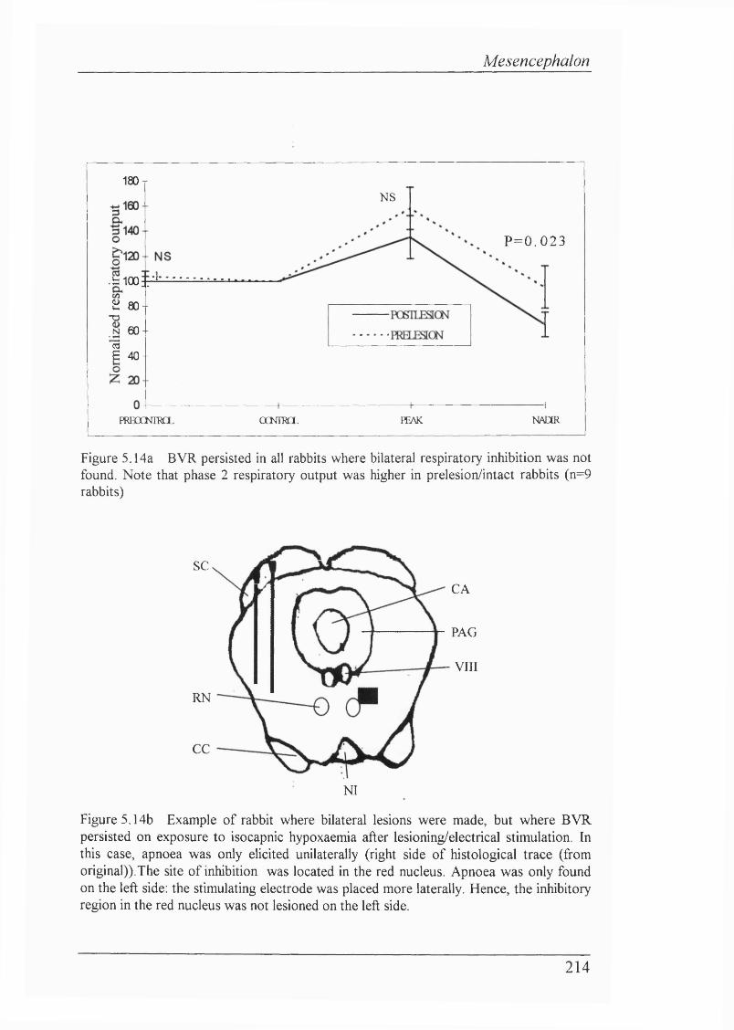
Mesencephalon
180 TNS
P=0 .023
.blOO r2 80 -
T3.N 6 0 -13E 40 -
NADERFRBOCKTRCL OCNTRCL PEAK
Figure 5.14a BVR persisted in all rabbits where bilateral respiratory inhibition was not found. Note that phase 2 respiratory output was higher in prelesion/intact rabbits (n=9 rabbits)
SC
CA
PAG
VIII
RN
CC
NI
Figure 5.14b Example of rabbit where bilateral lesions were made, but where BVR persisted on exposure to isocapnic hypoxaemia after lesioning/electrical stimulation. In this case, apnoea was only elicited unilaterally (right side of histological trace (from original)).The site of inhibition was located in the red nucleus. Apnoea was only found on the left side: the stimulating electrode was placed more laterally. Hence, the inhibitory region in the red nucleus was not lesioned on the left side.
214
Mesencephalon
Plate 5.5 Photograph showing slice illustrating start of stimulating electrode tracks made in one rabbit where BVR was not abolished. Here inhibition of respiratory output was elicited by stimulation in the most medial track at the depth of the red nucleus only on the left side: the most medial stimulating track (the start of which can be seen just medial to the cerebral aqueduct) can be seen to be projecting ventrally to the red nucleus. On the right side of the photograph, the start of two very lateral tracks can be seen, neither of which elicited inhibition of respiratory output. Neither projected ventrally to the red nucleus.
215
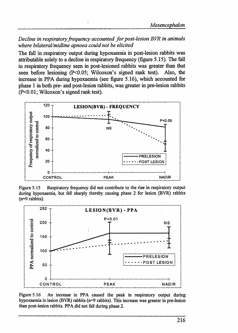
Mesencephalon
Decline in respiratory frequency accounted for post-lesion BVR in animals where bilateral/midline apnoea could not be elicitedThe fall in respiratory output during hypoxaemia in post-lesion rabbits was attributable solely to a decline in respiratory frequency (figure 5.15). The fall in respiratory frequency seen in post-lesioned rabbits was greater than that seen before lesioning (P<0.05; Wilcoxon’s signed rank test). Also, the increase in PPA during hypoxaemia (see figure 5.16), which accounted for phase 1 in both pre- and post-lesion rabbits, was greater in pre-lesion rabbits (P<0.01; Wilcoxon’s signed rank test).
120 T LESION(BVR) - FREQUENCY
100P < 0 . 0 5
8 0 -- N S
6 0 -
4 0 --P R E L E S I O N P O S T LESIONcr 20 - -u
NADIRP E A KC O N T R O L
Figure 5.15 Respiratory frequency did not contribute to the rise in respiratory output during hypoxaemia, but fell sharply thereby causing phase 2 for lesion (BVR) rabbits (n=9 rabbits).
2 5 0 X L E S IO N (B V R ) - PPA
P < 0 . 0 1200 - N S
1 5 0 -
100P R E L E S I O N P O S T L E S I O N5 0 -
C O N T R O L P E A K N A D I R
Figure 5.16 An increase in PPA caused the peak in respiratory output during hypoxaemia in lesion (BVR) rabbits (n=9 rabbits). This increase was greater in pre-lesion than post-lesion rabbits. PPA did not fall during phase 2.
216
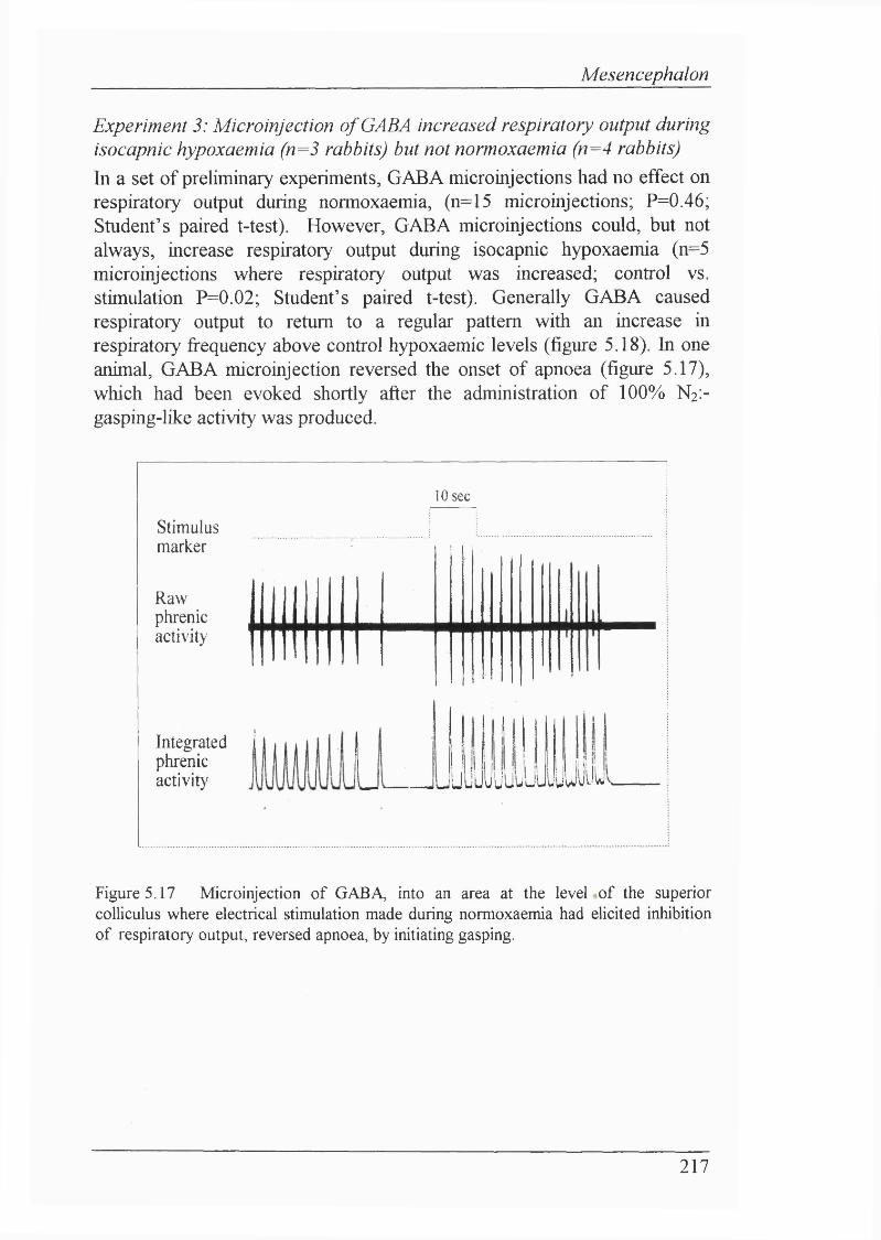
Mesencephalon
Experiment 3: Microinjection o f GABA increased respiratory output during isocapnic hypoxaemia (n=3 rabbits) but not normoxaemia (n=4 rabbits)In a set of preliminary experiments, GABA microinjections had no effect on respiratory output during normoxaemia, (n=15 microinjections; P=0.46; Student’s paired t-test). However, GABA microinjections could, but not always, increase respiratory output during isocapnic hypoxaemia (n=5 microinjections where respiratory output was increased; control vs. stimulation P=0.02; Student’s paired t-test). Generally GABA caused respiratory output to return to a regular pattern with an increase in respiratory frequency above control hypoxaemic levels (figure 5.18). In one animal, GABA microinjection reversed the onset of apnoea (figure 5.17), which had been evoked shortly after the administration of 100% N2 :- gasping-like activity was produced.
10 se c
Stimulusmarker
Rawphrenicactivity
Integratedphrenicactivity u juL'uijuJuiiuyyiiij
Figure 5.17 Microinjection of GABA, into an area at the level of the superior colliculus where electrical stimulation made during normoxaemia had elicited inhibition of respiratory output, reversed apnoea, by initiating gasping.
217
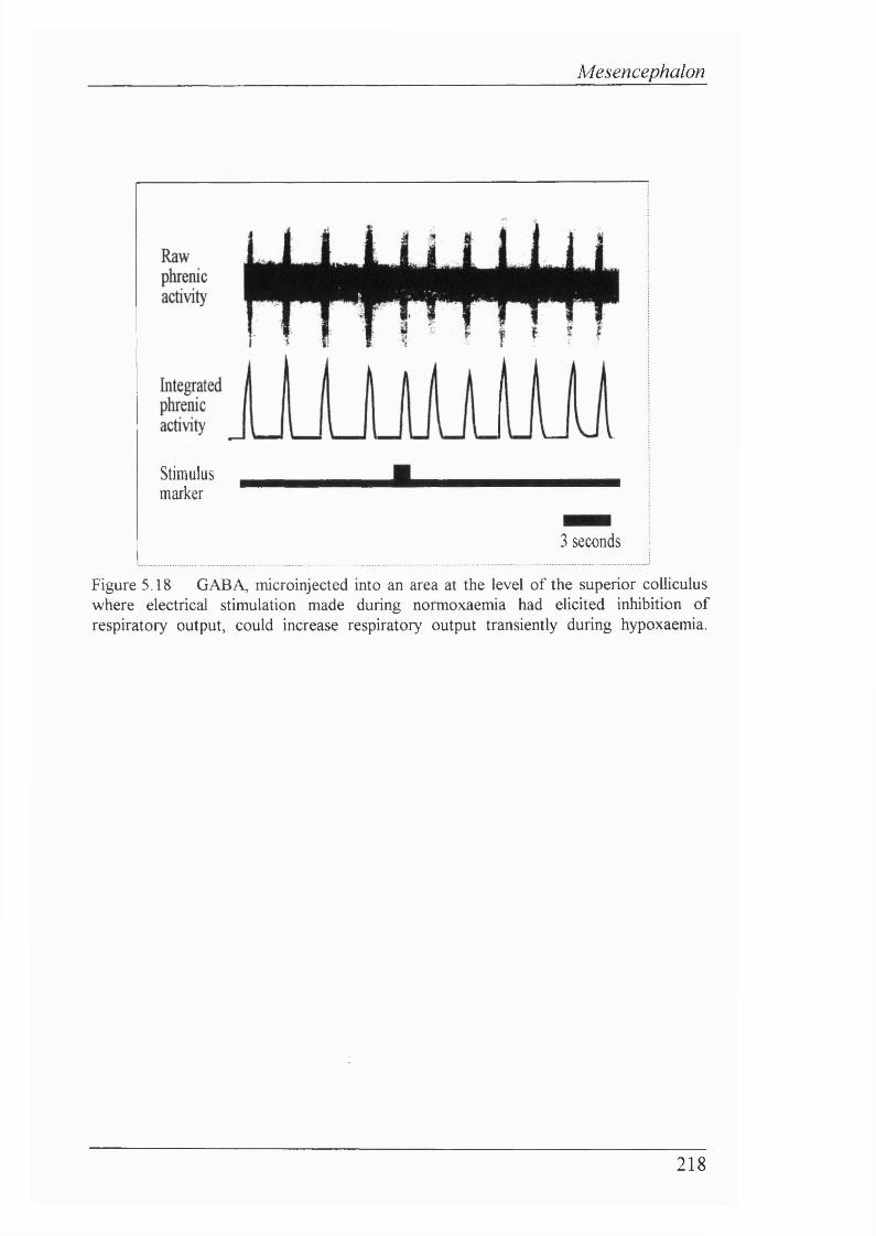
Mesencephalon
Stimulusmarker
3 seconds
Figure 5.18 GABA, microinjected into an area at the level of the superior colliculus where electrical stimulation made during normoxaemia had elicited inhibition of respiratory output, could increase respiratory output transiently during hypoxaemia.
218
Mesencephalon
5.7 D isc u ssio n
Methods
Electrical stimulationThe currents used to elicit changes in respiratory output were similar to those used in other studies (see Bradley, Ghelarducci, Paton & Spyer, 1987). The current spread in the study presented in this Chapter was estimated to be <0.25mm, since markedly different effects were ehcited within this range. The area of inhibition was also demarcated to be very discrete: stimulation of areas just ventral to the inhibitory area facihtated respiratory output.
Chemical stimulationChemical microinjections were undertaken using the guidelines set out by Lipski, Bellingham, West & Pilowsky (1988) who suggested that volumes <30rjl and concentrations <4 t]M should be microinjected. They found that microinjections of 10-150r|l of L-glutamate or DLH caused widespread and complex neuronal changes. Neurones that were distant from the site of microinjection remained excitable for considerable periods of time. Neurones in the direct vicinity of the microinjection site became silenced or inhibited after an initial period of excitation. The long-lasting inhibition was probably attributable to either depolarization blockade or extracellular ionic disruption. With these guidelines in mind, microinjection volumes were kept as close as possible to be within these guidelines. In addition, only effects on respiratory output that were ehcited during the period of microinjection, or within 2 seconds after the end of the pressure pulse, were regarded as being ehcited from the area of the micropipette tip.
219
Mesencephalon
Results
Identification o f inhibitory mesencephalic areasElectrical stimulation revealed that a discrete area within the mesencephalon in or near the red nucleus, at the level of the superior colhculus, mediated profound inhibition of breathing or apnoea. Inhibition of respiratory output consisted of both decreased PPA and f. Once electrical stimulation in areas that inhibited respiratory output had ceased, a rebound effect was always observed: respiratory output usually increased for a few seconds after the end of electrical stimulation before returning to control. This increase in post-stimulus respiratory output was attributable to both increased PPA and f.
No other inhibitory effects on respiratory output could be ehcited immediately outside the area of respiratory inhibition. Stimulatory effects were observed above, below and lateral to this area. As the stimulating electrode was driven deeper into the brain towards the region of inhibition, reaching the boundary of the inhibitory area could often be predicted by the change in the type of stimulation of respiratory output ehcited by electrical stimuh. Stimulation of areas near the boundary of the inhibitory area was characterised by an overaU increase in respiratory output, but the increases in PPA and/or f were smaUer than those seen for electrical stimulation nearer the dorsal surface of the mesencephalon. Areas that mediated apnoea were more difficult than regions that merely inhibited respiratory output to find consistently, which suggests that there is a very discrete region mediating apnoeic effects. Coles (1987) reported a similarly discrete area within the mesencephalon that mediated profound apnoea. Furthermore, Bassal & Bianchi (1982) found that the only areas in the mesencephalon where electrical stimulation produced apnoea were the red nucleus, or its efferent pathway, the rubrospinal tract. The type of effect ehcited by electrical stimulation cannot be attributed to the timing of the stimulus: nearly all stimulations were “blind”. The idea of a small group of cells mediating inhibition is supported by the work of Gallman & MiUhom (1991) who reported that the degree of inhibition of phrenic activity by chemical or electrical stimulation varied considerably. Furthermore, that a small region of the red nucleus may be responsible for respiratory effects is not surprising since the red nucleus is organised topographically.
220
Mesencephalon
The inhibition of respiratory output produced by stimulation o f the rubrospinal tract, at the level of the inferior colliculus reinforces the findings with electrical stimulation of the red nucleus. Single electrical shocks apphed to the rubrospinal tract have been shown to inhibit phrenic activity previously (Schmid, Bohmer & Fallert, 1988; see also Bassal & Bianchi, 1982).
Mixed effects of electrical stimulation on respiratory output were also observed. These effects consisted of an initial stimulation of breathing, followed by a prolonged apnoea lasting up to 15s. This was probably due to the stimulation of both excitatory and inhibitory projections, with the effects of inhibition outlasting those of excitation. The mixed effect of electrical stimulation was observed most commonly in the midline. This was due presumably to the mixture of both excitatory and inhibitory projections that travel through the midline or rubrospinal decussation.
EAA microinjectionThe effects of chemical stimuh on respiratory output were rapid and often transient, in contrast to the effects elicited by electrical stimulation. The rapidity and transient nature of the respiratory output response to chemical microinjection is predictable from the fast action, and breakdown, of neurotransmitters. However, the effects of chemical stimulation did outlast the period of microinjection in some cases. This may have been due to sustained depolarization of cell bodies, or the stimulation of particularly potent stimulatory areas.
Considering that EAA microinjection caused such stimulation it is surprising that EAA microinjection did not cause more profound inhibition of respiratory output. This may be due to the technique used: i.e. electrical stimulation to identify apnoeogenic areas followed by the placement of the micropipette in the same region. The disadvantage of this approach is that the micropipette may not necessarily be positioned in exactly the same locus as the electrical stimulating electrode. Ideally, the electrical stimulation electrode and microinjection pipette should be part of a common assembly, to improve greatly the accuracy of placement of stimuli. For this reason, a degree of caution must be attached to the microinjection results considering the methodology used.
221
Mesencephalon
DLH was found to be particularly potent. Because DLH takes longer to break down, cardiovascular variables tended to fall due to its persistence. For this reason glutamate was the choice of EAA in chemical microinjection experiments. However, regardless of which EAA was used extreme care had to be exercised to control the volume injected. The microinjection of unintentionally large volumes compromised both respiratory output and ABP and rendered a preparation unsuitable for Anther experimentation.
Extensive bilateral electrical stimulation in the mesencephalon could cause deleterious cardiovascular changesIn some rabbits extensive electrical stimulation caused ABP to fall. It was not possible to continue these experiments once ABP and/or HR had fallen. Cardiovascular collapse would ensue if this occurred. Since this was not a consistent finding, it is hard to account for. The release of adenosine, or perhaps neurotoxic quantities of glutamate, upon extensive electrical stimulation may explain this cardiovascular fall. However, the demise of any preparation was not related to the number of tracks, or even lesions, made. A possibility is the inadvertent lesioning of a cardiovascular efferent pathway.
Bilateral electrolytic lesions made only in the red nucleus did not affect subsequent respiratory output during normoxaemiaThere was no evidence to suggest that the areas lesioned bilaterally exerted a tonic effect on respiratory output. Some caution must be placed on this assertion, because in some preparations phrenic nerve activity altered due to repositioning of the nerve on the recording electrodes. In animals where phrenic nerve placement was not altered, no change in respiratory output during normoxaemia occurred as a direct result of bilateral lesions. In all cases where lesions were made, there was no effect on respiratory frequency. Rostrocaudal lesioning of the midline of the superior colliculi also had no effect on respiratory output in normoxaemia.
222
Mesencephalon
Bilateral electrolytic lesions made only in the red nucleus abolished phase 2 of BVR (lesionedfnon BVR animals)Lesions made in the region of the red nucleus abohshed phase 2 of BVR, and both PPA and f remained elevated throughout isocapnic hypoxaemia. Most strikingly, one animal was exposed to isocapnic hypoxaemia for 12 minutes yet continued to maintain phase 1 levels of respiratory output throughout this period.
The failure of other lesions to abolish phase 2 o f BVR (lesioned(BVR) animals) was due to placement o f the stimulating electrode outside the red nucleus/rubrospinal tractThe discrete location of the area that inhibits respiratory output was highhghted by the electrical stimulation studies. Thus it is conceivable that if a relatively smaU group of cells is responsible for mediating inhibition of respiratory output, it becomes more difficult to ensure that these ceUs have been destroyed by electrolytic lesion. The failure to abohsh phase 2 of BVR was attributable to the inabihty to find bilateraUy areas that caused apnoea/inhibition of breathing, as determined by electrical stimulation. The failure of a midline lesion to abohsh phase 2 is probably due to missing the projections within the rubrospinal tract or red nucleus efferents: the efferents fi'om the ceUs that inhibit respiratory output could have decussed rostraUy, or caudally, in relation to the site of the electrolytic lesion. Furthermore, for the reasons stated above, there may be a very smaU number of fibres involved.
Thus the failure to abohsh phase 2 consistently by electrolytic lesions highhghts the probabihty of a smah, discrete area of ceUs being responsible for mediating inhibition of respiratory output.
223
Mesencephalon
Higher incidence o f sham rage during hypoxaemia in lesion (BVR) and lesion(non-BVR) rabbits compared to controlsThe incidence of sham rage during hypoxaemia increased from 1/34 for non- lesioned rabbits to 4/5 lesion(non-BVR) and 3/8 lesion(BVR) rabbits. This striking increase in the incidence of sham rage suggests strongly that lesioning of mesencephahc structures removes an inhibition of peripheral chemoreflexes during isocapnic hypoxaemia.
Lesioning had no clear effect on phase 1 increaseSince lesioning did not cause a consistent change in the magnitude o f phase 1 respiratory output levels, so it seems unlikely to have altered profoundly the abihty of the respiratory centres to integrate peripheral chemoreflexes in the early stage of hypoxaemia.
'‘Biphasic “ arterial blood pressure response was not affected by lesioning The increase and subsequent fall in ABP during isocapnic hypoxaemia persisted even though respiratory output was maintained in rabbits where bilateral lesions in the red nucleus had been made. This suggests that there is a differential effect on respiratory and cardiovascular control during hypoxia, and is supports the findings of Giussani and co-workers in the fetus (see Chapter 1; p.57).
224
Mesencephalon
GABA microinjections indicate that an inhibitory mesencephalic inhibitory area is only active during isocapnic hypoxaemiaIn preliminary experiments, there were indications that (1) microinjections of GABA during normoxaemia, in an area where electrical stimulation caused an inhibition of respiratory output or apnoea, had no effect on respiratory output.(2) In contrast, microinjections of GABA during isocapnic hypoxaemia could produce an increase in respiratory output. Although the effect of GABA microinjection during hypoxaemia was not consistent, the results from these experiments indicate that the inhibition of inhibitory mesencephahc projections can reverse inhibition of respiratory output. The equivocal nature of these initial experiments may either be due to the micropipette not being directly adjacent to any putative inhibitory cell bodies, or due to the very small microinjectate volumes used. However, this data does indicate that the inhibitory pathway can be effectively switched off during phase 2 of BVR, thereby reheving inhibition of respiratory output. This supports the finding of Moore, Parkes, Noble & Hanson (1991) who showed that focal coohng in the upper rostral pons only affected respiratory output in anaesthetized lambs in phase 2 of BVR. This may suggest that they were cooling efferent red nucleus fibres, or cell bodies that act as relays for descending inhibitory projections from the red nucleus. No GABA microinjections were made during phase 1 in the present study.
The most striking example of reversal of respiratory inhibition was the onset of gasping like phrenic activity within a few hundred milhseconds of GABA microinjection during hypoxic apnoea. This may indicate that gasping mechanisms, as well as respiratory output, are inhibited during the early stages of hypoxia. However, once severe hypoxia ensues, the metaboHc inhibition of inhibitory mesencephahc neurones may permit the initiation of gasping mechanisms. If this is the case, then there must be differential sensitivity to hypoxia in areas of the brain stem (see Neubauer, Melton & Edelmann, 1990).
225
Mesencephalon
Abolition o f BVR by lesions in the red nucleus reconciles apparently contradictory supra- and rostral pontine lesioning/transection studiesThe results presented in this Chapter strongly suggest that the site mediating BVR is rostral to the pons. This does not conflict with previous studies: rather it strengthens their findings considerably. First, the transection studies undertaken in newborn rabbits (Martin Body & Johnston, 1988) and fetal lambs (Dawes, Gardner, Johnston & Walker, 1983) would have undoubtedly lesioned fibres of red nucleus origin, if not the red nuclei themselves. Similarly, although the lesion studies carried out by Gluckman & Johnston (1987) are hard to interpret, it is possible that red nucleus /ventromedial mesencephahc projections were lesioned, or afferent tracts that relay to it. This is indicated strongly fi’om the histological reconstructions shown for the area where electrolytic lesions were made in fetal lambs: the rubro-spinal tract runs adjacently through this site.
226
Mesencephalon
The structure o f the red nucleusThe structure of the red nucleus has been studied extensively in many different mammalian species (for extensive review see Massion, 1967). The red nucleus (or nucleus ruber) consists of two parts:-
Nucleus ruber magnocellularisThe caudal nucleus ruber magnocellularis consists mainly, though not exclusively, of large cells and is most strongly developed in lower mammals. All efferents from this part of the red nucleus decussate. The main efferent pathway from nucleus ruber magnocellularis is the rubrospinal tract. Fibres from nucleus ruber magnocellularis cross in the anterior tegmental decussation and form the rubrospinal tract immediately caudal to the red nucleus. The tract first passes laterally, mixing with other fibres, lying dorsal to the decussating fibres o f the superior cerebellar peduncle and medial lemniscus. At the level of the superior cerebellar peduncle, the tract runs caudally in the grey matter. In the pons the rubrospinal tract is most clearly identified between the ohve and the trigeminal nucleus. In the spinal cord, the fibres o f the rubrospinal tract he anterior, or overlap, the lateral corticospinal tract.
The nucleus ruber magnocellularis exhibits somatotopy (Pompeiano & Brodai, 1957), in that there are different origins for fibres that pass to the anterior and posterior limbs. This explains why the flexion o f either the anterior or posterior contralateral limb is dependent upon the placement o f stimulating electrodes within the nucleus ruber magnocellularis (Pompeiano, 1957). This highhghts the precedent for discrete areas within the red nucleus which have specialized physiological fimctions. The electrical stimulation studies reported in this Chapter suggest that there are discrete areas within the red nucleus which mediate inhibition of respiratory output. Clearly, there are functionally distinct areas or sub-divisions within the red nucleus: this may account for the variability in locating regions that mediate apnoea, and the strikingly different effects ehcited at loci close together.
227
Mesencephalon
Other decussated descending tracts of nucleus ruber magnocellularis origin include the rubronuclear tract, which is formed by collaterals from the rubrospinal tract which pass to the masticatory and facial nuclei, and fibres which pass to the nucleus interpositus in the cerebellum. There are also projections to the nucleus ambiguus.
Nucleus ruber parvicellularisIn contrast to nucleus ruber magnocellularis^ nucleus ruber parvicellularis consists only of medium and smaller sized cells. Nucleus ruber parvicellularis hes rostral to the nucleus ruber magnocellularis. It is highly developed in man but poorly developed in lower mammals. The efferent pathways fi*om nucleus ruber parvicellularis are uncrossed (i.e. ipsilateral) and consist of rubrothalamic and rubro-ohvary tracts.
The red nucleus controls motor output and spinal cord processingThe most extensively studied function of the red nucleus is that of motor control as mediated by the rubrospinal tract. Transections rostral to the superior coUiculus cause no change in muscle tone in intact animals. However, transections at the level o f the inferior colhculus produces rigidity of extensor muscles. The origin of this action on extensor postural muscles has been attributed to the red nucleus, since destruction of the nucleus ruber magnocellularis or section of the rubrospinal tract produces decerebrate rigidity in a precolhcular transected animal. However, this inhibitory action of the rubrospinal tract on the postural muscles has been questioned, since an isolated lesion in the red nucleus or rubrospinal tract does not produce similar postural changes in intact animals. Stimulation of the red nucleus has revealed its complex control over spinal cord processes. Via intemeurones the red nucleus stimulates alpha and gamma motoneurones of the contralateral flexor muscles. Concomitantly, alpha and static fusimotor fibres o f the contralateral extensor muscles are inhibited. The red nucleus also controls primary afférents to the spinal cord by presynaptic inhibition and both inhibitory and excitatory spinal reflexes by acting upon intemeurones.
228
Mesencephalon
Red nucleus inhibitory efferents have been identified in adult catsA number of studies have located suprapontine areas that affect respiratory output (see Hugelin, 1986 for review). With regard to the mesencephalon, the facihtatory effect of stimulating the reticular formation and central grey matter is clear (Martin & Booker, 1878; Kabat, 1936; Hugelin & Cohen, 1963; Evans & Pepler, 1974). Furthermore, inhibitory mesecephahc areas have been located. Bassal & Bianchi (1982) found that the onset of the expiratory phase, or apnoea, could only be elicited in the red nucleus (magnocellularis region) or rubrospinal tract at the level of the superior colhculus.
Single electrical shocks to the rubrospinal tract and adjacent reticular formation ehcit transient inhibition of phrenic nerve activity Extracellular recordings from medullary respiratory neurones showed that bulbospinal inspiratory neurones were inhibited paucisynaptically. Lesions of the nucleus parabrachiahs mediahs and locus coereleus (i.e.pneumotaxic centres) failed to alter this rubrospinally mediated inhibition of respiratory output (Schmid, Bohmer & Fallert, 1988). This work suggests strongly that there are powerful inhibitory efferent projections from the red nucleus to the respiratory centres. Furthermore, the inhibition of phrenic activity by red nucleus efferents is not dependent on pontine regions imphcated previously in BVR. Neuroanatomical tracing of red nucleus efferents illustrates that there are descending projections to medullary and pontine reticular nuclei (Edwards, 1972).
229
Mesencephalon
Respiratory related ajferents to the mesencephalon and red nucleusRespiratory related activity has been recorded in adult cats in the most ventral part of the red nucleus (Vibert, Caille, Bertrand, Gromysz & Hugelin, 1979).
Chen, Eldridge & Wagner (1992) reported that mesencephalic neurones in the central tegmentum of decerebrate, peripherally chemodenervated, paralyzed adult cats, displayed no or irregular tonic activity at low respiratory output, but developed rhythmic firing activity vyith increased respiratory output produced by either hypercapnia or carotid sinus nerve stimulation. The exact location of these neurones was not reported. This mesencephahc respiratory rhythm was not affected by spinal cord transection, and did not affect respiratory output when abohshed by short acting anaesthesia. Furthermore, some of the rhythmically active neurones, amongst others in the same region, were excited by chest wall and respiratory muscle afferents. The influence of afferent vagal input on mesencephahc respiratory rhythm (Eldridge & Chen, 1992) was investigated by vagal coohng, vagotomy, changing tidal volume and stopping and starting the ventilator for 60s.
Ah these methods clearly showed that vagal feedback inhibited mesencephahc respiratory rhythm. The authors rejected the idea that hypoxia caused the disinhibition of mesencephahc respiratory rhythm when the ventilator was turned off. This was based on the assumption that the cats would not become hypoxaemic in 60s since they were being ventilated with 100% O2 . It is unfortunate that the authors did not exclude the possibihty o f hypoxic excitation of these neurones by ventilating the cats with lower F1 0 2 .Furthermore, no histology was provided to determine the exact area of the mesencephalon in which they recorded respiratory activity.
Thus mesencephalic respiratory activity was concluded to have no role in generating respiratory drive. Rather it was suggested that the mesencephalon relayed information to higher centres, the functional role of this being the detection of dyspnoea and related distressful breathing patterns. Similar conclusions about the lack of a role for the mesencephalon in contributing to respiratory drive have also been made since no change in respiratory pattern is produced by ponto-mesencephahc transections (in normoxia). These studies however do not suggest, or have not sought to address, whether the mesencephalon contributes to respiratory drive under other conditions.
230
Mesencephalon
The fimctional significance of respiratory activity in the red nucleus may be that it contributes to the control of locomotion, or as suggested by the work presented in this Chapter, mediate BVR during hypoxia (i.e. inhibition of respiratory output only occurs during hypoxia, as suggested by the GABA experiments reported in this Chapter). In the fetus, the joint inhibition of FBM and body movements may also be mediated by this region.
Other afferent projections to the red nucleusThe main afferent pathway is from the cerebellum, although visual and auditory afferents also converge on the red nucleus. Removal of the cerebellum almost completely reduces spontaneous neuronal activity in nucleus ruber magnocellularis (Massion, 1961), indicating that the cerebellum exerts a tonic facihtation of motor control by nucleus ruber magnocellularis.
231
Mesencephalon
5.8 Su m m a ry
♦ Cell bodies in the region of the red nucleus in the mesencephalon mediate inhibition of respiratory output
♦ Bilateral electrolytic lesions in red nucleus prevent phase 2 o f BVR
232
Pons
CHAPTER 6
THE ROLE OF THE PONS IN MEDIATING INHIBITION OF RESPIRATORY OUTPUT IN NEWBORN DECEREBRATE RABBITS
233
Pons
6.1 In t r o d u c t io n
This Chapter reports a series of experiments that were undertaken to investigate the role of structures in the pons and at the level of the inferior collicular in mediating inhibition of respiratory output. The main part of this Chapter concentrates on electrical stimulation experiments, largely in the dorsorostral pons, to ascertain areas that caused an inhibition of breathing. The aim of die study was to serve as a basis for Anther work that would investigate whether areas that mediated inhibition of respiratory output contain cell bodies or project to the region of the red nucleus. Clearly descending inhibitory mesencephahc pathways project to areas of the pons; such areas may either relay afferents to the mesencephalon or project to the respiratory centres directly. It was also important to relate any inhibitory regions found in similar studies using newborn lambs (Coles, Kumar & Noble, 1989) and adult cats (Cohen, 1971) and rabbits (Evans & Pepler,1974). A series of microinjection experiments were also made in the pons to ascertain whether electrical stimulation of areas that had mediated inhibition of respiratory output was due to cell body stimulation.
In another preliminary experiment, the cooling probe used in the study of Moore, Parkes, Noble & Hanson (1991) was used in a newborn decerebrate rabbit to ascertain whether cooling in a similar area to that in newborn lambs would reverse phase 2 of BVR.
6.2 H y po t h e se s
Experiment 1: Electrical stimulation of areas in the pons causes inhibition of respiratory output.Experiment 2: Microinjection of EAA into dorsal pons at the level of the middle cerebellar peduncle causes an inhibition of respiratory output.Experiment 2: Cooling of upper rostral pontine regions reverses phase 2 of BVR.
234
Pons
6.3 M et h o d s
All general methods are described as in Chapter 4. Blood gases/pH were maintained within normal parameters, at hyper- or normoxic Pao2 levels.
Electrical stimulationElectrical stimulation of pontine structures in decerebrate newborn rabbits was carried out at 0.25-0.5mm intervals. The stimulation parameters were typically lOOHz, 0.1ms pulse width and 150pA-350jLiA. Electrical stimulation was also carried out in two rabbits anaesthetized with halothane. In each experiment at least 10 min elapsed between stimulations.
Cooling experimentThe cooling probe was a modified version of that used in newborn lambs by Moore & Hanson (1991), with the probe tip being smaller (0.25mm^) since the area of cooling needed to be kept to a minimum (see Chapter 4). The probe was placed 0.5mm from the midline just below the surface of the pons (0.25mm) at the level of the middle cerebellar peduncle.
6.4 A n a ly sis
Cooling experimentThe presence of BVR was tested for as described in Chapter 4. In addition, the effect of cooling on respiratory output during phase 2 of BVR was analysed by comparing 5 breaths at peak respiratory output (phase 1) with respiratory output (5 breaths) 1 min after the onset of the probe temperature being cooled, and at 1 min intervals thereafter (Student’s paired t-test). Statistical signifcance was accepted at P<0.05 level.
235
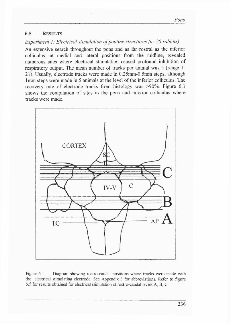
Pons
6.5 R esu lts
Experiment 1: Electrical stimulation o f pontine structures (n=20 rabbits)An extensive search throughout the pons and as far rostral as the inferior colhculus, at medial and lateral positions from the midline, revealed numerous sites where electrical stimulation caused profound inhibition of respiratory output. The mean number of tracks per animal was 5 (range 1- 21). Usually, electrode tracks were made in 0.25mm-0.5mm steps, although 1mm steps were made in 5 animals at the level of the inferior colhculus. The recovery rate of electrode tracks from histology was >90%. Figure 6.1 shows the compilation of sites in the pons and inferior colhculus where tracks were made.
C O R T E X
IV -V
A PT G
Figure 6.1 Diagram showing rostro-caudal positions where tracks were made with the electrical stimulating electrode. See Appendix 3 for abbreviations. Refer to figure 6.5 for results obtained for electrical stimulation at rostro-caudal levels A, B, C.
236
Pons
Loci where respiratory output was inhibitedApnoea or inhibition of respiratory output was produced in the following locations in the pons and inferior colhculus. A compilation of sites where inhibition of respiratory output was elicited is shown in figure 6.5. The number of rabbits in which inhibition of respiratory output was produced within a particular structure is given in parentheses.
Locus coeruleus (n=5 rabbits )At rostral locations near to the inferior colhculus or at the level of the superior cerebellar peduncle, ca. 1mm fi*om the midline, electrical stimulation caused inhibition of respiratory output. However this was an inconsistent effect. Nearer to the midline, stimulation or a mixed respiratory response, including apneusis, could occur. Inhibition of respiratory output was also elicited at the level of the inferior cerebellar peduncle.
INHIBITIONNVSG
V-M
MCP
■ NR
LMNVLL
NPMNPLP M C
Arterial 8U| blood pressure(mmHg) 3Q|
Rawphrenicactivity
Integratedphrenicactivity LAAAl
1 sec
Figure 6.2 Stimulation o f locus coeruleus could cause inhibition o f respiratory output. Apnoea occurred at all the points along the electrode track (black line) in the example shown here, including the medial reticular formation. Onset and cessation o f stimulus is shown by stimulation artefact (vertical lines).
237
Pons
Reticular formation (n=IO rabbits)The most consistent location of inhibition, or apnoea, was elicited in the dorsal pontine reticular formation. Generally, inhibition was mediated by more medial sites. Inhibition of respiratory output was produced by electrical stimulation in the reticular formation just below, and at, the level of the inferior colliculus. There were equivocal effects observed more caudally. Inhibition of respiratory output was also seen in the reticular formation, just medial to the motor nucleus of the trigeminal nerve (n=2 rabbits).
SCP*V-M
FLMMCP
NPLMPMO
NP
Arterial jqq bloodpressure 60 ( m m H g )
Rawphrenicactivity
Integratedphrenicactivity
I
1 sec
Figure 6.3 Electrical stimulation o f the reticular formation produced apnoea (see figure 6.2 also - different rabbit). Note stimulus artefact contaminated the raw phrenic signal. Black square denotes site o f stimulation on histological trace See Appendix 3 for abbreviations.
238
Pons
Raphe nuclei (n=3 rabbits)Stimulation of midline structures (at the level of the raphe nuclei) often caused respiratory output to be inhibited. This was observed from the region of the middle/superior cerebellar peduncle to more rostral locations at the level of the inferior colliculus.
LC
SCP
NRMCPRST
NVLLRF
LM
PMONPM
pressure(mmHg)
Rawphrenicactivity
Integratedphrenicactivity
I ;/(ivvwywvwwww
1 sec
Figure 6.4 Electrical stimulation produced inhibition in the region of the raphe/midline nuclei. Black square on histological trace shows the site of electrical stimulation to be in nucleus raphis. See Appendix 3 for abbreviations.
Other sites where electrical stimulation elicited inhibition o f respiratory outputApnoea was was also seen infrequently at the surface of the IV ventricle and at fasciculus longitudinalis dorsalis. These effects occurred in close proximity to the medial part of tractus solitarius. Below the most dorsal region, respiratory output was facilitated. Often apneusis was caused by stimulating the floor of the IV ventricle.
239
Pons
F L MFLD
NTS
NTSNT-
N A
NO! PMO
FLD FLM NVl
NCÜ
N P
NRMN I S N V
XFNRP
PMO
S C PL C F L M
F L D
V-M-
M C P N R
RF
L M
NPL N P M P M O
Figure 6.5 Compilation of sites where electrical stimulation produced inhibition of breathing or apnoea. Black cross indicates mixed effects, including inhibition, whereas black squares denote sites of inhibition or apnoea. The letters A, B, C refer to the corresponding rostro-caudal level indicated by figure 6 .1 . See Appendix 3 for abbreviations and scale.
240
_____________________ Pons
Experiment 2: Microinjections into the dorsal pons, including locus coeruleus (n=8 rabbits)This set o f microinjection experiments were the first to be undertaken during this project. As a first approach (n=4 rabbits) the Hamilton syringe injector set-up was used, as described in Chapter 4. Because of the problems associated with this apparatus in delivering an accurately known volume, the results of these experiments must be treated with some caution. The volumes injected were certainly in excess of 200^1. For this reason very few were made in any one experiment: hence the injections were limited to dorsal locations. However, because the results fi'om these experiments were analysed using the strict criteria described in Chapter 4 for micromjections the basic conclusions should hold. Furthermore, post-injection effects on tonic respiratory output were minimal in all but one case, so it seems reasonable under these circumstances to form some preliminary conclusions firom these experiments.
These experiments indicated that cell bodies may mediate a fall in respiratory output, in an area where electrical stimulation also caused profound inhibition of phrenic activity. Injection of glutamate dorsal (1mm firom the surface), in the region of locus coeruleus 1mm lateral firom the midline and l-2mm caudal to the inferior colhculus inhibited respiratory output, both during normoxaemia and isocapnic hypoxaemia (figures 6.6). The inhibition occurred transiently, within the period, or within 5 s after the end, of the microinjection. However, microinjections of glutamate at the level of the middle cerebellar peduncle (made 2-3mm from the junction of the colhcuh), again just below the brain stem surface, in the region of locus coeruleus, stimulated respiratory output in two rabbits. Furthermore, injections into these areas increased respiratory output during hypoxia, or reversed apnoeic spells during hypoxia (figure 6.7). However, since large volumes were used in these experiments, further work is required.
In 4 other rabbits, the picopump microinjection apparatus was used (see Chapter 4). The mean volume of glutamate injected was 28±3r|l. Control injections of Fast Green/Ringer’s saline (pH 7.4) had no effect. Histology revealed that microinjection of L-glutamate into the parabrachial region elicited an apneustic pattern in respiratory pattern (figure 6.8). More dorsal microinjections, in the periaqueductal grey at the level of the inferior colliculus, stimulated respiratory output (see Chapter 5). At the level o f the trigmeinal motor nucleus, equivocal results were obtained. No clear evidence for inhibition of respiratory output was seen, with either no effect or equivocal transient inhibition of respiratory output occurring.
241
Pons
Stimulus
Figure 6 . 6 Inhibition of respiratory output due to microinjection of glutamate in region of locus coeruleus.
Arterial ioo| blood pressure (mmHg)5o
Rawphrenicactivity
Integratedphrenicactivity
Stimulusmarker
1 1 sec
Figure 6.7 Stimulation of respiratory output in dorsorostral pons, again at level of locus coeruleus, stimulated respiratory output during normoxaemia and isocapnic hypoxaemia. The example shown illustrates the effect of glutamate injected during hypoxaemic apnoea. The site of glutamate microinjection is shown by the black square on the histological trace. See Appendix 3 for abbreviations.
242
Pons
L C
N D B C
R F
N RN L L
N P L
B P
N P MP M O
a c t i v i t y
S t i m u l u s
m a r k e r
Figure 6 . 8 Microinjection of glutamate into parabrachial region (indicated by black square) produced apneusis. The site of glutamate microinjection is shown by the black square on the histological trace. See Appendix 3 for abbreviations.
243
Pom
Experiment 3: Ejfect o f cooling at level o f middle cerebellar peduncle on respiratory output during isocapnic hypoxaemia (n=l rabbit)The result of this preliminary experiment is summarized in figure 6.9. Isocapnic hypoxaemia elicited BVR. However, when the probe tip was cooled from body temperature (38°C) to 23°C during phase 2, respiratory output increased to phase 1/peak respiratory output levels. The increase in respiratory output due to cooling was mainly attributable to increased PPA (figure 6.10). Thus phase 2 was abolished by cooling the dorsomedial pons at the level of the middle cerebellar peduncle. Histology confirmed that the cooling probe had been placed at the level of the middle cerebellar peduncle, in the area of locus coeruleus (plate 6.1).
RO
500 T
450 --
400 --
350 -
300 --
250 -
200 - -
- 2 - 1 0 1 2 3 4 5 6 7 Time (min)
8 9 10 11 12
T
Figure 6.9 Phase 2 of BVR was reversed by cooling the floor of the IV ventricle at the level of the middle cerebellar peduncle. Negative numbers refer to minutes before onset of isocapnic hypoxaemia. Diamonds linked by line refer to probe temperature T (°C); RO- absolute respiratory output (fxPPA). Mean±SEM values are shown; n=l rabbit.
Cooling o f dorsolateral pons reversed phase 2 by increasing peak phrenic activityThe reversal of phase 2 was attributable solely to an increase in peak phrenic activity, with no change in respiratory frequency. The changes in PPA and respiratory frequency due to hypoxaemia and cooling are illustrated overleaf (figure 6.10).
244
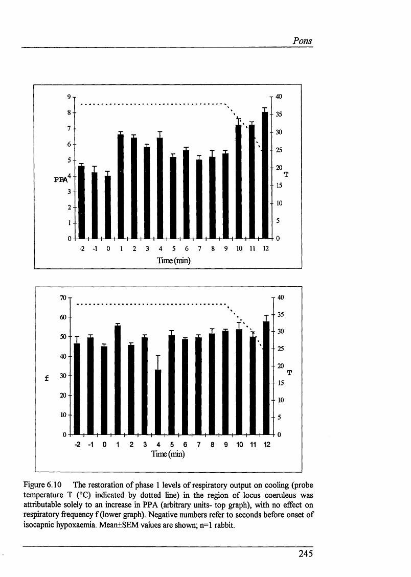
Pons
8- - 35
7 - - 30
6- 25
- • 204 - -
- 15
- ■ 10I -
Tune (min)
- 356 0 -
- 305 0 -
- 2540 -
- ■ 2030 -
- 15
2 0 -
- 10
1 0 -
Tune(min)
Figure 6.10 The restoration of phase 1 levels of respiratory output on cooling (probe temperature T (°C) indicated by dotted line) in the region of locus coeruleus was attributable solely to an increase in PPA (arbitrary units- top graph), with no effect on respiratory frequency f (lower graph). Negative numbers refer to seconds before onset of isocapnic hypoxaemia. MeantSEM values are shown; n=l rabbit.
245
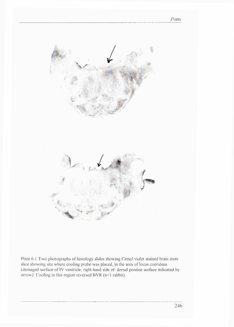
Pons
/-5- . ■ i/i*
\
Plate 6.1 Two photographs o f histology slides showing Cresyl violet stained brain stem slice showing site where cooling probe was placed, in the area o f locus coeruleus (damaged surface o f IV ventricle, right hand side o f dorsal pontine surface indicated by arrow). Cooling in this region reversed BVR (n=l rabbit).
246
Pons
6 . 6 D isc u s s io n
Although this Chapter has provided fiirther evidence to suggest that inhibitory pontine mechanisms could be involved in BVR, it also highhghts the difficulties of approaching these CNS mechanisms when a structure is not targeted (cf. Chapter 5). The lack of a proper stereotaxic atlas and frame for the newborn rabbit necessitated such an approach in the initial work investigating pontine structures.
Electrical stimulation o f pontine structures Pontine reticular formationThe most important results reported from the electrical stimulation study reported in this Chapter suggest strongly that the dorsal pontine reticlar formation mediates inhibition of respiratory output in the newborn. This supports strongly, and extends substantially, the findings of Coles, Kumar & Noble (1989) and Smith (1993) who investigated the effects of electrical stimulation of pontine and inferior colhcular structures on breathing in lambs and kittens respectively. The consistency of the location of inhibitory sites from the present Chapter compared to the latter studies is certainly striking. This strengthens strongly the idea that pontine structures have powerful inhibitory effects on respiratory output in the newborn.
That such pontine sites can inhibit breathing so profoundly is predictable from work conducted in the adult cat investigating the neurophysiology of sleep. Electrical stimulation (Kawahara, Nakazono, Kumagai, Yamauchi & Miyamoyo, 1988) or microinjection of ACh (Kimura, Kubin, Davies & Pack, 1990; Tojima, Kubin, Kimura & Davies, 1992) into the medial pontine reticular formation area causes profound inhibition of breathing in decerebrate adult cats. The inhibition of breathing is concomitant with suppression of postural tone (Mori, Kawahara, Sakamoto, Aoki & Tomiyama, 1982). The rostral pontine tegmentum controls other processes, including sympathetic visceromotor and nociceptive somatosensory fimction (Katayama, DeWitt, Becker & Hayes, 1984).
247
Pons
Inhibition of respiratory output seems to be causally linked with ACh release, caused by electrical stimulation of the pedunculopontine nucleus (Lydie & Baghdoyan, 1993). Microinjection of ACh into the pontine reticular formation also produces a REM-like sleep state (Lydie & Baghdoyan, 1992) including REM associated postural atonia (Taguchi, Kubin & Pack, 1992). Analysis of breathing during REM sleep or the cholinergically induced REM-like sleep state show that breathing is inhibited in comparison to breathing during the awake state (Lydie & Baghdoyan, 1989; Lydie, Baghdoyan, Wertz & White, 1991). Irregular breathing patterns, hypotonia in respiratory muscles, increased upper airway resistance and diminished ventilatory responses to hypoxia and hypercapnia are all features of REM sleep (Lydie & Biebuyck, 1988; Kryger, Roth & Dement, 1989). It is most notable that the red nucleus sends glutamatergic neurones to the pontine inhibitory area in the reticular formation (Lai, Clements & Siegel, 1993), and that sleep related activity has been recorded from the red nucleus (Gassel, Marchiafava & Pompeiano, 1965).
Raphe nucleusStimulation near or in the midline could inhibit respiratory output. This was in, and ventral to, nucleus raphe magnus. This is not surprising, since electrical stimulation just ventral to fasciculus longitudinahs mediahs (or medial longitudinal fasciculus) inhibits diaphragmatic, hypoglossal and soleus muscle EMG strongly (Kawahara, Nakazono, Kumagai, Yamauchi & Miyamoto, 1989). Neurones in the brain stem midline contribute to respiratory control (Hosogai, Matsuo & Nakao, 1993): neurones throughout the midline, from the the region of the dorsal nucleus raphe obscurus, pallidus and (to caudal) magnus seem to have divergent inhibitory and excitatory connections to stabihze their synchronous activity (Lindsey, Hernandez, Morris, Sharmon & Gerstein, 1992; Lindsey, Hernandez, Morris & Sharmon, 1992). Thus a network of cormections exist between the midline nuclei. With this in my mind, it is predictable that disruption of the network would affect respiratory output.
However, there are markedly conflicting reports on the effect of electrical stimulation or EAA microinjection into nucleus raphe obscurus: Man & Liu (1992) reported a diminuition or abohtion of phrenic activity in the adult urethane anaesthetized rabbit, whereas Holtman, Anastasi, Norman & Dretchen (1986) and Millhom (1986) reported an increase in phrenic nerve activity. One of the problems with investigations into the role of the midhne nuclei, particularly microinjections, is the close proximity of these structures
248
_______________________________________________________________________________ Pons
to one another. Thus, electrical or chemical stimulation may spread to another midline area, thereby producing apparently contradictory results.
The dorsal raphe nuclei are closely apposed to the surface of raphe vessels, which are branches from the basilar artery (Scheibel, Tomiyasu & Scheibel,1975). From electron microscopy studies it has been postulated that this close neurovascular relationship may indicate a chemosensory/barosensitive role for dorsal raphe neurones. Parabrachial and Kolliker-Fuse (pontine pneumotaxic centre) neurones also project to the raphe nuclei. Descending efferents include those to the reticular formation of the pons and cranial nerve nuclei (Bobilher, Seguin, Petitjean, Salbert, Touret & Jouvet, 1976), VLM (Connelly, Ellenberger & Feldman, 1989; Holtman, Marion & Speck, 1990) and nucleus tractus sohtarius (Thor & Helke, 1987). Like locus coeruleus there is a topographical organization to raphe nuclei.
Locus coeruleusLocus coeruleus is involved in mediating the effects of focal cooling on phase 2 in the newborn lamb (Moore, Parkes, Noble & Hanson, 1991). This idea is supported by the cooling experiment reported in this Chapter. The question therefore arises whether locus coeruleus may mediate phase 2 of BVR or simply be a relay from other sites of inhibition. Neurones in locus coeruleus are modulated by central respiratory activity. Furthermore, they are also activated by hypoxia, but whether this is due solely to peripheral chemoreceptor stimulation remains unclear (Guyenet, Koshiya, Huangfii, Verbeme & Riley, 1993).
Locus coeruleus is the main source of NE and 5HT. Its axons project extensively throughout the neocortex, thalamus, hippocampus, hypothalamus tectum and cerebellum. These projections are topographic, as shown by retrograde labelling techniques and electrophysiology which reveals that many neurones may innervate widely divergent forebrain areas via axon collaterals.Individual neurones within locus coeruleus respond to an array of external sensory stimuh (Aston-Jones & Bloom, 1981) and cardiovascular afferents (Elam, Yao, Svensson & Thoren, 1984). Clearly locus coeruleus has a highly complex role.
There is evidence to suggest that locus coeruleus regulates neurophysiological development. For example, differentiation of both the cerebral and cerebellar cortices is preceded by innervation from locus coeruleus afferents (Nakamura & Sakaguchi, 1990). Lesion studies also
249
_______________________________________________________________________________ Pons
suggest a trophic role for locus coeruleus for synaptic plasticity in the cortex (Felten, Hakan & Jonsson, 1982). Furthermore, electrophysiological recordings from locus coeruleus neurones in rat brain shces reveal that there are distinct membrane potential oscillations in the newborn period, which disappear after 4 weeks of age (Christie, Williams & North, 1989). Dye coupling studies also show an early maturation of electrotonic coupling between locus coeruleus neurones (Christie & Jelinek, 1993). Thus locus coeruleus exhibits striking electrophysiological maturation, plus a clear role in mediating general neurological development. Locus coeruleus also plays an important role in cardiovascular control: it facilitates the carotid baroreflex (Xu, Huang, Zhao, Xu & Yu, 1992).
In direct relation to the studies on the pontine reticular formation, highly localized areas around locus coeruleus are implicated in suppressing a range of reflexes including posture, visceromotor fimction and nociceptive somatosensation in conscious adult cats (Katayama, DeWitt, Becker & Hayes, 1984). Microinjections of a cholinergic agonist into the pontine reticular formation shows that an area just ventromedial to the principal nucleus of locus coeruleus suppresses postural somatomotor and sympathetic visceromotor fimction maximally. At a site lateroventral to the principal nucleus of the locus coeruleus, somatomotor fimction was suppressed predominantly. Clearly, such descending inhibition of spinal motor output in these sites corresponds to sites in the present Chapter where respiratory output was inhibited.
MicroinjectionsThe preliminary microinjection studies in this Chapter suggest that cell bodies may mediate many of the inhibitory/excitatory responses shown by electrical stimulation. These experiments are more extensive than those of Farber (1990), who localized apnoeogenic areas around the parabrachial and lateral lemniscal nuclei with glutamate microinjections. However, extensive microinjections were not made, or undertaken together with electrical stimulation. Clearly, the electrical stimulation work and preliminary microinjection experiments demand further and more extensive investigations into inhibitory pontine influences on respiratory output. However, the stimulatory effect of L-glutamate microinjections into locus coeruleus concurs with recent studies (Li, Xia & Huang, 1992). Furthermore, the reversal of inhibition of respiratory output by microinjecting glutamate during isocapnic hypoxaemia, even during apnoea.
250
_______________________________________________________________________________ Pons
suggests strongly that BVR cannot be attributable to direct hypoxic depression of neuronal function.
Confirmation that cooling the upper rostral pons reverses phase 2 o f BVR in the rabbitCooling the rostromedial pons, at the level of the middle cerebellar peduncle, reversed phase 2: respiratory output increased to peak phase 1 output. This supports the finding of Moore, Parkes, Noble & Hanson (1991) and shows that this technique could be invaluable in further neurophysiological studies in the newborn rabbit.
Since the cooling probe was placed just below the surface in the experiment reported in this Chapter (0.5mm), the neuroanatomical substrate most hkely to be affected was locus coeruleus, which hes on the floor of the IV ventricle. Locus coeruleus was within the area cooled in this Chapter, and in the study of Moore, Parkes, Noble & Hanson (1991) and Moore, Bohm & Hanson (1993). In the latter study not only did adenosine and hypoxia produce a fall in ventilation, but cooling reversed the decline in both cases. Whether a hypoxia-sensitive group of cells, which initiated the fall in ventilation, was cooled remains unknown. However, it is tempting to hypothesize that adenosine may have depolarized neurones in locus coeruleus (Harms, Finta, Tschopl & files, 1992; Tschopl, Harms, Norenberg & files, 1992), thereby initiating a fall in breathing, with the direct excitatory effect of adenosine being reversed by cooling.
251
Pons
6.7 Su m m a ry
♦ Electrical stimulation in numerous locations in the dorsal pons causes inhibition of respiratory output.
♦ Electrical stimulation of the pontine reticular formation (including midline structures) and the locus coeruleus inhibits respiratory output.
252

Final Discussion
CHAPTER 7
FINAL DISCUSSION
HYPOXIA AND BVR FROM AN EVOLUTIONARY PERSPECTIVE
253
Final Discussion
7.1 M ain find in gs o f th e th esis
The most important findings from the experiments I have conducted during this project are:(1) Peripheral chemoreceptors are unlikely to fail during acute isocapnic
hypoxaemia in the newborn.(2) Phase 2 of BVR is mediated by mesencephalic structures.
The development of the project is illustrated (figure 7.1) by an updated scheme presented originally in in Chapter 1 (section C).
Increased discharge maintained throughout hypoxaemic period Differential effect of
hypoxaemia on respiratory reflexes
Establish anaesthetized newborn rabbit model
Electrical stimulationMicroinjectionLesioning
Brain stem studies
Modulation of chemoreflex during hypoxaemia
How does hypoxaemia affect respiratory carotid chemoreflex?
Are other respiratory reflexes similarly affected?
Develop new Reparation suitable for brain stem studies
Record afferent chemorecqrtor fibre discharge in hypoxaemia
Figure 7.1 Summary of the development of the project.
254
Final Discussion
Section ASection A supports the idea that peripheral chemoreceptors do not adapt during acute isocapnic hypoxaemia, but rather continue to be stimulated throughout the hypoxic period. This conclusion is made from both direct recordings of carotid chemoreceptor activity and also the transient carotid chemoreceptor mediated respiratoiy reflex test. The results from these methods are mutually reinforced, in that both have provided evidence to support further the hypothesis that peripheral chemoreflexes must be modulated centrally during hypoxia, thereby permitting the decline in ventilation seen in newborns. Teleologically these findings are attractive, since they support the idea that carotid chemoreceptors act as a first line of defence in hypoxic conditions. Their failure during such times would make httle sense. Furthermore, the demonstration that peripheral chemoreceptors play a role in fetal cardiovascular control (Giussani, Spencer, Moore, Bennet & Hanson, 1992) and, under some circumstances, also in FBM (Gluckman & Johnston, 1987 -see Chapter 1) makes the inabihty of carotid chemoreceptors in the newborn to respond to hypoxia even more unlikely. It is also difficult to correlate the time course and/or magnitude of the adaptation in relation to the degree o f the ventilatory fall during BVR in studies where carotid chemoreceptors have been reported to fail or adapt during hypoxia (Marchai, Bairam, Haouzi, Crance, Di Guiho, Vert, & Lahiri, 1992; Carroll, Fitzgerald & Bamford, 1993).
However, as noted in both Chapters 2 and 3, it remains difficult to state with certainty that the quantifiable inputs measured in Section A can be assumed to be the same afferent information that the brain stem integrates. For this reason the findings of Chapters 2 and 3 should be treated with some caution. Understanding the results from the experiments conducted in Chapters 2 and 3 would therefore be probably improved if the inputs (i.e. afferent limb of the reflexes) investigated were also measured at the level of the brain stem. In the case of the carotid chemoreflex study, this could be achieved by recording VRG or DRG neurones that mediate carotid chemoreflexes. However, it is important to stress that these experiments measured the effect of hypoxaemia on respiratory reflexes where Ihe afferent limb of the reflex, the input, had been controlled and measured. This represents an improvement on similar previous studies which have based their conclusions on the effects of hypoxia on the efferent limb of respiratory reflexes, without knowing whether the afferent limb of the reflex is constant throughout normoxia and hypoxia or whether it alters during hypoxia. In summary, the findings of Section A led to the investigation of CNS mechanisms that mediate phase 2 of BVR.
255
Final Discussion
Section BThe work presented in Section B includes two important developments in newborn respiratory physiology. First, a novel newborn rabbit preparation has been developed which confers physiological stabihty plus the lack of anaesthesia. Newborn preparations are notoriously difficult to keep stable physiologically and this is highhghted particularly when newborn respiratory physiology is studied. The preparation developed in this thesis should be a significant step forward in the study of newborn systemic physiology. That said, it is also important to acknowledge that any experiments conducted in decerebrate preparations should, for completeness, also be repeated in anaesthetized animals.
Secondly, and most excitingly. Section B presents data that shows for the first time that a specific brain region mediates BVR. Thus there are CNS substrates activated by hypoxia that contribute to respiratory control under such conditions, as suggested by Moore, Parkes, Noble & Hanson (1991). The idea of active neurally mediated inhibition of breathing during hypoxia has previously been rejected, or treated with extreme caution. Since the inhibitory CNS mechanisms are activated by hypoxia, it is likely that a brain P0 2 sensor exists to detect falls in P0 2 and hence activate inhibitory neurophysiological responses. The presence of a brain P0 2 chemoreceptorwould alter radically our understanding of respiratory control, fi'om fetus to adult. Clearly, however, further experiments in vivo and in vitro are necessary to demonstrate the existence of P0 2 chemoreception in the brain. The idea of a central Po sensor may at first seem rather speculative. Theremainder of this final discussion considers the likelihood of such a mechanism in the Hght of processes that have evolved across species to cope with hypoxia, and also recent evidence for hypoxia-sensitive molecular mechanisms.
256
Final Discussion
7.2 H y po x ia a n d BVR fro m a n e v o l u t io n a r y pe r spe c t iv e
Numerous strategies exist across species for countering the effects o f hypoxic environmentsFrom an evolutionary standpoint, it is far from surprising that the experiments presented in this thesis indicate the presence o f a hypoxia- sensitive CNS mechanism. Diverse neural and biochemical mechanisms have evolved to enable organisms to cope with hypoxia.
The evolution o f an oxygenated atmosphereOxygen concentration in primitive atmospheres was very low: current geophysical evidence indicates that water, carbon dioxide, some nitrogen, hydrogen, carbon monoxide, sulpur dioxide, methane and ammonia were the main, almost exclusive, atmospheric gases with CO2 probably being the most abundant (Cloud, 1982). Thus the earhest organisms were anaerobic, due to the lack of O2 . At this point in evolution, the sources o f energy for synthesis were ultraviolet Hght, Hghtning and geothermal heat. Veiy primitive organisms broke down organic molecules anaerobically to derive energy. Indeed, many of these primitive enzymes of anaerobic metaboHsm persist today. The formation of organic molecules, less than one bilHon years after the formation of earth’s atmosphere eventually led to the formation of green plants, algae and bacteria. In turn, oxygen was produced, albeit in low concentrations, by photosynthesis and also by the photolysis of water by solar radiation. The advent of photosynthesis gradually changed the anaerobic atmosphere to an aerobic atmosphere.
At some point, an anaerobic enzyme pathway must have reversed for the oxidative transfer of energy, facihtated by compounds such as ATP. Initially, this may have been necessary to eliminate oxygen because of its toxicity to anaerobic organisms. With the generation of aerobic enzymes, aerobic metaboHsm demanded organisms to develop new ways of obtaining oxygen. Circulatory systems and oxygen carriers developed in tandem with increased tissue diversity and body size (see Cameron, 1989 for review). Genes within, and across, species encoding for different haemoglobins ensured that oxygen difrusion was facihtated and potentiated at different stages during development. The evolutionary importance of heme groups in oxygen transport is underlined by their presence as cytochromes in aU aerobic ceUs and many anaerobic bacteria (see Poyart, Wajcman & Kister, 1992 for review).
257
Final Discussion
Diversity o f mechanisms for counteracting hypoxia.Not only have species had to adapt to the availabihty of 0% over evolution due to geophysical and biochemical changes, but also to O2 availabihty in their immediate surroundings. Numerous examples illustrate how organisms overcome periods exposed to hypoxia. The strategies employed by evolutionarily distinct species are considered, with particular reference paid to the modulation of reflexes that may be initiated by hypoxia.
Aquatic breathersThe first organisms were aquatic. Fish that swim in deep parts o f the ocean, or live intertidally, are exposed often to periods of hypoxia. Under such circumstances they have to adapt to their immediate environment. Hypoxia stimulates ventilation (mediated by gill P0 2 chemoreceptors) and swimming,presumably to And normoxic water (Jones, 1952). Bradycardia also occurs (Randall & Shelton. 1963), but its significance is not understood (Barrett & Taylor, 1985). Oxygen seems to play the key role in controlling respiration in fish (Heisler, 1986) and Crustacea (Dejours & Beekenkamp, 1977): although hyperoxia reduces ventilation, the resultant persistent rise in Pco2
(and hence acidosis) does not stimulate respiration.
Air-breathers/air-aquatic breathersThe evolution of air-breathing fish is not surprising., considering the likelihood and extent to which the most primitive fish were exposed to hypoxia. The evolution of the air-breathing fish entailed the development of a lung, or the conversion of a swimbladder to a lung. P0 2 chemoreceptorsare located in the swimbladder of the gar (Smatresk, Burleson & Azizi, 1986). There are also gill P0 2 chemoreceptors in fish. The ventilatoryresponse of air-breathing fish to hypoxia is variable (Smatresk, 1988). Of interest, however, is the abihty of some air-breathers to modulate centrally air and aquatic reflexes. For example, in the gar initially hypoxia stimulates ventilation. However, as P0 2 falls further, gill ventilation is inhibited and airbreathing is initiated. Thus there is a differential inhibition o f air and aquatic breathing afférents, which is dependent on P0 2 . In addition, there aredifferential cardiovascular reflexes which optimize lung or systemic blood flow according to the availabihty of air. The role of the Po chemoreceptorsin mediating this reflex is unknown.
258
Final Discussion
Intermittent air-hreathersAmphibians and reptiles are intermittent air-breathers. Ventilation increases during hypoxia, probably mediated by Poj chemoreceptors located in thecarotid labyrinth (Ishii, Ishii & Kusakabe, 1985a, b). Perhaps the clearest evidence to suggest that there may be central modulation of respiratory reflexes and P0 2 chemoreception centrally is provided from studiesinvestigating the ventilatory response of intermittent air-breathers to hypoxia. First, Jones & Chu (1988) found that the African clawed frog (Xenopus) continued to increase ventilation during hypoxia, even after carotid denervation.
Secondly, the remarkable abihty of intermittent air breathers, particularly turtles (Belkin, 1963), to survive apnoeic dives highhghts that ventilatory reflexes initially stimulated by hypoxia must be overidden. This inhibition may originate from defence receptors located in the nares or glottis, which ehcit apnoea when exposed to water (Jones & Milsom, 1982). This reflex seems to be of fimdamental evolutionary importance, since it can also be ehcited in newborn (Harding, Johnson & McCleUand, 1978) and adult mammals (e.g. seals). However, the cardiorespiratory response to diving in the duck suggests strongly that there must be central modulation of cardiorespiratory afferents both before and during the dive. Heart rate increases in anticipation of the dive (Butler & Woakes, 1979), followed by bradycardia during the dive. Similarly to newborns, turtles initially increase their ventilation during the first 30 min exposure to anoxia, followed by a sharp fall towards pre-anoxic levels (Hitzig & Nattie, 1982). Thus inhibition of ventilation occurs, despite the strong stimulatory stimulus of combined hypercapnia and hypoxia. It is unlikely that the decline in breathing under such circumstances is caused by anoxic depression, since the respiratory centres are able to respond to respiratoiy stimuh, such as hypercapnia, in anoxia. Furthermore, only some of this inhibition can be attributed to the apnoeogenic defence areas, since hypercapnia stimulates ventilation via central Pco2 chemoreceptors (under non-diving conditions) with noconcomitant cardiovascular changes (Butler & Taylor, 1973).
The abihty of turtles to tolerate months of anoxia also depends on counteracting the ensuing combined respiratory and metabohc acidosis (Jackson & Silverblatt, 1974; Jackson & Ultsch, 1982). Ventilatory output during anoxia is modulated by the release of brain amino acid neurotransmitters, such as GABA and taurine (Hitzig, Kneussl, Shih, Brandsetter & Kazemi, 1985). Thus there is an adaptive response of central respiratory neurones to the severely hypoxic environment.
259
Final Discussion
Unlike turtles, seals have large oxygen stores for their normal diving, without having to resort to anaerobic metaboHsm (Kooyman, Castellini & Davis, 1981). Lactate production occurs only after 25 min of submergence. There is also no immediate change in heart rate over this period. However, when seals are submerged forcibly, they can hold their breath for as long as one hour, with bradycardia and the accumulation of lactic acid. Relatively large Bohr and Haldane effects also occur which may be an adaptive mechanism for maintaining large oxygen unloading and high P0 2 values intissue capillaries during dives. This is probably achieved by the complex interaction of haemoglobin with organic phosphates, CO2 , lactate and temperature (di Frisco, Condo, Tamburrini & Gardina, 1991).
Metabolic considerationsTwo patterns of metaboHsm occur in organisms when exposed to hypoxia. Metabohc regulators hold O2 consumption constant, at least to a critical P0 2
level. In contrast, metabohc conformers vary their O2 consumption in relation to the prevalent Po . Most animals are metabohc regulators, butinvertebrates are conformers. Generally, metabohc regulators reduce V0 2 hi hypoxia: if the hypoxia is sustained then V0 2 gradually recovers but the the critical P0 2 decreases. Generally, it is difficult to ascribe strictly organisms as being regulators or conformers, particularly for fish and invertebrates. Thus the effect o f hypoxia on the interaction of metabohc mechanisms and central modulation of cardiorespiratory reflexes is difficult to unravel. However, there is intraspecies adaptation to P0 2 levels (i.e. geneticdifferences even within the same species). For example, sea cucumbers such as Sclerodactyla, which hves in mud, is a metabohc regulator when P0 2 isreduced. In contrast, Cucumaria shows less metabohc regulation since it hves in normoxic rock pools.
A coordinated, centrally mediated response to hypoxia is very hkely to involve some modulation of metaboHsm, since Vo drops rapidly innewborns without a concomitant fall in body temperature (see Mortola, 1993 for review). Hence the role of CNS inhibitory mechanisms involved in the control of metaboHsm requires further investigation.
260
Final Discussion
Evidence for a hypoxia-sensitive mechanism at the molecular/genetic levelRecently, strong evidence based on celiular/molecular techniques has emerged to indicate that there is an oxygen sensor, probably operating in many cell types (apart from other specialized cells such as type 1 cells of the carotid body). The impetus for this idea has come from investigations into the production of erythropoietin, which controls the rate of erythrocyte production in bone marrow. Hypoxia stimulates erythrocyte production via erythropoietin production in specialized cells in the kidney and hver (Krantz, 1991; Jelkmann, 1992). The site of erythropoietin and oxygen sensing are co-located in isolated cell cultures (Goldberg, Gaut & Bunn, 1991). Furthermore, cobalt increases erythropoietin production whereas other inhibitors of cellular respiration, such as cyanide, antimycin or oligomycin fail to induce erythropoietin synthesis. Since the combined effects of hypoxia and cobalt are not additive, a common site of action for both is probable: this is likely to be a heme protein.
Genetic techniques have enabled further investigation of whether the hypoxia-sensitive mechanism underlying erythropoietin production is a general mechanism found throughout cells or unique to erythropoeitin producing cells. Part of the DNA sequence that enhances erythropoeitin production was placed in front of a “reporter gene”, the a-globin gene. The expression of the a-globin gene is then controlled by the enhancer DNA sequence. This enables critical base pairs of a gene to be identified by observing the effect of the enhancer DNA sequence on their production. If a hypoxia-sensitive mechanism is present in other non-erythropoeitin producing cells, transfecting cell lines with the enhancer-a-globin gene sequence should, on exposure to hypoxia, induce them to produce erythropoietin. Indeed, when other non-erythropoietin producing cells had the enhancer- a-globin gene sequence inserted into their genotype, they produced erythropoietin: furthermore, cobalt but not other respiratory chain inhibitors, mimicked the effects of hypoxia (Maxwell, Pugh & Ratcliffe, 1993). This strongly argues for a widespread oxygen sensing mechanism.
The effects of hypoxia on genetic function are further supported by the discovery of HIF-1, hypoxia-inducible factor (Semenza & Wang, 1992) which is expressed in many cell types (Wang & Semenza, 1993). This DNA-binding protein is activated in hypoxia and binds to an enhancer fragment at the site of the erythropoeitin gene. Both hypoxia and cobalt stimulate HIF-1 production. A similar DNA-binding protein has been revealed by characterization of the enhancer sequence for the human erythropoietin gene (Beck, Weinmaim & Caro, 1993).
261
Final Discussion
Whether HIF-1 is present in brain tissue, in particular the mesencephalon, remains to be seen. Even if HIF-1 is widespread throughout the CNS, it does not necessarily exclude particular brain stem regions from sensing hypoxia but having specific efferent projections thereby mediating phase 2 o f BVR. Thus although hypoxia-sensitive areas may exist throughout the brain, the collective specialized role of a group of hypoxia-sensitive cells in terms of the afferents they receive, and their efferent projections, may distinguish them functionally. There is a precedent for multi-chemosensitive sites in the CNS, since COi-sensitive cells have been demonstrated to exist throughout the brain stem (Coates, Li & Nattie, 1993).
Evidence from cellular in vivo studies for hypoxia-sensitive mechanismRecent evidence has emerged that suggests cells in the rostral ventrolateral reticular nucleus of the medulla oblongata in adult rats are hypoxia-sensitive (Sun & Reis, 1993). This region of the medulla oblongata contains many neurones which exhibit pacemaker activity and mediate the excitatory component ehcited by brain stem ischaemia (Guyton, 1948; Dampney & Moon, 1980; Guyenet & Brown, 1986), namely increased sympathetic activity and arterial blood pressure. Although ischaemia obviously causes hypercapnia, acidosis and accumulation, iontophoresis of cyanide and hypoxia stimulate reversibly only these rostroventrolateral- spinal vasomotor pacemaker neurones (Sun & Reis, 1994) increasing sympathetic activity and a rise in arterial blood pressure (Sun & Reis, 1992). The majority of respiratory cells adjacent to rostroventrolateral-spinal neurones are inhibited by cyanide or hypoxia. Hypoxia and cyanide directly excite these pacemaker neurones through the action of Ca dependent inward channels which depolarize the cell membrane and increase neuronal firing (Sun & Reis, 1994). However, the role of rostroventrolateral-spinal pacemaker cells in mediating responses to milder levels of hypoxia or their influence on respiratory output is as yet undetermined.
Many neurones are excited by hypoxia. Neurones in hippocampal shce preparations remain hyperexcitable 50 min after exposure to hypoxia (Schiff & Sony en, 1985). Hypoxia also excites cells in the NTS (Galhnan, Dean & Millhom, 1990) and in the upper rostral pons (Noble, WiUiams, Hanson & Smith, 1989). However, none of these studies have elucidated the exact mechanism of hypoxic excitation: for example, the stimulation o f cell activity could be due simply to accumulation of anaerobic metaboHtes, or EPSP/synaptic inputs.
262
Final Discussion
Potential role o f the mesencephalon, red nucleus and rostral brain stemClearly our understanding of mechanisms that mediate cellular and systemic physiological responses to hypoxia is changing rapidly. The data presented in this thesis shows that a specific brain region mediates inhibition of breathing during hypoxia in newborns. As this region also plays a key role in descending motor control, it is teleologically attractive to suggest that the red nucleus is an ideal substrate for the combined control of breathing and motor output during hypoxia. From an evolutionary standpoint, it is noteworthy that the red nucleus is divided into two distinct phylogenetic entities. Nucleus ruber magnocellularis is predominant in lower species. It is tempting to speculate that any hypoxia-sensitive mechanism would be present in this part of the red nucleus, since as discussed above, it is likely that a CNS mechanism that acts to prevent energy expenditure by ceasing body movements and also to inhibit breathing would be required across species. However, the role of nucleus ruber magnocellularis in mediating respiratory/locomotor inhibition may be doubtful. Although removal o f the cerebellum markedly diminishes the activity of nucleus ruber magnocellularis, BVR still occurs in newborn decerebrate, decerebellate rabbits (see Chapters 3, 5 and 6). Thus the role of nucleus ruber parvicellularis, which is predominant in higher species, may be more likely to be involved in mediating phase 2 of BVR. Presumably nucleus ruber parvicellularis developed in higher animals as the control of locomotion, and the development of specific respiratoiy muscles, became more complex. Perhaps at this stage of evolution the abihty to shut down locomotor activity during hypoxia became even more essential (due to relatively higher energy expenditure on body movements compared to lower animals), and so a group of hypoxia-sensitive cells: formed a more specialized region of the red nucleus to control movement with regard to Po status. However, furtherwork is required to determine the likelihood of these teleological possibihties.
Further work is also required to determine the role that a variety of pontine structures may play in inhibiting breathing during hypoxia, and their efferent and afferent projections to mesencephahc structures. The work presented in Chapter 6 highhghts a number of pontine structures that inhibit respiratoiy output, and many of these have been imphcated in the respiratory responses observed during hypoxia and sleep. Considered with the large body of circumstantial evidence, it seems very hkely that such pontine structures are involved in mediating BVR. Whether they initiate, or modulate, descending inhibitoiy projections to the respiratoiy centres remains to be seen.
263
Final Discussion
Clinical and pathophysiological implicationsTaken with the discovery of cellular hypoxia-sensing mechanisms, the work presented in this thesis suggests that it is not unreasonable to hypothesize the existence of a central Po chemosensor. The imphcations o f this wouldbe considerable, since our basic understanding of respiratory physiology would change, not least because it may provide a new insight into the pathology of a variety of respiratory and perhaps cardiovascular disorders. In particular the possibihty that SIDS could be due to either genetic defects and/or the poor development of specific brain stem fimctions would have to be considered. At present, the immediately obvious potential clinical consequence of a role for the red nucleus/central tegmentum of the mesencephalon is for patients with lesions in this area (Benedikt’s and Claude’s syndromes - see Massion, 1967). It would be interesting to measure their ventilatory response to hypoxia, and to record their breathing patterns during sleep.
In conclusionOur understanding of the effect of hypoxia on respiratory control in the newborn has increased dramatically in recent times. This is due largely to the idea that BVR/abohtion of FBM during hypoxia are appropriate responses, rather than pathophysiological manifestations of fetal and newborn development. Inherent in this concept is the idea that these responses are adaptive, cerainly when hypoxia is relatively mild and particularly because it is a common occurrence at this stage of life. Whether the mechanism for this adaptation can be attributed to a central oxygen chemosensor remains to be elucidated. However, the work in this thesis provides the key step, by demonstrating the role of a specific CNS region that not only inhibits breathing but is also mediates BVR. This enables a more focussed investigation into BVR on two fi*onts: the neurophysiological basis of BVR and its relation to other CNS areas that mediate inhibition of breathing (e.g. sleep and the pontine reticular formation), and the cellular mechanisms that may be involved in any putative chemosensor. The marriage of cellular and systems physiology is vital for furthering our understanding of BVR. Future work may not only elucidate the systemic and cellular mechanisms involved in BVR, but change substantially our understanding of cardiorespiratory control in general, fi’om fetus to adult.
264
Final Discussion
7.3 F in a l Su m m a r y
^ Phase 2 of BVR is unlikely to be due to adaptation of peripheralchemoreceptor discharge to acute isocapnic hypoxaemia
^ Carotid chemoreflexes are inhibited during isocapnic hypoxaemia
^ The inhibitory effects of isocapnic hypoxaemia on breathing can beoverridden by somatic afferent stimulation, which suggests that there may be a differential effect of hypoxaemia on a range of respiratory reflexes.
^ Further circumstantial evidence imphcates pontine regions in BVR
^ Phase 2 of BVR is mediated by a specific CNS substrate, located inthe red nucleus in the mesencephalon.
265

References
REFERENCES
266
References
ACHESON, G. H., DAWES, G. S. & MOTT, J. C. (1957). Oxygen consumption and the arterial O2 saturation in fetal and newborn lambs. Journal o f Physiology (London) 135, 623-643.
ACKLAND, G. L., MOORE, P. J. & HANSON, M. A. (1993). Hypoxia inhibits the effects of transient carotid chemoreceptor stimulation on breathing in the anaesthetized newborn rabbit. Journal o f Physiology (London) 467,280?
ADAMS, C. E. (1987). The laboratory rabbit. UFAW Handbook on the care and management of laboratory animals, ed. POOLE, T. B., pp. 415-435. UK: Longman Scientific & Technical.
ADAMS, M., VAN-LUNTEREN, E., CHERNIACK, N. S. & HAXHIU, M. A. (1985). Responses of upper airway dilating and thoracic pumping muscles to hindlimb muscle stimulation in anaesthetized dogs. FASEB Journal 2, 1507.
ADAMS, R. P. & CAIN, S. M. (1983). Total and hindlimb oxygen deficit and repayment in hypoxic anaesthetized dogs. Journal o f Applied Physiology 55, 913-922.
ADAMSON, K. (1959) Breathing and the thermal environment in young rabbits. Journal o f Physiology (London) 149, 144-153
ADRIAN, E D. (1933) Afferent impulses in the vagus and their effect on respiration. Journal o f Physiology (London) 79, 332-358
AGHAJANIAN, G. K. & BLOOM, F. E. (1967). The formation of synaptic junctions in developing rat brain: a quantitative electron microscopic study. Brain Research 6 , 716- 727.
AMINI-SERESHKI, L. & ZARRINDAST, M. R. (1984). Brain stem tonic inhibition of thermoregulation in the rat. American Journal o f Physiology 247, R154-R159.
AMIR, S. & DE-BLASIO, E. (1991). Activation of brown adipose tissue thermogenesis by chemical stimulation of the hypothalamic supraoptic nucleus. Brain Research 563, 349-352.
ANDRONIKOU, S., SHIRAHATA, M., MOKASHI, A. & LAHIRI, S. (1988). Carotid body chemoreceptor and ventilatory responses to sustained hypoxia and hypercapnia in the cat. Respiration Physiology 72, 361-374.
ANG,.R.C., HOOP, B., & KAZEMI, H. (1992) Role of glutamate as the central neurotransmitter in the hypoxic ventilatory response. Journal o f Applied Physiology 72(4),\A%M
ASTON-JONES, G. & BLOOM, F. E. (1981). Norepinephrine-containing locus coereleus neurons in behaving rats exhibits pronounced responses to non-noxious environmental stimuli. Journal of Neuroscience I, 887-990.
267
________________________________________________________________________ References
ASTRUP, J., REHNCHRONA, S. & SffiSJO, B. K. (1980). The increase in extracellular potassium concentration in the ishaemic brain in relation to the preischaemic functional activity and cerebral metabolic rate. Brain Research 199, 161-174.
BACON, D. S., AFIFI, M. S., GRIEBEL, J. A. & CAMPORESI, E. M. (1990). Cerebrocortical oxygenation and ventilatory response during sustained hypoxia. Respiration Physiology 80, 245-257.
BAKER, J. P., FRAZIER, D. T., HANLEY, M. & ZECHMAN, F. W. (1979). Behaviour of expiratory neurones in response to mechanical and chmemical loading. Respiration Physiology 36, 337-351.
BALLANYI, K., KUWANA, S., VGLKER,A, MORAWIETZ,G. & RICHTER,D.W.(1992) Developmental changes in the hypoxia tolerance of the in vitro respiratory network of rats. Neuroscience Letters 148, 141-144
BAND, D. M., CAMERON, I. R. & SEMPLE, S. J. G. (1970). The effect on respiration of abrupt changes in carotid artery pH and PC02 in the cat Journal o f Physiology (London) 2 1 1 , 479-494.
BARCROFT, J. (1920). Anoxemia. The Lancet 2, 485-489.
BARCROFT, J. (1920). Physiological effects of insufficient oxygen supply.Nature 106, 125.
BARCROFT, J. (1946) Researches on Pre-natal life. Blackwell Scientific Publications, Oxford.
BARCROFT, J. & KARVONEN, M..J. (1948) The action of carbon dioxide andcyanide on fetal respiratory movements: the development of chemoreflex function in sheep. Journal of Physiology (London) 107, 153-161
BARRACO, R. A., EL-RIDI, M. R , ERGENE, E. & PHILLIS, J. W. (1991). Adenosine receptor subtypes in the brainstem mediate distinct cardiovascular response patterns. Brain Research Bulletin 26, 59-84.
BARRACO, R. A. & JANUSZ, C. A. (1989). Respiratory effects of 5'- ethylcarboxamidoadenosine, an analog of adenosine, following microinjections into the nucleus tractus solitarius of rats. Brain Research 480, 360-364.
BARRETT, D.J. & TAYLOR, E.W. (1985) The location of cardiac vagal preganglionic neurones in the brain stem of the dogfish Scyliorhinus canicula. Journal Experimental Biology 117, 449-458
BARTLETT, D. Jr. (1980) Effects of vagal afferents on laryngeal responses to hypercapnia and hypoxia. iîes/7irfl/io/f 42, 189-198
268
References
B ASCOM, D. A., CLEMENT, L D., CUNNINGHAM, D. A., PAINTER, R. & ROBBINS, P. A. (1990). Changes in peripheral chemoreflex sensitivity during sustained, isocapnic hypoxia. Respiration Physiology 82, 161-176.
BAS SAL, M. & BIANCHI, A.L. (1982) Inspiratory onset ot termination induced by electrical stimulation of the brain. Respiration Physiology 50, 23-40.
BECK, I., WEINMANN, R. & CARO, J. (1993). Chareacterization of hypoxia responsive enhancer in the human erythropoietin gene reveals presence of hypoxia inducible 120kDa nuclear DNA-binding protein in erythropoietin producing and nonproducing cells. Blood
BELKIN, D. A. (1963). Anoxia: tolerance in reptiles. Science 139, 492-493.
BEN-ARI, Y., KRNJEVIC, K. & CREPEL, V. (1990). Activators of ATP-sensitive K+ channels reduce anoxic depolarization in CA3 hippocampal neurons. Neuroscience 37(1), 55-60.
BENNETT, J. A., MCWILLIAM, P. N. & SHEPHEARD, S. L. (1987). A gamma- aminobutyric-acid-mediated inhibition of neurones in the nucleus tractus solitarius of the cat. Journal o f Physiology (London) 392, 417-430.
BERGER, A.J. & MITCHELL, R.A. (1976) Lateralized phrenic nerve responses to stimulating respiratory afferents in the cat American Journal o f Physiology 230, 1314-1320.
BERT, P. (1870). Leçons sur la physiolgie comparée de la respiration. Paris: Balliere.
BERTEROTTIERE, D., D'ALLEST, A. M., DEHAN, M. & GAULTIER, C. (1990). Effects of increase in body temperature on the breathing pattern in premature infants. Journal o f Developmental Physiology 13, 303-308.
BESSOU, P., DEJOURS, P. & LAPORTE, Y. (1959). Effets ventilatoires reflexes de la stimulation de fibres afferentes de grand diametre, d'origine musculaire, chez le chat. ComptRendSoc.Biol 153, 477-481.
BISCOE, T. J. & DUCHEN, M. R. (1990). Cellular basis of transduction in carotid chemoreceptors. Journal o f Physiology
BISCOE, T. J. & PURVES, (1967). Carotid body chemoreceptor activity in the newborn lamb. Journal o f Physiology (London) 190, 443-454.
BISCOE, T. J. & PURVES, M. J. (1967). Factors affecting the cat carotid chemoreceptor and cervical sympathetic activity with special reference to passive hindlimb m o v e m e n t s . q / * Physiology (London) 190, 425-441.
269
________________________________________________________________________ References
BISSONNETTE, J. M., HOHIMER, A. R. & KNOPP, S. J. (1991). The effect of centrally administered adenosine on fetal breathing movements. Respiration Physiology 84, 273-285.
BISSONNETTE, J. M., HOHIMER, A. R. & RICHARDSON, B. S. (1981). Ventriculocistemal cerebrospinal perfusion in unanaesthetized fetal lambs. Journal o f Applied Physiology 50(4), 880-883.
BIZZI, E., LIBRETTI, A , MALLIANI, A. & ZANCHETTI, A. (1961) Reflex chemoreceptive excitation of diencephalic sham rage behaviour, American Journal of Physiology 200(5), 923-926.
BLACK, A. M. S. & TORRANCE, R. W. (1967). Chemoreceptor effects in the respiratory cycle. Journal of Physiology (London) 59P-61P.
BLACK, A. M. S., McCLOSKEY, & TORRANCE, R. W. (1971). The responses of carotid body chemoreceptors in the cat to sudden changes of hypercapnie and hypoxic stimuli. Respiration Physiology 13, 36-49.
BLACK, A. M. S., GOODMAN, , NAIL, B. S., RAO, P. P & TORRANCE, R. W. (1973). The significance of the timing of chemoreceptor impulses for their effect upon respiration. Acta Neurobiology Experentia 33, 139-147.
BLANCO, C. E., DAWES, G. S., HANSON, M. A. & MCCOOKE, H. B. (1988). Carotid baroreceptors in fetal and newborn sheep. Pediatric Research 24, 342-346.
BLANCO, C. E., HANSON, M. A , JOHNSON, P. & RIGATTO, H. (1984). Breathing pattern of kittens during hypoxia. Journal o f Applied Physiology 56(1), 12-17.
BLATTEIS, C. M. (1964). Hypoxia and the metabolic response to cold in new-born rabbits. Journal o f Physiology (London) 172, 358-368.
BOBILLIER, P., SEGUIN, S., PETITJEAN, P., SALBERT, D., TOURET, M. & JOUVET, M. (1976). The raphe nuclei of the cat brain stem: a topographical atlas of their efferent projections as revealed by autoradiography. Brain Research 113, 449-486.
BODDY, K., DAWES, G. S., FISHER, R., PINTER, S. & ROBINSON, J. S. (1974). Fetal respiratory movements, electrocortical and cardiovascular responses to hypoxaemia and hypercapnia. Journal of Physiology (London) 243, 599-618.
BOISSARD, C. G , LINDNER, M. D. & GRIBKÔFF, V. K. (1992). Hypoxia produces cell death in the rat hippocampus in the presence of an A1 adenosine receptor antagonist: an anatomical and behavioral study. Neuroscience. 48, 807-812.
BONDAREFF, W. & PSYH, J. J. (1968). Distribution of the extracellular space during postnatal maturation of rat cerebral cortex. Anatomical Records 160, 773-780.
BONORA, M. & GAUTIER, H. (1987). Maturational changes in body temperature and ventilation during hypoxia in kittens. Respiration Physiology 6 8 , 359-370.
27Ô
________________________________________________________________________ References
BONORA, M., MARLOT, D , GAUTIER, H. & DURON, B. (1984). Effects of hypoxia on ventilation during postnatal development in conscious kittens. Journal o f Applied Physiology 1464-1471.
BOWERY, N.G. (1989) GABA receptors. In: Neurotransmitters, drugs and disease. ed. WEBSTER, R.A. & JORDAN, C.C. Oxford, Blackwell Scientific Publications.
BOYLE, R. (1670). New pnuematical experiments about respiration. Trans, R Soc. Lond, BB iol Sol 5, 2011-2031
BRADLEY, D. J., GHELARDUCCl, B., PATON, J. F. & SPYER, K. M. (1987). The cardiovascular responses elicited from the posterior cerebellar cortex in the anaesthetized and decerebrate rabbit. Journal o f Physiology (London) 383, 537-550.
BRADY, J. P. & CERUTl, E. (1966). Chemoreceptor reflexes in the newborn infant: effects of varying degrees of hypoxia on heart rate and ventilation in a warm envoironment. Journal o f Physiology (London) 184, 631-645.
BRAZY, J. E., KINNEY, H. C. & OAKES, W. J. (1987). Central nervous system lesions causing apnea at birth. Journal o f Paediatrics 111, 163-175.
BRINLEY, F. J.,JR, TIFFERT, T., SCARPA, A. & MULLINS, L. J. (1977). Intracellular calcium buffering capacity in isolated squid axons. Journal o f General Physiology 70, 355-384.
BROWN, D. L. & LAWSON, E. E. (1988). Brain stem extracellular fluid pH and respiratory drive during hypoxia in newborn pigs. Journal o f Applied Physiology 64, 1055-1059.
BUCKLER, K.J. & VAUGHN-JONES, R. (1994) Effects of hypoxia on membrane potential and intracellular calcium in rat neonatal carotid body type 1 cells. Journal o f Physiology (London) 476(3), 71-78.
BUDZINSKA, K., VON-EULER, C., KAO, F. F., PANTALEO, T. & YAMAMOTO, Y. (1985). Release of expiratory muscle activity by graded focal cold block in the medulla. Acta Physiologica Scandinavia 124, 341-351.
BUREAU, M. A., COTE, A., BLANCHARD, P.W., HOBBS, S., FOULON, P. & DALLE, D. (1986). Exponential and diphasic ventilatory response to hypoxia in conscious lambs. Journal o f Applied Physiology 61(3), 836-842.
BUREAU, M. A., LAMARCHE, J., FOULON, P., & DALLE, D. (1985). Postnatal maturation of respiration in intact and carotid body chemodenervated lambs. Respiration Physiology 60, 869-874.
BUREAU, M. A., ZINMAN, R., FOULON, P. & BEGIN, R. (1984). Diphasic ventilatory response to hypoxia in newborn lambs. Journal o f Applied Physiology 56, 84-90.
271
________________________________________________________________________ References
BURGESS, PR. & PERL, E.R (1967) Myelinated afferent fibres responding specifically to noxious stimulation of the skin. Journal o f Physiology (London) 190(3), 541-62
BUSUA, D.W. (1984) Sympathetic nerves reduce cerebral blood flow during hypoxia in awake r?hh\\s American Journal o f Physiology 247, 417-430.
BUTLER, P.J. & TAYLOR, E.W. (1973) The effect of hypercapnie hypoxia, accompanied by different levels of lung ventilation, on heart rate in the duck. Respiration Physiology 19, 176-187
BUTLER, P.J. & WOAKES, A.J. (1979) Changes in heart rate and respiratory fi’equency during natural behaviours of ducks, with particular reference to diving. Journal o f Experimental Biology 79, 283-300.
CAMERON, J.N. (1989) The Respiratory Physiology of Animals. New York, Oxford University Press
CAO, K-Y, ZWILLICH, C. W., BERTHON-JONES, M. & SULLIVAN, C. E. (1992). Increased normoxic ventilation induced by repetitive hypoxia in conscious dogs. Journal of Applied Physiology 73(5), 2083-2088.
CARROLL, J. L., BAMFORD, O. S. & FITZGERALD, R. S. (1994) Postnatal maturation of carotdi chemoreceptor responses to O2 and CO2 in the cat. Journal of Applied Physiology 75(6 , 2383-2391
CARROLL, J. L. & BUREAU, M. A. (1987). Decline in peripheral chemoreceptor excitatory stimulation during acute hypoxia. Journal o f Applied Physiology 63(2), 795- 802.
CATTAROSSI, L., HAXHIU-POSKURICA, B., HAXHIU, M. A. & CARLO, W. A. (1993). Response of upper airway and chest wall muscles to selective brain stem hypoxia in the newborn. Journal of Applied Physiology 74(5), 2443-2449.
CAVAZZUTI, M. & DUFFY, T. E. (1982). Regulation of local cerebral blood flow in normal and hypoxic newborn dogs. Annals Neurology 11, 247-257.
CAVERSON, M. M., CIRIELLO, J. & CALARESU, F. R. (1984). Chemoreceptor and baroreceptor inputs to ventrolateral medullary neurons. American Journal o f Physiology 247, R872-R879.
CHEN, J. S., ELDRIDGE, F. L. & WAGNER, P. G. (1992). Respiratory associated thalamic activity is related to level of respiratory drive. Respiration Physiology 90(1), 99-113.
CHERNICK, V. & CRAIG, R.J. (1982) Naloxone reverses neonatal depression caused by fetal asphyxia. Science 216, 1252-1253.
272
________________________________________________________________________ References
CHERNIACK, N.S, EDELMAN, N.H. & LAHIRI, S. (1971) Hypoxia and hypercapnia as respiratory stimulants and depressants. Respiration Physiology 11, 113- 126
CHERUBINI, E., BEN-ARI, Y. & KRNJEVIC, K. (1989). Anoxia produces smaller changes in synaptic transmission, membrane potential, and input resistance in immature rat hippocampus. Journal of Neurophysiology 62(4), 882-895.
CHOI, D. W. & ROTHMAN, S. M. (1990). The role of glutamate toxicity in hypoxic- ischemic neuronal death. Annual Review of Neuroscience 13, 171-182.
CHRISTIANSEN, J., DOUGLAS, C. G. & HALDANE, J. S. (1914). The adsorption and dissociation of carbon dioxide by human blood Journal o f Physiology (London) 48, 244.
CHRISTIE, M.J. & JELINEK, H.F. (1993) Dye-coupling among neurons of the rat locus coeruleus during postnatal development. Neuroscience 56, 129-137
CHRISTIE, M.J., WILLIAMS, J.T. & NORTH, R.A. (1989) : Electrical coupling synchronizes subthreshold activity in locus coeruleus neurons in vitro from neonatal rats. Journal of-Neuroscience 9(10), 3584-9
CLARK, F.J. & VON EULER, C. (1972) On the regulation of depth and rate of breathing Journal o f Physiology (London) 222, 267-295.
CLARKE, J. A. & DALY, M. B. (1985). The volume of the carotid body and periadventitial type 1 and 2 cells in the carotid bifurcation region of the fetal cat and kitten. Anatomy and Embryology 173, 117-127.
CLANACHAN. A. S., JOHNS, A. & PATON, D. M. (1977). Presynaptic inhibitoiy actions of adenine nucleotides and adenosine on neurotransmission in the rat vas deferens. Neuroscience, 2 , 597-602.
CLOUD, P. (1982). How life began [news]. Nature. 296, 198-199.
COATES, E. L., LI, A. & NATTIE, E. E. (1993). Widespread sites of brain stem ventilatory chemoreceptors. Journal of Applied Physiology 75, 5-14.
COCHRANE, G. M., NEWSTEAD, C. G , NOWELL, R. V., OPENSHAW, P. & WOLFF, C. B. (1982). The rate of rise of alveolar carbon dioxide pressure during expiration in man. Journal o f Physiology (London) 333, 17-27.
COHEN, M l. (1971) Switching of the respiratory phases and evoked phrenic responses produced by rostral pontine electrical stimulation Journal of Physiology (London) 217, 133-158.
COHEN, P.J., ALEXANDER, S.C., SMITH, T.C., REIVICH, M. & WOLLMAN, H. (1967) Effects of hypoxia and normocarbia on cerebral blood flow and metabolism in conscious m an ./(O iirifa /Applied Physiology 23, 183-189.
273
________________________________________________________________________ References
COHN, H. E„ SACKS, E. J., HEYMANN, M. A. & RUDOLPH, A. M. (1974). Cardiovascular responses to hypoxemia and acidemia in fetal lambs. American Journal of Obstetrics and Gynecology. 1 2 0 , 817-824.
COLERIDGE, H. M., COLERIDGE, J. C. G. & HOWE, A. (1967). Pulmonary arterial chemoreceptors in the cat, with a comparison of the blood supply of the aortic bodies in the newborn and adult animal. Journal o f Physiology (London) 191, 353-376.
COLES, S.K. (1986) Mesencephalic apnoeic regions in the rat. Journal o f Physiology (London) 382, 176P
COLES, S. K., KUMAR, P. & NOBLE, R. (1989). Pontine sites inhibiting breathing in anaesthetized neonatal lambs. Journal of Physiology (London) 409, 6 6 P.
COMMISSION ON RESPIRATORY PHYSIOLOGY (1980) Standard symbols and abbreviations used in respiratory physiology. In: The Respiratory System: Handbook of Physiology. Bethesda, American Physiological Society
COMROE, J. H. (1939). The location and function of the chemoreceptors of the aorta. American Journal o f Physiology 127, 176-191.
COMROE, J. H. & SCHMIDT, C. F. (1943). Reflexes from the limbs as a factor in the hyperpnoea of muscular exercise. American Journal o f Physiology 138, 536-547.
CONNELLY, C.A., ELLENBERGER, H.H. & FELDMAN, J.L. (1989). Are there serotonergic projections from raphe and retrotrapezoid nuclei to the ventral respiratory group in the rat? Neuroscience Letters 105, 34-40.
CORRADETTI, R., LO CONTE, G., MORONI, F., PASSANI, M. B. & PEPEU, G. (1984). Adenosine decreases aspartate and glutamate release from rat hippocampal slices. European Journal o f Pharmacology 104, 19-26.
COTE, A., YUNIS, K., BLANCHARD, P. W., MORTOLA, J. P. & BUREAU, M. A. (1988). Dynamics of breathing in the hypoxic awake lamb. Journal o f Applied Physiology 64, 354-359.
CROSS, K. W., DAWES, G. S. & MOTT, J. C. (1959). Anoxia, oxygen consumption and cardiac output in newborn lambs and adult sheep. Journal of Physiology (London) 146,316-343.
CROSS, K. W. & MALCOLM, J. L. (1952). Evidence of carotid body and sinus activity in new-born and foetal animals. Journal o f Physiology (London) 118, 1 OP-IIP.
CROSS, K.W. & OPPE, T.E. (1952) The effect of inhalation of high and low oxygen concentrations on the respiration of the premature infant. Journal o f Physiology (London) 117,38-55
274
_________________________________________________________________________References
CROSS, K.W. & WARNER, P. (1951) The effect of high and low oxygen concentrations on the respiration of the newborn infant. Journal o f Physiology (London) 114,283-295
DALY, M. DeB. (1986). Interactions between respiration and circulation. In: The Respiratory System: Handbook of Physiology, pp.529-593. Bethesda, American Physiological Society
DALY, M. DeB. & SCOTT, M. J. (1962). An analysis of the primary cardiovascular reflex effects of stimulation of the carotid body chemoreceptors in the dog. Journal of Physiology (London) 162, 555-573.
DALY, M. DeB. & SCOTT, M. J. (1963). The cardiovascular responses to stimulation of the carotid body chemoreceptors in the dog. Journal o f Physiology (London) 165, 179-197.
DAMPNEY, R. A. L. & MOON, E. A. (1980). Role of ventrolateral medulla in vasometer response to cerebral ischaemia. American Journal o f Physiology 239, H349- H358.
DARNALL, R. A. (1985). Aminophylline reduces hypoxic ventilatory depression: possible role of adenosine. Pediatric Research 19(7), 706-710.
DAVIS, A. K., JANIGRO, D. & SCHWARTZKROIN, P. A. (1986). Effects of tissue preincubation and hypoxia on CA3 hippocampal neurons in the in vitro slice preparation. Brain Research 370, 44-53.
DAWES, G. S., FOX, H. E., LEDUC, B. M., LIGGINS, G. C. & RICHARDS, R. C. (1972). Respiratory movements and rapid eye movement sleep in the fetal lamb. Journal of Physiology (London) 220, 119-143.
DAWES, G. S., GARDNER, W. N , JOHNSTON, B. M. & WALKER, D. W. (1983). Breathing in fetal lambs: the effect of brain stem section. Journal o f Physiology (London) 335, 535-553.
DAWES, G. S. & MOTT, J. C. (1959). Reflex respiratory activity in the new bom rabbit. Journal o f Physiology (London) 145, 85-97.
DAWKINS, M. J. R. & HULL, D. (1964). Brown adipose tissue and the response of newborn rabbits to cold. Journal of Physiology (London) 172, 216-239.
DE CASTRO, F. (1928). Sur la stmcture et l'innervation du sinus carotidien del' homme et des mammifères. Nouveaux faits sur l'innervation et la fonction du glomus carotidum. Etude anatomiques et physiologiques. Trab.LabJnvestbiol 25, 331-380.
DECKERT, J. & JORGENSEN, M. B. (1988). Evidence for pre- and postsynaptic localization of adenosine Al receptors in the CAl region of rat hippocampus: a quantitative autoradiographic study. Brain Research 446, 161-164.
275
________________________________________________________________________ References
DEJOURS, P. (1962). Chemoreflexes in breathing. Physiological Reviews 42(3), 335- 358.
DEJOURS, P. & BEEKENKAMP, H. (1977) Crayfish respiration as a function of water oxygenation. Respiration Physiology 30, 241-251.
DIPRISCO, G., CONDO, S. G., TAMBURRINI, M. & GARDINA, B. (1991). Oxygen transport in extreme environment. Trends in Biological Science 16, 471-474.
DILLON, G. H., WELSH, D. E. & WALDROP, T. G. (1991). Modulation of respiratory reflexes by an excitatory amino acid mechanism in the ventrolateral medulla. Respiration Physiology 85, 55-72.
DOLPHIN, A. C. & ARCHER, E. R. (1983). An adenosine agonist inhibits and a cyclic AMP analogue enhances the release of glutamate but not GABA from slices of rat dentate gyrus. Neuroscience Letters 43, 49-54.
DONNELLY, D. F. & HADDAD, G. G. (1990). Prolonged apnea and impaired survival in piglets after sinus and aortic nerve section. Journal o f Appled Physiology 6 8 , 1048- 1052.
DONNELLY, D. P., JIANG, C. & HADDAD, G. G. (1992). Comparative responses of brain stem and hippocampal neurons to O2 deprivation: in vitro intracellular studies. American Journal o f Physiology 262, L549-L554.
DONOGHUE, S., FELDER, R. B., JORDAN, D. & SPYER, K. M. (1984). The central projections of carotid baroreceptors and chemoreceptors in the cat: a neurophysiological staày. Journal o f Physiology (London) 347, 397-409.
DUCHEN, M. R. & BISCOE, T. J. (1992). Mitochondrial function in type 1 cells isolated from rabbit arterial chemoreceptors. Journal o f Physiology (London) 450, 13- 31.
DUFFY, T. E., KOHLE, S. J. & VANNUCCI, R. C. (1975). Carbohydrate and energy metabolism in perinatal rat brain: relation to survival in anoxia. Journal o f Neurochemistry 24, 271-276.
DUNWIDDIE, T. V. (1985). The physiological role of adenosine in the central nervous system. International Reviews in Neurobiology. 27, 63-139.
DUNWIDDIE, T. V. & FREDHOLM, B. B. (1989). Adenosine A1 receptors inhibit adenylate cyclase activity and neurotransmitter release and hyperpolarize pyramidal neurons in rat hippocampus. Journal o f Pharmacological and Experimental Therapy 249, 31-37.
DUNWIDDIE, T. V. & HAAS, H. L. (1985). Adenosine increases synaptic facilitation in the in vitro rat hippocampus: evidence for a presynaptic site of action. Journal o f Physioogy (London) 369, 365-377.
276
________________________________________________________________________ References
EASTON, P. A. & ANTHONISEN, N. R. (1988). Ventilatory response to sustained hypoxia after pretreatment with aminophylline. Journal o f Applied Physiology 64, 1445-1450.EASTON, P. A. & ANTHONISEN, N. R. (1988b). Carbon dioxide effects on the ventilatory response to sustained hypoxia Journal o f Applied Physiology 64, 1451- 1456
EASTON, P. A., SLYKERMAN, L. J. & ANTHONISEN, N. R. (1986). Ventilatoiy response to sustained hypoxia in normal adults. Journal o f Applied Physiology 61, 906- 911.
EASTON, P. A , SLYKERMAN, L. J. & ANTHONISEN, N. R. (1988). Recovery of the ventilatory response to hypoxia in normal adults. Journal o f Applied Physiology 64, 521-528.
ECCLES, R. M. & LUNDBERG, A. (1959). Synaptic actions in motoneurones by afferents which may evoke the flexion reflex. Reviews in Archives o f Italian Biology 97, 199-221.
EDEN, J. & HANSON, M. A. (1987). Maturation of the respiratory response to acute hypoxia in the newborn rat. Journal of Physiology (London) 392, 1-9.
EDWARDS, S B. (1972) The ascending and descending projections of the red nucleus in the cat: an experimental study using an autoradiographic tracing method. Brain Research 48, 45-63.
ELAM, M., YAO, T., SVENSSON, T. H. & THOREN, P. (1984). Regulation of locus coereleus neurons and splanchnic, sympathetic nerves by cardiovascular afferents. Brain Research 290, 281-287.
ELDRIDGE, F.L. (1971) Relationship between phrenic nerve activity and ventilation Journal o f Physiology (London) 221, 535-543.
ELDRIDGE, F. L. (1972a). The importance of timing on the respiratory effects of intermittent carotid sinus nerve stimulation. Journal of Physiology (London) 222, 297- 318.ELDRIDGE, F. L. (1972b). The importance of timing on the respiratory effects of intermittent carotid body chemoreceptor stimulation. Journal o f Physiology (London) 222,319-333.
ELDRIDGE, F.L. (1976) Expiratory effects of brief carotid sinus nerve and carotid body stimulations. Respiration Physiology 26, 395-410.
ELDRIDGE, F. L. & CHEN, Z. (1992). Respiratory-associated rhythmic firing of midbrain neurons is modulated by vagal input. Respiration Physiology 90, 31-46.
277
________________________________________________________________________ References
ELDRIDGE, F. L. & GILL-KUMAR, (1980). Central respiratory efifects of carbon dioxide and carotid sinus nerve and muscle afferents. Journal of Physiology (London) 300, 75-87.
ELDRIDGE, F.L., GILL-KUMAR, P., MILLHORN, D.E. & WALDROP, T.G. (1981) Spinal inhibition of phrenic motoneurones by stimulation of afferents from peripheral muscles. Journal o f Physiological (London) 311,67-79
ELDRIDGE, F.L. & KILEY, J.P. (1987) Effects of hyperoxia on medullary EOF pH and respiration in chemodenervated cats. Respiration Physiology 70, 37-49.
ELDRIDGE, F. L., MILLHORN, D. E. & KILEY, J. P. (1985). Antagonism by theophylline of respiratory inhibition induced by adenosine. Journal o f Applied Physiology 59, 1428-1433.
ELDRIDGE, F. L., MILLHORN, D. E., WALDROP, T. G. & KILEY, J. P. (1983). Mechanism of respiratory effects of methylxanthines. Respiration Physiology 53, 239- 261.
ELDRIDGE, F. L., PAYDARFAR, D., WAGNER, P. G. & DOWELL, R T. (1989). Phase resetting of respiratory rhythm: effect of changing respiratory drive. American Journal o f Physiology 257, R271-R277.
ELNAZIR, B.K., KUMAR, P. & MARSHALL, J.M (1992; Modulation by theophylline of the ventilatory response to moderate hypoxia in conscious, newborn rats. Journal o f Physiology (London) 446, 120P
ENGLAND, S.J., BARTLETT, D. JR& KNUTH, S.L. (1982) Comparison of human vocal cord movements during isocapnic hypoxia and hypercapnia. Journal o f Applied- Physioogy 81-6
EVANS, M.H. & PEPLER, P.A. (1974) Respiratory effects mapped by focal stimulation in the rostral brain stem of the anesthetized rabbit. Brain Research 75, 41- 57.
EYZAGUIRRE, C. & LEWIN, J. (1961). The effect of sympathetic stimulation on carotid nerve activity. Journal o f Physiology (London) 159, 251-267.
FARBER, J.P. (1990) Effects on breathing of rostral pons glutamate injection during opossum development. Journal o f Applied Physiology 69(1), 189-195
FAZEKAS, J. F., ALEXANDER, F. A. D. & HIMWICH, H. E. (1941). Tolerance of the newborn to anoxia. American Journal o f Physiology 134, 281-287.
FELDER, R. B. & HEESCH, C. M. (1987). Interactions in nucleus tractus solitarius between right and left carotid sinus nerves. American Journal of. Physiology 253, H1127-H1135.
278
________________________________________________________________________ References
FELTEN, D.L., HAKAN, H. & JONSSON, G. (1982) Evidence for a neurotrophic role of noradrenaline neurons in the postnatal development of rat cerebral cortex. Journal of Neurocytology 11, 119-135
FEUSTEL, P.J., VUREK, G.G. & SEVERINGHAUS, J.W. (1983) Medullary respiratory chemoreceptor blood flow and Pco2 - hi: Modelling and Control ofBreathings edited by WHPP, B.J. & WIBERG, D M. p. 29-37 Elsevier, New York
FIDONE, S. J. & SATO, A. (1970). Efferent inhibition and antidromic depression of chemoreceptor A-fibres from the cat carotid body. Brain Research 22, 181-193.
FLOYD, W. F. & NEIL, E. (1952). The influence of the sympathetic innervation of the carotid bifurcation on the chemoreceptor and baroreceptor activity in the cat. Archives international pharmacodynamie 91, 230-239.
FOWLER, J. C. (1989). Adenosine antagonists delay hypoxia-induced depression of neuronal activity in hippocampal brain slice. Brain Research 490, 378-384.
FRAPPELL, P., SAIKI, C. & MORTOLA, J. P. (1991). Metabolism during normoxia, hypoxia and recovery in the newborn kitten. Respiration Physiology 86, 115-124.
FREDHOLM, B. B. (1976). Release of adenosine-like material from isolated perfused dog adipose tissue following sympathetic nerve stimulation and its inhibition by adrenergic alpha-receptor blockade. Acta Physiologica Scandinavia 96, 122-130.
FREGOSI, R. F. (1994). Influence of hypoxia and carotid sinus nerve stimulation on abdominal muscle activities in the cat. Journal o f Applied Physiology 76, 602-609.
FREGOSI, R. F., KNUTH, S. L., WARD, D. K. & BARTLETT, D. J. (1987). Hypoxia inhibits abdominal expiratory nerve activity. Journal o f Applied Physiology 63, 211- 220.
FUJIWARA, N., HIGASm, H , SfflMOH, K. & YOSHIMURA, M. (1987). Efifects of hypoxia on rat hippocampal neurones in vitro. Journal of Physiology (London) 384, 131-151.
FULLER, R. W., MAXWELL, D. L., CONRADSON, T. B., DIXON, C. M. & BARNES, P. J. (1987). Circulatory and respiratory efifects of infused adenosine in conscious man. British Journal o f Clinical Pharmacology 24, 306-317.
GALLMAN, E. A., DEAN, J. B. & MILLHORN, D. E. (1990). In vitro studies of hypoxic modulation of membrane properties in the dorsal vagal complex. Society for Neuroscience Abstracts
GALLMAN, E. A. & MILLHORN, D. E. (1988). Two long lasting central respiratory repsonses following acute hypoxia in glomectomised cats. Journal o f Physiology (London) 395, 333-347.
279
_________________________________________________________________________References
GALLMAN, E. A. & MILLHORN, D. E. (1991). Mesencephalic stimulation elicits inhibition of phrenic nerve activity in cat. Journal of Physiology (London) 436, 405- 420.
GASSEL, M. M., MARCHIAFAVA, P. L. & POMPEIANO, O. (1965). Activity of the red nucleus during deep desynchronized sleep in unrestrained cats. Archives Italian Biology 103, 369-396.
GATTI, P. J., NORMAN, W. P., TAVEIRA DASILVA, A. M. & GELLIS, R. A. (1986). Cardiorespiratory effects produced by microinjecting L-glutamic acid into medullary nuclei associated with the ventral surface of the feline medulla. Brain Research 381, 281-288.
GAUTHIER, H. (1986). Hypoxic enigma: central effects of hypoxia on ventilatory and thermoregulatory control. In Neurobiology of the control of breathings eds. VON EULER, C. & LAGERCRANTZ, H., pp. 19-26. New York: Raven Press.
GEORGOPOULOS, D., WALKER, S. & ANTHONISEN, N. R. (1989). Increased chemoreceptor output and ventilatory response to sustained hypoxia. Journal o f Applied Physiology 67, 1157-1163.
GERARD, R.W. (1930) The response of nerve to oxygen lack. American Journal o f Physiology 92, 498-541
GERBER, U., GREENE, R. W., HAAS, H. L. & STEVENS, D. R. (1989). Characteristion of inhibition mediated by adenosine in the hippocampus of the rat in vitro. Journal o f Physiology (London) 417, 567-578.
GERMANA, G , MUGLIA, U., SANTORO, M., ABBATE, F., LAURA, R , GUGLIOTTA, M. A., VITA, G. & CIRIACO, E. (1992). Morphometric analysis of sciatic nerve and its main branches in the rabbit. Biological Structure and Morphology 4, 11-15.
GESELL, R. (1939). Respiration and its adjustments. Annual Review of Physiology 1, 185-216.
GIBSON, G. E. & BLASS, J. P. (1983). Metabolism and neurotransmission. In Handbook of Neurochemistry, ed. LAJTHA, A., pp. 633-651. New York City: Plenum.
GINSBORG, B. L. & HIRST, G. D. (1972). The effect of adenosine on the release of the transmitter from the phrenic nerve of the rat. Journal o f Physiology (London) 224, 629-645.
GIUSSANI, D A. (1992) The role of the carotid chemoreceptors in the control of the fetal cardiovascular system during hypoxaemia. PhD. thesis (JJniversity o f London)
GIUSSANI, D. A., SPENCER, J. A. D., MOORE, P. J., BENNET, L. & HANSON, M. A. (1993). Afferent and efferent components of the cardiovascular reflex responses to acute hypoxia in term fetal sheep. Journal of Physiology (London) 461, 431-449.
28Ô
References
GLEESON, K. & ZWILLICH, C W. (1992). Adenosine infusion and periodic breathing during sleep. Journal of Applied Physiology 72, 1004-1009.
GLUCKMAN, P. D. & JOHNSTON, B. M. (1987), Lesions in the upper lateral pons abolish the hypoxic depression of breathing in unanaesthetized fetal lambs in utero. Journal o f Physiology (London) 382,373-383.
GOLDBERG, M. A , GAUT, C. C. & BUNN, H. F. (1991). Erythropoietin mRNA levels are governed by both the rate of gene transcription and posttranscriptional events. Blood 77, 271-277.
GONZALEZ, C., ALMARAZ, L., OBESO, A. & RIGUAL, R. (1992). Oxygen and acid chemoreception in the carotid body chemoreceptors. Trends in Neuroscience 15, 146- 153.
GOODCHILD, A.K., DAMPNEY,R.A.L. & BANDLER, R. (1982) A method for evoking physiological responses by stimulation of cell bodies, but not axons of passage, within localized regions of the central nervous system. Journal o f Neuroscience Methods 6, 351-363.
GOOTMAN, P.M., BUCKLEY, N.M. & GOOTMAN, N. (1979) Postnatal development of neural control of circulation. In: Reviews in Perinatal Medicine. Ed. SCARPELLI, E.M. & COSMI, E.V., New York, Raven Press.
GOOTMAN, P.M., GOOTMAN, N.M., BUCKLEY, N.M., PETERSON, B.J., STEELE, A M., SICA, AL. & GANDHI, M R. (1990) Efifects of hypoxia on developing swine. In: Chemoreceptors and chemoreceptor reflexes New York, Plenum Press
GOZAL, D., XIAO-WEI DONG, , RECTOR, D. M. & HARPER, R. M. (1993). Optical imaging of the ventral medullary surface of cats: hypoxia induced differences in neural activation. Journal o f Applied Physiology 74(4), 1658-1665.
GRIBKOFF, V. K. & BAUMAN, L. A. (1992). Endogenous adenosine contributes to hypoxic synaptic depression in hippocampus from young and aged rats. Journal o f Neurophysiology 68, 620-628.
GROSS, D., GRASSINO, A , ROSS, W. R. D. & MACKLEM, P. T. (1979). Electromyogram pattern of diaphragmatic fatigue. Journal o f Applied Physiology 46, 1- 7.
GROSSMAN, R. G. & WILLIAMS, V. F. (1971). Electrical activity and ultrastructure of cortical neurons and synapses in ischaemia. In Brain Hypoxia, eds. BRIERLY, J. B. & MELDRUM, B. S., pp. 61-75. London: Heineman.
GRUNSTEIN, M. M., DERENNE, J. P. & MILIC-EMILI, J. (1975). Control of depth and frequency of breathing during baroreceptor stimulation in cats. Journal o f Applied Physiology 39(3), 395-404.
__________________________________________________________________ References
GUYENET, P. G. & BROWN, D. L. (1986). Unit activity in nucleus paragigantocellularis lateralis durign cerebral ischaemia in the rat. Brain Research 364, 301-314.
GUYENET, P. G., KOSHIYA, N., HUANGFU, D., VERBERNE, A.J. & RILEY, T.A.(1993). Central respiratory control of A5 and A6 pontine noradrenergic neurons. American Journal of Physiology 264, R1035-1044
GUYTON, A. C. (1948). Acute hypertension in dogs with cerebral ischaemia. American Journal o f Physiology 154, 45-54.
HAAB, P. (1990). The effect of carbon monoxide on respiration. Experientia 46, 1202- 1206.
HAAS, H. L. & GREENE, R. W. (1984). Adenosine enhances after hyperpolarization and accommodation in hippocampal pyramidal cells. Pflugers Archiv 402, 244-247.
ELAAS, H. L. & GREENE, R. W. (1988). Endogenous adenosine inhibits hippocampal CAl neurones: further evidence from extra- and intracellular recording.Naunyn.Schmiedebergs,ArcluPharmacol. 337, 561-565.
HADDAD, G. G. & DONNELLY, D. F. (1990). 02 deprivation induces a major depolarization in brain stem neurons in the adult but not in the neonatal rat. Journal o f Physiology (London) 429, 411-428.
HADDAD, G. G., GANDHI, M. R. & MELLINS, R. B. (1982). Maturation of ventilatory response to hypoxia in puppies during sleep. Journal o f Applied Physiology 52(2), 309-314.
HAGBERG, H , ANDERSSON, P., LACAREWICZ, J., JACOBSON, I., BUTCHER, S. & SANDBERG, M. (1987). Extracellular adenosine, inosine, hypoxanthine and xanthine in relation to tissue nucleotides and purines in rat striatum during transient ischaemia. Journal of Neurochemistry 49, 227-231.
HANSEN, A. J. (1977). Extracellular potassium concentration in juvenile and adult rat brain cortex during anoxia. Acta Physiologica Scandinavia 99, 412-420.
HANSEN, A. J., HOUNSGAARD, J. & JAHNSEN, H. (1982). Anoxia increases potassium conductance in hippocampal nerve cells. Acta Physiologica Scandinavia 115, 301-310.
HANSEN, A. J. & NORDSTROM, C. E. (1979). Brain extracellular potassium and energy metabolism during ischaemia in juvenile rats after exposure to ischaemia in juvenile rats after exposure to hypoxia for 24 h.. Journal of Neurochemistry 32, 915- 920.
282
________________________________________________________________________ References
HANSON,' M. A., NYE, P. C. G. & TORRANCE, R. W. (1981). The exodus of an extracellular bicarbonate theory of chemoreception and the genesis of an intracellular one. \n Arterial Chemoreceptors, eds. BELMONTE, C., PALLOT, D. J., ACKER, H. & FIDONE, S., pp. 403-416. Leicester: Leicester University Press.
HANSON, M. A. & WILLIAMS, B. A. (1987). A role for brown adipose tissue in the biphasic ventilatory response of the newborn rat to hypoxia. Journal o f Physiology (London) 386, 70P.
HANSON, M. A. & WILLIAMS, B. A. (1989). The effect of decerebration and brain - stem transection on the ventilatory response to acute hypoxia in normoxic and chronically hypoxic newborn rats. Journal of Physiology (London) 414, 25P.
HARDING, R., JOHNSON, P. & McCLELLAND, ME. (1978) Liquid-sensitive laryngeal receptors in the developing sheep, cat and monkey. Journal o f Physiology (London) 277, 409-22
HARDING, R., HOOPER, S. B. & HAN, V. K. M. (1993). Abolition of fetal breathing movements by spinal cord transection leads to reductions in fetal lung liquid volume, lung growth and IGF-2 expression. Pediatric Research 34(2), 148-153.
HARMS, H. H., WARDEH, G. & MULDER, A. H. (1979). Effect of adenosine on depolarization-induced release of various radiolabelled neurotransmitters from slices of rat corpus striatum. Neuropharmacology 18, 577-580.
HARMS, L., FINTA, E. P., TSCHOPL, M. & ILLES, P. (1992). Depolarization of rat locus coeruleus neurons by adenosine 5'-triphosphate. Neuroscience 48, 941-952.
HAXHIU, M. A., EROKWU, B., VAN-LUNTEREN, E., CHERNIACK, N. S. & STROHL, K. P. (1993). Central and spinal effects of sodium cyanide on respiratory activity. Journal o f Applied Physiology 74(2), 574-579.
HEDNER, T., HEDNER, J., WESSBERG, P. & JONASON, J. (1982). Regulation of breathing in the rat: indications for a role of central adenosine mechanisms. Neuroscience Letters 33, 147-151.
HEDNER, J., HEDNER, T., WESSBERG, P. & JONASON, J. (1984). An analysis of the mechanism by which gamma-aminobutyric acid depresses ventilation in the rat. Journal o f Applied Physiology 56, 849-856.
HEISLER, N. (1986) Acid-hase Regulation in Animals. Amsterdam, Elsevier.
HEISTAD, D.D. & MARCUS, M.L. (1978) Evidence that neural mechanisms do not have important effects on cerebral blood flow. Circulation Research 42, 295-302.
HEISTAD, D.D., MARCUS, ML., EHRHARDT, J.C. & ABBOUD, F.M. (1976) Effect of stimulation of carotid chemoreceptors on total and regional cerebral blood flow. Circulation Research 38, 20-25.
283
References
HERMANSEN, M.C., KOTAGAL, U.R. & KLEINMAN, L.I. (1984) The effect of metabolic acidosis upon autoregulation of cerebral blood flow in newborn dogs. Brain Research 324, 101.
HEDNER, T., HEDNER, J., BERGMAN, B., MUELLER, R. A, & JONASON, J.(1985). Characterization of adenosine-induced respiratory depression in the preterm rabbit. Biology of the Neonate 47, 323-332.
HENDERSON-SMART, D. J. (1980). Vulnerability to hypoxaemia during sleep in the newborn. Sleep 1, 195-208.
HENDERSON-SMART, D. J. (1981). The effect of gestational age on the incidence and duration of recurrent apnoea in newborn babies. Australian Pediatric Journal 17, 273- 276.
HER MAJESTY’S GOVERNMENT - HOME OFFICE. Animal (Scientific Procedures) Act 1986. London: HMSO Publishing.
HER MAJESTY’S GOVERNMENT - HOME OFFICE. Code o f Practice for the Housing and Care of Animals used in Scientific Procedures. London: HMSO Publishing.
HEYMANS, C. & BOUCKAERT, J. J. (1930). Sinus caroticus and respiratory reflexes. Journal o f Physiology (London) 69, 254-266.
HEYMANS, C., BOUCKAERT, J. J. & DAUTREBANDE, L. (1930). Sinus carotidien et reflexes respiratoires. Influences respiratoires reflexes de I'acidose, de I'alcalose, de l'anhydride carbonique, de l'ion hydrogéné et de l'anoxemie. Sinus carotidiens et échangés respiratoires dans les poumons et au-dela des poumons. Archives international pharmacodynamie 39, 400-447.
HILL, J. M., PICKAR, J. G., PARRISH, M. D. & KAUFMAN, M. P. (1992). Effects of hypoxia on the discharge of group IB and IV muscle afferents in cats. Journal o f Applied Physiology 73, 2524-2529.
HILL, J. R. (1959). The oxygen consumption of newborn and adult mammals.Its dependence on the oxygen tension in the inspired air and on the enviromental temperature. Journal o f Physiology (London) 149, 346-373.
HITZIG, B. M., KNEUSSEL, M. P., SHIH, V., BRANDSETTER, R. D. & KAZEMI, H. (1985). Brain amino acid concentrations during diving and acid-base stress in turtles. Journal o f Applied Physiology 58(6), 1751-1754.
HITZIG, B. M. & NATTIE, E. E. (1982). Acid-base stress and central chemical control of ventilation in turtles. Journal o f Applied Physiology 53, 1365-1370.
HOCHACHKA, P. W. (1986). Defense strategies against hypoxia and hypothermia. Science 231, 234-241.
284
References
HOLLINS, C. & STONE, T. W. (1980). Adenosine inhibition of gamma-aminobutyric acid release from slices of rat cerebral cortex. British Journal o f Pharmacology 69, 107-112.
HOLTBY, S. G , BEREZANSKI, D. J. & ANTHONISEN, N. R. (1988). Effect of 100% O2 on hypoxic eucapnic ventilation. Journal o f Applied Physiology 65, 1157- 1162.
HOLTMAN, J. R., ANASTASI, N. C , NORMAN, W. P. & DRETCHEN, K. L. (1986). Effect of electrical and chemical stimulation of the raphe obscurus on phrenic nerve activity in the cat. Brain Research 362, 214-220.
HOLTMAN, J. R. , MARION, L.J. & SPECK, D.F. (1986). Origin of serotonin- containing projections to the ventral respiratory group in the rat. Neuroscience 37, 541- 552
HOSOGAI, M., MATSUO, S. & NAKAO, S. (1993). Firing pattern and locomotion of respiratory neurons in cat medullary raphe nuclei. Neuroscience Letters 161(2), 149- 152.
HOWARD, P., BROMBERGER-BARNEA, B., FITZGERALD, & BANE, H. N.(1969). Ventilatory responses to peripheral nerve stimulation at different times in the respiratory cycle. Respiration Physiology 7, 399-402.
HOWE, A., PACK, R. J. & WISE, J. C M. (1981). Arterial chemoreceptor-like activity in the abdominal vagus of the rat. Journal of Physiology (London) 320, 309-318.
HUDAK, M. L., KOEHLER, R. C., ROSENBERG, A. A., TRAYSTMAN, R. J. & JONES, M. D. J. (1986). Effect of hematocrit on cerebral blood flow. American Journal o f Physiology 251, H63-H70.
HUGELIN, A. (1986) Forebrain and midbrain influence on respiration. In: Handbook o f Physiology; Section 3: The Respiratory System. Volume II. Control o f Breathing Part /. Bethesda, American Physiological Society.
HUGELIN, A. & COHEN, M I. (1963) The reticular activating system and respiratory regulation in the cat. Annals of New York Academy of Science 109, 586-603.
HURLE, M. A., MEDIAVILLA, A. & FLOREZ, J. (1983). Participation of pontine structures in the respiratory action of opioids. Life Sciences 33 Suppl 1, 571-574.
HUSSAIN, S. N. A., WARD, M. E., GATENSBY, A. G., ROUSSOS, CH. & DESCHAMPS, A. (1991). Respiratory muscle activation by limb muscle afferent stimulation in anaesthetised dogs. Respiration Physiology 84, 185-198.
HUTT, D. A., PARISI, R. A , SANTIAGO, T. V. & EDELMAN, N. H. (1989). Brain hypoxia preferentially stimulates genioglossal EMG responses to CO2 Journal o f Applied Physiology 66, 51-56.
285
_________________________________________________________________________References
IOFFE, S., JANSEN, A.H., & CHERNICK, V. (1986) Efifects of hypercapnia and hypoxemia on fetal breathing after decortication. Journal o f Applied Physiology^ 61(3): 1071-6
ISCOE, S. & POLOS A, C. (1976). Synchronisation of respiratory fi'equency by somatic afferent stimulation. Journal of Applied Physiology 40(2), 138-148,
ISHn, K., ISHn, K. & KUSAKABE, T. (1985a) Chemo- and baroreceptor innervation of the aortic trunk of the toad Bufo vulgaris. Respiration Physiology 60, 365-375.
ISHH, K., ISHn, K. & KUSAKABE, T. (1985b) Electrophysiological aspects of reflexogenic area in the Chelonian, Geoclemmys reevesii. Respiration Physiology 59, 45-54.
ISHDCAWA, A., KALLMAN, C. H. & SAGAWA, K. (1984). Carotid sinus reflex under pentobarbital, urethane and chloralose anaesthesia. American Journal o f Physiology 246,
JACKSON, D C. & SILVERBLATT, H. (1974) Respiration and acid-base status of turtles following experimental dives. American Journal o f Physiology 226, 903-909
JACKSON, D C. & ULTSCH, G.R. (1982) Long term submergence at 3°C of the turtle Chrysemys picta bellii, in normoxic and severely hypoxic water. H. Extracellular ionic responses to extreme lactic acidosis. Journal o f Experimental Biology 96, 29-43.
JANSEN, A. H., IOFFE, S., RUSSELL, B. J. & CHERNICK, V. (1981). Effect of carotid chemoreceptor denervation on breathing in utero and after birth. Journal of Applied Physiology 51(3), 630-633.
JAVAHERI, S. (1986) Hypoxaemia lowers cerebrovascular resistance without chnaging brain and blood pC] Journal of Applied Physiology 60(3), 802-808.
JAVAHERI, S. & TEPPEMA, L. J. (1987). Ventral medullary extracellular fluid pH and Pco2 during hypoxemia. Journal o f Applied Physiology 63, 1567-1571.
JEFFREY, H. E. & READ, D. J. C. (1980). Ventilatory responses of newborn calves to progressive hypoxia in quiet and active sleep. Journal o f Applied Physiology 48, 892- 895.
JELKMANN, W. (1992). Erythropoietin: structure, control of production, and function. Physiological Reviews 72, 449-489.
JOHANNSSON, H. & SIESJO, B.K. (1975) Cerebral blood flow and oxygen consumption in the rat in hypoxic hypoxia. Acta Physiologica Scandinavia 93, 269- 276.
286
________________________________________________________________________ References
JOHNSTON, B. M. & GLUCKMAN, P. D. (1989). Lateral pontine lesions affect central chemosensitivity in unanesthetized fetal lambs. Journal o f Applied Physiology 67, 1113- 1118.
JONES, J.E. (1952) The reactions of fish to water of low oxygen content. Journal of Experimental Biology 29, 403-415
JONES, D.R. & CHU, C. (1988) Effect of denervation of carotid labyrinths on breathing in unrestrained Xenopus laevis. Respiration Physiology 73, 243-256
JONES, D.R. & MILSOM, W.K. (1982) Peripheral receptors affecting breathing and cardiovascular function in non-mammalian vertebrates. Journal o f Experimental Biology 100, 59-91.
JONES, M. D. & TRAYSTMAN, R. J. (1984). Cerebral oxygenation of the fetus, newborn and adult. Seminars in Perinatology 8, 205.
JONES, M.G., BICKAR, G.D., WILSON, M.T., BRUNORI, M., COLOSIMO,A. & SARTI, P. (1984) A re-examination of the reactions of cyanide with cytochrome c oxidase. Biochemical Journal 220, 57-66.
RABAT, H. (1936) Electrical stimulation of points in the forebrain and midbrain: the resultant alterations in respiration. Journal of Comparative Neurology 64,187-208.
RABAT, H. (1940) The greater resistance of very young animals to arrest of the brain circulation. American Journal o f Physiology 130, 588-599.
RALIA, M., SENAPTI, J. M., PARIDA, B. & PANDA, A. (1972). Reflex increase in ventilation by muscle receptors with nonmeduUated fibers (C fibers). Journal o f Applied Physiology 31, 189-193.
RATAYAMA, Y., DEWITT, D. S., BECRER, D. P. & HAYES, R. L. (1984). Behavioral evidence for a cholinoceptive pontine inhibitory area: descending control of spinal motor output and sensory input. Brain Research 296, 241-262.
KAUFMAN, M. P., ROZLOWSRI, G. P. & RYBICRI, R. J. (1985). Attenuation of the reflex pressor response to muscular contraction by a substance P antagonist. Brain Research 333, 182-184.
KAUFMAN, M. P., RYBICRI, R. J., ROZLOWSRI, G. P. & IWAMOTO, G. A.(1986). Immunoneutralization of substance P attenuates the reflex pressor response to muscular contraction. Brain Research 377, 199-203
RAWAHARA, R., RUMAGAI, S., NARAZONO, Y. & MIYAMOTO, Y. (1988) Analysis of entrainment of respiratory rhythm by somatic afferent stimulation in cats using phase response cmwQS.Biological Cybernetics 58(4) 235-42
287
_________________________________________________________________________References
KAWAHARA, K., NAKAZONO, Y., KUMAGAI, S., YAMAUCHI, Y. & MIYAMOTO, Y. (1988). Neuronal origin of parallel suppression of postural tone and respiration elicited by stimulation of midpontine dorsal tegmentum in the decerebrate cat. Brain Research 474, 403-406.
KAWAHARA, K., NAKAZONO, Y., KUMAGAI, S., YAMAUCHI, Y. & MIYAMOTO, Y. (1988). Parallel suppression of extensor muscle tone and respiration by stimulation of pontine dorsal tegmentum in decerebrate cat. Brain Research 473, 81- 90.
KAZEMI, H. & HOOP, B. (1991). Glutamic acid and y-aminobutyric acid neurotransmitters in central control of breathing. Journal o f Applied Physiology 70(1), 1-7.
KEIFER, J. C , BAGHDOYAN, H. A. & LYDIC, R. (1992). Sleep disruption and increased apneas after pontine microinjection of morphine. Anesthesiology 77, 973-982.
KEILIN, D. & HARTREE, E.F. (1939) On cytochrome and cytochrome oxidase. Proceedings o f the Royal Society (London) 127, 167-191.
KENNEDY, C., GRAVE, G. D., JUHLE, J. W. & SOKOLOFF, L. (1972). Changes in blood flow in the component structures of the dog brain during postnatal maturation. Journal o f Neurochemistry 19, 2423-2433.
KENNEDY, C , SAKURADA, O., SHINOHARA, M. & MIYAOKA, M. (1982). Local cerebral glucose utilization in the newborn macaque monkey. Annals o f Neurology 12, 333-340.
KENNEDY, C. & SOKOLOFF, L. (1957) An adaptation of the nitrous oxide method to the study of the cerebral circulation in children: normal values for cerebral blood flow and cerebral metabolic rate in children. Journal of Clinical Investigation 36, 1130.
KHOLWADWALA, D. & DONNELLY, D. F. (1992). Maturation of carotid chemoreceptor sensitivity to hypoxia: in vitro studies in the newborn rat. Journal o f Physiology (London). 453, 461-473.
KIMURA, H , KUBIN, L., DAVIES, R. O. & PACK, A. I. (1990). Cholinergic stimulation of the pons depresses respiration in decerebrate cats. Journal o f Applied Physiology 69, 2280-2289.
KITANO, S. & KOMATSU, A. (1988) Central respiratory oscillator: phase response dOvsXyûs Brain Research 439, 19-30.
KNUESSL, M.P., PAPPAGIANOPOULOS, P., HOOP, B. & KAZEMI, H. (1986) Reversible depression of ventilation and cardiovascular function by ventriculocistemal perfiision with gamma-amino butyric acid in dogs. American Review o f Respiratory Disease 133, 1024-1028
288
________________________________________________________ References
KOEHLER, R. C , TRAYSTMAN, R. J., ZEGER, S., ROGERS, M. C. & JONES, M.D. J. (1984). Comparison of cerebrovascular response to hypoxic and carbon monoxide hypoxia in newborn and adult sheep. Journal o f Cerebral Blood Flow and Metabolism 4, 115-122.
KOIZUMI, K., USHIYAMA, J. & BROOKS, C. MCC. (1961). Muscle afferents and activity of respiratory neurons. American Journal o f Physiology 200, 679-684.
KONTOS, H. A., WEI, E. P., RAPER, A. J., ROSENBLUM, W. L., NAVARI, R. M. & PATTERSON, JR.,J.L. (1978). Role of tissue hypoxia in local regulation of cerebral microcirculation. American Journal o f Physiology 234, H582-H591.
KOOS, B. J., CHAO, A. & DOANY, W. (1992). Adenosine stimulates breathing in fetal sheep with brain stem section. Journal o f Applied Physiology 72, 94-99
KOOS, B. J. & DOANY, W. (1991). Role of plasma adenosine in breathing responses to hypoxia in fetal sheep. Journal o f Developmental Physiology 16, 81-85.
KOOS, B. J., MURRAY, T. F. & DOANY, W. (1992). Anemia up-regulates pontine A1 adenosine receptors in fetal sheep. Brain Research 579, 291-295.
KOOYMAN, G. L., CASTELLINI, M.A. & DAVIS, R.W. (1981). Physiology of diving in marine mammals. Annual Review o f Physiology 43, 343-356.
KORNER, P.I. (1973) Integrative neural cardiovascular control. Physiological Reviews 51, 312-367
KOZELKA, J. W., CHRISTY, G. W. & WURSTER, R. D. (1982). Somato-autonomic reflexes in anesthetized and unanesthetized dogs. Journal o f the Autonomic Nervous System 5, 63-70.
KRANE, E. J. & SU, J. Y. (1987). Comparison of the efifects of halothane on newborn and adult rabbit myocardium. Anesthesia and Analgesia 66, 1240-1244.
KRANTZ, S. B. (1991). Erythropoietin. Blood. 77, 419-434.
KRNJEVIC, K. (1975). Coupling of neuronal metabolism and electrical activity. In Brain Work, eds. INGVAR, D. H. & LASSEN, N. A., pp. 65-78. Copenhagen: Munksgaard.
KRNJEVIC, K., CHERUBINI, E. & BEN-ARI, Y. (1989). Anoxia on slow inward currents of immature hippocampal neurons. Journal o f Neurophysiology 62, 896-906.
KRNJEVIC, K. & LEBLOND, J. (1989). Changes in membrane currents of hippocampal neurons evoked by brief anoxia. Journal o f Neurochemistry 62(1), 15-30.
KRNJEVIC, K. & MORRIS, M. E. (1976). Input-output relation of transmission through cuneate nucleus. Journal o f Physiology (London) 257, 791-815.
289
________________________________________________________________________ References
KRNJEVIC, K. & MORRIS, M. E. (1981). Electrical and functional correlates of changes in transmembrane ionic gradients produced by neural activity in the central nervous system. In The application of ion-selective microelectrodes, ed. ZEUTHEN, T., pp. 197-216. Amsterdam: Elsevier.
KRONENBERG, R., HAMILTON, R. F., GABEL, R., HICKEY, R., READ, D.J.C. & SEVERINGHAUS, J. (1972) Comparison of three methods for quantitating respiratory response to hypoxia in man. Respiration Physiology 16, 109-125.
KRYGER, M., ROTH, T. & DEMENT, W.C. (1989) Principles and Practice o f Sleep Medicine. Philadelphia, Saunders.
KUFFLER, S. W., NICHOLS, J. G. & ORKAND, R. K. (1966). Physiological properties of glial cells in the central nervous system of amphibia. Journal o f Neurophysiology 29, 768-787.
KUMAR, P. & HANSON, M. A. (1989). Resetting of the hypoxic sensitivity of aortic chemoreceptors in the newborn lamb. Journal o f Developmental Physiology 11, 199- 206.
KUMAR, P., THOMAS, T. & ELNAZIR, B. K. (1992). The reflex ventilatory responses of conscious, newborn rats to alternations of inspiratory oxygen concentration. Journal of Developmental Physiology 17, 109-118.
LAFRAMBOISE, W. A., GUTHRIE, R. D., STANDAERT, T. A. & WOODRUM, D.E. (1983). Pulmonary mechanics during the ventilatory response to hypoxaemia in the newborn monkey. Journal o f Applied Physiology 55(3), 1008-1014.
LAGERCRANTZ, H., YAMAMOTO, Y., FREDHOLM, B. B., PRABHAKAR, N. R. & VON-EULER, C. (1984). Adenosine analogues depress ventilation in rabbit neonates. Theophylline stimulation of respiration via adenosine receptors? Pediatric Research 18, 387-390.
LAHIRI, S. (1976). Depressant effect of acute and chronic hypoxia on ventilation. In Morphology and mechanisms of chemoreceptors, ed. PAINT AL, A. S., pp. 138-146. Delhi: Vallabhbhai Chest Institute.
LAHIRI, S. & DELANEY, R. G. (1974) Hypoxic depression of ventilation. The Physiologist 17, 269
LAHIRI, S. & DELANEY, R.G. (1975) Relationship between carotid chemoreceptor activity and ventilation in the cat. Respiration Physiology 24, 267-286.
LAHIRI, S., MATSUMOTO, & MOKASHI, A. (1986). Responses of gangliomerular nerve activity to respiratory stimuli in the cat. Journal o f Applied Physiology 50(3), 580.
290

________________________________________________________________________ References
LAHIRI, S., MULLIGAN, T., NISHINO, T., MOKASHI, A. & DAVIES, R. O. (1981). Relative responses of aortic body and carotid body chemoreceptors to carboxyhemoglobinemia. Journal of Applied Physiology 50(3), 580.
LAI, Y.Y., CLEMENTS, J R. & SIEGEL, J.M. (1993) Gluatamatergic and cholinergic projections to the pontine inhibitory area identified with horseradish peroxidase retrograde transport and immunohistochemistry. Journal o f Comparative Neurology 336, 321-330
LARA, J.P., PARKES, M.J., SILVA-CARVHALO, L., IZZO, P., DAWID-MILNER,M.S. & SPYER, K.M. (1994) Cardiovascular and respiratory effects of stimulation ofcell bodies of the parabrachial nuclei in the anaesthetized rat. Journal o f Physiology(London) 477(2), 321-329.
• »
LARRABEE, M.G. & HODES, R. (1948) Cyclic changes in the respiratory centers, revealed by the effects of afferent impulses. American Journal o f Physiology (London) 155, 147-164.
LAUDIGNON, N., FARRI, E., BEHARRY, K. & ARANDA, J. V. (1991). Rapid effects of hypoxia on the cerebrospinal fluid levels of adenosine and related metabolites in newborn and one-month old piglets. Biology o f the Neonate 59, 54-59.
LAUWERYNS, J. M., DE BOCK, P., GUELINCKX, P. & DECRAMER, M. (1983). Effects of unilateral hypoxia on neuroepithelial bodies in rabbit lungs. Journal of Applied Physiology 55, 1665-1668.
LAUWERYNS, J. M., VAN LOMMEL, A. T. & DOM, R. J. (1985). Innervation of rabbit intrapulmonary neuroepithelial bodies. Journal o f Neurological Sciences 67, 81- 92.
LAWSON, E. E. & LONG, (1983). Central origin of biphasic response during hypoxia in newborns. Respiration Physiology 483-488.
LAWSON, E. E. & LONG, W. A. (1984). Central neural respiratory response to carotid sinus nerve stimulation in newborns. Journal o f Applied Physiology 56, 1614-1620.
LE GALLOIS, M. (1812). Experiences sur la principe de la view. Paris: Hautel.
LEBLOND, J. & KRNJEVIC, K. (1989). Hypoxic changes in hippocampal neurons. Journal o f Neurochemistry 62(1), 1-14.
LEE, L.-Y. & MILHORN, HT. Jr. (1975) Central ventilatory response to 0% and CO2
at three levels of carotid chemoreceptor stimulation. Respiration Physiology 25, 319- 333.
LEHMANN, J.E. (1937) The effect of asphyxia on mammalian A nerve fibers. American Journal o f Physiology \\9 , 111-120
291
References
LI, K. Y., PONTE, J. & SADLER, C L. (1990). Carotid body chemoreceptor response to prolonged hypoxia in the rabbit: efifects of domperidone and propranolol. Journal o f Physiology (London) 430, 1-11.
LI, Z Y , XIA, B.L. & HUANG, C.J. (1992) Efifects of injection of L-glutamate into the locus coereleus complex area on the respiration. ShengLi Hsueh Pao 44, 520-530.
LINDSEY, B. G., HERNANDEZ, Y. M., MORRIS, K. F. & SHANNON, R. (1992). Functional connectivity between brain stem midline neurons with respiratory-modulated firing rates. Journal of Neurochemistry 67, 890-904.
LINDSEY, B. G , HERNANDEZ, Y. M., MORRIS, K. F., SHANNON, R. & GERSTEIN, G. L. (1992). Respiratory-related neural assemblies in the brain stem midline. Journal o f Neurophysiology 67(4), 905-922.
LIPSKI, L, BELLINGHAM, M. C., WEST, M. J. & PILOWSKY, P. (1988). Limitations of the technique of pressure microinjection of excitatory amino acids for evoking responses fi’om localized regions of the CNS. Journal o f Neuroscience Methods 26, 169-179.
LIPSKI, J.,TREBSKI, A., CHODOBSKA, J. & KRUK, P. (1984). Efifects of carotid chemoreceptor excitation on medullary expiratory neurons in cats. Respiration Physiology 57,279-291.
LLOYD, D. P. C. (1943). Neuron patterns controlling transmission of ipsilateral hind limb reflexes in cat. Journal of Neurochemistry 6, 243-315.
LLOYD, H. G. E., SPENCE, I. & JOHNSTON, G. A. R. (1988). Involvement of adenosine in synaptic depression induced by a brief period of hypoxia in isolated spinal cord of neonatal rat. Brain Research 462, 391-395.
LONG, W. A. & LAWSON, E. E. (1984).Neurotransmitters and biphasic respiratory response to hypoxia Journal o f Applied Physiology SI, 231-222.
LONG, W.Q., GIESBRECHT, G.G. & ANTHONISEN, N.R. (1993) Ventilatory response to moderate hypoxia in awake chemodenervated cats. Journal o f Applied Physiology 74, 805-810.
LONG, W.Q., LOBCHUK, D. & ANTHONISEN, N.R. (1994) Ventilatory responses to CO2 and hypoxia after sustained hypoxia in awake cats. Journal o f Applied Physiology 76, 2262-2266.
LONG, S. & DUFFIN, J, (1986). The neuronal determinants of respiratory rhythm. Progress in Neurobiology 27,101-182.
LUBBERS, D. W. (1968). The oxygen pressure field of the brain and its significance for the normal and critical oxygen supply of the brain. In Oxygen transport in blood and tissue, eds. LUBBERS, D. W., LUFT, U. P., THEWS, G. C. & WITZLEB, E., Stuttgart: Thieme.
ïm
________________________________________________________________________ References
LYDIC, R. & BAGHDOYAN, H.A. (1989) Cholinoceptive pontine reticular mechanisms cause state-dependent respiratory changes in the cat. Neuroscence Letters, 102(2-3), 211-6
LYDIC, R. & BAGHDOYAN, H. A. (1992). Cholinergic pontine mechanisms causing state dependent respiratory depression. News in Physiological Science 7, 220-224.
LYDIC, R. & BAGHDOYAN, H. A. (1993). Pedunculopontine stimulation alters respiration and increases ACh release in the pontine reticular formation. American Journal o f Physiology 264, R544-R554.
LYDIC, R., BAGHDOYAN, H. A , WERTZ, R. & WHITE, D. P. (1991). Choliner^c reticular mechanisms influence state-dependent ventilatory response to hypercapnia. American Journal o f 261, R738-R746.
LYDIC, R. & BIEBUYCK, I.E. (1988). The Clinical Physiology of Sleep. Bethesda: American Physiological Society.
MAN, H. Y. & LIU, L. (1992). Effect of electrical and L-glutamate stimulation of nucleus raphe obscurus on phrenic nerve activity in rabbits. Sheng,LlHsueh,Pao. 44, 92-97.
MARCHAL, P., BAIRAM, A , HAOUZI, P., CRANCE, J. P., DI GUILIO, C., VERT, P. & LAHIRI, S. (1992). Carotid chemoreceptor response to natural stimuli in the newborn kitten. Respiration Physiology 87, 183-193.
MARES, P., KRIZ, N., BROZEK, G. & BURES, J. (1976). Anoxic changes of extracellular potassium concentration in the cerebral cortex of young rats. Experimental Neurology 53, 12-20.
MARSHALL, C. & MARSHALL, B. (1983). Site and sensitivity for stimulation of hypoxic pulmonary vasoconstriction. Journal of Applied Physiology 55, 711.
MARSHALL, J. M. & METCALFE, J. D. (1988). Analysis of the cardiovascular changes induced in the rat by graded levels of systemic hypoxia. Journal o f Physiology (London) 407, 385-403.
MARTIN, H.N. & BOOKER, W.B. (1878) The influence of stimulation of the midbrain upon the respiratory rhythm of the mammal. Journal o f Physiology (London) 1, 370-376.
MARTIN, R. L. & SINCLAIR, J. D. (1990). Kainic acid on the rat ventral medullary surface depresses hypoxic and hypercapnie ventialtory responses. Respiration Physiology 80, 55-70.
MARTIN-BODY, R. L. (1988). Brain transections demonstrate the central origin of hypoxic ventilatory depression in carotid body-denervated rats. Journal o f Physiology (London) 407, 41-52.
293
References
MARTIN-BODY, R. L. & JOHNSTON, B. M. (1988). Central origin of the hypoxic depression of breathing in the newborn. Respiration Physiology 71, 25-32.
MARTIN-BODY, R. L., ROBSON, G. J. & SINCLAIR, J. D. (1985). Respiratoiy efifects of sectioning the carotid sinus glossopharyngeal and abdominal vagal nerves in the awake rat. Journal o f Physiology (London) 361, 345-353
MARTIN-BODY, R. L., ROBSON, G. J. & SINCLAIR, J. D. (1986). Restoration of hypoxic respiratory responses in the awake rat after carotid body denervation by sinus nerve section. Journal o f Physiology (London) 380, 61-73.
MASKREY, M. (1990). Body temperature efifects on hypoxic and hypercapnie responses in awake rats. American Journal o f Physiology 259, R492-R498.
MASKREY, M. & HIRICHSEN, C. F. L. (1994). Respiratory responses to combined hypoxia and hypothermia in rats after posterior hypothalamic lesions. Iflugers Archiv 426, 371-377.
MAS SION, J. (1967) The mammalian red nucleus. Physiological Reviews, 47, 383- 436
MASSION, J. (1961) Contribution à l’étude de la régulation cérébelleuse du système extrapyramidal. Contrôle réflexe et tonique de la voie rubro-spinale par le cervelet. In: Masson, Paris.
MAXWELL, P. H., PUGH, C. W. & RATCLIFFE, P. J. (1993). Inducible operation of the erythropoietin 3' enhancer in multiple cell lines: evidence for a widespread oxygen- sensing mechanism. Proceedings o f National Academy Sciences U,S.A, 90, 2423- 2427.
McAULIFFE, J. J. & HICKEY, P. R. (1987). The effect of halothane on the steady-state levels of high-energy phosphates in the neonatal heart. Anesthesiology 67, 231-235.
McCLOSKEY, D. I. & MITCHELL, J. H. (1972). Reflex cardiovascular and respiratory responses originating in exercising muscle. Journal o f Physiology (London) 224, 173- 186.
McCOOKE, H. B. & HANSON, M. A. (1985). Respiration of conscious kittens in acute hypoxia and effect of almitrine bismesylate. Journal o f Applied Physiology 59,18-23.
McCOY, K. W., ROTTO, D. M. & KAUFMAN, M. P. (1987). Inhibition of aortic chemoreceptor discharge by pressor response to muscular contraction. Journal o f Applied Physiology 62, 2258-2263.
McMAHON, S. E. & McWELLIAM, P. N. (1992). Changes in R-R interval at the start of muscle contraction in the decerebrate cat. Journal o f Physiology (London) 549- 562.
294
________________________________________________________________________ References
McWILLIAM, P.N. & SHEPHEARD, S.L. (1988) A GABA-mediated inhibition of neurones in the nucleus tractus solitarius of the cat that respond to electrical stimulation of the carotid sinus nerve. Neuroscience Letters 94(3), 321-6
McWILLIAM, P. N. & YANG, T. (1991). Inhibition of cardiac vagal component of baroreflex by group III and IV afferents. American Journal o f Physiology 260, H730- H734.
MELTON, I.E., NEUBAUER, J.A. & EDELMAN, N.H. (1988) CO2 sensitivity of cat phrenic neurogram during hypoxic respiratory depression. Journal o f Applied Physiology 65(2), 736-43
MERLET, C. L., HOERTER, I , DEVILLENEUVE, C. & TCHOBROUTSKY, C.(1970). Mise en evidence de mouvements respiratoires chez le foetus d'agneau in utero au cours du dernier mois de la gestation. Cr.hebdSeanc.AcadSci 270, 2462-2464.
MICHAELIS, M. L., JOHNE, K. K., MOGHADAM, B. & ADAMS, R. N. (1990). Studies on the ionic mechanism for the neuromodulatory actions of adenosine in the brain. Brain Research 473, 249-260.
MICHAELIS, M. L., MICHAELIS, E. K. & MYERS, S. L. (1979). Adenosine modulation of synaptosomal dopamine release. Life Science 24, 2083-2092.
MILLER, H.C. & SMULL, N.W. (1955) Further studies on the effects of hypoxia on the respiration of newborn infants. Pediatrics 16, 93-103
MILLER, M. J. & TENNEY, S. M. (1975). Hyperoxic hyperventilation in carotid- deafferented cats. Respiration Physiology 23, 23-30.
MILLHORN, D. E. (1986). Stimulation of raphe (obscurus) nucleus causes long-term potentiation of phrenic nerve activity in cat. Journal of Physiology (London) 381, 169-179.
MILLHORN, D. E., ELDRIDGE, F. L., KILEY, J. P. & WALDROP, T. G. (1984). Prolonged inhibition of respiration following acute hypoxia in glomectomised cats. Respiration Physiology 57, 331-340.
MILNER, A. D. & GREENOUGH, A. (1988). Adaptation of the respiratory system. British Medical Bulletin 31 (1) 44, 909-918.
MITCHELL, G. S. (1993). In vitro studies of respiratory control: an overview. In Respiratory control: central and peripheral mechanisms, eds. SPECK, D. F., DEKIN, M. S., REVELETTE, W. R. & FRAZIER, D. T., pp. 30-33. Kentucky: University Press Kentucky.
MITCHELL, J. H , KAUFMAN, M. P. & IWAMOTO, G. A. (1983). The exercise pressor reflex: its cardiovascular effects, afferent mechanisms, and central pathways. Annual Review of Physiology 45, 229-242.
295
________________________________________________________________________ References
MITRA, J., DEV, N. B., ROMANIUK, J. R , TRIVEDI, R„ PRABHAKAR, N. R. & CHERNIACK, N. S. (1992). Cardiorespiratory changes induced by vertebral artery injection of sodium cyanide in cats. Respiration Physiology 87 (1), 49-61.
MIYABE, M., JONES, M. D., KOEHLER, R. C. & TRAYSTMAN, R. J. (1989). Chemodenervation does not alter cerebrovascular response to hypoxic hypoxia. American Journal o f Physiology 257, H1413-H1418.
MOORE, P. J., ACKLAND, G. L. & HANSON, M. A. (1992). The anaesthetised neonatal guinea pig shows a biphasic ventilatory response in acute hypoxia. Proceedings of the Fetal and Neonatal Society, 19th meeting, Toronto,Canada,
MOORE, P.J., BOHM, M.C. & HANSON, M A. (1993) Journal o f Physiology (London) Reversible cooling in the pons abolishes the fall in respiratory activity due to adenosine in anaesthetized neonatal lambs. 473, 60P.
MOORE, P. J., PARKES, M. J., NIJHUIS, J. G. & HANSON, M. A. (1989). The incidence of breathing movements in fetal sheep in normoxia and hypoxia after peripheral chemodenervation and brain-stem transection. Journal o f Developmental Physiology
MOORE, P. J., PARKES, M. J., NOBLE, R. & HANSON, M. A. (1991). Reversible blockade of the secondary fall of ventilation during hypoxia in anaesthetised newborn sheep by focal cooling in the brainstem. Journal o f Physiology (London) 438, 242P.
MOORE, R. E. (1956). Hypoxia ,oxygen consumption and body temperature in newborn kittens. Journal o f Physiology (London) 133, 69-70.
MOORE, R. E. (1959). Oxygen consumption and body temperature in newborn kittens subjected to hypoxia and reoxygenation. Journal o f Physiology (London) 149, 500- 518.
MORI, S., KAWAHARA, K , SAKAMOTO, T., AOKI, M. & TOMIYAMA, T. (1982). Setting and resetting of level of postural muscle tone in decerebrate cat by stimulation of brain stem. Journal o f Neurophysiology 48, 737-748.
MORTOLA, J.P. (1993) Hypoxic hypometabolism in mammals. News in Physiological Sciences April Vol. 8, 79-81
MORTOLA, J. P. & REZZONICO, R. (1988). Metabolic and ventilatory rates in newborn kittens during acute hypoxia. Respiration Physiology 73, 55-67.
MORTOLA, J. P., REZZONICO, R. & LANTHIER, C. (1989). Ventilation and oxygen consumption during acute hypoxia in newborn mammals: a comparative analysis. Respiration Physiology 78, 31-43.
MORTOLA, J. P. & TENNEY, S. M. (1986). Effects of hyperoxia on ventilatory and metabolic rates of newborn mice. Respiration Physiology 63, 267-274.
296
________________________________________________________________________ References
MOSS, M., MOREAU, G. & LISTER, G. (1987). Oxygen transport and metabolism in the conscious lamb: the effects of hypoxaemia. Pediatric Research 22, 177-183.
MOTT, J. C. (1966). Cardiovascular function in new-born mammals. British Medical Bulletin 22, 66-69.
MULLIGAN, E. & BHIDE, S. (1989) Non-sustained responses to hypoxia of carotid body chemoreceptor afferents in the piglet. FASEB Journal 3, A399
MURAI, D. T., LEE, C. C., WALLEN, L. D. & KITTERMAN, J. A. (1985). Denervation of peripheral chemoreceptors decreases breathing movements in fetal sheep. Journal o f Applied Physiology 575-579.
MYERS, R. E. (1977). Experimental models of perinatal brain damage: relevance to human pathology. In Intrauterine asphyxia and the developing fetal brain, ed. GLUCK, L., pp. 37-97. New York: Year Book.
MYERS, R. E. (1979). A unitary theory of causation of anoxic and hypoxic brain pathology. In Advances in Neurology - Cerebral hypoxia and its consequences, eds. FAHN, S., DAVIS, J. N. & ROWLAND, L. P., pp. 195-213. New York: Raven Press.
NAKAMURA, S. & SAKAGUCHI, T. (1990). Development and plasticity of the locus coeruleus: a review of recent physiological and pharmacological experimentation. Progress in Neurobiology 34, 505-526.
NEDERGAARD, J., CONNOLLY, E. & CANNON, B. (1986). Brown adipose tissue in the mammalian neonate. In Brown Adipose Tissue eds. TRAYBURN, P. & NICHOLLS,D. G., pp. 152-213. London: Arnold.
NEIL, E. & OREGAN, R. G. (1971a). The effects of efferent electrical stimulation of the cut sinus and aortic nerves on peripheral arterial chemoreceptor activity in the cat. Journal o f Physiology (London) 215, 15-32.
NEIL, E. & OREGAN, R G (1971b). Efferent and afferent impulse activity recorded from few-fibre preparations of otherwise intact sinus and aortic nerves. Journal of Physiology (London) 215, 33-47.
NESLAND, R. S., PLUM, P., NELSON, J. R. & SIEDLER, H. D. (1966). The graded response to stimulation of medullary respiratory neurons. Experimental Neurology 14, 57-76.
NEUBAUER, J. A , MELTON, J. E. & EDELMAN, N. H. (1990). Modulation of respiration during brain hypoxia. Journal of Applied Physiology 68(2), 441-451.
NEUBAUER, J.A., POSNER, M.A., SANTIAGO, T V. & EDELMAN, N.H. (1987) Naloxone reduces ventilatory depression of brain hypoxia. Journal o f Applied Physiology 63(2), 699-706.
297
________________________________________________________________________ References
NEUBAUER, J. A., STRUMPF, D. A. & EDELMAN, N. H. (1983). Regional medullary blood flow during isocapnic hypemea in anaesthetized cats. Journal of Applied Physiology 55(2), 447-452.
NEYLON, M. & MARSHALL, J. M. (1991). The role of adenosine in the respiratory and cardiovascular response to systemic hypoxia in the rat. Journal o f Physiology (London) 440, 529-545.
NISSLEY, F.P., MELTON, I.E., NEUBAUER, J.A. & EDELMAN, N.H. (1986) Effect of adenosine antagonism on phrenic nerve output during brain hypoxia. Federation Proceedings 45, 1046
NOBLE, R. & WILLIAMS, B. A. (1989). Excitation of neurones in the rostral pons during hypoxia in anaesthetized neonatal lambs. Journal o f Physiology (London) 146
NOBLE, R., WILLIAMS, B. A., HANSON, M.A. & SMITH, J.A. (1990). Transient, intense carotid chemoreceptor stimulation does not affect discharge of rostral pontine neurones found to be excited during hypoxia in anaesthetized newborn lambs . Journal of Physiology (London) 429, P81
NONIDEZ, J.F. (1935) The aortic (depressor) nerve and its associated epithelioid body, the glomus aorticum. American Journal o f Anatomy 57, 259-301
NORBERG, K. & SIESJO, B. K. (1975). Cerebral metabolism in hypoxic hypoxia. Pattern of activation of glycolysis: a réévaluation. Brain Research 86, 31-44.
OKADA, Y. & OZAWA, S. (1980). Inhibitory action of adenosine on synaptic transmission in the hippocampus of the guinea pig in vitro. European Journal of Pharmacology 68, 483-492.
OLIEVIER, C.N., BERKENBOSCH, A., VAN BEEK, J.H.G.M., DeGOEDE, J. & QUANJER, P H. (1982) Hypoxia, cerebrospinal fluid PCO2 and central depression of ventilation. Bulletin European Physiopathological Respiration 18, 165-172.
O REGAN, R.G. & MAJCHERCZYK, S. (1983) Control of peripheral chemoreceptors by efferent fibers, pp. 257-298 In: Physiology of the Peripheral Arterial Chemoreceptors. ed. ACKER, H. & O’REGAN, R.G.. Amsterdam: Elesevier.
PALMER, C. & VANNUCCI, R. C. (1993). Potential new therapies for perinatal cerebral hypoxia-ischemia. Oinical Perinatology 20, 411-432.
PARIDA, B., SENAPATI, J. M., & KALIA, M. (1969) Role of carotid body in hyperpnea due to stimulation of muscle receptors in the dog. Journal o f Applied Physiology 27(4), 519-22
PARKES, M.J., LARA-MUNOZ, J.P., IZZO, P.N. & SPYER, K.M. (1994) Responses of ventral respiratory neurones in the rat to vagus stimulation and the functional division 0Î Journal of Physiology (London) 476,131-140.
298
________________________________________________________________________ References
PARRISH, M. D., HILL, J. M. & KALfFMAN, M. P. (1991). Cardiovascular and respiratory response to static exercise in the newborn kitten. Pediatric Research 30, 95- 99.
PARSONS, S.T., GRIFFITHS, T.L., CHRISTIE, J.M. & HOLGATE, S T. (1991) Effect of theophylline and dipyridamole on the respiratory response to isocapnic hypoxia in normal human subjects. Clinical Science, 80(2), 107-12
PAYDARFAR, D., ELDRIDGE, F.L. & KILEY, J.P. (1986) Resetting of mammalian respiratory rhythm: existence of a phase-singularity American Journal o f Physiology 250, R721-R727
PEDATA, F., ANTONELLI, T., LAMBERTINI, L., BEANI, L. & PEPEU, G. (1983). Effect of adenosine, adenosine triphosphate, adenosine deaminase, dipyridamole and aminophylline on acetylcholine release from electrically-stimulated brain slices. Neuropharmacology, 22, 609-614. .
PELLEGRINO, L.J., PELLEGRINO, A.S. & CUSHMAN, A.J. (1979) A Stereotaxic Atlas of the Rat Brain. 2nd ed. Plenum Press, New York and London.
PHILLIPSON, E. A., DUFFIN, J. & COOPER, J. D. (1981). Critical dependence of respiratory rhythmicity on metabolic CO2 load. Journal o f Applied Physiology 50, 45- 54.
POETS, C F., STEBBENS, V.A., SAMUELS, M.P. & SOUTHALL, D P. (1993) The relationship between bradycardia, apnea, and hypoxaemia in preterm infants. Pediatric Research 34(2), 144-147
POMPEIANO, O. (1957) Analisi degli effetti della stimolazione elettrica del nucleo rosso nel Gatto decerebrata Rend Acc, Naz> Lincei CL Scl Fis, Mat Nat 22, 100- 103
POMPEIANO, O. & BRODAL, A. (1957) Experimental demonstration of a somatotopical origin of rubrospinal fibres in the cat. Journal o f Comparative Neurology 108, 225-252.
PONTE, J. & PURVES, M. J. (1973). Types of afferent nervous activity which may be measured in the vagus nerve of the sheep foetus. Journal o f Physiology (London)229, 51-76.
PONTE, J. & SADLER, C L. (1989). Effect of halothane, enflurane and isoflurane on carotid body chemoreceptor activity in the rabbit and the cat. British Journal o f Anaesthesia 62, 33-40.
POYART, C , WAJCMAN, H. & KISTER, J. (1992). Molecular adaptation of hemoglobin function in mammals. Respiration Physiology 90, 3-17.
PROCTOR, W. R. & DUNWIDDIE, T. V. (1987). Pre- and postsynaptic actions of adenosine in the in vitro rat hippocampus. Brain Research 426, 187-190.
References
RANDALL, DJ. & SHELTON, G. (1963) The effects of changes in environmental gas concentrations on the breathing and heart rate of a teleost fish. Comparative Biochemistry and Physiology 9, 229-239.
REIVICH, M. (1964). Arterial Pco2 and cerebral hemodynamics. American Journal of Physiology 206, 25.
REMMERS, J. E., RICHTER, D. W., BALLANTYNE, D., BAINTON, C. R. & KLEIN, J. P. (1986). Reflex prolongation of stage I of expiration. Jflugers Archiv 407, 190-198.
RICHTER, D.W. (1982) Generation and maintainence of the respiratory rhythm. Journal o f Experimental Biology 100,93-107.
RICHTER, D. W., BALLANTYNE, D. & REMMERS, J. E. (1986). How is the respiratory rhythm generated ? A model. News in Physiological Science 1, 109-112.
RICHTER, D.W., BISCHOFF, A., ANDERS, K., BELLINGHAM, M. & WINDHORST, U. (1993). Modulation of respiratory patterns during hypoxia. In Respiratory control: peripheral and central mechanisms eds. SPECK, D. F., DEKIN, M. S., REVELETTE, W. R. & FRAZIER, D. T., pp. 21-29. Kentucky: University Press Kentucky.
RICHTER, D.W. & SPYER, K.M. (1990) Cardiorespiratory control. In: Central Regulation o f Autonomic Function, ed. LOEWY, A.D. & SPYER, K.M. pp. 189-207 Oxford University Press, Oxford.
RIDDERSTRALE, Y. & HANSON, M. A. (1984). Histochemical localization of carbonic anhydrase in the cat carotid body. Annals o f New York Academy Science 429, 398-400.
RIGATTO, H., WIEBE, C , RIGATTO, C , LEE, D. S. & CATES, D. (1988). Ventilatory response to hypoxia in unanesthetized newborn kittens. Journal o f Applied Physiology 64, 2544-2551.
ROSEN, A. S. & MORRIS, M. E. (1993). Anoxic depression of excitatory and inhibitoiy postsynaptic potentials in rat neonatal slices. Journal o f Neurophysiology 69(1), 109-117.
ROSENBERG,A.A., JONES, M.D. Jr. & TRAYSTMAN, R.J., SIMMONS, M.A. & MOLTENI, RA. (1982) Response of cerebral blood flow to changes in PC02 in fetal, newborn and adult sheep. American Journal o f Physiology 242, H862-H866.
ROTTO, D. M. & KAUFMAN, M. P. (1988). Effect of metabolic products of muscular contraction on discharge of group m and IV afferents.. Journal of Applied Physiology 64(6), 2306-2013.
300
________________________________________________________________________ References
RUNOLD, M., LAGERCRANTZ, H. & FREDHOLM, B. B. (1986). Ventilatory effect of an adenosine analogue in unanesthetized rabbits during development. Journal of Applied Physiology 61, 255-259.
RUNOLD, M., LAGERCRANTZ, H., PRABHAKAR, N. R. & FREDHOLM, B. B. (1989). Role of adenosine in hypoxic ventilatory depression. Journal of Applied Physiology 67, 541-546.
SATO, A. & SCHMIDT, R. F. (1973). Somatosympathetic reflexes: afferent flbers, central pathways, discharge characteristics. Physiological Reviews 53, 916-947.
SATTIN, A. & RALL, T. W. (1970). The effect of adenosine and adenine nucleotides on the cyclic adenosine 3', 5'-phosphate content of guinea pig cerebral cortex slices. Molecular Pharmacology 6, 13-23.
SAPRU, H.N. & KRIEGER, A.J. (1979) Cardiovascular and respiratory effects of some anaesthetics in the decerebrate rat. European Journal of Pharmacolgy 53, 151- 158.
SCHEIBEL, M. E., TOMIYASU, U. & SCHEIBEL, A. B. (1975). Do raphe nuclei of the reticular formation have a neurosecretory or vascular sensor function? Experimental Neurology 47, 316-329.
SCHIFF, S. J. & SOMJEN, G. G. (1985). Hyperexcitability following moderate hypoxia in hippocampal tissue slices. Brain Research 337, 337-340.
SCHLAEFKE, M. E., SEE, W. R., HERKER-SEE, A. & LOESCHCKE, H. H. (1979). Respiratory response to hypoxia and hypercapnia after elimination of central chemosensitivity. Iflugers Archiv 3S1, 241-248.
SCHLOZ, E. (1963). Topistic lesions. In Selective vulnerability of the brain in hypoxaemia, eds. SCHADE, J. P. & MCMENEMY, W. H., Philadelphia: F.ADavis Co..
SCHMID, K., BOHMER, G. & FALLERT, M. (1988) Influence of rubrospinal tract and adjacent mesencephalic reticular formation on the activity of medullary respiratory neurons and the phrenic nerve discharge in the rabbit. Iflugers Archiv 413, 23-31.
SCHMIDT, M. J., SCHMIDT, D. E. & ROBINSON, G. A. (1973). Cyclic adenosine monophosphate in brain areas: microwave irradiation as a means of tissue fixation. Science 113, 1142-1143.
SCHRAMM, C. M. & GRUNSTEIN, M. M. (1987). Respiratory influence of peripheral chemoreceptor stimulation in maturing rabbits. Journal of Applied Physiology 63, 1671-1680.
SCHWIELER, (1968). Respiratory regulation during postnatal development in cat and rabbit and some of its morphological substrate. Acta Physiologica Scandinavia (Supplementum) 304, 1-123.
301
________________________________________________________________________ References
SEAMER, J. & CHESTERMAN, F. C. (1967). A survey of diseases in laboratory animals. Laboratory Animals 1, 117-139.
SEGAL, M. (1982). Intracellular analysis of a postsynaptic action of adenosine in the rat hippocampus. European Journal o f Pharmacology 79, 193-199.
SELLADURI, S. & WRIGHT, S. (1932). Mode of action of respiratory stimulants: Mode of action of oxygen lack. Quarterly Journal o f Experimental Physiology 22, 233-248.
SEMENZA, G. L. & WANG, G. L. (1992). A nuclear factor induced by hypoxia via de novo protein synthesis binds to the human erythropoietin gene enhancer at a site required for transcriptional activation. Molecular Cell Biology 12, 5447-5454.
SENAPTI, J. M. (1966). Effect of stimulation of muscle afiferents on ventilation of dogs. Journal o f Applied Physiology 21(1), 242-246.
SHANNON, R. (1980) Intercostal and abdominal muscle afferent influence on medullary dorsal respiratory group neurons Respiration Physiology 39, 73-94.
SHAPIRO, W., WASSERMAN, A. J., BAKER, J. P. & PATTERSON, JR.,J.L. (1970). Cerebrovascular response to acute hypocapnic and eucapnic hypoxia in normal men. Journal o f Clinical Investigation 49, 2362-2368.
SHEK, J.W., WEN, GY. & WISNIEWSKI, H.M. (1986) Atlas of the rabbit brain and spinal cord. Karger, Basle.
SIDI, D., KUIPERS, J. R., HEYMANN, M. A. & RUDOLPH, A. M. (1983). Effects of ambient temperature on oxygen consumption and the circulation in newborn lambs at rest and during hypoxemia. Pediatric Research 17, 254-258.
SIESJO, B. K. (1978). Brain Energy Metabolism. New York: Wiley.
SIESJO, B. K. & BENGTSSON, F. (1989). Calcium fluxes, calcium antagonists, and calcium related pathology in brain ischaemia, hypoglycaemia and spreading depression: a unifying hypothesis. Journal of Cerebral Blood Flow and Metabolism 9, 127-140.
SMATRESK, N.J. (1988) Control of the respiratory mode in air-breathing fish. Canadian Journal o f Zoology 66, 144-151.
SMATRESK, N.J., BURLESON, ML. & AZIZI. S.Q. (1986) Chemoreflexive responses to hypoxia and NaCN in longnose gar: evidence for two chemoreceptor loci. American Journal o f Physiology 251, R116-R125.
SMITH, C. A., ENGWALL, M. J., DEMPSEY, J. A. & BISGARD, G. E. (1993). Effects of specific carotid body and brain hypoxia on respiratory muscle control in the awake goat. Journal o f Physiology (London) 460, 623-640.
SMITH, J.A. (1993). The effects of mild hypoxia on hypoglossal motoneurones in neonates. PhD. thesis (University o f London)
302
References
SMITH, J.A., FEI LI & NOBLE, R. (1993a) The effect of mild hypoxia on discharge frequency of hypoglossal motoneurones in anaesthetized neonatal kittens. Journal o f Physiology (London) 459, 143P
SMITH, J. A., FEI LI & NOBLE, R. (1993b) Effect of isocapnic hypoxia on membrane potentials of hypoglossal motoneurones recorded in anaesthetized neonatal kittens. Journal o f Physiology (London) 459, 335P
SMITH, I.e., ELLENBERGER, H.H., BALLANYI, K., RICHTER, D.W. & FELDMAN, J.L. (1991) Pre-Botzinger complex: a brainstem region that may generate respiratory rhythm in mammals. Science. 254(5032): 726-9
SPECKMANN, E. J. & CASPERS, H. (1974). The effect of O2 and CO2 tensions in the nervous tissue on neuronal activity and DC potentials. In Handbook of Electroencephalography and Clinical Neurophysiology, ed. REMOND, A., pp. v2 pt.C 71-89. Amsterdam: Elsevier.
SRINIVASAN, M., LAGERCRANTZ, H. & YAMAMOTO, Y. (1989) A possible dopaminergic pathway mediating hypoxic depression in neonatal rabbits. Journal o f - Applied Physiology 67(3), 1271-6
ST-JOHN, W. M. (1981). Respiratory neuron responses to hypercapnia and carotid chemoreceptor stimulation. Journal o f Applied Physiology 51(4), 816-822.
ST-JOHN, W. M. & BIANCm, A.L (1985). Responses of bulbospinal and laryngeal respiratory neurons to hypercapnia and hypoxia. Journal o f Applied Physiology 59, 1201-1207.
ST-JOHN, W. M. & WANG, W. (1977). Response of medullary respiratory neurons to hypercapnia and isocapnic hypoxia. Journal o f Applied Physiology 43(5), 812-821.
STEBBINS, C. L., BROWN, B., LEVINE, D. & LONGHURST, J. C. (1988). Reflex effect of muscle mechanoreceptor stimulation on the cardiovascular system. Journal o f Applied Physiology 1539-1547.
SUGUmARA, C , BANCALARI, E., & HEHRE, D. (1990). Brain blood flow and ventilatory response to hypoxia in sedated newborn piglets. Pediatric Research 27, 327- 331.
SUGUMARA, C , BANCALARI, A., HEHRE, D. & OSIOVICH, H. (1991). Effect of alpha-adrenergic blockade on brain blood flow and ventilation during hypoxia in newborn piglets. Journal of Developmental Physiology 15, 289-295.
SUN, M. -K. & REIS, D. J. (1992). Evidence nitric oxide mediates the vasodepressor response to hypoxia in sino-denervated rats. Life Sciences 50, 555-565.
SUN, M. -K. & REIS, D. J. (1993). Differential responses of barosensitive neurons of rostral ventrolateral medulla to hypoxia in rats. Brain Research 609, 333-337.
3Ô3
References
SUN, M. -K. & REIS, D. J. (1994). Hypoxia-activated Ca^ currents in pacemaker neurones of rat rostral ventrolateral medulla in vitro. Journal of Physiology (London) 476, 101-116.
TAGUCHI, O. KUBIN, L. & PACK, A.I. (1992) Evocation of postural atonia and respiratory depression by pontine carbachol in the decerebrate rat. Brain Research, 595(1), 107-15
TATSUMI, K., PICKETT, C.K. & WEIL, J.V. (1992) Effects of haloperidol and domperidone on ventilatory roll off during sustained hypoxia in cats. Journal of Applied Physiology, 72(5), 1945-52
TAYLOR, P. M. (1960). Oxygen consumption in new bom rats. Journal o f Physiology (London) 154, 153-168.
TAVEIRA-DA-SILVA, A. M., HARTLEY, B., HAMOSH, P., QUEST, J. A. & GILLIS, R. A. (1987). Respiratory depressant effects of GAB A alpha- and beta-receptor agonists in the cat. Journal of Applied Physiology 62, 2264-2272.
TAVEIRA-DA-SILVA, A. M., SOUZA, J. D., QUEST, J. A., PAGANI, F. D., MOERSCHBAECHER, J. M., BULLER, A , HAMOSH, P. & GILLIS, R. A. (1983). Central nervous system site of action for the respiratory depressant effect of diacetylmorphine (heroin) in the cat. Journal of Œnical Investigation 72, 1209-1217.
TENNEY, S. M. & OU, L. C (1977). Ventilatory response of decorticate and decerebrate cats to hypoxia and CO2 Respiration Physiology 29, 81-92.
THOR, K.A. & HELKE, C.J. (1987) Serotonin- and substance P- containing projections to the nucleus tractus solitarii in the cat. Journal of Comparative Neurology 265, 275- 293.
TOJIMA, H., KUBIN, L., KIMURA, H. & DAVIES, R. O. (1992). Spontaneous ventilation and respiratory motor output during carbachol-induced atonia of REM sleep in the decerebrate cat. Sleep 15, 404-414.
TORRANCE, R. W. (1974). An amendment to Winder's acid receptor hypothesis on the sensitivity of arterial chemoreceptors to hypoxia. Journal o f Physiology 244, 64P-66P.
TORRANCE, R. W. (1992). Update on the bicarbonate hypothesis. In Proceedings of International Society for Arterial Chemoreception, ed. DATA, G. New York, Plenum Press
TRIPPENBACH, T. (1993) Effects of hypoxia on phrenic neurogram response to vagal and somatic stimulation in newborn rabbits Biology o f the Neonate 63, 380-388.
304
________________________________________________________________________ References
TRUSSELL, L. O & JACKSON, M. B. (1985). Adenosine-activated potassium conductance in cultured striatal neurones. Proceedings of the National Academy of Science U.S.A. 82,4857-4861.
TSCHOPL, M., HARMS, L., NORENBERG, W. & ILLES, P. (1992). Excitatory effects of adenosine 5'-triphosphate on rat locus coeruleus neurones. European Journal of Pharmacology 213, 71-77.
VAN BEEK, J. H. G. M., BERKENBOSCH, A., DEGOEDE, J. & OLIEVIER, C. N. (1984). Effects of brainstem hypoxaemia on the regulation of breathing. Respiration Physiology 57, 171-188.
VERHAEGHE, R. H., VANHOUTTE, P. M. & SHEPHERD, J. T. (1977). Inhibition of sympathetic neurotransmission in canine blood vessels by adenosine and adenine nucleotides. Circulation Research 40,208-215.
VERNA, A. (1979). Ultrastructure of the carotid body in the mammals. International Review o f Cytology 60, 271-330.
VIBERT, J.F., CAILLE, D., BERTRAND, F., GROMYSZ, H. & HUGELIN, A. (1979) Ascending projection from the respiratory centre to mesencephalon and diencephalon. Neuroscience Letters 11,29-33.
VIZEK, M., PICKETT, C K. & WEIL, J. V. (1987). Biphasic ventilatory response of adult cats to sustained hypoxia has central origin. Journal o f Applied Physiology 63(4), 1658-1664.
VIZI, E. S. & KNOLL, J. (1976). The inhibitory effect of adenosine and related nucleotides on the release of acetylcholine. Neuroscience 1, 391-398.
VON-EULER, U. S. & LILJESTRAND, G. (1936). Chemical stimulation of carotid sinus and the regulation of respiration. Skandinavian Archives o f Physiology 74, 101- 128.
VON-EULER, C , HERRERO, F. & WEXLER, I. (1970) Control mechanisms determining rate and depth of respiratory movements. Respiration Physiology 10, 93- 108.
WAGERLE, L.C., HEFFERNAN, T.M., SACKS, L.M., DELIVORIA- PAPADOPOULOS, M. (1983) Sympathetic effect on cerebral blood flow regulation in hypoxic newborn lambs. American Journal o f Physiology 245, H487-494
WALDROP, T. G , ELDRIDGE, F. L. & MILLHORN, D. E. (1982). Prolonged poststimulus inhibition of breathing following stimulation of afferents from muscle. Respiration Physiology 50, 239-254.
305
________________________________________________________________________ References
WALDROP, T. G., RYBICKI, K. J. & KAUFMAN, M. P. (1984). Chemical activation of group I and II muscle afferents has no cardiorespiratory effects. Journal of Applied Physiology 56, 1223-1228.
WANG, G. L. & SEMENZA, G. L. (1993). General involvement of hypoxia-inducible factor 1 in transcriptional response to hypoxia. Proceedings o f the National Academy of Science U,SA 90, 4304-4308.
WANGSNES, K. M. & KOOS, B. J. (1991). Maturation of respiratory responses to graded hypoxia in rabbits. Biology of the Neonate 59, 219-225.
WARD, D. S., BERKENBOSCH, A., DEGOEDE, J. & OLIEVIER, C. N. (1990). Dynamics of the ventilatory response to central hypoxia in cats. Journal o f Applied Physiology 6 1107-1113.
WATANABE, T., KUMAR, P. & HANSON, M. A. (1992). Does the gain of respiratory chemoreflex decrease in a warmer temperature in newborn? Journal o f Physiology (London)
WATCHKO, J. F., LAFRAMBOISE, W. A., STANDAERT, T. A. & WOODRUM, D.E. (1986). Diaphragmatic function during hypoxemia: neonatal and developmental aspects Journal o f Applied Physiology 1599-1604.
WATCHKO, J. F., LAFRAMBOISE, W. A., MAYOCK, D. E., STANDAERT, T. A. & WOODRUM, D. E. (1987). Spectral analysis of diaphragmatic EMG during the neonatal biphasic hypoxic ventilatory response. Pediatric Research 21(3), 238-241.
WATT, J. G , DUMKE, P. R. & COMROE, J. H. (1943). Effects of inhalation of 100 per cent and 14 per cent oxygen upon respiration of unanaesthetized dogs before and after chemoreceptor denervation. American Journal o f Physiology 138, 610-617.
WAY, J.L. (1984) Cyanide intoxication and its mechanism of antagonism. Annual Review o f Pharmacology and Toxicology 130, 588-599.
WEAR, R., ROBINSON, S. & GREGORY, G. A. (1982). The effect of halothane on the baroresponse of adult and baby rabbits. Anesthesiology 56, 188-191.
WEISBROTH, S. H., FLATT, R. E. & KRAUS, A. L. (1974). The biology of the laboratory rabbit. Academic Press (New York).
WEISKOPF, R. B. & GABEL, R. A. (1975). Depression of ventilation during hypoxia in man. Journal o f Applied Physiology 39(6), 911-915.
WIEMER, W., SCHONE, H. & KIWULL, P. (1973). The influence of Pao^ on the effects of sinus nerve stimulation with intact and inactivated vagi. Pflugers Archiv 341, 331-345.
306
________________________________________________________________________ References
WILLIAMS, B. A., SMYTH, J., BOON, A. W., HANSON, M. A., KUMAR, P. & BLANCO, C E. (1991). Development of respiratory chemoreflexes in response to alternations of fractional inspired oxygen in the newborn infant. Journal o f Physiology (London) 442, 81-90.
WINDER, C. V. (1937). On the mechanism of stimulation of carotid gland chemoreceptors. American Journal o f Physiology 118, 389-398.
WINFREE, A T. (1980) The Geometry of Biological Time. Springer-Verlag, New York
WINN, H. R., RUBIO, R. & BERNE, R. M. (1981). Brain adenosine concentration during hypoxia in rats. American Journal of Physiology 241, H235-H242.
WOLFF, H. G. & LENNOX, W. G. (1930). Cerebral circulation. The effect on pial vessels of variation in the oxygen and carbon dioxide content of the blood. Archives in Neurological Psychiatry 32, 1097-1120.
WOLSINK, J.G., BERKENBOSCH, A., DeGOEDE, J. & OLIEVIER, C.N. (1994) Ventilatory interaction between hypoxia and hypercapnia in piglets shortly after birth. Respiration Physiology 96, 25-35
WOODRUM, D. E., STANDAERT, T. A., MAYOCK, D. E. & GUTHRIE, R. D. (1981). Hypoxic ventilatory response in the newborn monkey. Pediatric Research 15, 367-370.
XU, H.D., HUANG, W.Q., ZHAO, W.W. XU, B. & YU, Z.M. (1992) Effect of the central noradrenergic system on carotid sinus reflex in rats. Sheng-Li-Hsueh~Pao. 44(2), 127-32
XU, F. D., SPELLMAN, M. J., SATO, M., BAUMGARTNER, J. E., CIRICILLO, S. F. & SEVERINGHAUS, J. W. (1991). Anomalous hypoxic acidification of medullaiy ventral surface. Journal of Applied Physiology 71, 2211-2217.
YAMADA, K. A., NORMAN, W. P., HAMOSH, P. & GILLIS, R. A. (1982). Medullary ventral surface GABA receptors affect respiratory and cardiovascular function. Brain Research 248, 71-78.
YOUNES, M. & POLACHECK, J. (1985) Central adaptation to inspiratory-inhibiting expiratory-prolonging vagal input Journal o f Applied Physiology 59, 1072-1084.
YOUNGSON, C , NURSE, C., YEGER, H. & CUTZ, E. (1993). Oxygen sensing in airway chemoreceptors. Nature, 153-155.
ZETTERSTROM, T., VERNET, L., UNGERSTEDT, U., TOSSMAN, U., JONZON, B. & FREDHOLM, B. B. (1982).. Neuroscience Letters 29, 111-115.
ZEUTHEN, T., DORA, E., SILVER, I. A., CHANCE, B. & KOVACH, A. G. B. (1979). Mechanism of the cerebrocortical vasodilatation during anoxia. Acta Physiologica Academy 54, 305-318.
ZUPERKU, E.J. & HOPP, F A. (1985) On the relation between expiratory duration and subsequent inspiratory duration Journal o f Applied Physiology 58, 419-430.
307
Appendix 1: calibrations
APPENDIX 1
CALIBRATIONS
308
Appendix 1: calibrations
Calibration of blood pressure(a) Using Neurolog NL108 pressure amplifier module.The NL108 pressure amplifier provides a gain setting appropriate for blood pressure measurement. l.OV at the output corresponds to lOOmmHg pressure at the transducer. An internal cahbrate provides the deflection for this l.OV range. A push button switch enables setting a zero pressure baseline on chart records.
The NL108 amplifier was consistently checked to ensure the bridge voltage corresponded to the excitation voltage of the Viggo-Spectromed DT-XX disposable transducer, thereby ensuring that accurate blood pressure recordings were attained. Briefly, an isolated voltameter was used to measure the bridge voltage printed circuit board and a trimpot adjusted to match the bridge voltage to the correct excitation voltage of the transducer. With the Neurolog rack now turned on, a three-way stop-cock was attached to each of the two transducer ports. While one stop-cock was closed, the other either vented the port to the atmosphere (i.e. zero pressure) or to a tube connected to a column of water 1.36m high (the same pressure as that exerted by a 100mm column of mercury). Opening this port to the atmosphere, the zero adjust potentiometer on the NL108 module was adjusted to give OV output. Switching this port to the column of water produced an output of 1 .OV, if the excitation voltage and sensitivity o f the transducer were set correctly.
When setup, OmmHg or zero pressure dc level could be located on chart paper, or Spike2 data capture software, by simply pressing the OUT GND button, while lOOmmHg could be located by switching the CALIB switch to l.OV.
(b) Using Harvard Instruments blood pressure transducerHere, the blood pressure transducer was attached to a sphygmomanometer, so that 0-15 OmmHg pressure could be cahbrated on both Spike2 software and/or chart paper, in SOmmHg steps.
Temperature controlThe heating control unit for the homeothermic heating blanket was cahbrated by ensuring that the temperature probe reading matched that of a mercury thermometer when both were placed in a glass beaker containing hot water (ca. 60°C).
309
Appendix 1: calibrations
Ohmeda respiratory gas monitorAlthough the respiratory gas monitor calibrates internally, it was essential to calibrate it every two weeks or so, because sample contaminants distort the internal calibration set-points. Briefly, a calibration gas, containing 4% halocarbon-22, 6% carbon dioxide, 40% nitrous oxide and 50% oxygen, was attached to the respiratory gas monitor. This enabled the machine to set its calibration points against known gas concentrations. For full calibration details, see Operation and Maintenance Manual: 5250 respiratory gas monitor (Ohmeda, UK).
Halothane anaesthetic levelsThe level of halothane dispensed by the Fluotec 3 vaporizer was verified by using the Ohmeda respiratory gas monitor, which when calibrated itself, could detect the concentration of halothane flowing past its sample inlet.
Electrical stimulation currentsElectrical stimulation currents dehvered by the Isostim 2000 apparatus (World Precision Instruments, USA) were calibrated by measuring the voltage across a known resistance. The Isostim 2000 current dial settings were found to be inaccurate, and so currents reported in this thesis are based upon these calibration measurements.
pH meterThe pH meter (model 3070, Jenway, UK) was cahbrated before preparing all microinjection solutions. The pH electrode and temperature electrode were placed in standard pH7 buffer solution (Fisons, UK). The meter was set to read the value of the buffer solution. Following 5 min in pH7 solution, the pH electrode was rinsed thoroughly with distilled water, and placed in either pH4 or pHlO buffer (according to the pH of the solution required). The meter was then set at one of these pH values, and left for 5 min. Then, after thorough washing, the electrode was replaced in pH7 buffer. The process of alternating between pH7 and pH4/10 was continued until no further adjustments were required to match the meter reading with the pH of each buffer solution.
310
Appendix 1: calibrations
Microinjections(a) Pulling pipettesPipettes were produced by using Sutter Instruments (USA) programmable pipette puller. The capillary tubing was secured in clamps, and heated and pulled for a set time to produce a very fine tip. The tip was subsequently broken back on a paper towel.
(b) Diameter of pipette tipThe tip diameter of the pipettes used was measured by simply placing the pipette tip over the fine graticule used for measuring injectate volumes under the microscope. The 1cm graticule was divided into 100 lines.
311
Appendix 2: solutions
APPENDIX 2
SOLUTIONS
312
Appendix 2: solutions
Solutions
Saline: 0.9% w/v - 9g of sodium chloride (Sigma, UK) dissolved in 11distilled water.
Ringer’s saline:
Heparin saline:
NaC16.55g/l KCl 0.15g/lCaCh 0.22g/l NaHCOs 0.21g/l
10 units/ml - 1ml of heparin (5000 units per ml; Multiparin, UK) diluted in 9ml saline. 1 ml of this stock solution diluted further with 49ml (Ringer’s) saline
Gallamine triethiodide: 0.3ml Flaxedil (40mg/ml, May & Baker, UK)diluted in 1ml saline.
a-chloralose: 70mg/kg - 0.5g a-chloralose (Sigma, UK) dissolved in 100ml (Ringer’s) saline at 60°C. Administered when temperature of solution had cooled to ca. 35°C.
Urethane: 1.2g/kg - 20% solution w/v dissolved in Ringer’s saline
Sodium bicarbonate: 8.4% w/v dissolved in Ringer’s saline.
Glucose: 5% w/v dissolved in Ringer’s saline.
313
Appendix 3: histological atlas
HISTOLOGICAL ATLAS OF THE BRAIN STEM
AND MID-BRAIN IN THE NEWBORN RABBIT
314
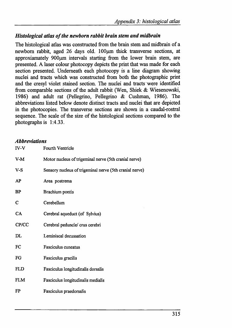
Appendix 3: histological atlas
Histological atlas of the newborn rabbit brain stem and midbrainThe histological atlas was constructed from the brain stem and midbrain of a newborn rabbit, aged 26 days old. 100pm thick transverse sections, at approxiamately 900pm intervals starting from the lower brain stem, are presented. A laser colour photocopy depicts the print that was made for each section presented. Underneath each photocopy is a hne diagram showing nuclei and tracts which was constructed from both the photographic print and the cresyl violet stained section. The nuclei and tracts were identified from comparable sections of the adult rabbit (Wen, Shiek & Wiesenowski, 1986) and adult rat (Pellegrino, Pellegrino & Cushman, 1986). The abbreviations hsted below denote distinct tracts and nuclei that are depicted in the photocopies. The transverse sections are shown in a caudal-rostral sequence. The scale of the size of the histological sections compared to the photographs is 1:4.33.
AbbreviationsTV-Y Fourth Ventricle
V-M Motor nucleus of trigeminal nerve (5th cranial nerve)
v -s Sensory nucleus of trigeminal nerve (5th cranial nerve)
AP Area postrema
BP Brachium pontis
C Cerebellum
CA Cerebral aqueduct (of Sylvius)
CP/CC Cerebral peduncle/ crus cerebri
DL Leminiscal decussation
FC Fasciculus cuneatus
FG Fasciculus gracilis
FLD Fasciculus longitudinalis dorsalis
FLM Fasciculus longitudinalis medialis
FP Fasciculus praedorsalis
315
Appendix 3: histological atlas
ICP Inferior cerebellar peduncle
IF Inferior colliculus
LC Locus coereleus
MCP Middle cerebellar peduncle
NA Nucleus ambiguus
NC Nucleus cuneatus
NCD Nucleus cochlearis dorsalis
NCVP Nucleus cochlearis ventralis posterior
NDLL Nucleus dorsalis lemniscus lateralis
NF Nucleus facialis
NG Nucleus gracilis
NH Nucleus hypoglossus
NX Nucleus interpeduncularis
NMNT Motor nucleus of the trigeminal nerve
NOI Nucleus olivaris inferior
NP Nucleus pontis
NPL Nucleus pontis lateralis
NPLM Nucleus proprius lemniscus medialis
NPM Nucleus pontis medialis
NR Nucleus raphis
NRM Nucleus raphis magnus
NRO Nucleus raphis obscurus
NRP Nucleus raphis pallidus
NRPL Nucleus reticularis proprius lemnisci
316
Appendix 3: histological atlas
NST Nucleus supratrochlearis
NT Nucleus trochlearis
NTS Nucleus tractus solitarius
NTSNT Nucleus tractus spinalis of trigeminal nerve
NV Nucleus vagus
NVBC Nucleus ventralis brachium conjunctivum
NVI Nucleus vestibularis inferior
NVL Nucleus vestibularis lateralis
NVLL Nucleus ventralis lemniscus lateralis
ON Olivary nucleus
PAG Periaqueductal grey
PMC Pyramis meduUae oblongata
RF Reticular formation
RN Red nucleus
RT Radix trigemini
SC Superior colliculus
SCP Superior cerebellar peduncle
SGC Substantia grisea centralis
RST Rubrospinal tract
TG Tuberculum gracilis
TH Tractus hypoglossus
TS Tractus solitarius
317
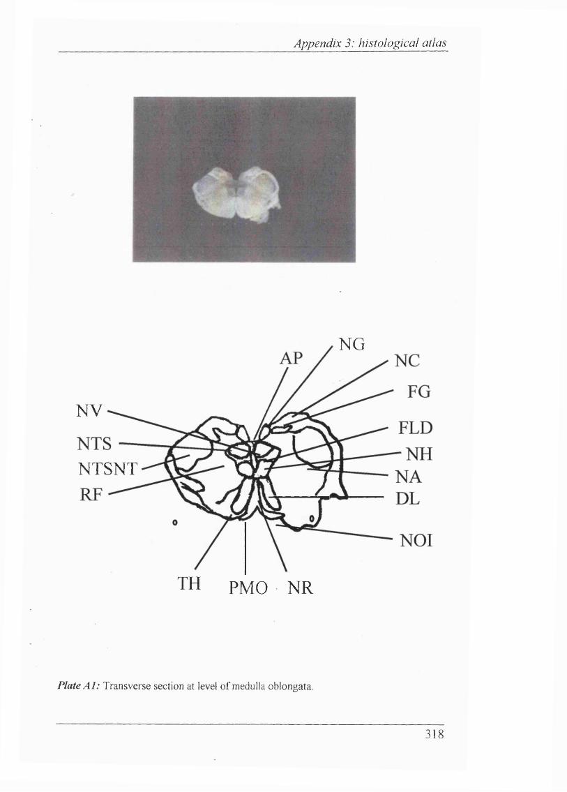
Appendix 3: histological atlas
NG
NV
NTSNT
TH PMO NR
Plate A l: Transverse section at level o f medulla oblongata.
318
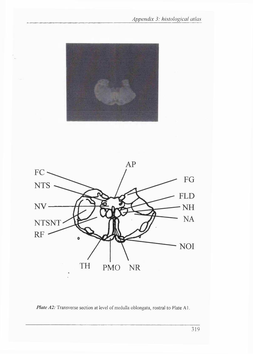
Appendix 3: histological atlas
NTSNT RF
TH PMO NR
Plate A2: Transverse section at level o f medulla oblongata, rostral to Plate A l.
319
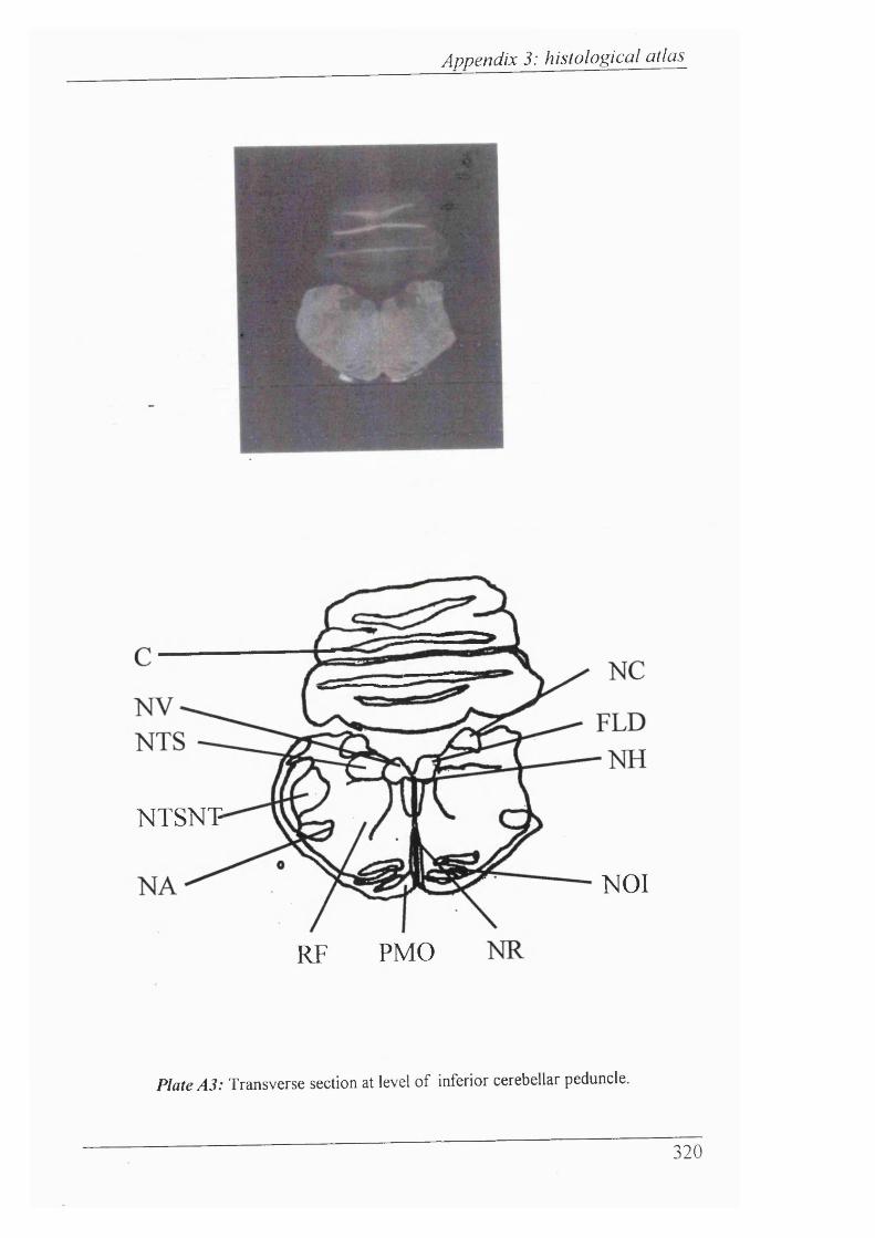
Appendix 3: histological atlas
NTSNT
NOI
RF PMO
Plate A3: Transverse section at level of inferior cerebellar peduncle.
320
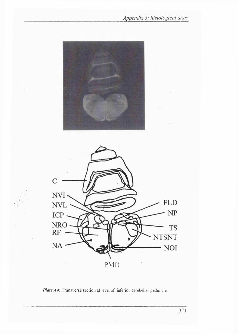
Appendix 3: histological atlas
NTSNT
PMO
Plate A4: Transverse section at level o f inferior cerebellar peduncle.
321
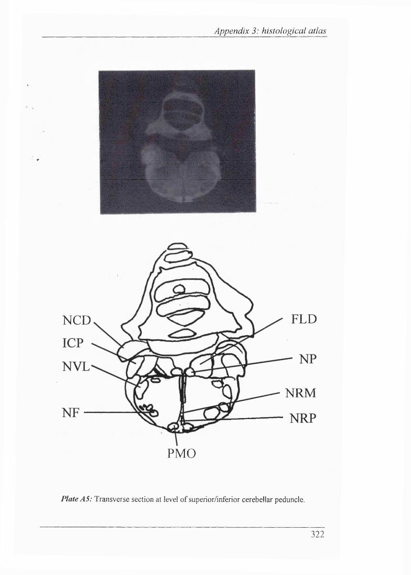
Appendix 3: histological atlas
PMO
FLDNCD
NRM
Plate AS: Transverse section at level o f superior/inferior cerebellar peduncle.
322
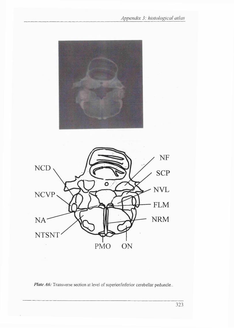
Appendix 3: histological atlas
NCD
NRM&NTSNT
NCVP
PMO ON
Plate A6: Transverse section at level o f superior/inferior cerebellar peduncle..
323
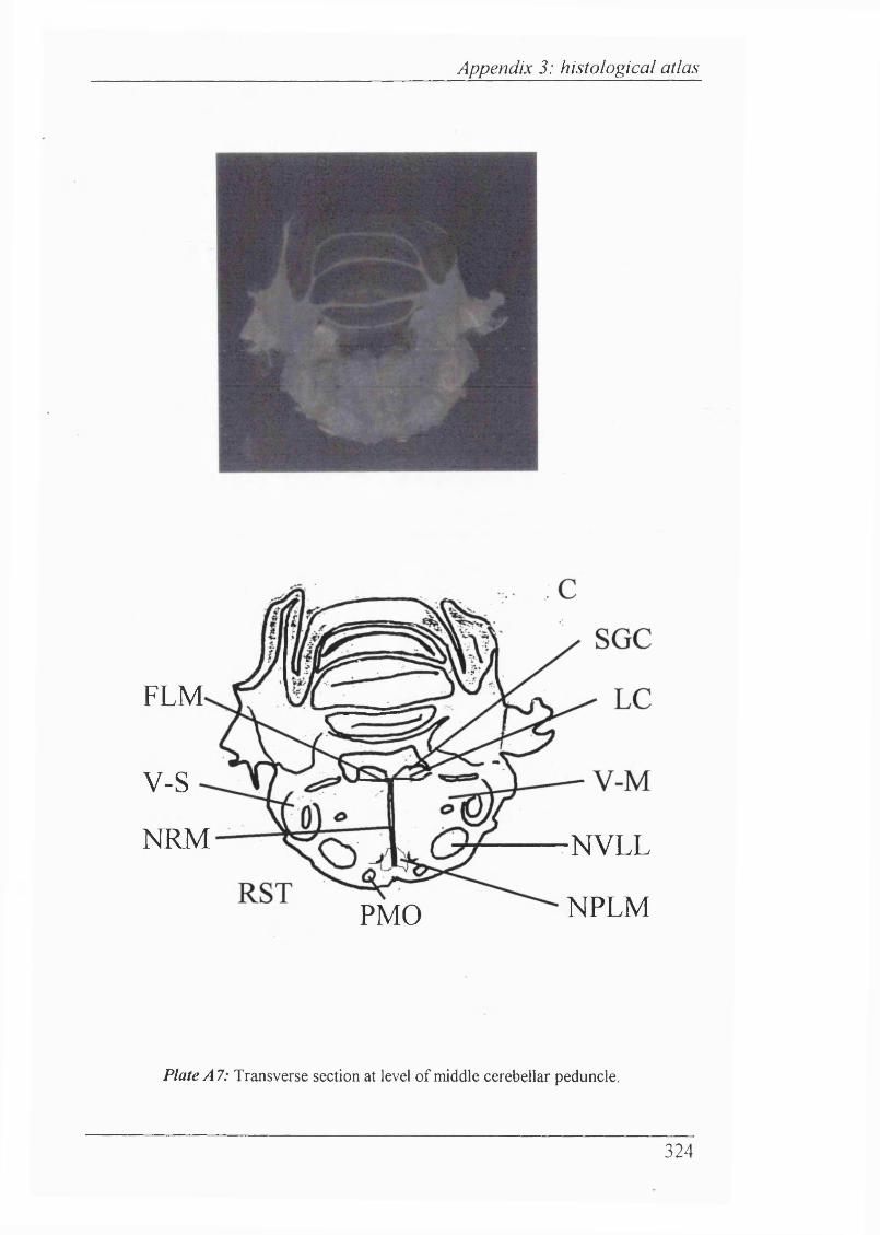
Appendix 3: histological atlas
FLM
v-sNRM NVLL
PMO NPLM
Plate A7: Transverse section at level o f middle cerebellar peduncle.
324
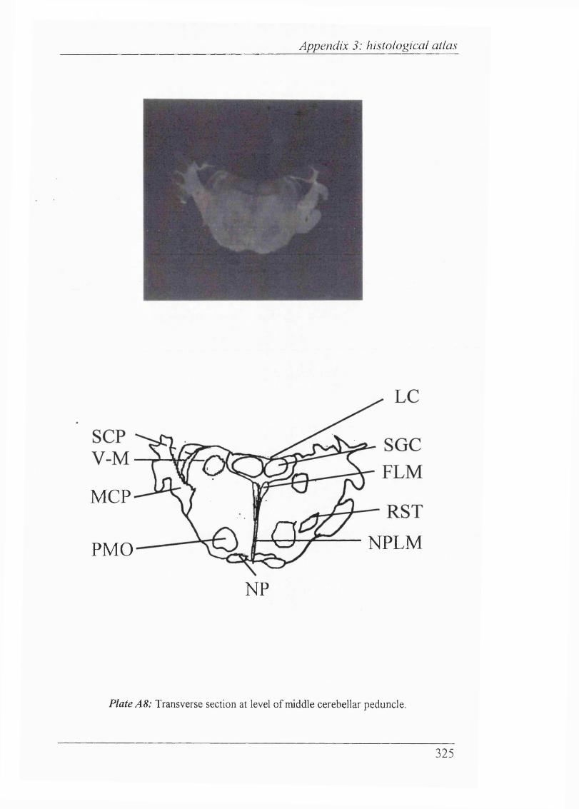
Appendix 3: histological atlas
Q NPLM
MCP
PMO
NP
Plate A8: Transverse section at level o f middle cerebellar peduncle.
325
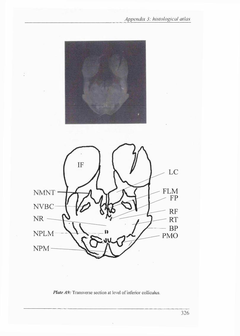
Appendix 3: histological atlas
NMNT
NVBC
NPLM5^NPM
PMO
Plate A9: Transverse section at level o f inferior colliculus.
326
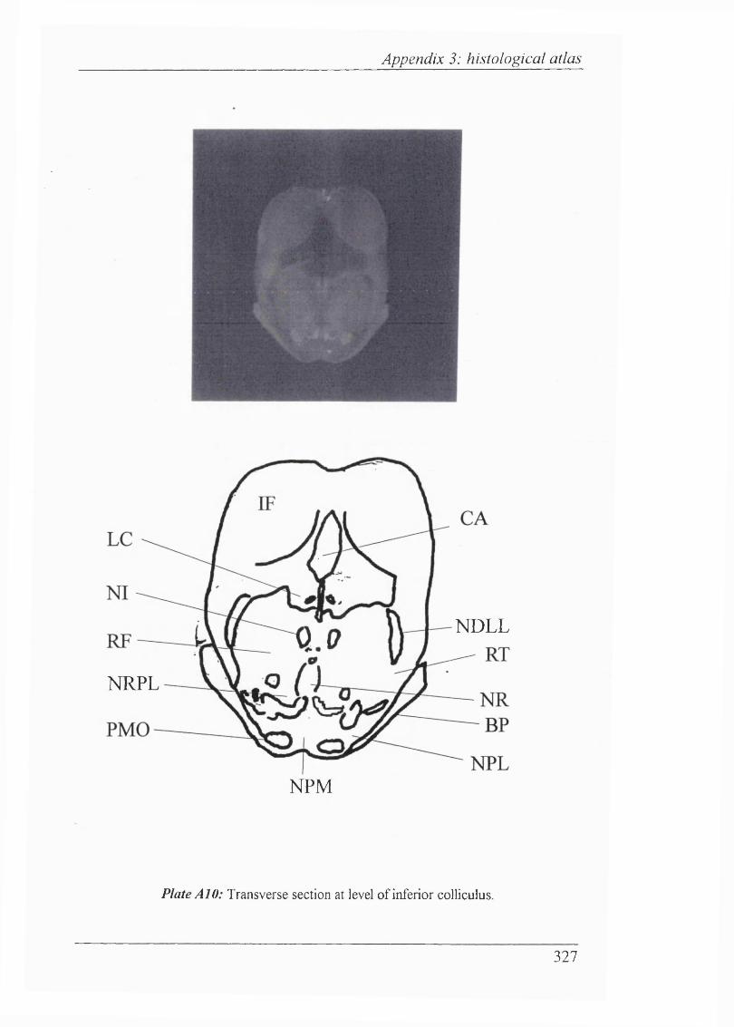
Appendix 3: histological atlas
N D L L
N R P L
N P M
Plate AlO: Transverse section at level o f inferior colliculus.
327
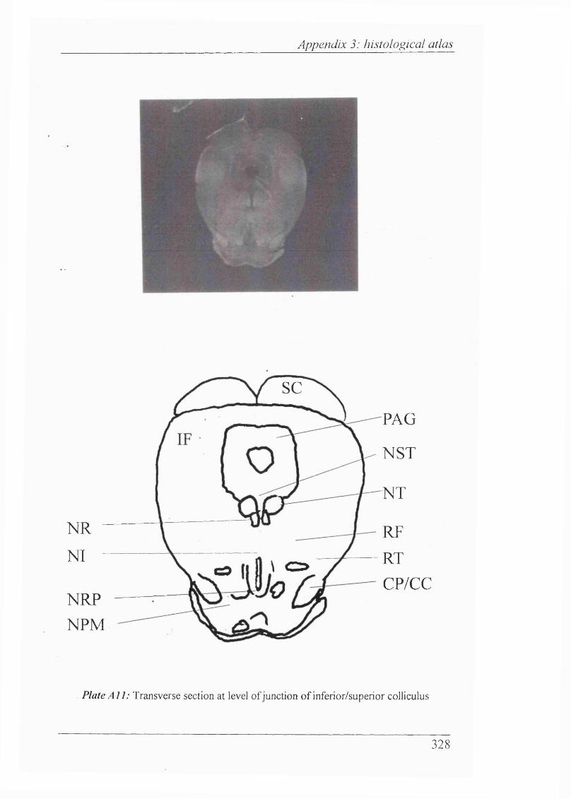
Appendix 3: histological atlas
scPAG
NST
■NT
NRNI
RFRTCP/CC
\ C = >
NRPNPM
Plate A ll: Transverse section at level o f junction o f inferior/superior colliculus
328

Appendix 4
APPENDIX 4
CAROTID CHEMORECEPTOR RECORDINGS
INDIVIDUAL EXPERIMENTAL RESULTS
329
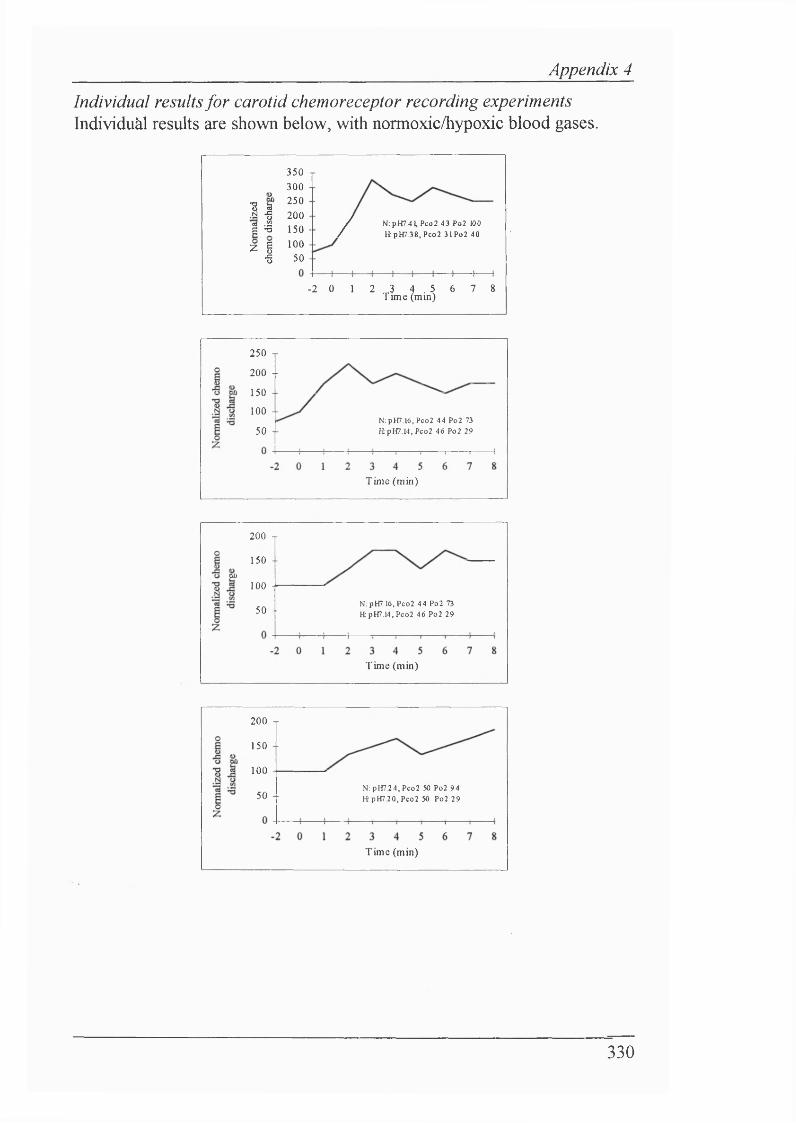
Appendix 4
Individual results for carotid chemoreceptor recording experiments Individuàl results are shown below, with normoxic/hypoxic blood gases.
350 1 300 -
•S S 250.a -g 200 I •! 150 - Z I 100
•s 50
/ N:pH7.41,Pco2 43 Po2 100 / H pH7.38,Pco2 31Po2 40
- 2 0 1 2 3 4 . 5 6 7 8 T une (mm)
250 T
200 - -
150 --
100 - -N;pH7.16,Pco2 44 Po2 73 H pH7.14,Peo2 46 Po2 2950 -
Tim e (m in)
200 T
150 --ao
100 -
N: pH7,16, Pco2 44 Po2 73 RpH7.14.Pco2 46 Po2 2950 --
Tim e (min)
200 T
150 --
100
N:pH7.24,Pco2 50 Po2 94 R pH 7.20.Pco2 50 Po2 2950 --
T im e (min)
330
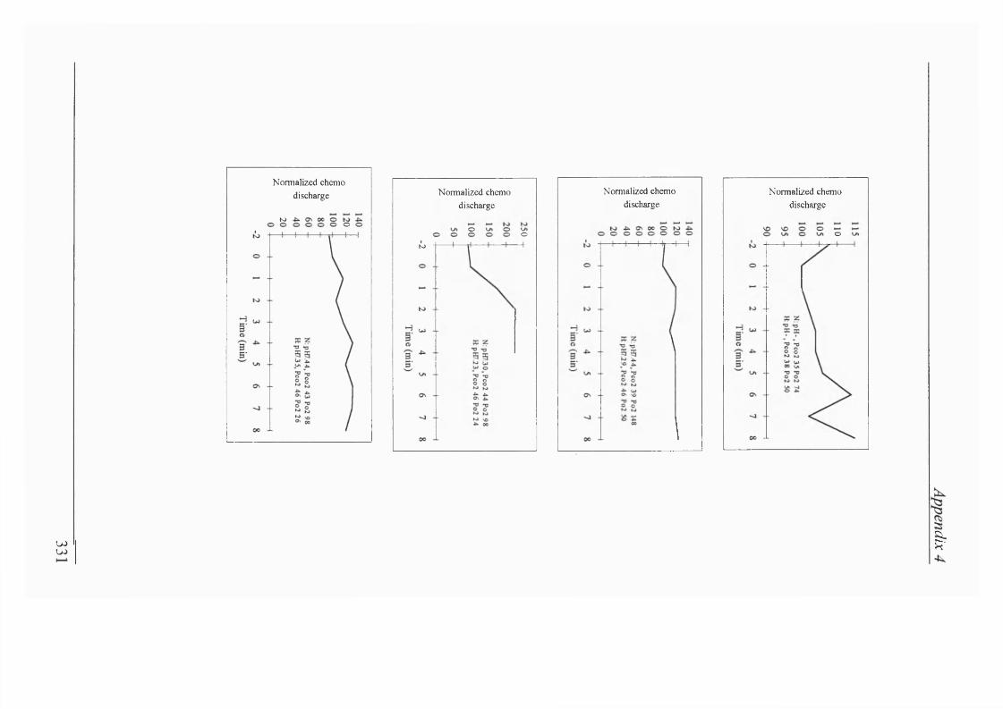
Normalized chemo discharge
K> o
Normalized chemo discharge
o o(O
Normalized chemo discharge
Normalized chemo discharge
o
U)W I
Appendix 5
APPENDIX 5
HISTOLOGICAL RECONSTRUCTION OF MESENCEPHALIC AREA WHERE ELECTRICAL
STIMULATION INHIBITED RESPIRATORY OUTPUT
332
_________________________________Appendix 5
Localization o f mesencephalic site where respiratory output was inhibited (n=29 rabbits)Since the red nucleus was targetted, it was important to ascertain the exact relationship between sites of inhibition and the location of the red nucleus. The site of inhibition of respiratory output was determined by histological reconstruction and experimental coordinates, in relationship to the individual weight (and hence brain size) of rabbits. The following graph shows the site of the centre of the red nucleus (median) within the mesencephalon indicated by the X (weight) and Y (distance) interquartile range, and the stereotaxic coordinates (individual points) at which electrical stimulation inliibited respiratory output (graph refers to the same rostro-caudal coordinate). On the graph legend. R o s t r a l refers to the distance from the inferior colliculus, DEPTH is the distance from the surface of superior colliculus, and m i d l i n e is the distance from the midline of the mesencephalon.
Although it is difBcult to estimate where the red nucleus circumference ends, since the brain tissue merges into the reticular formation, it is clearly also important to measure the area that the red nucleus encompasses from the centre. From its centre, the red nucleus extends in a medial and lateral direction ca. 30% of the distance from the midline of the mesencephalon to the centre of the red nucleus. The depth of the red nucleus extends dorsally and ventrally from its centre by 10% of the depth from the surface of the mesencephalon to the centre of the red nucleus. This is important to bear m mind since the graphs shown central coordinates of the red nucleus, and hence the total diameter of the red nucleus extends beyond this coordinate and thus includes inhibitory sites points outside the range of the centre of the red nucleus.
9.00
8.00
7.00
6.00
5.00
4.00
3.00
2.00
1.00
0.00
À À
4 Fbstral
■ Mdfine
A Depth
0.50 0.55 0.60 0.65
Weight (kg)0.70 0.75
333
APPENDIX 6
VENTILATION SET-UP
Appendix 6
334
______________________________ Appendix 6
Method o f ventilation
In all experiments animals were paralyzed, vagotomized and artificially ventilated. A positive end-expiratory pressure was applied, ca. 2-3cm water. Inspiratory pressure was ca. 15cm water. The rate of ventilation was typically 10-12 breaths per minute, in order to control blood gases to be within the normoxic range.
The ventilator (Harvard Apparatus 707 ventilator) was not pressure- controlled but rather drew in gas from a low pressure reservoir. An air bag served as this low pressure reservoir, into which gases making up the desired inspired mixture were fed, controlled via rotameters (Platon Flowbits). Thus during normoxia, 100% air was fed into the air bag reservoir. In order to expose the animal to isocapnic hypoxia, the reservoir bag was emptied of air and refilled with an air:nitrogen mixture: this swapover was complete within 5s. In most experiments end-tidal gases were monitored (Ohmeda RG5250). The set-up is illustrated below. Arrows indicate direction of gas flow.
ROTAMETERS
C02
GASBAG RABBIT
335


