Embed Size (px)
Citation preview
BRAINA JOURNAL OF NEUROLOGY
Prosody meets syntax: the role of thecorpus callosumDaniela Sammler, Sonja A. Kotz, Korinna Eckstein, Derek V. M. Ott and Angela D. Friederici
Max Planck Institute for Human Cognitive and Brain Sciences, Leipzig, Germany
Correspondence to: Daniela Sammler,
Department of Neuropsychology,
Max Planck Institute for Human Cognitive and Brain Sciences,
04103 Leipzig, Germany
E-mail: [email protected]
Contemporary neural models of auditory language comprehension proposed that the two hemispheres are differently specialized
in the processing of segmental and suprasegmental features of language. While segmental processing of syntactic and lexical
semantic information is predominantly assigned to the left hemisphere, the right hemisphere is thought to have a primacy for
the processing of suprasegmental prosodic information such as accentuation and boundary marking. A dynamic interplay be-
tween the hemispheres is assumed to allow for the timely coordination of both information types. The present event-related
potential study investigated whether the anterior and/or posterior portion of the corpus callosum provide the crucial brain basis
for the online interaction of syntactic and prosodic information. Patients with lesions in the anterior two-thirds of the corpus
callosum connecting orbital and frontal structures, or the posterior third of the corpus callosum connecting temporal, parietal
and occipital areas, as well as matched healthy controls, were tested in a paradigm that crossed syntactic and prosodic ma-
nipulations. An anterior negativity elicited by a mismatch between syntactically predicted phrase structure and prosodic inton-
ation was analysed as a marker for syntax–prosody interaction. Healthy controls and patients with lesions in the anterior corpus
callosum showed this anterior negativity demonstrating an intact interplay between syntax and prosody. No such effect was
found in patients with lesions in the posterior corpus callosum, although they exhibited intact, prosody-independent syntactic
processing comparable with healthy controls and patients with lesions in the anterior corpus callosum. These data support the
interplay between the speech processing streams in the left and right hemispheres via the posterior portion of the corpus
callosum, building the brain basis for the coordination and integration of local syntactic and prosodic features during auditory
speech comprehension.
Keywords: syntax; prosody; interhemispheric transfer; corpus callosum
Abbreviations: antCC = lesions in the anterior two-thirds of the corpus callosum; ELAN = early left anterior negativity;postCC = lesions in the posterior third of the corpus callosum
IntroductionContemporary neural models of language comprehension and pro-
duction testify to a growing interest in the role of cortico-cortical
pathways and the information flow within parallel, but inter-
dependent large-scale networks for the processing of different
linguistic aspects such as phonology, syntax and semantics (Price
et al., 2005; Vigneau et al., 2006; Hickok and Poeppel, 2007;
Friederici, 2009). A great deal of work has concentrated on the
intrahemispheric topographical connectivity between specialized
fronto-temporal (and parietal) ‘language-areas’ via white matter
fibre bundles such as the arcuate, superior longitudinal or uncinate
doi:10.1093/brain/awq231 Brain 2010: 133; 2643–2655 | 2643
Received April 13, 2010. Revised June 22, 2010. Accepted June 28, 2010
� The Author (2010). Published by Oxford University Press on behalf of the Guarantors of Brain. All rights reserved.
For Permissions, please email: [email protected]
at Max-P
lanck-Gesellschaft on S
eptember 2, 2010
http://brain.oxfordjournals.orgD
ownloaded from
fasciculi, in the left (Catani et al., 2005; Anwander et al., 2007;
Saur et al., 2008; Xiang et al., 2010) and/or the right hemisphere
(Parker et al., 2005; Powell et al., 2006; Glasser and Rilling,
2008). It is only with the growing awareness of specific
right-hemispheric language functions such as the processing of
pragmatic (Jung-Beeman, 2005) and prosodic aspects (Gandour
et al., 2000; Friederici and Alter, 2004) that the importance of
interhemispheric connectivity during language comprehension
has come into the focus of scientific interest. Here, we report on
the role of the corpus callosum in the coordination of sentence
level syntactic and (linguistic) prosodic information.
Recent neurocognitive models of auditory language comprehen-
sion assume a differential relative specialization of the two hemi-
spheres in the processing of linguistic features (Zatorre et al.,
2002; Poeppel, 2003; Scott and Johnsrude, 2003; Friederici and
Alter, 2004). While they predominantly assign segmental process-
ing of syntactic and lexical semantic information to left-
hemispheric fronto-temporal brain areas (Friederici, 2002;
Hagoort, 2005; Shalom and Poeppel, 2008), homologue areas in
the right hemisphere are thought to have primacy for the process-
ing of sentence level suprasegmental, prosodic information such as
accentuation and boundary marking. For example, the processing
of filtered or degraded speech merely carrying prosodic informa-
tion has been related to a left ear advantage in dichotic listening
studies (i.e. indicating processing in the right hemisphere;
Blumstein and Cooper, 1974; Shipley-Brown et al., 1988) and
predominant right fronto-temporal brain activations in functional
MRI experiments (Meyer et al., 2002, 2004). Likewise, deficits in
the processing of sentence level prosodic intonation have been
described in patients with lesions in the right hemisphere
(Weintraub et al., 1981; Bradvik et al., 1991), although other
studies using natural speech stimuli reported impairment of pros-
odic processing in both patients with lesions in the left and right
hemispheres (Heilman et al., 1984; Bryan, 1989; Perkins et al.,
1996). This suggests that the actual lateralization of prosodic
processing partly depends on task demands (Plante et al., 2002;
Gandour et al., 2004; Kotz et al., 2006) and on the presence
of concurrent segmental information (Van Lancker and Sidtis,
1992; Baum and Pell, 1999; Friederici and Alter, 2004;
Pannekamp et al., 2005).
If there is a relative hemispheric division of labour between
segmental (left hemisphere) and suprasegmental information pro-
cessing (right hemisphere), the coordination of the lateralized
speech streams requires smooth and rapid information exchange
between the two hemispheres. This is likely to occur via the com-
missural fibres crossing through the corpus callosum. The corpus
callosum is not a homogeneous structure but the fibre tracts are
topographically ordered according to their cortical origin. Diffusion
tensor imaging studies consistently locate projections between the
orbital and frontal lobes through the anterior two-thirds of the
corpus callosum (genu and truncus), whereas fibres connecting
the temporal, parietal and occipital lobes cross through the pos-
terior third of the corpus callosum (isthmus and splenium; Huang
et al., 2005; Hofer and Frahm, 2006; Zarei et al., 2006;
Dougherty et al., 2007; Park et al., 2008).
It is unclear though which part of the corpus callosum is par-
ticularly critical for the interhemispheric exchange of syntactic and
prosodic information, because both the syntax (Just et al., 1996;
Caplan et al., 1998; Dapretto and Bookheimer, 1999; Ni et al.,
2000) and the prosody processing stream (Meyer et al., 2002,
2004) span temporal and frontal brain areas in the respective
hemisphere connected via the posterior and anterior corpus
callosum, respectively (for reviews, see Friederici, 2002; Friederici
and Alter, 2004). A first answer to this question has been obtained
in an event-related potential study testing the impact of prosodic
intonation on syntactic processing in patients with corpus callosum
lesions (Friederici et al., 2007). In this experiment, patients with
posterior corpus callosum lesions did not process violations of verb
argument structure that could only be detected if preceding
prosodic phrase structure information was processed. More pre-
cisely, these patients did not exhibit a N400-like effect for intransi-
tive verbs that did not match the prosody-induced syntactic
expectation to encounter a transitive verb. Conversely, patients
with anterior corpus callosum lesions showed the N400-like
effect (although modulated in morphology and topography).
Altogether, these data suggest that prosodic information guides
syntactic processing predominantly via the posterior corpus
callosum with less strong involvement of the anterior corpus
callosum.
The present studyImportantly, the prosody–syntax interface does not work unidirec-
tionally. Not only does prosody influence the syntactic structuring
and interpretation of spoken utterances as tested by Friederici
et al. (2007) (for review, see Cutler et al., 1997; Eckstein and
Friederici, 2006), but also syntactic structure is highly predictive
of prosodic phrasing, with syntactic and prosodic boundaries often
(though not always) coinciding (Selkirk, 1986; Inkelas and Zec,
1996; Truckenbrodt, 1999). The current electroencephalography
lesion study incorporated this idea and aimed to specify which
portion of the corpus callosum crucially interfaces syntactic parsing
effects on the processing of prosodic pitch contour. Therefore, the
present paradigm used sentences with a highly predictable syntac-
tic phrase structure allowing for strong predictions about the
prosodic intonation of a forthcoming word (i.e. rising or falling
pitch indicating sentence continuation or closure, respectively).
Conditions containing a mismatch between the syntactically
predicted and the actually perceived prosodic intonation have
been shown to evoke an anteriorly distributed negativity in
healthy students (Eckstein and Friederici, 2006).
The present study tested whether this index of syntax–prosody
interaction is based in the corpus callosum communication be-
tween the left and right hemispheres, and whether syntactic in-
formation takes an anterior or posterior route via the corpus
callosum to guide prosodic processing. Therefore, two patient
groups were recruited according to the topographical organization
of the corpus callosum (Huang et al., 2005; Hofer and Frahm,
2006), one group with lesions in the anterior two-thirds of the
corpus callosum and the other with lesions in the posterior third of
the corpus callosum, as well as matched healthy controls. If the
anterior negativity evoked by a mismatch between syntactic con-
text and forthcoming prosodic intonation (i.e. indicating an impact
of syntactic on prosodic processing) (Eckstein and Friederici, 2006)
2644 | Brain 2010: 133; 2643–2655 D. Sammler et al.
at Max-P
lanck-Gesellschaft on S
eptember 2, 2010
http://brain.oxfordjournals.orgD
ownloaded from
is due to an interaction between the left and right hemispheres,
then this effect should be absent in patients with lesions in the
relevant portion of the corpus callosum. In other words, syntactic
information processed in the left hemisphere should not influence
prosodic processes in the right hemisphere. Based on the findings
of Friederici et al. (2007), we hypothesized that lesions of
posterior fibres connecting the temporal lobes involved in both
prosodic and syntactic processing (Friederici, 2002; Friederici and
Alter, 2004) would lead to an interruption of syntax–prosody
interactions. Alternatively, given the strong involvement of the
left inferior frontal gyrus and frontal operculum in syntactic
processing (Friederici, 2002; Grodzinsky and Friederici, 2006;
Vigneau et al., 2006), syntactic guidance of prosodic processing
may also be disrupted after lesion of anterior corpus callosum
fibres connecting the orbital and frontal lobes.
Material and methods
ParticipantsEleven patients were classified into two groups according to their
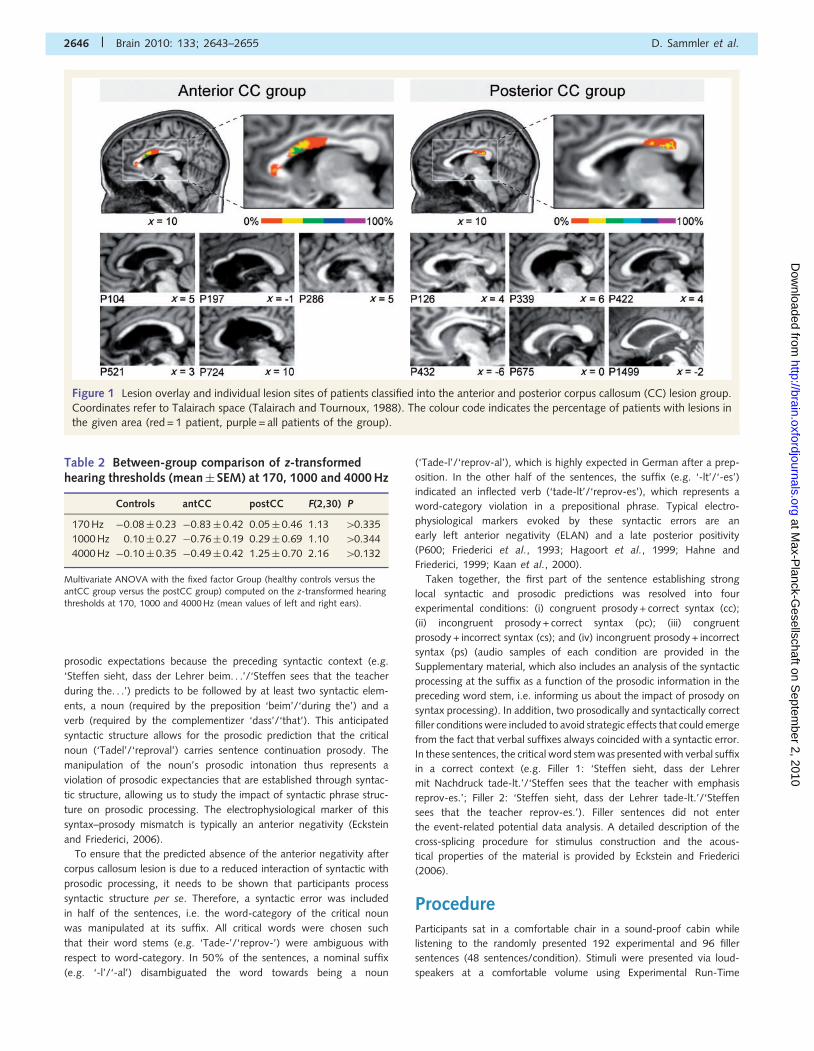
lesion sites, involving either the anterior two-thirds of the corpus cal-
losum (n = 5, antCC group), or the posterior third of the corpus callo-
sum (n = 6; postCC group; see Table 1 and Fig. 1 for patient histories
and lesion sites). Furthermore, 22 healthy controls, matched in hand-
edness, gender and age were tested (age range: 23–71 years, mean:
50 years; 16 males, 6 females). Informed consent according to the
Declaration of Helsinki (1964) was obtained from each participant
prior to the experiment, which was approved by the local Ethical
Committee.
To ensure that patients and controls did not differ in their peripheral
hearing abilities, monaural pure tone thresholds were determined in all
participants for frequencies of 170, 1000 and 4000 Hz, chosen to
cover the main frequency range of speech. Notably, 170 Hz was
tested because the perception of prosodic manipulations in the stimu-
lus material required the detection of F0 shifts either exceeding or
dropping below 170 Hz indicating sentence continuation or closure,
respectively (for details, see Eckstein and Friederici, 2006).
Audiograms for each ear and frequency were obtained in an adaptive
up-down method using the test equipment of Bungert-Kahl et al.
(2004) in a sound-proof cabin. The test values of each participant
(average of left and right ears) were z-transformed based on the nor-
mative data provided by Bungert-Kahl et al. (2004) for different age
groups. Missing normative data (e.g. for 170 Hz and ages 470 years)
were interpolated using the cubic spline function in MATLAB 7.1. All
experimental groups exhibited normal hearing thresholds in all tested
frequencies [i.e. not exceeding 2 standard deviations (SD) of the
standard normal distribution; Table 2], assuring that all groups were
principally able to perceive the prosodic manipulations. Moreover, the
hearing threshold did not differ between groups (all P0-values40.132),
ensuring that potential group differences in electrophysiological effects
cannot be attributed to differential hearing abilities.
Stimulus materialThe stimulus material employed has been used in a previous study
with healthy students (Eckstein and Friederici, 2006). Participants lis-
tened to 288 German sentences starting with a proper name
(‘Steffen’) and a verb (‘sieht’/‘sees’) forming the matrix clause. The
following subordinate clause was composed of a complementizer
‘dass’/‘that’, a subject (‘der Lehrer’/‘the teacher’), a prepositional
phrase consisting of a preposition fused with an article (‘beim’/
‘during the’) and a noun (‘Tadel’/‘reproval’), followed by the sentence
final verb (‘schmunzelt’/‘smiles’; Table 3 and Supplementary Stimuli).
Prosodic intonation of the noun at penultimate position was
manipulated.
The critical noun (stem + suffix) was either marked for sentence con-
tinuation (congruent; rising pitch contour) or spoken with sentence
final prosody (incongruent; falling pitch contour). The latter violates
Table 1 Individual patient histories
Patient Gender Age Hand Onset Aetiology Callosal lesion Additional lesions
Anterior corpus callosum group (involving the anterior two-thirds of the corpus callosum)
104 F 69 R 9;9 CMA Rostrum, anterior c-body Pontine, left basal ganglia lesion
197 F 64 R 8;10 IS/SAH Rostrum, middle c-body Left frontolaterala and temporopolar contusion,left basal ganglia lesion, left parietal necrosis
286 M 66 R 8;4 IS Anterior c-body CMA with pontine lacune
521 M 51 R 6;5 SAH Anterior knee, left middlec-body, EVD
Right basal forebrain and temporopolar lesion
724 M 25 L 5;4 CH Anterior c-body Left thalamus, left internal capsule lesion
Posterior corpus callosum group (involving the posterior third of the corpus callosum)
126 M 75 R 12;0 CH Chronic ischemic posteriorCC lesion
CMA, right thalamic lacune, right occipital bleeding
339 M 59 R 7;11 SAH Posterior c-body (presplenial) Post SAH, post EVD
422 M 43 R 9;0 TBI Presplenial lesion Minor atrophy of cerebellar vermis
432 F 26 R 8;6 CH Posterior CC lesion Embolized AVM, left posterior thalamus lesion
675 M 24 R 5;2 TBI Small presplenial lesion Left preinsular region,a left cerebral peduncle (midbrain)
1499 M 53 R – TBI (pre)splenial lesion CMA
a Patients were included despite lesions in the left frontal cortex known to be involved in syntactic processing (Friederici and Kotz, 2003; Vigneau et al., 2006; Grodzinsky
and Santi, 2008) as they showed a normal ELAN and P600 pattern (Fig. 3B and C) suggesting normal syntactic processing.Handedness is as defined by the Edinburgh Handedness Inventory (Oldfield, 1971); Lesion onset is in years; months—no information available for Patient 1499.AVM = arteriovenous malformation; CH = cerebral haemorrhage; CMA = cerebral microangiopathy; EVD = external ventricular drainage; F = female; IS = ischemic stroke;L = left handed; M = male; R = right handed; SAH = subarachnoid haemorrhage; TBI = traumatic brain injury.
Callosal transfer of prosody and syntax Brain 2010: 133; 2643–2655 | 2645
at Max-P
lanck-Gesellschaft on S
eptember 2, 2010
http://brain.oxfordjournals.orgD
ownloaded from
prosodic expectations because the preceding syntactic context (e.g.
‘Steffen sieht, dass der Lehrer beim. . .’/‘Steffen sees that the teacher
during the. . .’) predicts to be followed by at least two syntactic elem-
ents, a noun (required by the preposition ‘beim’/‘during the’) and a
verb (required by the complementizer ‘dass’/‘that’). This anticipated
syntactic structure allows for the prosodic prediction that the critical
noun (‘Tadel’/‘reproval’) carries sentence continuation prosody. The
manipulation of the noun’s prosodic intonation thus represents a
violation of prosodic expectancies that are established through syntac-
tic structure, allowing us to study the impact of syntactic phrase struc-
ture on prosodic processing. The electrophysiological marker of this
syntax–prosody mismatch is typically an anterior negativity (Eckstein
and Friederici, 2006).
To ensure that the predicted absence of the anterior negativity after
corpus callosum lesion is due to a reduced interaction of syntactic with
prosodic processing, it needs to be shown that participants process
syntactic structure per se. Therefore, a syntactic error was included
in half of the sentences, i.e. the word-category of the critical noun
was manipulated at its suffix. All critical words were chosen such
that their word stems (e.g. ‘Tade-’/‘reprov-’) were ambiguous with
respect to word-category. In 50% of the sentences, a nominal suffix
(e.g. ‘-l’/‘-al’) disambiguated the word towards being a noun
(‘Tade-l’/‘reprov-al’), which is highly expected in German after a prep-
osition. In the other half of the sentences, the suffix (e.g. ‘-lt’/‘-es’)
indicated an inflected verb (‘tade-lt’/‘reprov-es’), which represents a
word-category violation in a prepositional phrase. Typical electro-
physiological markers evoked by these syntactic errors are an
early left anterior negativity (ELAN) and a late posterior positivity
(P600; Friederici et al., 1993; Hagoort et al., 1999; Hahne and
Friederici, 1999; Kaan et al., 2000).
Taken together, the first part of the sentence establishing strong
local syntactic and prosodic predictions was resolved into four
experimental conditions: (i) congruent prosody + correct syntax (cc);
(ii) incongruent prosody + correct syntax (pc); (iii) congruent
prosody + incorrect syntax (cs); and (iv) incongruent prosody + incorrect
syntax (ps) (audio samples of each condition are provided in the
Supplementary material, which also includes an analysis of the syntactic
processing at the suffix as a function of the prosodic information in the
preceding word stem, i.e. informing us about the impact of prosody on
syntax processing). In addition, two prosodically and syntactically correct
filler conditions were included to avoid strategic effects that could emerge
from the fact that verbal suffixes always coincided with a syntactic error.
In these sentences, the critical word stem was presented with verbal suffix
in a correct context (e.g. Filler 1: ‘Steffen sieht, dass der Lehrer
mit Nachdruck tade-lt.’/‘Steffen sees that the teacher with emphasis
reprov-es.’; Filler 2: ‘Steffen sieht, dass der Lehrer tade-lt.’/‘Steffen
sees that the teacher reprov-es.’). Filler sentences did not enter
the event-related potential data analysis. A detailed description of the
cross-splicing procedure for stimulus construction and the acous-
tical properties of the material is provided by Eckstein and Friederici
(2006).
ProcedureParticipants sat in a comfortable chair in a sound-proof cabin while
listening to the randomly presented 192 experimental and 96 filler
sentences (48 sentences/condition). Stimuli were presented via loud-
speakers at a comfortable volume using Experimental Run-Time
Figure 1 Lesion overlay and individual lesion sites of patients classified into the anterior and posterior corpus callosum (CC) lesion group.
Coordinates refer to Talairach space (Talairach and Tournoux, 1988). The colour code indicates the percentage of patients with lesions in
the given area (red = 1 patient, purple = all patients of the group).
Table 2 Between-group comparison of z-transformedhearing thresholds (mean� SEM) at 170, 1000 and 4000 Hz
Controls antCC postCC F(2,30) P
170 Hz �0.08� 0.23 �0.83� 0.42 0.05� 0.46 1.13 40.335
1000 Hz 0.10� 0.27 �0.76� 0.19 0.29� 0.69 1.10 40.344
4000 Hz �0.10� 0.35 �0.49� 0.42 1.25� 0.70 2.16 40.132
Multivariate ANOVA with the fixed factor Group (healthy controls versus theantCC group versus the postCC group) computed on the z-transformed hearing
thresholds at 170, 1000 and 4000 Hz (mean values of left and right ears).
2646 | Brain 2010: 133; 2643–2655 D. Sammler et al.
at Max-P
lanck-Gesellschaft on S
eptember 2, 2010
http://brain.oxfordjournals.orgD
ownloaded from
System (ERTS; BeriSoft Cooperation, Frankfurt am Main, Germany).
Each trial started with a fixation star visible from 500 ms prior to sen-
tence onset to 1500 ms after sentence off-set, followed by a 2000 ms
response screen during which participants were asked to judge the
grammaticality of the sentence by pressing a left or right button
(key assignment was counterbalanced across participants). Each trial
was followed by a 2500 ms interstimulus interval showing a blank
screen. To familiarize participants with the procedure, they received
training with 12 randomly presented trials (two sentences/condition).
The experimental session was divided into four blocks and had a dur-
ation of �60 min (training and breaks included).
Data acquisition and analysisThe electroencephalogram was recorded from 25 Ag/AgCl electrodes
placed according to the extended international 10–20 system
(Sharbrough et al., 1991). The electrode positions were: FP1, FP2,
F7, F8, F3, F4, FZ, FT7, FT8, FC3, FC4, T7, T8, C3, C4, CZ, CP5,
CP6, P7, P8, P3, P4, PZ, O1, O2. Left mastoid (M1) served as refer-
ence; an additional electrode was placed on the right mastoid bone
(M2) for off-line re-referencing. The ground electrode was located on
the sternum. Horizontal and vertical electrooculograms were bipolarly
recorded from electrodes placed on the outer canthus of each eye, as
well as above and below the right eye. Impedances were kept below
5 k�. Signals were amplified with two synchronized PORTI-32/MREFA
amplifiers (Twente Medical Systems International B.V., Enschede, The
Netherlands) and digitized with a sampling rate of 250 Hz.
EEP 3.2 (ANT-software) was used to re-reference the data to linked
mastoids. Further processing steps were accomplished using EEGLAB
5.03 (Delorme and Makeig, 2004) in MATLAB 7.7. Data were filtered
using a 0.3 Hz highpass filter (fir, 2725 points, Blackman window), and
strong muscle artefacts, electrode drifts or technical artefacts were
manually rejected before entering the continuous data into an inde-
pendent component analysis. The resulting component structure was
used to reject eye movement and blink artefacts, muscle artefacts and
slow drifts. Afterwards, the data were filtered with a 20 Hz lowpass
filter (fir, 277 points, Blackman window), and cut into two different
epochs. The first epoch of �100 to 1600 ms was time-locked to the
onset of the critical word and ideally covered the prosodic violation in
the word stem. The second epoch of �100 to 1200 ms was
time-locked to the suffix of the critical word, i.e. the onset of the
syntactic violations. Epochs were rejected whenever one or more elec-
trodes exhibited voltages of �55 mV, linear trends of �50 mV in a
400 ms gliding window or small blinks and drifts as identified by
visual inspection. Non-rejected epochs were averaged separately for
each condition. Only correctly answered trials were included. Averages
were aligned to the respective �100 to 0 ms baseline. An average of
42.14 trials was included for each participant and each condition
(mean� SD of healthy controls: 43.07� 3.96, antCC: 39.85� 4.44,
postCC: 40.63�5.79; no significant differences between groups and
conditions).
Effects of syntax on prosodic processing were analysed time-locked
to the onset of the critical word (i.e. time-locked to the prosodic vio-
lation). Statistical analyses were carried out on the mean amplitudes of
the prosodically congruent (cc and cs) and incongruent conditions (pc
and ps) calculated in a time window from 200 to 500 ms after word
onset (centred around the peak of the anterior negativity as identified
by visual inspection) for six regions of interest: (i) left fronto-temporal
(F7, FT7, T7); (ii) right fronto-temporal (F8, FT8, T8); (iii) left
fronto-central (F3, FC3, C3); (iv) right fronto-central (F4, FC4, C4);
(v) left posterior (P7, P3, O1); and (vi) right posterior (P8, P4, O2).
Prosody-independent effects of syntactic violations were analysed
time-locked to the onset of the suffix of the critical word. Statistical
analyses were carried out on the mean amplitudes of syntactically
correct (cc and pc) and incorrect trials (cs and ps) for the regions of
interest described above, in two time windows: (i) from 250 to 350 ms
(centred around the peak of the ELAN); and (ii) from 450 to 850 ms
after suffix onset (centred around the peak of the P600). The Results
in Supplementary materials contain an additional analysis of the modu-
lation of the ELAN and P600 at the word suffix by the prosodic in-
formation in the preceding word stem. In healthy controls and (as a
trend) in the antCC group, the ELAN lost its left lateralization, showing
up bilaterally when the syntactic error was preceded by incongruent
compared to congruent prosody. This indicates an immediate influence
of phrasal prosody on early syntactic parsing stages. The patients in
the postCC group showed no such ELAN topography shift, suggesting
that prosodic information does not influence early steps of syntactic
analysis after disconnection of posterior callosal fibres and supporting
the notion that the posterior third of the corpus callosum is particularly
relevant for interfacing prosodic and syntactic information during audi-
tory language comprehension (Friederici et al., 2007). No consistent
results were found for the P600. Statistical details and a more exten-
sive discussion are provided in the Results section of Supplementary
materials. The analysis of the behavioural performance was restricted
to the error rates. No reaction times were analysed due to the delayed
response.
Results
Behavioural dataParticipants showed an average performance of 94.59% correct
(healthy controls: 97.51%, antCC group: 87.5%, postCC group:
89.76%), demonstrating that they were able to detect the syntac-
tic violations. An ANOVA with the repeated measures factors
Syntax (correct versus incorrect), Prosody (congruent versus incon-
gruent) and the between-subjects factor Group (healthy controls
versus antCC group versus postCC group) revealed a marginally
better performance for prosodically congruent compared with
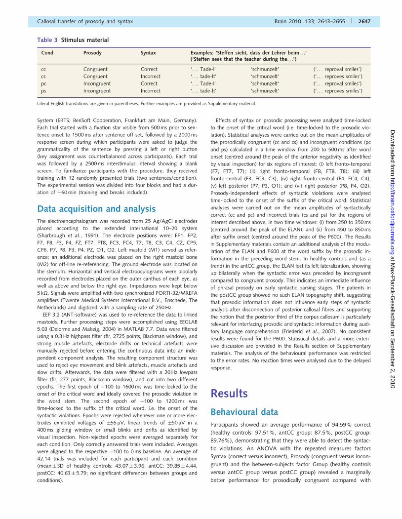
Table 3 Stimulus material
Cond Prosody Syntax Examples: ‘Steffen sieht, dass der Lehrer beim. . .’(‘Steffen sees that the teacher during the. . .’)
cc Congruent Correct ‘. . . Tade-l’ ‘schmunzelt’ (‘. . . reproval smiles’)
cs Congruent Incorrect ‘. . . tade-lt’ ‘schmunzelt’ (‘. . . reproves smiles’)
pc Incongruent Correct ‘. . . Tade-l’ ‘schmunzelt’ (‘. . . reproval smiles’)
ps Incongruent Incorrect ‘. . . tade-lt’ ‘schmunzelt’ (‘. . . reproves smiles’)
Literal English translations are given in parentheses. Further examples are provided as Supplementary material.
Callosal transfer of prosody and syntax Brain 2010: 133; 2643–2655 | 2647
at Max-P
lanck-Gesellschaft on S
eptember 2, 2010
http://brain.oxfordjournals.orgD
ownloaded from
incongruent sentences (main effect of Prosody: F(1,30) = 4.08,
P50.053), indicating that prosodic violations were processed
although they were task irrelevant. No group differences
(P-values40.245) or other effects were found.
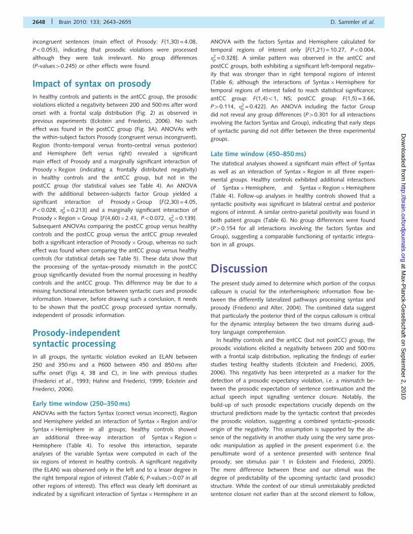
Impact of syntax on prosodyIn healthy controls and patients in the antCC group, the prosodic
violations elicited a negativity between 200 and 500 ms after word
onset with a frontal scalp distribution (Fig. 2) as observed in
previous experiments (Eckstein and Friederici, 2006). No such
effect was found in the postCC group (Fig. 3A). ANOVAs with
the within-subject factors Prosody (congruent versus incongruent),
Region (fronto-temporal versus fronto-central versus posterior)
and Hemisphere (left versus right) revealed a significant
main effect of Prosody and a marginally significant interaction of
Prosody�Region (indicating a frontally distributed negativity)
in healthy controls and the antCC group, but not in the
postCC group (for statistical values see Table 4). An ANOVA
with the additional between-subjects factor Group yielded a
significant interaction of Prosody�Group [F(2,30) = 4.05,
P50.028, �2p = 0.213] and a marginally significant interaction of
Prosody�Region�Group [F(4,60) = 2.43, P50.072, �2p = 0.139].
Subsequent ANOVAs comparing the postCC group versus healthy
controls and the postCC group versus the antCC group revealed
both a significant interaction of Prosody�Group, whereas no such
effect was found when comparing the antCC group versus healthy
controls (for statistical details see Table 5). These data show that
the processing of the syntax–prosody mismatch in the postCC
group significantly deviated from the normal processing in healthy
controls and the antCC group. This difference may be due to a
missing functional interaction between syntactic cues and prosodic
information. However, before drawing such a conclusion, it needs
to be shown that the postCC group processed syntax normally,
independent of prosodic information.
Prosody-independentsyntactic processingIn all groups, the syntactic violation evoked an ELAN between
250 and 350 ms and a P600 between 450 and 850 ms after
suffix onset (Figs 4, 3B and C), in line with previous studies
(Friederici et al., 1993; Hahne and Friederici, 1999; Eckstein and
Friederici, 2006).
Early time window (250–350 ms)
ANOVAs with the factors Syntax (correct versus incorrect), Region
and Hemisphere yielded an interaction of Syntax�Region and/or
Syntax�Hemisphere in all groups; healthy controls showed
an additional three-way interaction of Syntax�Region�
Hemisphere (Table 4). To resolve this interaction, separate
analyses of the variable Syntax were computed in each of the
six regions of interest in healthy controls. A significant negativity
(the ELAN) was observed only in the left and to a lesser degree in
the right temporal region of interest (Table 6; P-values40.07 in all
other regions of interest). This effect was clearly left dominant as
indicated by a significant interaction of Syntax�Hemisphere in an
ANOVA with the factors Syntax and Hemisphere calculated for
temporal regions of interest only [F(1,21) = 10.27, P50.004,
�2p = 0.328]. A similar pattern was observed in the antCC and
postCC groups, both exhibiting a significant left-temporal negativ-
ity that was stronger than in right temporal regions of interest
[Table 6; although the interactions of Syntax�Hemisphere for
temporal regions of interest failed to reach statistical significance;
antCC group: F(1,4)51, NS; postCC group: F(1,5) = 3.66,
P40.114, �2p = 0.422]. An ANOVA including the factor Group
did not reveal any group differences (P40.301 for all interactions
involving the factors Syntax and Group), indicating that early steps
of syntactic parsing did not differ between the three experimental
groups.
Late time window (450–850 ms)
The statistical analyses showed a significant main effect of Syntax
as well as an interaction of Syntax�Region in all three experi-
mental groups. Healthy controls exhibited additional interactions
of Syntax�Hemisphere, and Syntax�Region�Hemisphere
(Table 4). Follow-up analyses in healthy controls showed that a
syntactic positivity was significant in bilateral central and posterior
regions of interest. A similar centro-parietal positivity was found in
both patient groups (Table 6). No group differences were found
(P40.154 for all interactions involving the factors Syntax and
Group), suggesting a comparable functioning of syntactic integra-
tion in all groups.
DiscussionThe present study aimed to determine which portion of the corpus
callosum is crucial for the interhemispheric information flow be-
tween the differently lateralized pathways processing syntax and
prosody (Friederici and Alter, 2004). The combined data suggest
that particularly the posterior third of the corpus callosum is critical
for the dynamic interplay between the two streams during audi-
tory language comprehension.
In healthy controls and the antCC (but not postCC) group, the
prosodic violations elicited a negativity between 200 and 500 ms
with a frontal scalp distribution, replicating the findings of earlier
studies testing healthy students (Eckstein and Friederici, 2005,
2006). This negativity has been interpreted as a marker for the
detection of a prosodic expectancy violation, i.e. a mismatch be-
tween the prosodic expectation of sentence continuation and the
actual speech input signalling sentence closure. Notably, the
build-up of such prosodic expectations crucially depends on the
structural predictions made by the syntactic context that precedes
the prosodic violation, suggesting a combined syntactic–prosodic
origin of the negativity. This assumption is supported by the ab-
sence of the negativity in another study using the very same pros-
odic manipulation as applied in the present experiment (i.e. the
penultimate word of a sentence presented with sentence final
prosody; see stimulus pair 1 in Eckstein and Friederici, 2005).
The mere difference between these and our stimuli was the
degree of predictability of the upcoming syntactic (and prosodic)
structure. While the context of our stimuli unmistakably predicted
sentence closure not earlier than at the second element to follow,
2648 | Brain 2010: 133; 2643–2655 D. Sammler et al.
at Max-P
lanck-Gesellschaft on S
eptember 2, 2010
http://brain.oxfordjournals.orgD
ownloaded from
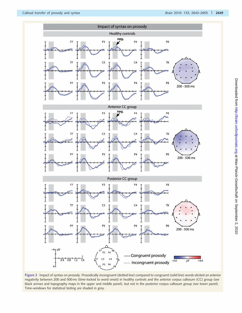
Figure 2 Impact of syntax on prosody. Prosodically incongruent (dotted line) compared to congruent (solid line) words elicited an anterior
negativity between 200 and 500 ms (time-locked to word onset) in healthy controls and the anterior corpus callosum (CC) group (see
black arrows and topography maps in the upper and middle panel), but not in the posterior corpus callosum group (see lower panel).
Time-windows for statistical testing are shaded in grey.
Callosal transfer of prosody and syntax Brain 2010: 133; 2643–2655 | 2649
at Max-P
lanck-Gesellschaft on S
eptember 2, 2010
http://brain.oxfordjournals.orgD
ownloaded from
the syntactic context in Eckstein and Friederici (2005) (e.g. ‘Peter
weiß, dass der Onkel. . .’/‘Peter knows, that the uncle. . .’) left
open how many syntactic nodes were to follow, precluding as
clear a prediction on prosodic phrasing. These combined findings
lead us to conclude that the observed negative brain potential is a
response to violations of local prosodic expectancies that are es-
tablished through syntactic structure.
Along these lines, the fact that prosodic manipulations elicited
this negativity in healthy controls and the antCC group indicates
that both groups processed the syntactic context and used it to
build up expectancies regarding upcoming prosodic boundaries,
altogether suggesting an intact (interhemispheric) interaction of
syntactic with prosodic information in these groups. The absence
of this effect in the postCC group suggests that online syntactic
processes do not trigger the build-up of prosodic expectations
after disconnection of posterior callosal fibres, qualifying the pos-
terior third of the corpus callosum as the crucial interface between
syntactic and prosodic information. This conclusion is based on
two additional findings. First, the postCC group, like healthy con-
trols and the antCC group, had normal hearing abilities in the
speech frequency range, and particularly hearing thresholds
around 170 Hz (i.e. the critical frequency for discriminating proso-
dically congruent and incongruent sentences in our study) were
highly similar in all groups, suggesting equal preconditions for the
processing of the prosodic manipulation. Moreover, as in the study
of Friederici et al. (2007), the behavioural judgements of prosodi-
cally congruent and incongruent sentences did not differ between
the postCC and antCC groups although their brain responses
Figure 3 Individual data of all participants. (A) Amplitude of the prosody-related negativity (mean of fronto-temporal and fronto-central
regions of interest). The majority of the postCC group did not show the effect. (B) ELAN amplitude in the left fronto-temporal region of
interest. (C) P600 amplitude (mean of posterior regions of interest). Diamonds indicate group mean values. Vertical lines cross zero.
Numbers indicate patient IDs. HC = healthy controls.
2650 | Brain 2010: 133; 2643–2655 D. Sammler et al.
at Max-P
lanck-Gesellschaft on S
eptember 2, 2010
http://brain.oxfordjournals.orgD
ownloaded from
diverged, suggesting a comparable sensitivity to prosody alone but
a differential interaction between prosody and syntax in both
groups. Secondly, the postCC group, similar to healthy controls
and the antCC group, exhibited normal prosody-independent syn-
tactic processing, i.e. an intact early detection and late integration
of word-category violations as indicated by the ELAN and P600,
respectively (Friederici et al., 1993; Hahne and Friederici, 1999;
Lau et al., 2006). Furthermore, the behavioural performance in
detecting these syntactic errors did not differ between groups.
Altogether, these combined findings suggest that the insensitivity
of the postCC group towards the syntax–prosody mismatch is not
due to deficient syntactic or prosodic processing per se, but relates
to a disrupted transcallosal interaction of syntactic with prosodic
information.
These data, along with the findings of Friederici et al. (2007),
converge on the splenial and presplenial regions of the corpus
callosum as the prosody–syntax interface. Notably, the combined
results suggest a balanced bidirectional information exchange via
this posterior route. The current data suggest a deficient transfer
of syntactic information to the prosodic parser after posterior
corpus callosum lesions, while the paradigm employed by
Friederici et al. (2007) revealed deficient transfer of prosodic in-
formation to the syntactic parser after similar brain damage
(for partly converging evidence in the present data, see Results
in Supplementary materials showing the absence of a
prosody-induced topography shift of the syntactic ELAN in the
postCC group). More generally, although both studies tested the
prosody–syntax interplay by presenting the relevant prosodic and
syntactic information with a temporal delay to determine the dir-
ectionality of the interaction, it appears highly likely that both
processing streams interact at any time of the parsing process
reciprocally, dynamically and inseparably.
The relevance of the posterior corpus callosum as the crucial
neural substrate for the transfer of auditory linguistic information
(for the posterior corpus callosum’s role in literacy see Dougherty
et al., 2007; Frye et al., 2008; Carreiras et al., 2009; Odegard
et al., 2009) is principally in line with previous behavioural
studies showing that a reduced connectivity within the isthmus
and/or splenium has most pronounced effects on the performance
in verbal dichotic listening tasks (Sugishita et al., 1995;
Pollmann et al., 2002; Bamiou et al., 2007; Gadea et al., 2009;
Westerhausen et al., 2009). Moreover, modern probabilistic maps
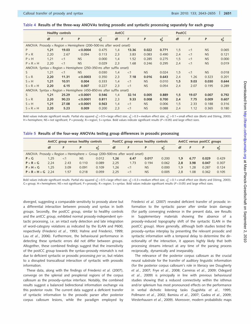
Table 4 Results of the three-way ANOVAs testing prosodic and syntactic processing separately for each group
Healthy controls AntCC PostCC
df F P g2p df F P g2
p df F P g2p
ANOVA: Prosody�Region�Hemisphere (200–500 ms after word onset)
P 1,21 19.03 _0.0004 0.475 1,4 13.36 0.022 0.771 1,5 51 NS 0.065
P�R 2,20 2.67 0.094 0.113 2,3 3.83 0.083 0.490 2,4 51 NS 0.121
P�H 1,21 51 NS 0.000 1,4 1.52 0.285 0.275 1,5 51 NS 0.000
P�R�H 2,20 51 NS 0.029 2,3 1.68 0.246 0.295 2,4 51 NS 0.019
ANOVA: Syntax�Region�Hemisphere (250–350 ms after suffix onset)
S 1,21 51 NS 0.030 1,4 51 NS 0.024 1,5 51 NS 0.018
S�R 2,20 11.31 _0.0003 0.350 2,3 7.18 0.016 0.643 2,4 1.26 0.323 0.201
S�H 1,21 10.51 0.004 0.333 1,4 51 NS 0.010 1,5 9.03 0.030 0.644
S�R�H 2,20 6.15 0.007 0.227 2,3 51 NS 0.054 2,4 2.07 0.195 0.289
ANOVA: Syntax�Region�Hemisphere (450–850 ms after suffix onset)
S 1,21 25.29 _0.0001 0.546 1,4 32.14 0.005 0.889 1,5 19.07 0.007 0.792
S�R 2,20 90.23 _0.0001 0.811 2,3 9.33 0.008 0.700 2,4 7.75 0.009 0.607
S�H 1,21 27.08 _0.0001 0.563 1,4 51 NS 0.006 1,5 2.33 0.188 0.316
S�R�H 2,20 5.23 0.009 0.200 2,3 51 NS 0.088 2,4 1.12 0.365 0.180
Bold values indicate significant results. Partial eta squared �2p40.5 = large effect size; �2
p40.3 = medium effect size; �2p 40.1 = small effect size (Bortz and Doring, 2003).
H = hemisphere; NS = not significant; P = prosody; R = region; S = syntax. Bold values indicate significant results (P50.05) and large effect sizes.
Table 5 Results of the four-way ANOVAs testing group differences in prosodic processing
AntCC group versus healthy controls PostCC group versus healthy controls AntCC versus postCC groups
df F P g2p df F P g2
p df F P g2p
ANOVA: Prosody�Region�Hemisphere� Group (200–500 ms after word onset)
P�G 1,25 51 NS 0.012 1,26 6.47 0.017 0.200 1,9 6.77 0.029 0.429
P�R�G 2,24 2.43 0.110 0.089 2,25 1.73 0.194 0.062 2,8 3.98 0.047 0.307
P�H�G 1,25 3.09 0.091 0.109 1,26 51 NS 0.000 1,9 1.28 0.287 0.124
P�R�H�G 2,24 1.57 0.218 0.059 2,25 51 NS 0.005 2,8 1.08 0.362 0.105
Bold values indicate significant results. Partial eta squared �2p40.5 = large effect size; �2
p40.3 = medium effect size; �2p 40.1 = small effect size (Bortz and Doring, 2003).
G = group; H = hemisphere; NS = not significant; P = prosody; R = region; S = syntax. Bold values indicate significant results (P50.05) and large effect sizes.
Callosal transfer of prosody and syntax Brain 2010: 133; 2643–2655 | 2651
at Max-P
lanck-Gesellschaft on S
eptember 2, 2010
http://brain.oxfordjournals.orgD
ownloaded from
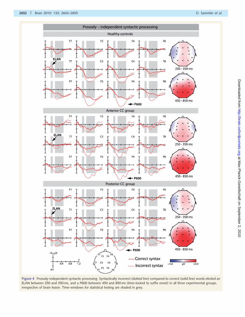
Figure 4 Prosody-independent syntactic processing. Syntactically incorrect (dotted line) compared to correct (solid line) words elicited an
ELAN between 250 and 350 ms, and a P600 between 450 and 850 ms (time-locked to suffix onset) in all three experimental groups,
irrespective of brain lesion. Time-windows for statistical testing are shaded in grey.
2652 | Brain 2010: 133; 2643–2655 D. Sammler et al.
at Max-P
lanck-Gesellschaft on S
eptember 2, 2010
http://brain.oxfordjournals.orgD
ownloaded from
obtained with diffusion tensor imaging consistently located inter-
hemispheric temporal connections, including auditory fibres, to the
(anterior) splenium (Huang et al., 2005; Hofer and Frahm, 2006;
Zarei et al., 2006; Park et al., 2008; Westerhausen et al., 2009),
and other studies showed a degeneration particularly in splenial
and presplenial portions of the corpus callosum as a consequence
of temporal lesions (de Lacoste et al., 1985) or temporal lobe
epilepsy (Weber et al., 2007). Therefore, the (pre)splenium
appears to be the optimal route for the transfer of auditory
linguistic information, particularly if processed in the temporal
lobes like word-category (Friederici et al., 2000, 2003;
Ruschemeyer et al., 2005; Brauer and Friederici, 2007;
Herrmann et al., 2009) and prosodic intonation (Meyer et al.,
2002, 2004; Hesling et al., 2005a,b; Humphries et al., 2005;
Ischebeck et al., 2008).
ConclusionTaken together, the present findings show that the lateralized
prosody and syntax processing streams dynamically communicate
via the posterior corpus callosum connecting auditory areas in the
temporal lobes. Healthy controls and patients with anterior corpus
callosum lesions showed an anterior negativity in response to vio-
lations of syntactically induced prosodic expectations, demonstrat-
ing an intact interaction of syntax and prosody. No such effect
was found in patients with lesions in the posterior third of the
corpus callosum, although they exhibited intact (prosody-
independent) syntactic processing like healthy controls and the
antCC group. In keeping with the dynamic dual pathway model
(Friederici and Alter, 2004) and combined with earlier findings
(Friederici et al., 2007), these data argue for a reciprocal interplay
between the speech processing pathways in the left and right
hemispheres through the posterior corpus callosum, building the
relevant brain basis for the timely coordination and smooth inte-
gration of syntactic and prosodic features during auditory speech
comprehension.
AcknowledgementsThe authors wish to thank Prof. Dr D.Y. von Cramon for kindly
providing the patients, Anne-Kathrin Franz for patient recruitment,
Heike Bothel for help with data acquisition, Dr Beate Sabisch for
parts of the data analysis, Prof. R Rubsamen for kindly providing
the equipment for auditory testing, Dr Alexandra Ludwig for
advice in auditory testing and Kerstin Flake for help with the
figures. We also thank the editor and two anonymous reviewers
for helpful comments.
Supplementary materialSupplementary material is available at Brain online.
ReferencesAnwander A, Tittgemeyer M, von Cramon DY, Friederici AD,
Knosche TR. Connectivity-based parcellation of Broca’s area. Cereb
Cortex 2007; 17: 816–25.
Bamiou DE, Sisodiya S, Musiek FE, Luxon LM. The role of the interhemi-
spheric pathway in hearing. Brain Res Rev 2007; 56: 170–82.Baum SR, Pell MD. The neural bases of prosody: Insights from lesion
studies and neuroimaging. Aphasiology 1999; 8: 581–608.
Blumstein S, Cooper WE. Hemispheric processing of intonation contours.
Cortex 1974; 10: 146–58.
Bortz J, Doring N. Forschungsmethoden und Evaluation: Fur Human-
und Sozialwissenschaftler. Berlin: Springer; 2003. p. 603–18.
Brauer J, Friederici AD. Functional neural networks of semantic and
syntactic processes in the developing brain. J Cogn Neurosci 2007;
19: 1609–23.
Bryan K. Language prosody and the right hemisphere. Aphasiology
1989; 3: 285–99.
Bradvik B, Dravins C, Holtas S, Rosen I, Ryding E, Ingvar DH.
Disturbances of speech prosody following right hemisphere infarcts.
Acta Neurol Scand 1991; 84: 114–26.
Bungert-Kahl P, Biedermann F, Dorrscheidt GJ, von Cramon DY,
Rubsamen R. Psychoacoustic test tools for the detection of deficits
in central auditory processing: Normative data. Z Audiol 2004; 43:
48–71.
Table 6 Mean amplitude and significance of the ELAN and P600 in separate regions of interest
Healthy controls AntCC PostCC
� (mV) t(21) P d � (mV) t(4) P d � (mV) t(5) P d
ELAN: Syntax incorrect versus correct (250–350 ms after suffix onset)
LT �1.10 �5.10 _0.0001 1.537 �1.04 �4.29 0.013 2.716 �0.88 �3.70 0.015 2.135
RT �0.49 �2.24 0.037 0.676 �0.78 �1.50 0.210 0.947 �0.19 �0.75 0.488 0.432
P600: Syntax incorrect versus correct (450–850 ms after suffix onset)
LT �0.74 �3.41 0.003 1.027 +0.36 1.31 0.263 0.827 �0.18 �0.62 0.563 0.357
RT +0.23 0.98 0.338 0.296 +0.55 2.58 0.063 1.631 +0.40 0.88 0.422 0.505
LC +0.61 2.09 0.050 0.630 +1.59 3.34 0.029 2.112 +1.65 2.41 0.061 1.389
RC +1.29 3.59 0.002 1.084 +1.56 2.71 0.054 1.712 +1.99 2.68 0.044 1.547
LP +2.65 9.79 _0.0001 2.953 +3.04 5.08 0.007 3.210 +2.62 3.99 0.011 2.301
RP +2.96 10.40 _0.0001 3.134 +2.98 5.33 0.006 3.371 +3.29 5.54 0.003 3.200
T-tests for paired samples compared event-related potentials evoked by syntactically incorrect and correct words in the respective time windows. Bold values indicatesignificant results (P50.05) and large effect sizes. Cohen’s d40.8 = large effect size; d40.5 = medium effect size; d40.3 = small effect size (Cohen, 1992).
Regions of interest: LT = left fronto-temporal; RT = right fronto-temporal; LC = left fronto-central; RC = right fronto-central; LP = left posterior; RP = right posterior.
Callosal transfer of prosody and syntax Brain 2010: 133; 2643–2655 | 2653
at Max-P
lanck-Gesellschaft on S
eptember 2, 2010
http://brain.oxfordjournals.orgD
ownloaded from
Caplan D, Alpert N, Waters G. Effects of syntactic structure and
propositional number on patterns of regional cerebral blood flow.
J Cogn Neurosci 1998; 10: 541–52.
Carreiras M, Seghier ML, Baquero S, Estevez A, Lozano A, Devlin JT,
et al. An anatomical signature for literacy. Nature 2009; 461: 983–6.Catani M, Jones DK, ffytche DH. Perisylvian language networks of the
human brain. Ann Neurol 2005; 57: 8–16.Cohen J. A power primer. Psychol Bull 1992; 112: 155–9.
Cutler A, Dahan D, van Donselaar W. Prosody in the comprehension of
spoken language: a literature review. Lang Speech 1997; 40 (Pt 2):
141–201.
Dapretto M, Bookheimer SY. Form and content: dissociating
syntax and semantics in sentence comprehension. Neuron 1999; 24:
427–32.
de Lacoste MC, Kirkpatrick JB, Ross ED. Topography of the human
corpus callosum. J Neuropathol Exp Neurol 1985; 44: 578–91.
Delorme A, Makeig S. EEGLAB: An open source toolbox for analysis of
single-trial EEG dynamics including independent component analysis.
J Neurosci Methods 2004; 134: 9–21.
Dougherty RF, Ben-Shachar M, Deutsch GK, Hernandez A, Fox GR,
Wandell BA. Temporal-callosal pathway diffusivity predicts
phonological skills in children. Proc Natl Acad Sci USA 2007; 104:
8556–61.Eckstein K, Friederici AD. Late interaction of syntactic and prosodic
processes in sentence comprehension as revealed by ERPs. Brain Res
Cogn Brain Res 2005; 25: 130–43.
Eckstein K, Friederici AD. It’s early: event-related potential evidence for
initial interaction of syntax and prosody in speech comprehension.
J Cogn Neurosci 2006; 18: 1696–711.Friederici AD. Towards a neural basis of auditory sentence processing.
Trends Cogn Sci 2002; 6: 78–84.Friederici AD. Pathways to language: fibre tracts in the human brain.
Trends Cogn Sci 2009; 13: 175–81.Friederici AD, Alter K. Lateralization of auditory language functions: a
dynamic dual pathway model. Brain Lang 2004; 89: 267–76.
Friederici AD, Kotz SA. The brain basis for syntactic processes: functional
imaging and lesion studies. NeuroImage 2003; 20: S8–17.
Friederici AD, Pfeifer E, Hahne A. Event-related brain potentials
during natural speech processing: effects of semantic, morpho-
logical and syntactic violations. Brain Res Cogn Brain Res 1993; 1:
183–92.Friederici AD, Ruschemeyer SA, Hahne A, Fiebach CJ. The role of left
inferior frontal and superior temporal cortex in sentence comprehen-
sion: localizing syntactic and semantic processes. Cereb Cortex 2003;
13: 170–7.
Friederici AD, von Cramon DY, Kotz SA. Role of the corpus callosum in
speech comprehension: interfacing syntax and prosody. Neuron 2007;
53: 135–45.
Friederici AD, Wang Y, Herrmann CS, Maess B, Oertel U. Localization of
early syntactic processes in frontal and temporal cortical areas: a
magnetoencephalographic study. Hum Brain Mapp 2000; 11: 1–11.
Frye RE, Hasan K, Xue L, Strickland D, Malmberg B, Liederman J, et al.
Splenium microstructure is related to two dimensions of reading skill.
Neuroreport 2008; 19: 1627–31.
Gadea M, Marti-Bonmatı L, Arana E, Espert R, Salvador A, Casanova B.
Corpus callosum function in verbal dichotic listening: inferences from a
longitudinal follow-up of relapsing-remitting multiple sclerosis patients.
Brain Lang 2009; 110: 101–5.Gandour J, Tong Y, Wong D, Talavage T, Dzemidzic M, Xu Y, et al.
Hemispheric roles in the perception of speech prosody. NeuroImage
2004; 23: 344–57.
Gandour J, Wong D, Hsieh L, Weinzapfel B, Lancker DV, Hutchins GD. A
crosslinguistic PET study of tone perception. J Cogn Neurosci 2000;
12: 207–22.Glasser MF, Rilling JK. DTI tractography of the human brain’s language
pathways. Cereb Cortex 2008; 18: 2471–82.Grodzinsky Y, Friederici AD. Neuroimaging of syntax and syntactic
processing. Curr Opin Neurobiol 2006; 16: 240–6.
Grodzinsky Y, Santi A. The battle for Broca’s region. Trends Cogn Sci
2008; 12: 474–80.
Hagoort P. On Broca, brain, and binding: A new framework. Trends
Cogn Sci 2005; 9: 416–23.
Hagoort P, Brown CM, Osterhout L. The neurocognition of syntactic
processing. In: Brown CM, Hagoort P, editors. The neurocognition
of language. New York: Oxford University Press; 1999. p. 273–316.
Hahne A, Friederici AD. Electrophysiological evidence for two steps in
syntactic analysis. Early automatic and late controlled processes.
J Cogn Neurosci 1999; 11: 194–205.
Heilman KM, Bowers D, Speedie L, Coslett HB. Comprehension of
affective and nonaffective prosody. Neurology 1984; 34: 917–21.
Herrmann B, Maess B, Hasting AS, Friederici AD. Localization of the
syntactic mismatch negativity in the temporal cortex: an MEG study.
NeuroImage 2009; 48: 590–600.
Hesling I, Clement S, Bordessoules M, Allard M. Cerebral mechanisms of
prosodic integration: evidence from connected speech. NeuroImage
2005a; 24: 937–47.Hesling I, Dilharreguy B, Clement S, Bordessoules M, Allard M. Cerebral
mechanisms of prosodic sensory integration using low-frequency
bands of connected speech. Hum Brain Mapp 2005b; 26: 157–69.
Hickok G, Poeppel D. The cortical organization of speech processing. Nat
Rev Neurosci 2007; 8: 393–402.
Hofer S, Frahm J. Topography of the human corpus callosum revisited–
comprehensive fibre tractography using diffusion tensor magnetic
resonance imaging. NeuroImage 2006; 32: 989–94.
Huang H, Zhang J, Jiang H, Wakana S, Poetscher L, Miller MI, et al. DTI
tractography based parcellation of white matter: application to the
mid-sagittal morphology of corpus callosum. NeuroImage 2005; 26:
195–205.
Humphries C, Love T, Swinney D, Hickok G. Response of anterior tem-
poral cortex to syntactic and prosodic manipulations during sentence
processing. Hum Brain Mapp 2005; 26: 128–38.Inkelas S, Zec D. Syntax-phonology interface. In: Goldsmith JA, editor.
The handbook of phonological theory, Blackwell Handbooks
in Linguistics. Vol. 1. Oxford: Blackwell Publishers Ltd; 1996.
p. 535–49.
Ischebeck AK, Friederici AD, Alter K. Processing prosodic boundaries in
natural and hummed speech: an FMRI study. Cereb Cortex 2008; 18:
541–52.Jung-Beeman M. Bilateral brain processes for comprehending natural
language. Trends Cogn Sci 2005; 9: 512–8.Just MA, Carpenter PA, Keller TA, Eddy WF, Thulborn KR. Brain activa-
tion modulated by sentence comprehension. Science 1996; 274:
114–16.
Kaan E, Harris A, Gibson E, Holcomb PJ. The P600 as an index of
syntactic integration difficulty. Lang Cognitive Proc 2000; 15:
159–201.
Kotz SA, Meyer M, Paulmann S. Lateralization of emotional prosody in
the brain: an overview and synopsis on the impact of study design.
Prog Brain Res 2006; 156: 285–94.
Lau E, Stroud C, Plesch S, Phillips C. The role of structural prediction in
rapid syntactic analysis. Brain Lang 2006; 98: 74–88.
Meyer M, Alter K, Friederici AD, Lohmann G, von Cramon DY. FMRI
reveals brain regions mediating slow prosodic modulations in spoken
sentences. Hum Brain Mapp 2002; 17: 73–88.
Meyer M, Steinhauer K, Alter K, Friederici AD, von Cramon DY. Brain
activity varies with modulation of dynamic pitch variance in sentence
melody. Brain Lang 2004; 89: 277–89.Ni W, Constable RT, Mencl WE, Pugh KR, Fulbright RK, Shaywitz SE,
et al. An event-related neuroimaging study distinguishing form
and content in sentence processing. J Cogn Neurosci 2000; 12:
120–33.
Odegard TN, Farris EA, Ring J, McColl R, Black J. Brain connectivity in
non-reading impaired children and children diagnosed with develop-
mental dyslexia. Neuropsychologia 2009; 47: 1972–7.Oldfield RC. The assessment and analysis of handedness: the Edinburgh
inventory. Neuropsychologia 1971; 9: 97–113.
2654 | Brain 2010: 133; 2643–2655 D. Sammler et al.
at Max-P
lanck-Gesellschaft on S
eptember 2, 2010
http://brain.oxfordjournals.orgD
ownloaded from
Pannekamp A, Toepel U, Alter K, Hahne A, Friederici AD. Prosody-drivensentence processing: an event-related brain potential study. J Cogn
Neurosci 2005; 17: 407–21.
Park HJ, Kim JJ, Lee SK, Seok JH, Chun J, Kim DI, et al. Corpus callosal
connection mapping using cortical gray matter parcellation andDT-MRI. Hum Brain Mapp 2008; 29: 503–16.
Parker GJM, Luzzi S, Alexander DC, Wheeler-Kingshott CAM,
Ciccarelli O, Ralph MAL. Lateralization of ventral and dorsal
auditory-language pathways in the human brain. NeuroImage 2005;24: 656–66.
Perkins JM, Baran JA, Gandour J. Hemispheric specialization in processing
intonation contours. Aphasiology 1996; 10: 343–62.Plante E, Creusere M, Sabin C. Dissociating sentential prosody from
sentence processing: activation interacts with task demands.
NeuroImage 2002; 17: 401–10.
Poeppel D. The analysis of speech in different temporal integrationwindows: Cerebral lateralization as’asymmetric sampling in time’.
Speech Communication 2003; 41: 245–55.
Pollmann S, Maertens M, von Cramon DY, Lepsien J, Hugdahl K.
Dichotic listening in patients with splenial and nonsplenial callosallesions. Neuropsychology 2002; 16: 56–64.
Powell HWR, Parker GJM, Alexander DC, Symms MR, Boulby PA,
Wheeler-Kingshott CAM, et al. Hemispheric asymmetries in
language-related pathways: a combined functional MRI and tractogra-phy study. NeuroImage 2006; 32: 388–99.
Price CJ, Thierry G, Griffiths T. Speech-specific auditory processing:
Where is it? Trends Cogn Sci 2005; 9: 271–6.Ruschemeyer S, Fiebach CJ, Kempe V, Friederici AD. Processing lexical
semantic and syntactic information in first and second language: fMRI
evidence from German and Russian. Hum Brain Mapp 2005; 25:
266–86.Saur D, Kreher BW, Schnell S, Kummerer D, Kellmeyer P, Vry MS, et al.
Ventral and dorsal pathways for language. Proc Natl Acad Sci USA
2008; 105: 18035–40.
Scott SK, Johnsrude IS. The neuroanatomical and functional organizationof speech perception. Trends Neurosci 2003; 26: 100–7.
Selkirk E. On derived domains in sentence phonology. Phonology 1986;
3: 371–405.
Shalom DB, Poeppel D. Functional anatomic models of language:Assembling the pieces. Neuroscientist 2008; 14: 119–27.
Sharbrough F, Chatrian G, Lesser R, Luders H, Nuwer M, Picton T.
American Electroencephalographic Society guidelines for standard
electrode position nomenclature. J Clin Neurophysiol 1991; 8: 200–2.Shipley-Brown F, Dingwall WO, Berlin CI, Yeni-Komshian G,
Gordon-Salant S. Hemispheric processing of affective and linguistic
intonation contours in normal subjects. Brain Lang 1988; 33: 16–26.
Sugishita M, Otomo K, Yamazaki K, Shimizu H, Yoshioka M,Shinohara A. Dichotic listening in patients with partial section of the
corpus callosum. Brain 1995; 118: 417–27.
Talairach J, Tournoux P. Co-planar Stereotaxic Atlas of The Human Brain.3-Dimensional proportional system: an approach to cerebral imaging.
Stuttgart: Thieme; 1988.
Truckenbrodt H. On the relation between syntactic phrases and phono-
logical phrases. Linguist Inq 1999; 30: 219–55.Van Lancker D, Sidtis JJ. The identification of affective-prosodic stimuli
by left- and right- hemisphere-damaged subjects: All errors are not
created equal. J Speech Hear Res 1992; 35: 963–70.
Vigneau M, Beaucousin V, Herve PY, Duffau H, Crivello F, Houde O,et al. Meta-analyzing left hemisphere language areas: Phonology,
semantics, and sentence processing. NeuroImage 2006; 30: 1414–32.
Weber B, Luders E, Faber J, Richter S, Quesada CM, Urbach H, et al.
Distinct regional atrophy in the corpus callosum of patients withtemporal lobe epilepsy. Brain 2007; 130: 3149–54.
Weintraub S, Mesulam MM, Kramer L. Disturbances in prosody. A right-
hemisphere contribution to language. Arch Neurol 1981; 38: 742–744.Westerhausen R, Gruner R, Specht K, Hugdahl K. Functional relevance of
interindividual differences in temporal lobe callosal pathways: a DTI
tractography study. Cereb Cortex 2009; 19: 1322–9.
Xiang HD, Fonteijn HM, Norris DG, Hagoort P. Topographical functionalconnectivity pattern in the perisylvian language networks. Cereb
Cortex 2010; 20: 549–60.
Zarei M, Johansen-Berg H, Smith S, Ciccarelli O, Thompson AJ,
Matthews PM. Functional anatomy of interhemispheric corticalconnections in the human brain. J Anat 2006; 209: 311–20.
Zatorre RJ, Belin P, Penhune V. Structure and function of auditory
cortex: music and speech. Trends Cogn Sci 2002; 6: 37–46.
Callosal transfer of prosody and syntax Brain 2010: 133; 2643–2655 | 2655
at Max-P
lanck-Gesellschaft on S
eptember 2, 2010
http://brain.oxfordjournals.orgD
ownloaded from


