Embed Size (px)
Citation preview

Protein chaperones Q8ZP25_SALTY from Salmonellatyphimurium and HYAE_ECOLI from Escherichia coli exhibitthioredoxin-like structures despite lack of canonical thioredoxinactive site sequence motive
David Parish, Jordi Benach, Goahua Liu, Kiran Kumar Singarapu, Rong Xiao, ThomasActon, Min Su, Sonal Bansal, James H. Prestegard, John Hunt, Gaetano T. Montelione, andThomas SzyperskiDavid Parish · Gaohua Liu · Kiran Kumar Singarapu · Thomas Szyperski, Department of Chemistry,Northeast Structural Genomics Consortium, The State University of New York at Buffalo, Buffalo,NY 14260, [email protected]
Jordi Benach · Min Su · John F. Hunt, Department of Biological Sciences, Northeast StructuralGenomics Consortium, Columbia University, New York, NY 10027
Rong Xiao · Thomas Acton · Gaetano T. Montelione, The Center for Advanced Biotechnology andMedicine, Department of Molecular Biology and Biochemistry, Northeast Structural GenomicsConsortium, Rutgers University and Robert Wood Johnson Medical School, Piscataway, NJ 08854
Sonal Bansal · James H. Prestegard, Complex Carbohydrate Research Center and Department ofChemistry, University of Georgia, Athens, Georgia, 30602-4712
AbstractThe structure of the 142-residue protein Q8ZP25_SALTY encoded in the genome of Salmonellatyphimurium LT2 was determined independently by NMR and X-ray crystallography, and thestructure of the 140-residue protein HYAE_ECOLI encoded in the genome of Eschericia coli wasdetermined by NMR. The two proteins belong to Pfam [1] PF07449, which currently comprises 50members, and belongs itself to the ‘thioredoxin-like clan’. However, protein HYAE_ECOLI and theother proteins of Pfam PF07449 do not contain the canonical Cys-X-X-Cys active site sequence motifof thioredoxin. Protein HYAE_ECOLI was previously classified as a [NiFe] hydrogenase-1 specificchaperone interacting with the twin-arginine translocation (Tat) signal peptide. The structurespresented here exhibit the expected thioredoxin-like fold and support the view that members of Pfamfamily PF07449 specifically interact with Tat signal peptides.
KeywordsChaperones; GFT NMR; HYAE_ECOLI; Q8ZP25_SALTY; Structural genomics; Thioredoxin
IntroductionPfam[1] family HyaE, which was named after its functionally characterized memberHYAE_ECOLI [2] (accession number PF07449) and currently contains 50 members, wasselected by the Protein Structure Initiative-2 (PSI-2) of the United States National Institutes of
Correspondence to: Thomas Szyperski.
NIH Public AccessAuthor ManuscriptJ Struct Funct Genomics. Author manuscript; available in PMC 2010 April 7.
Published in final edited form as:J Struct Funct Genomics. 2008 December ; 9(1-4): 41–49. doi:10.1007/s10969-008-9050-y.
NIH
-PA Author Manuscript
NIH
-PA Author Manuscript
NIH
-PA Author Manuscript

Health and assigned to the Northeast Structural Genomics consortium (NESG;http://www.nesg.org). Here we describe the structures of two proteins belonging to PF07449,that is, protein Q8ZP25_SALTY encoded by gene STM1790 (UniProtKB/TrEMBL entryQ8ZP25; NESG target ID StR70) of Salmonella typhimurium LT2 [3] and the homologousprotein HYAE_ECOLI (73% identity) encoded in gene hyaE (UniProtKB/Swiss-Prot entryP19931; NESG target ID ER415) of Escherichia coli. The NMR structures were determinedusing the previously described rapid NMR data collection and analysis protocol for high-throughput structure determination [4]. The structure of Q8ZP25_SALTY was also determinedindependently by X-ray crystallography. The three-dimensional structures ofQ8ZP25_SALTY (17 kDa) and HYAE_ECOLI (16.7 kDa) are the first for Pfam familyPF07449.
Materials and methodsProtein sample preparation for NMR
Proteins Q8ZP25_SALTY and HYAE_ECOLI were cloned, expressed and purified followingstandard protocols to produce uniformly U-13C,15N-labeled protein samples.[5] Briefly, thefull length STM1790 gene from Salmonella typhimurium LT2 was cloned into a pET21(Novagen) derivative, yielding the plasmid StR70-21.4. The full length hyaE gene fromEscherichia coli was also cloned into a pET21 derivative, yielding the plasmid ER415-21.1.The resulting sequence verified constructs contain the complete native protein coding sequenceplus eight nonnative residues at the C-terminus (LEHHHHHH) that facilitate proteinpurification. Escherichia coli BL21 (DE3) cells containing the pMGK plasmid, whichexpresses several tRNAs to enhance translation of rare codons, were transformed withStR70-21.4 and separately with ER415-21.1, and cultured in MJ9 minimal medium containing(15NH4)2SO4 and U-13C-glucose as sole nitrogen and carbon sources.[6] U-13C,15NQ8ZP25_SALTY and U-13C,15N HYAE_ECOLI were purified using an AKTAxpress (GEHealthcare) based two step protocol consisting of IMAC (HisTrap HP) and gel filtration(HiLoad 26/60 Superdex 75) chromatography. The final yield of purified U-13C,15NQ8ZP25_SALTY (> 98% homogenous by SDS-PAGE; 17.05 kDa by MALDI-TOF massspectrometry) was about 26 mg/L. The final sample of U-13C, 15N labeled Q8ZP25_SALTYwas prepared at a concentration of ~1.2 mM in 95% H2O/5% D2O solution containing 50 mMMES, 10 mM DTT, and 50 mM arginine, at pH 6.0. The final yield of purified U-13C,15NHYAE_ECOLI (> 98% homogenous by SDS-PAGE; 16.72 kDa by MALDI-TOF massspectrometry) was about 42 mg/L. The final sample of U-13C, 15N labeled HYAE_ECOLI wasprepared at a concentration of ~0.6 mM in 95% H2O/5% D2O solution containing 20 mM MES,10 mM DTT, 100 mM NaCl, 5mM CaCl2 and 0.02% NaN3 at pH 6.5.
Isotropic overall rotational correlation times of ~8 ns were inferred from 15N spin relaxationtimes for both proteins, indicating that these proteins are monomeric in solution. Thisconclusion was further confirmed by gel-filtration and dynamic light scattering (data not shownbut available online at www.nesg.org). In order to align protein Q8ZP25_SALTY formeasurement of backbone 15N-1H residual dipolar couplings, Pf1 phages [7] were added tothe protein solution, yielding a 0.25 mM Q8ZP25_SALTY solution in the presence of 15.5mg/mL Pf1 phage.
NMR structure determinationAll NMR experiments for resonance assignment, identification of slowly exchanging amideprotons and measurement of residual dipolar couplings (RDCs) were performed on a VarianINOVA 600 spectrometer equipped with a cryogenic 1H{13C,15N} probe, while NOESY datato derive 1H-1H distance constraints were recorded on a 750 MHz spectrometer equipped witha conventional 1H{13C,15N} probe. For Q8ZP25_SALTY, a standard set of five through-bond
Parish et al. Page 2
J Struct Funct Genomics. Author manuscript; available in PMC 2010 April 7.
NIH
-PA Author Manuscript
NIH
-PA Author Manuscript
NIH
-PA Author Manuscript

G-matrix Fourier transform (GFT) NMR experiments [6;8;9] was acquired for resonanceassignment (total measurement time: 110 hours), and a simultaneous3D 15N/13Caliphatic/13Caromatic-resolved NOESY spectrum [9] (measurement time: 48 hours)was acquired to derive distance constraints. For HYAE_ECOLI, the comparably low proteinconcentration made conventional 3D HNNCACB, CBCA(CO)NHN, and HBHA(CO)NHN[10] recorded in conjunction with (4,3)D HCCH [4] the preferred choice (total measurementtime: 216 hours). These spectra were complemented with 3D 15N-resolved (measurement time:24 hours) and 13C-resolved NOESY spectra (measurement time: 48 hours) to derive distanceconstraints. All other spectra were processed with the program nmrPipe [11] and analyzedusing the program XEASY [12]. Polypeptide backbone (1HN, 15N, 13Cα) and 13Cβ resonanceassignments were obtained by using the program AutoAssign [13].
For Q8ZP25_SALTY, polypeptide backbone 15N-1H RDCs were extracted from pairs of 2D[15N,1H]-TROSY and 2D [15N,1H]-HSQC spectra [10] (measurement time 10 hours) acquiredfor both unaligned and partially aligned protein. A virtually complete set of RDCs wasobtained. The spectra were processed using the program NMRPipe [11] and automatically peakpicked using the program NMRDraw [11]. To identify slowly exchanging backbone amideprotons a 2D [15N,1H]-HSQC spectrum was recorded (measurement time 10 minutes) starting5 minutes after lyophilizing and re-suspending protein Q8ZP25_SALTY in 99.8% D2O.
The programs CYANA [14;15] and AUTOSTRUCTURE [16] were used in parallel toautomatically assign long-range NOEs. Identical assignments from both programs (“consensusassignments”) were used as the starting point for manual completion of iterative NOEassignment, peak picking and structure calculation [4]. For protein Q8ZP25_SALTY,hydrogen bond constraints were derived for slowly exchanging amide protons whenever NOEssupported hydrogen bond formation [10]. The final structure calculations were performed usingversion 2.1 of CYANA [15]. For Q8ZP25_SALTY, the alignment tensor was fittedto 15N-1H RDC values using the program REDCAT [17]. Subsequently, orientationalconstraints derived from RDCs measured for residues in rigid regular secondary structureelements were implemented in the program XPLOR [18;19] for structure refinement. Finally,additional refinement for the structures of proteins Q8ZP25_SALTY and HYAE_ECOLI wasperformed in an explicit ‘water bath’ [20] by using the program CNS [21]. Prior to submissionto the protein data bank [22], structures were validated using the PSVS server [23].
Protein sample preparation for X-ray crystallographySelenomethionine labeled Q8ZP25_SALTY was produced as described above for the NMRsample, except that an MJ9 minimal medium containing selenomethionine was used. The yieldof purified Q8ZP25_SALTY (> 98% homogenous by SDS-PAGE; 16.19 kDa by MALDI-TOF mass spectrometry) was about 57 mg/L. The final sample of selenomethionine labeledQ8ZP25_SALTY was prepared at a concentration of 10 mg/mL in a solution containing 10mM Tris, 5 mM DTT and 100 mM NaCl.
Crystallization, X-ray diffraction data collection, and processingMonoclinic-shaped crystals of Q8ZP25_SALTY grew at 293 K in 1 + 1 μl hanging-drop vapor-diffusion reactions over a reservoir containing 18% PEG 1000, 15 mM NaBr and 100 mMNaOAc at pH 5.0. Crystals were frozen in liquid propane using paratone-N as cryoprotectant.Multi-wavelength anomalous diffraction data were collected from a single crystal at theselenium edge on a Quantum-4 CCD detector (ADSC, San Diego, CA) on beamline X4A atthe National Synchrotron Light Source in consecutive 400° sweeps by the inverse beam methodat 0.97925 Å (peak), 0.97955 Å (edge) and 0.96782 Å (remote) using 1°, 8 s oscillations. Datawere processed using the programs DENZO and SCALEPACK [24]. For each data set, the
Parish et al. Page 3
J Struct Funct Genomics. Author manuscript; available in PMC 2010 April 7.
NIH
-PA Author Manuscript
NIH
-PA Author Manuscript
NIH
-PA Author Manuscript

scaling B factors of the final frames were within 3 Å2 of the initial frames, indicating that therewas minimal decay.
Results and DiscussionNMR solution structures of proteins Q8ZP25_SALTY and HYAE_ECOLI
Resonance assignments were obtained for 78% (Q8ZP25_SALTY, which comprises 134residues, excluding affinity purification tag) and 89% (HYAE_ECOLI, which comprises 132residues, excluding affinity purification tag) of the assignable backbone (excluding the C-terminal NH3
+, the Pro 15N and the 13C′ shifts) and 13Cβ shifts, and for 74% (Q8ZP25_SALTY)and 88% (HYAE_ECOLI) of the side chain shifts (excluding Lys NH3
+, Arg NH2, OH, sidechain 13C′ and aromatic quaternary 13C shifts; Table I). Stereo-specific assignments wereobtained for 28% (Q8ZP25_SALTY) and 25% (HYAE_ECOLI) of the β-methylene groupsexhibiting non-degenerate proton chemical shifts using GLOMSA [25]. GLOMSA was alsoused to stereo-specifically assign 20% of the HYAE_ECOLI Val and Leu isopropyl moieties(Table I). For Q8ZP25_SALTY, a constant-time 2D 13C HSQC was preformed on a 5%fractionally 13C-labeled sample and used to assign 67% of the Val and Leu isopropyl moieties[26]. Upper distance limit constraints for structure calculations were obtained from NOESY,and backbone dihedral angle constraints for residues located within well defined secondarystructure were derived from chemical shifts as described [27]. For Q8ZP25_SALTY, 10hydrogen bond constraints and 69 RDC constraints[28] were used in addition for structurerefinement. The RDC “quality factor” Q (i.e., the r.m.s.d. calculated between measured RDCvalues and RDC values predicted based on the structure) improved from 0.52 to 0.42 as a resultof the RDC-based refinement, which also reduced the r.m.s.d. value calculated for heavy atomsN, Cα, and C′ between the NMR structure and the x-ray structure from 1.60 to 1.38 Ǻ(residues11–25, 34–42, 55–90, 94–101, 106, 107, 113–122). The statistics for the structuredeterminations of Q8ZP25_SALTY and HYAE_ECOLI (Table I) show that high-quality NMRstructures were obtained. Chemical shifts, distance constraints and RDCs were deposited inthe BioMagResBank [29] (BMRB accession number 7178 for Q8ZP25_SALTY and 7256 forHYAE_ECOLI) and coordinates were deposited in the Protein Data Bank [22] (PDB 2JZT forQ8ZP25_SALTY and 2HFD for HYAE_ECOLI).
X-ray crystal structure of protein Q8ZP25_SALTYGiven four molecules of StR70 per asymmetric unit in the P21 lattice, the packing density inthe crystal is 2.1 Å3/Da, in the most probable range for proteins [30]. SOLVE [31] identified7 of the 12 Se atoms in the asymmetric unit, yielding a map that was used for non-crystallographic symmetry (NCS) averaging, density modification (with 41% solvent content)and automated iterative model building in AUTO_RESOLVE [31]. This program identified50% of the backbone and 35% of the side chains in the final model and produced a map thatenabled the structure to be built by hand using O [32]. Completion of the structure requirediterative cycles of refinement in CNS [33] and manual rebuilding using standard geometricand van der Waals parameters [34]. The refinement was monitored by a randomly selectedRfree set containing 10% of the reflections. B-factors were refined using standard vicinalrestraints (1.5–2.0 Å2 for main-chain atoms and 2.0–2.5 Å2 for side-chain atoms). Strong NCSrestraints (1050 kJÅ−2, σB = 1.5) were maintained throughout the refinement. Water-moleculesites were selected automatically using CNS and checked for consistency with 2Fo − Fcelectron-density and hydrogen-bonding criteria. The statistics of the 2.8 Å X-ray structuredetermination are shown in Table II, and the coordinates were deposited in the PDB (id: 2ES7).
Structure description and comparisonsThe structures of proteins Q8ZP25_SALTY (NMR and X-ray) and HYAE_ECOLI (NMR) areshown in Figure 1. They exhibit the expected canonical “thioredoxin-like” fold characterized
Parish et al. Page 4
J Struct Funct Genomics. Author manuscript; available in PMC 2010 April 7.
NIH
-PA Author Manuscript
NIH
-PA Author Manuscript
NIH
-PA Author Manuscript

by an internal twisted 5-stranded β-sheet surrounded by 5 α-helices. Starting at the N-terminus,the topology of the regular secondary structure elements can be summarized as α1-βA(↑)-α2-βB(↑)-α3-βC(↑)-α4-βD(↓)-βE(↑)-α5. The close similarity of the structures is evidenced byrather small r.m.s.d. values (Table III) calculated for the backbone heavy atoms N, Cα and C′of residues for which resonance assignments were obtained for the NMR structures and electrondensity was observed in the X-ray structure.
Complementary information is obtained from the NMR and X-ray structures. The polypeptidesegments comprising residues 1–10, 43–54, 91–93, 108–112 and 128–142 (in HYAE_ECOLI:1–2, 43–55, 91–92, and 108–111) are locally not well defined and appear flexibly disorderedin the NMR structures, whereas no electron density is observed for residues 1–6, 26–33, 48–51, 102–105, 126–142 in the X-ray structure. Furthermore, α-helix 2 comprising residues 26–35 in the NMR structure (in HYAE_ECOLI: residues 23–34), was not observed in the X-raystructure, suggesting that the structure of this segment is stable in solution but that it may haveassumed a range of conformations in the crystal. Conversely, a defined conformation isobserved for loop residues 91–92 and 108–111 in the X-ray structure but remained poorlydefined in both NMR structures. This suggests that these segments are flexibly disordered insolution but became trapped in a fixed conformation during crystallization.
Given the fact that the level of sequence identity between some members of Pfam familyPF07449 is very low (as low as 13%), it is expected that the structures of both proteinsQ8ZP25_SALTY and HYAE_ECOLI will be useful as templates in deriving high-qualityhomology models for most members of Pfam PF07449. In fact, conservation of residuesparticipating in the molecular core of proteins Q8ZP25_SALTY and HYAE_ECOLI withinPF07449 is high (Figure 2).
A search of structurally similar proteins domains using the DALI server [35] identifies 30structurally similar proteins with Z-scores > 4.0, many of which are thioredoxins containingthe catalytic sequence motif Cys-X-X-Cys. The best scoring proteins are (i) thioredoxin fromAnabaena sp. Strain PCC1720 (PDB ID 1THX; Z-score 8.8, 102 aligned residues with rmsd.3.3 Å), (ii) a C73S mutant human thioredoxin (1ERV; Z-score 7.9, 99 aligned residues; 3.2Å), (iii) an unannotated protein from Haemophilus ducreyi (2DO8; Z-score 7.7, 108 alignedresidues; 4.6 Å), and (iv) a theoretical model of a disulfide isomerase from Plasmodiumchabuadi (1Y9N; Z-score 7.5, 96 aligned residues; 3.7 Å).
Functional implications of of Q8ZP25_SALTY and HYAE_ECOLI structuresIn contrast to the structurally similar thioredoxins identified by the DALI server, proteinsQ8ZP25_SALTY and HYAE_ECOLI do not possess the canonical thioredoxin catalytic motifCys-X-X-Cys and thus cannot participate in the biochemical reactions characteristic forthioredoxins. In contrast, protein HYAE_ECOLI has been shown to interact with the Tat signalpeptide-bearing subunit of hydrogenase-1 HyaA, and was thus classified as a hydrogenase-1β-subunit-specific chaperone [2]. Consistently, the deletion of the gene encoding protein HoxOin Ralstonia eutropha, another member of PF07449, leads to complete loss of the uptake [NiFe]hydrogenase activity, suggesting that hoxO has a critical role in the assembly of thehydrogenase [36].
The multiple sequence alignment for proteins of Pfam PF07449 (Figure 2) reveals conservationof surface exposed residues which have no obvious structural role in the molecular core. Theprogram ConSurf [37] was used to map those residues from onto the surface ofQ8ZP25_SALTY (Figure 3a). Intriguingly, three of the most highly conserved residues (shownin red) are the negatively charged residues Asp 44, Asp 53 and Glu 50 located in the flexiblydisordered loop comprising residues 43–55, which align structurally with the catalytic motifof thioredoxin (Figure 2). Calculation of the electrostatic surface using the program GRASP
Parish et al. Page 5
J Struct Funct Genomics. Author manuscript; available in PMC 2010 April 7.
NIH
-PA Author Manuscript
NIH
-PA Author Manuscript
NIH
-PA Author Manuscript

[38] shows the resulting negatively charged patch on the protein surface (Figure 3b). Thisstructural bioinformatics analysis suggests that the highly conserved, negatively chargedsurface residues interact with the arginine rich, positively charged, Tat signal peptide.
ConclusionsThe NMR solution and X-ray structures of the protein Q8ZP25_SALTY and the NMR solutionstructure of homologous protein HYAE_ECOLI were determined and shown by their structurestatistics to be of high quality. Both proteins exhibit a “thioredoxin-like” fold likelyrepresentative of all members of protein family PF07449 which currently contains 50 proteins.However, none of the members of PF07449 bear the Cys-X-X-Cys active site motifcharacteristic of thioredoxins. Conserved, negatively charged surface patches onHYAE_ECOLI and Q8ZP25_SALTY could potentially be involved in binding with thearginine rich Tat signal peptide that regulates hydrogenase complex assembly and export.
AcknowledgmentsThis work was supported by the National Institutes of Health (U54 GM074958-01) and the National ScienceFoundation (MCB 0416899 to T.S.). We thank Kellie Cunningham, Chi Kent Ho, Haleema Janjua, Li-Chung Ma, andLi Zhao (Rutgers University) for help preparing the protein samples.
Abbreviations
NESG Northeast Structural Genomics Consortium
NMR nuclear magnetic resonance
NSLF national synchrotron light source
PDB protein data bank
SAD single-wavelength anomalous diffraction
RDC residual dipolar coupling
PSVS protein structure validation suite
References1. Finn RD, Mistry J, Shuster-Bockler B, Griffiths-Jones S, Hollich V, Lassmann T, Moxon S, Marshall
M, Khanna A, Durbin R, Eddy SR, Sonnhammer EL, Bateman A. Nucl Acids Res 2006;34:D247–D251. [PubMed: 16381856]
2. Dubini A, Sargent F. FEBS Lett 2003;549:141–146. [PubMed: 12914940]3. McClelland M, Sanderson KE, Spieth J, Clifton SW, Latreille P, Courtney L, Porwollik S, Ali J, Dante
M, Du F, Hou S, Layman D, Leonard S, Nguyen C, Scott K, Holmes A, Grewal N, Mulvaney E, RyanE, Sun H, Florea L, Miller W, Stoneking T, Nhan M, Waterson R, Wilson RK. Nature 2001;413:852–856. [PubMed: 11677609]
4. Liu GH, Shen Y, Atreya HS, Parish D, Shao Y, Sukumaran DK, Xiao R, Yee A, Lemak A, BhattacharyaA, Acton T, Arrowsmith C, Montelione G, Szyperski T. Proc Natl Acad Sci USA 2005;102:10487–10492. [PubMed: 16027363]
5. Acton TB, SGK, Xiao r, Ma LC, Aramini JM, Baran MC, Chiang YW, Climent T, Cooper B, DenissovaN, Douglas SM, Everett JK, Ho CK, Macapagal D, Paranji RK, Shastry R, Shih LJ, Swapna GVT,Wilson M, Wu MJ, Gerstein M, Inouye M, Hunt JF, Montelione GT. Methods Enzymol 2005;394:210–243. [PubMed: 15808222]
6. Atreya HS, Szyperski T. Proc Natl Acad Sci USA 2004;101:9642–9647. [PubMed: 15210958]
Parish et al. Page 6
J Struct Funct Genomics. Author manuscript; available in PMC 2010 April 7.
NIH
-PA Author Manuscript
NIH
-PA Author Manuscript
NIH
-PA Author Manuscript

7. Hansen, MR.; Hanson, P.; Pardi, A. Rna-Ligand Interactions Pt A. ACADEMIC PRESS INC; SanDiego: 2000. Filamentous bacteriophage for aligning RNA, DNA, and proteins for measurement ofnuclear magnetic resonance dipolar coupling interactions; p. 220-240.
8. Kim S, Szyperski T. J Am Chem Soc 2003;125:1385–1393. [PubMed: 12553842]9. Shen Y, Atreya HS, Liu GH, Szyperski T. J Am Chem Soc 2005;127:9085–9099. [PubMed: 15969587]10. Cavanagh, J.; Fairbrother, WJ.; Palmer, AG.; Rance, M.; Skelton, NJ. Protein NMR Spectroscopy.
Academic Press; San Diego: 2007.11. Delaglio F, Grzesiek S, Vuister GW, Zhu G, Pfeifer J, Bax A. J Biomol NMR 1995;6:277–293.
[PubMed: 8520220]12. Bartels C, Xia T, Billeter M, Guntert P, Wuthrich K. J Biomol NMR 1995;6:1–10.13. Zimmerman DE, Kulikowski CA, Huang YP, Feng WQ, Tashiro M, Shimotakahara S, Chien CY,
Powers R, Montelione GT. J Mol Biol 1997;269:592–610. [PubMed: 9217263]14. Herrmann T, Güntert P, Wüthrich K. J Mol Biol 2002;319:209–227. [PubMed: 12051947]15. Güntert P, Mumenthaler C, Wüthrich K. J Mol Biol 1997;273:283–298. [PubMed: 9367762]16. Huang YJ, Moseley H, Baran MC, Arrowsmith C, Powers R, Tejero R, Szyperski T, Montelione G.
Methods Enzymol 2005;394:111–141. [PubMed: 15808219]17. Valafar H, Prestegard JH. J Magn Reson 2004;167:228–241. [PubMed: 15040978]18. Schwieters CD, Kuszewski JJ, Tjandra N, Clore GM. J Magn Reson 2003;160:65–73. [PubMed:
12565051]19. Schwieters CD, Kuszewski J, Clore GM. Prog Nucl Magn Reson Spectrosc 2006;48:47–62.20. Linge JP, Williams MA, Spronk CA, Bonvin AM, Nilges M. Proteins 2003;50:496–506. [PubMed:
12557191]21. Brunger AT, Adams PD, Clore GM, DeLano WL, Gros P, Grosse-Kunstleve RW, Jiang JS, Kuszewski
J, Nilges M, Pannu NS, Read RJ, Rice LM, Simonson T, Warren GL. Acta Cryst D 1998;54:905–921. [PubMed: 9757107]
22. Berman HM, Westbrook J, Feng Z, Gilliland G, Bhat TN, Weissig H, Shindyalov IN, Bourne PE.Nucl Acids Res 2000;28:235–242. [PubMed: 10592235]
23. Bhattacharya A, Tejero R, Montelione G. Proteins 2007;66:778–795. [PubMed: 17186527]24. Otwinowski Z, Minor W. Meth Enzymol 1997;276:307–326.25. Guntert P, Braun W, Wuthrich K. J Mol Biol 1991;217:517–530. [PubMed: 1847217]26. Neri D, Szyperski T, Otting G, Senn H, Wüthrich K. Biochemistry 1989;28:7510–6. [PubMed:
2692701]27. Cornilescu G, Delaglio F, Bax A. J Biomol NMR 1999;13:289–302. [PubMed: 10212987]28. Prestegard JH, Bougault CM, Kishore AI. Chem Rev 2004;104:3519–3540. [PubMed: 15303825]29. Ulrich EL, Akutsu H, Doreleijers JF, Harano Y, Loannidis YE, Lin J, Livny M, Mading S, Maziuk
D, Miller Z, Nakatani E, Schulte CF, Tolmie DE, Wenger KR, Yao H, Markley JL. Nucl Acids Res.200710.1093/nar/gkm957
30. Matthews BW. J Mol Biol 1968;33:491–497. [PubMed: 5700707]31. Terwilliger TC, Berendzen J. Acta Cryst Sect D 1999;55:849–861. [PubMed: 10089316]32. Jones TA, Zou JY, Cowan SW, Kjeldgaard M. Acta Cryst Sect A 1991;47:110–119. [PubMed:
2025413]33. Brünger AT, Adams PD, Clore GM, DeLano WL, Gros P, Grosse-Kunstleve RW, Jiang JS, Kuszewski
J, MN, Pannu NS, Read RJ, Rice LM, Simonson T, Warren GL. Acta Cryst Sect D 1998;54:905–921. [PubMed: 9757107]
34. Engh R, Huber R. Acta Cryst Sect A 1991;47:392–400.35. Holm L, Sander CR. Trends Biochem Sci 1995;20:478–480. [PubMed: 8578593]36. Bernhard M, Schwartz E, Rietdorf J, Friedrich B. J Bacteriol 1996;178:4522–4529. [PubMed:
8755880]37. Glaser F, Pupko T, Paz I, Bell R, Bechor-Shental D, Martz E, Ben-Tal N. Bioinformatics
2003;19:163–164. [PubMed: 12499312]38. Nicholls A, Sharp KA, Honig B. Proteins: Structure, Function, and Genetics 1991;11:281–296.39. Laskowski RA. J Mol Graph 1995;13:323–330. [PubMed: 8603061]
Parish et al. Page 7
J Struct Funct Genomics. Author manuscript; available in PMC 2010 April 7.
NIH
-PA Author Manuscript
NIH
-PA Author Manuscript
NIH
-PA Author Manuscript

40. Zhang Y, Skolnick J. Nucl Acids Res 2005;33:2302–2309. [PubMed: 15849316]41. Mayrose I, Graur D, Ben-Tal N, Pupko T. Molecular Biology and Evolution 2004;21:1781–1791.
[PubMed: 15201400]42. Laskowski RA, Rullmann JAC, MacArthur MW, Kaptein R, Thornton JM. J Biomol NMR
1996;8:477–486. [PubMed: 9008363]43. Word JM, Bateman RC, Presley BK, Lovell SC, Richardson DC. Protein Sci 2000;9:2251–2259.
[PubMed: 11152136]44. Huang YJ, Powers R, Montelione G. J Am Chem Soc 2005;127:1665–1674. [PubMed: 15701001]
Parish et al. Page 8
J Struct Funct Genomics. Author manuscript; available in PMC 2010 April 7.
NIH
-PA Author Manuscript
NIH
-PA Author Manuscript
NIH
-PA Author Manuscript
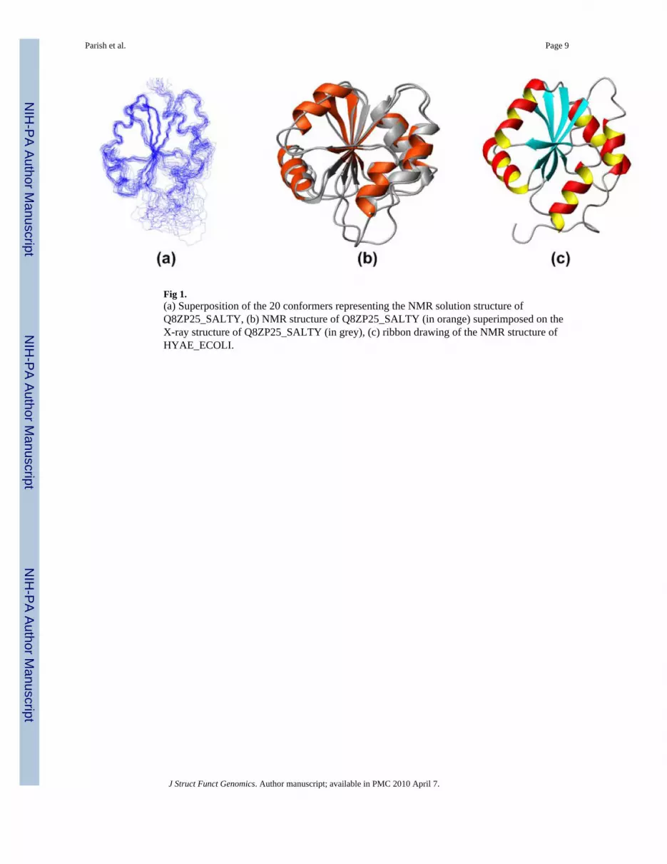
Fig 1.(a) Superposition of the 20 conformers representing the NMR solution structure ofQ8ZP25_SALTY, (b) NMR structure of Q8ZP25_SALTY (in orange) superimposed on theX-ray structure of Q8ZP25_SALTY (in grey), (c) ribbon drawing of the NMR structure ofHYAE_ECOLI.
Parish et al. Page 9
J Struct Funct Genomics. Author manuscript; available in PMC 2010 April 7.
NIH
-PA Author Manuscript
NIH
-PA Author Manuscript
NIH
-PA Author Manuscript
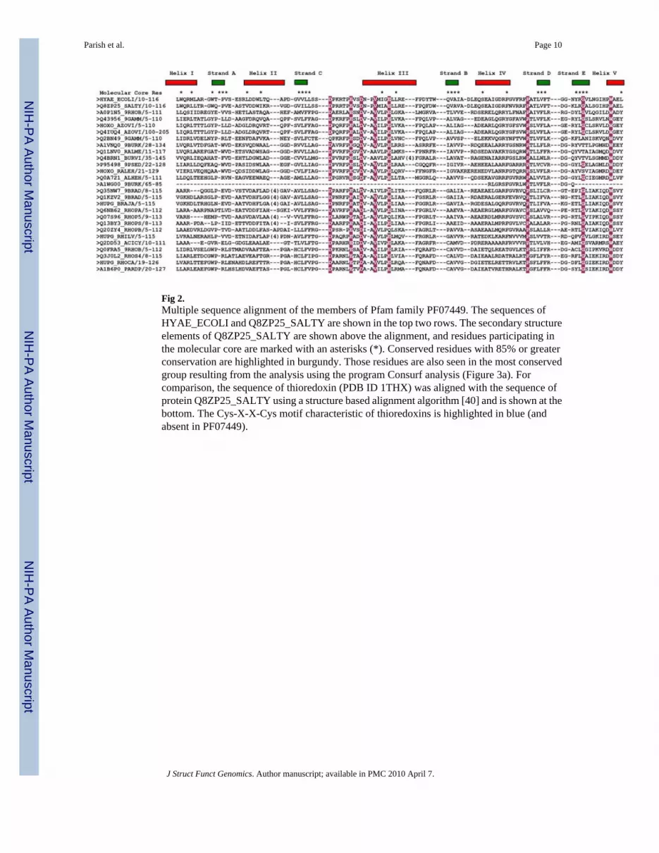
Fig 2.Multiple sequence alignment of the members of Pfam family PF07449. The sequences ofHYAE_ECOLI and Q8ZP25_SALTY are shown in the top two rows. The secondary structureelements of Q8ZP25_SALTY are shown above the alignment, and residues participating inthe molecular core are marked with an asterisks (*). Conserved residues with 85% or greaterconservation are highlighted in burgundy. Those residues are also seen in the most conservedgroup resulting from the analysis using the program Consurf analysis (Figure 3a). Forcomparison, the sequence of thioredoxin (PDB ID 1THX) was aligned with the sequence ofprotein Q8ZP25_SALTY using a structure based alignment algorithm [40] and is shown at thebottom. The Cys-X-X-Cys motif characteristic of thioredoxins is highlighted in blue (andabsent in PF07449).
Parish et al. Page 10
J Struct Funct Genomics. Author manuscript; available in PMC 2010 April 7.
NIH
-PA Author Manuscript
NIH
-PA Author Manuscript
NIH
-PA Author Manuscript

Fig 3.(a) Conserved residues from PFAM PF07449 are mapped onto the surface of proteinQ8ZP25_SALTY. Colors are based on Bayesian conservation score [41] which characterizesthe evolution rate of each residue compared to the average rate for all residues in the proteinas follows: burgundy −1.6 to −1.3 (highly conserved), dark pink −1.29 to −0.9, pink −0.89 to−0.5, light pink −0.49 to −0.2, white −0.19 to 0.19, very light blue 0.2 to 0.5, light blue 0.51to 0.7, blue 0.71 to 1.25, dark blue 1.26 to 2.2 (highly variable). (b) Electrostatic surfacepotential for Q8ZP25_SALTY. Negative and positive electrostatic potentials are shown,respectively, in red and blue.
Parish et al. Page 11
J Struct Funct Genomics. Author manuscript; available in PMC 2010 April 7.
NIH
-PA Author Manuscript
NIH
-PA Author Manuscript
NIH
-PA Author Manuscript

NIH
-PA Author Manuscript
NIH
-PA Author Manuscript
NIH
-PA Author Manuscript
Parish et al. Page 12
Table 1
Statistics of Q8ZP25_SALTY and HYAE_ECOLI NMR structures.
Protein Q8ZP25_SALTY HYAE_ECOLI
Conformationally-restricting distance constraints
Intraresidue [i = j] 397 533
Sequential [(i − j) = 1] 428 542
Medium Range [1 < (i − j) ≤ 5] 206 200
Long Range [(i − j) > 5] 510 339
Total 1541 1524
Dihedral angle constraints
Φ 41 56
ψ 41 56
Number of constraints per residue 15.7 12.4
Number of long-range constraints per residue 5.2 2.5
Completeness of stereo-specific assignments a[%]
βCH2 28 (16/58) 25 (19/77)
Val and Leu isopropyl groups 67 (16/24) 20 (5/25)
CYANA target function [Å2] 1.1 ± 0.17 1.88 ± 0.3
Hydrogen bond constraints 10 0
NH RDC-derived orientational constraints 69 0
Average r.m.s.d. to the mean CYANA coordinates [Å]
regular secondary structure elements, backbone heavy atoms N, Cα, C′ 0.72 ± 0.15b 0.82 ± 0.09c
regular secondary structure elements, all heavy atoms 1.14 ± 0.12 1.22 ± 0.09
residues 11–41, 56–127, backbone heavy atoms 0.95 ± 0.12 0.94 ± 0.13
residues 11–41, 56–127, all heavy atoms 1.49 ± 0.13 1.52 ± 0.12
heavy atoms of best-defined side-chains 0.5 ± 0.07d 0.58 ± 0.1e
PROCHECK G-factorsf (ϕ and Ψ/all dihedral angles) 0.04/−1.6 0.04/−1.48
MOLPROBITY clash scoreg −1.74 −2.43
AutoQF R/P/DP scores (%)h 0.94/0.93/0.76 0.97/0.93/0.66
Ramachandran plot summary ordered residues: [%]
most favored regions 93.5 92.3
additionally allowed regions 6.4 7.2
generously allowed regions 0.1 0.5
disallowed regions 0 0
Average number of distance constraints violations per CYANA conformer [Å]
0.2–0.5 0.1 0.1
> 0.5 0 0
Average number of dihedral-angle constraint violations per CYANA conformer [degrees]
J Struct Funct Genomics. Author manuscript; available in PMC 2010 April 7.

NIH
-PA Author Manuscript
NIH
-PA Author Manuscript
NIH
-PA Author Manuscript
Parish et al. Page 13
Protein Q8ZP25_SALTY HYAE_ECOLI
> 5 0 0
aRelative to pairs with non-degenerate chemical shifts.
bResidues: 20–22, 37–40, 71–74, 95–99, 102–107 (β-strands), and 8–16, 26–34, 58–62, 79–86, 115–123 (α-helices).
cResidues: 19–22, 35–42, 69–75, 94–99, 102–108 (β-strands), and 5–16, 24–33, 56–63, 77–87, 112–124 (α-helices).
d34 residues: 15, 19, 21, 22, 24, 27, 30, 31, 34, 38–41, 58, 59, 68, 70, 72–75, 82, 83, 94–99, 103, 106, 118, 122, 123.
e30 residues: 10, 11, 14–16, 19, 21, 38–40, 61, 62, 70, 72–75, 80, 82, 83, 94, 97, 107, 113, 114, 117–120, 123
fDefined in Reference [42],
gDefined in Reference [43],
hDefined in Reference [44]
J Struct Funct Genomics. Author manuscript; available in PMC 2010 April 7.

NIH
-PA Author Manuscript
NIH
-PA Author Manuscript
NIH
-PA Author Manuscript
Parish et al. Page 14
Table 2
Statistics of Q8ZP25_SALTY X-ray crystal structure
Crystal Parameters:
Space group P21
Unit-cell at 100 K [Å] 39.5, 71.2, 95.0 90.0°, 90.1°, 90.0°
Data quality:
Resolution [Å] 20–2.8 (2.85–2.80)
No. of measured reflections 88422 (3992)
No. of unique reflections 24561 (1174)
Rsym [%] 4.9 (12.2) (I ≥ −3σ1 for observations)
Mean redundancy 3.6 (3.4)
Completeness [%] 98.6 (95.5) (All measured reflections)
88.6 (83.9) (I ≥ 2σ1)
Mean I/σI 35.4 (9.2) (I ≥ σ1 after merging)
Refinement residuals (F≥2σF):
Rfree [%] 33.8
Rwork [%] 28.5
Model quality:
RMSD bond lengths [Å] 0.017
RMSD bond angles 2.4 °
Ramachandran plot summary [%]
most favored regions 86.6
allowed regions 12.2
generously allowed regions 1.2
Average B factors (Å2):
All 43.3
Main chain 33.7
Side chain 35.2
Waters 45.8
Model contents:
Protein residues 4×10³
Water molecules 560
J Struct Funct Genomics. Author manuscript; available in PMC 2010 April 7.

NIH
-PA Author Manuscript
NIH
-PA Author Manuscript
NIH
-PA Author Manuscript
Parish et al. Page 15
Table 3
R.m.s.d. values calculated for structure comparison
Backbonea r.m.s.d values Q8PZ25_SALTY (X-RAY)b HYAE_ECOLI (NMR)b,c
Q8PZ25_SALTY (NMR)b,c 1.23 Å 1.58 Å
Q8PZ25_SALTY (X-RAY)b - 1.87 Å
aHeavy atoms N, Cα, and C′ were superimposed for minimal r.m.s.d.
bResidues well defined in both structures: 11–25, 34–42, 55–90, 94–101, 106, 107, 113–124
cMean conformer calculated for the 20 best conformers (Table I)
J Struct Funct Genomics. Author manuscript; available in PMC 2010 April 7.