Embed Size (px)
Citation preview
Molecular Biology of the CellVol. 19, 3040–3051, July 2008
Protein Phosphatase Type 1 Directs Chitin Synthesis at theBud Neck in Saccharomyces cerevisiaeJennifer R. Larson, Jennifer P. Bharucha,* Shantelle Ceaser, Joanna Salamon,†Charles J. Richardson, Segalit M. Rivera, and Kelly Tatchell
Department of Biochemistry and Molecular Biology, Louisiana State University Health Sciences Center,Shreveport, LA 71130
Submitted February 7, 2008; Revised April 30, 2008; Accepted May 5, 2008Monitoring Editor: Daniel Lew
Yeast chitin synthase III (CSIII) is targeted to the bud neck, where it is thought to be tethered by the septin-associatedprotein Bni4. Bni4 also associates with the yeast protein phosphatase (PP1) catalytic subunit, Glc7. To identify regions ofBni4 necessary for its targeting functions, we created a collection of 23 deletion mutants throughout the length of Bni4.Among the deletion variants that retain the ability to associate with the bud neck, only those deficient in Glc7 bindingfail to target CSIII to the neck. A chimeric protein composed of the septin Cdc10 and the C-terminal Glc7-binding domainof Bni4 complements the defects of a bni4� mutant, indicating that the C-terminus of Bni4 is necessary and sufficient totarget Glc7 and CSIII to the bud neck. A Cdc10-Glc7 chimera fails to target CSIII to the bud neck but is functionalin the presence of the C-terminal Glc7-binding domain of Bni4. The conserved C-terminal PP1-binding domain ofmammalian Phactr-1 can functionally substitute for the C-terminus of Bni4. These results suggest that the essentialrole of Bni4 is to target Glc7 to the neck and activate it toward substrates necessary for CSIII recruitment andsynthesis of chitin at the bud neck.
INTRODUCTION
Vesicle targeting and fusion are key regulatory steps inmany cellular processes including neurotransmitter releaseand platelet secretion. Although many of the proteins in-volved in the secretory pathway have been identified, thefinal stages of vesicle targeting and fusion are not yet fullyunderstood. Targeting of the chitin synthase machinery inthe yeast Saccharomyces cerevisiae has served as a model forstudying targeted secretion. During vegetative growth,chitin is deposited first as a ring on the mother cell at thebase of the incipient bud. This chitin ring will comprise thebud scar in the cell wall, which persists through successivecell divisions. Chitin is again deposited at cytokinesis in theprimary septum (Cabib and Duran, 2005). The majority ofchitin is synthesized by chitin synthase III (CSIII), one ofthree chitin synthases in yeast, whose catalytic subunit,Chs3, is targeted to the bud neck for the deposition of thechitin ring (Choi et al., 1994; Chuang and Schekman, 1996;Cos et al., 1998). Chs3 cycles between an endosomal com-partment, termed the chitosome, and the cell surface where
it is activated upon association with its regulatory subunitChs4/Skt5 (Bulawa, 1993; Trilla et al., 1997). Many of theproteins involved in trafficking Chs3 to the plasma mem-brane have been identified, but it is still not known whichproteins are involved in chitosome fusion to the membraneat the bud neck. The cellular distribution of Chs4 differsfrom Chs3 in that a greater fraction of Chs4 is membraneassociated and Chs4 does not appear to be present in cyto-plasmic punctae as observed with Chs3 (Reyes et al., 2007).This suggests that Chs3 and Chs4 have different routes ofintracellular trafficking to the cell surface. Both Chs3 andChs4 localize to the presumptive bud site as a ring andremain at the neck of budding cells until buds are medium-sized, when they both dissociate from the neck. At cytoki-nesis, Chs3 and Chs4 reappear at the neck (Chuang andSchekman, 1996; DeMarini et al., 1997; Santos and Snyder,1997; Kozubowski et al., 2003). Chs3 and Chs4 are codepen-dent for their recruitment and/or retention at the neck ofsmall-budded cells (DeMarini et al., 1997), but it is notknown whether Chs3 and Chs4 are delivered to the neck asa complex or first associate at the neck.
Targeted CSIII activity at the site of bud emergence alsorequires Bni4, a 100-kDa protein that has been proposed toact as a scaffold to tether Chs4 to septin filaments (DeMariniet al., 1997). Like Chs3 and Chs4, Bni4 localizes as a ring tothe presumptive bud site and remains asymmetrically re-stricted to the mother side of the bud neck. However, Bni4remains at the bud neck throughout the cell cycle until justbefore cytokinesis, when its levels drop. BNI4 null mutantsfail to target CSIII to the neck early in the cell cycle, resultingin poorly defined bud scars (DeMarini et al., 1997; Ko-zubowski et al., 2003). Bni4 also binds to the protein phos-phatase type 1 (PP1) catalytic subunit, Glc7. Yeast cells con-taining a mutation of a conserved PP1/Glc7-binding motifnear the C-terminus of Bni4 (Bni4V831A/F833A, hereafter
This article was published online ahead of print in MBC in Press(http://www.molbiolcell.org/cgi/doi/10.1091/mbc.E08–02–0130)on May 14, 2008.
Present addresses: * Laboratory of Human Retrovirology, ClinicalServices Program, Applied and Developmental Research SupportProgram, Science Application International Corporation (SAIC)-Frederick, National Cancer Institute at Frederick, Frederick, MD21702; † Department of Biology, New Mexico State University, LasCruces, NM 88003.
Address correspondence to: Kelly Tatchell ([email protected]).
Abbreviations used: CSIII, chitin synthase III; PP1, protein phospha-tase type 1.
3040 © 2008 by The American Society for Cell Biology http://www.molbiolcell.org/content/suppl/2008/05/14/E08-02-0130.DC1.htmlSupplemental Material can be found at:
referred to as Bni4VA/FA) do not recruit Glc7 or CSIII to thebud neck and exhibit chitin staining similar to bni4� mutants(Kozubowski et al., 2003). Hyperphosphorylation ofBni4VA/FA and its reduced abundance at the neck led to theproposal that Glc7 is necessary for regulating the associationof Bni4 with the septin ring (Kozubowski et al., 2003). How-ever, a more direct role for the phosphatase in CSIII target-ing has not been ruled out. Here, we present new dataindicating a direct role for Glc7 in recruiting the chitinsynthase machinery to the bud neck and propose that therole of Bni4 at the bud neck is not as a tether for CSIII, butrather to target Glc7 to the septin ring and activate it towardone or more substrates necessary to recruit active CSIII.
MATERIALS AND METHODS
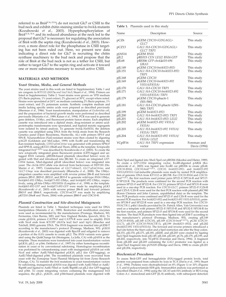
Yeast Strains, Media, and General MethodsThe yeast strains used in this work are listed in Supplementary Table 1 andare congenic to KT1112 (MATa ura3 leu2 his3; Stuart et al., 1994). Primers arelisted in Supplementary Table 2. Yeast strains were grown on YPD medium(2% Bacto peptone, 1% yeast extract, 2% glucose) at 30°C, except where noted.Strains were sporulated at 24°C on medium containing 2% Bacto peptone, 1%yeast extract, and 2% potassium acetate. Synthetic complete medium andmedia lacking specific amino acids were prepared as described previously(Sherman et al., 1986). Yeast transformation, manipulation of Escherichia coli,and the preparation of bacterial growth media were performed as describedpreviously (Maniatis et al., 1989; Kaiser et al., 1994). PCR was used to generategene deletion, 13-Myc, and fluorescent protein fusion strains. Each amplifiedcassette was introduced into a diploid strain, drug-resistant or amino acid-prototrophic transformants were sporulated, and haploid meiotic segregantswere isolated by tetrad analysis. To generate bni4�::NatMX4, the deletioncassette was amplified using DNA from the bni4� strain from the ResearchGenetics panel (Open Biosystems, Huntsville, AL) with primers SP5-F andSP6-R. Nourseothricin (Nat)-resistant strains were then created by digestingpAG25 (Goldstein and McCusker, 1999) with EcoRI and transforming theKan-resistant haploids. CHS3-yEmCitrine was generated with primers SP94-Fand SP95-R, using pKT211 (Sheff and Thorn, 2004) as the template. Ectopicallyintegrated bni4VA/FA was described by Kozubowski et al. (2003). To create thenew ectopically integrated green fluorescent protein (GFP)-tagged and un-tagged BNI4 variants, pRS306-based plasmids (described below) were di-gested with StuI and introduced into JRL360. To create an integrated GFP-CHS4 fusion, NheI-digested pJL68 (described below) was integrated intoyeast. The chs3�::HIS3 allele was derived from backcrosses of DDY181 (De-Marini et al., 1997). Construction of GLC7-tdimer2, GLC7-yEmCitrine, andGLC7-13myc was described previously (Bharucha et al., 2008). The 13Myc-integration cassettes were amplified with reverse primer JB6-R and forwardprimers JB5-F, JB29-F, JB32-F, and JB33-F to generate wild-type BNI4-13Myc,bni4�863-892-13Myc, bni4�873-892-13Myc, and bni4�882-892-13Myc, respec-tively, using pFA6a-13Myc-HIS3MX6 (Longtine et al., 1998) as the template.bni4�863-892-GFP and bni4�873-892-GFP were made by amplifying pLK3(Kozubowski et al., 2003) with reverse primer JB6-R and forward primersJB43-F and JB44-F, respectively. CDC10-mCFP was made by amplifyingpKT210 (Sheff and Thorn, 2004) with primers JB80-F and CDC10-R.
Plasmid Construction and Site-directed MutagenesisPlasmids are listed in Table 1. Standard techniques were used for DNAmanipulation (Maniatis et al., 1989). Restriction and modification enzymeswere used as recommended by the manufacturers (Promega, Madison, WI;Fermentas, Glen Burnie, MD; and New England Biolabs, Ipswich, MA). Tocreate pJAS14, primers CAT78-F and CAT79-R were used to amplify BNI4with NcoI ends from KT1357 (MATa leu2 his3 ura3 trp1) (Bloecher andTatchell, 2000) genomic DNA. The BNI4 fragment was inserted into pGEM-Taccording to the manufacturer’s protocol (Promega, Madison, WI). pLK10(Kozubowski et al., 2003) was digested with Bpu10I and religated to removea portion of the KAN cassette to create pJL2. The BNI4 variants were gener-ated using the QuikChange kit (Stratagene, La Jolla, CA) with pJAS14 as thetemplate. These mutations were transferred into yeast transformation vectors(pLK10, pJL2, or p366; DeMarini et al., 1997) by either homologous recombi-nation in yeast or by conventional subcloning. Homologous recombinationwas performed by cotransforming yeast with mutagenized pJAS14 cut withNcoI and either AatII/NheI-digested pLK10, pJL2 (for GFP-tagging), orAatII/NheI-digested p366. The recombined plasmids were recovered fromyeast with the Zymoprep Yeast Plasmid Miniprep kit from Zymo Research(Orange, CA). To transfer the bni4 mutations to yeast transformation vectorsby subcloning, mutagenized pJAS14 was digested with AatII/NheI or AatII/Bsp68I and ligated to AatII/NheI or AatII/Bsp68I cut pJL2 (for GFP-tagging)and p366. To create integrating vectors containing the mutagenized bni4sequence, the pJL2-, pLK10-, and p366-based plasmids were digested with
XhoI/SpeI and ligated into XhoI/SpeI cut pRS306 (Sikorksi and Hieter, 1989).To create a GFP-CHS4 integrating vector, EcoRI-digested pAR24 (Ko-zubowski et al., 2003) was ligated into EcoRI cut pRS306 to create pJL68.CDC10-BNI4, CDC10-bni4VA/FA, CDC10, bni4(823-892), and bni4(823-892V831A/F833A) Gal-inducible plasmids were made by nested PCR amplifica-tion of genomic DNA from KT1112 or JRL708. For CDC10-BNI4 and CDC10-bni4VA/FA, the first reactions used primer pairs SP121-F/SP116-R and SP117-F/SP122-R. The products were combined for the second PCR reaction withprimers SP121-F and SP122-R. For CDC10, primers SP121-F and SP131-R wereused in a one-step PCR reaction. For CDC10-GLC7, primers SP121-F/CZ4-Rand CZ5-F/CZ6-R were used for the first PCR reaction with plasmid pKC980(Karen Clemens and John Cannon, unpublished data) as the template. Thetwo PCR products were combined and SP121-F and CZ6-R were used for thesecond PCR reaction. For bni4(823-892) and bni4(823-892 V831A/F833A), prim-ers SP138-F and SP122-R were used in a one-step PCR reaction. For CDC10-PHACTR-1, p40c1 (kindly provided by Dr. Patrick Allen, Yale University) wasused as a template with primers SP121-F/SP133-R and SP132-F/SP134-R forthe first reaction, and primers SP121-F and SP134-R were used for the secondreaction. The final PCR products were then ligated into pGEM-T according tothe manufacturer’s protocol (Promega, Madison, WI), creating pJL149(CDC10-BNI4), pJL168 (CDC10), pJL169 (CDC10-bni4VA/FA), pCZ6 (CDC10-GLC7), pJL179 (CDC10-PHACTR-1), pJL199 (bni4(823–892)), and pJL202(bni4(823-892 V831A/F833A)). The forward and reverse primers introduced aSacI site before the Start codon and a SpeI restriction site after the Stop codon.pJL151, pJL170, pJL171, pJL181, pJl200, and pJL203 were created by ligatingSacI/SpeI fragments from pJL149, pJL168, pJL169, pC76, pJL179, pJL199, andpJL202, respectively, into YCpIF16 (Foreman and Davis, 1994). A fragmentfrom pJL200 and pJL203 containing the GAL1 promotor was ligated as aApaI/SacI fragment into pUN105 (Elledge and Davis, 1988) to create pJL201and pJL204, respectively.
Biochemical ProceduresTo assess Bni4-GFP and hemagglutinin (HA)-tagged protein levels, totalprotein was prepared from cultures by lysis in TCA (Davis et al., 1993; Stuartet al., 1994). Proteins were electrophoresed on 8% (Figure 1) or 10% (Figures3, 5, and 6) polyacrylamide-SDS gels. Immunoblot analysis was performed asdescribed (Stuart et al., 1994) using the 12CA5 anti-HA antibody or BD LivingColors A.v. monoclonal anti-GFP (JL-8) antibody, with subsequent detection
Table 1. Plasmids used in this study
Name Description Source
pCZ6 pGEM CDC10-(GDGAGL)-GLC7
This study
pCZ13 GAL1 HA-CDC10-(GDGAGL)-GLC7 TRP1
This study
pJAS14 pGEM BNI4 This studypJL2 pRS315 CEN LEU2 BNI4-GFP This studypJL68 pRS306 GFP-chs4�610-696
URA3This study
pJL149 pGEM CDC10-bni4(823-892) This studypJL151 GAL1 HA-CDC10-bni4(823-892)
TRP1This study
pJL168 pGEM CDC10 This studypJL169 pGEM CDC10-bni4(823-892
V831A/F833A)This study
pJL170 GAL1 HA-CDC10 TRP1 This studypJL171 GAL1 HA-CDC10-bni4(823-892
V831A/F833A) TRP1This study
pJL179 pGEM CDC10-phactr-1(501-580)
This study
pJL181 GAL1 HA-CDC10-phactr-1(501-580) TRP1
This study
pJL199 pGEM bni4(823-892) This studypJL200 GAL1 HA-bni4(823-892) TRP1 This studypJL201 GAL1 HA-bni4(823-892) LEU2 This studypJL202 pGEM bni4(823-892 V831A/
F833A)This study
pJL203 GAL1 HA-bni4(823-892 V831A/F833A) TRP1
This study
pJL204 GAL1 HA-bni4(823-892 V831A/F833A) LEU2
This study
YCpIF16 GAL1 HA TRP1 expressionvector
Foreman andDavis (1994)
PP1 Directs Chitin Synthesis
Vol. 19, July 2008 3041
using the Enhanced Chemiluminescence System (Amersham ECL Plus, GEHealthcare, Chalfont St. Giles, United Kingdom). HsfI expression was used asa loading control (antibody kindly provided by Dr. David Gross, LSUHSC,Shreveport).
To test for coimmunoprecipitation of Glc7-13myc and Bni4-GFP, cells wereharvested and lysed as described in Kozubowski et al. (2003). Lysates werethen subsequently used with the ProFound c-Myc Tag IP/CoIP ApplicationSet according to the manufacturer’s instructions (Pierce, Rockford, IL). Afteradding 2� sample buffer to the beads, 2 �l �-mercaptoethanol was added,and beads were heated to 100°C for 5 min. Samples were eluted from thecolumns and electrophoresed on an 8% polyacrylamide-SDS gel. Immunoblotanalysis was performed as described above using anti-GFP (JL-8) antibody oranti-Myc 9E10 ascites antibody with subsequent detection using the En-hanced Chemiluminescence System.
MicroscopyCells were placed onto a pad of 2% agarose in synthetic medium containing2% glucose or 2% galactose and imaged for GFP, CFP, YFP, and RFP (green,cyan, yellow, and red fluorescent protein, respectively) as previously de-scribed (Kozubowski et al., 2003). Fluorescence images in different Z-axisplanes (0.5 �m apart) were acquired using Slidebook software (OlympusImaging Systems, Melville, NY). Fluorescence levels were quantitated aspreviously described (Kozubowski et al., 2003) using the average of fouradjacent pixels and subtracting background fluorescence from the cytoplasm.Calcofluor staining was done as described previously (Robinson et al., 1999).Images of Calcofluor staining are projections of 20 planes through the Z-axis.
RESULTS
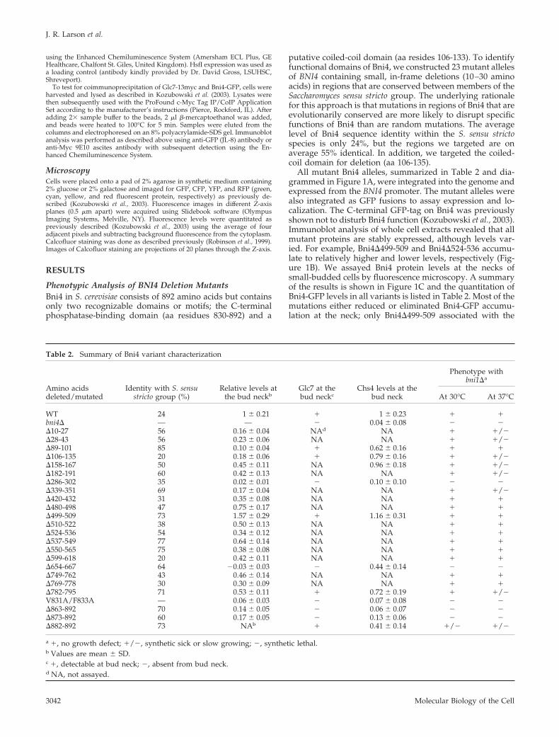
Phenotypic Analysis of BNI4 Deletion MutantsBni4 in S. cerevisiae consists of 892 amino acids but containsonly two recognizable domains or motifs; the C-terminalphosphatase-binding domain (aa residues 830-892) and a
putative coiled-coil domain (aa resides 106-133). To identifyfunctional domains of Bni4, we constructed 23 mutant allelesof BNI4 containing small, in-frame deletions (10–30 aminoacids) in regions that are conserved between members of theSaccharomyces sensu stricto group. The underlying rationalefor this approach is that mutations in regions of Bni4 that areevolutionarily conserved are more likely to disrupt specificfunctions of Bni4 than are random mutations. The averagelevel of Bni4 sequence identity within the S. sensu strictospecies is only 24%, but the regions we targeted are onaverage 55% identical. In addition, we targeted the coiled-coil domain for deletion (aa 106-135).
All mutant Bni4 alleles, summarized in Table 2 and dia-grammed in Figure 1A, were integrated into the genome andexpressed from the BNI4 promoter. The mutant alleles werealso integrated as GFP fusions to assay expression and lo-calization. The C-terminal GFP-tag on Bni4 was previouslyshown not to disturb Bni4 function (Kozubowski et al., 2003).Immunoblot analysis of whole cell extracts revealed that allmutant proteins are stably expressed, although levels var-ied. For example, Bni4�499-509 and Bni4�524-536 accumu-late to relatively higher and lower levels, respectively (Fig-ure 1B). We assayed Bni4 protein levels at the necks ofsmall-budded cells by fluorescence microscopy. A summaryof the results is shown in Figure 1C and the quantitation ofBni4-GFP levels in all variants is listed in Table 2. Most of themutations either reduced or eliminated Bni4-GFP accumu-lation at the neck; only Bni4�499-509 associated with the
Table 2. Summary of Bni4 variant characterization
Amino acidsdeleted/mutated
Identity with S. sensustricto group (%)
Relative levels atthe bud neckb
Glc7 at thebud neckc
Chs4 levels at thebud neck
Phenotype withbni1�a
At 30°C At 37°C
WT 24 1 � 0.21 � 1 � 0.23 � �bni4� — — � 0.04 � 0.08 � ��10-27 56 0.16 � 0.04 NAd NA � �/��28-43 56 0.23 � 0.06 NA NA � �/��89-101 85 0.10 � 0.04 � 0.62 � 0.16 � ��106-135 20 0.18 � 0.06 � 0.79 � 0.16 � �/��158-167 50 0.45 � 0.11 NA 0.96 � 0.18 � �/��182-191 60 0.42 � 0.13 NA NA � �/��286-302 35 0.02 � 0.01 � 0.10 � 0.10 � ��339-351 69 0.17 � 0.04 NA NA � �/��420-432 31 0.35 � 0.08 NA NA � ��480-498 47 0.75 � 0.17 NA NA � ��499-509 73 1.57 � 0.29 � 1.16 � 0.31 � ��510-522 38 0.50 � 0.13 NA NA � ��524-536 54 0.34 � 0.12 NA NA � ��537-549 77 0.64 � 0.14 NA NA � ��550-565 75 0.38 � 0.08 NA NA � ��599-618 20 0.42 � 0.11 NA NA � ��654-667 64 �0.03 � 0.03 � 0.44 � 0.14 � ��749-762 43 0.46 � 0.14 NA NA � ��769-778 30 0.30 � 0.09 NA NA � ��782-795 71 0.53 � 0.11 � 0.72 � 0.19 � �/�V831A/F833A — 0.06 � 0.03 � 0.07 � 0.08 � ��863-892 70 0.14 � 0.05 � 0.06 � 0.07 � ��873-892 60 0.17 � 0.05 � 0.13 � 0.06 � ��882-892 73 NAb � 0.41 � 0.14 �/� �/�
a �, no growth defect; �/�, synthetic sick or slow growing; �, synthetic lethal.b Values are mean � SD.c �, detectable at bud neck; �, absent from bud neck.d NA, not assayed.
J. R. Larson et al.
Molecular Biology of the Cell3042
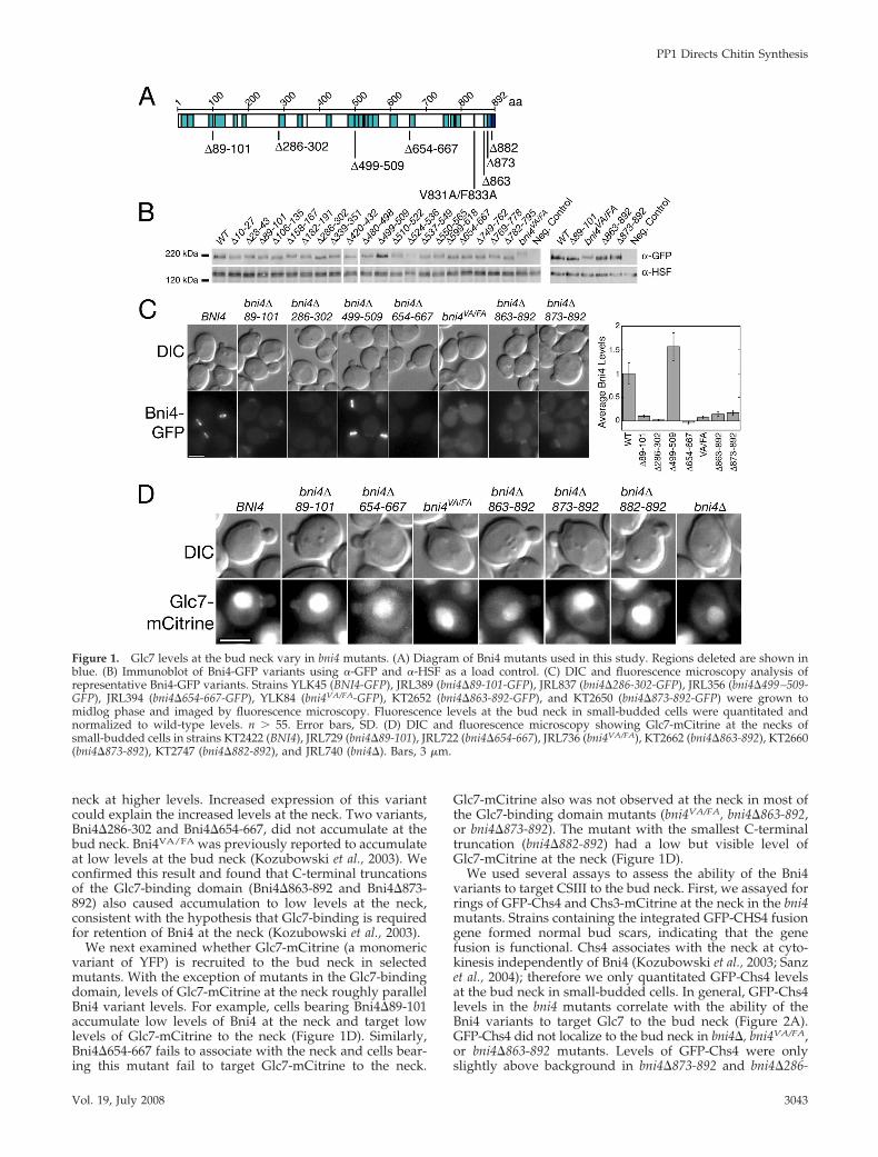
neck at higher levels. Increased expression of this variantcould explain the increased levels at the neck. Two variants,Bni4�286-302 and Bni4�654-667, did not accumulate at thebud neck. Bni4VA/FA was previously reported to accumulateat low levels at the bud neck (Kozubowski et al., 2003). Weconfirmed this result and found that C-terminal truncationsof the Glc7-binding domain (Bni4�863-892 and Bni4�873-892) also caused accumulation to low levels at the neck,consistent with the hypothesis that Glc7-binding is requiredfor retention of Bni4 at the neck (Kozubowski et al., 2003).
We next examined whether Glc7-mCitrine (a monomericvariant of YFP) is recruited to the bud neck in selectedmutants. With the exception of mutants in the Glc7-bindingdomain, levels of Glc7-mCitrine at the neck roughly parallelBni4 variant levels. For example, cells bearing Bni4�89-101accumulate low levels of Bni4 at the neck and target lowlevels of Glc7-mCitrine to the neck (Figure 1D). Similarly,Bni4�654-667 fails to associate with the neck and cells bear-ing this mutant fail to target Glc7-mCitrine to the neck.
Glc7-mCitrine also was not observed at the neck in most ofthe Glc7-binding domain mutants (bni4VA/FA, bni4�863-892,or bni4�873-892). The mutant with the smallest C-terminaltruncation (bni4�882-892) had a low but visible level ofGlc7-mCitrine at the neck (Figure 1D).
We used several assays to assess the ability of the Bni4variants to target CSIII to the bud neck. First, we assayed forrings of GFP-Chs4 and Chs3-mCitrine at the neck in the bni4mutants. Strains containing the integrated GFP-CHS4 fusiongene formed normal bud scars, indicating that the genefusion is functional. Chs4 associates with the neck at cyto-kinesis independently of Bni4 (Kozubowski et al., 2003; Sanzet al., 2004); therefore we only quantitated GFP-Chs4 levelsat the bud neck in small-budded cells. In general, GFP-Chs4levels in the bni4 mutants correlate with the ability of theBni4 variants to target Glc7 to the bud neck (Figure 2A).GFP-Chs4 did not localize to the bud neck in bni4�, bni4VA/FA,or bni4�863-892 mutants. Levels of GFP-Chs4 were onlyslightly above background in bni4�873-892 and bni4�286-
Figure 1. Glc7 levels at the bud neck vary in bni4 mutants. (A) Diagram of Bni4 mutants used in this study. Regions deleted are shown inblue. (B) Immunoblot of Bni4-GFP variants using �-GFP and �-HSF as a load control. (C) DIC and fluorescence microscopy analysis ofrepresentative Bni4-GFP variants. Strains YLK45 (BNI4-GFP), JRL389 (bni4�89-101-GFP), JRL837 (bni4�286-302-GFP), JRL356 (bni4�499–509-GFP), JRL394 (bni4�654-667-GFP), YLK84 (bni4VA/FA-GFP), KT2652 (bni4�863-892-GFP), and KT2650 (bni4�873-892-GFP) were grown tomidlog phase and imaged by fluorescence microscopy. Fluorescence levels at the bud neck in small-budded cells were quantitated andnormalized to wild-type levels. n � 55. Error bars, SD. (D) DIC and fluorescence microscopy showing Glc7-mCitrine at the necks ofsmall-budded cells in strains KT2422 (BNI4), JRL729 (bni4�89-101), JRL722 (bni4�654-667), JRL736 (bni4VA/FA), KT2662 (bni4�863-892), KT2660(bni4�873-892), KT2747 (bni4�882-892), and JRL740 (bni4�). Bars, 3 �m.
PP1 Directs Chitin Synthesis
Vol. 19, July 2008 3043
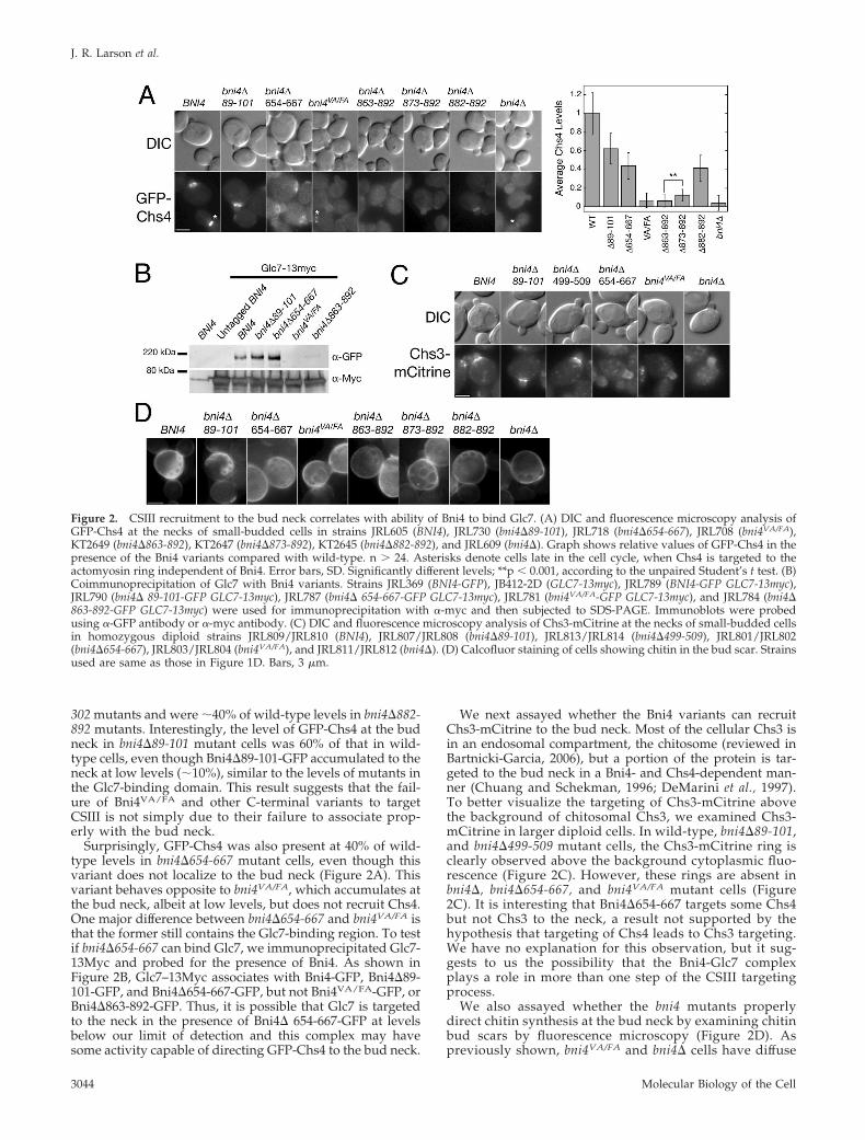
302 mutants and were �40% of wild-type levels in bni4�882-892 mutants. Interestingly, the level of GFP-Chs4 at the budneck in bni4�89-101 mutant cells was 60% of that in wild-type cells, even though Bni4�89-101-GFP accumulated to theneck at low levels (�10%), similar to the levels of mutants inthe Glc7-binding domain. This result suggests that the fail-ure of Bni4VA/FA and other C-terminal variants to targetCSIII is not simply due to their failure to associate prop-erly with the bud neck.
Surprisingly, GFP-Chs4 was also present at 40% of wild-type levels in bni4�654-667 mutant cells, even though thisvariant does not localize to the bud neck (Figure 2A). Thisvariant behaves opposite to bni4VA/FA, which accumulates atthe bud neck, albeit at low levels, but does not recruit Chs4.One major difference between bni4�654-667 and bni4VA/FA isthat the former still contains the Glc7-binding region. To testif bni4�654-667 can bind Glc7, we immunoprecipitated Glc7-13Myc and probed for the presence of Bni4. As shown inFigure 2B, Glc7–13Myc associates with Bni4-GFP, Bni4�89-101-GFP, and Bni4�654-667-GFP, but not Bni4VA/FA-GFP, orBni4�863-892-GFP. Thus, it is possible that Glc7 is targetedto the neck in the presence of Bni4� 654-667-GFP at levelsbelow our limit of detection and this complex may havesome activity capable of directing GFP-Chs4 to the bud neck.
We next assayed whether the Bni4 variants can recruitChs3-mCitrine to the bud neck. Most of the cellular Chs3 isin an endosomal compartment, the chitosome (reviewed inBartnicki-Garcia, 2006), but a portion of the protein is tar-geted to the bud neck in a Bni4- and Chs4-dependent man-ner (Chuang and Schekman, 1996; DeMarini et al., 1997).To better visualize the targeting of Chs3-mCitrine abovethe background of chitosomal Chs3, we examined Chs3-mCitrine in larger diploid cells. In wild-type, bni4�89-101,and bni4�499-509 mutant cells, the Chs3-mCitrine ring isclearly observed above the background cytoplasmic fluo-rescence (Figure 2C). However, these rings are absent inbni4�, bni4�654-667, and bni4VA/FA mutant cells (Figure2C). It is interesting that Bni4�654-667 targets some Chs4but not Chs3 to the neck, a result not supported by thehypothesis that targeting of Chs4 leads to Chs3 targeting.We have no explanation for this observation, but it sug-gests to us the possibility that the Bni4-Glc7 complexplays a role in more than one step of the CSIII targetingprocess.
We also assayed whether the bni4 mutants properlydirect chitin synthesis at the bud neck by examining chitinbud scars by fluorescence microscopy (Figure 2D). Aspreviously shown, bni4VA/FA and bni4� cells have diffuse
Figure 2. CSIII recruitment to the bud neck correlates with ability of Bni4 to bind Glc7. (A) DIC and fluorescence microscopy analysis ofGFP-Chs4 at the necks of small-budded cells in strains JRL605 (BNI4), JRL730 (bni4�89-101), JRL718 (bni4�654-667), JRL708 (bni4VA/FA),KT2649 (bni4�863-892), KT2647 (bni4�873-892), KT2645 (bni4�882-892), and JRL609 (bni4�). Graph shows relative values of GFP-Chs4 in thepresence of the Bni4 variants compared with wild-type. n � 24. Asterisks denote cells late in the cell cycle, when Chs4 is targeted to theactomyosin ring independent of Bni4. Error bars, SD. Significantly different levels; **p � 0.001, according to the unpaired Student’s t test. (B)Coimmunoprecipitation of Glc7 with Bni4 variants. Strains JRL369 (BNI4-GFP), JB412-2D (GLC7-13myc), JRL789 (BNI4-GFP GLC7-13myc),JRL790 (bni4� 89-101-GFP GLC7-13myc), JRL787 (bni4� 654-667-GFP GLC7-13myc), JRL781 (bni4VA/FA-GFP GLC7-13myc), and JRL784 (bni4�863-892-GFP GLC7-13myc) were used for immunoprecipitation with �-myc and then subjected to SDS-PAGE. Immunoblots were probedusing �-GFP antibody or �-myc antibody. (C) DIC and fluorescence microscopy analysis of Chs3-mCitrine at the necks of small-budded cellsin homozygous diploid strains JRL809/JRL810 (BNI4), JRL807/JRL808 (bni4�89-101), JRL813/JRL814 (bni4�499-509), JRL801/JRL802(bni4�654-667), JRL803/JRL804 (bni4VA/FA), and JRL811/JRL812 (bni4�). (D) Calcofluor staining of cells showing chitin in the bud scar. Strainsused are same as those in Figure 1D. Bars, 3 �m.
J. R. Larson et al.
Molecular Biology of the Cell3044
and irregular shaped bud scars that are difficult to detectby Calcofluor staining (DeMarini et al., 1997; Kozubowskiet al., 2003). bni4�654-667, bni4�863-892, and bni4�873-892mutant cells show bud scars like those of a bni4� mutant.In contrast, bni4�89-101 mutant cells exhibit wild-typebud scars. bni4�882-892 mutants have bud scars interme-diate between those of the wild type and those of a bni4�mutant.
We tested the BNI4 mutants for synthetic genetic de-fects in combination with bni1�. bni4�, chs4�, and chs3�mutants are inviable in the absence of the formin proteinBni1 (Tong et al., 2001), suggesting that bni1� mutantsrequire targeted CSIII activity for viability. Because bni4�mutants retain near normal levels of CSIII activity andchitin but are specifically defective in targeting CSIII ac-tivity to the bud neck (Sanz et al., 2004), the growth rate ofbni4 bni1� mutants provides a sensitive indicator for Bni4function. We therefore isolated the bni4 bni1� doublemutants from diploid strains heterozygous for bni1� andeach of our bni4 mutants. A summary of the growthproperties of these mutants is presented in Table 2. bni1�mutants in combination with bni4�286-302, bni4�654-667,bni4VA/FA, bni4�863-892, or bni4�873-892 germinate butfail to grow into macroscopic colonies. All other doublemutant pairs grow into colonies but some have noticeablegrowth defects. For example, bni1� bni4�882-892 mutantsare slow growing at 30° and 37°C, and other double
mutants grow slowly at 37°C (Table 2). Together, theseresults demonstrate a relationship between the ability ofBni4 to target Glc7 to the neck and CSIII targeting.
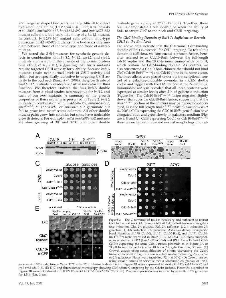
The Glc7-binding Domain of Bni4 Is Sufficient to RecruitCSIII to the Bud NeckThe above data indicate that the C-terminal Glc7-bindingdomain of Bni4 is essential for CSIII targeting. To test if thisdomain is sufficient, we constructed a protein fusion, here-after referred to as Cdc10-Bni4, between the full-lengthCdc10 septin and the 70 C-terminal amino acids of Bni4,which contain the Glc7-binding domain. As controls, wealso constructed a Cdc10-Bni4 chimera that should not bindGlc7 (Cdc10-Bni4VA/FA) and Cdc10 alone in the same vector.The three alleles were placed under the transcriptional con-trol of a galactose-inducible promoter in a CEN shuttlevector and tagged with the HA epitope at the N-terminus.Immunoblot analysis revealed that all three proteins wereexpressed at similar levels after 2 h of galactose induction(Figure 3A). The Cdc10-Bni4VA/FA fusion migrates slightlyslower than does the Cdc10-Bni4 fusion, suggesting that theBni4VA/FA portion of the chimera may be hyperphosphory-lated, as is the full-length Bni4VA/FA protein (Kozubowski etal., 2003). Cells expressing the CDC10-BNI4 gene fusion haveelongated buds and grow slowly on galactose medium (Fig-ure 3, B and C). Cells expressing Cdc10 or Cdc10-Bni4VA/FA
show normal growth rates and normal morphology, indicat-
Figure 3. The C-terminus of Bni4 is necessary and sufficient to recruitGlc7 to the bud neck. (A) Immunoblot of Cdc10-Bni4 fusions after galac-tose induction. Glu, 2% glucose; Raf, 2% raffinose; 2, 2-h induction 2%galactose; 4, 4-h induction 2% galactose. Asterisks denote nonspecificband. Plasmids pJL170 (Cdc10), pJL151 (Cdc10-Bni4), and pJL171 (Cdc10-Bni4VA/FA) were expressed in strain JRL43 (bni4�). (B) Colony morphol-ogies of strains JRL873 (bni4� GFP-CHS4) and JRL912 (chs3� bni4� GFP-CHS4) expressing the same Cdc10-fusion plasmids as in Figure 3A orYCpIF16 (empty vector), after 18 h on 2% galactose. Bar, 50 �m. (C)Growth assays using serial dilutions of strains expressing the Cdc10fusions described in Figure 3B on selective media containing 2% glucoseor 2% galactose. Plates were incubated 72 h at 30°C. (D) Growth assaysusing serial dilutions on selective media containing 2% glucose or 1.95%
sucrose � 0.05% galactose at 24 or 37°C after 72 h. Plasmids described in Figure 3B were expressed in strain KT2328 (MAT� leu2 his3trp1 ura3 cdc10-1). (E) DIC and fluorescence microscopy showing Glc7-tdimer2 targeting by the Cdc10 fusions. Plasmids described inFigure 3B were introduced into KT2737 (bni4� GLC7-tdimer2 CDC10-mCFP). Protein expression was induced by growth in 2% galactosefor 1.5 h. Bar, 3 �m.
PP1 Directs Chitin Synthesis
Vol. 19, July 2008 3045
ing that the growth and morphological defects induced byCDC10-BNI4 expression are likely due to Glc7 at the budneck. The CDC10 and CDC10-BNI4VA/FA chimeras comple-ment the temperature sensitivity of the cdc10–1 mutant (Fig-ure 3D), indicating these fusion proteins likely associatewith the septin ring. The elongated bud phenotype alsoappears unrelated to aberrant chitin synthesis, as cells lack-ing CHS3 and expressing the CDC10-BNI4 fusion still be-come elongated (Figure 3B).
We induced the Cdc10 fusions in a GLC7-tdimer2 CDC10-mCFP bni4� strain to confirm the ability of Cdc10-Bni4 totarget Glc7 to the septin ring. Glc7-tdimer2 rather than Glc7-mCitrine was used for these experiments because the formerfusion protein accumulates at lower levels in the nucleus(Bharucha et al., 2008), where high fluorescence can obscurethe signal at the bud neck. As predicted, Cdc10-Bni4VA/FA isunable to target Glc7 to the neck (Figure 3E). However,Glc7-tdimer2 is targeted to the bud neck by Cdc10-Bni4(Figure 3E). In cells expressing Cdc10-Bni4, Glc7-tdimer2was observed on both sides of the bud neck and at all stagesof the cell cycle. This is in contrast to the wild-type situation,where Glc7 is restricted to the mother side of the neck. Theseresults confirm that the last 70 amino acids of Bni4 aresufficient to bind Glc7.
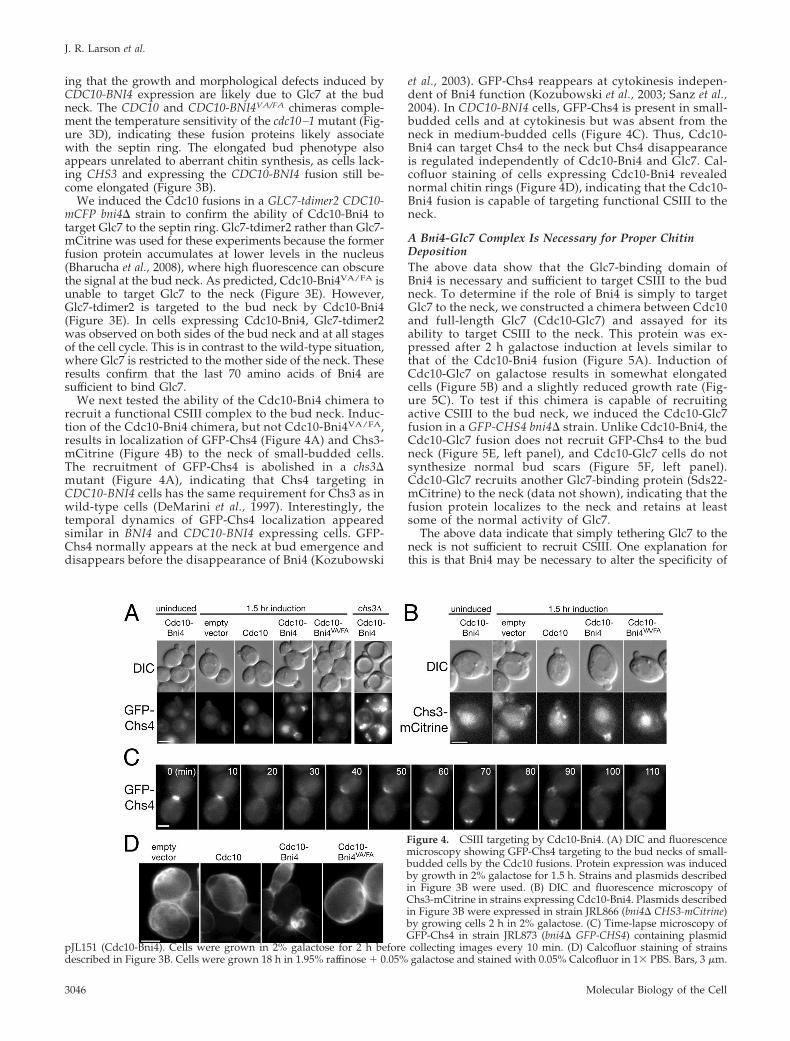
We next tested the ability of the Cdc10-Bni4 chimera torecruit a functional CSIII complex to the bud neck. Induc-tion of the Cdc10-Bni4 chimera, but not Cdc10-Bni4VA/FA,results in localization of GFP-Chs4 (Figure 4A) and Chs3-mCitrine (Figure 4B) to the neck of small-budded cells.The recruitment of GFP-Chs4 is abolished in a chs3�mutant (Figure 4A), indicating that Chs4 targeting inCDC10-BNI4 cells has the same requirement for Chs3 as inwild-type cells (DeMarini et al., 1997). Interestingly, thetemporal dynamics of GFP-Chs4 localization appearedsimilar in BNI4 and CDC10-BNI4 expressing cells. GFP-Chs4 normally appears at the neck at bud emergence anddisappears before the disappearance of Bni4 (Kozubowski
et al., 2003). GFP-Chs4 reappears at cytokinesis indepen-dent of Bni4 function (Kozubowski et al., 2003; Sanz et al.,2004). In CDC10-BNI4 cells, GFP-Chs4 is present in small-budded cells and at cytokinesis but was absent from theneck in medium-budded cells (Figure 4C). Thus, Cdc10-Bni4 can target Chs4 to the neck but Chs4 disappearanceis regulated independently of Cdc10-Bni4 and Glc7. Cal-cofluor staining of cells expressing Cdc10-Bni4 revealednormal chitin rings (Figure 4D), indicating that the Cdc10-Bni4 fusion is capable of targeting functional CSIII to theneck.
A Bni4-Glc7 Complex Is Necessary for Proper ChitinDepositionThe above data show that the Glc7-binding domain ofBni4 is necessary and sufficient to target CSIII to the budneck. To determine if the role of Bni4 is simply to targetGlc7 to the neck, we constructed a chimera between Cdc10and full-length Glc7 (Cdc10-Glc7) and assayed for itsability to target CSIII to the neck. This protein was ex-pressed after 2 h galactose induction at levels similar tothat of the Cdc10-Bni4 fusion (Figure 5A). Induction ofCdc10-Glc7 on galactose results in somewhat elongatedcells (Figure 5B) and a slightly reduced growth rate (Fig-ure 5C). To test if this chimera is capable of recruitingactive CSIII to the bud neck, we induced the Cdc10-Glc7fusion in a GFP-CHS4 bni4� strain. Unlike Cdc10-Bni4, theCdc10-Glc7 fusion does not recruit GFP-Chs4 to the budneck (Figure 5E, left panel), and Cdc10-Glc7 cells do notsynthesize normal bud scars (Figure 5F, left panel).Cdc10-Glc7 recruits another Glc7-binding protein (Sds22-mCitrine) to the neck (data not shown), indicating that thefusion protein localizes to the neck and retains at leastsome of the normal activity of Glc7.
The above data indicate that simply tethering Glc7 to theneck is not sufficient to recruit CSIII. One explanation forthis is that Bni4 may be necessary to alter the specificity of
Figure 4. CSIII targeting by Cdc10-Bni4. (A) DIC and fluorescencemicroscopy showing GFP-Chs4 targeting to the bud necks of small-budded cells by the Cdc10 fusions. Protein expression was inducedby growth in 2% galactose for 1.5 h. Strains and plasmids describedin Figure 3B were used. (B) DIC and fluorescence microscopy ofChs3-mCitrine in strains expressing Cdc10-Bni4. Plasmids describedin Figure 3B were expressed in strain JRL866 (bni4� CHS3-mCitrine)by growing cells 2 h in 2% galactose. (C) Time-lapse microscopy ofGFP-Chs4 in strain JRL873 (bni4� GFP-CHS4) containing plasmid
pJL151 (Cdc10-Bni4). Cells were grown in 2% galactose for 2 h before collecting images every 10 min. (D) Calcofluor staining of strainsdescribed in Figure 3B. Cells were grown 18 h in 1.95% raffinose � 0.05% galactose and stained with 0.05% Calcofluor in 1� PBS. Bars, 3 �m.
J. R. Larson et al.
Molecular Biology of the Cell3046
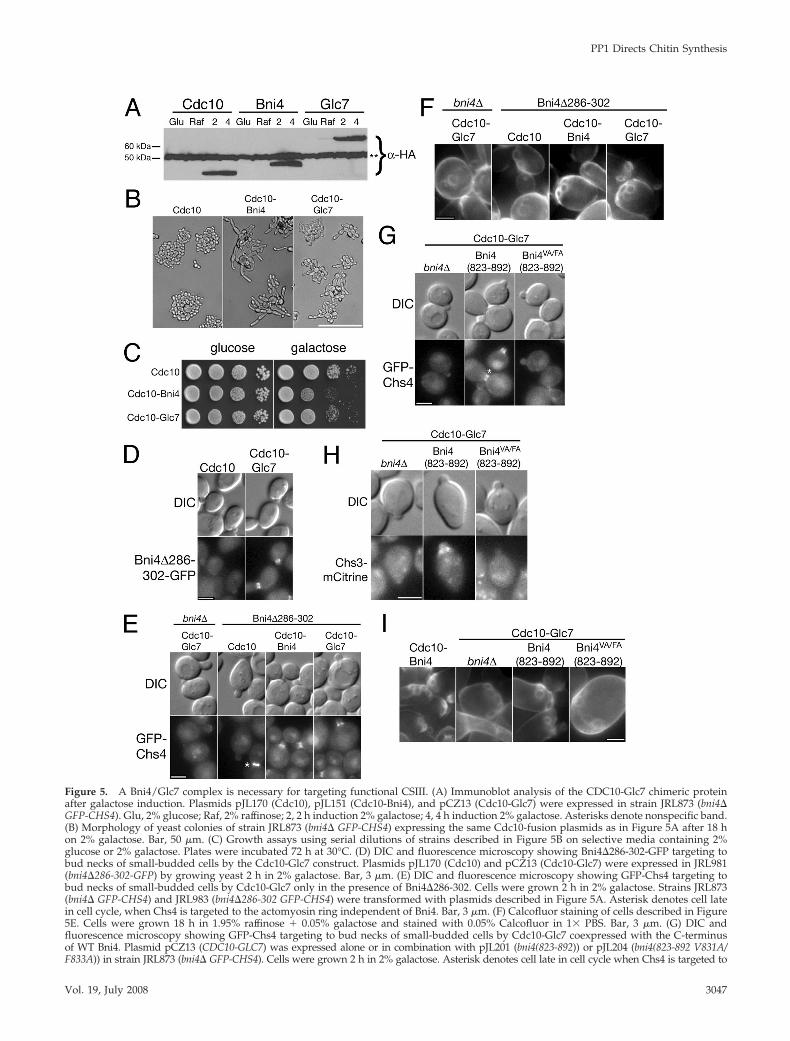
Figure 5. A Bni4/Glc7 complex is necessary for targeting functional CSIII. (A) Immunoblot analysis of the CDC10-Glc7 chimeric proteinafter galactose induction. Plasmids pJL170 (Cdc10), pJL151 (Cdc10-Bni4), and pCZ13 (Cdc10-Glc7) were expressed in strain JRL873 (bni4�GFP-CHS4). Glu, 2% glucose; Raf, 2% raffinose; 2, 2 h induction 2% galactose; 4, 4 h induction 2% galactose. Asterisks denote nonspecific band.(B) Morphology of yeast colonies of strain JRL873 (bni4� GFP-CHS4) expressing the same Cdc10-fusion plasmids as in Figure 5A after 18 hon 2% galactose. Bar, 50 �m. (C) Growth assays using serial dilutions of strains described in Figure 5B on selective media containing 2%glucose or 2% galactose. Plates were incubated 72 h at 30°C. (D) DIC and fluorescence microscopy showing Bni4�286-302-GFP targeting tobud necks of small-budded cells by the Cdc10-Glc7 construct. Plasmids pJL170 (Cdc10) and pCZ13 (Cdc10-Glc7) were expressed in JRL981(bni4�286-302-GFP) by growing yeast 2 h in 2% galactose. Bar, 3 �m. (E) DIC and fluorescence microscopy showing GFP-Chs4 targeting tobud necks of small-budded cells by Cdc10-Glc7 only in the presence of Bni4�286-302. Cells were grown 2 h in 2% galactose. Strains JRL873(bni4� GFP-CHS4) and JRL983 (bni4�286-302 GFP-CHS4) were transformed with plasmids described in Figure 5A. Asterisk denotes cell latein cell cycle, when Chs4 is targeted to the actomyosin ring independent of Bni4. Bar, 3 �m. (F) Calcofluor staining of cells described in Figure5E. Cells were grown 18 h in 1.95% raffinose � 0.05% galactose and stained with 0.05% Calcofluor in 1� PBS. Bar, 3 �m. (G) DIC andfluorescence microscopy showing GFP-Chs4 targeting to bud necks of small-budded cells by Cdc10-Glc7 coexpressed with the C-terminusof WT Bni4. Plasmid pCZ13 (CDC10-GLC7) was expressed alone or in combination with pJL201 (bni4(823-892)) or pJL204 (bni4(823-892 V831A/F833A)) in strain JRL873 (bni4� GFP-CHS4). Cells were grown 2 h in 2% galactose. Asterisk denotes cell late in cell cycle when Chs4 is targeted to
PP1 Directs Chitin Synthesis
Vol. 19, July 2008 3047
Glc7 toward its relevant substrates at the bud neck to allowCSIII recruitment. To test this, we first assayed the ability ofCdc10-Glc7 to recruit CSIII in a bni4�286-302 mutant. TheBni4�286-302 variant does not localize to the bud neck anddoes not recruit GFP-Chs4 to the bud neck (Figure 1C andTable 2), but it retains the Glc7-binding domain. If the Bni4-Glc7 complex is required for CSIII recruitment, our hypoth-esis is that the Cdc10-Glc7 will bind Bni4�286-302, forminga Glc7 holoenzyme able to target CSIII to the neck. As shownin Figure 5D, the induction of Cdc10-Glc7 induces Bni4�286-302-GFP localization to the bud neck. As expected, Bni4�286-302-GFP is located on both sides of the bud neckthroughout the cell cycle, corresponding to the location ofCdc10-Glc7. Induction of Cdc10-Glc7 in bni4�286-302 GFP-CHS4 cells results in GFP-Chs4 localization to the bud neck(Figure 5E) and visible chitin bud scars (Figure 5F).
To determine if expression of the C-terminus of Bni4alone could induce Cdc10-Glc7 to recruit CSIII to theneck, we expressed the C-terminus of Bni4 (aa 823– 892)with an N-terminal HA epitope tag from a galactose-inducible promoter. Expression of this 12.5-kDa proteinalone failed to target CSIII activity to the neck (data notshown). However, coexpression of Bni4(832-892) andCdc10-Glc7 resulted in the targeting of GFP-Chs4 (Figure5G) and Chs3-mCitrine (Figure 5H) to small-budded cellsand the formation of normal bud scars (Figure 5I). Coex-pression of Cdc10-Glc7 with the VA/FA variant ofBni4(832-892) failed to target CSIII components to theneck (Figure 5, G–I). Together, these results suggest thatBni4 not only recruits Glc7 to the neck but also alters itsspecificity toward those substrate(s) at the bud neck re-quired for proper chitin deposition.
The C-Terminus of Mammalian Phactr-1 Can FunctionallyReplace Bni4 at the Bud NeckThe recently characterized mammalian Phactr/Scapininfamily of proteins (Sagara et al., 2003; Allen et al., 2004)contains a C-terminal PP1-binding domain that is 32% iden-tical to the Glc7-binding domain of Bni4 (Figure 6A) butshares little, if any, similarity with the rest of Bni4. Proteinscontaining this C-terminal domain are found widely in eu-
Figure 5 (cont). the actomyosin ring independent of Bni4. Bar, 3�m. (H) DIC and fluorescence microscopy of Chs3-mCitrine.Plasmids described in Figure 5G were expressed in strain JRL866(bni4� CHS3-mCitrine) by growing cells 2 h in 2% galactose. Bar,3 �m. (I) Calcofluor staining of strains described in Figure 5G orwith plasmid pJL151 (CDC10-BNI4). Cells were grown 18 h in1.95% raffinose � 0.05% galactose and stained with 0.05% Cal-cofluor in 1� PBS. Bar, 3 �m.
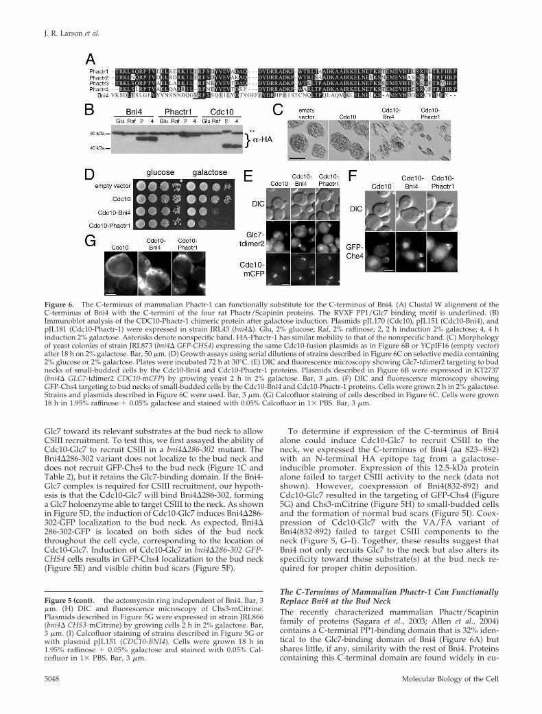
Figure 6. The C-terminus of mammalian Phactr-1 can functionally substitute for the C-terminus of Bni4. (A) Clustal W alignment of theC-terminus of Bni4 with the C-termini of the four rat Phactr/Scapinin proteins. The RVXF PP1/Glc7 binding motif is underlined. (B)Immunoblot analysis of the CDC10-Phactr-1 chimeric protein after galactose induction. Plasmids pJL170 (Cdc10), pJL151 (Cdc10-Bni4), andpJL181 (Cdc10-Phactr-1) were expressed in strain JRL43 (bni4�). Glu, 2% glucose; Raf, 2% raffinose; 2, 2 h induction 2% galactose; 4, 4 hinduction 2% galactose. Asterisks denote nonspecific band. HA-Phactr-1 has similar mobility to that of the nonspecific band. (C) Morphologyof yeast colonies of strain JRL873 (bni4� GFP-CHS4) expressing the same Cdc10-fusion plasmids as in Figure 6B or YCpIF16 (empty vector)after 18 h on 2% galactose. Bar, 50 �m. (D) Growth assays using serial dilutions of strains described in Figure 6C on selective media containing2% glucose or 2% galactose. Plates were incubated 72 h at 30°C. (E) DIC and fluorescence microscopy showing Glc7-tdimer2 targeting to budnecks of small-budded cells by the Cdc10-Bni4 and Cdc10-Phactr-1 proteins. Plasmids described in Figure 6B were expressed in KT2737(bni4� GLC7-tdimer2 CDC10-mCFP) by growing yeast 2 h in 2% galactose. Bar, 3 �m. (F) DIC and fluorescence microscopy showingGFP-Chs4 targeting to bud necks of small-budded cells by the Cdc10-Bni4 and Cdc10-Phactr-1 proteins. Cells were grown 2 h in 2% galactose.Strains and plasmids described in Figure 6C were used. Bar, 3 �m. (G) Calcofluor staining of cells described in Figure 6C. Cells were grown18 h in 1.95% raffinose � 0.05% galactose and stained with 0.05% Calcofluor in 1� PBS. Bar, 3 �m.
J. R. Larson et al.
Molecular Biology of the Cell3048
karyotes (Sagara et al., 2003) and in the yeast proteins Afr1and Yer158c (DeMattei et al., 2000). This domain in Afr1 isnecessary to target Glc7 to mating projections and for theirnormal morphological development (J. Bharucha, J. R. Lar-son, J. B. Konopka, and K. Tatchell, unpublished data).Phactr (phosphatase and actin regulator) proteins bind actinand PP1 and are expressed most abundantly in the brain.Although not much is known about the physiological rolesof the Phactr proteins, a mutation in the PP1-binding do-main of Phactr4 is responsible for the serious defects in theearly development of the CNS in the humdy mouse mutant(Kim et al., 2007). To determine if the PP1-binding domain ofPhactr/Scapinin is functionally conserved, we created a fu-sion protein containing the C-terminal PP1-binding regionof rat Phactr-1 (amino acids 501–580) and Cdc10 (Cdc10-Phactr-1). The Cdc10-Phactr-1 fusion protein is expressed atlevels similar to those of the other Cdc10 fusions (Figure 6B).Induction of Cdc10-Phactr-1 on galactose results in a greaterreduction in growth rate than Cdc10-Bni4 (Figure 6D), butfor unknown reasons, it has a relatively minor effect onmorphology (Figure 6C). As with Cdc10-Bni4, cells express-ing Cdc10-Phactr-1 accumulate Glc7-tdimer2 and GFP-Chs4at the bud neck and show normal bud scars on their surfaces(Figure 6, E–G). Thus, the C-terminus of mammalianPhactr-1 can functionally substitute for Bni4 to recruit CSIIIand Glc7 to the bud neck.
DISCUSSION
Bni4 was originally described as a scaffold to tether Chs4 tothe septins at the neck (DeMarini et al., 1997). Although Glc7binds directly to Bni4 and localizes to the bud neck in aBni4-dependent manner (Kozubowski et al., 2003), the role ofGlc7 at the neck has not been defined. To examine thefunction of Bni4, we assayed a collection of Bni4 deletionvariants for association with the bud neck and for theirability to target Glc7 and CSIII. Mutant alleles with severedefects in CSIII targeting were limited to two classes: thosewhose gene products fail to associate with the neck, andpresumably septin filaments, and those located at the 3 endof the gene that fail to target Glc7 to the neck. Importantly,no mutants were identified that were specifically defectivein targeting Chs4 to the neck, a class of mutant predicted bythe current targeting model. Furthermore, most deletionvariants near the N-terminus accumulate at the neck atreduced levels similar to that of the Bni4VA/FA variant, yetretain biological activity.
The surprising conclusion of our work is that the C-terminal 70-amino acid Glc7-binding domain of Bni4 issufficient to target CSIII to the bud neck. What then is therole of the prior 822 amino acids? The most obviouspossibility is that the remainder of the protein assures theproper temporal and spatial association of Bni4 with sep-tin neck filaments. Bni4 binds asymmetrically to the septinring before bud emergence and then only to the motherside of the neck during budding (Kozubowski et al., 2005).Levels of Bni4 at the neck decline as the bud grows and atcytokinesis, little Bni4 is normally associated with theseptin rings. Another possibility is that Bni4 has activitiesin addition to regulating CSIII. Gladfelter et al. (2005) haveevidence that Bni4 regulates septin functions, and wehave noted that bni4� mutants have a more severe growthdefect when combined with chs3� or chs4� (unpublishedobservations). This would not be predicted if Bni4 onlyregulates CSIII localization. Along similar lines, a bni4deletion in Candida albicans leads to significantly reduced
formation of hyphae, independent of chitin synthesis(Rowbottom et al., 2004).
The sufficiency of the Glc7/PP1-binding domains ofBni4 and Phactr-1 for CSIII targeting provides importantinsights into the mechanism of CSIII recruitment to thebud neck. The evidence leads us to argue against a modelin which Bni4 simply tethers Chs4 to the neck. Chs4 hasbeen shown to bind Bni4 in two-hybrid studies (DeMariniet al., 1997), but we have been unable to detect Chs4 incoimmunoprecipitation experiments with Bni4 (unpub-lished observations). Also, there is no simple correlationbetween Bni4 and Chs4 levels at the bud neck. Severalvariants, including Bni4�89-101, are present at low levelsat the neck, presumably because of defects in septin bind-ing, but these variants target Chs4 to the neck at nearnormal levels, whereas mutants defective in Glc7 bindingare completely defective in CSIII targeting. Although theinability of the Cdc10-Bni4VA/FA fusion to target CSIIIprovides a strong argument that Glc7 plays a direct role inthe process, it does not exclude the possibility that an-other component of CSIII also binds Cdc10-Bni4. How-ever, this possibility is unlikely given that the C-terminusof Phactr-1 is only 32% identical to Bni4 but can targetboth Glc7 and CSIII to the neck.
The relevant substrates of Bni4-Glc7 are unknown but theability of this complex to target CSIII to the bud neck sug-gests several possibilities. Chs3 and Chs4 could be targets ofBni4-Glc7. Both are phosphorylated (Valdivia and Schek-man, 2003; Ptacek et al., 2005; Chi et al., 2007), although thereis no evidence that their phosphorylation is relevant to theiractivity or recruitment. Another possibility is that Bni4-Glc7could directly participate in the fusion of Chs3 vesicles withthe membrane at the bud neck. Phosphorylation and de-phosphorylation of t-SNAREs play a critical role in mem-brane fusion (Elbert et al., 2005; Weinberger et al., 2005), andGlc7 is required for a late stage of homotypic vacuole fusion(Peters et al., 1999; Bryant and James, 2003) and transportvesicle fusion (Peters et al., 1999; Bryant and James, 2003).Components of the exocyst complex, Sec3 (Ficarro et al.,2002; Chi et al., 2007; Smolka et al., 2007), Sec5 (Li et al., 2007),Sec8 (Smolka et al., 2007), Sec10 (Gruhler et al., 2005; Li et al.,2007), and Exo84 (Chi et al., 2007; Smolka et al., 2007), are alsophosphorylated in vivo.
One prediction of our model is that Glc7 tethered directlyto septins should also target CSIII to the bud neck. How-ever, the Cdc10-Glc7 fusion protein does not recruit Chs4or CSIII to the bud neck, even though it does cause alteredbud morphology. Cdc10-Glc7 is able to recruit CSIII instrains containing Bni4�286-302, a variant that does notassociate with the neck and normally has a null pheno-type, or when coexpressed with just the C-terminal 70 a.a.of Bni4. On the basis of these results, we hypothesize thatan important role of the Glc7 binding domain of Bni4 is toalter Glc7 substrate specificity. There is ample precedentfor such a model. The MYPT1 subunit of myosin phos-phatase activates PP1 toward myosin light chain and in-hibits the activity toward glycogen synthase (Hartshorneet al., 2004) by changing the conformation of the active site(Terrak et al., 2004). It is unlikely that simply binding to anRVXF-containing protein is sufficient to activate Glc7 to-ward its appropriate substrates because a protein fusionbetween Cdc10 and the Glc7-binding domain of Gac1, aglycogen targeting subunit (Francois et al., 1992; Stuart etal., 1994) is unable to target CSIII to the bud neck (data notshown).
In summary, we propose that the primary role of Bni4 isto target Glc7 to the bud neck in a temporally and spatially
PP1 Directs Chitin Synthesis
Vol. 19, July 2008 3049
restricted manner and to regulate its activity, allowing it toact on an as yet undefined substrate required to target CSIIIto the neck. The observation that a PP1-binding domainfrom a Phactr/Scapinin protein can substitute for the Glc7-binding domain of Bni4 suggests that these vertebrate pro-teins may have a similar role in regulating PP1 activity.
ACKNOWLEDGMENTS
We thank Patrick Allen (Yale University) for the phactr-1 cDNA clone, JohnCannon (University of Missouri) for the GLC7 cDNA clone, David Gross(Louisiana State University Health Sciences Center) for the anti-HSF anti-body, and Lucy Robinson for reading the manuscript and thoughtful discus-sion. This work was supported by the National Institutes of Health GrantGM-47789.
REFERENCES
Allen, P. B., Greenfield, A. T., Svenningsson, P., Haspeslagh, D. C., andGreengard, P. (2004). Phactrs 1-4, A family of protein phosphatase 1 and actinregulatory proteins. Proc. Natl. Acad. Sci. USA 101, 7187–7192.
Bartnicki-Garcia, S. (2006). Chitosomes: past, present and future. FEMS YeastRes. 6, 957–965.
Bharucha, J. P., Larson, J. R., Gao, L., Daves, L. K., and Tatchell, K. (2008).Ypi1, a positive regulator of nuclear protein phosphatase type 1 activity inSaccharomyces cerevisiae. Mol. Biol. Cell 19, 1032–1045.
Bloecher, A., and Tatchell, K. (2000). Dynamic localization of protein phos-phatase type 1 in the mitotic cell cycle of Saccharomyces cerevisiae. J. Cell Biol.149, 125–140.
Bryant, N. J., and James, D. E. (2003). The Sec1p/Munc18 (SM) protein,Vps45p, cycles on and off membranes during vesicle transport. J. Cell Biol.161, 691–696.
Bulawa, C. E. (1993). Genetics and molecular biology of chitin synthesis infungi. Annu. Rev. Microbiol. 47, 505–534.
Cabib, E., and Duran, A. (2005). Synthase III-dependent chitin is bound todifferent acceptors depending on location on the cell wall of budding yeast.J. Biol. Chem. 280, 9170–9179.
Chi, A., Huttenhower, C., Geer, L. Y., Coon, J. J., Syka, J. E., Bai, D. L.,Shabanowitz, J., Burke, D. J., Troyanskaya, O. G., and Hunt, D. F. (2007).Analysis of phosphorylation sites on proteins from Saccharomyces cerevisiae byelectron transfer dissociation (ETD) mass spectrometry. Proc. Natl. Acad. Sci.USA 104, 2193–2198.
Choi, W. J., Sburlati, A., and Cabib, E. (1994). Chitin synthase 3 from yeast haszymogenic properties that depend on both the CAL1 and the CAL3 genes.Proc. Natl. Acad. Sci. USA 91, 4727–4730.
Chuang, J. S., and Schekman, R. W. (1996). Differential trafficking and timedlocalization of two chitin synthase proteins, Chs2p and Chs3p. J. Cell Biol. 135,597–610.
Cos, T., Ford, R. A., Trilla, J. A., Duran, A., Cabib, E., and Roncero, C. (1998).Molecular analysis of Chs3p participation in chitin synthase III activity. Eur.J. Biochem. 256, 419–426.
Davis, N. G., Horecka, J. L., and Sprague, G. F., Jr. (1993). Cis- and trans-actingfunctions required for endocytosis of the yeast pheromone receptors. J. CellBiol. 122, 53–65.
DeMarini, D. J., Adams, A.E.M., Fares, H., De Virgilio, C., Valle, G., Chuang,J. S., and Pringle, J. R. (1997). A septin-based hierarchy of proteins required forlocalized deposition of chitin in the Saccharomyces cerevisiae cell wall. J. CellBiol. 139, 75–93.
DeMattei, C. R., Davis, C. P., and Konopka, J. B. (2000). Point mutationsidentify a conserved region of the Saccharomyces cerevisiae AFR1 gene that isessential for both the pheromone signaling and morphogenesis functions.Genetics 155, 43–55.
Elbert, M., Rossi, G., and Brennwald, P. (2005). The yeast par-1 homologs kin1and kin2 show genetic and physical interactions with components of theexocytic machinery. Mol. Biol. Cell 16, 532–549.
Elledge, S. J., and Davis, R. W. (1988). A family of versatile centromericvectors designed for use in the sectoring-shuffle mutagenesis assay in Saccha-romyces cerevisiae. Gene 70, 303–312.
Ficarro, S. B., McCleland, M. L., Stukenberg, P. T., Burke, D. J., Ross, M. M.,Shabanowitz, J., Hunt, D. F., and White, F. M. (2002). Phosphoproteomeanalysis by mass spectrometry and its application to Saccharomyces cerevisiae.Nat. Biotechnol. 20, 301–305.
Foreman, P. K., and Davis, R. W. (1994). Cloning vectors for the synthesis ofepitope-tagged, truncated and chimeric proteins in Saccharomyces cerevisiae.Gene 144, 63–68.
Francois, J. M., Thompson-Jaeger, S., Skroch, J., Zellenka, U., Spevak, W., andTatchell, K. (1992). GAC1 may encode a regulatory subunit for protein phos-phatase type 1 in Saccharomyces cerevisiae. EMBO J. 11, 87–96.
Gladfelter, A. S., Kozubowski, L., Zyla, T. R., and Lew, D. J. (2005). Interplaybetween septin organization, cell cycle and cell shape in yeast. J. Cell Sci. 118,1617–1628.
Goldstein, A. L., and McCusker, J. H. (1999). Three new dominant drugresistance cassettes for gene disruption in Saccharomyces cerevisiae. Yeast 15,1541–1553.
Gruhler, A., Olsen, J. V., Mohammed, S., Mortensen, P., Faergeman, N. J.,Mann, M., and Jensen, O. N. (2005). Quantitative phosphoproteomics appliedto the yeast pheromone signaling pathway. Mol. Cell Proteomics 4, 310–327.
Hartshorne, D. J., Ito, M., and Erdodi, F. (2004). Role of protein phosphatasetype 1 in contractile functions: myosin phosphatase. J. Biol. Chem. 279,37211–37214.
Kaiser, C., Michaelis, S., and Mitchell, A. (1994). Methods in Yeast Genetics: ACold Spring Harbor Laboratory Manual, Cold Spring Harbor, NY: ColdSpring Harbor Laboratory.
Kim, T. H., Goodman, J., Anderson, K. V., and Niswander, L. (2007). Phactr4regulates neural tube and optic fissure closure by controlling PP1-, Rb-, andE2F1-regulated cell-cycle progression. Dev. Cell 13, 87–102.
Kozubowski, L., Larson, J. R., and Tatchell, K. (2005). Role of the septin ringin the asymmetric localization of proteins at the mother-bud neck in Saccha-romyces cerevisiae. Mol. Biol. Cell 16, 3455–3466.
Kozubowski, L., Panek, H., Rosenthal, A., Bloecher, A., DeMarini, D. J., andTatchell, K. (2003). A Bni4-Glc7 phosphatase complex that recruits chitinsynthase to the site of bud emergence. Mol. Biol. Cell 14, 26–39.
Li, X., Gerber, S. A., Rudner, A. D., Beausoleil, S. A., Haas, W., Villen, J., Elias,J. E., and Gygi, S. P. (2007). Large-scale phosphorylation analysis of alpha-factor-arrested Saccharomyces cerevisiae. J. Proteome. Res. 6, 1190–1197.
Longtine, M. S., McKenzie, A., 3rd, Demarini, D. J., Shah, N. G., Wach, A.,Brachat, A., Philippsen, P., and Pringle, J. R. (1998). Additional modules forversatile and economical PCR-based gene deletion and modification in Sac-charomyces cerevisiae. Yeast 14, 953–961.
Maniatis, Sambrook, T. J., and Fritsch, E. F. (1989). Molecular Cloning: ALaboratory Manual, Cold Spring Harbor, NY: Cold Spring Harbor Labora-tory.
Peters, C., Andrews, P. D., Stark, M. J., Cesaro-Tadic, S., Glatz, A., Podtele-jnikov, A., Mann, M., and Mayer, A. (1999). Control of the terminal step ofintracellular membrane fusion by protein phosphatase 1. Science 285, 1084–1087.
Ptacek, J. et al. (2005). Global analysis of protein phosphorylation in yeast.Nature 438, 679–684.
Reyes, A., Sanz, M., Duran, A., and Roncero, C. (2007). Chitin synthase IIIrequires Chs4p-dependent translocation of Chs3p into the plasma membrane.J. Cell Sci. 120, 1998–2009.
Robinson, L. C., Bradley, C., Bryan, J. D., Jerome, A., Kweon, Y., and Panek,H. R. (1999). The Yck2 yeast casein kinase 1 isoform shows cell cycle-specificlocalization to sites of polarized growth and is required for proper septinorganization. Mol. Biol. Cell 10, 1077–1092.
Rowbottom, L., Munro, C. A., and Gow, N. A. (2004). Candida albicans mutantsin the BNI4 gene have reduced cell-wall chitin and alterations in morphogen-esis. Microbiology 150, 3243–3252.
Sagara, J., Higuchi, T., Hattori, Y., Moriya, M., Sarvotham, H., Shima, H.,Shirato, H., Kikuchi, K., and Taniguchi, S. (2003). Scapinin, a putative proteinphosphatase-1 regulatory subunit associated with the nuclear nonchromatinstructure. J. Biol. Chem. 278, 45611–45619.
Santos, B., and Snyder, M. (1997). Targeting of chitin synthase 3 to polarizedgrowth sites in yeast requires Chs5p and Myo2p. J. Cell Biol. 136, 95–110.
Sanz, M., Castrejon, F., Duran, A., and Roncero, C. (2004). Saccharomycescerevisiae Bni4p directs the formation of the chitin ring and also participates inthe correct assembly of the septum structure. Microbiology 150, 3229–3241.
Sheff, M. A., and Thorn, K. S. (2004). Optimized cassettes for fluorescentprotein tagging in Saccharomyces cerevisiae. Yeast 21, 661–670.
Sherman, F., Fink, G. R., and Hicks, J. B. (1986). Methods in Yeast Genetics,Cold Spring Harbor, NY: Cold Spring Harbor Laboratory Press.
J. R. Larson et al.
Molecular Biology of the Cell3050
Sikorksi, R. S., and Hieter, P. (1989). A system of shuttle vectors and yeast hoststrains designed for efficient manipulation of DNA in Saccharomyces cerevisiae.Genetics 122, 19–27.
Smolka, M. B., Albuquerque, C. P., Chen, S. H., and Zhou, H. (2007). Pro-teome-wide identification of in vivo targets of DNA damage checkpointkinases. Proc. Natl. Acad. Sci. USA 104, 10364–10369.
Stuart, J. S., Frederick, D. L., Varner, C. M., and Tatchell, K. (1994). The mutanttype 1 protein phosphatase encoded by glc7-1 from Saccharomyces cerevisiaefails to interact productively with the GAC1-encoded regulatory subunit. Mol.Cell Biol. 14, 896–905.
Terrak, M., Kerff, F., Langsetmo, K., Tao, T., and Dominguez, R. (2004).Structural basis of protein phosphatase 1 regulation. Nature 429, 780 –784.
Tong, A. H. et al. (2001). Systematic genetic analysis with ordered arrays ofyeast deletion mutants. Science 294, 2364–2368.
Trilla, J. A., Cos, T., Duran, A., and Roncero, C. (1997). Characterization ofCHS4 (CAL2), a gene of Saccharomyces cerevisiae involved in chitin biosynthesisand allelic to SKT5 and CSD4. Yeast 13, 795–807.
Valdivia, R. H., and Schekman, R. (2003). The yeasts Rho1p and Pkc1pregulate the transport of chitin synthase III (Chs3p) from internal storesto the plasma membrane. Proc. Natl. Acad. Sci. USA 100, 10287–10292.
Weinberger, A., Kamena, F., Kama, R., Spang, A., and Gerst, J. E. (2005).Control of Golgi morphology and function by Sed5 t-SNARE phosphoryla-tion. Mol. Biol. Cell 16, 4918–4930.
PP1 Directs Chitin Synthesis
Vol. 19, July 2008 3051


