Embed Size (px)
Citation preview
Proteomic Analysis of Schistosoma mansoni Egg Secretions
Cynthia L. Cass1, Jeffrey R. Johnson2, Lindsay L. Califf1, Tao Xu2, Hector J. Hernandez3,Miguel J. Stadecker3, John R. Yates III2, and David L. Williams1,*
1Department of Biological Sciences, Illinois State University, Normal, IL 61790
2Department of Cell Biology, The Scripps Research Institute, La Jolla, CA 92037
3Department of Pathology, Tufts University School of Medicine, Boston, MA 02111
AbstractSchistosomiasis remains a largely neglected, global health problem. The morbid pathology of thedisease stems from the host's inflammatory response to parasite eggs trapped in host tissues. Longterm host/parasite survival is dependent upon the successful modulation of the acute pathologicalresponse, which is induced by egg antigens. In this study, using Multidimensional ProteinIdentification Technology, we identified the Schistosoma mansoni egg secretome consisting of 188proteins. Notably we identified proteins involved in redox balance, molecular chaperoning andprotein folding, development and signaling, scavenging and metabolic pathways, immune responsemodulation, and 32 novel, previously uncharacterized schistosome proteins. We localized a subsetof previously-characterized schistosome proteins identified in egg secretions in this study, to thesurface of live S. mansoni eggs using the circumoval precipitin reaction. The identification of proteinsactively secreted by live schistosome eggs provides important new information for understandingimmune modulation and the pathology of schistosomiasis.
KeywordsSchistosoma mansoni; Egg secretome; egg secreted proteins; MudPIT proteomics
1. IntroductionSchistosomiasis remains a neglected tropical parasitic disease with more than two hundredmillion human infections, and an estimated 280,000 deaths, per year in more than 70 countries[1]. Reassessment of disability due to schistosomiasis along with recent estimates of the globalincidence of schistosome infection indicates that the actual public health burden due toschistosomiasis has been greatly underestimated [2]. The pathology of infection with thehelminth Schistosoma mansoni is surprisingly varied, ranging from a relatively mild intestinalpresentation to severe hepato-splenic disease, and is dependent upon not only parasiticantigens, but also the host's genetic milieu and state of concomitant schistosome infection orconcurrent infection with other pathogens [3]. Specifically, morbid pathology stems from acomplex host MHC-class II-restricted, CD4+ T cell-mediated hepato-intestinal granulomatousand fibrosing inflammatory response to parasite egg antigens [4]. In contrast, after initialinfection, larval and adult parasites produce minimal inflammatory pathology in the host.
*Corresponding author. Mailing address: Department of Biological Sciences, Illinois State University, Normal, IL 61790-4120. Phone:(309) 438-2608. Fax: (309) 438-3722. E-mail: [email protected]'s Disclaimer: This is a PDF file of an unedited manuscript that has been accepted for publication. As a service to our customerswe are providing this early version of the manuscript. The manuscript will undergo copyediting, typesetting, and review of the resultingproof before it is published in its final citable form. Please note that during the production process errors may be discovered which couldaffect the content, and all legal disclaimers that apply to the journal pertain.
NIH Public AccessAuthor ManuscriptMol Biochem Parasitol. Author manuscript; available in PMC 2008 October 1.
Published in final edited form as:Mol Biochem Parasitol. 2007 October ; 155(2): 84–93.
NIH
-PA Author Manuscript
NIH
-PA Author Manuscript
NIH
-PA Author Manuscript
Successful immunomodulation to limit excessive inflammation is critical to enhance the longterm survival of both host and parasite [5,6].
After infection and a five week maturation period, schistosomes migrate to the mesentericvenous plexus, mate, and begin oviposition. Initial egg granuloma formation amidst pro-inflammatory cytokines normally shifts to a Th2-type anti-inflammatory cytokine environment[7], directly in response to secreted egg antigens [7,8]. The cumulative effect of granulomatousinflammation and fibrosis can lead to hepatic scarring, portal hypertension, gastro-intestinalhemorrhage, and death [9]. However, granulomas likely protect the host liver from egg secretedhepatotoxins [10] and possibly serve as foci of neovascularization that preserve host liverfunction and viability [11].
While jeopardizing host health, granulomatous inflammation facilitates the passage of liveeggs from the mesenteric vasculature across endothelial and mucosal barriers to the lumen ofthe intestine with subsequent excretion, thus, allowing the parasitic lifecycle to continue. It isnot clear how this process occurs, although it is dependent upon the inflammatory response,because egg excretion is minimal in immunocompromised mice [12]. Moreover, in HIV+
patients co-infected with S. mansoni, decreased CD4+ T-cell counts correlated with diminishedegg excretion [13]. It is unclear how eggs initially attach to the endothelium and initiatepenetration during extravasation, but factors released by eggs may bind extracellular matrixproteins.
Because of the role of egg antigens in regulating the cytokine environment and the subsequentimmunopathology of schistosomiasis, identifying the components of soluble egg antigens(SEA) and egg secretory proteins (ESP) is of considerable interest. To date, only a handful ofESP and SEA components have been characterized. One of the most abundant egg proteinscomprising 10% of total SEA is Schistosoma mansoni 40 kDa protein (Sm-p40, [14]), a proteinwith homology to small heat shock proteins (HSPs) and α-crystallin [15]. By examining theability of SEA fractions to stimulate proliferation of CD4+ Th cells isolated from S. mansoniinfected mice, not only Sm-p40, but p150/166 (albumin), phosphoenolpyruvate carboxykinase,glutathione S-transferase (GST)-(28K), peroxiredoxin 1, and SmEP25 [16] proteins have beenidentified. SmEP25 is identical to IL-4-inducing principle of S. mansoni eggs (IPSE)/alpha-1(the name used henceforth) [17]. IPSE/alpha-1 is an egg-specific 20 kDa glycoprotein thatforms a 40 kDa homodimer possessing the ability to stimulate basophils to degranulate andrelease IL-4, thus, perhaps playing a role in initiating the Th-2 phenotype [18]. High mobilitygroup box 1 protein, an abundant nuclear protein involved in DNA replication, transcription,and chromatin assembly, also is found in egg secretions [19]. Additionally, the egg-specific100 kDa protein kappa-5 remains poorly characterized [20], the egg-specific 31 kDaglycoprotein omega-1 [17,20] has been identified as a hepatotoxic ribonuclease [21], and 12kDa thioredoxin has been identified in egg secretions by indirect immunofluorescence [22].
We hypothesized that antigens actively secreted by live eggs profoundly modulate the host-parasite interaction. To more fully characterize the proteinaceous components of ESP, we tooka high throughput proteomic approach to identify live-egg secreted proteins by the extremelysensitive and comprehensive Multidimensional Protein Identification Technique (MudPIT,[23]. We identified 188 proteins, 156 with known identities and 32 novel, previouslyuncharacterized proteins. Having a comprehensive list of egg secreted proteins will facilitateour understanding of this complex host/parasite relationship.
Cass et al. Page 2
Mol Biochem Parasitol. Author manuscript; available in PMC 2008 October 1.
NIH
-PA Author Manuscript
NIH
-PA Author Manuscript
NIH
-PA Author Manuscript
2. Materials and methods2.1 Collection of ESP and egg culture
For ESP collection, eggs were isolated and cultured as previously described [22]. Hatchingexperiments after culture and collection of ESP showed an >80% hatching rate indicating thatthe eggs used to make ESP were viable. Unhatched eggs (<20%) were either dead orunembryonated. Hatching of eggs during the collection period was <1%. Thus, the ESPcollected for MudPIT analysis was representative of proteins secreted by live and not dead orhatched eggs.
2.2 MudPIT analysis of ESPTo identify the full proteomic signature of egg secreted proteins, ESP was treated as previouslypublished [23]. Briefly, trichloroacetic acid-precipitated proteins were denatured (in 8 M urea)and cysteine residues were reduced (with 5 mM TCEP) and alkylated (with 20 mMiodoacetamide), to disrupt and prevent the reformation of disulfide bonds, and proteolyticallydigested at pH 8.5 with endoproteinase LysC (Roche) overnight at 37 °C. The samples werethen digested overnight with trypsin (Roche) in 2M urea and 2 mM CaCl2 at 37 °C. Thedigestion reaction was quenched and acidified with 5% formic acid to aid in the ionization ofthe peptides for mass spectrometry. Peptides were loaded onto a fused-silica microcapillarycolumn (100 μm inner diameter, 365 μm outer diameter, Polymicro) pulled with a laser puller(Model P-2000, Sutter Instrument Co.) to create a 5 μm tip for electrospray ionization, andpacked first with 8-10 cm of 5 μm C18 reverse phase material (Aqua, Phenomenex) andfollowed by 4-5 cm of 5 μm strong cation exchange material (Partishpere SCX, Whattman).
Loaded biphasic microcapillary columns were installed in-line with a Quaternary Agilent 1100series HPLC pump, such as to spray directly into an LCQ-Deca ion trap mass spectrometer(ThermoFinnigan, Palo Alto, CA) equipped with a nano-LC electrospray ionization source.Overflow tubing was used to decrease the flow rate to about 200-300 nl/min. Three differentelution buffers were used: Buffer A (5% acetonitrile), Buffer B (80% acetonitrile, 0.1% formicacid), and Buffer C (500mM ammonium acetate, 5% acetonitrile, 0.1% formic acid). A fullyautomated, 12-step chromatography run was carried out whereby the peptides weresequentially eluted, first from the SCX resin to the RP resin, then from the RP resin directlyinto the mass spectrometer. The full MS spectra of eluted peptides were recorded over the 400to 1600m/z range. Tandem MS (MS/MS) spectra were acquired in a data-dependent manner,sequentially on the first, second and third most intense ions selected from the full MS scan.SEQUEST [24] correlated MS/MS spectra to S. mansoni cDNA sequences in the S. mansoniGene Index Release 6.0 (http://compbio.dfci.harvard.edu/tgi/). The SMGI database was six-frame translated and all open reading-frames larger than 50 amino acids were included in thedatabase used for searching. In addition, the protein sequence database was concatenated to areversed version of the database in order to define Xcorr and DeltaCN values for an appropriatefalse positive rate of protein identification. DTASelect [25] was used to apply theseidentification criteria in a manner similar to that employed by Peng et al. [26]. No cleavagebias was assumed when searching MS/MS spectra against the S. mansoni sequence database.It is widely accepted that endogenous proteases released upon cell lysis will contribute toproteolytic cleavage of proteins to be analyzed, thus it is not necessarily expected that allpeptides be fully digested only at tryptic residues. The majority of peptides resulting inidentification tended to contain at least one tryptic end. Proteins identified were required tohave a minimum of two peptides per locus. For proteins whose corresponding EST sequencesare incomplete or missing entirely, they will have underestimated sequence and spectral countvalues, but their identification would not be entirely hindered, as even partial sequences maybe sufficient to identify peptides mapping to the regions of the gene that are represented in theEST database. Naturally, a complete genome sequence would be optimal to gain a more
Cass et al. Page 3
Mol Biochem Parasitol. Author manuscript; available in PMC 2008 October 1.
NIH
-PA Author Manuscript
NIH
-PA Author Manuscript
NIH
-PA Author Manuscript
impartial view of all proteins, but unfortunately the S. mansoni genome sequence is notavailable at this time.
2.3 Bioinformatic analysis of S. mansoni ESPMass spectra, generated by LC/LC-MS/MS analysis of ESP, were assigned peptide identitiesbased upon similarity to predicted fragmentation patterns of protein sequences by theSEQUEST algorithm [27]. Peptide sequences were then assigned to the S. mansoni Gene Indexdatabase (http://compbio.dfci.harvard.edu/tgi/) by sequence homology, BLASTed against thenon-redundant database at NCBI (http://www.ncbi.nlm.nih.gov/) and annotated using Swiss-Prot/TrEMBL/KB primary accession numbers when possible. To quantify the relativeabundance of any one protein in ESP, spectrum counts for the corresponding peptidesbelonging to each protein were combined.
Identified proteins were assigned annotated gene ontology using the Goblet server (http://goblet.molgen.mpg.de) and the human Swiss-Prot database [28,29]. Because the proteins wereidentified as secreted proteins, the presence or absence of N-terminal signal/targetingsequences was determined using SecretomeP, an algorithm capable of predicting the presenceor absence of both classical (signal peptide, P) and non-classical (alternative sequence, +)secretion sequences [30].
2.4 RNA expression analysisControlled, semi-quantitative reverse transcription (RT)-PCR was performed using cDNAgenerated from 1 μg total RNA from each parasite lifecycle developmental stage (cercariae,skin (4 hour, 24 hour, 5 day) and lung (10 day) schistosomulae, liver (23 day), adult (male andfemale), and egg) as described [31]. Glyceraldehyde-3-phosphate dehydrogenase (GAPDH,M92359) was used as an internal loading control. PCR primers, reaction conditions, and cyclenumbers are shown in Supplemental Table 1.
2.5 Circumoval precipitin reactionsSecretion by live eggs of selected proteins was confirmed by the circumoval precipitin reaction(COP) as described [22] using antibodies listed in Supplemental Table 2. For COP reactionsusing anti-Sm-p40 mouse monoclonal antibodies, consistently high background, suggestive ofthe presence of mouse IgG on the surface of the egg, was circumvented by using eggs isolatedfrom hamster liver. Images were captured digitally as 1028 × 712 .tif files and compiledunaltered in Adobe Photoshop and Illustrator.
3. Results and discussion3.1 Egg secretome analysis
Successful parasitism is the equilibrium between the necessity of survival of both host andparasite and the continuation of the parasite species. Adult schistosomes may survive withintheir vascular environment for up to 30 years [32]. The morbid pathology of schistosomiasisstems not from the resident adult parasites, but from a complex host immune response toparasite egg antigens [5]. This same granulomatous inflammatory reaction, which encapsulatesthe egg within an oxidatively stressful microenvironment, simultaneously mediates the passageof live parasite eggs across host intestinal tissues, and thus, is fundamental for the continuationof the parasite life cycle [12,13]. Because of the critical role of egg antigens during theschistosome infection, identifying the egg secretome is of interest.
The present study took a high-throughput, proteomic approach to comprehensively identifythe secretome of the S. mansoni egg. The power of MudPIT analysis is precisely that proteinsin a heterogeneous protein sample can be positively identified by this extremely sensitive
Cass et al. Page 4
Mol Biochem Parasitol. Author manuscript; available in PMC 2008 October 1.
NIH
-PA Author Manuscript
NIH
-PA Author Manuscript
NIH
-PA Author Manuscript
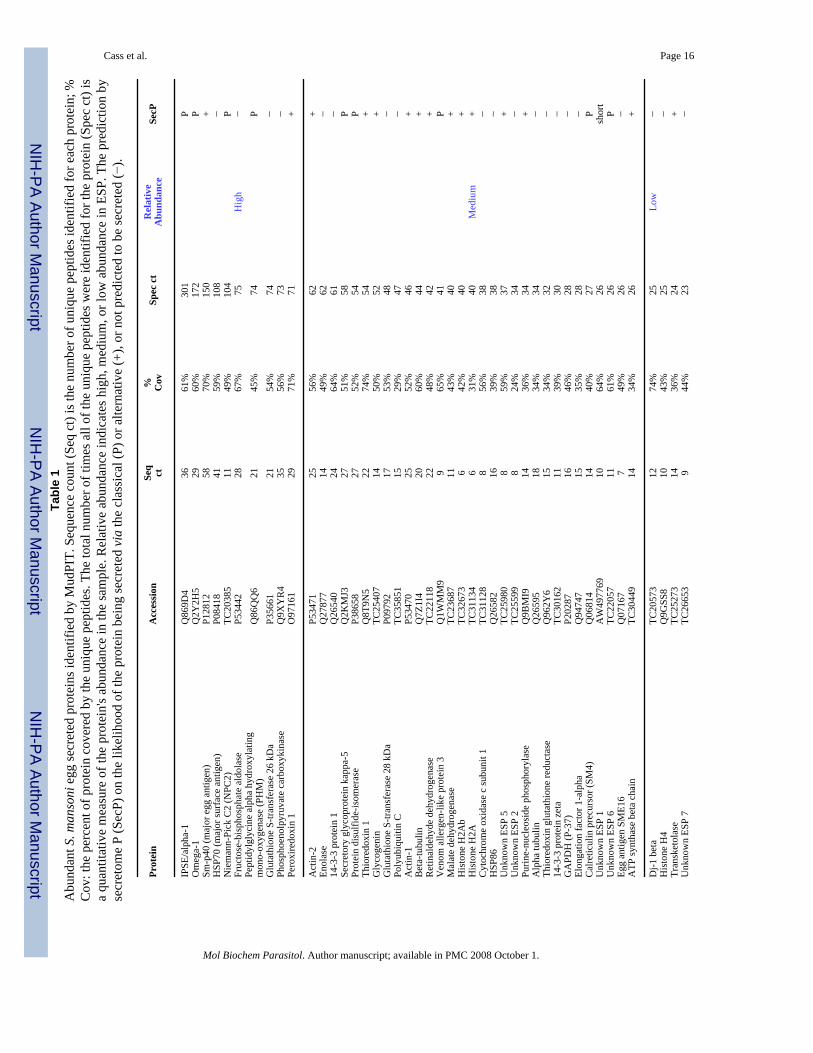
method. The S. mansoni egg secretome was found to be surprisingly complex. Of the 188proteins identified by MudPIT analysis (Supplemental Table 3), 156 proteins had similarity topreviously annotated proteins and 32 proteins were uncharacterized, novel proteins. There isprecedent for using spectrum count to reflect the relative abundance of individual proteins[33-35]. Original data correlating identified peptides to TC sequences are shown inSupplemental Table 4. It should be noted that this approach is not exactly quantitative but ratherprovides a semi-quantitative view of the components represented in a protein mixture. Theadvantage to other approaches is that the identification of components is much morecomprehensive, but the relative abundance of those components can never be exactly quantifiedwithout absolute quantitative measures that are quite low-throughput and costly [36]. Theproteins identified as components of ESP were ranked by descending total spectrum count(Spec ct, Table 1, Supplemental Table 3). This ranking does not represent a true molarconcentration of proteins in ESP, but rather serves to delineate highly abundant proteins fromthose of lesser abundance. The number of unique peptides for each protein (Seq ct) and theminimum peptide coverage (% cov) were also listed (Table 1 and Supplemental Tables 3 and4).
Many abundant egg proteins are known to be heavily glycosylated [17,18,21]. During MudPITanalysis, glycoproteins cleaved by a protease generate glycopeptides that are part of theheterogeneous peptide mix. However, because the mass of glycosylated peptides cannot bepredicted, they are not identified by MudPIT analysis. However, proteins known to be heavilyglycosylated, such as IPSE/alpha-1, omega-1, and kappa-5 [17,18,21], were identifiedsuccessfully by the MudPIT analysis here, via their non-glycosylated peptides. Although it ispossible that every tryptic peptide in a protein could be glycosylated, and therefore the proteinwould not be identified by MudPIT, our results indicate that even heavily glycosylated proteinsare not missed by MudPIT analysis.
In the present study, we noted that a small fraction of the total proteins comprised the bulk ofthe egg secretions. While spectrum count is not an exact molar representation of trueabundance, it can be interpreted to support the notion that abundant proteins will generate ahigher percentage of total peptides analyzed by mass spectral analysis, while less-abundantproteins will contribute far fewer peptides to the analysis, and thus, will have fewer relativespectrum counts. For analysis, we divided the egg secretome into thirds (high, medium, andlow) to represent highly abundant proteins versus those of lesser abundance. The first 10 most-abundant proteins comprised the top third of total protein in ESP (Table 1). Among the mosthighly-abundant proteins identified in egg secretions were IPSE/alpha-1, omega-1, Sm-p40,HSP70, Niemann-Pick C2 (NPC2) and peptidylglycine alpha hydroxylating monooxygenase(PHM) [37]. A mere 31 proteins comprised the middle third of total ESP and the following147 proteins comprised the low-abundance tier (Table 1). Proteins previously identified inESP, namely, IPSE/alpha-1, omega-1, thioredoxin, and peroxiredoxin 1, were all identified ascomponents of ESP by the analysis here. In contrast, high mobility group box-1, a nuclearprotein also previously found in ESP [19], was not identified in this study. The absence of highmobility group box-1 may be explained by the presence of 42 potential trypsin cleavage sitesthat would generate peptides shorter than the peptide length minimum used in this analysis (7residues, see Supplemental Table 4).
In addition to the proteins already mentioned, many other S. mansoni proteins in severalfunctional categories were found in egg secretions: antioxidant proteins (peroxiredoxin 1,thioredoxin, thioredoxin glutathione reductase, GST-26K, GST-28K, GST-omega, superoxidedismutase), heat shock, chaperone and protein folding proteins (HSP70, HSP86/HSP90,polyubiquitin C, protein disulfide isomerase, 14-3-3 proteins), calcium-binding proteins(translationally controlled tumor protein, calpain, 16 kDa calcium-binding protein),inflammation-inducing proteins (IPSE/alpha-1, venom allergen-like proteins 2, 3, 5 and 9),
Cass et al. Page 5
Mol Biochem Parasitol. Author manuscript; available in PMC 2008 October 1.
NIH
-PA Author Manuscript
NIH
-PA Author Manuscript
NIH
-PA Author Manuscript
glycolytic or glycolytic feeder pathway enzymes (fructose bisphosphate aldolase, enolase,phosphoenolpyruvate carboxykinase, GAPDH), and scavenging pathway proteins (NPC2(cholesterol trafficking)).
3.2 Secretome P predictionsSecreted proteins would be expected to have a secretory sequence (either N-terminal orleaderless) and the presence of such sequences would further validate the identification ofindividual proteins in ESP. Protein sequences identified by MudPIT were analyzed for thepresence or absence of secretion signals, including both the classic N-terminal signalingpeptide and the non-classical or alternative internal secretion sequences [38]. Non-classicallysecreted proteins may be secreted by lysosomal or exosomal vesicular fusion, by membranetransporters, or by membrane blebbing [38].
Using the most complete protein sequences available, the presence or absence of either aclassic, N-terminal signal peptide sequence or a non-classical, internal protein secretionsequence was predicted by the SecretomeP v. 2.0 algorithm [30], which assigned a neuralnetwork value to the protein sequences. Any protein sequence receiving an neural networkvalue ≥0.5 was predicted to be secreted by either a classic signal sequence (P), or via a non-classical pathway (+). Protein sequences with a neural network value ≤0.5 were predicted tobe non-secreted (−).
Of the 188 identified proteins in ESP, 118 proteins (63% of total) had an neural network value≥0.5, and thus, were predicted by SecretomeP to be secreted (SecP in Table 1 and SupplementalTable 3); the proteins contained either a classic signal peptide leader sequence (P, 32 proteins,17%) or were predicted to be secreted through a non-classical pathway (+, 86 proteins, 46%).Concomitantly, 67 proteins (35% of total) were predicted by SecretomeP to be not secretedvia either pathway, and 3 proteins (short, 2%) were too short for the SecretomeP algorithm toanalyze.
Because Sm-p40 was the third most-abundant protein in egg secretions contributing 4% oftotal spectrum counts, it was surprising that SecretomeP predicted Sm-p40 to be non-secreted.However, examination of available transcript and genomic databases revealed that Sm-p40 isa gene family. Of note, tentative consensus sequence TC28308 encoded a Sm-p40 familymember encoding peptides identified by MudPIT. When this TC was used for the SecretomePprediction, the neural network value was 0.774 and was therefore reported as (+) for Sm-p40(Table 1, Supplemental Table 3).
One explanation for the number of proteins identified in ESP that were predicted by SecretomePto be not secreted by either the classical or alternative pathway is that the Secretome P algorithmwas trained using mammalian, and not platyhelminth, protein sequences [30], and that thesequences available in the databases potentially encode truncated ORFs, as described abovefor Sm-p40.
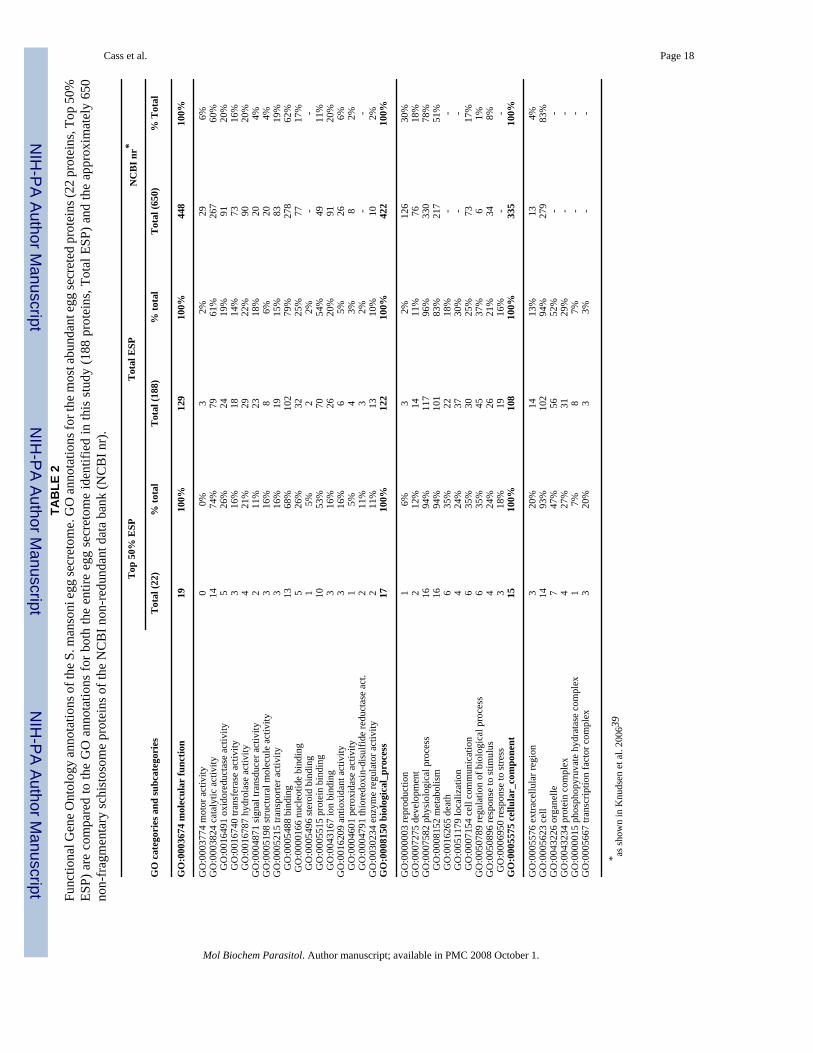
3.3 Gene ontologyGene ontology was assigned to the array of identified proteins to gain a basic understandingof protein function and the biological processes in which they are involved. The gene ontologyof the most-abundant egg secreted proteins (22 proteins comprising 50% of total ESP) wascompared to the ontology of both the entire complement of egg secreted proteins (188 proteins,100% of ESP) and the ontology of annotated Schistosoma mansoni proteins in the NCBI non-redundant database, as analyzed by the GOblet server previously (650 proteins, [39]). Table 2displays the results of GO annotation for the three major categories of molecular function,biological process, and cellular component.
Cass et al. Page 6
Mol Biochem Parasitol. Author manuscript; available in PMC 2008 October 1.
NIH
-PA Author Manuscript
NIH
-PA Author Manuscript
NIH
-PA Author Manuscript
Molecular function was assigned to 129 proteins. Fully 79% (102 proteins) had bindingcapacity (GO:0005488) including ion binding (GO:0043167), nucleic acid binding (GO:0000166), and protein binding (GO:0005515). Catalytic activity (GO:0003824) was identifiedfor 61% of the proteins with hydrolase (GO:0016787), oxidoreductase (GO:0016491), andtransferase (GO:0016740) activities specifically enriched. Similarly, 122 proteins wereinvolved in annotated biological processes. Of these, 96% were involved in physiologicalprocesses (GO:0007582) such as cell death (GO:0016265), homeostasis (GO:0019725),metabolism (GO:0044237), and cell communication (GO:0007154). Finally, 108 proteinsassigned to cellular component categories (GO: 0005575) were frequently localized tocytoplasmic organelles (GO:0043226) and protein complexes (GO:0043234).
3.4 RT-PCR resultsMudPIT identified 36 unknown, previously uncharacterized proteins in egg secretions(unknown ESP 1-36, Table 1 and Supplemental Table 3). The developmental expressionpattern of a subset of some of the more abundant unknown ESP proteins (Unknown ESP 2-5)was determined by RT-PCR. The results in Figure 1 show that the expression of unknownsESP 2-4 were specific to egg, while unknown ESP 5 was expressed not only in egg, but alsoin all stages tested except for adult females. The expression pattern of unknown ESP 5 wasinteresting; expression was seen to be highest during stages adapting to new environments(Figure 1). Although unknown ESP 5 was expressed at very low levels in cercariae, itsexpression was rapidly induced after transformation to schistosomula. However, within 24hour (24 hr) the expression of unknown ESP 5 was greatly reduced and remained so until 10days (10 d) after transformation, at which point its expression increased and remained sothrough juvenile liver stage (23 d) until again being reduced in adult worms. Thereforeunknown ESP 5 expression reflects a pattern in which maximum expression occurs as parasitesadapt to the skin, lungs and liver of the host.
3.5 Circumoval precipitin reaction resultsThe COP reaction was first described as a serodiagnostic tool for schistosome infection [40].In the COP reaction, proteins exit the egg through pores in the eggshell, interact with hostantibodies, and form a precipitate at the point of secretion [41]. We confirmed the MudPITdata by localizing some of the more abundant egg secreted proteins on the surface of live eggsby both direct and indirect immunofluorescent COP reactions (Figure 2 and SupplementalFigure 1). Figure 2 shows the COP reactions obtained using antibodies against HSP70 and Sm-p40, visualized by fluorochrome labeled secondary antibodies. Although it is not possible touse the COP reaction to confirm the secretion of every protein identified in this study, thelocalization to the surface of live eggs in a COP reaction of a subset of proteins identified inESP supports the presence in ESP of other proteins identified in this study.
Additional proteins (thioredoxin, ubiquitin, PHM, tubulin, albumin, thioredoxin glutathionereductase, and GST(26K), all identified by MudPIT to be present in egg secretions, also werelocalized to the surface of live eggs via the COP reaction and are shown in Supplemental Figure1. As an additional control, ribosomal subunit S6, which was not identified in ESP, also wasnot co-localized in a COP reaction indicating that the presence of antibodies alone during thein vitro culture of eggs does not produce a COP reaction (Supplemental Figure 1).
3.6 Functions of proteins found in ESPThe 188 egg secreted proteins fall into some general categories including energy production,cell signaling, and maintenance of redox balance (thioredoxin, peroxiredoxins, albumin,GST-26K, GST-28K, GST-omega). Previously identified egg-specific glycoproteins (IPSE/alpha-1, omega-1, kappa-5) were positively identified as components of ESP, and yet some ofthe most abundant proteins are still relatively uncharacterized. A comparison of our proteomic
Cass et al. Page 7
Mol Biochem Parasitol. Author manuscript; available in PMC 2008 October 1.
NIH
-PA Author Manuscript
NIH
-PA Author Manuscript
NIH
-PA Author Manuscript
identification of egg secreted proteins and the results of both peptide mass fingerprinting ofSEA [42] and cercarial secretions [39] revealed that many proteins were identified in all threestudies: 14-3-3 proteins, actin, enolase, GAPDH, HSP70, thioredoxin, and GST −26K and−28K. However, in this study we also identified protein components not previously identifiedin parasitic secretions including NPC2 and venom allergen-like proteins.
One of the most abundant protein in ESP was Sm-p40. Sm-p40 shows similarity to α-crystallin,which can function as a molecular chaperone, prevent protein aggregation, and facilitatesprotein refolding [43]. Based upon Sm-p40's similarity to small heat shock proteins and α-crystallin, it is intriguing to speculate that this extremely abundant secreted egg protein mayfunction as a molecular chaperone during protein folding or secretion, or may protect the eggin some capacity within the oxidatively stressful granuloma environment.
Similarly, heat shock proteins, including HSP86/90, -70, -60, -40, and -10, were found to bepresent in ESP. HSPs are typically immunodominant antigens and major targets of host immuneresponses, including autoimmunity [44]. As a group, heat shock proteins perform manydifferent tasks within the same cellular compartments, thus, regulation of access to substratesis essential and relies upon the presence of co-chaperones to facilitate targeting to specificsubstrates and functions. Co-chaperones include J-domain proteins, such as Dj-1 beta(TC20573), also identified as a component of ESP in this study.
NPC2, a ubiquitous, essential protein involved in cholesterol trafficking, was also identifiedas one of the most-abundant protein in ESP; this is the first time NPC2 has been identified inSchistosoma. NPC2 is a small intralysosomal cholesterol-binding protein involved in the egressof cholesterol out of endosomal/lysosomal compartments [45,46]. Although NPC2, along withNPC1, is essential for proper cellular cholesterol trafficking, its precise function has remainedelusive [45]. The S. mansoni and human NPC2 proteins shared 37% identity and 55%similarity, and Phe-66, Val-96, and Tyr-100, which have been found to be essential forcholesterol binding [47], were conserved in the S. mansoni protein, as was the predictedsecondary structures of the proteins (Figure 3). Glycosylation has been shown to be essentialfor human NPC2 function [48] and a conserved N-glycosylation site was predicted in the S.mansoni NPC2 (Figure 3). Furthermore, molecular modeling of S. mansoni NPC2 generateda predicted structure similar to human NPC2 [49] that included conservation of the order ofβ-sheets, Cys-Cys bonds, and localization of the residues implicated in cholesterol binding toa hydrophobic pocket in the protein (Figure 3) suggesting a similar cholesterol-binding functionfor the S. mansoni NPC2 protein. One possible explanation for the presence of NPC2 in ESPis to facilitate the movement of cholesterol (which eggs must scavenge). However, NPC2 mayalso facilitate the egress of other glycolipids [50]. Moreover, parasite glycoproteins circulatingon host low-density lipoprotein particles have been shown to disrupt lipid homeostasis in hostimmune cells and induce cellular apoptosis [51] and parasite glycolipids may influencepathogenesis and immunity to infection [52]. NPC2 may participate in this exchange ofparasite-derived immunomodulatory molecules.
Several glycolytic enzymes, including enolase, were identified in ESP. Enolase, plays a keyrole in glycolysis and gluconeogenesis; however, it is also a multifunctional, non-classicallysecreted protein found on the surface of hematopoetic, epithelial, and endothelial cells, whereit serves as not only a plasminogen receptor (plasminogen possesses fibrinolyticcharacteristics) and is implicated in tissue invasion, but as a heat-shock protein, a hypoxic stressprotein, and a cytoskeletal and chromatin binding protein [53]. Enolase has been implicated ina wide variety of autoimmune diseases, including lupus erythematosus, ulcerative colitis,Crohn's disease, autoimmune hepatitis, apoptosis, and endometriosis [53]. The intriguingsimilarity between enolase, T-crystallins (monomers), and Sm-p40, which is similar to alphacrystallin, remains to be elucidated. Another glycolytic enzyme identified in ESP was GAPDH.
Cass et al. Page 8
Mol Biochem Parasitol. Author manuscript; available in PMC 2008 October 1.
NIH
-PA Author Manuscript
NIH
-PA Author Manuscript
NIH
-PA Author Manuscript
GAPDH has been shown to bind fibronectin and laminin [54] and may be involved in mediatingthe attachment of the egg to host tissues, or somehow aid in the passage of live eggs acrosshost tissues into the external environment.
Some of the proteins secreted by eggs may have pleiotropic effects upon the surroundinggranuloma cells or the host's immunoflammatory response. For example, many neuropeptidetransmitters require amidation for biological activity and PHM was identified in egg secretions[37]. Several 14-3-3 proteins, which act as molecular scaffolds or chaperones and may haveisoform specific functions during development [55], were present in egg secretions. Andfinally, the presence of polyubiquitin C in egg secretions is intriguing in that ubiquination ofproteins has diverse functions in a myriad of cellular processes such as protein stability andintracellular localization. Several dynamic classes of proteins involved in cell communication(connexins) and adhesion (cadherin/catenin complexes) are regulated by ubiquination [56].What effects these egg secreted proteins have upon the host remains to be determined.
In conclusion, we used MudPIT to identify the S. mansoni egg secretome, a complex array of188 proteins with an intriguing array of molecular functions. Additionally, 32 novel ESPproteins were identified. Since egg secreted proteins profoundly influence the Th1/Th2-cytokine environment and serve as the focus of the host immunoinflammatory response, theidentification of proteins secreted by live eggs is of fundamental interest. Now that acomprehensive list of egg secreted proteins is available, future research may elucidate theirspecific role in the immunobiology and pathogenesis of schistosomiasis.
Supplementary MaterialRefer to Web version on PubMed Central for supplementary material.
Acknowledgements
The authors thank Dr. Tim Day for the gift of anti-PHM antiserum and Dr. Philip LoVerde for the infected hamsterliver. This work was funded by NIH/NIAID grant R03AI059037. Schistosome materials for this work were suppliedin part through NIH-NIAID contract N01-AI-55270.
AbbreviationsCOP, circumoval precipitin; ESP, egg secreted proteins; GST, glutathione-S-transferase; HSP,heat shock protein; IPSE, IL-4-inducing principle of S. mansoni eggs; MudPIT,multidimensional protein identification technique; NPC2, Niemann-Pick C2; PHM,peptidylglycine alpha-hydroxylating monooxygenase; SEA, soluble egg antigens; Sm-p40, S.mansoni egg protein 40.
References1. Hotez PJ, Molyneux DH, Fenwick A, Ottesen E, Ehrlich Sachs S, et al. Incorporating a rapid-impact
package for neglected tropical diseases with programs for HIV/AIDS, tuberculosis, and malaria. PLoSMed 2006;3:e102. [PubMed: 16435908]
2. King CH, Dickman K, Tisch DJ. Reassessment of the cost of chronic helminthic infection: a meta-analysis of disability-related outcomes in endemic schistosomiasis. Lancet 2005;365:1561–9.[PubMed: 15866310]
3. Abath FGC, Morais CNL, Montenegro CEL, Wynn TA, Montenegro SML. Immunopathogenicmechanisms in schistosomiasis: what can be learnt from human studies? Trends Parasitol 2006;22:86–91.
4. Hernandez HJ, Wang Y, Tzellas N, Stadecker MJ. Expression of class II, but not class I, majorhistocompatibility complex molecules is required for granuloma formation in infection withSchistosoma mansoni. Eur J Immunol 1997;27:1170–6. [PubMed: 9174607]
Cass et al. Page 9
Mol Biochem Parasitol. Author manuscript; available in PMC 2008 October 1.
NIH
-PA Author Manuscript
NIH
-PA Author Manuscript
NIH
-PA Author Manuscript
5. Pearce EJ, MacDonald AS. The immunobiology of schistosomiasis. Nat Rev Immunol 2002;2:499–511. [PubMed: 12094224]
6. Gause WC, Urban JF Jr, Stadecker MJ. The immune response to parasitic helminths: insights frommurine models. Trends Immunol 2003;24:269–77. [PubMed: 12738422]
7. Pearce E, Caspar P, Grzych J, Lewis F, Sher A. Downregulation of TH1 cytokine productionaccompanies induction of TH2 responses by a parasitic helminth, Schistosoma mansoni. J Exp Med1991;173:159–66. [PubMed: 1824635]
8. Grzych JM, Pearce E, Cheever A, Caulada ZA, Caspar P, et al. Egg deposition is the major stimulusfor the production of Th2 cytokines in murine schistosomiasis mansoni. J Immunol 1991;146:1322–7. [PubMed: 1825109]
9. Boros DL. Immunopathology of Schistosoma mansoni infection. Clin Microbiol Rev 1989;2:250–69.[PubMed: 2504481]
10. Dunne DW, Doenhoff MJ. Schistosoma mansoni egg antigens and hepatocyte damage in infected T-cell-deprived mice. Contrib Microbiol Immunol 1983;7:22–9. [PubMed: 6600671]
11. Loeffler DA, Lundy SK, Singh KP, Gerard HC, Hudson AP, Boros DL. Soluble egg antigens fromSchistosoma mansoni induce angiogenesis-related processes by up-regulating vascular endothelialgrowth factor in human endothelial cells. J Inf Dis 2002;185:1650–6. [PubMed: 12023772]
12. Doenhoff MJ. A role for granulomatous inflammation in the transmission of infections disease:schistosomiasis and tuberculosis. Parasitol 1997;115:113–25.
13. Karanja DM, Colley DG, Nahlan BL, Ouma JH, Secor WE. Studies of schistosomiasis in westernKenya. I. Evidence for immune-facilitated excretion of schistosome eggs from patients withSchistosoma mansoni and human immunodeficiency virus coinfections. Am J Trop Med Hyg1997;56:515–21. [PubMed: 9180601]
14. Stadecker MJ, Hernandez HJ, Asahi H. The identification and characterization of new immunogenicegg components: implications for evaluation and control of the immunopathogenic T cell responsein Schistosomiasis. Mem Inst Osw Cruz 2001;96:29–34.
15. Nene V, Dunne DW, Johnson KS, Taylor DW, Cordingley JS. Sequence and expression of a majoregg antigen from Schistosoma mansoni. Homologies to heat shock proteins and alpha-crystallins.Mol Biochem Parasitol 1986;21:179–88. [PubMed: 3097539]
16. Asahi H, Stadecker MJ. Analysis of egg antigens inducing hepatic lesions in schistosome infection.Parasitol Int 2003;52:361–7. [PubMed: 14665394]
17. Dunne DW, Jones FM, Doenhoff MJ. The purification, characterization serological activity andhepatotoxic properties of two cationic glycoproteins (α1 and ω1) from Schistosoma mansoni eggs.Parasitol 1991;103:225–36.
18. Schramm G, Gronow A, Knobloch J, Wippersteg V, Grevelding CG, et al. IPSE/alpha-1: a majorimmunogenic component secreted from Schistosoma mansoni eggs. Mol Biochem Parasitol2006;147:9–19. [PubMed: 16480783]
19. Gnanasekar M, Velusamy R, He Y-X, Ramaswamy K. Cloning and characterization of a high mobilitygroup box 1 (HMGB1) homologue protein from Schistosoma mansoni. Mol Biochem Parasitol2006;145:137–46. [PubMed: 16246438]
20. Dunne DW, Agnew AM, Modha J, Doenhoff MJ. Schistosoma mansoni egg antigens: preparation ofrabbit antisera with monospecific immunoprecipitating activity, and their use in antigencharacterization. Parasite Immunol 1986;8:575–86. [PubMed: 3101031]
21. Fitzsimmons CM, Schramm G, Jones FM, Chalmers IW, Hoffman KF, et al. Molecularcharacterization of omega-1: a hepatotoxic ribonuclease from Schistosoma mansoni eggs. MolBiochem Parasitol 2005;144:123–27. [PubMed: 16143411]
22. Alger HM, Sayed AA, Stadecker MJ, Williams DL. Molecular and enzymatic characterization ofSchistosoma mansoni thioredoxin. Int J Parasitol 2002;32:1285–92. [PubMed: 12204228]
23. Washburn MP, Wolters D, Yates JR 3rd. Large-scale analysis of the yeast proteome bymultidimensional protein identification technology. Nat Biotechnol 2001;19:242–7. [PubMed:11231557]
24. Eng JK, McCormack AL, Yates JR. An approach to correlate tandem mass spectral data of peptideswith amino acid sequences in a protein database. J Am Soc Mass Spectrom 1994;5:976–89.
Cass et al. Page 10
Mol Biochem Parasitol. Author manuscript; available in PMC 2008 October 1.
NIH
-PA Author Manuscript
NIH
-PA Author Manuscript
NIH
-PA Author Manuscript
25. Tabb DL, McDonald WH, Yates JR 3rd. DTASelect and Contrast: tools for assembling and comparingprotein identifications from shotgun proteomics. J Proteome Res 2002;1:21–6. [PubMed: 12643522]
26. Peng J, Elias JE, Thoreen CC, Licklider LJ, Gygi SP. Evaluation of multidimensional chromatographycoupled with tandem mass spectrometry (LC/LC-MS/MS) for large-scale protein analysis: the yeastproteome. J Proteome Res 2003;2:43–50. [PubMed: 12643542]
27. Link AJ, Eng J, Schieltz DM, Carmack E, Mize GJ, et al. Direct analysis of protein complexes usingmass spectrometry. Nature Biotechnol 1999;17:676–82. [PubMed: 10404161]
28. Groth D, Lehrach H, Hennig S. GOblet: a platform for Gene Ontology annotation of anonymoussequence data. Nuc Acids Res 2004;32:313–17.
29. Hennig S, Groth D, Lehrach H. Automated Gene Ontology annotation for anonymous sequence data.Nuc Acids Res 2003;31:3712–15.
30. Bendtsen JD, Jensen LJ, Blom N, von Heijne G, Brunak S. Feature-based prediction of non-classicaland leaderless protein secretion. Protein Eng Design & Selection 2004;17:49–356.
31. Sayed AA, Cook SK, Williams DL. Redox balance mechanisms in Schistosoma mansoni rely onperoxiredoxins and albumin and implicate peroxiredoxins as novel drug targets. J Biol Chem2006;281:17001–10. [PubMed: 16606626]
32. Vermund SH, Bradley DJ, Ruiz-Tiben E. Survival of Schistosoma mansoni in the human host:estimates from a community-based prospective study in Puerto Rico. Am J Trop Med Hyg1983;32:1040–8. [PubMed: 6625059]
33. Liu H, Sadygov RG, Yates JR 3rd. A model for random sampling and estimation of relative proteinabundance in shotgun proteomics. Anal Chem 2004;76:4193–201. [PubMed: 15253663]
34. Gao J, Friedrichs MS, Dongre AR, Opiteck GJ. Guidelines for the routine application of the peptidehits technique. J Am Soc Mass Spectrom 2005;16:1231–8. [PubMed: 15978832]
35. Zybailov B, Coleman MK, Florens L, Washburn MP. Correlation of relative abundance ratios derivedfrom peptide ion chromatograms and spectrum counting for quantitative proteomic analysis usingstable isotope labeling. Anal Chem 2005;77:6218–24. [PubMed: 16194081]
36. Gerber SA, Rush J, Stemman O, Kirschner MW, Gygi SP. Absolute quantification of proteins andphosphoproteins from cell lysates by tandem MS. PNAS 2003;100:6940–5. [PubMed: 12771378]
37. Mair GR, Niciu MJ, Stewart MT, Brennan G, Omar H, et al. A functionally atypical amidating enzymefrom the human parasite Schistosoma mansoni. FASEB J 2004;1:114–21. [PubMed: 14718392]
38. Nickel W. Unconventional secretory routes: direct protein export across the plasma membrane ofmammalian cells. Traffic 2005;6:607–14. [PubMed: 15998317]
39. Knudsen GM, Medzihradszky KF, Lim K-C, Hansell E, McKerrow JH. Proteomic analysis ofSchistosoma mansoni cercarial secretions. Mol Cell Proteomics 2005;4:1862–75. [PubMed:16112986]
40. Oliver-González J. Anti-egg precipitins in the serum of humans infected with Schistosomamansoni. J Inf Dis 1954;95:86–91. [PubMed: 13184169]
41. Demaree RS, Hillyer GV. Schistosoma species: transmission electron microscopy of the circumovalimmune precipitin reaction on eggs. Exp Parasitol 1981;52:77–85. [PubMed: 7195345]
42. Curwen RS, Ashton PD, Johnston DA, Wilson RA. The Schistosoma mansoni soluble proteome: acomparison across four life-cycle stages. Mol Biochem Parasitol 2004;138:57–66. [PubMed:15500916]
43. Horwitz J. α-Crystallin can function as a molecular chaperone. Proc Natl Acad Sci USA1992;89:10449–53. [PubMed: 1438232]
44. Mayer MP, Bukau B. Hsp70 chaperones: cellular functions and molecular mechanism. Cell & MolLife Sci 2005;62:670–84. [PubMed: 15770419]
45. Vanier MT, Millat G. Structure and function of the NPC2 protein. Biochim Biophys Acta2004;1685:14–21. [PubMed: 15465422]
46. Cheruku SR, Xu Z, Dutia R, Lobel P, Storch J. Mechanism of cholesterol transfer from the Niemann-Pick type C2 protein to model membrane supports a role in lysosomal cholesterol transport. J BiolChem 2006;281:31594–604. [PubMed: 16606609]
Cass et al. Page 11
Mol Biochem Parasitol. Author manuscript; available in PMC 2008 October 1.
NIH
-PA Author Manuscript
NIH
-PA Author Manuscript
NIH
-PA Author Manuscript
47. Ko DC, Binkley J, Sidow A, Scott MP. The integrity of a cholesterol-binding pocket in Niemann-Pick C2 protein is necessary to control lysosome cholesterol levels. Proc Natl Acad Sci USA2003;100:2518–25. [PubMed: 12591949]
48. Chikh K, Vey S, Simonot C, Vanier MT, Millat G. Niemann-Pick type C disease:importance of N-glycosylation sites for function and cellular location of the NPC2 protein. Mol Genet Metab2004;83:220–30. [PubMed: 15542393]
49. Friedland N, Liou H-L, Lobel P, Stock AM. Structure of a cholesterol-binding protein deficient inNiemann-Pick type C2 disease. Proc Nat Acad Sci USA 2003;100:2512–7. [PubMed: 12591954]
50. Zhang M, Sun M, Dwyer NK, Comly ME, Patel SC, et al. Differential trafficking of the Niemann-Pick C1 and 2 proteins highlights distinct roles in late endocytic lipid trafficking. Acta Paediatr Suppl2003;92:63–73. [PubMed: 14989468]
51. Sprong H, Suchanek M, van Dijk SM, van Remoortere A, Klumperman J, et al. Aberrant receptor-mediated endocytosis of Schistosoma mansoni glycoproteins on host lipoproteins. PLoS Med2006;3:1360–70.
52. van der Kleij D, van den Biggelsar AH, Kruize YC, Retra K, Fillie Y, et al. Responses to Toll-likereceptor ligands in children living in areas where schistosome infections are endemic. J Infec Dis2004;15:1044–51. [PubMed: 14999608]
53. Pancholi V. Multifunctional α-enolase: its role in diseases. Cell Mol Life Sci 2001;58:902–20.[PubMed: 11497239]
54. Gozalbo D, Gil-Navarro I, Azorín I, Renau-Piqueras J, Martinez JP, et al. The cell wall-associatedglyceraldehyde-3-phosphate dehydrogenase of Candida albicans is also a fibronectin and lamininbinding protein. Infect Immun 1998;66:2052–9. [PubMed: 9573088]
55. McGonigle S, Soschiavo M, Pearce E. 14-3-3 proteins in Schistosoma mansoni; identification of asecond epsilon isoform. Int J Parasitol 2002;32:685–93. [PubMed: 12062487]
56. Berthoud VM, Minogue PJ, Laing JG, Beyer EC. Pathways for degradation of connexins and gapjunctions. Cardiovas Res 2004;62:256–67.
57. Thompson JD, Higgins DG, Gibson TJ. CLUSTAL W: improving the sensitivity of progressivemultiple sequence alignment through sequence weighting, position-specific gap penalties and weightmatrix choice. Nuc Acids Res 1994;22:4673–80.
58. McGuffin LJ, Bryson K, Jones DT. The PSIPRED protein structure prediction server. Bioinformatics2000;16:404–5. [PubMed: 10869041]
59. Guex N, Peitsch MC. SWISS-MODEL and the Swiss-PdbViewer: An environment for comparativeprotein modeling. Electrophoresis 1997;18:2714–23. [PubMed: 9504803]
60. Alger HM, Williams DL. The disulfide redox system of Schistosoma mansoni and the importance ofa multifunctional enzyme, thioredoxin glutathione reductase. Mol Biochem Parasitol 2002;121:129–39. [PubMed: 11985869]
Cass et al. Page 12
Mol Biochem Parasitol. Author manuscript; available in PMC 2008 October 1.
NIH
-PA Author Manuscript
NIH
-PA Author Manuscript
NIH
-PA Author Manuscript
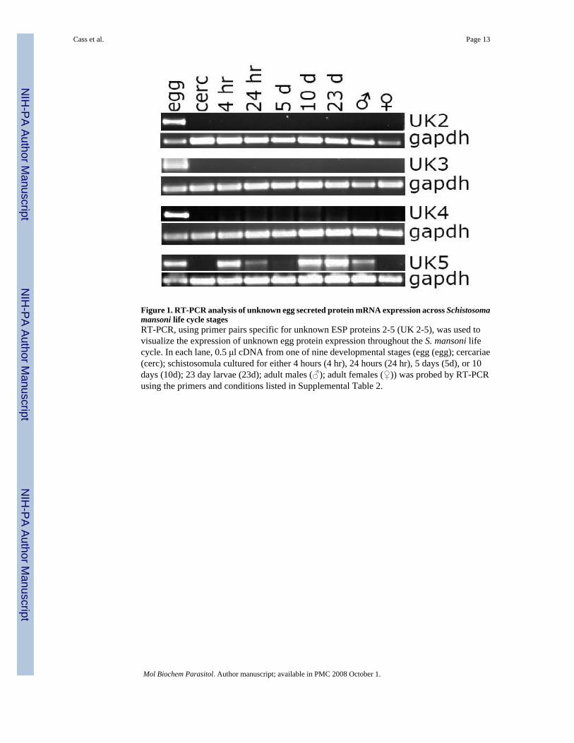
Figure 1. RT-PCR analysis of unknown egg secreted protein mRNA expression across Schistosomamansoni life cycle stagesRT-PCR, using primer pairs specific for unknown ESP proteins 2-5 (UK 2-5), was used tovisualize the expression of unknown egg protein expression throughout the S. mansoni lifecycle. In each lane, 0.5 μl cDNA from one of nine developmental stages (egg (egg); cercariae(cerc); schistosomula cultured for either 4 hours (4 hr), 24 hours (24 hr), 5 days (5d), or 10days (10d); 23 day larvae (23d); adult males (♂); adult females (♀)) was probed by RT-PCRusing the primers and conditions listed in Supplemental Table 2.
Cass et al. Page 13
Mol Biochem Parasitol. Author manuscript; available in PMC 2008 October 1.
NIH
-PA Author Manuscript
NIH
-PA Author Manuscript
NIH
-PA Author Manuscript

Figure 2. Egg secreted proteins can be visualized in circumoval precipitin reactionsPanel (A) shows eggs cultured with rabbit polyclonal antibodies against HSP70 visualized withanti-rabbit IgG-Alexa Fluor 555-labeled secondary antibody, or with secondary antibody alone(goat anti rabbit-AF555). Panel (B) shows eggs cultured with either a mixture of equal amountsof several purified monoclonal antibodies against Sm-p40 visualized by anti-mouse IgG-AlexaFluor 555-labeled secondary antibody, and the secondary antibody alone (goat anti mouse-AF555). Exposure times are: bright field (1/30 sec), immunofluorescence (1 sec). Bar equals30 μm.
Cass et al. Page 14
Mol Biochem Parasitol. Author manuscript; available in PMC 2008 October 1.
NIH
-PA Author Manuscript
NIH
-PA Author Manuscript
NIH
-PA Author Manuscript
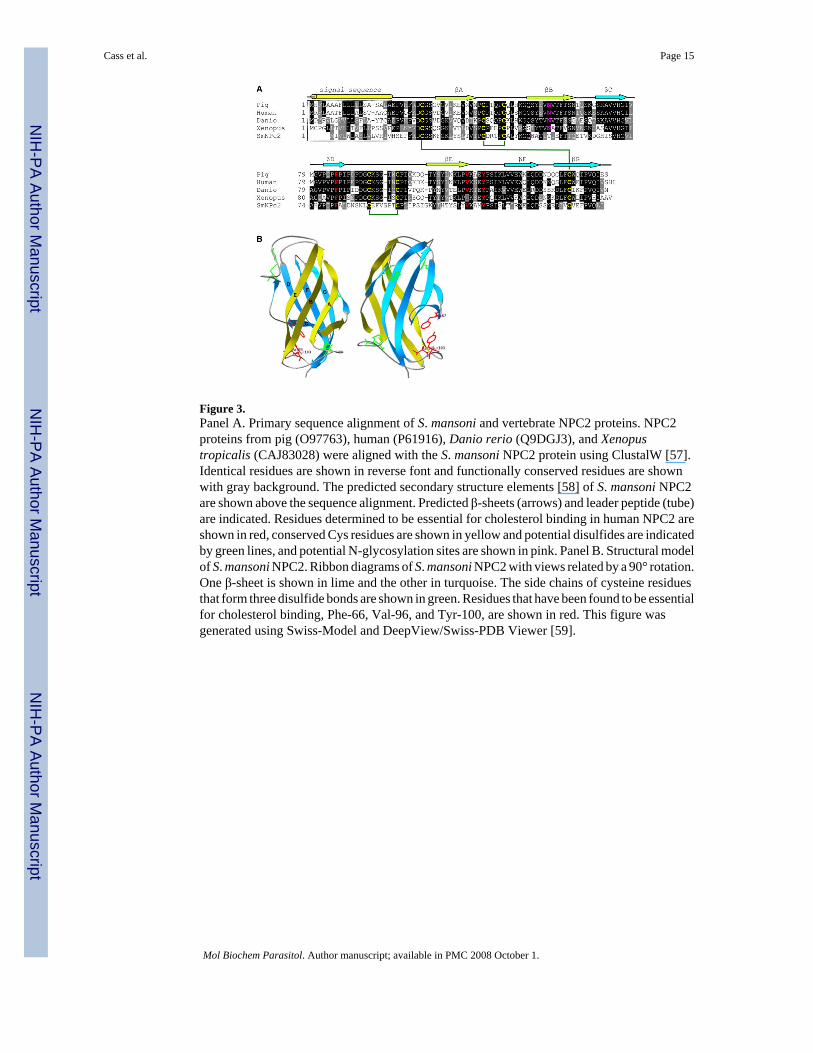
Figure 3.Panel A. Primary sequence alignment of S. mansoni and vertebrate NPC2 proteins. NPC2proteins from pig (O97763), human (P61916), Danio rerio (Q9DGJ3), and Xenopustropicalis (CAJ83028) were aligned with the S. mansoni NPC2 protein using ClustalW [57].Identical residues are shown in reverse font and functionally conserved residues are shownwith gray background. The predicted secondary structure elements [58] of S. mansoni NPC2are shown above the sequence alignment. Predicted β-sheets (arrows) and leader peptide (tube)are indicated. Residues determined to be essential for cholesterol binding in human NPC2 areshown in red, conserved Cys residues are shown in yellow and potential disulfides are indicatedby green lines, and potential N-glycosylation sites are shown in pink. Panel B. Structural modelof S. mansoni NPC2. Ribbon diagrams of S. mansoni NPC2 with views related by a 90° rotation.One β-sheet is shown in lime and the other in turquoise. The side chains of cysteine residuesthat form three disulfide bonds are shown in green. Residues that have been found to be essentialfor cholesterol binding, Phe-66, Val-96, and Tyr-100, are shown in red. This figure wasgenerated using Swiss-Model and DeepView/Swiss-PDB Viewer [59].
Cass et al. Page 15
Mol Biochem Parasitol. Author manuscript; available in PMC 2008 October 1.
NIH
-PA Author Manuscript
NIH
-PA Author Manuscript
NIH
-PA Author Manuscript
NIH
-PA Author Manuscript
NIH
-PA Author Manuscript
NIH
-PA Author Manuscript
Cass et al. Page 16Ta
ble
1A
bund
ant S
. man
soni
egg
secr
eted
pro
tein
s ide
ntifi
ed b
y M
udPI
T. S
eque
nce
coun
t (Se
q ct
) is t
he n
umbe
r of u
niqu
e pe
ptid
es id
entif
ied
for e
ach
prot
ein;
%C
ov: t
he p
erce
nt o
f pro
tein
cov
ered
by
the
uniq
ue p
eptid
es. T
he to
tal n
umbe
r of t
imes
all
of th
e un
ique
pep
tides
wer
e id
entif
ied
for t
he p
rote
in (S
pec
ct) i
sa
quan
titat
ive
mea
sure
of t
he p
rote
in's
abun
danc
e in
the
sam
ple.
Rel
ativ
e ab
unda
nce
indi
cate
s hig
h, m
ediu
m, o
r low
abu
ndan
ce in
ESP
. The
pre
dict
ion
byse
cret
ome
P (S
ecP)
on
the
likel
ihoo
d of
the
prot
ein
bein
g se
cret
ed v
ia th
e cl
assi
cal (
P) o
r alte
rnat
ive
(+),
or n
ot p
redi
cted
to b
e se
cret
ed (−
).
Prot
ein
Acc
essi
onSe
q ct% Cov
Spec
ct
Rel
ativ
eA
bund
ance
SecP
IPSE
/alp
ha-1
Q86
9D4
3661
%30
1P
Om
ega-
1Q
2Y2H
529
60%
172
PSm
-p40
(maj
or e
gg a
ntig
en)
P128
1258
70%
150
+H
SP70
(maj
or su
rfac
e an
tigen
)P0
8418
4159
%10
8−
Nie
man
n-Pi
ck C
2 (N
PC2)
TC20
385
1149
%10
4P
Fruc
tose
-bis
phos
phat
e al
dola
seP5
3442
2867
%75
Hig
h−
Pept
idyl
glyc
ine
alph
a hy
drox
ylat
ing
mon
o-ox
ygen
ase
(PH
M)
Q86
621
45%
74P
Glu
tath
ione
S-tr
ansf
eras
e 26
kD
aP3
5661
2154
%74
−Ph
osph
oeno
lpyr
uvat
e ca
rbox
ykin
ase
Q9X
YR
435
56%
73−
Pero
xire
doxi
n 1
O97
161
2971
%71
+
Act
in-2
P534
7125
56%
62+
Enol
ase
Q27
877
1449
%62
−14
-3-3
pro
tein
1Q
2654
024
64%
61−
Secr
etor
y gl
ycop
rote
in k
appa
-5Q
2KM
J327
51%
58P
Prot
ein
disu
lfide
-isom
eras
eP3
8658
2752
%54
PTh
iore
doxi
n 1
Q8T
9N5
2274
%54
+G
lyco
geni
nTC
2540
714
50%
52+
Glu
tath
ione
S-tr
ansf
eras
e 28
kD
aP0
9792
1753
%48
−Po
lyub
iqui
tin C
TC35
851
1529
%47
−A
ctin
-1P5
3470
2552
%46
+B
eta-
tubu
linQ
7Z1I
420
60%
44+
Ret
inal
dehy
de d
ehyd
roge
nase
TC22
118
2248
%42
+V
enom
alle
rgen
-like
pro
tein
3Q
1WM
M9
965
%41
PM
alat
e de
hydr
ogen
ase
TC23
687
1143
%40
+H
isto
ne H
2Ab
TC32
673
642
%40
+H
isto
ne H
2ATC
3113
46
31%
40M
ediu
m+
Cyt
ochr
ome
oxid
ase
c su
buni
t 1TC
3112
88
56%
38−
HSP
86Q
2658
216
39%
38−
Unk
now
n ES
P 5
TC25
980
859
%37
+U
nkno
wn
ESP
2TC
2559
98
24%
34−
Purin
e-nu
cleo
side
pho
spho
ryla
seQ
9BM
I914
36%
34+
Alp
ha tu
bulin
Q26
595
1834
%34
−Th
iore
doxi
n gl
utat
hion
e re
duct
ase
Q96
2Y6
1534
%32
−14
-3-3
pro
tein
zet
aTC
3016
211
39%
30−
GA
PDH
(P-3
7)P2
0287
1646
%28
−El
onga
tion
fact
or 1
-alp
haQ
9474
715
35%
28−
Cal
retic
ulin
pre
curs
or (S
M4)
Q06
814
1440
%27
PU
nkno
wn
ESP
1A
W49
7769
1064
%26
shor
tU
nkno
wn
ESP
6TC
2205
711
61%
26P
Egg
antig
en S
ME1
6Q
0716
77
49%
26−
ATP
synt
hase
bet
a ch
ain
TC30
449
1434
%26
+
Dj-1
bet
aTC
2057
312
74%
25Lo
w−
His
tone
H4
Q9G
SS8
1043
%25
−Tr
ansk
etol
ase
TC25
273
1436
%24
+U
nkno
wn
ESP
7TC
2665
39
44%
23−
Mol Biochem Parasitol. Author manuscript; available in PMC 2008 October 1.
NIH
-PA Author Manuscript
NIH
-PA Author Manuscript
NIH
-PA Author Manuscript
Cass et al. Page 17
Prot
ein
Acc
essi
onSe
q ct% Cov
Spec
ct
Rel
ativ
eA
bund
ance
SecP
Phos
phog
lyce
rate
mut
ase
Q8W
T66
632
%23
−Ph
osph
ogly
cera
te k
inas
eP4
1759
1129
%23
−
Mol Biochem Parasitol. Author manuscript; available in PMC 2008 October 1.
NIH
-PA Author Manuscript
NIH
-PA Author Manuscript
NIH
-PA Author Manuscript
Cass et al. Page 18TA
BLE
2Fu
nctio
nal G
ene
Ont
olog
y an
nota
tions
of t
he S
. man
soni
egg
secr
etom
e. G
O a
nnot
atio
ns fo
r the
mos
t abu
ndan
t egg
secr
eted
pro
tein
s (22
pro
tein
s, To
p 50
%ES
P) a
re c
ompa
red
to th
e G
O a
nnot
atio
ns fo
r bot
h th
e en
tire
egg
secr
etom
e id
entif
ied
in th
is s
tudy
(188
pro
tein
s, To
tal E
SP) a
nd th
e ap
prox
imat
ely
650
non-
frag
men
tary
schi
stos
ome
prot
eins
of t
he N
CB
I non
-red
unda
nt d
ata
bank
(NC
BI n
r).
Top
50%
ESP
Tot
al E
SPN
CB
I nr*
GO
cat
egor
ies a
nd su
bcat
egor
ies
Tot
al (2
2)%
tota
lT
otal
(188
)%
tota
lT
otal
(650
)%
Tot
al
GO
:000
3674
mol
ecul
ar fu
nctio
n19
100%
129
100%
448
100%
GO
:000
3774
mot
or a
ctiv
ity0
0%3
2%29
6%G
O:0
0038
24 c
atal
ytic
act
ivity
1474
%79
61%
267
60%
G
O:0
0164
91 o
xido
redu
ctas
e ac
tivity
526
%24
19%
9120
%
GO
:001
6740
tran
sfer
ase
activ
ity3
16%
1814
%73
16%
G
O:0
0167
87 h
ydro
lase
act
ivity
421
%29
22%
9020
%G
O:0
0048
71 si
gnal
tran
sduc
er a
ctiv
ity2
11%
2318
%20
4%G
O:0
0051
98 st
ruct
ural
mol
ecul
e ac
tivity
316
%8
6%20
4%G
O:0
0052
15 tr
ansp
orte
r act
ivity
316
%19
15%
8319
%
GO
:000
5488
bin
ding
1368
%10
279
%27
862
% G
O:0
0001
66 n
ucle
otid
e bi
ndin
g5
26%
3225
%77
17%
G
O:0
0054
96 st
eroi
d bi
ndin
g1
5%2
2%-
-
GO
:000
5515
pro
tein
bin
ding
1053
%70
54%
4911
%
GO
:004
3167
ion
bind
ing
316
%26
20%
9120
%G
O:0
0162
09 a
ntio
xida
nt a
ctiv
ity3
16%
65%
266%
G
O:0
0046
01 p
erox
idas
e ac
tivity
15%
43%
82%
G
O:0
0047
91 th
iore
doxi
n-di
sulfi
de re
duct
ase
act.
211
%3
2%-
-G
O:0
0302
34 e
nzym
e re
gula
tor a
ctiv
ity2
11%
1310
%10
2%G
O:0
0081
50 b
iolo
gica
l_pr
oces
s17
100%
122
100%
422
100%
GO
:000
0003
repr
oduc
tion
16%
32%
126
30%
GO
:000
7275
dev
elop
men
t2
12%
1411
%76
18%
GO
:000
7582
phy
siol
ogic
al p
roce
ss16
94%
117
96%
330
78%
G
O:0
0081
52 m
etab
olis
m16
94%
101
83%
217
51%
G
O:0
0162
65 d
eath
635
%22
18%
--
G
O:0
0511
79 lo
caliz
atio
n4
24%
3730
%-
-
GO
:000
7154
cel
l com
mun
icat
ion
635
%30
25%
7317
%G
O:0
0507
89 re
gula
tion
of b
iolo
gica
l pro
cess
635
%45
37%
61%
GO
:005
0896
resp
onse
to st
imul
us4
24%
2621
%34
8% G
O:0
0069
50 re
spon
se to
stre
ss3
18%
1916
%-
-G
O:0
0055
75 c
ellu
lar_
com
pone
nt15
100%
108
100%
335
100%
GO
:000
5576
ext
race
llula
r reg
ion
320
%14
13%
134%
GO
:000
5623
cel
l14
93%
102
94%
279
83%
GO
:004
3226
org
anel
le7
47%
5652
%-
-G
O:0
0432
34 p
rote
in c
ompl
ex4
27%
3129
%-
-G
O:0
0000
15 p
hosp
hopy
ruva
te h
ydra
tase
com
plex
17%
87%
--
GO
:000
5667
tran
scrip
tion
fact
or c
ompl
ex3
20%
33%
--
* as sh
own
in K
nuds
en e
t al.
2006
39
Mol Biochem Parasitol. Author manuscript; available in PMC 2008 October 1.


