Embed Size (px)
Citation preview

RESEARCH ARTICLE
Proteomic profiling of 13 paired ductal infiltrating breast
carcinomas and non-tumoral adjacent counterparts
Ida Pucci-Minafra1, 2, Patrizia Cancemi1, 2, Maria Rita Marabeti1, Nadia Ninfa Albanese1,Gianluca Di Cara1, Pietra Taormina3 and Antonio Marrazzo1, 3
1 Dipartimento di Oncologia Sperimentale e Applicazioni Cliniche (DOSAC), Università di Palermo,Palermo, Italy
2 Centro di Oncobiologia Sperimentale (COBS), Università di Palermo, Palermo, Italy3 Ospedale “La Maddalena” D.O. III Livello, Palermo, Italy
According to recent statistics, breast cancer remains one of the leading causes of death amongwomen in Western countries. Breast cancer is a complex and heterogeneous disease, presentlyclassified into several subtypes according to their cellular origin. Among breast cancer histotypes,infiltrating ductal carcinoma represents the most common and potentially aggressive form. De-spite the current progress achieved in early cancer detection and treatment, including the newgeneration of molecular therapies, there is still need for identification of multiparametric bio-markers capable of discriminating between cancer subtypes and predicting cancer progressionfor personalized therapies. One established step in this direction is the proteomic strategy,expected to provide enough information on breast cancer profiling. To this aim, in the presentstudy we analyzed 13 breast cancer tissues and their matched non-tumoral tissues by 2-DE. Col-lectively, we identified 51 protein spots, corresponding to 34 differentially expressed proteins,which may represent promising candidate biomarkers for molecular-based diagnosis of breastcancer and for pattern discovery. The relevance of these proteins as factors contributing to breastcarcinogenesis is discussed.
Received: May 3, 2006Revised: July 20, 2006
Accepted: September 23, 2006
Keywords:
Breast cancer / Differential expression
118 Proteomics Clin. Appl. 2007, 1, 118–129
1 Introduction
The application of proteomic strategies to cancer detectionand other clinical purposes is now holding a focal position.The main reason is the complexity of cancer biology, whichresults in the consistently observed heterogeneous responsesto therapies across patient populations. Within femalepopulations of Western countries, breast cancer is the most
frequent type of cancer. Breast cancer is not a single disease,but includes several different forms that can be grouped intoinvasive and non-invasive histotypes. Among the invasiveones, ductal infiltrating carcinoma is the most common andaggressive form [1]. The evolution to a malignant phenotypeinvolves mutation and/or misexpression of a variety of genescontrolling cell proliferation, differentiation and death.However, since the transcriptional activity of a gene does notnecessarily reflect cellular protein expression, the identifica-tion and quantification of proteins are essential steps for theunderstanding of molecular events leading to malignanttransformation. In fact, different protein factors can be over-/underexpressed simultaneously and can activate/deactivatedistinct cell functions. Consequently, biomarker searchingand tumor profiling of breast cancer are the ultimate goals inthe scientific community, necessary for defining phenotypiccharacteristics of individual cancers and for a better predic-
Correspondence: Professor Ida Pucci-Minafra, Dipartimento diOncologia Sperimentale e Applicazioni Cliniche, Università diPalermo, Via San Lorenzo Colli 312, 90146 Palermo, ItalyE-mail: [email protected]: 139-91-6806420
Abbreviations: BCT, breast cancer tissues; NAT, non-tumoraladjacent tissues
DOI 10.1002/prca.200600334
© 2007 WILEY-VCH Verlag GmbH & Co. KGaA, Weinheim www.clinical.proteomics-journal.com

Proteomics Clin. Appl. 2007, 1, 118–129 119
tion of the clinical outcome. The standard clinical and path-ological approaches to breast cancer staging are the AJCC(American Joint Committee on Cancer) criteria of tumorsize, axillary lymph node status, and presence or absence ofdistant metastases. Other validated predictive factors includethe assessment of the estrogen receptor, progesterone recep-tor, and epidermal growth factor receptor 2 (ErbB2). Unfor-tunately, no single marker has sufficient predictive value forbreast cancer evolution. Indeed, patients with the same typeand stage of disease often display significantly different clin-ical typologies and responses to therapy. This emphasizesthe need to identify multiparametric biological markers formore accurate cancer detection and management. The pro-teomic approach, based on 2-DE combined with protein se-quencing, is one of the most promising techniques for theidentification of protein species related to malignancy, andhas provided powerful analytical tools for identifying the dif-ferentially expressed and/or post-translationally modifiedproteins as potential biomarkers in tumors (reviewed in [2]).
With this aim, in the present study we analyzed 13 breastcancer tissues and their matched non-tumoral tissues by2-DE. Collectively, we identified 51 protein spots, corre-sponding to 34 differentially expressed proteins. These weregrouped into eight categories based on the closest affinity fortheir major biological functions, namely: (i) cytoskeleton andassociated proteins; (ii) metabolic enzymes; (iii) molecularchaperones; (iv) proliferation and differentiation regulators;(v) detoxification and redox proteins; (vi) protein degrada-tion; (vii) other proteins; and (viii) serum proteins. Theseventh group contains proteins which have no clear classi-fication at present.
Relative expression levels of differentially expressed pro-teins between tumoral and non-tumoral tissues were con-sidered statistically significant according to the Student’st-test.
We suggest that this new approach of generating differ-ential proteomic profiles, based on functional categories, isof interest for two reasons: firstly, for its obvious contribu-tion to the biomarker detection, and secondly because itoffers new insights into the molecular biology of breastcancer.
2 Materials and methods
2.1 Clinical specimens
Sample management was performed according to the bio-ethical recommendations. Aliquots of breast cancer and itsadjacent non-tumoral tissues were obtained during surgicalintervention and immediately frozen in liquid nitrogen andstored at –807C until use. The patients did not receive anycytotoxic/endocrine treatment prior to surgery. In each case,non-tumoral tissue was located at least 5 cm away from theprimary tumor. Diagnosis of ductal breast cancer (G2/G3)was confirmed histopathologically.
2.2 Sample preparations
The frozen breast tissue samples were washed severaltimes with PBS and homogenized in RIPA buffer (50 mMTris pH 7.5, 0.1% Nonidet P-40, 0.1% deoxycholate,150 mM NaCl, 4 mM EDTA) and a mixture of proteaseinhibitors (0.01% aprotinin, 10 mM sodium pyropho-sphate, 2 mM sodium orthovanadate, 1 mM PMSF). Theextraction was carried out overnight at 47C with the samebuffer. The total cellular lysate was centrifuged at15 000 rpm for 20 min to clear debris and the supernatantwas dialyzed against ultrapure distilled water, lyophilizedand stored at –807C until analysis. Protein concentrationin the cellular extracts was determined using the Bradfordmethod [3].
2.3 2-DE
The proteins extracted from breast cancer tissue and normaladjacent tissue were solubilized in a buffer containing 4%CHAPS, 40 mM Tris, 65 mM DTE (1, 4-Dichioerythritol) in8 M urea. Aliquots of 45 mg (analytical gels) or 1.5 mg (pre-parative gels) of total proteins were separately mixed with350 mL of rehydration solution containing 8 M urea, 2%CHAPS, 10 mM DTE and 0.5% carrier ampholytes (Resolyte3.5–10), and were applied for IEF using commercial sigmoi-dal IPG strips, 18 cm long with a pH range 3.5–10. The sec-ond dimension was carried out on 9–16% linear gradientpolyacrylamide gels (SDS-PAGE), and the separated proteinswere visualized by ammoniacal silver staining.
2.4 Image acquisition and data analysis
Silver-stained gels were digitized using a computing densi-tometer and analyzed with ImageMaster 2D Platinum soft-ware (Amersham Biosciences, Sweden). Gel calibration wascarried out using an internal standard and the support ofthe ExPASy molecular biology server, as described else-where [4]. Quantitative variations in protein expressionlevels were calculated as the volume of the spots (i.e., inte-gration of OD over the spot area). In order to correct fordifferences in gel staining, spot volumes relative to the sumof the volume of all spots on each gel (%Vol) were calculatedby the software. The differences in expression betweenbreast cancer and normal adjacent tissues were analyzed bythe Student’s t-test; p values ,0.05 were considered signifi-cant (*), p,0.01 highly significant (**) and p,0.001 veryhighly significant (***).
2.5 Protein identification
N-terminal microsequencing was performed by automatedEdman degradation in a protein sequencer (Procise, 419Applied Biosystems), as described elsewhere [5].
© 2007 WILEY-VCH Verlag GmbH & Co. KGaA, Weinheim www.clinical.proteomics-journal.com

120 I. Pucci-Minafra et al. Proteomics Clin. Appl. 2007, 1, 118–129
3 Results and discussion
Although many studies in the last few years have been per-formed to detect phenotypic changes occurring in the multi-stage carcinogenesis of the mammary gland, more informa-tion is still needed concerning extensive proteomic profiling
of cancer tissues as distinguished from non-tumoral coun-terparts to be used for patient stratification and monitoring.In the present study we report the comparative proteomicprofiles of 13 pairs of surgical samples obtained frompatients with ductal infiltrating breast cancer, histologicallydiagnosed as G2/G3 grade. Figure 1 shows the miniatures of
Figure 1. Panel showing the miniaturesof the 2-D matching maps from 13 sur-gical specimens of BCT and its NAT. 2-Dseparation was performed on IPG gelstrips (18 cm, 3.5–10 NL) followed by theSDS-PAGE on a vertical linear-gradientslab gel (9–16 %T).
© 2007 WILEY-VCH Verlag GmbH & Co. KGaA, Weinheim www.clinical.proteomics-journal.com

Proteomics Clin. Appl. 2007, 1, 118–129 121
the proteomic maps of the 13 paired samples. Here it is pos-sible to observe that the cancer-derived proteomics display afar higher level of complexity than non-tumoral tissues:around 1200 protein spots were detected by the ImageMastersoftware in the non-tumoral tissues, while an average of 1500were detectable in the cancer tissues.
Figure 2 shows one representative proteomic pair,among the 13 matched pairs of breast cancer and non-tumoral adjacent tissue. The protein identities are markedwith labels corresponding to the abbreviated name of theSwiss-Prot database. Fifty-one protein spots, correspondingto 34 distinct proteins, were identified in the maps. The pro-tein identity was assessed by N-terminal sequencing and bygel matching with reference maps previously obtained in ourlaboratory, where identification was performed by N-termi-nal microsequencing and by MALDI-TOF [5]. Matching vali-dation was made by Western blotting with anti-actin andanti-enolase (not shown), which occupy a strategic positionon proteomic maps and are among the proteins used asinternal standard for map calibration, and by N-terminal se-quencing of six randomly selected protein spots, among theones previously identified, (namely, TPIS acidic form,GSTP1, SODM, THIO, and two isoforms of UBIQ).
The identified proteins, listed in Table 1, are groupedinto eight functional categories, according to our previouslydescribed criteria [6]: (i) cytoskeleton and associated proteins(ii) metabolic enzymes; (iii) molecular chaperones; (iv) pro-liferation and differentiation regulators; (v) detoxificationand redox proteins; (vi) protein degradation; (vii) other pro-teins; and (viii) serum proteins.
To compare the pattern and intensity of protein expres-sion between the paired samples of breast cancer and non-tumoral tissues we applied the densitometry algorithm of theImageMaster software, using the %Vol parameter in order toavoid interfering staining differences between the maps [7].
Figures 3–10 show the collection of differentially expres-sed protein spots between breast cancer tissues (BCT) andnon-tumoral adjacent tissues (NAT). The panel displays thecropped images from 2-D gels alongside the densitometricgraphs, analyzed using the Student’s t-test; *p,0.05 wasconsidered significant; **p,0.01 was considered highly sig-nificant, and ***p,0.001 very highly significant. The data inthe graphs are expressed as mean number 6 SD.
3.1 Cytoskeleton and associated proteins (Fig. 3)
The proteins identified within this category are ACTB/G,TPM2, TPM4 and a short form of ACTB/G and of TAGL2. Asalready reported [6], beta and gamma actin are not distin-guishable in the map, since they are highly homologous,differing by only four amino acids at the amino-terminalregion. In both proteins, the mature forms are N-terminalblocked and this precludes their N-terminal sequencing byEdman degradation, which would have offered unequivocalidentity of each isoelectric form and the possibility of dis-criminating between the two gene products. Therefore, we
Figure 2. Representative proteomic maps of the matched BCTand NAT derived from a random select patient. Protein spots ofknown identity are labeled with the abbreviated name of theSwiss-Prot database. When present, different isoforms of thesame protein are jointly labeled. sf = short form.
applied both designations, B and G, to the actin formsdetected in the present maps. Generally, we detected threeisoelectric variants of ACTB/G in the silver-stained gels.Interestingly, two of them appear significantly overexpressedin all the tumor tissues, while one of three shows no signifi-cance level, indicating a high variability among the patient’sproteomic profiles.
© 2007 WILEY-VCH Verlag GmbH & Co. KGaA, Weinheim www.clinical.proteomics-journal.com
122 I. Pucci-Minafra et al. Proteomics Clin. Appl. 2007, 1, 118–129
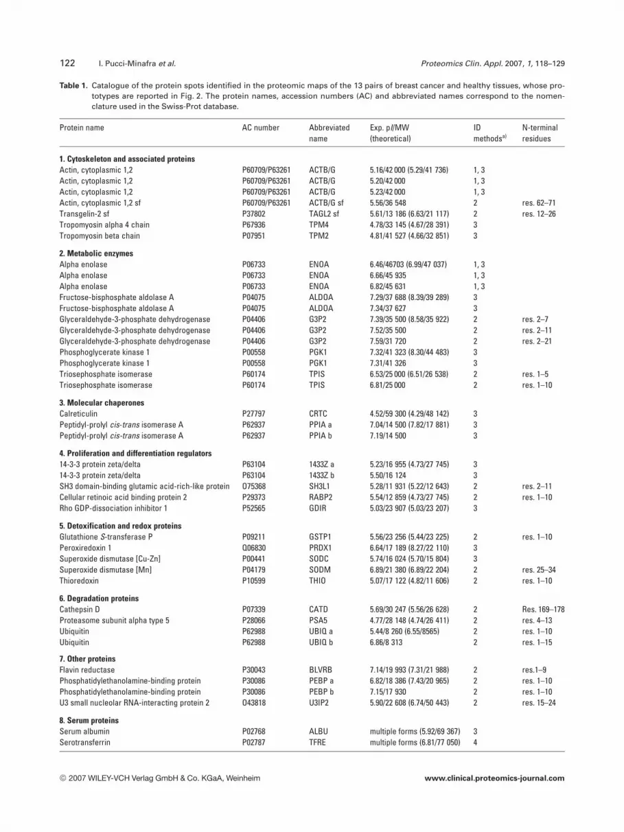
Table 1. Catalogue of the protein spots identified in the proteomic maps of the 13 pairs of breast cancer and healthy tissues, whose pro-totypes are reported in Fig. 2. The protein names, accession numbers (AC) and abbreviated names correspond to the nomen-clature used in the Swiss-Prot database.
Protein name AC number Abbreviatedname
Exp. pI/MW(theoretical)
IDmethodsa)
N-terminalresidues
1. Cytoskeleton and associated proteinsActin, cytoplasmic 1,2 P60709/P63261 ACTB/G 5.16/42 000 (5.29/41 736) 1, 3Actin, cytoplasmic 1,2 P60709/P63261 ACTB/G 5.20/42 000 1, 3Actin, cytoplasmic 1,2 P60709/P63261 ACTB/G 5.23/42 000 1, 3Actin, cytoplasmic 1,2 sf P60709/P63261 ACTB/G sf 5.56/36 548 2 res. 62–71Transgelin-2 sf P37802 TAGL2 sf 5.61/13 186 (6.63/21 117) 2 res. 12–26Tropomyosin alpha 4 chain P67936 TPM4 4.78/33 145 (4.67/28 391) 3Tropomyosin beta chain P07951 TPM2 4.81/41 527 (4.66/32 851) 3
2. Metabolic enzymesAlpha enolase P06733 ENOA 6.46/46703 (6.99/47 037) 1, 3Alpha enolase P06733 ENOA 6.66/45 935 1, 3Alpha enolase P06733 ENOA 6.82/45 631 1, 3Fructose-bisphosphate aldolase A P04075 ALDOA 7.29/37 688 (8.39/39 289) 3Fructose-bisphosphate aldolase A P04075 ALDOA 7.34/37 627 3Glyceraldehyde-3-phosphate dehydrogenase P04406 G3P2 7.39/35 500 (8.58/35 922) 2 res. 2–7Glyceraldehyde-3-phosphate dehydrogenase P04406 G3P2 7.52/35 500 2 res. 2–11Glyceraldehyde-3-phosphate dehydrogenase P04406 G3P2 7.59/31 720 2 res. 2–21Phosphoglycerate kinase 1 P00558 PGK1 7.32/41 323 (8.30/44 483) 3Phosphoglycerate kinase 1 P00558 PGK1 7.31/41 326 3Triosephosphate isomerase P60174 TPIS 6.53/25 000 (6.51/26 538) 2 res. 1–5Triosephosphate isomerase P60174 TPIS 6.81/25 000 2 res. 1–10
3. Molecular chaperonesCalreticulin P27797 CRTC 4.52/59 300 (4.29/48 142) 3Peptidyl-prolyl cis-trans isomerase A P62937 PPIA a 7.04/14 500 (7.82/17 881) 3Peptidyl-prolyl cis-trans isomerase A P62937 PPIA b 7.19/14 500 3
4. Proliferation and differentiation regulators14-3-3 protein zeta/delta P63104 1433Z a 5.23/16 955 (4.73/27 745) 314-3-3 protein zeta/delta P63104 1433Z b 5.50/16 124 3SH3 domain-binding glutamic acid-rich-like protein O75368 SH3L1 5.28/11 931 (5.22/12 643) 2 res. 2–11Cellular retinoic acid binding protein 2 P29373 RABP2 5.54/12 859 (4.73/27 745) 2 res. 1–10Rho GDP-dissociation inhibitor 1 P52565 GDIR 5.03/23 907 (5.03/23 207) 3
5. Detoxification and redox proteinsGlutathione S-transferase P P09211 GSTP1 5.56/23 256 (5.44/23 225) 2 res. 1–10Peroxiredoxin 1 Q06830 PRDX1 6.64/17 189 (8.27/22 110) 3Superoxide dismutase [Cu-Zn] P00441 SODC 5.74/16 024 (5.70/15 804) 3Superoxide dismutase [Mn] P04179 SODM 6.89/21 380 (6.89/22 204) 2 res. 25–34Thioredoxin P10599 THIO 5.07/17 122 (4.82/11 606) 2 res. 1–10
6. Degradation proteinsCathepsin D P07339 CATD 5.69/30 247 (5.56/26 628) 2 Res. 169–178Proteasome subunit alpha type 5 P28066 PSA5 4.77/28 148 (4.74/26 411) 2 res. 4–13Ubiquitin P62988 UBIQ a 5.44/8 260 (6.55/8565) 2 res. 1–10Ubiquitin P62988 UBIQ b 6.86/8 313 2 res. 1–15
7. Other proteinsFlavin reductase P30043 BLVRB 7.14/19 993 (7.31/21 988) 2 res.1–9Phosphatidylethanolamine-binding protein P30086 PEBP a 6.82/18 386 (7.43/20 965) 2 res. 1–10Phosphatidylethanolamine-binding protein P30086 PEBP b 7.15/17 930 2 res. 1–10U3 small nucleolar RNA-interacting protein 2 O43818 U3IP2 5.90/22 608 (6.74/50 443) 2 res. 15–24
8. Serum proteinsSerum albumin P02768 ALBU multiple forms (5.92/69 367) 3Serotransferrin P02787 TFRE multiple forms (6.81/77 050) 4
© 2007 WILEY-VCH Verlag GmbH & Co. KGaA, Weinheim www.clinical.proteomics-journal.com

Proteomics Clin. Appl. 2007, 1, 118–129 123
Table 1. Continued
Protein name AC number Abbreviatedname
Exp. pI/MW(theoretical)
IDmethodsa)
N-terminalresidues
Ig/gamma-1- chain C region P01857 IGHG1 multiple forms (8.46/36 106) 4Ig/gamma-1- chain C region P01857 IGHG1 multiple forms (8.46/36 106) 4Haptoglobin P00738 HPT a 5.74/17 422 (6.13/45 205) 2 res. 19–28Haptoglobin P00738 HPT b multiple forms (6.13/45 205) 4Alpha-1-antitrypsin P01009 A1AT multiple forms (5.37/46 736) 4Hemoglobin alpha subunit P69905 HBA 5.83/12 007 (8.73/15 126) 2 res. 1–7Hemoglobin alpha subunit P69905 HBA multiple forms (8.73/15 126) 4Transthyretin P02766 TTHY 5.54/14 022 (5.35/13 761) 2 res. 21–27Apolipoprotein A-I P02647 APOA1 5.22/24 311 (5.27/28 079) 2 res. 25–32
a) Identification methods: 1, Western blotting; 2, N-terminal sequencing by automated Edman degradation; 3, gel matching with previouslyidentified protein spots by MALDI-TOF and N-terminal sequencing (see [6]); 4, gel matching with human plasma of SWISS-2D PAGE.
Figure 3. Cytoskeleton and associated proteins. The panel shows differences of spot features between BCT and NAT. Differential expres-sion of spot density was calculated as Vol%. The differences in expression between breast cancer and normal adjacent tissues were ana-lyzed by the Student’s t-test: *p,0.05 was considered significant; **p,0.01 highly significant; ***p,0.001 very highly significant. The datain the graphs are expressed as mean number 6 SD.
Recently, several authors [8] have suggested the existenceof a relationship between actin organization and changes inactin isoform expression with the ability of cancer cells toform metastases.
Two other cytoskeletal proteins, TPM4 and TPM2, wereidentified in all tumor tissues, while small amounts or eventraces were observed in the non-tumoral counterpart. TPM4was differentially expressed at a higher significance level,while the TPM2 intensity level was more variable amongpatients. TPM is a major structural protein associated with
the actin microfilaments. Multiple TPM isoforms have beenreported in several cell lines, including breast cancer cells [6],and some of them have been thought to be associated withthe metastatic potential of several primary tumors [9].
Another interesting protein significantly overexpressedin the tumor samples is transgelin 2. Recently, the over-expression of transgelin 2 mRNA was reported in a largepercentage (69%) of hepatocellular carcinomas [10], whichsuggests its potential role as a diagnostic marker for cancerdetection.
© 2007 WILEY-VCH Verlag GmbH & Co. KGaA, Weinheim www.clinical.proteomics-journal.com

124 I. Pucci-Minafra et al. Proteomics Clin. Appl. 2007, 1, 118–129
Figure 4. Metabolic enzymes. Spot comparison was performed as described in Fig. 3.
Figure 5. Molecular chaperones. Spot comparison was per-formed as described in Fig. 3.
3.2 Metabolic enzymes (Fig. 4)
In this category of proteins, we identified five enzymes of theglycolytic pathway: ALDOA, TPIS, G3P2, PGK1, ENOA,which are collectively overexpressed in the tumor tissue vs.the non-tumoral counterparts. This observation appears to beof particular interest when considering the anaerobic shift ofthe metabolism of cancer cells, already described in the pio-neering work of Warburg [11] and presently used for clinicalcancer detection by FDG-PET ((18)F-fluorodeoxyglucose-Positron Emission Tomography) imaging analyses [12].
Moreover, these data confirm our previous report show-ing increased levels of glycolytic enzymes in breast cancercells vs. non-tumoral mammary-derived cells [4]. It is alsolikely that the incremented expression level of some glyco-lytic enzymes may be related to additional functions per-formed by the cells. As an example, G3P2, besides its pivotalrole in the glycolytic pathway and energy production, fulfils amultiplicity of functions such as membrane fusion, micro-tubule bundling, phosphotransferase activity, and nucleicacid binding, all aspects deserving attention from the pointof view of transformation [13, 14].
3.3 Molecular chaperones (Fig. 5)
Presently, we have identified only two proteins in this cate-gory: CRTC and PPIA. Both of them are significantly over-expressed in the tumor tissue vs. the non-tumoral counter-
© 2007 WILEY-VCH Verlag GmbH & Co. KGaA, Weinheim www.clinical.proteomics-journal.com

Proteomics Clin. Appl. 2007, 1, 118–129 125
Figure 6. Cell cycle regulation and differentiation. Spot comparison was performed as described in Fig. 3.
Figure 7. Detoxification and redox proteins. Spot comparison was performed as described in Fig. 3.
© 2007 WILEY-VCH Verlag GmbH & Co. KGaA, Weinheim www.clinical.proteomics-journal.com

126 I. Pucci-Minafra et al. Proteomics Clin. Appl. 2007, 1, 118–129
Figure 8. Protein degradation. Spot comparison was performed as described in Fig. 3.
Figure 9. Other proteins. Spot compar-ison was performed as described inFig. 3.
part, stressing the hypothesis that they have a function incancer. CRTC has been shown to be overexpressed in humanbreast by other authors [15], and also in bladder carcinomas[16]. Recently, it has also been demonstrated that calreticulin
has a role in regulating p53 function by affecting its rate ofdegradation and nuclear localization [17]. Likewise, PPIA hasbeen found to be associated with the growth of colon cancercells [18].
© 2007 WILEY-VCH Verlag GmbH & Co. KGaA, Weinheim www.clinical.proteomics-journal.com

Proteomics Clin. Appl. 2007, 1, 118–129 127
3.4 Proliferation and differentiation regulators (Fig. 6)
We identified four proteins in this category: RABP2, SH3L1,1433Z and GDIR. RABP2 is a retinoid-binding protein,thought to regulate the access of retinoic acid to the nuclearretinoic acid receptors, and therefore to participate in a reg-ulatory feedback mechanism to control the action of retinoicacid on cell differentiation. This protein was found among themost variable within the tumor samples: a result suggestingthat its overexpression, when present, may be used to dis-criminate among subtypes of ductal infiltrating carcinomas.
The 1433Z family exhibits diverse biological activities, andmay be involved in regulating cell division, differentiation,survival, apoptosis [19–21] and cancer [22, 23]. It is noteworthythat one of the two isoelectric forms is significantly over-expressed in the tumor samples, while the other is more vari-able. Similarly, the expression level of SH3L1, a gene productbelonging to the SH3BGR (SH3 domain-binding glutamicacid-rich-like protein) family and structurally related to thio-redoxin (Trx) super family [24], was found to be rather variable.
Conversely, GDIR (Rho GDP-dissociation inhibitor 1)was found significantly overexpressed in the tumor samplesvs. the non-tumoral counterparts.
3.5 Detoxification and redox proteins (Fig. 7)
Detoxification and redox proteins are related enzymes per-forming important roles in cell catabolism and protectionagainst metabolic stresses. Indeed, experimental evidence
has suggested that oxidative stress mediates various cellularresponses, and that, in turn, the control of reduction/oxida-tion (redox) is fundamental in maintaining the homeostasisof the whole organism. The group includes the followingenzymes: SODC, SODM, THIO, PRDX1, GSTP1, whichwere collectively overexpressed in all of our cancer-derivedproteomics. Currently, there is much interest in the thior-edoxin and glutathione systems, due to their major role asredox systems in animal cells and putative targets for cancertherapy (see [25] for review). In recent years, the peroxi-redoxin system has also received much attention for its highantioxidant efficiency. The mammalian Prdx gene family hassix distinct members located both in the cytoplasm and invarious subcellular locations, including peroxisomes andmitochondria. Some of the Prdx members also have effectson cell differentiation and apoptosis and have been found tobe overexpressed in breast cancer [26].
3.6 Protein degradation (Fig. 8)
The protein-degradation machinery plays an important rolein protein homeostasis and cellular health. Within this cate-gory, we found three enzymes significantly overexpressed inthe cancer tissues, namely, CATD, UBIQ, and PSA5.
Cathepsin D is a lysosomal protease involved in proteincatabolism and is supposed to play important roles in anti-gen processing, degenerative diseases, and cancer progres-sion. In breast cancer it has been associated with anincreased risk of relapse and metastasis [27].
Figure 10. Serum proteins. Spot comparison was performed as described in Fig. 3.
© 2007 WILEY-VCH Verlag GmbH & Co. KGaA, Weinheim www.clinical.proteomics-journal.com

128 I. Pucci-Minafra et al. Proteomics Clin. Appl. 2007, 1, 118–129
Our present results, showing its consistent over-expression in the tumor samples vs. the non-tumoral coun-terparts, add meaning to the recent statement by Rochefort etal. [27] confirming “the clinical value of cathepsin D as aprognostic marker in breast cancer, when using well-stan-dardized assays.”
Similarly, our data confirm UBIQ and PSA5 as reliablecandidate markers for breast carcinomas [28]. Indeed, theubiquitin-proteasome pathway system is involved in thedegradation of many key regulatory cellular proteins such astumor suppressors and transcriptional regulators, and ofmisfolded/denatured proteins, thus regulating different cel-lular processes including apoptosis, proliferation, differ-entiation and stress response [29]. Thus, is not surprisingthat the pathogenesis of many malignancies and other dis-orders is correlated, to different extents, with aberrations inthe system.
3.7 Other proteins (Fig. 9)
We have included in this category three proteins with mis-cellaneous functions, namely, U3IP2, BLVRB, and PEBP.
The protein U3IP2 is a component of a small nucleolarribonucleoprotein particle (snoRNPs) thought to participatein the processing and modification of pre-ribosomal RNAand to regulate complex-associated protein shuttle betweenthe nucleus and cytoplasm [30]. The occurrence and over-expression in cancer tissues renders intriguing its role incarcinogenesis, but at present more information is needed toclarify its function.
BLVRB, biliverdin reductase B (flavin reductase-NADPH), is a member of the insulin receptor substratefamily with serine/threonine/tyrosine kinase activity,involved in the conversion of biliverdin to bilirubin [31].Interestingly, BLVRB was also found to translocate to thenucleus in cells treated with cGMP [32] and to function asa transcription factor for activator protein 1-regulatedgenes and for activation of c-jun and CREB/ATF-2 [33,34].
Finally, PEBP is a member of the phosphatidylethanol-amine-binding protein family, also named RKIP (Raf kinaseinhibitor protein). It has been identified in a wide variety oftissues and is thought to regulate several intracellular sig-naling pathways, while its deregulation may contribute totissue pathology. Among the reported effects, it has beenobserved that loss of RKIP expression in prostate cancer cellsconfers a metastatic phenotype to them. This effect that maybe reverted by restoring RKIP expression, suggesting a roleof the metastasis suppressor gene for PEBP/RKIP [35].
3.8 Serum proteins (Fig. 10)
The serum proteins are clearly more abundant in the non-tumoral tissues than in the cancer fragments. This is notsurprising, due to the lower amount of cells and the absenceof neoplastic foci within the host matrix. The majority of
them, i.e., ALBU, A1AT, HPT, IGHG1, HBA, and TFRE,migrate in the 2-D gels as multiple isoelectric isoforms (seeTable 1). Therefore, adequate evaluation and statistical com-parison between samples were allowed only for other identi-fied serum proteins migrating as individual spots (Fig. 10),one of which, the TTHY, showed a highly significant differ-ence in expression in normal vs. cancer tissues.
4 Concluding remarks
This study demonstrates quantitative and qualitative differ-ences in the proteomic profiles between breast cancer tis-sues, namely, ductal infiltrating carcinomas, and the non-tumoral adjacent tissues. The work is intended to offer a newcontribution to breast cancer clinical proteomics from afunctional point of view.
Appreciable differences of protein expression weredetected in all the cancer tissues from the following cate-gories: cytoskeleton and associated proteins, metabolicenzymes, molecular chaperones, detoxification and degrada-tion, and cell cycle regulation proteins. Conversely, almost allserum proteins displayed lower levels in breast cancer tissuethan in normal adjacent tissue.
The functional role of each category has been describedin the result section.
In conclusion, we suggest that the present collection ofdifferentially expressed proteins, while signifying a novelcontribution to the molecular biology of breast cancer, mayrepresent promising candidate biomarkers for molecular-based diagnosis of breast cancer and for pattern discovery.
The present research is part of the project DIAMOL (PorSicilia misura 3.4). The work was also supported by a MIURgrant to Professor Ida Puci-Minafra and the University ofPalermo (ex 60%). The authors thank nurse Gilda Barbera’svaluable support in sample collection and the excellent coopera-tion of the Breast Unit Staff, directed by Dr. A. Marrazzo, at theMaddalena Hospital.
5 References
[1] Donegan, W. L., Cancer J. Clin. 1997, 47, 28–51.
[2] Somiari, R. I., Somiari, S., Russella, S., Shriverc, C. D., J.Chromatogr. B 2005, 815, 215–225.
[3] Bradford, M. M., Anal. Biochem. 1976, 72, 248–254.
[4] Pucci-Minafra, I., Fontana, S., Cancemi, P., Alaimo, G. et al.,Ann. N. Y. Acad. Sci. 2002, 963, 122–139.
[5] Pucci-Minafra, I., Fontana, S., Cancemi, P., Basirico, L. et al.,Proteomics 2002, 2, 919–927.
[6]. Pucci-Minafra, I., Cancemi, P., Fontana, S., Minafra, L. et al.,Proteomics 2006, 6, 2609–2625.
[7] Wheelock, A. M., Goto, S., Expert Rev. Proteomics 2006, 3,129–142.
© 2007 WILEY-VCH Verlag GmbH & Co. KGaA, Weinheim www.clinical.proteomics-journal.com

Proteomics Clin. Appl. 2007, 1, 118–129 129
[8] Nowak, D., Skwarek-Maruszewska, A., Zemanek-Zboch, M.,Malicka-Błaszkiewicz, M., Acta Biochim. Pol. 2005, 52, 461–468.
[9] Qi, Y., Chiu, J. F., Wang, L., Kwong, D. L., He, Q.Y., Proteomics2005, 5, 2960–2971.
[10] Shi, Y. Y., Wang, H. C., Yin, Y. H., Sun, W. S. et al., Br. J. Can-cer 2000, 92, 929–934.
[11] Warburg, O., The Metabolism of Tumors, Richard Smith,New York 1931, pp. 129–161.
[12] Port, E. R., Yeung, H., Gonen, M., Liberman, L. et al., Ann.Surg. Oncol. 2006, 13, 677–684.
[13] Sirover, M. A., Biochim. Biophys. Acta 1999, 1432, 159–184.
[14] Savagner, P., Bioessays 2001, 23, 912–923.
[15] Franzen, B., Linder, S., Alaiya, A. A., Eriksson, E. et al., Elec-trophoresis 1997, 18, 582–587.
[16] Iwaki, H., Kageyama, S., Isono, T., Wakabayashi, Y. et al.,Cancer Sci. 2004, 95, 955–961.
[17] Mesaeli, N., Phillipson, C., Mol. Biol. Cell 2004, 15, 1862–1870.
[18] Obama, K., Kato, T., Hasegawa, S., Satoh, S. et al., Clin.Cancer Res. 2006, 12, 70–76.
[19] van Hemert, M. J., Steensma, H. Y., van Heusden, G. P. H.,Bioessays 2001, 23, 936–947.
[20] Chen, X. Q., Yu, A. C. H., Biochem. Biophys. Res. Commun.2002, 296, 657–663.
[21] Margolis, S. S., Walsh, S., Douglas, C., Yoshida, W. M.,EMBO J. 2003, 22, 5734–5745.
[22] Lodygin, D., Hermeking, H., Cell Res. 2005, 15, 237–246.
[23] Hermeking, H., Nat. Rev. Cancer 2003, 3, 931–943.
[24] Xu, C., Zheng, P., Shen, S., Xu, Y., FEBS Lett. 2005, 579, 2788–2794.
[25] Biaglow, J. E., Miller, R. A., Cancer Biol. Ther. 2005, 4, 6–13.
[26] Karihtala, P., Mäntyniemi, A., Kang, S. W., Vuokko, L. et al.,Clin. Cancer Res. 2003, 9, 3418–3424.
[27] Rochefort, H., Garcia, M., Glondu, M., Laurent, V. et al., Clin.Chim. Acta 2000, 291, 157–170.
[28] Dees, E. C., Orlowski, R. Z., Future Oncol. 2006, 2, 121–135.
[29] Nandi, D., Tahiliani, P., Kumar, A., Chandu, D., J. Biosci. 2006,1, 137–155.
[30] Leary, D. J, Terns, M. P., Huang, S., Mol. Biol. Cell 2004, 15,281–293.
[31] Lerner-Marmarosh, N., Shen, J., Torno, M. D., Kravets, A. etal., PNAS 2005, 102, 7109–7114.
[32] Maines, M. D., Ewing, J. F., Huang, T. J., Panahian, N., J.Pharmacol. Exp. Ther. 2001, 296, 1091–1097.
[33] Ahmad, Z., Salim, M., Maines, M. D., J. Biol. Chem. 2002,277, 9226–9232.
[34] Kravets, A., Hu, Z., Miralem, T., Torno, M. D., Maines, M.D., J.Biol. Chem. 2004, 279, 19916–19923.
[35] Keller, E. T., Fu, Z., Brennan, M. J., Cell Biochem. 2005, 94,273–278.
© 2007 WILEY-VCH Verlag GmbH & Co. KGaA, Weinheim www.clinical.proteomics-journal.com


![[Tumoral angiogenesis: physiopathology, prognostic value and therapeutic perspectives]](https://img.pdfslide.net/doc/110x75/634a777d6bb2dc8f25052f94/tumoral-angiogenesis-physiopathology-prognostic-value-and-therapeutic-perspectives.jpg)