Embed Size (px)
Citation preview
Volume 58(3): 221–228, 2010Journal of Histochemistry & Cytochemistry
http://www.jhc.org
PERSPECTIVE
Proteomics Out of the Archive: Two-dimensionalElectrophoresis and Mass Spectrometry Using HOPE-fixed,Paraffin-embedded Tissues
Daniel Kähler,1 Christian Alexander,1 Holger Schultz, Mahdi Abdullah, Detlev Branscheid,Buko Lindner, Peter Zabel, Ekkehard Vollmer, and Torsten Goldmann
Clinical and Experimental Pathology (DK,HS,MA,EV,TG) and Department of Immunochemistry and Biochemical Microbiology(CA,BL), Research Center Borstel, Borstel, Germany; Department of Thoracic Surgery, Hospital Großhansdorf, Großhansdorf,Germany (DB); and Department of Clinical Medicine, Medical University Hospital III Lübeck, Lübeck, Germany (PZ)
SUMMARY Proteome analyses provide diagnostic information which can be essential fortherapeutic predictions. The application of such techniques for analyzing paraffin-embedded tissue samples is widely hampered by the use of formalin fixation requiring anti-gen retrieval procedures in molecular pathology. In prior studies, the HEPES-glutamic acidbuffer-mediated organic solvent protection effect (HOPE) technique of tissue fixation hasbeen shown to provide a broad array of biochemical investigations with excellent preserva-tion of morphological structures, DNA, RNA, and proteins, thus supporting the multimethodanalysis of archived specimens. Here we show that HOPE fixation is also useful in proteomicinvestigations by allowing two-dimensional electrophoresis (2DE) and mass spectrometry,using lung cancer tissues. Two-dimensional gels of two-protein extraction protocols derivedfrom HOPE-fixed material displayed characteristic spot patterns with high reproducibility.For comparison, 2DE analysis of ethanol-fixed, formalin-fixed, and frozen samples fromthe same tissues was performed. Western blotting confirmed immunoreactivity of 2DE-separated proteins from HOPE-fixed tissue samples. Additionally, distinct spots were excisedfrom HOPE-derived 2D gels and successfully subjected to peptide mass fingerprinting.In conclusion, paraffin archives containing HOPE-fixed tissues are applicable to a widespectrum of molecular investigations including common biochemical methods for proteomeanalyses and therefore represent a unique source for molecular investigations in the rapidlygrowing field of molecular pathology. This manuscript contains online supplemental mate-rial at http://www.jhc.org. Please visit this article online to view these materials.
(J Histochem Cytochem 58:221–228, 2010)
KEY WORDS
HOPE technique
two-dimensional
electrophoresis
NSCLC
paraffin material
proteomics
mass spectrometry
RECENTLY, MANY PATHOLOGICAL CHALLENGES have evokedan increasing demand for confident molecular read-outtechniques for elaborating diagnoses, as well as for dis-covering more specific disease-relevant marker mole-cules (Coleman 2000). Such a group of standardmarkers for lung carcinomas are members of the kera-tin family, ranging in molecular mass between 40 and
80 kDa, and keratin 10 can be used as a marker forsquamous cell carcinomas (Moll et al. 1982; Eliaset al. 1988; Nhung et al. 1999; Tsubokawa et al.2002). As far as analysis of cellular macromolecules,i.e., proteins and nucleic acids, is concerned, large-scalestudies of paraffin-embedded materials are still con-stricted due to the standard procedure of conserving
Correspondence to: Daniel Kähler, Clinical and ExperimentalPathology Research Center Borstel Parkallee 3a, 23845 Borstel,Germany. E-mail: [email protected]
1These authors contributed equally to this work.Received for publication April 1, 2009; accepted November 17,
2009 [DOI: 10.1369/jhc.2009.954065].
© 2010 Kähler et al. This article is distributed under the terms of aLicense to Publish Agreement (http://www.jhc.org/misc/ltopub.shtml). JHC deposits all of its published articles into the U.S.National Institutes of Health (http://www.nih.gov/) and PubMedCentral (http://www.pubmedcentral.nih.gov/) repositories for publicrelease twelve months after publication.
0022-1554/09/$3.30 221
TheJourna
lof
Histoch
emistry&
Cytoc
hemistry
tissue samples with formalin because of good morpho-logical maintenance. This shows that semistandardizeddetection and characterization of molecular com-pounds in such archived materials does not supplythe needs of detailed biochemical investigations. Pro-tein cross-linking and degradation of nucleic acidshinder the achievement of high-quality results formolecular characterizations. Furthermore, the applica-tion of frozen tissues that provide good molecular read-out options is limited by cost-intensive problems suchas the necessity for permanent nitrogen storage forlong-term appropriation of specimens and the weakpreservation of morphologic details. Moreover, afterlonger storage times, successive losses of molecular in-tegrity occur (Srinivasan et al. 2002).
Since the HEPES-glutamic acid buffer-mediatedorganic solvent protection effect (HOPE) fixation tech-nique covers both formalin-like morphological mainte-nance of tissues and excellent conservation of nucleicacids, as well as antigenic structures, it has meanwhileenabled highly reproducible molecular analyses offixed tissues by several protein- and nucleic acid-targeting methods (Olert et al. 2001; Goldmann et al.2002,2003,2004; Wiedorn et al. 2002; Droemannet al. 2003; Sen Gupta et al. 2003; Umland et al.2003; Uhlig et al. 2004). As to preservation of proteins,immunodetection techniques benefit from the avoid-ance of antigen retrieval procedures if tissues are con-served by HOPE technology instead of formalin(Goldmann et al. 2003). Therewith, the epitopes ofprotein antigens have been shown to persist even inlonger-conserved tissues, and hence, any archivedHOPE-fixed material can be adducted for high qualityimmunostaining analyses. Use of the HOPE techniqueallows an extended spectrum of analytical methods(immunohistochemistry, RT-PCR, DNA/RNA in situhybridization, transcription arrays, and protein anal-yses by SDS-PAGE and immunoblotting) that areapplicable for analysis of single-donor material. Wehave now additionally established a protocol whichoffers proteome analyses of paraffin-embeddedHOPE-fixed tissues by two-dimensional electropho-resis (2DE) employing non-equilibrium pH gradientgel electrophoresis (NEPHGE) in the first dimensionand conventional SDS-PAGE techniques in the seconddimension to enlarge the methodical spectrum of theHOPE technology. Results were compared with thoseof corresponding formalin-fixed, ethanol-fixed, andfrozen tissues from the same patient. We show in addi-tion that 2DE gels obtained from HOPE-fixed tissuesamples are applicable for immunostaining of disease-related marker antigens as well as for standard pro-tocols of protein identification by tryptic digestionand matrix-assisted laser desorption ionization–time-of-flight mass spectrometry (MALDI-TOF MS) analy-sis of single protein spots.
Materials and Methods
Tissues
The examined tissue samples were characterized asnon–small-cell lung cancer, which required surgical re-section. We analyzed 10 different cases using each ofthe three fixation techniques and corresponding frozenmaterial. We show four adenocarcinomas, three squa-mous cell carcinomas, and one healthy lung tissue.After tumor or tumor-free parts were removed fromfresh lobectomies, the tissues were fixed and embeddedin paraffin, using the HOPE technology as previouslydescribed (Olert et al. 2001). Formalin fixation andfrozen material work-up followed standard protocolsusing a Shandon Pathcentre unit for formalin fixation(Thermo Electron Corporation; Karlsruhe, Germany)and liquid nitrogen for frozen material (Srinivasan et al.2002). Ethanol fixation was performed for 24 hr atroom temperature, using 70% ethanol, and processedsimilarly to formalin-fixed material with the ShandonPathcentre unit before paraffin embedding. The par-affin blocks were shelved at 4C (HOPE-fixed samples)or room temperature (formalin- and ethanol-fixed sam-ples); frozen material was stored at 280C.
Preparation of Samples
From the HOPE-fixed paraffin blocks, samples of 8 to10 sections were deparaffinized by two cycles ofisopropanol treatment (5 ml; 5 min in a tube rotor)and centrifugation (13,000 rpm; 5 min) with respectiveremoval of the supernatants. Subsequently, a single de-hydration step of acetone treatment was performed, andthe tissue samples were dried in a vacuum centrifuge for10min (Savant SpeedVac 110; Life Science International,Frankfurt, Germany). Ethanol-fixed specimens wereprocessed using xylol and ethanol for deparaffinization.
Frozen material was crumbled in a dish filled withliquid nitrogen and placed directly into a reaction tubefor protein extraction. Formalin material was processedadequately using a special retrieval kit (see below).
Protein Extraction
Two protocols of protein extraction arbitrarily desig-nated as aqueous and complex protein extraction werecompared within this study.
For aqueous protein extracts from the tissues, the driedsediments were resuspended in diethylpyrocarbonate-water at a sample concentration of 100 mg/ml by incu-bation at room temperature for 30 min with vortexingevery 5 min. To remove insoluble material, this solutionwas ultracentrifuged at 50,000 rpm for 30 min, and theprotein concentrations of the resulting aqueous tissue ex-tracts were subsequently determined by the bicinchoninicacid assay (Pierce; Rockford, IL). After Speed Vac ly-ophilization, the dried samples were resuspended inampholyte-phosphate buffer containing 9 M urea at a
222 Kähler, Alexander, Schultz, Abdullah, Branscheid, Lindner, Zabel, Vollmer, Goldmann
TheJourna
lof
Histoch
emistry&
Cytoc
hemistry
final protein concentration of 20 mg/ml by incubation atroom temperature for 30 min with vortexing every 5 minand subsequent incubation at room temperature for an-other 30 min. After precipitated material was removedby ultracentrifugation (50,000 rpm, room temperature,30 min), the clear supernatants were finally diluted at aratio of 1:5 in ampholyte phosphate buffer containing1% (w/v) of agarose that had been tempered at 65C.
For obtaining complex protein extracts from HOPE-and ethanol-fixed tissues and frozen material, we used anextraction buffer containing 7 M urea (Roth; Karlsruhe,Germany), 2 M thiourea (Merck; Darmstadt, Germany),2% octyl phenol (Igepal; Sigma-Aldrich, Munich, Ger-many), 1% Triton-X (Merck), 100 mM dithiothreitol(Roth), 5 mmol PMSF (Roth), 4% CHAPS (Roth),and 0.5 mM EDTA.
Formalin-fixed material was deparaffinized andprotein-extracted using a QProteome FFPE Tissue2D-PAGE kit (Qiagen; Hilden, Germany) followingthe manufacturer’s instructions to ensure adequateprocessing for formalin-fixed samples for 2DE. Fiftymg of protein from each sample was applied to thefirst dimension of the 2DE system.
2DE
The proteomes of the tissue-derived samples wereanalyzed by using a WitaVision high-resolution 2Dgel electrophoresis system (WITA GmbH; Teltow,Germany). The charge-dependent initial separation ofthe tissue-derived protein solutions in the first dimen-sion of 2DE was performed in pH gradient rod gels(pH range, 4–10) according to the protocol of the man-ufacturer using a set of standardized materials. Briefly,two gel solutions were cast in succession in a verticaldevice for preparation of the two-layered rod gels ofthe first dimension (quantities sufficient for a total ofeight rod gels): 1.5 ml of separation gel solution plus36 ml of 0.8% ammonium persulfate (APS) was pre-pared for polymerization of the first gel layer, and600 ml of cap gel solution (WitaVision) was mixed with15 ml of 0.8% APS for formation of the second gellayer of the rod gels (all solutions were degassed bysonication). For complete polymerization, the gels ofthe first dimension were held at room temperaturefor 30 min and then kept in a damp chamber for anadditional 72 hr.
Running the First Dimension (NEPHGE Technique)
The first-dimensional separation of proteins in the rodgels was performed in a vertical electrophoresis deviceaccording to the operating instructions of the manufac-turer (WitaVision). Briefly, the lower chamber of thedevice was filled with 400 ml of degassed cathodebuffer (10-fold stock solution prepared on a 40C heat-ing plate, containing 20 g of glycine, 216 g of urea,
200 ml of aqua dest, filled up to 380 ml; and the addi-tion of 20 ml of ethylenediamine). Following fixation ofthe rod gels in the device, the freshly prepared samplesolutions in agarose-supplemented ampholyte phos-phate buffer were applied to the anodic sides of thecapillary gels, and the remaining volumes of the capil-lary glass tubes were then covered with a sample sta-bilizing overlay solution (WitaVision). Subsequently,400 ml of degassed anode buffer was applied (10-foldstock solution of 72 g of urea, 250 ml of aqua dest,filled up to 380 ml; addition of 20 ml of 85% phos-phoric acid) to the upper chamber of the device, andthe electrophoretic separation of the first dimensionwas started by using the following sequence of pro-grammed running conditions: 100 V for 1 hr 15 min;200 V for 1 hr 15 min; 400 V for 1 hr 15 min; 600 Vfor 1 hr 15 min; 800 V for 10 min; 1000 V for 5 min.After termination of electrophoresis, the rod gels werecarefully pushed out of the glass tubes onto plasticrails, and adaptation to the conditions of the seconddimension was achieved by a series of five 15-minequilibrations in a corresponding incubation solution.Optionally, the equilibrated rod gels of the first di-mension were stored at 280C before application tothe second dimension of the 2DE system.
Running the 2nd Dimension (SDS-PAGE)
For separation in the second dimension of 2DE,standard SDS-PAGE was performed with 15% (w/v)polyacrylamide gels, using a Mini Protean 3 Cell unit(Bio-Rad; Hercules, CA). Briefly, the rod gels of thefirst dimension were gently transferred from equilibra-tion and storage rails to the top of the stacking gelzones and covered with 1% (w/v) agarose containing0.1% (w/v) bromophenol blue to fix the rod gels andfor visualization of the progress in sample migrationduring SDS-PAGE. The electrophoresis running con-ditions of the second dimensional separation were setas follows: 35 V for 5 min; 55 V for 10 min; 100 Vfor 15 min; and 150 V for 1 hr, until the bromophenolblue front reached the bottom of the gel.
Fixing and Staining of 2D Gels
After 2DE protein separation was complete, gels werefixed and silver stained according to Mortz et al.(2001). For protein identification by tryptic digestionand mass spectrometry, Coomassie staining of the cor-responding 2DE gels, using a Simply Blue Safe system(Invitrogen; Karlsruhe, Germany) was performed ac-cording to the protocol of the manufacturer, until dis-tinct spots began to appear.
Western Blotting and Immunodetection
Blotting was performed using 2DE gels produced fromsquamous cell carcinoma tissue lysates and an iBlot
Proteomics Using Paraffin-embedded Tissues 223
TheJourna
lof
Histoch
emistry&
Cytoc
hemistry
semidry electrotransfer system (Invitrogen) accordingto the protocol of the manufacturer (blotting in 7 minonto a nitrocellulose membrane). Immunodetection ofpan-keratin representing a group of standard markerproteins for lung carcinomas was carried out by the fol-lowing procedure: Blocking of unspecific antibodybinding [1.5 hr, digoxygenin wash and block buffer(Boehringer Ingelheim; Ingelheim, Germany)]; primaryantibody reaction [1.5 hr, anti-pan-keratin antibody(DAKO; Hamburg, Germany), concentration of1:100]; three wash steps (10 min each, Tween-TBS-buffer, pH 7.4); secondary antibody reaction [45 min,mouse anti-human antibody (Dianova; Hamburg,Germany)], concentration of 1:10,000]; two wash steps(10 min each, Tween-TBS-buffer, pH 7.4); one wash step(10 min, TBS buffer, pH 9.5); determination of colorreaction using the nitro blue tetrazolium/5-bromo-4-chloro-3-indolyl system [(Boehringer Ingelheim),stopped after 5 min by washing with deionized water].
Tryptic Digest and Mass Fingerprinting
The protein spots separated by 2DE were excised, de-stained, and digested overnight with sequence-gradetrypsin from bovine pancreas (Roche DiagnosticsGmbH; Mannheim, Germany) as described previously(Shevchenko et al. 1996). For MALDI-TOF MS,the extracted tryptic fragments were mixed 1:1 (v/v)with saturated matrix solution [purified a-cyano-4-hydroxycinnamic acid, (Bruker Daltonics; Bremen,Germany)] and analyzed with a Reflex III spectrometer(Bruker Daltonics) in the reflector TOF configurationwith an applied acceleration voltage of 20 kV. Themass spectra were processed by Biotools version 3.2software (Bruker Daltonics), and mass lists were eval-uated by a MASCOT database search, using a molec-ular weight search (MOWSE) scoring algorithm to
create probability-based scoring as described under“Scoring Schemes.” (Matrix Science; http://www.matrixscience.com/help/scoring_help.html#PBM).
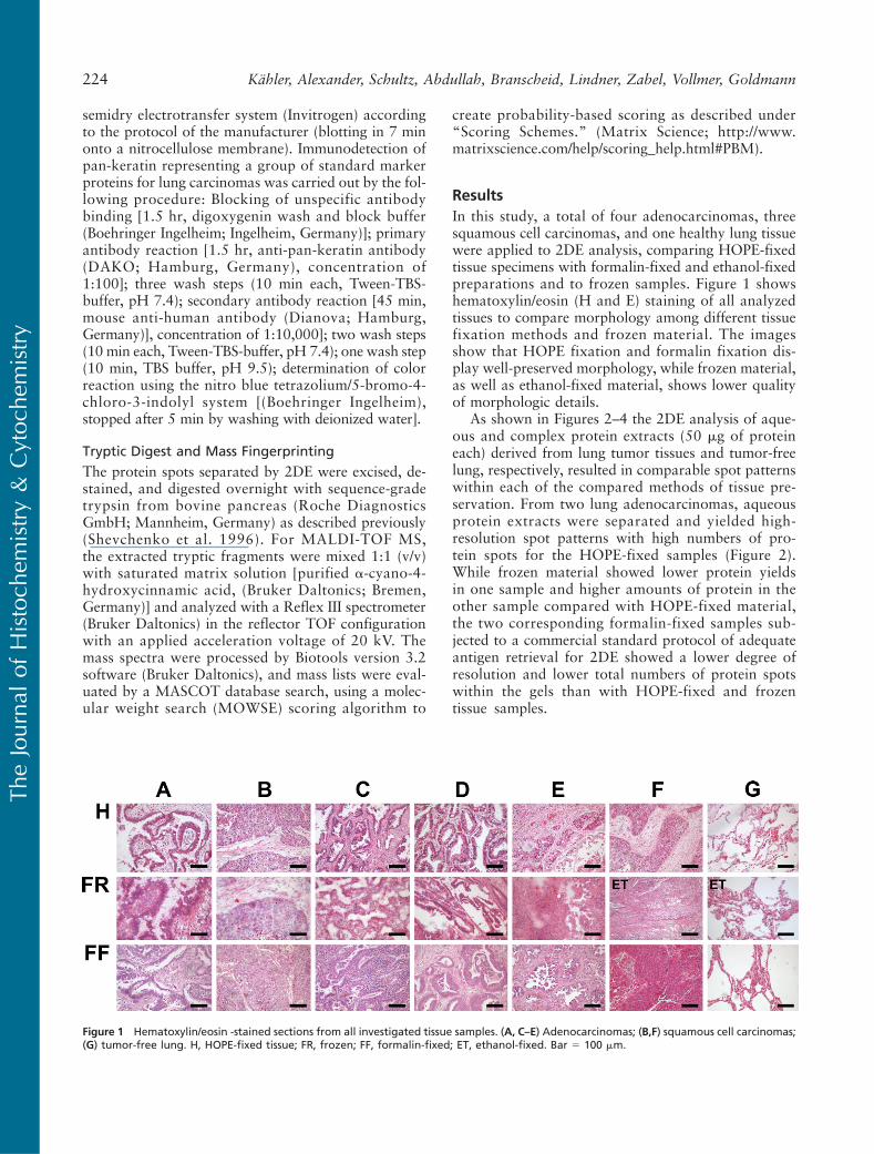
ResultsIn this study, a total of four adenocarcinomas, threesquamous cell carcinomas, and one healthy lung tissuewere applied to 2DE analysis, comparing HOPE-fixedtissue specimens with formalin-fixed and ethanol-fixedpreparations and to frozen samples. Figure 1 showshematoxylin/eosin (H and E) staining of all analyzedtissues to compare morphology among different tissuefixation methods and frozen material. The imagesshow that HOPE fixation and formalin fixation dis-play well-preserved morphology, while frozen material,as well as ethanol-fixed material, shows lower qualityof morphologic details.
As shown in Figures 2–4 the 2DE analysis of aque-ous and complex protein extracts (50 mg of proteineach) derived from lung tumor tissues and tumor-freelung, respectively, resulted in comparable spot patternswithin each of the compared methods of tissue pre-servation. From two lung adenocarcinomas, aqueousprotein extracts were separated and yielded high-resolution spot patterns with high numbers of pro-tein spots for the HOPE-fixed samples (Figure 2).While frozen material showed lower protein yieldsin one sample and higher amounts of protein in theother sample compared with HOPE-fixed material,the two corresponding formalin-fixed samples sub-jected to a commercial standard protocol of adequateantigen retrieval for 2DE showed a lower degree ofresolution and lower total numbers of protein spotswithin the gels than with HOPE-fixed and frozentissue samples.
Figure 1 Hematoxylin/eosin -stained sections from all investigated tissue samples. (A, C–E) Adenocarcinomas; (B,F) squamous cell carcinomas;(G) tumor-free lung. H, HOPE-fixed tissue; FR, frozen; FF, formalin-fixed; ET, ethanol-fixed. Bar 5 100 mm.
224 Kähler, Alexander, Schultz, Abdullah, Branscheid, Lindner, Zabel, Vollmer, Goldmann
TheJourna
lof
Histoch
emistry&
Cytoc
hemistry
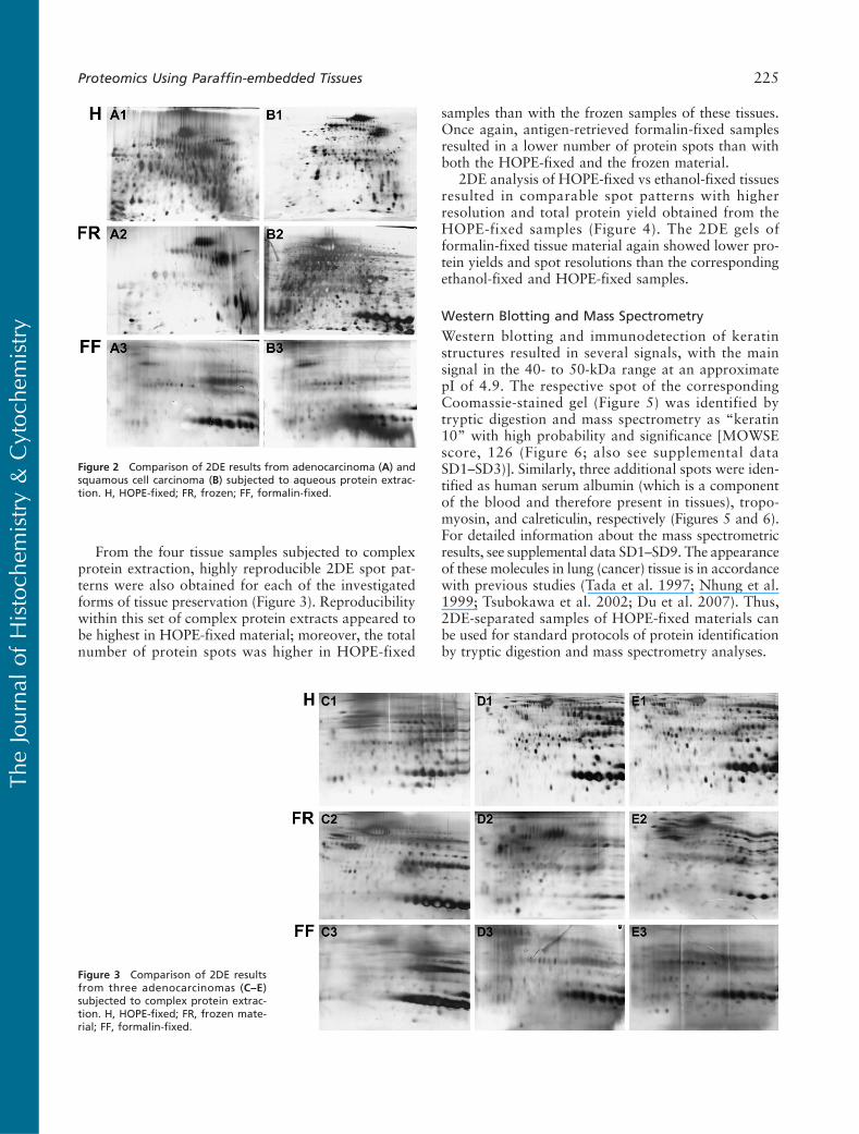
From the four tissue samples subjected to complexprotein extraction, highly reproducible 2DE spot pat-terns were also obtained for each of the investigatedforms of tissue preservation (Figure 3). Reproducibilitywithin this set of complex protein extracts appeared tobe highest in HOPE-fixed material; moreover, the totalnumber of protein spots was higher in HOPE-fixed
samples than with the frozen samples of these tissues.Once again, antigen-retrieved formalin-fixed samplesresulted in a lower number of protein spots than withboth the HOPE-fixed and the frozen material.
2DE analysis of HOPE-fixed vs ethanol-fixed tissuesresulted in comparable spot patterns with higherresolution and total protein yield obtained from theHOPE-fixed samples (Figure 4). The 2DE gels offormalin-fixed tissue material again showed lower pro-tein yields and spot resolutions than the correspondingethanol-fixed and HOPE-fixed samples.
Western Blotting and Mass Spectrometry
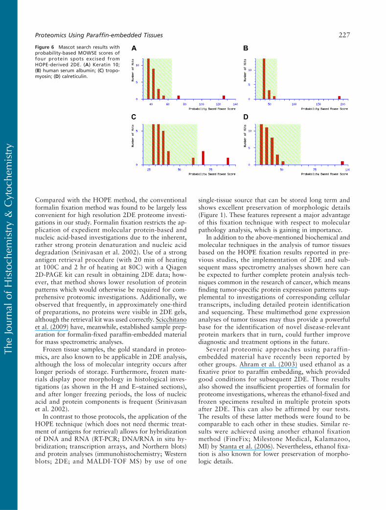
Western blotting and immunodetection of keratinstructures resulted in several signals, with the mainsignal in the 40- to 50-kDa range at an approximatepI of 4.9. The respective spot of the correspondingCoomassie-stained gel (Figure 5) was identified bytryptic digestion and mass spectrometry as “keratin10” with high probability and significance [MOWSEscore, 126 (Figure 6; also see supplemental dataSD1–SD3)]. Similarly, three additional spots were iden-tified as human serum albumin (which is a componentof the blood and therefore present in tissues), tropo-myosin, and calreticulin, respectively (Figures 5 and 6).For detailed information about the mass spectrometricresults, see supplemental data SD1–SD9. The appearanceof these molecules in lung (cancer) tissue is in accordancewith previous studies (Tada et al. 1997; Nhung et al.1999; Tsubokawa et al. 2002; Du et al. 2007). Thus,2DE-separated samples of HOPE-fixed materials canbe used for standard protocols of protein identificationby tryptic digestion and mass spectrometry analyses.
Figure 2 Comparison of 2DE results from adenocarcinoma (A) andsquamous cell carcinoma (B) subjected to aqueous protein extrac-tion. H, HOPE-fixed; FR, frozen; FF, formalin-fixed.
Figure 3 Comparison of 2DE resultsfrom three adenocarcinomas (C–E)subjected to complex protein extrac-tion. H, HOPE-fixed; FR, frozen mate-rial; FF, formalin-fixed.
Proteomics Using Paraffin-embedded Tissues 225
TheJourna
lof
Histoch
emistry&
Cytoc
hemistry
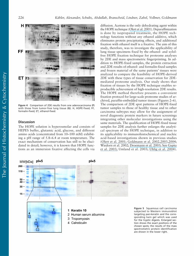
DiscussionThe HOPE solution is hyperosmolar and consists ofHEPES buffer, glutamic acid, glucose, and differentamino acids (concentrated from 10–100 mM) exhibit-ing a pH range of 5.8–6.4 at room temperature. Theexact mechanism of conservation has still to be eluci-dated in detail; however, it is known that HOPE func-tions as an immersion fixative affecting the cells via
diffusion. Acetone is the only dehydrating agent withinthe HOPE technique (Olert et al. 2001). Deparaffinizationis done by isopropanol treatment, the HOPE tech-nology functions without any ethanol additive, whicheliminates protein precipitating effects, and additionalfixation with ethanol itself is a fixative. The aim of thisstudy, therefore, was to investigate the applicability oflung tissue specimens fixed by the ethanol- and xylol-free HOPE fixation technique for proteome analysesby 2DE and mass spectrometric fingerprinting. In ad-dition to HOPE-fixed samples, the protein extractionand 2DE results of ethanol- and formalin-fixed samplesand frozen material of the same patients’ tissues wereanalyzed to compare the feasibility of HOPE-derived2DE with these types of tissue conservation for 2DE-mediated proteome analysis. Our study shows thatfixation of tissues by the HOPE technique enables re-producible achievement of high-resolution 2DE results.The HOPE method therefore presents a convenientfixation protocol for large-scale proteome studies of ar-chived, paraffin-embedded tumor tissues (Figures 2–6).The comparison of 2DE spot patterns of HOPE-fixedtumor samples to those of healthy tissue and to othercarcinoma subtypes may allow for the assignment ofnovel diagnostic protein markers in future screeningsintegrating other molecular investigations using thesame materials. The qualification of HOPE-fixed tissuesamples for 2DE analysis further enlarges the analyti-cal spectrum of the HOPE technique, in addition toits applicability to immunohistochemical and nucleicacid-based investigations shown in previous studies(Olert et al. 2001; Goldmann et al. 2002,2003,2004;Wiedorn et al. 2002; Droemann et al. 2003; Sen Guptaet al. 2003; Umland et al. 2003; Uhlig et al. 2004).
Figure 4 Comparison of 2DE results from one adenocarcinoma (F)with those from tumor-free lung tissue (G). H, HOPE-fixed; FF,formalin-fixed; ET, ethanol-fixed.
Figure 5 Squamous cell carcinomasubjected to Western immunoblottargeting pan-keratin and the corre-sponding twin gel which was usedfor the tryptic digests. Enlarged sec-tion shows the exact positions of thechosen spots; the results of the massspectrometric protein identificationare shown in the lower right.
226 Kähler, Alexander, Schultz, Abdullah, Branscheid, Lindner, Zabel, Vollmer, Goldmann
TheJourna
lof
Histoch
emistry&
Cytoc
hemistry
Compared with the HOPE method, the conventionalformalin fixation method was found to be largely lessconvenient for high resolution 2DE proteome investi-gations in our study. Formalin fixation restricts the ap-plication of expedient molecular protein-based andnucleic acid-based investigations due to the inherent,rather strong protein denaturation and nucleic aciddegradation (Srinivasan et al. 2002). Use of a strongantigen retrieval procedure (with 20 min of heatingat 100C and 2 hr of heating at 80C) with a Qiagen2D-PAGE kit can result in obtaining 2DE data; how-ever, that method shows lower resolution of proteinpatterns which would otherwise be required for com-prehensive proteomic investigations. Additionally, weobserved that frequently, in approximately one-thirdof preparations, no proteins were visible in 2DE gels,although the retrieval kit was used correctly. Scicchitanoet al. (2009) have, meanwhile, established sample prep-aration for formalin-fixed paraffin-embedded materialfor mass spectrometric analyses.
Frozen tissue samples, the gold standard in proteo-mics, are also known to be applicable in 2DE analysis,although the loss of molecular integrity occurs afterlonger periods of storage. Furthermore, frozen mate-rials display poor morphology in histological inves-tigations (as shown in the H and E–stained sections),and after longer freezing periods, the loss of nucleicacid and protein components is frequent (Srinivasanet al. 2002).
In contrast to those protocols, the application of theHOPE technique (which does not need thermic treat-ment of antigens for retrieval) allows for hybridizationof DNA and RNA (RT-PCR; DNA/RNA in situ hy-bridization; transcription arrays, and Northern blots)and protein analyses (immunohistochemistry; Westernblots; 2DE; and MALDI-TOF MS) by use of one
single-tissue source that can be stored long term andshows excellent preservation of morphologic details(Figure 1). These features represent a major advantageof this fixation technique with respect to molecularpathology analysis, which is gaining in importance.
In addition to the above-mentioned biochemical andmolecular techniques in the analysis of tumor tissuesbased on the HOPE fixation results reported in pre-vious studies, the implementation of 2DE and sub-sequent mass spectrometry analyses shown here canbe expected to further complete protein analysis tech-niques common in the research of cancer, which meansfinding tumor-specific protein expression patterns sup-plemental to investigations of corresponding cellulartranscripts, including detailed protein identificationand sequencing. These multimethod gene expressionanalyses of tumor tissues may thus provide a powerfulbase for the identification of novel disease-relevantprotein markers that in turn, could further improvediagnostic and treatment options in the future.
Several proteomic approaches using paraffin-embedded material have recently been reported byother groups. Ahram et al. (2003) used ethanol as afixative prior to paraffin embedding, which providedgood conditions for subsequent 2DE. Those resultsalso showed the insufficient properties of formalin forproteome investigations, whereas the ethanol-fixed andfrozen specimens resulted in multiple protein spotsafter 2DE. This can also be affirmed by our tests.The results of these latter methods were found to becomparable to each other in these studies. Similar re-sults were achieved using another ethanol fixationmethod (FineFix; Milestone Medical, Kalamazoo,MI) by Stanta et al. (2006). Nevertheless, ethanol fixa-tion is also known for lower preservation of morpho-logic details.
Figure 6 Mascot search results withprobability-based MOWSE scores offour protein spots excised fromHOPE-derived 2DE. (A) Keratin 10;(B) human serum albumin; (C) tropo-myosin; (D) calreticulin.
Proteomics Using Paraffin-embedded Tissues 227
TheJourna
lof
Histoch
emistry&
Cytoc
hemistry
Taken together, replacing formalin fixation withHOPE-fixed, paraffin-embedded tissues seems to en-sure better conditions for 2DE proteomic approachesbesides the other-mentioned molecular methods as re-ported predominantly by Goldmann et al. (2002,2003,2004). According to the results from this study and ourprior investigations, standardized HOPE technologyrepresents an efficient and, analytically, most versatilefixation method, enabling multiple data to be obtainedfrom (single) archived tissue materials. As this fixationmethod is comparably inert and as it has been shownto provide a powerful base for all standard types ofmolecular analyses tested so far, the HOPE techniqueappears to be well suited in future screenings of multipletissues for the identification and characterization ofnovel disease-relevant molecules, as well as the feasi-bility of 2DE for describing changes in posttransla-tional protein modifications such as phosphorylation,acetylation, and glycosylation status of proteins.
The results obtained from Western blotting andmass spectrometry analyses in this study confirm ourprevious reports of the maintenance and immuno-reactivity of protein structures in HOPE-fixed tissuesamples. The observed suitability for proteomic inves-tigations represents an additional advantageous featureof the HOPE technique compared with other fixationmethods because, first, there is no ethanol and no xylolnecessary (replaced through acetone and isopropanol)and, second, HOPE-fixed tissue samples are applicableto all common techniques of RNA/DNA and proteinresearch for the respective analysis of single-donormaterials, resulting in high inter-method comparability.
Acknowledgments
The authors thank Nina Grohmann, Jasmin Tiebach,Stefanie Fox, Maria Lammers, Helga Lütje, and Helge Meyerfor excellent technical assistance.
Literature Cited
Ahram M, Flaig MJ, Gillespie JW, Duray PH, Linehan WM,Ornstein DK, Niu S, et al. (2003) Evaluation of ethanol-fixed,paraffin-embedded tissues for proteomic applications. Proteomics3:413–421
Coleman R (2000) The impact of histochemistry: a historical per-spective. Acta Histochem 102:5–14
Droemann D, Goldmann T, Branscheid D, Clark R, Dalhoff K,Zabel P, Vollmer E (2003) Toll-like receptor 2 is expressed byalveolar epithelial cells type II and macrophages in the humanlung. Histochem Cell Biol 119:103–108
Du XL, Hu H, Lin DC, Xia SH, Shen XM, Zhang Y, Luo ML,et al. (2007) Proteomic profiling of proteins dysregulated inChinese esophageal squamous cell carcinoma. J Mol Med 85:863–875
Elias AD, Cohen BF, Bernal SD (1988) Keratin subtypes of small celllung cancer. Cancer Res 48:2724–2729
Goldmann T, Flohr AM, Escobar HM, Gerstmayer B, Janssen U,Bosio A, Loeschke S, et al. (2004) The HOPE-technique permitsNorthern blot and microarray analysis in paraffin-embeddedtissues. Pathol Res Pract 200:511–515
Goldmann T, Vollmer E, Gerdes J (2003) What’s cooking? Detectionof important biomarkers in HOPE-fixed paraffin embeddedtissues eliminates the need for antigen retrieval. Am J Pathol163:2638–2640
Goldmann T, Wiedorn KH, Kühl H, Olert J, Branscheid D, PechovskyD, Zissel G, et al. (2002) Assessment of transcriptional gene activityin-situ by application of HOPE-fixed, paraffin-embedded tissues.Pathol Res Pract 198:91–95
Moll R, Franke WW, Schiller DL, Geiger B, Krepier R (1982) Thecatalog of human cytokeratins: patterns of expression in normalepithelia, tumors, and cultured cells. Cell 31:11–24
Mortz E, Krogh TN, Vorum H, Görg A (2001) Improved silver stain-ing protocols for high sensitivity protein identification usingmatrix-assisted laser desorption/ionization time-of-flight analysis.Proteomics 1:1359–1363
Nhung NV, Mirejowsky P, Mirejowsky T, Melinova L (1999) Cyto-keratins and lung carcinomas. Cesk Patol 35:80–84
Olert J, Wiedorn KH, Goldmann T, Kühl H, Mehraein Y, ScherthanH, Niketeghad F, et al. (2001) HOPE-fixation: a novel fixingmethod and paraffin embedding technique for human soft tissues.Pathol Res Pract 197:823–826
Scicchitano MS, Dalmas DA, Boyce RW, Thomas HC, Frazier KS(2009) Protein extraction of formalin-fixed, paraffin-embeddedtissue enables robust proteomic profiles by mass spectrometry.J Histochem Cytochem 57:849–860
Sen Gupta R, Hillemann D, Kubica T, Zissel G, Muller-Quernheim J,Galle J, Vollmer E, et al. (2003) HOPE-fixation enables improvedPCR-based detection and differentiation of Mycobacteriumtuberculosis complex in paraffin-embedded tissues. Pathol ResPract 199:619–623
Shevchenko A, Wilm M, Vorm O, Mann M (1996) Mass spectro-metric sequencing of proteins silver-stained polyacrylamide gels.Anal Chem 68:850–858
Srinivasan M, Sedmark D, Jewel S (2002) Effect of fixatives andtissue processing on the content and integrity of nucleic acids.Am J Pathol 161:1961–1971
Stanta G, Mucelli SP, Petrera F, Bonin S, Bussolati G (2006) A novelfixative improves opportunities of nucleic acids and proteomicanalysis in human archive’s tissues. DiagnMol Pathol 15:115–123
Tada A, Kato H, Takenaga K, Hasegawa S (1997) Transforminggrowth factor beta1 increases the expressions of high molecularweight tropomyosin isoforms and vinculin and suppresses thetransformed phenotypes in human lung carcinoma cells. CancerLett 121:31–37
Tsubokawa F, Nishisaka T, Takeshima Y, Inai K (2002) Heterogene-ity of expression of cytokeratin subtypes in squamous cell carci-noma of the lung: with special reference to CK14 overexpressionin cancer of high-proliferative and lymphogeneous metastatikpotential. Pathol Int 52:286–293
Uhlig U, Uhlig S, Brandscheid D, Zabel P, Vollmer E, Goldmann T(2004) HOPE-technique enables Western blot analysis fromparaffin-embedded tissues. Pathol Res Pract 200:469–472
Umland O, Ulmer AJ, Vollmer E, Goldmann T (2003) In situ hybridiza-tion and immunohistochemistry using HOPE-fixed, cultured humancells on cytospin preparations. J Histochem Cytochem 51:977–980
Wiedorn KH, Olert J, Stacy R, Goldmann T, Kühl H, Matthus J,Vollmer E, et al. (2002) Preservation of high molecular weightnucleic acids by application of the novel HOPE-fixative. PatholRes Pract 198:735–740
228 Kähler, Alexander, Schultz, Abdullah, Branscheid, Lindner, Zabel, Vollmer, Goldmann
TheJourna
lof
Histoch
emistry&
Cytoc
hemistry


