Embed Size (px)
Citation preview
Cellular/Molecular
PSD-95 Is Required to Sustain the Molecular Organization ofthe Postsynaptic Density
Xiaobing Chen,1 Christopher D. Nelson,2,3 Xiang Li,4 Christine A. Winters,1 Rita Azzam,1 Alioscka A. Sousa,5
Richard D. Leapman,5 Harold Gainer,6 Morgan Sheng,2,3 and Thomas S. Reese1
Laboratories of 1Neurobiology and 6Neurochemistry, National Institute of Neurological Disorders and Stroke, National Institutes of Health, Bethesda,Maryland 20892, 2Picower Institute for Learning and Memory, Massachusetts Institute of Technology, Cambridge, Massachusetts 02139, 3Department ofNeuroscience, Genentech Inc., South San Francisco, California 94080, 4Neuroscience Program, Columbia University, New York, New York 10027, and5Laboratory of Cellular Imaging and Macromolecular Biophysics, National Institute of Biomedical Imaging and Bioengineering, National Institutes ofHealth, Bethesda, Maryland 20892
PSD-95, a membrane-associated guanylate kinase, is the major scaffolding protein in the excitatory postsynaptic density (PSD) and a potentregulator of synaptic strength. Here we show that PSD-95 is in an extended configuration and positioned into regular arrays of vertical filamentsthat contact both glutamate receptors and orthogonal horizontal elements layered deep inside the PSD in rat hippocampal spine synapses. RNAinterference knockdown of PSD-95 leads to loss of entire patches of PSD material, and electron microscopy tomography shows that the patchyloss correlates with loss of PSD-95-containing vertical filaments, horizontal elements associated with the vertical filaments, and putative AMPAreceptor-type, but not NMDA receptor-type, structures. These observations show that the orthogonal molecular scaffold constructed fromPSD-95-containing vertical filaments and their associated horizontal elements is essential for sustaining the three-dimensional molecularorganization of the PSD. Our findings provide a structural basis for understanding the functional role of PSD-95 at the PSD.
IntroductionSignal transduction at glutamatergic excitatory synapses, alongwith information storage underlying learning and memory(Malenka and Bear, 2004), take place at the postsynaptic density(PSD) (Garner et al., 2000; Kennedy, 2000; Sheng and Hoogen-raad, 2007). PSD-95, the most abundant scaffolding proteinin PSDs (Cheng et al., 2006), is a member of the membrane-associated guanylate kinase (MAGUK) family comprising PSD-95, PSD-93, SAP102 (synapse-associated protein-102), andSAP97, which share three conserved PDZ domains and oneSH3-GK (Src homology 3-guanylate kinase) module. ThesePSD-95 family MAGUKs are known to play prominent roles insynaptic plasticity (Kim and Sheng, 2004; Funke et al., 2005).
A typical PSD contains 200–300 PSD-95 molecules (Chen et al.,2005; Sugiyama et al., 2005), a number far exceeding the number of
glutamate receptors (Cheng et al., 2006). PSD-95 potentially bindsmany key constituent PSD proteins such as NMDA receptors(NMDARs) (Kornau et al., 1995; Niethammer et al., 1996), AMPAreceptor (AMPAR) complexes via Stargazin/TARP (Nicoll et al.,2006; Bats et al., 2007), adhesion molecules (Irie et al., 1997; Futai etal., 2007), and other scaffolding proteins, such as GKAP (Kim et al.,1997) and Shank (Sala et al., 2001). The diversity of proteins bindingto PSD-95 suggests that it has an important role in the molecularorganization of the PSD (Elias and Nicoll, 2007; Sheng and Hoogen-raad, 2007; Feng and Zhang, 2009). Consistent with an organizingrole, PSD-95 is one of the most stable proteins in PSDs at excitatorysynapses (Gray et al., 2006; Kuriu et al., 2006; Sharma et al., 2006;Blanpied et al., 2008; Sturgill et al., 2009). PSD-95 is also a potentregulator of synaptic strength through its dominant role in control-ling AMPA receptor numbers at synapses (Chen et al., 2000; Elias etal., 2006; Bats et al., 2007). However, how PSD-95 actually organizesthe molecular architecture of the PSD to support its functional prop-erties is still not well understood.
Schematic diagrams often depict PSD-95 molecules parallel tothe postsynaptic membrane at the PSD (Kim and Sheng, 2004).Electron microscopy (EM) images of individual recombinantPSD-95 molecules (Nakagawa et al., 2004) show a C-shaped cir-cular conformation consistent with results from modeling (Kor-kin et al., 2006), while SAP97 molecules reveal both circular andopen, extended conformations. Results from immunolabelingEM tomography in intact hippocampal neurons suggest thatthere are filaments in the PSD that appear to contain PSD-95 inan open configuration (Chen et al., 2008a).
To resolve the configuration of PSD-95 molecules in the PSD,epitopes at both ends of the PSD-95 molecule were mapped by
Received Nov. 12, 2010; revised March 3, 2011; accepted March 9, 2011.Author contributions: X.C., X.L., M.S., and T.R. designed research; X.C., C.D.N., C.A.W., R.A., A.A.S., and R.D.L.
performed research; C.D.N., X.L., H.G., and M.S. contributed unpublished reagents/analytic tools; X.C., C.D.N., andT.R. analyzed data; X.C., M.S., and T.R. wrote the paper.
The work was supported by National Institute of Neurological Disorders and Stroke (NINDS) and National Instituteof Biomedical Imaging and Bioengineering intramural research programs and Morgan Sheng was an investigator ofHoward Hughes Medical Institute. We thank Dr. Susan Cheng and Virginia Crocker of NINDS Electron MicroscopyFacility for their help on immunogold labeling; Dr. Carolyn Smith and Dr. Paul Gallant of NINDS Light Imaging Facilityfor their guidance on immunofluorescence microscopy and laser confocal light microscopy; Dr. Ayse Dosemeci forPSD-95 antibody; Drs. Carolyn Smith, Brian Andrews, and Ronald Petralia for critical reading of the manuscript; andJohn Chludzinski, Sam Carton, and Austin Hou for help on data analysis.
Morgan Sheng and Chris Nelson are currently employees of Genentech Inc., South San Francisco, a member of theRoche Group.
Correspondence should be addressed to Thomas S. Reese, Building 49 3A60, NIH, Bethesda, MD 20892. E-mail:[email protected].
DOI:10.1523/JNEUROSCI.5968-10.2011Copyright © 2011 the authors 0270-6474/11/316329-10$15.00/0
The Journal of Neuroscience, April 27, 2011 • 31(17):6329 – 6338 • 6329
EM, and were identified with EM tomography to show thatPSD-95 is in extended filamentous form in the PSD and that it isoriented vertical to the postsynaptic membrane. Loss of verticalfilaments seen by EM tomography of the PSD after RNA interfer-ence (RNAi) knockdown of PSD-95 demonstrates that these ver-tical filaments consist of, or at least are associated with, PSD-95.Correlated loss of entire patches of vertical filaments along withhorizontal elements and AMPAR-type structures demonstratesthe key role for PSD-95 in maintaining the molecular organiza-tion of the PSD.
Materials and MethodsDissociated hippocampal cultures, transfection, and lentivirus infection.Dissociated rat hippocampal neurons (embryonic day 20, both male andfemale) were plated on confluent glia layers, either on a coverslip or in agold specimen chamber with a well 3 mm in diameter for high-pressurefreezing (Techno Trade) (Chen et al., 2008a,b). Cultures were main-tained for 3 weeks with 10% CO2 in a HeraCell incubator (Hereaus) at35°C in custom MEM (Invitrogen), supplemented with 2 mM Glutamax1 (Invitrogen), N3, 2% fetal bovine serum (Invitrogen), and 5% horseserum (Hyclone). Transfection of 3-week-old cultures with enhancedyellow fluorescent protein (EYFP) constructs used the Clontech CalPhosMammalian Transfection Kit followed by 16 –20 h of incubation at 35°Cbefore fixing the transfected neurons for immunolabeling. For lentivirusinfection, aliquots (10 �l) of high-titer virus (10 8/ml) were added to3-week-old hippocampal cultures, either on a coverslip or in a gold spec-imen chamber, and inoculated overnight. The culture medium waschanged daily for 3 d before fixation for immunolabeling or high-pressure freezing. Cells expressing the reporter, including those grown ingold wells, were examined with an epi-illumination fluorescence micro-scope. All estimates of transfection rates were from overlaying phasecontrast or differential interference contrast images with fluorescent im-ages of the same area. The ratio of the number of fluorescent neurons tothe total number of neurons defines the transfection efficiency.
PSD-95-EYFP construct. Rat PSD-95 cDNA (NM_019621) was clonedby PCR. Then pEYFP-N1 (Clontech) was fused in frame to the PSD-95coding region to generate PSD-95 with EYFP at its C terminal. The PSD-95-EYFP construct was released using restriction enzymes and eventuallycloned into the pCAGGS vector (Niwa et al., 1991).
Lentiviral short hairpin RNA construct. The lentiviral plasmidFHSynPW was a generous gift from Dr. Carlos Lois (Massachusetts In-stitute of Technology, Cambridge, MA). Enhanced green fluorescentprotein (EGFP) was inserted into the BamHI and EcoRI restriction sitesfor expression under the control of human synapsin I promoter. Con-structs for short hairpin RNA (shRNA)-mediated knockdown of PSD-95were synthesized, annealed, and ligated into the pSUPER vector (Oligo-engine). The H1 promoter and shRNA sequences were subsequentlycloned by PCR to insert knockdown cassettes into the XbaI site of FH-SynPW. The shRNA targeting sequences are as follows: KD1, GGTCA-GACGGTCACGATCA; and KD2, CGAGAGTGGTCAAGGTTAA. Asequence directed against firefly luciferase previously described by See-burg et al. (2008) was used as a nonsilencing control. Lentiviral particleswere produced and titer determined as previously described (Lois et al.,2002). For experiments with cultured mouse hippocampal cells, �2 �10 6 particles were added to 2.25 � 10 5 day in vitro 17 neurons per 35 mmculture dish. After 4 d of incubation, lysates were harvested in ice-coldRIPA buffer with protease and phosphatase inhibitors, normalized forequivalent total protein concentrations, and separated by SDS-PAGE.PSD-95 (K28/43) and SAP97 (N19/2) antibodies were obtained fromNeuromab. ERK 1/2 (extracellular signal-regulated kinase 1/2) antibodywas purchased from Cell Signaling Technology, and antibody to�-tubulin (B512) from Sigma. Anti-GluR1 (C3T) was from Millipore.Antibodies to GKAP were obtained as previously described (Kim et al.,1997).
Immunofluorescence microscopy of lentivirus-treated cultures. Culturesgrown on 35 mm coverslips were treated with lentivirus for 4 d beforewashing once with D-PBS (37°C), fixing in 4% paraformaldehyde for 10min, and washing three times in PBS (5 min each). Cultures were then
permeabilized with 0.1% Triton X-100 for 10 min, washed with PBSonce, and followed with blocking solution containing 3% normal goatserum, 2% horse serum, and 1% BSA for 15 min. The fixed cultures wereincubated with primary antibody overnight at 4°C. After washing threetimes with PBS, secondary antibody was incubated at room temperaturefor one hand then washed four times in PBS before mounting for fluo-rescence microscopy with a Zeiss LSM 510 confocal microscope. Anti-bodies were to PSD-95 (rabbit polyclonal, 1:500) (Chen et al., 2008a),synaptophysin (mouse monoclonal, clone SY38, 1:100, Dako), and pan-MAGUK (mouse monoclonal, 1:500, Neuromab).
Immunogold electron microscopy. Rat hippocampal cultures were pro-cessed for immunogold labeling. After fixation in 4% paraformaldehydein 0.1 M phosphate buffer at pH 7.4 for 45 min, cultures were washed withbuffer, permeabilized with 0.1% saponin, and blocked with 5% normalgoat serum in PBS for 1 h. They were then incubated with the primaryantibody for 1 h and washed, then incubated with the secondary antibodyconjugated to 1.4 nm gold (Nanogold, Nanoprobes) for 1 h, washed, andfixed with 2% glutaraldehyde in PBS. Samples were silver enhanced for5–10 min (HQ silver enhancement kit, Nanoprobes), treated with 0.2%osmium tetroxide in buffer for 30 min and then with 0.25% uranyl ace-tate overnight, washed, dehydrated in ethanol, and finally embedded inEpon. No specific labeling was detected at PSDs when primary antibodywas eliminated from the protocol. Antibodies used were to PSD-95(mouse monoclonal, clone 7E3–1B8, 1:200, ABR), anti-GFP (mousemonoclonal, 1:500, Invitrogen; clone N86/38, 1:500, Neuromab; rabbitpolyclonal, 1:300, Novus).
High-pressure freezing. Cultures on gold specimen chambers werehigh-pressure frozen at 2100 bar with a Bal-Tec HPM 010 freezing ma-chine (Technotrade International). The freezing medium, exchangedwith the culture medium at the last moment, contained 124 mM NaCl, 2mM KCl, 1.24 mM KH2PO4, 1.3 mM MgCl2, 2.5 mM CaCl2, and 30 mM
glucose in 25 mM HEPES plus 0.5% ovalbumin, pH 7.4 at osmolarity of325. Samples were covered with hexadecane, a nonaqueous filler, imme-diately before freezing.
Freeze substitution. Samples were cryotransferred to an AFS Unit(Leica) for freeze substitution, a series of temperature ramps and plateausfrom �160 up to �60°C, as previously described (Chen et al., 2008b).Saturated uranyl acetate (Polysciences) and 2% acrolein (Sigma) inHPLC-grade acetone (Sigma-Aldrich) were first layered in a glass scin-tillation vial by freezing each successively in liquid nitrogen. The goldspecimen carrier sample was then placed on top under liquid nitrogenand left at �160°C for 15 min. The temperature was then rampedfrom �160 to �90°C over a period of 14 h, and then held at 90°C for8 h. Samples were then ramped to �60° over 6 h, held there for 12 h,and rinsed before being infiltrated in ascending concentrations ofnitrogen-degassed Lowicryl HM20 resin (EMS) in acetone. Lowicrylwas polymerized in the AFS with a filtered Leica UV lamp at �50°Cfor 2 d.
Electron microscopy and EM tomography. For conventional thin sectionEM, grids were unselectively sampled and images collected of all synapsesencountered in a JEOL 200CX transmission electron microscope with abottom-mounted AMT CCD camera. Measurements were done blindoff-line by someone who had not collected the initial data. All measure-ments are reported as mean � SD unless otherwise indicated. For EMtomography, embedded blocks were extracted from the gold speci-men carriers and sectioned �100 –200-nm-thick en face, approachingthrough the glial layer. Sections were mounted on Formvar-coated, 300mesh copper/nickel grids, and �3 nm of carbon was evaporated onto thegrid for stability. Gold particles (�10 nm) were applied to both sides ofthe grid as fiducial markers. Sections were scanned to identify well frozenareas in a JEOL 200CX transmission electron microscope. PSDs at ma-ture synapses cut in cross section were selected and mapped so they couldbe found again in the electron microscope used to acquire the tomogra-phy series. Dual-axis EM tomography series of selected synapses weretaken on a FEI Tecnai 300 kV transmission electron microscope with afield emission gun and bottom-mounted CCD camera at a dose of �300electrons/nm 2 for each image in the tilt series. After the first series wasacquired, the grid was rotated 90°, and a second series was taken. Tilt
6330 • J. Neurosci., April 27, 2011 • 31(17):6329 – 6338 Chen et al. • PSD-95 in Postsynaptic Density
increments were 2°, extending from �70° to �70°, and pixel sizes wereeither 0.48 or 0.75 nm (2048 � 2048 pixel image).
Dual-axis image series were reconstructed by back-projection, and thethree-dimensional (3D) volume data were merged with IMOD (Kremeret al., 1996). The fine alignment error was typically �0.3 pixels. The 3Dvolume data (tomogram) was analyzed and interpreted with EM3D(Harlow et al., 2001), and segmented and surface rendered with Amira(Visage Imaging). Details on segmentation, measurements, and surfacerendering were performed as previously described (Chen et al., 2008a,b).
Analysis of tomograms. Classification of structures was initially basedon their size, as previously discussed (Chen et al., 2008a,b). The verticalfilaments were classified based on length and diameter. Classification ofmajor glutamate receptor structures such as NMDAR or AMPAR typeswas initially based on segmenting their prominent extracellular domains(�16 � 10 � 10 nm) at the postsynaptic membrane in the synaptic cleft,which matched the known size of the extracellular domains of AMPAR(Nakagawa et al., 2005; Sobolevsky et al., 2009). The criterion for distin-guishing these structures derived from the difference in the size andappearance of their cytoplasmic domains, which for NMDAR-type struc-tures is a �20 nm globular structure consistent with the large molecularmass of the NR2 tails and perhaps other associated PSD proteins (Chen etal., 2008a). The cytoplasmic domain of the AMPAR type of structure isthin and flat, consistent with the expected much smaller tails of GluRs.Sizes of domains are measured in both surface-rendered images and invirtual sections from tomograms. All other measurements, including thenumber of structures of different types, and their nearest-neighbor dis-tances are made on images of surface-rendered tomograms. All compar-ative sets of measurements were tested for significance of difference withStudent’s t test allowing for unequal distributions. All measurements arereported as the mean � SD, unless stated otherwise.
Quantitative comparison effects of PSD-95 knockdown in renderings oftomograms. Zones designated for measurements began at the boundaryof the cluster formed by NMDAR-type structures, extended laterally 125nm toward the peripheral region of the PSD, and also covered the entiresection thickness (typically �100 nm), an area large enough to includemuch of the peripheral region of the PSD. The absolute number of ver-tical filaments, their nearest-neighbor distances, and the number ofAMPAR-type structures were counted or measured in surface-renderedtomograms from control as well as in PSDs apparently affected by theknockdown of PSD-95 (see Figs. 4, 5, legends). The counts of the numberof vertical filaments and AMPAR-type structures in zones 125 nm wideby actual section thickness (�100 nm) around the periphery of NMDARclusters are normalized to a section thickness of 100 nm in Table 1. Thenumber of vertical filaments contained in clusters of NMDAR-typestructures is measured from subregions in Figure 5C (control) and inFigure 5G (PSD-95 knockdown) to estimate the density of the verticalfilaments within the NMDAR clusters.
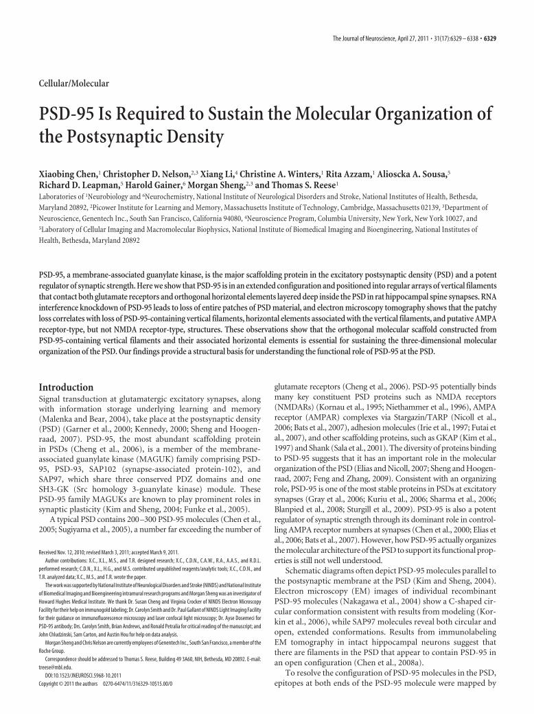
ResultsPSD-95 is in an extended conformation at the PSDPSD-95 tagged at its C terminus with an EYFP fusion construct istargeted to synaptic sites (Craven et al., 1999). Overexpression ofthis construct recruits more AMPA receptors to synapses andincreases synaptic transmission (El-Husseini et al., 2000; Futai etal., 2007; Kim et al., 2007), suggesting that EYFP-tagged PSD-95is incorporated into the PSD. Three-week-old cultured hip-pocampal neurons transfected with this construct showed a spinedensity of 2.3 spines per 10 running �m of dendrite (range, 1.3–3.5 spines/10 �m; 246 spines on 15 neurons) (Fig. 1A) (transfec-
tion rate was estimated to be 12% among 293 neurons). Cultureswere fixed and immunolabeled at 16 –20 h of expression, eitherwith an antibody against the N-terminal PDZ1 domain (Chen etal., 2008a) or separately with an antibody against the C-terminalEYFP (Fig. 1B). Immunogold labeling for EYFP occurredthroughout the spines but was concentrated near PSDs, where itlined up �27 nm from the postsynaptic membrane (Fig. 1D–F).Parallel control experiments with cultures transfected with GFPalone showed essentially no labeling for GFP at PSDs, but labelingwas intense throughout the spine (data not shown), confirmingthat specific localization of many of the tagged PSD-95 moleculesin close proximity to the PSD is due to their incorporation intothe PSD (Fig. 1C–F). The label for PDZ1 was located 11.9 � 4.0nm (mean � SD, N � 261, 111 spines) from the postsynapticmembrane. Three different antibodies to EYFP yielded essentiallyidentical distances from the postsynaptic membrane: 27.4 � 8.1nm (N � 230, 24 spines); 27.1 � 7.2 nm (N � 326, 33 spines); and26.9 � 9.0 nm (N � 155, 30 spines) ( p � 0.91– 0.99, one-wayANOVA, Tukey HSD post-tests) (Fig. 1G). The distance from thepostsynaptic membrane for PDZ1 site label is distinct from thosefor EYFP site ( p � 0.0001, one-way ANOVA, Tukey HSD post-tests) (Fig. 1G). The PDZ1 domain is on average 15.2 nm closer tothe postsynaptic membrane than the EYFP domain, consistentwith PSD-95 filaments in extended form and vertically orientedwith respect to the postsynaptic membrane.
EM tomography of hippocampal spines transfected withtagged PSD-95 and immunolabeled for EYFP showed labels spe-cifically at the distal ends of vertical filaments lying away from thepostsynaptic membrane (Fig. 1H, I). The length of the labeledvertical filaments measured directly from the tomogram—17.1 � 0.7 nm (N � 14, 2 spines)—matched not only the lengthof vertical filaments in intact PSDs from EM tomographic recon-structions (Chen et al., 2008a), but also the lengths of individualextended recombinant PSD-95 and SAP97 molecules measuredby single-particle electron microscopy (Nakagawa et al., 2004).
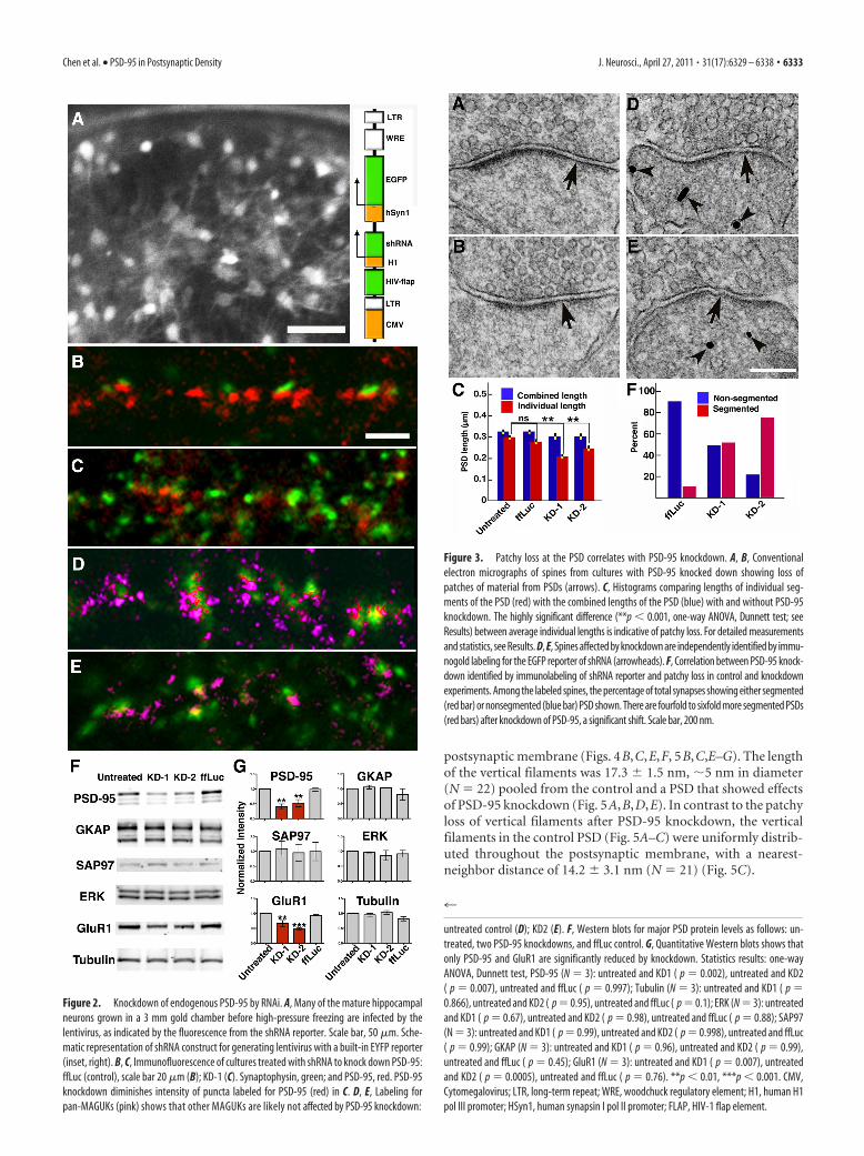
PSD-95 knockdown with RNAiAlthough some vertical filaments seen with tomography immu-nolabeled for PSD-95 in this and our previous study (Chen et al.,2008a), the molecular identities of the population of vertical fil-aments as a whole remain an open question. Lentivirus-basedshRNA constructs (KD-1 and KD-2 are targeting different se-quences; see Materials and Methods) (Fig. 2A, inset), wereused to knock down PSD-95, and were compared with a con-trol shRNA construct targeting fruit fly luciferase (ffLuc). Thehigh-titer virus (�10 8 particles/ml) infected �73, 83, and95% of neurons (see Materials and Methods), as indicated byan EGFP reporter (Fig. 2 A). The high rate of infection assuredthat affected neurons could be sampled readily by electronmicroscopy.
To minimize off-target effects, neurons were examined only3 d after virus infection, at which time overall PSD-95 expression,as evaluated by Western blot, was reduced to 40 –50% of controls(Fig. 2F,G). The levels of GKAP, SAP97, and ERK appeared un-affected, but GluR1 was reduced as expected (Nakagawa et al.,2004) (Fig. 2F,G). Immunofluorescent colabeling for PSD-95and synaptophysin after PSD-95 knockdown (Fig. 2C) showed,in contrast to ffLuc control cultures (Fig. 2B), a decrease in thenumber of PSD-95 puncta relative to synaptophysin puncta.However, colabeling by pan-MAGUK and synaptophysin anti-bodies (Fig. 2E) was indistinguishable among PSD-95 knock-downs (Fig. 2E) and controls (Fig. 2B, 2D). We interpret this
Table 1. Density of vertical filaments and AMPARs at the periphery of the PSD
Vertical filaments/0.1 �m 2
AMPAR structures/0.1 �m 2
Vertical filamentsnearest neighbor (nm)
Control (N � 4) 304 � 56 71 � 16 16 � 4Knockdown (N � 5) 195 � 45* 48 � 16* 28 � 7**
The density is an area 125 nm wide and 100 nm thick surrounding the central cluster of NMDAR-type structures.*p � 0.03, ** p � 0.0001, two-tailed Student’s t test.
Chen et al. • PSD-95 in Postsynaptic Density J. Neurosci., April 27, 2011 • 31(17):6329 – 6338 • 6331
result to mean that other MAGUK pro-teins remain at affected synapses afterknockdown of PSD-95.
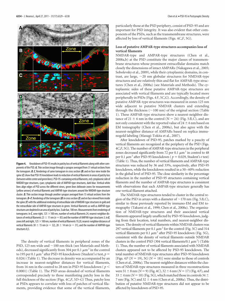
PSD-95 knockdown leads to patchy lossat PSDsRat hippocampal cultures inoculated withshRNA constructs (KD-1, KD-2, andffLuc, a control), as well as parallel un-treated controls, were examined withconventional electron microscopy. Alto-gether 386 PSDs at dendritic spines wererandomly sampled in a blind analysis. Ap-proximately 40% of PSDs at spine syn-apses showed some loss of electron densematerial, which appeared either as thebreakup of continuous PSDs or the short-ening of PSDs (Fig. 3A,B). The extent ofloss was variable, sometimes affecting thewhole length of the PSD (Fig. 3A), butmore typically manifesting as patches ofloss at the periphery of the PSD (Fig. 3B),leaving each individual PSD segmentshorter than the corresponding presynap-tic specialization. The lengths of the indi-vidual segments within each PSD weremeasured to compare the segmentation inPSDs among untreated controls, ffLuccontrols, and PSD-95 knockdowns. Theaverage length of individual segments ineach PSD was compared with the totalcombined length of all the fragments atthat PSD. PSDs that are more segmentedwere expected to show a lower ratio of av-erage length of individual segments to the total length of the PSD(Fig. 3E). The average combined PSD lengths (325 � 12 nm,mean � SEM; N � 95) in untreated cultures did not differ sig-nificantly from the average lengths of individual segments inPSDs (300 � 12 nm; N � 103; Student’s t test, p � 0.14). In fact,the averaged combined PSD lengths varied little between differ-ent groups and generally matched the length of postsynapticmembrane directly apposed to the presynaptic active zone. Com-bined lengths for untreated controls (325 � 12 nm; N � 95),ffLuc controls (326 � 13 nm; N � 87; one-way ANOVA, Dunnetttest, p � 1), KD-1 (301 � 16 nm; N � 48; p � 0.56), and KD-2(301 � 14 nm; N � 90; p � 0.37) are not significantly different,though there may be some reduction in total PSD length afterPSD-95 knockdown (Fig. 3E). The individual PSD lengths fromuntreated neurons (300 � 12 nm; N � 103) were not significantlydifferent from those from the ffLuc control (276 � 11 nm; N �103; one-way ANOVA, Dunnett test, p � 0.29), but were signif-icantly shorter by 30 and 19%, respectively, in the knockdownsKD-1 (210 � 12 nm; N � 69; p � 0.0001) and KD-2 (244 � 12nm; N � 111; p � 0.0012) (Fig. 3E). These measurements con-firm that knockdown of PSD-95 results in the breaking up andshortening of PSDs, giving rise to the appearance of patchyloss.
Patchy loss at PSDs is directly associated with PSD-95knockdownWe next determined whether morphological changes in PSDs inindividual spines directly correlate with knockdown of PSD-95.Immunogold labeling of the EGFP reporter coexpressed from the
lentiviral shRNA constructs served to identify spines in whichPSD-95 was knocked down, providing for direct correlation ofPSD-95 knockdown with patchy loss at PSDs (Fig. 3C,D). InffLuc controls, 86% spines (N � 79) had continuous, nonseg-mented PSDs, either labeled (43%) or nonlabeled (43%), while14% showed segmented PSDs (5% labeled, 9% unlabeled), asexpected (Neuhoff et al., 1999). In the PSD-95 knockdown KD-1,60% of spines (N � 50) had nonsegmented PSDs (34% labeled,26% nonlabeled), while 40% of spines had segmented PSDs (38%labeled, 2% not labeled). In KD-2, 42% of spines (N � 45) hadnonsegmented PSDs (16% labeled, 26% not labeled), and 58% oftotal spines had segmented PSDs (54% is labeled, 4% nonla-beled). When the labeled spines alone are analyzed, there arefourfold to sixfold more synapses showing segmented PSDs afterPSD-95 knockdown (Fig. 3F, red bars). Thus, at the single spinelevel, PSD-95 knockdown is strongly correlated with patchy lossand thinning of PSDs.
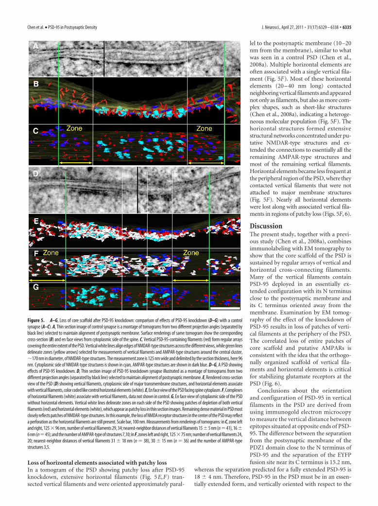
Patchy loss at PSDs is associated with loss of verticalfilamentsTwenty-four dual-axis tomographic reconstructions of spinePSDs showing patchy loss due to PSD-95 knockdown were se-lected for detailed analysis, and areas of patchy loss 30 – 80 nm indiameter were marked by a paucity of vertical filaments (Figs.4C,F, 5F,G). One tomogram from untreated control cultures wassurface rendered (Fig. 5A–C) to produce a three-dimensionalimage of the control PSD for comparison. The dominant struc-tural elements in PSDs from both control and knockdown exper-iments were the arrays of vertical filaments perpendicular to the
Figure 1. Conformation of PSD-95 in spine PSDs. A, Confocal image of 3 week hippocampal culture transfected with PSD-95-EYFP. B, Immunolabeled sites (arrowheads) on the tagged PSD-95 molecule showing predicted distance between these sites whenPSD-95 is in extended configuration. C, Distribution of immunolabels using antibody to PDZ1 domain in 3 week hippocampalculture transfected with PSD-95-EYFP. D–F, Distributions of immunolabels with three different antibodies to EYFP site in parallelwith 3 week hippocampal culture transfected with PSD-95-EYFP. G, Comparison of vertical separations of the immunolabels to thePDZ1 domain and to the EYFP site in the proximity of the PSD (�60 nm from the postsynaptic membrane). Each distribution (PDZ1,black; EYFPs red, green, blue) is fitted with a Gaussian (PDZ1, black; EYFP, green). H, Virtual section derived from the tomographicreconstruction of a spine synapse, immunolabeled with an antibody to the EYFP site. Green dots highlight centers of silver-enhanced immunogold particles associated with vertical filaments in the PSD (I ). I, Surface rendering of the PSD in H. Verticalfilaments �17 nm long (red) bridge immunogold particles and the postsynaptic membrane. Silver-enhanced Nanogold particlesshown in green and structures thought to be parts of bridging antibodies shown in blue. For detailed measurements and statistics,see Results. Scale bars, 100 nm.
6332 • J. Neurosci., April 27, 2011 • 31(17):6329 – 6338 Chen et al. • PSD-95 in Postsynaptic Density
postsynaptic membrane (Figs. 4B,C,E,F, 5B,C,E–G). The lengthof the vertical filaments was 17.3 � 1.5 nm, �5 nm in diameter(N � 22) pooled from the control and a PSD that showed effectsof PSD-95 knockdown (Fig. 5A,B,D,E). In contrast to the patchyloss of vertical filaments after PSD-95 knockdown, the verticalfilaments in the control PSD (Fig. 5A–C) were uniformly distrib-uted throughout the postsynaptic membrane, with a nearest-neighbor distance of 14.2 � 3.1 nm (N � 21) (Fig. 5C).
Figure 2. Knockdown of endogenous PSD-95 by RNAi. A, Many of the mature hippocampalneurons grown in a 3 mm gold chamber before high-pressure freezing are infected by thelentivirus, as indicated by the fluorescence from the shRNA reporter. Scale bar, 50 �m. Sche-matic representation of shRNA construct for generating lentivirus with a built-in EYFP reporter(inset, right). B, C, Immunofluorescence of cultures treated with shRNA to knock down PSD-95:ffLuc (control), scale bar 20 �m (B); KD-1 (C). Synaptophysin, green; and PSD-95, red. PSD-95knockdown diminishes intensity of puncta labeled for PSD-95 (red) in C. D, E, Labeling forpan-MAGUKs (pink) shows that other MAGUKs are likely not affected by PSD-95 knockdown:
4
untreated control (D); KD2 (E). F, Western blots for major PSD protein levels as follows: un-treated, two PSD-95 knockdowns, and ffLuc control. G, Quantitative Western blots shows thatonly PSD-95 and GluR1 are significantly reduced by knockdown. Statistics results: one-wayANOVA, Dunnett test, PSD-95 (N � 3): untreated and KD1 ( p � 0.002), untreated and KD2( p � 0.007), untreated and ffLuc ( p � 0.997); Tubulin (N � 3): untreated and KD1 ( p �0.866), untreated and KD2 ( p � 0.95), untreated and ffLuc ( p � 0.1); ERK (N � 3): untreatedand KD1 ( p � 0.67), untreated and KD2 ( p � 0.98), untreated and ffLuc ( p � 0.88); SAP97(N � 3): untreated and KD1 ( p � 0.99), untreated and KD2 ( p � 0.998), untreated and ffLuc( p � 0.99); GKAP (N � 3): untreated and KD1 ( p � 0.96), untreated and KD2 ( p � 0.99),untreated and ffLuc ( p � 0.45); GluR1 (N � 3): untreated and KD1 ( p � 0.007), untreatedand KD2 ( p � 0.0005), untreated and ffLuc ( p � 0.76). **p � 0.01, ***p � 0.001. CMV,Cytomegalovirus; LTR, long-term repeat; WRE, woodchuck regulatory element; H1, human H1pol III promoter; HSyn1, human synapsin I pol II promoter; FLAP, HIV-1 flap element.
Figure 3. Patchy loss at the PSD correlates with PSD-95 knockdown. A, B, Conventionalelectron micrographs of spines from cultures with PSD-95 knocked down showing loss ofpatches of material from PSDs (arrows). C, Histograms comparing lengths of individual seg-ments of the PSD (red) with the combined lengths of the PSD (blue) with and without PSD-95knockdown. The highly significant difference (**p � 0.001, one-way ANOVA, Dunnett test; seeResults) between average individual lengths is indicative of patchy loss. For detailed measurementsand statistics, see Results. D, E, Spines affected by knockdown are independently identified by immu-nogold labeling for the EGFP reporter of shRNA (arrowheads). F, Correlation between PSD-95 knock-down identified by immunolabeling of shRNA reporter and patchy loss in control and knockdownexperiments. Among the labeled spines, the percentage of total synapses showing either segmented(red bar) or nonsegmented (blue bar) PSD shown. There are fourfold to sixfold more segmented PSDs(red bars) after knockdown of PSD-95, a significant shift. Scale bar, 200 nm.
Chen et al. • PSD-95 in Postsynaptic Density J. Neurosci., April 27, 2011 • 31(17):6329 – 6338 • 6333
The density of vertical filaments in peripheral zones of thePSD, 125 nm wide and �100 nm thick (see Materials and Meth-ods), decreased significantly from 304 per 0.1 �m 2 in the controlto 195 per 0.1 �m 2 after PSD-95 knockdown (Student’s t test, p �0.026) (Table 1). The decrease in density was accompanied by anincrease in nearest-neighbor distances for vertical filaments,from 16 nm in the control to 28 nm in PSD-95 knockdown ( p �0.0001) (Table 1). The PSD areas denuded of vertical filamentscorresponded precisely to those manifesting patchy loss in thefull thickness of the section (Figs. 4C,F, 5D–G) Thus, patchy lossat PSDs appears to correlate with loss of patches of vertical fila-ments, providing evidence that some of the vertical filaments,
particularly those at the PSD periphery, consist of PSD-95 and areimportant for PSD integrity. It was also evident that other com-ponents of the PSDs, such as the transmembrane structures, wereaffected by loss of vertical filaments (Figs. 4C,F, 5G).
Loss of putative AMPAR-type structures accompanies loss ofvertical filamentsNMDAR-type and AMPAR-type structures (Chen et al.,2008a,b) at the PSD constitute the major classes of transmem-brane structures whose prominent extracellular domains matchclosely the dimensions of intact AMPARs (Nakagawa et al., 2005;Sobolevsky et al., 2009), while their cytoplasmic domains, in con-trast, are large, �20 nm globular structures for NMDAR-typestructures and are relatively thin and flat for AMPAR-type struc-tures (Chen et al., 2008a) (see Materials and Methods). The cy-toplasmic sides of these putative AMPAR-type structures areassociated with vertical filaments and are typically located moreperipherally in PSDs (Figs. 4F, 5C,G). Accordingly, the density ofputative AMPAR-type structures was measured in zones 125 nmwide adjacent to putative NMDAR clusters and extendingthrough the thickness (�100 nm) of the original section (Table1). These AMPAR-type structures show a nearest-neighbor dis-tance of 21 � 6 nm in the control (N � 24) (Fig. 5B,C), and arenot only consistent with the reported value of 24 � 6 nm based onEM tomography (Chen et al., 2008a), but also agree with thenearest-neighbor distance of AMPARs based on replica immu-nogold labeling (Masugi-Tokita et al., 2007).
After knockdown of PSD-95, patches marked by a paucity ofvertical filaments are recognized at the periphery of the PSD (Figs.4C,F, 5G). The number of AMPAR-type structures in the peripheralzones decreased significantly from 72 per 0.1 �m2 in control to 48per 0.1 �m2 after PSD-95 knockdown ( p � 0.029, Student’s t test)(Table 1). Thus, the number of vertical filaments and AMPAR-typestructures was reduced by 36 and 33%, respectively, after PSD-95knockdown, while the knockdown resulted in a 50–60% reductionin the global level of PSD-95. The close similarity in the percentagereduction in the number of PSD-95 structures containing verticalfilaments and the number of AMPAR-type structures is consistentwith observations that each AMPAR-type structure generally hasone vertical filament attached.
The NMDAR-type structures tended to cluster in the central re-gion of the PSD in arrays with a diameter of �170 nm (Fig. 5B,C),similar to those previously reported by immuno-EM and EM to-mography (Takumi et al., 1999; Chen et al., 2008a). The organiza-tion of NMDAR-type structures and their associated verticalfilaments appeared largely unaffected by PSD-95 knockdown, judg-ing from their location, total numbers, and nearest-neighbor dis-tances. The density of vertical filaments within NMDAR clusters was297 vertical filaments per 0.1 �m2 for the control (Fig. 5C) and 314vertical filaments per 0.1 �m2 after PSD-95 knockdown (Fig. 5G),consistent with the density of vertical filaments outside NMDARclusters in the control PSD (304 vertical filaments/0.1 �m2) (Table1). Thus, the number of vertical filaments associated with NMDARclusters appeared not to be affected by PSD-95 knockdown. Thetotal number of NMDAR-type structures after PSD-95 knockdown[Figs. 4F (N � 19), 5G (N � 18)] were similar to those of controls(Chen et al., 2008a). The nearest-neighbor distances within the clus-ters of NMDAR-type structures measured in three reconstructionswere 31�8 nm (N�9) (Fig. 4C), 32�6 nm (N�17) (Fig. 4F), and33�4 nm (N�10) (Fig. 5G), which matched those in controls 34�7 nm (Fig. 5C) and 32 � 4 nm (Chen et al., 2008a). Thus, the distri-bution of putative NMDAR-type structures did not appear to beaffected by knockdown of PSD-95.
Figure 4. Knockdown of PSD-95 results in patchy loss of vertical filaments along with other com-ponents of the PSD. A, Thin section image through a synapse averaged from 31 virtual sections fromthe tomogram. B, C, Renderings of same tomogram in cross section (B) and en face view inside thespine (C) show that PSD-95 knockdown leads to reduction of vertical filaments in areas of patchy loss(betweenwhitecenterandgreenlines).PSD-95-containingverticalfilaments,red;cytoplasmicsideofNMDAR type structures, cyan; cytoplasmic side of AMPAR-type structures, dark blue. Vertical whitelines align edges of PSD across the different views, green lines delineate zones for measurements(yellow arrows) of vertical filaments and AMPAR-type structures around the NMDAR-type structurecluster. D, Thin section image through another synapse averaged from 15 virtual sections from thetomogram. D–F, Renderings of the tomogram (D) in cross section (E) and en face viewed from insidethe spine (F) with the additional rendering of extracellular side of NMDAR-type structures in gold andthe extracellular side of AMPAR-type structures in green. Vertical filaments as well as AMPAR-typestructures are depleted in an area of patchy loss. Scale bar, 100 nm. Measurements from renderings oftomograms: in C, zone right, 125 �100 nm, number of vertical filaments 24, nearest-neighbor dis-tances of vertical filaments 23 � 11 nm (n � 85) and the number of AMPAR-type structures 3; in E,zones left and right, 125�100 nm, number of vertical filaments 19,20, nearest-neighbor distances ofvertical filaments 30 � 15 nm (n � 32), 28 � 14 nm (n � 31), and the number of AMPAR-typestructures 8,5.
6334 • J. Neurosci., April 27, 2011 • 31(17):6329 – 6338 Chen et al. • PSD-95 in Postsynaptic Density
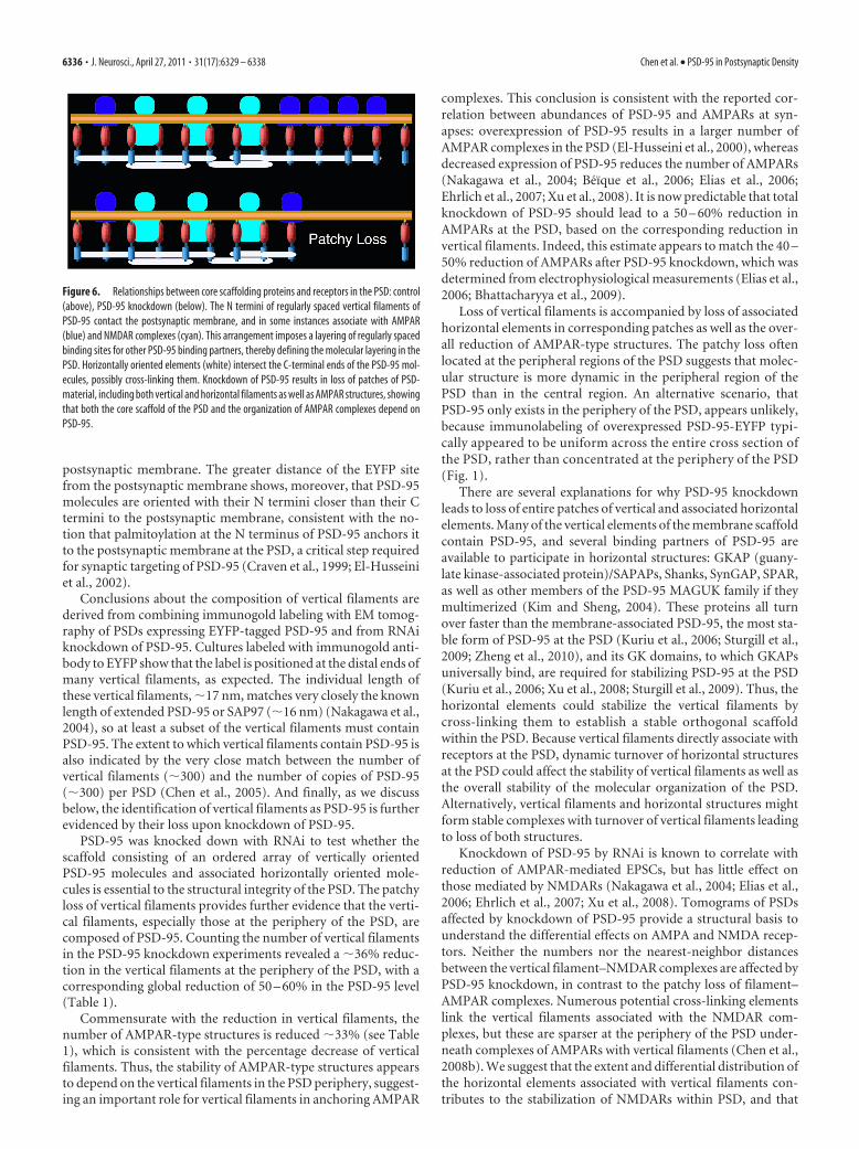
Loss of horizontal elements associated with patchy lossIn a tomogram of the PSD showing patchy loss after PSD-95knockdown, extensive horizontal filaments (Fig. 5E,F) tran-sected vertical filaments and were oriented approximately paral-
lel to the postsynaptic membrane (10 –20nm from the membrane), similar to whatwas seen in a control PSD (Chen et al.,2008a). Multiple horizontal elements areoften associated with a single vertical fila-ment (Fig. 5F). Most of these horizontalelements (20 – 40 nm long) contactedneighboring vertical filaments and appearednot only as filaments, but also as more com-plex shapes, such as sheet-like structures(Chen et al., 2008a), indicating a heteroge-neous molecular population (Fig. 5F). Thehorizontal structures formed extensivestructural networks concentrated under pu-tative NMDAR-type structures and ex-tended the connections to essentially all theremaining AMPAR-type structures andmost of the remaining vertical filaments.Horizontal elements became less frequent atthe peripheral region of the PSD, where theycontacted vertical filaments that were notattached to major membrane structures(Fig. 5F). Nearly all horizontal elementswere lost along with associated vertical fila-ments in regions of patchy loss (Figs. 5F, 6).
DiscussionThe present study, together with a previ-ous study (Chen et al., 2008a), combinesimmunolabeling with EM tomography toshow that the core scaffold of the PSD issustained by regular arrays of vertical andhorizontal cross-connecting filaments.Many of the vertical filaments containPSD-95 deployed in an essentially ex-tended configuration with its N terminusclose to the postsynaptic membrane andits C terminus oriented away from themembrane. Examination by EM tomog-raphy of the effect of the knockdown ofPSD-95 results in loss of patches of verti-cal filaments at the periphery of the PSD.The correlated loss of entire patches ofcore scaffold and putative AMPARs isconsistent with the idea that the orthogo-nally organized scaffold of vertical fila-ments and horizontal elements is criticalfor stabilizing glutamate receptors at thePSD (Fig. 6).
Conclusions about the orientationand configuration of PSD-95 in verticalfilaments in the PSD are derived fromusing immunogold electron microcopyto measure the vertical distance betweenepitopes situated at opposite ends of PSD-95. The difference between the separationfrom the postsynaptic membrane of thePDZ1 domain close to the N terminus ofPSD-95 and the separation of the EYFPfusion site near its C terminus is 15.2 nm,
whereas the separation predicted for a fully extended PSD-95 is18 � 4 nm. Therefore, PSD-95 in the PSD must be in an essen-tially extended form, and vertically oriented with respect to the
Figure 5. A–G, Loss of core scaffold after PSD-95 knockdown: comparison of effects of PSD-95 knockdown (D–G) with a controlsynapse (A–C). A, Thin section image of control synapse is a montage of tomograms from two different projection angles (separated byblack line) selected to maintain alignment of postsynaptic membrane. Surface renderings of same tomogram show the correspondingcross-section (B) and en face views from cytoplasmic side of the spine. C, Vertical PSD-95-containing filaments (red) form regular arrayscovering the entire extent of the PSD. Vertical white lines align edges of NMDAR-type structures across the different views, while green linesdelineate zones (yellow arrows) selected for measurements of vertical filaments and AMPAR-type structures around the central cluster,�170 nm in diameter, of NMDAR-type structures. The measurement zone is 125 nm wide and delimited by the section thickness, here 96nm. Cytoplasmic side of NMDAR-type structures is shown in cyan, AMPAR-type structures are shown in dark blue. D–G, A PSD showingeffects of PSD-95 knockdown. D, Thin section image of PSD-95 knockdown synapse illustrated as a montage of tomograms from twodifferent projection angles (separated by black line) selected to maintain alignment of postsynaptic membrane. E, Rendered cross-sectionview of the PSD (D) showing vertical filaments, cytoplasmic side of major transmembrane structures, and horizontal elements associatewith vertical filaments, color coded like control horizontal elements (white). E, En face view of the PSD facing spine cytoplasm. F, Complexesof horizontal filaments (white) associate with vertical filaments, data not shown in control. G, En face view of cytoplasmic side of the PSDwithout horizontal elements. Vertical white lines delineate zones on each side of the PSD showing patches of depletion of both verticalfilaments (red) and horizontal elements (white), which appear as patchy loss in thin section images. Remaining dense material in PSD mostclosely reflects patches of NMDAR-type structures. In this example, the loss of NMDA receptor structures in the center of the PSD may reflecta perforation as the horizontal filaments are still present. Scale bar, 100 nm. Measurements from renderings of tomograms: in C, zone leftand right, 125�96 nm, number of vertical filaments 29, 34; nearest-neighbor distances of vertical filaments 15�5 nm (n�41), 16�6 nm (n�45); and the number of AMPAR-type of structures 7,10; in F, zones left and right, 125�75 nm; number of vertical filaments 24,20; nearest-neighbor distances of vertical filaments 31 � 18 nm (n � 38), 30 � 15 nm (n � 36) and the number of AMPAR-typestructures 3,5.
Chen et al. • PSD-95 in Postsynaptic Density J. Neurosci., April 27, 2011 • 31(17):6329 – 6338 • 6335
postsynaptic membrane. The greater distance of the EYFP sitefrom the postsynaptic membrane shows, moreover, that PSD-95molecules are oriented with their N termini closer than their Ctermini to the postsynaptic membrane, consistent with the no-tion that palmitoylation at the N terminus of PSD-95 anchors itto the postsynaptic membrane at the PSD, a critical step requiredfor synaptic targeting of PSD-95 (Craven et al., 1999; El-Husseiniet al., 2002).
Conclusions about the composition of vertical filaments arederived from combining immunogold labeling with EM tomog-raphy of PSDs expressing EYFP-tagged PSD-95 and from RNAiknockdown of PSD-95. Cultures labeled with immunogold anti-body to EYFP show that the label is positioned at the distal ends ofmany vertical filaments, as expected. The individual length ofthese vertical filaments, �17 nm, matches very closely the knownlength of extended PSD-95 or SAP97 (�16 nm) (Nakagawa et al.,2004), so at least a subset of the vertical filaments must containPSD-95. The extent to which vertical filaments contain PSD-95 isalso indicated by the very close match between the number ofvertical filaments (�300) and the number of copies of PSD-95(�300) per PSD (Chen et al., 2005). And finally, as we discussbelow, the identification of vertical filaments as PSD-95 is furtherevidenced by their loss upon knockdown of PSD-95.
PSD-95 was knocked down with RNAi to test whether thescaffold consisting of an ordered array of vertically orientedPSD-95 molecules and associated horizontally oriented mole-cules is essential to the structural integrity of the PSD. The patchyloss of vertical filaments provides further evidence that the verti-cal filaments, especially those at the periphery of the PSD, arecomposed of PSD-95. Counting the number of vertical filamentsin the PSD-95 knockdown experiments revealed a �36% reduc-tion in the vertical filaments at the periphery of the PSD, with acorresponding global reduction of 50 – 60% in the PSD-95 level(Table 1).
Commensurate with the reduction in vertical filaments, thenumber of AMPAR-type structures is reduced �33% (see Table1), which is consistent with the percentage decrease of verticalfilaments. Thus, the stability of AMPAR-type structures appearsto depend on the vertical filaments in the PSD periphery, suggest-ing an important role for vertical filaments in anchoring AMPAR
complexes. This conclusion is consistent with the reported cor-relation between abundances of PSD-95 and AMPARs at syn-apses: overexpression of PSD-95 results in a larger number ofAMPAR complexes in the PSD (El-Husseini et al., 2000), whereasdecreased expression of PSD-95 reduces the number of AMPARs(Nakagawa et al., 2004; Beïque et al., 2006; Elias et al., 2006;Ehrlich et al., 2007; Xu et al., 2008). It is now predictable that totalknockdown of PSD-95 should lead to a 50 – 60% reduction inAMPARs at the PSD, based on the corresponding reduction invertical filaments. Indeed, this estimate appears to match the 40 –50% reduction of AMPARs after PSD-95 knockdown, which wasdetermined from electrophysiological measurements (Elias et al.,2006; Bhattacharyya et al., 2009).
Loss of vertical filaments is accompanied by loss of associatedhorizontal elements in corresponding patches as well as the over-all reduction of AMPAR-type structures. The patchy loss oftenlocated at the peripheral regions of the PSD suggests that molec-ular structure is more dynamic in the peripheral region of thePSD than in the central region. An alternative scenario, thatPSD-95 only exists in the periphery of the PSD, appears unlikely,because immunolabeling of overexpressed PSD-95-EYFP typi-cally appeared to be uniform across the entire cross section ofthe PSD, rather than concentrated at the periphery of the PSD(Fig. 1).
There are several explanations for why PSD-95 knockdownleads to loss of entire patches of vertical and associated horizontalelements. Many of the vertical elements of the membrane scaffoldcontain PSD-95, and several binding partners of PSD-95 areavailable to participate in horizontal structures: GKAP (guany-late kinase-associated protein)/SAPAPs, Shanks, SynGAP, SPAR,as well as other members of the PSD-95 MAGUK family if theymultimerized (Kim and Sheng, 2004). These proteins all turnover faster than the membrane-associated PSD-95, the most sta-ble form of PSD-95 at the PSD (Kuriu et al., 2006; Sturgill et al.,2009; Zheng et al., 2010), and its GK domains, to which GKAPsuniversally bind, are required for stabilizing PSD-95 at the PSD(Kuriu et al., 2006; Xu et al., 2008; Sturgill et al., 2009). Thus, thehorizontal elements could stabilize the vertical filaments bycross-linking them to establish a stable orthogonal scaffoldwithin the PSD. Because vertical filaments directly associate withreceptors at the PSD, dynamic turnover of horizontal structuresat the PSD could affect the stability of vertical filaments as well asthe overall stability of the molecular organization of the PSD.Alternatively, vertical filaments and horizontal structures mightform stable complexes with turnover of vertical filaments leadingto loss of both structures.
Knockdown of PSD-95 by RNAi is known to correlate withreduction of AMPAR-mediated EPSCs, but has little effect onthose mediated by NMDARs (Nakagawa et al., 2004; Elias et al.,2006; Ehrlich et al., 2007; Xu et al., 2008). Tomograms of PSDsaffected by knockdown of PSD-95 provide a structural basis tounderstand the differential effects on AMPA and NMDA recep-tors. Neither the numbers nor the nearest-neighbor distancesbetween the vertical filament–NMDAR complexes are affected byPSD-95 knockdown, in contrast to the patchy loss of filament–AMPAR complexes. Numerous potential cross-linking elementslink the vertical filaments associated with the NMDAR com-plexes, but these are sparser at the periphery of the PSD under-neath complexes of AMPARs with vertical filaments (Chen et al.,2008b). We suggest that the extent and differential distribution ofthe horizontal elements associated with vertical filaments con-tributes to the stabilization of NMDARs within PSD, and that
Figure 6. Relationships between core scaffolding proteins and receptors in the PSD: control(above), PSD-95 knockdown (below). The N termini of regularly spaced vertical filaments ofPSD-95 contact the postsynaptic membrane, and in some instances associate with AMPAR(blue) and NMDAR complexes (cyan). This arrangement imposes a layering of regularly spacedbinding sites for other PSD-95 binding partners, thereby defining the molecular layering in thePSD. Horizontally oriented elements (white) intersect the C-terminal ends of the PSD-95 mol-ecules, possibly cross-linking them. Knockdown of PSD-95 results in loss of patches of PSD-material, including both vertical and horizontal filaments as well as AMPAR structures, showingthat both the core scaffold of the PSD and the organization of AMPAR complexes depend onPSD-95.
6336 • J. Neurosci., April 27, 2011 • 31(17):6329 – 6338 Chen et al. • PSD-95 in Postsynaptic Density
their sparse distribution at the periphery of the PSD allows verti-cal filaments and AMPARs there to turn over more rapidly.
The first PSD-95 knock-out mice (Migaud et al., 1998), whichstill expressed a truncated portion of PSD-95 (PDZ1–2) appearedto have normal, electron-dense PSDs that included typical levelsof NMDARs. Subsequent characterization of complete knockoutof PSD-95 in mice demonstrated that PSD-95 is necessary forsynaptic targeting of AMPARs (Beïque et al., 2006; Elias et al.,2006), but knockout of a single MAGUK protein (PSD-95 orPSD-93) generally does not significantly affect synaptic transmis-sion. Double knockout of PSD-95/PSD-93 in mice clearly dem-onstrated compensation by a remaining MAGUK SAP-102 (Eliaset al., 2006). Thus, the patchy loss in PSDs so apparent uponacute knockdown of PSD-95 knockdown could well be maskedin knock-out mice by compensation from other members ofMAGUK family proteins.
A scaffold, consisting of vertical filaments containing PSD-95anchored in the membrane of the PSD, along with associatedhorizontal elements, appears to be fundamental to sustaining themolecular organization of the PSD. Because these scaffoldingmolecules directly associate with transmembrane structures (re-ceptors) in the PSD, they might function as slot proteins (Lismanand Raghavachari, 2006). Deletion of core components of thescaffold, as exemplified by acute knockdown of PSD-95 withRNAi, is shown here to be an important approach to unravelingthe molecular organization of synapses.
ReferencesBats C, Groc L, Choquet D (2007) The interaction between Stargazin and
PSD-95 regulates AMPA receptor surface trafficking. Neuron53:719 –734.
Beïque JC, Lin DT, Kang MG, Aizawa H, Takamiya K, Huganir RL (2006)Synapse-specific regulation of AMPA receptor function by PSD-95. ProcNatl Acad Sci U S A 103:19535–19540.
Bhattacharyya S, Biou V, Xu W, Schluter O, Malenka RC (2009) A criticalrole for PSD-95/AKAP interactions in endocytosis of synaptic AMPAreceptors. Nat Neurosci 12:172–181.
Blanpied TA, Kerr JM, Ehlers MD (2008) Structural plasticity with pre-served topology in the postsynaptic protein network. Proc Natl Acad SciU S A 105:12587–12592.
Chen L, Chetkovich DM, Petralia RS, Sweeney NT, Kawasaki Y, Wenthold RJ,Bredt DS, Nicoll RA (2000) Stargazin regulates synaptic targeting ofAMPA receptors by two distinct mechanisms. Nature 408:936 –943.
Chen X, Vinade L, Leapman RD, Petersen JD, Nakagawa T, Phillips TM,Sheng M, Reese TS (2005) Mass of the postsynaptic density and enu-meration of three key molecules. Proc Natl Acad Sci U S A102:11551–11556.
Chen X, Winters C, Azzam R, Li X, Galbraith JA, Leapman RD, Reese TS(2008a) Organization of the core structure of the postsynaptic density.Proc Natl Acad Sci U S A 105:4453– 4458.
Chen X, Winters CA, Reese TS (2008b) Life inside a thin section: tomogra-phy. J Neurosci 28:9321–9327.
Cheng D, Hoogenraad CC, Rush J, Ramm E, Schlager MA, Duong DM, XuP, Wijayawardana SR, Hanfelt J, Nakagawa T, Sheng M, Peng J (2006)Relative and absolute quantification of postsynaptic density proteomeisolated from rat forebrain and cerebellum. Mol Cell Proteomics5:1158 –1170.
Craven SE, El-Husseini AE, Bredt DS (1999) Synaptic targeting of the post-synaptic density protein PSD-95 mediated by lipid and protein motifs.Neuron 22:497–509.
Ehrlich I, Klein M, Rumpel S, Malinow R (2007) PSD-95 is required foractivity-driven synapse stabilization. Proc Natl Acad Sci U S A104:4176 – 4181.
El-Husseini Ael-D, Schnell E, Dakoji S, Sweeney N, Zhou Q, Prange O,Gauthier-Campbell C, Aguilera-Moreno A, Nicoll RA, Bredt DS (2002)Synaptic strength regulated by palmitate cycling on PSD-95. Cell108:849 – 863.
El-Husseini AE, Schnell E, Chetkovich DM, Nicoll RA, Bredt DS (2000)
PSD-95 involvement in maturation of excitatory synapses. Science290:1364 –1368.
Elias GM, Nicoll RA (2007) Synaptic trafficking of glutamate receptors byMAGUK scaffolding proteins. Trends Cell Biol 17:343–352.
Elias GM, Funke L, Stein V, Grant SG, Bredt DS, Nicoll RA (2006) Synapse-specific and developmentally regulated targeting of AMPA receptors by afamily of MAGUK scaffolding proteins. Neuron 52:307–320.
Feng W, Zhang M (2009) Organization and dynamics of PDZ-domain-related supramodules in the postsynaptic density. Nat Rev Neurosci10:87–99.
Funke L, Dakoji S, Bredt DS (2005) Membrane-associated guanylate kinasesregulate adhesion and plasticity at cell junctions. Annu Rev Biochem74:219 –245.
Futai K, Kim MJ, Hashikawa T, Scheiffele P, Sheng M, Hayashi Y (2007)Retrograde modulation of presynaptic release probability through signal-ing mediated by PSD-95-neuroligin. Nat Neurosci 10:186 –195.
Garner CC, Nash J, Huganir RL (2000) PDZ domains in synapse assemblyand signalling. Trends Cell Biol 10:274 –280.
Gray NW, Weimer RM, Bureau I, Svoboda K (2006) Rapid redistribution ofsynaptic PSD-95 in the neocortex in vivo. PLoS Biol 4:e370.
Harlow ML, Ress D, Stoschek A, Marshall RM, McMahan UJ (2001) Thearchitecture of active zone material at the frog’s neuromuscular junction.Nature 409:479 – 484.
Irie M, Hata Y, Takeuchi M, Ichtchenko K, Toyoda A, Hirao K, Takai Y,Rosahl TW, Sudhof TC (1997) Binding of neuroligins to PSD-95. Sci-ence 277:1511–1515.
Kennedy MB (2000) Signal-processing machines at the postsynaptic den-sity. Science 290:750 –754.
Kim E, Sheng M (2004) PDZ domain proteins of synapses. Nat Rev Neuro-sci 5:771–781.
Kim E, Naisbitt S, Hsueh YP, Rao A, Rothschild A, Craig AM, Sheng M(1997) GKAP, a novel synaptic protein that interacts with the guanylatekinase-like domain of the PSD-95/SAP90 family of channel clusteringmolecules. J Cell Biol 136:669 – 678.
Kim MJ, Futai K, Jo J, Hayashi Y, Cho K, Sheng M (2007) Synaptic accumu-lation of PSD-95 and synaptic function regulated by phosphorylation ofserine-295 of PSD-95. Neuron 56:488 –502.
Korkin D, Davis FP, Alber F, Luong T, Shen MY, Lucic V, Kennedy MB, SaliA (2006) Structural modeling of protein interactions by analogy: appli-cation to PSD-95. PLoS Comput Biol 2:e153.
Kornau HC, Schenker LT, Kennedy MB, Seeburg PH (1995) Domain inter-action between NMDA receptor subunits and the postsynaptic densityprotein PSD-95. Science 269:1737–1740.
Kremer JR, Mastronarde DN, McIntosh JR (1996) Computer visualizationof three-dimensional image data using IMOD. J Struct Biol 116:71–76.
Kuriu T, Inoue A, Bito H, Sobue K, Okabe S (2006) Differential control ofpostsynaptic density scaffolds via actin-dependent and -independentmechanisms. J Neurosci 26:7693–7706.
Lisman J, Raghavachari S (2006) A unified model of the presynaptic andpostsynaptic changes during LTP at CA1 synapses. Sci STKE 2006:re11.
Lois C, Hong EJ, Pease S, Brown EJ, Baltimore D (2002) Germline transmis-sion and tissue-specific expression of transgenes delivered by lentiviralvectors. Science 295:868 – 872.
Malenka RC, Bear MF (2004) LTP and LTD: an embarrassment of riches.Neuron 44:5–21.
Masugi-Tokita M, Tarusawa E, Watanabe M, Molnar E, Fujimoto K, Shige-moto R (2007) Number and density of AMPA receptors in individualsynapses in the rat cerebellum as revealed by SDS-digested freeze-fracturereplica labeling. J Neurosci 27:2135–2144.
Migaud M, Charlesworth P, Dempster M, Webster LC, Watabe AM, Makh-inson M, He Y, Ramsay MF, Morris RG, Morrison JH, O’Dell TJ, GrantSG (1998) Enhanced long-term potentiation and impaired learning inmice with mutant postsynaptic density-95 protein. Nature 396:433– 439.
Nakagawa T, Futai K, Lashuel HA, Lo I, Okamoto K, Walz T, Hayashi Y,Sheng M (2004) Quaternary structure, protein dynamics, and synapticfunction of SAP97 controlled by L27 domain interactions. Neuron44:453– 467.
Nakagawa T, Cheng Y, Ramm E, Sheng M, Walz T (2005) Structure anddifferent conformational states of native AMPA receptor complexes. Na-ture 433:545–549.
Chen et al. • PSD-95 in Postsynaptic Density J. Neurosci., April 27, 2011 • 31(17):6329 – 6338 • 6337
Neuhoff H, Roeper J, Schweizer M (1999) Activity-dependent formation ofperforated synapses in cultured hippocampal neurons. Eur J Neurosci11:4241– 4250.
Nicoll RA, Tomita S, Bredt DS (2006) Auxiliary subunits assist AMPA-typeglutamate receptors. Science 311:1253–1256.
Niethammer M, Kim E, Sheng M (1996) Interaction between the C terminus ofNMDA receptor subunits and multiple members of the PSD-95 family ofmembrane-associated guanylate kinases. J Neurosci 16:2157–2163.
Niwa H, Yamamura K, Miyazaki J (1991) Efficient selection for high-expressiontransfectants with a novel eukaryotic vector. Gene 108:193–199.
Sala C, Piech V, Wilson NR, Passafaro M, Liu G, Sheng M (2001) Regulationof Dendritic Spine Morphology and Synaptic Function by Shank andHomer. Neuron 31:115–130.
Seeburg DP, Feliu-Mojer M, Gaiottino J, Pak DT, Sheng M (2008) Criticalrole of CDK5 and Polo-like kinase 2 in homeostatic synaptic plasticityduring elevated activity. Neuron 58:571–583.
Sharma K, Fong DK, Craig AM (2006) Postsynaptic protein mobility indendritic spines: long-term regulation by synaptic NMDA receptor acti-vation. Mol Cell Neurosci 31:702–712.
Sheng M, Hoogenraad CC (2007) The postsynaptic architecture of excitatorysynapses: a more quantitative view. Annu Rev Biochem 76:823–847.
Sobolevsky AI, Rosconi MP, Gouaux E (2009) X-ray structure, symme-try and mechanism of an AMPA-subtype glutamate receptor. Nature462:745–756.
Sturgill JF, Steiner P, Czervionke BL, Sabatini BL (2009) Distinct domainswithin PSD-95 mediate synaptic incorporation, stabilization, andactivity-dependent trafficking. J Neurosci 29:12845–12854.
Sugiyama Y, Kawabata I, Sobue K, Okabe S (2005) Determination of abso-lute protein numbers in single synapses by a GFP-based calibration tech-nique. Nat Methods 2:677– 684.
Takumi Y, Ramírez-Leon V, Laake P, Rinvik E, Ottersen OP (1999) Differ-ent modes of expression of AMPA and NMDA receptors in hippocampalsynapses. Nat Neurosci 2:618 – 624.
Xu W, Schluter OM, Steiner P, Czervionke BL, Sabatini B, Malenka RC(2008) Molecular dissociation of the role of PSD-95 in regulating synap-tic strength and LTD. Neuron 57:248 –262.
Zheng CY, Petralia RS, Wang YX, Kachar B, Wenthold RJ (2010) SAP102 isa highly mobile MAGUK in spines. J Neurosci 30:4757– 4766.
6338 • J. Neurosci., April 27, 2011 • 31(17):6329 – 6338 Chen et al. • PSD-95 in Postsynaptic Density


