Embed Size (px)
Citation preview
The Journal of Molecular Diagnostics, Vol. 13, No. 2, March 2011
Copyright © 2011 American Society for Investigative Pathology
and the Association for Molecular Pathology.
Published by Elsevier Inc. All rights reserved.
DOI: 10.1016/j.jmoldx.2010.11.018
Quality Assurance for Duchenne and BeckerMuscular Dystrophy Genetic Testing
Development of a Genomic DNA Reference Material Panel
Lisa Kalman,* Jay Leonard,† Norman Gerry,†
Jack Tarleton,‡ Christina Bridges,‡
Julie M. Gastier-Foster,§¶� Robert E. Pyatt,¶�
Eileen Stonerock,� Monique A. Johnson,**C. Sue Richards,** Iris Schrijver,†† Tianhui Ma,‡‡
Vanessa Rangel Miller,§§ Yetsa Adadevoh,§§
Pat Furlong,¶¶ Christine Beiswanger,†
and Lorraine Toji†
From the Division of Laboratory Science and Standards,* Centers
for Disease Control and Prevention, Atlanta, Georgia; the Coriell
Institute for Medical Research,† Camden, New Jersey; the Fullerton
Genetics Center,‡ Asheville, North Carolina; the Departments of
Pediatrics § and Pathology,¶ Ohio State University, Columbus,
Ohio; Nationwide Children’s Hospital,� Columbus, Ohio; the
Department of Molecular and Medical Genetics,�� Oregon Health
and Science University, Portland, Oregon; the Department of
Pathology,†† Stanford University School of Medicine, and Stanford
University Medical Center,‡‡ Stanford, California; the Department
of Medical Genetics,§§ Emory University, Atlanta, Georgia; and
Parent Project Muscular Dystrophy,¶¶ Middletown, Ohio
Duchenne and Becker muscular dystrophies (DMD/BMD) are allelic X-linked recessive disorders that af-fect approximately 1 in 3500 and 1 in 20,000 maleindividuals, respectively. Approximately 65% of pa-tients with DMD have deletions, 7% to 10% have du-plications, and 25% to 30% have point mutations in oneor more of the 79 exons of the dystrophin gene. Mostclinical genetics laboratories test for deletions, and someuse technologies that can detect smaller mutations andduplications. Reference and quality control materials forDMD/BMD diagnostic and carrier genetic testing are notcommercially available. To help address this need, theCenters for Disease Control and Prevention–based Ge-netic Testing Reference Material Coordination Program, incollaboration with members of the genetic testing and theDMD/BMD patient communities and the Coriell Cell Re-positories, have characterized new and existing cell linesto create a comprehensive DMD/BMD reference materialpanel. Samples from 31 Coriell DMD cell lines from maleprobands and female carriers were analyzed using theAffymetrix SNP Array 6.0 and Multiplex Ligation-Depen-
dent Probe Amplification (MRC-Holland BV, Amsterdam,the Netherlands), a multiplex PCR assay, and DNA se-quence analysis. Identified were 16 cell lines with dele-tions, 9 with duplications, and 4 with point mutationsdistributed throughout the dystrophin gene. There wereno discordant results within assay limitations. Thesesamples are publicly available from Coriell Institute forMedical Research (Camden, NJ) and can be used forquality assurance, proficiency testing, test development,and research, and should help improve the accuracy ofDMD testing. (J Mol Diagn 2011, 13:167–174; DOI:10.1016/j.jmoldx.2010.11.018)
Duchenne muscular dystrophy (DMD) is a severe pro-gressive neuromuscular disorder that is manifested inearly childhood. Patients with this disorder reach mile-stones such as sitting and walking later than expected,experience progressive symmetric muscular weaknessthroughout childhood, and are usually wheelchair-depen-dent by age 13 years. In addition, these patients canhave cognitive impairment and involvement of other or-gans including the heart. Becker muscular dystrophy(BMD) is similar to DMD but with later onset and lessseverity. Patients with BMD usually become wheelchair-dependent at a later age than those with DMD. Both DMDand BMD result in a shortened lifespan, and most af-fected individuals die before their third or fourth decade.DMD is relatively common, occurring in 1 in 3500 livemale births worldwide.1 Both disorders exhibit X-linkedinheritance and are fully penetrant in male patients. Fe-male carriers are at risk for X-linked cardiomyopathy.
DMD and BMD, and DMD-associated cardiomyopathyare caused by mutations in the dystrophin (DMD) gene,which is located at X-p21.22,3 and encodes the 427-kDadystrophin protein4 (1988 Leiden Open Variation Data-
Accepted for publication November 10, 2010.
MRC-Holland BV donated reagents for this project.
The findings and conclusions in this report are those of the authors and do notnecessarily represent the official position of the Centers for Disease Control andPrevention or the Agency for Toxic Substances and Disease Registry.
Address reprint requests to Lisa Kalman, Ph.D., Laboratory Researchand Evaluation Branch, Division of Laboratory Science and Standards,Office of Surveillance, Epidemiology, and Laboratory Services, Centersfor Disease Control and Prevention (CDC), 1600 Clifton Rd, Mailstop G23,
Atlanta, GA 30333. E-mail: [email protected].167
168 Kalman et alJMD March 2011, Vol. 13, No. 2
base, muscular dystrophy, http://www.dmd.nl/DMD_deldup.html, accessed May 18, 2010). DMD has 79 ex-ons and covers nearly 2400 kb5 (1988 Leiden Open Varia-tion Database, muscular dystrophy, http://www.dmd.nl/DMD_deldup.html, accessed May 18, 2010; andGeneCards database, http://www.genecards.org/cgi-bin/carddisp.pl?gene�DMD, accessed May 19, 2010). DMD isbelieved to be one of the largest genes in the human ge-nome. Deletions, duplications, and point mutations ac-count for approximately 65%, 7% to 10%, and 25% to30%, respectively, of mutations in patients withDMD.6,7 Point mutations may occur throughout the gene.Deletions and duplications frequently occur in two “mu-tational hot spot” regions of the gene, located betweenexons 2 to 20 or exons 44 to 53, but may occurthroughout the gene. Deletions and duplications aremuch less common in the 3= end of the gene (LeidenOpen Variation Database, muscular dystrophy, http://www.dmd.nl/DMD_deldup.html, accessed May 18, 2010).
Treatment for DMD and BMD is currently limited to ste-roids to prolong and improve quality of life. In addition,several therapies are being developed for muscular dystro-phy that target particular DMD mutations. Pharmaceuticalagents such as aminoglycosides8 and PCT1249 allow trans-lation of certain nonsense mutations, enabling production ofa full-length functional protein. Morpholino antisense oligo-nucleotides that mediate exon skipping10 are under devel-opment. This experimental therapy would preserve thereading frame of DMD with frame-shifting deletions andenable generation of potentially functional dystrophin mol-ecules. These experimental therapies are targeted to pa-tients with specific types of mutations or mutations in certainexons. Thus, it is vitally important for patients with DMD orBMD to have accurately characterized DMD mutations sothat those eligible for these new therapies or future thera-pies will be able to receive them. Accurate testing is alsoimportant for patient management and carrier testing.
A number of molecular diagnostic methods are used todetect mutations in the DMD gene.7 Targeted multiplex PCRcan be used to quantitatively detect exon and promoterdeletions in male patients. Quantitative PCR analysis can beused to identify DMD exon duplications, and multiplex liga-tion-dependent probe amplification (MLPA) can be used todetect both deletions and duplications in all 79 exons inboth affected male and heterozygous female individuals. Inaddition, newer chip-based technologies such as arraycomparative genomic hybridization and single nucleotidepolymorphism (SNP) chips can enable identification of de-letions and duplications in DMD. Smaller deletions, duplica-tions, splice-site mutations, and point mutations may beidentified in patients with no identified deletion or duplica-tion using mutation scanning or DNA sequence analysis.There are currently no US Food and Drug Administration–approved assays for DMD genetic testing; all testing isperformed using laboratory-developed tests.
Clinical laboratories use characterized reference ma-terials for a variety of quality assurance purposes includ-ing test development, test validation, quality control, andproficiency testing. Ideally, reference materials shouldresemble a patient sample and all variant alleles or mu-
tation types that the assay is designed to detect.11 Use ofreference materials for laboratory quality assurance is alsomandated by regulatory requirements and professionalguidelines12–16 (Washington State Legislature, http://www.doh.wa.gov/hsqa/fsl/lqa_home.htm, accessed Au-gust 30, 2010; New York State Clinical Laboratory Eval-uation Program, http://www.wadsworth.org/labcert/clep/clep.html, accessed July 15, 2010; College of AmericanPathologists, http://www.cap.org/apps/docs/laboratory_accreditation/checklists/molecular_pathology_sep07.pdf,accessed August 30, 2010; and American College of Med-ical Genetics, http://www.acmg.net/Pages/ACMG_Activities/stds-2002/g.htm, accessed July 15, 2010). For genetic testingof DMD and BMD, assays should be developed and evalu-ated using reference materials that represent deletions, dupli-cations, and point mutations throughout the DMD gene. Ref-erence materials should also include both male and femalesamples, and should be thoroughly characterized using meth-ods different from the laboratory’s routine clinical assay. Use ofa characterized and comprehensive reference material panelwill help to ensure proper design and function of the clinicalassay.
Reference materials for clinical DMD genetic testing are notcommercially available. Laboratories commonly use genomicDNA from residual patient specimens or from publicly avail-able cell lines such as those in the Coriell Cell Repositories(Camden, NJ) as reference materials. To help address theneed for publicly available, renewable, and characterizedgenomic DNA reference materials, the Centers for DiseaseControl and Prevention coordinated the Genetic Testing Ref-erence Material Coordination Program (GeT-RM), in collabo-ration with members of the genetic testing community and theCoriell Institute for Medical Research, have identified and char-acterized the mutations in existing DMD cell lines. In addition toexon deletions, some of the cell lines had duplications or pointmutations, which are DMD mutation types that had not previ-ously been identified in the Coriell collection. The GeT-RM hasalso established a collaboration with a DMD patient registry,DuchenneConnect, to collect blood from consenting patientswith DMD with mutations not yet represented at Coriell for cellline development. In the present study, these new and existingcell lines were characterized using a variety of methods tocreate a comprehensive publicly available reference materialpanel for DMD genetic testing.
Materials and Methods
Cell Line Selection
Reference material needs for DMD and BMD genetictesting were identified through discussions with clinicallaboratory directors. Twenty-four cell lines from the Coriellcollection were selected for study based on the sex of thedonor and the exons expected to be deleted or dupli-cated based on preliminary characterization studies.Seven cell lines from patients with DMD with no identifieddeletion or duplication were selected for DNA sequence
analysis.
DMD Reference Materials 169JMD March 2011, Vol. 13, No. 2
Anonymous Blood Collection from ConsentingPatients with DMD
Patients with mutations considered important for creationof a comprehensive reference material panel by the com-mittee of laboratory directors were selected from a de-identified list of patients and their previously determinedDMD genotype provided by the DuchenneConnect PatientRegistry. These patients were issued written requests forparticipation by genetic counselors from DuchenneCon-nect, explaining the project and its goals. If the familywished to participate, DuchenneConnect obtained in-formed consent using the Coriell Model Consent documentand sent a code representing each consenting patientalong with the shipping address and telephone number(required by the shipping service) to the data manager ofthe National Institute of General Medical Science HumanGenetic Cell Repository, part of the Coriell Cell Repositories.Prepaid blood collection kits were mailed to each family,with blood collection tubes coded as directed by the reg-istry. When the patient visited his or her physician for aregular visit, blood was drawn and sent to the Coriell Re-pository using the return shipping label and materials pro-vided; the Repository has no record of the patient name orother identifying information. When the blood specimenswere received, each was assigned a new Repository num-ber and prepared for cell line establishment. Once the cellline was established, the data manager requested the mu-tation and clinical information from the DuchenneConnectPatient Registry using the appropriate sample code.
Establishment of New Cell Lines
Whole-blood samples collected from consenting patientsor their families were sent to the Coriell Cell Repositoriesfor Epstein-Barr virus transformation of B lymphocytes, aspreviously described.17,18 All samples were placed inculture and expanded to yield approximately 2 � 108
total viable cells. The culture medium was antibiotic-freeto increase the likelihood that contamination would bereadily detected. The cell suspension was dispersed intoforty 1-ml ampules containing 5 � 106 viable cells each.Cultures were cryopreserved in heat-sealed borosilicateglass ampules and stored in liquid nitrogen (liquidphase). Successful cultures were free from bacterial, fun-gal, and mycoplasma contamination and were viable af-ter cryopreservation in liquid nitrogen, as evidenced bydoubling of the cell number within 4 days of recovery.
DNA Preparation
Approximately 2 mg of DNA was prepared from culturesof each of the selected cell lines by the Coriell Cell Re-positories using the Gentra Autopure system (QiagenCorp., Gaithersburg, MD) per the manufacturer’s instruc-tions or previously described methods.19
Testing Laboratories
Four clinical genetic laboratories volunteered to participate in
the study. Laboratories were solicited based on their currentDMD assay methods. Thus, each of the DNA samples wastested using a variety of platforms. All of the laboratories arelocated in the United States and are accredited by the Collegeof American Pathologists. All samples were also tested in theCytogenetics Laboratory at the Coriell Cell Repositories.
Protocol
Each of the four volunteer testing laboratories received one50-�g aliquot of DNA from each of the cell lines they wereasked to test. The two laboratories using MLPA analysis re-ceived all 24 samples to test. The laboratory using the PCRassay, which is not designed to test samples from femalecarriers or to detect duplications, received only the 10 samplesfrom male probands expected to exhibit deletions. The labo-ratory that performed DNA sequence analysis received theseven samples in which deletions or duplications were notdetected using the other assay methods. The expected mu-tation in each of the samples was not revealed to the labora-tories. The laboratories genotyped each DNA sample usingtheir standard assay methods. Each laboratory performed oneassay, except for the Coriell Cytogenetics Laboratory, whichperformed both the Affymetrix Human SNP Array 6.0 and 2.7assays (Affymetrix, Santa Clara, CA). The results were sent tothe study coordinator (L.K.), who examined the data fordiscrepancies.
Assays
Targeted Multiplex Assay for Qualitative Detection ofDeletions in Male Patients
This assay, as previously described,20 amplifies 24 ex-ons (exons 3, 4, 6, 8, 12, 13, 16, 17, 19, 32, 34, 41 to 52, and60) and two promoter regions of DMD, which comprise thegene regions in which most mutations are identified. Themultiplex PCR reactions enable detection of as many as 10different exons in a single reaction. Three separate reac-tions are performed per sample. Collectively, the PCR as-says detect greater than 98% of deletions.20 For deletions ofmore than a single exon, the absence of missing PCR prod-ucts in more than one reaction provides assurance that anactual deletion has occurred in the patient. Furthermore, theend points of many deletions can be identified, which canguide prediction of disease severity (eg, DMD versus BMD)based on maintenance or loss of the translational readingframe. Predictions based on the reading frame hypothesis,however, are inaccurate in approximately 10% of the cases,and should be applied with caution.21 Detection of a dele-tion is based on the absence of a PCR product after elec-trophoresis on an agarose gel stained with ethidium bro-mide. This quantitative targeted multiplex PCR assaydetermines the presence or absence of an amplicon, whichreflects the presence or absence of a specific exon. Thus,this assay is only suitable for use in male patients, who havea single X chromosome. Female patients would demon-strate amplicons for all tested exons from their unaffected Xchromosome. This method is not appropriate for femalecarrier or prenatal screening and is not designed to detect
exon duplication or point mutations.
170 Kalman et alJMD March 2011, Vol. 13, No. 2
Multiplex Ligation-Dependent Probe Amplification
The multiplex ligation-dependent probe amplification as-say, which detects duplications and deletions in all DMDexons, was performed in two of the participating clinicallaboratories. Both laboratories used the same procedure.MLPA reagents were developed and manufactured byMRC-Holland BV (Amsterdam, the Netherlands). The spe-cific MLPA kits (PO34 and PO35) used for amplification ofDMD screen for the copy number of all 79 exons in twomultiplex reactions. In brief, 100 ng of target DNA wasdenatured for 5 minutes at 98°C, after which 3 �L of theprobe cocktail was added. The mixture was heated at 95°Cfor 1 minute, and incubated at 60°C overnight (16 hours).Ligation was performed with the temperature-stable li-gase-65 enzyme for 15 minutes at 54°C. After inactivation ofthe ligase, the ligated products were amplified using PCRaccording to the manufacturer’s protocol using one primerlabeled with 6-carboxyfluorescein. PCR was performed for35 cycles (30 seconds at 95°C, 30 seconds at 60°C, and 60seconds at 72°C), and the resulting DNA products werefractionated by size using a capillary electrophoresis instru-ment (model 3130; Applied Biosystems, Inc., Foster City,CA). Fragment mobility was analyzed using GeneMapperFragment Analysis software (Applied Biosystems, Inc.). Thesizes of exon-specific peaks were identified accordingto their migration relative to size standards. Further dataanalysis was performed using Coffalyser Software (MRC-Holland BV) or MLPA analysis–specific software (SoftGe-netics LLC, State College, PA). Exon dosage was calcu-lated for samples from affected male patients usingaveraged peak heights generated during the run fromcharacterized normal male control individuals. Exon dos-age for samples from affected female patients was cal-culated similarly using characterized normal female con-trol individuals. Approximate dosage with no exondeletion is 1 (range, 0.65 to 1.35) for both male andfemale patients. Exon deletions result in no signal formale patients, and reduced signal for female patients(�0.5). A duplication in a male patient is detected as adosage of approximately 2 (range, 1.65 to 2.35), and in afemale patient is approximately 1.5.
Affymetrix Human SNP Array 6.0
The Affymetrix Human SNP Array 6.0 assay (Af-fymetrix, Inc.) detects deletions and duplications but isnot designed specifically for DMD genetic testing. Inbrief, 250 ng of genomic DNA was digested with eitherNsp1 or StyI (New England Biolabs, Inc., Ipswich, MA). Auniversal adaptor oligonucleotide was then ligated to thedigested DNA. The ligated DNA was diluted with water,and three 10-�L aliquots from the StyI digestion and four10 �L aliquots from the Nsp1 digestion were transferredto fresh 96-well plates. PCR master mix (Titanium DNAAmplification Kit; Clontech Laboratories Inc., MountainView, CA) was added to each well, and the reactionswere cycled as follows: 94°C for 3 minutes; 30 cycles of94°C for 30 seconds, 60°C for 45 seconds, and 68°C for15 seconds; 68°C for 7 minutes; and 4°C hold. After PCR,
the 7 reactions for each sample were combined andpurified using Agencourt SNPClean beads (BeckmanCoulter Genomics; Brea, CA). The purified PCR productswere quantified using spectrophotometry to ensure ayield of at least 4 �g/�L. Forty-five microliters (�180 �g)of each PCR product was digested using DNase-1 toproduce fragments of fewer than 185 bp. The fragmentedPCR products were then end-labeled with a biotinylatednucleotide using terminal deoxynucleotidyl transferase.The end-labeled PCR products were combined with ahybridization cocktail, denatured at 95°C for 10 minutes,and incubated at 49°C. Two hundred microliters of eachmixture was applied to an SNP 6.0 array and hybridizedwith rotation at 60 rpm overnight at 50°C. After 16 to 18hours of hybridization, the arrays were washed andstained using the GenomeWideSNP6-450 fluidics proto-col (Affymetrix) with the appropriate buffers and stains.After washing and staining, the arrays were scanned on aGeneChip Scanner 3000 (Affymetrix, Inc., Santa Clara,CA). Analysis was performed using Affymetrix Genotyp-ing Console software (version 3.0). All samples achievedrecommended thresholds for both copy number and SNPdata quality. Data from both SNP and copy numberprobes were used to identify copy number aberrationswhen compared with an internal copy number referenceset created from samples as determined at the CoriellGenotyping and Microarray Center.
Affymetrix Cytogenetics 2.7M
The Affymetrix Cytogenetics 2.7 array (Affymetrix, Inc.)detects deletions and duplications but is not designed spe-cifically for DMD genetic testing. This assay was used toresolve a discrepancy in one sample (GM23127) but wasnot used to test the other samples. One hundred nano-grams of each genomic DNA sample was prepared forwhole-genome amplification. Each sample was treated in1� denaturing solution for 3 minutes at room temperature,followed by incubation on ice and the addition of 1� neu-tralization solution. Amplification master mix was added tothe DNA, and the mixtures were incubated at 30°C for 16hours, followed by inactivation at 65°C for 3 minutes. Theamplified DNA products were purified by binding to mag-netic beads (Affymetrix, Inc.), washing twice with cytosolwashing buffer and eluting with cytosol elution buffer. Thepurified products were quantified using spectrophotometry.A DNA concentration of more than 55 �g/�L indicatedsuccessful amplification. Fragmentation and labeling mas-ter mix was added to the amplified DNA, and the mixtureswere incubated at 37°C for 2 hours, followed by inactivationat 95°C for 10 minutes. Cyto Hyb buffer was added to thefragmented labeled DNA, and the mixtures were denaturedat 95°C for 5 minutes, incubated at 50°C for 15 minutes, andheld at 50°C. The hybridization mixtures were applied to theAffymetrix Whole-Genome 2.7M Array (Affymetrix, Inc.) andallowed to hybridize with rotation at 60 rpm overnight at50°C. After 16 to 19 hours of hybridization, the arrays werewashed and stained using the Cytogenetics-Array-450 flu-idics protocol with the buffers and stains supplied with thereagent kit. The arrays were then scanned on a GeneChip
Scanner 3000 (Affymetrix, Inc.).
DMD Reference Materials 171JMD March 2011, Vol. 13, No. 2
DNA Sequence Analysis
DMD sequence analysis was performed essentially asdescribed.22 PCR was performed under standard condi-tions using primers designed to amplify promoter and ex-onic regions of the gene, with 92 amplicons ranging in sizefrom 1.2 to 1.4 kb. PCR products were separated at capil-lary gel electrophoresis using a Qiaxcel system (QiagenCorp), which enabled visualization of amplicons and detec-tion of deletions. Purification of PCR products was per-formed using the Ampure method (Beckman Coulter) on theBiomek NX (Beckman Coulter). Bidirectional sequencingwas performed on each amplicon using terminator chem-istry (Big Dye, version 3.1; Applied Biosystems). Reactionswere purified with size-exclusion columns (Performa; EdgeBiosystems, Inc., Gaithersburg, MD), and sequencingproducts were separated using capillary electrophoresis(AB3730; Applied Biosystems). Sequence electrophero-grams were analyzed electronically using Sequencher 4.9(GeneCodes Corp., Ann Arbor, MI) or Mutation Surveyor(SoftGenetics, Inc.). Any suspected pathogenic DMD mu-tations were confirmed via a second independent PCRreaction.
Results
The goal of the present study was to create a comprehen-sive panel of publicly available genomic DNA referencematerials for DMD genetic testing. Input about needed ref-erence materials was obtained through discussions withclinical laboratory directors who perform DMD testing. Theclinical laboratory directors decided that the reference ma-terials panel should include DMD deletions, duplications,and point mutations covering as many of the DMD exons aspossible. Both large and small deletions, duplications rang-ing in size from a single exon to many exons, and samplesfrom female DMD deletion and duplication carriers shouldbe represented in the panel.
The National Institute of General Medical Sciences Re-pository at Coriell maintains a large collection of DMD celllines, many with known deletions. There were, however, noknown DMD lines in the Coriell collection with duplicationsor point mutations, and no cell lines were known to havedeletions in the 3= end of the gene. Cell lines from male andfemale donors with deletions that fit the recommendations ofthe clinical laboratory directors were selected for furtherstudy. In addition, DNA samples from DMD cell lines withoutknown mutations were sent to a clinical DMD testing labo-ratory for MLPA analysis. Data were also obtained aboutthese and other cell lines as part of an independent effort bythe Coriell Cell Repositories to characterize their collectionusing the Affymetrix Human SNP Array 6.0 copy numberassay. These analyses resulted in identification of a numberof DMD cell lines with duplications, as well as previouslyunidentified deletions. No deletions or duplications wereidentified in DNA samples from seven of the cell lines usingMLPA or Affymetrix Human SNP Array 6.0 analysis.
To create new cell lines with duplications and deletionsthat were not available in the Coriell collection, a collabora-tion was established with a DMD patient registry, Duchen-
neConnect, to collect blood from consenting patients withDMD with mutations for cell line development. A protocolwas developed for anonymous contact, consent, and bloodcollection from patients with DMD, and was approved bythe Coriell Institutional Review Board. Patients with deletionsand duplications in parts of DMD that were not representedin the Coriell collection were selected from a list of deiden-tified patients with previously identified DMD mutations pro-vided by the registry. The families of the selected patientswere contacted by genetic counselors with the Duchen-neConnect Patient Registry and asked to participate. If thepatient or family agreed, they were provided informationabout the study and asked to sign a consent form. Bloodcollection kits with prepaid mailers were provided to theconsenting patients, who obtained and submitted bloodsamples during their routine medical care. In some cases,mothers and/or siblings of the proband also provided bloodsamples. Through this process, 11 blood samples werereceived and 10 new cell lines were created.
Based on preliminary data from MPLA and AffymetrixHuman SNP Array 6.0 analysis and information fromDuchenneConnect, 16 cell lines with exon deletions, 8with exon duplications, and 7 with no identified deletionor duplication in the DMD gene were selected for furthercharacterization. Cell lines were selected to create acomprehensive reference material panel with exon dele-tions and duplications throughout the DMD gene. Sevenof the selected cell lines are from female deletion orduplication carriers.
All of the DMD cell lines with deletions or duplicationsselected for the panel were analyzed using MLPA analysisin two laboratories and Affymetrix Human SNP Array 6.0analysis in one laboratory. The 10 samples from male pro-bands with expected deletions were also analyzed using atargeted multiplex PCR assay (Table 1).
The results obtained using the various assays fully agreedwith each other when the limitations of the assay methods wereconsidered. The MPLA results from both laboratories using thisassay were identical for the 24 lines with deletions or duplica-tions. Results for MLPA and the Affymetrix Human SNP Array6.0 were identical for all but two of the 24 samples, GM10283and GM23127. In both cases, MLPA detected a deletion thatwas one exon larger than that detected by the Affymetrix Hu-man SNP Array 6.0. Analysis of the array data suggested thatlimited probe coverage for exons 28 and 72 resulted in thesmaller deletion call in these two samples. Sample GM23127was also tested using the Affymetrix Cytogenetics 2.7M array.Similar to the Affymetrix Human SNP Array 6.0, this test de-tected a duplication in exon 27 but was unable to detectduplication in exon 28 because of lack of probe coverage inthis exon. Results from the targeted multiplex PCR assayagreed with results from the other assays. While this assay wasdesigned to detect deletions in 24 of the 79 exons, it cannotreliably detect duplications or mutations in female carriers be-cause amplicons would be produced for all tested exons fromthe unaffected X chromosome. Only samples for which pre-liminary data suggested a deletion in a male sample weretested using this method. When results of this assay werecompared with the data obtained from MLPA and AffymetrixHuman SNP Array 6.0 analyses, discrepant data were found insamples GM07691, GM05089, GM04981, GM04364, and
GM10283 (Table 1). In all cases, the deleted exons not de-
8 and 7nable to
172 Kalman et alJMD March 2011, Vol. 13, No. 2
tected using the PCR method were not included in the designof the assay.
No deletions or duplications were identified in DNA sam-ples from seven existing DMD cell lines at MLPA and Af-fymetrix Human SNP Array 6.0 analysis during preliminarytesting. DNA from these cell lines was analyzed using DNAsequence analysis in one laboratory to identify smaller mu-tations. Point mutations were found in four of the sevensamples. Three of the mutations were translation terminationcodons in exons 39, 41, and 59, and one sample had aone-bp deletion that resulted in a frame shift in exon 54(Table 1). No mutations were identified by sequence anal-ysis in the other three samples. It is possible that thesesamples had mutations in introns or other regulatory re-gions; however, these parts of the gene were not analyzedin this study.
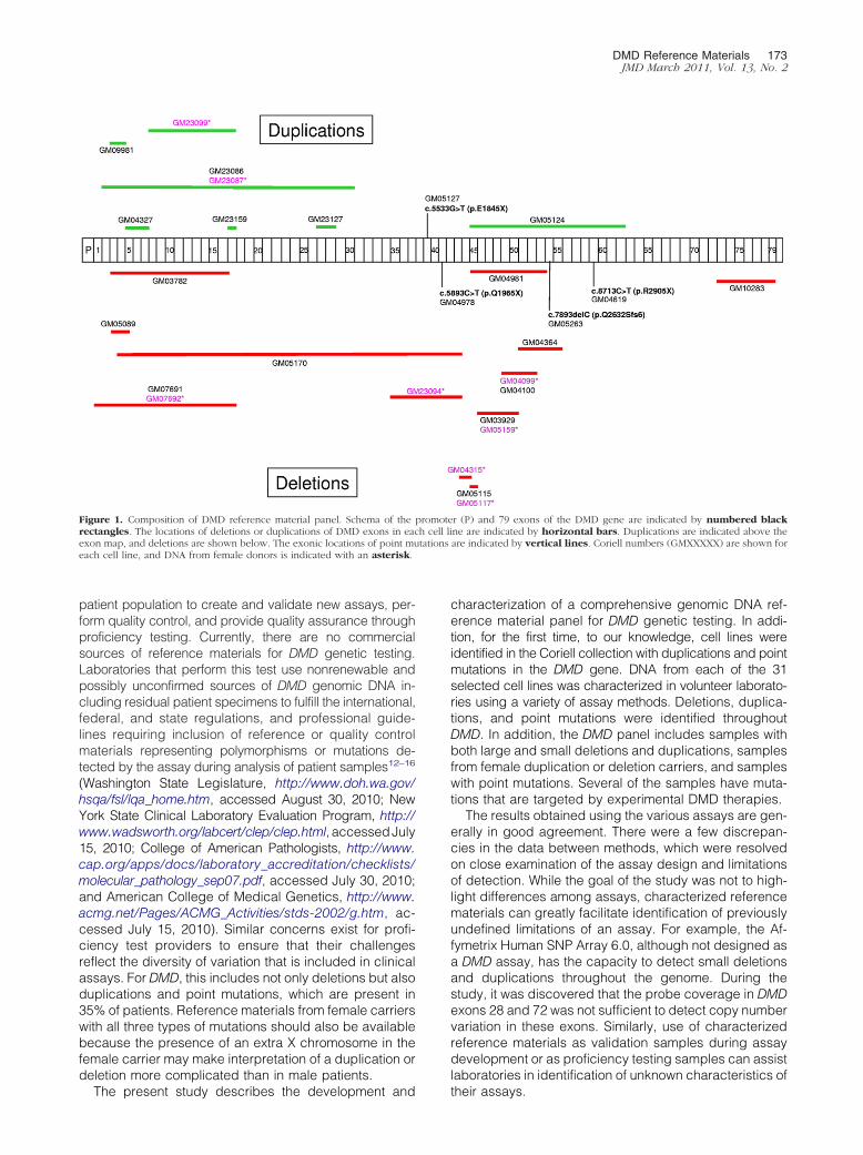
A schema of the DMD gene is shown in Figure 1. Thedrawing depicts the locations of the deletion, duplication, and
Table 1. Genotype of DMD Cell Lines
Coriellcell line Sex
Preliminarydata source*
Consensusgenotype
AffymeSNP
array
GM07691 Male Coriell catalog del 5= end-18 del 5= enGM07692 Female Coriell catalog del 5= end-18 del 5=-18GM03782 Male Coriell catalog del 3–17 del 3–17GM05089 Male Coriell catalog del 3–5 del 3–5GM05170 Male Coriell catalog del 4–43 del 4–43GM23094 Female Registry del 35–43 del 35–4GM04315 Female Present study del 44 del 44GM05115 Male Coriell catalog del 45 del 45GM05117 Female Present study del 45 del 45GM04981 Male Coriell catalog del 45–53 del 45–5GM03929 Male Coriell catalog del 46–50 del 46–5GM05159 Female Coriell catalog del 46–50 del 46–5GM04100 Male Coriell catalog del 49–52 del 49–5GM04099 Female Present study del 49–52 del 49–5GM04364 Male Present study del 51–55 del 51–5GM10283 Male Present study del 72–79 del 73–7GM23086 Male Registry dup 2–30 dup 2–30GM23087 Female Registry dup 2–30 dup 2–30GM09981 Male Present study dup 2–4 dup 2–4GM04327 Male Present study dup 5–7 dup 5–7GM23099 Female Registry dup 8–17 dup 8–17GM23159 Male Registry dup 17 dup 17GM23127 Male Registry dup 27–28 dup 27§
GM05124 Male Present study dup 45–62 dup 45–6GM05263 Male Present study ND No del/d
GM05127 Male Present study ND No del/d
GM02298 Male Present study ND No del/dGM04569 Male Present study ND No del/dGM04619 Male Present study ND No del/d
GM04978 Male Present study ND No del/d
GM05082 Male Present study ND No del/d
del, deletion; dup, duplication; MLPA, multiplex ligation-dependent p*Coriell catalog: deletion in this line was listed in the Coriell catalog whe
identified in a preexisting Coriell line during the study. Registry: this cell line†MRC-Holland BV, Amsterdam, the Netherlands.‡This assay does not detect all deleted exons.§The Affymetrix SNP Array 6.0 is unable to detect mutations in exons 2
in GM23127 using the Affy Cytogenetics 2.7 mol/L array, which is also u
point mutations in each cell line, and the sex of the donor.
Discussion
A number of assays are used in clinical laboratories todetect mutations in DMD. Historically, targeted PCR assayswere designed to detect gene deletions in a subset of the79 DMD exons (representing deletion hotspots), and wereunable to detect exon duplications or point mutations, whichtogether account for about 35% of mutations detected inpatients with DMD.7 With the availability of a commercialMLPA assay, widespread use of quantitative PCR, and DNAsequencing and array-based analyses, identification of du-plications and point mutations in DMD is becoming morefeasible and commonly available in testing laboratories. Theability to detect point mutations and to precisely map dele-tions and duplications is important to identify patients whocould benefit from several new treatments currently underdevelopment.
Clinical laboratories need access to reference materials
Characterization method used
MLPA†
Targetedmultiplex‡
DNA sequenceanalysisLaboratory 1 Laboratory 2
del 5= end-18 del 5=-18 del pb-17 NDdel 5=-18 del 5=-18 ND NDdel 3–17 del 3–17 del 3–17 NDdel 3–5 del 3–5 del 3–4 NDdel 4–43 del 4–43 del 4–43 NDdel 35–43 del 35–43 ND NDdel 44 del 44 ND NDdel 45 del 45 del 45 NDdel 45 del 45 ND NDdel 45–53 del 45–53 del 45–52 NDdel 46–50 del 46–50 del 46–50 NDdel 46–50 del 46–50 ND NDdel 49–52 del 49–52 del 49–52 NDdel 49–52 del 49–52 ND NDdel 51–55 del 51–55 del 51–52 NDdel 72–79 del 72–79 ND NDdup 2–30 dup 2–30 ND NDdup 2–30 dup 2–30 ND NDdup 2–4 dup 2–4 ND NDdup 5–7 dup 5–7 ND NDdup 8–17 dup 8–17 ND NDdup 17 dup 17 ND NDdup 27–28 dup 27–28 ND NDdup 45–62 dup 45–62 ND NDNo del/dup ND ND c.7893delC
(p.Q2632Sfs6)No del/dup ND ND c.5533G�T
(p.E1845X)No del/dup ND ND No mutationNo del/dup ND ND No mutationNo del/dup ND ND c.8713C�T
(p.R2905X)No del/dup ND ND c.5893C�T
(p.Q1965X)No del/dup ND ND No mutation
plification; ND, not determined.resent study was initiated. The present study: the mutation in this line waseated by donation of blood from consenting patients for the present study.
2 because of lack of probes in these regions. A duplication was identifiedidentify mutations in exon 28 because of lack of probe coverage.
trix
6.0
d-18
3
3002259§
2up
up
upupup
up
up
robe amn the pwas cr
containing the spectrum of mutations present in their clinical
tations
DMD Reference Materials 173JMD March 2011, Vol. 13, No. 2
patient population to create and validate new assays, per-form quality control, and provide quality assurance throughproficiency testing. Currently, there are no commercialsources of reference materials for DMD genetic testing.Laboratories that perform this test use nonrenewable andpossibly unconfirmed sources of DMD genomic DNA in-cluding residual patient specimens to fulfill the international,federal, and state regulations, and professional guide-lines requiring inclusion of reference or quality controlmaterials representing polymorphisms or mutations de-tected by the assay during analysis of patient samples12–16
(Washington State Legislature, http://www.doh.wa.gov/hsqa/fsl/lqa_home.htm, accessed August 30, 2010; NewYork State Clinical Laboratory Evaluation Program, http://www.wadsworth.org/labcert/clep/clep.html, accessedJuly15, 2010; College of American Pathologists, http://www.cap.org/apps/docs/laboratory_accreditation/checklists/molecular_pathology_sep07.pdf, accessed July 30, 2010;and American College of Medical Genetics, http://www.acmg.net/Pages/ACMG_Activities/stds-2002/g.htm, ac-cessed July 15, 2010). Similar concerns exist for profi-ciency test providers to ensure that their challengesreflect the diversity of variation that is included in clinicalassays. For DMD, this includes not only deletions but alsoduplications and point mutations, which are present in35% of patients. Reference materials from female carrierswith all three types of mutations should also be availablebecause the presence of an extra X chromosome in thefemale carrier may make interpretation of a duplication ordeletion more complicated than in male patients.
Figure 1. Composition of DMD reference material panel. Schema of therectangles. The locations of deletions or duplications of DMD exons in eacexon map, and deletions are shown below. The exonic locations of point mueach cell line, and DNA from female donors is indicated with an asterisk.
The present study describes the development and
characterization of a comprehensive genomic DNA ref-erence material panel for DMD genetic testing. In addi-tion, for the first time, to our knowledge, cell lines wereidentified in the Coriell collection with duplications and pointmutations in the DMD gene. DNA from each of the 31selected cell lines was characterized in volunteer laborato-ries using a variety of assay methods. Deletions, duplica-tions, and point mutations were identified throughoutDMD. In addition, the DMD panel includes samples withboth large and small deletions and duplications, samplesfrom female duplication or deletion carriers, and sampleswith point mutations. Several of the samples have muta-tions that are targeted by experimental DMD therapies.
The results obtained using the various assays are gen-erally in good agreement. There were a few discrepan-cies in the data between methods, which were resolvedon close examination of the assay design and limitationsof detection. While the goal of the study was not to high-light differences among assays, characterized referencematerials can greatly facilitate identification of previouslyundefined limitations of an assay. For example, the Af-fymetrix Human SNP Array 6.0, although not designed asa DMD assay, has the capacity to detect small deletionsand duplications throughout the genome. During thestudy, it was discovered that the probe coverage in DMDexons 28 and 72 was not sufficient to detect copy numbervariation in these exons. Similarly, use of characterizedreference materials as validation samples during assaydevelopment or as proficiency testing samples can assistlaboratories in identification of unknown characteristics of
r (P) and 79 exons of the DMD gene are indicated by numbered blackne are indicated by horizontal bars. Duplications are indicated above theare indicated by vertical lines. Coriell numbers (GMXXXXX) are shown for
promoteh cell li
their assays.
174 Kalman et alJMD March 2011, Vol. 13, No. 2
Results from the present study also highlight the need forextensive characterization of genomic DNA, either from celllines or residual patient samples, using a variety of methodsbefore their use as reference materials in the laboratory.Many of the existing Coriell cell lines used in the study hadbeen previously characterized using PCR or Southern blotanalysis. In some cases, deletions, duplications, and pointmutations were identified using the methods used in thisstudy, whereas previous methods had not identified a mu-tation in the cell line (labeled “Present study” in Table 1).Similarly, mutations in reference materials derived from pa-tient specimens may be incorrectly identified, dependingon the method used to characterize it initially. Thus, CLSIdocument MM-1711 recommends characterization of refer-ence materials using a method different from that used toidentify the mutation initially.
The present study also highlights the important rolethat patient groups and registries, such as Duchen-neConnect, can have in improvement of laboratory ser-vices. Resources, such as DNA, for rare disorders are, bydefinition, limited, and may be difficult to obtain. Patientgroups are becoming aware of their role in the develop-ment of new tests and treatments for their rare disordersand are increasingly becoming active partners with theresearch community. The willingness of patients withDMD or BMD and their families to share their resourceswith the genetics community through the Coriell Cell Re-positories will benefit not only the quality and availabilityof genetic testing for DMD but also may contribute to thedevelopment of treatments for this disorder.
The genomic DNA reference materials characterized inthis study will be useful for quality assurance, proficiencytesting, test development and validation, and research.DNA samples purified from these cell lines, and other DNAsamples characterized by the GeT-RM, are publicly avail-able from the National Institute of General Medical ScienceRepository at the Coriell Cell Repositories (http://ccr.coriell.org/Sections/Collections/NIGMS/?SsId�8, accessed May14, 2010). More information about the GeT-RM program andavailable reference materials are available on the GeT-RMwebsite (http://wwwn.cdc.gov/dls/genetics/rmmaterials/default.aspx, accessed May 14, 2010).
Acknowledgments
We thank MRC-Holland BV, which donated reagentsfor this project. We also thank the patients with DMDand their families, whose donated blood provided aninvaluable resource for this project and for biomedicalresearch.
References
1. Emery AEH: Duchene muscular dystrophy. Oxford Monographs onMedical Genetics, No 15. Oxford, England: Oxford University Press,1988
2. Murray JM, Davies KE, Harper PS, Meredith L, Mueller CR, WilliamsonR: Linkage relationship of a cloned DNA sequence on the short armof the X chromosome to Duchenne muscular dystrophy. Nature 1982,
300:69–713. Bodrug SE, Ray PN, Gonzalez IL, Schmickel RD, Sylvester JE, WortonRG: Molecular analysis of a constitutional X-autosome translocation ina female with muscular dystrophy. Science 1987, 237:1620–1624
4. Koenig M, Monaco AP, Kunkel LM: The complete sequence of dys-trophin predicts a rod-shaped cytoskeletal protein. Cell 1988, 53:219–228
5. Hoffman EP, Kunkle LM: Dystrophin abnormalities in Duchenne/Becker muscular dystrophy. Neuron 1989, 2:1019–1029
6. Den Dunnen JT, Grootscholten PM, Bakker E, Blonden AJ, GinjaarHB, Wapenaar MC, van Paassen HMB, van Broeckhoven C, PearsonPL: Topography of the Duchenne muscular dystrophy (DMD) gene:FIGE and cDNA analysis of 194 cases reveals 115 deletions and 13duplications. Am J Hum Genet 1989, 45:835–847
7. Darras BT, Korf BR, Urion DK. Dystrophinopathies (Updated) InGeneReviews at Gene Tests: Medical Genetics Information Resource(database online). Copyright University of Washington, Seattle, 1993–2010. Available at http://www.ncbi.nlm.nih.gov/books/NBK1119/; lastupdated March 21, 2008
8. Barton-Davis ER, Cordier L, Shoturma DI, Leland SE, Sweeney HL:Aminoglycoside antibiotics restore dystrophin function to skeletalmuscles of mice. J Clin Invest 1999, 104:375–381
9. Hirawat S, Northcutt VJ, Welch EM, Elfring GL, Hwang S, AlmsteadNG, Ju W, Miller LL: Phase 1 multiple-dose safety and PK study ofPTC124 for nonsense mutation suppression therapy of Duchennemuscular dystrophy (DMD). Neurology 2005, 54(Suppl 1):A176
10. Aartsma-Rus A, Janson AA, Heemskerk JA, De Winter CL, Van Om-men GJ, Van Deutekom JC: Therapeutic modulation of DMD splicingby blocking exonic splicing enhancer sites with antisense oligonucle-otides. Ann NY Acad Sci 2006, 1082:74–76
11. MM-17A Verification and Validation of Multiplex Nucleic Acid Assays:Approved Guideline. Wayne, PA: Clinical and Laboratory StandardsInstitute; 2008:27
12. Association for Molecular Pathology statement: recommendations forin-house development and operation of molecular diagnostic tests.Am J Clin Pathol 1999, 111:449–463
13. Chen B, O’Connell CD, Boone DJ, Amos JA, Beck JC, Chan MM,Farkas DH, Lebo RV, Richards CS, Roa BB, Silverman LM, Barton DE,Bejjani BA, Belloni DR, Bernacki SH, Caggana M, Charache P, De-queker E, Ferreira-Gonzalez A, Friedman KJ, Greene CL, Grody WW,Highsmith WE Jr, Hinkel CS, Kalman LV, Lubin IM, Lyon E, Payne DA,Pratt VM, Rohlfs E, Rundell CA, Schneider E, Willey AM, Williams LO,Willey JC, Winn-Deen ES, Wolff DJ: Developing a sustainable processto provide quality control materials for genetic testing. Genet Med2005, 7:534–549
14. Code of Federal Regulations. The Clinical Laboratory ImprovementAmendments (CLIA) 2007, 42 CFR Part 493
15. MM01–A2 Molecular Diagnostic Methods for Genetic Diseases-Ap-proved Guideline. 2nd ed. Wayne, PA: Clinical and Laboratory Stan-dards Institute; 2006;45–46
16. Centers for Disease Control and Prevention. Good laboratory prac-tices for molecular genetic testing for heritable diseases and condi-tions. MMWR 2009; 58(No. RR-6):1–37
17. Beck JC, Beiswanger CM, John EM, Satraiano E, West D: Successfultransformation of cryopreserved lymphocytes: a resource for epidemio-logical studies. Cancer Epidemiol Biomarkers Prev 2001, 10:551–554
18. Bernacki SH, Stankovic AK, Williams LO, Beck JC, Herndon JE,Snow-Bailey K, Prior TW, Matteson KJ, Wasserman LM, Cole EC,Stenzel TT: Establishment of stably EBV-transformed cell lines fromresidual clinical blood samples for use in performance evaluation andquality assurance in molecular genetic testing. J Mol Diagn 2003,5:227–230
19. Miller SA, Dykes DD, Polesky HF: A simple salting out procedure forextracting DNA from human nucleated cells. Nucleic Acids Res 1988,16:1215
20. den Dunnen JT, Beggs AH: Multiplex PCR for identifying DMD genedeletions. Curr Protoc Hum Genet 2006; Chapter 9, Unit 9.3
21. Prior TW, Bridgeman SJ: Experience and strategy for the moleculartesting of Duchenne muscular dystrophy. J Mol Diagn 2005, 7:317–326
22. Flanigan KM, von Niederhausern A, Dunn DM, Alder J, Mendell JR,Weiss RB: Rapid direct sequence analysis of the dystrophin gene.
Am J Hum Genet 2003, 72:931–939


