Embed Size (px)
Citation preview
Quantitation of CXCR4 Expression in MyocardialInfarction Using 99mTc-Labeled SDF-1a
Preeti Misra1, Djamel Lebeche2, Hung Ly2, Martina Schwarzkopf2, George Diaz3, Roger J. Hajjar2, Alison D. Schecter2,4,and John V. Frangioni1,5
1Division of Hematology/Oncology, Beth Israel Deaconess Medical Center, Boston, Massachusetts; 2The Cardiovascular ResearchCenter, Mount Sinai School of Medicine, New York, New York; 3Department of Human Genetics, Mount Sinai School of Medicine,New York, New York; 4Departments of Medicine and Immunobiology, Mount Sinai School of Medicine, New York, New York; and5Department of Radiology, Beth Israel Deaconess Medical Center, Boston, Massachusetts
The chemokine stromal-derived factor-1a (SDF-1a, CXCL12)and its receptor CXCR4 are implicated as key mediators of he-matopoietic stem cell retention, cancer metastasis, and HIVinfection. Their role in myocardial infarction (MI) is not as welldefined. The noninvasive in vivo quantitation of CXCR4 expres-sion is central to understanding its importance in these diverseprocesses as well in the cardiac response to injury. Methods:Recombinant SDF-1a was radiolabeled under aprotic condi-tions and purified by gel-filtration chromatography (GFC) usinghigh-specific-activity 99mTc-S-acetylmercaptoacetyltriserine-N-hydroxysuccinimide ([99mTc-MAS3]-NHS) prepared by solid-phasepreloading. Radiotracer stability and transmetallation under harshconditionswere quantified by GFC.Affinity, specificity, and maximumnumber of binding sites (Bmax) were quantified, with adenoviral-expressed CXCR4 on nonexpressing cells and endogenous re-ceptor on rat neonatal cardiomyocytes, using a high-throughputlive-cell–binding assay. Blood half-life, biodistribution, and clear-ance of intravenously injected [99mTc-MAS3]-SDF-1a were quanti-fied in Sprague–Dawley rats before and after experimentallyinduced MI. Results: [99mTc-MAS3]-SDF-1a could be preparedin 2 h total with a specific activity of 8.0 · 107 MBq/mmol (2,166Ci/mmol) and a radiochemical purity greater than 98%. Degrada-tion of the radiotracer after boiling for 5 min, with and without1 mM dithiothreitol, and transmetallation in 100% serum at 37�Cfor 4 h were negligible. [99mTc-MAS3]-SDF-1a exhibits high speci-ficity for CXCR4 on the surface of living rat neonatal cardiomyo-cytes, with an affinity of 2.7 6 0.9 nM and a Bmax of 4.8 · 104
binding sites per cell. After intravenous injection, 99mTc-labeledSDF-1a displays a blood half-life of 25.8 6 4.6 min, rapid renalclearance with only 26.2 6 6.1 percentage injected dose remain-ing in the carcass at 2 h, consistently low uptake in most organs(,0.1 percentage injected dose per gram), and no evidence ofblood–brain barrier penetration. After MI was induced, CXCR4expression levels in the myocardium increased more than 5-fold,as quantified using [99mTc-MAS3]-SDF-1a and confirmed usingconfocal immunofluorescence. Conclusion: We describe a 99mTc-labeled SDF-1a radiotracer that can be used as a sensitive andspecific probe for CXCR4 expression in vivo and demonstrate
that this radiotracer is able to quantify changes in CXCR4 expres-sion under different physiologic and pathologic states. Takentogether, CXCR4 levels should now be quantifiable in vivo in avariety of animal model systems of human diseases.
Key Words: SDF-1a; CXCR4; chemokines; SPECT radiotracers;myocardial infarction
J Nucl Med 2008; 49:963–969DOI: 10.2967/jnumed.107.050054
Chemokines are a family of structurally related glyco-proteins, ranging in size from 6 to 14 kDa, with potent
leukocyte activation or chemotactic activity. They are grouped
into 4 classes, CC, CXC, C, and CX3C, based on the position
of the first 2 conserved cysteine residues in the mature protein
(1). On the basis of its amino acid motif, stromal-derived
factor-1 (SDF-1, CXCL12) belongs to the CXC class of
chemokines.SDF-1a and its receptor CXCR4, an important chemokine–
receptor pair, play a crucial role in numerous biologic pro-
cesses including hematopoiesis, vasculogenesis, neuronal
development, and immune cell trafficking (2–4). They also
have been implicated in various pathologic conditions such
as cancer (5,6), infection with HIV (7), and various in-
flammatory conditions (8,9).Recently, there has been increased focus on the role of
chemokines in cardiovascular development (10) and in adult
cardiac disease such as acute coronary syndromes (11) and
heart failure (12). Chemokines and their receptors are present
and functional on the myocardium and on vascular smooth
muscle cells (13) and are capable of mediating important
biologic processes in a manner that is independent of
inflammatory cells (14). Both SDF-1a and its receptor
CXCR4 are essential for cardiogenesis (15), and CXCR4
is expressed on early hematopoietic stem cells (SCs) and
endothelial progenitor cells (4). SDF-1a even appears to
modulate myocyte contractility in rodent model systems
(12). Recently, several studies have demonstrated that circu-
lating chemokine levels, including those of SDF-1a, are
Received Dec. 18, 2007; revision accepted Feb. 28, 2008.For correspondence or reprints contact: John V. Frangioni, Beth Israel
Deaconess Medical Center, 330 Brookline Ave., Room SL-B05, Boston, MA02215.
E-mail: [email protected] ª 2008 by the Society of Nuclear Medicine, Inc.
QUANTITATION OF CXCR4 EXPRESSION IN VIVO • Misra et al. 963
by on February 2, 2015. For personal use only. jnm.snmjournals.org Downloaded from
elevated in animals and humans with cardiac dysfunction(16,17), and both chemokines and their receptors are upre-gulated in the hearts of patients with congestive heart failure(18). After myocardial infarction (MI), chemokines mediatethe retention of SCs that could potentially repair damagedmyocardium (19), and reestablishing SDF-1a expressionsome time after undergoing MI may restore SC homing to thedamaged cardiac tissue (20,21).
Although associated with cardiac dysfunction, the mech-anisms by which chemokines and their receptors exert theireffects are largely unknown. Because the role of chemokinesin the cardiac response to inflammation may be quite distinctfrom that of cytokines (22), and because of the recent focuson using chemokines to modulate the SC milieu in the heart,there is an urgent need to quantify chemokine and chemokinereceptor levels in vivo, noninvasively, and over time.
Our laboratory has previously developed a solid-phasepreradiolabeling technique for rapid and efficient 99mTc-labeling of small molecules and peptides under aprotic con-ditions (23). In the present study, we use this technique toprepare a biologically active SDF-1a radiotracer with highspecific activity. We also demonstrate that CXCR4 levels cannow be quantified in vivo and over time and that CXCR4 issignificantly overexpressed in myocardium subjected toinfarction.
MATERIALS AND METHODS
ReagentsRecombinant murine SDF-1a was purchased from Peprotech.
The N-hydroxysuccinimide (NHS) ester of hydrazinonicotinamide(HYNIC) was the generous gift of Jean-Luc Vanderheyden (The-seus Imaging). Ultradry dimethylsulfoxide (DMSO) was purchasedfrom Acros Organics; high-performance liquid chromatography(HPLC)–grade triethylammonium acetate, pH 7, was obtainedfrom Glen Research; and HPLC-grade water was purchased fromAmerican Bioanalytic. All other chemicals were purchased fromFisher Scientific and were ACS- or HPLC-grade.
Synthesis and Purification of [99mTc-MAS3]-SDF-1aThe NHS ester of 99mTc-S-acetylmercaptoacetyltriserine
(99mTc-MAS3) was prepared to high purity (.99%) and highspecific activity (average, 1.1 · 108 MBq/mmol; 3 · 103 Ci/mmol)in DMSO using a 20-min protocol described in detail previously(23). For radiolabeling, SDF-1a dry powder was resuspended inDMSO at a concentration of 12.6 mM. A total of 185 MBq (5mCi; 0.45 nmol) of [99mTc-MAS3]-NHS (550 mL) was mixed with50 mL (0.63 nmol) of SDF-1a, followed by the addition of 2 mL (8mmol) of 4 M triethylamine. Constant stirring at room temperaturewas maintained for 1 h. Radiolabeled [99mTc-MAS3]-SDF-1a waspurified by gel-filtration chromatography (GFC) using an 8 · 300mm, 60-A Diol (catalog number DL06S053008WT; YMC) gel-filtration column and phosphate-buffered saline (PBS), pH 7.4, asmobile phase. Details of the HPLC system and detectors havebeen described in detail previously (24). GFC-purified [99mTc-MAS3]-SDF-1a (specific activity, 8.0 · 107 MBq/mmol; 2,166 Ci/mmol) was concentrated to �148 MBq/mL (4 mCi/mL) usinga 5,000-molecular-weight cutoff cartridge (Vivaspin; SartoriusStedim Biotech).
Synthesis of 99mTc-HYNIC-AlbuminCovalent conjugation of the HYNIC-NHS to albumin was
performed by mixing 1 mL (72 nmol) of a 5 mg/mL solution ofCohn Fraction V bovine serum albumin (Sigma-Aldrich) in PBS and100 mL (320 nmol) of 3.2 mM HYNIC-NHS in DMSO. Afterincubation for 2 h at room temperature, unreacted ligand wasremoved by the GFC on a prime pump and a 10/300 GL column(AKTA; Amersham Biosciences and Superose-6; Amersham Bio-sciences) using 114 mM tricine, pH 6.6, as mobile phase and a flowrate of 1 mL/min. Exchange labeling using tricine as weak ligandwas performed by adding 1 mg of tricine powder to 1 mL of thederivatized albumin solution, followed by 25 mg of SnCl2�2H2Odissolved in 12.5 mL of 0.01N HCl and 100 mL (185–296 MBq; 5–8mCi) of 99mTc-pertechnetate. After a 30-min incubation, radiola-beled albumin was purified and analyzed by GFC using an 8 ·300 mm, 200-A Diol (catalog number DL06S053008WT; YMC)column and PBS as mobile phase. GFC-purified 99mTc-HYNIC-albumin (specific activity, 1.8 · 107 MBq/mmol; 476 Ci/mmol)was concentrated to �148 MBq/mL (4 mCi/mL) using a 5,000-molecular-weight cutoff cartridge (Vivaspin).
Quantification of Chemical Stability[99mTc-MAS3]-SDF-1a was boiled for 5 min in PBS in the
presence or absence of 1 mM dithiothreitol and also incubated for4 h at 37�C in 100% calf serum. Stability and transmetallation werequantified using GFC and the 60-A Diol column as described earlier.GFC molecular weight markers (Bio-Rad) were as follows: M1 5
thyroglobulin (670 kDa, rt 5 4.4 min), M2 5 g-globulin (158 kDa,rt 5 5.1 min), M3 5 ovalbumin (44 kDa, rt 5 6.9 min), M4 5
myoglobin (17 kDa, rt 5 8.0 min), and M5 5 vitamin B12 (1.3 kDa,rt 5 12.4 min).
Adenoviral Expression of Transgenes and Isolation ofNeonatal Cardiomyocytes
Human prostate cancer cell line PC-3 was obtained from theAmerican Type Culture Collection. Adenoviruses coexpressingCXCR4 and green fluorescent protein (GFP) or human bone mor-phogenetic protein-2 and GFP were prepared as described previ-ously (25). Cells were transduced at a multiplicity of infection of500:1 and incubated for 48 h at 37�C, under humidified 5% CO2,in RPMI 1640 medium (Mediatech Cellgro) supplemented with10% fetal bovine serum (Gemini Bio-Products) and 5% penicillin/streptomycin (Cambrex Bioscience).
Spontaneously beating cardiomyocytes were prepared from1- to 2-d-old Sprague–Dawley rat pups (Charles River Laborato-ries) using an isolation system (Neonatal Cardiomyocyte IsolationSystem; Worthington Biochemical). Cardiomyocytes were cul-tured in 96-well filter plates (model MSHAS4510; Millipore) at20,000 cells per well in F-10 medium (Invitrogen) in the presenceof 5% fetal calf serum and 10% horse serum, 100 U of penicillin/streptomycin per milliliter, and 2 mM L-glutamine over 48 h.
High-Throughput Live Cell–Binding AssayTo quantify absolute affinity and Bmax for the surface of living
cells, a high-throughput homologous competition assay was usedas described in detail previously (23). Briefly, cells were split onto96-well filter plates (model MSHAS4510) and grown to 50% con-fluence (approximately 40,000 cells per well) over 48 h. The assaywas performed at 4�C to avoid internalization of the radioligand dueto constitutive endocytosis. Cells were washed twice with ice-coldPBS or serum and incubated for 20 min at 4�C with 0.02 MBq (0.5mCi) of radiotracer in the presence or absence of homologous cold
964 THE JOURNAL OF NUCLEAR MEDICINE • Vol. 49 • No. 6 • June 2008
by on February 2, 2015. For personal use only. jnm.snmjournals.org Downloaded from
compound. Cells were then washed 3 times with PBS or serum usinga vacuum manifold (catalog number MSVMHTS00; Millipore), andthe well contents were transferred directly to 12 · 75 mm plastictubes placed in g-counter racks and counted on a 10-detectorg-counter (model 1470 Wallac Wizard; Perkin Elmer). Imaging(96-well plates) was performed using a g-camera (Isocam Tech-nologies) equipped with a 1.27-cm (0.5-in) sodium iodide crystaland a high-resolution (1 mm), low-energy collimator.
Animal Model SystemsAnimal studies were performed in accordance with approved
Institutional Animal Care and Use Committee protocols. MaleSprague–Dawley inbred rats (200–250 gm) were anesthetized witha 50 mg/kg concentration of intraperitoneal pentobarbital, intu-bated, and mechanically ventilated with a rodent ventilator at 80breaths/min using room air. After a left lateral thoracotomy, the leftanterior descending artery was ligated for 30 min with 6.0 silk suture(26). MI was confirmed by visualization of myocardial blanchingand ballooning distal to the point of ligation. The incision was thenclosed by suturing all 3 layers (bone, muscle, and skin), and excessair was evacuated with a 22-gauge angiocath (BD) to preventpneumothorax. Rats were extubated when spontaneous breathingreturned and received 0.5 mL of 0.5% bupivicaine analgesia for thenext 48 h until used for radiotracer studies as described later. For thesham group, rats of comparable body weight and age underwentsimilar procedures, with the exception of left anterior descendingligation.
Blood Activity Curves, Biodistribution, and ClearanceA total of 37 MBq (1 mCi) of 99mTc-MAS3 or [99mTc-MAS3]-
SDF-1a in 200 mL of saline was administered intravenously via theretroorbital sinus. Blood was collected at 0, 1, 2, 5, 10, 15, 30, 60, 90,and 120 min after injection from the tail vein using microcapillarytubes, weighed, and counted as described earlier. Curve fitting wasperformed using software (Prism, version 4.0a; GraphPad). Formeasurement of total-body retention and clearance at 2 h after in-jection, we ligated the ureters and urethra with silk sutures, removedthe bladder en masse, and combined it with excreted urine and fecesbefore measurement of radioactivity in a dose calibrator. Theremaining carcass was also measured in a dose calibrator, then theheart, lungs, spleen, liver, kidneys, stomach, intestine, and brainwere resected, washed twice in PBS, weighed, and counted asdescribed earlier. In rats undergoing MI, the heart was separated intononinfarcted and infarcted tissue before quantitation. An additionalcohort of rats was injected with 37 MBq (1 mCi) of 99mTc-labeledalbumin in 200 mL of saline to measure perfusion. Statisticalanalysis was performed with an unpaired t test using Prism.
Confocal Fluorescence MicroscopyIn parallel with radiotracer experiments, an additional cohort of
rats was sacrificed 24 h after surgery by sodium pentobarbitaloverdose, and hearts were excised and frozen on dry ice. Cryosec-tions (16 mm) were collected on slides (Superfrost Plus; Menzel),postfixed with 2% paraformaldehyde and 2% sucrose, and permea-bilized with 0.3% Triton X-100 (Sigma). Sections were blocked in2% BSA in PBS (blocking solution). Mouse anti-SDF-1a monoclo-nal antibody (catalog number MAB350; R&D Systems) or rabbitanti-CXCR4 polyclonal antibody (catalog number TP503; TorreyPines Biolabs) was diluted 1:80 or 1:100, respectively, in blockingsolution; incubated overnight at 4�C with the specimen; andvisualized using fluorophore-labeled goat anti-mouse IgG or goatanti-rabbit IgG secondary antibodies, respectively. F-actin was
visualized with rhodamine phalloidin (catalog number R415;Invitrogen). Negative controls were incubated with nonimmunerabbit IgG (catalog number AB-105-C; R&D Systems) and mouseIgG isotype control (catalog number MAB002; R&D Systems).Nuclei were counterstained with 49-6-diamidino-2-phenylindole(DAPI). Photomicrographs were captured on a Leica TCS-SP(ultraviolet) confocal microscope at the Mount Sinai School ofMedicine Microscopy Shared Research Facility.
RESULTS
Radiolabeling and Purification of [99mTc-MAS3]-SDF-1a
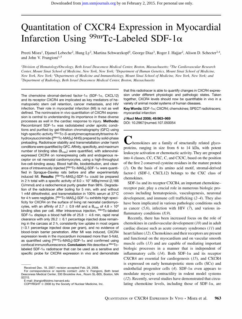
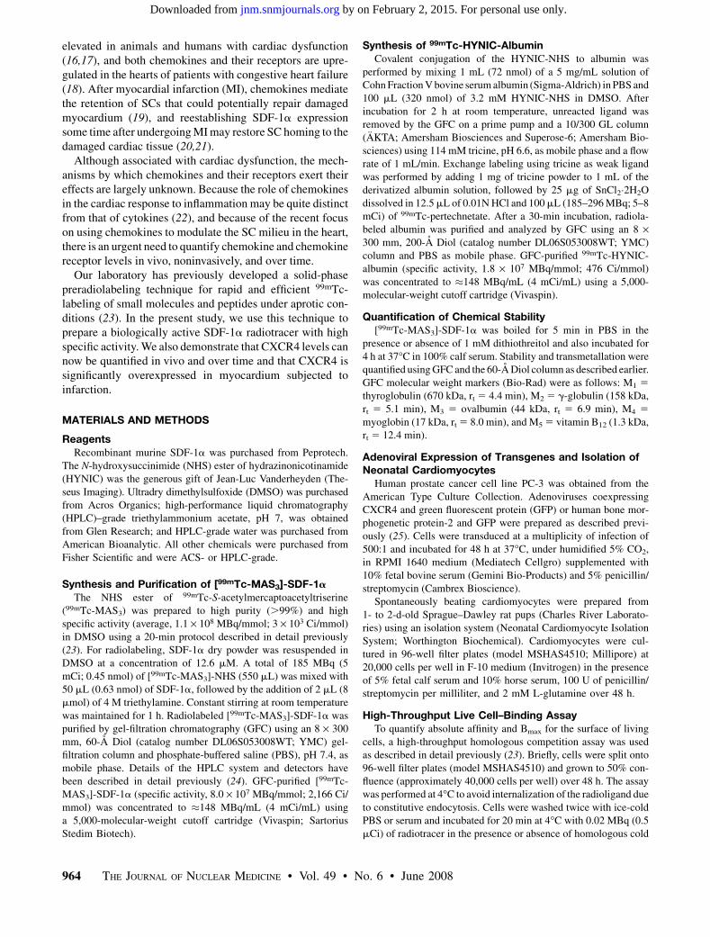
Using high-specific-activity, solid-phase preloaded[99mTc-MAS3]-NHS, SDF-1a was labeled under nonaque-ous conditions (Fig. 1) and purified using GFC (Fig. 2),resulting in a radiotracer with more than 98% radiopurity anda minimum specific activity of 8.0 · 107 MBq/mmol (2,166Ci/mmol). For the initial 99mTcO4
2, the average radiolabel-ing yield was 70% (range, 65%–75%) for more than 20separate labeling procedures. For [99mTc-MAS3]-NHS, ra-diolabeling yield was greater than 98% for more than 20separate labeling procedures. Although the major peptidepeak of recombinant SDF-1a has a retention time (rt) of 9.6min and was labeled efficiently, other minor peaks werelabeled with varying efficiency (Fig. 2). The specific-activitycalculation is based on the entire mass of SDF-1a present inthe labeling reaction and is thus likely an underestimate of thetrue specific activity. No attempt was made to identify whichof the 9 (1 a, 8 e; Fig. 1) possible primary amines werelabeled, although the low molar ratio of [99mTc-MAS3]-NHSto SDF-1a (0.7:1) ensured that, on average, fewer than 1radioatom per molecule was incorporated. [99mTc-MAS3]-SDF-1a was readily separable from spontaneously hydro-lyzed 99mTc-MAS3 at rt of 13.5 min (data not shown).
FIGURE 1. 99mTc-radiolabeling of SDF-1a. Solid-phase prela-beled [99mTc-MAS3]-NHS was conjugated covalently to recom-binant SDF-1a in DMSO in presence of base, releasing onlyNHS as byproduct. Primary amines available for conjugationare indicated by * in peptide sequence.
QUANTITATION OF CXCR4 EXPRESSION IN VIVO • Misra et al. 965
by on February 2, 2015. For personal use only. jnm.snmjournals.org Downloaded from
Stability of [99mTc-MAS3]-SDF-1a
Although one of the features of 99mTc-MAS3 complexes ishigh stability, and conjugation of the complex to SDF-1a wasdesigned to be covalent, we tested the stability of [99mTc-MAS3]-SDF-1a under various conditions. There was noevidence of complex dissociation after boiling for 5 min inthe presence or absence of 1 mM dithiothreitol and noevidence of transmetallation when incubated with 100%serum for 4 h at 37�C. Quantitation of the chromatographs(data not shown) suggested that more than 98% of [99mTc-MAS3]-SDF-1a remained intact under these conditions.
Affinity and Specificity of [99mTc-MAS3]-SDF-1a forCXCR4 on Surface of Living Cells
Using a previously described live cell homologous com-petition assay (23), we were able to characterize the affinityand specificity of [99mTc-MAS3]-SDF-1a for CXCR4 on thesurface of living cells. First, nonactivated PC-3 humanprostate cancer cells, which express low levels of CXCR4mRNA (27) and no measurable protein, were transduced withan adenovirus coexpressing CXCR4 and GFP. GFP was usedto confirm adenoviral transduction and to normalize levels of
expression (data not shown). Negative controls includedtransduction with a similar adenovirus expressing bonemorphogenetic protein-2 and GFP or untransduced cells.[99mTc-MAS3]-SDF-1a exhibited high specificity forCXCR4, an affinity of 1.0 6 0.1 nM (mean 6 SD), and aBmax of 2.6 · 105 binding sites per cell. Indeed, cells could bevisualized in situ on the filter plates by g-ray imaging (datanot shown). Binding of [99mTc-MAS3]-SDF-1a to neonatalrat cardiomyocytes exhibited a slightly lower affinity (2.9 6
0.5 nM) than did adenovirus-overexpressed CXCR4 butexhibited a relatively high Bmax of 4.8 · 104 binding sitesper cell. This measured affinity is consistent with the previ-ously published value of 8.3 6 1.2 nM for SDF-1a binding toCXCR4 on human corneal fibroblasts (28). Results werenearly identical when the assay was performed in PBS(affinity of 2.9 6 0.5 nM) or 100% serum (affinity of 2.7 6
0.9 nM) with the same Bmax of 4.8 · 104 binding sites per cell,suggesting that serum does not contain natural inhibitors ofthe binding process.
Blood Half-Life, Biodistribution, and Clearance
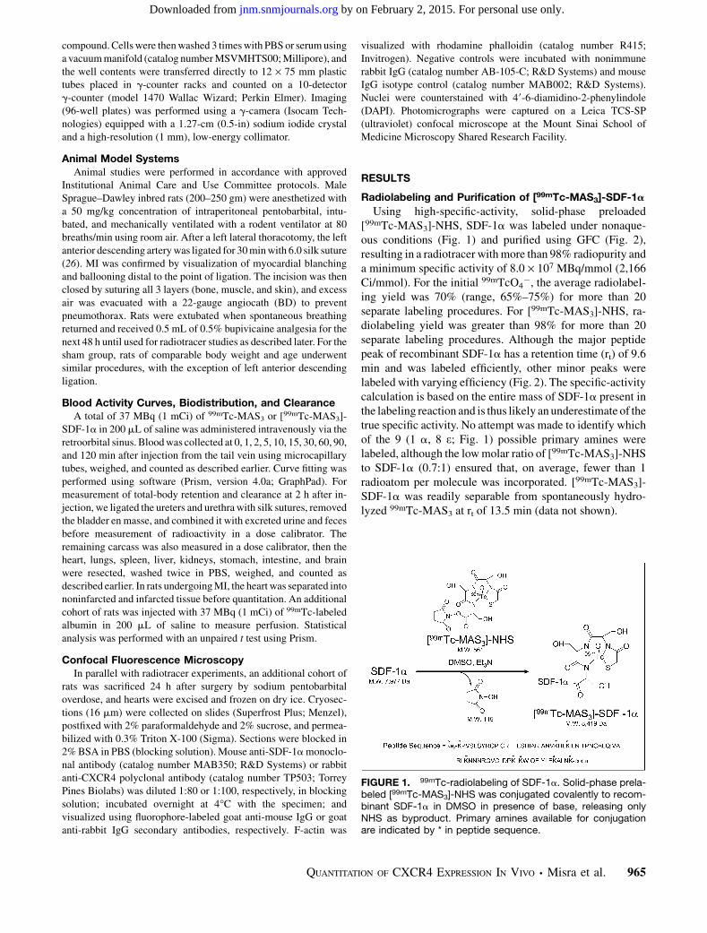
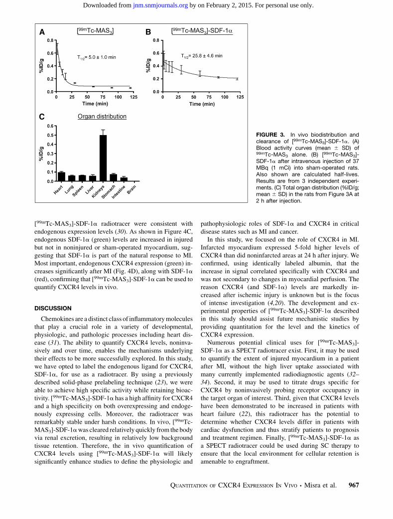
After intravenous injection, 99mTc-MAS3 alone exhibitsan extremely short blood half-life of 5.0 6 1.0 min (Fig. 3A)and rapid renal clearance (data not shown), results consistentwith previous reports (29). However, [99mTc-MAS3]-SDF-1a exhibits a prolonged blood half-life of 25.8 6 4.6 min(Fig. 3B). At 2 h after injection, 73.8% 6 6.1% injecteddose (%ID) was found in excrement, primarily urine, with26.2 6 6.1 percentage injected dose (%ID) remaining inthe carcass (data not shown). Analysis of the major organs(Fig. 3C), except for the kidneys, the major site of excretion,revealed a relatively low uptake of less than 0.1 %ID/g. Nobrain uptake was measured, suggesting that [99mTc-MAS3]-SDF-1a does not cross the blood–brain barrier.
Quantitation of Functional CXCR4 in Normal and InjuredMyocardium
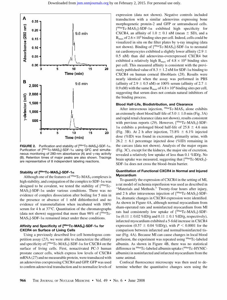
To quantify the expression of CXCR4 in the setting of MI,a rat model of ischemia reperfusion was used as described in‘‘Materials and Methods.’’ Twenty-four hours after injury,and 2 h after intravenous injection of [99mTc-MAS3]-SDF-1a, dramatic changes in CXCR4 expression were identified.As shown in Figure 4A, although normal myocardium fromsham-operated rats and noninfarcted myocardium from MIrats had consistently low uptake of [99mTc-MAS3]-SDF-1a (0.11 6 0.02 %ID/g and 0.11 6 0.1 %ID/g, respectively),infarcted myocardium exhibited a 5-fold increase in CXCR4expression (0.57 6 0.04 %ID/g), with P , 0.0001 for thecomparison between infarcted and normal/noninfarcted tis-sue (Fig. 4A). Because MI can cause changes in local bloodperfusion, the experiment was repeated using 99mTc-labeledalbumin. As shown in Figure 4B, there was no statisticaldifference in 99mTc-labeled albumin uptake (99mTc-HYNIC-albumin) in noninfarcted and infarcted myocardium from thesame animal.
Confocal fluorescence microscopy was then used to de-termine whether the quantitative changes seen using the
FIGURE 2. Purification and stability of [99mTc-MAS3]-SDF-1a.Purification of [99mTc-MAS3]-SDF-1a using GFC and simulta-neous monitoring of 280-nm absorbance (A) and g-ray activity(B). Retention times of major peaks are also shown. Tracingsare representative of 8 independent labeling reactions.
966 THE JOURNAL OF NUCLEAR MEDICINE • Vol. 49 • No. 6 • June 2008
by on February 2, 2015. For personal use only. jnm.snmjournals.org Downloaded from
[99mTc-MAS3]-SDF-1a radiotracer were consistent withendogenous expression levels (30). As shown in Figure 4C,endogenous SDF-1a (green) levels are increased in injuredbut not in noninjured or sham-operated myocardium, sug-gesting that SDF-1a is part of the natural response to MI.Most important, endogenous CXCR4 expression (green) in-creases significantly after MI (Fig. 4D), along with SDF-1a
(red), confirming that [99mTc-MAS3]-SDF-1a can be used toquantify CXCR4 levels in vivo.
DISCUSSION
Chemokines are a distinct class of inflammatory moleculesthat play a crucial role in a variety of developmental,physiologic, and pathologic processes including heart dis-ease (31). The ability to quantify CXCR4 levels, noninva-sively and over time, enables the mechanisms underlyingtheir effects to be more successfully explored. In this study,we have opted to label the endogenous ligand for CXCR4,SDF-1a, for use as a radiotracer. By using a previouslydescribed solid-phase prelabeling technique (23), we wereable to achieve high specific activity while retaining bioac-tivity. [99mTc-MAS3]-SDF-1a has a high affinity for CXCR4and a high specificity on both overexpressing and endoge-nously expressing cells. Moreover, the radiotracer wasremarkably stable under harsh conditions. In vivo, [99mTc-MAS3]-SDF-1a was cleared relatively quickly from the bodyvia renal excretion, resulting in relatively low backgroundtissue retention. Therefore, the in vivo quantification ofCXCR4 levels using [99mTc-MAS3]-SDF-1a will likelysignificantly enhance studies to define the physiologic and
pathophysiologic roles of SDF-1a and CXCR4 in criticaldisease states such as MI and cancer.
In this study, we focused on the role of CXCR4 in MI.Infarcted myocardium expressed 5-fold higher levels ofCXCR4 than did noninfarcted areas at 24 h after injury. Weconfirmed, using identically labeled albumin, that theincrease in signal correlated specifically with CXCR4 andwas not secondary to changes in myocardial perfusion. Thereason CXCR4 (and SDF-1a) levels are markedly in-creased after ischemic injury is unknown but is the focusof intense investigation (4,20). The development and ex-perimental properties of [99mTc-MAS3]-SDF-1a describedin this study should assist future mechanistic studies byproviding quantitation for the level and the kinetics ofCXCR4 expression.
Numerous potential clinical uses for [99mTc-MAS3]-SDF-1a as a SPECT radiotracer exist. First, it may be usedto quantify the extent of injured myocardium in a patientafter MI, without the high liver uptake associated withmany currently implemented radiodiagnostic agents (32–34). Second, it may be used to titrate drugs specific forCXCR4 by noninvasively probing receptor occupancy inthe target organ of interest. Third, given that CXCR4 levelshave been demonstrated to be increased in patients withheart failure (22), this radiotracer has the potential todetermine whether CXCR4 levels differ in patients withcardiac dysfunction and thus stratify patients to prognosisand treatment regimen. Finally, [99mTc-MAS3]-SDF-1a asa SPECT radiotracer could be used during SC therapy toensure that the local environment for cellular retention isamenable to engraftment.
FIGURE 3. In vivo biodistribution andclearance of [99mTc-MAS3]-SDF-1a. (A)Blood activity curves (mean 6 SD) of99mTc-MAS3 alone. (B) [99mTc-MAS3]-SDF-1a after intravenous injection of 37MBq (1 mCi) into sham-operated rats.Also shown are calculated half-lives.Results are from 3 independent experi-ments. (C) Total organ distribution (%ID/g;mean 6 SD) in the rats from Figure 3A at2 h after injection.
QUANTITATION OF CXCR4 EXPRESSION IN VIVO • Misra et al. 967
by on February 2, 2015. For personal use only. jnm.snmjournals.org Downloaded from
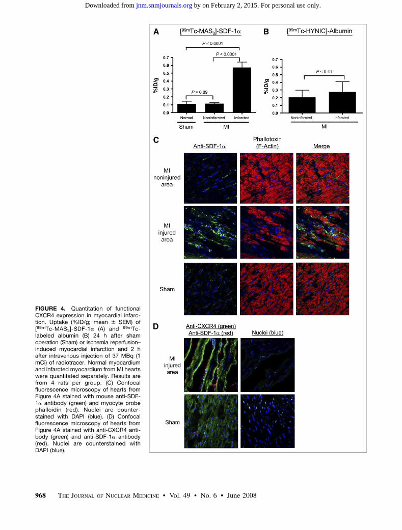
FIGURE 4. Quantitation of functionalCXCR4 expression in myocardial infarc-tion. Uptake (%ID/g; mean 6 SEM) of[99mTc-MAS3]-SDF-1a (A) and 99mTc-labeled albumin (B) 24 h after shamoperation (Sham) or ischemia reperfusion–induced myocardial infarction and 2 hafter intravenous injection of 37 MBq (1mCi) of radiotracer. Normal myocardiumand infarcted myocardium from MI heartswere quantitated separately. Results arefrom 4 rats per group. (C) Confocalfluorescence microscopy of hearts fromFigure 4A stained with mouse anti-SDF-1a antibody (green) and myocyte probephalloidin (red). Nuclei are counter-stained with DAPI (blue). (D) Confocalfluorescence microscopy of hearts fromFigure 4A stained with anti-CXCR4 anti-body (green) and anti-SDF-1a antibody(red). Nuclei are counterstained withDAPI (blue).
968 THE JOURNAL OF NUCLEAR MEDICINE • Vol. 49 • No. 6 • June 2008
by on February 2, 2015. For personal use only. jnm.snmjournals.org Downloaded from
CONCLUSION
We describe the preparation of a high-specific-activity,99mTc-labeled SDF-1a radiotracer with high radiochemicalpurity and stability. [99mTc-MAS3]-SDF-1a also possesseshigh affinity and specificity for its endogenous chemokinereceptor, CXCR4, and can be used in vitro and in vivo as anoninvasive probe of CXCR4 expression levels. Using thisradiotracer, we demonstrate that myocardial CXCR4 levelsare increased 5-fold in myocardium subjected to ischemicinjury, compared with levels in noninjured myocardium inthe same heart. The potential clinical uses of the radiotracerin drug development and in patient management have beendiscussed.
ACKNOWLEDGMENTS
We thank Barbara L. Clough for editing and EugeniaTrabucchi and Alice Gugelmann for administrative assis-tance. This work was supported by NIH grants R01-CA-115296, R01-HL-073458, and R01-HL-078691 and grantsfrom the Lewis Family Fund and the Ellison Foundation.
REFERENCES
1. IUIS/WHO Subcommittee on Chemokine Nomenclature. Chemokine/chemo-
kine receptor nomenclature. Cytokine. 2003;21:48–49.
2. Doitsidou M, Reichman-Fried M, Stebler J, et al. Guidance of primordial germ
cell migration by the chemokine SDF-1. Cell. 2002;111:647–659.
3. Hatse S, Princen K, Liekens S, Vermeire K, De Clercq E, Schols D. Fluorescent
CXCL12AF647 as a novel probe for nonradioactive CXCL12/CXCR4 cellular
interaction studies. Cytometry A. 2004;61:178–188.
4. Yamaguchi J, Kusano KF, Masuo O, et al. Stromal cell-derived factor-1 effects
on ex vivo expanded endothelial progenitor cell recruitment for ischemic
neovascularization. Circulation. 2003;107:1322–1328.
5. Muller A, Homey B, Soto H, et al. Involvement of chemokine receptors in breast
cancer metastasis. Nature. 2001;410:50–56.
6. Staller P, Sulitkova J, Lisztwan J, Moch H, Oakeley EJ, Krek W. Chemokine
receptor CXCR4 downregulated by von Hippel-Lindau tumour suppressor
pVHL. Nature. 2003;425:307–311.
7. Berger EA. Introduction: HIV co-receptors solve old questions and raise many
new ones. Semin Immunol. 1998;10:165–168.
8. Nagaraju K. Update on immunopathogenesis in inflammatory myopathies. Curr
Opin Rheumatol. 2001;13:461–468.
9. MacDermott RP. Chemokines in the inflammatory bowel diseases. J Clin
Immunol. 1999;19:266–272.
10. Smith TK, Bader DM. Signals from both sides: control of cardiac development
by the endocardium and epicardium. Semin Cell Dev Biol. 2007;18:84–89.
11. Weber C. Platelets and chemokines in atherosclerosis: partners in crime. Circ
Res. 2005;96:612–616.
12. Pyo RT, Sui J, Dhume A, et al. CXCR4 modulates contractility in adult cardiac
myocytes. J Mol Cell Cardiol. 2006;41:834–844.
13. Schober A, Zernecke A. Chemokines in vascular remodeling. Thromb Haemost.
2007;97:730–737.
14. Aukrust P, Ueland T, Muller F, et al. Elevated circulating levels of C-C chemokines
in patients with congestive heart failure. Circulation. 1998;97:1136–1143.
15. Nagasawa T, Tachibana K, Kishimoto T. A novel CXC chemokine PBSF/SDF-1
and its receptor CXCR4: their functions in development, hematopoiesis and HIV
infection. Semin Immunol. 1998;10:179–185.
16. Behr TM, Wang X, Aiyar N, et al. Monocyte chemoattractant protein-1 is
upregulated in rats with volume-overload congestive heart failure. Circulation.
2000;102:1315–1322.
17. Shioi T, Matsumori A, Kihara Y, et al. Increased expression of interleukin-1 beta
and monocyte chemotactic and activating factor/monocyte chemoattractant
protein-1 in the hypertrophied and failing heart with pressure overload. Circ Res.
1997;81:664–671.
18. Aukrust P, Damas JK, Gullestad L, Froland SS. Chemokines in myocardial
failure: pathogenic importance and potential therapeutic targets. Clin Exp
Immunol. 2001;124:343–345.
19. Abbott JD, Huang Y, Liu D, Hickey R, Krause DS, Giordano FJ. Stromal cell-
derived factor-1alpha plays a critical role in stem cell recruitment to the heart
after myocardial infarction but is not sufficient to induce homing in the absence
of injury. Circulation. 2004;110:3300–3305.
20. Askari AT, Unzek S, Popovic ZB, et al. Effect of stromal-cell-derived factor 1 on
stem-cell homing and tissue regeneration in ischaemic cardiomyopathy. Lancet.
2003;362:697–703.
21. Zhang M, Mal N, Kiedrowski M, et al. SDF-1 expression by mesenchymal stem
cells results in trophic support of cardiac myocytes after myocardial infarction.
FASEB J. 2007;21:3197–3207.
22. Damas JK, Eiken HG, Oie E, et al. Myocardial expression of CC- and CXC-
chemokines and their receptors in human end-stage heart failure. Cardiovasc
Res. 2000;47:778–787.
23. Misra P, Humblet V, Pannier N, Maison W, Frangioni JV. Production of
multimeric prostate-specific membrane antigen small-molecule radiotracers
using a solid-phase 99mTc preloading strategy. J Nucl Med. 2007;48:1379–1389.
24. Humblet V, Misra P, Frangioni JV. An HPLC/mass spectrometry platform for the
development of multimodality contrast agents and targeted therapeutics:
prostate-specific membrane antigen small molecule derivatives. Contrast Media
Mol Imaging. 2006;1:196–211.
25. He TC, Zhou S, da Costa LT, Yu J, Kinzler KW, Vogelstein B. A simplified sys-
tem for generating recombinant adenoviruses. Proc Natl Acad Sci USA. 1998;
95:2509–2514.
26. del Monte F, Lebeche D, Guerrero JL, et al. Abrogation of ventricular
arrhythmias in a model of ischemia and reperfusion by targeting myocardial
calcium cycling. Proc Natl Acad Sci USA. 2004;101:5622–5627.
27. Sun YX, Wang J, Shelburne CE, et al. Expression of CXCR4 and CXCL12
(SDF-1) in human prostate cancers (PCa) in vivo. J Cell Biochem. 2003;89:462–
473.
28. Bourcier T, Berbar T, Paquet S, et al. Characterization and functionality of
CXCR4 chemokine receptor and SDF-1 in human corneal fibroblasts. Mol Vis.
2003;9:96–102.
29. Rusckowski M, Qu T, Gupta S, Ley A, Hnatowich DJ. A comparison in monkeys
of 99mTc labeled to a peptide by 4 methods. J Nucl Med. 2001;42:1870–1877.
30. Czarnowska E, Gajerska-Dzieciatkowska M, Kusmierski K, et al. Expression of
SDF-1-CXCR4 axis and an anti-remodelling effectiveness of foetal-liver stem
cell transplantation in the infarcted rat heart. J Physiol Pharmacol. 2007;58:729–
744.
31. Viola A, Luster AD. Chemokines and their receptors: drug targets in immunity
and inflammation. Annu Rev Pharmacol Toxicol. 2008;48: 171–197.
32. Kailasnath P, Sinusas AJ. Comparison of Tl-201 with Tc-99m-labeled myocar-
dial perfusion agents: technical, physiologic, and clinical issues. J Nucl Cardiol.
2001;8:482–498.
33. Wackers FJ, Berman DS, Maddahi J, et al. Technetium-99m hexakis 2-methox-
yisobutyl isonitrile: human biodistribution, dosimetry, safety, and preliminary com-
parison to thallium-201 for myocardial perfusion imaging. J Nucl Med. 1989;30:
301–311.
34. Okada RD, Glover D, Gaffney T, Williams S. Myocardial kinetics of technetium-
99m-hexakis-2-methoxy-2-methylpropyl-isonitrile. Circulation. 1988;77:491–
498.
QUANTITATION OF CXCR4 EXPRESSION IN VIVO • Misra et al. 969
by on February 2, 2015. For personal use only. jnm.snmjournals.org Downloaded from
Doi: 10.2967/jnumed.107.050054Published online: May 15, 2008.
2008;49:963-969.J Nucl Med. John V. FrangioniPreeti Misra, Djamel Lebeche, Hung Ly, Martina Schwarzkopf, George Diaz, Roger J. Hajjar, Alison D. Schecter and
αSDF-1Tc-Labeled99mQuantitation of CXCR4 Expression in Myocardial Infarction Using
http://jnm.snmjournals.org/content/49/6/963This article and updated information are available at:
http://jnm.snmjournals.org/site/subscriptions/online.xhtml
Information about subscriptions to JNM can be found at:
http://jnm.snmjournals.org/site/misc/permission.xhtmlInformation about reproducing figures, tables, or other portions of this article can be found online at:
(Print ISSN: 0161-5505, Online ISSN: 2159-662X)1850 Samuel Morse Drive, Reston, VA 20190.SNMMI | Society of Nuclear Medicine and Molecular Imaging
is published monthly.The Journal of Nuclear Medicine
© Copyright 2008 SNMMI; all rights reserved.
by on February 2, 2015. For personal use only. jnm.snmjournals.org Downloaded from


