Embed Size (px)
Citation preview
SPECTROCHEMICAL CHARACTERIZATION OF REDPIGMENTS USED IN CLASSIC PERIOD MAYA FUNERARY
PRACTICES*
P. QUINTANA,1† V. TIESLER,2 M. CONDE,1 R. TREJO-TZAB,3 C. BOLIO,2
J. J. ALVARADO-GIL1 and D. AGUILAR1
1CINVESTAV Unidad Mérida, Departamento de Física Aplicada, A.P. 73, C.P. 97310, Mérida, México2Facultad de Ciencias Antropológicas, Universidad Autónoma de Yucatán, Mérida, Yucatán, México
3Facultad de Ingeniería Química, Universidad Autónoma de Yucatán, Mérida, Yucatán, México
We studied the composition, colour chromaticity and form of application of red pigments inhuman bone samples from seven Classic period Lowland Maya sites. The samples wereanalysed by X-ray diffraction, scanning electron microscopy (SEM) and X-ray energy-dispersive spectroscopy (EDS). Colour was measured using conventional colour identificationstandards (Munsell) and reflectance spectroscopy. Cinnabar and hematite were identified asthe pigments used. We conclude that the reflectance method has advantages over conventionalvisual results, as it provides precise, objective and quantifiable optical data to distinguish thechromaticity, colour saturation and brightness of the pigments.
KEYWORDS: MAYA, PIGMENTS, BONE REMAINS, HEMATITE, CINNABAR, COLORIMETRY,XRD, REFLECTANCE SPECTROSCOPY, SEM
INTRODUCTION
Pigments were widely used by the ancient Maya in Mesoamerica; they have been found asdecorations in pottery, murals, codices, sculptures, burials and other archaeological materials.Red colours were generally produced from earth-based red pigments containing iron oxide. Themineral hematite (α-Fe2O3) was very common, while cinnabar, a mercury sulphide (HgS), had tobe imported from the highland territories. Red pigments could be applied on wood, stone or usedin ritual contexts in wall paintings or on selected human bones buried in graves, below buildingfloors or even in skin-paintings (Morley 1982; Ruz-Lhuillier 1991; González 1998; Tiesler et al.2004; Vandenabeele et al. 2005; Vázquez de Agredos 2009).
The colorimetric range among red pigments embraces pale and dark reds, brick red and violettones; the latter two colours are reached by using iron oxide or cinnabar as chromophores,respectively. When hematite is the main iron oxide, a red colour is observed (Munsell chart,various saturation levels between HUE 7.5R and HUE 5R). Cinnabar, HgS, usually shows abrighter reddish colour (Munsell chart, saturated HUE 7.5R), also known as vermillion.
Among the ancient Maya, red pigments were especially prominent in elite funerary practices.Their uses span at least two millennia of the pre-Hispanic Maya past, as indicated in thearchaeological record (Ruz-Lhuillier 1991). Not only were red hematite or cinnabar the pigmentsof choice to fill funerary vessels and to paint the floors and walls of the tombs of paramountchiefs, but they were also used to cover the corpses of their occupants. Our notion regarding the
*Received 19 March 2014; accepted 28 May 2014†Corresponding author: email [email protected]
bs_bs_banner
Archaeometry ••, •• (2014) ••–•• doi: 10.1111/arcm.12144
© 2014 University of Oxford
technical processes implied in pigment preparation and its forms of application in ancient Mayamortuary treatment is still relatively vague, although recent scholarship has unveiled a greatdiversity in these colourful corpse coatings. These appear to range from powder sprinkling toblends of cinnabar and organic adhesives, and from interceded layers of cinnabar and bitumencoats to glazed blends of smooth reddish pastes (García-Moreno and Granados 2000; Tiesler andCucina 2006; Vázquez de Agredos 2006, 2007).
Regarding its mineral properties and provenience, hematite constitutes an anhydrous ironoxide, which forms in the interior of caves and in termite mounds in the territories that encompassthe Maya area (Morley 1982). Cinnabar is a mercury sulphuric compound, which was procuredin mines operating in the volcanic ridges of the Highlands of Chiapas, Guatemala, Honduras andEl Salvador. In the Maya Lowlands, which saw a peak in cultural achievement and socialcomplexity towards the Late Classic period (ad 600–800), cinnabar was imported after process-ing and consumed abundantly in elite workshops. Its uses were predominantly for funerary ritualsand were restricted mainly to the higher social sectors. Well known is its application as body andshroud paint in Calakmul’s dynastic tomb occupied by Yuknom Yich’ak K’ak (García-Morenoand Granados 2000; Vázquez de Agredos 2006, 2007), or the last precinct of Palenque’s Janaab’Pakal and his female consort from adjacent Structure XIII sub, who has popularly been named the‘Red Queen’ due to her massive cinnabar cover (Tiesler and Cucina 2006). Both bodies had beencovered with thick layers of this vermillion pigment prior to the sealing of their mortuarymonoliths.
In this work, we combine different analytical tools in a series of selected pigmented bonesamples from available pre-Hispanic burial contexts, which include different social sectors, asinferred from the burial accessories, and geographical locations within the Maya area. To identifythe mineral composition, we examine, using X-ray diffraction (XRD), small areas with a het-erogeneous sample surface that contains pigmented grains. The pigment morphology is observedby scanning electron microscopy (SEM), whereas energy-dispersive spectroscopy (EDS) pro-vides information on the make-up of the chemical elements: diffuse reflection UV-Vis spectros-copy determines the absorption properties, together with spectro-photo-colorimetry, whichmeasures the trichromatic coordinates in the CIE (1976) L*a*b* space.
MATERIALS AND METHODS
The archaeological context and sampling procedures
We studied 88 pigmented bones from seven archaeological sites in Mexico and Guatemala, alldated to the Classic period (ad 250–900). From these, 76 samples sufficed in amount to beincluded for this study on pigment use, as part of ongoing collaborative bioarchaeological workin these collections since 2000 (see the Acknowledgements). The majority of the samples(N = 49) come from the small settlement of Xcambó, located on the north coast of Yucatán. Thesite was once an important salt production centre and trading port, which flourished during thefirst half of the Late Classic (ad 550–700). Xcambo’s dead, who were allotted simple but richburial outfits, reflect the general wealth of the living population and emulate the exclusivefunerary attires of Maya dynasts from the urban inland centres further south.
Inland elite contexts make up most of the remainder of the available sample for this study(N = 27). These include a sample from the remains of king Okit Kan L’ek Tok from Ek Balam,who reigned during the Terminal Classic (N = 1). From the Puuc area, two specimens (N = 2)were sampled from elite contexts at Oxkintok. Another series, which pertains to mostly privileged
2 P. Quintana et al.
© 2014 University of Oxford, Archaeometry ••, •• (2014) ••–••
burials (N = 17), comes from the ancient capital of the ‘Snake Empire’ of Calakmul (Folan et al.2001) and neighbouring Dzibanché (N = 2). Further south, we included two pigment samples(N = 2) from tomb burials, interred in the ancient city of Sacul from the south-eastern Petén,Guatemala. On the western fringes of the Maya area lies Palenque (N = 3), where cinnabar waspopular and abundantly used in the last rites of the site’s dynastic paramount chiefs (Tiesler2006). From here, we sampled pigmented, embedded bone fragments from the body of rulerJanaab’ Pakal (PAL01). This paramount chief lived during most of the seventh century ad andwas buried inside a monumental funerary chamber in the core of the Temple of the Inscriptions.A second sample (PAL02) was processed from a thin section of a female dignitary from theadjacent Temple XIIIsub (Tiesler and Cucina 2010) This mature woman has been popularlycalled the ‘Red Queen’ due to the lack of positive personal identification and because of the brightvermillion coating of her body.
The pigmented samples from the above-described contexts range from 1 to 5 cm2 (and varyfrom 0.07 g up to 2.8 g) for the histomorphological and molecular procedures. For the latteranalyses, a tiny portion of the pigmented covers (around 15 mg) was abraded off the surface andpowdered. In the case of more deteriorated, smaller specimens, the sample was left intact to avoidfurther disintegration. Each sample was labelled according to its place of origin, followed by thesample number. For example, sample number yy = 18 from Calakmul appears as CAL18.
Chemical analysis
Morphological characterization was carried out in a scanning electron microscope (SEM, PhilipsXL30ESEM). To identify the chemical element composition of the pigments, an energy-dispersive X-ray spectrometer system (EDS), equipped with a Si (Li) X-ray detector at 30 kV,was used. The samples were deposited on a standard carbon tape for observation and the imageswere taken under low-vacuum conditions (secondary electrons).
Some samples were analysed by X-ray diffraction (XRD) by scratching the painted surface, orthe whole bone was used when the pigment covered a flat surface (approximately 0.5 cm2). Thesamples were registered with a diffractometer (Siemens D-5000), operated at 35 kV and 25 mAwith monochromatic Cu–Kα radiation (λ = 1.5418Å), using a step time of 3 s and a step size of0.02°.
Optical microscopy
Thin sections were obtained in five samples. The sections were processed following a standard-ized protocol for undecalcified bone sections in the Laboratory of Bioarchaeology (UniversidadAutónoma de Yucatán) in Mérida, Mexico (Tiesler et al. 2006). Biodur™ resin was used as anembedding medium and the enclosed specimens were left to dry in this polymeric medium for atleast a week. The processed blocks were removed from the casts and sectioned using an Isomet™diamond blade to obtain 1 mm thick cross-sections. The slices were mounted on a glass slide, andwere manually abraded and polished until reduced to a thickness of 50–70 μm. The microscopicanalysis of the pigmented surfaces was carried out with a Leica® loup microscope, using reflectedand transmitted light.
Chromatic characterization
The chromatic characterization of a pictorial layer is commonly measured by applying subjectivemethods based on visual comparison between the sample and a pre-established colour table such
Red pigments in Classic period Maya funerary practices 3
© 2014 University of Oxford, Archaeometry ••, •• (2014) ••–••
as the Munsell code. However, the information generated is prone to be influenced by extrinsicfactors, such as the illumination used during the comparison, the colorimetric recognition abili-ties of each observer and the chromatic stability of the patterns used for comparison. For thisstudy, we compared our conventional visual colour chart identifications with the results obtainedfrom optical reflectance spectra measurements of the samples, which were performed in thespectral range from 400 nm to 800 nm, using an AvaSpec-2048 fibre optic spectrometer coupledwith a bifurcated fibre probe (FCR-7UV100-2-1X25) and an AFH-Ocular from Avantes at 45°.The latter minimized the specular/diffuse reflectance ratio of the captured radiation (Edreira et al.2001; Tücks and Beck 2005). In this configuration, the sample is illuminated with a deuterium–halogen light source (model AvaLight DH-S-BAL) and the sample reflectance is sent to thespectrometer. For the reflectance measurements, an Ocean Optics WS-1-SL, made usingSpectralon as the reflective material, was used as a reference, with 99% of the reflectivity in therange from 200 nm to 2.5 μm. The reflectance spectra of the pictorial surfaces of all the sampleshave been registered in the visible range over several areas (an illumination spot 1 mm indiameter). Five measurements were performed for each sample in order to obtain a representativevalue. A ceramic WS-1-SL (Ocean Optics) standard tablet was used as a white pattern.
The colorimetric information was obtained from the reflectance data in the visible region usinga D65 deuterium–halogen lamp, which simulates daylight, as an illuminant. The colour wasdescribed in terms of the L*a*b* colour space, defined by the CIE (1976), where L* is theachromatic lightness and there are two chromatic components, a* (green–red axis) and b*(blue–yellow axis). These variables define the hue and colour saturation and are represented inthe colour space diagram.
RESULTS
Mineral phase identification by X-ray diffraction
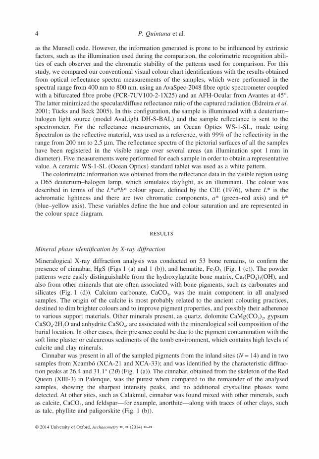
Mineralogical X-ray diffraction analysis was conducted on 53 bone remains, to confirm thepresence of cinnabar, HgS (Figs 1 (a) and 1 (b)), and hematite, Fe2O3 (Fig. 1 (c)). The powderpatterns were easily distinguishable from the hydroxylapatite bone matrix, Ca5(PO4)3(OH), andalso from other minerals that are often associated with bone pigments, such as carbonates andsilicates (Fig. 1 (d)). Calcium carbonate, CaCO3, was the main component in all analysedsamples. The origin of the calcite is most probably related to the ancient colouring practices,destined to dim brighter colours and to improve pigment properties, and possibly their adherenceto various support materials. Other minerals present, as quartz, dolomite CaMg(CO3)2, gypsumCaSO4·2H2O and anhydrite CaSO4, are associated with the mineralogical soil composition of theburial location. In other cases, their presence could be due to the pigment contamination with thesoft lime plaster or calcareous sediments of the tomb environment, which contains high levels ofcalcite and clay minerals.
Cinnabar was present in all of the sampled pigments from the inland sites (N = 14) and in twosamples from Xcambó (XCA-21 and XCA-33); and was identified by the characteristic diffrac-tion peaks at 26.4 and 31.1° (2θ) (Fig. 1 (a)). The cinnabar, obtained from the skeleton of the RedQueen (XIII-3) in Palenque, was the purest when compared to the remainder of the analysedsamples, showing the sharpest intensity peaks, and no additional crystalline phases weredetected. At other sites, such as Calakmul, cinnabar was found mixed with other minerals, suchas calcite, CaCO3, and feldspar—for example, anorthite—along with traces of other clays, suchas talc, phyllite and paligorskite (Fig. 1 (b)).
4 P. Quintana et al.
© 2014 University of Oxford, Archaeometry ••, •• (2014) ••–••
Hematite, Fe2O3, was detected in 35 burials from Xcambó, showing high intensity peaks at24.2° and 33.2° (2θ), indicating that the pigment has a high crystallinity. Calcite, calcitemagnesian (Ca,Mg)CO3 and quartz, SiO2, were also found (Fig. 1 (c)). In many samples fromXcambó, the main reflection of calcite at 29.5° (2θ) exhibits a dissymmetric peak, whichconfirms the presence of other types of calcium carbonates; that is, a stoichiometric calciteand other carbonates enriched with magnesium and/or iron that replace some of calcium atoms(Fig. 1 (d)), such as ankerite, Ca(Fe,Mg)(CO3)2 (Cailleau et al. 2005). The formation of ankeriteidentifies a diagenetic process due to dissolution of the hematite and subsequent substitution ofcalcium in the carbonates of the soil (Xu et al. 2005). We also identified traces of clay minerals,such as smectite interstratified with kaolinite or illite and palygorskite.
Optical microscopy
Surface images and thin sections of different bone samples were obtained in an optical micro-scope using transmitted and reflected light. The magnifications ranged from 2.5× to 40×. Thehistological results suggest that cinnabar pigmentation was usually applied in the form of a paste,
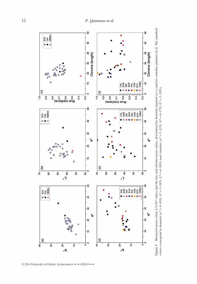
Figure 1 X-ray diffraction patterns of the pigments and the detected minerals: (a) a cinnabar sample from Palenque;(b) cinnabar and calcite from Calakmul; (c, d) hematite and other carbonate minerals from different samples fromXcambó.
Red pigments in Classic period Maya funerary practices 5
© 2014 University of Oxford, Archaeometry ••, •• (2014) ••–••
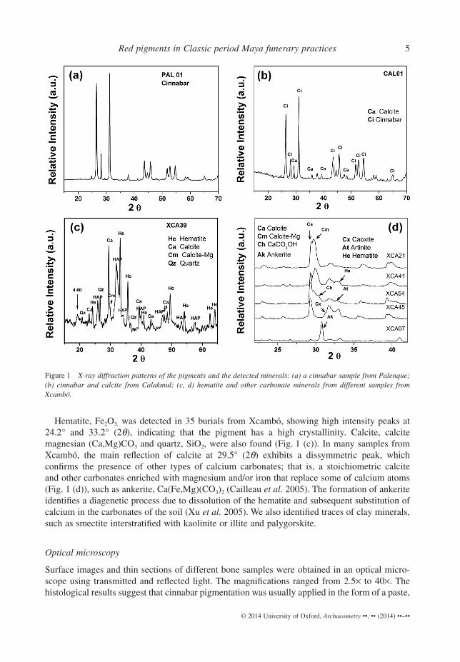
as evidenced by the homogeneously compact texture of the coloured layer, with the visibleadmixture of amorphous particles. This is the case, for example, in specimen CAL01 fromCalakmul’s Tomb II-5a (Fig. 2), showing that the bone surface is covered by a homogeneous,compacted layer of vermillion cinnabar without inclusions (0.09 mm thick). Its distributionsuggests that it had been applied together with the addition of an amorphous material of organicorigin, possibly to form the red paste to be applied. Additional substances, such as amorphousblackish agglutinants, which we have also documented in this study for Calakmul and Palenque,could have been added as paint vehicles, perhaps to improve durability and facilitate applicationof the colourants as paint or paste. However, there were also exceptions such as the specimenfrom Calakmul’s Tomb II-4 (CAL16); in this case, a thin, dispersed coat of cinnabar powder canbe observed on top of a vertebra. The grain distribution forms a heterogeneous pigmentation layerwith a thickness between 0.09 mm and 0.3 mm (Fig. 2).
Differently from cinnabar, it appears that red hematite was more commonly applied on corpsesin the form of powder among the Classic period Maya (Fig. 2). For example, XCA25 (Burial 76)from Xcambó, Yucatán, displays inclusions of other substances in the hematite layer (maximumthickness of 0.20 mm). Probably, the powder presentation facilitated contamination with exog-enous matter, given that the pigmented layer (with a thickness between 0.18 mm and 0.30 mm)clearly contains particles that originally did not form part of the pigment application, as shownin sample XCA23 (Fig. 2). However, there are exceptions. In some few cases, such as in sampleXCA39, it looks as though the reddish hematite was applied as a paste, resulting in a homoge-neously pigmented layer on top of the body, then bone. Its homogeneous distribution suggeststhe presence of an organic medium within the coloured layer, to vanish in the course of thedecomposition process.
Morphology and chemical analysis by SEM–EDS
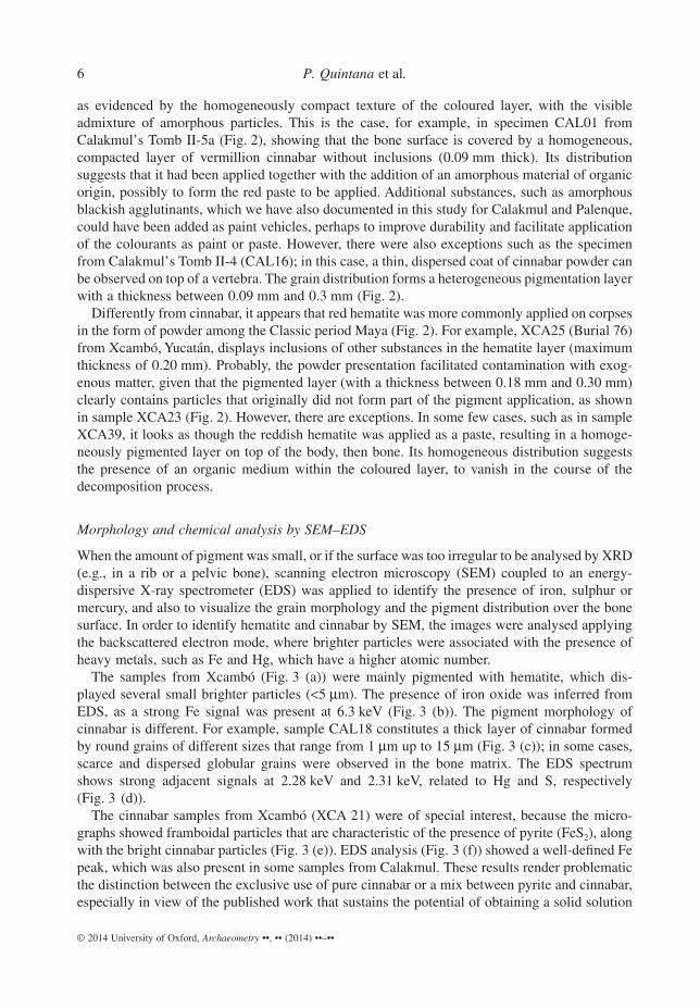
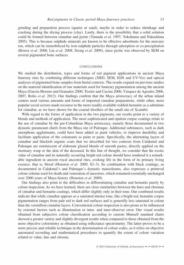
When the amount of pigment was small, or if the surface was too irregular to be analysed by XRD(e.g., in a rib or a pelvic bone), scanning electron microscopy (SEM) coupled to an energy-dispersive X-ray spectrometer (EDS) was applied to identify the presence of iron, sulphur ormercury, and also to visualize the grain morphology and the pigment distribution over the bonesurface. In order to identify hematite and cinnabar by SEM, the images were analysed applyingthe backscattered electron mode, where brighter particles were associated with the presence ofheavy metals, such as Fe and Hg, which have a higher atomic number.
The samples from Xcambó (Fig. 3 (a)) were mainly pigmented with hematite, which dis-played several small brighter particles (<5 μm). The presence of iron oxide was inferred fromEDS, as a strong Fe signal was present at 6.3 keV (Fig. 3 (b)). The pigment morphology ofcinnabar is different. For example, sample CAL18 constitutes a thick layer of cinnabar formedby round grains of different sizes that range from 1 μm up to 15 μm (Fig. 3 (c)); in some cases,scarce and dispersed globular grains were observed in the bone matrix. The EDS spectrumshows strong adjacent signals at 2.28 keV and 2.31 keV, related to Hg and S, respectively(Fig. 3 (d)).
The cinnabar samples from Xcambó (XCA 21) were of special interest, because the micro-graphs showed framboidal particles that are characteristic of the presence of pyrite (FeS2), alongwith the bright cinnabar particles (Fig. 3 (e)). EDS analysis (Fig. 3 (f)) showed a well-defined Fepeak, which was also present in some samples from Calakmul. These results render problematicthe distinction between the exclusive use of pure cinnabar or a mix between pyrite and cinnabar,especially in view of the published work that sustains the potential of obtaining a solid solution
6 P. Quintana et al.
© 2014 University of Oxford, Archaeometry ••, •• (2014) ••–••
Figure 2 Optical micrographs of different bones and thin sections of samples: (a) surface images; (b) thin sections withtransmitted light; (c) thin sections with reflected light.
Red pigments in Classic period Maya funerary practices 7
© 2014 University of Oxford, Archaeometry ••, •• (2014) ••–••
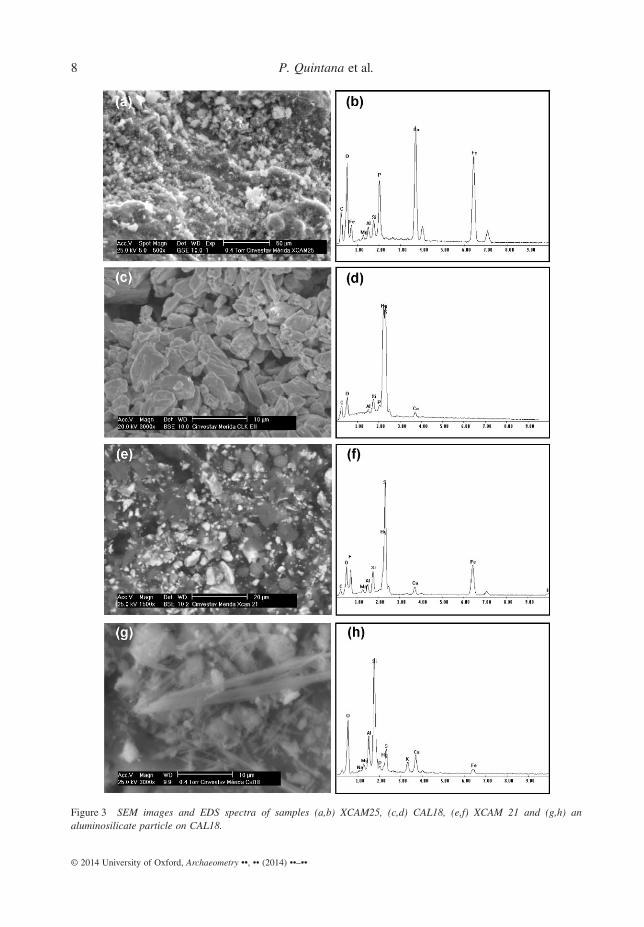
Figure 3 SEM images and EDS spectra of samples (a,b) XCAM25, (c,d) CAL18, (e,f) XCAM 21 and (g,h) analuminosilicate particle on CAL18.
8 P. Quintana et al.
© 2014 University of Oxford, Archaeometry ••, •• (2014) ••–••
of pyrite with cinnabar by replacement of Hg atoms by Fe intrusions into the crystalline structure(Edreira et al. 2003).
In several samples, signals related to Ca or P were observed, associated with the presence ofcalcium carbonate and hydroxylapatite. In some sections of CAL18, a fibre texture can beobserved, bearing a morphology that is similar to those that have been reported in the literature(Vandenabeele et al. 2005; Krekeler and Kearns 2009) and that were identified as palygorskite orsmectites (Fig. 3 (g)): this result is supported by XRD analyses and also confirmed by EDS(Fig. 3 (h)). The latter shows the presence of Si, Al, Mg, Na, K and Fe, which probably belongto quartz and clays. Some researchers have argued that other minerals, such as calcite and clays,were added to the pigment to prepare a consolidated suspension (or slip) that could be spreadeasily over the body (García et al. 2006).
Chromatic characterization
The conventional chromatic characterization of the red pigments was performed on 86 samplesand rendered unequivocal results in 72 specimens according to the Munsell code. The pig-mented layers from Calakmul, Ek Balam, Oxkintok, Dzibanché, Sacul and Palenque that thechemical analyses had identified as cinnabar were determined as dark to bright red (HUE7.5R). The colour values of the confirmed cinnabar pigments range from 3 to 5, and theirchroma from 4 to 14, with the majority identified within the saturated range (10 and above).The visually determined surface colours of the confirmed hematite pigments from the coastalcommunity of Xcambó differ slightly from the colour range of the cinnabar layers. Here,both HUE 7.5R (reddish towards orange tones) and HUE 5R (reddish towards purple tones)were determined. The hematite chroma also exhibit a greater variability and at the same timeidentify paler tones (‘weak red’) than the cinnabar covers, with values that run from 4 to 10.The hematite colour values range from 3 to 5, similar to those of the cinnabar specimens. Fromthe above colour codes, we observe that the red tones produced with hematite are generallymore variable, weaker and colder than the warm, intense, bright reds produced in cinnabarsubstrates.
Characterization of diffuse reflectance
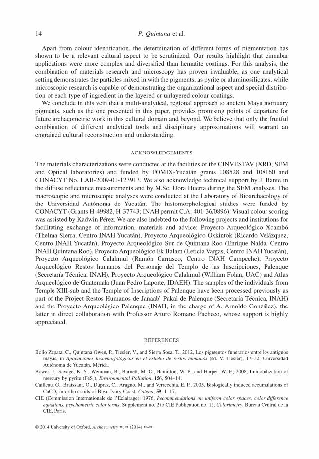
The reflectance spectra were registered in the visible range over three different areas of thepictorial surface by obtaining a mean value for each sample. The experimental data profiles forhematite and cinnabar standards are shown in Figure 4. The cinnabar pigment obtained from theRed Queen from Palenque and the pure hematite mineral obtained from Almagres, Veracruz,Mexico were considered as standards. A wide reflectance band is observed between 350 and600 nm for both pigments (Figs 4 (a) and 4 (c)).
The diffuse reflectance spectra show characteristic features of hematite, namely a sharppositive slope at wavelengths higher than 600 nm (Fig. 4 (a)). The spectra for the red ochresidentify a strong peak around 730 nm. To differentiate the red hue, it is necessary to charac-terize the wavelength range of the sharp slope that corresponds to the absorption edge in thevisible range. For this purpose, the wavelength of the inflexion point, λm, is considered becauseit is easily recognized as the abscissa of the maximum of the first derivative of each spectrum.From the measured spectra, λm is evaluated as 587.5 nm for pure hematite. When comparedto the analysed samples, λm varies between 577.9 nm and 586.9 nm according to the compo-sition of the hue and thus to their colour (Fig. 4 (b)). On the other hand, regarding cinnabar
Red pigments in Classic period Maya funerary practices 9
© 2014 University of Oxford, Archaeometry ••, •• (2014) ••–••
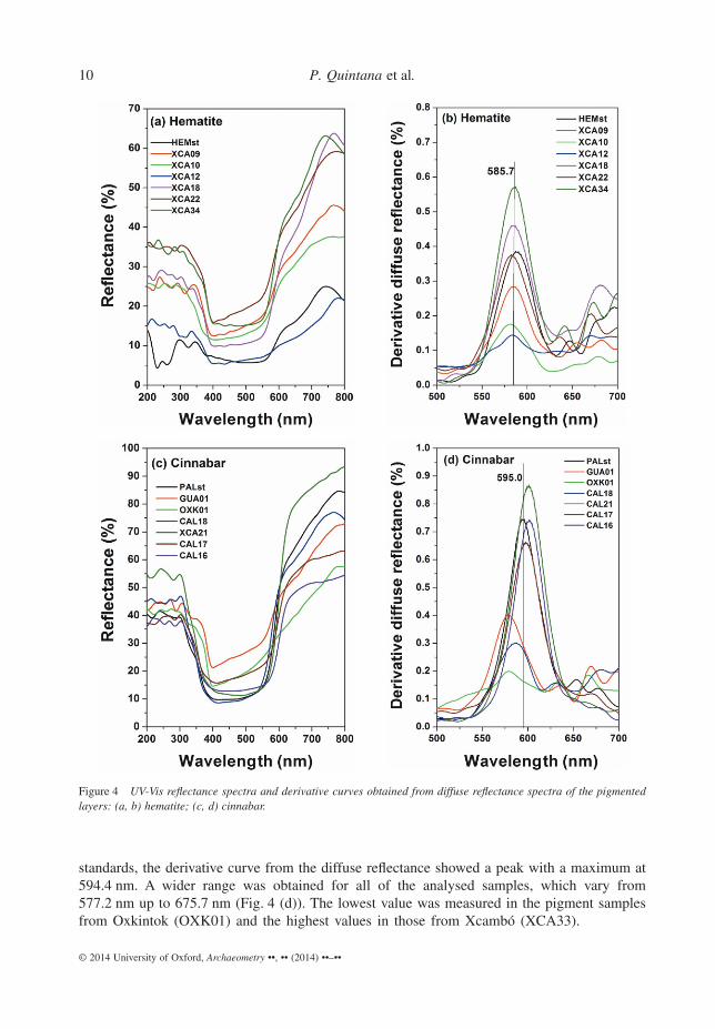
standards, the derivative curve from the diffuse reflectance showed a peak with a maximum at594.4 nm. A wider range was obtained for all of the analysed samples, which vary from577.2 nm up to 675.7 nm (Fig. 4 (d)). The lowest value was measured in the pigment samplesfrom Oxkintok (OXK01) and the highest values in those from Xcambó (XCA33).
Figure 4 UV-Vis reflectance spectra and derivative curves obtained from diffuse reflectance spectra of the pigmentedlayers: (a, b) hematite; (c, d) cinnabar.
10 P. Quintana et al.
© 2014 University of Oxford, Archaeometry ••, •• (2014) ••–••
Chromaticity parameters
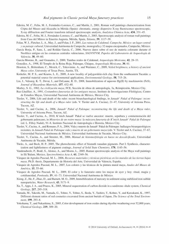
The obtained chromaticity parameters L*a*b*, hue and chroma for both pigments are presentedin Figure 5. The colour L*a*b* coordinates for the hematite standard are 16, 6 and 34, respec-tively. A colour comparison in terms of the brilliance and tone of the pigmented surfaces for allof the samples from Xcambó shows relatively little variation, since the values for a* and b* aregrouped together; the a* parameter (the green–red axis) falls between 7 and 22, and the b*parameter (the blue–yellow axis) between 8 and 18 (Fig. 5 (a)). Only one analysed sample fromCalakmul shows higher values than that, namely 35 and 24, respectively. The lightness L* variesfrom 32 up to 60, with a mean a* value of 48 (Fig. 5 (b)).
According to CIE (1976), colour has a three-dimensional appearance, consisting of a lightnessattribute (L*) and two chromatic attributes, namely the hue and the chroma. The hue (H°ab) isdetermined by the dominant wavelength and identifies a colour as found in its pure state in thespectrum. It is represented by the hue angle in the a*–b* plane, which increases by a counter-clockwise rotation around the a*, b* axis, with 0° being red, 90° being yellow, 180° beinggreen and 270° being blue. The chroma (Cab*) refers to the saturation of a colour and is ameasure of how much grey and white light is mixed in with the pure focal colour. The chromaranges from neutral to brilliant and is represented as the distance from the origin of the axis onthe a*–b* plane. Therefore, the hue and chroma were calculated according to the formulaeH°ab = arctan(b*/a*) and Cab* = (a*2 + b*2)1/2.
The hematite pigments, measured in the samples from Xcambó, show a hue direction anglethat ranges from 0.6 up to 0.9 (radians) and a colour vector length for the chroma values that runsfrom 15 to 27 (Fig. 5 (c)). This means that the variation of the hue angles falls within the middlerange of the red colour (from 35° up to 52°, with a mean value of 43°), while the chroma valuesshow a neutral tendency, as short lengths are obtained on the colour vector. In comparison withthe mineral standard, a stronger red colour is obtained, with a hue value of 21°.
On the other hand, the L*a*b* values for the cinnabar standard were 48, 27 and 15, respec-tively. Regarding the changes observed in chromatic coordinates a* and b* for the pigmentedbones recovered from different archaeological sites, rather large a* values are observed, whichcorrespond to a reddish colour that ranges from 11 to 43 (Fig. 5 (d)). The range variation for theb* parameter falls between 12 and 37, while the lightness varies from 29 up to 69, with a meanvalue of 49 (Fig. 5 (e)).
The hue and chroma values indicate that all samples fall within the reddish range but presentsignificant differences between groups (Fig. 5 (f)). The hue values for the direction angle rangefrom 0.6 up to 1.0 (radians). Therefore, the hue angles in degrees vary between 31° and 56°, witha mean value of 44°. The chroma values range from 16 to 57. The variation of the hue angles iswithin the obtained range for hematite. However, an extensive variation of the length of the colourvector is determined, indicating that a wide chroma is observed on the pigmentation, whichdepends on the geographical mineralogical sources where the mineral was extracted or ondifferences in pigment concentrations within its medium. For example, samples from Guatemalahave smaller a* and b* values and a corresponding low chroma. Samples from Dzibanché showthe lowest hue values and the highest chroma values. Pigments measured from the remains ofPalenque have intermediate chroma values, whereas Calakmul shows a wide range of tonalities.
The differences in these values suggest that variation does occur in the crystalline structure.There could be several reasons for this. First, different source materials for cinnabar might havebeen used by the ancient undertakers. Second, in the cases in which cinnabar was applied as apaste, other complementary minerals were incorporated and mixed with the pigments during the
Red pigments in Classic period Maya funerary practices 11
© 2014 University of Oxford, Archaeometry ••, •• (2014) ••–••
Figu
re5
Mea
sure
dm
ean
colo
urL
*a*b
*va
lues
and
the
hue
and
chro
ma
mea
nva
lues
,det
erm
ined
for
hem
atit
epi
gmen
t(a
–c)
and
for
cinn
abar
pigm
ent
(d–f
).T
hest
anda
rder
rors
corr
espo
ndto
hem
atit
e(a
*=
4–16
%;
b*=
3–16
%;
L*
=4–
16%
)an
dci
nnab
ar(a
*=
5–21
%;
b*=
4–17
%;
L*
=3–
20%
).
12 P. Quintana et al.
© 2014 University of Oxford, Archaeometry ••, •• (2014) ••–••
grinding and preparation process (quartz or sand), maybe in order to reduce shrinkage andcracking during the drying process (clay). Lastly, there is the possibility that a solid solutioncould be formed between cinnabar and pyrite (Yamada et al. 1997; Yokohama and Nakashima2005). This is because sulphide minerals are known to be effective adsorbents for the mercuryion, which can be immobilized by iron sulphide particles through adsorption or co-precipitation(Bower et al. 2008; Liu et al. 2008; Xiong et al. 2009), since pyrite was observed by SEM onseveral pigmented bone surfaces.
CONCLUSIONS
We studied the distribution, types and forms of red pigment applications in ancient Mayafunerary rites by combining different techniques (XRD, SEM, EDS and UV-Vis) and opticalanalyses of pigmented bone samples from burial contexts. The results expand on previous studieson the material identification of raw materials used for funerary pigmentation among the ancientMaya (García-Moreno and Granados 2000; Tiesler and Cucina 2006; Vázquez de Agredos 2006,2007; Bolio et al. 2012). Our findings confirm that the Maya aristocracy of the urban inlandcentres used various amounts and forms of imported cinnabar preparations, while other, morepopular social sectors made recourse to the more readily available reddish hematite as a substitutefor cinnabar, as we have shown for the coastal dwellers of the small site of Xcambó.
With regard to the forms of application in the two pigments, our results point to a variety ofblends and methods of application. The most sophisticated and opulent corpse coatings relate tothe use of cinnabar by the pre-Columbian Maya aristocracy, namely those documented in twodynastic paramount chiefs from the Maya site of Palenque. Additional substances, such as darkamorphous agglutinants, could have been added as paint vehicles, to improve durability andfacilitate application of the colourants as paint or paste. Specifically, the alternating layers ofcinnabar and blackish organic coats that we described for two contexts from Calakmul andPalenque are reminiscent of elaborate glazed blends of smooth pastes, directly applied on themortuary wrap or the skin of the deceased. In this line of thought, we consider that the exoticvalue of cinnabar and its naturally occurring bright red colour should have rendered it a remark-able ingredient in ancient royal ancestral rites, evoking life in the form of its primary livingessence: that is, blood (Houston et al. 2009, 82–3). Its combination with black coatings, asdocumented in Calakmul’s and Palenque’s dynastic mausoleums, also expresses a primevalcolour scheme used for death and veneration of ancestors, which remained essentially unchangedover 2000 years of Maya history (Houston et al. 2009).
Our findings also point to the difficulties in differentiating cinnabar and hematite purely bycolour inspection. As we have learned, there are close similarities between the hues and chromasof cinnabar and hematite coatings, which differ slightly only in their tone. Our combined resultsindicate that while cinnabar appears as a saturated intense tone, like a bright red, hematite corpsepigmentation ranges from pale red to dark red surfaces and is generally less saturated in colourthan the vermillion cinnabar layers. Conventional colour inspection is also prone to be influencedby external factors such as illumination or intra- and inter-observer error. Our visual resultsobtained from subjective colour classification according to custom Munsell standard chartsshowed a greater variety and slightly divergent results when compared to those obtained from themore objective colorimetry as obtained using reflectance spectrometry. The latter proves to be amore precise and reliable technique in the determination of colour codes, as it relies on objectiveautomated recording and mathematical procedures to quantify the extent of colour variationrelated to value, hue and chroma.
Red pigments in Classic period Maya funerary practices 13
© 2014 University of Oxford, Archaeometry ••, •• (2014) ••–••
Apart from colour identification, the determination of different forms of pigmentation hasshown to be a relevant cultural aspect to be scrutinized. Our results highlight that cinnabarapplications were more complex and diversified than hematite coatings. For this analysis, thecombination of materials research and microscopy has proven invaluable, as one analyticalsetting demonstrates the particles mixed in with the pigments, as pyrite or aluminosilicates; whilemicroscopic research is capable of demonstrating the organizational aspect and special distribu-tion of each type of ingredient in the layered or unlayered colour coatings.
We conclude in this vein that a multi-analytical, regional approach to ancient Maya mortuarypigments, such as the one presented in this paper, provides promising points of departure forfuture archaeometric work in this cultural domain and beyond. We believe that only the fruitfulcombination of different analytical tools and disciplinary approximations will warrant anengrained cultural reconstruction and understanding.
ACKNOWLEDGEMENTS
The materials characterizations were conducted at the facilities of the CINVESTAV (XRD, SEMand Optical laboratories) and funded by FOMIX-Yucatán grants 108528 and 108160 andCONACYT No. LAB-2009-01-123913. We also acknowledge technical support by J. Bante inthe diffuse reflectance measurements and by M.Sc. Dora Huerta during the SEM analyses. Themacroscopic and microscopic analyses were conducted at the Laboratory of Bioarchaeology ofthe Universidad Autónoma de Yucatán. The histomorphological studies were funded byCONACYT (Grants H-49982, H-37743; INAH permit C.A: 401-36/0896). Visual colour scoringwas assisted by Kadwin Pérez. We are also indebted to the following projects and institutions forfacilitating exchange of information, materials and advice: Proyecto Arqueológico Xcambó(Thelma Sierra, Centro INAH Yucatán), Proyecto Arqueológico Oxkintok (Ricardo Velázquez,Centro INAH Yucatán), Proyecto Arqueológico Sur de Quintana Roo (Enrique Nalda, CentroINAH Quintana Roo), Proyecto Arqueológico Ek Balam (Leticia Vargas, Centro INAH Yucatán),Proyecto Arqueológico Calakmul (Ramón Carrasco, Centro INAH Campeche), ProyectoArqueológico Restos humanos del Personaje del Templo de las Inscripciones, Palenque(Secretaría Técnica, INAH), Proyecto Arqueológico Calakmul (William Folan, UAC) and AtlasArqueológico de Guatemala (Juan Pedro Laporte, IDAEH). The samples of the individuals fromTemple XIII-sub and the Temple of Inscriptions of Palenque have been processed previously aspart of the Project Restos Humanos de Janaab’ Pakal de Palenque (Secretaría Técnica, INAH)and the Proyecto Arqueológico Palenque (INAH, in the charge of A. Arnoldo González), thelatter in direct collaboration with Professor Arturo Romano Pacheco, whose support is highlyappreciated.
REFERENCES
Bolio Zapata, C., Quintana Owen, P., Tiesler, V., and Sierra Sosa, T., 2012, Los pigmentos funerarios entre los antiguosmayas, in Aplicaciones histomorfológicas en el estudio de restos humanos (ed. V. Tiesler), 17–32, UniversidadAutónoma de Yucatán, Mérida.
Bower, J., Savage, K. S., Weinman, B., Barnett, M. O., Hamilton, W. P., and Harper, W. F., 2008, Immobilization ofmercury by pyrite (FeS2), Environmental Pollution, 156, 504–14.
Cailleau, G., Braissant, O., Dupraz, C., Aragno, M., and Verrecchia, E. P., 2005, Biologically induced accumulations ofCaCO3 in orthox soils of Biga, Ivory Coast, Catena, 59, 1–17.
CIE (Commission Internationale de l’Eclairage), 1976, Recommendations on uniform color spaces, color differenceequations, psychometric color terms, Supplement no. 2 to CIE Publication no. 15, Colorimetry, Bureau Central de laCIE, Paris.
14 P. Quintana et al.
© 2014 University of Oxford, Archaeometry ••, •• (2014) ••–••
Edreira, M. C., Feliu, M. J., Fernández-Lorenzo, C., and Martín, J., 2001, Roman wall paintings characterization fromCripta del Museo and Alcazaba in Mérida (Spain): chromatic, energy dispersive X-ray fluorescence spectroscopic,X-ray diffraction and Fourier transform infrared spectroscopic analysis, Analytica Chimica Acta, 434, 331–45.
Edreira, M. C., Feliu, M. J., Fernández-Lorenzo, C., and Martín, J., 2003, Spectroscopic analysis of roman wall paintingsfrom Casa del Mitreo in Emerita Augusta, Mérida, Spain, Talanta, 59, 1117–39.
Folan, W. J., Fletcher, L. A., May, J., and Folan, I. F., 2001, Las ruinas de Calakmul, Campeche, México: un lugar centraly su paisaje cultural, Universidad Autónoma de Campeche, monografía y 32 mapas encajonados, Campeche, México.
García Borja, P., Sanz, I., and Roldán García, C., 2006, Nuevos datos sobre el uso de materia colorante durante elNeolítico antiguo en las comarcas centrales valencianas, SAGVNTVM, Papeles del Laboratorio de Arqueología deValencia, 38, 49–60.
García-Moreno, R., and Granados, J., 2000, Tumbas reales de Calakmul, Arqueología Mexicana, 42, 28–33.González, A., 1998, El Templo de la Reina Roja, Palenque, Chiapas, Arqueología Mexicana, 30, 61.Houston, S., Brittenham, C., Mesick, C., Tokovinine, A., and Warinner, C., 2009, Veiled brightness, a history of ancient
Maya color, University of Texas Press, Austin, TX.Krekeler, M. P. S., and Kearns, L. E., 2009, A new locality of palygorskite-rich clay from the southeastern Yucatán: a
potential material source for environmental applications, Environmental Geology, 58, 715–26.Liu, J., Valsaraj, K. T., Devai, I., and DeLaune, R. D., 2008, Immobilization of aqueous Hg(II) by mackinawite (FeS),
Journal of Hazardous Materials, 157, 432–40.Morley, S. G., 1982, La civilización maya, FCE, Sección de obras de antropología, 3a, Reimpresión, Mexico City.Ruz-Lhuillier, A., 1991, Costumbres funerarias de los antiguos mayas, Instituto de Investigaciones Filológicas, Centro
de Estudios Mayas, Universidad Nacional Autónoma de México, Mexico City.Tiesler, V., 2006, Life and death of the ruler: recent bioarchaeological findings, in Janaab’ Pakal of Palenque: recon-
structing the life and death of a Maya ruler (eds. V. Tiesler and A. Cucina), 21–47, University of Arizona Press,Tucson, AZ.
Tiesler, V., and Cucina, A., 2006, Janaab’ Pakal of Palenque: reconstructing the life and death of a Maya ruler,University of Arizona Press, Tucson, AZ.
Tiesler, V., and Cucina, A., 2010, K’inich Janaab’ Pakal se vuelve ancestor: muerte, sepultura y conmemoración delgobernante palencano, in Misterios de un rostro maya: la máscara funeraria de K’inich Janaab’ Pakal de Palenque(ed. L. Filloy Nadal), 91–8, Instituto Nacional de Antropología e Historia, Mexico City.
Tiesler, V., Cucina, A., and Romano, P. A., 2004, Vida y muerte de Janaab’ Pakal de Palenque: hallazgos bioarqueológicosrecientes, in Janaab Pakal de Palenque vida y muerte de un gobernante maya (eds. V. Tiesler and A. Cucina), 37–67,Universidad Nacional Autónoma de México, Universidad Autónoma de Yucatán, Mexico City.
Tiesler, V., Cucina, A., and Streeter, M., 2006, Manual de histomorfología en hueso no descalcificado, UniversidadAutónoma de Yucatán, Mérida.
Tücks, A., and Beck, H. P., 2005, The photochromic effect of bismuth vanadate pigments. Part I: Synthesis, character-ization and lightfastness of pigment coatings, Journal of Solid State Chemistry, 178, 1145–56.
Vandenabeele, P., Bodé, S., Alonso, A., and Moens, L., 2005, Raman spectroscopic analysis of the Maya wall paintingsin Ek’Balam, Mexico, Spectrochimica Acta A, 61, 2349–56.
Vázquez de Agredos Pascual, M. L., 2006, Recursos materiales y técnicas pictóricas en los murales de las tierras bajasmaya, Ph.D. thesis, Departamento de Historia del Arte, Universidad de Valencia, España.
Vázquez de Agredos Pascual, M. L., 2007, Los colores y las técnicas de la pintura mural maya, Anales del Museo deAmérica, 15, 55–66.
Vázquez de Agredos Pascual, M. L., 2009, El color y lo funerario entre los mayas de ayer y hoy: ritual, magia ycotidianeidad, Península, IV, 61–73, Universidad Nacional Autónoma de México.
Xiong, Z., He, F., Zhao, D., and Barnett, M. O., 2009, Immobilization of mercury in sediment using stabilized iron sulfidenanoparticles, Water Research, 43, 5171–9.
Xu, T., Apps, J. A., and Pruess, K., 2005, Mineral sequestration of carbon dioxide in a sandstone–shale system, ChemicalGeology, 217, 295–318.
Yamada, M., Takeshi, M., Yamada, G., Tohno, Y., Tohno, S., Ikeda, Y., Tashiro, T., Kohno, Y., and Kawakami, K., 1997,Different element ratios of red cosmetics excavated from ancient burials of Japan, The Science of the Total Environ-ment, 199, 293–8.
Yokohama, T., and Nakashima, S., 2005, Color development of iron oxides during rhyolite weathering over 52,000 years,Chemical Geology, 219, 309–20.
Red pigments in Classic period Maya funerary practices 15
© 2014 University of Oxford, Archaeometry ••, •• (2014) ••–••