Embed Size (px)
Citation preview
James A. Brink is radiolo-gist-in-chief at Massachusetts General Hospital, Boston and Juan M. Taveras Professor of Radiology at Harvard Medical School, in the United States.
Before completing his residency and fellowship at Massachuse�s General Hospital in 1990, he received a Bachelor of Science degree in elec-trical engineering from Purdue University, Indiana and his medical degree from Indiana University. He then joined the faculty at the Mall-inckrodt Institute of Radiology at the Washington University School of Medicine in St. Louis, where he was promoted to associate profes-sor. He then went to Yale University in 1997 and served as chair of the department of diagnostic radiology from 2006 to 2013 before returning to Massachuse�s General Hospital as radiologist-in-chief.
A highly experienced clinical radi-ologist, especially in the areas of the utilisation and management of imaging resources, Dr. Brink also has a particular interest and expertise in issues related to the monitoring and control of medical radiation exposure.
A major figure in the field of medi-cal radiation protection, Dr. Brink serves as scientific vice-president for radiation protection in medicine on the National Council for Radiation Protection and Measurements. He is past-president of the American Roentgen Ray Society and a fellow of the Society of Computed Body Tomography & Magnetic Resonance, as well as a fellow of the American College of Radiology, where he also serves as vice-chair of the Board of Chancellors.
“Ours is a time when the practice and science of radiology are evolv-
ing at a rapid rate, and it is critical that we remain unified in our quest for new imaging technologies and care models. The European Society of Radiology provides a forum for diverse imaging professionals from Europe and beyond to come together in pursuit of these goals,” said Dr. Brink.
Over the course of his career, Dr. Brink has wri�en 119 publications and 19 book chapters. He has also given 239 presentations. In acknowledge-ment of his many achievements, he has received honorary membership from the Italian Society of Medical Radiology and the American Associ-ation of Physicists in Medicine.
At ECR 2015, Dr. Brink will deliver the Josef Lissner Honorary Lecture entitled ‘Is the ‘Art of Medicine’ dead in the era of population health management?’.
“The European Society of Radiol-ogy provides wonderful resources to thousands of radiologists worldwide, with a rich tradition of cultural diversity and inclusion. I am most honoured to present the Josef Lissner Honorary Lecture for this wonderful organisation.”
Radiation safety expert will deliver today’s Honorary Lecture
Challenges and innovations will shape development of
quantitative imaging
Advanced brain MRI provides insights into brain
malformations
Hopes rise that hybrid imaging can welcome in a
new era for MRI
ESOR furthers ESR’s commitment to education
and training
7 10 17 25
DAILY NEWS FROM EUROPE’S LEADING IMAGING MEETING | THURSDAY, MARCH 5, 2015
ECR TODAY 2015
HIGHLIGHTS CLINICAL CORNER TECHNOLOGY FOCUS COMMUNITY NEWS
Dr. James A. Brink from Boston, United States, will deliver today’s Honorary Lecture.
BY MICHAEL CREAN
myESR.org
In recognition of his outstanding achievements in the field of radiation safety and his commitment to improving safety in radiology, Professor James A. Brink has been invited to deliver the Josef Lissner Honorary Lecture at ECR 2015.
Don’t miss today’s Honorary Lecture
Thursday, March 5, 12:15–12:45, Room A #ECR2015AJosef Lissner Honorary Lecture
Is the ‘Art of Medicine’ dead in the era of population health
management?
James A. Brink; Boston, MA/USL
HIGHLIGHTS
myESR.org
3ECR TODAY | THURSDAY, MARCH 5, 2015
“There is a general consensus that the sensitivity and specific-ity of screening mammography is unsatisfactorily low,” Prof. Dr. Willi Kalender, PhD, chairman of the Institute of Medical Physics at the University of Erlangen in Germany, told ECR Today. “MRI offers much higher values but lacks imaging of microcalcifications, is more costly, and [is] time-consuming.”
As an alternative, he suggests dedicated breast CT, first described in detail in 2012. The goal of breast CT is to offer high-resolution CT imag-ing at a very low dose and increased sensitivity and specificity. CT can offer superposition-free imaging with a very high spatial resolution of 100 μm or be�er in all three spatial dimensions, dose levels low enough to conform with mammographic screening regulations, dynamic imaging with contrast medium application when indicated, and
integrated biopsy capabilities, he explained.
“The overall goal is a ‘one-stop shop-ping device’ for breast patients allow-ing for plain scans, for contrast-en-hanced studies, and for support of interventions,” said Kalender. “This will potentially replace the lengthy step-wise procedure (mammogra-phy, ultrasound, MRI, biopsy), which means consecutive with long wait times.”
The first two breast CT scanners, also called demonstrators, are in preparation for clinical installation at the university hospitals in Aachen and in Erlangen. Clinical testing is due to start around the middle of this year.
Breast MRI is also progressing. Unenhanced MRI may simplify the approach to breast MRI by omi�ing costs, time effort, and potential side effects related to contrast agents, including circulation-related effects, allergies, nephrogenic systemic fibro-
sis, according to Dr. Pascal Baltzer, associate professor in the radiology department at the Medical Univer-sity of Vienna. An imaging protocol using unenhanced MRI only – as in diffusion weighted imaging (DWI) together with T2-weighted anatom-ical images – would require about 5–10 minutes magnet time, thus allowing the examination of at least six patients per hour, he said. Also, reduced costs could broaden the availability of breast MRI.
“The issue is the broad variability of DWI imaging results,” he noted. “Although several initial studies have demonstrated a similar perfor-mance of unenhanced breast MRI compared with breast MRI, these studies did not answer how good unenhanced MRI is for specific indi-cations (such as high-risk screening, etc.). Furthermore, potential pitfalls such as mastopathic changes, small and non-mass lesions have not been closely investigated.”
At the moment, contrast-enhanced breast MRI is the standard of care and this won’t change until more research has covered the field of unenhanced breast MRI. “However, we have to keep in mind that lead-ing experts in prostate MRI openly discuss whether contrast agent application is really necessary,” he said.
Unenhanced breast MRI could be used in nearly all fields of breast MRI, given an appropriate DWI tech-nique, he remarked. However, typical pitfalls include chronic inflamma-tory conditions such as regularly seen in post-surgical conditions.
“As a ma�er of fact, I would not primarily recommend the use of enhanced MRI in the differentia-tion between scar and cancer recur-rence,” he stated. “Even simple MRI techniques might help in the differ-entiation of multiple mass lesions or cancer staging, or, more generally, in the characterisation of a definite anatomical area of uncertainty.”
At today’s special focus session, Baltzer promises to give an overview of what’s possible with unenhanced breast MRI. He said he always tries to be critical, will provide an evidence-based overview on possibilities, limi-tations, and developments in unen-hanced breast MRI.
“Breast imaging is rapidly evolving as a dedicated imaging modality,” said session moderator Dr. Marc Lobbes, PhD, from the radiology department at Maastricht University Medical Centre in the Netherlands. “Many imaging tools currently exist, from basic mammography, ultrasound,
and MRI, to elastography, digital breast tomosynthesis, contrast-en-hanced spectral mammography, state-of-art MRI sequences, and PET/MRI, to name but a few.”
He plans to provide an overview for both dedicated and general radiologists of the current most promising developments, including contrast-enhanced mammography, also called contrast-enhanced spec-tral mammography (CESM), which became commercially available in 2011. It uses an iodine-based contrast agent and a dual-energy technique to increase the diagnostic accuracy of mammography.
“In recent years, many studies have shown that CESM is consist-ently superior to full-field digital mammography [FFDM] in breast cancer detection and able to achieve results similar to breast MRI, which is still considered to be the most accurate breast cancer detection modality,” Lobbes noted.
CESM could easily be implemented in everyday clinical practice, but there are some disadvantages, such as the use of iodine-based contrast agents and the 80% increase in radi-ation dose. Also, CESM doesn’t have proper indications, he added.
“Although it is superior to FFDM and the principle is similar to that of breast MRI, proper indications are still lacking,” he stated. “None-theless, many studies are currently ongoing to clarify the strengths and weaknesses of CESM, so it is safe to assume that more knowledge on CESM indications will be gathered in the next few years.”
BY REBEKAH MOAN
CT and MRI contribute to major shake-up in breast imagingRadiologists are always on the lookout for the latest and greatest technology, and this is particularly true in breast imaging, because the primary modality, mammography, is not exactly perfect. Rapid progress in both CT and MRI are being made in this field.
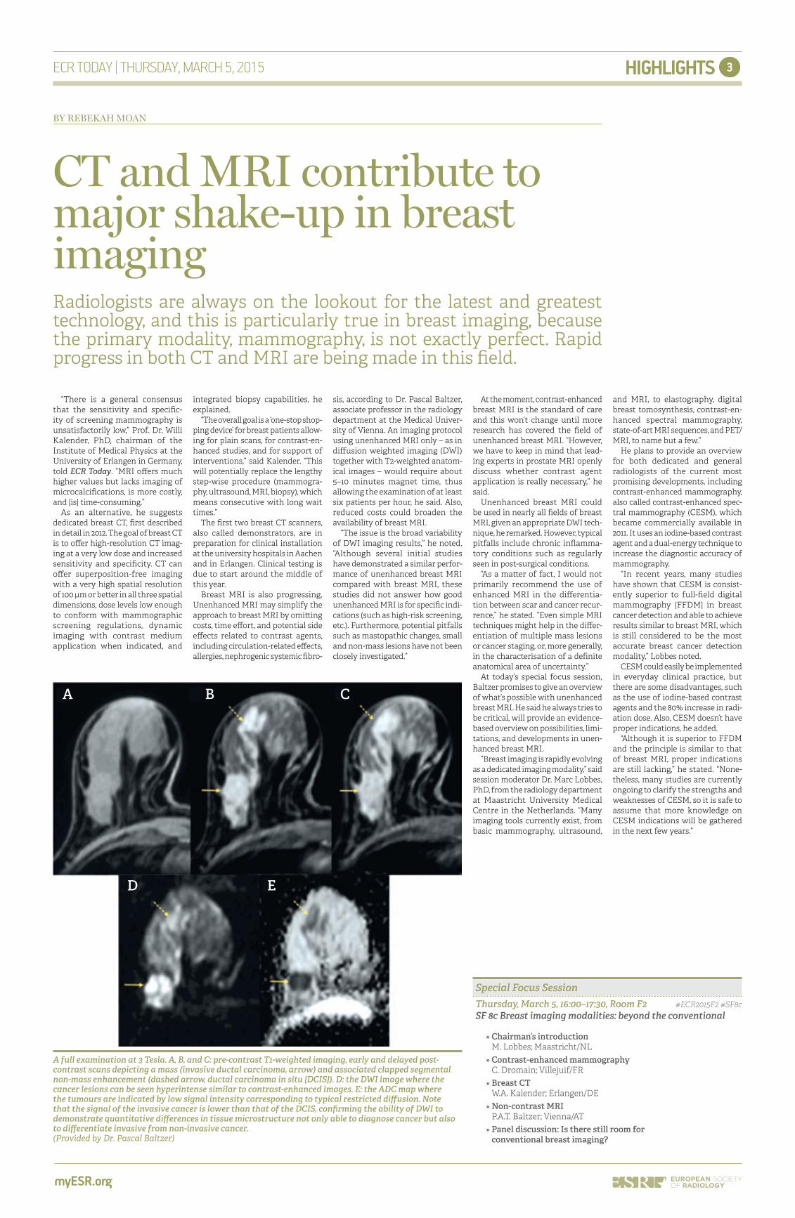
A full examination at 3 Tesla. A, B, and C: pre-contrast T1-weighted imaging, early and delayed post-contrast scans depicting a mass (invasive ductal carcinoma, arrow) and associated clapped segmental non-mass enhancement (dashed arrow, ductal carcinoma in situ [DCIS]). D: the DWI image where the cancer lesions can be seen hyperintense similar to contrast-enhanced images. E: the ADC map where the tumours are indicated by low signal intensity corresponding to typical restricted diffusion. Note that the signal of the invasive cancer is lower than that of the DCIS, confirming the ability of DWI to demonstrate quantitative differences in tissue microstructure not only able to diagnose cancer but also to differentiate invasive from non-invasive cancer. (Provided by Dr. Pascal Baltzer)
Special Focus Session
Thursday, March 5, 16:00–17:30, Room F2 #ECR2015F2 #SF8cSF 8c Breast imaging modalities: beyond the conventional
» Chairman’s introduction M. Lobbes; Maastricht/NL
» Contrast-enhanced mammography
C. Dromain; Villejuif/FR» Breast CT
W.A. Kalender; Erlangen/DE» Non-contrast MRI
P.A.T. Baltzer; Vienna/AT» Panel discussion: Is there still room for
conventional breast imaging?
A
D E
CB
Hot Shots from Day 1

C RSE Cardiovascular and Interventional Radiological Society of Europe
April 22-25, 2015Nice, France
LEADERS IN ONCOLOGIC INTERVENTIONS
ECIO 2015Sixth European Conference on Interventional Oncology including a joint session with the European Society for Medical Oncology (ESMO)
Join us for ...Multidisciplinary tumour boards, new horizons sessions and lots of tips and tricks forlocal tumour management
www.ecio.org
HIGHLIGHTS
myESR.org
7ECR TODAY | THURSDAY, MARCH 5, 2015
To enable patients to fully benefit from these advances, a number of steps must be taken. Experts will present these solutions in today’s dedicated Professional Challenges session at the ECR.
There is no doubt that quantita-tive imaging is the future of radiol-ogy. Not only does it help to detect disease at a much earlier stage, but also it considerably improves treat-ment monitoring.
In healthy tissue for instance, researchers can use quantitative imaging to study the functional properties of this tissue in order to get the most important meaning for biologists, physicists, etc., in a non-in-vasive way. Based on this informa-tion, they can develop biomarkers, which help to study the disease and its natural history, use the data to predict outcome and, most impor-tantly, to determine the responsive-ness of individual person to therapy – and assess its efficiency.
Applications for imaging biomark-ers include the detection and treat-ment of cardiovascular diseases, neurological diseases, musculoskel-etal disorders, metabolic diseases, autoimmune pathologies and inflammation. Biomarkers also play a key role in new drug development.
“Using biomarkers to streamline drug, tumour and disease progres-sion discovery represents a huge advancement in healthcare,” Guy Frija, consultant radiologist at the Imaging Department at Hôpital Européen Georges Pompidou in Paris, summed up.
But several bo�lenecks currently prevent imaging from unfolding its full potential in the contribution to medicine. Imaging biomarkers have to be technically validated, robust and reproducible. Some quantitative imaging biomarkers are currently being developed and proposed for clinical application, but their repro-ducibility has not yet been well established. Biomarkers should
also be clinically validated. Last but not least, they are currently not included in the landscape of Euro-pean biobanks.
“There is a great and urgent need to do so, since imaging repositories are dealing with big data, and have specific technical requirements in terms of codification, standards and interoperability,” said Frija, who will chair the session.
Healthcare providers need to consider imaging biomarkers in clinical trials and drug approval processes as a mandatory add-on whenever possible, and they must push for the development of a Euro-pean platform for their standardi-sation, according to Frija, Past-Pres-ident of the European Society of Radiology.
Under his presidency, the society took a decisive leap by establishing a European Biomarkers Task Force, creating synergies with RSNA’s Quantitative Imaging Biomarkers Alliance and tackling clinical valida-
tion of biomarkers at the European level.
Along those efforts, quantita-tive imaging must also overcome a number of technical difficulties. While in CT the Hounsfield unit is the only parameter that can be used, MR on the contrary offers a variety of techniques to measure tissue and obtain biochemical information on the tissue of interest.
Diffusion imaging for instance enables to calculate the apparent diffusion coefficient, a parameter used in tumour imaging. Dynamic contrast enhanced MR allows to analyse uptake grades of contrast medium and obtain information on the density, vascular permeabil-ity, perfusion, and plasma volume of tissue – to name a few. Finally MR spectroscopy enables to quan-tify metabolites and see how their numbers change by disease, help-ing to evaluate tumour grade or proliferation, extension, solidity and aggressiveness.
Unfortunately, MR examinations are still not reproducible, and this is a major obstacle for quantifica-tion, according to Siegfried Tra�nig, Professor and Head of the Centre of Excellence for High Field MR, Department of Biomedical Imag-ing and Image-guided Therapy at Vienna General Hospital (AKH), who will also speak during the session.
“When we perform the examina-tion of the same patient on a differ-ent machine, at a different site and with different technicians, I am convinced that we will get different data, so we can’t use it in a reliable way,” he said.
Another issue is that perform-ing quantitative imaging takes time compared with regular scans. Patients must lie still longer in the scanner to obtain multiparametric data, so they are more likely to move and this can hamper data quality.
“What we really need is a fast technique and the best would be if we could acquire simultaneously multiple parameters,” Tra�nig said.
There is hope on the horizon, as sequences were recently developed with MR, in which researchers can acquire with the same sequence T1 and T2 relaxation times of a certain tissue or region.
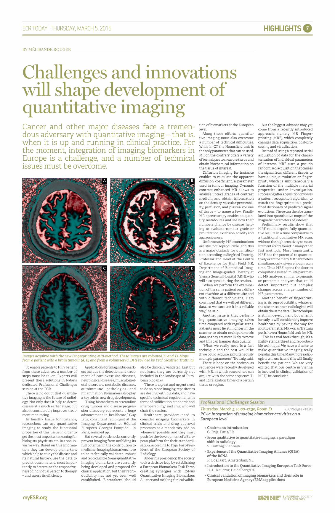
But the biggest advance may yet come from a recently introduced approach, namely MR Finger-printing (MRF), which completely changes data acquisition, post-pro-cessing and visualisation.
Instead of using a repeated, serial acquisition of data for the charac-terisation of individual parameters of interest, MRF uses a pseudo randomised acquisition that causes the signal from different tissues to have a unique evolution or ‘finger-print’, which is simultaneously a function of the multiple material properties under investigation. Processing a�er acquisition involves a pa�ern recognition algorithm to match the fingerprints to a prede-fined dictionary of predicted signal evolutions. These can then be trans-lated into quantitative maps of the magnetic parameters of interest.
Preliminary results show that MRF could acquire fully quantita-tive results in a time comparable to a traditional qualitative MR scan, without the high sensitivity to meas-urement errors found in many other fast methods. Most importantly, MRF has the potential to quantita-tively examine many MR parameters simultaneously, given enough scan time. Thus MRF opens the door to computer-assisted multi-paramet-ric MR analyses, similar to genomic or proteomic analyses that could detect important but complex changes across a large number of MR parameters.
Another benefit of fingerprint-ing is its reproducibility: whatever the site or scanner, radiologists will obtain the same data. The technique is still in development, but when it is ready, it will considerably improve healthcare by paving the way for multiparametric MR – or, as Tra�nig put it, have a Hounsfield unit for MR.
“This is a real breakthrough, it’s a highly standardised and reproduci-ble technique. We have a chance to make quantitative imaging really popular this time. Many more radiol-ogists will use it, and this will finally benefit the patient. We are very excited that our centre in Vienna is involved in clinical validation of MRF,” he concluded.
BY MÉLISANDE ROUGER
Challenges and innovations will shape development of quantitative imagingCancer and other major diseases face a tremen-dous adversary with quantitative imaging – that is, when it is up and running in clinical practice. For the moment, integration of imaging biomarkers in Europe is a challenge, and a number of technical issues must be overcome.
Professional Challenges Session
Thursday, March 5, 16:00–17:30, Room F1 #ECR2015F1 #PC8aPC 8a Integration of imaging biomarker activities on a
European level
» Chairman’s introduction G. Frija; Paris/FR
» From qualitative to quantitative imaging: a paradigm
shi� in radiology S. Tra�nig; Vienna/AT
» Experience of the Quantitative Imaging Alliance (QIBA)
of the RSNA R. Boellaard; Amsterdam/NL
» Introduction to the Quantitative Imaging European Task Force H.-U. Kauczor; Heidelberg/DE
» Clinical validation of imaging biomarkers and their role in
European Medicine Agency (EMA) applications
Images acquired with the new Fingerprinting MRI-method. These images are coloured T1 and T2-Maps from a patient with a brain tumour (A, B) and from a volunteer (C, D) (Provided by Prof. Siegfried Tra�nig).
A
C
B
D
www.siemens.com/ecr
Join the Siemens Industry Workshops at ECR 2015March 4th – 7th, Austria Center Vienna, Room 0.93 / Entrance Level
Answers for life.
Siemens is looking forward to welcoming you at ECR 2015. We invite you to join the Siemens Industry Workshops to discover more about innovative applications.
Benefit from experts’ experience and receive an update on state-of-the-art techniques in computed tomography, magnetic resonance and breast imaging. As a registered attendee for ECR 2015 these workshops are free of charge.
Location for Industry Workshops:
Austria Center ViennaRoom 0.93 / Entrance Level / Next to room F2
Registration on-site. Please note that only a limited number of seats are available on a first come – first serve basis.
And don’t forget to visit the Siemens Booth #11 located in Extension Expo A, Entrance Level.
There is an obvious need for the development and implementation of imaging biobanks in Europe. Following ECR, the ESR Research Commi�ee will launch the European imaging biobank initiative in order to achieve significant progress in 2015, explained Professor Hans-Ul-rich Kauczor from the University of Heidelberg, Germany, chair of the ESR Research Commi�ee.
By definition, imaging biobanks are organised databases of medi-cal images and associated imag-ing biomarkers from healthy and diseased tissue. These can be shared among multiple researchers, and should be linked to a biorepository of human tissue and genetic samples.
Following the white paper on imaging biobanks prepared by the ESR Working Group on Imaging Biobanks, the ESR Research Commit-tee is spearheading the programme to build the first European-wide network of imaging biobanks. Issues under discussion include the development of intelligent tools for the analysis and processing of imaging biomarkers; the promotion of standardisation, validation and benchmarking of those imaging data; and the integration of data with existing data repositories. The working group is also exploring the economical, ethical and legal issues associated with the management of imaging biobanks.
Currently, the Biobanking and Biomolecular resources Research Infrastructure (BBMRI-ERIC) is the closest existing organisation to these ambitions. It aims to establish, operate, and develop a pan-European research infrastructure of biobanks and biomolecular resources.
“This is a very successful example of a pan-European biobank research infrastructure,” said Kauczor. “Our activity should generate something similar for imaging biobanks and if possible, we would like to link it with the BBMRI framework. We are look-ing forward to liaise strongly with BBMRI.”
He is convinced the time is right to develop a European imaging biobank. “Most existing biobanks allow only for the collection of genotype data,
but do not simultaneously come with a system to gather the related clinical or phenotype data, such as imaging biomarkers.”
These phenotypic imaging data, including multiple imaging biomark-ers of the same patient, might be derived from radiology and nuclear medicine providing a far more complete pool of personalised infor-mation on the disease or health status of any one patient.
“Quantitative data derived from CT, MRI, PET, SPECT, US, and x-ray, can provide imaging biomarkers,” said Professor Kauczor. “Moreover, beyond radiology, other types of images can be collected from endos-copy, microscopy, surgery, etc., also providing measurable personalised data.”
Support for the programme is sought from the European Union (EU). Ideally, the effort to develop a European imaging biobank would fall under the umbrella of the Euro-pean Commission as a combined effort of several Directorates-Gen-eral (DG Connect, DG Sanco, DG Research).
“This would be very welcome and add significant value to such an initiative,” points out Prof. Guy Frija, immediate Past-President of the ESR. “We are currently monitor-ing the EU Horizon 2020 research programme for suitable calls, and there is a project submission under development within the Virtual Research Environment funding stream that is potentially of interest.”
If an ESR-led imaging biobank network is established, it will make an unprecedented amount of imag-ing data widely available and compa-rable with other biological data from across the Europe. “A European imag-ing biobank would provide imaging data that is not widely available at the moment. It is a resource that can be used by multiple researchers for a variety of different purposes,” Frija told ECR Today.
Some biobanks such as the Euro-pean DeCODE genetics biobank have been in existence for up to 20 years. The UK Biobank, which stores cell and tissue samples, began in 2006, while the German PATH Biobank,
which stores breast tumour tissue, has been existed for 13 years. Very recently, the German National Cohort (GNC) was started, collect-ing data and samples from 200,000 participants and MRI data from 30,000 participants. These collec-tion and storage facilities for human biological material and information provide scientists and clinicians with a new resource to find associations and pa�erns of disease in large populations of patients and healthy volunteers, ultimately leading to clin-ical solutions and improved under-standing of health and disease.
Frija stressed the importance of building imaging data and biomark-ers into the existing landscape of European biobanks. “There is a great and urgent need to do so, since imag-ing repositories are dealing with big data,” he said.
Se�ing up such an extensive, integrated imaging biobank would require expertise to implement specific technical requirements in terms of codification, standards and semantic interoperability, which is why the ESR felt strongly about the need to dedicate a formal working group to advance the scheme.
Currently, one major use of biobank data is in tumour profiling, and the majority of tissue samples stored in biobanks are cancer-re-lated. Frija highlighted that the inte-gration of imaging data with tissue biobanks would be a key objective for an imaging biobank.
“Currently, heterogeneous data are used for tumour profiling, and there-fore the development of interopera-ble databases is of key importance. By integrating imaging biobanks into other biobanks, the exchange of information and data would be facil-itated. Combining various sources of information will improve indi-vidualised treatment selection and monitoring.”
Frija noted that building the imag-ing biobank is as much a political as a technical task, the enormity of which he does not underestimate, and the challenge is to raise awareness among politicians and other stake-holders, and secure much needed political support and buy-in.
“Development of imaging biobanks, their integration with ‘omics’ data and establishing a radiogenomics programme are complex and challenging endeav-ours, but they are timely initiatives in the era of personalised medicine, as this concept moves higher up the scientific and political agenda,” Frija stated.
Finally, he emphasised the role of the EU in advancing the project. “Crucially, the complexity and wide-ranging implications necessi-tate political and financial support at the EU level, which is essential for these efforts to move forward.”
Today’s session entitled ’Imaging Biobanks: from genomic to radiomic in the era of personalised medicine’ will be chaired by Frija and Dr. Emanuele Neri, from the University of Pisa, Italy. They will be joined by Professor Alan Jackson from the University of Manchester, U.K. and Professor Bernard Gibaud from the University of Rennes, France, and Professor Kauczor will present the findings of the ESR survey.
Further information on the ESR working group on imaging biobanks and the ESR research commi�ee can be found at h�p://www.myESR.org/cms/website.php?id=/en/research/esr_research_commi�ee.htm
Information on the BBMRI-ERIC organisation can be found here: h�p://bbmri-eric.eu
ESR project to set up a pan-European imaging biobank moves a step closer
myESR.org
CLINICAL CORNER10 13 15Advanced brain MRI
provides insights into brain malformations
Breast cancer screening controversies look certain to generate fierce debate
EFSUMB joint session takes double-sided look at ultrasound integration
THURSDAY, MARCH 5, 2015
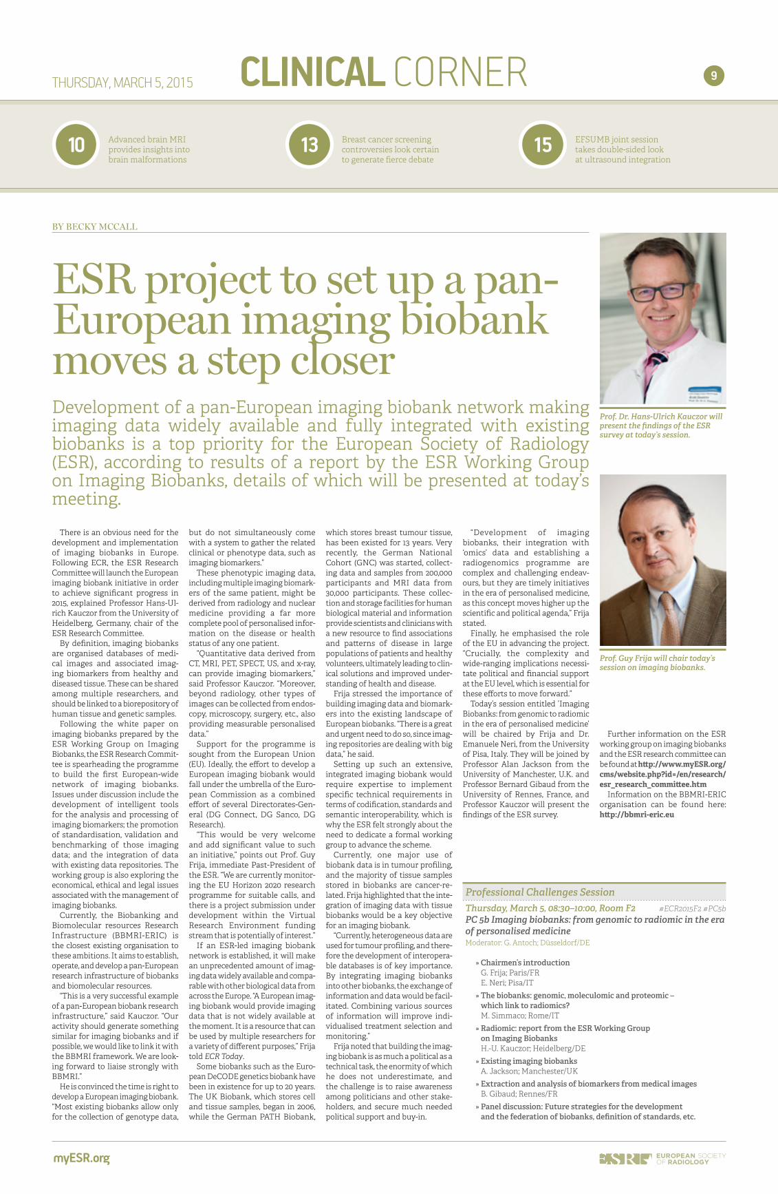
BY BECKY MCCALL
Development of a pan-European imaging biobank network making imaging data widely available and fully integrated with existing biobanks is a top priority for the European Society of Radiology (ESR), according to results of a report by the ESR Working Group on Imaging Biobanks, details of which will be presented at today’s meeting.
9
Professional Challenges Session
Thursday, March 5, 08:30–10:00, Room F2 #ECR2015F2 #PC5bPC 5b Imaging biobanks: from genomic to radiomic in the era
of personalised medicine
Moderator: G. Antoch; Düsseldorf/DE
» Chairmen’s introduction
G. Frija; Paris/FR E. Neri; Pisa/IT
» The biobanks: genomic, moleculomic and proteomic –
which link to radiomics?
M. Simmaco; Rome/IT
» Radiomic: report from the ESR Working Group
on Imaging Biobanks
H.-U. Kauczor; Heidelberg/DE
» Existing imaging biobanks
A. Jackson; Manchester/UK
» Extraction and analysis of biomarkers from medical images
B. Gibaud; Rennes/FR
» Panel discussion: Future strategies for the development
and the federation of biobanks, definition of standards, etc.
Prof. Dr. Hans-Ulrich Kauczor will present the findings of the ESR survey at today’s session.
Prof. Guy Frija will chair today’s session on imaging biobanks.
CLINICAL CORNER
myESR.org
10 ECR TODAY | THURSDAY, MARCH 5, 2015
DTI provides qualitative and quantitative information about the microarchitecture of white ma�er. It can reveal information about the brain microstructure that may not be detected in conventional MR sequences of infants and children with brain malformations. With its ability to be�er categorise various brain malformations that may look similar on conventional MR imaging but may be caused by different path-omechanisms, DTI may allow tran-scending MRI from basic anatomic imaging toward function and embry-ology-based imaging. Conventional MRI is excellent in depicting the big functional centres, the nuclei of the brain. But it does not show how different parts of the brain are connected to each other or if there is an aberrant connection. DTI with fibre tracking do.
Prof. Thierry A.G.M. Huisman, director of paediatric radiology and paediatric neuroradiology at Johns Hopkins Hospital in Baltimore, Maryland, U.S., hopes to convey his excitement about DTI in today’s session.
“Conventional anatomic MR imaging shows only the tip of the iceberg of microstructural brain malformations. DTI can be quite easily performed in any patient and provide a wealth of information. It is a ma�er of having the patience, perseverance, and a good knowledge of brain anatomy and function to ‘connect the dots’ and extract correct and relevant information out of a completed three-dimension tractog-raphy reconstruction,” he explained.
While powerful post-processing so�ware programmes may identify hundreds or thousands of white ma�er tracts within a sampled brain, too many reconstructed fibres can obscure the relevant, aberrant, maldeveloped, or missing white ma�er tracts, he added.
Many brain malformations are suspected from prenatal ultrasound data. DTI enables a quantitative anal-ysis, and a structural analysis can be
created through post-processing and reconstructing the course of fibre tracts. As an example, if a neonate is not adequately moving its arms and/or legs, the cortical spinal tract can be reconstructed, specifically the fibres responsible for connect-ing the motor cortex and extremities. Huisman pointed out that it is neces-sary to know the clinical history of the patient to be able to pinpoint precisely what fibre tracts need to be reconstructed.
“If I am told that the child is not moving extremities, cortico-spinal fibre needs to be reconstructed, if there are memory problems, the limbic system, or if vision problems, the visual system,” he stated.
If diagnosed at the prenatal stage, some malformations can be treated.
When a neural tube defect can be closed in a foetus, for example, the degree of malformation will not be as severe. And the additional informa-tion provided by DTI also may help guide a decision about continuing or ending a pregnancy.
“DTI and FT (fibre tracking) allow us to study the microstructure of the central nervous system in vivo. Collected data will help to be�er classify malformations and may give important hints to the genetic bases of the encountered findings,” he noted. “I think that DTI will help to guide the future identification and treatment of genetic diseases. DTI analysis together with MRI analysis will enable researchers to identify patients with similar characteristics and to help us know where to search for the gene locus.”
Furthermore, knowing exactly what and where a defective gene is located will help clinicians to iden-tify different subgroups, make be�er diagnoses, and gain a greater under-standing of pa�erns of inheritance.
ASL: A NON-INVASIVE METHOD TO ASSESS HIE
Hypoxic-ischaemic encephalop-athy (HIE), brain tissue ischaemia caused by a lack of blood flow or oxygen perinatally, occurs in two of every 1,000 full-term births. It is a significant cause of adverse neurode-velopmental outcome. Outcome biomarkers for HIE in neonates are critically important, because they
can aid in therapeutic decision-mak-ing and can be used to evaluate the effect of neuroprotective therapies.
Infants with HIE tend to show foetal distress prior to delivery, have abnormal Apgar scores, may require resuscitation at birth, and display neurological abnormalities such as seizures, decreased levels of consciousness, irritability, and feed-ing difficulties. HIE is classified into three grades, ranging from mild to severe. Infants with mild HIE will recover, but those with moderate HIE have a 30% risk of disabilities and a 10% risk of death. More than half of neonates diagnosed with severe HIE will die in infancy. The majority of those who survive will suffer from severe neurological abnormalities.
Cranial ultrasound and MRI – conventional, diffusion-weighted imaging (DWI), DTI, magnetisation transfer imaging (MTI), MR spectros-copy, and ASL perfusion – are being used to image neonates with HIE. ASL perfusion MRI, a technique that inverts arterial blood water flowing toward the brain and serves as a tracer to evaluate brain perfusion, has been used with adults for more than 20 years. However, due to tech-nical hurdles that only recently have been resolved, it has not been used to image newborns and infants, explained Dr. Jeroen Hendrikse of the Department of Radiology of University Medical Center Utrecht. He is head of a team investigating the use of non-invasive ASL MRI in
neonates to visualise and quantify brain perfusion in ml/min/100gr brain tissue.
“With most MRI exams that are performed, structural damage can be assessed, and the infarction can be seen, but no perfusion parame-ter has been available. ASL provides a relatively simple non-invasive method to obtain haemodynamic (perfusion) information from a scan that takes approximately five minutes,” he said. “In a study of 28 neonates imaged four days a�er birth published in the January 2015 issue of European Radiology, ASL not only complemented known MRI parame-ters in the prediction of outcome, but also provided additional unique and valuable information. Perfusion adds additional prognostic information, which was a gratifying surprise to us.”
The study determined that basal ganglia and thalami perfusion (deep grey ma�er) was higher a few days a�er birth in neonates followed for up to 18 months who had adverse outcomes of cerebral palsy or death. “We think that in conjunction with MR spectroscopy, it can be used as an outcome biomarker, as ASL perfusion had positive and negative predictive values of 100% and 96% respectively. It allows for an evalu-ation of the reperfusion phenome-non, which is related to delayed cell death. We also think that ASL may be useful to evaluate the effective-ness of neuroprotective therapies,” he added.
BY CYNTHIA E. KEEN
Advanced brain MRI provides insights into brain malformationsWhen a child is born with a serious brain-related defect, understanding the cause and its impact at the earliest stages of life is of utmost importance to determine the best course of treatment and to counsel parents about developmental problems, quality of life, or odds of survival. Advanced MRI techniques like diffusion tensor imaging (DTI) and arterial-spin labelling (ASL) are yielding clinically exciting discoveries and valuable information.
Special Focus Session
Thursday, March 5, 16:00–17:30, Room B #ECR2015B #SF8aSF 8a Advanced brain MRI techniques in paediatrics: toys or
tools in daily practice?
» Chairman’s introduction
A. Rossi; Genoa/IT
» Arterial spin-labelling: measuring perfusion non-invasively in
neonates and children
J. Hendrikse; Utrecht/NL
» MR spectroscopy: information vs time
J.F. Schneider; Basle/CH
» Diffusion tensor imaging: connecting the dots
T.A.G.M. Huisman; Baltimore, MD/US
» Panel discussion: Do advanced brain MRI techniques
really change current practice?
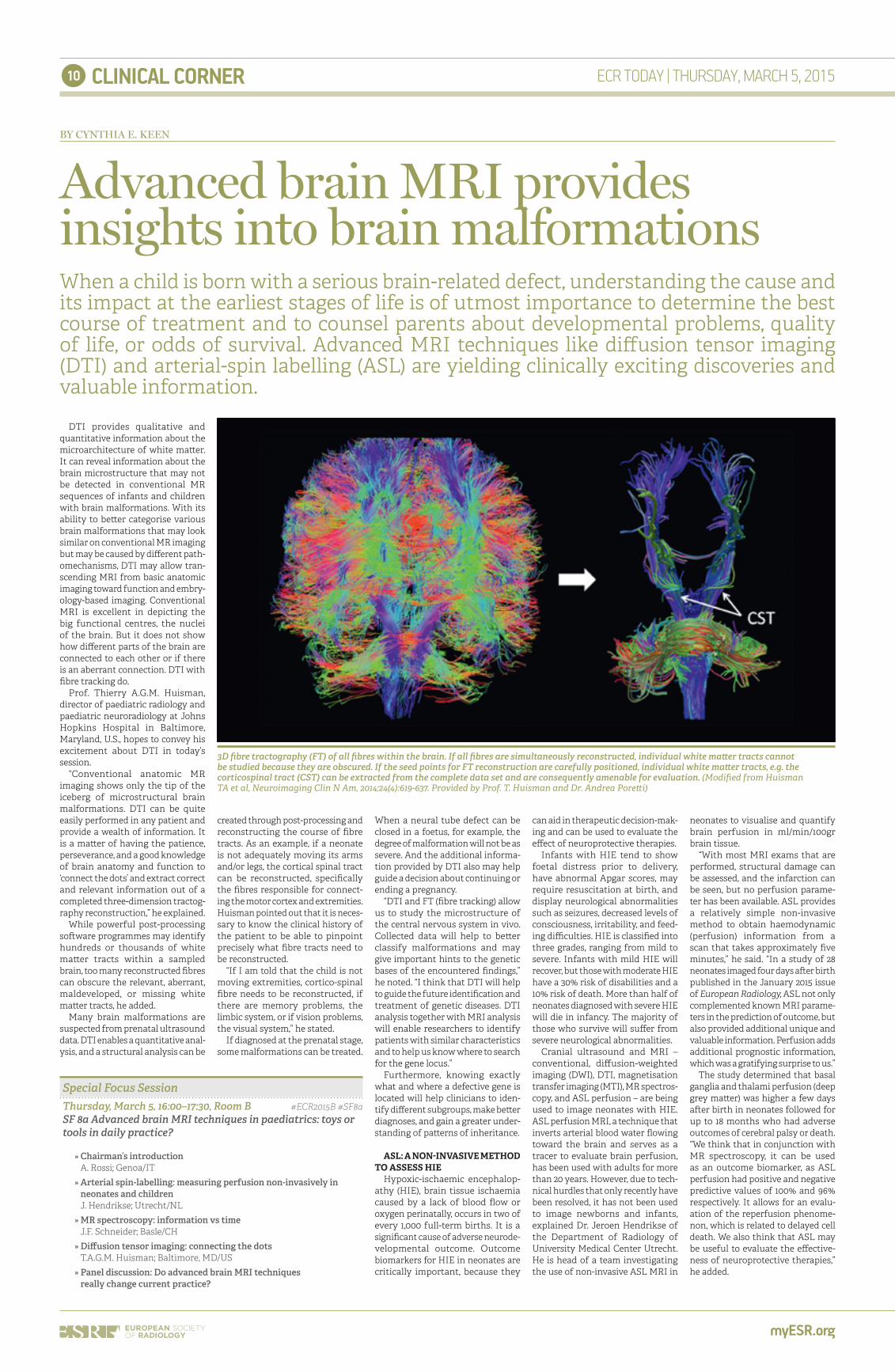
3D fibre tractography (FT) of all fibres within the brain. If all fibres are simultaneously reconstructed, individual white ma�er tracts cannot be studied because they are obscured. If the seed points for FT reconstruction are carefully positioned, individual white ma�er tracts, e.g. the corticospinal tract (CST) can be extracted from the complete data set and are consequently amenable for evaluation. (Modified from Huisman TA et al, Neuroimaging Clin N Am, 2014;24(4):619-637. Provided by Prof. T. Huisman and Dr. Andrea Pore�i)
CLINICAL CORNER
myESR.org
11ECR TODAY | THURSDAY, MARCH 5, 2015
When radiologists follow a cohort of people for 20 or 30 years, the bene-fits for the patient increase tremen-dously. If a radiologist performs a CT examination of a patient’s coro-nary arteries and finds calcification, chances are that the patient will have a heart a�ack within the next few years. Unfortunately at this stage, the patient is usually out of the radiolo-gist’s reach.
However, if patients chose to partic-ipate in a population study, they will be checked on a regular basis, and radiologists will be able to access previous information and initiate appropriate treatment earlier, signif-icantly improving patient outcome.
Securing imaging data is always tricky and population imaging studies are an opportunity for radiologists to access this data. Showing the relevance of imaging findings highlights radiology’s role in the medical continuum, accord-ing to Prof. Norbert Hosten, of the Ernst-Moritz-Arndt University in Greifswald, Germany, who will chair the session today.
“Our way to prove that radiol-ogy can make people healthier and happier is to do large population imaging studies. Radiology can develop the kind of data that are necessary to prove that our meth-ods really help the patient,” he said.
In Germany’s neighbour the Neth-erlands, significant population imag-ing studies have been performed over the past two decades. Researchers at the University Medical Center Ro�erdam have, for instance, been repeatedly imaging a population cohort of 10,000 to 15,000 inhabitants to look at determinants like blood values and cognitive performance, and determine whether they corre-late with the occurrence of certain diseases later in life.
They have found that population imaging helps to predict neuro-degen-erative diseases. Biomarkers such as regional brain volumes, distribution and quantification of white ma�er lesions, subclinical brain infarcts or microbleeds have been identified.
Researchers have also been able to pinpoint the structural and micro-structural integrity of the white ma�er associated with the develop-ment of mild cognitive impairment and full-blown dementia long before any symptoms arise. Prof. Gabriel Krestin, chairman of the depart-ment of radiology at Erasmus MC, University Medical Center Ro�er-dam, will focus on this particular aspect during the session.
Population imaging is also useful in cardiac and oncology applica-tions, to track early signs of tumour development, and a number of other conditions ranging from liver cirrhosis to osteoporosis. Prof. Fabian Bamberg from Tübingen University Hospital in Germany and Steffen Petersen from the London Chest Hospital at Queen Mary University in the U.K. will present the multicen-tre studies done in their respective countries during the session.
Perhaps a more unexpected area of interest is imaging of polytrauma patients, a topic Dr. Sönke Langner, a radiologist working at Ernst-Moritz-Arndt University in Greifswald, will talk about during the session.
Langner is heading the Trauma Cohort Multicentre Study by the German Society of Traumasurgery (DGU) and the German Röntgen Society (DRG). The first of its kind in Europe, the study will try to iden-tify the best methods for perform-ing trauma imaging and determine standardised protocols in Germany.
“Although there is a common sense in the literature, there is a lack of
standardisation in the daily routine. Trauma imaging may be done in a standardised way in each hospital, but there are significant differences between hospitals, mainly because of the available equipment,” Lang-ner said.
The study will initially collect data from ten trauma centres and expand to major centres across the country. It plans to eventually include all patients scanned for polytrauma over a year in Germany. The Trauma Cohort will combine imaging and clinical data, and the main challenge will be to extract equally meaningful information from the different centres.
“Data extraction will take a long time but we hope to have our first results ready for the ECR. Even-tually, we hope we will be able to suggest what’s the best way to treat polytrauma patients,” Langner said.
A central question experts will try to answer during the session concerns the radiologist’s responsi-bility in these studies, especially if he or she finds an abnormality.
“In a traditional epidemiologi-cal study, you don’t intervene. In radiology, you know what a kidney tumour looks like, so if you do an MR scan and spot such a tumour, I think you are obliged to intervene. Otherwise you will just observe how the patient gets sicker and in my opinion that is not possible being a doctor. The big question is: when do we and do we not inter-vene? I think you should intervene when you are very sure of what you have and that you shouldn’t if you are not sure what a finding means,” Hosten said.
Prof. Dr. Reinhold Schmücker from the University of Münster will deal with this delicate issue in his presentation, which will end the session.
BY MÉLISANDE ROUGER
Population imaging studies gain ground in healthcareImaging large cohorts of people enables scientists to collect information useful for science and emphasises radiology’s role in healthcare. From the most recently availa-ble imaging biomarkers to data such as genomics and metabolomics, today’s dedicated Professional Challenges Session will show just how useful population imaging studies have become in the prognosis of countless diseases.
Professional Challenges Session
Thursday, March 5, 16:00–17:30, Room L 1 #ECR2015L1 #PC8bPC 8b Imaging in population-based studies
» Chairman’s introduction
N. Hosten; Greifswald/DE
» Population imaging for the prediction of neurodegenerative diseases
G.P. Krestin; Ro�erdam/NL
» The German National Cohort: population based imaging in a nation-wide multi-centre se�ing
F. Bamberg; Munich/DE
» Population-based cardiac imaging S. Petersen; London/UK
» The Trauma Cohort: a joint project of the German Röntgen Society and the German Society of Trauma
Surgery
S. Langner; Greifswald/DE
» Ethical aspects of population imaging
R. Schmücker; Münster/DE
» Panel discussion: What does the individual gain from population imaging studies?
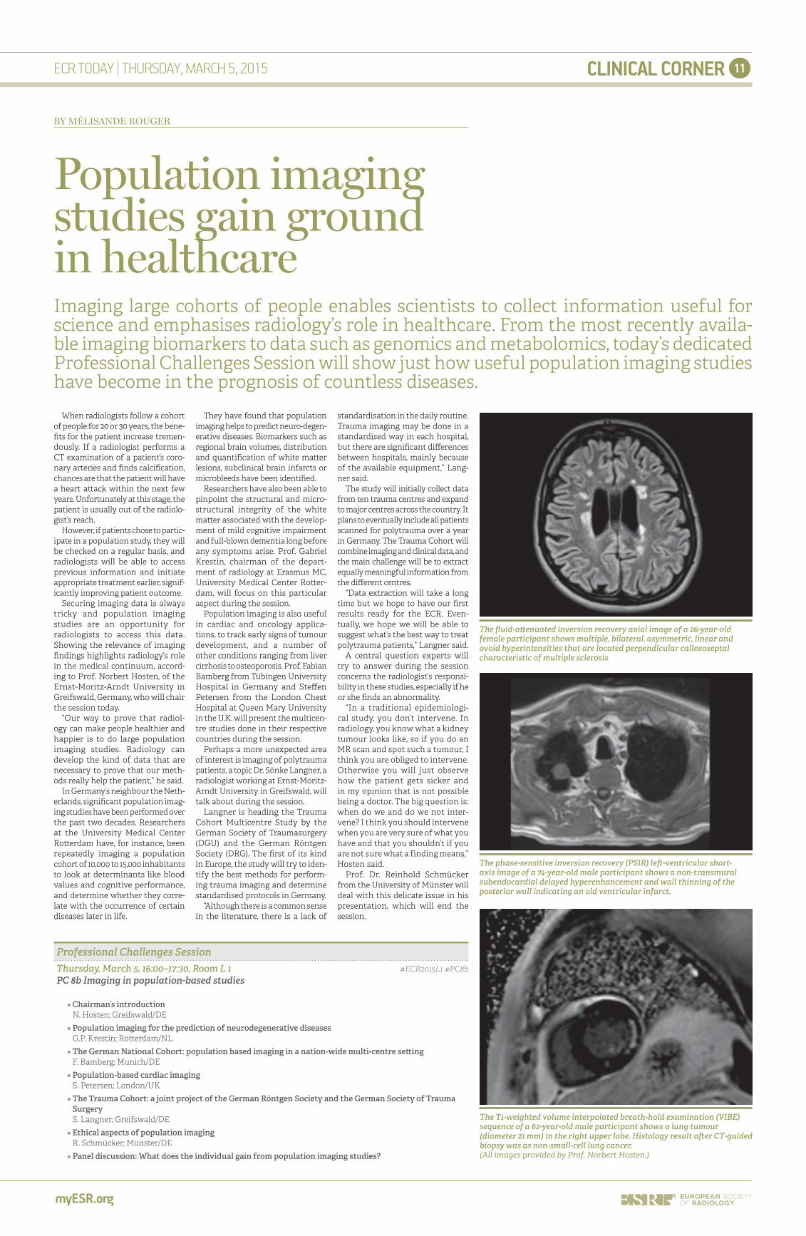
The fluid-a�enuated inversion recovery axial image of a 26-year-old female participant shows multiple, bilateral, asymmetric, linear and ovoid hyperintensities that are located perpendicular callososeptal characteristic of multiple sclerosis
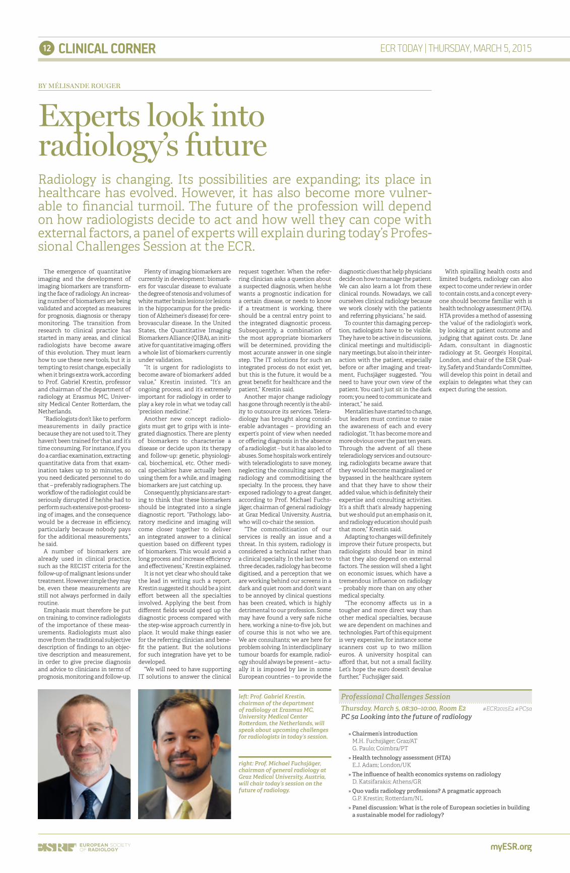
The phase-sensitive inversion recovery (PSIR) le�-ventricular short-axis image of a 74-year-old male participant shows a non-transmural subendocardial delayed hyperenhancement and wall thinning of the posterior wall indicating an old ventricular infarct.
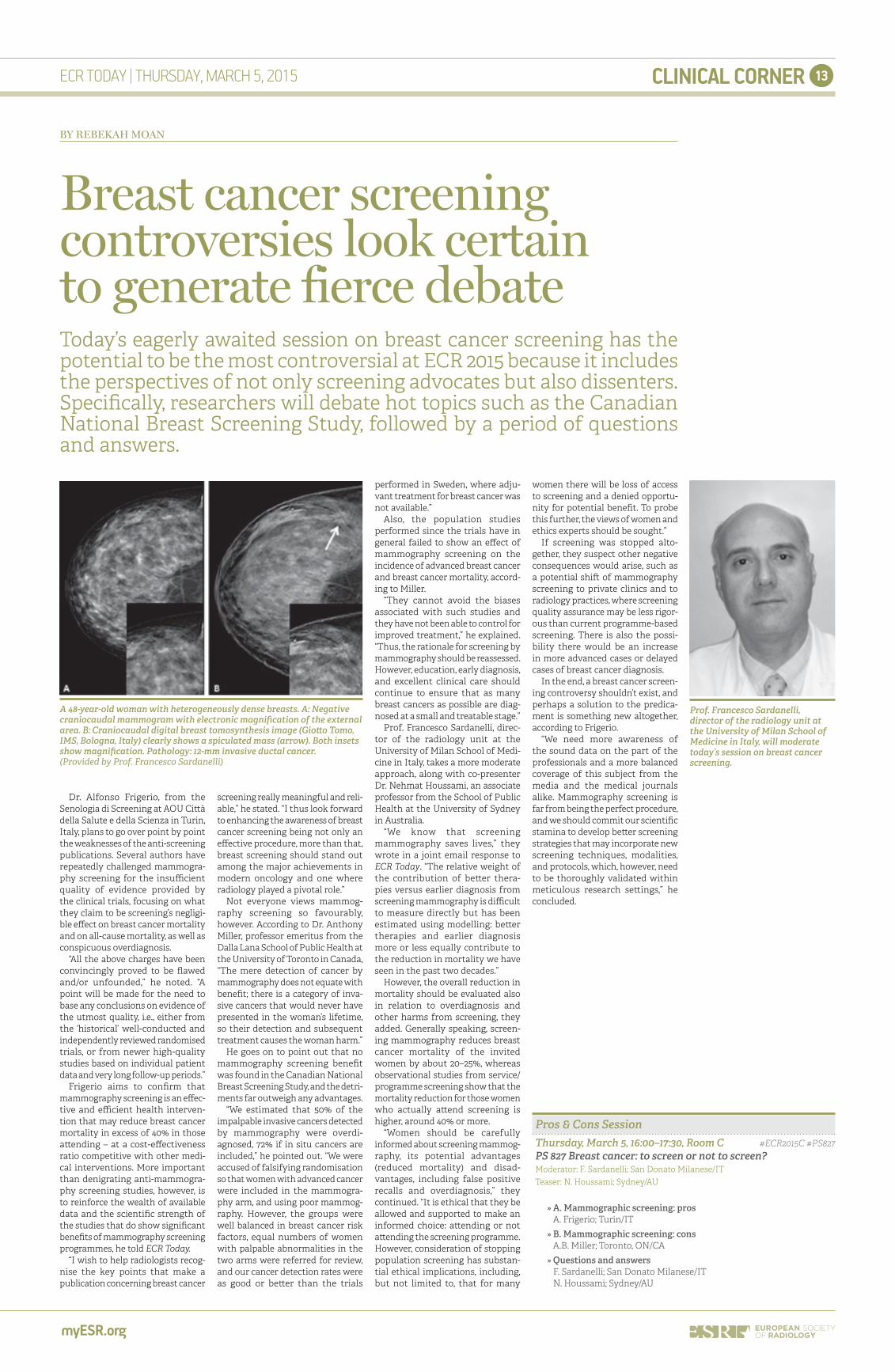
The T1-weighted volume interpolated breath-hold examination (VIBE) sequence of a 62-year-old male participant shows a lung tumour (diameter 21 mm) in the right upper lobe. Histology result a�er CT-guided biopsy was as non-small-cell lung cancer.(All images provided by Prof. Norbert Hosten.)
CLINICAL CORNER
myESR.org
12 ECR TODAY | THURSDAY, MARCH 5, 2015
The emergence of quantitative imaging and the development of imaging biomarkers are transform-ing the face of radiology. An increas-ing number of biomarkers are being validated and accepted as measures for prognosis, diagnosis or therapy monitoring. The transition from research to clinical practice has started in many areas, and clinical radiologists have become aware of this evolution. They must learn how to use these new tools, but it is tempting to resist change, especially when it brings extra work, according to Prof. Gabriel Krestin, professor and chairman of the department of radiology at Erasmus MC, Univer-sity Medical Center Ro�erdam, the Netherlands.
“Radiologists don’t like to perform measurements in daily practice because they are not used to it. They haven’t been trained for that and it’s time consuming. For instance, if you do a cardiac examination, extracting quantitative data from that exam-ination takes up to 30 minutes, so you need dedicated personnel to do that – preferably radiographers. The workflow of the radiologist could be seriously disrupted if he/she had to perform such extensive post-process-ing of images, and the consequence would be a decrease in efficiency, particularly because nobody pays for the additional measurements,” he said.
A number of biomarkers are already used in clinical practice, such as the RECIST criteria for the follow-up of malignant lesions under treatment. However simple they may be, even these measurements are still not always performed in daily routine.
Emphasis must therefore be put on training, to convince radiologists of the importance of these meas-urements. Radiologists must also move from the traditional subjective description of findings to an objec-tive description and measurement, in order to give precise diagnosis and advice to clinicians in terms of prognosis, monitoring and follow-up.
Plenty of imaging biomarkers are currently in development: biomark-ers for vascular disease to evaluate the degree of stenosis and volumes of white ma�er brain lesions (or lesions in the hippocampus for the predic-tion of Alzheimer’s disease) for cere-brovascular disease. In the United States, the Quantitative Imaging Biomarkers Alliance (QIBA), an initi-ative for quantitative imaging, offers a whole list of biomarkers currently under validation.
“It is urgent for radiologists to become aware of biomarkers’ added value,” Krestin insisted. “It’s an ongoing process, and it’s extremely important for radiology in order to play a key role in what we today call ‘precision medicine’.”
Another new concept radiolo-gists must get to grips with is inte-grated diagnostics. There are plenty of biomarkers to characterise a disease or decide upon its therapy and follow-up: genetic, physiologi-cal, biochemical, etc. Other medi-cal specialties have actually been using them for a while, and imaging biomarkers are just catching up.
Consequently, physicians are start-ing to think that these biomarkers should be integrated into a single diagnostic report. “Pathology, labo-ratory medicine and imaging will come closer together to deliver an integrated answer to a clinical question based on different types of biomarkers. This would avoid a long process and increase efficiency and effectiveness,” Krestin explained.
It is not yet clear who should take the lead in writing such a report. Krestin suggested it should be a joint effort between all the specialties involved. Applying the best from different fields would speed up the diagnostic process compared with the step-wise approach currently in place. It would make things easier for the referring clinician and bene-fit the patient. But the solutions for such integration have yet to be developed.
“We will need to have supporting IT solutions to answer the clinical
request together. When the refer-ring clinician asks a question about a suspected diagnosis, when he/she wants a prognostic indication for a certain disease, or needs to know if a treatment is working, there should be a central entry point to the integrated diagnostic process. Subsequently, a combination of the most appropriate biomarkers will be determined, providing the most accurate answer in one single step. The IT solutions for such an integrated process do not exist yet, but this is the future, it would be a great benefit for healthcare and the patient,” Krestin said.
Another major change radiology has gone through recently is the abil-ity to outsource its services. Telera-diology has brought along consid-erable advantages – providing an expert’s point of view when needed or offering diagnosis in the absence of a radiologist – but it has also led to abuses. Some hospitals work entirely with teleradiologists to save money, neglecting the consulting aspect of radiology and commoditising the specialty. In the process, they have exposed radiology to a great danger, according to Prof. Michael Fuchs-jäger, chairman of general radiology at Graz Medical University, Austria, who will co-chair the session.
“The commoditisation of our services is really an issue and a threat. In this system, radiology is considered a technical rather than a clinical specialty. In the last two to three decades, radiology has become digitised, and a perception that we are working behind our screens in a dark and quiet room and don’t want to be annoyed by clinical questions has been created, which is highly detrimental to our profession. Some may have found a very safe niche here, working a nine-to-five job, but of course this is not who we are. We are consultants; we are here for problem solving. In interdisciplinary tumour boards for example, radiol-ogy should always be present – actu-ally it is imposed by law in some European countries – to provide the
diagnostic clues that help physicians decide on how to manage the patient. We can also learn a lot from these clinical rounds. Nowadays, we call ourselves clinical radiology because we work closely with the patients and referring physicians,” he said.
To counter this damaging percep-tion, radiologists have to be visible. They have to be active in discussions, clinical meetings and multidiscipli-nary meetings, but also in their inter-action with the patient, especially before or a�er imaging and treat-ment, Fuchsjäger suggested. “You need to have your own view of the patient. You can’t just sit in the dark room; you need to communicate and interact,” he said.
Mentalities have started to change, but leaders must continue to raise the awareness of each and every radiologist. “It has become more and more obvious over the past ten years. Through the advent of all these teleradiology services and outsourc-ing, radiologists became aware that they would become marginalised or bypassed in the healthcare system and that they have to show their added value, which is definitely their expertise and consulting activities. It’s a shi� that’s already happening but we should put an emphasis on it, and radiology education should push that more,” Krestin said.
Adapting to changes will definitely improve their future prospects, but radiologists should bear in mind that they also depend on external factors. The session will shed a light on economic issues, which have a tremendous influence on radiology – probably more than on any other medical specialty.
“The economy affects us in a tougher and more direct way than other medical specialties, because we are dependent on machines and technologies. Part of this equipment is very expensive, for instance some scanners cost up to two million euros. A university hospital can afford that, but not a small facility. Let’s hope the euro doesn’t devalue further,” Fuchsjäger said.
With spiralling health costs and limited budgets, radiology can also expect to come under review in order to contain costs, and a concept every-one should become familiar with is health technology assessment (HTA). HTA provides a method of assessing the ‘value’ of the radiologist’s work, by looking at patient outcome and judging that against costs. Dr. Jane Adam, consultant in diagnostic radiology at St. George’s Hospital, London, and chair of the ESR Qual-ity, Safety and Standards Commi�ee, will develop this point in detail and explain to delegates what they can expect during the session.
BY MÉLISANDE ROUGER
Experts look into radiology’s futureRadiology is changing. Its possibilities are expanding; its place in healthcare has evolved. However, it has also become more vulner-able to financial turmoil. The future of the profession will depend on how radiologists decide to act and how well they can cope with external factors, a panel of experts will explain during today’s Profes-sional Challenges Session at the ECR.
Professional Challenges Session
Thursday, March 5, 08:30–10:00, Room E2 #ECR2015E2 #PC5aPC 5a Looking into the future of radiology
» Chairmen’s introduction
M.H. Fuchsjäger; Graz/AT G. Paulo; Coimbra/PT
» Health technology assessment (HTA)
E.J. Adam; London/UK
» The influence of health economics systems on radiology
D. Katsifarakis; Athens/GR
» Quo vadis radiology professions? A pragmatic approach
G.P. Krestin; Ro�erdam/NL
» Panel discussion: What is the role of European societies in building
a sustainable model for radiology?
le�: Prof. Gabriel Krestin, chairman of the department of radiology at Erasmus MC, University Medical Center Ro�erdam, the Netherlands, will speak about upcoming challenges for radiologists in today’s session.
right: Prof. Michael Fuchsjäger, chairman of general radiology at Graz Medical University, Austria, will chair today’s session on the future of radiology.
CLINICAL CORNER
myESR.org
13ECR TODAY | THURSDAY, MARCH 5, 2015
Dr. Alfonso Frigerio, from the Senologia di Screening at AOU Ci�à della Salute e della Scienza in Turin, Italy, plans to go over point by point the weaknesses of the anti-screening publications. Several authors have repeatedly challenged mammogra-phy screening for the insufficient quality of evidence provided by the clinical trials, focusing on what they claim to be screening’s negligi-ble effect on breast cancer mortality and on all-cause mortality, as well as conspicuous overdiagnosis.
“All the above charges have been convincingly proved to be flawed and/or unfounded,” he noted. “A point will be made for the need to base any conclusions on evidence of the utmost quality, i.e., either from the ‘historical’ well-conducted and independently reviewed randomised trials, or from newer high-quality studies based on individual patient data and very long follow-up periods.”
Frigerio aims to confirm that mammography screening is an effec-tive and efficient health interven-tion that may reduce breast cancer mortality in excess of 40% in those a�ending – at a cost-effectiveness ratio competitive with other medi-cal interventions. More important than denigrating anti-mammogra-phy screening studies, however, is to reinforce the wealth of available data and the scientific strength of the studies that do show significant benefits of mammography screening programmes, he told ECR Today.
“I wish to help radiologists recog-nise the key points that make a publication concerning breast cancer
screening really meaningful and reli-able,” he stated. “I thus look forward to enhancing the awareness of breast cancer screening being not only an effective procedure, more than that, breast screening should stand out among the major achievements in modern oncology and one where radiology played a pivotal role.”
Not everyone views mammog-raphy screening so favourably, however. According to Dr. Anthony Miller, professor emeritus from the Dalla Lana School of Public Health at the University of Toronto in Canada, “The mere detection of cancer by mammography does not equate with benefit; there is a category of inva-sive cancers that would never have presented in the woman’s lifetime, so their detection and subsequent treatment causes the woman harm.”
He goes on to point out that no mammography screening benefit was found in the Canadian National Breast Screening Study, and the detri-ments far outweigh any advantages.
“We estimated that 50% of the impalpable invasive cancers detected by mammography were overdi-agnosed, 72% if in situ cancers are included,” he pointed out. “We were accused of falsifying randomisation so that women with advanced cancer were included in the mammogra-phy arm, and using poor mammog-raphy. However, the groups were well balanced in breast cancer risk factors, equal numbers of women with palpable abnormalities in the two arms were referred for review, and our cancer detection rates were as good or be�er than the trials
performed in Sweden, where adju-vant treatment for breast cancer was not available.”
Also, the population studies performed since the trials have in general failed to show an effect of mammography screening on the incidence of advanced breast cancer and breast cancer mortality, accord-ing to Miller.
“They cannot avoid the biases associated with such studies and they have not been able to control for improved treatment,” he explained. “Thus, the rationale for screening by mammography should be reassessed. However, education, early diagnosis, and excellent clinical care should continue to ensure that as many breast cancers as possible are diag-nosed at a small and treatable stage.”
Prof. Francesco Sardanelli, direc-tor of the radiology unit at the University of Milan School of Medi-cine in Italy, takes a more moderate approach, along with co-presenter Dr. Nehmat Houssami, an associate professor from the School of Public Health at the University of Sydney in Australia.
“We know that screening mammography saves lives,” they wrote in a joint email response to ECR Today. “The relative weight of the contribution of be�er thera-pies versus earlier diagnosis from screening mammography is difficult to measure directly but has been estimated using modelling: be�er therapies and earlier diagnosis more or less equally contribute to the reduction in mortality we have seen in the past two decades.”
However, the overall reduction in mortality should be evaluated also in relation to overdiagnosis and other harms from screening, they added. Generally speaking, screen-ing mammography reduces breast cancer mortality of the invited women by about 20–25%, whereas observational studies from service/programme screening show that the mortality reduction for those women who actually a�end screening is higher, around 40% or more.
“Women should be carefully informed about screening mammog-raphy, its potential advantages (reduced mortality) and disad-vantages, including false positive recalls and overdiagnosis,” they continued. “It is ethical that they be allowed and supported to make an informed choice: a�ending or not a�ending the screening programme. However, consideration of stopping population screening has substan-tial ethical implications, including, but not limited to, that for many
women there will be loss of access to screening and a denied opportu-nity for potential benefit. To probe this further, the views of women and ethics experts should be sought.”
If screening was stopped alto-gether, they suspect other negative consequences would arise, such as a potential shi� of mammography screening to private clinics and to radiology practices, where screening quality assurance may be less rigor-ous than current programme-based screening. There is also the possi-bility there would be an increase in more advanced cases or delayed cases of breast cancer diagnosis.
In the end, a breast cancer screen-ing controversy shouldn’t exist, and perhaps a solution to the predica-ment is something new altogether, according to Frigerio.
“We need more awareness of the sound data on the part of the professionals and a more balanced coverage of this subject from the media and the medical journals alike. Mammography screening is far from being the perfect procedure, and we should commit our scientific stamina to develop be�er screening strategies that may incorporate new screening techniques, modalities, and protocols, which, however, need to be thoroughly validated within meticulous research se�ings,” he concluded.
BY REBEKAH MOAN
Breast cancer screening controversies look certain to generate fierce debateToday’s eagerly awaited session on breast cancer screening has the potential to be the most controversial at ECR 2015 because it includes the perspectives of not only screening advocates but also dissenters. Specifically, researchers will debate hot topics such as the Canadian National Breast Screening Study, followed by a period of questions and answers.
Pros & Cons Session
Thursday, March 5, 16:00–17:30, Room C #ECR2015C #PS827PS 827 Breast cancer: to screen or not to screen?
Moderator: F. Sardanelli; San Donato Milanese/ITTeaser: N. Houssami; Sydney/AU
» A. Mammographic screening: pros
A. Frigerio; Turin/IT
» B. Mammographic screening: cons
A.B. Miller; Toronto, ON/CA
» Questions and answers
F. Sardanelli; San Donato Milanese/IT N. Houssami; Sydney/AU
Prof. Francesco Sardanelli, director of the radiology unit at the University of Milan School of Medicine in Italy, will moderate today’s session on breast cancer screening.
A 48-year-old woman with heterogeneously dense breasts. A: Negative craniocaudal mammogram with electronic magnification of the external area. B: Craniocaudal digital breast tomosynthesis image (Gio�o Tomo, IMS, Bologna, Italy) clearly shows a spiculated mass (arrow). Both insets show magnification. Pathology: 12-mm invasive ductal cancer. (Provided by Prof. Francesco Sardanelli)
CLINICAL CORNER
myESR.org
14 ECR TODAY | THURSDAY, MARCH 5, 2015
An artefact is an element that may be visible in an image, but that is not present in the object under investi-gation. Occasionally image artefacts are the result of inappropriate use of technical parameters; sometimes they appear due to the inevitable consequence of natural properties of the human body. In both cases, it is vital to be familiar with the appear-ance of these artefacts because they can obscure the morphology and physiology of the scanned part of the patient’s body. In the worst case, artefacts may even mimic pathology,
as in the case of CT partial volume artefacts. It is also well known that artefacts can so seriously degrade the quality of images that sometimes they are diagnostically useless. A literature search focused solely on artefacts in CT lists more than 4,500 papers, whereas in MRI the number comes up close to 9,000 papers.
Image artefacts are an area of increasing importance in hybrid imaging, namely in PET/CT and PET/MRI. According to a recent market study (PET/CT imaging: facts, opin-ions, hopes, and questions. J Nucl
Med. 2004;45(suppl):1S), sales of PET/CT scanners have surpassed those of PET scanners by 65% since 2003, and are anticipated to grow by more than 95% over the next few years. Over 5,000 systems have been installed worldwide, with the number growing continuously and with most clini-cal PET-only systems being replaced gradually by PET/CT. However, the use of the CT scan for a�enuation correction in PET has the drawback of producing artefacts on the result-ing PET images. While the integra-tion of PET and CT into a hybrid system was challenging but techni-cally feasible, the integration of PET and MRI was considered extremely demanding, if not impossible. The last couple of years the technical difficulties have been surpassed and about 60 clinical PET/MRI systems are operational worldwide.
To optimise image quality, it is necessary to understand why arte-facts occur and how they can be prevented or corrected. The Phys-ics in Radiology subcommi�ee has devoted two of the five refresher courses at ECR 2015 to this very important topic. Refresher courses 513 ‘Artefacts and pitfalls in tomog-raphy’ and 1613 ‘MR: artefacts and devices’ will help a�endees under-stand common sources of artefacts and provide adequate solutions to either prospectively limit the chance of occurrence or correct
for artefacts retrospectively. The courses intend to refresh the partic-ipants’ knowledge of the physical origins of artefacts, teach methods for minimising them – including the artefacts in perfusion and diffusion MRI – and explain how to image regions of the body close to metal implants, as well as solutions to frequent image distortions.
BY VIRGINIA TSAPAKI
Artefacts in imaging: the highlight of the Physics in Radiology Refresher Course programme
Refresher Courses: Physics in Radiology
Thursday, March 5, 08:30–10:00, Room M #ECR2015M #RC513RC 513 Artefacts and pitfalls in tomography
Moderator: J. Damilakis; Iraklion/GR
» A. CT
M. Kachelries; Heidelberg/DE
» B. PET/CT
T. Beyer; Vienna/AT
» C. MR/PET
H.H. Quick; Essen/DE
Saturday, March 7, 16:00–17:30, Room M #ECR2015M #RC1613RC 1613 MR: artefacts and devices
Moderator: D. Bor; Ankara/TR
» A. Image artefacts in MRI and their mitigation
D.J. Lurie; Aberdeen/UK
» B. Imaging around metal implants: artefact reduction in MRI
C. McGrath; Belfast/IE
» C. Artefacts in perfusion and diffusion MRI
I. Tsougos; Larissa/GR
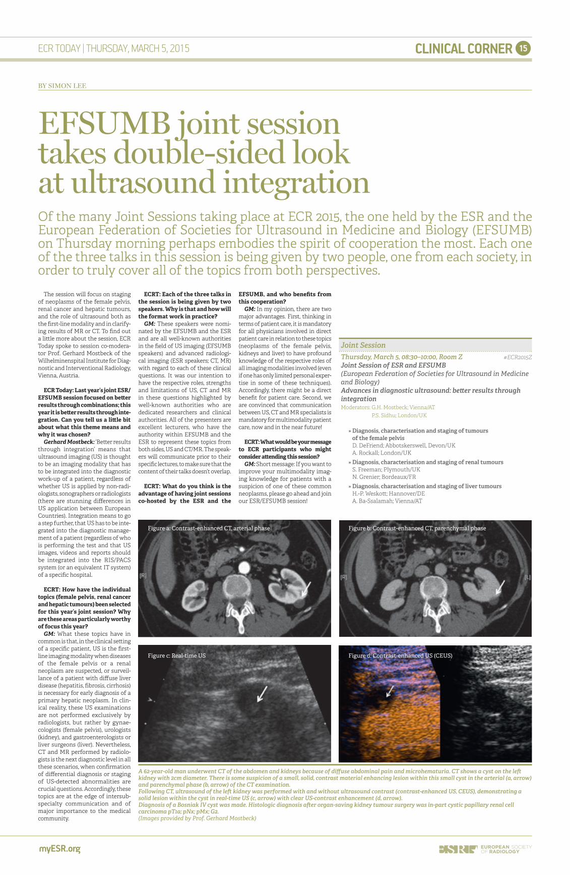
Magnetic susceptibility artefact; this artefact is seen in the MRI image while imaging metallic orthopaedic inserts (Case courtesy of Dr. Prashant Mudgal, Radiopaedia.org).
Dr. Virginia Tsapaki from Athens, Greece, is the ECR 2015 Radiology Subcommi�ee Chair and EFOMP
Project commi�ee Vice Chair.
Basic Sessions – Special sessions suitable for residents, students, radiographers and radiographers-in-training
Thursday, March 5, 08:30–10:00, Studio 2015 #ECR2015StudioBasic Session 1: Breast imaging
» Mammography
F.J. Gilbert; Cambridge/UK
» Breast US
C.S. Balleyguier; Villejuif/FR
» Breast MRI
F. Sardanelli; San Donato Milanese/IT
Thursday, March 5, 10:30–12:00, Studio 2015 #ECR2015StudioBasic Session 2: Neuroradiology
» Aging and degeneration in the brain
B. Gomez-Anson; Barcelona/ES
» Brain trauma
M. Stajgis; Poznan/PL
» Vascular malformations
P. Vilela; Almada/PT
Student Sessions – Students will present their work
Student Sessions – Students will present their work
» Evaluation of choroid plexus with foetal MRI: what happens in ventriculomegaly?
C. Turam; Istanbul/TR
» Anatomic and morphometric variations of the intracranial vertebrobasilar system on
MSCT and MR angiography
S. Jankovic; Nis/RS
Radiology Trainees Forum Programme
Thursday, March 5, 13:30–14:30, Rising Stars Lounge RTF Quiz
Quiz-Master: J. Cáceres; Barcelona/ES
RISING STARS Programme today at ECR
Thursday, March 5, 14:00–15:30, Studio 2015 #ECR2015StudioStudent Session 1
» SWI or T2* – which MRI sequence to use in the detection of cerebral microbleeds? The
Karolinska Imaging Dementia Study
S. Shams; Stockholm/SE
» Background parenchymal enhancement on breast MRI in women receiving chest
radiotherapy for childhood Hodgkin’s lymphoma
L. Zeng; Toronto, ON/CA
» 3D quantitative assessment of lesion response to MR-guided high-intensity focused
ultrasound treatment of uterine fibroids
J. Savic; Berlin/DE
Thursday, March 5, 16:00–17:00, Studio 2015 #ECR2015StudioStudent Session 2
» The positive effects on CT reporting with a radiologist present at the initial clinical
evaluation of polytrauma
V. Sanfilippo; Catania/IT
» An audit of the practices of reporting of staging CT scans in primary malignancies
R.D.T. Price; Birmingham/UK
» Reproducibility of a novel semi-automated so�ware programme for Pre-TAVR CT
assessment
K. Rohan; Belfast/UK
» Analysing CT images of patients who died a�er thrombolysis
E. Tarjanyi; Debrecen/HU
» Trunk paediatric CT diagnostic reference levels
D. Fernandes; Coimbra/PT
CLINICAL CORNER
myESR.org
15ECR TODAY | THURSDAY, MARCH 5, 2015
The session will focus on staging of neoplasms of the female pelvis, renal cancer and hepatic tumours, and the role of ultrasound both as the first-line modality and in clarify-ing results of MR or CT. To find out a li�le more about the session, ECR Today spoke to session co-modera-tor Prof. Gerhard Mostbeck of the Wilhelminenspital Institute for Diag-nostic and Interventional Radiology, Vienna, Austria.
ECR Today: Last year’s joint ESR/EFSUMB session focused on be�er results through combinations; this year it is be�er results through inte-gration. Can you tell us a li�le bit about what this theme means and why it was chosen?
Gerhard Mostbeck: ‘Be�er results through integration’ means that ultrasound imaging (US) is thought to be an imaging modality that has to be integrated into the diagnostic work-up of a patient, regardless of whether US is applied by non-radi-ologists, sonographers or radiologists (there are stunning differences in US application between European Countries). Integration means to go a step further, that US has to be inte-grated into the diagnostic manage-ment of a patient (regardless of who is performing the test and that US images, videos and reports should be integrated into the RIS/PACS system (or an equivalent IT system) of a specific hospital.
ECRT: How have the individual topics (female pelvis, renal cancer and hepatic tumours) been selected for this year’s joint session? Why are these areas particularly worthy of focus this year?
GM: What these topics have in common is that, in the clinical se�ing of a specific patient, US is the first-line imaging modality when diseases of the female pelvis or a renal neoplasm are suspected, or surveil-lance of a patient with diffuse liver disease (hepatitis, fibrosis, cirrhosis) is necessary for early diagnosis of a primary hepatic neoplasm. In clin-ical reality, these US examinations are not performed exclusively by radiologists, but rather by gynae-cologists (female pelvis), urologists (kidney), and gastroenterologists or liver surgeons (liver). Nevertheless, CT and MR performed by radiolo-gists is the next diagnostic level in all these scenarios, when confirmation of differential diagnosis or staging of US-detected abnormalities are crucial questions. Accordingly, these topics are at the edge of intersub-specialty communication and of major importance to the medical community.
ECRT: Each of the three talks in the session is being given by two speakers. Why is that and how will the format work in practice?
GM: These speakers were nomi-nated by the EFSUMB and the ESR and are all well-known authorities in the field of US imaging (EFSUMB speakers) and advanced radiologi-cal imaging (ESR speakers; CT, MR) with regard to each of these clinical questions. It was our intention to have the respective roles, strengths and limitations of US, CT and MR in these questions highlighted by well-known authorities who are dedicated researchers and clinical authorities. All of the presenters are excellent lecturers, who have the authority within EFSUMB and the ESR to represent these topics from both sides, US and CT/MR. The speak-ers will communicate prior to their specific lectures, to make sure that the content of their talks doesn’t overlap.
ECRT: What do you think is the advantage of having joint sessions co-hosted by the ESR and the
EFSUMB, and who benefits from this cooperation?
GM: In my opinion, there are two major advantages. First, thinking in terms of patient care, it is mandatory for all physicians involved in direct patient care in relation to these topics (neoplasms of the female pelvis, kidneys and liver) to have profound knowledge of the respective roles of all imaging modalities involved (even if one has only limited personal exper-tise in some of these techniques). Accordingly, there might be a direct benefit for patient care. Second, we are convinced that communication between US, CT and MR specialists is mandatory for multimodality patient care, now and in the near future!
ECRT: What would be your message to ECR participants who might consider a�ending this session?
GM: Short message: If you want to improve your multimodality imag-ing knowledge for patients with a suspicion of one of these common neoplasms, please go ahead and join our ESR/EFSUMB session!
BY SIMON LEE
EFSUMB joint session takes double-sided look at ultrasound integrationOf the many Joint Sessions taking place at ECR 2015, the one held by the ESR and the European Federation of Societies for Ultrasound in Medicine and Biology (EFSUMB) on Thursday morning perhaps embodies the spirit of cooperation the most. Each one of the three talks in this session is being given by two people, one from each society, in order to truly cover all of the topics from both perspectives.
Joint Session
Thursday, March 5, 08:30–10:00, Room Z #ECR2015ZJoint Session of ESR and EFSUMB
(European Federation of Societies for Ultrasound in Medicine and Biology)Advances in diagnostic ultrasound: be�er results through
integration
Moderators: G.H. Mostbeck; Vienna/AT P.S. Sidhu; London/UK
» Diagnosis, characterisation and staging of tumours
of the female pelvis
D. DeFriend; Abbotskerswell, Devon/UK A. Rockall; London/UK
» Diagnosis, characterisation and staging of renal tumours
S. Freeman; Plymouth/UK N. Grenier; Bordeaux/FR
» Diagnosis, characterisation and staging of liver tumours
H.-P. Wesko�; Hannover/DE
A. Ba-Ssalamah; Vienna/AT
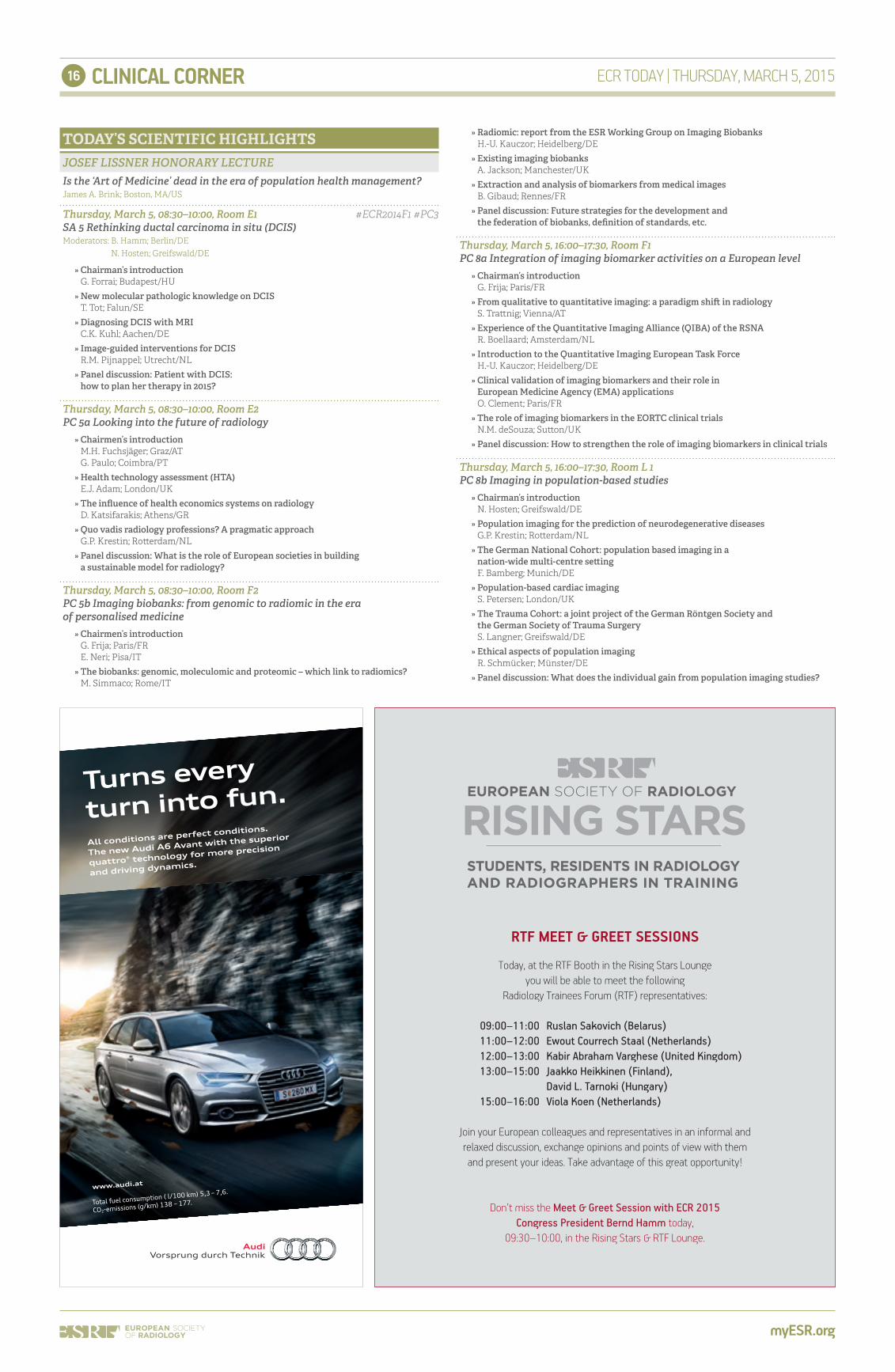
A 62-year-old man underwent CT of the abdomen and kidneys because of diffuse abdominal pain and microhematuria. CT shows a cyst on the le� kidney with 2cm diameter. There is some suspicion of a small, solid, contrast material enhancing lesion within this small cyst in the arterial (a, arrow) and parenchymal phase (b, arrow) of the CT examination.Following CT, ultrasound of the le� kidney was performed with and without ultrasound contrast (contrast-enhanced US, CEUS), demonstrating a solid lesion within the cyst in real-time US (c, arrow) with clear US-contrast enhancement (d, arrow).Diagnosis of a Bosniak IV cyst was made. Histologic diagnosis a�er organ-saving kidney tumour surgery was in-part cystic papillary renal cell carcinoma pT1a; pNx; pMx; G2.(Images provided by Prof. Gerhard Mostbeck)
Figure a: Contrast-enhanced CT, arterial phase
Figure c: Real-time US
Figure b: Contrast-enhanced CT, parenchymal phase
Figure d: Contrast-enhanced US (CEUS)
myESR.org
CLINICAL CORNER16 ECR TODAY | THURSDAY, MARCH 5, 2015
Turns every
turn into fun.
All conditions are perfect conditions.
The new Audi A6 Avant with the superior
quattro® technology for more precision
and driving dynamics.
www.audi.at
Total fuel consumption ( l/100 km) 5,3 – 7,6.
CO2-emissions (g/km) 138 – 177.
JOSEF LISSNER HONORARY LECTURE
Is the ‘Art of Medicine’ dead in the era of population health management?
James A. Brink; Boston, MA/US
Thursday, March 5, 08:30–10:00, Room E1 #ECR2014F1 #PC3SA 5 Rethinking ductal carcinoma in situ (DCIS)
Moderators: B. Hamm; Berlin/DE N. Hosten; Greifswald/DE
» Chairman’s introduction
G. Forrai; Budapest/HU
» New molecular pathologic knowledge on DCIS
T. Tot; Falun/SE
» Diagnosing DCIS with MRI
C.K. Kuhl; Aachen/DE
» Image-guided interventions for DCIS
R.M. Pijnappel; Utrecht/NL
» Panel discussion: Patient with DCIS:
how to plan her therapy in 2015?
Thursday, March 5, 08:30–10:00, Room E2
PC 5a Looking into the future of radiology
» Chairmen’s introduction
M.H. Fuchsjäger; Graz/AT
G. Paulo; Coimbra/PT
» Health technology assessment (HTA)
E.J. Adam; London/UK
» The influence of health economics systems on radiology
D. Katsifarakis; Athens/GR
» Quo vadis radiology professions? A pragmatic approach
G.P. Krestin; Ro�erdam/NL
» Panel discussion: What is the role of European societies in building
a sustainable model for radiology?
Thursday, March 5, 08:30–10:00, Room F2
PC 5b Imaging biobanks: from genomic to radiomic in the era
of personalised medicine
» Chairmen’s introduction
G. Frija; Paris/FR
E. Neri; Pisa/IT
» The biobanks: genomic, moleculomic and proteomic – which link to radiomics?
M. Simmaco; Rome/IT
TODAY’S SCIENTIFIC HIGHLIGHTS » Radiomic: report from the ESR Working Group on Imaging Biobanks
H.-U. Kauczor; Heidelberg/DE
» Existing imaging biobanks
A. Jackson; Manchester/UK
» Extraction and analysis of biomarkers from medical images
B. Gibaud; Rennes/FR
» Panel discussion: Future strategies for the development and
the federation of biobanks, definition of standards, etc.
Thursday, March 5, 16:00–17:30, Room F1
PC 8a Integration of imaging biomarker activities on a European level
» Chairman’s introduction
G. Frija; Paris/FR
» From qualitative to quantitative imaging: a paradigm shi� in radiology
S. Tra�nig; Vienna/AT
» Experience of the Quantitative Imaging Alliance (QIBA) of the RSNA
R. Boellaard; Amsterdam/NL
» Introduction to the Quantitative Imaging European Task Force
H.-U. Kauczor; Heidelberg/DE
» Clinical validation of imaging biomarkers and their role in
European Medicine Agency (EMA) applications
O. Clement; Paris/FR
» The role of imaging biomarkers in the EORTC clinical trials
N.M. deSouza; Su�on/UK
» Panel discussion: How to strengthen the role of imaging biomarkers in clinical trials
Thursday, March 5, 16:00–17:30, Room L 1
PC 8b Imaging in population-based studies
» Chairman’s introduction
N. Hosten; Greifswald/DE
» Population imaging for the prediction of neurodegenerative diseases
G.P. Krestin; Ro�erdam/NL
» The German National Cohort: population based imaging in a
nation-wide multi-centre se�ing
F. Bamberg; Munich/DE
» Population-based cardiac imaging
S. Petersen; London/UK
» The Trauma Cohort: a joint project of the German Röntgen Society and
the German Society of Trauma Surgery
S. Langner; Greifswald/DE
» Ethical aspects of population imaging
R. Schmücker; Münster/DE
» Panel discussion: What does the individual gain from population imaging studies?
RTF MEET & GREET SESSIONS
Today, at the RTF Booth in the Rising Stars Lounge you will be able to meet the following
Radiology Trainees Forum (RTF) representatives:
09:00–11:00 Ruslan Sakovich (Belarus)11:00–12:00 Ewout Courrech Staal (Netherlands)12:00–13:00 Kabir Abraham Varghese (United Kingdom)13:00–15:00 Jaakko Heikkinen (Finland),
David L. Tarnoki (Hungary)15:00–16:00 Viola Koen (Netherlands)
Join your European colleagues and representatives in an informal and relaxed discussion, exchange opinions and points of view with them and present your ideas. Take advantage of this great opportunity!
Don’t miss the Meet & Greet Session with ECR 2015 Congress President Bernd Hamm today,
09:30–10:00, in the Rising Stars & RTF Lounge.
myESR.org
17TECHNOLOGY FOCUS
GE Healthcare hopes its Signa PET/MR system, which received a European licensing authorisation at the end of last year, can demonstrate its clinical value a�er a lengthy trial at three leading academic centres.
“PET is a very exciting method for visualising metabolic activity and MRI offers excellent so� tissue visualisation and good functional detail. If you combine the two, you can have a really good idea of what is going on at a cellular level and so detect changes before any anatom-ical lesions can be observed. If you identify an area of cell growth early in oncology patients, you can shorten the time between disease onset and treatment,” explained Karl Blight, GE’s general manager for Northern Europe.
The technology is being tested at two Swedish and one Swiss univer-sity hospital to identify clinical applications for the hybrid system and compare its performance with more established technologies like PET/CT and MRI alone. Most of the focus has been on oncology, but the three groups are now working with GE staff to assess its potential value in other clinical areas, notably cardiology and neurodegenerative disorders.
Those systems currently being tested feature the company’s MR-compatible silicon photomul-tiplier detector (SiPM) technology, which is characterised by its sensitiv-ity, with a NEMA (National Electrical
Manufacturers Association) rating of 21 cps/kBq, and this compact digital detector is also fast enough to enable time of flight (TOF) acquisition and reconstruction, he explained.
If the technology continues to prove its worth, GE intends to speed its adoption throughout the global customer base.
“Anybody that has a MR750w 3T scanner can easily upgrade to a PET/MR because the PET ring is built into the radiofrequency coil and you can just take out the RF coil, put the new ring in and off you go. So if this system does fulfil its promise as an imaging tool then people can upgrade without having to buy a new system or agree to major configura-tion of an existing one,” Blight said.
Siemens Healthcare is also making progress with its Biograph mMR system, which combines 3T MRI and PET in an integrated machine that helps boost produc-tivity and reduce costs, according to the vendor.
In the sector as a whole, the company is striving to expand its customer base for conventional MRI by making the technology more affordable for a greater number of
centres. It is keenly aware that both energy costs and helium coolant costs make a significant and growing contribution to the costs of running these systems.
“We have been looking at all the ways that we can optimise power consumption and reduce operating costs and have developed the zero helium boil off system,” said Ferdi-nand Lipps, PhD, global product manager for Siemens’ MRI business. “Normally with an MRI scanner you have a liquid helium reservoir which is boiled off all the time and so you are pumping and re-liquefying the gas in a continuous cycle. But our systems are so well insulated that we are cooling more than we need to. We have found we can turn off the pump during the stand-by time and at night, when the system is not in use, which accounts for about 30% of the time.”
Siemens estimates that by replac-ing an 8 to 10 year old high helium consumption scanner with its new Magnetom Amira 1.5T technology, its customers in a typical European country would save around 20,000 euros a year in running costs. The 60 cm bore device is equipped with Quiet Suite technology that helps
minimise the noise generated during an MRI examination, and opti-mised gradient switching reduces the sound pressure by up to 97% during complete neurological and orthopaedic examinations, with no compromise in image quality and no prolonged examination times, according to the manufacturer.
Siemens has designed the new system to serve the interests of smaller hospitals and private radiology centres, where there is great pressure to reduce the time needed for routine examinations. The 45-minute-plus investigations carried out at large academic centres would be uneconomic in those situ-ations, and the company is working with its customers to develop proto-cols that will allow reliable 10-minute examinations for many standard clinical applications.
Toshiba is also planning to take MRI technology to places where it has not been before, and is making its Vantage Elan 1.5T scanner available to smaller centres needing high-qual-ity imaging systems but where space and time are very limited. With a minimum footprint of only 23 m2, the system needs nearly 30% less space
than previous 1.5T systems, and improvements to the design of the control cabinet mean that it does not require a separate computer room.
“The Vantage Elan has the small-est footprint of any scanner in the world and is engineered to deliver the lowest running costs,” said Alain Bertina�i, Toshiba Medical Systems’ MR Business Unit Manager in Europe.
Energy-saving features built into the Vantage Elan reduce the total power requirement to 25 kVA, the lowest level in its class, resulting in significantly lower running costs. The maximum power consumption is approximately half that of previ-ous scanners, which also contributes to cost reduction and environmen-tal gains. Although small in size, the machine is also geared to producing outstanding clinical and economic benefits, Bertina�i suggests.
“The M-Power interface from our 3T scanner is now on board the Vantage Elan, which includes the best positioning so�ware for cardiac, neuro, and orthopaedic imaging. M-Power is a very intuitive interface
BY JOHN BONNER
Hopes rise that hybrid imaging can welcome in a new era for MRI
19 21 21Proposal preparation with EIBIR Support for Horizon 2020
PiDRL: A new EC project on paediatric DRLs
ESR and NDSC launch ESR iGuide
THURSDAY, MARCH 5, 2015
Will 2015 be the breakthrough year for PET and MRI fusion? This and other hybrid techniques have been under investigation for many years, but they have tended to be regarded as research tools for academic groups or a hobby for the engineering staff of the large equipment vendors. This situation may be about to change.
Brain imaging is one important area where the Signa PET/MRI of the brain is showing clinical promise. (Provided by GE Healthcare)
In oncology imaging, simultaneous MR and PET imaging can deliver important information during early detection and staging, and may be a decisive factor for treatment planning, therapy selection and monitoring, and follow-up. (Provided by Siemens Healthcare)
A full-body non-contrast MR angiography scan is feasible on Toshiba’s Vantage Elan.
continued on page 18
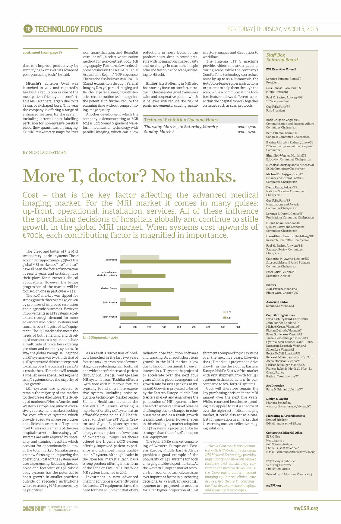
myESR.org
TECHNOLOGY FOCUS18 ECR TODAY | THURSDAY, MARCH 5, 2015
The ‘bread and bu�er’ of the MRI sector are cylindrical systems. These account for approximately 75% of the global MRI market. 1.5T, 3.0T and 7.0T have all been the focus of innovation in recent years and certainly have their place for numerous clinical applications. However, the future progression of the market will be focused on one in particular – 1.5T.
The 3.0T market was tipped for strong growth three years ago, driven by promises of improved resolution and diagnostic outcomes. However, improvements in 1.5T systems accel-erated through demand for more advanced mid-priced systems and concerns over the price of 3.0T equip-ment. The 1.5T market also meets the needs of both emerging and devel-oped markets, as it splits to include a multitude of price tiers offering premium and economy systems. In 2014, the global average selling price of 1.5T systems was two thirds that of 3.0T systems and this is not expected to change over the coming 5 years. As a result, the 3.0T market will remain a smaller, more specialized segment as 1.5T systems drive the majority of unit growth.
1.5T systems are projected to remain the majority of the market for the foreseeable future. The devel-oped markets of North America and Western Europe are almost exclu-sively replacement markets looking for cost effective systems which provide adequate image resolution and clinical outcomes. 1.5T systems meet these requirements of the core hospital market and increasingly 3.0T systems are only required by speci-ality and training hospitals which account for approximately 10–15% of the total market. Manufacturers are now focusing on improving the operational costs of the systems and user experiencing. Reducing the cost, noise and footprint of 1.5T whole body systems has the potential to boost growth in smaller practices outside of specialist institutions where extremity MRI scanners may be prioritised.
As a result a succession of prod-ucts launched in the last two years focused on 4 key areas: cost of owner-ship, noise reduction, small footprint and wider bore for increased patient throughput. The 1.5T Vantage Elan MR systems from Toshiba offers a 64cm bore with numerous features typically found in a more expen-sive system, including noise-re-duction technology. Market leader Siemens Healthcare launched the MAGNETOM Amira offering a high-functionality 1.5T system at an affordable price point. GE Health-care launched the 1.5T Signa Crea-tor and Signa Explorer systems, offering smaller footprint, reduced energy consumption and lower cost of ownership. Philips Healthcare offered the Ingenia 1.5TS system offering improved patient experi-ence and advanced image quality in a 1.5T system. Although leader in the Open MRI market, Hitachi has a strong product offering in the form of the Echelon Oval 1.5T Ultra-Wide MR system launched in 2012.
Investment in new advanced imaging solutions is currently being focused on CT equipment due to the need for new equipment that offers
radiation dose reduction so�ware and tracking. As a result short term growth in the MRI market is low due to lack of investment. However, interest in 1.5T systems is projected to accelerate over the next four years with the global average annual growth rate for units peaking at 11% in 2015. Growth is projected to be led by the Eastern Europe, Middle East & Africa market and Asia where the penetration of MRI systems is low. The North American market remains challenging due to changes in reim-bursement and as a result growth is significantly lower. However, even in this challenging market adoption of 1.5T systems is projected to be far stronger than that of 3.0T and open MRI equipment.
The total EMEA market compris-ing of Western Europe and East-ern Europe, Middle East & Africa provides a good example of the popularity of 1.5T systems for both emerging and developed markets. As the Western European market recov-ers from economic turmoil, cost is an ever important factor in purchasing decisions. As a result, advanced 1.5T systems are projected to account for a far higher proportion of unit
shipments compared to 3.0T systems over the next five years. Likewise the 1.5T market is projected to drive growth in the developing Eastern Europe, Middle East & Africa market with unit shipment growth for 1.5T systems estimated at 17% in 2015 compared to 10% for 3.0T systems.
Cost will therefore remain the key purchasing decision in the MRI market over the next five years. Whilst restricted healthcare spend-ing may appear to cast a shadow of over the high-cost medical imaging market, it could also act as a cata-lyst for innovation in a market that is searching most cost effective imag-ing solutions.
Nicola Goatman is a senior ana-lyst with IHS Medical Technology. IHS Medical Technology provides high quality and in-depth market research and consultancy ser-vices to the medical device indus-try. Coverage includes medical imaging equipment, clinical care devices, healthcare IT, consumer medical devices, medical displays and wearable technologies.
BY NICOLA GOATMAN
More T, doctor? No thanks.Cost – that is the key factor affecting the advanced medical imaging market. For the MRI market it comes in many guises: up-front, operational, installation, services. All of these influence the purchasing decisions of hospitals globally and continue to stifle growth in the global MRI market. When systems cost upwards of €700k, each contributing factor is magnified in importance.
Unit Shipments – 2014
0 100 200 300 400 500 600 700 800
North America
Latin America
Western Europe
Eastern Europe, Middle East & Africa
Asia Pacific
3.0T
1.5T
that can improve productivity by simplifying exams with its advanced post-processing tools,” he said.
Hitachi’s Echelon Oval was launched in 2012 and reportedly has built a reputation as one of the most patient-friendly and comfort-able MRI scanners, largely due to its 74 cm, oval-shaped bore. This year the company is offering a range of enhanced features for the system, including arterial spin labelling perfusion for non-invasive cerebral blood flow quantification imaging, T2 MRI relaxometry maps for liver
iron quantification, and BeamSat vascular ASL, a selective saturation method for non-contrast body MR angiography. Further so�ware devel-opments include the RADAR (Radial Acquisition Regime) TOF sequence. The vendor also believes its K-RAPID (Rapid Acquisition through Parallel Imaging Design) parallel imaging and IR-RAPID parallel imaging with iter-ative reconstruction technology has the potential to further reduce the scanning time without compromis-ing image quality.
Another development which the company is demonstrating at ECR 2015 is So� Sound S gradient wave-form modification technology with parallel imaging, which can allow
reductions in noise levels. It can produce a 90% drop in sound pres-sure with no impact on image quality and no change in scan time in spin echo and fast spin echo scans, accord-ing to Hitachi.
Philips’ latest offering in MRI also has a strong focus on comfort, intro-ducing features designed to ensure a calm and cooperative patient which it believes will reduce the risk of panic movements, causing unsat-
isfactory images and disruption to workflow.
The Ingenia 1.5T S machine provides videos to distract patients during scans, while the company’s ComforTone technology can reduce noise by up to 80%. Meanwhile, the AutoVoice feature gives instructions to patients to help them through the scan, while a communications tool-box feature allows different users within the hospital to work together on issues such as scan protocols.
Technical Exhibition Opening Hours
Thursday, March 5 to Saturday, March 7 10:00–17:00
Sunday, March 8 10:00–14:00
continued from page 17
ESR Executive Council
Lorenzo Bonomo, Rome/IT President
Luis Donoso, Barcelona/ES 1st Vice-President
Paul M. Parizel, Antwerp/BE 2nd Vice-President
Guy Frija, Paris/FR Past-President
Boris Brkljačić, Zagreb/HR Communication and External Affairs Commi�ee Chairperson
Bernd Hamm, Berlin/DE Congress Commi�ee Chairperson
Katrine Åhlström Riklund, Umea/SE 1st Vice-Chairperson of the Congress Commi�ee
Birgit Ertl-Wagner, Munich/DE Education Commi�ee Chairperson
Nicholas Gourtsoyiannis, Athens/GR ESOR Commi�ee Chairperson
Michael Fuchsjäger, Graz/AT Finance and Internal Affairs Commi�ee Chairperson
Deniz Akata, Ankara/TR National Societies Commi�ee Chairperson
Guy Frija, Paris/FR Nominations and Awards Commi�ee Chairperson
Lorenzo E. Derchi, Genoa/IT Publications Commi�ee Chairperson
E. Jane Adam, London/UK Quality, Safety and Standards Commi�ee Chairperson
Hans-Ulrich Kauczor, Heidelberg/DE Research Commi�ee Chairperson
Paul M. Parizel, Antwerp/BE Strategic Review Commi�ee Chairperson
Catherine M. Owens, London/UK Subspecialties and Allied Sciences Commi�ee Chairperson
Peter Baierl, Vienna/AT Executive Director
Editors Julia Patuzzi, Vienna/AT Philip Ward, Chester/UK
Associate Editor Simon Lee, Vienna/AT
Contributing Writers Edna Astbury-Ward, Chester/UK John Bonner, London/UK Michael Crean, Vienna/AT Florian Demuth, Vienna/AT Peter Gordebeke, Vienna/AT Javeni Hemetsberger, Vienna/AT Cynthia Keen, Sanibel Island, FL/US Katharina Krischak, Vienna/AT Simon Lee, Vienna/AT Becky McCall, London/UK Rebekah Moan, San Francisco, CA/US Alena Morrison, Vienna/AT Mélisande Rouger, Madrid/ES Frances Rylands-Monk, St. Meen Le Grand/France Kathrin Tauer, Vienna/AT
Art Direction Petra Mühlmann, Vienna/AT
Design & Layout Marlene Schaufler, www.studio-marlene.at, Vienna/AT
Marketing & Advertisements Konrad Friedrich E-Mail: [email protected]
Contact the Editorial Office ESR Office Neutorgasse 9 1010 Vienna, Austria Phone: (+43-1) 533 40 64-0 E-Mail: [email protected]
ECR Today is published 5x during ECR 2015. Circulation: 20,000
Printed by Holzhausen, Vienna 2015
myESR.org
Staff Box Editorial Board
myESR.org
TECHNOLOGY FOCUS 19ECR TODAY | THURSDAY, MARCH 5, 2015
When it comes to realising collab-orative project ideas under Euro-pean funding calls and coordinat-ing your international projects, the European Institute for Biomedical Imaging Research (EIBIR) offers professional guidance and support through a wide variety of different research-related services for its network members.
For the first calls under Horizon 2020 in 2014, EIBIR was involved as project coordinator or partner in more than 20 proposals. In these proposals, EIBIR has collaborated with over 100 different partners, including 23 small and medium-sized enterprises (SME), from 22 countries.
The proposals that were submit-ted over the course of 2014 covered various topics, including, but not limited to, radiology, paediatrics, cardiovascular disease, oncology, and gene therapy. All proposals featured a strong biomedical imag-ing component.
Among the submi�ing members were Prof. Laure Fournier from University Paris Descartes, France, and Dr. Dirk Smeets from icoMetrix, an SME located in Belgium – below are their thoughts on working with EIBIR during the preparation of the proposal and its submission.
ECR Today: Professor Fournier, what was your impression of the proposal preparation process compared to other funding you have applied for?
Laure Fournier: I have previ-ously been involved in smaller projects in France; the main differ-ence was the amount of administra-tive tasks that come with a larger project. It was very good to have EIBIR’s support, as sometimes the administrative side of things can get complicated. This allowed me to concentrate on the science aspects of the projects. This had not been the case in prior projects, where I would also have to focus on other aspects as well.
ECRT: Why did you choose EIBIR’s proposal preparation and management services?
LF: I knew EIBIR existed and was familiar with the services they provide. Basic research, including animal studies, would be a big part of the proposed project and from speaking with EIBIR, EORTC and ESR it seemed like a natural fit to have EIBIR on board.
ECRT: How did EIBIR support you during proposal preparation?
LF: All aspects of proposal writing were supported by EIBIR. I particu-larly enjoyed that the organisation of for instance conference calls and completing administrative forms was being taken care of by EIBIR in the background.
It was also very good to know the proposal would be re-read by staff with a scientific background. The initial round of feedback from EIBIR’s Scientific Advisory Board was appreciated very much, as it showed us that we were moving in the right direction.
EIBIR supported the proposal exactly as I imagined it, allowing me to focus on the scientific parts of the proposal.
ECR Today: Dr. Smeets, your SME icoMetrix was part of a consortium that submi�ed a proposal under Horizon 2020. What was your moti-vation to apply for funding?
Dirk Smeets: Our mission is to bring imaging biomarkers from research to the patient. This translation often starts with the development of new biomarkers by innovative research projects. Horizon 2020 offers great oppor-tunities for such projects, limiting the risk for SMEs through the high funding rates. Moreover, Horizon 2020 projects allow us to strengthen our network due to its international character.
ECRT: As one of our new Industry Partners, what is the added value of the EIBIR membership for you?
DS: We believe EIBIR enhances the visibility of icoMetrix in the field of biomedical imaging and helps us to enlarge our network, towards potential customers and future collaborators, who can visit us at ECR booth 630.
ECRT: How would you rate your experience working with a part-ner for proposal preparation and project management?
DS: EIBIR really helps taking away the administrative burden of the proposal. As such, the scientific contributors can focus on the writing of the scientific parts.
BY PETER GORDEBEKE
Proposal preparation with EIBIR Support for Horizon 2020Feedback from EIBIR Network Members on the first Horizon 2020 submissions
EIBIR Session
Thursday, March 5, 14:00–15:30, Room L 1 #ECR2015L1The complexity of personalised breast care
» Chairman’s introduction T.H. Helbich; Vienna/AT
» A life-course approach to understanding breast cancer genesis:
connecting epidemiology, genetics, and pathology I. Dos Santos Silva; London/UK
» VPH-PRISM aiding the therapy decision making process by
quantitative evaluation of personal imaging and non-imaging data H.K. Hahn; Bremen/DE
» Stroma and peritumoural stiffness: latest evidence for its importance and
novel stroma imaging approaches to predict therapy response A. Evans; Dundee/UK
» Personalised treatment decisions: how to reason using multi-modal,
multi-disciplinary data
R.M. Mann; Nijmegen/NL
EIBIR Session
Friday, March 6, 10:30–12:00, Room L 1 #ECR2015L1What’s new in biomedical imaging research: an update of EIBIR activities
» Chairman’s introduction G.P. Krestin; Ro�erdam/NL
» Presentation from the European Commission on Horizon 2020
A.-S. Costescu; Brussels/BE» EIBIR Joint Initiative for Paediatric Radiology
K. Rosendahl; Bergen/NO» EIBIR Joint Initiative: Biomedical Image Analysis Platform
W.J. Niessen; Ro�erdam/NL» EIBIR Joint Initiative for Image Guided Radiotherapy
V. Valentini; Rome/IT» EIBIR Joint Initiative for Euro-BioImaging
S. Aime; Turin/IT
EIBIR Session
Friday, March 6, 14:00–15:30, Room L 1 #ECR2015L1MITIGATE consortium: state of the art imaging and therapy in GIST
Chairpersons: S.O. Schönberg; Mannheim/DE I. Virgolini; Innsbruck/AT
» Selective internal radiotherapy in GIST patients
S. Diehl; Mannheim/DE» Multimodal imaging in GIST
D. Longo; Torino/IT» Principle of X-Nuclei MR imaging: what the radiologist should know
L. Schad; Mannheim/DE
Prof. Laure Fournier from University Paris Descartes, France.
Dr. Dirk Smeets from icoMetrix in Belgium.
For more details about EIBIR services, please visit www.eibir.org or visit us at our booth in the entrance hall to discuss your project ideas.
myESR.org
TECHNOLOGY FOCUS20 ECR TODAY | THURSDAY, MARCH 5, 2015
The main aims of the Association of Radiologists of Ukraine (ARU) are the development and improvement of radiology in our country, along with the integration of national radiologists into the European radi-ological family.
A�er the adoption of the Euro-pean Training Charter at the Euro-pean Congress of Radiology in 2014, the ARU began the process of imple-menting it into our national radiol-ogy services.
Last year, during the National Radiological Congress in Kiev and at a radiological forum in Odessa, radiologists from our country discussed the European Commis-sion document ‘Radiation protection 118 referral guidelines for imaging.’ Currently, we are trying to introduce this document into clinical practice a�er its approval by the Ministry of Health.
Radiological science is a very important branch of radiology, which gives us the ability to educate the next generation of Ukrainian radiologists.
Scientific research in the field of radiology in Ukraine is conducted in two radiological institutions belonging to the Academy of Medi-cal Sciences of Ukraine: the Grig-or’ev Institute of Medical Radiology in Kharkov and the Institute of Nuclear Medicine in Kiev, as well as at specialised radiological departments at 11 medical universities in Ukraine. The research work of these institu-tions is aimed at working out new techniques in diagnostic radiology, radiotherapy and nuclear medicine. It also develops new methods of prevention, diagnosis and treatment of radiation lesions. Furthermore it focuses on the improvement of dosimetry provision of radiotherapy, optimising radiation load from medi-cal procedures, medical application of ionising radiation, and research development in radiology.
The Institute of Medical Radiology in Kharkov has been participating in scientific programmes in biolog-ical dosimetry under the aegis of the Commission of the European Union since 1993. Together with the
National Commi�ee for Radiation Protection (Great Britain), the Insti-tute of Nuclear Safety of France and the Institute of Genetics and Cytology at the Belarus Academy of Science our members worked on the project ‘Doses to the Belarus and Ukraine populations resulting from the Chernobyl accident’.
The radiological department of Kharkov Medical Academy of post-graduate training investigated the possibilities of micro-focus x-ray systems in clinical practice.
Major areas of research are tomo-synthesis in mammography, opti-misation of the control system for x-ray equipment and teleradiology in clinical practice.
Because of the purchase of three systems for PET/CT diagnostics, as well as several modern linear accel-erators for radiation therapy by the National Medical Academy of Postgraduate Training in Kiev, some scientific research in the oncologic field has been planned.
The Institute of Nuclear Medicine has performed investigation into
CT-perfusion in patients with hepatic diseases and cancer processes of the head and neck.
The Ukrainian mammographic system for tomosynthesis (3D mammography) is currently under clinical testing for the screening and early detection of breast cancer.
Unfortunately, the government of Ukraine does not provide sufficient funding for research in the field of radiology. Hospitals have govern-ment-funded budgets, but only for day-to-day radiological services. Departments at medical universi-ties do not have their own modern equipment, and university depart-ments have to cooperate with be�er-equipped general hospitals for inves-tigations. Research o�en a�racts young practising radiologists, who work in these hospitals. During the last two years, eight young radiolo-gists completed a PhD programme and defended their theses.
Every year at the end of March in Kiev, the ARU organises the National Radiological Congress, during which our radiologists report the results of
their scientific research. They must publish scientific articles in two Ukrainian radiological journals.
The international activity of our members consists of research coop-eration with foreign societies (RSNA, ESTRO, EANM), scientific centres, institutions, foundations and other organisations. It also includes partici-pation in scientific conferences, visits by researchers to other countries for scientific research, fellowships and training.
One of the aims of the ARU is the scientific development of radiology in Ukraine for the period 2014–2017. The government of our country has decided to establish high-tech centres of nuclear medicine, diag-nostic radiology and radiotherapy in some areas of Ukraine. But this is still a work in progress.
More information about the Asso-ciation of Radiologists of Ukraine can be found at aru-ua.org
Ukrainian radiologists keep working to improve their discipline
for Radiographers, Physicians, Interventionalists and Radiologists
March 5th – 7th, Room 2.33 Second Level/ Next to room A
Consult the offi cial ECR programme for the detailed agenda of workshops.
Advancing dose management… together.
gehealthcare.com
Dose Management and Ionising Radiation Perspectives and Training
myESR.org
TECHNOLOGY FOCUS 21ECR TODAY | THURSDAY, MARCH 5, 2015
Introducing clinical decision support for imaging referral guidelines in Europe will help doctors choose the best exam for every patient
ESR iGuide is a Clinical Decision Support (CDS) system developed by the ESR in cooperation with the National Decision Support Company (NDSC) for distribution in Europe. This product will make imaging referral guidelines available in a user-friendly way, supporting refer-ring physicians in choosing the most appropriate exam for each of their patients. Used as a stand-alone tool or integrated with hospital order-ing systems and electronic health records, ESR iGuide not only offers referrers up-to-date and scientifically valid guidance; it also provides an efficient, traceable and reliable way of selecting and ordering imaging exams in daily practice, improving the clinical workflow to the benefit of general practitioners and other specialists, radiologists, hospital managers and patients alike. Accord-ing to incoming ESR President Prof. Luis Donoso, “imaging referral guide-lines are essential for improving appropriateness, and CDS solutions are the best way to achieve this in practice. ESR iGuide will enable health professionals to make the best possible use of medical imaging, and it is an important part of the ESR’s overall drive to improve quality and safety for patients.”
In Europe, the appropriateness of the countless radiological examina-
tions conducted year in, year out can be improved significantly. Imaging referral guidelines are an essential tool to achieve this, but they are only available in 70 percent of Euro-pean countries. The recommenda-tions that are available o�en differ starkly from country to country, even though there is no sound evidence for such divergences. Moreover, as developing and updating guidelines is a complex, arduous process, it is o�en questionable how up-to-date these recommendations are. And, not least, they are usually only avail-able as a booklet or in PDF format. These and other factors contribute to a significant lack of use of imaging referral guidelines.
At a time of budgetary constraints on health systems in many European countries, amid rising public concern over the risks of radiation exposure, and in the context of new EU legis-lation requiring member states to ensure guidelines are available to referrers, the ESR yesterday officially presented ESR iGuide as its solution to address these issues.
Based on the American College of Radiology’s Appropriateness Crite-ria, the ESR has developed guidelines adapted to European standards of practice, and in cooperation with NDSC embedded them in a CDS platform. Through this approach, the
guidelines and the functionality of the system can be tailored to national requirements or institutional prefer-ences and significantly improve the clinical workflow.
“This is a tremendous opportu-nity to work alongside the ESR and provide a robust content delivery mechanism, capable of deliver-ing European wide criteria while supporting country or site specific requirements. The content delivery mechanism integrates directly into EHR or other IT platforms used by an institution and ensures that the correct guidelines and feedback are presented to referring physicians,” says Michael Mardini, CEO of NDSC. “Our in depth experience in deliver-ing this solution nationally within the US will guide us as we implement across the European market,” contin-ues Mardini. “The medical expertise of the ESR together with our tech-nical experience will create a more efficient way of selecting, ordering and monitoring radiological services, in turn improving the quality and safety of imaging services across Europe,” concludes Mardini.
ESR iGuide will give referrers medically sound guidance, provide radiologists with clear orders , and enable hospital managers to monitor performance; most importantly, ESR iGuide will give patients confidence
that the care they receive adheres to the highest standards of quality and safety. Following pilot tests in the coming months, ESR iGuide will be available from autumn 2015.
Be sure to visit our booth in the entrance hall of the ACV to find out how ESR iGuide can benefit your institution!
BY FLORIAN DEMUTH
ESR and NDSC launch ESR iGuide
INTRODUCTION In 1999 the European Commission
(EC) issued the Radiation Protec-tion 109 (RP 109) publication on Guidance on diagnostic reference levels (DRLs) for medical exposure (1). This document highlights the importance of establishing DRLs for high-dose medical examinations, in particular computed tomogra-phy and interventional radiology procedures. The utility of DRLs in radiation protection of patients is also very well acknowledged in the newly published Euratom BSS (2).
THE PIDRL PROJECTEuropean DRLs for Paediatric
Imaging (PiDRL) is a new 27-month EC project which aims to a) develop a methodology for establishing and using DRLs for paediatric medical imaging, and b) update and extend the European DRLs to cover as many procedures as possible. The PiDRL consortium is headed by the European Society of Radiology, with other participating organisa-tions including the European Feder-
ation of Organisations for Medical Physics (EFOMP), the European Society of Paediatric Radiology (ESPR), the European Federation of Radiographer Societies (EFRS) and the Finnish Radiation and Nuclear Safety Authority (STUK), with Public Research Centre Henri Tudor (CRP-HT) as a subcontractor.
The main task of PiDRL is to develop a methodology for establish-ing and using DRLs for paediatric imaging and produce new European guidelines on paediatric DRLs. PiDRL also aims to update and extend the European DRLs as provided in RP 109 (1) to cover more procedures.
PIDRL ACHIEVEMENTSA worldwide review of literature
on patient doses and DRLs for children of different age groups, or other distributions, and for differ-ent examinations has been carried out with an emphasis on European literature. Questionnaires have been distributed to confirm and update the data on paediatric DRLs in Euro-pean countries as available from the
EC project Dose Datamed2 (DDM2) database (3), and to collect infor-mation to prepare sections of the guidelines. The first complete dra� of the guidelines has already been submi�ed to the EC.
The review of DRLs has indicated that for interventional, fluorosco-py-guided cardiac procedures, no national DRLs exist but only a few local DRLs have been suggested, and for interventional non-cardiac proce-dures, no DRLs have been suggested at all. PiDRL efforts are in progress to establish multi-national DRLs for paediatric interventional procedures.
PiDRL will hold a workshop in Lisbon, Portugal, on October 15–17, 2015. A range of topics pertaining to the current status, difficulties and future opportunities in the field of paediatric DRLs will be discussed. The outcomes will form the basis for European Guidelines on DRLs for paediatric imaging covering plain radiography, fluoroscopically-guided interventional procedures and CT. The scientific commi�ee of the PiDRL workshop has endeavoured
to set up an exciting programme, which includes round tables and panel discussions, as well as oral and poster presentations. The call for abstract submissions for oral and poster presentations on the topic ‘Patient Doses from Paediat-ric Diagnostic and Interventional Procedures – Establishment and Use of Paediatric DRLs’ will close in April 2015. Besides our scientific programme, participants can enjoy sightseeing, architecture, museums and Portuguese cuisine in Lisbon. More information about the PiDRL project and workshop can be found at www.eurosafeimaging.org/pidrl
REFERENCES1. European Commission (EC), 1999.
Guidance on Diagnostic Reference Levels (DRLs) for medical exposures, Radiation Protection 109.
2. Council of the European Union. (2013). Council Directive 2013/59/Euratom laying down basic safety standards for protection against the dangers arising from exposure to ionising radiation, and repealing
Directives 89/618/Euratom, 90/641/Euratom, 96/29/Euratom, 97/43/Euratom and 2003/122/Euratom. Offi-cial Journal L-13 of 17.01.2014.
3. European Commission (EC), 2014. Dose Datamed 2 (DDM2) Project Report Part 2: Diagnostic Reference Levels (DRLs) in Europe.
Prof. John Damilakis is the ESR Representative and PiDRL Scien-tific Coordinator. He is Professor of Medical Physics at the Univer-sity of Crete, Faculty of Medicine, in Iraklion, Greece
BY JOHN DAMILAKIS
PiDRL: A new EC project on paediatric DRLs
EuroSafe Imaging Session 1
Thursday, March 5, 14:00–15:30, Room M #ECR2015MClinical decision support: making imaging referral guidelines
work for patients, doctors and hospital managers
» Chairman’s introduction
G. Frija; Paris/FR» CDS impact on guidelines development
K.J. Dreyer; Boston, MA/US» Adapting and updating guidelines
M.G.M. Hunink; Ro�erdam/NL» ACR select implementation experience
J.A. Brink; Boston, MA/US» ESR iGuide
L. Donoso; Barcelona/ES» Discussion
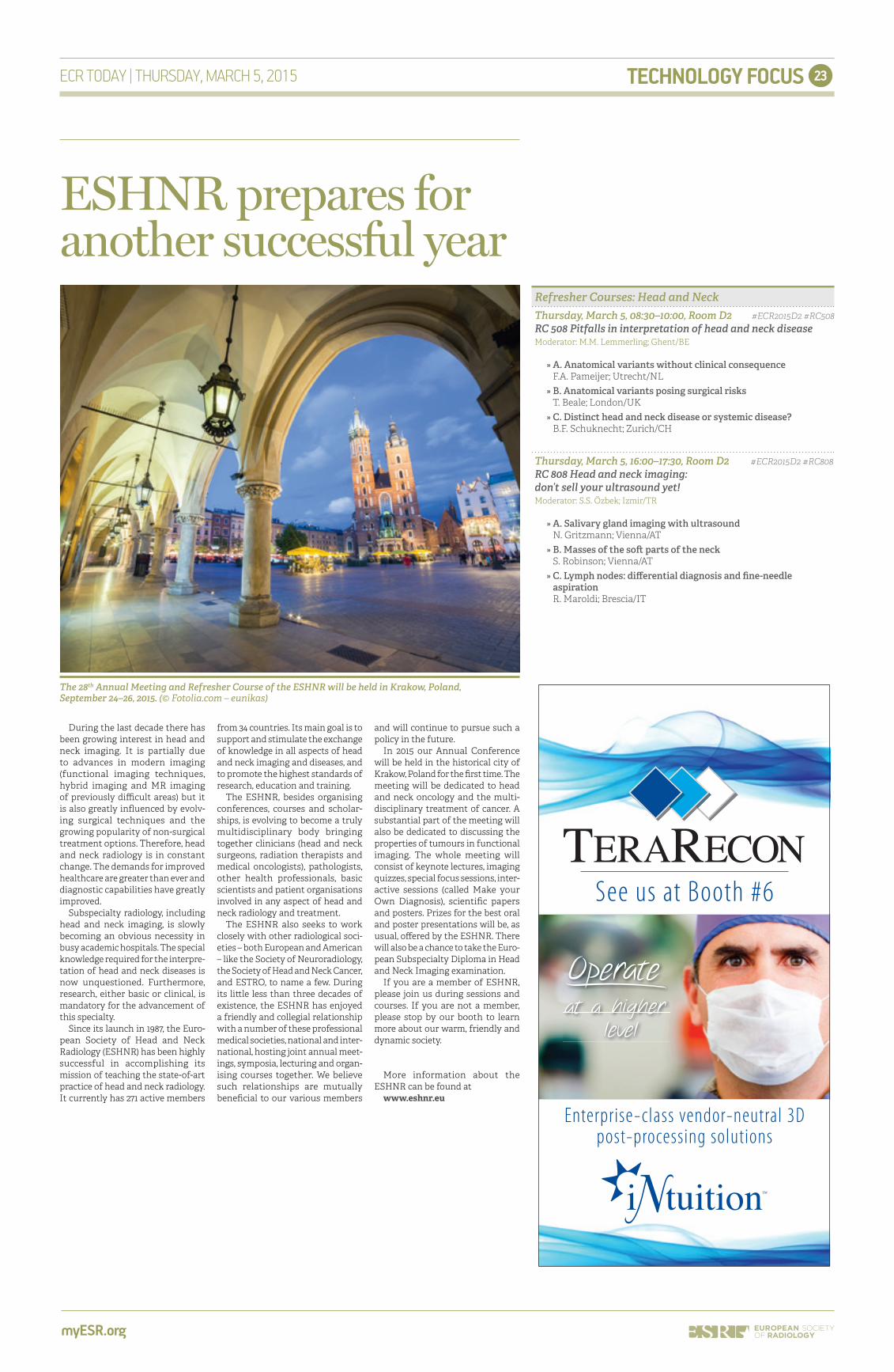
myESR.org
TECHNOLOGY FOCUS 23ECR TODAY | THURSDAY, MARCH 5, 2015
During the last decade there has been growing interest in head and neck imaging. It is partially due to advances in modern imaging (functional imaging techniques, hybrid imaging and MR imaging of previously difficult areas) but it is also greatly influenced by evolv-ing surgical techniques and the growing popularity of non-surgical treatment options. Therefore, head and neck radiology is in constant change. The demands for improved healthcare are greater than ever and diagnostic capabilities have greatly improved.
Subspecialty radiology, including head and neck imaging, is slowly becoming an obvious necessity in busy academic hospitals. The special knowledge required for the interpre-tation of head and neck diseases is now unquestioned. Furthermore, research, either basic or clinical, is mandatory for the advancement of this specialty.
Since its launch in 1987, the Euro-pean Society of Head and Neck Radiology (ESHNR) has been highly successful in accomplishing its mission of teaching the state-of-art practice of head and neck radiology. It currently has 271 active members
from 34 countries. Its main goal is to support and stimulate the exchange of knowledge in all aspects of head and neck imaging and diseases, and to promote the highest standards of research, education and training.
The ESHNR, besides organising conferences, courses and scholar-ships, is evolving to become a truly multidisciplinary body bringing together clinicians (head and neck surgeons, radiation therapists and medical oncologists), pathologists, other health professionals, basic scientists and patient organisations involved in any aspect of head and neck radiology and treatment.
The ESHNR also seeks to work closely with other radiological soci-eties – both European and American – like the Society of Neuroradiology, the Society of Head and Neck Cancer, and ESTRO, to name a few. During its li�le less than three decades of existence, the ESHNR has enjoyed a friendly and collegial relationship with a number of these professional medical societies, national and inter-national, hosting joint annual meet-ings, symposia, lecturing and organ-ising courses together. We believe such relationships are mutually beneficial to our various members
and will continue to pursue such a policy in the future.
In 2015 our Annual Conference will be held in the historical city of Krakow, Poland for the first time. The meeting will be dedicated to head and neck oncology and the multi-disciplinary treatment of cancer. A substantial part of the meeting will also be dedicated to discussing the properties of tumours in functional imaging. The whole meeting will consist of keynote lectures, imaging quizzes, special focus sessions, inter-active sessions (called Make your Own Diagnosis), scientific papers and posters. Prizes for the best oral and poster presentations will be, as usual, offered by the ESHNR. There will also be a chance to take the Euro-pean Subspecialty Diploma in Head and Neck Imaging examination.
If you are a member of ESHNR, please join us during sessions and courses. If you are not a member, please stop by our booth to learn more about our warm, friendly and dynamic society.
More information about the ESHNR can be found at
www.eshnr.eu
ESHNR prepares for another successful year
Refresher Courses: Head and Neck
Thursday, March 5, 08:30–10:00, Room D2 #ECR2015D2 #RC508RC 508 Pitfalls in interpretation of head and neck disease
Moderator: M.M. Lemmerling; Ghent/BE
» A. Anatomical variants without clinical consequence
F.A. Pameijer; Utrecht/NL» B. Anatomical variants posing surgical risks
T. Beale; London/UK» C. Distinct head and neck disease or systemic disease?
B.F. Schuknecht; Zurich/CH
Thursday, March 5, 16:00–17:30, Room D2 #ECR2015D2 #RC808RC 808 Head and neck imaging:
don’t sell your ultrasound yet!
Moderator: S.S. Özbek; Izmir/TR
» A. Salivary gland imaging with ultrasound
N. Gritzmann; Vienna/AT» B. Masses of the so� parts of the neck
S. Robinson; Vienna/AT» C. Lymph nodes: differential diagnosis and fine-needle
aspiration R. Maroldi; Brescia/IT
See us at Booth #6
Enterprise-class vendor-neutral 3D post-processing solutions
TM
The 28th Annual Meeting and Refresher Course of the ESHNR will be held in Krakow, Poland, September 24–26, 2015. (© Fotolia.com – eunikas)
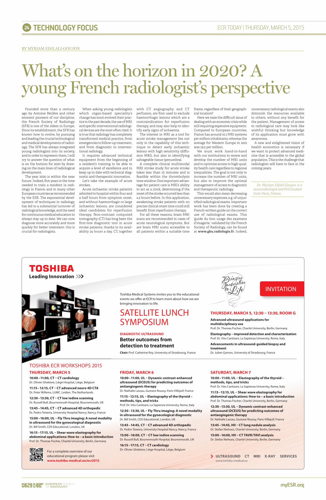
myESR.org
TECHNOLOGY FOCUS24 ECR TODAY | THURSDAY, MARCH 5, 2015
www.toshiba-medical.eu
TOSHIBA ECR WORKSHOPS 2015THURSDAY, MARCH 510:00 – 11:00, CT – CT cardiology Dr. Olivier Ghekiere, Liège Hospital, Liège, Belgium
11:15 – 12:15, CT – CT advanced neuro 4D CTA Dr. Peter Willems, LUMC, Leiden, The Netherlands
12:30 – 13:30, CT – CT low iodine scanning Dr. Russell Bull, Bournemouth Hospital, Bournemouth, UK
13:45 – 14:45, CT – CT advanced 4D orthopedic Dr. Pedro Teixeira, University Hospital Nancy, Nancy, France
15:00 – 16:00, UL – Fly Thru imaging: A novel modality in ultrasound for the gynecological diagnostic Dr. Bill Smith, CDS Educational, London, UK
16:15 – 17:15, UL – Shear wave elastography for abdominal applications: How-to – a basic introduction Prof. Dr. Thomas Fischer, Charité University, Berlin, Germany
FRIDAY, MARCH 610:00 – 11:00, UL – Dynamic contrast-enhanced ultrasound (DCEUS) for predicting outcomes of antiangiogenic therapy Dr. Nathalie Lassau, Gustave Roussy, Paris-Villejuif, France
11:15 – 12:15, UL – Elastography of the thyroid – methods, tips, and tricks Prof. Dr. Vito Cantisani, La Sapienza University, Rome, Italy
12:30 – 13:30, UL – Fly Thru imaging: A novel modality in ultrasound for the gynecological diagnostic Dr. Bill Smith, CDS Educational, London, UK
13:45 – 14:45, CT – CT advanced 4D orthopedic Dr. Pedro Teixeira, University Hospital Nancy, Nancy, France
15:00 – 16:00, CT – CT low iodine scanning Dr. Russell Bull, Bournemouth Hospital, Bournemouth, UK
16:15 – 17:15, CT – CT cardiology Dr. Olivier Ghekiere, Liège Hospital, Liège, Belgium
SATURDAY, MARCH 710:00 – 11:00, UL – Elastography of the thyroid – methods, tips, and tricks Prof. Dr. Vito Cantisani, La Sapienza University, Rome, Italy
11:15 – 12:15, UL – Shear wave elastography for abdominal applications: How-to – a basic introduction Prof. Dr. Thomas Fischer, Charité University, Berlin, Germany
12:30 – 13:30, UL – Dynamic contrast-enhanced ultrasound (DCEUS) for predicting outcomes of antiangiogenic therapy Dr. Nathalie Lassau, Gustave Roussy, Paris-Villejuif, France
13:45 – 14:45, HII – CT lung nodule analysis Dr. Stefan Niehues, Charité University, Berlin, Germany
15:00 – 16:00, HII – CT TAVR/TAVI analysis Dr. Stefan Niehues, Charité University, Berlin, Germany
Toshiba Medical Systems invites you to the educational events we offer at ECR to learn more about how we are bringing innovation to life.
SATELLITE LUNCH SYMPOSIUMDIAGNOSTIC ULTRASOUND
Better outcomes from detection to treatmentChair: Prof. Catherine Roy, University of Strasbourg, France
THURSDAY, MARCH 5, 12:30 – 13:30, ROOM GAdvanced ultrasound applications for multidisciplinary use Prof. Dr. Thomas Fischer, Charité University, Berlin, Germany
Elastography – improved detection and characterization Prof. Dr. Vito Cantisani, La Sapienza University, Rome, Italy
Advancements in ultrasound-guided biopsy and treatment Dr. Julien Garnon, University of Strasbourg, France
For a complete overview of our educational program please visit www.toshiba-medical.eu/ecr2015
TOSHIBA ECR WORKSHOPS 2015
INVITATION
Founded more than a century ago by Antoine Béclère and other eminent pioneers of our discipline, the French Society of Radiology (SFR) is one of the oldest in Europe. Since its establishment, the SFR has known how to evolve, by pursuing and leading the crucial technological and medical developments of radiol-ogy. The SFR has always integrated young radiologists into its society, and in order to represent them, I will try to answer the question of what is on the horizon for 2020 by draw-ing on the main lines of radiological development.
The year 2020 is within the near future. Indeed, five years is the time needed to train a resident in radi-ology in France, and in many other European countries as recommended by the ESR. The exponential devel-opment of techniques in radiology has led to a substantial turnover of radiological knowledge, and the need for continuous medical education to always stay up to date. We can now diagnose more accurately and more quickly for be�er treatment: this is crucial for radiologists.
When asking young radiologists which organ-based speciality’s change has most evolved their prac-tice in the past decade, the use of MRI and specific interventional radiologi-cal devices are the most o�en cited. It is true that radiology has completely transformed medical practice, from emergencies to follow-up treatment, and from diagnostic to interven-tional radiology.
It requires advanced technical equipment from the beginning of a resident’s training to be able to acquire a level of excellence and to keep up to date with technical diag-nostic and therapeutic innovation.
Let’s take the example of acute strokes.
Acute ischaemic stroke patients admi�ed to hospital within four and a half hours from symptom onset, and without haemorrhagic or large ischaemic lesions, are considered ideal candidates for reperfusion therapy. Non-contrast computed tomography (CT) has long been the first-line diagnostic tool in acute stroke patients, thanks to its avail-ability 24 hours a day. CT, together
with CT angiography and CT perfusion, are first used to exclude haemorrhagic lesions which are a contraindication for reperfusion therapy, and may also help to iden-tify early signs of ischaemia.
The interest in MRI as a tool for acute stroke management lies not only in the capability of this tech-nique to detect early ischaemic lesions with high sensitivity (diffu-sion MRI), but also in identifying salvageable tissue (penumbra).
A complete clinical multimodal MRI stroke study for acute stroke takes less than 15 minutes and is feasible within the thrombolysis time window. One important advan-tage for patient care is MRI’s ability to act as a clock, determining if the onset of the stroke occurred less than 4.5 hours before. In this application, awakening stroke patients with no precise clinical onset time could still benefit from reperfusion therapy.
For all these reasons, brain MRI scans are recommended in cases of acute neurological symptoms. But are brain MRI scans accessible to all patients within a suitable time
frame, regardless of their geograph-ical location?
Here we raise the difficult issue of dealing with an economic crisis while still requiring expensive equipment. Compared to European countries, France has around 10.1 MRI systems per million inhabitants, whereas the average for Western Europe in 2012 was 19.5 per million.
We must work hand-in-hand with our institutions to renew and develop the number of MRI units and to optimise access to high-qual-ity health care regardless to regional inequalities. The goal is not only to increase the number of MRI units, but also to improve the optimal management of access to diagnostic and therapeutic radiology.
This would also mean decreasing unnecessary expenses, e.g. of unjus-tified radiological exams. Important work has been done by creating a French-wri�en guide on the correct use of radiological exams. This ‘guide du bon usage des examens d’imagerie,’ validated by the French Society of Radiology, can be found at www.gbu.radiologie.fr. Indeed,
unnecessary radiological exams also diminish the resources available to others, without any benefit for the patient. Management of access to radiological care may look like wishful thinking but knowledge of its application must grow with awareness.
A new and enlightened vision of health economics is necessary if we want to protect advanced medi-cine that is accessible to the global population. This is the challenge that radiologists will have to face in the coming years.
Dr. Myriam Edjlali-Goujon is a neuroradiologist and PhD student from Paris, France.
BY MYRIAM EDJLALI-GOUJON
What’s on the horizon in 2020? A young French radiologist’s perspective
myESR.org
25COMMUNITY NEWS
The past eight years of ESOR have been marked by an outstanding growth in a wide range of modular activities, including visiting schools, seminars, tutorials, teach-the-teach-ers programmes, visiting professor-ship programmes, visiting scholar-ships, and exchange programmes for fellowships. So far, ESOR has delivered structured continuing education to almost 14,000 residents and board-certified radiologists worldwide.
Furthermore, ESOR has gradually evolved into a major, if not the major, international provider of comple-mentary education in radiology, as it is now active worldwide. With its expanded outreach programmes, the ASKLEPIOS Courses, the AIMS School and Seminars, the Visiting Professorship Programmes and the
intercontinental scholarships and exchange programmes for fellow-ships, ESOR is able to respond adequately to the actual needs of radiologists in the ‘developing’ world following agreements between the ESR and corresponding member societies. The importance of ESOR’s contribution to this end can be be�er appreciated if one considers the statistics from the past few years. Out of the 1,900 yearly participants in ESOR activities 80% are stead-ily coming from the radiologically ‘developing’ world, while 43% of the scholarships along with 54% of the exchange programmes for fellow-ships were granted to applicants from Eastern Europe, Latin America, China, the Middle East and Africa.
In addition to impressive numbers, ESOR is proud of being instrumental
in mobilising the resources available for radiological education in Europe and the world, and for creating a long-term educational commitment and structured network, the ESOR community, through partnership. All of this hugely appreciated utili-sation of teaching resources has been implemented with the unlim-ited support of highly esteemed and renowned lecturers, tutors, mentors, volunteer reference training centres, local organisers, subspecialty and national societies, academic institu-tions, and valued industrial partners. ESOR is greatly indebted to them all.
In 2015, ESOR is consolidating its programmes, those in line with the strategic budgetary review of the ESR, while enriching its portfolio by introducing two additional outreach programmes in Latin America, one
in cooperation with the Asociación Colombiana de Radiología (ACR) and one with the Colegio Interamericano de Radiología (CIR). In addition, ESOR is moving to offer a small number of structured, competitive and certified full one year fellowship programmes, to be implemented in Europe for the first time. These fellowships will be covered by a grant, offered by the enrolling reference institutions and are intended not only to serve the needs, but also to show the step forward and the high standards of this major European education endeavour in partnership.
I would like to encourage all of you to participate and benefit from all
ESOR programmes, use the oppor-tunities offered for exchange and interaction, and share the knowledge and skills needed to meet require-ments of today and tomorrow. I am confident that ESOR will maintain its leading role in serving our vibrant community for years to come and will continue to deliver successfully, connecting the world of radiology.
Prof. Nicholas Gourtsoyiannis is Educational & Scientific Director of the European School of Radiol-ogy (ESOR) and chairman of the ESR’s ESOR Commi�ee.
BY NICHOLAS GOURTSOYIANNIS
In its ninth year, the European School of Radio-logy continues to promote the European Society of Radiology’s strong and unfailing commitment to invest in radiological education while pursuing its two main goals: to assist in harmonising radio-logical education throughout Europe, by suppor-ting the implementation of the European Training Curriculum, and to raise the scientific profile of radiological education.
26 29 30ESR Support Initiative gives boost to less affluent regions
Top tips for traineesand teachersPart 2: Work-life balance
Special Exhibition at the Kunstforum: Landscape in my mind
THURSDAY, MARCH 5, 2015
ESOR furthers ESR’s commitment to education and training
ESOR Session (European School of Radiology)
Thursday, March 5, 14:00–15:30, Room N #ECR2015NStriving in radiological education
Moderators: L. Bonomo; Rome/IT N. Gourtsoyiannis; Athens/GR
» Introduction
L. Bonomo; Rome/IT » ESOR in action 2015
N. Gourtsoyiannis; Athens/GR» The role of the European Training Curriculum:
present and future
B. Ertl-Wagner; Munich/DE» Spoon-feeding: present and future
P. Ros; Cleveland/USA» E-learning portfolios: present and future
M. Maas; Amsterdam/NL» Awards
SCHOLARSHIP PROGRAMME 2014 IN EUROPE
Alireza Abrishami, Tehran, IRNMaría Cruz Ageitos Casais, Santiago de Compostela, ESSimone Altobelli, Rome, ITAndreas P. Arnoldi, Munich, DEDubravko Bajramović, Zagreb, HRJohn Oladapo Bamidele, Ilorin, NGSilvia Bernardo, Rome, ITChandra Bortolo�o, Pavia, ITTeodora Buzan, Sremska Kamenica, ROAlessandro Campari, Milan, ITRiddhika Chakravar�y, London, UKDan Costăchescu, Timisoara, ROLara Cristiano, Rome, ITKristina Davidovic, Belgrade, RSArianna D’Errico, Naples, ITArmando Fusco, Rome, ITMa�hias Gawlitza, Leipzig, DEDinara Jumadilova, Astana, KZPia M. Jungmann, Munich, DESoheil Kooraki, Tehran, IRNUlrike Löbel, Hamburg, DEHardi Madani, London, UKHerman Paulo Marquez Masquiaran, Barcelona, ESAurelian Costin Minoiu, Bucharest, ROEliska Mlynarova, Prague, CZAna Nicolescu, Bucharest, ROSlobodanka Pena Karan, Sremska Kamenica, RSKatarzyna Pospieszny, Zabrze, PL
Francesco Ruschi, Pisa, ITRosa Severino, Naples, ITGeorgios Stathis, Athens, GRKarin Sundström, Gothenburg, SESiddharth Narendra Thaker, Jamnagar, INMathias Van Borsel, Ghent, BEJaime Verdugo, Santiago, CLAisté Vidžiūnaitė, Kaunas, LTValeria Vinci, Rome, IT
SCHOLARSHIP PROGRAMME 2014 IN USA
Ulrich Grosse, Tübingen, DE(Case Western Scholarship Programme)Francesca Iacobellis, Napoli, IT(Case Western Scholarship Programme)Diana Sorina Feier, Cluj-Napoca, RO(MSKCC Scholarship Programme)Nicola Lindsay Robertson, London, UK(MSKCC Scholarship Programme)
EXCHANGE PROGRAMME FOR ABDOMINAL RADIOLOGY FELLOWSHIP 2014 (IN PARTNERSHIP WITH ESGAR)
Bella Chamokova, Moscow, RURajiv Bhuepndra Karia, No�ingham, UKAndrei Lebovici, Cluj-Napoca, ROOlga Nikolaidou, Thessaloniki, GRSasa Rudolf, Maribor, SI
Pilar Sánchez de Medina Alba, Sala-manca, ESAna Šverko Peternac, Zagreb, HR
EXCHANGE PROGRAMME FOR BREAST RADIOLOGY FELLOWSHIP 2014(IN PARTNERSHIP WITH EUSOBI)
Andrea Luparia, Brussels, BEMukta Mahajan, Bangalore, INMaria Papavasilopoulou, Patras, GR
EXCHANGE PROGRAMME FOR BREAST IMAGING FELLOWSHIP 2014 (IN PARTNERSHIP WITH MSKCC)
Oshaani Abeyakoon, London, UKNelson Bedoya, Bogotá, CO
EXCHANGE PROGRAMME FOR CAR-DIAC RADIOLOGY FELLOWSHIP 2014 (IN PARTNERSHIP WITH ESCR)
Ekaterina Pershina, Moscow, RUMarian Pop, Tg. Mures, RO
EXCHANGE PROGRAMME FOR HEAD AND NECK RADIOLOGY FELLOW-SHIP 2014 (IN PARTNERSHIP WITH
ESHNR)
Anthony Carlyle Bartley, Joondalup, AUMontserrat Domingo Ayllón, Nancy, FR
Lavinia Manuela Lenghel, Cluj-Napoca, RO
EXCHANGE PROGRAMME FOR MUSCULOSKELETAL RADIOLOGY FELLOWSHIP 2014 (IN PARTNERSHIP WITH ESSR)
Mislav Čavka, Zagreb, HRFabian Springer, Tübingen, DE
EXCHANGE PROGRAMME FOR NEU-RORADIOLOGY FELLOWSHIP 2014 (IN PARTNERSHIP WITH ESNR)
Bernardo Corrêa de Almeida Teixeira, Curitiba, BRMohammad Hanafiah, Sungai Buloh, MYJelena Kostić, Belgrade, RSEnes Özlük, Istanbul, TRNumfon Pothichamnan, Bangkok, TH
EXCHANGE PROGRAMME FOR PAE-DIATRIC RADIOLOGY FELLOWSHIP 2014 (IN PARTNERSHIP WITH ESPR)
Andreana Bompoti, Exochi Thessalon-iki, GRIldikó Lukács, Prague, CZErika Pace, Chieti, IT
EXCHANGE PROGRAMME FOR THO-RACIC RADIOLOGY FELLOWSHIP 2014 (IN PARTNERSHIP WITH ESTI)
Manuel Gutiérrez Gimeno, Barcelona, ESAleksander Marin, Golnik, SIInneke Willekens, Brussels, BE
VISITING PROFESSORSHIP SCHOL-ARSHIPS PROGRAMME FOR CHINESE RADIOLOGISTS 2014 (IN PARTNERSHIP WITH
GE HEALTHCARE)
Zhang Kai, Jinan, ChinaXubin Li, Tianjin, CNXiaonan Mao, Shenyang, CNGang Niu, Xi’an, CNYa-Min Wan, Zhengzhou, CNNing Xing, Beijing, CN
VISITING SCHOLARSHIP PRO-GRAMME FOR CHINESE RADIOLO-GISTS JO LI 2014(IN PARTNERSHIP WITH THE
JO LI CHARITABLE TRUST)
Jun Zhang, Shanghai, CN
The certificates will be awarded during the ESOR Session today at 14:00 in Room N.
We proudly present the scholars and fellows 2014
COMMUNITY NEWS
myESR.org
26 ECR TODAY | THURSDAY, MARCH 5, 2015
The European Society of Radiology (ESR) is more than a just a profes-sional society, it’s also the centre of a global community of radiologists. Of course, being part of a commu-nity means helping one another and taking responsibility for future generations. The ESR has always been a strong advocate for this inter-national radiological community. Bringing trainees from low-income countries to the European Congress of Radiology (ECR) free of charge and funding open-access publications are just two examples of the many ways the society has served the commu-nity so far.
The ESR Support Initiative is the society’s new social responsibility project. The aim is to improve access to training, education and resources for radiologists and related scientists
in less affluent regions, thereby improving healthcare standards for patients. As the economy continues to struggle in many parts of Europe, the ESR Support Initiative comes at exactly the right time to help allevi-ate major shortages in radiological resources and training.
Each year, one country will be selected as the focus of the ESR Support Initiative. This country will then benefit from special offers from the ESR’s Invest in the Youth programme, ECR Live, ECR on Demand, the European School of Radiology and EuroSafe Imaging as well as reduced ESR membership fees. Radiologists in the selected country will also have access to fellowships at hospitals and medi-cal institutions across Europe, and trainee radiologists will have the
opportunity to get involved in the ECR, either by visiting the congress or watching it online, and benefit from a range of career-enhancing educational resources from the ESR. This should provide a much-needed boost to those members of the radi-ological community who have had to make do with fewer resources and funding over the last few years.
The eligibility criteria for the ESR Support Initiative includes factors such as the official length of training in the country, the average salary of trainees, the number of trainees per year, the total number of radiologists and the amount of equipment avail-able. All national radiological soci-eties which are ESR Institutional Members are invited to apply. All applications are then reviewed by the ESR’s Board of Directors.
The ESR has selected Bulgaria to be the ESR Support Initiative country for 2015. The ESR’s current president and founder of the ESR Support Initiative, Prof. Lorenzo Bonomo, is already looking forward to seeing more Bulgarian radiologists taking part in the ESR’s activities as a result of the initiative, which he sees as just the start of a long and successful project.
“I am delighted that we now have the chance to share our resources with colleagues in Bulgaria. For me, this is just the beginning of the ESR Support Initiative’s great work, as we will continue to serve our commu-nity so that they can be�er serve their patients,” said Prof. Bonomo.
BY MICHAEL CREAN
ESR Support Initiative gives boost to less affluent regions
ESR President Lorenzo Bonomo is the founder of the ESR Support Initiative, the society’s new social responsibility project.
Pick up some fiction at Buchkontor's ECR Bookstore. Entrance Hall
www.buchkontor.at
COMMUNITY NEWS
myESR.org
27ECR TODAY | THURSDAY, MARCH 5, 2015
The Royal College of Radiologists (RCR) has more than 9,500 Fellows and members worldwide in the disciplines of clinical radiology and clinical oncology. The RCR’s role is to advance the science and practice of clinical radiology and clinical oncology, further public education and promote study and research by se�ing the standards of training and practice. The RCR groups its activities under the themes of lead-ership, education and support, and has been developing and delivering a programme of Fellow and member engagement and value for the last three years. The RCR is also a regis-tered charity and delivers significant public benefit through its activities.
Our primary concern in 2015 is the state of U.K. radiology services. There is a relentless increase in demand particularly for complex imaging such as CT and MR scans, which is far outstripping the ability of services to cope. The situation is compounded by a failure to increase the radiol-ogy workforce in all parts of the UK. The results are increasing delays in reporting imaging tests with delayed diagnosis of cancer and other serious conditions.
The U.K. compares poorly with other European nations with around 48 trained radiologists per million population – a figure which has remained almost static for the past five years. Meanwhile, the aver-age increase in imaging activity in
England has been 10.3% for CT and 12% for MRI for each of the last ten years. Despite that, imaging rates in the UK remain significantly below those found in other healthcare systems for most tests, suggesting that further growth is inevitable.
The RCR has built the case for greater investment in the training of diagnostic and interventional radiologists in all four UK countries. The key has been using data derived from our workforce censuses (www.rcr.ac.uk/radiology/census) since 2009 and our more recent workload surveys (http://www.rcr.ac.uk/radiology/workload). Our reporting backlog snapshot survey in England in October 2014 (h�p://www.rcr.ac.uk/radiology/backlog) starkly illustrated the impact on patients. The RCR has used various means to convey its message, engaging with policy makers, stakeholders and the media – with some success so far.
The RCR is keen to assist those from outside the U.K. who wish to work by making links with U.K. employers. Please enquire at the RCR stand at ECR 2015.
Allied to our sustained lobbying for more radiologists has been the promotion of alternative models of service provision through the crea-tion of regional radiology networks (h�p://www.rcr.ac.uk/radiology/networks). Bigger than existing local collaborations, these more extensive networks would share diagnostic
expertise between hospitals for the benefit of patients. The RCR has received positive feedback on the idea which is being developed further.
Over the last year, the RCR has begun to offer leadership and mentoring development for Fellows and members. Clinical leadership of services in the U.K. is of increasing importance and prominence: our new initiatives have been very well received and will be built on in 2015 and beyond.
Since 2012, the RCR has devel-oped its priorities for international development. Further automation of the FRCR examinations will facil-itate the exams being sat in more overseas countries, where there is major unmet demand. In response to growing interest in international cooperation, the RCR has created a network of interested Fellows and members who wish to deliver radi-ological education and training on a philanthropic basis to low and middle income countries. A number of projects are being considered or developed.
2014 saw our Annual Scientific Meeting, held at the Barbican, London fully established as a major event in the radiological calendar. Across the three-day meeting, a programme of lectures and inter-active workshops offered teaching across a broad range of clinical and non-clinical subjects. Feedback from
a�endees was very positive indeed. The 2015 ASM once again to be held at the Barbican in London from September 7–9, will offer a similarly excellent programme and bookings can be made at www.rcr.ac.uk/ASM
More information about the Royal College of Radiologists can be found at www.rcr.ac.uk
The Georgian Association of Radi-ology, since its foundation in 1995, has played a very important role in the development of radiology in Georgia and continues to contribute a lot to radiology in Georgia. Its members include senior and junior radiolo-gists. The official journal of the GAR, the Georgian Journal of Radiology, has been published since 1997 under the aegis of Georgian National Acad-emy of Sciences. Due to its endeav-ours, the fields of radiology and therapy, such as CT, MRI, US diag-nostics, nuclear medicine and radio-therapy, have been brought together as one discipline; medical radiology. The GAR also hosts international congresses, symposia, seminars. There are also training courses for young specialists who are dedicated to the various fields of radiology.
Scientific research takes priority in the activities of the Georgian Asso-ciation of Radiology. Many members
of the GAR have gained scientific degrees; young radiologists have been allo�ed grants. Many young members of the association have participated in the ECR, as well as the RSNA Annual Meeting, and in conferences and workshops in vari-ous European countries. In Georgia, a physician-radiologist must master all areas of radiology. We have a three-year residency programme, one year for subspecialty and a doctorate in radiology. In the first term of resi-dency they study roentgenoanatomy. A�er residency, young specialists perfect their skills in the subspecial-isation of their choice. We consider our post-diploma period of teach-ing to be our greatest achievement. A�er receiving their diploma, young doctors pass tests and get licenses which are in accordance with Euro-pean standards. During this period, together with radiology, they a�end lectures and have courses in different
subjects: e.g. surgery, neurology, etc. As a result we achieve our main goal; we create physician-radiologists with clinically oriented minds.
One of the main goals of our Asso-ciation is the development of medical radiology by unifying the medical, scientific and technical potential of our country.
The Research Institute of Clinical Medicine is the main scientific base of the Association. It is equipped with four MR Scanners on which the investigations are carried out at modern standards: functional MRI imaging, tractography, MRI spec-troscopy. These methods are used for pre-surgical diagnostics of the patients. There are 64-slice, 128-slice and 640-slice CT scanners as well. The 640-slice CT enables our radiol-ogists to make precise diagnoses of heart vessels and brain pathologies; it is also possible to make the inves-tigations of brain perfusion and CT
coronarography. Today in Georgia, the Research Institute of Clinical Medicine is the only institution that has a department of nuclear medicine equipped with two E. Cam Systems. All these conditions a�ract the young specialists from all over the South Caucasus to master the radiology here.
In the recent years with the achievements of advanced methods in medical radiology, the Institute of Clinical Medicine, as a leading insti-tution in this field in Georgia, is corre-spondingly applying new methods of investigation, research and therapy in our country. The new high-field MR and 640-slice CT scanners have given us the opportunity to inves-tigate not only structural, but also functional changes during different diseases by using fMRI, arterial spin labelling, diffusion tensor imaging, magnetic resonance spectroscopy and perfusion CT. These advanced
methods have given us the unique opportunity for research as well. At the same time the obtained results can be implemented in the clinical routine. In Georgia, surgery and neurosurgery in particular have benefited much from these tech-niques. Surgery can be planned with more precision and post-surgical outcome is be�er in these cases, and it is very efficient for general surgery on the whole.
Considering the modern techni-cal equipment and high professional standards of radiologists in Georgia, it has developed a name as a regional leader in the Caucasus. Specialists are referring patients from different parts of South Caucasus to Georgian medical centres.
Prof. Fridon Todua is the Presi-dent of the Georgian Association of Radiology.
BY FRIDON TODUA
Royal College calls for greater investment in diagnostic and interventional training
Radiology develops further in Georgia
ARE YOU THINKING OF WORKING IN THE UK?
There is a shortage of radiologists in the UK, with vacancies in many areas of the country. In response, the Royal College of Radiologists (RCR) will run a session on Working in the UK at the European Congress of Radiology.It will be held on Thursday, March 5 between 4:00pm and 6:00pm in Room 1.86 at the Austria Center. Light refreshments will be served.The session will open with introductory presentations by RCR Presi-dent, Dr. Giles Maskell and NHS England National Clinical Director for Diagnostics, Prof. Erika Denton.Dr. Richard FitzGerald, RCR Radiology Vice-President, will speak on ‘Cultural adaptation to the UK and NHS.’Dr. Christiane Nyhsen, Consultant Radiologist in Sunderland, will speak on the practicalities of moving to work as a radiologist in the UK.A�erwards there will be an informal opportunity for radiologists consid-ering a career move to the UK to meet some radiology department clinical directors/radiologists from different parts of the UK who are seeking to recruit. A�endees will have a valuable opportunity to ask questions and meet radiologists from non-UK countries who have already made the move.To confirm your participation, please email [email protected] or visit the RCR stand in the societies area. If you cannot get to the session please come and speak to us at the RCR stand.
Take the European Diploma in Radiology
EDiR YOUR PASSPORT
TO A BETTER CAREERNext Exams:
Warsaw, Poland: April 16–17 (exclusive for Members of the Polish Medical Society of Radiology)
ESR Learning Centre Barcelona, Spain: June (International edition, open to all countries)
Malmö, Sweden: September, 8–9 (exclusive for members of the Societies of Radiology in Nordic Countries)
JFR, Paris, France: October (exclusive for members of the French Society of Radiology)
TURKRAD, Antalya, Turkey: November (exclusive for members of the Turkish Society or Radiology)
www.myEBR.org [email protected]
European Board of Radiology
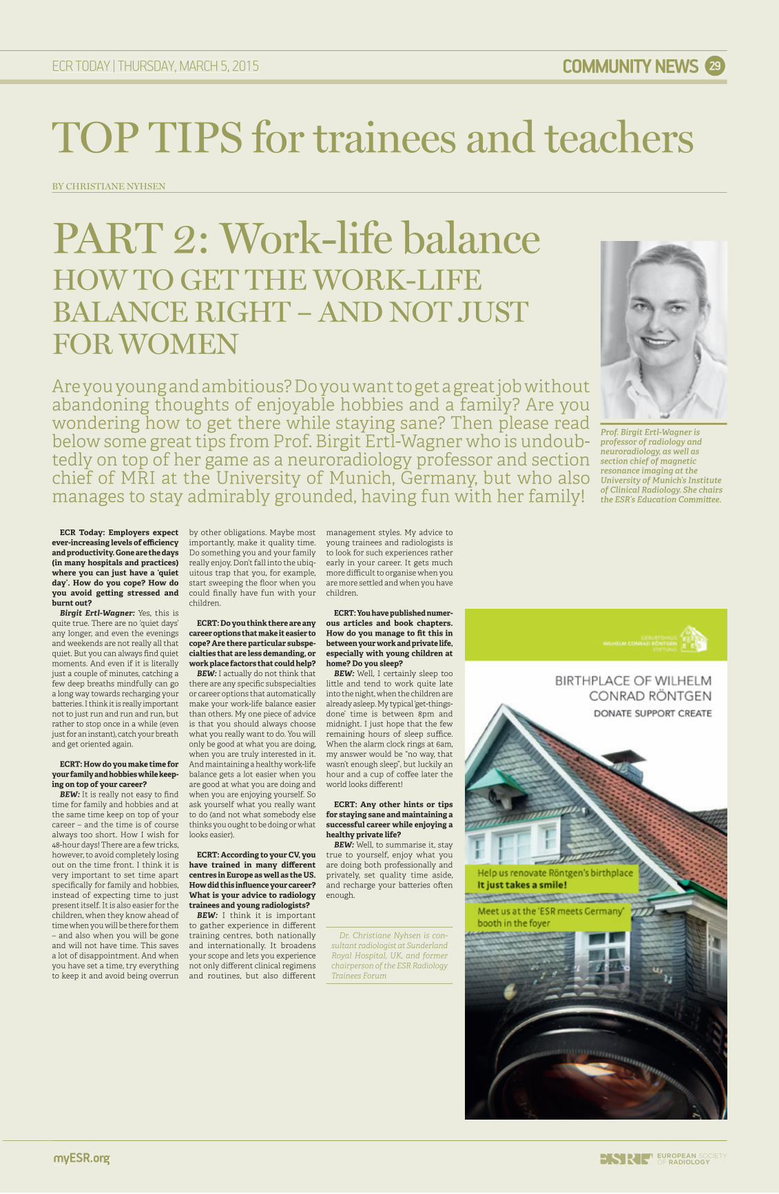
COMMUNITY NEWS
myESR.org
29ECR TODAY | THURSDAY, MARCH 5, 2015 COMMUNITY NEWS
myESR.org
29ECR TODAY | THURSDAY, MARCH 5, 2015
ECR Today: Employers expect ever-increasing levels of efficiency and productivity. Gone are the days (in many hospitals and practices) where you can just have a ‘quiet day’. How do you cope? How do you avoid ge�ing stressed and burnt out?
Birgit Ertl-Wagner: Yes, this is quite true. There are no ‘quiet days’ any longer, and even the evenings and weekends are not really all that quiet. But you can always find quiet moments. And even if it is literally just a couple of minutes, catching a few deep breaths mindfully can go a long way towards recharging your ba�eries. I think it is really important not to just run and run and run, but rather to stop once in a while (even just for an instant), catch your breath and get oriented again.
ECRT: How do you make time for your family and hobbies while keep-ing on top of your career?
BEW: It is really not easy to find time for family and hobbies and at the same time keep on top of your career – and the time is of course always too short. How I wish for 48-hour days! There are a few tricks, however, to avoid completely losing out on the time front. I think it is very important to set time apart specifically for family and hobbies, instead of expecting time to just present itself. It is also easier for the children, when they know ahead of time when you will be there for them – and also when you will be gone and will not have time. This saves a lot of disappointment. And when you have set a time, try everything to keep it and avoid being overrun
by other obligations. Maybe most importantly, make it quality time. Do something you and your family really enjoy. Don’t fall into the ubiq-uitous trap that you, for example, start sweeping the floor when you could finally have fun with your children.
ECRT: Do you think there are any career options that make it easier to cope? Are there particular subspe-cialties that are less demanding, or work place factors that could help?
BEW: I actually do not think that there are any specific subspecialties or career options that automatically make your work-life balance easier than others. My one piece of advice is that you should always choose what you really want to do. You will only be good at what you are doing, when you are truly interested in it. And maintaining a healthy work-life balance gets a lot easier when you are good at what you are doing and when you are enjoying yourself. So ask yourself what you really want to do (and not what somebody else thinks you ought to be doing or what looks easier).
ECRT: According to your CV, you have trained in many different centres in Europe as well as the US. How did this influence your career? What is your advice to radiology trainees and young radiologists?
BEW: I think it is important to gather experience in different training centres, both nationally and internationally. It broadens your scope and lets you experience not only different clinical regimens and routines, but also different
management styles. My advice to young trainees and radiologists is to look for such experiences rather early in your career. It gets much more difficult to organise when you are more se�led and when you have children.
ECRT: You have published numer-ous articles and book chapters. How do you manage to fit this in between your work and private life, especially with young children at home? Do you sleep?
BEW: Well, I certainly sleep too li�le and tend to work quite late into the night, when the children are already asleep. My typical ‘get-things-done’ time is between 8pm and midnight. I just hope that the few remaining hours of sleep suffice. When the alarm clock rings at 6am, my answer would be “no way, that wasn’t enough sleep”, but luckily an hour and a cup of coffee later the world looks different!
ECRT: Any other hints or tips for staying sane and maintaining a successful career while enjoying a healthy private life?
BEW: Well, to summarise it, stay true to yourself, enjoy what you are doing both professionally and privately, set quality time aside, and recharge your ba�eries o�en enough.
Dr. Christiane Nyhsen is con-sultant radiologist at Sunderland Royal Hospital, UK, and former chairperson of the ESR Radiology Trainees Forum
BY CHRISTIANE NYHSEN
Are you young and ambitious? Do you want to get a great job without abandoning thoughts of enjoyable hobbies and a family? Are you wondering how to get there while staying sane? Then please read below some great tips from Prof. Birgit Ertl-Wagner who is undoub-tedly on top of her game as a neuroradiology professor and section chief of MRI at the University of Munich, Germany, but who also manages to stay admirably grounded, having fun with her family!
PART 2: Work-life balanceHOW TO GET THE WORK-LIFE BALANCE RIGHT – AND NOT JUST FOR WOMEN
TOP TIPS for trainees and teachers
Prof. Birgit Ertl-Wagner is professor of radiology and neuroradiology, as well as section chief of magnetic resonance imaging at the University of Munich’s Institute of Clinical Radiology. She chairs the ESR’s Education Commi�ee.
30 ECR TODAY | THURSDAY, MARCH 5, 2015
LANDSCAPE PHOTOGRAPHY TODAY: HAMISH FULTON TO ANDREAS GURSKY
AN EXHIBITION AT THE BANK AUSTRIA KUNSTFORUM
LANDSCAPE IN MY MIND
Sonja Braas, Flood (The Quiet of Dissolution), 2006Courtesy Galerie Tanit, München © Sonja Braas
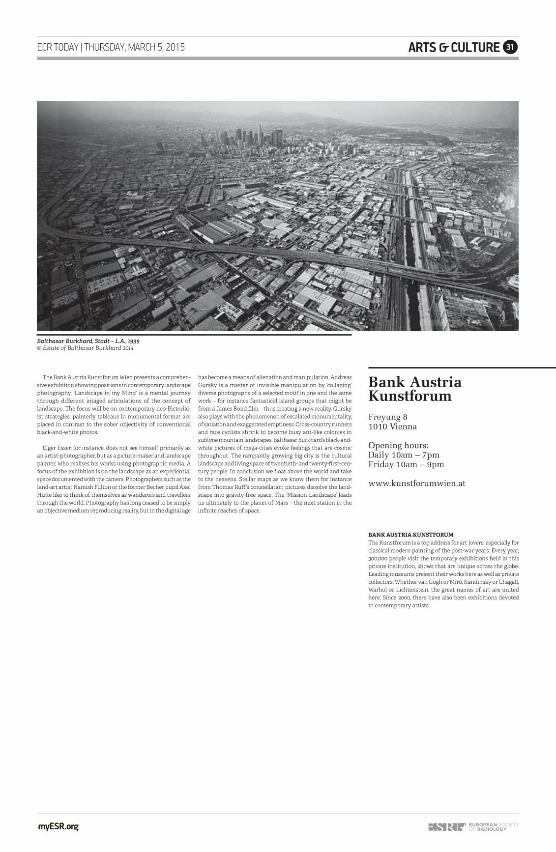
myESR.org
31ECR TODAY | THURSDAY, MARCH 5, 2015
myESR.org
ARTS & CULTURE 31
The Bank Austria Kunstforum Wien presents a comprehen-sive exhibition showing positions in contemporary landscape photography. ‘Landscape in my Mind’ is a mental journey through different imaged articulations of the concept of landscape. The focus will be on contemporary neo-Pictorial-ist strategies: painterly tableaus in monumental format are placed in contrast to the sober objectivity of conventional black-and-white photos.
Elger Esser, for instance, does not see himself primarily as an artist-photographer, but as a picture-maker and landscape painter, who realises his works using photographic media. A focus of the exhibition is on the landscape as an experiential space documented with the camera. Photographers such as the land-art artist Hamish Fulton or the former Becher pupil Axel Hü�e like to think of themselves as wanderers and travellers through the world. Photography has long ceased to be simply an objective medium reproducing reality, but in the digital age
has become a means of alienation and manipulation. Andreas Gursky is a master of invisible manipulation by ‘collaging’ diverse photographs of a selected motif in one and the same work – for instance fantastical island groups that might be from a James Bond film – thus creating a new reality. Gursky also plays with the phenomenon of escalated monumentality, of satiation and exaggerated emptiness. Cross-country runners and race cyclists shrink to become busy ant-like colonies in sublime mountain landscapes. Balthasar Burkhard’s black-and-white pictures of mega-cities evoke feelings that are cosmic throughout. The rampantly growing big city is the cultural landscape and living space of twentieth- and twenty-first-cen-tury people. In conclusion we float above the world and take to the heavens. Stellar maps as we know them for instance from Thomas Ruff’s constellation pictures dissolve the land-scape into gravity-free space. The ‘Mission Landscape’ leads us ultimately to the planet of Mars – the next station in the infinite reaches of space.
Bank Austria KunstforumFreyung 81010 Vienna
Opening hours:Daily 10am – 7pmFriday 10am – 9pm
www.kunstforumwien.at
BANK AUSTRIA KUNSTFORUMThe Kunstforum is a top address for art lovers, especially for classical modern painting of the post-war years. Every year, 300,000 people visit the temporary exhibitions held in this private institution, shows that are unique across the globe. Leading museums present their works here as well as private collectors. Whether van Gogh or Miró, Kandinsky or Chagall, Warhol or Lichtenstein, the great names of art are united here. Since 2000, there have also been exhibitions devoted to contemporary artists.
Balthasar Burkhard, Stadt – L.A., 1999 © Estate of Balthasar Burkhard 2014
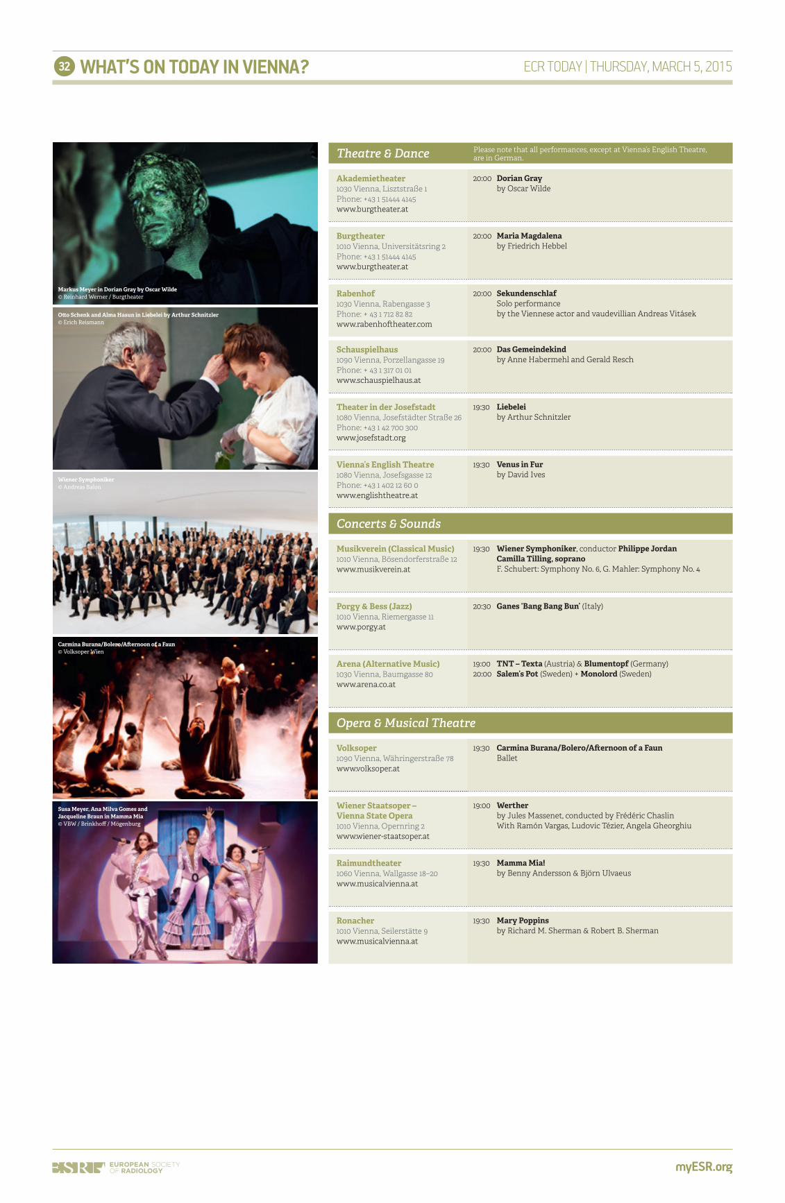
myESR.org
32 ECR TODAY | THURSDAY, MARCH 5, 2015WHAT’S ON TODAY IN VIENNA?
Theatre & Dance
Akademietheater1030 Vienna, Lisztstraße 1Phone: +43 1 51444 4145www.burgtheater.at
20:00 Dorian Gray by Oscar Wilde
Burgtheater1010 Vienna, Universitätsring 2Phone: +43 1 51444 4145www.burgtheater.at
20:00 Maria Magdalena by Friedrich Hebbel
Rabenhof1030 Vienna, Rabengasse 3Phone: + 43 1 712 82 82www.rabenho�heater.com
20:00 Sekundenschlaf Solo performance by the Viennese actor and vaudevillian Andreas Vitásek
Schauspielhaus1090 Vienna, Porzellangasse 19Phone: + 43 1 317 01 01www.schauspielhaus.at
20:00 Das Gemeindekind by Anne Habermehl and Gerald Resch
Theater in der Josefstadt1080 Vienna, Josefstädter Straße 26Phone: +43 1 42 700 300www.josefstadt.org
19:30 Liebelei by Arthur Schnitzler
Vienna’s English Theatre1080 Vienna, Josefsgasse 12Phone: +43 1 402 12 60 0www.englishtheatre.at
19:30 Venus in Fur by David Ives
Concerts & Sounds
Musikverein (Classical Music)1010 Vienna, Bösendorferstraße 12www.musikverein.at
19:30 Wiener Symphoniker, conductor Philippe Jordan Camilla Tilling, soprano F. Schubert: Symphony No. 6, G. Mahler: Symphony No. 4
Porgy & Bess (Jazz)1010 Vienna, Riemergasse 11www.porgy.at
20:30 Ganes ‘Bang Bang Bun’ (Italy)
Arena (Alternative Music)1030 Vienna, Baumgasse 80www.arena.co.at
19:00 TNT – Texta (Austria) & Blumentopf (Germany)20:00 Salem’s Pot (Sweden) + Monolord (Sweden)
Opera & Musical Theatre
Volksoper1090 Vienna, Währingerstraße 78www.volksoper.at
19:30 Carmina Burana/Bolero/Aernoon of a Faun Ballet
Wiener Staatsoper – Vienna State Opera1010 Vienna, Opernring 2www.wiener-staatsoper.at
19:00 Werther by Jules Massenet, conducted by Frédéric Chaslin With Ramón Vargas, Ludovic Tézier, Angela Gheorghiu
Raimundtheater1060 Vienna, Wallgasse 18–20www.musicalvienna.at
19:30 Mamma Mia! by Benny Andersson & Björn Ulvaeus
Ronacher1010 Vienna, Seilerstä�e 9www.musicalvienna.at
19:30 Mary Poppins by Richard M. Sherman & Robert B. Sherman
Markus Meyer in Dorian Gray by Oscar Wilde © Reinhard Werner / Burgtheater
O�o Schenk and Alma Hasun in Liebelei by Arthur Schnitzler © Erich Reismann
Wiener Symphoniker © Andreas Balon
Susa Meyer, Ana Milva Gomes and Jacqueline Braun in Mamma Mia © VBW / Brinkhoff / Mögenburg
Carmina Burana/Bolero/Aernoon of a Faun © Volksoper Wien
Please note that all performances, except at Vienna’s English Theatre, are in German.


