Embed Size (px)
Citation preview

Radiography Observed: An Ethnographic Study Exploring Contemporary Radiographic
Practice.
Mr Christopher M Hayre
PhD - Faculty of Health and Social Care
Canterbury Christ Church University
2015

i
Table of Contents
Table of Contents ........................................................................................................................... i
Acknowledgements ...................................................................................................................... vi
Abstract ........................................................................................................................................ vii
Glossary ...................................................................................................................................... viii
Introduction .................................................................................................................................... 1
Purpose of PhD study ............................................................................................................ 1
Research questions ............................................................................................................... 2
Chapter One: Introducing me as the researcher ................................................................... 4
Chapter Two: Literature Review ............................................................................................ 4
Chapter Three: Methodology ................................................................................................. 5
Chapter Four: Findings .......................................................................................................... 5
Chapter Five: Contemporary issues effecting radiographers learning in the clinical
environment. .......................................................................................................................... 6
Chapter Six: Person-centred approach: The facilitation and hindrances in general
radiography ............................................................................................................................ 6
Chapter Seven: Radiography Observed: Optimising ionising radiation ................................ 7
Limitations of this PhD Research .......................................................................................... 7
Chapter Eight: Conclusions ................................................................................................... 7
Recommendations ................................................................................................................. 8
The Radiographic Environment ............................................................................................. 8
Chapter One: Introducing me as the researcher ..................................................................... 11
1.1. Introduction ................................................................................................................... 11
1.2. Biography: ‘Who I am’ and my values and beliefs ....................................................... 11
1.2.1. Family and illness ........................................................................................ 12
1.2.2. Experiences of education ............................................................................ 14
1.2.3. The X-ray environment ................................................................................ 16
1.3. Conclusion .................................................................................................................... 18

ii
Chapter Two: Literature Review ................................................................................................ 20
2.1. Introduction ................................................................................................................... 20
2.2. Radiobiology: The linear-no threshold debate ............................................................. 20
2.3. Radiation protection: Contemporary principles and methods of dose optimisation .... 27
2.4. Advancing technology: The impact on general radiographic practices ....................... 32
2.5. Person-centred care: Exploring ‘what radiographers do and how they do it’ .............. 39
2.6. Research, workplace and education: Cultural studies facilitating radiographic
practice. ................................................................................................................................ 46
2.7. Conclusion .................................................................................................................... 55
Chapter Three: Methodology ..................................................................................................... 56
3.1. Introduction ................................................................................................................... 56
3.2. Ontological and epistemological viewpoint .................................................................. 56
3.3. Ethnography: Collaboration of research methods ........................................................ 60
3.4. The study ...................................................................................................................... 62
3.4.1. Selection of sites .......................................................................................... 62
3.4.2. Access to the field ........................................................................................ 64
3.4.3. Ethical considerations .................................................................................. 68
3.5. Ethnography .................................................................................................................. 71
3.5.1. Initial data collection .................................................................................... 75
3.5.2. Participant observation ................................................................................ 78
3.5.3. Interviews ..................................................................................................... 83
3.5.4. X-ray experiments ........................................................................................ 89
3.5.5. ‘Ethno-radiographer’: Reflexivity in the field ................................................ 93
3.5.6. Data analysis ............................................................................................. 101
3.6. Leaving the field .......................................................................................................... 103
3.7. Conclusion .................................................................................................................. 104
Chapter Four: Findings ............................................................................................................. 106
4.1. Introduction ................................................................................................................. 106
4.2. Values and Beliefs ...................................................................................................... 106
4.3. Observations and interviews ...................................................................................... 107
4.4. X-ray experiments ....................................................................................................... 108
4.5. Rigour and Validity ...................................................................................................... 114
4.6. Conclusion .................................................................................................................. 119

iii
Chapter Five: Contemporary issues affecting radiographers learning in the clinical
environment. .............................................................................................................................. 121
5.1. Introduction ................................................................................................................. 121
5.2. “On the job”: Learning digital radiography .................................................................. 121
5.3. Digital radiography: Deskilling the profession? .......................................................... 128
5.4. Are radiographers competent practitioners? .............................................................. 134
5.6. Conclusion .................................................................................................................. 140
Chapter Six: Person-centred approach: The facilitation and hindrances in general
radiography ................................................................................................................................ 142
6.1. Introduction ................................................................................................................. 142
6.2. Time Pressures and Culture of the Red Dot: The impact on health care .................. 142
6.3. General radiography: A hospital or industrial environment? ...................................... 151
6.4. Care and Technology: Shoehorning patient care? .................................................... 158
6.6. Conclusion .................................................................................................................. 166
Chapter Seven: Radiography Observed: Optimising ionising radiation ............................ 167
7.1. Introduction ................................................................................................................. 167
7.2. ‘Cranking up’; ‘whacking up’ and ‘opening up’: Dose creep in action ........................ 167
7.3. Art and Science: The impact of digital radiography ................................................... 177
7.4. To ‘Err is Human’: Assessing pitfalls in contemporary radiography .......................... 186
7.5. To protect or not protect?: Lead protection in general radiography ........................... 196
7.6. Conclusion .................................................................................................................. 202
Limitations to this PhD research ............................................................................................. 204
Chapter Eight: Conclusions ..................................................................................................... 206
8.1. Suboptimal learning: Impact on radiographers and students. ................................... 206
8.2. Person-centred care: The impact of digital radiography ............................................ 208
8.3. Optimising ionising radiation: A dichotomy with new technology. ............................ 210
8.4. What does this tell us about digital radiography in contemporary radiographic
practice? ............................................................................................................................. 213
8.5. The value of ethnography as a research methodology: The ‘ethno-radiographer’ .... 215
Recommendations .................................................................................................................... 218
Reflection of the PhD ................................................................................................................ 223
References ................................................................................................................................. 224

iv
Bibliography ............................................................................................................................... 281
Appendices ................................................................................................................................ 295
Appendix 1: The Mammalian Cell ...................................................................................... 295
Appendix 2: Levels of risk for common X-ray examinations ............................................. 296
Appendix 3: Methodological Framework ........................................................................... 297
Appendix 4: Patient Information Sheet .............................................................................. 299
Appendix 5: Departmental Poster ...................................................................................... 301
Appendix 6: Participant Information Sheet ........................................................................ 302
Appendix 7: Participant Consent Form .............................................................................. 305
Appendix 8: Layout of general imaging sites A, B, C & D ................................................. 306
Appendix 9: Initial Observation Data Sheet ....................................................................... 310
Appendix 10: Scanned copy altered data collection ......................................................... 312
Appendix 11: Observational transcription .......................................................................... 313
Appendix 12: Radiographic extremities documented ........................................................ 322
Appendix 13: Interview Schedule (final version) ............................................................... 326
Appendix 14: Example of transcribed semi-structured interview ...................................... 328
Appendix 15: Quality Assurance on Digital Equipment ..................................................... 349
Appendix 16: Qualitative categories and themes .............................................................. 359
Appendix 17: X-ray experiment results .............................................................................. 361
Appendix 18: SPSS output data ........................................................................................ 372
Appendix 19: Variation in Wrist Examinations at Site A .................................................... 377
List of Tables
Table One: Shortcut symbols used in data collection ........................................... 89
Table Two: Features included in observations...................................................... 91
Table Three: Observation record at each hospital site ......................................... 92
Table Four: Participants – Site ‘A’ ......................................................................... 98
Table Five: Participants – Site ‘C’ ......................................................................... 98
Table Six: Digital Radiography Equipment .......................................................... 104
Table Seven: EI value and relationship to DAP .................................................. 105
Table Eight: Prediction of collimation and SID on DAP ..................................... 121
Table Nine: Pearson’s Correlation (DAP and EI) ................................................ 126

v
Table Ten: kVp² Test - Site A .............................................................................. 362
Table Eleven: Tube Output (mAs) – Site A ......................................................... 362
Table Twelve: kVp² Test - Site C ......................................................................... 365
Table Thirteen: Tube Output (mAs) – Site C ....................................................... 365
List of Graphs
Graph One: LNT dose response model ................................................................ 35
Graph Two: DP Hand – Dose effects on SID & Field Size ................................. 123
Graph Three: Oblique Hand – Dose effects on SID & Field Size ....................... 124
Graph Four: DP Wrist – Effects on SID & Field Size .......................................... 125
Graph Five: Lateral Wrist – Dose effects on SID & Field Size ........................... 126
Graph Six: kVp² Test - Site A .............................................................................. 363
Graph Seven: Tube Output Test – Site A ........................................................... 364
Graph Eight: kVp² Test - Site C ........................................................................... 366
Graph Nine: Tube Output Test - Site C .............................................................. 367
List of Figures
Figure One: NHS Trust One .................................................................................. 75
Figure Two: NHS Trust Two .................................................................................. 75
Figure Three: Application to Trust one .................................................................. 78
Figure Four: Application to Trust two .................................................................... 79
Figure Five: Data collection and analysis process ................................................ 86
Figure Six: The positioning of the anthropomorphic phantom during X-ray
experiments .......................................................................................................... 105
Figure Seven: Depiction of culture within the diagnostic radiography
envionments ......................................................................................................... 120

vi
Acknowledgements
There are a number of individuals who have contributed towards the development and
completion of this PhD thesis, these all deserve special thanks and recognition for their
support and encouragement throughout the last three and a half years.
To my supervisors Professor Shane Blackman and Dr Alison Eyden I am continuously
reminded of the unconditional support and guidance you have provided me on this
journey. I am grateful for your valued feedback, motivation and positivity and it has been a
pleasure to work with you both. Other colleagues that deserve considerable thanks are Dr
Hilary Bungay, for her supervisory support in my first year; her guidance and feedback
were instrumental throughout. Similarly I would like to thank Dr Kevin Carlton for his
supervision, valued feedback and keen interested in the topic over the period of this study.
Thank you all!
To my colleagues in the graduate school and others members of staff in a number of
disciplines that have given up their own time for feedback on draft chapters, listening to
any concerns and providing any administrative support. Thank you.
To all research participants and gatekeepers at all research sites, I would like to thank you
all very much for allowing me be part of your day-to-day practices, your unselfish input has
provided a valued insight, which owe great thanks.
Finally I would like to thank and dedicate this thesis to my family, their support and
guidance have been instrumental in the completion of this PhD, from listening to the
endless discussions of my research and support during the time spent doing it.
Thank you all for making this happen!

vii
Abstract
This study explores the day-to-day application of digital radiography (DR) in the X-ray
environment. The focus is on advances in radiographic technology such as cassette and
DR, which can facilitate patient care, reduce processing time and produce superior image
quality. The PhD study presents the voices of the radiographers’ untold views, attitudes
and experiences of DR through the process of observing, listening, retelling and
interpreting junior and senior radiographers’ responses. There were three stages to this
ethnographic study. Firstly, exploring ‘what radiographers did’ in their clinical environment
by observing clinical practices. This provided ‘first-hand’ experience of action-in-process.
Secondly, 22 semi-structured interviews were undertaken, directed by emerging themes
and discussions from the clinical observations. Semi-structured interviews provided an
understanding of the experiences, behaviours and attitudes of radiographers providing a
deeper understanding of the relationship between practice and context. Thirdly, X-ray
experiments were undertaken contributing to ‘what had been seen and said by
participants’. This data was later triangulated to support the research objectives outlined in
this PhD research. Observation and interview data were analysed using thematic analysis
and grouped into four overarching categories; learning, radiographer challenges, ionising
radiation and patient care delivery. X-ray experimental data was inputted into SPSS and
later coded. The qualitative data had numerous codes, which generated themes and
could be linked in order to generate theoretical descriptions. Multiple-linear regression
analysis and Pearson’s Correlation provide statistically significant values (p < 0.001) for
the experimental models contributing to ‘what had been seen and said’ by radiographers in
the clinical environment. This thesis provides new insights into general radiographic
practices using advancing technology. This includes concerns regarding a radiographer’s
clinical training and education, implications surrounding the delivery of patient care and the
suboptimum use of ionising radiation. The conclusion that can be drawn from the empirical
data is that advancing technology has impacted the day-to-day practices of diagnostic
radiographers. Complex phenomena include; current knowledge and understanding, the
practice of keeping doses ‘as low as reasonably practicable’ and impact on patient care
delivery. These insights suggest that healthcare and academic environments may require
additional support in the aim of delivering optimum patient care.

viii
Glossary
Accident and Emergency department (A&E) - A department specialising in acute care
for patients presenting without appointment. A&E departments are generally found in
hospitals or other primary care centre.
Agenda for Change (AfC) – The restructure of the pay and working conditions for NHS
workers putting them onto a single pay spine.
Assistant Practitioner – A member of healthcare staff who assists radiographers and
radiologists with radiographic examinations during their day-today practices.
Cassette Radiography (CR) – Cassette radiography is a radiographic cassette that
houses an imaging plate made of photostimulable phosphor. The image that is taken is
transferred onto the computer system to be viewed.
Computed Tomography (CT) – Computed tomography is an imaging method using X-
rays to produce tomographic images. It can examine body organs and structures that can
be enhanced using contrast agents.
Continuing professional development (CPD) – Healthcare professionals are required to
maintain their competency. CPD allows professionals to do this by attending courses,
reading articles and reflecting on their clinical practice. Each radiographer should have
and maintain a CPD portfolio.
Deterministic effects – In diagnostic radiography ‘deterministic effects’ generally occur
after high-dose acute exposure and thought to arise from the killing of large groups of cells
in the tissues concerned, leading to functional deterioration in the organs affected.
Diagnostic Radiographer – Employ a range of different imaging techniques and
sophisticated equipment to produce high quality images of an injury or disease.

ix
Digital radiography (DR) - DR captures the image of the patient directly onto a flat panel
detector without the use of a cassette. This image is transferred directly onto a computer
system removing the need to leave the patient and process elsewhere.
Ethnography - First pioneered in the field of socio-cultural anthropology to learn and
understand cultural phenomena, which reflects the knowledge and system of means
guiding the life of a cultural group.
Fluoroscopy - An image intensifier that uses X-rays producing a live image feed that is
displayed on a TV screen often used in operating theatres to produce live images during
surgical procedures.
General X-ray – The main part of the imaging department where plain radiographic
images are carried out and the hub for organizing theatre and mobile examinations.
Hospital – Undertake many procedures managing and treating various diseases and
injury and often have accident and emergency departments to deal with immediate and
urgent threats to health.
Health and Care Professions Council (HCPC) - The Health and Care Professions
Council (HPC) is a UK health regulator set up to protect the public. It aims to do this by
setting and maintaining standards of proficiency and conduct for the professions it
regulates. It currently regulates fourteen professions including Diagnostic Radiography.
Imaging Department - A department in a hospital that consists of varying imaging
modalities (X-ray, Computed Tomography (CT), Magnetic Resonance Imaging (MRI),
Ultrasound, Breast Screening and Radio Nuclide Imaging (RNI)).
Imaging modalities – Different methods of imaging the body, for example general
radiography and computed tomography are different imaging modalities.
Inpatient – A patient admitted into hospital that has been allocated a bed.

x
Interprofessional learning (IPL) – Interprofessional learning allows people from different
professional groups to learn with, from and about one another.
Ionising Radiation (Medical Exposure) Regulations 2000 (IR(ME)R 2000) - The
IR(ME)R (2000) came into force on 13th May 2000 to implement the European Directive
97/43/Euratom (The Medical Exposures Directive). These regulations aim to keep doses
‘as low a reasonably practicable’ through correct application by X-ray operators.
Magnetic Resonance Imaging (MRI) – An imaging modality that uses nuclear magnetic
resonance to produce images of the molecules that makes up a substance. MRI is an
alternative imaging modality used in medicine to diagnose disorders of body structures
that may not show using X-rays.
Mammography – An X-ray imaging procedure using low-energy X-rays to examine the
human breast and is used as both a diagnostic and screening tool.
Nuclear Medicine – Nuclear medicine involves the use of radioactive substances in the
diagnosis and treatment of disease.
Out-of-hours – Outside the hours of 9-5 Monday to Friday.
Outpatient – A patient that visits the hospital from outside, normally from their own home,
attending a clinic.
Picture Archiving and Communications System (PACS) – PACS uses a combination of
hardware and software dedicated to the short and long term storage, retrieval,
management, distribution, and presentation of medical images. Electronic images and
reports are transmitted digitally via PACS; this eliminates the need to manually file,
retrieve, or transport film jackets.
Practitioner – A practitioner is someone who is engaged in a specialism including
medicine. Radiographers are often regarded as practitioners and are required in
accordance with IR(ME)R 2000 to justify radiological examinations.

xi
Protocol – Protocols govern the radiological examinations in the clinical environment.
They provide local guidance to individual radiographers and assistant practitioners to
practice in accordance with the employers’ rules.
Radiographic projections – A detailed description of how to position the patient, image
receptor and X-ray tube in order to achieve a diagnostic image.
Radiographic reporting – the writing of a report is performed to identify any findings from
the radiographic images obtained during an imaging procedure.
Radiographic technique - The radiographic technique is delivered to ensure an optimum
radiation level is given to the patient in order to obtain an image of diagnostic quality.
These include the correct amount, energy, distance and collimation in order to produce the
optimum radiographic technique.
Radiologist – Doctors specially educated to utilise an array of imaging technologies to
diagnose or treat disease.
Red dot system – A red dot can be placed on a radiograph by a radiographer to indicate
an abnormality.
Radiotherapy – Uses high levels of radiation doses to treat cancer and other diseases.
Referring clinician (referrer) – A health care practitioner that refers the patient for a
radiographic examination and completes the X-ray request form in accordance with
IR(ME)R 2000.
Reflective practice – Looking (back) at ones practice and learning from it.
Reflexivity – Thinking about oneself, in terms of research this term is used when
considering the role of the researcher in the research.

xii
Society and College of Radiographers (SCoR) - The charitable subsidiary of the Society
of Radiographers. The College's objectives are directed towards education, research and
other activities in support of the science and practice of radiography.
Stochastic effects – Stochastic effects are the main late health effects that are expected
to occur in populations exposed to ionising radiation; somatic risks dominate the overall
estimate of health detriment. For somatic and genetic effects the probability of their
occurrence, but not their severity, is taken to depend on the radiation dose.
Ultrasound – An imaging modality using ultrasonic frequencies to produce images of the
body, commonly used to image pregnant women but can be used to treat and diagnose
other disease.
Viewing area – Part of general radiography whereby staff work from and view their
images. The X-ray rooms are normally joined to this area.
X-ray Operators – An individual that can undertake a radiological exposure under the
supervision of a radiographic practitioner. For example assistant practitioners and
students are X-ray operators that can perform radiological examinations.
X-ray tube - A vacuum tube producing X-rays through thermic emission from the cathode
and accelerated towards the anode. It can be moved by the operators in various positions
to image the patient.

1
Introduction
Purpose of PhD study
The purpose of this PhD was to explore contemporary general radiographic practices
in a general imaging environment. The use of ionising radiation in medicine continues
to increase with X-ray exposures remaining the largest artificial source to human
beings contributing between 10% and 20% (Martin et al, 2009). Current estimates
suggest 4 billion procedures annually worldwide (UNSCEAR, 2010). Since the
discovery of ionising radiation its uses in medicine have significantly increased. Five
yearly reviews continue to monitor the frequency of radiological examinations. From
April 1997 to March 1998 the Health Protection Agency (HPA) (2000) assessed all
types of radiological examinations undertaken in the United Kingdom (UK). The results
identified that 41.5 million medical and dental X-ray examinations were conducted
between 1997-1998, corresponding to 704 examinations per 1000 inhabitants. The
latest results estimated that 46 million medical and dental X-ray examinations were
carried out across the UK, an increase of 10 per cent since 1997 with significant
increases in computed tomography (CT), interventional and mammographic
examinations (HPA, 2011). In the past two decades technological advances have
occurred within the radiography profession. The development of technology in general
radiography has altered the collection of X-ray photons, which produces the
radiographic image. The terminology is traditionally split into two categories, cassette
radiography (CR) and digital radiography (DR) (Kotter and Langer, 2002), which
replaced conventional X-ray film. In short, ‘CR’ uses a photostimulable luminescent
technology to capture X-ray photons, later ‘processed’ into digital data whereas ‘DR’
uses detectors that directly or indirectly convert X-ray photons into digital data using a
thin-film transistor (TFT) array. Throughout this thesis ‘CR’ and ‘DR’ are both used to
identify the separate technologies in the clinical environment. These taxonomies are
important to identify, firstly because of the differences in technological hardware and
secondly the interchangeable use by radiographers, which can impact a patient’s
experience.
Advances in CR and DR were generally accepted to facilitate the reduction of ionising
radiation and improve patient experience (Philips, 2013). However, little research has
explored ‘what radiographers do’ with CR and DR units in the X-ray environment. This

2
is important because current estimates suggest that general radiography (combined
with fluoroscopy) constitute 90% of all radiological examinations undertaken in the
radiology department (Health Protection Agency, 2011). This number is likely to
increase as emergency departments in England recorded 20.5 million attendances
during 2009-2010, with the large majority (19.8 million) being new rather than follow-up
attendances (Hardy and Snaith, 2011). Thus with increasing demands placed upon
general radiological services throughout the UK this PhD study sought to explore the
general radiographic environment and the technology used (CR and DR) examining
the effects of healthcare delivery.
Evidence claims that individual X-ray doses have reduced in practice following the
introduction of advanced technologies (CR and DR) (Herrmann, 2008), yet when
working as a diagnostic radiographer, it was noticed that not all staff (including myself)
understood the technological difference of DR (and CR) because most radiographers
received minimal training as part of their radiographic education (Patefield, 2010).
Carter and Veale (2010) claim that a lack of education in radiographic technology may
produce instances of operator error and increase patient doses, thus the primary focus
of the PhD thesis was to explore the impact of DR during a radiographers day-to-day
practices within the X-ray environment.
Research questions
This PhD research explores contemporary radiographic practices using the research
methodology ‘ethnography’. As a practicing radiographer this research position offered
a significant advantage within the clinical environment to collect detailed research data.
The research questions were as followed:
1) What is the current knowledge and understanding of DR in the clinical
environment?
2) What impact does advancing technology have on the delivery of patient care?
3) How effective is contemporary practice at dose and image optimisation?
4) What does this tell us about DR in contemporary radiographic practice?
The aim was to explore radiographers’ clinical practices using the latest technology
(DR) within the general radiography environment to develop new knowledge for
practice. The first objective of this PhD study aimed to examine government policies,

3
primary sources and research publications relating to DR, radiobiology, radiation
protection and the use of DR in practice. Further research objectives developed
throughout the study based on current literature in chapter two. This resulted in the
exploration of a radiographers’ knowledge and understanding of DR, the impact of
advancing technology (CR and DR) on patient care and an examination into the
optimal delivery of ionising radiation to produce images of diagnostic quality using DR.
The rationale of this PhD study is grounded by the potential hazard patients are
exposed to when undergoing radiological examinations. Ionising radiation may damage
a patient’s health adding to the risk of developing cancer (Hall and Giaccia, 2006). X-
ray examinations differ in ‘additional risk’ ranging from 1 in 1,000,000 to 1 in 1,000 thus
generally accepted that no safe dose exists (ICRP, 2007). Radiographic practices in
the UK are governed by legislation ensuring dose optimisation by keeping doses ‘as
low as reasonably practicable’ (ALARP) (Ionising Radiations (Medical Exposure)
Regulations, 2011). The importance to optimise ionising radiation in contemporary
practices is arguably a problematic one following the introduction of DR (and CR).
Both DR and CR provide an alternative method to capturing X-ray photons.
Conventional X-ray film would illustrate when an inappropriate radiographic exposure
had been used, for example images were ‘too white’ or ‘too black’ when ‘too little’ or
‘too much’ radiation was used resulting in under or overexposure. However the
International Atomic Energy Agency (IAEA) (2010) identify that DR (and CR) may
always provide the operator with a ‘good image’ since it is able to compensate for
wrong settings even if the dose is higher than necessary, thus not indicating whether a
patient has been unnecessarily overexposed. This is discussed later in chapter two
(section 2.4). More recently, it is argued that since operators and observers tend to
favour excellent image quality a patients radiation dose may increase, thus a higher
exposure than normal is selected for a particular examination, referred to as ‘exposure
creep’ or ‘dose creep’ (Seeram, et al, 2013). This strengthens the argument that
standards of radiographic procedures should, therefore, be revisited in order to ensure
the optimal use and application of radiographic practices (Ween, et al, 2009). The
rationale for this PhD study is grounded in the potential dichotomy facing radiographers
in the clinical environment, because while further advances in radiographic technology
(DR) may facilitate dose reduction the International Commission on Radiological
Protection (2005a) argue that without appropriate research and education DR also has
the potential to increase radiation doses. Thus while the application and use of DR
could improve image quality and facilitate dose optimisation, this thesis explores

4
contemporary radiographic practices in order to contribute to existing knowledge and
inform radiographers during their day-to-day practices.
Chapter One: Introducing me as the researcher Chapter one provides an insight into ‘me as the researcher’. In this chapter I examine
my own values and beliefs in relation to the concepts discussed within this PhD thesis.
My reflections reveal that my personal and professional experiences have impacted on
the values and beliefs I hold. Firstly, I discuss the impact of my father’s diagnosis of
throat cancer and how his illness provided an additional viewpoint of the medical
imaging department. Secondly, I explore the importance of mentoring and support
throughout my secondary and tertiary education, justifying the importance of support
and mentoring in radiography learning. Thirdly, I explore ‘self’ as the healthcare
professional and how my own values and beliefs were influence by workplace cultures.
This chapter argues that as individuals we have our own values and beliefs that will
impact on our own professional practices. Because values and beliefs may be
historically laden and culturally constructed it is important to consider ‘values and
beliefs’ as an important part of the construction of this PhD thesis.
Chapter Two: Literature Review In chapter two an assessment of the current radiographic literature is critically
discussed identifying a gap concerning the application and use of radiographic
practices within a DR (and CR) environment. The review identifies little research
exploring ‘how radiographers conduct radiographic examinations’ using DR. Historically
medical practitioners and medical physicists undertook research within radiography,
however as a practicing diagnostic radiographer this chapter discusses the importance
of current knowledge in association with contemporary practices. Radiobiology, which
informs radiation protection measures within the clinical environment are discussed
providing a sound rationale ensuring that ionising radiation remains optimised. Whilst
the introduction of advancing technology (CR and DR) provides the potential for dose
reduction it is argued that innovative technology could be facilitating ‘dose creep’. It
discusses the ‘technological push’ and ‘demand pull’ theory and how this may have
impacted general radiography. Person-centredness is discussed because it is
generally accepted that healthcare professionals should deliver holistic care to
individuals whilst being treated and cared for thus aligning themselves with the
National Health Services core values and beliefs. Workplace culture is discussed

5
because of the potential impact it can have on behaviours and actions of staff. This is
important to consider within diagnostic radiography because alternate actions or
behaviours may facilitate poor practice, which can then become the ‘cultural norm’.
This review strengthens the importance of observing radiographic practice best
answering the research objectives. This informed the research methodology discussed
in chapter three.
Chapter Three: Methodology Chapter three discusses the research methodology undertaken within the radiographic
environment(s). My epistemological and ontological viewpoint is considered describing
my philosophical outlook of the research undertaken and how it supports the
methodology. Ethnography as the research methodology explored a range of practices
associated with DR (and CR) within the radiographic environment. It provided the tools
to explore the local cultures in accordance with the research objectives inductively
using participant observation, observing ‘what radiographers did’. Later semi-
structured interviews were undertaken based on ‘what had been seen’ discussing
topical themes that emerged from the participant observations. X-ray experiments
using DR technology were performed contributing to the overall conclusions adding
relevance to this PhD study and to the field of diagnostic radiography because it could
be mimicked in future research. Within this chapter I discuss my dual role, as the
ethnographer and radiographer termed the ‘ethno-radiographer’ highlighting the
advantages and disadvantages of undertaking research within one’s own discipline.
Throughout the methodology I discuss the importance of reflexivity both in and out of
the research environment and how this facilitated my research position and data
collection.
Chapter Four: Findings This chapter presents the research findings collected throughout the PhD research. It
portrays the cultures of radiographic practices using advanced technology. Central to
the findings were the values and beliefs of radiographers. This core variable suggests
that the values and beliefs of radiographers were not unified within the clinical
environment, which impact on actions and behaviours of radiographers. X-ray
experiments and statistical data are illustrated contributing to the overall findings.
Statistical analyses were performed to predict increases and decreases to dose area
product and correlations between dose area product and exposure indexes. The

6
empirical findings were triangulated to provide a holistic picture of the clinical
environment, which is argued to enable professional reflexivity. Rigour and validity of
the empirical data uncovered is critically discussed ensuring ‘trustworthiness’ of the
data later portrayed in empirical chapters.
Chapter Five: Contemporary issues effecting radiographers learning in the clinical environment. Chapter five examines the contemporary issues surrounding radiographers’ knowledge
and understanding using advancing technology. It highlights educational issues within
higher education and within the clinical department and how this can be cascaded to
student radiographers, hindering their clinical development. An important finding was
that radiographers were often required to ‘learn on the job’, receiving an inappropriate
level of knowledge and understanding, arguably questioning their own professional
competency. This chapter seeks to argue that advanced technologies could be
‘deskilling’ the radiography profession whereby radiographers may ‘know how’ to take
an X-rays, but fail to ‘understand how’ it has been acquired thus failing to perform
general radiographic examinations optimally.
Chapter Six: Person-centred approach: The facilitation and hindrances in general radiography Chapter six discusses the facilitation and hindrances of patient care within the
radiographic environment. The chapter examines how advancing technology can
impact radiographic practice by alleviating time pressures in the clinical environment.
On the one hand this chapter highlights that patients may be pleased with the ‘speed’
of their radiological examinations within the clinical environment, yet on the other hand
DR raises professional challenges for some radiographers with anxious patients
seeking an immediate diagnosis due to the instant image display. The empirical data
from the participants suggests that DR environment may resemble that of an industrial
environment with radiographers concerned about the time pressures and the
importance DR plays in this process, resulting in a lack of person-centredness amongst
radiographers. This chapter provides a comparative insight into the dual use of CR
and DR technology, which can enhance the patients experience during radiographic
examinations.

7
Chapter Seven: Radiography Observed: Optimising ionising radiation Chapter seven examines current issues impacting the optimisation of ionising radiation
within the clinical environment. It highlights the actions and behaviours of
radiographers that are attributing to the known phenomenon ‘dose creep’. There is a
discussion surrounding radiography as an ‘art and science’, which suggests a review of
the terms used in diagnostic radiography and how it may help combat ‘dose creep’
through critical reflection. This chapter suggests that the use of DR may facilitate new
radiographic errors, and near misses in comparison with CR. Additionally, a subculture
of ‘radiological myths’ may be attributing to an increase in patient dose but until now
were kept ‘hidden’ within the clinical environment.
Limitations to this PhD Research The limitations of the PhD work are outlined in this section of the thesis. It highlights
some important weaknesses inherent throughout this PhD research and thus important
for readers and prospective researchers to consider. A central component of this PhD
research was the researcher in the clinical field. Experiences are discussed that
arguably impacted both researcher and participants upon data collection. The
limitations provide an original illustration of the data collection methods employed
across the multi-sites explored in this PhD work.
Chapter Eight: Conclusions This concluding chapter evaluates the main findings of the PhD study undertaken. The
empirical evidence is discussed and concluded in three broad areas, radiographer
learning, delivery of patient care and the optimisation of ionising radiation. Each
section aims to provide sound conclusions based on the empirical evidence derived
from the research methodology. The impact of the latest innovative technology (DR) is
discussed because although advancing technologies are generally accepted to
facilitate healthcare, it is argued that this may be hindering the delivery of radiographic
practice. The value of ethnography as a research methodology is discussed
highlighting how it may facilitate future radiographic research and research in other
health disciplines. The discussions in this concluding chapter provide an original
outlook of radiographic practice in contemporary radiographic healthcare contributing
to existing knowledge with the aim of delivering optimum patient care.
8
Recommendations
Recommendations are outlined in this section following the PhD research conclusions.
The recommendations are directly linked to the findings uncovered in this thesis and
are offered to radiology managers, educators/researchers and diagnostic
radiographers.
The Radiographic Environment
Diagnostic radiographers provide a wide range of imaging procedures to patients,
including general imaging, computed tomography (CT), dual energy X-ray
absorptiometry (DEXA) bone density, mammography, magnetic resonance image
(MRI) scanning, radioisotope scanning and ultrasound imaging, each employing a
range of techniques providing images of diagnostic quality. This PhD study explores
general radiographic practices in the clinical environment. The development of CR and
DR technology in general radiography has provided additional security, less moving
and handling of X-ray films and enhanced image quality (Carter and Adler, 2013).
Historically the ‘processing’ of X-ray film required staff to ‘wash’, ‘fix’, and ‘dry’ films
prior to sending it to the referring clinicians for diagnosis, yet today CR and DR
technologies process and display digital images to physicians in several minutes.
Whilst this reaffirms the technological importance for patients and staff within the
National Health Service (NHS) (NHS, 2013) the primary focuses in this PhD research is
exploring DR, the latest technological advancement within the general radiographic
environment.
X-rays are a form of ionising radiation used in diagnostic radiography that have short
wavelengths (beyond ultraviolet), passing through matter to varying degrees depending
on the density (Graham and Cloke, 2006). X-rays branch into the ‘natural sciences’
and in radiography are produced by an X-ray tube accelerating electrons towards a
tungsten anode target. On impact with the target they undergo sudden deceleration to
produce X-rays and heat and pass through the patient providing visual representations
of their internal structures (Graham and Cloke, 2006). The NHS continuously strives to
‘work at the limit of science – bringing the highest levels of human knowledge and skill
to save lives and improve health’ (NHS Constitution, 2013:2). Advancing technology
and the production of X-rays remain central to a radiographers practice and predicted

9
to continue to act as a significant aid in diagnosis management for all medical
specialties (NHS, 2013). Radiographers work to professional and ethical codes of
conduct, setting out and underpinning values and principles to promote, maintain and
disseminate the highest quality of care to patients (SCoR, 2010, SCoR, 2013), yet
these values and principles are debated in later chapters of this PhD thesis. In a
recent publication Whitaker (2013:1) questions ‘who are diagnostic radiographers’,
fearing that radiographers will continue to be misunderstood, with the profession failing
to reach its full potential. The Society and College of Radiographers (SCoR) (2013:1)
assert that the best radiography students ‘have a balance between good understanding
of the sciences and a genuinely caring attitude’ suggesting sound collaboration
between scientific understandings and an empathic attitude towards patients. The
advancement of technology however within diagnostic radiography provides an
additional sociological interest discussed throughout this PhD thesis. The sociologist
Harry Braverman (1974:319) argued that maximising technological control can
minimise the autonomy of workers:
‘The more science is incorporated into technology, the less science the worker possesses; and the more machinery that has been developed as an aid to labour, the more labour becomes a servant of machinery’.
The extent to which autonomy is minimised for radiographers and the extent in which it
impacts clinical practice and patient care was a primary focus throughout this PhD
thesis and discussed in chapters five, six and seven. This required close engagement
with radiographers exploring ‘what they did and how they did it’. Subject matters
generally differ between ‘science’ and ‘sociology’, the former studying ‘nature’ and the
latter studying ‘people and social groups’ (Gabe et al, 2006). Medicine has historically
relied on science to inform medical practices (Gabe et al, 2006), however medical
sociology can produce knowledge exploring the actions and interactions of healthcare
professionals and the social or cultural effects of medical practice (Bird et al 2000;
Scambler, 2008). Thus it was important throughout this thesis to consider both ‘nature’
and ‘people’ following recent failures at the Mid Staffordshire Hospital (2014),
suggesting a change in healthcare culture: ‘the failure of the system shown in this
report suggests a fundamental culture change is needed’ (Francis, 2013:5). Hospitals
are regarded as institutions providing specialist medical care and services for the
general public. Cockerham (1978:201) reported that hospitals have passed through
four distinct phases of development: ‘centres of religious practice; as poorhouses; as
deathhouses and as centres of medical technology’. Radiology is a medical specialty

10
aiming to facilitate an improvement in health using imaging technology to diagnose and
treat disease and regarded as one of the ‘most expanding specialties’ in recent years
(NHS, 2013:1). In short the conjecture in this PhD study explores phenomena at the
intersection of both social and radiographic sciences in the general radiography
environment with the aim to add to existing knowledge enhancing patient care delivery.

11
Chapter One: Introducing me as the researcher “Craftsmanship is the centre of yourself and you are personally involved in every intellectual product upon which you may work. To say that you ‘have experience’ means, for one thing, that your past plays into and affects your present, and that defines your capacity for future experience.”
(Wright Mills [1959] 2000:196 cited in Howatson-Jones, 2010:16).
1.1. Introduction The aim of this chapter is to explore the development of my own human life and
journey of becoming a diagnostic radiographer and educator in higher education. It is
important to consider because ‘who I am’ and ‘what I bring’ to this PhD research is
arguably constructed by my own experiences. This chapter will discuss some personal
and professional experiences that impact on my own values and beliefs and how they
relate to the concepts discussed throughout this PhD work. Howatson-Jones
(2010:16), in her PhD thesis reflects on her own life story in describing her experiences
and the journey of her PhD research, interrogating what she brought to it in order to
avoid taking her own ‘data’ for granted. This is important to consider because my
values and beliefs throughout this PhD will inform my relationships with participants,
colleagues and patients.
1.2. Biography: ‘Who I am’ and my values and beliefs It is generally accepted that as human beings we have our own values and beliefs,
which we develop throughout the course of our lives. Additionally, our family, friends,
community and experiences contribute to our sense of ‘who we are’ and ‘how we view
the world’ (Manley et al, 2011). Prior to undertaking a critical literature review and
discuss the research methodology I will consider ‘who I am’ as the researcher and the
values and beliefs I hold in relation to the concepts discussed throughout this PhD
research. This is important for two reasons. Firstly, it can help identify to readers my
position as the researcher in the context of the data. Secondly, it can help strengthen
rationales for the concepts discussed throughout this PhD work using ‘values and
beliefs’ as a core variable. Savin-Baden and Major (2013:68) assert that ‘qualitative
researchers should know and be able to articulate who they are and what they believe

12
personally, so that they may understand and acknowledge how these factors in turn
influence the research’. I am currently employed in a higher education institution
delivering radiography education. I also maintain clinical competencies by undertaking
‘bank radiography’ shifts. Upon embarking onto the PhD studentship program at
Canterbury Christ Church University (2010) I had one year radiographic experience
and looking back I remained both the novice researcher and radiographer. The person
‘I was then’ is arguably not the same person ‘I am now’ and following life experiences
these have impacted on the values and beliefs I hold. Savin-Baden and Major (2013)
maintain that ‘personal stance’ is not static and tends to change, move and grow as
people’s views about life, culture and identity shift. Whilst my personal values have
been ‘constructed’ it is important to discuss these in detail because they are closely
connected to ‘knowing self’ and based on the assumption that ‘before we can help
others we need to have insight into how we function as a person’ (McCormack and
McCance, 2006:475). It is argued that the ‘values and beliefs’ of a researcher are
central to a ‘researcher’s position’ in the context of his/her research because beliefs
and values affect the way research is designed, planned, undertaken and written up
(Savin-Baden and Major, 2013:82). Contemplating one’s values and beliefs involves
questions surrounding in what ways education and life experiences have influenced the
way I think about my research (Howatson-Jones, 2010; ibid). Because this can help
interrogate my deeply held beliefs about undertaking this PhD research it will be
discussed in relation to the broad concepts discussed throughout this PhD thesis.
1.2.1. Family and illness My sister and I were raised in a working class home by my father and mother. My
father (a builder) and mother (shop assistant) did what many parents do, support,
encourage and prepare their children for adulthood. Historically we spent many days
together as a family and this is something I am continuously grateful for. As a family
our lives were impacted dramatically following my father’s diagnosis of cancer in June
2014. My father was diagnosed with a squamous cell carcinoma in the throat (primary
source of cancer remained unknown). The news had emotional effects on our family,
unsure of his prognosis. Because of my experiences as a diagnostic radiographer I
was often asked by my mother and father to accompany them during outpatient
appointments and attend radiotherapy sessions in order to clarify terminology and ask
‘anything that came to mind’. Throughout his appointments I often observed the
‘person-centred’ and ‘family-centred’ approaches often cited in nursing literature
(McCormack et al, 2010). On occasions we were consulted as ‘a family’ because we

13
were told that we would all play a ‘big part’ in his journey of treatment, care and
recovery. Throughout his radiotherapy and chemotherapy treatment I began to
understand the complexity and ramifications of cancer treatment and the impact it can
have on family members. My mother, a shop assistant now became my father’s full
time carer, administering palliative drugs when appropriate whilst managing his day-to-
day concerns. As a diagnostic radiographer my experiences with patients are often
short when compared to other health professions, limiting my experiences of patient’s
treatment and recovery. However after experiencing first hand cancer treatment and
management this altered my perspective of ‘the patient’, and the family members who
remain unselfishly immersed in their day-to-day care. Additionally, this altered my
perspective of radiological and magnetic resonance examinations following
observations and later discussions with my father after his computed radiography (CT)
and magnetic resonance image (MRI) scans. The skills developed throughout data
collection of this PhD research helped me question the experiences my father
underwent. These were both insightful as a family member and as a diagnostic
radiographer. Firstly the experiences of the radiological contrast injected intravenously
during his CT scan gave him an intense ‘warm feeling’ throughout his body, which he
mentioned he had not been informed of and thus remained confused by the biological
sensation occurring after his injection. In support, the speed of ‘the scan’ remained a
surprise to him. He felt that the time of the scan (five minutes) did not reflect the
‘waiting time’ to underdo the examination (approximately two weeks) thus questioned
why it ‘takes so long to fit everybody in’. Secondly, during his MRI examination he was
required to place his head centrally in the magnet. This resulted in him lying flat in the
bore of the magnetic for an approximate time of 45 minutes, feeling increasingly
claustrophobic. Additionally, he mentioned the important part I played being allowed to
sit within the ‘Faraday cage’ (inside MRI room) during his MRI scan, enabling him to
focus on a ‘subject’ taking his mind off the uncomfortable and claustrophobic
environment. At this stage I was observing radiographic healthcare from ‘the other side’
and after conversations with my father it allowed me to critically reflect on what we do
as diagnostic radiographers and how our actions and communications can impact on
the experiences of the patient. In relation to the concepts discussed in this thesis this
strengthens the argument that sound communication, treatment and care of patients
(and family members) should be considered for all examinations because patients may
feel increasingly anxious about their imaging procedure(s). Like my father, patients
may be undergoing scans to explore the ‘extent’ of their illness or pathology. In short
my father’s journey and my experiences as a family member have allowed me to think
more holistically about patients and their family members entering the imaging

14
environment. Patients, like my father are likely to experience a wide range of emotions
and concerns in attempts to uncover their suspected pathology. Because cross
sectional imaging is central to early diagnosis and staging of cancerous tissues my
experiences as a close family member will impact on my communication with patients.
It has encouraged me to listen more carefully to the concerns of patients and the
concerns of family members. The ‘family centredness’ experienced has highlighted the
importance of involving family members in the care and treatment throughout imaging
procedures I undertake because family members often play an central role in the care
and management of patients arriving to radiology departments.
1.2.2. Experiences of education My experiences of education significantly contribute to the values and beliefs I hold as
a radiographer and educator. Prior to commencing my secondary education I was
required to undertake an academic examination, commonly known as the ’11 plus
exam’ in the south east of England. This examination would assess my academic level
at eleven years of age, leading me to undergo either ‘grammar’ or ‘independent’
education at a selected secondary school, the former superior to the latter. My failure
of the ’11 plus’ exam was a disappointing outcome for me (and my family) and
therefore required me to attend an ‘independent school’. This was disappointing
because it was generally accepted that grammar school pupils were more successful
than those from in from independent schools in England in getting into higher
education (BBC News, 2005). However, on reflection my experiences within the
independent school were important to my own values and beliefs. Two experiences
stand out. The first was my mathematics teacher, who commenced employment during
key stage level four. He immediately assured us that we could achieve a ‘C’ at GCSE
prior to entering the final year of key stage level four. The second involved my history
teacher who introduced me to the importance of reading and writing, providing detailed
feedback of my GCSE assignment, personally mentoring my progress and ensuring I
succeeded in producing a sound piece of GCSE work. Retrospectively I achieved a C
one year earlier than anticipated in mathematics and achieved a B grade at GCSE in
history. Looking back I now realise the values and beliefs of the individual teachers
because of their desire to teach and develop students instilling a philosophy that ‘every
student who enters my classroom can succeed’ (Buyck, 2003:1). This was achieved by
firstly offering extra curricula activities such as ‘Saturday morning workshops’ and ‘after
school clubs’ in the aim of providing success to the students involved in acquiring

15
sound GCSE grades. Secondly, their constructivist beliefs created a stimulating,
challenging, and individually adapted learning environment, supporting my construction
of knowledge by means of one-to-one tuition, group discussion, and graphical
demonstrations. Not only did this reveal that there were different methods of learning
that achieved my aims it suggests the practices delivered by each teacher were
‘shaped by their pedagogical and cultural traditions’ (OECD, 2009:93).
After completing my secondary education I moved onto a foundation college to begin
my National Vocation Qualification NVQ (level 2) and Higher National Certificate (HNC)
studies in computing. This was part of an apprenticeship undertaken within a multi-
national pharmaceutical company. This environment was new to me; my ‘working role’
involved supporting chemists in research laboratories with any ‘IT issues’ encountered.
I began studying for my NVQ and HNC within a two year period, the latter more
academically challenging. During my apprenticeship I often stood out from others
because I was undertaking two academic qualifications, whereas my peers were
undertaking the mandatory NVQ qualification, essential in any apprenticeship. At times
this was challenging, encompassing both academic and work duties in order to fulfill
the apprenticeship. Initially due to the level of the HNC I would often struggle with
modules such as ‘computer programming’ and ‘networking systems’ as I had little
knowledge or understanding of these areas at sixteen years of age. Additionally, the
pedagogy was teacher-centred with knowledge delivered by means of lectures,
requiring self-study on subject areas. At sixteen years of age this was my first
experience of self-directed study. I had little connection with this new learning
environment whereby I was required to ‘go away and learn subject material and
feedback’. This required self-discipline that I was unfamiliar with. On reflection without
the sound mentoring and support from my apprenticeship supervisor and tutors at the
college I may not have passed HNC with success. Howatson-Jones (2010:19) thesis
similarly reported difficulties with learning in a new environment and how it caused her
‘work to suffer’ leading her to leave her course of study. Without the support of key
members during this apprenticeship my learning would have been hindered with the
possibility of course failure, thus preventing my entry into higher education. Looking
back my experiences of both secondary and tertiary education demonstrate two central
tenets. Firstly, depending on our levels of knowledge and understanding individuals
may require additional support and mentoring regarding aspects of professional work.
Secondly, as an educator my aim is to instill the philosophy experienced by my
previous teachers, tutors and mentors that in my view have allowed me to develop and
facilitated my achievements in the field of diagnostic radiography. Because we all have

16
differing levels of knowledge and understanding it is important to consider those who
may require additional support and mentoring and develop individuals in a supportive
educational culture.
1.2.3. The X-ray environment
Upon embarking into a career as a diagnostic radiographer I had no previous
experiences working in a caring environment. At twenty-one years of age and as a
student radiographer I began to observe vulnerable and ill patients following instances
of direct trauma and progressive pathological diseases. My desire to become a
radiographer was based on two perspectives. Firstly, I wanted to care for people and
secondly optimise technology and science to facilitate this. The person centred nursing
framework advocated by McCormack and McCance (2010) claims that clarity of values
and beliefs are important to consider for healthcare professionals working within their
professional environments. Manley (2004:55) maintains that ‘values determine what
people think ought to be done’ and are closely linked with moral and ethical codes,
whereas beliefs are ‘what people think is true or not true’ (ibid:55). As a radiography
student and recent graduate I felt that whilst working as part of a healthcare team we
often treated patients holistically delivering the optimum amount of ionising radiation
and producing images of diagnostic quality. These basic assumptions involve the
interpretation of beliefs plus values and emotions and are generally understood as
accepted truths that are held unconsciously (Brown, 1998). However, McCormack and
McCance (2010:54) argue that one cannot question the values and beliefs
underpinning my own assumptions and thus one should observe ‘them in action’. The
link between clarifying values and beliefs and workplace culture is described by Manley
(2004:54):
“Values and beliefs contribute to shared meanings, understandings and expectations which are tactic and distinctive to a particular group and passed on to new members… they underpin the way things are done within any cultural focus.”
Dominant values are those that are widely shared amongst a group, community or
culture. In a more recent publication, Manley et al (2011:1) asserts that culture is
influential in delivering care that is person-centred, clinically effective and continually
improving in response to a changing context. McCormack and McCance (2010:55)

17
provide an example whereby nursing staff found themselves in task-orientated ways
whereby the ward became focused on the busyness of tasks expected to be completed
by the end of each shift, thus becoming the ‘cultural norm’. Throughout my
radiographic learning I became immersed within one professional group. Further, upon
graduating I was employed within this professional group whereby I continued my
clinical practiced as a diagnostic radiographer prior to embarking on the PhD
studentship. A recent study by the HCPC (2014:14) reported that professionalism is
based on well-established, or even innate personal qualities and values and beliefs, as
identified by participants:
“To me, people’s values underpin everything they do as a professional… and so, from my point of view, professionalism has come from before I even entered the profession.”
“I think you have a core belief as well, it’s your core standards of what you think is acceptable and not acceptable”.
Whilst I believed I was confirming to ‘professionalism’ as described above looking back
I was unaware of the cultural influences impacting my clinical practice. As maintained
by Manley et al (2011) our values and beliefs are interconnected with the workplace
culture and form our behaviours and attitudes. This was important following the
implementation and delivery of ionising radiation. On reflection, my observations as a
diagnostic radiographer questioned the use of ionising radiation and application of
person-centred care in the clinical environment following advances in technology. It is
reported that following the introduction of CR and DR, not all radiographers (including
myself) fully understood the advances of this technological change (Patefield, 2010).
Looking back, I felt that I lacked optimum knowledge and/or understanding to operate
DR equipment effectively as a diagnostic radiographer. Thus throughout my day-to-
day practices I, like others discussed in later chapters would conform to practices that
were suboptimum, leading to increases of ionising radiation. My ‘personal stance’ was
central to embarking on this PhD topic within the general radiography environment
because it aimed to uncover and clarify my concerns as a healthcare professional
(Savin-Baden and Major, 2013). At the time this may have been associated with
becoming part of the ‘cultural norm’ and throughout this PhD journey I reflect on the
relevance of cultural influences on my ‘personal stance‘ and how it impacted on my
own dose optimisation practices to patients. As healthcare professionals we should not

18
impose our own values and beliefs on patients, we should work with patients in relation
to what is right for them. As autonomous professionals we should exercise sound
judgment and decision making through a complex process of assessment and action
involving the interaction of knowledge, experience, values and practical skills. This
remains a central focus throughout this PhD research because failure to become
autonomous arguably neglects ‘what is right’ for the patient. Since embarking on the
PhD in 2010, my values and beliefs on the use of technology in accordance with
ionising radiation have developed as part of ‘my own professional journey’ (West,
2001). Radiographers should remain autonomous regardless of advancing technology
ensuring that ionising radiation is kept in accordance with the theoretical linear-no
threshold dose response model (ICRP, 2012). At present I continue to work as a
senior radiographer, research within my own profession and educate at higher
education, which continues to impact on my own radiographic practice. My focus
throughout this PhD work reinforces the importance of reflecting in and on practice and
also ‘on self’ because it has the potential to uncover strongly held personal and
professional values and beliefs and can enhance understanding of our own actions and
the influences of workplace culture. Central to my own personal and professional
‘enlightenment’, it is essential to highlight the actions and behaviours of radiographers’
and their values and beliefs in the field of diagnostic radiography. This consideration is
essential because challenges often arise when espoused values (values we talk about)
do not match the behaviours we see in practice.
1.3. Conclusion This chapter sought to clarify ‘who I am’ as the researcher and my values and beliefs
held. I have discussed experiences in my life that have shaped my own values and
beliefs towards patient care, the value of support and mentoring students in education
and the impact on optimum use of ionising radiation. Exploring my own values and
beliefs were central to my actions and behaviours as a diagnostic radiographer and
researcher. This processes has enabled me to critically self-reflect on my life
experiences and understand the impact it has on ‘who I am’. I have discussed the
development of my own values and beliefs following personal experiences and the
‘journey’ of embarking onto the PhD study and becoming an educator. Thus whilst the
actions and behaviours of individuals are documented in this thesis it arguably
strengthens the rationale that values and beliefs of individuals are not ‘set in stone’ and
can develop through self-reflection. It is important to consider that if radiographers
reflect ‘auto/biographically’ this could promote a more holistic approach to
19
radiographers actions thus having a positive impact on person-centered delivery.
Additionally, the important interconnection between culture and values and beliefs
supports the rationale that in order to understand the values and beliefs of
radiographers an in-depth understanding of the radiographic culture is required.

20
Chapter Two: Literature Review
2.1. Introduction
This chapter aims to review literature of radiobiological evidence, current radiation
protection measures, advancements in imaging technology, patient care delivery and
research and education in radiography. The review will discuss literature currently
informing and legislating radiographic practice in the United Kingdom (UK). Further,
this chapter identifies problematic issues surrounding contemporary general
radiographic practices thus identifying a gap to advance existing knowledge that can
inform the radiographic profession.
2.2. Radiobiology: The linear-no threshold debate All life has a cellular basis, if a man weighs 70kg his body will consist of approximately
7 x 10¹³ cells, which contain structures that are defined as the ‘the building blocks of
life’ and the basic unit of reproduction (Becker et al, 2009). Cells in tissues may
undergo one of three processes: they can divide and grow in number, they can stay
alive without dividing, and they can die by apoptosis (programmed cell death) or
necrosis. These can occur at different rates, higher in children, which slow when we
get older (Becker et al, 2009). These make children more sensitive to radiobiological
effects as they have a greater number of dividing cells and thus potential effects are
increased later in life (Schonfeld et al, 2011; Seeram et al, 2013). An illustration and
description of the mammalian cell can be found in appendix one (p.295) for the reader
illustrating the cells common features and internal structures. Ionising radiation is
radiation composed of particles that carry enough kinetic energy to typically remove
electrons, protons or neutrons from an atom or molecule. Such events can alter
chemical bonds and are considered the most damaging forms of radiation due to their
high frequency (Graham and Cloke, 2006). When an X-ray beam passes through a
patient they receive a dose of ionising radiation, absorbed dose is a measure of the
amount of energy from ionising radiation deposited in a mass of some material (i.e. a
patient). The unit of absorbed dose is ‘Gray’ (Gy) which is defined as a dose of one
joule per kilogram (Jkg-1) (Graham and Cloke, 2006). Dose Area Product (DAP) is a
multiplication of the dose and the area exposed, expressed in Gycm2 and recorded
after general radiographic exposures as X-ray units are fitted with DAP meters thus the

21
most convenient method of monitoring patient doses. The ‘effective dose’ is measured
in Sieverts (Sv), introduced by the ICRP (1991) because different organs in the human
body have different sensitivities to radiation and although all tissues are radiosensitive;
some are more sensitive than others (ICRP, 1992) e.g. breast tissue is recognised
more sensitive to the gonads (ICRP, 2012). Radiographic examinations such as
‘hands’ and ‘wrists’ are defined as ‘negligible dose’ in comparison with other
radiological exams and give an effective dose of less than 0.1 millisievert (mSv). This
is an accumulation of other types of background radiation a person receives over their
lifetime from sources including the sun, outer space, naturally radioactive materials
present in the earth, buildings, the food and water we consume and radioactive gases
present in the air equating to on average 2.7 mSv per year (Graham and Cloke, 2006).
These natural background sources provide patients and healthcare professionals a
benchmark to explain the risks associated with certain radiological examinations
(Calman, 1996) because life on earth has evolved amongst this continuous exposure
to background radiation. Comparisons are available to the reader in appendix two
(p.296). Ionising radiation exposure is linked with popular fictional comic book
characters including the Hulk, Spider Man and Teenage Mutant Nina Turtles; however
ionising radiation can alter chemical bonds and are considered the most damaging
forms of radiation due to their high frequency (Graham and Cloke, 2006). Radiobiology
is the field of science that studies the effects of ionising radiation on cells, generally
classified into two categories, deterministic and stochastic.
Deterministic effects: Deterministic effects occur following high doses of ionising
radiation due to the killing and malfunction of biological cells (Hall and Giaccia, 2006).
Some medical procedures exploit the hazardous effects of ionising radiation with
radiotherapy successfully contributing to 28% of the lives saved to cancer when
compared to other treatments (Chemotherapy 4% and Surgery 68%), saving 5000 lives
annually in England (Cooper, 2011). Deterministic effects are generalised by a
threshold dose below which no detrimental effects are seen. Muller (1927) first
demonstrated this measuring mutation rates in the Drosophila fruit fly using X-rays to
demonstrate a dose-response relationship. Examples of deterministic effects in
humans include cataract of the lens, cell depletion in the bone marrow leading to
haematological deficiencies and impairment of fertility (Nias, 1998; Hall and Giaccia,
2006). Radiological procedures have demonstrated deterministic effects on patients,
ranging from visually noticeable reddening of the skin at 2Gy, to painful deep necrotic
ulcers at 18Gy (Vlietstra, 2004). The objectives outlined by the ICRP (2012) are in

22
general to prevent deterministic effects in humans and to restrict stochastic risks to
acceptable levels.
Stochastic effects: Radiation-induced cancer, cognitive decline and heart disease are
examples of stochastic effects. Stochastic effects are demonstrated in forms of non-
ionising radiation, for example infrared radiation from an electric radiator may be
beneficial but too much heat will burn. Similarly exposure to ultraviolet (UV) provides a
valued source of vitamin D, however it is a known carcinogen and through ‘exciting’
orbital electrons overexposure to UV can result in skin damaged (Tortora and
Grabowski, 2003), increasing the chances of developing certain forms of skin cancer
by inducing gene mutations through deoxyribonucleic acid (DNA) damage (BAD, 2008;
SCoR, 2010; NICE, 2011). In the UK more than 1 in 3 people will develop some form
of cancer during their lifetime and since the mid-1970s cancer incidence rates in the
UK have risen by 22% in males and 42% in females (Sasieni et al, 2011). Regardless
of the underlying cause, cancer development is a complicated process involving up to
ten stages over a period of years or even decades (Tortora and Grabowski, 2003).
The risks associated with X-ray examinations explored in this PhD thesis are termed
‘negligible’, ‘minimal’ and ‘very low risk’ (Calman, 1996) and stochastic due to their
probability of occurrence increasing with dose. The term ‘stochastic’ is used to
describe detrimental effects for which there is ‘no threshold’ dose with the severity
independent of radiation dose (ICRP, 2012), however this argument remains highly
controversial.
Attempts to understand the induction of stochastic cancers in diagnostic radiography
have been debated for decades. Within the current radiobiological paradigm it is
generally accepted that a safe radiation exposure (however small) does not exist
(ICRP, 2012:1), thus remaining a central tenet during a radiographer’s day-to-day
clinical practices:
“The primary aim of radiological protection is to provide an appropriate standard of protection for man without unduly limiting the beneficial actions giving rise to radiation exposure. This aim cannot be achieved on the basis of scientific concepts alone. All those concerned with radiological protection have to make value judgements about the relative importance of different kinds of risk and about the balancing of risks and benefits.”

This
assu
radia
basis
pract
photo
of ini
1992
brea
biolo
throu
throu
supe
stran
ring
targe
Harb
view stems
uming that t
ation dose r
s of scientif
tices in num
on or partic
itiating a sig
2; ICRP, 20
k in one or
ogically. T
ugh oxidativ
ugh ionisat
eroxide (O2-
nd breaks w
formation,
et of radiat
bron, 2012).
s from the h
he long ter
received (se
fic concepts
merous cou
cle of ionisin
gnificant DN
007; ICRP,
r both stran
he main m
ve stress re
tion of wa
-) and hydr
which result
deletions a
tion damag
Gr
hypothetica
m biologica
ee graph on
s alone’ (IC
untries (inclu
ng radiation
NA lesion, p
2012). Th
nds of the D
mechanism
sulting from
ater molecu
roxyl radica
t in a range
and translo
ge (Hall an
aph One: L
al linear-no
al damage t
ne). Becaus
CRP, 2012:
uding the U
n has the sa
potentially l
he radiobiol
DNA, with a
of a radia
m the forma
ules. ROS
als (−OH).
e of outcom
ocations an
nd Giaccia
LNT dose re
threshold (L
to ionising r
se ‘this aim
1), it is use
United Kingd
ame random
leading to a
ogical effec
a double-st
ation induc
ation of reac
S include
It is the i
mes includin
d potential
, 2006; Pr
esponse mo
LNT) dose
radiation is
cannot be
ed to legisla
dom) sugge
m and inde
a pre-neopl
cts on a ce
rand break
ed double-
ctive oxygen
hydrogen
nteractions
ng cell deat
cancer, th
rise and O
odel
response m
proportiona
achieved o
ate radiogr
esting that
ependent ch
astic cell (I
ell can inclu
k more impo
-strand bre
n species (R
peroxide (
s of two do
th, chromos
hus the prin
O’Sullivan, 2
23
model
ate to
on the
raphic
every
hance
CRP,
ude a
ortant
ak is
ROS)
(H202)
ouble-
some
ncipal
2009;
24
Rothkamm and Löbrich (2003) support the LNT model suggesting that a cancer
induced by 1Gy is no worse than one induced by 0.1Gy, but the probability of its
induction is increased. Brenner et al (2003) concur suggesting that by decreasing a
dose, say by a factor of 10 will result in proportionately fewer electron tracks and fewer
hit cells, therefore decreasing the number of damaged cells by a factor of 10 decreases
the biological response by the same factor of 10 (decreasing linearly with decreasing
dose). Harbron (2011b:26) recently used the ‘National Lottery’ as a ‘linear-no threshold’
example arguing that ‘providing you are exposed to the ‘risk factor’ at all – buying a
ticket – there is no minimum number you need to buy to win’, even though the odds of
winning are negligible (1 in 14 million). In the UK health agencies have estimated that
between 100-250 deaths occur each year from cancers directly related to medical
exposures (Royal College of Radiologists and National Radiological Protection Board,
1990), but methodological difficulties are inherent in quantifying cancer risks in low-
dose epidemiological studies lower than 10 mSv (doses in diagnostic radiography)
(Brenner et al, 2003). Little et al (2009:1) argue that ‘linearity may be (almost) the best
we can do’ because if the current LNT model is to be rejected then an alternative
model needs to take its place in order for radiographers to base their radiation
protection practices (Little et al, 2009). This is important following recent reports
suggesting an increased risk of cancer induction during ‘negligible risk’ dental
examinations (Macrae, 2012) and ‘low risk’ computed tomography (CT) examinations
(Wang, 2009). Further, Schonfeld et al (2011) highlighted the associated risk between
ionising radiation and thyroid cancer among children concluding that national and
international efforts are required to raise awareness of paediatric sensitivity to radiation
in order to maximize the benefit of procedures involving radiation while minimising
harm.
Evidence continues to suggest increasing risks to adult and paedatric patients and
because science fails to disprove the hypothetical LNT model, ionising radiation should
not be regarded as ‘safe’ (ICRP, 2012). The difficulties to prove small increased risks
are generally accepted where there is substantial natural variation from one population
to another (Harbron, 2011). The most useful information has come from human
epidemiology studies of the atomic bombings in Japan in 1945 (UNSCEAR, 2006a).
Survivors were able to recall exactly where they were at the time of the explosions thus
enabling modeling techniques to determine the dose each survivor received. To
improve estimates, replica Japanese houses were built and exposed to nuclear
explosions to determine the level of shielding they provided for occupants (Schull,
1995). The data was correlated with recorded health effects, forming what is known as

25
the Life Span Study (LSS). In 2000 the LSS had recorded 479 excess solid cancers
and 93 excess cases of leukaemia among the survivors of Hiroshima and Nagasaki
(UNSCEAR, 2006a). However these excess malignancies are only detectable between
5 to over 50 years later as around 25% of excess cancer deaths recorded in the LSS
have arisen between 1991 and 2000, 46-55 years after the bombings (UNSCEAR,
2006b). Results of animal and plant studies suggest a potential for inducement of
genetic effects by ionising radiation (UNSCEAR, 2001) with the possibility for genetic
effects remaining theoretically possible in humans (Schull, 1995; UNSCEAR, 2001;
Boice et al, 2003; Monson et al, 2006; WHO, 2006; UNSCER, 2006). Findings of a
large cohort known as the Oxford study suggest the association of increased cancer
rates with doses greater than 10 mSv (UNSCEAR, 2006b), however no convincing
evidence of radiation related genetic damage has emerged among the 27,000 children
of atomic bomb survivors (Schull, 1995; Miller et al, 2010) with similar effects from the
Chernobyl disaster (IAEA, 2005; Bennett et al, 2006) and offspring of patients treated
with radiotherapy for childhood cancer (Boice et al, 2003). Little evidence remains
highlighting radiation-induced testicular cancer (Bushberg et al, 2002) and only limited
evidence of radiation-induced ovarian cancer (UNSCEAR, 2006) thus reinforcing the
importance to consider protecting the gonads during radiographic examinations.
Despite data from the LSS and other publications the LNT dose response model is
currently debated within radiobiology. Since its introduction in the 1960s it is argued
that the theories and models of risk require some modification as scientific
understanding of radiation and cancers have improved (French Academy of Sciences;
Farlie and Resnikoff, 1997; Monson et al, 2006; Allison, 2009). Allison (2009) claims
that because no adverse effects have been observed in humans exposed to less than
100 mSv there should be no concern regarding radiation levels delivered during
diagnostic radiography i.e. 0.1-20 mSv. It is argued that for most biological affects the
relationship between dose and effect is not linear, e.g. if paracetamol were
administered to a group of subjects at low doses there would be no response, but as
the dose increased the effects on the subjects would be proportional to the dose
(Monson et al, 2006; Allison, 2009). Similarly, the world wide web (Corrice, 2011)
reports the benefits of low-dose radiation, comparing ionising radiation with Iron,
Magnesium and Zinc arguing that although these substances can be life threatening at
high doses, at low doses they are essential for good health, found in vitamins (ibid).
Publications suggest that the risk of developing cancer following acute or cumulative
doses at so-called ‘low doses’ of 100 mSv or less, damaged cells are simply eliminated
and only at higher doses are attempts of DNA repair made, with the subsequent risk of

26
repair failure and carcinogenesis (Aurengo et al, 2005; Monson et al, 2006). These
arguments suggest that cancer risk is much lower than predicted by the LNT. In
addition there has been the introduction of a new model in radiobiology in the last 10-
15 years known as ‘hormesis’ predicting that low levels of radiation are not harmful and
yet may even have an beneficial effect to cells and reduce spontaneous cancer risk
(Luckey, 2003; Vaiserman, 2010). At the cellular level ‘apoptosis’ is known to be part
of a complex system of communication governing basic cellular activity coordinating
cell actions that respond to their microenvironment and argued as the basis of
development, tissue repair, immunity as well as normal tissue homeostasis. When a
cell suffers DNA damage through irradiation the cell can enter apoptosis via the activity
of the p53 protein, which switches off replication of the cell in order to allow extra time
for the repair of the DNA damage. If the repair fails the p53 gene may trigger cell
suicide by apoptosis (Becker et al, 2009). Foray’s et al (1995) study identified that at
low dose rates of 0.01Gy a small fraction of double-strand breaks in the DNA were
slowly repaired. Mothersill and Seymour (2004) indicated that cells not directly
exposed to radiation show responses that may be part of a higher order of homeostatic
control. The adaptive responses had shown to occur when cells were previously
exposed to a low ‘priming’ dose of as little as 0.001 mGy exhibiting increased
resistance to subsequent higher doses, which provides the argument and mechanism
by which hormetic responses to radiation may occur (ibid). Audette-Stuart et al (2011)
concluded that a decreased sensitivity to radiation damage was seen in liver cells from
frogs collected in their natural environment suggesting that some stressors was acting
to induce protection of DNA from the effects of high dose radiation exposure.
These views are in contrast with the hypothetical LNT model proposed by the ICRP
over 50 years ago, which remains to inform radiation safety today. Allison (2009)
argues that the current safety rate proposed by the ICRP is an estimate of the extent to
which the current radiation safety regime is over-protective in the respect of dose rates.
It could be argued that this ‘over-protective model’ has impacted individuals whom
have been exposed to ‘lower levels’ of ionising radiation, such as the Chernobyl
disaster in 1986. Reports suggest that it was not the ‘radiobiological affects’ that
affected the individuals per se but the ‘potential radiobiological affects’ as mental health
issues are now accepted as the largest public health problem created by the Chernobyl
accident, resulting in 70% of the evacuees having depression and 40% of these
individuals having alcohol problems (Horizon BBC 2004; WHO 2005). Similarly
hazards facing pregnant (or non-pregnant) healthcare professionals including
chemicals, drugs, needles, viruses, bacteria and pathogens when compared to ionising

27
radiation, ‘rank at the very bottom of the list!’ (Strubler, 2004:66). Moreover, contrasting
views are arising within the radiographic community. A joint publication made by the
British Institute of Radiology and the Royal College of Radiologists titled ‘airport
scanners are safe’ (SCoR, 2011:1) arguably disagrees with the current LNT dose
response model claiming ‘no safe radiation dose’ (ICRP, 2012). The word ‘safe’ implies
that an individual is protected from or not exposed to any danger or risk (Oxford
English Dictionary, 2011), which a senior radiographer asserted; ‘there’s no such thing
as a safe X-ray’ in a newspaper article titled ‘Hospital radiographer blasts ‘naked’ body
scanners at Manchester Airport (Manchester Evening News, 2011), which remains
advocated by the ICRP (2011:284).
“It must be realised that neither dose and risk constraints nor reference levels represent a demarcation between ‘safe’ and ‘dangerous’ or reflect a step change in the associate health risk for individuals.”
This suggests that the unknown hazards associated with low levels of ionising radiation
may influence individuals’ actions and perspectives, which is considered within this
PhD research in later chapters. In summary the radiobiological effects at low levels are
currently debated, but current evidence fails to disprove the hypothetical LNT dose
response model proposed by the ICRP, thus radiation levels should not be viewed as
‘safe’. It has been suggested that low levels of ionising radiation could have a
beneficial effect; however the risk of most radiation-induced endpoint will decrease
linearly, without a threshold to even negligible doses used in diagnostic radiography.
Thus because ‘scientific evidence’ fails to inform us as healthcare professionals it is
arguably a radiographers responsibility to recognise limitations within the
radiobiological paradigm and strive to optimise radiation doses where applicable.
Further this review highlights a gap in current literature exploring the beliefs, attitudes
and feelings held by diagnostic radiographers regarding low levels of ionising radiation,
thus an aim of this PhD research.
2.3. Radiation protection: Contemporary principles and methods of dose optimisation The debate surrounding radiobiological effects at low levels of ionising radiation has
been discussed and remains the current basis for protecting patients in medicine today
with the possibility that every photon of ionising radiation has the same random and
independent chance of initiating a significant DNA lesion, potentially leading to a pre-

28
neoplastic cell (ICRP, 1992; ICRP, 2007). The ICRP bases its radiation protection
philosophy on three principles, justification, optimisation and limitations to individuals
exposed to ionising radiation. In the 1980s this remained a voluntary act and the ICRP
recommended that radiation doses should be kept ‘as low as reasonably achievable’
(ALARA) (later adapted to ‘as low as reasonably practicable’ (ALARP)). The three
principles aim to minimise radiation exposures to patients, operators and others (ICRP,
1992) and are used by various radiation protection organisations worldwide including
the Health Protection Agency (HPA), National Council on Radiation Protection (NCRP)
and RPB-Canada, providing guidance for the safe use of radiation in medicine. Later
the European Directives (97/43/Euratom) and (96/29/Euratom) required member
States such as the UK to bring into force the laws, regulations and administrative
provisions necessary to comply with the two Directives (ICRP, 2007; ICRU, 2012). The
directives ensured that optimum radiation doses became legislative practice on 1st
January 2000 ensuring that the use and control of ionising radiation was performed
and recorded (Ionising Radiation Regulations, 1999). Additionally, the Ionising
Radiation (Medical Exposure) Regulations (IR(ME)R) were released on 13th May 2000
replacing the ‘Protection of Persons Undergoing Medical Examinations or Treatment’
with additional amendments in 2006 and 2011. Today it requires healthcare
practitioners to play a pivotal role in the protection of service users, staff and members
of the public from the perceived risks of ionising radiation while ensuring optimum
image quality.
Currently the medical use of ionising radiation constitutes more than 99.9% of radiation
exposure to the world’s population from man-made sources and in 2007 it was
estimated that 4 billion radiological procedures were performed worldwide (UNSCEAR,
2010). Following the implementation of legislative practice and requirement for
radiographers to protect individuals from all levels of radiation the use of X-rays in the
UK has continued to increase (HPA, 2011). Since the discovery of the X-ray in 1895
over a century ago the diagnostic use of X-rays has been routinely used in all forms of
general radiography, fluoroscopy and computed tomography (CT) (Harvey, 2008). In
2008 it had been estimated that 46 million medical and dental X-ray examinations were
performed, an increase in 10% since the previous study in 1997 (HPA, 2011). The
study revealed that the average annual radiation dose to each member of the public
from all diagnostic X-rays had increased from 0.33 mSv in 1997, to 0.4 mSv and it is
estimated that 1.2 million X-ray examinations were taken in independent hospitals in
2008, representing a rise of 40% since 1997. It is estimated that this number will
continue to increase as X-ray departments continue to work closely with accident and

29
emergency (A&E) departments, outpatient clinics and inpatient referrals. In 2009-2010
a record number of patients (20.5 million) in the UK attended A&E (Hardy and Snaith,
2011), thus resulting in an increase of almost five per cent from the previous year
(Smith, 2010). The continuing use and increase in radiological procedures worldwide
has raised concerns regarding the cumulative doses some patients are receiving.
Previously it was argued that the LNT dose response model is an ‘over-protection’ of
current radiation limits as doses below 100 mSv are not assumed to cause any harm to
patients and rarely exceeded in diagnostic procedures (Allison, 2009). In recent cases
patients are receiving cumulative radiation doses exceeding 100 mSv (Rehani et al,
2012). Arguably such cumulative dose levels have resulted in patients undergoing
scores of CT and interventional procedures (Berrington de Gonzalez, 2007) within a
few years with some cases reaching up to 1Sv resulting in radiation-skin injury
(erythema or hair loss) and are now reported every month or two in the United States
of America (Holmberg et al, 2010). Following the sudden increases in radiation
exposure over the past decade it required additional consideration and in 2006 the
International Atomic Energy Agency (IAEA) (2013) introduced the ‘smart card’ project
for individual medical exposure tracking, currently named ‘SmartRadTrack’, which was
welcome amongst 76 countries worldwide (IAEA, 2013). This suggests that while the
use of ionising radiation in medicine is continuing to increase, with patients exceeding
100 mSv, further reflection on all clinical practices to optimise radiation doses may be
required in order to keep doses ‘as low as reasonably practicable’ throughout.
The clinical benefits of undergoing X-rays have been reported (Dalrymple-Hay et al,
2002) with some doses remaining lower than the estimated annual exposure from
background sources (2.7 mSv). However this is not to say that radiation is ‘safe’ (ICRP,
2007) hence all exposures require justification (ibid) and monitoring of diagnostic
reference levels by relevant agencies (ICRP, 2007; HPA, 2011; RCR and SCoR,
2007). Ionising radiation in medicine remains the single most important artificial
contribution to humans (Graham and Cloke, 2006). In disciplines such as medicine
risks are embedded, decisions are made by referring clinicians and practitioners daily
such as patient referrals for specific treatments, undergoing a particular procedure or
taking a particular drug; each outweighed by the chances of something beneficial
happening. In diagnostic radiography a patient may present with a suspected scaphoid
injury, the chances of something ‘good’ happening is that while undergoing the X-ray
examination a fracture is positively identified, which can be treated and managed
effectively. The chances of something bad happening if not X-rayed can lead to
complications including avascular necrosis, non-union of bone fragments and post-

30
traumatic arthritis (Kumar and Clark, 2012). Thus through justification the risks
associated with ionising radiation do not exist and prevent the ‘hazard’ (radiation)
becoming active, however once the ‘hazard’ becomes active it is the responsibility of
the operator to keep radiation doses ‘as low as reasonably practicable’ (ALARP) whilst
maintaining an image of diagnostic quality (IR(ME)R, 2000). It could be argued that
while radiographers are responsible for risk/benefit decisions they are required to
minimise the risk whilst producing the benefits, yet unnecessary exposures can arise
from medical procedures that are not justified for a specific objective. Further, medical
exposures that are not appropriately optimised for the situation can lead to
unnecessary radiobiological risks (Holmberg et al, 2010). Thus it is important in this
PhD study to examine the current techniques used in the clinical environment to
ascertain their appropriateness in general radiography.
Radiographers are responsible for maintaining the health, safety and welfare of their
patients in the radiographic environment (SCoR, 2008) because many patients are not
guided or instructed regarding the hazards of radiation and long-term potential damage
(Rassin et al, 2005). It is a requirement for referring physicians to inform patients of
risks associated with ionising radiation (IR(ME)R, 2000). A study by Bosanquet et al
(2011) concluded that although the general knowledge of radiation exposure amongst
doctors remains poor there is a suggestion that there is an improvement in the general
appreciation of radiation doses. Although this suggests a lack in knowledge, Matthews
et al (2013:6) recently aimed to establish how common paediatric radiography
examinations were performed in Ireland. They reported that findings were
‘encouraging’ with strong indications of the pivotal importance of the radiographer’s
input to justification and optimisation (ibid). The studies demonstrate positive
developments in radiographic practice. A focus of this PhD study is to explore the
actions, beliefs and attitudes of radiographers optimising radiation dose in order to
further understand whether feelings, beliefs and attitudes affect professional practices
and are discussed in chapter seven. The Commission for European Communities
(CEC) published recommendations in attempts to minimise radiographic parameters
and achieve sound radiographic images for both adult (ECE, 1996a) and paediatric
examinations (ECE, 1996b) in general radiography. Radiographic literature has
focused on the use of lead due to its high atomic number (Z = 82) thus providing
significant photoelectric absorption for energies used within the diagnostic range
reducing doses to radiosensitive organs (Eyden, 2001; Whitely et al, 2005; Clancy et
al, 2010). This has subsequently led to the manufacturing of lead-rubber devices,
including gonad shields, lead-rubber sheets, lead-aprons and lead-rubber gloves for
31
use within clinical environments to keep ionising radiation ALARP (Williams and
Adams, 2006; Ball and Moore, 2008). Similarly studies have suggested that irradiating
patients in the posterior anterior (PA) projection rather than the anterior posterior (AP)
for sacroiliac examinations significantly reduces dose to the gonads (Mekis et al,
2010). Other techniques such as the X-ray operators’ choice of beam kilovoltage (kVp)
for adult examinations (Fung and Gilboy, 2001), paediatric examinations (Guo et al,
2013), beam filtration (Hamer et al, 2004; Samei et al, 2005), strict operator collimation
(Powys et al, 2012) and source to image detector (SID) (Dilger et al, 1997; Doolan et
al, 2004) have demonstrated dose optimisation for general radiographic examinations
whilst maintaining optimum image quality. X-ray experimentation has provided a valued
contribution of knowledge for X-ray operators within the clinical environment and
because medical exposures do present risks, there is a general focus to reduce
ionising radiation. The publications above provide valued insight of radiographic
techniques however in recognition of a ‘potential slip’ in standards clinical audits are
required in radiographic practices, described as a:
“Systematic examination or review of medical radiological procedures which seeks to improve the quality and outcome of patient care through structural review whereby radiological practices, procedures, and results are examined against agreed standards if necessary”.
(European directive 97/43/Euratom, 1997:2).
Several studies have demonstrated sound compliance with national diagnostic
reference levels within diagnostic radiology (Livingston et al, 2008; White et al, 2013).
These studies demonstrate adaptations with positive outcomes from the conventional
radiographic techniques demonstrated in radiographic literature (Whitley et al 2005;
Sutherland and Thomson, 2007). Brennan et al (2004) suggest that such an
implementation of dose reduction techniques would allow for an important reduction in
collective dose, but Rassin et al (2005) demonstrated that only 65% of radiographers
were adequately protecting patients from radiation exposure to organs not undergoing
examination during X-ray examinations. This is supported by others who suggest
differences in radiographic techniques from one X-ray environment to another
(Brennan and Johnston, 2002; Rainford et al, 2007) revealing that individuals do differ
in decision-making in different circumstances (Arnold et al, 1998). While differences in
applied knowledge may be apparent in the clinical environment, Heyer et al (2010)

32
highlight an existential issue concerning referring physicians with paediatricians as they
were unable to correctly estimate the radiological dose for numerous radiological
examinations which could be argued is a failure in applying radiation safety. Holmberg
et al (2010:4) assert that it is important to increase communication between clinicians
and healthcare professionals in radiological practice who are knowledgeable in
effective dose reduction strategies, because for high standards to be achieved in
radiographic practice radiographers must be aware of how their own day-to-day
practices compare with such objective research evidence.
2.4. Advancing technology: The impact on general radiographic practices Medicine is an ever changing field and is becoming increasingly reliant upon technical
equipment and practices (Becker et al, 1961; Fett, 2000). Similarly the use of X-rays in
medical applications aims to provide new techniques and refinement for better image
quality for both static and dynamic examinations. Currently advanced technologies
acquire X-ray images of diagnostic quality within the general radiography environment
and are traditionally split into two categories, cassette radiography (CR) and digital
radiography (DR) (Kotter and Langer, 2002) replacing conventional X-ray film. These
taxonomies are commonly accepted and are used throughout this PhD thesis. Today
flat detectors have gained field in both radiography and fluoroscopy, replacing film
screen (FS), CR and image intensifiers (Seeram, 2011). The detector classification is
related to the conversion of X-ray photons into an electric charge and can use a direct
or indirect process to convert X-rays into electric charges using a thin-film transistor
(TFT) array. Direct conversion detectors have an X-ray photoconductor, such as
amorphous selenium (a-Se) converting X-ray photons directly into electrical charges.
Indirect conversion systems use two-stage techniques for conversion consisting of a
scintillator usually cesium iodide (CsI) that converts X-rays into visible light, then into
an electric charge by means of an amorphous silicon photodiode array (Carter and
Veale, 2010). The digital X-ray detector is the key component of a DR system and
consists of pixel sizes affecting the systems resolution with typical ranges from 127-
200µm (Lança and Silva, 2009b) providing optimal detective quantum efficiency (DQE)
which is established as the most suitable parameter for describing the imaging
performance of an X-ray digital imaging device (Ranger et al, 2007). Digital detectors
that have higher sensitivity or higher detective quantum efficiency values allow for
better image quality at all frequencies showing the ability to represent both small and
large image structures (Huysmans et al, 1997; Hintze et al, 2002). Other digital

33
advances in the UK have included the radiology information system (RIS), used for
administrative purposes that include communicating and managing patient data,
patient registration, scheduling radiological examinations and the creation of
radiological reports. In support the Picture, Archiving and Communication Systems
(PACS) is commonly used for electronic storage, retrieval, distribution, communication,
display, and processing of medical imaging data. The radiographic profession has
seen dramatic advances in technology since the discovery of the X-ray over a century
ago and whilst its implementation has improved better ergonomics, patient access, and
image quality for radiologists and reporting radiographers (Seibert, 1999; Holmes et al,
2004; Spahn, 2005) this PhD study aims to explore the issues ‘on the ground’ within
the general imaging environment uncovering how technological advances may
facilitate and hinder radiographers during their day-to-day practices.
Modern information and communication technologies provide an array of tools for
collecting, processing and distributing information from which clinicians, researchers,
administrators, policy makers in health and the public can all benefit. In Radiology
departments for example technologies have substantially relieved tensions by
capturing X-rays electronically, which are archived and readily retrievable through rapid
searching capabilities. However, it could be argued that a paradox exists within the
health care environment whereby polar tensions exist in information overload alongside
information gaps experienced by healthcare professionals (Ho et al, 2004; Murphy,
2006). Following the widespread recognition of the role of technology in economic
growth (Solow, 1956) a debate emerged in the 1960s and 1970s about whether the
rate and direction of technological change has been more heavily influenced by
changes in market demand or by advances in science and technology (Nemet, 2009).
It was argued that demand drives the rate and direction of innovation and changes in
market conditions create opportunities for firms to invest in innovation to satisfy unmet
needs (ibid). The theory of ‘technology push’ and ‘demand pull’ remains to be a catalyst
for debate amongst historians and economists of science and technology (Schumpeter,
1969; Nemet, 2009). Academics have responded to the technology-push and demand-
pull debate recognising that it is not simply that both factors contribute, they also
interact (Arthur, 2007). Mowery and Rosenerg (1979) claim that the technology-push
and demand-pull theory are ‘necessary, but not sufficient, for innovation to result; both
must exist simultaneously’ (Mowery and Rsenberg, 1979). It is generally accepted that
social development is achieved by technology alone and that technology has become
the driving force in healthcare as we enter the 21st century (Murphy, 2006). The
development of technology in general radiography has progressed rapidly in recent

34
years. The continued improvement of image storage, ‘bit depth’, image matrix and
crystal structure of DR hardware is generally accepted to enhance the delivery of
radiographic practice (Seeram, 2013). Ho et al (2004) argue that practitioners may
remain in a passive role as information receivers as technology is ‘pushed’ into working
environments. This is important to consider because radiographers may be ‘thrown
into the deep end’ following a ‘technological push’ of new hardware and may not have
appropriate understanding to support their day-to-day clinical practices. Murphy
(2006:170) identifies that radiography as a profession has failed to ‘critique or inquire
into what is after all a technology driven environment and as a result there is
inadequate consideration of radiological technology that examines its emergence or
impact on both society and the profession itself’, thus an important aspect to consider
in this PhD research. ‘Market pull’ innovations could arguably be adapting to the
changing characteristics of the industry in diagnostic radiography (Di Stefano et al,
2012) such as improving the ease of radiological examinations and reduction in
radiation doses. This is documented by one X-ray vendor that introduces a new piece
of DR equipment asserting that: ‘exams are done easier and may result in fewer
retakes and a low X-ray dose for your patients’ (Philips DR Brochure 2011:3). As
diagnostic radiography embarks into the 21st century it could be argued that both
technological-push and demand-pull are interconnected and thus collectively impact on
current radiographic practices and culture. It is this social interaction with new
technology that is the decisive factor in a general imaging procedure, which aims to be
explored. Murphy (2006:170) supports this view identifying ‘soft technology’ as central
to understanding the impact of technology on values, politics, patient care and the
humanistic interactions. This is important to consider because anthropologists,
sociologists, historians and economists have repeatedly shown that technologies
transform societies, alter relations of production and implicate organizational forms
(Blau et al 1976 cited in Barley, 1986). Barley’s (1986) observation of Computed
Tomography (CT) practices revealed that technology is but one among many elements
of social context that can influence patterns of action. Barley (1986) concluded that CT
scanners became social objects to radiographers and radiologists whose meanings
were defined by the context of their use with technical uncertainty and complexity
socially constructed in the clinical setting even when identical technologies were
deployed. This is an important consideration in this PhD work because it suggests that
identical general radiographic technology may uncover different outcomes in respect to
the local culture within the radiography department. It is important to consider the use
of multi-sited research to allude to the different outcomes technology can have on a
radiographic department(s).

35
Radiographic work itself is highly structured, production-orientated and technical with
an image-centered activity (Strudwick et al, 2011c) and due to the increasing demand
noted earlier, this has subsequently led to an increase in costs, which have risen
rapidly in many parts of the world for the last 30 years (Hofmann and Lysdahl, 2006).
The use of digital technology to undertake X-ray examinations is used by a number of
different healthcare professionals including radiologists, radiographers, assistant
practitioners and dentists. The advances in capturing X-ray photons digitally have
eliminated the need for manual handling of films, light boxes, envelopes and
elimination of chemical pollution with the heavy and usually dusty archive work
becoming redundant. Therefore whilst the X-ray environment advances technologically
facilitating the production and transportation of X-ray images are there other elements
that may affect the clinical environment and the patients care? Diagnostic radiography
is often generally viewed as conservative and resistant to change (Messer and
Meldrum, 2001), however the digitalisation and rapid expansion of diagnostic
radiography coincided with the increases in the demand for radiography services
suggests that the work of radiographers has in fact become more complex (Brown,
2004; Larsson et al, 2007). Research in Sweden suggests that since the introduction
of digital technology, boundaries between the radiologists and radiographers in the
work environment have dissolved (Uffmann and Schaefer-Prokop, 2009) altering the
culture of the department and its routines (Saranummi et al, 2001; Siegel and Reiner,
2002). For example Uffmann and Schaefer-Prokop (2009) demonstrated unnecessary
collimation during their fieldwork with advanced technology observing ‘post-exposure’
images on PACS.
It is reported that advances in technology can facilitate workflow improvements within
the clinical environment (May et al, 2000) however research has suggested that
advances in technology do not necessarily guarantee a similar advance in patient care
(Crowe, 2008) as it can often be ignored or overlooked as the focus of the radiographer
can be directed largely towards the technology and not the patient. Thus while the
development of equipment and production of radiographic images could significantly
affect a workplace culture (Beals et al, 1977; Manley et al, 2011), the exploration of DR
and the impact on radiographers practices became a key focus in this PhD study.
Adler (1990) claimed that because scans such as Computed Tomography (CT),
Magnetic Resonance Imaging (MRI), Position Emission Tomography (PET) and Single
Photon Emission Computed Tomography (SPECT) are more complex compared to
general radiography this has impacted on the care delivered to patients distancing the

36
radiographer from their patients. Studies have begun to concentrate on the patients’
experiences in relation to the technological changes in cross-sectional imaging
modalities (Chesson et al, 2002a; Murphy, 2006; Munn and Jordan, 2011) and this is a
consideration throughout this PhD thesis. Radiographers have previously been
criticised for viewing patients as a translucent screen upon which to find diagnostic
issues within (Gadow, 1982) and more recently suggested as a ‘hit and run’ process
(Murphy, 2009). Murphy (2001) argues that instead of laying claim to a strict definition
about technology, the unfolding paradoxes and characteristics of technology in medical
imaging should be further understood. There have been few studies exploring general
radiography as a culture using advanced technologies and thus in support to Murphy’s
argument the exploration of contemporary radiographic technology could provide
additional perspectives on patients experiences as general radiography constitutes to
approximately 90% of the workload completed by staff in the radiology department
(HPA, 2011). Moreover because it is the radiographers’ social interaction and not the
hard technology that is the decisive factor in any imaging procedure (Murphy, 2006)
this PhD thesis aims to explore the interaction between radiographer and ‘fix-detector’
technology and its impact on patient care.
Willis et al (2004) argue that DR is subject to many of the same inaccuracies as film
screen (FS) and regardless of the acquisition technology sound radiographic practices
should be based on certain fundamental principles e.g. kVp, mAs, SID, collimation,
central-ray alignment and positioning of detector. However errors including
malpositioning, patient motion, incorrect patient identification markers, incorrect
examination and double exposure (Willis et al, 1996) are still present amongst
advanced technologies. Dekker (2011:122) is of the opinion that in fields such as
medicine, positivist views encourage practitioners to view ‘an error as a failure of
character – you weren’t careful enough’, however reviewing human error from socially-
constructed theoretical model can be instructive. New pitfalls in dentistry have
emerged; Berkhout et al (2003) identified the ease for dental practitioners to take
additional radiographs in a relatively short period of time suggesting that there may be
a temptation for dental practitioners to take more digital radiographs than actually
required due to its fast acquisition. The main reasons for repeat examinations amongst
radiographers in FS were exposure and patient positioning (Arvanitis et al, 1991; Nixon
et al, 1995; Peer et al, 1999). More recently Sheung-ling Lau et al (2004)
demonstrated that rejected images using digital imaging were mainly due to positioning
errors (55.4%) whereas with FS exposure errors (38.6%) and positioning errors
(28.2%) were the most common. This suggests that while a reduction in repeat

37
radiographs is noted following the implementation of digital technology within the
general radiography environment alternate issues may be arising from the use of DR.
The introduction of DR systems has been recognised as an important step in
numerous publications offering significant dose reduction in chest (Trotzer et al, 2002;
Bacher et al, 2003) and skeletal (Strotzer et al, 1998a; Völk et al, 2000; Geijer et al,
2001; Geijer, 2002) radiography offering between 33-80% dose reduction without
compromising image quality (depending on the clinical query) (Carlton and Alder,
2013). Manning-Stanley et al (2012) recently identified a dose reduction technique by
switching the current automatic exposure control recommendations for pelvic
radiography resulting in a significant (36.8%) reduction in mean radiation dose with no
loss in observable image quality. These studies support the paradigm shift from the
principle of ‘image quality as good as possible’ to ‘image quality as good as needed’
(Uffmann and Schaefer-Prokop, 2009) thus keeping doses ALARP. However the
detector must receive enough X-rays to make a sound diagnostic image because a
reduction in exposure cannot make up for extra noise, loss in subject contrast and
signal out of its range of adjustment (Huda et al, 1996; Willis et al, 2004).
The balance between delivered radiation dose and the production of an optimal X-ray
image can be optimised with digital technologies conforming to the ALARP principle.
Conversely McEntee et al (2004) suggest that current practices may not be appropriate
because they have not had sufficient time to adapt as exposure factors and imaging
techniques known to be suitable for FS (and CR) are used with DR under the
assumption that they are appropriate. For example, Livingston et al (2008) concluded
that DR can reduce the radiation doses to paediatric patients, but a recent initiative
‘Image Gently’ aims to refocus professional attention to digital radiography in its ‘Back
to Basics’ campaign for paediatric examinations (Image Gently, 2014:1) in order to
prevent the occurrence of dose creep with advancing technologies. This suggests that
following the demonstration of dose reduction techniques, initiatives such as ‘image
gently’ alludes to ‘something else’ impacting the radiographers approach to dose
optimisation. One argument could be that evidence based radiography (EBR) is not
yet used widely in radiography and that research evidence still seems to be lacking
among radiographers with 68.6% of radiographers rating their knowledge of EBP as
“poor” while 22.9% rated it as “midpoint” and only 8.6% rated it as high (Upton and
Upton, 2006). This lack of EBR may become more problematic with the increasing use
of digital technologies because X-ray films historically illustrated inappropriate
radiographic techniques with X-ray films appearing ‘too white’ or ‘too black’ when too

38
little or too much radiation was used resulting in an under or overexposed film. A
digital detector for example can have a dynamic range of 1:10.000 (higher than FS
systems 1:30), providing a greater exposure latitude and contrast resolution (Antonuk
et al, 1992; Hufton et al, 1998; Spahn et al, 2000) superior than that of FS and CR.
This is a key concern for exposure error and increase in radiation dose (Carter and
Veale, 2010) because ‘film blackening’ would indicate an ‘overexposure’ with FS.
However, this no longer exists in digital systems as studies suggest that the transitional
process from film screen (FS) to digital could increase patient doses by up to 40-103%
(Vaño et al, 2007) with DR systems auto-correcting exposures by up to 500%
(McConnell, 2011). Therefore a 10-fold overexposure may not be recognised as ‘too
black’ on a digital radiograph and thus images deemed ‘diagnostic’ by the operator
even though the patient may have been overexposed (Persliden, 2004; Uffmann and
Schaefer-Prokop, 2009; IAEA, 2010). Thus while it is recognised that individual X-ray
examination doses may have reduced in practice (Herrmann, 2008) the technological
advances can provide the operator with a ‘diagnostic image’ since it is able to
compensate for wrong settings (IAEA, 2010) suggesting that due to a lack of education
there may be instances of operator error increasing patient exposures (Carter and
Veale 2010). Matthews et al (2013) recently reported that radiographers were more
likely to collimate closely to the relevant anatomy when the receptor is smaller. A
recent publication reports the potential for dose creep on a chest phantom varying the
exposure factors and source to image distance. In their conclusion the authors
suggested that further research should be conducted investigating the potential for
dose creep (Ma et al, 2013), something which this PhD research aims to undertake.
X-ray operators may be unknowingly ‘overexposing’ patients, but the exposure index
(EI) provides the operator ‘feedback’ on the appropriateness of the radiographic
technique employed. The EI estimates the signal-to-noise ratio providing a numerical
indication of the image quality produced. The EI estimates the measure of radiation
transmitted through the patient onto the detector (IEC, 2008) however Warren-Forward
et al (2007) warn that the manufactures recommended range of EI values may not be
optimal and are often set to high. Gibson and Davidson (2011) demonstrated that over
a 43-month period radiographers were not using the EI and ‘exposure creep’ was
taking place supporting Cohen’s et al (2012) recent argument that while the EI has
demonstrated limitations in its use it can still be used as a guideline for radiographers
in the clinical environment.
This discussion has demonstrated contrasting aspects of advancing technology in the

39
clinical environment because whilst DR has the potential to reduce doses in practice,
without appropriate research and education it also has the potential to increase them
(ICRP, 2005). A current gap exists in the literature around the observation of clinical
practice that could provide additional insights of the behaviours of radiographers in a
DR environment. Seeram (2011) suggests that “exposure or dose creep” may be
encouraged due to operators favouring excellent image quality, however image quality
may be one of a number of phenomena effecting exposure or dose creep in the clinical
environment. These issues are explored within this PhD thesis highlighting the
radiographers’ current radiographic techniques and their application of current
knowledge and understanding using DR, a central tenet to the ALARP principle (Ween
et al, 2009; Seibert and Morin, 2011; Baker, 2012). The arrival of new technology
should not diminish the need for enhancing current knowledge and practice (Bick and
Lenzen, 1999; Busch, 2004), thus a primary focus of this PhD research is to identify
current attitudes and behaviours associated with DR and how it may impact on the
patient, the department and the future of the profession.
2.5. Person-centred care: Exploring ‘what radiographers do and how they do it’ The National Health Service (NHS) was created out of the ideal that sound healthcare
should be available to all regardless of wealth and based on three core principles; to
meet the needs of everyone; be free at the point of delivery and be based on clinical
need, not ability to pay (NHS, 2013a). In the NHS a large number of people continue
to be successfully cared for and treated for (Vincent et al, 2001). A patient is a person
that receives health care services and is often in an ill or injured state in need of
treatment by a physician, nurse and allied health professional. The word ‘patient’
derives from the Latin word ‘patiens’, meaning ‘I am suffering’ and akin to the Greek
verb ‘to suffer’. It is generally accepted that patients remain at the centre of their
healthcare and that care is delivered to the highest of standards of excellence and
professionalism, which is effective and focused on the patient experience (NHS
Constitution, 2010). A recent report investigating the ‘Stafford Hospital’ incident
(Francis, 2013:1) revealed a significant lack in patient care and dignity coincided with
overworked staff, later claimed as the ‘worst in NHS history’ (ibid:1). The concept of
person-centred care is an important element to consider in this PhD thesis ensuring
optimum delivery of care. Academics have generally pointed away from the more
widely used term ‘patient-centred care’ (Ekman et al, 2011). Person-centred care
(PCC) refers to an approach that guides practice of medicine and healthcare to deliver

40
optimum patient care and is widely advocated as a key component of effective illness
management. ‘Person-centredness’ is generally used rather than patient-centredness
because this recognises that the set of values underpinning patient-centred
approaches are the same as those that underpin good staff relationships (Manley et al,
2011:10). In nursing literature where nurses listen to a person’s story, engage with
them in meaningful dialogue, recognise them as human beings with dignity and honor
in their unique beliefs and values (McCance et al, 2011; Manley, et al, 2011). For PCC
to be experienced in a consistent and continuous way by patients/residents, families
and professionals McCormack et al (2010:106) assert that ‘the culture of practice has
to support ways of practicing that enable care teams to flourish’. This is important to
consider in diagnostic radiography because while ‘patient’ is akin to Latin meaning ‘to
suffer’, suffering of a ‘patient’ is likely to vary depending on the referral pathway in
diagnostic radiography, thus considering the patient as ‘a person’ who is an individual
may facilitate the care and treatment offered to individuals in the general radiographic
environment. Further, it is important to consider the radiographers in the person-
centredness, what are their views and attitudes towards contemporary general
radiographic practices? Ekman et al (2011) claim that patients are persons and should
not be reduced to their disease alone, but rather that their subjectivity and integration
within a given environment, their strengths, their future plans and their rights should
also be taken into account. PCC is generally accepted to relate to a broad overarching
(or underpinning) ethical idea that patients should be ‘treated as persons’ (Entwistle
and Watt (2013:29). Frank (2013:59) concurs, ‘PCC is the king of care that promotes
patients person-al capabilities, which are a subset of human capability and those
associated with the concepts of persons being treated as persons’. This holistic
approach to caring is instilled early on in healthcare professionals’ education, yet the
mentality of the institutional efficiency has been reported to impact on nursing
practices. For example, Nilson et al (2012) suggest that prior experiences of learning
and present work cultures can erect barriers to the ability to reflect on and learn from
care experience. This PhD research aims to explore a ‘person-centred culture’ within
the general radiographic environment using advanced technology, which may promote
reflexivity amongst other radiographers. McCormack et al (2010) support such
exploration because person-centred cultures can maximise the potential of individuals
for thriving as they change the way they engage with others at individual, team/group,
community and societal levels. This is important because if attitudes and behaviours
are put into words this may allow current and pre-registered radiographers to reflect on
their own radiographic practices in the aim of delivering optimum PCC (Howatson-
Jones et al, 2013). In diagnostic radiography, there is little qualitative research

41
exploring the actions of general radiographic practice and thus the narratives in chapter
six could provide radiography learners not only an insight into radiographic culture but
a perspective previously untold within the clinical environment (ibid). Love and Kelly
(2011:128) identified that nurses should look beyond the short-term task
accomplishment and towards the long-term goal of person-centredness in practice with
admonishments of ‘it takes too much time to be person-centered’ and ‘it’s too hard’.
This task-orientated perspective identified is explored in this PhD research to provide
insight into persons undergoing radiographic examinations. This is important because
human beings have reason, will, feelings and needs and healthcare professionals
should engage the person as an active partner in his/her care and treatment’ (Ekman
et al, 2011:249). Additionally, Entwistle and Watt (2013:32) reported that patients were
treated ‘not as a person but rather as a lump of meat, a number, or a thing on a
conveyor belt’. This unfortunate finding does not resonate with the NHS Constitution
and the values and beliefs that underpin it. Nor does it conform to a healthcare
professional’s ethical code of conduct. Howatson-Jones et al (2013) concluded that it
is in the interest of organisations and educational institutions to develop cultures that
are encouraging of learning in order to maximise the potential of their staff and learners
and ultimately the care they provide. In short, it is important to consider the
radiographic culture(s) of general imaging environments in order to bring out sustained
development of clinical practice and PCC (McCormack et al, 2010:96).
Other elements of healthcare practice is the significant number of errors and other
forms of harm reported in the NHS (NPSA, 2005) suggesting that 10% of patients
admitted into hospital are subjected to a safety incident whereby half could have been
prevented (Vincent et al, 2001; NSPA, 2004). The National Patient Safety Agency
(2004:1) defines patient safety as:
“Any unintended or unexpected incident which could have or did lead to harm for one or more patients receiving NHS funded care”.
Healthcare professionals are taught to practice without errors however ‘adverse
events’, ranging from near misses to loss of life can occur within hospital environments
(Carrafiello et al, 2012). Near misses are not commonly reported in healthcare
practices because the staff involved fear they may be blamed, criticized and viewed as
incompetent and face possible litigation (Balka et al, 2007). Errors in radiotherapy have
been reported (Yeung et al, 2005) whereby if left uncorrected leave errors in treatment
and can be extremely serious due to the impact of high radiation doses. Radiation

42
exposures in radiotherapy are considerably higher than those used in diagnostic
radiography, however any diagnostic exposure substantially greater than the intended
dose either exceeding the established diagnostic reference level or those arising as a
result of equipment or operator fault should be investigated (ICRP, 1996; IAEA, 2011).
It is generally accepted that humans make errors and happen in the medical
professions (National Institute of Medicine (NIM), 1999). The NIM (1999:1) maintain
that ‘to err is human’ however following continuous healthcare errors this continuously
ignites concerns surrounding hospital and patient safety (Balka et al, 2007) hence
important to consider throughout this PhD work. Further, following the recent
integration of advancing technologies within general radiography departments, this
PhD study explored the beliefs amongst radiographers examining how DR contributes
to patient safety within the clinical environment.
Technology and equipment are often recognised as contributors to patient care and
adverse incidents (generally seen as a performing factor) such as the facilitation to
dispense drugs by physicians. Technology is rarely identified as the source of medical
error or adverse event (Chapuis et al, 2010), but in the United States of America (USA)
it is reported that technical complications account for 13% of adverse incidents, the
third largest category (Rooney et al, 2002). Thus due to the continuing advances in
technology in diagnostic radiography following the ‘connecting for health’ strategy,
technical complications may be apparent for radiographers within the DR environment
(Lança and Silva, 2009b). This supports Bauer’s (2003:84) argument, ‘while
automation holds substantial promise for improved safety, error experts caution that all
technology introduces the potential for new and different errors’. Historically in medical
culture it was believed that mistakes should not be made and are indicative of personal
and professional failure, however this view may lead to overt denials of errors occurring
within the healthcare environment (Messer and Meldrum, 1995). In contemporary
healthcare it is argued that a cultural change is essential to recognise, manage, and
learn from errors in the National Health Service (NHS) to improve patient outcomes
(Chief Medical Officer, 2000). No manager wishes for their department to be held up
as an example of an organisation that makes errors, however Larsson et al (2009)
argue that if knowledge is not reflected there is a substantial risk that radiation dose
and images will not be optimum. Thus as radiographic examinations continue to
increase (HPA, 2011) with radiographers reported to have sound knowledge and
understanding of image production (SCoR, 2013) this PhD research revisits the
competency of radiographic practitioners who deliver care to patients. This is
important following the suggestion that a lack of research and education may increase

43
radiation doses in the clinical environment (ICRP, 2005), hence an important area of
inquiry in this exploratory PhD thesis aiming to facilitate sound patient care.
Studies exploring how healthcare professionals operate and practice have been
demonstrated (Jensen, 1989; Greenfield et al, 2007 and Adams et al 2008), including
the complex nature of the patient-practitioner interaction (Kerr et al, 1997; Allen, 2004).
Little research has been undertaken exploring general radiography practice following
the introduction of digital technologies. In the last 10-15 years a proposed research
framework has sought to explore and to more clearly define ‘what radiographers do
and how they do it’ (Adams and Smith, 2003:194). This alternative perspective can
investigate new and challenging areas in radiography practice including the use of
ionising radiation on patients in the clinical environment (Adams and Smith, 2003;
Freudenberg et al 2009). For example Murphy (2009) discusses the radiographic
profession as a ‘performance’ using theatrical concepts in order to elucidate the social
world, known as ‘dramaturgy’. This article highlights that as medical technology
evolves radiographers must be aware of the social phenomena surrounding them,
including the physical lay-out of the room being the ‘stage for the performance’ (ibid:
39). This insight was important for this study due to the development of the
radiographic profession technologically and professionally in the last 20-30 years
aiming to provide a unique insight through observation of the general radiographic
‘arena’ in order to capture the ‘performance’ of radiographers in a contemporary
context and impact on patient care. In general, experimental research designs
demonstrate ‘practical aids’ to radiographers in their clinical environments at reducing
radiation doses however since Adams and Smiths (2003) proposal a significant lack of
literature remains exploring experiences of radiographers performing radiographic
examinations in the clinical environment in the UK (Munn and Jordan, 2011).
Throughout this PhD research a primary focused emerged, to explore radiographers’
experiences upon conducting general radiographic examinations enabling a holistic
outlook of patient and practitioner experiences with advancing technology.
Research by Stewart-Lord et al (2013) explored the perceptions and experiences of
assistant practitioners (APs) within numerous radiographic departments, the study
highlighted supervision constraints surrounding ethical and legal accountability in the
APs role. In addition Lundvall et al (2014) recently conducted a phenomenological
study in Sweden highlighting the main responsibilities in the radiography process
covering both safety aspects for the patient and issues related to how to obtain optimal
image quality, taking account of the patient’s needs and ability to cooperate. The
findings in Lundvall et al study demonstrated that the production phase is experienced

44
as an autonomous professional area that must be considered in relation to the
professional scope of Sweden. As new areas of interest are explored this literature
review is limited in publications exploring attitudes, behaviours and beliefs of advancing
technologies and its impact on patient care delivery within the clinical environment.
This PhD study aims to illuminate and improve patient care and the profession. For
example Vaughan’s (1996) study exploring cultural patterns at National Aeronautics
and Space Administration (NASA) observed patterns that contributed to accidents on
missions and identified the normalisation of deviance whereby small changes and new
behaviours gradually became the norm. Similarly this research wishes to explore
practices that can be improved upon because whilst it is generally accepted that
radiographic practices are as valid with DR as they were with CR and FS in delivering
optimum patient care (Uffmann and Schaefer, 2009) this area of knowledge in relation
to DR remains unexplored.
The exploration of radiographic practices can add to existing knowledge gaining an
insight into a radiographers’ world. Radiographers are in a unique position of
interceding between patient and potentially threatening heath care technology (Adams
and Smith, 2003). Technological advancements may constitute a challenge for
radiographers and other healthcare staff, including image acquisition, management of
patient dose and digital image quality (Lança and Silva, 2009). This PhD research
seeks to inform policy and clinical practice behaviour (Morse et al, 1996; Ng and White,
2005) recognising whether practitioners require further support in current knowledge
and understanding within a digital technology environment (Lança and Silva 2009b).
The importance of exploring contemporary practices is essential because
radiographers historically had little control over the procedures they performed
(Polworth, 1982) and were considered subordinate to radiologists (Barley, 1986;
Campeau, 1999). Yet in contemporary practices radiographers have more
responsibilities in the clinical environment, such as ensuring optimum dose levels and
embarking on advanced practice. Fridell et al (2007) highlight that radiographers’ work
in greater isolation, justify radiological requests, assess image quality and provide a
diagnosis prior to sending patients home. Thus while diagnostic radiography in the past
has struggled to be recognised as a profession when compared to medicine or law
(Freidson, 1994), the radiographic profession has progressed significantly in recent
years. Further, radiography has progressed technologically with the development of
other imaging modalities including CT, MRI, mammography, ultrasound and nuclear
medicine, often requiring full time commitment by radiographers. An additional
advancement within radiographic practice is the reporting of medical images, this role

45
was originally performed by hospital doctors (radiologists). Thus while in the past the
radiographic profession may have struggled to become recognised as a profession this
PhD study sought to examine the role of the radiographer within the general imaging
department exploring how radiographers have adapted to professional development
and its impact on patient care.
As radiographers take on advancing roles such as ‘image interpretation’ documenting
clinical conclusions, Nightingale and Hogg (2003) argue that radiographers will need to
be more reflexive in viewing their images. This progression in image interpretation is
supported by the SCoR (2011) with numerous undergraduate programmes preparing
students to interpret images of suspected injury or pathology, however it could be
argued whether the core principles in the radiographic profession have been lost. For
example radiographers have historically required ‘to know and understand’ the
principles in producing an X-ray image of diagnostic quality using the optimum level of
radiation required. However due to the rapid introduction of DR technologies this PhD
study sought to explore current knowledge and understanding within the clinical
environment and whether radiographers believed it to be optimum practice. Image
interpretation is considered an important factor in professional development, yet a
central tenet of radiographic practice is to maintain patient safety by delivering optimum
radiation doses and continue to develop such practices to provide quality care (Kim et
al, 2011) because patients believe that staff will protect them from unnecessary
exposure to radiation (Rassin et al 2005). It could be argued that the role of the
radiographer is in danger of shifting ‘too far’ towards image interpretation and moving
away from the expertise in image acquisition, arguably failing to protect and limit doses
to patients. This remains imperative because general radiography constitutes to
approximately 90% of examinations performed in the medical imaging department and
it is our responsibility as healthcare professionals to undertake research that expands
our knowledge, forestalls poor performance, adverse incidents and ensures that
lessons are learned in our practice and shared if poor practice is identified (Department
of Health, 2005).
In summary, this section has discussed the importance of putting patients first, central
to the healthcare delivered. The recent Mid Staffordshire incident (2014) illustrated a
diminishment in patient care failing to conform to the principles outlined by the NHS
Constitution. It is generally accepted that patient care remains at the centre of
radiographic practice however following recent technological developments and the
requirement for radiographers to ‘report X-ray images’ it could be argued that

46
radiographers may be failing to adhere to core principles i.e. optimising X-ray images,
thus arguably failing to conform to optimum patient care delivery. Little research has
explored the practitioner-patient relationship within the general X-ray room, but this
section highlights the importance to more clearly define ‘what radiographers do and
how they do it’ in order to reflect on technological advances and continue to deliver
optimum patient care.
2.6. Research, workplace and education: Cultural studies facilitating radiographic practice. Traditionally knowledge used by radiographers has been a result of research
undertaken by medical physicists and medical practitioners (Adams and Smith, 2003),
with little research undertaken by radiographers (Scutter, 2002; Scutter and Halkett,
2003; Sim and Radloff, 2009; Probst et al, 2011). Price et al (2010) maintain that high
quality clinical education and training is key to the provision of safe and effective
practice in diagnostic radiography (Price et al, 2010); with an increased focus on the
manner in which clinical skills are learnt and that learning opportunities are present
within the clinical setting (Williams et al, 2006). It is generally accepted that
radiographers have considerable knowledge to carry out their work (NHS, 2013b)
producing radiographs of optimum diagnostic quality due to an awareness of the
fundamental physics used in image production (Easton, 2009). MacKay et al (2008)
revealed that radiographers graduating from their degree program were prepared for
practice with strengths in image interpretation and patient care however weaknesses
remained in skull, traumatic radiography and the ability to justify request forms.
Recently, computer simulation has been integrated into radiography education,
allowing students to use virtual scenarios enabling the development and awareness of
equipment controls, understanding concepts, learning terminology and developing
basic clinical skills (Cosson, 2012). Cosson (2012) demonstrated that radiographic
techniques such as source to image distance (SID) and collimation had improved
amongst radiography students using computer simulation in higher education. Griffiths
(2012:6) supports this claim suggesting that work-based learning is interconnect
requiring a ‘blended’ approach that includes self-assessment and e-learning to
accommodate learning, yet Ferris (2009) highlights that little emphasis is reported on
the progression of general radiographic practice. In current higher education and
clinical placement learning, students undertake 27 weeks observing and practicing
general radiography, which involves undertaking radiographic assessments and the
collection of ‘radiographic examinations’ before registering as a diagnostic

47
radiographer. Radiography teaching today often resides in the lecture theatres,
whereby the responsibility for the curriculum determines both delivery methods and
content (Sluming, 1996) becoming a desirable approach for research-led teaching
(DoH, 2003; DoH, 2006). It is generally accepted that radiography students should
receive the appropriate education within the clinical environment; however few studies
identify what students may encounter during their clinical placements (Murphy, 2008).
Murphy (2011:137) recently highlighted that radiography students with dyslexia had
had to deal with ‘discrimination and a lack of understanding’ from clinical staff and
other students within the clinical environment. Recent advances in technology have
required radiographers to be able to deal with advancing equipment and be responsive
to the continuous technological development within their field (Jordan, 1995; Hafslund
et al, 2008). This PhD study aims to explore learning within the clinical environment
following Pratt and Adam’s (2003) claim that a ‘hidden curriculum’ may exist. This is a
central topic for this PhD research due to the known benefits training and education
can have on organisations and employees in terms of improved employee skills,
knowledge, attitudes, and behaviours (Jensen, 2001; Treven, 2003; Sommerville,
2007). Further, it can enhance staff performance (Brown, 1994), job satisfaction,
productivity and profitability (Hughey and Mussnug, 1997).
Evidence-based practice (EBP) has become essential in health care because it
enables the use of the best evidence in making decisions about the individual care of
patients (Flores-Mateo and Argimon, 2007). It is claimed that practical and personal
barriers are reported amongst allied health educators in performing academic research
(Elliott et al, 2009; Williams, 2013). Additionally, the use of research evidence in
radiography may also be hindered by inadequate knowledge and competence, lack of
training or support from colleagues or the organisation and inadequate skills in critical
appraisal (Hafslund et al, 2008; Elliott et al, 2009). It remains imperative to continue the
growth of professional knowledge to ‘excel in the provision of best patient care by
supporting radiographers to appraise evidence and implement best practice’ (SCoR,
2010:2). Adams and Smith (2003) highlight three subject areas: intra-professional,
inter-professional and clinical practice (patient and health delivery) within their
proposed research framework. These areas are important because within radiography
there are sub-groups and the differences between sub-groups may be central to
internal professional debates surrounding the professional organisation, practice
guidelines, professional behavior and educational priorities (Adams and Smith, 2003).
Thus in order to understand internal issues facing and affecting the radiography
community now and in the future an investigation into the perceptions and experiences

48
of a number of the sub-groups is currently required. More recently, research in
diagnostic radiography has moved from practically non-existent to a core academic
module in universities across the UK in response to the continued five-year plan
(SCoR, 2010) with established peer-reviewed and impact-rated radiography journals
publishing radiography research specific to the profession. Poor involvement in
research may include a lack of understanding of the research process, lack of
confidence in undertaking research, lack of time and lack of financial support from
employers to pursue research (Sim and Radloff, 2009). Scutter and Halkett (2003)
claim that it is the general poor attitude towards research in radiography whereby
practitioners are resistance to change. In addition to this reluctance amongst
radiographic members Harbron (2012:33) asserts that while the LNT remains pivotal in
radiographic practice, research exploring dose reduction methods may no longer be
appropriate:
“Although essence of the LNT model still holds, a strict interpretation for the purposes of risk assessment and research in radiation protection no longer appears to be appropriate”.
The suggestion that research in radiation protection may no longer be appropriate
following the debate in section 2.3 could further hinder the profession. Paterson
(2012a) claims that there has not been a greater need for excellent research and
education for the radiographic profession providing evidence-based service delivery
solutions. General radiographic practices (combined with fluoroscopic examinations)
approximately contribute to 19% of the collective doses patients receive in medicine,
thus significantly lower when compared to CT (42%) (HPA, 2009; UNSCEAR, 2010),
but the LNT module is yet to be disproved plus as individual doses continue to rise
research in all modalities involving ionising radiation should be kept ALARP. Recently,
there has been renewed emphasis on the importance of appropriate justification,
optimisation and dose limitation for CT procedures (Holmberg et al, 2013), yet as
general radiographic examinations still constitute for approximately 90% of medical
imaging examinations failure to continue research excellence in general radiography
arguably prohibits the progression of radiographic research ‘as a whole’. For example
uncovering ‘what happens to a culture adapting to an advancing technology’ in the
general radiographic environment may present similar behaviours, beliefs and attitudes
within other clinical environments where technology continues to advance. Thus
exploring the culture of dose optimisation in general radiography will encourage new
discussions in other medical imaging and healthcare environments, such as computed

49
tomography, fluoroscopically-guided, therapeutic and interventional procedures such
as percutaneous coronary interventions (cardiology interventional procedure). Such an
understanding will inform and enhance a radiographers and radiologists practice within
their environments whereby patients still receive radiation-induced skin burns and deep
ulcerations which require skin grafts, adding to considerable distress to patients
(Vlietstra et al, 2004). It could highlight ‘dose optimisation’ opportunities to patients
and further limit dose to staff exposed to ionising radiation. Therefore exploring the
‘culture’ in this PhD thesis not only provides an insight into a radiographers’ world it
adds to existing methodological knowledge providing an insight into the rapidly
advancing field of diagnostic radiography, where new concepts and research
techniques provide the ‘building blocks’ of theories, which connect research and theory
to practice (Ahonen, 2007). In nursing Loke et al (2014:137) call for an ‘urgent review
of the existing nursing curriculum as well as an organisational culture and
infrastructure’ based on their findings exploring the amount of research taking place
with the profession. Thus this PhD study does not merely aim to further expand the
radiographers’ knowledge and understanding based on the research objectives, it aims
to provide a methodological framework (chapter three) for others to review and employ
if necessary. For radiography this may become increasingly important within
healthcare because it can provide a framework for issues allowing practitioners to stay
up-to-date with current best practices within their field (Alderson and Hogg, 2003).
In the 20th century ‘culture’ emerged as a central concept in anthropology,
encompassing the range of human phenomena that cannot be attributed to genetic
inheritance (Malinowski, 1923). The term ‘medical anthropology’ first used by Scotch
(1963) described this discrete field of study within medical environments. Culture is
predominantly accustomed to a literary or artistic heritage with prevailing values and
ethos of a particular nation, but hospitals have cultural significances due to their
complex organisational structures, and diverse specialised functions within the number
of professional roles. Vincent (2010:272) cites Schein (1985:272) identifying the
complexity in which organisational culture may impact on behaviour or performance:
“1) shared basic assumptions that are 2) invented, discovered or developed by a given group as it 3) learns to cope with its problem of external adaptation and internal integration in ways that 4) have worked well enough to be considered valid and therefore 5) can be taught to new members of the group as the 6) correct way to perceive, think feel in relation to those problems.”

50
Culture dates back to almost a century in anthropological literature. It was first
described by Sir Edward Tylor: ‘culture… is that complex whole which includes
knowledge, beliefs, arts, morals, law, customs and any other capabilities and habits
acquires by [a human] as a member of society’ (Tylor, 1884). During the past decade,
researchers have applied the same ideas and concepts to workplaces. The way in
which attitudes are expressed within a specific organisation is described as a
‘corporate’ or ‘workplace culture’. One generally accepted and widely cited definition of
workplace culture is provided by Smircich (1983:339):
“Workplace cultures revolve around the shared values and attitudes and the shared experiences that validate them. A culture includes everything that is learned and shared by its members: its social heritage and rules of behaviour, its own customs and traditions, jargon and stories.”
Since 2000, there has been increasing attention on workplace cultures within
healthcare (Manley et al, 2011). Diagnostic radiography departments have their own
workplace cultures and have similarities. For example, radiographic projections
imaging the human body are generally accepted amongst radiographers and
radiologists globally, thus represent a professional culture (Trompenaars and
Hampden-Turner, 2012). Williams (1976) asserts that if culture is the way of life of a
people, then it seems that studying workplace culture might not end in the reception
area (Williams 1976 cited in Westwood and Rhodes, 2007:85) suggesting that close
engagement is required with radiographers. Drennan (1992:3) defines workplace
culture as ‘the way things are done around here’ encompassing a shared
understanding of beliefs and actions obtained through group socialisation and learning.
Workplace culture is considered important within healthcare industry because the
culture may affect the nature and quality of the services to patients (Manley et al,
2011). The World Health Organization (2008:vii) maintains that workplace cultures are
key to understanding factors that can prevent the mistreatment of people because
without recognition of incidences they may continue to be ‘overlooked’. It is the
‘culture’ that is the one attribute of practice context that can provide key information to
generate hypotheses around ‘what [strategies] might work, for whom [may] and in what
circumstances [will they work]?’ (Pawseon & Tilley 1997:39 cited in McCormack et al,
2002). One example is the use of advancing technology in radiography departments.
For example technologies work by the same universal rules everywhere, even on the
moon, but when applied through humanistic interaction and workplace cultures
technologies are in danger of becoming handicapped (Trampeanaars and Hampden-

51
Turner, 2012). It could be argued that following recent advances in technology ‘ways of
doing things, ways of talking, beliefs, values, power relations’ may have altered in
radiographic environments because culture is not about individuals but about social
contexts that influence the way people behave and the social norms that are accepted
and expected (Manley et al, 2011). Eckert and McConnell-Ginet (1992:464) affirm that
a ‘community of practice’ has been defined as ‘an aggregate of people who come
together around mutual engagement in an endeavor’, thus alternate practices may
emerge within a radiographic workplace culture(s) thus exploration of such practices
may enlighten areas of the profession currently hidden. In recent years healthcare and
social care organisations have introduced whistle-blowing policies in an attempt to
guide employees who wish to report concerns about malpractice at work (Jones and
Kelly, 2014). In one study it was reported that 70% or poor care observed by nurses
were reported (Moore and McAuliffe, 2010:166), yet authors from another study
revealed that whistle blowing was creating a ‘culture of silence’ (Moore and McAuliffe
2012:333). The term ‘safety culture’ emerged following the nuclear accident in
Chernobyl (IAEA, 1986), and is generally recognised and remains prominent in
hospitals, nuclear power stations and aeronautics (Vincent, 2010). Reiman and
Rollenhagen (2014) claim that looking at an organisation and its safety culture will most
likely bring about important insights, however it should not be taken as an all-
encompassing truth about the workplace. An example of poor workplace culture had
emerged at the NASA. A gradual acceptance of more risk became standard behavior
in the interest of perceived efficiency and on-time schedules, with boundaries pushed
to new extremes without referring to the reasons where, or why, the original safety
limits had been established (Jones and Kelly, 2014). Further, verbal complaints and
memos sent by engineers to signal potential dangerous events were apparently
ignored or dismissed by management at all levels (ibid). This is an important
consideration in this PhD thesis due to increasing patient numbers attending
radiological departments, which may facilitate an inferior quality of care in order to
meet time schedules. Norms and values are reflected in culture and Trampeanaars
and Hampden-Turner (2012:31) express that norms are the mutual sense a group has
of what is ‘right’ and ‘wrong’. Norms can develop on a formal level as written laws and
on an informal level as social control, whereas values on the other hand determine the
definition of ‘good’ and ‘bad’ and are therefore closely related to the ideas shared by a
group (ibid:31). Gherardi et al (1998) introduced the concept of ‘situated curriculum’,
which is part of a learning curriculum, in order to address the pattern of learning
opportunities available to newcomers within a specific workplace culture. The situated
curriculum is embedded in the general habits and traditions of the community and it is

52
situated and tacitly transmitted from one generation to the next. Little research has
explored this aspects of radiographic practice in general radiography; therefore this
study aims to provide insight into the norms and values placed of what is ‘right’ and
‘wrong’ with insight into learning behaviours, which may lead to ‘bad habits’ with DR.
This is important because ineffective and toxic cultures ‘have resulted in serious
implications for patient outcomes and staff well-being’ (Manley et al, 2011:2).
Workplace cultures can install such things as rules, myths, stories and special
language defined as ’ways of thinking, behaving, and believing that members have in
common’ (Thomas et al, 1990:18 cited in Wilson et al, 2005). This is important to
consider in this PhD research because a workplace culture could facilitate a gradual
acceptance of radiation misuse or other hidden practices, which could increase risks to
both patients and staff. The ‘culture of silence’ identified in the nursing literature may
resonate within the radiography environments, whereby errors of practice are ‘covered
up’ or ‘hushed’ in order to conceal the actions of healthcare professionals. Jones and
Kelly (2014) recognize the importance of uncovering such practices as it can present
opportunities to practitioners who can learn and reflect in an open and supportive
workplace culture. Manley et al (2011) comprehensive review of healthcare literature
and policies suggest that cultural change is achieved though leadership and team
work, which can enhance patient safety and person-centredness in caring
environments (Manley et al, 2011:2). Promoting workplace culture that fosters an open
communication between staff and managers may encourage more foresight and the
confronting issue that may otherwise remain unvoiced by staff or ignored by managers
(ibid). It is evident from the literature above that workplace culture is multifaceted and
may impact on individuals and/or groups of healthcare professionals’ behavior in the
workplace, which may impact of patient care. In short, the exploration of workplace
culture(s) in this PhD research is paramount to answering the research objectives and
aims to highlight radiographic practices and encourage more openness about errors
and potential suboptimal care in diagnostic radiography.
Previous exploratory research in healthcare environments include Becker’s et al (1961)
study of medical students ‘becoming doctors’, uncovering aspects such as patient care,
staff attitudes and professional boundaries. Nursing anthropology has been used to
explore cultural significances associated with various illnesses and its impact on
individual patients and their community (Kay, 1977; Estroff, 1981). Key nursing studies
have focused on a specific problem within contexts such as the traditional birth
attendants among the Annang of Nigeria and comparing them with practices in the
USA (Brink, 1982). The exploration of individuals with the human immunodeficiency

53
virus (HIV) and the meaning of drug use among these individuals have contributed to
existing knowledge in community nurses (Carr, 1996). Bungay’s (2001) PhD thesis
examined the patients’ pathway in the diagnosis and treatment of breast cancer
providing a deeper understanding of the practices and treatment through the patients
care. These studies in alternate disciplines to diagnostic radiography highlight the
importance of exploratory research that can add to existing knowledge. In diagnostic
radiography there have been few observational studies examining elements of
radiographic practice as previous research activity undertaken has largely stemmed
from the positivist paradigm (Munn and Jordan, 2011) where hypotheses are tested
through quantitative research designs to test hypotheses, controls variables,
measures, identifies cause and effect (Murphy and Yielder, 2006; Murphy, 2009; Munn
and Jordan, 2011). While quantitative methods of investigation have been used in
exploring cultural aspects in health settings, Hammersley and Aktinson (2007) maintain
that it is unlikely to elicit deeper aspects of an organisations culture such as the core
assumptions or beliefs held by the staff. Cultural differences can be interpreted in
terms of the beliefs people hold about the way the world works and about one another,
in addition this area of research arguably remains a central tenet to the development of
professional knowledge and understanding. Previous studies exploring culture in the
field of diagnostic radiography include Larsson et al (2009) whose research focused
upon the cultural implications of the picture, archiving, communication systems on
diagnostic radiographers’ clinical practice. The study used observations and in-depth
interviews with the radiographers highlighting essential topics including communication
and relationships amongst peers identifying the radiographers ability to work more
independently, perform new activities, leading to an increase in responsibility that
provided a high level of prestige. Similarly Tillack and Breiman (2012) examined the
technological challenges associated with PACS among radiologists and a community
of clinical subspecialists using semi-structured interviews and three months of
participant observation, highlighting concerns that had not been previously identified in
radiography research. Strudwick’s (2011) thesis investigated the culture of the imaging
department using participant observations and interviews, which produced subsequent
publications discussing a ‘blame culture’ within the radiographic profession (Strudwick
et al 2011b) and suggestion to potential barriers involving technology that may have
subsequent impact on patient care (Murphy, 2005; Strudwick et al, 2011c).
The use of cultural studies and medical sociology in radiography can explore behaviour
(what people do), cultural knowledge (what people know) and cultural artifacts (what
people make and use) providing an insight into the knowledge, customs, objects,

54
events and activities that are shared in a group (Becker et al, 1961; Adams and Smith,
2003). In Strudwicks (in press) recent review of cultural studies she identifies that
imaging technology, X-ray equipment and radiographic images are artefacts within the
workplace culture. This PhD study explores the cultural implications of advancing
technology surrounding dose optimisation, patient care delivery and clinical learning.
These previous studies provide a valued insight into the radiographers’ daily practices
and experiences which Ng and White (2005:263) suggest is important to help inform
the future of the profession:
“Knowledge of the experiences of the early practitioners and the changes they have witnessed in the course of their career could help shed light on what those practices were like, how they have informed current practice and might inform the direction of future practice.”
The exploration of a radiographers day-to-day practices using DR can provide insight
into behaviours, attitudes and beliefs towards optimising radiation. It is an important
consideration with higher education aiming to deliver speicalised knowledge via
professional organisation, codes of professional conduct, autonomy and altruism
(Millerson, 1973; Hartley and Ellis, 1999; Sim and Radloff, 2008; SCoR, 2008). Lim
(2003) argues that organisational culture can affect loyalty, motivation and
performance of members but also overall effectiveness. Additionally, because
organisations have become more complex, knowledge may be becoming fragmented,
difficult to locate, share and subsequently redundant, inconsistent or not used at all
(Ellingsen, 2003). Other issues such as work-related stress (WRS) is reported in
approximately 428,000 individuals in Britain and believed to be at a level that was
making them ill (HSE, 2012), thus organizations imposing excessive pressure on
employees may have reduced efficiency and decrease cost effective working (Spiers,
2003). Thus as significant pressures could be placed on diagnostic radiography
services resulting in increased waiting times adding further demands to healthcare
professionals (Rees and Cooper, 1992; Tennant, 2001; Raj, 2006) this PhD study uses
‘ethnography’ as a research methodology to explore the culture of the general
radiography environment. In this PhD thesis the use of ethnography provides a valued
insight into the healthcare environment providing a range of understanding as well as
cultural engagement with the participants in learning about their knowledge and
understanding, attitudes, behaviours, beliefs and concerns on a specific topic.
Participating in research is mainly positive among healthcare professionals (Thiel and
Ghosh, 2008) and this PhD study aims to support previous research undertaken in the

55
field of radiography with the aim of adding to existing knowledge and thus enhancing
patient care.
2.7. Conclusion In conclusion, this critical review has demonstrated an important gap in the literature
concerning the experiences and attitudes of radiographers operating DR equipment;
optimising ionising radiation and delivery of sound person-centred care. Recent
publications have reported the potential for ionising radiation to be misused in practice
(Carlton and Adler, 2013; Seeram et al, 2013; Ma et al, 2013), yet research continues
to demonstrate new methods and techniques informing clinical practice that can reduce
doses to patients whilst providing images of diagnostic quality. The rationale to keep
doses ALARP is grounded from the hypothetical LNT dose response model thus
central to a radiographers clinical practices, however this could be problematic due to
advancing technologies in the clinical environment raising concerns regarding the
possibility of ‘dose creep’ (Seeram et al, 2013). This PhD study aims to ‘bridge the
gap’ between ‘what is recommended’ by the evidence above with ‘what is currently
performed’ within the general radiography environment. The notion that workplace
culture is ‘the way we do things around here’ is central to consider because it may
highlight alternate aspects of learning, application of radiographic techniques and
delivery of person-centred care. This PhD work aims to uncover ‘what is being done’ in
a general radiographic environment because after all radiography is a technologically
driven environment. The theory of technological push/demand pull strengthens the
importance of exploring how radiographers operate with advancing technology in the
clinical environment highlighting potential professional paradoxes that could impact on
radiographic practices. Ethnography allows the identification and reflection of
phenomena highlighted within this critical literature review enabling the production of
original research that can provide insight into the radiographic profession. It can open
future discussions concerning general radiography and take steps to reduce or
eliminate certain actions or practices to move the profession forward delivering
optimum person-centredness. An exploratory research model will contribute to our
understanding of the radiographic profession in the general radiographic environment,
will ultimately further develop our understanding and inform the future management of
radiographic practice.

56
Chapter Three: Methodology
3.1. Introduction
This chapter discusses the methods used in the fieldwork for this PhD. A methodology
refers to principles and ideas on which researchers base their procedures and
strategies (Holloway and Wheeler, 2002). Thus whilst the research methods are
discussed, this chapter addresses ‘the ideas’ behind method selection and how it
enabled the researcher to answer the research objectives. The previous chapter
identified a gap in the literature surrounding the behaviours, attitudes and beliefs of
exploring ‘what radiographers do; how they do it and what they know about’ DR and its
application in the clinical environment. This chapter discusses the use of ethnography
within diagnostic radiography research and how it facilitated data collection answering
the research objectives. Throughout this chapter I discuss my positionality as both a
radiographer and ethnographer and how it on occasions facilitated and hindered my
presence in the field. In short this ethnographic methodology provided me with the
opportunity to seek information from both induction and hypothetical deduction
involving radiographic practice in order to answer the research objectives and add to
existing knowledge.
3.2. Ontological and epistemological viewpoint At the philosophical level it has been claimed that dichotomies exist between
qualitative and quantitative research paradigms (Hammersley, 1992), each an ‘entire
constellation of beliefs, values, techniques and so on shared with members of a given
community’ allowing researchers to commit to different ontological and epistemological
positions (Kuhn, 1962:162). My ontological and epistemological viewpoint is now
discussed. Ontology is the science or theory of being. It concerns the question of how
the world is built: is there a ‘real’ world out there that is independent of our knowledge
of it? Two distinctions are made: firstly, there is a real world that is independent from
our knowledge and upon these foundations life is built – hence the expression
‘foundationalism’ (Courvallis, 2004). Secondly, there is no ‘real’ world but the world is
socially and discursively constructed and hence dependent from a particular time or
culture. Epistemology is the theory of knowledge (ibid). An ’epistemological position’
reflects the ‘view of what we can know about the world and how we can know it’ (Marsh
et al, 2002:19). There are two major distinctions within epistemology. Firstly, it is

57
possible to acquire knowledge about the world unmediated and with no interferences.
This implies that objectivity is possible, because everyone observes things in the same
way. Secondly that observation is never objective but always ‘affected by the social
constructs of ‘reality’ (Benton and Craib, 2001). These two distinctions in relation to
ontology and epistemology are generally accepted to have nothing in common.
Positivism adopts the foundationalist ontology and thus an according epistemology.
Developed from the empiricist tradition from the natural sciences, which maintains that
it is possible to observe everything that happens and understand it as such without any
mediation. The opposite position is taken by interpretivists that argue it is not possible
to make objective statement about the real world because there is no such thing as a
‘real world’ and that it is only socially constructed, with an ontological position that is
clearly anti-foundationalist (Weber, 1946:152). Some researchers see themselves on
one side or the other of quantitative/qualitative paradigms. Sherman and Webb
(1988:13) claim that the ‘compatibility and the call for cooperation between qualitative
and quantitative enquiry cannot be sustained since the two approaches come close to
speaking different languages’. Yet, Pring (2002:44) argues that these sharp divisions
are often ‘institutionalised’, with member of one ‘institution’ attacking members of the
other.
Whilst researchers may hold indifferent positions and believe that the worldviews of
qualitative and quantitative researches to be incompatible, the Chicago School of
Sociology historically recognised the collaborative use of both research paradigms as
we cannot guarantee valid or direct knowledge of the real world (Hammersley, 1992).
My ontological and epistemological viewpoint throughout this PhD thesis primarily fits
with social constructionism and interpretivism coincided with a small but relevant
quantitative element deriving from the positivist tradition. My justification for this
perspective is grounded in the professional ethos of healthcare professionals, in
particular diagnostic radiographers. Constructionism forms our experiences, history,
culture, use of language, knowledge and social action and are all interconnected and
over time lead to shared meanings (Brewer, 2000). This is arguably important towards
the delivery of person-centred care whereby student radiographers (and other
healthcare professionals) ‘learn by observing’, learning through the experiences of
others and their social interactions with individuals (including patients) and medical
devices surrounding them within the clinical environment. In support Benton and Craib
(2001:91) maintain that ‘positivist methods can perhaps help us understand some
aspects of human social life’, hence the contribution of X-ray experiments within this
PhD work because it is the ‘social interaction with natural sciences’ that can impact the

58
delivery of optimum ionising radiation. Hammersley and Atkinson (2007) argue that a
lack of paradigmatic thinking can obscure the breadth and depth of issues and
arguments involved in the methodology of social research, thus impeding the extension
of knowledge. Numerous authors have successfully combined qualitative and
quantitative methodologies in a number of healthcare studies (Knafl, 1988; Morse,
1991; Casbeer and Verhoef, 1997; Morgan, 1998; Bungay, 2001; Candy et al, 2011;
Badger et al, 2012), which continue to explore and contribute to the development of
theory and workplace interventions in the clinical environment (Medical Research
Council, 2008). It is important to consider and reflect on paradigmatic approaches in
healthcare research because the development of research accompanying academic
development has been punctuated by ‘paradigm wars’ in the field of nursing (Griffiths
and Norman, 2013:583). However, whilst the paradigm war may not end, there is a
degree of recognition that they can both contribute to a single research study (Holliday,
2004). West (2001) in his ‘auto/biographical’ study of inner city doctors maintained that
‘research is always and inevitably a work of fiction’ (West, 2001:39). In short, the use of
social constructionism, interpretivism and contribution from the positivist tradition all
theoretically remains the ‘work of fiction’ with a focus of developing knowledge,
understanding and cultural underpinnings of the general radiographic environment.
This ontological and epistemological viewpoint facilitated the exploration of specific
findings, discussed in later chapters of this PhD research. Firstly, the use of symbolic
interactionism provided insight into the individuals’ realities, created through attaching
meaning to situations and symbols such as words, dress and objects in the X-ray
environment (Rock, 1979:227). For example through participant observation symbolic
interactionism was used to express meanings and beliefs of X-ray technology, use of
lead protection and use of ionising radiation. It offered meanings to specific clinical
situations that involved patients receiving care, all central to the exploration of ‘what
radiographers do and how they do it’ (Adams and Smith, 2003:194). In the work of
Goffman (1968:154) the instrumentalism of symbolic interactionism becomes clear: ‘I
use myself as a tool in my relationship with others, doing my best to manage the
impressions I make on others in order to achieve my purposes’ (Goffman, 1968:154).
Thus as a radiographer, educator and researcher symbolic interactionism allowed me
to engage with radiographers in the clinical environment. My personal and professional
experiences within the research environment(s), the people observed and the
technology used are arguably linked to symbolic interactionism because it facilitated an
insider perspective thus enabling me to engage in professional world of diagnostic
radiographers and construct realities of radiographic practice. Baszanger (1997:30)

59
asserts that ‘the world of medicine has boundaries set by a body of theoretical
knowledge’. This knowledge has historically developed based on theories ‘grounded’
from medical practices. This arguably resonates within the field of diagnostic
radiography and thus important to consider because little research explores
radiographic practices. Hence the use of ‘grounded theory’, developed by Barney
Glaser and Anselm Strauss (1967) was central to the development of findings
throughout this PhD work. The ‘exploratory’ approach of this PhD work required first
hand contact allowing the development of theoretical social processes ‘grounded’ on
the lives of individuals. Strauss (1987) maintains that the process of induction,
deduction and verification are essential in grounded theory. Throughout this PhD thesis
I used a ‘grounded theory approach’ that facilitated a progressive and reflexive
‘journey’ throughout. The use of induction, deduction and verification were adopted to
‘map my ethnographic journey’, illustrated in figure five (p.74). This allowed me to
inductively observe ‘what radiographers did in the clinical setting’, which enabled
hypothetical deduction of X-ray experimentation, later enabling verification through
triangulation of research data contributing to the overall findings and answering the
research objectives. This methodological approach to grounded theory enabled me to
interpret ‘data as I went along’ and illustrates the diverse understandings of data
collection methods and research findings that occur during ethnographic research. It
provided flexibility to ‘go back and forth’ within the data strengthening and focusing
subsequent methods, which could be linked back to the research objectives. Further,
Glaser and Strauss (1967:246) identify that grounded theory can have practical
applications whereby doctors and nurses may change their clinical context upon telling
a patient they have a terminal illness, which allows them to respond more honestly.
This is known as the ‘awareness context’. Similarly, I was required to be aware of
radiographic contexts. My awareness of particular occurrences facilitated the research
process by holistically acting and reacting to situations with participants. For example,
participants would ask me about a particular ‘X-ray exposure’ or ‘what I would do in this
situation?’. More importantly I would alert radiographers to incorrect settings selected
on the DR console, such as a wrong detector selected prior to the radiographer
exposing. Further incidences are reflected in section 3.5.5 discussing my role I term
the ‘ethno-radiographer’. These practical adaptations facilitated my relationships with
participants, which allowed them to respond more honestly about their original query or
concern. This highlights that grounded theory had ‘practical applications’ throughout
the ethnographic fieldwork whereby my own reflexivity as radiographer and researcher
influence my own behaviour and actions thus impacting the data collected (Glaser and
Strauss, 1967:246). Charmaz (1997:35) alludes to the use of ‘grounded theory steps’ in

60
her research objectives. In this PhD research ‘steps’ were used throughout, which are
discussed further in this chapter (3.5). Theorists rarely discussed or acknowledged the
use of a ‘mixed-method approach’ (Sale et al, 2002), however I now discuss the use of
X-ray experiments ‘within an ethnographic sphere’.
3.3. Ethnography: Collaboration of research methods The identified gap in radiographic literature in the previous chapter focused this study
to employ an ethnographic methodology. Generally ethnographic data takes the form
of observations, interviews and examination of documents aiming to explore a culture
amongst a group (Holloway and Wheeler, 2002:136). Following this position my initial
response deemed this methodology as a ‘mixed method approach’ based on the
general philosophical positions associated with ‘ethnography’ (naturalism) and the
‘scientific experiment’ (positivism). Hammersley (1992:186) claims that ‘all research
involves both deductive and inductive in the broad sense of those terms; in all research
we move from ideas to data as well as data to ideas’. The hypothetical-deductive
element and divide was initially grounded by my own epistemology as a diagnostic
radiographer. Published papers often use controlled ‘scientific equipment’ in ‘controlled
environments’ informing radiographers of new knowledge produced in a laboratory
environment, often transferable to the clinical environment. Initially I had concluded
that a ‘mixed method approach’ would answer the research objectives. Hammersley
(1992:162) recognises the ‘stark contrast between experiments and ethnographic
research’ because while experimenters study artificial settings, ethnographers study
‘natural settings’. However to treat these as natural and scientific is to forget that social
research is itself part of the social world, something that never should be forgotten
(O’Reilly, 2011). The competing paradigms have ‘created barriers to that free flow of
communication that is an essential requirement for scientific progress’ (Hammersley
and Atkinson 2007:234) thus suggesting an alternate approach to the logic of
ethnography that incorporates both interpretivism and positivism depends on the goals,
circumstances and judgements about the consequences of actions in a research study.
Holloway and Wheeler (2002:19) term this a ‘sequential strategy’, whereby
ethnography is used to explore the research environment and then on the basis of the
participant observation develop other research methods in order to examine the
healthcare practices observed in this PhD study.

61
The ethnographer in qualitative research arguably adopts a ‘person-centred’ and
‘holistic’ picture helping to develop an understanding of human experiences in the
clinical environment. This role is also important for health care professionals who
focus on care, communication and interaction. It suggests a connection between a
radiographers ‘ethics and code of conduct’ (the philosophy) (SCoR, 2008) with the
qualitative researcher whereby professionalism, commitment, patience, understanding
and trust are considered vital throughout. Further, radiographers use inductive thinking
to clinically assess patients prior to irradiation. Radiographers interpret their social
environment and the patient leading them to subjectively set a radiation exposure.
Other aspects such as the type of radiographic positioning required, infection control
methods needed and type of injury and risk of cross contamination all adds to the full
picture of a patient’s treatment and management. In addition, diagnostic radiography is
a ‘scientific discipline’ historically relying upon publications to provide insights into
optimal techniques as ionising radiation remains undetected to the human senses. I do
not suggest that we should revert from two paradigms to one, nor however should the
variety of approaches be regarded as stemming simply from fundamental philosophical
or political commitments (O’Reilly, 2011). Firstly ethnography and X-ray
experimentation can be undertaken depending on what one wishes to know or to find
out to relate to theory in one’s own field; ethnographers themselves do not decide
before hand the questions to ask (Hammersley and Atkinson, 2007). Secondly as a
radiographer and ethnographer my ability to triangulate research paradigms by means
of induction, deduction and reflexivity is arguably a central tenet to radiographic
practice. For example it can question what one knows, how such knowledge has been
acquired, the degree of certainty of such knowledge and what further lines of inquiry
are implied (Hammersley and Atkinson, 2007) in order to uncover the complex nature
of clinical practice and potentially improve a patient’s care. The use of ethnography
and experimentation is thus not a ‘mixed approach’; ethnography has been used as an
‘umbrella methodology’ encompassing the use of induction and hypothetical-deduction
to capture radiographic practices with the aim of contributing to existing knowledge
(O’Reilly, 2011). The notion of reflexivity is that all social research and indeed all social
life is founded upon participant observation (ibid), thus in this study there was more to
be gained by collaborating observations, interviews and X-ray experiments answering
the research objectives and adding to existing knowledge (Hammersley and Atkinson,
2007). The ‘triangulation between-methods’ was used to present specific phenomena
within the clinical environment (Denzin 1978:301) and discussed in later chapters
(seven and eight). In the proceeding headings of this methodology chapter ‘X-ray

62
experiments remain a subheading under ‘ethnography’ as it is seen as collaborative
rather than dichotomous from my own epistemological and ontological viewpoint.
3.4. The study
The research study was conducted in two National Health Service (NHS) Trusts in the
south east of England. The study required close engagement with radiographers in
their clinical setting to explore the cultural impact of DR. The qualitative method
‘ethnography’ was used to explore the environment documenting cultural phenomena
within the general imaging environment. The research involved various members of
staff including radiographers, assistant practitioners, clerical and managerial staff.
Approximately between 2-6 radiographers were present in the DR environment at any
one time consisting of varying experiences and expertise. During the fieldwork I was
continually reflexive, adjusting where necessary in order to collect the appropriate data
to answer the research questions. Throughout, I did not assume levels of education,
ideas and perspectives of the research participants and remained committed to an
open approach to allow the ethnographic data to emerge and be collected on an
interactive basis with the staff.
3.4.1. Selection of sites
The hospital sites were selected based upon the clinical use of DR in the clinical
environments. Sites at Trust one (A, B and D) were ‘circled out’ by the researcher due
to previous working relationships and easily contactable, with site A the preferred site
because it provided better access for experimentation and established working history,
which Hammersley and Atkinson (2007:41) identify as ‘casing the joint’. Choosing the
research site(s) were important in the time presented on the PhD scholarship.
Researchers often choose a setting where they are already members, know the people
and have good familiarity with the scene, termed ‘opportunistic’ research and ‘member-
research’ (NCRM, 2013:10). The selection of sites proved problematic in deciding
whether to perform an intensive or extensive study on participants within the DR
environment, for example selecting one site and observing over a period of time or
observing multiple sites but over a shorter period of time? It was decided to observe
over a shorter period of time and observe as many operators as possible to explore
how DR was used in the clinical environment. This decision allowed the researcher to

explo
envir
demo
inclu
Sites
beca
ore a rang
ronments, t
onstrate the
sion of four
s A, B, C an
ause DR wa
ge of attit
thus able to
e sites in Tr
r research s
nd D were c
as installed
Site A – with DR
Siteno
tudes and
o determine
rust one an
sites in this
Figure O
Figure T
hosen from
and clinical
‘Any
Site B –with DR
‘Any
e A – DR
behaviou
e common
nd Trust two
PhD study.
One: NHS T
Two: NHS T
m the two NH
ly used.
y town’ NHS
– R
Site Cno D
y town’ NHS
Site Cwith D
rs of clini
themes. F
o that had D
Trust One
Trust Two
HS hospital
S Trust One
C – R
Sitewith
S Trust Two
C – DR
cal staff i
Figures one
DR capabili
Trusts by t
e
D – h DR
Sin
o
Site Bno D
in their cl
e and two b
ity, totaling
the research
ite E – no DR
B – DR
63
linical
below
to an
her

64
3.4.2. Access to the field
Approval to collect research data was sought separately at each hospital site
establishing contact with relevant gatekeepers (Bailey, 1996). To request access to
‘NHS Trust one’ I approached the superintendent radiographer at ‘site A’ by sending a
formal letter printed on headed notepaper from the University’s Graduate School
requesting a meeting to discuss the possibility of undertaking my research within their
X-ray department. I had known the superintendent for approximately four years, three
years as a student radiographer and one year working as a full time diagnostic
radiographer. I later received a response requesting my attendance with herself and
the principal radiographer of the Trust. The initial discussions lead the principal
radiographer to question ‘why do you want to watch my radiographers?’, this presented
an immediate nervousness about my proposed research topic. After discussing my
research and presenting presentation slides I clarified that I would not be ‘spying’ and
merely ‘exploring the culture following the implementation of DR in the clinical
environment’. After this meeting the gatekeepers allowed the study to proceed in the
Trust welcoming the research. The principal radiographer was very helpful in providing
numerous forms and provided other gatekeepers details because I would need to liaise
with them in order to negotiate access to the other sites within the Trust. There were
elements of self-interest and manipulation in the negotiations and discussions with key
participants because I knew that empirical data from the observations may raise
problematic discussions in the later chapters because ‘being a radiographer’ I was
aware of certain pitfalls and dangers associated with DR.
After establishing access different procedures were required in order to gain access to
the NHS Foundation Trusts. In Trust one a ‘license to operate’ was required, yet in
Trust Two a ‘research passport’ was required. The ‘license to operate’ and ‘research
passport’ both provided me with an honorary research contract to undertake research
in NHS Trusts however it highlighted that different Trusts may have different
procedures in place to gain access. I was requested by the research and development
teams at each Trust to provide a personal criminal record bureau check, participant
consent forms, participant information sheets, research protocol, and patient
information sheets. Formal meetings were necessary to discuss the research topic
with each Clinical Director. After successful negotiations with numerous individuals
formal letters were issued by the Clinical Division Director in Trust One and the Chief
Medical Director in Trust Two confirming that access to perform the researcher had
been approved and that the study could commence immediately. In Trust one I was

65
provided 6 months to undertake the research and one year to undertake the research
in Trust two, illustrated by figures three and four (p.66 and 67). Following the
acceptance to undertake the research the negotiation of access did not stop there,
gaining access was a process of continual re-negotiation, bargaining, and establishing
trustful relations with gatekeepers and those whom were to be studied (Lee, 1992;
Blackman and Commane, 2012).

Figgure Three
: Applicatio
n to Trust o
one
66

F
igure Four:
Applicationn to Trust twwo
67

68
3.4.3. Ethical considerations
Ethical applications were submitted to the individual ‘research and development’
departments in each NHS Trust with an additional application sent for ethical review at
Canterbury Christ Church University. Ethnography relies on the first hand experiences
of other people’s lives, thus with careful consideration the core ethical principle in this
research was the respect for every individual (Pole and Morrison, 2003; O’Reilly, 2011)
throughout the different stages of the research:
“It is essential that existing sources of evidence especially systematically reviews are considered carefully prior to undertaking research. Research which duplicates other work unnecessarily of which is not of sufficient quality to contribute something useful to existing knowledge is in itself unethical.”
(Department of Health 2005:13)
Participants were considered throughout this study because at any point they could
feel vulnerable. The research aimed to explore their clinical practices thus could be
reluctant to make negative comments in fear that their colleagues identified their
comments or actions. Observations were vital in this PhD research yet the observation
of unsafe practice had to be considered, particularly when studying in your own
practice (Rudge, 1995; Dixon-Woods, 2003). To overcome this I decided to ensure a
safe environment for myself and others with an obligation to intervene if a patient or
member of staff were at risk with instances of malpractice reported to the departmental
manager in accordance with the hospital Trust policy. Ethical consideration was
important in relation to the patient, because although they were not directly involved
with the research per se, their involvement remained and thus consent was sought. It
was decided that patient consent would be requested verbally, with my role compared
to the role of a student radiographer whereby students observe patients undergoing
various radiographic procedures for educational purposes and require verbal consent
from patients prior to their radiographic examination commencing (Nursing and
Midwifery Council, 2013). In support ‘patient information sheets’ and ‘departmental
posters’ were distributed in the X-ray waiting rooms at each of the sites allowing the
patient the opportunity to inform a member of staff if they did not want a researcher
present during their radiographic examination, these examples can be found in
appendices four (p.299-300) and five (p.301) for the reader.

69
Consent: At each research site every effort was made to ensure that participants made
an informed decision about taking part in the research and did not feel obliged to do so
because of a previous professional/personal relationship and/or because the senior
managers had given me permission for me to work in the imaging department. A
participant information sheet (PIS) and consent form were distributed by electronic mail
by the superintendent radiographers at each site making all potential participants
aware of the research objectives. This method of distribution was more effective than
face-to-face discussions due to the recent introduction of various shift patterns
radiographers were currently undertaking, thus it was difficult to speak to all
participants simultaneously at each site. I made it aware to participants that they did
not need to take part if necessary and even if they did want to take part they could
leave the project at any time, the PIS and written consent forms can be found in
appendices six (p.302-304) and seven (p.305). Throughout the data collection at the
four research sites only 3 staff members requested not be observed or interviewed.
Therefore agreement and consent to participate in the imaging department(s) was
strong as 37 staff consented, which left it easy to ensure that those staff that did not
consent were avoided during the observations and also not asked to interview.
Ensuring no harm: Throughout the undertaking of this PhD research a primary focus
was to ensure that no harm came to any member of staff and/or patient. It was
decided that any form of mal-practice would be highlighted and then reported to the
line manager at the responsible site. During the initial data collection (site B) one
incident occurred whilst undertaking observations as an elderly patient fell from a chair
whilst undergoing an X-ray examination. This immediately illustrated that as a member
of the ‘radiographic community’ I would not be able to take on a ‘novice role’. The
operator (an assistant practitioner) came behind the protective lead screen and the
patient fell off his chair, hitting his head on the floor. The assistant practitioner (and I)
intervened to ensure that the patient was not injured and I continued to help the
assistant practitioner in moving the patient into a stable sitting position followed by
placing him in a hospital chair. I felt that this intervention was essential in preventing
any further physical harm to the patient or assistant practitioner. I felt I had a statutory
and moral obligation as I still remained a ‘practicing practitioner’, although an
‘ethnographer’ I remained the most senior person in that X-ray room! Other studies
have reported the dilemma of observing practice whereby intervention has been
necessary (Hobbs and May, 1993; McGarry, 2007). This intervention proved a dilemma

70
throughout the research because of instances whereby I would intervene pre-empting
a mistake from the practitioner as they would have unnecessarily exposed a patient to
ionising radiation and thus posed a potential radiation risk. I felt that I had a ‘level’ of
care of which I would feel that I would have to intervene, which Johnson (2004: 254)
calls the ‘intervention dilemma’, the physical bottom line in which a researcher feels
they must intervene. This would have resulted in any patient or staff member being
physically harmed or at an increased risk to ionising radiation. Therefore I did
intervene on occasions and the individual operators did thank me as I prevented
particular events from occurring. These accounts identify that research undertaken
within the healthcare environment presents a range of challenges that can impact on
numerous patient demographics. Moreover it illustrates the unpredictable nature within
the X-ray room as radiological exams are undertaken on various patients with varying
physical and metal conditions. In short ethnography in an X-ray environment requires
the individual to be prepared for all situations in order to prevent any further harm to
patients, staff members or oneself. Other aspects which included less than satisfactory
practice in relation to communication with particular patients and with other members of
staff, which as a researcher and radiographer I found it difficult to stand by and
observe, however throughout these interactions it was not my role as a researcher to
intervene.
‘Ensuring no harm’ is arguably a cultural issue too. At ‘site D’ a senior radiographer
questioned my presence as a researcher in the X-ray viewing room stating ‘have you
nothing better to do than watch us at work?’ (Observation 01/11/12: Audio note). On
reflection the senior radiographer was not happy with my presence in the field,
therefore to ensure no harm I asked ‘if there was an issue with me being here then I
could leave if he so wished’. He explained that because the department was busy ‘it
would be a good idea to come back another day’. I left the field on his request
however this illustrated that participants can become hostile within the clinical
environment. In order to prevent any further disruption I returned as suggested on a
different day to perform further observations. On reflection I felt that because the
department was busy radiographers were placed under increasing pressures, thus
creating a ‘bad atmosphere’, of which I became part of. Additionally because I was ‘not
physically helping out’ with the workload I was arguably seen as a hindrance, someone
who was ‘in the way’, which may have facilitated such remarks from the senior
radiographer. On reflection I began to realise that this particular environment was not
suitable for observing radiographers (see appendix eight for department layout, p.306-

71
309). This highlights that not all environments in the study were suitable places for
participant observation; this is discussed later in this chapter.
Confidentiality: The confidentiality of information was paramount in this PhD study. I
initially decided to be open with my note taking, leaving them in the ‘viewing area’ for
participants to see throughout the observations. This approach was found to be useful
in Strudwicks’ (2009) research, resulting in the participants feeling less threatened.
However as the research developed I felt it increasingly uncomfortable leaving my
notes ‘lying around’ as I would be collecting data that highlighted pitfalls and ‘near
misses’ and felt that this could be seen as ‘grassing on my colleagues’ (Blackman,
1997). To reduce elements of anxiety consenting participants were reassured that the
information they provided would be treated confidentially and that their contributions
would remain anonymised in any publications or reports stemming from the study. No
patient demographics were documented during the observations and all documentation
was securely stored on my personally secured student drive at Canterbury Christ
Church University. Names of staff were anonymised by their profession, grade and
provided with a gender specific pseudonym, which are later used in the empirical
chapters.
3.5. Ethnography
As discussed in section 3.3 this PhD study considers ‘ethnography’ as the ‘overarching
methodology’ providing a flexible approach integral to the research objectives. Crucially
the nature of this research and in particular its relationship to practice allowed me to
get closer to social reality uncovering radiographic practices (Hammersley, 1992).
The methods included:
1) Participant observation: Observing contemporary radiographic practices
noting ‘what the radiographers did’.
2) In-depth semi-structure interviews: Explored key themes derived from the
clinical observations and uncovering deeper meanings into ‘what had been
seen’.
72
3) X-ray experiments: Reflecting on specific radiographic examinations
observed to contribute to ‘what had been seen’ with ‘what had been said’
regarding specific phenomena.
Ethnography offers a valued insight into the specific culture underexplored in
radiographic practice in the United Kingdom (UK). It was first pioneered in the field of
socio-cultural anthropology deriving from Greek words ‘ethnos’ (folk/people) and
‘grapho’ (to write) to learn and understand cultural phenomena which reflects the
knowledge and system of means guiding the life of a cultural group (Philipsen, 1992;
Creswell, 2007). This holistic approach to culture is cited in the early work of the
Chicago School of Sociology, creating an ethnographic mosaic using a variety of
methods to better understand the social and cultural world (Blackman, 2010).
Ethnography is qualitative and open-ended providing an ‘umbrella’ framework, which
encompassed quantitative methods and analysis thus enhancing the understanding of
relationships between practices, value systems and the organisational (and even
societal) context (Atkinson and Hammersley, 2007).
Ethnography is a tool that explored the world from the practitioners and patients’
perspective. Hammersely (1992:35) terms this ‘practitioner ethnography’ following its
recent uses in education and other professional disciplines. The ethnographic methods
applied in radiography made it possible to recognise the distinction between ‘what
people say they do and what they actually do’, which is crucial for effective and
beneficial workplace redesign. The use of social constructionism and interpretivism
allowed the ethnographic fieldwork to explore the knowledge, understanding and
cultural underpinnings of the imaging department (Brewer, 2000). Interpretivism
identifies and searches for patterns of descriptive meanings helping to understand the
participants view. Constructionism is a form of interpretive research (Taylor, 2002),
which states that meaning is not discovered but constructed (Crotty, 2005).
Constructionism forms our experiences, history, culture, use of language, knowledge
and social action and are all interconnected and over time lead to shared meanings
(Brewer, 2000). Grounded theory, developed by Barney Glaser and Anselm Strauss
(1967) was adopted for this PhD study identifying social phenomena by direct first
hand contact developing theories of social processes ‘grounded’ on the lives of
individuals when experiencing the processes. In diagnostic radiography this body of
knowledge may be linked in many ways in the clinical environment thus theory is
developed inductively to explain human behaviours or processes by observation
(Woodgate, 2001). Methodologically grounded theory provided the flexibility to ‘go back

73
and forth’ within the data strengthening my conclusions and linking back to the
research objectives. Charmaz (1997:35) alludes to the use of ‘grounded theory steps’
answering the research objectives. Similarly, ‘steps’ were used throughout this PhD
research, which included:
1) Examine informal discussions with participants and ‘what had been
observed’ in the clinical environment.
2) Construct analytical categories with ‘what had been observed’ and the
informal discussions had.
3) Conduct interviews and X-ray experiments on phenomena ‘observed’ whilst
reflecting on initial interview responses, later refine categories for further
interviews.
4) Linking ‘what had been observed’ with ‘what radiographers had said’ about
particular phenomena, contributed with X-ray experimentation.
This exploratory approach to ‘discover theory’ is supported by Strauss and Corbin
(1997:64) whereby ‘scholars are thinkers and keep thinking, which is after all the
exciting part of your research’, which is paramount of a grounded theory methodology.
This approach in this PhD research is reflected in figure five (p.74), illustrating the
procedural steps undertaken within this ethnographic methodology. As demonstrated I
commenced with ‘initial data collection’, which enabled reflection of data collection
methods and informal discussions. Participant observations allowed me to collect rich
data investigating ‘what radiographers do with DR and how they do it’, thus leading
onto interviews with radiographers and X-ray experiments using a phantom limb. The
use of interviews and X-ray experiments were conducted in order to ‘clarify the
situations being uncover’ in the clinical environment, and reducing any observer bias in
data collection.

The
phys
profe
patie
chara
analy
that
1996
mean
and
used
is un
actio
intera
rega
adva
spec
inductive a
siotherapy
essionals ef
ent-practition
acteristics o
ysis takes p
can help d
6; Mellion an
ns that ind
symbols su
d to express
nique in natu
ons and in
actions with
rding ‘oper
ancing our
cific culture.
Figure F
nd deductiv
and radio
ffectiveness
ner interac
of grounded
place concu
develop a t
nd Tovin, 2
ividuals’ re
uch as word
s their mean
ure, symbo
nteractions.
h multiple o
rator actions
understand
Five: Data c
ve approach
ography (H
s in manag
ctions (Bryn
d theory in
urrently to d
theory refle
002), thus a
alities are
ds, dress, h
nings or bel
lic meaning
Through
others, pat
s’, which a
ding of tec
collection an
h is becom
Henwood,
ging clinical
ne, 2001).
which data
drive the sa
ecting varia
attaining da
created thr
hairstyles o
iefs (Rock,
gs are share
symbolic
ients and s
are similarly
chnology an
nd analysis
ing wide sp
1996) us
cases (Me
Theoretica
a collection
ampling pro
ation, proce
ata saturatio
rough attac
r objects of
1979). Alth
ed among g
interaction
staff and in
y attached
nd the ‘act
process
pread in dis
sed to st
ellion and T
al sampling
and const
ocess to se
ss and den
on. Symbol
hing mean
f worship, w
hough an in
groups and
ism resear
n addition X
to symbolic
tions of ot
sciplines su
tudy health
Tovin, 2002
g is one o
tant compa
elect partici
nsity (Henw
lic interactio
ing to situa
which in tur
ndividual’s r
for the bas
rchers see
X-ray equip
c interactio
thers’ within
74
ch as
hcare
) and
of the
rative
pants
wood,
onism
ations
rn are
reality
sis for
e the
pment
nism,
n the

75
The aim of ethnography is to provide thick descriptions of patterns of behaviour
belonging to individuals and groups within a particular culture (Geertz, 1973; Agar,
1986; Bernard, 1994). It can play a pivotal role to a professional group that seeks to
understand the behaviour of its members (Massey, 1990). Saks and Alsop (2010)
argue that ethnography can be more integral to professional groups that seek to yield
understanding of the behaviour and practices of its members’, illuminating hitherto
covert patterns of behaviour and decision-making in the field. The fieldwork
contexualised behaviour and decision-making in a particular work domain during a
recurring but delimited time such as a normal working day (Pole and Morrison, 2003)
seeking to understand participants actions and their experiences of the world through
observing the participants by learning about people by learning from them (Roper and
Shapira, 2000). The intention of the fieldwork was to gain a rich description of
radiographic practices from participant observations, tracing the process of ‘definition
of the situation’ (Holloway and Wheeler, 2002:153) and interpret the findings and
understand the basis of this PhD. This supported the humanistic discovery aiming to
capture and understand naturally occurring world activities in real-world settings
(Bailey, 1997) because it was believed that radiography has its own culture in the
development of digital technology in the clinical environment. In order to collect the
necessary data my aim was to ‘immerse’ myself within the radiographic culture(s). This
immersion and inductive approach facilitated the development of theories that were
later explored by means of semi- structured interviews and X-ray experiments.
3.5.1. Initial data collection
The first stage of data collection began at ‘site B’. The initial data collection provided
an insight into the way data would be collected, my physical position and how I would
immerse myself into the culture of the department(s). Because I was already part of the
radiographic culture, as a locum radiographer in the two NHS Trusts I would be aware
of boundaries and barriers associated with general radiographic practice. However,
this did not assist my initial data collection with participants at site B. For example
speaking with the gatekeeper prior to undertaking research at site B, I requested the
distribution of the PIS to prospective participants explaining the intention of my PhD
research. On the first day of observation members of staff had not read their emails
and knew nothing of my aims in the field. I ensured that in future I had numerous

76
copies of the PIS, consent forms, patient information sheets and posters available if
necessary in order to explain my intentions and ‘what I hoped to achieve’. The initial
data collection demonstrated the importance of ‘note taking’ during the observations
and reflected my ignorance because I was unaware how this would develop into a
‘delicate method of data collection’. I created an A4 ‘scientific sheet’ with information ‘I’
required. This approach did not fit to such data collection in the field as recording
radiographers’ techniques displayed elements of hostility, with comments such as ‘are
we doing well enough for you?’. Hammersley and Atkinson (2007:91) describe this as
a ‘culture shock’ that puts the ethnographer in immediate confrontation with his/her
participants. The ‘scientific sheet’ became a problematic tool for data collection in the
field and in response I exchanged it for a ‘blank’ A5 notebook from my rucksack. At
this stage of the research I had not intended to use statistical analysis, however
following the initial data collection I had observed that radiographic techniques may
have been used inappropriately. The ethnographic methods illustrated that a statistical
analysis would need to be sought. This initial data collection provided an insight into
the ‘field research’ later proving essential for the remaining observations at other
research sites. A detailed account on the development of note taking is discussed next.
Development of note taking: A reflexive approach was carried out on the materials
gathered and the tools used to gather the materials (Atkinson and Hammersley, 2007).
As described above note taking took place within the X-ray room. There were
elements left out of the data observed and what was included or left out of the field
notes depended on the problems I pursued at the time such as taking detailed field
notes of a wrist examination (Burgess et al, 1961). Hammersley and Atkinson
(2007:146) support such actions claiming that satisfactory note taking needs to be
worked at, as fieldworkers will have to ask ‘what to write down, how to write down and
when to write down’. The incorrect method of using a ‘scientific sheet’ to collect the
appropriate data can be seen in appendix nine (p.310-311). This method presumed
that the data in an ethnographic study needed to be collected in a preformatted
‘scientific’ way, however whilst in the ‘ethnographic’ position this method of data
collection proved highly intrusive to the research participants and obstructive in
understanding the cultural underpinnings within a research environment (Blackman,
2007). Hammersley (1992:21) supports this claiming that ‘if ethnographers are to
pursue the development and testing of theory they will need at the very least to
coordinate their studies in a systematic way and may need to modify their data
collection and analytic procedures’. This ‘refocussing’ was important as participants
identified the scientific sheet as a form of ‘questioning their practice’, which later

77
encouraged some operators to alter their techniques to suit the research. Firstly it
became impractical, due to its size, carrying around an A4 sheet in a folder thus
hindering my position in the field where participants requested assistance. Secondly it
lacked space to document the cultural aspects surrounding the use of optimising
ionising radiation in practice and the clinical department as a whole based on the nine
dimensions (Spradley, 1980). A ‘grid’ was created in the back of the A5 notebook that
documented the specific X-ray examinations, see appendix ten (p.312) for a scanned
copy of alternate notes. Moreover as the observations continued similar practices were
beginning to be identified, such as a radiographer ‘cropping’ an X-ray image post
exposure, adjusting the exposure factors or adjusting the brightness and contrast of an
X-ray image post exposure. This coincided with the speed at which a typical
radiographic examination would take, on occasions in under one minute! Thus
following the speed in which an X-ray procedure could be taken I decided to create a
time saving strategy, creating shapes to represent specific actions made by the
operator, this concept provided an increased amount of time in the field (Wolfinger
2002) as illustrated in table one.
Table One: Shortcut symbols used in data collection
Action made by operator Symbol
Operators altering exposure factors. Ø
Operators adjusting contrast and brightness setting post exposure. □
Operator ‘cropping’ X-ray image post exposure. ∆
The notion of recording data using ‘short hand’ tactics was recognised in Barley’s
(1986) research observing practices of computed tomography (CT) scanning. It
provided the researcher the tool to capture actions of events first hand. Throughout the
remaining observations in this PhD research the developments in data collection
played a pivotal role in documenting the cultural and scientific information at sites ‘A’,
‘B’ and ‘C’ in a short space of time. This allowed me to gather data in a way that
informed the interview schedule and laboratory experiments. Moreover it later
enhanced the analysis of data, as particular actions made by the operators would be
identifiable culturally during the radiographic examinations. On other occasions I felt
that the continuous ‘note taking’ of actions made by radiographers were perceived as a
potential threat, because in the X-ray room it was generally myself and the

78
radiographer, and by ‘scribbling’ information when they were operating felt on various
occasions ‘rude’. Thus note taking developed into ‘on the spot’ note taking
(Hammersley and Atkinson 2007:147) not only across each site but with different
radiographers’. Therefore on occasions I felt it necessary to have my notebook out
and then would find it necessary on some occasion to leave my notepad out of sight.
To more junior radiographers for example I felt like I had more ‘power‘ and was more
‘in control’ of the note taking, whereas for more senior radiographers I felt ‘rude’ in
questioning their authority and senior titles, some of whom had trained me as a student
radiographer.
At site ‘D’ the method of data collection altered once more, due to the immediate
hostility encountered by senior staff members. To ensure that I continued to work
within my ethical boundaries and due to my ‘position’ in the field (see appendix eight p.
306-309) I decided not to record data to pen and paper. This decision was made from
the previous observations at other sites, such as encounters of participants questioning
‘what I was writing’. Therefore I decided to dictate my research findings with an audio
device. The environment at ‘Site D’ proved problematic as it became difficult to
‘physically observe’. To appreciate this a graphical layout in appendix eight (p.306-
309) (site D) depicts the researcher primarily in the ‘viewing area’ due to 3-4
radiographers continually being present behind the ‘small’ protective lead screen at any
one time, thus restricting access to the X-ray room and thus observing from afar. After
several days of difficult observation, I had decided to leave the environment after two
days of observation due to no original data being collected and thus fully saturated.
3.5.2. Participant observation
Following the initial data collection I aimed for complete immersion as a ‘participant
observer’ at the remaining three research sites. Barley (1986:83) acknowledges that to
map emergent patterns of action and interpretation accurately requires at least partial
reliance on participant observation to record interactions. The observations were
detailed in the nine dimensions identified by Burgess (1993) and included the features
identified in table two (p.79). Observations are an integral part of the healthcare
process within the context of healthcare delivery; all healthcare professionals start and
end with observation when a patient is in their care. The observations were vital in this
process as Larsson et al (2009) highlight; how work is done in radiographic
departments depends on the individuals’ knowledge as well as on his or her openness,
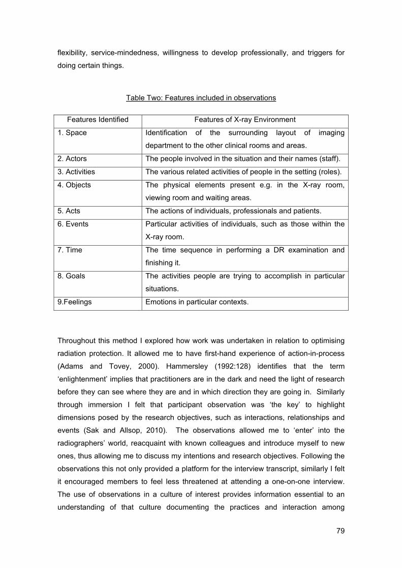
79
flexibility, service-mindedness, willingness to develop professionally, and triggers for
doing certain things.
Table Two: Features included in observations
Features Identified Features of X-ray Environment
1. Space Identification of the surrounding layout of imaging
department to the other clinical rooms and areas.
2. Actors The people involved in the situation and their names (staff).
3. Activities The various related activities of people in the setting (roles).
4. Objects The physical elements present e.g. in the X-ray room,
viewing room and waiting areas.
5. Acts The actions of individuals, professionals and patients.
6. Events Particular activities of individuals, such as those within the
X-ray room.
7. Time The time sequence in performing a DR examination and
finishing it.
8. Goals The activities people are trying to accomplish in particular
situations.
9.Feelings Emotions in particular contexts.
Throughout this method I explored how work was undertaken in relation to optimising
radiation protection. It allowed me to have first-hand experience of action-in-process
(Adams and Tovey, 2000). Hammersley (1992:128) identifies that the term
‘enlightenment’ implies that practitioners are in the dark and need the light of research
before they can see where they are and in which direction they are going in. Similarly
through immersion I felt that participant observation was ‘the key’ to highlight
dimensions posed by the research objectives, such as interactions, relationships and
events (Sak and Allsop, 2010). The observations allowed me to ‘enter’ into the
radiographers’ world, reacquaint with known colleagues and introduce myself to new
ones, thus allowing me to discuss my intentions and research objectives. Following the
observations this not only provided a platform for the interview transcript, similarly I felt
it encouraged members to feel less threatened at attending a one-on-one interview.
The use of observations in a culture of interest provides information essential to an
understanding of that culture documenting the practices and interaction among

80
organisational actors (Spindler and Hammond, 2000). I observed the behaviour of
others and informally discussed topics focusing the research in ‘what was going on’
and ‘how I intended to explore this further’. My position was predominantly ‘participant
as observer’, however this role became blurred into ‘observer as participant’, which
facilitated the open recognition of the research objectives (Pole and Morrison, 2003)
and allowed the observation of cultures, which helped in understanding them. The
purpose of this method was to depict the cultural and technical factors that occurred
during radiographic examinations in order to help overcome the discrepancy between
‘what people say they do and what they actually do’. The observations were performed
on various days of the week within 15 weeks. I had liaised with the appropriate
gatekeepers continuously in order to ensure my presence in the field remained
appropriate. Table three demonstrates the observation time spent at each research
site.
Table Three: Observation record at each hospital site
Sampling: The four research sites were chosen because they had integrated DR
systems that were used on a day-to-day basis. In ‘Trust one’ (sites A, B and D) were
established with DR with the first installation occurring in 2004 and consisted of seven
Site A Site B Site C Site D
24/08/12 AM
24/08/12 PM
31/08/12AM
31/08/12 PM
07/09/12 AM
07/09/12 PM
28/09/12 AM
28/09/12 PM
05/10/12 AM
05/10/12 PM
16/10/12 AM
16/10/12 PM
22/10/12 AM
22/10/12 PM
12/07/12 AM
12/07/12 PM
18/07/12 AM
18/07/12 PM
30/07/12 AM
30/07/12 PM
21/08/12 AM
21/08/12 PM
07/08/12 AM
07/08/12 PM
10/08/12 AM
10/08/12 PM
23/08/12 AM
23/08/12 PM
28/08/12 AM
28/08/12 PM
05/09/12 AM
05/09/12 PM
10/09/12 AM
10/09/12 PM
01/11/12 AM
01/11/12 PM
08/11/12 AM
08/11/12 PM

81
DR machines in total. In contrast ‘Trust two’ had recently installed their first DR system
in 2011 and remained the only DR unit within the Trust. The sample satisfied several
criteria for this study through the inclusion of both academic and community hospitals
of varying size and location in the South of England, willingness to participate in the
study and varying experiences with DR technology. The decision on ‘who to observe’
and for how long varied according to the individual research sites, for example at site
A, B and D numerous DR rooms were granted access. I did strategically carry out
observations at all research sites predominately at site A due to known relationships
with participants and distance from researchers home address. Site C had the only DR
technology within the Trust, which made it easier for the placement of the
ethnographer. One disadvantage was that it supported outpatients only. Thus at site C
I was not able to observe radiographers using DR neither in an accident and
emergency setting nor for ‘GP patients’ or ‘in-patients’ This affected data saturation in
each NHS Trust, for example at sites ‘A’, ‘B’ and ‘D’ participants varied in seniority,
thus providing a general range of radiographers. However at site C participants often
consisted of the same participants on the days of observation due to the departmental
shift patterns thus data saturation and sample size were smaller at Trust two when
compared with Trust one and as a result ended earlier than expected (Charmaz, 2006).
Data collection: To collect observational data within the hospital environment I would
attach myself to a DR room and its surrounding clinical areas ‘shadowing’ participants
throughout their daily activities. Where informed consent was forthcoming data
collection of the observations were undertaken and it was estimated in total that 30-40
operators were observed using DR over 19 days (approximately 142 hrs.). On a typical
09:00 – 17:00 working day, the observation commenced at 09:00 and ended at 12:00, I
would break for lunch, gather notes and then begin the second observational block
from 12:30 to 17:00. Data was collected throughout the day and on occasions with and
without the notepad for example when in the staff room taking lunch, I would be sitting
with the participants and they would discuss radiographic and personal aspects, for
example the new shift system recently implemented would become a regular topic of
conversation. These occasions of ‘time out’ in the staff room or the ‘viewing area’
provided me with the opportunity of ‘small talk’ amongst participants and previous
colleagues. These social interactions became a major source of data collection.
Furthermore, I would remain in the X-ray rooms when radiological examinations were
undertaken and would move accordingly when a DR room became in use. Throughout
the observations data was collected using an A5 note pad at sites ‘A’, ‘B’, ‘C’, and
audio notes taken at site ‘D’. Whilst my presence as an ethnographer was overt, there

82
were elements of covert note taking. Burgess (1990) reports when researchers have
had to dash to the toilet to jot down notes. I was not dissimilar, at site D due to the
‘tension’ felt between a number of participants I felt it necessary to leave the ‘clinical
area’ in order to record my data in private, thus dashing to the nearest toilet and
recording notes on my digital dictaphone as any form of note taking ‘within the
immediate vicinity of participants’ in my view would have been extremely
uncomfortable and highly intrusive.
An example of the transcribed observational data can be found in appendix eleven
(p.313-321). It was the intention to document as much as possible in the research field
while continually reflecting on the data collated and myself as the ethnographer.
Throughout the note taking I would record my own feelings and thoughts in the
processes throughout the study. Site ‘D’ remained problematic because of the layout
where radiographic rooms are open to the viewing room. Furthermore there was
limited space behind the protective lead screen and because there were several
operators behind this protective screen I felt like a ‘spare part’ as I failed to place
myself in an ‘appropriate location’ in which I felt comfortable. The data documented the
social setting and intimate detail of the culture/events of interest (Pole and Morrison,
2003) in order to build on theoretical propositions for the development of the interview
schedule (Saks and Allsop, 2010) and laboratory experiments. It was decided to collect
specific data important to the study and thus leave certain aspects out. For example,
for the laboratory experiments intimate technical data about what the operator did in
order to obtain an image of diagnostic quality, such as exposure factors, source to
image distance (SID) and collimation (field size). This data provided the development
of the experimental method and can be found in appendix twelve (p.322-325). In
addition I aimed to uncover cultural phenomena, thus I set ‘mini objectives’ throughout
the observations by ensuring a recorded ‘number’ of upper limb examinations. This
realistic approach was important because the collection of ‘unused’ data would not
have contributed to the overall study and may have caused further intrusion and harm
to the participants. In addition the method of obtaining the experimental data differed
with different X-ray equipment. For example when recording radiographic techniques
at sites ‘A’, ‘B’, and ‘D’ I was required to ‘physically walk’ to the X-ray tube next to the
patient to note the SID and collimation dimensions from the LCD display on the X-ray
tube, whereas at site ‘C’ the SID and collimation information was displayed behind the
protective lead screen on a monitor thus felt less intrusive.

83
Throughout the observations some participants altered their actions upon being
observed and this was verbally expressed too, for example operators became
increasingly self-aware of their working behaviour such as ensuring that the SID was
set to precisely 100cm (after discussing that it should be) illustrating the halo
(Hawthorne) effect (Burgess, 1993). The best evidence however to suggest that my
presence did not noticeably alter all participants behaviours lies in the fact that they
were willing to practice in a way that some other radiographers may have disapproved
of while in their presence, these are discussed in chapters seven and eight.
Furthermore, participants saw me in different ways; some were often unaware of my
presence in the viewing room, while others would see me as a potential judge of their
clinical practice techniques. The majority recognised that this study provided the
opportunity to add to existing knowledge.
3.5.3. Interviews
I conducted 22 semi-structured interviews across two NHS trusts (sites A and C). Sites
A and C were selected due to the frequency of observations performed coincided with
the rapport developed with individual staff members. The observations provided
detailed accounts relevant to DR in the clinical environment; the general themes were
interactive and sensitive to the language and concepts used by the interviewee (Denzin
and Lincoln, 1998). The interviews were directed by a list of potential questions of
themes observed during the observations and were interactive and sensitive to the
language and concepts used by the interviewee (Denzin and Lincoln 1998). This
schedule provided an understanding of the experiences, behaviours and attitudes of
radiographers in the clinical environment through face-to-face discussion, which could
be hidden in actual clinical practices (Hammersley, 1998) providing this thesis with a
deeper understanding of the relationship between practice and context (Burgess,
1993). Interviews provided considerable data generation and the ‘semi-structure style’
of interviewing allowed a set of topics to form questions in the course of conversation
‘conversations with purpose’ (Burgess, 1984:102). The interviews were conducted
under the following headings and supported with an interview schedule (see appendix
thirteen p.326-327) to aid the researcher to deliver particular questions in a particular
order, these included:
84
General questions about how long they had been working with DR and the
education and training received within the imaging department and at higher
education.
Identification of areas in which DR hinders and facilitates their radiographic
practices and the affects it has on the profession and the implications for future
practice.
The patient experience within a DR environment and the effects it may have
towards patient care.
Operator techniques, such as radiation protection, collimation, exposure indices
and adjustment of brightness and contrast of X-ray images.
Limitations of the digital equipment.
Workflow issues with the use of DR in practice.
Informal interviewing occurred with various members of staff in the clinical
environment. I was approached by participants in the clinical environment who
highlighted certain issues experienced within the DR environment knowing I was
undertaking research. Moreover the informal process facilitated those senior members
including superintendents who felt that they did not have sufficient time to formally ‘sit
down’ and discuss issues formally. The informal discussions provided valued insight
into the observed patterns of behaviour within the clinical environment (Roper and
Shapira, 2000) and have been used in later chapters. Attention was placed upon the
observed practice and explored with each interviewee exploring behavior, meanings
and understandings of the events that took place. Although a schedule was developed
for the interviews it was not used as a structured questionnaire but acted more as an
aide mémoire to ensure that similar topics were covered in all the interviews with any
omissions checked at the end of the interviews (Bailey, 1996). This approach allowed
the interview to remain flexible as topics would arise in different orders and therefore
the research would move to that topic. On this basis it enabled the research
participants to ask and answer questions that I had not previously considered.
Sampling: The intention was to interview a sample of 20-25 participants in this PhD
study across two research sites (A and C). Participants at sites B and D were not
approached for interview because it was important to find the ‘right people’ to study in
terms of theory development (Brannen cited in NCRM, 2013:16) and with the majority
of observations conducted at sites A and C I deemed it unnecessary to pursue any
further research at other sites. This method of theoretical sampling aimed to inform the

85
research questions and build on the criteria identified during the observations, helping
develop theory (Glaser and Strauss, 1967). My ‘knee jerk’ reaction was to merely take
as many interviews as possible as it would somehow seem ‘better’ (NCRM 2013:5) and
arguably at times I would possibly postpone my analysis of the interviews by
undertaking ‘just one more interview’. However interviews did cease following data
saturation when I had interrogated the purpose of their research objectives (NCRM,
2013). The radiographers interviewed varied in professional experience. The variation
of experiences was important in this study, as it could provide various points of view,
producing rich and varied data. For example it was important to consider
radiographers who had rarely used FS and conversely important to interview senior
radiographers’ whom had used FS in order to identify any differences in practice
amongst junior and senior radiographers.
When considering whom to interview I sampled participants from ‘those I knew’ and
had ‘social ties’ beginning with direct personal contacts, which I felt aided ‘others’ to
come forward having ‘little to fear’. This also facilitated the recruitment of ‘new
participants’ (those previously unobserved). When I interviewed ‘friends’ their persona
had changed in front of the audio recorder and immediately became less vocal within
the ‘interview environment’. Other participants were chosen who had been friendly
towards me and were interested in the topic supported by those I had built a sound
rapport with throughout the observations. For some (who I had observed) I received
responses such as ‘I’m a bit busy at the moment’ and ‘how about next week?’ For
some recently qualified radiographers they believed the interview to be a ‘test of
knowledge’ regarding me as the expert (Burgess, 1990). I appreciated their concerns
and would move onto another participant, this ad hoc approach led me to ‘pull
participants off the corridor’ during the quieter periods in the X-ray environment. On
reflection a sufficient number of staff had been selected in accordance with the
research objectives, stemming across two hospital sites with varying experiences and
viewpoints, tables four and five (p.86-87) identifies the participants, their job title,
clinical experience and gender specific pseudonym. Rather than specifying a precise
sample size the researcher continued interviewing until the analytical themes derived
from the data become ‘saturated’, i.e. until new themes stopped emerging from the
interviews (Glaser and Strauss, 1967; Saks and Allsop, 2010).

86
Table Four: Participants – Site ‘A’
Pseudonym Participant ID Gender Title and Grade Experience
Margaret DR1 Female Band 7 Practitioner –
reporting radiographer 18 years
Eric DR2 Male Band 6 Practitioner – works
in general and cardiac suite 9 years
Bernard DR3 Male Band 6 Practitioner – works
in general radiography 8 years
Sebastian DR4 Male Band 5 Practitioner – works
in general radiography 2 years
Rosemary DR5 Female Band 6 Practitioner – works
in general and CT 7 years
Sharon DR6 Female Band 6 Practitioner – works
in general area and CT 12 years
Fred DR7 Male Band 6 Practitioner – works
in general and CT 5 years
Harold DR8 Male Band 6 Practitioner – Works
in general and cardiac suite 9 years
Michael DR9 Male
Band 5 Practitioner – works
in general area and cardiac
suite
2 years
Table Five: Participants – Site ‘C’
Pseudonym Participant ID Gender Title and Role Experience
Geoff DR1 Male Band 6 Practitioner -works in
general area 5 years
Elizabeth DR2 Female
Superintendent Practitioner
(Band 7) – works in
angiography
6 years
Terry DR3 Male Band 6 Practitioner – works
in general department 7 years
Mick DR4 Male
Band 6 Practitioner – works
in general radiography and
CT
5 years

87
Victoria DR5 Female Band 5 Practitioner – works
in general radiography 1 year
Abigail DR6 Female Band 5 Practitioner – works
in general radiography 11 years
Kirsty DR7 Female Band 5 Practitioner – works
in general radiography 2 years
Helen DR8 Female
Band 5 Practitioner – works
in general radiography and
cardiac suite
2 years
Danny DR9 Male Band 5 Practitioner – works
in general radiography 8 months
Alex DR10 Male
Band 5 Practitioner – works
in general radiography and
MRI
4 years
James DR11 Male Band 6 Practitioner – works
in general area and CT 3 years
Annabelle DR12 Female Band 7 Practitioner –
Reporting Radiographer 7 years
Emile DR13 Male Band 5 Practitioner – works
in general radiography 9 months
Data collection: The interviews had taken approximately 3 months to complete. The
undertaking of interviews varied at each site as one to four interviews could be
performed in any one day. This was due to the unpredictable nature of the X-ray
emergency department, thus I would visit the research sites on specific days. For
example at site A I would attend Thursday and Friday as outpatient clinics were limited
and thus the radiographers less busy. At site C I attended Saturday and Sunday as
the radiographers that worked in the DR room often worked weekends. The interviews
ranged from 25-60 minutes each, the first taking 25 minutes with the concern that I
may not have had enough data. During the initial interviews some radiographers
discussed their lack of empathy, care and failure to optimise radiation doses, this often
‘drained’ me emotionally hearing the day-to-day ‘qualms’ of radiographers and how it
can affect their radiographic practices and care to patients. Throughout the interviews I
was sometimes considered an ‘expert’; however I was often surprised by this
assumption because while I had developed certain knowledge I did often feel
unequipped to answer such information. For example during one interview a
participant began to question ‘when and where is it appropriate to place lead on a

88
patient?’ While as a researcher and radiographer I would reference certain studies, I
did feel ‘lost at sea’ in answering specific questions with full conviction.
As the data was transcribed information and debates emerged about the research
process, findings and my abilities as an interviewer. I had learnt that I talked too much
during the interview and would interrupt the informant unnecessarily. On reflection it
proved problematic to transcribe. Gradually I learnt to speak less and from the
transcripts it was clear that I did talk less in the latter interviews (Pole and Morrison,
2003). Initially I had carried out some interviews in an unsuitable location, with building
work being carried out next door, the noise pollution lead to the temporary
abandonment of an interview. We were required to move to an alternate location, this
later impacted the transcription therefore I was correct to change the location of
interviews as data would have been missed. Following this a quiet vacant office was
selected for optimal audio recordings and also provided confidentiality for participants.
On occasions interviews were abandoned because participants were required urgently
for a ‘clinical matter’ thus postponed and continued at a later date. Interviews were
recorded verbatim using a digital audio recorder. I found this device appropriate in
order to transcribe the data accordingly as the individual files could be removed and
played in a media player and ‘slowed down’ to transcribed accordingly. Some
participants were more aware of the recording device, periodically looking at it prior to
answering a question, this highlighted that participants were aware of their actions and
I felt this did influence some respondents responses, for some this produced short
answers, not in depth and reflected on the time taken to complete their interview.
Throughout this thesis words from research participants’ are presented verbatim in
order to have an accurate account of the formal interviews undertaken to preserve the
meaning for the analysis (Burgess, 1993 and Saks and Allsop, 2010). Benton and
Craib (2001) claim that one should take note of the things that exist in the social world
such as dialectic and communication boundaries because these are defined by our
culture and language. Thus gaps or uncertainties in the quotations are clearly
indicated, including ‘pause’, ‘laughs’ and ‘Ummmmms’ representing the context of the
participants’ responses (Hammersley and Atkinson, 2007). During the transcriptions
difficulties were noted as participants speaking English as a second language were
difficult to transcribe thus words unrecognisable to the author were noted in the
transcripts as ‘unclear’. The transcriptions were emailed to participants allowing them
to verify the transcripts, with a time frame on each email stating that ‘no response
within 30 days would assume that the transcript was verified by the participants’. An
example of one of the transcriptions can be found in appendix fourteen (p.328-348).

89
Developing the interviews: The topics and questions were formed as a result of the
informal discussions and observations identified within the clinical environment.
Throughout the interviews I continuously reflected on the topics identified by the
participants and further topics were incorporated into the interview schedule. In early
interviews I would not listen carefully to the respondents’ answers as I appeared more
conscious of keeping to the interview schedule while conscious of the time the
interview was taking up. With little postgraduate experience my interview techniques
altered upon interviewing senior radiographers with extensive experience. For example
although the interview transcript provided the script my interview technique primarily
aimed to ‘seek guidance’, delicately questioning their radiographic techniques, thus
preventing a defensive barrier from the participant. In contrast I felt more comfortable
discussing radiographic techniques with radiographers with similar experience and who
I had previously collaborated with clinically. The initial interviews were in-depth
interviews allowing the participant to ‘tell me your story….’. As the interviews
developed I was able to ‘fine tune’ the questions and after completing five interviews I
would listen, transcribe and note the responses to certain phenomena and develop the
interview schedule accordingly. Burgess (1990) claims that the quality of your data is
dependent on the quality of the relationship you build with the people being
interviewed. Additionally I felt that my own reflexivity was essential before and
throughout the interviews, impacting on the discussion and approachability among the
participants (Howatson-Jones, 2010). For example, I felt that my time spent ‘in the
field’ as a researcher and radiographer facilitated my relationships with participants
because as rapport developed over time I was able to question ‘more personal’
phenomena relating to the DR and the application of care to patients. In short the
interview became like an informal conversation with additional probes added where
appropriate. The interview schedule (final version) can be found in appendix thirteen
(p.326-327).
3.5.4. X-ray experiments
The qualitative data collected during the participant observations informed the X-ray
experiments at sites A and C. Hammersley and Atkinson (2007:90) speak of the
collaboration between ethnographers and alternate methodological approaches, ‘it is
worth noting that these strategies are complementary, not mutually exclusive’. Each
DR room was selected based on their availability ‘out of hours’ as other rooms were in

90
24 hour use providing emergency care. Upon approval from each gatekeeper,
radiographic experiments were conducted at site A (hand) and site C (wrist). The
experimental design developed as a result of the ethnographers feelings towards the
qualitative data (ibid). This was part of the reflexivity throughout the data collection
whereby I assessed linkages with practice using observations, interviews and
experiments triangulating across different data sources and stages of the research
(Denzin, 1978; Howatson-Jones, 2010). Burgess (1990:110) is of the opinion that
hypothesis should develop from the ethnographic work rather than provide restrictions
and distortions from its inception, because in observing people’s behaviour we derive
hypothesis from our cultural knowledge to describe and explain their actions. In support
Hammersley and Atkinson (2007:15) identify that experimentation is a way of ‘trying
things out’, put to the world ‘what would happen if’, they argue that experimentation in
this form is normal in everyday life and the ‘genuine experience’, thus collaborative in
the ethnographic methodology. The experimental design was not only based on
inductive theories from participant observation (Couvalis, 1999); it subjected my
theories to rigorous test in the field, which evolved as part of the social world capturing
a holistic picture of the clinical environment through ethnography.
Aim: The aim of the experiments were to complement ‘what I had observed’ with ‘what
radiographers had said’ about phenomena in the clinical environment. In order to
discuss the variations observed it was necessary to test them in the actual clinical
setting using the DR systems. The specific hypotheses were to test the collimation and
source to image distance (SID) and its effects on the dose area product (DAP) with the
EI a strong indicator of dose delivered to the patient.
The objectives of the X-ray experiments were to:
Reflect on collected observational data and design X-ray experiments based on
‘what the radiographers did…’
Perform quality assurance tests on DR equipment ensuring that the doses
delivered during the experiments were the ‘expected doses’.
Perform experiments in designated DR rooms at sites A and C to mimic the
radiographic practice observed.
Organise, code data and perform statistical analysis using SPSS.
Throughout the observations, the common independent variables altered by operators

91
were collimation and SID. Reflecting on the data observed it was decided to increase
the SID by increments of 5 cm from the lowest to the highest values observed for hand
and wrist. In addition for collimation, it was decided to select the collimation based on
the smallest and largest dimensions for each radiographic projection observed in
practice, followed by a median value. The anthropomorphic images produced were
stored securely and safely archived to each hospitals PACS (picture, archive,
communication, system) however the ‘image quality’ was not explored in this PhD
study because it is not within the scope of this thesis nor does it answer the research
objectives. No ethical considerations were required for the X-ray experiments as no
patients were involved and my first supervisor and I were present during the
experiments.
The DR equipment contained two flat panel digital detectors, one horizontally mounted
in a table and other vertically mounted on a supporting structure. Table six (p.92)
details the equipment for the reader for comparison. The digital detectors were single
panelled (non-tiled) amorphous silicon detectors with a Cesium Iodide scintillator (CsI).
The CsI, with its needle like crystal-line structure together with the small pixel size
provides a high detective quantum efficiency (DQE) of more than 60% at zero line pairs
per millimeter and a sound modulation transfer function (MTF). Detective quantum
efficiency is the most important single parameter, which reflects image quality and
depends on spatial frequency. The detector converts the analog signal into a digital
signal with an image depth of 14 Bits.
Quality assurance: Quality assurance (QA) checks were performed on the digital
equipment in accordance with local protocols. The QA checks ensured that the
exposures delivered in the experiments were the expected exposures. The tests were
used to identify any improper central X-ray misalignment, which may distort a
radiographic image and/or increase in output of the kVp and mAs thus potentially
effecting dose (or image quality) (IPEM, 2010). It ensured that no change in radiation
output had occurred due to ageing, drift, component failure or miscalibration (ibid). The
QA tests undertaken prior to experimentation conformed to current safety measures
and deemed the equipment safe and optimum in its delivery of ionising radiation (IPEM
2010). The results and graphs are illustrated in appendix fifteen (p.349-358).

92
Table Six: Digital Radiography Equipment
Site A - Equipment Specifications Site C - Equipment Specifications
Make/Model: Philips Digital Diagnost
System 1.5
Focal Spot Size: 0.6 and 1.20 mm
Grids: Oscillating grid Table and Wall
Matrix size: 3000 x 3000 pixels
Active Area: 43cm x 43cm
Bit depth: 14 Bit
Make/Model: GE Healthcare Discovery
XR650
Focal Spot Size: 0.6 and 1.25 mm
Grids: Table 100cm focus | Wall 100cm,
130cm and 180cm focus
Matrix size: 2022 x 2022 pixels
Active Area: 41cm x 41cm
Bit depth: 14 Bit
Anthropomorphic phantom: A hand/wrist anthropomorphic phantom was used for the
experiments and while no ethical approval was required I did document it in the original
research protocol in order to provide an overview of the study. The anthropomorphic
phantom limb was designed such that radiation absorption was similar to that in real
patients (Williams and Adams, 2006). The phantom material contains a density of
0.985 g/cm³, an effective atomic number of 7.3, identical to human tissues and
contents i.e. muscle, fat and bone thus absorbing radiation in manner similar to the
human body (Williams and Adams 2006). Positioning and centering of the
anthropomorphic phantom was undertaken using aids including radiolucent sponges
and lead in order to mimic a patient’s radiographic position on the digital detector (see
figure six, p.93). The parameters under investigation were set up and centering on the
limbs was accomplished following the radiographic literature (Whitley et al, 2005).
Recording dose and exposure index (EI): The DAP and EI readings were measured
from the DR console in the clinical environment. The DAP unit was recorded in this
study because this was the method and unit collated by radiographers during the
observations and used for the routine monitoring of patient dose (NRPB, 1990). The
exposure index (EI) was a secondary dependent variable collated, this can be used to
evaluate dose and was present within the clinical rooms as a guide to the operators.
Three exposures for each individual technique were performed to increase the validity
of the findings. The EI values obtained during the experiments differed between sites
A and C, table seven (p.93) provides a visual account for the reader.

Figu
3.5.5
Whe
key
Ham
ure Six: The
DAP
Low
High
5. ‘Ethno-rad
n ethnogra
instrument
mmersly and
e positioning
Table
P Site
w
h
diographer’
phy is a res
for data
d Aktinson,
g of the anth
Seven: EI v
A - Philips D
400
125
: Reflexivity
search tool
collection
2007), with
hropomorph
value and re
DR EI Sit
y in the field
of an inve
(Ragucci,
h status cha
hic phantom
elationship t
te C – Gener
0
1
d
stigation, th
1972; Burg
aracteristics
m during X-r
to DAP
ral Electric D
0.50
.50
he research
gess, 1984
s (race, sex
ray experim
DR EI
her become
4; Morse, 1
x, ethnicity,
93
ments
es the
1989;
age,
94
sexual orientation and social class) becoming relevant to the production of knowledge
(Burgess, 1993 and Bailey, 1996). The importance of using this approach was to
provide a deep description of the culture of a particular group in action. This led to an
opportunity to understand the group through immersion into their culture i.e.
participating in the specific culture for an extended period of time. However this part of
ethnography in turn becomes its contradiction. Since the application of ethnography
can be either from an etic (outsider’s view) or emic (insiders’s view) perspective, for the
etic approach it is difficult for the outsider to establish a relationship with the
participants and gain permission to immerse themselves into a group and hence
cannot be an ethnographic study. As a radiographer my perspective was emic
providing insight into the radiographic profession. Becker et al (1961:15) remind the
reader ‘this is how things look and feel down under’, although it feels like a true setting,
its merely an account of those professions and their practices at a point in time and
that there would be no organisation in which things look the same from all positions.
Clifford and Marcus (1986) concur, maintaining that ethnography similarly captures
partial moments in time. Richardson and St. Pierre (2005) identify crystallisation as a
process of undertaking exploratory research because we need to acknowledge that
there are many dimensions in which to approach the world (just as a crystal has many
facets and dimensions) and what we see depends on our viewpoint and perspective.
The emic approach facilitated the study allowing me to identify key points within the
clinical environment that could be explored later in the PhD study.
Researcher positionality: My position in the field is important to discuss because while
ethnographic research explores the social action and deeper elements of participants,
the relationship between the participants and I was an issue which inevitably pervaded
all aspects (Holliday, 2007). My ‘physical position’ (6 foot man) ethnographer at the
research sites are depicted in appendix eight (p.306-309) and often proved problematic
in the clinical environment. On reflection the ‘protective lead screen’ in which a
radiographer places him/herself behind limiting their radiological dose became ‘key’ to
positioning. For example, at site D it proved difficult to observe radiographic
examinations merely due to the layout of the clinical department (Bowling, 2004). In
addition the protective lead screen was extremely small in comparison to other sites,
thus my placement merely involved being in the ‘viewing area’ behind the X-ray rooms.
At sites A, B and C I found myself ‘tucked away’ in a corner behind the protective
screen as the space was significantly bigger and was able to accommodate myself,
and other members of staff. Site D was significantly difficult to observe and as
discussed above led to leaving the field earlier than envisaged. This highlights that

95
environmental and cultural contexts in which the researcher was exposed added to the
complexity of the ethnographic fieldwork but similarly strengthened it. For example the
protective lead screen at site A was considerably larger than other sites, this facilitated
my ‘position’ in the field, allowing me to ‘keep out of the way’ having little impact on the
operators and their day-to-day tasks. On reflection future studies conducting
ethnographic fieldwork within the X-ray room should consider their ‘potential site’ prior
to undertaking field work as it may influence the relationships and data collection of the
study.
Due to my physical presence ‘in the field’ the aim of my role as a researcher was to
remain objective, accessible and friendly whilst reducing the possibility of ‘going native’
(Hammersley and Atkinson, 2007:100). Ethnographers may adopt a variety of roles
however my aim throughout was to maintain a more or less marginal position. The
ethnographer must be intellectually poised between ‘familiarity and strangeness’, while
socially poised between ‘stranger’ and ‘friend’ (ibid: 100). While I acknowledge my
‘connection’ with numerous participants throughout the thesis becoming the ‘friend’ my
choice to undertake advanced postgraduate study alienated me amongst my peers and
others as participants often questioned: ‘why on earth did you decide to do that?’ and
‘what do you intend to do after it?’. This alternate approach to my career undertaking a
PhD helped me distance myself from the research participants and allowed me to
‘become the stranger amongst other radiographers’, thus allowing me to ‘ask the
unfamiliar’ and ‘ask the obvious’. This research identified areas of practice in which my
presence was as an advantage in the field as I had an obligation to protect the
participants and patients as a registered healthcare professional by abiding to my
professional code of practice outlined by the Society of Radiographers (2008:3):
“You must conduct yourself with honour and dignity and demonstrate trustworthiness and integrity in both your personal and professional life in order to maintain the widest public trust and confidence in the profession”.
On reflection I developed a moral stance within the fieldwork. It is generally accepted
that individuals entering a healthcare profession generally want to care for people
(Bolderston et al 2010 cites Goldins’ 1979:190). Throughout this PhD research I felt I
had moral and professional obligations to patients and participants, which resulted in
leaving my ‘research position’ to aid where appropriate. Glaser and Strauss
(1967:246) identify the ‘awareness context’ doctors and nurses have over their forms of
interaction for those people involved in a ‘dying situation’. Similarly, I was required to

96
approach situations holistically, controlling my own forms of interaction with
participants, aiding them where necessary. Participants would ask me about a
particular ‘X-ray exposure’ or ‘what would you do in this situation?’. More importantly I
would alert radiographers to incorrect settings selected on the DR console, such as a
wrong detector selected (discussed further in chapter seven) prior to the radiographer
exposing. If I had not made the operators aware of this incorrect selection a patient
would have likely received a dose of radiation with no X-ray image produced. Another
example included the light-beam collimation (LBD) box ‘opening up’ due to the
selection of another radiographic examination on the DR console with the operator
unaware of the actions taking place from the equipment. This would have given the
patient an increased dose of ionising radiation and reduced image quality (Jeffery,
1997), which was something professionally and morally I did not allow. This highlights
that grounded theory had ‘practical applications’ throughout the ethnographic fieldwork
whereby my own reflexivity as radiographer and researcher influence my own
behaviour and actions thus impacting the data collected (Glaser and Strauss,
1967:246). Other aspects of the ‘ethno-radiographer’ role included conforming to ‘bad
practices’ whereby upon request I would inform the operator ‘how to crop’ an image or
‘how to adjust the brightness and contrast’ of the image, generally seen as
inappropriate radiographic practice. While in the field and arguably conforming to
suboptimal practices I felt like a corresponding sense of ‘betrayal’ or at least divided
loyalties to my ‘ethnographer position’. In contrast my betrayal did not stop there,
during my ‘locum radiographer’ role I would apply typical practices and use specific
functions comparable to those identified in chapter seven amongst the participants,
later reflected in my field diary:
Field Diary: “Working as a radiographer I would initially use the similar functions available to me ‘masking’ improper collimation, pay little attention to the SID and rarely use lead protection on numerous patients. Upon reflection and continuous observations I realised I had performed and maybe even contributed to suboptimal practices thus facilitating the ‘way things were done around there’. On reflection this allowed me to become part of the culture, ‘fit into the norm of everyday practices’, yet on the other hand by realising my faults I was obliged to remain within my ethical boundaries and later began to take note of my practices, trying to apply stricter collimation, carefully select exposure factors and apply carefully SID positioning.”

97
On reflection my ignorance towards my own radiographic practices was arguably
important throughout this study as it allowed me to resonate and ‘fit in’ with participants
I did not know that well. For example as ‘the locum radiographer’ I began to work with
others building sound relationships and ‘as the radiographer’ (prior to becoming the
ethnographer) I took part in the ‘suboptimal’ practices highlighted in chapter seven.
However as my role altered to what I refer to as the ‘ethno-radiographer’ my
positionality changed, and through continuous reflection I began to note the suboptimal
practices in others that impacted my own practice. Howatson-Jones (2010) identifies
how adopting an autobiographical approach can enable different elements of the
research process to be brought together, including the researchers struggles.
Howatson-Jones (2012:47) further discusses how a ‘learning biography’ can provide
the student a strategy to evaluate their contribution to learning, how to undertake
learning and how they can develop further. Similarly the ‘ethno-radiographer’ position
and continuous reflexivity continued throughout this PhD study facilitating my own
practices and positionality, allowing me to become more objective to the actions and
behaviours around me (ibid). Moreover as I remained employed at sites A and C I
naturally felt less informal around some participants, which had a direct impact on the
way the participants acted. At these sites I ‘the ethnographer’ would become ‘the
radiographer’ as reflected in my field diary:
Field Diary: “An assistant practitioner (AP) was positioning a radiographic projection (an angled view of the scaphoid examination). The AP experienced difficulties undertaking this projection and asked for my help. I verbally explained the technique I would employ for the patient before intervening however the AP insisted that I demonstrated it to her asking my advice and with other staff extremely busy I decided to intervene. I became ‘the radiographer’, I had to come out from behind the screen and position the patient while advising the AP of the technique I was employing. This felt uncomfortable at first however I was aiding the AP and the patient thus felt essential in order for the patient to receive the appropriate images of diagnostic quality diagnosis.”
This example highlights the danger of ‘going native’ within the clinical environment. It
identified that being ‘in the field’ in your own discipline opens the research to more
complex and nuanced understandings of issues. However while I would intervene I felt
it facilitated my ‘presence’, my ‘reason for being there’ was to explore and possibly aid
where appropriate, providing guidance and reduce the possibility of work related stress
(Akroyd et al, 2002). This empathic attitude allowed me to listen to staff members,

98
absorb their problems and provide a voice to the participants involved in this study
(West, 2000), thus arguably gaining a deeper insight into an environment (Randle et al,
2007; Verrier and Harvey, 2010). Furthermore, it reinforced the argument that
remaining objective in my own professional field remained problematic but in fact was
further engaging with participants using the digital technology which highlighted other
areas of concern. In addition failing to aid the patient and participants could be
interpreted as a diminishment in care and responsibility from the researcher due to the
professional ethos governed and required by healthcare professionals.
I became more reflexive in my dual roles aiming to understand the process whilst
critically examining power relations in the data collection and interpretation (Jones et
al, 1997). This was first highlighted by the numerous ‘labels’ I attached to individuals in
the clinical environment, for example ‘informant’, ‘participant’, ‘healthcare professional’
and ‘radiographer’ are used interchangeably throughout. By becoming more reflexive
under the conditions of ethnography I later removed the term ‘informant’ because I felt
that all individuals involved in this PhD study had ‘participated’ with me in this PhD
research and had not merely ‘informed’ it. I had built rapport with the participants
involved in my fieldwork thus throughout this PhD thesis I use terms ‘participant’
‘radiographer’ and ‘healthcare profession’ interchangeably providing an holistic picture
of the individuals involved in this PhD work. Secondly this highlighted my own
individual stance during the fieldwork whereby I played the ‘friend’ and the
‘professional’ simultaneously depending on the individual participant (Blackman, 2007).
Throughout the research some participants would be well known, some more than
others, some whom I had collaborated on my undergraduate degree and some I had
collaborated working as a radiographer. Therefore there was an element of
professional/personal relationship with the participants from previous experiences and I
would involve myself in personal discussions, listening to their concerns where
appropriate. At ‘site C’ I had socialised with the participants outside of work, attending
various social events including an invitation to the ‘Christmas party’. At 27 years of age
I would attend ‘night clubs’, drink alcohol socially, this allowed me to ‘get closer’ and
became part of a ‘social group’. Emotional well-being and self-identity became
fieldwork opportunities being personal with subjects, creating bonds of commitment for
both researcher and researched. Burgess (1990:71) claims that the ‘value of all field
research and perhaps all research regardless of methods is to measure these
dimensions, its contribution to empirical findings, to theory and to social change’.
Therefore while I developed ‘friendships’ and thus felt more comfortable around certain
individuals in the field, this allowed more in-depth questioning about their clinical

99
practice providing insightful empirical findings. It would have been naïve to believe that
I became the ‘insider’ or that the relationships throughout the research were ever fully
equal, however ‘who I was’ and the interactions I developed with individuals helped
form the relations of trust that are important in the field (Holliday, 2007). In addition this
felt more comfortable as a researcher asking problematic questions to ‘friends’ due to
the common ground established (Nagar and Raju, 2003) and because I knew their
personalities I was aware of the type of responses they may provide (Haraway, 1991).
Therefore using this to my advantage it produced flexibility in the research
environment, for example I would feel much more comfortable taking notes in front of
them. On occasions it would be entertaining, whereby jokes were shared and
expressed as ‘friendly banter’. While it is argued that ethnographers must strenuously
avoid ‘feeling at home’ (Hammersley and Atkinson 2007:102), this was not apparent
with all participants during the observations whereby I had not attained sound relations
often feeling cautious regarding my research objectives. At sites A and C my ‘informal
research role’ allowed the participants to feel less threatened by my presence and
facilitated the data collection. Conversely there were occasions where I was required
to become ‘more formal’, such as approaching ‘friends’ to sign the informed consent
sheet, this felt intrusive possibly hindering the relationship through the ‘reality of
research’. At the other research sites (B and D) a majority of the staff were not known
to me and I felt comfortable asking for informed consent, the informed consent sheet
was a method of ‘keeping me safe’ in the thought that I had been undertaking the
ethical duties appropriately. Moreover once the informed consent sheet was signed,
although it may have allowed me to feel ‘safer’ it altered the interactions with staff
allowing the researcher to develop a relationship and act more informally around the
‘consenting participant’.
As the study developed I became immersed in the clinical environment. This was
advantageous allowing me to ask in-depth questions about specific operators practice
or ask their opinion on a particular area of interest. Peake and Trotz (1999:37) support
this acknowledging that one’s positionality or subjectivity should not mean abandoning
fieldwork, it can ‘strengthen our commitment to conduct sound research based on
building relations of mutual respect and recognition’. This became apparent as I began
to engage with the participants more often, their patterns of behaviour would change at
all sites and I felt I could ask more and more regarding ‘sensitive’ topics relating to
practice. Kobayashi (2003) supports the concept that being reflexive is not about one’s
own positionality to self-indulge but to reflect on how one is inserted in grids of power
relations and how that influences methods, interpretations and knowledge production.

100
Therefore although during the research I had been concerned with attempting to
remain objective at all costs, becoming the radiographer and ethnographer
strengthened my position in the field in developing relations and trust with the
participants. This was discovered in the work of Sultana (2007) who identified that
through reflecting on her positionality vis-à-vis the way others constructed her identity
helped in more fully engaging reflexivity, which enabled engagement with the research
process.
The ‘white coat’: It is generally accepted that researchers should have an acceptable
appearance, which includes dress, demeanor and speech (Coffey, 1999). Throughout
the research I ensured that every effort was put into ‘blend in’ and become part of the
clinical environment (Becker et al, 1961). Hammersley and Atkinson (1995:83) discuss
the role of ‘impression management’ through dress, speech and demeanour, as well as
the presentation of ‘different selves’ in different research environments. In the
research environment(s) I wore smart trousers, a short sleeved shirt (for infection
control purposes) and shoes. I decided not to wear a clinical uniform in order to remain
separate from staff, thus attempting to remain objective due to working relations
developed previously with certain members as attaching a uniform may have
encourage ‘radiographic practice’ and thus prevented the research. My attire was a
symbol justifying my presence as a ‘researcher’ and not a ‘radiographer’ and
throughout the research at sites B, C and D this attire was generally accepted by all
gatekeepers during observations and interviews. However, following a discussion with
the gatekeeper at site A she requested that I wore a ‘white doctors laboratory coat’
throughout the observations. Dress or clothing can be shared as a symbol of culture
(Pole and Morrison 2003), however Allen (2004:18) asserts that ‘uniform is a signifier
of group membership, status and rank’. Therefore while being the only individual in a
white coat it remained difficult to be part of the culture and environment and on
reflection did not help the researcher ‘fit in’. It had a significant impact on the research
participants and patients. The participants would comment on the ‘white coat’ as a
symbol of my research degree; ‘here comes the Prof’ or ‘here comes the nutty
professor’. There were also occasions whereby some participants found it intimidating
especially more senior staff members claiming ‘no-one wears a white coat anymore’.
My presence in the ‘white coat’ had an impact on the patients also, following
radiographic examinations patients would ask me what I found from their X-ray, for
example a patient with persistent chest pain would ask if ‘everything was clear?’, thus
impacting on the patient, confusing me with a medical doctor who could provide an
imminent diagnosis for their symptoms. Becker et al (1961) observations of medical

101
students identified that the physician in the ‘white coat’ presents the most powerful
figure within a clinical environment. This supports a figure of power I had obtained in
the clinical environment; a sense of ‘seriousness’, ‘professionalism’ and anxiety
amongst colleagues and patients through association of the ‘white coat’, whereas
previously shirt and trousers alone had little effect on participants reactions in the
clinical setting. Although wearing the ‘white coat’ proved problematic in the clinical
environment I was requested to wear it by the superintendent radiographer of the
general radiography department thus felt unable to remove this item of clothing. Whilst
the ‘white coat’ illustrated attrition amongst some members in the field it did facilitate
data collection, helping me remain ‘the scientist’ as visitors and prospective students
would wear the ‘white coat’ and simply be ‘observing radiographic practice’ with no
input required. Hammersley and Atkinson (2007:79) concur suggesting that such
forms of dress are reported to ‘give off’ the message that the ethnographer seeks to
maintain the position of an acceptable marginal member. This allowed me to ‘sit back’,
observe and collect data whilst engaging in informal discussions. Moreover the feeling
of anxiety in which the ‘white coat’ made me feel may have had further implications if I
had not known the key participants in this study. The research in general is an intrusive
topic and with the added attire a ‘stranger’ entering their clinical environment may have
felt more threatened thus I feel my historical relations with the participants help
considerably in collecting the research data and preventing any added discomfort in
the field.
3.5.6. Data analysis
The research was approached in an open manner and refined as the nature of
radiographic practice began to emerge. This PhD study was conducted in the natural
clinical environment assuming that ‘reality is constructed, multidimensional and ever-
changing and that there is no such thing as a single, immutable reality waiting to be
observed and measured’ (Miles and Huberman, 1994:262). Glaser and Strauss
(1967:40) support this suggesting that the general relations are discovered ‘in vivo’
through participant observation, that is ‘the fieldworker literally sees them occurring’.
This thesis is an ethnographic report crafting the moments observed at each site
representing the participants voice (Allan, 2006) through the researcher’s voice. It was
decided to present the data as raw as possible in later chapters in order for the reader
to review the interpretations of the data presented and the data analysis process.
Within the analysis I provide site specific contexts about each research site which are

102
important to consider due to the significant differences technologically and the
individual experiences of performing radiological examinations.
As the research progressed additional developments were made and implemented
throughout. I continually moved ‘back and forth’ between the methods and the
emerging data thus providing an ‘on-going’ analysis developing my own voice and
actions through reflexivity and positionality (Holliday, 2007; Howatson-Jones, 2010;
Murphy and Yielder, 2010). Observations were transcribed on the evening of data
collection for it to remain ‘current’ in the authors mind whilst providing valued insight
into the radiographers’ world. This reflexive process allowed me to continuously reflect
on the observational data, which would later impact on interviews and experimental
data. The observational data ‘developed’ throughout the development of this thesis, for
example data that was not ‘that obvious’ in the beginning became relevant upon
analysing the interviews, authors field diary and writing up the empirical chapters. This
highlights that while participant observation data was collected and analysed ‘at the
beginning’ it continually impacted the overall analysis following data from interviewees,
X-ray experiments, researcher’s field diary and conversations with friends and
colleagues. Thus the ‘observational data’ presented in the empirical chapters is a
‘refinement’ providing a holistic picture to the readers.
Ethnography can provide detailed account on clinical practice, patient and health
delivery issues which can focus on the patients or healthcare professionals’
perceptions (Adams and Smith, 2003). It was important to systematically search,
arrange and make sense of the data (Brewer, 2000). Research data was transcribed
into a word processing application, dated and indexed with spelling and grammatical
errors searched and corrected, with participants’ voices (formally and informally)
presented in the form of quotations. Files were printed, categorised and grouped into
the research sites in specific folders to prepare easy reading. I did not use a
‘qualitative analysis computer program’ thus preferred a word processing application
coincided with hard copies to make notes and assign relevant codes to emerging
themes. Each line of text was reviewed and coded by the researcher and reread using
thematic analysis, which produced general themes. Emerging themes were structured
using a coding framework involving data reduction, data display and conclusion
drawing/verification (Miles and Huberman, 1994). Upon ‘sorting’ of the themes, four
overarching categories emerged: ‘learning’, ‘radiographer challenges’, ‘ionising
radiation’ and ‘patient care delivery’. Themes were later printed on A4 paper, ‘cut out’
and placed on A1 pieces of card, whereby theory could then be linked across the

103
themes. Colour pencils were used to cross reference themes, which provided the basis
of theory development. Grounded theory supported the generation of theory
development through comprehensive explanations of phenomena that were grounded
in data, thus demonstrating relevance in the clinical environment (Glaser and Strauss,
1967) and applied throughout this PhD study by means of induction, deduction and
verification (Strauss, 1987). Thus although grounded theory does not start with
hypothesis I did have ‘hunches’ (Holloway and Wheeler, 2002:155) and after collecting
the initial data and establishing relationships, provisional hypotheses were conceived
from grounded theory (Glaser and Strauss, 1967). In chapters five, six and seven
observations and interview data are presented to provide the reader with the context,
such as whether ‘it is done in public’ and ‘what is done in private’ (Hammersley and
Atkinson 2007:192). In support the experimental results at sites A and C were entered
and organised into SPSS and coded, preparing statistical analyses. Hammersley
(1992:90) speaks of collaboration between ethnographers and survey methods in that
‘it is worth noting that these strategies are complementary not mutually exclusive’.
Thus SPSS provided me with ‘the tool’ to run Pearson Correlations to examine
correlations between DAP and EI and multiple regression analyses on independent
variables to predict effects of DAP (Tabachnick and Fidell, 2007) with separate
analyses conduced for each body part. The X-ray experiments were complementary in
this thesis and formed part of the whole data analysis in this exploratory methodology.
Moreover ‘triangulating’ the research methods allowed me to approach particular
phenomena from different angles, for example as ‘cropping’ was grounded from the
observations, this analysis prompted further exploration through interviewing and X-ray
experimentation. Hammersley and Atkinson (2007:198) claim that relying on a source
can present a danger because if ‘diverse kinds of data lead to the same conclusion,
one can be little more confident in that conclusion’. Thus it could be argued that
analysing specific phenomena using different kinds of data can further develop theory
thus enhancing the ‘reflective practitioner’ in a radiographic environment.
3.6. Leaving the field
Upon collecting the relevant data through observations, interviews and X-ray
experiments I had decided to leave the field of research. I had enjoyed the research
and it was sad to leave the different departments as a researcher. I was grateful to all
of those who participated in this study. In Strudwicks (2011) thesis she identifies the
attachment experienced with her participants and the sad feeling upon leaving the

104
environment. I had not only left research participants, but also friends and colleagues
behind, however I did feel that I had a positive impact on the teams in my approach to
research and had become part of the team. Firstly, when I would challenge or ask a
specific question, it allowed the radiographers to reflect themselves as professionals in
their own practice. Secondly, since undertaking the research I have been asked
(personally) to ‘cover a few shifts’ at each of the sites. On the last days I thanked all
staff and wrote personal emails to all the gatekeepers for their patience and
acceptance during this research process.
3.7. Conclusion
The use and selection of ethnography has illustrated a unique insight into various
cultures within the radiographic profession. My epistemological and ontological position
has been discussed and whilst the research objectives were initially believed to be
addressed using a ‘mixed method’ approach, in light of the ‘exploratory’ use of
ethnography this chapter has illustrated an exciting method of enquiry into the
generation of knowledge in the field of diagnostic radiography that combines methods
such as participant observation, interviews and X-ray experimentation. This
methodological position facilitated by symbolic interactionism and a ‘grounded theory
approach’ provides an original illustration concerning subject matters such as current
training and education, use of ionising radiation and the delivery of person-centred
care, discussed in chapters five, six and seven. This chapter discussed the importance
of positionality and reflexivity throughout this ethnographic research as I engaged with
new and existing relations as the ‘ethno-radiographer’, facilitating rich data and building
on existing knowledge. In contrast this chapter recognises the challenges
ethnographers may encounter researching their own field. For example, I experienced
conflict and questioning by numerous members of staff, highlighting that although
ethnographic fieldwork can be informative for practice, it can be seen as an invasion
into a radiographers practices. Through preparation, reflection and working within the
ethical codes of conduct, elements of ‘intrusion’ were resolved amicably and
professionally. It is important to consider that within qualitative methods person-
centred care is integrally related to the grounded theory approach towards the data
analysis enabling a thick description to emerge. In this sense ethnography was a
sound methodology of choice to both promote and support the exploration of
radiographic practices in a person-centred care environment because of ethnography's
105
core concern with participatory methods and empathy to aid reflexivity. This
methodology sought to provide a new approach to research in radiography that could
underpin medical imaging practices for the development of the profession.

106
Chapter Four: Findings
4.1. Introduction The main focus of this PhD study was to explore the clinical practices of radiographers
following the introduction of digital radiography (DR). The previous chapter discussed
the importance of using ethnography as a methodology to explore the clinical
environment and uncover original phenomena. The values and beliefs of the
researcher identified in chapter one was central how the ‘ethno-radiographer’
practiced, collected and interpreted the findings. Similarly the values and beliefs of
radiographers were central in uncovering ‘how radiographers felt and behaved in the
clinical environment’ thus described as a core variable in this chapter. Rigour and
validity of the research findings are considered, providing a systematic approach to the
research data strengthening its relevance and importance to the field of diagnostic
radiography.
4.2. Values and Beliefs In chapter one the values and beliefs of ‘me as the researcher’ were important to
consider as part of this PhD research in accordance with exploring the impact ‘values
and beliefs’ can have on clinical and educational practices. Values and beliefs
connected the categories in this PhD work and were inherent in the radiographic
environments explored following the individual values and beliefs held by
radiographers. It is evident from the research findings that values and beliefs were not
clearly defined in general radiography. They often differed amongst individuals
resulting in varying actions and behaviours in the use of ionising radiation and
approaches to person-centred care. The core category depicted in figure seven (p.108)
illustrates ‘values and beliefs’ acts as a unifying category of the workplace culture
within this PhD research (Wilson et al, 2005:34). The findings show that there were
alternate views and attitudes towards phenomena within the radiographic environment
supporting the view that ‘values and beliefs’ do vary amongst individuals. The findings
and critical reflection in chapter one suggest that for practice reform and improved
person-centeredness, strategies uncovering the values and beliefs of radiographers
may develop a collective vision or philosophy of sound radiographic practice in the
future.

107
4.3. Observations and interviews
Findings from the qualitative data were collated and analysed. Following complete
immersion within the data ‘codes’ were assigned to key words and phrases, producing
general themes. ‘Sorting’ of the themes was undertaken and placed in an overarching
category. Each category then contained central themes in the initial analysis of the
data and can be seen in appendix sixteen (p.359). The overarching categories derived
from the thematic analysis were:
1. Learning
2. Radiographer challenges
3. Ionising Radiation
4. Patient care delivery
Themes within each category were compared with one another and interconnected
across categories, enabling the development of theoretical descriptions (see appendix
sixteen, p.360). This enabled the identification of central themes coincided with the
core variable ‘values and beliefs’ (Streubert and Carpenter, 1995). Values and beliefs
were central to the authors views and attitudes as a radiographer, researcher and
educator as discussed in chapter one. Thus values and beliefs of radiographers were
similarly central to this PhD research uncovering views, attitudes and behaviours of
radiographers as it may impact on radiographic practice and the delivery of person-
centred care. Wilson et al (2005:27) conclude that understanding values and beliefs is
an important part of understanding a workplace culture and attempts at changing
workplace cultures should start from the clarification of values held among staff in that
culture. It is evident in subsequent chapters that values and beliefs of radiographers
are central to workplace practices and subsequently any potential workplace reform.
Categories, with their related central themes are outlined in figure seven (p.108),
together with descriptors from radiographers highlighting the contradictions in the data
and the tensions that exist in a radiographic environment. Empirical chapters five, six
and seven provide an in-depth descriptive and analytic discussion of the categories
and central themes that emerged throughout the qualitative data. The length of
quotations varies demonstrating the numerous responses from participants while
providing the context for the data (Glaser and Strauss, 1967). Literature was sought
contributing to the overall analysis in order to tell the story to the reader (Hammersley,
1998).

4.4. X
Expe
triang
with
collim
varia
decid
Figure Sev
X-ray exper
eriments w
gulation’ of
‘what they
mation and
ables altere
ded to test t
ven: Depicti
riments
were condu
certain phe
had said a
source to
ed by X-ra
the effects o
on of cultur
cted as p
enomena w
bout it’. Ob
image dis
y operators
of collimatio
re within dia
art of this
with ‘what th
bservations
stance (SID
s during h
on and SID
agnostic rad
s ethnograp
he radiograp
of radiogra
D) as the m
hand and w
on the dos
diography e
phic study
phers were
aphic practic
most comm
wrist exam
e area prod
nvironment
facilitating
observed d
ce had iden
mon indepen
inations. It
duct (DAP)
108
t
g ‘the
doing’
ntified
ndent
was
using

109
an anthropomorphic phantom and subsequently analyse the correlations between the
DAP and exposure index (EI). A sample of the experimental data from sites A and C is
presented in appendix seventeen for the reader (p.372-376), contributing to ‘the
actions’ undertaken by the radiographers. A multiple regression analysis was
undertaken to predict the effects of collimation and SID on DAP and are reported in
table eight.
Table Eight: Prediction of collimation and SID on DAP
***p < 0.001
The multiple regression analysis shows that collimation was the strongest positive
predictor of the experiment models. This means that an increase in collimation
significantly (p < 0.001) increased the DAP to the anthropomorphic phantom in this
PhD study at separate sites. This strongly suggests that unnecessary collimation by
radiographers is predicted to increase radiation doses to patients. This is discussed
further in chapter seven (7.2. p.167-177) whereby data from observations and
interviews are triangulated with experimental findings contributing to the research
objectives. Additionally, the multiple regression analysis predicted that an increase in
SID significantly reduces the DAP (p < 0.001) as illustrated by the negative predictors
in table ten for hand and wrist. This strongly suggests that a reduction to SID can
increase radiation dose to patients and an increase in SID can reduce radiation dose to
patients. This is discussed in collaboration with participant observations made in the
clinical environment in chapter seven. Graphs two, three, four and five demonstrate the
positive correlation and are illustrated on pages 110-113. The multiple regression
analysis also illustrates that when predicting dose it is the collimation which has been
more important than SID in predicting an increase in DAP and thus reiterates the
importance of collimation when optimising the DAP using DR.
Site A Site C
Hand Wrist
β sr2 β sr2
Collimation .89*** 78% .88*** 77%
SID -.41*** 17% -.33*** 10%

GGraph Two:: DP Hand –
– Dose effeects on SID & Field Sizee
110

Graaph Three: OOblique Han
nd – Dose eeffects on SSID & Field SSize
111

Graph Foour: DP Wri
ist – Effectss on SID & FField Size
112

The
and
to as
ethno
opera
discu
betw
of the
The
(p <
Gr
dependent
EI. The DA
scertain the
ographic st
ators may
ussed furth
ween DAP a
e linear dep
analysis hig
0.001) dem
raph Five: L
variables p
AP is the re
e image qu
tudy identifi
ignore or d
er in chapt
nd EI a Pea
pendency is
ghlights tha
monstrating
Lateral Wris
presented to
corded dos
ality of the
ed that som
disregard its
ter seven (
arson’s corr
s shown in t
at at site ‘A’
g that the h
st – Dose ef
o X-ray ope
se delivered
X-ray prod
me operato
s value dur
(7.3. p.177-
relation ana
table nine (p
’ DAP and
higher the
ffects on SID
erators in a
d to the pati
duced by th
ors may refe
ring radiogr
-186). To
alysis was u
p.114).
EI were str
DAP at sit
D & Field S
DR environ
ent and the
he radiation
er to the EI
raphic exam
understand
undertaken
rongly nega
e ‘A’ the lo
Size
nment were
e EI can be
n delivered.
I; however
minations, t
d the corre
and the stre
atively corre
ower the E
113
DAP
used
This
other
his is
lation
ength
elated
I. In

114
contrast, at site ‘C’ the relationship demonstrates that a high DAP in practice will
produce a high EI and is strongly positively correlated (p < 0.001).
Table Nine: Pearson’s Correlation (DAP and EI)
Site A Site C
Hand Wrist
β β
DAP and EI -.69*** .69***
***p < 0.001
4.5. Rigour and Validity It is claimed that both novice and experienced qualitative researchers often struggle
with the term ‘qualitative rigour’ (Thomas and Magilvy, 2011:151). Murphy and Yielder
(2010) suggest that the term ‘rigour’ should be replaced by the ‘goodness criteria’ or
‘trustworthiness criteria’, however because these are not used by all researchers they
can create an immediate impression of a simplified and non-systematic approach.
Rigour in qualitative terms is a way to establish trust or confidence in the findings of a
research study to repeat a study with different research sample. ‘Replication’ is not a
term used in qualitative research because like a river, the water is not the same even if
one’s stance and perspective from the bank is from the same spot (Thomas and
Magilvy, 2011:154). The term rigour means stiffness, from the Latin word ‘rigere’ to be
stiff and implies rigidity, harshness, strict permission, unyielding or inflexible. Thus, the
term ‘qualitative rigour’ has been argued as an oxymoron, considering that qualitative
research is a journey of explanation and discovery moving back and forth between
design and implementation thus lending to stiff boundaries (Murphy and Yeilder, 2010;
Thomas and Magilvy, 2011). In the classic work of naturalistic enquiry Lincoln and
Guba (1985:290) explain the basic questions of qualitative research rigour:
‘How can an inquirer persuade his or her audiences (including self) that the findings or an inquiry are worth paying attention to, worth taking account of?’

115
Maxwell (1992:284) resonates with Lincoln and Guba maintaining that ‘validity is not an
inherent property of a particular method, but pertains to the data, accounts, or
conclusions reached by using that method in a particular context for a particular
purpose’. Philosophically it is generally accepted that a single, generalizable, external
‘truth’ held and perceived by all would be impossible. The author and readers of this
PhD thesis have his/her own personal perspectives seen through a lens of differing
cultures, experiential, environmental and other contextual influences (Thomas and
Magilvy, 2011). Rolfe (2006:305) asserts that a study is trustworthy if and only if the
reader of the research report judges it to be so. In order to test the ‘trustworthiness’ of
the qualitative research undertaken in this PhD work, credibility, dependability,
transferability and confirmability are considered because they are generally accepted
terms assessing rigor and validity (Lincoln and Guba, 1985; Denzin and Lincoln, 1994;
Murphy and Yielder, 2010; Thomas and Magilvy, 2011) and the most useful criterion
appropriate within the radiography qualitative framework (Adams and Smith, 2003;
Murphy and Yeilder, 2010:65).
Achievement of credibility occurs by checking for representativeness of the data. To
establish credibility qualitative data transcripts were read and analysed looking for
similarities within and across the participants. Krefting (1991:218) supports this
claiming that ‘a qualitative study is considered credible when it presents an accurate
description or interpretation of the human experience that people who also share the
same experience would immediately recognise’. One strategy to strengthen the
credibility includes peer debriefing and examination of publications at national and
international conferences (Murphy and Yielder, 2010; Thomas and Magilvy, 2011)
discussing empirical findings (Hayre, 2014a; Hayre, 2014b; Hayre, 2015a: Hayre,
2015b). Credibility was further enhanced by the prolonged engagement with research
participants as both researcher and locum radiographer (Sharts-Hopko, 2002). Sharts-
Hopko (2002:85) maintains that the findings of a study can be shared with the
participants so that they can determine the accuracy of the research report commonly
referred to as ‘member checks’. The use of electronic mail enabled interview
transcripts to be sent to participants allowing them to confirm or refute any
discrepancies. The prolonged observations coincided with professional and social
relationships facilitated rapport with research participants in the role as the ‘ethno-
radiographer’. My role as ‘the locum radiographer’ and ‘the researcher’ added to the
credibility of this study because informants could contact me at any stage of the
research if they had concerns over ‘what had been said’ or ‘what I had observed’
(Sharts-Hopko, 2002). Further, after ‘leaving the field’ I remained contactable by

116
means of mobile telephone, social media (i.e. facebook) and ‘locum shifts’ undertaken
as a diagnostic radiographer. This dual role developed ‘trust’ with the participants
because without persistent fieldwork and prolonged engagement with participants
‘delicate’ subjects may not have been explored (Clissett, 2008). Member checks
additionally occurred as part of triangulating observational data with participants during
interviews. The rational for triangulation is that ‘it attempts to overcome any inherent
weaknesses or bias of a single research strategy’ (Tuckett, 2005:31). Triangulation
included discussions regarding suboptimal practices such as ‘cropping’ and lack of
autonomy regarding exposure factors. The observations were complemented with
open ended questioning in one-to-one interviews with radiographers ‘quizzing’ them on
the actions observed, with some concurring on such incidences. A further source of
periphery data were the observations in other radiographic environments as a locum
radiographer, whereby the actions observed by other radiographers in numerous
hospitals in the United Kingdom could be compared with observations in this PhD
study, again an essential part of triangulation (Holliday, 2007:43). There were also
informal discussions held with lecturers and researchers at conferences and within the
University regarding the actions observed. Thus the interconnected data within this
PhD work were strengthened by triangulating with the periphery and equally
interconnected in a wider radiographic context.
Transferability is the ability to transfer research findings or methods from one group to
another. Lincoln and Guba (1985:290) assert that transferability ‘determines the extent
to which the findings of a particular inquiry have applicability in other contexts or with
other subjects/participants’ and in qualitative language is equivalent to external validity
in quantitative research. One strategy establishing transferability is providing dense
descriptions of the population studied by providing descriptions of demographics and
geographical boundaries of the study (Thomas and Magilvy, 2011). The in-depth
descriptive accounts provided in chapter three demonstrate an original insight for
prospective researchers aiming to undertake multi-sited research within a healthcare
environment(s). It highlights the difficulties encountered and discusses methods that
facilitated or hinder the researcher and participants. Transferability can present
challenges in qualitative research as studies are small and often concentrate on one
research environment, however while it is generally accepted that not all radiographic
techniques and procedures are identical from one clinical environment to the next
(Brennan and Johnston 2002; Rainford et al 2007; McEntee and Kinsella 2010) this
study aimed to explore the impact of DR at multiple radiographic sites. The use of
‘multi-site ethnography’ explored cultural practices involving science and technology

117
and is generally recognised as a method that provides a valued insight into the
environments under investigation highlighting numerous complex cultural phenomena
(Marcus 1998; Colerman and Hellermann 2011). Furthermore it could be argued that
disciplines including diagnostic radiography that sit in a more public sphere require
‘multi-site social constructivism’ in order to highlight different cultural phenomena in
similar clinical environments. Thus whilst observations produced reoccurring themes in
different radiographic departments illustrating a common theme and ‘common ground’
between research environments, this naturalistic enquiry, which has individual
subjective meaning only concerns case-to-case transfer, not a generalisation to the
wider population (Murphy and Yielder, 2010:65). In short this PhD work provided the
‘lived experiences’ of numerous diagnostic radiographers providing a unique insight
into various cultural behaviours that may provide similar occurrences in other
radiological environments.
Dependability was achieved by a clear audit trail. It is generally accepted that an audit
trail is inherent within a methodology of qualitative research (Murphy and Yielder
2010:65; Thomas and Magilvy, 2011:153). Reflexive data analysis and discussions
provided an assessment of the assumptions made throughout the methodology. Raw
data such as field notes and development of field notes surmise and enhance the
dependability of the data (Emden and Sandelowski, 1999). Thus by following the audit
trails through to the final discussions, the emerging theoretical descriptions become
apparent. My interactions with participants throughout this PhD study were complex
requiring a holistic approach. I reflect on key features of interactions, emerging
changes in the research design together with a justification for the decisions made
enabling the readers to follow the process undertaken whilst conducting multi-sited
ethnography. In conducting multi-sited research one finds oneself with all sorts of
cross-cutting and contradictory personal commitments, which are resolved perhaps
ambivalently (Marcus 1988). Marcus (1998:98) claims that when conducting multi-sited
ethnography researchers may become the ‘ethnographer-activist’ renegotiating
identities in different sites as one learns more about a slice of the world system.
Similarly, my identity at each site was unique and individualistic due to previous
experiences. At site A I was previously employed full time and undertook occasional
locum shifts, at sites B and D I had experience as a student radiographer and at site C
I was perhaps regarded as member of staff due to my ‘fix term’ position as a locum
radiographer. At sites I had developed more rapport it felt I would be ‘working with’
participants, yet others ‘working against’, thus depending on my personal position I
would ‘shift’ my persona within the fieldwork depending on the site and those

118
participants taking part. This highlights that although my goal was to provide unity
throughout the fieldwork I provide a sense of activism in my role throughout this
research study.
Confirmability refers to the degree to which the results can be collaborated or
confirmed by others (Murphy and Yielder, 2010). Sharts-Hopko (2002:85) affirms that
confirmability refers to the adequacy of information reported from the research
questions and protocol for data collection through the raw data, through various stages
in the analysis of data to the interpretation of findings. It is generally accepted that
different researchers might produce different constructions with the same data (Glaser
and Strauss, 1967) yet it should be possible to trace constructions and assertions to
their original sources and made available to outside reviewers of the study (Guba and
Lincoln, 1989). Throughout this PhD research my biographical and reflexive accounts
provided a sense of awareness and openness. Reflexivity requires a self-critical
attitude on the part of the researcher about any preconceptions affecting the research
(Lincoln and Guba, 1985; Murphy and Yielder, 2010:65-66). Firstly, this is inherent
throughout the methodological chapter, providing an audit trail that includes raw data
from data collection methods through to data transcriptions (appendix 8-13, pages 306-
327). Secondly, the empirical and concluding chapter connects emerging concepts,
relationships and interpretations and is supported with existing literature with a focus
on the original research questions (Lincoln and Guba, 1985:319-320). This reaffirms
that the interpretations and conclusions derive from the data (Tobin and Begley, 2004)
whilst taking into account individual subjectivity or bias. A central tenet of confirmability
was the recognition of my role and involvement with participants in this study, acting
upon my positionality to inform the research outcomes and collect data professionally
and ethically (Holliday, 2007; Murphy and Yielder, 2010:65-66).
The use of ethnography provides the exploration of social life and in-depth accounts of
social action, something that quantitative methods would not have provided. In
ethnography there is a tendency to describe and characterize events rather than
quantifying them (Kirk and Miller, 1986). Adams and Smith (2003:194) argue the
urgent need for qualitative research methodologies within diagnostic radiography to
‘define more clearly what radiographers do and how they do it’. The criteria above
judges’ qualitative research and are put forward as an alternative to more traditional
quantitative-orientated criteria revealing how I managed the subjectivity inherent within
the qualitative research paradigm. It has been argued methodologically that while
ethnography is exploratory, events may be quantified. The contribution from the

119
positivist tradition facilitated ‘exploratory methods’ by quantifying radiographic
phenomena, which is argued as an important aspect to consider in future radiographic
research due to X-rays falling into the natural sciences. The philosophical discussion
in section 3.3 (p.60) argues the importance of collaborating paradigms in diagnostic
radiography, which could be applied to the radiography research framework to study
the intra-professional issues, inter-professional issues and clinical practice (Adams and
Smith, 2003). Reliability of X-ray experiments undertaken describes how far a
particular test procedure or tool such as observations will produce similar results in
different circumstances assuming that no other factors are altered (Roberts et al,
2006). The quantification of X-ray experiments in this PhD research also highlights an
existential application. It is proposed that the experimental findings provide a reflexive
outlook not only to the actions and behaviours of radiographers, but of the researcher.
In chapter one I discuss professional actions resembling the observed actions by
radiographers in this PhD study. On reflection the X-ray experiments not only provide
critical reflection to the actions and views of radiographers but to the researcher as a
practicing healthcare professional. The statistical tests arguably reaffirm the
importance of strict adherence to radiographic technique thus arguably central to the
rigor and validity of this PhD work. The reliability and validity of the experiments were
tested using statistical analysis questioning how likely results could be misleading (p-
values) thus estimating how accurate measurements are and thus their usefulness in
later chapters (ibid). The strong predictions and correlations undertaken using
statistical analysis strengthens the argument that quantitative forms of analysis can
highlight key aspects of radiographic practices. This approach best equipped the
researcher to answer the research questions because the ethnographic methods
provided a holistic account of the social life of radiographers, contributed with an
experimental design that can support professional reflexivity (Tobin and Begley, 2004).
4.6. Conclusion This chapter has presented the research findings and discussed the methods of
analysis undertaken as part of this PhD study. Central to the findings were the values
and beliefs of radiographers. Values and beliefs were central to the researchers’
personal and professional development throughout this PhD journey and could be
insightful amongst other radiographers. ‘Values and beliefs’ as a core variable
identified that the values and beliefs were not unified amongst radiographers and
impacted on the workplace culture and the day-to-day practices of radiographers

120
delivering person-centred care. Qualitative methods facilitated an inductive exploration
of radiographic practice, which were contributed with quantification using X-ray
experiments. Participant observation and semi-structure interviews generated
theoretical descriptions and uncovered wide spread phenomena. X-ray
experimentation contributed to ‘what had been seen’ and ‘what had been said’ about
certain phenomena significantly predicting the effects of SID and FS on DAP and the
correlation between DAP and EI at research sites A and C. Rigour and validity of the
empirical data has been considered in this chapter strengthening the integrity and
assumptions of the research methods, data and authenticity of the researcher in
relation to the context and outcomes of this PhD research. This is important to identify
for it to carry ‘conviction and strength’ to readers (Long and Johnson, 2000:35). The
next three chapters provide detailed discussions based on the empirical findings
supported with thick descriptions uncovered from research setting that can be linked
back to clinical practice.

121
Chapter Five: Contemporary issues affecting radiographers learning in
the clinical environment.
5.1. Introduction
This chapter discusses contemporary issues affecting a radiographers learning in the
clinical environment. It is generally accepted that radiographers have sound knowledge
and understanding to produce images of diagnostic quality (SCoR, 2013).
Radiographic examinations are continuing to increase, coincided with advancing
radiographic technology. In chapter two the dangers associated with digital
radiographic technology were highlighted suggesting that without continuing research
and education doses may increase (ICRP, 2005). This chapter discusses issues
impacting a radiographers’ learning within the clinical environment impacting their day-
to-day clinical practices. This chapter uses the empirical data to uncover and analyse
current attitudes and beliefs held about the training and education received by
radiographers. It further examines ‘on the job’ learning, potential deskilling of the
profession and issues surrounding current competency of diagnostic radiographers.
5.2. “On the job”: Learning digital radiography
Learning can be identified as an overarching term where education, training and
development take place (Garavan 1997; Kitson 2003). Training is identified as ‘a
learning activity that is designed for immediate impact, for the job or role that one does
at present’ (Kitson, 2003:23). Rosemary describes her ‘technological transition’ of
advancing technology within the clinical environment with recognition of little
departmental support:
Rosemary: If I remember back, it was quite a quick transition from CR to DR. I mean, we had the machines where you put the cassettes through. And one day, they were gone, basically. It was like, “Oh, get used to DR”.
Rosemary’s description provides an insight into the installation of advancing
technology, moreover it suggests that radiographers may be required to ‘hit the ground
running’ with technology upon its arrival. Gibbs (2011:155) investigations

122
demonstrated the ‘two-way communication problems generally between higher
education institutions and clinical staff’ with higher education institutions not always
communicating adequately with clinical staff and conversely clinical staff not always
communicating their needs to higher education institutions. Further, while ‘experts’
may use factual knowledge to solve problems and apply scientific formulae in an
educational setting they may not be able to apply this into ‘real-life’ situations (Chi et al,
1981). Radiographers Victoria and Sharon identify their struggles applying theoretical
knowledge in the clinical environment suggesting that a practical approach is required
in order for them to learn effectively.
Victoria: I would need to be doing them sort of together, if you see what I mean. Like putting it into practice - the education side of it and the theory behind it is not going to sink in for me until I get that training on the equipment.
Sharon: I’m one of those that is better learning on the job, really. I can read any number of books, but I don’t always apply it. It’s like driving a car - I wouldn’t read a book on it; I’m better off doing it. I’m a practical person.
This resonates with Overmans (1994:62) statement that ‘what people hear they forget,
what they see they remember, what they do they understand’. It is generally accepted
that radiographers need to be able to deal with advancing equipment and be
responsive to the continuous technological development within their field (Jordan,
1995; Hafslund et al, 2008). Price et al (2010) are of the opinion that high quality
clinical education and training is key to the provision of safe and effective practice in
diagnostic radiography; with an increased focus on the manner in which clinical skills
are learnt and that learning opportunities are present within the clinical setting (Williams
et al, 2006). The delivery of digital radiography (DR) training for radiographers was a
positive experience for Fred and Harold at site A asserting that no additional training
would have further benefited their day-to-day clinical use:
Fred: I think the department’s training was fine. You’re training to the equipment you have at the time, and with the range of CR and DR rooms at that stage (it’s all DR now), you actually had quite a good mix. So I don’t think any extra formal training or anything would have really benefited over the introduction to the rooms that they generally give students anyway.

123
Harold: Yes, I’d have said so. It was part of the induction pack. It’s a tick list of things that you have to work through. They have to be ticked off and signed by a Band 6 or above. How to change exposure factors, how to select your patient, the nuts and bolts of how to move the tube around, and the idiosyncrasies of the different rooms - despite the fact that they’re all from the same manufacturer, we’ve got slight differences between the three rooms that are flagged up on the induction sheet.
Fred and Harold experienced sound departmental training following the installation of a
DR system. This was supported with ‘induction packs’ and ‘tick lists’, ensuring that key
features of the equipment were understood by the radiographers. This demonstrates
that within general radiographic practices radiographers are required to relearn ‘tube
movements’ and the ‘nuts and bolts’ of advancing technology to ensure optimum
practical use. Mick (site C) similarly felt that his training with the ‘applications
specialist’ prepared him for the use of the DR equipment for clinical use whilst being
able to ‘fix things if they go wrong’.
Mick: I had training, which was done by the trainer in the room,
essentially to say you’re competent in every aspect to use that room, and to know where all the buttons are and what all of the programs do. And how to fix things if they go wrong.
In contrast to the effective clinical training above radiographers from sites A and C
expressed that they had not received ‘official’ or ‘formal’ training on the DR equipment
resulting in radiographers having to learn DR ‘on the job’ and ‘learn as they went
along’:
Sharon: Well, it was on the job training isn’t it? So you’re coming into it from scratch - just “this is how it works”. There wasn’t any sit-down - “this is DR, this is how this works” - it was “position there, and take the picture”. You just pick it up from there. There wasn’t any formal training - it was what you were here for, just to learn on the job anyway.
Rosemary: Yes, obviously they teach you how to adapt your technique at uni, but that was obviously more for CR purposes. So you were sort of learning on the job really.
Helen: I didn’t get any official training to use the DR equipment
here - I just worked with a lot of different people, spent quite a lot of time in there as you know, and just gradually
124
picked it up, really. But yes, I would have liked to have had some proper training.
This highlights that some radiographers do not receive ‘official’ training in the clinical
environment and are required to ‘get along’ demonstrating a lack of congruity amongst
healthcare professionals. Training carries many benefits by which organisations and
employees are rewarded in terms of improved employee skills, knowledge, attitudes,
and behaviours (Jensen, 2001; Treven, 2003; Sommerville, 2007). Training can
enhance staff performance (Brown, 1994), job satisfaction, productivity and profitability
(Hughey and Mussnug, 1997), but this study has recognised that some radiographers
may not be receiving the benefits associated with ‘official training’. The data highlights
that some radiographers may be in subordinate positions compared to colleagues that
have received official training. Additionally, the lack of clinical support claimed by
radiographers required radiographers to receive ‘anecdotal tips’ from peers in aim of
achieving ‘the basics’ from the DR system:
Annabelle: The two radiographers that did have some training from the specialists that came in - they weren’t able to give the kind of comprehensive training on it that I would have liked. It’s very much been on the job training, and just work it out as you go. With anecdotal tips and passed down skills from other radiographers who have spent more time in the room.
Kirsty: There was a lot of stuff I don’t know about in there, but I
could do the basics, I could run clinics and do most things I think.
It could be assumed that radiographers receive appropriate training on new X-ray
equipment ensuring optimum job performance, yet the views above demonstrate a
‘learning as you going along’ approach. This is significant because it demonstrates
that some radiographers may not be receiving adequate training to optimise their
clinical practices. Annabelle above felt that the ‘cascade training’ delivered by the
radiographer could have been more ‘comprehensive’ in applying her day-to-day
practices. Geoff (a core trainer and senior radiographer) provides insight recognising
his own faults when cascading knowledge to other radiographers, thus failing to pass
down essential information:
Geoff: For example, if I took someone in there to pass on the information to them, I don’t give them every part of the

125
information that was passed on to me. You can’t retain it all.
This suggests that the method of cascading knowledge and understanding to other
radiographers is problematic in the clinical environment because those radiographers
receiving suboptimal learning potentially lack critical knowledge associated with using
the equipment. This illustrates that as technology continues to advance within the
radiographic environment ‘core trainers’ may need additional support from the
application specialists ensuring sound training to all X-ray operator, which is
periodically reviewed preventing radiographers simply ‘learning on the job’. In addition,
Victoria claimed that her delivery of cascade training is hindered if the department
becomes busy, consequently impacting the training delivered to her trainee:
Victoria: But I suppose if you’ve got someone in there who’s training, there’s a tendency to just get them to book in. And I would just be like “Oh, I’ll do them all, because we’re busy”. And I can’t train them, and because if I were to leave them to do them, it would take ages. And they’ll be asking me questions anyway. “Oh, it’s not doing this - what am I doing wrong?” So that’s happened. So that has a knock-on effect on their training in a sense, doesn’t it?
This suggests that workplace pressures and stress factors influence the delivery of
sound DR training within the clinical environment. As this remains problematic for
healthcare professionals, how may this affect a radiography student? The experiences
of radiography students have been examined in higher education (HE) (Foster, 2008),
however Murphy (2008) reports that few studies have identified what students may
encounter during their clinical placements. In a more recent publication Murphy
(2011:137) highlights that radiography students with dyslexia had had to deal with
‘discrimination and a lack of understanding’ from clinical staff and other students within
the clinical environment. In current HE and clinical placement learning, students
undertake 27 weeks observing and practicing general radiography, which involves
undertaking radiographic assessments and the collection of ‘radiographic
examinations’ before registering as healthcare professionals. It is generally accepted
that radiography students should receive the appropriate education within the clinical
environment, however Pratt and Adams (2003) claim that a ‘hidden curriculum’ may
exist. An ‘official curriculum’ consists of guidelines and recommendations documented
by the Society of Radiographers (SCoR) and Health and Care Professions Council
(HCPC). A ‘hidden curriculum’ however may exist within the clinical environment
whereby clinical staff bias their clinical teaching to students guided by a particular set of

126
behaviours or attitudes. Van Deven et al (2013:887) identified a ‘hidden curriculum’
with radiologists as radiologists often work in isolation; the authors concluded that such
isolation may fail to ‘recognise what they (radiologists) do not know’. In this PhD study
a ‘hidden curriculum’ was acknowledged by Bernard and Kirsty at sites A and C:
Bernard: We should be teaching them the correct way, and they’re going to pick up on our bad habits. And we [then] can’t blame them if they’re doing it that way, if they’re going to just be copying. So we need to set the example really, don’t we?
Kirsty: It’s definitely down to the way you’re trained in practice. That’s the key issue. Its learned behaviour, isn’t it? You watch your peers, and you do as they do... So if I don’t do something perfectly well, and I’m teaching somebody else, it’s not going to be passed down.
This insight identifies the dangers associated with ‘learned behaviour’ as radiography
students may ‘watch, learn and replicate’ behaviour performed by their ‘clinical
teachers’. Although this may provide students with an ‘active’ educational experience
preparing them for their professional life in a hospital environment it may not meet the
standards of education and training set out by the SCoR and HCPC. The move to
educating radiographers from ‘diploma’ to ‘degree’ suggested that radiographers
should offer services, which are relevant and offer value for money whilst maintaining
quality standards. However Kirsty recollects her experiences as a student undergoing
clinical training in a DR environment and suggests the student experience is not an
‘active’ one with her experiences of feeling ‘left out’:
Kirsty: I think as a student you’re sort of in the background and you’re not using it so much.
Other student radiographers may feel ‘left out’; this was supported by an informal
discussion and recorded in the field diary with the superintendent radiographer at site
D:
Field Diary: A senior radiographer expressed the educational issues for radiography students during busy periods in general radiography as students may be sent from the learning environment to the hospital library due to the demand and pressures places on the department. This additional pressure placed on radiographers would prevent students from undertaking radiographic examinations and thus potentially affecting their learning.

127
These educational concerns were expressed by radiographers at site C who often felt
that the relationships with their students were hindered in the healthcare environment.
Radiographers acknowledged that a students’ learning may be suboptimal and ‘ad hoc’
because radiographers felt unable to ‘pass on’ sound knowledge due to the demands
of workflow coincided with the lack of knowledge and understanding radiographers
have with DR:
Geoff: They might not learn. So you need an environment for them to be able to learn properly. It’s very difficult - this is an example of a department where it’s very difficult for students to… for you to get a one-to-one with a student and pass on knowledge - it’s very difficult.
Terry: I’m not sure that students get the best… It’s a bit sort of
ad hoc, the way that DR is used here. So that’s probably not very positive... the negative things for students here in relation to DR I think are failures in the training of staff in DR within this hospital. If you can’t really explain it to yourself, then you can’t explain it to anyone else.
Elizabeth: As a student you like it when a radiographer takes you
aside and while the patient’s still sitting in the waiting room, you just go through a bit of anatomy and teaching… Because it’s a bit of a quicker system, you’ll be expected to still keep up that speed, even though you’ve got a student with you. So maybe their practical teaching will decrease a little bit. You don’t have time in the viewing room to go and stand with them and go through the anatomy, because it’s in the room.
The data suggests that the introduction of advanced technology in the radiographic
environment can affect a student’s education. It highlights that there could be a
‘disconnect’ between theoretical knowledge and the application of practical skills
because radiographers do not possess the knowledge and understanding themselves.
Students approximately undertake 27 weeks of ‘general radiographic practice’ during
their undergraduate degree; however this data demonstrates that students may not
receive optimum learning during this period, thus potentially hindering their
development of becoming sound healthcare professionals. The integration and use of
DR in this study has highlighted that the relationships between the radiographer and
student radiographer could be impacted within the clinical environment. Although
experiential learning for radiography students is undertaken by radiographers in order
to develop clinical judgments and decision making skills in their daily interaction with

128
patients, this data suggests that radiographers are merely ‘getting by’ due to
inappropriate training received in the clinical environment. The concerns expressed
among radiographers’ supports the argument that ‘actual’ clinical practice may not be
the ‘best’ practice (Brown et al 2004; Flores-Mateo and Argimon, 2007), which is
unfortunately cascaded onto student radiographers.
5.3. Digital radiography: Deskilling the profession?
In recent years medical imaging technologies have developed in all modalities within
the hospital environment. It is generally accepted that modalities such as computed
tomography (CT), magnetic resonance imaging (MRI), ultrasound and nuclear
medicine are ‘specialisms’ (Easton, 2009) which may be regarded as ‘exclusive’ or
‘exceptional’ (Oxford Dictionary 2010). These often require radiographers to undertake
post qualification training with radiographers often moving into their chosen ‘specialty’;
however the fundamental purpose of a radiographer’s professional education is to
achieve occupational competence in their clinical practice. It is generally accepted that
practitioners operating X-ray equipment have a sound knowledge base and understand
the production of a digital radiograph and the awareness of the fundamental physics
used in image production (Easton, 2009). In the United Kingdom (UK) radiographers
have a pivotal role in modern health care (Hendry, 2013). Paterson (2012b) asserts
that high quality graduates are needed to provide the workforce of the future and
central to the development of the radiographic profession. However Braverman
(1974:319) argues that the introduction of advancing technologies can ‘deskill’ a skilled
profession: ‘The more science is incorporated into technology, the less science the
worker possesses; and the more machinery that has been developed as an aid to
labour, the more labour becomes a servant of machinery’. Diagnostic radiography has
undergone enormous changes, both educationally and professionally, including
teaching sites, learning methods, curriculum, professional status, educational funding
and public expectations (Malamateniou, 2009), yet are radiographers being deskilled?
Student radiographers spend on average between 46-67 weeks of learning in the
clinical environment (Pratt and Adams, 2003) and since the educational shift from the
traditional ‘hospital experience’ to the ‘University experience’ and award of ‘diploma’ to
the award of a ‘degree’ radiography teaching today often resides in the lecture
theatres, whereby the responsibility for the curriculum determines both delivery
methods and content (Sluming, 1996) becoming a desirable approach for research-led

129
teaching (DoH, 2003; DoH, 2006). In addition interprofessional education (IPE) is now
undertaken in academic institutions involving members of different professional groups,
for example attention on ‘collaborative practice’ has been to focus on the practicalities
enabling teams drawn from various professions to work together and serve as a central
feature of optimal patient care (Eraut, 1994). It is generally accepted that radiography
students learn to reflect on clinical observations in practice, experiment and develop
better understandings and formulate answers to verify the scientific concepts observed
(White, 1988; Hartman and Glasglow, 2002). This pedagogical approach aims to
educate and prepare individuals to understand problems through a critical
understanding (Hughey and Mussnug, 1997) and is considered a broader term than
training (Davidson and Marco, 1999):
“Training for instance, can be associated with ‘learning by doing’ whereas education is more synonymous with ‘learning by thinking’; development involves learning thinking, doing and feeling….”
(Garavan, 1997: 42)
Studies have previously recognised that students can experience ‘surface’ and ‘deep’
learning (Marton, 1975; Marton and Saljo, 1976) for example students may know ‘how
to do something’ but may not understand ‘the principles in how it was done’. It is
perceived that radiographers need considerable knowledge of technology to carry out
their work (NHS, 2013b). The joint validation committee (JVC) aims to produce
graduates that are safe and competent to practice and are composed of
representatives from the SCoR and HCPC reviewing and monitoring the pre-
registration undergraduate courses for diagnostic radiography. This learner-centred
paradigm encourages learners to become independent and actively involved and
responsible for their learning. This paradigm shift from teacher-centred to learner-
centred education requires students to be involved in the decision-making processes
(Gqweta, 2012). Requirements for radiography education and training are outlined by
the Health Care Professions Council (HCPC) (HCPC, 2009) with collaboration from the
Quality Assurance Agency (QAA) accrediting courses against set benchmarks, such as
the ‘identification of abnormalities in medical images’. Currently, radiographers’
undertake barium enema sessions; administer injections; undertake radiographer
review clinics and provide medico-legally radiological reports from ultrasound, isotope,
radiological and contrast imaging (White and McKay, 2002). Image interpretation is an
essential element of radiographic proficiency and often used in selection procedures for
newly qualified staff during interviews. With the advancement of ‘reporting’ continuing

130
for radiographers, are the core principles associated with diagnostic radiography lost?
Empirical studies in other health disciplines including medicine (Jones et al, 2002) and
nursing (Failla et al, 1999) have investigated the learning outcomes of students in the
clinical environment, however little research remains within radiography (MacKay et al
2008). It is reported that advancements in technology have resulted in an increase in
quality-conscious radiographers expecting to fulfil the technological challenges while
possessing patient skills (Malamateniou, 2009). Mick a senior radiographer felt that
DR should be part of the University curriculum:
Mick: I think it [DR] should be part of university, because it’s the university that trains students in the first place before they become qualified. And we have to have an idea of what we’re going to face at the end of the day when we come up for placement.
Recent graduates Rosemary, Alex and Geoff affirm the importance of receiving
theoretical DR knowledge at University, providing them with an appropriate level of
understanding, facilitating the use of DR in the clinical environment:
Rosemary: It all [taught theory at University] helped towards us learning on the job. As I said, once I qualified, then obviously that helps me learn more about it as we’ve worked on the job. Because it was quite easy once DR had been put into place here - then I found it quite easy to get used to it after being trained and just run with it, basically. It was fine.
Alex: It was part of the curriculum to understand the difference
between CR and DR - the difference in image acquisition.
Geoff: I think the university taught us everything. They covered everything. Because they basically teach you all the products that are involved, what the products are made up of - the detectors, how they’re produced, how they’re manufactured, what they do.
It has been argued that academic institutions may tend to emphasise the value of
‘fitness for award’ rather than ‘fitness for practice’ (Williams and Berry, 1999; Williams
and Berry, 2000). In diagnostic radiography Vosper et al (2005) identified that 114
(92.7%) of the total number of respondents felt that their radiography degree from the
University of Hertfordshire had equipped them for their working life. In contrast, recent
graduates at sites A and C in this PhD study identified that the introduction of DR in the

131
clinical environment presented challenges due to the insufficient knowledge and
understanding received at University:
Fred: There wasn’t anything really in depth about DR at all…there wasn’t really a huge amount of focus on how you obtain the images. There was quite a lot about processing film and stuff, which I know that they withdrew after our year [2009].
Kirsty: Very little. There wasn’t a great deal at all. It was
mentioned, but it wasn’t in-depth. It could have been better in hindsight.
Abigail It wasn’t extensive [DR education at University] - it was
like sort of pros and cons, comparison to CR. It wasn’t anything in-depth.
Danny: I think it was OK. I wouldn’t say that it was kind of, you
know… It wasn’t in-depth. Not in-depth.
Previous studies have highlighted that newly qualified radiographers’ may struggle to
justify radiological requests (McKay et al 2008) and perform radiographic duties in
theatre (Feusi et al 2006). The data above highlights that radiographers may struggle
with the essential underlying principles of advanced technology due to a lack of
theoretical knowledge and understanding. It could be argued that this lack of
knowledge and understanding prohibits recent graduates from producing ‘images of
optimum diagnostic quality’, a central tenet to the ALARP principle and recognised by
participants in this PhD research:
Elizabeth: I don’t know any of the aspects of it behind how it works. I think that’s a lack of knowledge that we don’t actually have [sic].
Emile: Because up to now - I have to be honest with you - I still
don’t quite understand the concept. Though I can imagine what is going on I would like to know. Maybe it’s not within my level, but you’d want to know the technical side of things a bit more, so that you understand the process.
Annabelle: Definitely. And I think I can honestly say that I don’t have the necessary understanding and I am using it without that knowledge. And I think it’s important.

132
The data highlights that while some radiographers ‘know how’ to undertake general
radiographic examinations, they lack understanding of digital technology and image
acquisition. Howatson-Jones (2012) recently identified that some students become
dependent on dominant forms of knowledge, such as scientific evidence, which means
they fail to recognise alternatives. The empirical evidence above suggests that some
radiographers undertaking radiographic examinations using DR may not fully
understand ‘how the diagnostic image is acquired’ thus arguably undermining the
central tenets of radiographic practice. Furthermore, whilst increasing importance
surrounds interprofessional education that encourages a more effective collaboration
between health and social care professionals ensuring a seamless service of patient-
centred care (DoH, 2000; DoH, 2001; DoH, 2003; Milburn and Colyer, 2008) the
understanding of core principles remains essential in order for radiographers to
conform to the professional codes of conduct and contribute to the interprofessional
discussions and delivery of patient care. Hackman and Oldham (1975) describe the
mental challenges that an employee may associate with work activities:
1) Task identity (the extent to which an individual can see their work from
beginning to end).
2) Task significance (the degree to which the work is seen as important).
3) Skill variety (how much the role allows individuals to undertake a variety of
tasks and use their skills).
4) Autonomy (how much individuals can make decisions about how to do their job)
5) Feedback (how much the job itself provides information about how well the
work has been completed).
Ferris (2009) identified ‘deskilling’ within radiography attributing it to the removal of
‘reporting sessions’ amongst radiographers:
“…they (radiologists) just took it (reporting) off us” (p.81).
However in this PhD study an alternate dimension of ‘deskilling’ is noted as participants
from sites A and C felt that the introduction of DR made participants question whether
their skill base was essential in general radiography as images could be ‘produced
without as much knowledge’, thus arguably facilitating mental challenges in diagnostic
radiography amongst radiographers because the technology ‘does the image for them’:

133
Harold: Well, I do think in some respects initially, it kind of led everyone to question whether the skill base in our profession was as high as it used to be, to achieve the same results. In that we can produce an image without as much knowledge.
Fred: It’s de-skilling a bit, because it does take away… It applies to CR as well - the fact that you don’t have to be so close on your doses as you would with film.
Elizabeth: The skill has gone down from being a radiographer using plain film, to DR… You sort of become reliant on the technology to do the image for you.
Terry: With digital type technologies, there’s always that worry that you’re going to bump your dose up. Or bump your exposure up in order to ensure that [you’re getting] an acceptable image. You can see that as deskilling in professional.
In the fieldwork it was found that some radiographers feel less enthusiastic about
general radiography due to the radiographers’ current beliefs regarding the skill
required to produce a diagnostic image. Previously it was accepted that if a
radiographer performed competently in film screen then there would be no need to be
concerned with what he or she knows (Jessup, 1990). Yet in contemporary
radiography practice this remains problematic as individuals who have not had the
theoretical understanding or ability to articulate the underpinning knowledge associated
with DR may still display effective levels of professional practice in producing X-ray
images thus becoming reliant on the technology due to a lack of understanding. The
danger associated with DR is expressed by Eric as a ‘disadvantage’ for students
learning DR in comparison to film exposures because with DR you could give ‘any old
exposure’:
Eric: I think you’re at a disadvantage being a student learning radiography on digital. Because you don’t get to learn film exposures. You can just give any old exposure virtually and you’ll get a picture. On film, you have to be pretty much spot on within 5 kV or mA.
The data suggests that the removal of conventional film has limited a student
radiographer’s ability to use exposure factors optimally. Ferris (2009) suggests that
little emphasis is reported on the progression of general radiographic practice, yet this

134
PhD thesis highlights that technological advances may be affecting job satisfaction as
radiographic practices historically required a strict application of knowledge by
selecting an appropriate radiographic technique. Moreover, as the radiographic
profession extends into other areas such as image interpretation, treatment simulation,
video fluoroscopy, barium studies and angiography, Milburn and Colyer (2008) assert
that an educational philosophy supporting changes in practice must be one that
continues to value professional knowledge and expertise. Thus although radiographic
reporting is an established practice amongst radiographers in the UK and undertaken
in general, CT, MRI, mammography and screening procedures, this research highlights
that educational support should remain for general radiography as radiographers with
little understanding in the future could become ‘deskilled’ in a modality that remains to
undertake approximately 90% of all imaging procedures in a hospital environment. It is
suggested that further reflection is required amongst higher education and clinical
institutions as alternate pedagogical methods may be required to inform radiographers
in the clinical environment and prospective radiographers in producing healthcare
professionals that are ‘fit for practice’ in the future.
5.4. Are radiographers competent practitioners?
‘Competency’ is defined as the ability to do something successfully or efficiently
(Oxford Dictionary, 2010). In general there have been many changes in information
technology (IT), including home computer and domestic electrical devices. As
technology advances in health, barriers have been identified. Clamp et al (2006)
revealed low level IT competency amongst surgeons, while Bartholomew and Curtis
(2004) recognised that nurses saw IT as a distraction away from their more established
work patterns. Other aspects such as organisational resources, lack of time, lack of
education (Koivunen et al, 2008) and age (Coiera, 1998) are all reported barriers in the
use of advancing technology. Radiography degree programmes are under increasing
pressures to ensure that graduates not only have the necessary academic
development but the practice based skills required by their registering authorities and
employers (Ng et al, 2008). This suggests that practitioner competency is paramount
and an essential requirement for all healthcare professionals which are regulated by
professional bodies in order to meet the standards for training, professional skills,
behaviour and health (HCPC, 2013). The HCPC (2013:1) assert that healthcare
professionals should also continue their professional development and defined as:

135
‘A range of learning activities through which health and care professionals maintain and develop throughout their career to ensure that they retain their capacity to practice safely, effectively and legally within their evolving scope of practice’.
This PhD research has highlighted training and educational concerns for
radiographers, but how does this affect their clinical competency? Professional
competency in nursing has been regarded as a controversial issue with no accepted
definition (Khomeiran et al, 2006). This debate is grounded in the belief that it is
impossible to reduce the professional’s role to a series of tasks and functions without
taking into account the individuals personal beliefs, attitudes and experiences including
their creativity, sensitivity, and intuition. Professions such as nursing and medicine
lead the way into research demonstrating the transition of nursing and medical
students to healthcare practitioners (Ross and Clifford, 2002). Recently Howatson-
Jones (2012:2) reported that nurses’ may be ‘learning by mistakes’ within the clinical
environment. Similarly radiography higher education is strongly vocational and
professionally orientated preparing undergraduate students to become professionally
knowledgeable and competent in their expertise. Payne and Nixon (2001) highlighted
that clinical demand required newly qualified radiographers to ‘hit-the-ground running’
and adapt to pressures and stresses within the clinical environment. Furthermore,
Upton and Upton (2006) claimed that radiographers rate their abilities lower than
medical physicists with 68.6% of radiographers rating their knowledge as being “poor”
and knowledge of clinical effectiveness and evidence-based lower than
physiotherapists, psychologists and dieticians. MacKay et al (2008) identified that
radiographers graduating from their degree program were well prepared for practice
with strengths in image interpretation and patient care however weaknesses remained
in skull, traumatic radiography and the ability to justify request forms. It is generally
accepted that the dynamic nature and context of clinical practice requires the
profession to provide adequate support and monitoring for its newly qualified members;
who may struggle with ‘learning about the job, ‘learning about the organisation’ and
‘learning about self’ (Holden and Hamblett, 2007). A number of participants (junior and
senior) at site C expressed their lack of confidence using the DR technology during
radiographic examinations:
Annabelle: I was going to say, I don’t think I feel fully confident. I think I feel confident that I can get by. Which I still find a bit disconcerting. And I still occasionally have to call somebody and say “I can’t get it to work on the table - I

136
don’t know what I’m doing wrong.” So I don’t think I am confident in there yet, to be honest.
Terry: Sometimes a detector won’t lock in, and because of a lack of familiarity you can’t work it out. You don’t feel very confident moving it… “I’ll just be a minute” I’m sure that affects the patient’s experience to some degree - they’re sort of saying “who is this incompetent idiot operating this equipment?
Mick: I think with that system, there is. Unfortunately so. I think
compared to some other units that I’ve used previously, the GE system doesn’t fill you with confidence when it gets a bit finicky.
In contrast participants at site A did not share the feelings expressed by the
participants at site C as Margaret, Eric and Sebastian felt confident using the DR and
equipment in their hospital site as it facilitated their clinical practices.
Margaret: I would say a couple of days to feel confident with the system, and I would say maybe a month to feel confident to work in the EI parameters.
Eric: About an hour. Well actually, I don’t remember not being confident. It just didn’t bother me. Because to me, it was something that was making the job a lot easier. It wasn’t something that was making the job harder, it was making it a hell of a lot easier.
Sebastian: I think it took me [to feel confident] about a couple of weeks, three or four weeks. That’s how I felt.
The data resonates with Rogers et al (2010) survey suggesting that there is no clear
indication that confidence is affected by age amongst diagnostic radiographers using
the picture, archiving and communication (PACS) and radiology information (RIS)
systems. Additionally, participants at site C expressed concerns with their ability to
perform specific radiographic examinations including ‘long leg examinations’ and
‘whole spine examinations’:
Terry: There are still elements of it that I’m not confident in using. Particularly the long leg and long spine stitched together type things. I still haven’t done any, and I’ve been here for years.

137
Abigale: There are still things in there that I’m not overly… Only recently, it’s been like the long legs and long spines.
The difficulties identified by Terry and Abigail were often observed in the field work by a
number of participants in the clinical environment that resulted in an increase in waiting
times for patients:
Observation: The radiographers in this DR environment are unable to perform a ‘long leg’ examination. The two radiographers assigned to work in the DR room could not obtain an image of diagnostic quality and as a result many patients were sent away. Although an experienced radiographer was consulted to solve the issue the problem remained unresolved. Following many attempts another member of member of staff was consulted and correctly moved the equipment into position. This selection allowed the radiographers to undertake the X-ray however there were many staff members who lacked the competency to set up and perform the ‘long leg’ procedure. This led to approximately 40 minutes delay in the patients examination and prevent other patients from been seen quicker as they were diverted to the main department. The radiographer who knew how to set this procedure up did so in approximately two minutes.
One senior radiographer approached me whilst working as a locum radiographer at site
C and informally expressed the action he took, as he was unable to perform the ‘long
leg’ examination on his own:
Field Diary: The senior radiographer acknowledged that he was unable to perform a ‘long leg examination’ for an outpatient outside the departments normal working hours, as he and his colleagues failed to correctly set-up and perform the examination required by the referring clinician. This resulted in the patient needing to rebook their appointment on an alternate date later in the week.
The findings suggest that radiographers may lack key skills to obtain certain images of
diagnostic quality, resulting in patient dissatisfaction due to recalling the patient. It also
highlights that practitioner competency may be ‘culturally specific’ due to differences in
DR equipment, training, type of radiographic exam and level of knowledge held by the
practicing radiographer. Historically (1970s) it was reported that radiographers received
no formal mentoring and although it impacted ‘confidence’ levels associated with the
technical component of the job (Decker, 2009), Price (2008) reports of the ‘get on with

138
it attitude’ associated with radiographers in the clinical environment. This ‘get on with it
attitude’ was felt by Abigail using DR, she states:
Abigail: It was just sort of “Do it”. Are you happy by lunchtime? If you are, you’re on your own!
This suggests that some radiographers may lack support and mentoring in specific
environments. Importantly, this PhD work highlights that the ‘get on with it attitude’
could encourage ‘hidden practices’ for those who are not confident with the DR
environment or machinery. For example Terry felt that the DR environment facilitated
the production of suboptimal practices due to a lack of critical reflection, facilitating a
potential ‘slip’ in his own radiographic standards because no one has previously
approached him for putting poor radiographs through:
Terry: Now one of the problems is if you’re in a difficult environment and you’re putting through films that perhaps are rushed and you think you shouldn’t be putting through, but they’re going through and they’re not getting criticised, there’s no pick-up on that coming back to you. Next time [it’s] “Well that last one was acceptable, so…” So you get that slip in standards. And supervision helps bring that back.
While Terry suggests that DR may be facilitating ‘hidden practices’, Margaret claims
that DR allows radiographers to ‘sneak in and sneak out’ without an X-ray image being
optimal:
Margaret: Yes, I think it’s having a big impact on the standard of radiography. I really do. I really feel that it’s become a sneaky kind of radiography - you can sneak in and do things, and sneak out again. Whereas of course in the olden days, even with CR, you can’t get away so easily. Because people can see your images. When you had films and viewing boxes, it was even worse.” Everybody would see them. So you were constantly being judged by your peers.
The data suggests that advancing technology has altered the ‘working dynamics’ of
radiographers in the general radiography environment, thus arguably impacting on
practitioner competency. For example, historically the ‘viewing area’ facilitated critical
assessment for radiographers, judging the quality of X-ray images produced, yet DR
displays images within the X-ray room allowing radiographers to assess their own
clinical work. This ‘lone working’ may encourage ‘hidden’ or ‘sneaky’ radiographic

139
practices whereby suboptimal images fail to be recognised, potentially impacting on a
patient’s diagnosis. Further, it may hinder the competency of radiographers because if
DR facilitates ‘hidden’ practices radiographers may be unknowingly performing
suboptimal images that are unnoticed. Because DR can ‘shut’ the radiographer off
from the world and their peers, clinical environments will need to find alternate
approaches to monitor and assess image quality in order to ensure practitioner
competency.
Evidence-based practice (EBP) has become essential in health care because it
enables the use of the best evidence in making decisions about the individual care of
patients (Flores-Mateo and Argimon, 2007). However a lack of time or other resources
has been reported as a common barrier to EBP among radiographers, sonographers
and among health care professions (Elliott et al, 2009). The use of research evidence
in radiography (identified in chapter two) may also be hindered by inadequate
knowledge and competence, lack of training or support from colleagues or the
organisation and inadequate skills in critical appraisal (Hafslund et al, 2008; Elliott et al,
2009). This lack of applying research evidence was identified by participants at sites A
and C (Harold, Michael and Terry) recognising that while they may ‘know how’ to take
an X-ray using a DR system, ‘understanding why’ and using it optimally remains a
challenge for them:
Harold: Yes, I suppose to get it to work would only take maybe twenty minutes of training. To get it to produce an image. Optimising performance is another matter in terms of exposure factors and one thing and another. That’s a bit more tricky.
Michael: And it needs to be very focused training, so “You’re doing this examination - hang on, you can do it this way.” It’s one thing having a general knowledge of how it all works, but it’s another thing specifically saying “What that means is you can do this” all of a sudden. You can change the kV a lot more, and you can do this and you can do that.
Terry: No - for me, it’s education and training issues really. Like
you were asking earlier if there’s any way you’d adapt your technique: maybe there is, but I don’t know enough about the system - would using a greater distance and a harder portion of the beam affect your exposure? I can’t make those judgements, because I’ve not been educated enough, and I think.

140
The views above demonstrate that practicing radiographers require additional support
‘connecting theory with practice’. Chapter two identified appropriate dose optimising
techniques with DR, yet radiographers failed to acknowledge this evidence based
research. Other participants in this PhD research felt that they needed to be ‘inventive’
to ‘see what happens’, whereas another radiographer was reluctant to ‘play around’
suggesting a lack of autonomy following the introduction of DR:
Michael: And you have to be a bit inventive with things and
sometimes have a play around to see what happens with stuff, and see how it does change the image.”
Elizabeth: You feel like you’re changing exposure factors even less
than with CR. You rely on what’s pre-set on there. It’s not really a hindrance, but it takes away from your job a little bit. You become more of a technician, don’t you? You’re like “Oh, I don’t feel like I know enough to adapt the actual exposure factors and use the extra buttons on the system.” Because I have a lack of knowledge.
The data suggests that although some radiographers express their competency
‘performing’ examinations with DR, there is an element of concern amongst senior and
junior members of staff. A competent radiographer is a practitioner who operates and
delivers the optimum radiation doses whilst upholding person-centred care. This PhD
work reveals that some radiographers require additional support in the clinical
environment. It suggests new challenges within contemporary radiographic practice
whereby the practical application of radiographic techniques are not optimum, thus
failing to ‘connect theory with practice’. This raises concerns in an imaging modality
that constitutes approximately 90% of all radiological examinations whereby staff
acknowledge a lack in professional ability to perform their task to optimally deliver
radiation using DR and as a result facilitates the feeling of a ‘technician’.
5.6. Conclusion
Diagnostic radiographers entering into the profession are required to be competent in
several areas of clinical practice. This chapter has identified that junior and senior
radiographers may feel that they do not possess the adequate knowledge and
understanding to use the DR equipment optimally in the clinical environment. The key
issues participants identified suggest a lack of ‘official’ training within the clinical
environment coincided with a lack of theoretical delivery from their academic

141
institutions. This absence of training and education highlighted competency issues as
radiographers felt they lacked confidence using the DR equipment, which could result
in ‘hidden’ or ‘sneaky’ practices. At site C specific issues with ‘long leg’ and ‘whole
spine’ examinations were noted, resulting in suboptimum patient care and subsequent
increases in patient waiting times. In addition radiographers reported concerns
applying theoretical knowledge and understanding, which hindered the student
radiographers learning in the clinical environment. The general acceptance that ‘HCPC
registered radiographers’ are ‘fit to practice’ could be debated because while
radiographers ‘know how to take an X-ray using DR’, some radiographers lack
knowledge and understanding to optimally produce X-rays of diagnostic quality in
accordance with the ALARP principle. The daily practices of radiographers ‘getting
along’ illustrates that although DR technology provides enhanced images it highlights
that the professional is being deskilled, a concept that will hinder future developments
for professionals and care to patients. Thus while the transition from ‘student to
practitioner’ is reported as a ‘transitional phase’ this chapter has identified that
continuous support and education in the clinical environment is required in order to
inform radiographers and use the equipment optimally.

142
Chapter Six: Person-centred approach: The facilitation and hindrances in general
radiography
6.1. Introduction
Despite the ubiquitous use of the term ‘caring’, Beck (1999:24) identifies ‘caring’ as a
“complex, elusive concept to define”. It is generally accepted that to be cared for is a
basic human need (Peabody, 1927; Maslow, 1970). A central tenet of the patient-
centred care was the movement, which “consciously adopts the patient’s
perspective…above what matters” (Gerteis et al, 1993:28). Person-centredness’ is
generally used rather than patient-centredness because this recognises the set of
values underpinning patient centred approaches are the same as those that underpin
good staff relationships (Manley et al, 2011:10). This unselfish approach relates to the
principle of altruism, the practice and principle of concern and welfare of others.
Altruism is one of the fundamental expectations of a healthcare profession (Stevenson
et al, 2001) whereby ethically individuals are morally obliged to benefit others. This
chapter will explore elements of person-centred delivery in the general radiography
(DR) environment using the empirical data to explore the changes that have aspired
following the introduction of advanced technologies. The data presented will highlight
time pressures and the affects these can have on a radiographer’s clinical practices,
followed by the use of DR and the effects it can have on the delivery of patient care.
6.2. Time Pressures and Culture of the Red Dot: The impact on health care
In the NHS patients are considered the most important people in the healthcare
community receiving help in preserving health through solving health-related problems
(Ehrlich and Coakes, 2012; Field, 2012). The continuing demands placed on the NHS
suggest that more will be expected for less (DoH, 2004). Currently radiographers
spend short periods of time with patients but remain essential throughout a patient’s
treatment and care (Gahan, 2010). Approximately 96% of all patients entering a
hospital may be expected to undergo an imaging procedure (Dowd and Ott, 1998), thus
albeit brief radiographers remain central to a patients care pathway, acknowledged by
Reeves and Decker (2012:78):
“It is a specialised role, offering both responsibility (in the use of radiation) and variety. It is a caring role but tends

143
to be characterised by less time or close involvement with patients, when compared to other professions”.
The role of the radiographer is characterised as one where less time is spent with
patients. Today patients are more active in their care and have the right to participate
individually and collectively in the planning and implementation of their health care.
Ehrlich and Coakes (2012) advocate that a key focus of the radiographers’ role is
devotion providing a high quality all-rounded service for each and every individual
patient. Historically radiographers were excluded from expressing opinions on
radiographs, yet in 1995 over half of radiology departments in the United Kingdom had
a ‘red dot’ system in place, which indicated a suspected injury on radiographic images
(Loughran, 1994). This ‘red dot’ service increased to 81% in 2004 suggesting that
Hospital Trusts embraced this development of the radiographic profession, noticeably
facilitating patient care. Further, in 2006 the Society of Radiographers (SCoR)
envisaged that radiographers would be able to make ‘initial interpretation’ on trauma
radiographs by 2010, supported by the Health Care Professions Council (2003) and
Quality Assurance Agency (2001). This benchmark for radiographers does not state
that radiographers should be able to provide definitive image reports but promotes role
development into a ‘middle ground’ between ‘red dot’ and definitive reporting. Grant
(2006) claims that uncertainties regarding ‘radiographer commenting’ may be apparent
due to the rapid technological advances in medical imaging, but little research remains
examining the impact of abnormality detection by radiographers in the clinical
environment (SCoR, 2011). Howard (2013) identified that participants performing
‘radiographic commenting’ claimed to have received little in the way of training and
would embrace education. Howard’s qualitative methods concentrated on interviews
and focus groups, the observations performed throughout this PhD work highlight an
additional theme. Patients often requested whether ‘anything was wrong’ with their X-
ray due to the immediate image display to the radiographer often placing them ‘in the
spot light’:
Observation: Patients often requested whether abnormalities were identifiable on the patients radiograph. The anxiety surrounding the patients’ possible pathology prompts the patient to request for an immediate diagnosis. However radiographers would explain that they are not the ones who diagnose the films and said that the images would be reviewed in A&E or sent back to their GP in 7 to 10 working days. Occasionally radiographers would mention that there was ‘nothing obvious’.

144
In one study the introduction of DR was reported to enable ‘quicker checking of images
whilst the patient was still in the room’, allowing patients’ to feel ‘calm’ (Martin et al,
2013: 5). The data in this PhD research reveals conflicts between ‘what patients want
to know’ and ‘what the radiographer can tell them’. For example Abigail a junior
radiographer identified that the DR room made her feel ‘uncomfortable’ when a parent
is positioned behind the lead screen, while Annabelle (a reporting radiographer) felt
that DR facilitated ‘abnormality detection’ as she was able to alleviate any anxiety the
patient would have:
Abigail: My main thing in the DR room is when I feel uncomfortable when I get adults in with children. Parents are like “Oooh!” And they see it and they start thinking to themselves “Oh, well that doesn’t look any better than before.” Or “What’s that?” And it’s difficult.
Annabelle: The speed of it, the fact that they know we’re looking at it straight away - they find it very impressive. So from their point of view, hopefully it’s a good experience and it’s nice and quick and they think we’ve got the best of the technology and that gives them more faith in us.
The data identifies that radiographers using DR may be put into challenging situations
when using DR. It highlights that radiographers with little image interpretation
experience may be encouraged to provide immediate radiological diagnoses. It
suggests that radiographers may be expected to ‘perform’ in a scenario whereby they
do not have sufficient knowledge, understanding or confidence to do so. Additionally,
while some radiographers with adequate knowledge may provide a comment for
patients this may add confusion for patients as they may ‘not be told next time’ if their
X-ray examination is undertake by a junior radiographer for example. Previously, film
screen (FS) and cassette radiography (CR) allowed radiographers to view and interpret
image quality in alternative locations. For example, ‘films’ and ‘cassettes’ would be
moved from the X-ray room, processed and viewed in the ‘viewing area’ away from
patients thus removing the pressure to provide an immediate diagnosis, as highlighted
by Abigail:
Abigail: And it’s difficult. Because it’s there, whereas in CR you take it out of the room and process it don’t you. So that’s not really an issue. When you’re in the CR room, they ask to see them and you go “Oh no, I can’t – sorry”.

145
The processing of films and cassettes in alternate locations arguably ‘split’ the
radiographic examinations into two elements, ‘image acquisition’ and the ‘detection of
abnormalities’. This questionably removed the ‘inquisitiveness’ posed by patients,
parents or carers requesting to ‘have a look’ or ‘quiz’ the radiographer of any health
issues. The introduction of advancing technology arguably interconnects both ‘image
acquisition’ and ‘detection of abnormalities’ during radiographic examinations. Thus
recognition of this cultural transformation with DR emphasises the need to adequately
prepare radiographers in order to manage patients’ queries appropriately in the clinical
environment.
In 2001 undergraduate radiography programmes began to prepare students for image
interpretation (Quality Assurance Agency, 2001). However ‘abnormality detection’ in
diagnostic radiography is still considered voluntary in the majority (73.9%) of hospitals,
thus presenting confusion surrounding the voluntary nature of ‘abnormality detection’
(Hampshire, 2007; SCoR, 2011). It is generally accepted that the voluntary application
of ‘abnormality detection’ impedes on the level of consistency and reliance accident
and emergency and minor injury departments place on ‘abnormality detection’
(Diamond, 2000; SCoR, 2011). In this PhD research Trust One no longer practiced
‘red dotting’. Radiographers at Trust Two however continued to ‘red dot’ radiographs
yet Emile and Geoff at site C reported how ‘time pressures’ hindered their ability to ‘see
if that’s your best image’ with their ‘routine practices’ merely ensuring that the images
produced were clear and acceptable, without providing a diagnostic opinion:
Emile: You get into this routine where you just bring in the patient and do the X-ray, and you don’t have time to really look at their X-ray - time to see if that’s your best image that you could come out with.
Geoff: Yes, you don’t have much time to look at the images in
that sense. As long as your image is clear and acceptable, then you send it through.
The radiographers’ highlight a lack of ‘time’ to critically reflect on the ‘image quality’
produced in the DR environment, suggesting that radiographers may place ‘waiting
times’ above image evaluation. Because ‘red dotting’ remains to be undertaken
voluntary in the majority (73.9%) of hospitals radiographers may be under increasing
pressures to ‘red dot’ whilst aiming to keep waiting times to a minimum. This presents
a potential paradox in contemporary radiographic practice whereby increasing time
pressures encourage radiographers to ‘just bring the next patient in’ thus reducing the

146
autonomy of radiographers critically evaluating images for abnormalities. Bringing the
patient ‘in and out’ may further impede a radiographers ability to evaluate a diagnostic
image ensuring to keep waiting times to a minimum, yet this could facilitate ‘missed
pathology’. The continuation to employ ‘red dotting’ with the possible introduction of
‘radiographer commenting’ may be more problematic in general radiography than
previously envisaged. Radiographic departments will need to holistically review their
general radiographic workflow ensuring that clinical abnormalities and comments can
be optimally identified without hindering patient care delivery.
The introduction of immediate image acquisition arguably presents new challenges for
image evaluation in the general radiographic environment. However participants at
sites A and C recognised ‘speed’ and ‘time’ as important factors with DR enabling X-
rays examinations to be completed in shorter periods of time when compared to
previous image capturing methods, thus essential for managing clinical workflow:
Rosemary: I think DR is brilliant and I don’t think we’d cope without it with the number of patients that we’re doing, and the demand of X-rays that we’re getting. We’re probably doing way over half more than we were doing six years ago. So in that respect, it’s far quicker for us, and you can see the image straight away on the system, send it straight away… For the doctors, it’s quicker. For everyone it’s quicker really.”
Michael: It definitely increases the workflow. I think if we had three
CR rooms, we wouldn’t be able to do the workload that we have here at the moment, personally. I think you’d need four or five CR rooms.
Emile: I think it helps with patient waiting times, and the workflow is much quicker…it’s less time to go and process it. Unlike with CR, where you wait for it to come out and then if there’s any mistake, you have to go back again and do it.
Here it is suggested that advanced technology can facilitate ‘workflow’ in the
radiography environment by providing less time to acquire an X-ray image. The
professional views and attitudes support Martin et al (2013) conclusions in that staff
were more satisfied delivering care using advancing technology in diagnostic
radiography. It is argued that the NHS rarely encounters examples where demand
exceeds capacity (Audit Commission, 2002; Martin et al, 2003), but radiology
departments remain criticised for ‘bottlenecking’ the patients’ pathway (Garvey et al,

147
2003) whereby work is built up toward a deadline and not completed. Bertakis and
Azari (2011) argue that staff have limited control over workflow and may come under
increasing pressures to provide such quality of care. Increasing pressures were
commonly observed at sites A and C:
Observation: During busy periods radiographers often worked under increasing time pressures and were often conscious of increasing waiting times for other patients in pain and discomfort. They would often work at pace in order to reduce waiting times for patients.
The importance of DR was expressed by participants at sites A and C because it
provided radiographers with the ability to get the patients ‘in and out’ and ‘get it over
and done quickly’ deeming it advantageous with paediatric and agitated patients in the
clinical environment:
Michael: With kids it makes it easier because you can set everything up before and get them in and… “That’s alright, that’s alright…” get them out and then… it’s quick. That’s sometimes very important with kids… If they’re going to get distressed, the longer they’re in there the more distressed they’ll get.
Rosemary: The thing is with radiography; you want to get the best
image possible for the patient and the doctor, because you want a quick diagnosis for the patients to get their care as quick as they can.
Margaret: With agitated patients. You get it over and done with
quickly. Yes, I would say it has facilitated it in that respect. If you have somebody old, you’re not messing, you know what I mean?
Abigail: Any chests would be great. But male chests in there are
just in and out. It’s great in there. Brilliant [sic].
DR can provide an ‘in and out’ culture among radiographers, but what impact can this
have on the delivery of health care to patients? Crawford and Brown (2011:3) suggest
that ‘fast healthcare’ could be a characterization of current healthcare service in the
NHS and propose numerous practical strategies to facilitate the fast paced care. From
the field work the accounts expressed by the radiographers could be attributed as poor
practice because less time is being spent with the patient thus new methods of
communication may be required for radiography to facilitate the demand in radiological

148
services. There is significant value on patient experiences within the NHS (DoH, 2009);
the two common complaints since 1996 have been “communication” and “attitude of
staff” (National Statistics, 2010). The recognition to satisfy patients has continuously
evolved within the NHS (Hulka et al, 1971) with patient-centred care considered a
proxy for high quality interpersonal care (Bertakis and Azari, 2011). There is a lack of
research evidence associated with person-centred care and improved patient
outcomes in general radiography, yet Geoff a senior radiographer felt that the speed of
DR acquiring an image enhances his delivery of care to patients because he can
communicate longer with patients and ensure the correct information is delivered to
them:
Geoff: You’ve got so much time with the patient. You’ve got more time to explain things, because you know you can always catch up. You know, like there’s no pressure on you meeting your targets with your appointment times and all that. If you’ve got a ten-minute slot for every patient, you can absolutely do an X-ray within five minutes. And the other five minutes is spent on patient care and talking to the patient, and giving them directions on how to get the reports.
Geoff provides a positive narrative of advancing technology facilitating his radiographic
practice providing more time with his patients. Other participants describe their
‘humanistic’ relationships and interactions with patients regardless of advancing
technology suggesting that it does not impact on the healthcare delivered:
Alex: As far as patient care goes in my opinion, it’s really about the staff interaction with the patients. I can’t really think how the equipment itself would affect the patient, because I don’t see how it impacts patient care. I think the input of the equipment is just the technical aspect.
Terry: I think it wouldn’t make any difference, to be
honest…even if you’re in CR and DR there is a relationship. It will be still the same with CR and DR.
Kirsty: I don’t think it affects their care anymore than any other
X-ray technique. They get a diagnostic image, which the consultant/doctor can view. It’s probably a positive thing, because it’s done very quickly. Their care pathway should be a quicker transgression [sic] from consultant room to X-ray and back to consultant.
Although participants in this study felt that patient care was not affected by advancing
technology, Reeves and Decker (2012) reported that (with CR) radiographers do

149
deliver care in a shorter period of time, which can distance themselves from patients,
something which Murphy (2006) recognised as a ‘blip’ or ‘hit and run’ culture.
Participants in this PhD research acknowledged the ‘time pressures’ in the DR
environment, resulting in participants questioning whether they delivered optimum care
as patients would often be ‘rushed’ or ‘hurried’ throughout their X-ray examinations as
commented upon by radiographers:
Victoria: I always try and be really nice, and obviously sometimes though you are rushing and I suppose you do try and just say “Right, let’s knock these out - get them in, get them out, get them done.” And you probably don’t spend as much time with them as you’d like, because you know people keeping are waiting.
Emile: Yes, you do alter your [practice] because you’re aware that you’ve got patients waiting. And you want to reduce that waiting time. And also with the patient - they come in and they want to talk to you and you try and say “come on, sit down!” And you can’t explain to them “OK, we’re going to do three X-rays for you - one lateral…” So that they know what to expect. You just go there and move their hand that way. You just go into auto mode - “take a seat, quick, quick!”.
Rosemary: You don’t have a lot of time with the patient nowadays. If
it’s busy, it’s literally one in; one out…it’s like a bit of a conveyor belt. It’s not nice for the patient I don’t think, having to be quick. It does look like we’re not caring very much”. “And you just want to hurry them up. Trying to open the door when they’re trying to get their shoes on… they think, “Oh, I’ll be quick.” You don’t want to make them feel like that, but you can see the queue outside, and it’s pressure on you as well. As I’ve said, the last few years have been like that - a lot more patients to deal with. And I suppose the care has gone down somewhat.
Margaret: On the one hand it’s better because it’s quicker and more efficient. Maybe on the other hand, it’s not as good because you’re pushing them in and out much faster, so you have very little communication with them if you know what I mean. Your communication time with your patient is now reduced.
The radiographers suggest that increases in work pressures can reduce the
practitioner-patient relationship by encouraging ‘them out of the door’ and failing to

150
correctly inform patients of ‘what is going to happen to them and why’ thus arguably
delivering an inferior quality of care. This supports Murphy’ (2006) suggestion of a
potential diminishment in care due to the primary focus being on maximum efficiency
and throughput in other radiographic modalities. Thus although professionals should
aim to communicate with their patients by actively listening and responding to the
patients’ verbal and non-verbal cues tailouring the patients’ needs (Halkett et al, 2011),
radiographers may fail to meet the patients’ needs in the general radiographic
environment following an advancement in technology. Because time pressures
potentially impact on communication, attitude and behaviour of radiographers
participants at sites A and C acknowledge that time pressures in general radiography
occasionally lead to radiation incidents, administrative errors and near misses
throughout their day-to-day clinical practices with DR as discussed below:
Emile: Only after you finish [the X-ray exam] that you think “Oh
no, he didn’t sign that” or the consultant has written “left knee” or “left hand” and you’ve already set the table and the patient is lying there and saying “no, it’s my right!” Just because you’re going quickly. Yes. One should normally check anyway. If you’re not busy, you double check - you say “is it your left we’re doing” and they say yes - they confirm that it’s the left. And if it’s not, they’ll tell you: “no, it’s not my left - it’s my right”. But because you’re busy, you just look at the form [and go] “OK, it’s the left - please take a seat”. You say it’s for the left and if they’re sensible enough, they’ll say “Why are we doing my left?” [But] some of them, they just sit there [and let] you do it. And they’ll say “Oh, how about my right? It’s my right.” And you’ve already done it [sic]. Near misses, yes. Quite a few near misses. I don’t want to lie - when it’s really busy, I’ve had… maybe because I was just starting and all the confusion of getting used to it - I’ve had cases with so many mistakes where the patient details are wrong, the date of birth is not right… The left and right thing - that’s very, very common, I’ve learned in DR. So every time, I have to double check with the patient.
Bernard: We’re quite rushed, and if the person is going to set the
room up, it’s really easy to click onto somebody else’s name. And when you’re put under pressure for the amount that we do, that can happen regularly sometimes. And then you have all the rigmarole of sorting it out. So I’d say that was one of the major problems.”
Terry: However, I think - like in any job - if you’ve got that thing
of not enough time to do your job properly, which has certainly been the case here at times… it’s generally been more staffing issues than other things, but whether

151
within DR or CR, I think there have been times here when people have been under so much pressure that for example, that a piece of work that would perhaps have got repeated at a different point in the day when things were quieter… perhaps “ankle or there’s only a bit of the talus - I’ll let that go through”, rather than repeating it and getting a full ankle.
The data suggests that radiographers in the DR environment are exposed to ‘latent
failures’ whereby busy environments present excess pressures for radiographers, thus
impacting on the patients care (Rix et al, 2003). The responses above reveal the
individualistic difficulties encountered trying to keep ‘on top’ of their clinical workload,
some which arguably result in suboptimum practices. The nature of general
radiography has historically been a small component of the patients’ hospital treatment
and management. It is accepted that although DR can facilitate the delivery of
healthcare providing additional time with patients, radiographers may provide ‘fast
paced radiography’ aiming of keep waiting times to a minimum. During busy periods
participants acknowledge the delivery of suboptimum care as patients were often
‘hurried’ or ‘rushed’ out of the X-ray room, suggesting that radiographers engage an
‘autopilot’ response, which may appear uncaring. Lastly, participants felt that
significant time pressures did not allow them to perform their radiographic tasks
appropriately, which lead to radiation, administration and technical errors within the
clinical environment. These accounts suggest that during busy periods the delivery of
patient care in general radiographic environments could be suboptimum in
contemporary practices. This is a concern in an imaging modality that approximately
constitutes 90% of all radiographic examinations.
6.3. General radiography: A hospital or industrial environment?
Diagnostic radiographers require a scientific understanding underpinning the advancing
technology; it is also argued that radiographers require the professional and social
skills to interact with their patients (Ehrlich and Coakes, 2012). It is generally accepted
that advances in technology can increase patient satisfaction (Silverman et al, 2005)
but organisational culture may impose pressures, which can reduce efficiency and
decrease cost effective working (Health and Safety Executive, 2007) with psychological
morbidity present among 27% of healthcare professionals, higher compared to the
general population (Borrill et al, 1996). An increase in hours and pace of work can
result in psychological strain on staff as treatment begins to resemble “a production
line” (Bridge and Jenkinson, 2003) thus yielding negative experiences and frustration

152
with radiographers in the clinical environment (Kutner et al, 1999). An observation
made at each research site revealed that radiographic examinations could be
performed in quick succession:
Observation: Radiographic examinations requiring one X-ray projection on ambulant patients could be completed in several minutes. Staff members would assist colleagues with manual handling, image acquisition and administrative tasks in order to ensure patient waiting times were not increased. It illustrated that sound team working in a digital environment allowed staff to perform X-ray examinations in a considerably short period of time.
The relationship between the radiographer and patient has arguably been the sole
focus on the technical aspects of the examination (Booth and Manning, 2006).
Participants at sites A and C acknowledged that radiographic practices can mimic that
of a “production line”. Radiographers reported to ‘pre-empt’ the use of a specific
technology based on their working shift, or patients mental, infectious or injured state in
order to obtain a diagnostic image ‘quickly’ in order to provide patients with the
‘quickest care’ as discussed below:
Sharon: If they’re non-compos mentis, you can just slide them over onto the table. They won’t know any better.
Terry: I think you could make a decision based on that - I can see how you would look at someone and say “well, I think that CR or DR would be more suited.
Helen: When I was training, if someone was very ill, they might be inclined to do CR very quickly, get it done, get the patient back to where they needed to be, rather than fiddling around locking things in and pat sliding and things like that.
Elizabeth: Yes, I’d use DR because it’s a lot quicker - get them in and out…you could probably clean a detector a lot easier than you could a CR plate, to be honest. Whereas a CR plate has got little gaps.
Emile: It’s different for a patient with - say - MRSA, where you
want them [to be X-rayed] quickly to minimise any contamination or whatever.

153
Michael: There’s always that pressure, isn’t there? If you’re on a
night shift, what would feel like [busyness] on a night shift is a huge number of people. If there’s six people sitting there, it feels like nothing. Sometimes on a night shift, someone will come down for an abdomen, and you’re just “they’re having that done on the trolley” And how can you handle them if you’re spending half an hour trying to pull people out of A&E and getting porters down? It’s better for their care to get them done and get them the care they need.
On the one hand the data suggests that radiographers may favour CR or DR
technology to facilitate the care a patient receives, for example reducing risks of cross-
contamination, reducing manual handling and holistically assessing the patients’
medical condition. On the other hand it reveals that radiographers aim to perform
general radiographic examinations ‘quicker’ in the clinical environment. This is
reinforced by a senior radiographer (Margaret) who maintained that the technological
advances in contemporary radiographic practices resembled that of a ‘sausage
factory’, whereas in the past CR and FS provided additional time with patients due to
’processing time’ thus able to listen to the patients’ needs:
Margaret: Because they’re now more sausage factories. Before, you would have to get them in, sit them on the table, get the cassette and pop it through. They would stay there, you’d come back, have a chat, da-da-da, get them off. And you’d only send them out if you were really busy... If we were on CR, we’d keep them on the table, wouldn’t we? And she would have said something to me - she’d have said “Oh, my heel is sore as well” and I would have had time to absorb what the patient was telling me, and respond. Whereas now, I often don’t have time.
This highlights a cultural difference within contemporary practices using DR, whereby
clinical healthcare is comparable to an ‘industrial environment’ with the patient seen as
a ‘product’ and part of a manufacturing process. This ‘industrial perception’ is
supported by Geoff and Victoria who recognise the importance to ‘get twenty patients
in ten minutes’ with concerns surrounding less ambulant patients in wheelchairs
recognising that they would take longer:
Geoff: I think one of the major advantages is the ease of use, which facilitates patient throughput as well. Even the volume of work and the demand… if you compare that to CR, you can easily get twenty patients in ten minutes… in terms of patient throughput, we are experiencing high

154
numbers of patients - high numbers of X-ray requests. And the only way you can meet them is by either employing high numbers of staff, or if you come up with things like digital, which is quick. You can pretty much do so many patients within a short period of time compared to CR.
Victoria: You’ve got to prioritise these ambulance patients… And
they’re in a wheelchair and you know they’re going to take even longer.
These comments may go against current philosophical viewpoints associated with
‘caring’ and ‘person-centredness’ for the ill and vulnerable in a hospital environment
(Sorlie et al, 2000), suggesting an ‘industrialisation of medical care’, yet recent
evidence suggests that not all patient groups may be equally responsive to a person-
centred approach. Boer et al (2013) reported that patients may consider patient-
centred care to be of above average importance, compared to other aspects of their
care experiences including waiting times, activities to monitor disease, medication,
accommodation, privacy, and care following discharge from hospital. Similarly research
demonstrates that younger and older cancer patients differed regarding the type of
information they like to receive about their cancer treatment (Janssen et al, 2007) with
‘patient-centred care’ considered more important by patients who were younger,
female, well-educated and healthier (Krupat, 2006; Boer et al, 2013). Within medical
imaging Mathers et al (2011) reported experiences of men with prostate cancer
highlighting that patients were more concerned with their diagnosis and treatment
rather than the attendance for imaging procedures and was seen as a ‘means to an
end’ and routine. This suggests that patients’ entering the X-ray environment may
differ in the care they wish to receive from the radiographer, as suggested by a
respondent in Bolderston et al (2010:202) study:
“Caring is different because when you’re going in for a one time X-ray or something like that, you don’t expect that much care. I want you to scan it and get me out of there, quick and easy”
Therefore do patients undergoing digital radiography see their examinations as a
means to an end? Historically radiographers have continuously aimed to reduce the
objectification of patients due to the potential technological-humanistic role of both
technologist and carer (McKenna-Adler, 1990) ensuring a sense of emotional and
physical comfort as ‘caring is the very essence of radiologic technology’ (Ireland and
Hansen, 1978:240). Terry, however acknowledges the potential to further

155
‘depersonalise’ patients with advancing technology due to the increased speed in
which examinations can be acquired using DR:
Terry: Well, radiography has always been a relatively brief intervention. Part of it is that ability to interact quickly with somebody. If it’s getting even faster and you’ve got less time again with the patient, then that… I suppose there may be an element of depersonalisation there. Perhaps there’s a tendency amongst radiographers to depersonalise patients anyway - the reductionist thing of “there’s a foot in the waiting room”. So if we’re having even less time with them, there is a danger of even more reductionalism, a more production line type… “get the foot in and out”, sort of thing”.
Technological advances can facilitate workflow improvements (May et al, 2000), but
this may present a paradox towards patient care in general radiography. For example
the data suggests that as technology advances less time is required to produce an
image of diagnostic quality, reducing the practitioner-patient interaction thus
encouraging depersonalisation amongst radiographers. Conversely, it was found that
radiographers saw that some patients were significantly impressed with the speed in
which their diagnostic X-ray examinations were acquired and sent to the referring
clinician:
Terry: Well, sometimes for the patient, their amusement to see how it is working more quickly - they come in and it’s done, and they’re going. So they’re more like “Wow!” Every time when they come in they say that.
Mick: I think the experience… because from my experiences, when I’ve X-rayed patients these days, they are like “that’s fast compared to..
Rosemary: They’re always shocked at how quickly we do the X-rays.
“Oh, is that it?”, and “Oh, there’s no film nowadays”… and it’s like “No, no film now!” So I think viewing the public’s opinion I think they’d be quite happy with how we do it nowadays. It’s a lot quicker for them.
The data above may support the argument that the ‘caring’ component of a
radiotherapists’ role is seen as “the distinguishing factor that sets radiation therapy
apart from the other radiologic science disciplines” (Bolderson et al, 2010:205) due to
the amount of time diagnostic radiographers spend with their patients (Steves, 1993).
However Murphy’s (2001:197) study reported that patients undergoing computed

156
tomography (CT) and magnetic resonance imaging (MRI) examinations were satisfied
about the procedures and level of care received within the imaging department by both
radiographers and nurses:
CTMALE1: The girls [radiographers] were lovely. It didn’t worry me at all, I wasn’t frightened, it was very pleasant to be honest.
MRFEM2: The nurses, or whoever they were, they were absolutely smashing, they came over as very caring, very concerned… I mean I know it is their job to be caring, but they actually sounded very concerned and were really nice.
Thus with increasing ‘time pressures’ how may this impact radiographers?
Radiographers are able to undertake general radiographic examinations with greater
speed due to technological advances; however this places contradictory pressures on
both professionals and patients. For example, if patients prefer faster radiological
examinations radiographers may become increasingly pressurised to perform
examinations quicker in order to meet the demands or both employer and patient. This
arguably can facilitate reductions in patient contact and care. One concern is that
students entering a healthcare profession often want to care or help a person, which
provides a significant source of professional satisfaction. Bolderston et al (2010) cites
Goldins’ (1979:190) definition of caring in reference to diagnostic radiographers as:
“Providing emotional support, explaining the procedure in a manner the patient can understand, permitting the patient to express emotion, actively listening to a patient’s concerns and responding in an empathetic manner and recognising the patient as a unique individual rather than just another care.”
Sound holistic care should be delivered throughout all radiographic procedures (Ehrlich
and Coakes, 2012) yet job satisfaction may be diminishing in diagnostic radiography.
Hutton and Eddy (2012) investigated job satisfaction in therapeutic radiographers
identifying ‘workflow’ as having a significant impact on job satisfaction. In this study
the delivery of care described by Goldin (1979) may be more problematic for
radiographers and patients alike. For example with increasing pressures placed on
medical imaging departments, increasing public expectations, ambitious guidelines,
targets and penalties resulting from the NHS Plan (2000) and NHS Cancer Plan (2006)
radiographers may feel under increasing pressures to perform optimally. It has been
157
found that radiographic environments have previously been reported to be isolating and
depersonalising environments for patients making them feel alone (Faithfull and Wells,
2003). On a similar note, the fieldwork discovered that patients may place ‘waiting
times’ above the care they receive, viewing diagnostic procedures as a ‘means to an
end’. Elizabeth, a senior radiographer describes how radiographers may feel isolated in
the working environment even though it may provide a positive experience for the
patient:
Elizabeth: Yes, that’s what I think. The patient might not mind as they’re - like you say - getting their appointment done quicker. But from our point of view, you spend less time with the patient, and you’re in a room… sometimes you could be in the room on your own. Because you don’t really need a second person with you. And it could become quite a lonely job. And like you say, a conveyor belt system - in and out. Yes.
The data highlights that advances in technology may isolate individuals in the general
imaging environment, potentially impacting job satisfaction. A study of sixteen NHS
hospitals compared the management of occupational problems and stress amongst
diagnostic and therapy radiographers (Casselden, 1988). It identified that professionals
in both groups were working in stressful environments and that inter-staff
communication was the greatest source of stress in the diagnostic workgroup. More
recently following the impact of Agenda for Change less than 10% of radiographers
had positive views relating to this additional impact on the profession, with band 5 and
6 radiographers being most dissatisfied (Edwards et al, 2009). While the Royal
College of Radiologists (RCR) and Society and College of Radiographers (2007)
highlighted significant incidences of work related stress in radiography, this PhD work
suggests that radiographers may face additional challenges regarding job satisfaction
with advancing technology.
This section has argued that the hospital environment may resemble that of an
industrial environment as radiographers undertake ‘twenty patients in ten minutes’ with
‘wheelchair’ patients taking longer to accommodate. Patients are generally impressed
with the speed of the examinations however further research is required exploring
patients experiences undergoing general radiographic examinations. The DR
environment may similarly facilitate a lack in job satisfaction whereby ‘loneliness’ and
lack of opportunities to provide holistic patient care in the so called ‘sausage factory’.
Whilst radiographers may be under increasing pressures within the clinical environment

158
to accommodate patients, radiographers may also require additional support to
address not only the needs of the patient but for themselves.
6.4. Care and Technology: Shoehorning patient care?
In healthcare the concept of caring is prevalent in logos, visions and mission
statements of many institutions, health authorities, professional bodies and
organisations including diagnostic radiography (Buchanan, 1992). The X-ray
environment is familiar for the radiographer but may appear ‘alien’ to patients
undergoing radiographic examinations:
Observation: For a radiographer the clinical environment is common
place, however for the patient the clinical setting may provide anxiety and confusion, the setting is unlike any ward and or outpatient department or appointment, X-ray equipment suspends from the ceiling often situated in a dark room whereby technology and operator examine the patient. There are no windows; the environment is cold due regulated temperate control because of advances in new radiographic technology. The DR equipment can be ‘pre-set’, mechanically moving into the desired position by the operator, something previously uncommon in the medical imaging department. Throughout the X-ray examinations the operator determines the correct anatomical area to irradiate, delivering the acceptable level of radiation required. Recent advances in technology allow the operator to control these functionalities via a remote control within the X-ray room, thus limiting an element of patient contact.
The X-ray environment is a diagnostic environment that aims to acquire detailed
images of the human body. As the environment has developed technologically, the
delivery of patient care remains a central tenet of radiographic practice. Howatson-
Jones (2001) identified that extrinsic forces, such as pressure, combined with intrinsic
disease such as vascular problems can overwhelm tissue tolerance and initiate tissue
damage during radiological procedures. The author suggests that a Vacufix mattress
complies most closely with requirements for an X-ray table mattress and reduce
pressure for the patient (ibid). This demonstrates that advances in equipment can
facilitate patient comfort and care, yet authors have argued that the development of
technology can be seen as an obstacle to patient care (Benfield, 1979; Cockburn,
1985). Following the introduction of DR chapter two revealed few publications
attempting to ascertain the behaviours and attitudes of diagnostic radiographers
surrounding the concept of caring. This is essential because radiographers are often in

159
control in their environment i.e. “they (radiographer) are considered to have the right to
give directions, orders, make rules, and prescribe treatments” (Hewison, 1995:79),
however studies suggest that technology can provide a barrier to patient care. Murphy
(2001) investigated the experiences of men undergoing complex cross sectional
imaging such as CT and MRI whereby the needs of the patient were sometimes
forgotten with technology being described as the barrier to communication between
staff and patient. In radiotherapy Colyer (2005) warned that while the profession is
developing a high degree of technical specialism, which is increasingly valued as the
central role of radiation therapists in the treatment pathway, the profession is in danger
of relying on this technology to sustain its role in favour of other affective skills such as
emotional and informational support. Patients attending a hospital may be exposed to
new situations whereby the staff may be the patients’ only source of information to their
condition and an important source of support and comfort. In medical imaging patients’
have been reported to feel fear (Murphy, 2001), claustrophobia (Davies and Channon,
2004) and dehumanisation (Murphy, 2006) and although the X-ray environment
contains distractions such as paintings and magazines reported to alleviate pain (Keefe
et al, 1994) and anxiety (Ong and Austoker, 1997) the radiographic environment treats
and cares a variety of patient demographics and medical conditions. This is explained
from the following observation:
Observation: Patients would arrive into the X-ray room by various transportation methods with various medical conditions. Each patient was unique by their age, suspected illness and injury. Due to the patients vulnerable state patients were often observed to be in vulnerable positions, which may include mental capacity, patients’ mobility, and risk of infection. The junior and senior radiographers were observed providing patient care with specific consideration to those in pain and discomfort. Radiographers reassure their patients and keeping them calm and warm where necessary throughout their radiographic examinations. Throughout the examinations radiographers explain and discuss the methods required to move the patient, positioning them appropriately in order to obtain an image of diagnostic quality.
Sound patient care was observed in the clinical environment; similarly some
radiographers felt that DR facilitated the patients’ care due to the immediate image
acquisition available with DR, which coincided with the ease of repeating an X-ray
exposure whilst the patient remained in their radiographic position. Fred and Margaret
expressed this during their interviews maintaining it facilitated patient care helping them

160
build rapport with the patient, unlike CR which required patients to be left in an
unfamiliar environment:
Margaret: You don’t have to leave the room, you don’t have to disappear. You’re there the whole time with your patient. And I think from that point of view that is better for the patient.
Fred: Because of you not abandoning the patient in the room. Especially nervous patients, [they] aren’t being left alone in a scary room. So it does help that rapport with the patient. You see the images straight away, so you can help reassure patients quickly, you haven’t got to leave the room and leave the patient unattended for two or three minutes at a time, like you do with CR.
In the fieldwork it was discovered that while diagnostic radiographers claim to be
patient centred, it can be argued that this claim may not be borne out by reality as
patients can be seen as a translucent screen upon which a radiographer peers to find a
diagnostic entity (Coulehan, 1985). For Alder and Carlton (2007) the production of a
diagnostic image is the long-term goal with the humanistic interaction being the short-
term goal amongst radiographers supporting Strudwick et al (2011) argument that the
diagnostic image can be at the centre of radiography practice and not the patient. This
view that radiographic practice is centred on obtaining a diagnostic image was
recognised by participants at site A, as CR and fixed-detector technology were
available in all clinical rooms. Sharon, Rosemary and Michael felt that the selection of
CR technology facilitated their patients’ care in the clinical room with patients with
decreased mobility and physical or mental impairment:
Observation: More often ambulant patients would undergo DR as they may be required to move into position for the operator due to the fix detectors. However for non-ambulant patients the preferred method of image acquisition was CR in order to reduce patient movement and enhance flexibility whilst obtaining and image of diagnostic quality.
Sharon: Really, the only thing again is if they’re so unwell that we
end up using CR. Because you can’t get them over to the DR. The pathology doesn’t really change what you use - it’s more how much pain or how unwell they are. If they’re very unwell, then you don’t want to move them too much.

161
Rosemary: Yes… it’s difficult. I mean if the patient’s basically unconscious, usually you can’t move them. If the doctor’s with them and they’ve intubated and whatnot and it’s going to be far more difficult to move them onto our bed to get good image quality, then if it’s a case of just doing a chest X-ray, then you try and get the best CR image quality that you can, because there’s more risk in moving the patient sometimes, than leaving them where they are.
Michael: Sometimes it’s easier to get a CR cassette out and just
pop it straight under their arm. Especially if someone’s really injured - you go straight back to the CR.
Here radiographers are ‘image focused’ to obtain a diagnostic image. The dual use of
CR and DR at site A suggests that radiographers may be ‘technologically-focused’
selecting the ‘appropriate’ technology to acquire a patients image in a safe manner.
This suggests that radiographers are ‘patient focused’ in the clinical environment using
technology to accommodate a patients needs. Patients are reliant on health
professionals performing tasks effectively (Feldman-Stewart et al, 2005) particularly
when they may feel anxious about the symptoms they are experiencing. Kirsty and
Emile highlight that they would ensure that optimum technology is selected regardless
of a patients hygiene for example:
Kirsty: I don’t really think it makes any difference to whether it’s DR or CR, whether a patient is as savoury as you might wish. It’s got no bearing on it. It’s a room and equipment. And us. That’s a personal preference. We’d like them all to come in smelling fragrant [laughter]. But it doesn’t affect the way we examine them, does it? Doesn’t make any difference.
Emile: But for a smelly patient, I wouldn’t… No, no - I wouldn’t do that. I’d do the best I can. I mean, people have different preferences. Some prefer CR or whatever. I’d use the one that I prefer - that I feel is the one where I’d get a good image. Whether it’s time consuming or not.
Radiographers in this fieldwork highlight the importance of combining technology,
holistically considering the patients’ conditions and illness within the clinical
environment. But other participants at sites A and C acknowledge a potential lack of
patient care as patients may be ‘shoehorned’ within the DR environment in attempts to
obtain an image using fixed detectors, whereas CR would reduce discomfort and
answered the clinical question posed by the referring clinician:

162
Michael: I think sometimes, when we try and shoehorn a patient into DR it makes the experience worse for the patient. Sometimes we try and use DR - we use the horizontal bucky and somebody’s broken their arm or their elbow, and you sort of yank them over and you think “You will get the image on DR! When you could almost not move the patient at all. There are instances when we’re yes, shoehorning a patient into DR is actually reducing the patient experience [sic]. And it makes it more unpleasant for the patient sometimes.
Rosemary: It’s horrible to say, but you try not to cause them any pain
but if you’ve got to get the best image possible, you’re going to have to move them… And you don’t want to keep getting them back up when doctors keep saying, “that’s not good enough” and keep re-irradiating someone. You might as well get it done straight away.
Michael: You have to move them, and you have to be a lot more confident in saying “Right, your arm needs to be over here.
Sharon: For the digital maybe they do at the moment. That’s true. If we’re trying to use a digital instead of the CR, then yes, we might be trying to manipulate them to a specific position just to get the X-ray. So I suppose there could be a downside in that it’s not as comfortable for them than having just a cassette shoved between them… There’s a possibility of “She’s being cruel, pushing me over to DR”.
Abigail: The only thing I think that changes their experience is if
they’re in a chair and with a humerus and that, because it’s such a big detector, it’s uncomfortable putting a CR cassette behind the patient. And just to put that… I mean, it’s so big. To put that behind them - I think that’s uncomfortable or even worse. So I think that would change their experience.
Here the use of advancing technologies in general radiography suggests that patient
care can often be overlooked as the focus of the radiographer may be directed towards
the technology and not the patient. This highlights that while skilled operators are
‘doing it their way’ it illustrates variation in the quality and consistency of care. At the
same time participants highlight that advancing technologies in the general
radiographic environment may hinder a patients experiences and care because DR
restricts the flexibility during radiographic examinations. A common theme identified
amongst radiographers at sites A and C was ‘patient movement’ using DR. For
example participants expressed a lack of flexibility associated with the fixed detectors,

163
as patients were often required to move over detectors for the benefit of radiographic
position due to the DR equipment requiring to be ‘locked in’. As a result participants
felt that those patients struggling to move could be exposed to further discomfort and
pain when compared to CR:
Eric: They [the patient] have to lean across [the detector] too much.
Kirsty: Obviously you’ve got to move the patient … You can’t
necessarily just move the detector, because the free-stand one is a bit heavier and it’s a bit more awkward to get in behind wheelchairs and things like that.
Victoria: If they’re mobile patients, it’s a lot easier. But as I say,
sometimes even getting someone to move a fraction for a standing lumbar spine can be very difficult for them. You’ve got to move them, rather than the equipment. So sometimes I think for those people, it’s not as easy always. Some people really struggle to move at all. Obviously you do your best for them, but… So yes, I think having to move the patient is sometimes not always good.
Here it is demonstrated that advancing technology can restrict a radiographers’
performance during certain radiographic examinations. Additionally it highlights that
radiographic ‘procedural set ups’ preprogrammed by equipment manufactures may not
be wholly optimising patient care delivery. Thus collaboration between radiographers
and equipment manufactures could facilitate the optimum use of advancing technology
in a general radiographic environment due to the flexibility required to produce an
image of diagnostic quality. Mick explains his difficulties at site C whereby DR is the
only technology available in a clinical room and believes that because CR is not
available a patients experience is hindered with the examination time prolonged:
Mick: The patient’s moving a lot more for our benefit, really…so in a way, some people might think you’re more mean or subconsciously they’re associating you with more pain, perhaps. Because of the fact that you’re getting them to move, when they don’t need to if it was in a CR environment, you would perhaps do it slightly differently. Recently I had a lady, and she was quite old and we had a lot of stuff to do. And she couldn’t turn, and things like that. In which case, with the way the system works in that room, it was restricted for her. If she’d gone to the CR room, maybe perhaps she would have had a better patient experience… she would have spent less time unfortunately, in the CR room.

164
Firstly this suggests that the single use of two 43 x 43 cm detectors in an X-ray
environment can impact on the delivery of patient care radiographers provide in
comparison to other environments (site A) whereby CR and DR can be used
interchangeably. Secondly by undertaking radiological examinations using two
tethered digital detectors highlights the complex needs of patients thus causing further
discomfort. Participants at site C reported specific hindrances affecting the patients’
experiences, including ‘skyline knees’ whereby patients were requested to hold the
‘heavy’ detector as participants failed to demonstrate an alternate positioning method
to illustrate the anatomy:
Helen: “With the skyline knees, getting people to hold something that’s really heavy because you don’t know how else to do it with that equipment. That’s not good really. It’s putting the needs of the room before the patient’s needs, just to try and get the image. I think people might not take too kindly to doing something that’s difficult or uncomfortable for them. Which is going to change the way they feel about you and their experience.”
Terry: “The detector plate in the room is quite bulky. So some views - like skyline knees - if the patient isn’t very mobile, skyline knees can be very difficult if you end up having to ask somebody to hold the plate”.
Kirsty: “With the equipment we have here, I think the bulky floating cassettes really can be a problem. The skyline knees - we’ve had to think of different ways of doing things, so that people don’t have to clutch onto the great big films. And sometimes you just can’t use it.
This suggests that radiographers using advancing technology may be failing to adapt
and accommodate patients during specific radiographic projections, thus affecting the
patients’ experiences. It also suggests that radiographers may require additional
training and education due to a lack of knowledge in ‘how to better’ acquire an image of
diagnostic quality using DR in order to prevent added discomfort to patients. Thus
while the DR environment may illustrate concerns for radiographers and patients alike,
the assumption that patients can tolerate specific examinations ‘shoehorning’ them into
radiographic positions highlights that the ‘most appropriate’ methods to obtain a
diagnostic image with DR are not fulfilled. Radiographers only receive a brief X-ray
request form with clinical information justifying the radiological examination alone; thus
with limited information about their patients radiographers do not receive information

165
regarding the patients’ mobility or mental state. Other healthcare professionals use
‘history-taking’ to formulate a patient’s diagnosis or determine what further tests might
be required to achieve a definitive diagnosis (Roter and Hall, 2006). In this PhD
research radiographers assess a patient’s mobility or condition upon entering the X-ray
room and thus decide the appropriate methods of obtaining an image of diagnostic
quality ‘on the spot’, as acknowledged by Rosemary:
Rosemary: You have to assess it - as soon as the patient gets into the room, you have to assess what your plan is.
The implications for assessing patients in the X-ray room were highlighted by
participants at site C as X-ray examinations were on occasions abandoned because
the fix-detector technology failed to accommodate patients in wheelchairs or could not
be tolerated by patients.
Abigail: We’ve had patients in where they’re fixed in their position, and we’ve got them onto the table, and it’s just been impossible to do it. I can’t remember exactly what it was for now, but it had to go round to CR because we just couldn’t do it. The detector was too heavy for anyone to hold… they wouldn’t have been diagnostic. They wouldn’t have been anything. So we did have to go back to CR because it was just impossible.
Victoria: There was a really difficult patient - she couldn’t move at all - really elderly. But it was a humerus… In one of those wheelchairs where you can’t take the back down - the handles wouldn’t move, so you couldn’t get the cassette there. They had to be taken to CR so you could use the equipment to position around them.
Geoff: If I had a patient in the DR room and I can’t X-ray the patient for whatever reason - probably because the detector can’t go on the trolley or the patient can’t handle the detector because it’s heavy or something like that - then I have to take them across to CR and do it in there.
The data shows that radiographers may require additional support using ‘fixed detector’
technology in adapting radiographic techniques. It highlights the ‘battles’ radiographers
and patients may experience in a sole tethered DR environment strengthening the
concept (as at site A) that CR and DR should be combined where DR lacks flexibility to
obtain images of diagnostic quality.

166
To summarise, the fieldwork shows that the introduction of advancing technologies can
individually affect general radiographic practice, which arguably hinders patient care.
Common advantages towards patient care include the ability to repeat X-ray exposures
without leaving the patient, whereas common disadvantages related to ‘shoehorning’
patients whereby radiographers become ‘technologically-focused’ to get the ‘best
image possible’ and not the ‘optimum image required’. Radiographers using fixed
detector technology may benefit ascertaining the patients history prior to the entering
the X-ray room such as mobility and pathology. The ability to select CR or DR
depending on the patient’s condition at site A was not available for radiographers at
site C and thus lacked the flexibility to move around the patient forcing the patient to
move. Participants felt this placed the needs of the room before the needs of the
patient resulting in an abandonment of X-ray examinations due to additional distress
and discomfort patients received.
6.6. Conclusion
To conclude this chapter has illustrated that while DR can facilitate clinical workflow,
patient care and minimise patient discomfort, there is a danger that contemporary
radiographic practice is under increasing pressures in the NHS. Such pressures are
impacting on the radiographers’ ability to adequately review X-ray images using DR,
thus placing pressures on the radiographers’ to provide a ‘red dot’. In addition it could
be argued that general imaging is mimicked as an industrial environment rather than a
caring environment where radiographers feel increasingly distant from their patients
regarding clinical practice as a ‘sausage factory’ due to the workload and in keeping
with lowering patient waiting times. Data from participants reported that this impacted
on the delivery of optimum person-centred care as radiographers were reported to
‘hurry’ and ‘rush’ their patients allowing radiographers to go into ‘autopilot’, encouraging
radiation errors, technical errors and near misses. The introduction of advanced
technologies in general radiography led to radiographers becoming increasingly
‘technology-focused’, ‘shoehorning’ patients into radiographic positions which
radiographers suggested could hinder their patients experiences and care. For those
radiographers working with DR at site C the lack of flexibility ‘placed the needs of the
room before the needs of the patient’ and resulted in numerous abandoned X-ray
examinations, increased waiting times and adding to additional distress and discomfort
to patient’s care.

167
Chapter Seven: Radiography Observed: Optimising ionising radiation
7.1. Introduction
This chapter explores contemporary dose optimising methods using digital radiography
(DR) within the general imaging environment. While general radiographic (and
fluoroscopic) examinations approximately account for 19% of the collective dose, they
constitute up to 90% of radiological examinations undertaken in the clinical
environment (HPA, 2010). In diagnostic radiography the ‘radiographic technique’
employed is used to obtain an image of diagnostic quality whilst ensuring that radiation
doses arising from the exposure are kept ‘as low as reasonably practicable’ (ALARP)
(IR(ME)R, 2000). The ‘diagnostic image’ should demonstrate the anatomy under
consideration, correct positioning, centering of the X-ray beam, appropriate choice of
exposure technique (Bontrager, 2014). In support protective tools and avoiding
repeats are also actions that radiographers control in the clinical environment (Jones et
al, 2011). These concepts remain central tenets to a radiographers practice but as
identifed in chapter two ‘dose optimisation’ in contemporary radiography required
revisiting. The focus of this chapter will discuss the application of radiographic
techniques and dose and image optimisation methods using DR. In addition,
radiography as an ‘art and science’ will be discussed in the context of potential pitfalls
with the DR environment and current application of lead protection.
7.2. ‘Cranking up’; ‘whacking up’ and ‘opening up’: Dose creep in action
Due to the properties of ionising radiation, its use in medicine aims to be minimised
limiting the probability of stochastic effects and the occurrence of deterministic effects.
The three principles; justification, optimisation and dose limits ensure that radiological
doses arising in medicine are kept to a minimum because although it is of great value
in medicine it remains the largest artificial source of ionising radiation to the human
race (ICRP, 1991; Graham and Cloke, 2006; ICRP, 2007). All radiological exposures
require justification in the United Kingdom (UK), providing information to aid the
patients’ management or prognosis in order to outweigh potential detriments of the
radiological exposure (IR(ME)R, 2000). These practices were commonly observed at
all research sites in this study suggesting that sound justification of radiological
examinations are undertaken in order for the X-ray exposure to benefit the patient:

168
Observation(s): Patients referred for an X-ray examination would often discuss their mechanism and site of injury with the X-ray operator. On occasions X-ray exams offering no net benefit to the patient were not justified by practitioners. These included requests of incorrect body parts, comparison views and specific pathology that would not be detected using ionising radiation. These requests would not be performed in accordance with IR(ME)R. Thus radiographers prevented radiological exposures not benefiting the patient and the clinical question posed by the referring clinician.
Following sound justification, two elements facilitate dose optimisation in the clinical
environment; the manufacture design and X-ray operator use (Graham and Cloke,
2003). The day-to-day operation of equipment by X-ray operators should ensure that
radiation arising is optimised whilst maintaining sound image quality. Geoff and Harold
from sites A and C concur expressing that the optimisation of radiation is essential in
their day-to-day clinical practices and that a lack of recognition is arguably a lack of
patient care:
Geoff: The dose has to come to your foremind [sic] every time you do an X-ray. If you don’t think about doses, you don’t think a lot of patients [sic].
Harold: Well, yes. The phrase “photon monkey” came up at one
point - it was bandied about in A&E by some of the nurses. But I don’t think it’s applicable, purely because as radiographers we’ve been trained to protect the patient from radiation. We’re the last stand - the last point of protection for the patient. And that part of our role needs to be taken seriously.
Thus while radiographers are ‘the last stand – the last point of protection’ the delivery
of radiographic techniques do differ from one hospital environment to another (Brennan
and Johnston, 2002; Rainford et al, 2007; McEntee and Kinsella, 2010), this was
observed across all sites:
Observations: Throughout the observations the variations in radiographic techniques were apparent. This was supported by different ‘set-ups’ with pre-programmed techniques on the DR consoles, for example performing an AP shoulder examination on a central chamber was observed at site B in contrast to a manual exposure at site A. Similarly chest X-rays within Trust one were performed differently, at site D the use of high kVp

169
techniques were used with lower kVp techniques used at site A. This observation suggested that acquiring an image of diagnostic quality varies within the radiographic environment.
Radiographic techniques vary, yet radiographers are responsible for radiation safety
during all radiographic procedures and aim to fulfill this obligation by adhering to
legislation (IR(ME)R, 2000). The Health Protection Agency (HPA) (2011) reported a
wide range (a factor of 10 for some procedures) in mean DAP (dose area product)
values from one hospital to another. Several reasons may account for this, including
differences in patient groups, varying techniques, different complexity of procedures
and different equipment quality (Honey and Hogg, 2012). This empirical research
provides an additional insight into factors that can impact on an increase in DAP and
the known phenomena ‘dose creep’ as reported in chapter two. In this study the
independent variables commonly altered for ‘small body parts’ wrist and hand were
source to image distance (SID) and collimation (field size), demonstrated in appendix
twelve (p.322-325). The observations revealed that while most radiographic
examinations are accepted to be carried out at a SID of 100cm providing acceptable
focus-to-skin distance, geometric unsharpness and preventing unnecessary thermal
stress on the X-ray tube (Whitley et al, 2005) the SID did not observably remain
constant amongst numerous operators in the clinical environment:
Observations: A variation in SID was observed amongst various radiological examinations when the X-ray tube did not need to be ‘locked’ into position. As a result the SID varied amongst X-ray operators with distances varying between X-ray operators, i.e. 77cm to 120cm with little attention applied to the exposure factors, this suggested that operators do not confirm to strict SID rules during certain examinations within the clinical environment.
The increase and decreases of SID can impact dose delivery to patients (Graham and
Cloke, 2006) and because this was a common variation observed with selected
examinations this was tested in the DR environment. Findings from X-ray experiments
in chapter four strongly suggest that increases in SID strongly predict (p < 0.001)
reductions to DAP on hand and wrist phantoms at sites A and C. Further, it strongly
predicted (p < 0.001) increases to DAP following decreasing values of SID. This
supports the argument that SID can impact on dose and highlights that a radiographers
lack of awareness of SID can increase radiation doses to patients in the general
radiography environment.

170
The collimator attachment to the X-ray tube emits visible light for the operator to
localise and identify the direction of the X-rays upon exposure. It is generally controlled
by two lead shutters mounted within the device at different levels allowing operators the
ability to ‘collimate’ the light box on the X-ray tube thus allowing operators to define the
size and shape of the radiographic beam. Once the light source has been centred over
the body part of interest the operator may vary the size of the radiation field to a
specific interest of the patients’ anatomy. Upon exposure the collimation size emits X-
ray photons, which are primarily projected towards the body part and the image
receptor. The importance of ‘strict collimation’ in diagnostic radiography can enhance
radiographic image contrast (Jeffery, 1997) and reduce dose to the irradiated area
(DAP) while limiting radiation scatter to other radiosensitive organs (Powys et al, 2012;
Carlton and Adler, 2013). Certain studies have identified ‘over-collimation’ by
observing images on the picture archiving and communication system (PACS) (Uffman
and Schaefer-Prokop, 2009; Titley and Cosson, 2013) with specific interest to the
clavicle (McEntee and Kinsella, 2010) and lumbar spine (Zetterberg and Espeland,
2011; Titley and Cosson, 2013). Thus whilst research has highlighted that ‘collimation
may be inefficient’ (Titley and Cosson, 2013:5) little research has explored the
radiographers’ attitudes and behaviours of collimation within the clinical environment.
This was important to explore because upon observing radiographic practice
radiographers often ‘cropped’ or ‘shuttered’ the X-ray image ‘post exposure’ masking
unnecessary collimation thus hiding improper radiographic techniques:
Observations: The ability to ‘crop’ the X-ray image post exposure was observed during numerous radiographic examinations at all research sites. The frequency of cropping depended on the individual operators, some would crop an image by 1cm or others would crop more than 5cm. This tool provided operators the ability to produce an image which ‘looked radiographically sound’ although the radiation dose to the patient would have remained the same due to the area exposed.
Radiographers were observed cropping radiographic images using DR. The data
collated (appendix twelve p.322-325) demonstrated unnecessary over-collimation in
relation to the radiographic examinations undertaken. Some radiographers in this PhD
study maintained that ‘cropping’ the radiographic image was an indication of ‘personal
gain’ and highlighted its ‘dangers’ facilitating ‘poor practices’, as discussed by
radiographers:

171
Harold: I tend not to do it, because I think - particularly in paediatric cases and possible NAI cases - it’s dangerous, actually. And also it’s not unheard of, that when you X-ray one part of the body, you see problems with another that even the radiographer didn’t spot. I think it’s dangerous - I don’t think we should be doing it. And also I think as I say, in a sort of backwards logic if you allow it to any great extent, it does reduce people’s ability to centre and collimate properly.
James: Firstly, I don’t really follow the cropping. Because when I
was training here, they had bad things to say about people who do that. So we don’t really practise that. I think it’s important to practise proper collimation - that’s basic radiography. Again, just to make it look good. Make them look good as well. Personal [reasons]. It’s not really going to affect the patient whether you crop it or not - it’s not really for the patient’s benefit. What’s the point? It’s for personal gain.
Geoff: It’s down to poor practice, isn’t it? Because if you do your radiography well, you know your positioning, you do your collimation right - why should you do the cropping? You understand? So it’s down to poor practice. Nothing more than that.
In contrast to the views above some radiographers at sites A and C admitted the ease
of becoming ‘lazy’ and ‘relaxed’ about their collimation using DR knowing that the
image could be ‘cropped’, hiding their unnecessary collimation. Additionally,
participants felt that with DR they could ‘open it out a bit more’ or ‘leave it a bit wide’
ensuring they would get the image on and ‘crop it afterwards’ whereas with CR
radiographers considered collimation more important because the ability to ‘crop’ was
not available to the operator:
Michael: It’s easy to open it out... When you’re doing something on CR, you do think suddenly “Ooh, I better not open it out too wide because I can’t collimate on the CR and whatever I take is going to go through.” Whereas you don’t think that when you’re on DR - the thought’s not there.
Rosemary: The amount of times you think “Oh God, I’ve clipped a
little bit off!” So you tend to leave it a little bit wide and you think “Oh, I’ll just crop it afterwards.” Which is not great - you know it’s more radiation… definitely more radiation [laughs], but it’s less radiation than doing it again.

172
Emile: Maybe speaking for myself, you become a bit relaxed about your collimation. Because you know you’ve got that back-up thing. It’s subconscious - you just sort of think, “Oh, I’ll crop it”.
Sharon: But with CR you’re more aware of it, even to the point of
putting lead on to reduce scatter. But with DR you’re not. It makes it too easy really, doesn’t it? If you have poor collimation on CR it won’t let you get away with it. Because cassettes are so sensitive - they’re probably more sensitive than the old film. But with digital, you can be sloppy again. You don’t have to collimate right.
This data highlights that current software tools associated with advancing technologies
can hinder the delivery of optimum ionising radiation during general radiography, which
can impact on image quality for patients. The ability to ‘open it up’ and ‘crop it later’
suggests that current radiographic practices are ‘hidden’ as poor techniques can be
‘hidden from view’ and sent for reporting. The acknowledgement to ‘over-collimate’
and use the ‘cropping’ tool within the clinical environment suggests suboptimum use of
ionising radiation. This suboptimum use is supported by findings from the experiments
discussed in chapter four. The experiments demonstrated a strong prediction that
increasing collimation significantly (p < 0.001) increased DAP to an anthropomorphic
phantom throughout the X-ray experiments. This contribution from the experimental
data supports the view that radiographers ‘opening up the collimation’ and ‘cropping it
afterwards’ are not conforming to legislative practices ensuring that does are kept ‘as
low as reasonably practicable’. It also strengthens the argument that this is increasing
DAP patients who receive no added benefit diagnostic benefit.
It would appear that patients are unknowingly being exposed to unnecessary levels of
ionising radiation, which could be prevented. Not only is this a diminishment in
providing optimal healthcare it arguably increases a stochastic risk. The ability to hide
improper collimation through ‘cropping’ was suggested by participants to prevent
‘professional embarrassment’ of improper collimation used by radiographers within the
DR environment:
Emile: Yes. “Or maybe if… sometimes you open, and you walk away and you think “oh, is that enough? I’ll just open it up a bit more.” And then you open up a bit more. It comes up, and it’s over… you didn’t collimate enough, so you end up cropping it. In DR, because you’ve got that option, I think… yes. You relax a little bit, I think. But in CR, rarely do you… I mean, unless you’re maybe doing

173
paediatrics, then sometimes it can be… you want chest, you end up with maybe three quarters of the abdomen. That’s when you really wish… But in most cases, you don’t really… because you are collimating.
Danny: So if you do that exposure, then suddenly you see for
example instead of - say - the ankle, you might see up to the knee - then obviously you see that it’s kind of an embarrassment to send the whole thing to them. So in this case, maybe the cropping helps.
Michael: You don’t want somebody to look at it and go “Bloody
hell, look at all that air they’ve got in there.
Strudwick (in press) recently concluded that radiographers appear to take ownership of
the images they produce, which allows radiographers to be sensitive about their
images and seem to take negative judgments’ personally. This PhD study supports
this assertion highlighting that while radiographers are consciously aware of their
radiographic techniques, radiographers may use cropping in fear of being ‘picked out’
for inappropriate techniques thus resulting in the use of post exposure tools.
It is important for radiographers to keep doses in their foremind (Graham and Cloke,
2007). Radiographers control radiation exposure by selecting the appropriate
kilovoltage (kVp), milliamperage (mA) and exposure time (s). These selections are
often subjective, based on the region under examination, its thickness, density,
pathology and whether a grid is inserted or not. However on observation the
adjustments of kVp and mA/s were often altered depending on individual operators:
Observation: The adjustment of kVp and mA/s was observed by some radiographers, however others failed to adjust the exposure settings and relied solely on the ‘pre-set’ exposures for varying patients, including children.
Participants at sites A and C acknowledged the importance of ‘adjusting’ their
radiographic exposures in order to keep radiation dose to a minimum, thus suggesting
that some radiographers holistically examine patients in attempts to keep radiation
doses optimum:
Michael: I try to keep the mAs down as much as I can really… I feel that you can bring the kV up a bit, take a bit of the mAs off with DR.
174
Geoff: But with DR, it’s so sensitive that you would tend to alter the kV more than the mAs. And that improves image quality as much as possible.
Rosemary: But yes, you obviously adapt it when you need to for
either children or for things that need a bit more - for a bigger gentleman, say.
Kirsty: I’d adjust for children, a bariatric patient, or a really, really
petite patient - patients who are really, really skinny. Elizabeth: Obviously you adjust the exposure factors depending on
the size and weight/mass of the patient. You do an image on one patient and then you learn from that how you could have adapted what you’ve done to make your next one that little bit better. Rather than being told “actually, if you use high kVs or whatever, then this reduces dose.
The fieldwork showed that some radiographers aim to conform to the ALARP principle
using advanced technology, yet ‘dose creep’ has been reported with advancing
technology (Carter and Veale, 2010) suggesting an alternate in radiographic practices.
The phenomena ‘dose creep’ was important to explore in this PhD research. The
variation and application of radiographic techniques with advancing technologies was
essential to uncover because the avoidance of unnecessary exposure remains central
to a radiographer’s practice and care delivered to patients, grounded in UK legislation:
“A health care professional who is entitled in accordance with the employer’s procedures to take responsibility for an individual medical exposure”.
(IR(ME)R, 2000:2)
It is generally accepted that radiographers are responsible for individual exposures in
their clinical environments, but it is arguably not being performed optimally. The report
‘Managing Patient Dose in Digital Radiology’ (ICRP, 2004) was devised in an effort to
educate operators on dose issues when imaging patients. Similarly clinical
environments provide ‘technique charts’ to operators suggesting typical exposure
factors (kVp, mAs and SID) for specific examinations, yet participants at sites A and C
acknowledged that because the image quality was sound it encouraged them to unalter
the ‘pre-set’ exposure on the DR console regardless of patients size or stature:

175
Bernard: So I was told just to use the pre-set, and not actually fiddle with it.
James: So far I haven’t actually adjusted any exposure factors,
because compared to CR they’re pretty low on exposure. So I think it’s a good… I think they’re pretty accurate, because I haven’t had any problems with them.
Helen: We tend not to change them too much in that room. No - I
think we just tend to go with what’s there. I’ve never had a problem with image quality or anything like that. Really, we should be a bit more careful with paediatrics - there are paediatric settings on there.
Victoria: I suppose I don’t change the exposures as much as I do
in CR. I think because it is quite good image quality, and it’s a lower dose as well. So I suppose I sort of trust it more in that way.
Abigail: I know you can go up and down for small children and that, but unless it’s a baby or something, I don’t change them. I generally just leave them if it’s just a child.
Seeram et al (2013) argue that the selection of the most appropriate exposure factors
is one of the fundamental options for implementing the ALARP principle. The data
above shows that some radiographers may not manipulate exposures and thus fail to
use subjective knowledge to inform their radiological exposures, arguably failing to
conform to the ALARP principle. This PhD research highlights that by using pre-set
exposures alone operators may unknowingly be under or over exposing their patients
as pre-set exposures are often based on ‘a medium adult’ and thus not appropriate for
larger or smaller patients. DR can reportedly overexpose a patient up to 500%
(McConnell, 2011) providing the operator with a ‘diagnostic image’ compensating for
the wrong settings (IAEA 2010), thus failure to reduce exposures for paediatric and
infant patients amongst radiographers suggests that a ‘higher than necessary dose’
may be delivered. Uffmann and Schaefer-Prokop (2009) support this argument
suggesting that dose increases due to inappropriate techniques may be unnoticed by
practitioners in the digital environment, which this PhD thesis highlights. Paediatric and
infant patients are known to be more sensitive to ionising radiation due to the increase
in dividing cells (Hall and Giaccia, 2006) however radiographers may not be
appropriately selecting the correct parameters because the image is objectively
diagnostic. Radiographers at sites A and C acknowledged the ability to ‘whack’,
‘crank’, and ‘bump’ up their X-ray exposures while still being able to produce a
diagnostic image:

176
Helen: Yes, but the important thing is that you can achieve the same image by whacking the exposure factors up.
Harold: The one drawback of DR (and CR to an extent) is that you can basically bump up the exposure factors and if anything, it will improve the image, not make it worse.
Bernard: The fact that you’ve got quite a large latitude in exposure
factors - the adjustable nature of the images. It’s the same as CR in that respect. Of course, there’s always the danger of a certain laziness or a “I’ll make sure this image will come out, by cranking up the exposure a bit.
Terry: Particularly if it’s busy and you’re under pressure, then I think there is a tendency where you think “Right, if I set this to 1.2 mAs, that might be a bit under-exposed. If I set it at 2 mAs, it’ll definitely be there.
It is a radiographers responsibility to ensure that doses are kept to a minimum,
however this study highlights that radiographers may be knowingly overexposing their
patients ensuring they ‘get the image first time around’ thus avoiding a repeat. Terry
suggests that ‘time pressures’ limits his evaluation of sound exposure selection and
that by increasing it will reduce the possibility of having to do another exposure. This
demonstrates a reduction in autonomy; moreover it suggests that operators are in
danger of becoming ‘lazy’ within the DR environment due to technological advances.
From the evidence it could be argued that the introduction of new technology can
hinder radiographers professionally in keeping doses ALARP, thus increasing doses to
patients. It is suggested that a ‘cultural shift’ may be occurring within general
radiography environments thus caution should be taken from these findings as in the
last year radiographers have been suspended from the HCPC register failing to
demonstrate sound ‘appropriate levels of exposure’, ‘radiographic technique’ (HCPC,
2013b:1), ‘not accurately making assessments of centering points and bony landmarks’
and ‘not making accurate assessments of collimation and Focus Film Distance’ (HCPC,
2013c:1). This further highlights the importance of quality assurance in the DR
environment promoting good practices supported by sound training and education. In
other disciplines Davis et al (2005) examined dental practices and the inappropriate
use of ionising radiation revealing that specific recommended dose reducing
techniques such as sound collimation were not universally employed (ibid). Similarly in
this PhD study some radiographers were not employing appropriate dose optimising

177
techniques and although radiographers aim to optimise radiation operators may
deliberately overexpose a patient, which is not only unacceptable but unethical clinical
practice (Sherer et al, 2006).
This section has highlighted ‘dose creep in action’. While it is generally reported that
DR can lower radiation dose to patients in the clinical environment its uses and
application in practice arguably remain problematic. Honey and Hogg (2012) identify
the lack of literature surrounding ‘dose creep’ within the clinical environment, however
this PhD research has provide key insights into the attitudes and feelings that may
facilitate ‘dose creep’. Firstly radiographers acknowledge the ability to ‘open out’ the
collimation and ‘crop’ afterwards ‘masking’ improper collimation techniques, secondly
radiographers recognised that they could ‘bump up’ their exposures to ensure the
image is acquired first time.
7.3. Art and Science: The impact of digital radiography
In diagnostic radiography professionals use scientific knowledge to produce X-ray
images. The previous section highlighted the application of ‘scientific knowledge’ and
the dangers of using it sub-optimally. Carter and Veale (2010) claim that technological
advances place radiological science among the most dynamic, expanding and high-
demanding fields in medicine. Groover (1931:1) claimed that diagnostic radiography is
both an ‘art and science’; art because it required a degree of technical accuracy and
science because it reached out into every field of medical knowledge. The association
that radiography is an ‘art and science’ remains generally accepted for contemporary
radiographic practices (BCTC, 2014:1). Inglis and Hughson (2005:17) maintain that the
word ‘art’ is commonly taken to refer to a set of things that contain certain types of
‘paintings, sculptures, books, theatrical and music performances’. Carlton and Adler
(2013:227) regard radiography as an ‘art form’ because ‘innovative adaptions of routine
procedures should be considered technical artistry’, such as ‘reverse angles to produce
a diagnostic quality modified image of a clavicle on a trauma patient who cannot be
moved due to thoracic injuries’. This view suggests that the ‘art form’ associated with
radiography is the ‘practical innovation’ of X-ray operators, however this PhD thesis
suggests that the visual representation of radiographic images may be central to the
‘art’ of radiographic practice, which can impact dose optimisation in the clinical
environment.

178
X-rays have been used to investigate and produce ‘artwork’ with demonstrations of
flowers (Meyers, 2009) and everyday objects (Wright, 2011). Inglis and Hughson
(2005:1) assert that ‘looking at art is an excellent way of looking at a culture’ because
the sociology of art can investigate all the processes by which art is produced (Wolff,
1993; Inglis and Hughson, 2005) thus could provide further insights into the dose and
image optimisation techniques used in radiography. Recommendations published by
the Commission for European Communities (CEC) provide minimum parameters
required to achieving optimum image quality in adult (CEC, 1996) and paediatric
patients (CEC, 1996). The production of a ‘sound radiographic image’ was observed in
the clinical environment whereby radiographers ‘crop’ the image, masking improper
collimation:
Observation: Radiographers would use the ‘cropping’ tool post exposure on DR systems to hide over-collimated radiographs enabling them to ‘tidy up’ their images. Through informal discussions this resulted in the radiographers’ recognition of practicing too much collimation to produce an image of diagnostic quality.
This is supported by research participants at sites A and C whereby the use of the
‘cropping’ function facilitated operators with ‘neat’, ‘tidy’, ‘pretty’ pieces of art:
Sebastian: We use it a lot to shutter the image. And there’s no problem with it, but we enjoy it. It makes for a very tidy, good picture.
Eric: Well, because that’s how they’re meant to look. There’s
no need - you just crop it down. You just make it look pretty.
Sebastian: Sometimes you over-collimate it, and then you do the
shutters to make it very sharp and good and tidy. Annabelle: Because radiographers want their images to look
luuuurvly!
Fred: Well, the main reason is to make the image look pretty.
You want it looking neat and tidy.
Here we see that radiographers identify their radiographs as ‘art forms’ through
manipulating ‘suitability’. Ingis and Hughson (2005:17) claim that ‘what counts as ‘art’

179
in general, let alone as ‘good art’ is historically contingent and rooted in the life
conditions of that group to which the people making classifications belong’. This
arguably resonates with the views of participants in this PhD study whereby ‘cropping’
facilitated the aesthetic appearance of ‘the textbook image’ in fear of conforming to
poor practices. Radiographers are individually responsible for producing images of
diagnostic quality, but participants suggest that ‘cropping’ may help produce images
that are ‘socially acceptable’ in the clinical environment supporting Wolffs (1993:61)
claim that ‘art forms’ are generally produced by socially located individuals or groups.
This demonstrates that radiographers may be increasingly conscious of ‘image
acceptance’ amongst their peers, thus encouraging ‘cropping’ of radiographs post
exposure. Importantly, there is a danger associated with the aesthetic appearance of
X-ray images, whereby radiographers may forget the ‘radiographic science’ with
patients receiving higher doses than necessary as demonstrated above. Helen who
uses ‘cropping’ acknowledges this danger in her clinical practices acknowledging the
increases in dose through improper collimation:
Helen: If something I’ve done isn’t very pretty or perfect, then I will crop it. Because you can - that’s the only reason. You can crop your images, that kind of thing, so you might be inclined not to be as careful with your collimation. So any benefit you’re getting with reducing dose you might over-collimate, and give them more dose anyway.
It is argued that X-ray operators tend to favour excellent image quality thus increasing
the radiation dose to patients, facilitating ‘dose creep’ (Willis, 2004; Uffmann and
Schaefer-Prokop, 2009; Cohen, et al 2011; Seibert and Morin, 2011; Gibson and
Davidson, 2012; Seeram et al, 2013). Additionally, the PhD research challenges the
currently held view that the ‘art’ of diagnostic radiography is associated with the
‘practical innovation’ of X-ray operators (Carlton and Adler, 2013). Inglis and
Hughson’s (2005:17) claim that art can be ‘seen to be unique individual expressions of
his/her temperament and personality’, thus because X-ray operators tend to favour
aesthetically sound images, this facilitates the use of ‘masking’ improper collimation,
hidden from peers and other healthcare professionals.
The radiograph is formed when X-rays pass through the patient and are attenuated
producing a ‘silhouette’ of the body part under investigation. Exposures using film
screen (FS) demonstrated under or over exposed radiographs that had received too
much or too little radiation. Overexposed radiographs using DR do not appear ‘black’

180
as previously demonstrated by FS and are adjusted by the computer and brought into
the optimal range (Carlton and Adler, 2013). Image quality has historically been
difficult to define due to the subjectivity held by the radiographer, however this may
present further challenges with DR and may hinder the ‘science’ of radiographic
practice. The benefits associated with DR include higher sensitivity allowing for better
image quality and the ability to represent both small and large image structures
(Huysmans et al, 1997; Hintze et al, 2002). In support Sebastian and Sharon identified
the scientific advantages associated with DR technology providing ‘high’ quality images
and dose reduction in the clinical environment:
Sebastian: I think the DR system is having a lot of effect on what we’re achieving at the end of the day. I think the diagnosis of the images that we eventually produce is getting very [high] quality [sic].
Sharon: I think it’s been a pretty good system to be honest. And I
think for dose reduction, I think it’s helped quite a lot. It obviously provides very good images anyway - quality images as opposed to CR.
The exposure index (EI) is the value produced following an X-ray exposure using DR
and is based on the emerging photons hitting the digital detector that forms a reliable
tool assessing whether the radiographic technique employed was appropriate
(Schaefer-Prokop et al, 2008; Seibert and Morin, 2011). It is possible for operators to
overexpose a patient and still produce an image of diagnostic quality; the EI quantifies
the radiographic technique providing ‘the key’ to controlling exposure levels in the
clinical environment (Seibert et al, 1996; Seibert and Morin, 2011). It could be argued
that the EI is central to the ‘radiographic science’ historically associated with diagnostic
radiography. In practice the EI was intermittently referenced by radiographers:
Observation: The EI is intermittently referred to be radiographers. Some radiographers appeared to assess their image quality on the radiographic appearance alone, without recognition of the EI.
Some radiographers at sites A and C found the EI useful upon reflecting on their
radiographic technique, providing them a tool to ascertain an under or overexposed
image:

181
Margaret: Yes. So you can recognise an under-exposed film by looking at it, but for an over-exposed film, you’ve got to know what ball park your exposure index should be in.
Sebastian: On an average patient, most of them will put out exposures that are within the quoted exposure index range.
Mick: I think usually it’s quite reliable in that DR room. It’s hard in a way, because obviously it goes back to how I was taught and that. I’m much more accustomed to the Kodak and the Fuji exposure indexes for the CR units. You can look at it and say “That compares to what I’ve seen in the past”. Because when I had my training, I don’t remember the applications specialist saying “This is what it should be”. In terms of consistency, it seems fairly consistent.
This suggests that the EI provides valued feedback for some radiographers regarding
the radiographic technique employed in the clinical environment. Uffmann et al (2008)
found that X-ray operators understood the physical aspects of digital detectors, the EI
as a control mechanism and other such technical parameters as methods of controlling
‘dose creep’, when attempting to optimise the imaging system. In contrast academics
argue that further staff training and a better understanding is still required amongst X-
ray operators regarding the EI to keep ‘exposure creep’ down (Warren-Forward et al,
2007; Gibson and Davidson, 2011). In the United Kingdom (UK) little research has
been undertaken exploring the radiographers’ attitudes and use of the EI in the clinical
environment (Talaharla, 2013). This PhD study highlights that some participants from
sites A and C only refer to the EI if the image produced is not ‘observably’ diagnostic
on the DR console. Only then would they refer to the EI to assess the diagnostic quality
of the image:
Bernard: I tell you when I do use it - if the image doesn’t look that good, then I’ll look at it.
Geoff: The exposure index and that comes into play - if you did an X-ray at first and it wasn’t a diagnostic image and then you say “How do I correct it?” you then go and refer to your exposure index and see what it says. And you say “OK, this is how it’s going to guide me. It guides me now to make an adjustment and produce a diagnostic [image].
Annabelle: Erm, I don’t regularly use it. I would probably look at it - to my shame, I would probably look at it when I thought

182
there was a problem with the image, “Oh yes, I’m happy with that”, or “Oh crumbs, why has that…?” So no, I don’t really, apart from if I think “Hang on - this image doesn’t look right to me.” And then I would have a look.
Thus some radiographers only reflect on the EI when the digital image is observably
underexposed. Because DR can autocorrect an image that has been overexposed by
up to a 500% (Carlton and Alder, 2012) the reluctance to reflect on the EI for all
radiographic examinations suggests that radiographers may miss overexposures
because of the technologies ability to resolve an overexposed image. Geoff a senior
radiographer highlighted this issue stating that ‘…you can have an absolutely high
exposure index, and the image is still good’, as a result he felt he could not rely on the
EI:
Geoff: I don’t think I rely on the exposure index a lot. Because sometimes you can have a low exposure index, but the image quality is good. Or you can have an absolutely high exposure index, and the image quality is still good… So the exposure index to me definitely doesn’t make any difference.
This highlights two issues; firstly it suggests that Geoff is unaware of the technologies
ability to autocorrect images that have been overexposed, secondly while Geoff is
believed to have produced ‘radiographic art’ (the image) of diagnostic quality, his lack
of scientific knowledge and understanding of EIs arguably resembles ‘false art’ as it is
likely that patients are being overexposed because of a lack of knowledge and
understanding surrounding ‘dose creep’ in the clinical environment. More specifically
senior and junior participants at site C recognised that they do not use the EI in their
practice and judge the radiograph on their subjective knowledge alone without referring
to the EI, with participants reporting difficulties in locating the EI and being able to
decipher it, which may have resolved through proper training, as described below:
Danny: I don’t really go with the exposure index on DR. I do it in CR, because you can see the exposure index. But I never actually pay attention because even with the small exposure, you always have a proper image.
Emile: I don’t really know… I know somebody showed me that it’s within these figures which I don’t remember. And I’m not sure they knew what the figure was. It’s a range - I think it’s got to be green or something? …But it’s not
183
enough training - I really can’t tell about the index with DR.
Victoria: Probably not, actually… Because when I’m using CR, I always look at my exposure index. No. I don’t actually [laughs]. I just look at the image and think “Yes, that looks OK.” Obviously you look at the dose, but no I don’t as much. No, it’s true.
Terry: I don’t, with DR. Again, I’m so unfamiliar with the system,
I just look at the image quality. I know what my exposure factors are, I know what the dose is, and what I expect as a dose…I don’t think I’d know how to look at the exposure index on that machine.
Elizabeth: I have not got a clue where to find the exposure index on a DR system! So I can’t answer that question. If it was there, if I knew where it was, then I’d be able to… like with CR I can tell, but with DR I haven’t got a clue. No one’s ever taught me that. Yes, because there’s no technical way of looking at it - I just go on my own experience of what an image should look like.
It has been reported that the EI has limitations in its reliability including tube potentials,
patient size, image processing time, collimation, filtration, patient positioning and AEC,
gonadal shielding and prosthesis (Talaharla, 2013). However it is generally accepted
that radiation exposures can be managed by applying the EI values in the clinical
environment (Kishimoto et al, 2011). Participants at site C expressed their lack of use
and application of EI values in the clinical environment, thus suggesting that the
radiographers ‘artwork’ (the image) is ‘visually assessed’ alone. Wolff (1993:31) cites
Hadjinicolaou’s (1978) view claiming that ‘the pleasure involved in viewing a picture is
merely the coincidence of the ideology of that picture with that of the viewer’. This
claim to perceive paintings as ideological could be transferred into the radiography
environment following the findings detailed in this PhD research. Inglis and Hughson
(2005:18) assert that ‘de-centering’ an artist from their artwork can show that ‘art’ is
collectively rather than individually produced. For example by removing the artistic
nature/association of the image within the clinical environment, it may provide a better
understanding of the collective elements used to produce the visual appearance of the
diagnostic image (Ingis and Hughson, 2005). It could be argued that if radiographers
‘disassociate’ themselves from their ‘visual artwork’ and ‘reflect ideologically’ this could
facilitate a holistic outlook of the ‘radiographic sciences’ such as X-ray exposures, SID,
and collimation used to produce the image and the EI. The two phantom experiments
184
conducted in this PhD work highlighted that at site ‘A’ DAP and EI were strongly
negatively correlated (p < 0.001) demonstrating that the higher the DAP at site ‘A’ the
lower the EI. In contrast, at site ‘C’ the relationship demonstrated that a high DAP in
practice would produce a high EI and is strongly positively correlated (p < 0.001). This
suggests that while participants in this study state their reluctance and lack of
awareness of using the EI in the clinical environment, the statistical analysis strongly
suggest that radiation doses can provide valued feedback to operators illustrating
whether the DAP is ‘too high’ or ‘too low’, thus identify whether too much or too little
radiation has been used. Not only does this keep in accordance with the ALARP
principle it can provide a valued tool for radiographers who can clinically reflect on
radiographic techniques and decide whether a radiation exposure was too high or too
low and in future adjust accordingly. Michael and Alex highlight their concerns
surrounding the use of the EI at site A whereby its inconsistency on a day-to-day basis
made them reluctant to use the EI questioning is ‘reliability’ and ‘user-friendliness’ in
the clinical environment:
Alex: I think one reason I didn’t really take on the idea of the exposure index was that I don’t think it’s really consistent all the time. You can produce a good image sometimes, when the exposure index seems off. So it’s not something I’ve given a lot of attention to. But in general, I don’t think it’s really down to an exact science. Sometimes the exposure reading can be good, and the image is bad visually. So I don’t know, maybe it’s something in the system or the technology itself. But I’ll say that it didn’t match or marry consistently enough for me to see it as reliable.
Michael: It’s not a specific set value, is it? And the same with the software on the system - when you do a spine, it’s got to be between 320 and 400. And when you’ve got to do a chest, it’s got to be about 400. And hands are different. Everything’s different…You really need a thing that gives you… “1,000 is the amount you want, 500 is too low and 1,500 is too high.” It’s not very user-friendly in regards to getting people to actually look at their exposures.
Patiefield (2011) argues that although radiographic equipment provides exposure
indices to guide X-ray operators, the companies do not explain its application to
radiographic practice. The data from sites A and C in this research suggest that the
use of the EI is not consistently employed by radiographers, facilitated by the varying
‘cultural’ implications with lack of knowledge, understanding and application in the
clinical environment. The inconsistent application and reflection of the EI questions

185
whether radiography remains a ‘science’ because although the application of
radiographic techniques and EI may be problematic, Carter and Veale (2010) assert
that the EI does provide a visual clue as to how much radiation was absorbed by the
detector identifying an optimal technique. Peters and Brennan (2002:2385) concluded
‘although dose levels to the patient have not been directly measured, it is safe to
assume that a strong correlation exists between higher exposure indices and higher
patient dose’. The argument that radiographic practice requires revisiting to ensure the
optimal use of radiographic techniques (Ween et al, 2009) is strengthened by this PhD
research because without appropriate research and education DR may continue to
increase radiation doses to patients (ICRP, 2005). It is important for higher education
institutions and clinical practices to collaborate on particular phenomena because this
PhD research strongly suggests that the variability in delivering ionising radiation may
not be optimum, which Terry acknowledges following a lack in ‘understanding’ and the
requirement of a X-ray department that has a positive reflective culture:
Terry: I think with anything - if you’re trying to maximise your image quality and keep your doses as low as possible, if you haven’t got a reasonable understanding of the processes that are going on, how can you hope to adjust those factors to their optimums?” it depends on the culture. In a positive, supportive culture people will look at it and say “look, you’re not doing this quite right - perhaps look at your factors there and keep an eye on it.
In the past diagnostic radiography has been accepted as an ‘art and science’ because
X-ray operators require practical innovation (an artistic nature) and a sound scientific
understanding to produce images of diagnostic quality. The ‘art and science’ of
radiographic practice has arguably been separate from one another, yet in
contemporary practices this PhD thesis argues that the ‘art and science’ of producing a
diagnostic image may be more interconnected than previously thought. For example,
radiographers may mask their images (artwork) to suite the aesthetic requirements of
the ‘typical text book image’, however this ‘aesthetic pleasing’ impacts on hidden and
suboptimum use of collimation in the clinical environment. Thus within a radiographic
culture radiographers may need to ‘disassociate’ themselves from the aesthetic
requirements of the image enabling them to ‘reflect ideologically’ on the X-ray image
produced. This could enable radiographers to critically reflect on the important
scientific factors used to produce an image of diagnostic quality, thus not accepting it at
‘face value’. In the past over and underexposed films appeared too black or too white if
too much or too little radiation was delivered, however the introduction of DR suggests

186
that the notion of ‘art and science’ may be more problematic as radiographers may not
fully apply or understand the key ‘scientific’ concepts within the clinical environment.
Lastly, patients remain at the center of healthcare delivery however it could be argued
that a failure to reassess the ‘art and science’ of radiographic practice may present
professional paradoxes in attempts to keep doses ALARP.
7.4. To ‘Err is Human’: Assessing pitfalls in contemporary radiography
This section will discuss findings from the PhD fieldwork that illustrates alternate errors
in diagnostic radiography, attributed to new technology, which can present new
challenges to radiographers and managers in the clinical environment. For medicine
there is sufficient evidence that closing hospitals would do more harm than good to the
people they serve and while accidents happen, there is no way to avoid them in their
entirety (Kavaler and Speigel, 1997). Historically in medical culture it was believed that
mistakes should not be performed and is indicative of personal and professional failure.
Messer and Meldrum (2001) argue that this view could lead to overt denials of errors
occurring within a healthcare environment. There has been a shift in medical culture
that recognises, manages, and learns from errors in the National Health Service (NHS)
(Chief Medical Officer, 2000). This research demonstrates an awareness of the
potential pitfalls radiographers may face using advanced technologies. Through this
awareness the future use of DR can be enhanced, preventing errors that facilitate the
radiographers and the enhancement of patient care in the clinical environment. It is
generally accepted that mistakes in radiology reporting are part of the human condition
(Pinto et al, 2012) however errors in medicine have become headline news in recent
years with legal action against hospital Trusts and physicians. Malpractice is an
increasing problem in all industrialised countries and within all specialties including
radiology, thus reducing risks has become central tenets in the NHS because humans
are fallible and errors do occur. Errors are generally seen as being shaped and
provoked by workplace culture, risk management or lack of resources.
The concept of radiological error is unfortunately reported as a familiar one (Toms,
2010). The common and often cited errors featured in radiological practice are the
failures of detecting pathology due to image reviewers missing things leading to errors
of interpretation through either ignoring them or getting the diagnosis wrong (Berlin,
1996; Cristofaro, 2007). Errors in radiological interpretation have been reported
between 3-5% in general radiographic practice (Berlin, 2001), 30-90% for

187
mammography (Berlin, 1999) and the detection of lung cancers (Muhm et al, 1983).
The aim to reduce these errors is helped using computer-assisted detection (CAD)
programs improving operator performance for detection and interpreting radiographs
(Toms, 2010). Diagnostic radiology aims for the complete detection of all abnormalities
in all imaging examinations, however the traditional view that errors in radiology exist
solely with interpretation and the ‘failure to diagnose’ is not apparent following the
introduction of technological advances. In radiotherapy there is increased emphasis
working ‘towards safer radiotherapy’ (British Institute of Radiology et al, 2008:1)
because of the high dose errors surrounding radiotherapy procedures (Yeung et al,
2005). In diagnostic radiography radiation doses are considerably lower in comparison
to radiotherapy however any diagnostic exposure that is substantially greater than the
intended dose either exceeding the diagnostic reference level or arising as a result of
equipment or operator fault should be investigated (ICRP, 1996; IAEA, 2011).
Rushton et al (2013) have reported errors in diagnostic radiography concerning the
production of dental radiographs concluding that further training was required amongst
X-ray operators in order to optimise radiation doses. For general radiographic
examinations it has been suggested that DR facilitates radiographic practice and
patient care as ‘exams are done easier and may result in fewer retakes and a low X-ray
dose for your patients’ (Philips DR Brochure 2011:3). In addition Murphy (2006:171)
cites Freeman’s claim that:
‘The push from technologists who wish to promote technical advance is hard for society to resist, as new applications for these developments are identified continuously.’
Technological advancement can be seen as an advantage, benefiting society.
Participants at sites A and C concur reporting that DR facilitates ‘radiographic repeats’
whereby patients can remain in their radiographic position when a radiograph is
reviewed, thus enabling ‘fine adjustments’ to correct the radiograph if necessary:
Emile: You can quickly adjust because the patient is probably in the same position… with DR you can actually see as soon as the image comes out: you look at it and you think “Oh, I think I need to adjust my angle.” You look at them and you think it’s right, but then you look at the radiograph and you think “I think I need more angle” or whatever.

188
Victoria: And you know, if you don’t get a great image, it’s just so quick to do it again. You don’t have to get the patient out and get them back in the room. So as long as it is all running smoothly, it can be quicker.
Danny: It used to be if you did a wrist and it was a bit internally
rotated or something and you’d have to then go back out to the waiting room and get the person back in. Or they’d be waiting in the room while you were processing it. Whereas with DR it’s so easy to go “externally rotated!” and it’s there, straight away.
Margaret: If you need to do a repeat, the patient’s pretty much
sitting in the position that you’ve just previously done it in. So if you’re doing an AP ankle, and you just needed to turn it in slightly more, you know what position you were in before, just to adapt it again.
The advantages above offer timesaving opportunities in the clinical environment
whereas CR or FS required radiographers to ‘develop’ the image in an alternate
location, ‘decide if a repeat was required and if so take the patient back into the X-ray
room for another’. Thus although ‘the ease’ of performing radiographic exams has
become easier and appears to facilitate the radiographers examinations, a common
observation was the use of DR software enhancing image quality preventing
radiological repeats:
Observation: When a radiographer produced a radiograph of inferior quality, he/she may alter the imaging brightness/contrast and deem the image diagnostically acceptable answering the clinical query.
Participants support this observation from sites A and C acknowledging that DR can
reduce their X-ray repeats due to software advances enabling radiographers to
‘recover’ underexposed images by manipulating the ‘contrast and brightness’ of the
digital image:
Mick: DR’s got a lot more sort of recovery in it. The system can create a better image from when it’s under-exposed. So sometimes it can be amber, but it’s still a nice image which isn’t worth repeating.
Victoria: Obviously it saves you re-X-raying them and re-irradiating
them. If you want to just slightly change the image. That’s the advantage.

189
Margaret: The reason why they’re adjusting all the contrast and
whatever is just to get a diagnostic image. If the image is diagnostic enough, then there’s no point in doing all that.
This helps explain Sheung-ling Lau et al (2004) findings reporting that a higher
percentage of repeats occurred with film-screen in comparison to digital technologies,
with the majority being positioning errors, not radiation exposure. While software
advances with DR may facilitate a reduction of repeat exposures amongst
radiographers, Rosemary and Elizabeth recognised that using the tool to adjust the
brightness and contrast of an image allowed them to become ‘lazy’ failing to select the
appropriate radiation required and failing to correctly assess the EI:
Rosemary: The disadvantage is that you don’t necessarily look at the exposure index or think about your exposures enough because you think “well, I can just manipulate it afterwards, so it will be fine.” So [laughs].
Elizabeth: You become lazy and you sort of rely on that tool, rather
than actually using the right radiation in the first place to get as good an image as you can.
This suggests that the ability to ‘rely’ and ‘manipulate’ the image using enhancing tools
may prevent radiographers from following the basic principles to produce an image of
diagnostic quality. Berkhout et al (2003) study identified that the installation of digital
detectors in dental practices increased the amount of ‘repeats’ dentists performed due
to the ease of acquiring the image (Berkhout et al, 2003). In diagnostic radiography
repeating radiological examinations is prevented following the correct use of
radiographic projection, technique, collimation and positioning, all of which occur under
direct control of the radiographer, something that Elizabeth felt was mimicked in her
radiographic practices irrelevant of technology:
Elizabeth: If you’re good enough anyway, you shouldn’t have to do a repeat. I don’t think for me… for me, it hasn’t increased the amount of repeats that I’ve done. That’s a good thing.
DR is reported to facilitate a reduction in diagnostic repeats, but Victoria and Abigail
acknowledge that the use of DR during their clinical practice may encourage
radiological repeats. Victoria suggested that she may perform less repeats in a CR
environment aiming to ‘get it right first time’ whereas with DR, because of speed in

190
which an image can be captured provided a ‘hit and miss’ approach to image
acquisition as the operator could ‘…just do another if it’s not quite right’. Abigail
suggests that during her clinical practices she may be inclined to ‘just take it (the
image) and see what happens’ if it doesn't look right due to the ease of repeating
radiographs using DR:
Victoria: Erm, I think it probably is, yes. Sometimes I think - you know, like earlier I was saying it’s easy to do a repeat - sometimes I think are we giving more dose because it’s so easy to do a repeat. Would you necessarily always do that repeat if it was CR and you had to get the patient back in? I’ve wondered that before, whether there would be as many repeats. It’s still in the same position; you can just slightly adjust and… Also I think maybe with CR, you have to think a little bit more about your exposures, and try to get it right first time. Whereas [with DR] you might think “Well, I can just do another one if it’s not quite right.
Abigail: I think with DR it’s so easy to repeat something. It’s so easy to do, and sometimes I’ve done it myself - where I’ve repeated something and I think “I don’t think I would have repeated that if it was CR.” Because you look at it differently and you think “The positioning - if I just turn that a tiny little bit…” and you think “Oh, just do it again.” And before you know it, you’ve done it three times…So I think that DR is increasing the amount of repeats people do. Because I think it’s just so easy to so easy to repeat, if you know that something’s not quite right, you’re so inclined to say “Oh, I’ll just take it and see what happens.” Whereas with the CR, you’re more likely to just try and persevere a little bit more, because you don’t want to get… in trouble. [laughs].
The advances in technology have highlighted a reduction in radiographic examination
time due to the fast image acquisition. This study supports the paradigm shift from
acquiring an “image as good as possible” to acquiring an “image as good as needed”
(Uffmann and Schaefer-Prokop, 2009). Radiographers in this PhD research expressed
examples when they would send ‘underexposed’ radiographs to referring clinicians
because it answered the clinical question. Conversely the fieldwork highlighted a
potential pitfall for radiographers whereby they use the ‘speed’ of DR to their advantage
failing to deliver the correct radiographic projection by ‘seeing what happens’ and then
‘correcting it afterwards’. Thus radiographers may not appear to be critically evaluating
the delivery of correct technique and merely ‘hit and hoping’ and observing the
outcome.
191
Patient safety plays an important role in healthcare affecting the entire radiological
pathway (Pinto et al, 2012). Peloschek et al (2009) suggested that the wide
dissemination of DR had facilitated the clinical environment by producing enhanced
images of diagnostic quality, yet this PhD study highlights problematic instances at a
local level. A common error observed at site A was the incorrect selection of a digital
detector on the DR console, which would deliver a radiation dose to the patient with no
image acquired:
Observation: A radiological dose had been delivered to a patient with no X-ray image. This increase in radiological exposure was due to the incorrect selection of a detector e.g. the ‘table’ detector was selected, however the patient was imaged on the ‘wall detector’. As the ‘wall’ detector was unselected it was not activated and when the operator exposed the patient, the DAP produced a reading however there was no X-ray image. The patient has received a radiological dose without any benefit.”
This radiation incident observed at site A was a common theme expressed amongst
radiographers. Participants acknowledged that during their daily practices this was a
‘safety issue’ and that the equipment should ‘intelligently’ know its position in the X-ray
room preventing such radiation errors:
Fred: There’s one really annoying thing with the equipment that we use, which is that it will expose when it’s not over a detector. Which everyone tends to comment on - it must know where it is in the room, yet it will still let you expose if you’re not over the detectors. Sometimes you can have the wrong detector selected, and it will expose and give you no image.
Michael: Yes, because you can have the wrong detector selected
as well, and end up really messing things up. It should intelligently know which detector it’s pointing at. And at least suggest that you select the one that you’re actually pointing the tube at. I mean, the point about preventing exposures when you’re obviously pointing at the wrong detector - there has to be some technological improvement there, to try and prevent that from happening.
Sharon: Because they’ve got a dual system, so sometimes it’ll let you expose even though it’s not in the right place. The whole bucky can be in a completely different, random

192
place. Its safety measures are backwards - they’re actually anti-safety measures…even when you’ve got it linked up/locked in and it’s not in the right place sometimes, it lets you expose. It won’t let you know. It’ll tell you that everything’s OK and that it’s happy, because it’s all locked in. But it isn’t.
Harold: No, not really. I mean, the point about preventing
exposures when you’re obviously pointing at the wrong detector - there has to be some technological improvement there, to try and prevent that from happening.
Rosemary: Well, they’re radiological hazards, aren’t they? And that’s a clinical incident that shouldn’t really be happening. But it does happen.
The incidents identified above are radiological hazards occurring within the clinical
environment. It shows that patients can receive ‘higher than necessary’ doses with no
added benefit, thus arguably an extension to ‘dose creep’ within the clinical
environment. Mankad et al (2009) reported that radiologists had not embraced
personal error recording, the authors accept that the reluctance towards one’s own
errors will always exist. Throughout the observations I did not observe radiographers
‘error reporting’ the radiation incident above. I later reflected in my field diary:
Field Diary: Throughout my time ‘in the field’ at site A I had not observed radiographers reporting the radiological error associated with the digital detectors. This highlighted that radiographers may be reluctant to ‘error report’ in fear of being blamed, thus such incidents were merely ‘forgotten about’.
Scalliet (2006) maintains that errors and anomalies can remain undetected for long
periods. Malpractice is identified when a patient believes that improper medical care
has resulted in bodily harm (Carraifella et al, 2012). Whilst an additional X-ray
exposure could potentially cause harm it could be argued that because X-rays remain
undetected to human senses, such errors in radiography are undistinguishable by the
patient, thus unreported. Furthermore because radiographers failed to recognise and
report this ‘anti-safety’ measure it arguably remains ‘silent malpractice’ within
contemporary diagnostic radiography through failure to deliver the optimum radiological
exposure required to conform to the ALARP principle (IR(ME)R 2000).

193
The placement of anatomical side markers (ASM) in diagnostic radiography has been
reportedly used since the beginning of the last century whereby brass ‘L’ and ‘R’
identified the anatomical side of the patient (Williams, 1903). The introduction of
advanced technologies allows operators to add ‘digital markers’ representing the
patients’ anatomical side post exposure, yet the SCoR (2014:1) assert that ‘best
practice is that ASM should be present in the primary beam for all images’. The
importance of correct placement of anatomical markers is that it can cause harm to
patients. For example surgeons in Wales removed a patient’s healthy kidney because
markers were not place on the X-ray film; the patient later died (Dyer, 2005). Another
example involved an infant child whereby a thoracotomy was inserted incorrectly in a
patient with a pneumothorax. The radiologist however could not determine the correct
side because no anatomical representation was present (Finnbogason et al, 2001). It
remains essential to place the correct ASM on radiographs, but in this PhD research
radiographers were generally observed placing digital markers on radiographs post
exposure at all research sites. An incorrect marker was observed at site A on a
radiograph, which later required editing on the picture, archiving and communication
system (PACS):
Observation: Digital markers were used by junior and senior radiographers at all sites. At site B I had observed an operator placing an incorrect marker on an X-ray image, which required intervention from the researcher. I was thanked by the operator for noticing their mistake, moreover the operator had to ‘flip’ the image because the examination was performed on the wrong settings on the DR console thus automatically ‘flipped’ the image. This highlighted that a single incorrect selection made on the DR console could have ramifications for the operator and the patients.
The importance of placing a marker ‘pre-exposure’ ensures that if an image is ‘flipped’
by the equipment the operator can identify the correct side of the patient. The incorrect
selection on a DR console (i.e. anterior posterior examination on a posterior anterior
performed projection) could mimic other physical anomalies such as dextrocardia or
situs inversus due to the programming automatically ‘flipping’ the image. Without
previous imaging and lack of a lead marker it would be almost impossible to confirm if a
patient had either of these abnormalities or whether the image has been flipped by the
technology (Miles, 2013b). Platt and Strudwick (2008) identified a reduction in the use
of lead ASM following the introduction of advanced technologies, Taylor and Strudwick
(2010) re-examined a wider range of examinations in the general environment
194
correlating post-exposure ASMs on PACS highlighting a lack of ASM with facial bones
and AP chest images. Miles (2013a) has investigated reasons why markers were not
included in the primary beam, the three most frequent responses were: no marker
used, marker in scatter outside of primary beam and time was of the utmost
importance. Miles (2013b) in another article concluded that the advancement in
technology has declined clinical practice in relation to the application of ASMs. The
research participants in this study felt that the digital markers facilitated the ‘speed’
required by radiographers within the clinical environment, to get the patient ‘in and out’.
Thus, radiographers felt it minimised cross infection and the incorrect placement of
lead ASM over important anatomy, which led to repeat exposures:
Margaret: I personally - nobody agrees with me - think there is no difference between putting your own marker on, and putting a digital marker on. If you’re going to make a mistake, you’ll make one.
Rosemary: I don’t think there’s anything wrong with putting one on
afterwards. Because of the speed of your work and the high turnover of your work, you can get the wrong marker on the cassette or the DR. And yes, you can put the wrong marker on afterwards as well. I think either/or to be honest - both are equally fallible.
Michael: You can choose where you position it after you can see
the image - you can make sure it’s not over any bone or any other important anatomy. It’s less bad for infection control.
Bernard: I think you should be able to pass them post-digitally, as
long as you’re putting your initials on. So I don’t know what the problem is with putting the marker on afterwards, as long as your initials are on it. Because you’re still saying “yes, that’s the left side of the patient”… I’d rather put it on post while still in the room, than get it wrong and have to repeat because the marker is overlaying.
This adds to previous studies highlighting that radiographers feel that digital markers
facilitate their day-to-day clinical practice, which supports why 50% of audited images
(n= 150) in Miles (2013a) study only had some indication of a lead ASM within the
primary beam (Miles 2013a). One radiographer did not see the issue using of digital
markers in the clinical environment, as described below:

195
Margaret: The only time I would say that you must put markers on - and it doesn’t have to be a marker, is when you’re doing things like bilateral hands, bilateral feet… Things like that, where you could actually accidentally flip it. If our machinery wasn’t set up right – mandibles weren’t at one stage - you had to put a marker on. But I don’t understand what the fuss is about, quite frankly. Because if I’m going to make a mistake and put a right on a left or a left on a right, I’ll do that whether I’m doing it digitally or whether I’m putting the markers on. If I’m in a divvy mood, it’s going to happen [laughter]. It makes no difference. It doesn’t seem logical to me - to me, it’s like a step backwards. It’s like going backwards - why are we doing this?
This narrative from the participant suggests that the use of lead markers in a ‘digital
age’ felt like a step back in history. The reporting radiographer recognised the
possibility of the imaging being ‘flipped’, yet felt that it is not essential in all radiological
examinations. A radiographers’ code of professional conduct and ethics (SCoR, 2008)
and Standards of Proficiency (HCPC, 2012) identify key provisions for good
radiographic practice. Although no legislation exists forcing radiographers to use
ASMs, new guidance by the SCoR (2014:1) reaffirms the importance of ASMs
asserting that ‘markers applied to an image at the post-processing stage should be
regarded as a safety net and not as standard practice’. For Fred and Abigail they felt
that the placement of ASM in the primary beam was important during their radiographic
examinations, but recognise the reluctance to place ASMs:
Fred: Yes - there’s more scope for mistakes in post-exposure
markers, obviously. There are implications with not having a marker put into the original image. Again, I think it comes down to laziness as well - laziness and losing markers. Those little plastic markers are quite easy to lose.
Abigail: I think it’s laziness. Everyone’s guilty of it. Sometimes, I
think “Oh shit, I haven’t put markers on!” But I do try and use mine as much as I can. But I do think it’s laziness. A lot of people don’t even have them on them.
The use of digital and lead markers in radiographic practice have been identified in
previous studies, however this PhD work highlights that the use of digital markers could
be attributed to emerging cultures within individual clinical environments. The correct
application of ASMs is a fundamental task in diagnostic radiography, reinforced by
recent guidance from SCoR (2014) and literature in radiographic learning (Whitely et al
2005; Frank et al 2007). Thus although it is generally accepted that correct markers

196
should be placed on every radiograph (Frank et al, 2007) failure to do so has led to
radiographers being removed from the professional register (HCPC, 2005; HCPC,
2007). This PhD research demonstrates that digital markers could be becoming ‘the
cultural norm’ in everyday practices amongst junior and senior radiographers and
because there is a danger for images to automatically (by the equipment) or accidently
(by the operator) ‘flipped’ radiographic examinations may require repeating because an
ASM was not used in the primary beam, thus increasing the radiation dose. Guidance
of placing ASMs have been published by the SCoR however the fieldwork here
highlights that radiographers may require continued guidance and support on placing
ASMs due to the problematic ‘self-justification’ radiographers claim facilitates patient
care delivery in not placing ASMs within the primary beam.
Overall this section has highlighted new pitfalls following the introduction of DR within
the general imaging department. DR has been reported to accommodate patients’
needs providing quick repeats reducing time in the clinical environment, however there
is a danger for some radiographers to ‘position, expose and see what happens’ and
repeat if necessary. This suggests that the philosophy of ‘getting it right first time’
(NHS, 2013) may not be at the foremind of some radiographers. Further, ‘site specific’
phenomenon was highlighted whereby participants could irradiate a patient without
projecting at the correct detector. Thus while safety features on other DR equipment
(site C) (GE X-ray, 2013) prevented such errors this suggests that radiation errors may
be ‘site’ or ‘culture’ specific due to variances in radiographic equipment. Lastly the
placement of digital ASMs using advanced technology revealed contrasting attitudes in
comparison to the SCoRs recent publication on ASM placement. The data suggests
that radiographers may ‘self-justify’ the use of digital ASMs thus may require additional
learning to support the placement of physical ASMs.
7.5. To protect or not protect?: Lead protection in general radiography
In chapter two it was discussed that although X-ray examinations may benefit patients,
it is generally accepted that ‘no safe dose’ exists (ICRP, 2007) thus keeping radiation
doses to a minimum is essential throughout an X-ray operators day-to-day practices.
The severity of radiation risk depends on the amount of radiation received and the
organs irradiated. Some organs within the human body are more radiosensitive than
others (Graham and Cloke, 2003; ICRP, 2012). Methods to protect patients and staff

197
in the clinical environment are used, which involve high atomic number devices such as
gonad shields, flexible lead-rubber, lead-rubber aprons and lead rubber gloves (Ball
and Moore, 2008). The atomic number of lead is 82, its high density results in high
photoelectric absorption, sufficient within diagnostic beam energies (ibid) and
appropriate for reducing the radiation exposures. The application of lead in diagnostic
radiography is documented in the latest publication of ‘Clark’s positioning in
radiography’ depicting patients wearing protective measures whilst undergoing
shoulder and distant extremities including the feet and ankle (Whitley et al, 2005).
Publications have demonstrated significant dose reduction to radiosensitive organs
with no observable impact on image quality (Clancy et al, 2010; Mekis et al, 2013). The
ICRP (2000) claim that a lack of knowledge regarding the effects of radiation on
developing foetus may lead to patient anxiety (ICRP, 2000) (for women) who might be
more emotional and sensitive during pregnancy (Kumar and Robson, 1984). An
inadvertent foetal irradiation to a distressed family member may feel that the hospital
has a case of clinical negligence to answer, yet it is unlikely that such a case would
succeed as the family must prove that the radiation caused harm to the foetus (Krovack
and Nightingale, 2007). Thus while radiographers are not likely to be found guilty of
causing damage by irradiation, practical, professional and ethical issues should be
considered because radiographers are obliged to ensure and maintain the health,
safety and welfare of their patients (SCoR, 2004). Moreover, if found responsible for
inadvertently irradiating an unborn child radiographers could be answerable to
employer or professional body disciplinary proceedings (Krovak and Nightingale, 2007)
as patients themselves are not informed of the potential hazards associated with
radiation (Rassin et al, 2005). Thus the philosophy to use lead protection in general
radiography was expressed by participants at sites A and C who felt obliged to protect
their patients during particular examinations:
Geoff: The law of radiation protection applies to every radiation protection environment… For example, [someone] who has had previous abdomens and all that, I’d deem it necessary to protect their gonads or whatever. I’m going to use a gonad shield. It doesn’t matter whether it’s DR or CR. It’s an individual thing. You have to use protection. You’re using X-rays - protection is for the X-rays. It’s radiation, isn’t it? It’s poor practice. It has to be used in every sense. We’ve got lead coats, we’ve got lead gonad shields…You have to use them when necessary.
Sebastian: We have an obligation to protect the patient. And we
have to do it every time we’re X-raying, apart from

198
peripheral… anything that’s axial radiography, we have to protect the patient and think about some form of protection.
Alex: But as far as I know, if it’s between the knee and the diaphragm then it’s an issue, but generally speaking with extremity work, you don’t really think about putting lead or anything.
Rosemary: When I was taught at ‘University’, you were told to protect them and use lead.” I mean, I have covered before, because I think that it’s better to cover and to try and get it and just try and get it protected. Things like that.
It is generally accepted that lead protection can be used to absorb ionising radiation
and protect tissues (Williams and Adams, 2006). Some radiographers above illustrate
‘when and when not’ to apply lead protection during general radiographic examinations
but do recognise the importance of using lead protection during certain general
radiographic practices. A common observation made at all research sites was that
lead protection was offered within the clinical X-ray rooms:
Observations: An array of radiation protection devices including lead gonad protectors and various sized lead-rubber devices were available in the clinical rooms in each of the hospital sites. However throughout the adult examinations lead protection was not observed to be implemented on any axial and appendicular examinations on patients aged between 18 – 75+ years of age.
The lack of lead protection used during the participant observations led the researcher
to question whether lead remained an essential tool for radiographers? It is claimed
that if radiosensitive tissues are located in the image-acquisition field, they should be
excluded whenever possible by the use of protective materials. Protection should be
regularly employed in extremity radiography (Whitely et al, 2005) because patients
believe that medical staff will protect them from unnecessary radiation exposure
(Rassin et al, 2005). Individuals can differ in their decision-making (Arnold et al, 1998)
and was highlighted in this PhD research whereby radiographers at sites A and C felt
that a lack of lead was ‘not horrific’ and because doses are minimal in comparison to
computed tomography (CT). For example, participants state:
Sharon: Let’s face it, it’s not horrific if they don’t use it [lead], is it?
199
Margaret: You’re speaking to somebody who didn’t use a lot of lead in the first place. If I do extremities, I always sit my patient straight on the table so the gonads are protected by the patient’s legs.
Michael: I don’t tend to use it [lead] a lot for a lot of things… Plus
you’re fostering more paranoia in the people you’re X-raying, and making them uncomfortable. I think the doses that we’re using for things like extremities and stuff are quite minimal in reality [compared] to what people get in CT and stuff. It seems people… now that CT is a lot more prevalent and you’re doing things with a lot higher dose, you sort of think “What’s the point of trying to protect someone from the scatter from a finger, when they’re going to be blasted and have a CAT that’s going to give them a one in a thousand chance of getting cancer?” It’s such a massive difference.
The data suggests that some radiographers’ may be beginning to alter their attitudes
towards radiation risks within general radiography suggesting that the use of lead could
do ‘more harm than good’. Vinorum (1998) study in Norway used interviews with
radiographers discovering that the application of policies regarding pregnancy status
was random across hospitals, lacking clarity, causing confusion, which resulted in only
50% (n= 5/10) of her respondents routinely following radiation protection
recommendations. This is deemed unacceptable and may inadvertently lead to
inconsistency for patients and at worst inadvertent radiation of the foetus (Krovak and
Nightingale, 2007). This PhD study provides additional insight into contemporary
attitudes and beliefs of diagnostic radiographers in the UK, for example a common
theme identified amongst participants at sites A and C was the suggestion of ‘new
research’ (which could not be found by the radiographers) suggesting that the use of
lead was ‘trapping/tunnelling’ radiation dose to the patients and thus ceased to be used
in all examinations in the clinical environment:
Eric: ‘Named Radiologist’ told me there’s new research which says that it’s better not to use lead protection, because it reflects it back and stuff like that.
Rosemary: But now we’re told that it doesn’t help, it traps the
radiation. That’s the last thing I heard, anyway. And so we stopped doing it.
Annabelle: I was told by a senior radiographer that there’s no need
to use lead on an adult patient, because research showed (whether this research is true or whether it’s

200
word of mouth, I don’t know; I haven’t seen the research) that if you put the lead on, then the scatter that goes into the patient can’t come out of the patient. You’re actually trapping it.
Annabelle: “Well, the reason it’s not used locally is something we need to re-examine, because those that have been in charge of radiation protection here - specifically the RPAs - actually told us not to be using it. The reasons that were given were supposed to be linked to evidence-based research about this tunnel effect. Which none of us can actually find - this research? But we were told not to.”
These comments detail the issues surrounding the use of lead protection, which impact
on its use in the clinical environment. It suggests that the use of lead protection is
arguably ‘socially constructed’ using their own and others ideologies to justify the
limited or lack of lead protection in practice. This may help explain Rassin et al (2005)
conclusions that only 65% of radiographers adequately protected patients from
radiation exposure to organs not undergoing examination. Further, it raises the issue
that radiographers could be acting ‘on instinct’ or ‘word of mouth’ from colleagues, with
little reference to evidence based research. For example Rosemary above asserted
that she stopped using lead on patients because she heard it ‘trapped’ the radiation,
however for a pregnant patients she claims to use lead following ‘requests’ from the
expecting mother in order to protect her developing fetus:
Rosemary: If it’s a pregnant lady and you’re doing something like an ankle, then I’ll try and protect. Especially in the first trimester. I think any protection. I’m not just going to leave her unprotected. Because the patient often says “Are you going to cover me up?” You can’t just say “Naah, it’s fine!”.
This demonstrates that the attitudes and beliefs towards the application of lead
protection may be individually conceptualised and employed if the patients’ questions
‘are you going to cover me up?’ Moreover this could present local challenges because
if Rosemary decides to use lead protection on a pregnant patient for an ankle
examination and another radiographer decides not to, based on their own
understandings patients may begin to question ‘what is actually best for my unborn
child?’ This lack of congruity regarding the application of lead was generally grounded
by ‘new research’ at sites A and C, which prohibited its use, however this is arguably
‘culturally constructed’ due to developing attitudes and beliefs in contemporary
radiographic practice. Following the theory of Mead (1939:99) he claims that ‘there are

201
an infinite number of possible perspectives and each will give a different definition to
the parts and reveal different relations between them’. In support Burchell et al
(2002:35) identifies that ‘what is best may not be known, or there may be several
solutions of nearly equal value. In some cases, medical experts may disagree because
of predispositions due to training and experience’. The use of lead protection in this
PhD work is an example of radiographers struggling with a phenomenon and
associating their cultural values and beliefs within the clinical environment. The
differing perspectives present a potential danger for radiographers within the same
hospital or NHS Trust as it may highlight inconsistent knowledge that may arouse
concerns with patients following the request: ‘can I have lead during my X-ray? I had it
last time!’ This lack in general consensus regarding the use of lead protection on
pregnant patients was highlight by Terry a senior radiographer who felt that lead
protection is ‘a personal choice’:
Terry: And also the issue with pregnancy, about the suggestion that in some way, lead may increase the dose, has been put forward. And I think that it’s a bit… there seems to be a lack of consensus about which way to go forward. And I think it’s just been left hanging. It’s become more or less a sort of personal choice of radiographers, rather than clear policy guidelines. I think that when you’re working at this level, what you want is clear guidance from the experts in dosimetry. And that’s not available, or hasn’t been available to us. And I think everybody is very confused. I think that confusion is leading to it not being used.
It could be argued that the use of lead presents confusion in the clinical environment.
For example when and where to place it raises the topic of ‘lead protection’ as a ‘taboo’
subject in the clinical environment. A common theme presented among participants
was the claim that ‘new research’ (which had not been read by participants) identified
that lead can ‘trap/tunnel’ radiation suggesting that ‘lead protection’ could be ignored.
It is neither possible nor practical to drape every patient in lead, but there is a danger of
overlooking the primary focus of keeping doses ALARP in the clinical environment and
replacing it with a ‘radiological myth’ or fantasy. Participants from sites A and C specify
the danger of disregarding lead protection, attributing it to ‘laziness’ and ‘learned
behaviour’:
Helen: At uni we were always told to use it, but in practice you’re always told not to. And I guess we all just follow suit, don’t we? I think the reason that we say here is that the radiation can potentially go into the body and the lead

202
can prevent it coming back out. I don’t know how true that is. I guess that’s something that we should look at properly, because it’s quite important!
Kirsty: I think it’s probably down to initial training. It’s definitely
down to the way you’re trained in practice. It’s definitely down to learned behaviour. Definitely. You watch your peers, and you do as they do.
Victoria: Because I’ve trained here throughout the whole three years that I trained, and I guess you just get into that way of working. Because nobody does it. Which is quite bad, really.
Fred: I think its just laziness to be honest. There is various
research that shows for some examinations that lead can certainly not prevent radiation and can sometimes increase it. But I think that even though radiographers will cite that, I think generally it’s laziness.
It is generally accepted that lead protection can reduce radiation dose with publications
supporting this conjecture (Eyden, 2001; Whitely et al, 2005; Clancy et al, 2010),
however its uses observed in this PhD fieldwork remain problematic. The outcome of
the PhD fieldwork highlights diversity combined with a lack of consensus amongst
radiographers and other professionals regarding the application of lead in the clinical
environment. The fabrication of ‘new research’ that ‘traps/tunnels radiation’ was
claimed by radiographers, resulting in discarding its use. This illustrates that
radiographers may be confused ‘when or when not to use lead’ during radiographic
examinations. These individualistic approaches highlight that patients may receive
different opinions from radiographers i.e. protecting the fetus of a pregnant mother,
which in turn questions the reliability of the method used.
7.6. Conclusion
In conclusion, this chapter has explored contemporary attitudes and beliefs concerning
dose and image optimisation using DR. DR is reported to produce improved images of
diagnostic quality whilst reducing doses confirming to the ALARP principle.
Radiographers may attempt to optimise radiation doses with DR, but this PhD study
reveals ‘dose creep in action’ with radiographers’ acknowledging suboptimum
techniques, including over-collimation and improper exposure factor selection. Further,
this chapter has argued that ‘art and science’ of radiographic practice is interconnected

203
within the digital era, rather than individualistic as previously suggested. This argument
is grounded in the belief that because radiographers prefer the aesthetic ‘textbook
images’ this may facilitate hidden and suboptimum use of collimation in the clinical
environment. This chapter revealed that radiographers might be unaware of the
capability DR has to ‘autocorrect’ an overexposed digital image leading to assume that
the EI is unreliable, thus neglecting its use. It was argued that radiographers may need
to ‘disassociate’ themselves from their ‘artwork’ (the image) enabling ideological
reflection on the X-ray image produced, thus facilitating a radiographers critical
reflection of the technical factors used to produce an image of diagnostic quality. Data
from this PhD research reveals pitfalls with advancing technology such as the ease of
‘repeating’ radiological examinations facilitating ’hit and miss’ practices, coincided with
radiation incidents, whereby patients received increased doses with no added benefit,
deemed by participants as ‘anti-safety' measures. The use of lead protection was
rarely used during the observations and radiographers often cited fabricated ‘new
research’, however with no evidence to suggest the ‘trapping/tunneling’ effect. The
lack of consensus and disparity amongst the radiographers demonstrate that ‘lead
protection’ can be a confusing topic with its application in the clinical environment
problematic.

204
Limitations to this PhD research
This study has explored contemporary general radiographic practices using advancing
technology. Whilst the findings uncovered in this PhD work should be considered
among both clinical and academic peers there were some limitations inherent within
the research:
The findings relied on verbal consent from patients, yet this often remained
obscure. On occasions radiographers failed to introduce ‘me as the researcher’.
Some patients queried ‘who I was’, which prompted clarification from the
radiographer. Whilst I did not have any direct involvement with patients by
introducing myself as the researcher this may have aroused informal
discussions with patients or discussions between the radiographer and patient
regarding the research topic.
Observational data was collected using field notes. There were occasions I felt
uncomfortable recording radiographers actions and behaviours because of
radiographers questioning ‘I hope we are pleasing you?’ and ‘what are you
scribbling about?’ Further, at site D audio notation was required because
recording ‘written’ notes felt intrusive and inappropriate. Because note taking
did cause some discomfort in the field (Blackman, 1997) I became selective in
‘what to note down and when it was appropriate to do so’. This required me to
prioritise certain phenomena thus resulting in ‘picking and choosing’ what I
believed to be more important in the context of the research topic.
The data had been collected from four hospital sites across two National Health
Service (NHS) Trusts in the United Kingdom. The study was large for an
individual researcher and data collection took approximately six months to
complete. The selection of research sites and staff facilitated data collection
and diversity, yet this study cannot be generalised to all general radiography
departments due to the small scale of the radiographers observed.
This study has explored the practices of diagnostic radiographers within the
clinical environment, but does not explore educators, patients or student
radiographers’ perspectives, feelings or attitudes in the context of advancing
technology with general radiography, which all have a pivotal role.
Whilst the X-ray experiments contribute to the overall study it only highlights a
‘small aspect’ of radiographic practices undertaken in the general radiography
environment, the experiments were selected because of the available tools

205
within the university and based on the aim to complete this PhD research in
three years.
Whilst acknowledging such limitations, this PhD research has provided insight into the
general radiographic environment and the use of advancing technology. It has provided
an original outlook into phenomena such as radiographer/student learning, person-
centred delivery, and use of ionising radiation. It provides an introductory account of
the general radiographic environment and how certain factors can impact onto a
radiographer’s healthcare delivery.

206
Chapter Eight: Conclusions
This PhD study set out to explore contemporary radiographic practices within the digital
radiography (DR) environment. The research questions of the PhD will now be
addressed in terms of the findings of the study.
8.1. Suboptimal learning: Impact on radiographers and students.
This PhD study critically examined current knowledge and understanding of DR
amongst radiographers within the clinical environment. This exploration was essential
as it was claimed that not all radiographers understood the technological aspects of DR
with complete certainty (Patfield, 2010). The following research question was explored:
1) What is the current knowledge and understanding of DR in the clinical
environment?
The empirical data identified that a radiographer’s learning in both clinical and
academic environments may be more problematic than previously thought. While DR
is reported to offer dose reduction opportunities during radiographic examinations (as
identified in chapter two), supported by research and testimonials by manufacturers,
some radiographers in this study felt that they did not fully ‘know’ or ‘understand’ DR
technology. The data suggests that radiographers receive varying levels of training and
education within their clinical environments and throughout their undergraduate
studies. For example, although ‘cascade training’ was essential for some
radiographers learning, the training was not congruent amongst all staff. This led to
radiographers feeling ‘lost at sea’ with key features of the DR equipment because it
was not demonstrated thus requiring radiographers to ‘learn on the job’, ‘working it out
as they went along’ and receiving ‘anecdotal tips’ from colleagues. This inappropriate
level of training was reported to impact on some radiographers’ confidence and stress
levels feeling suboptimal in their ‘competency’ as a diagnostic radiographer, not helped
with the ‘get on with it attitude’. Similarly this lack in competency and clinical support
given to radiographers impacted on the delivery of ‘clinical learning’ for radiography
students. Students would adopt a ‘learned behaviour’ approach thus arguably receiving
suboptimal clinical education because the radiographers themselves felt they did not
have the appropriate knowledge and understanding to explain and deliver the

207
necessary information to their students. Because of a lack of confidence reported by
some radiographers this lead to ‘hidden and sneaky’ radiographic practices, whereby
radiographers ‘sneak in’, perform a radiological examination and ‘sneak out’ again. This
was in fear of being ‘picked out’ for suboptimal image quality. Historically images could
be peer reviewed within CR and film screen environments, yet following advances in
technology DR arguably facilitates a ‘hidden workplace culture’ within general
radiography because images can be hidden from senior peers and sent for reporting. It
was found that whilst individual departments are required to deliver and update
radiographers and provide appropriate training on new equipment this was not unified,
which can hinder both the radiographer and his/her student who attempts to develop
their clinical practices. Moreover, it suggests that in contemporary radiographic
environments a ‘hidden curriculum’ may exist whereby departments are failing to
accommodate both radiographer and students clinical learning needs. This suggests
that standards of student education in both healthcare and academia may need
revisiting because radiographers may not be optimally prepared for practice as
radiographers identify a lack of competency in general radiography.
Whilst delivery of ‘in-house training’ may be suboptimal for some radiographers,
another key theme emerging from the data was the lack of academic theory reported
amongst radiographers. Recently graduating practitioners (2009-2012) at sites A and
C felt that their higher education program had not appropriately equipped them with the
knowledge and understanding to understand ‘how an X-ray image is acquired’ using
DR. This lack of knowledge and understanding highlighted a further obstacle in
‘connecting theory with practice’. Radiographers felt that their lack of ‘in-depth’
knowledge and understanding was not optimum making them feel like ‘technicians’ not
radiographers. This data suggests that as technology continues to advance in general
radiography, without sound training and education the ability to ‘connect theory with
practice’ may become increasingly fragmented leading to disparity within the
radiographic profession. Radiographers are required to ‘know’ and ‘understand’ key
principles to acquire X-ray images of diagnostic quality (SCoR, 2013), however this
PhD work suggests that some radiographers do not fully understand the principles in
which a digital radiograph is acquired. Thus whilst radiographers may ‘know how’ to
take X-rays using DR, they may fail to understand ‘how it has been acquired’. This
arguably suggests that radiographers are being deskilled in an imaging modality that
constitutes approximately 90% of all radiological examinations undertaken in a
radiology department. The continuation of suboptimal learning for radiographers may
support the argument that radiographers are currently ‘button pushers’ or ‘technicians’.

208
A radiographers inability to ‘know’ and ‘understand’ how a radiograph is produced is
essential in understanding the optimum radiation required to produce an image of
diagnostic quality and meeting the needs of the patient and current UK legislation.
8.2. Person-centered care: The impact of digital radiography
The delivery of person-centred care is a central tenet in all healthcare professionals’
clinical work. This PhD explored the clinical practices of radiographers using DR. An
important objective was to explore the delivery of patient care examining the
advantages and disadvantages associated with advancing technology. The second
research question sought:
2) What impact does advancing technology have on the delivery of patient care?
Patients arriving into radiology are often in an ill and vulnerable state. A common issue
identified amongst participants at sites A and C was the facilitation of ‘workflow’ using
DR, allowing an instant image display, thus reducing individual examination time.
Although this reduced waiting times and was reported to facilitate anxious and infant
patients, radiographers felt that on occasions they were required to ‘hurry’ patients ‘out
of the X-ray room’ in order to ‘get the next patient in’. This suggests ‘putting the needs
of workflow before the needs of the patient’, arguably delivering an inferior quality of
care and practitioner-patient interaction. It also resembled an ‘industrial’ setting, not a
caring environment as radiographers described the DR environment as a ‘conveyor
belt‘ and ’sausage factory’ with an intended target to get ‘twenty patients done in ten
minutes’. It was found that the current workload and culture required radiographers to
proceed into ‘autopilot’, in attempts to ‘get the next patient in’, which subsequently led
to radiographers acknowledging administrative and radiological errors during their day-
to-day practices. This strongly suggests that an ‘in and out culture’ exists within the
general imaging environment with the focus on ‘time’ per examination thus arguably
delivering an inferior quality of care to patients. Few research studies have been
undertaken exploring the role of abnormality detection in radiographic practice and the
‘instant image display’ of DR. Whilst this allowed some radiographers to view
radiographs immediately providing an immediate diagnosis (reporting radiographers) to
patients, it was problematic for some junior radiographers with limited knowledge and
understanding of image evaluation. A number of junior radiographers felt
‘uncomfortable’ when anxious patients, carers and parents requested an immediate

209
diagnosis. On this basis, radiographers without sufficient time to critically analyse
radiographs suggested that increasing demands and time pressures associated with
the ‘in and out’ culture led radiographers to be unable to check for abnormalities or
critically assess the diagnostic quality of their images. This is important to consider as it
may hinder radiographer commenting in the future and within individual environments.
The thesis found that radiographers are ‘image centred’ during radiological
examinations. Additionally, following the rapid introduction of technology this PhD study
identified that radiographers may become ‘technologically-focused’ within a single
modality, which may hinder or facilitate patient care. The amalgamation of both CR and
DR technologies highlighted the emergence of a sub-culture whereby radiographers at
site A often selected DR or CR (or both) based on workflow, patient comfort and its
‘ability to answer the clinical question’, thus arguably adopting a person-centred
approached. Conversely, radiographers acknowledged their ‘technological-focus’
aiming to get the ‘best images possible’ with DR. This was reported to hinder the care
of patients as radiographers would ‘shoehorn’ patients into uncomfortable positions in
order to achieve ‘excellent’ image quality. This suggested that radiographers may not
be aiming to achieve an optimum image of diagnostic quality on traumatic patients,
thus resulting in suboptimum care and arguably person-centredness. This data
illustrated that patient care could be overlooked as the main focus of the radiographer
could be the use of the technology and workflow. In addition the PhD work identified
that patient care may vary in different radiological departments depending on sole or
dual use of DR/CR technologies. At site C the sole use of DR (with tethered 43 x 43
cm detectors) highlighted that non-ambulant patients were required to move ‘more for
the radiographers benefit’ as they lacked the availability of cassettes, which would have
facilitated a ‘work around’, thus impeding the patients comfort and care whilst
undergoing a general radiographic procedure. Patients at site C were often required to
hold and support the 43 x 43 cm heavy detector for skyline knees on the table couch,
as alternate methods were unknown to junior radiographers in this study. This showed
that the hindrances associated with patient care can be affected by a culture in
radiographic environments whereby the ‘norm’ can provide lack in flexibility, placing
additional stress to radiographers leading to radiographers abandoning radiological
examinations ‘halfway through’ as image acquisition is deem ‘unobtainable’ with new
technology.

210
8.3. Optimising ionising radiation: A dichotomy with new technology.
The PhD thesis explored dose optimising practices within the DR environment. This
examination was essential because the literature suggests that DR can facilitate dose
reduction. As a researcher and clinical radiographer it was noticed that the advances in
technology might impede dose reduction within the clinical environment. Thus the
following research question was constructed:
3. How effective is contemporary practice at dose and image optimisation?
In the United Kingdom (UK) radiographers are required to keep radiation doses ‘as low
as reasonably practicable’ (ALARP), however DR is reported to facilitate the increase
in radiation dose, commonly known as ‘dose creep’ (Seeram et al, 2013). The PhD
fieldwork identified a suboptimum approach to dose optimisation. Through observations
of DR in the clinical environment it was found that the inappropriate techniques
arguably impacted on the core radiation protection principles outlined by IR(ME)R
(2000). This stemmed from the observations discovering a lack of exposure
adjustment and knowingly ‘bumping up’ or ‘whacking up’ exposures ensuring an image
is obtained ‘first time’. Radiographers also acknowledged a lack of exposure
manipulation, relying on manufacturer pre-set exposures thus failing to use subjective
knowledge to adjust kVp and mA/s settings during adult and paediatric examinations.
Furthermore, improper techniques highlighted the reluctance to employ strict
collimation and source to image distance (SID). This thesis discovered that
radiographers took advantage of software tools to ‘shutter’ or ‘crop’ an image post
exposure masking improper collimation. Radiographers recognised that they have
become ‘lazy’ and ‘relaxed’ about their collimation by leaving ‘it a little bit wide’ and
‘opening it out a bit more’ and ‘cropping it down afterwards’. Whilst radiographers felt
this ‘made the image look good’ preventing ‘professional embarrassment’ it arguably
contradicts a radiographers professional autonomy to produce an optimal radiographic
image with a little radiation as possible. This lack of adherence to strict collimation and
SID were tested on separate DR systems each contributing to the argument that a
reduction in SID and large collimation (as observed) strongly predicted an increase in
radiation dose (DAP) to patients. Thus through a lack of applying strict radiographic
techniques there is a strong indicator that these techniques facilitate ‘dose creep’ within
the radiographic environment.

211
The exposure index (EI) can provide the radiographer with the necessary information to
identify an over or underexposed image. It was identified that radiographers may ignore
an underexposed image if it answers the clinical question, supporting the notion that
radiographers in this study do ‘acquire images as good as needed’. Other
radiographers acknowledge their lack of EI use and only refer to it if an image is
grossly under or overexposed, other radiographers failed to locate the EI on the DR
system. The EI can contribute to ascertain whether an image is under or overexposed,
however this study shows that its use remains inconsistent in contemporary practices.
As technology advances, images will continue to be ‘auto-corrected’ following over-
exposures. In this PhD research some radiographers were less inclined to ‘look’ at the
EI value, with some failing to locate it. This could present challenges for radiographers
critically assessing their own radiographic techniques at producing images of
diagnostic quality, then sent for reporting. Whilst the use of the EI was not generally
employed phantom experiments at each research site demonstrated strong
correlations between dose and exposure index suggesting that it can be used to
monitor the dose delivered to the patient. The general assertion that radiography is an
‘art and science’ may be more problematic and debatable in contemporary practices as
it was found that radiographers struggle to conform to optimum levels of ionising
radiation following the advancement in technology. In this thesis it was found that the
‘art and science’ of radiographic practice is more interconnected than previously
described in the literature. Radiographers aim to produce sound pieces of radiographic
‘artwork’ (the diagnostic image); however radiographers suggested that the production
of their radiographic image required ‘social acceptance’ amongst peers. The production
of the ‘textbook image’ arguably results in the use of ‘cropping’ and ‘shuttering’ tools
thus masking increased doses in ionising radiation. It is proposed that radiographers
need to ‘ideologically reflect’ on the image produced, allowing a critical evaluation of
radiographic technique employed without merely conforming to the aesthetic
requirements.
An important outcome of the PhD was the discovery of ‘pitfalls’ and ‘bad habits’ that
radiographers may fall into with DR in order to acquire diagnostic images. For
example, radiographers suggested that the adjustment of brightness and contrast ‘post
exposure’ on the digital system can facilitate a ‘relaxed’ approach to employing the
correct radiation dose in the first place. While this neglects the philosophy of ‘getting it
right first time’ this supported a ‘hit and miss’ approach by ‘just taking it (the image) and
seeing what happens’, due to the ease of repeating with DR. This suggests that
radiographers may not be ‘thinking’ about the positioning and technique with DR,

212
whereas film screen encouraged a ‘get it right first time’ approach. The introduction of
DR has highlighted potential local radiation hazards that require further investigation;
the common fallible error at site A was the ability to irradiate a patient with zero net
benefit through improper detector selection. This suggested that while the progression
from FS to digital technologies facilitated a reduction in manual handling, lack of
chemical use and image storage and retrieval, it was found that the developments in
technology within the ‘digital sphere’ in general radiography can have a cultural impact
on the X-ray operators and patients whereby ‘silent malpractices’ are becoming part of
the cultural norm’. Therefore although DR can help reduce waiting times, advancing
technology is affecting the radiographers’ day-to-day practices, facilitating the
production of radiological errors that require further investigation at a local and
manufacturing level.
The use of lead protection is accepted to protect radiosensitive organs of individuals
undergoing radiological examinations. However in this PhD work the application of lead
protection clinically remained problematic. The data identified that radiographers may
socially construct and self-justify the lack of lead protection on patients during general
radiography. A common theme identified amongst participants from sites A and C
suggested a construction of ‘radiological myths’ and fantasy justifying the removal of
lead protection through the suggestion it traps/tunnels the radiation causing increased
doses. It was found that lead protection can be ‘culturally constructed’, whereby
participants act on ‘instinct’, ‘word of mouth’ and thus conform to ‘the norm of everyday
practices’ within the department. This thesis acknowledges that not every patient can
be covered in lead for all radiographic examinations, but to remove it in its entirety by
some or ‘picking and choosing’ its application may place healthcare professionals in
contentious positions locally and hospital sites nationally if radiographers are not
conforming to local protocols.
Research evidence from the fieldwork aims to inform radiographic practices in
producing optimum diagnostic images with a reduction in radiation dose, however
these are not always adhered to using DR. This PhD work has identified that in order
to develop and conform to current legislation radiographers and prospective
radiographers may need to change their radiographic perspective through reflecting on
the potential pitfalls, bad habits and radiographic techniques employed using DR in
order to conform to ethical and professional codes of practice.

213
8.4. What does this tell us about digital radiography in contemporary radiographic practice?
This PhD research has provided insight into DR as an advancing technology and its
application and uses within the clinical environment. This thesis aimed to question
aspects that concern me as a radiographer and researcher thus remained an important
research question throughout this PhD work:
4. What does this tell us about DR in contemporary radiographic practice?
These issues are highlighted and through identifying such concerns, it can contribute to
existing knowledge and continuously strive to provide optimum person-centred care.
The three key points are identified below:
1. Lack of training and education.
2. Implications surrounding patient care.
3. Suboptimum use of ionising radiation.
Advanced technology can enhance image quality and deliver lower radiation doses, but
this PhD found that within a local workplace culture(s) it might become problematic for
radiographers, radiography students and patients. It was found that in an environment
where radiographers are experiencing increasing ‘time pressures’ it could be argued
that DR has facilitated the current demands and pressures due to its fast image
acquisition. Conversely, it could be argued that it has started a ‘chain of events’ that
are beginning to alter radiographic workplace practices and affect the central tenets
ensuring ionising radiation is kept ALARP whilst ensuring optimum person-centred
delivery.
The concerns surrounding advancing technologies amongst radiographers in this PhD
study suggest that additional clinical and academic support may be required to
enhance the radiographers and prospective radiographers learning. In this PhD
research it was found that a lack of knowledge and understanding can affect the
confidence and the application of dose optimisation amongst radiographers in the
clinical environment. Thus, the core principles associated with diagnostic radiography
are in danger of becoming isolated following the advancement of technology because
without continuing support there is an increasing danger that radiographers are making
erroneous decisions, which may result in deskilling the profession and hindering patient
care. This raises a concern because general radiography approximately constitutes
90% of all radiological examinations in the UK, thus without sound training and
214
education the issues found in this research may resonate with other radiographers in
other X-ray environments as DR becomes integrated in their other departments.
For radiographers the ‘fast image acquisition’ associated with DR appeared to facilitate
an ‘industrial setting’ not a ‘hospital environment’ as radiographers were conscious of
‘waiting times’, which led to radiographers ‘hurrying’ and ‘rushing’ patients out of the X-
ray room. The time pressures not only resonates with an ‘in and out culture’ it
uncovered a deeper account of the radiographers using DR to deliver person-centred
care. Firstly it highlighted that some junior radiographers may feel ‘uncomfortable’ and
‘put under pressure’ to provide a diagnosis for a patient or parent/carer. Secondly in a
DR environment radiographers may become ‘technologically-focused’ upon delivering
patient care with some ‘shoehorning’ patients into uncomfortable positions that could
be prevented by using a phosphorous cassette instead of a digital detector. This
suggests that advancing technologies can impact on the delivery of patient care within
a general imaging environment as radiographers focus their attention to attain ‘the
image’. In response, radiology departments may need to assess their environments
prior to purchasing DR technology and how it may impact on their clinical workflow.
As technology changes evidence from this study has revealed that in the clinical
environment radiographers can disassociate themselves from the ‘core radiographic
techniques’ to produce an image of diagnostic quality, thus failing to conform to
ALARP. For instance participants recognised the ability to ‘bump up’ exposures and
‘open up’ the collimation with DR as the image quality appeared ‘diagnostic’. Similarly a
lack of awareness regarding source to image distance (SID) and the exposure index
highlighted that some radiographers may not adhere to the strict techniques in order to
produce an image of diagnostic quality. The suboptimal techniques acknowledge by
some radiographers could be later ‘masked’ by the operators suggesting that by using
DR the software can be used to ‘brighten up’ or ‘crop’ in order to make the image ‘look
appropriate’. This shows that the acquisition and critical analysis of the image can be
performed within one X-ray room without peer review suggesting that contemporary
radiographic practices may be ‘hidden from others’, thus facilitating suboptimal
techniques and practices in the attempt to optimise radiation doses. A ‘reject analysis’
of radiographs may demonstrate positioning, artifacts and patient movement in the
digital era, but other methods of quality assurance should be considered in order to
prevent hidden radiographic techniques. Another finding resonates with the ‘hit and
miss’ approach some radiographers identified in their radiographic practice, with DR
allowing radiographers to ‘quickly repeat’ if an inappropriate image was produced, thus

215
possibly reducing the importance for sound radiographic technique in ‘getting it right
first time’.
The qualitative and quantitative data collected throughout this PhD thesis highlights
that in contemporary radiographic practices, not all procedures and techniques are
performed optimally. The introduction of DR has impacted on the culture associated
with diagnostic radiography introducing new challenges and phenomena that are
interconnected with one another. It could be argued that the introduction and
development of new technology in diagnostic radiography has crystallized and we need
to acknowledge the many dimensions associated with radiographic practice to deliver
optimum patient care. Thus a ‘multi-faceted’ approach may be required by both clinical
and higher education institutions in order to maintain optimum clinical practices in the
future. It was found that while radiographers are legally obliged to conform to the three
principles (justification, optimisation and dose limits), these are not always performed
optimally. The phantom studies undertaken in this PhD research show an important
contribution providing a reflexive outlook on the radiographic culture observed.
Similarly phantom studies within the clinical environment could be performed
reemphasising the importance of dose optimising methods such as strict collimation
and correct SID.
8.5. The value of ethnography as a research methodology: The ‘ethno-radiographer’ The value of ethnography as a research methodology is demonstrated by a number of
outcomes throughout this PhD research. To understand ‘how things were done’ an
exploratory methodology was required to study radiographers within their clinical
environment. This approach enabled me to observe ‘what radiographers did and how
they did it’ using participant observation to answer the research objectives. The key
research instrument in ethnographic research is the researcher and through exploring a
sensitive subject the ethnographic method allowed me to ‘be me’ utilising both
professional and personal relationships to probe deeper into the actions, attitudes and
beliefs held by radiographers in relation to certain phenomena. An ethnographic
approach facilitated access to the field by being familiar with the research context,
gatekeepers and the duties performed by radiographers, this meant that empathy was
originally connected with the experiences of radiographers and gatekeepers in this PhD
study, which continued throughout. Within qualitative methods person-centred care is
integrally related to the grounded theory approach towards the data analysis enabling a
thick description to emerge. In this sense the choice and application of ethnography

216
both promoted and supported the exploration of radiographic practices in a person-
centred care environment because of ethnography's core concern with participatory
methods and empathy to aid reflexivity. In this sense being an ‘ethno-radiographer’
enabled the collection of rich data offering a thick description of people, places and
performances in a clinical environment whilst maintaining an empathetic approach to
those involved in the PhD research.
The use of ethnography within my own professional discipline required continuous
reflexivity. As a practicing diagnostic radiographer I was never solely an ethnographer,
thus both roles became ‘blurred’ allowing me to identify specific phenomena within
certain contexts. This dual role described as an ‘ethno-radiographer’ allowed me to not
only explore actions of people but allowed me to continuously reflect as a healthcare
professional using my radiographic knowledge to contribute to the actions and attitudes
identified throughout. The exploration and reflection of my own biography was
important and interconnected throughout allowing me to critically engage with subject
matters arising in the field. My biographical experiences enabled me to develop
conversations ‘as things went along’, which became normalised through daily
interactions and being an insider. The application of ethnography in this thesis provides
an argument that X-ray experimentation can be incorporated to facilitate ethnographic
studies, contributing to the research objectives. The use of both inductive and
hypothetical deductive reasoning within this PhD study could be transferred into other
imaging studies within the medical imaging department. For example in higher dose
examinations such as interventional procedures and computed tomography (CT)
scanning observing the actions and uncovering the attitudes and beliefs of
radiographers may provide additional insights into the optimisation of radiographic
practice. Arguably this is increasingly pivotal in contemporary radiographic practices
because as medical imaging procedures increase, with ionising radiation remaining the
largest artificial contributor to the population it is our responsibility as radiographers to
provide optimum radiological doses, produce images of diagnostic quality whilst
maintaining person-centred care. The recent introduction of the ‘IAEA Smart Card’
(2012) that aims to record individual radiological doses may require continuous
ethnographic research that explores culture and application of ionising radiation in
order to continuously strive for optimum healthcare delivery.
Whilst ethnography could facilitate other research topics within the medical imaging
department I feel that the value of reflexivity and biographical approaches of ‘values
and beliefs’ would contribute to present and prospective radiographers. For example

217
like the ethnographer, student radiographers begin their clinical learning through
participant observation, learning by observing others. As the ‘ethno-radiographer’ I
was not only able to reflect on the actions of others but also my own. Throughout the
observations, discussions and experimentation I arguably developed as a healthcare
professional understanding the importance of conforming to sound radiographic
practices. Similarly if radiography students reflect on the technical actions and
attitudes of radiographers with contributions from X-ray experimentation at universities,
student radiographers may learn to develop an awareness of the pitfalls pertaining to
DR thus facilitating their learning whilst providing optimum holistic care to patients
within the medical imaging environment.

218
Recommendations
The following recommendations are taken from the ethnographic fieldwork undertaken
in this PhD study from two National Health Service (NHS) Trusts. The
recommendations are now discussed with reference to key members of staff with future
intentions:
Recommendations for Radiology Managers The following recommendations are offered for radiology managers in the field of
diagnostic radiography:
1. Given the advancing nature of technology within general radiography radiology
managers should critically assess the clinical advantages and disadvantages of
advancing technology prior to procurement and installation. Aspects of
consideration for technological upgrades and new installations include equipment
mobility and user-interface, patient comfort and radiation safety mechanisms
reducing operator error. It is recommended that wireless detectors or CR
technology accompany fixed detector technology in order to alleviate any
discomfort to patients and anxiety amongst radiographers upon undertaking
certain radiographic procedures. A holistic outlook of imaging technologies prior to
equipment purchase would be of value to imaging managers in the pursuit of
enhancing patient and operator experience.
2. Radiographer training and continuing professional development with new
radiographic equipment should be strictly adhered to. Radiographers in this study
were ‘simply getting along’ and ‘picking things up as they went along’
demonstrating inconsistency in radiographic competency. Radiographers must
have the necessary training to competently undertaken radiographic examinations
within the clinical environment. A clear audit trail of training records for all X-ray
radiographic staff should be a pre-requisite in the clinical environment where lone
working is necessary.

219
3. An exploration of other imaging modalities contributing to the optimum delivery of
radiation doses could be investigated using this exploratory methodology
undertaken in this PhD research. Computed tomography (CT) and angiography
procedures contribute to approximately 80% of radiological doses received by
individuals in the United Kingdom (UK) (Health Protection Agency, 2011), thus by
considering ‘what radiographers do and how they do it’ in ‘high dose’ modalities
would provide further insight into radiography departments and community
evaluating dose reducing strategies whilst ensuring optimum care delivery.
4. Historically quality assurance involved a senior radiographer ‘going through the
reject bin’ or ‘checking rejected images on CR systems’ for poor positioning,
exposure, movement and artifacts. Yet in contemporary radiographic practices it is
recommended that quality assurance requires readdressing due to new pitfalls
identified in the empirical findings. Firstly, a ‘silent malpractice’ is suggested within
diagnostic radiography concerning the potential to expose a patient without any net
benefit following inappropriate detector selection identified. Secondly,
radiographers may be instilling a philosophy that encourages radiological repeats
facilitated by fast image acquisition as radiographers could expose a patient ‘see
what happens’ and ‘do another if it is not quite right’ thus negating the ‘get it right
first time’ approach, generally accepted in diagnostic radiography.
Recommendations for Educators and Researchers The following recommendations are offered to educators and researchers in the field of
diagnostic radiography:
1. It is argued within this PhD research that radiographers may become ‘deskilled’ in
an imaging modality that consists of approximately 90% of all radiological
examinations undertaken in the UK. In this context higher education institutions
should reflect on current pedagogical approaches ensuring that sound knowledge
and understanding is delivered to prospective radiographers enabling them to keep
doses ‘as low as reasonably practicable’ (ALARP). It is suggested that the delivery
of theoretical knowledge in conjunction with experimentation would help ‘connect
theory with practice’ amongst prospective radiographers and forestall limiting

220
knowledge of advancing technology, which is arguably a central tenet of
radiographic practice.
2. The lack of confidence and competence identified by some radiographers
suggests that students could be experiencing a ‘hidden curriculum’ within the
general imaging environment. It is recommended that original research be
undertaken exploring the learning experiences of student radiographers using
advancing technology in contemporary radiography. This would contribute to
existing knowledge and provide insight of knowledge transfer within the clinical
environment. This would be of value to the radiography profession in order to
reflect critically on the clinical pedagogy radiography students received.
3. In the methodology chapter it was argued how ethnography enabled inductive
observations of radiographic practice with contributions of X-ray experiments. This
reflexive ethnographic journey could have a pedagogical significance for
radiography students. For example, adopting a reflexive and biographical outlook
throughout this thesis enabled a critical evaluation of ‘how I operated’ as a
practitioner and person and how influences such as workplace culture impacted on
my day-to-day practices. It is recommended that educators consider the influences
of workplace cultures and how reflexivity and biographical approaches may
support radiography students learning by observing ‘what radiographers do and
how they do it’ whilst critically reflecting on controlled laboratory experimentation,
self and cultures to develop sound healthcare delivery.
4. It is recommended that academic institutions challenge the commonly held view
that the ‘art and science’ of radiography are separate and consider them
interconnected. In this PhD work the findings suggest that radiographers may be
using software tools to aesthetically enhance image quality ensuring that images
are ‘socially acceptable’ and that of a ‘textbook image’. This is important to
consider because radiographers may not be critically evaluating image quality
using the exposure index and/or their radiographic technique and thus may need
to ‘ideologically disassociate’ themselves from ‘their image’ and review it
appropriately in terms of radiographic technique used.
5. Research exploring numerous clinical environments would identify the delivery of
general radiographic practices in the UK. Further, research exploring alternate
environments should include private hospitals providing additional insights into a

221
radiographic culture. This is important following the latest report by the Health
Protection Agency highlighting a 40% rise in radiological examinations within
independent hospitals since 1997 (HPA, 2011). Whilst general radiographic
practice only constitutes approximately 19% of all radiological doses within the
medical imaging department, this PhD study has highlighted that these may not be
delivered optimally.
Recommendations for Radiographers
The following recommendations are offered for practitioners in the field of diagnostic
radiography:
1. Based on the research findings it is recommended that radiographers holistically
consider patients’ mobility and illness prior to undertaking their general
radiographic examination(s). This is important in scenarios whereby ‘fix-detector’
technology is solely used in a general radiography rooms thus potentially
impacting on patients comfort and care by putting the needs of the room before the
needs of the patient. In addition, it is recommended that radiographers consider
obtaining ‘images as good as needed’ rather than ‘shoehorning’ patients into
uncomfortable positions whereby they seek to achieve a diagnostic image with
digital radiography.
2. This PhD research identified that radiographic practices could mimic that of an
‘industrial environment’ rather than a ‘caring environment’ with regards to the
observed ‘in and out’ culture. This could be seen as uncaring as radiographers
‘hurry’ and ‘rush’ their patients out of the general X-ray rooms. It is recommended
that radiographers adopt a person-centred approach within the healthcare setting
ensuring that the needs of the patient that outweigh the needs of time pressures.
Further research exploring the patients views, attitudes and expectations would
add value in the clinical environment in order to support or refute claims that
patients could see diagnostic procedures ‘as a means to an end’.
3. As technology continues to develop it is imperative that diagnostic radiographers
remain autonomous in the use of ionising radiation. This PhD research highlights a
danger following the suboptimum use of ionising radiation, facilitated by advancing

222
technology. Whilst concerns identified are arguably facilitated by workplace
cultures with ‘the way things are done around here’. Radiographers are individually
responsible for strict exposure selection, source to image distance (SID) and
collimation thus it is recommended that all radiographers critically review their
practices and ensuring their techniques conform to the ‘as low as reasonably
practicable’ (ALARP) principle. It is recommended that radiographers support each
other in a positive workplace culture and mentor where appropriate. The actions
observed by radiographers are interconnected with the known phenomena ‘dose
creep’ thus radiographers may require additional support regarding the relevance
of dose creep and the association with advancing technologies.
4. Another challenge highlighted in this PhD work included ‘red dotting’ amongst
junior radiographers. The clinical application of ‘red dotting’ may require further
reflection by superintendent radiographers due to the inconsistency and anxiety
felt amongst radiographers. It is recommended that further training or ‘sit in
sessions’ with reporting radiographers are performed in order for junior
radiographers to develop image interpretation skills and become increasingly
confident in their ability to provide a ‘red dot’ or ‘radiographic comment’ in the
future. This would provide a more uniformed approach to immediate image
interpretation amongst radiographers within general imaging environment and
remove any disparity whereby patients may or may not receive an immediate
diagnosis.
5. Some radiographers in the clinical environment identified a lack of using lead
protection during general radiographic examinations. Radiographers often
constructed their own ‘radiological myths’ self-justifying their complete lack or
intermittent use of lead protection. It is recommended that those radiographers
unsure of the use of lead protection refer to local protocols and seek guidance
from senior radiographers and radiation protection supervisors and advisers.
6. Recognising our own errors or near misses are important steps in becoming
compliant and safe practitioners. Radiation errors and near misses observed were
unreported in the clinical environment. It is recommended that radiographers’
report radiographic errors and near misses, which could hinder a patient or
member of staff. This will recognise potential pitfalls and allow for development of
safe working radiographic practices with advancing technology.
223
Reflection of the PhD
Since starting the PhD in October 2010 it has presented numerous obstacles that have
required elements of ‘juggling things around’, more notably the continuation of
balancing clinical work with academic study. However the completion of this PhD
thesis has allowed me to develop personally and professionally. Personally I feel a
great sense of achievement, through hard work, determination and the support of my
supervisors I have learnt a great deal whilst contributing to the radiographic profession.
It has provided me with the ability to be more critical of my own radiographic practices
and pedagogical approaches. It has developed my person-centred outlook, not just for
patients but my colleagues and radiography students. I have recognised that we share
a common goal; ensuring we perform optimally for the individuals we care for.
Radiographers and educators should ensure that their colleagues and students are
supported because by sharing and learning from new knowledge we can all develop
personally and professionally and unite the radiographic community. Initially I had
intended to complete a vast array of scientific experiments however a critical account of
current literature identified that the originality of such research laid elsewhere. Being
the key instrument of the research methods I am pleased to say I have enjoyed the
ethnographic research, it has developed my communication skills, which I hope will
support me in my future career.

224
References
Adams, J et al (2008) ‘Qualitative methods in chiropractic research: one framework for
future inquiry’, Journal of Manipulative and Physiological Therapeutics, 31 (6), pp.455-
460.
Adams, J. & Smith, T. (2003) ‘Qualitative methods in radiography research: a proposed
framework’, Radiography, 9 (1), pp.193-199.
Adams, J. & Tovey, P. (2000) ‘Complementary medicine and primary care: towards a
grassroots focus.’ In: Tovey, P. ed. Contemporary Primary Care: the Challenges of
Change, Buckingham: Open University Press
Adler, A.M.K. (1990) ‘High technology: miracle or malady for patient care’,
Radiographic Technology, 61 (6), pp.478-481.
Agar, M.H. (1986) Speaking of ethnography, Newbury Park, CA: Sage
Ahonen, S. (2008) ‘Radiography – A conceptual approach’, Radiography, 14 (4), pp.
288-293.
Akroyd, D. (2002) ‘Patterns or burnout among U.S. radiographers’, Radiologic
Technology, 73 (1), pp.215-223.
Alder, A.M. & Carlton, R.R. (2007) Introduction to Radiologic Sciences and Patient
Care, 4th edn. Missouri: Elsevier
Alderson, C.J. & Hogg, P. (2003) ‘Advanced radiographic practice – the legal aspects’
Radiography, 9 (4), pp.305-314.
Allan, H.T. (2006) ‘Using participant observation to immerse oneself in the field: The
relevance and importance of ethnography for illuminating the role of emotions in
nursing practice’, Journal of Research in Nursing, 11 (5), pp.397- 407.

225
Allen, D. (2004) ‘Ethnomethodological insights into insider-outsider relationships in
nursing ethnographies of healthcare settings’, Nursing Inquiry, 11 (1), pp.14-24.
Allison, W. (2009) Radiation and Reason–The Impact on a Culture of Fear, York: Wade
Allison Publishing
Antonuk, L.E. et al (1992) ‘Demonstration of megavoltage and diagnostic x-ray imaging
with hydrogenated amorphous silicon arrays’, Medical Physics, 19 (6), pp.1455-1466.
Arnold, J. et al (1998) Work Psychology – Understanding Human Behaviour in the
Workplace, 3rd Edn, Harlow: Prentice Hall
Arthur, W.B. (2007) ‘The structure of invention’, Research policy, 36 (2), pp.274-287.
Arvaniti, T.N. et al (1991) ‘Reject analysis: a pilot programme for image quality
management’, European Journal of Radiology, 12 (3) pp.171-176.
Atkinson, P. (1990) The Ethnographic Imagination: textual construction of reality,
London, Routledge.
Atkinson, P. & Hammersley, M. (2007) Ethnography – principles in practice, 3rd Edn,
New York: Routledge
Audette-Stuart, M et al (2011) ‘Adaptive response in frogs chronically exposed to low
doses of ionising radiation in the environment’, Journal of Environment Radioactivity,
102 (6), pp.566-573.
Audit Commission (2002), Acute hospital portfolio: review of national findings –
radiology (HNR 2777).
Aurengo, A et al (2005) ‘Dose-effect relationships and estimate of the carcinogenic
effects of low doses of ionizing radiation’ Academy of Sciences, [Online], Available at:
http://radscihealth.org/rsh/docs/Correspondence/BEIRVII/TubianaAurengo5Oct05.pdf
(Accessed: 12/12/12)
Bacher, K. et al (2003) ‘Dose reduction in patients undergoing chest imaging: digital
amorphous silicon flat-panel detector radiography versus conventional film-screen
226
radiography and phosphor-based computed radiography’, American Journal of
Roentgenology, 181 (4), pp.923-929.
Baddeley, H. (2008) Physics and the Human Body, Milton Keynes: Author House
Bailey, C.A. (1996) A Guide to Field Research, California: Sage
Baker, M. (2012) ‘Investigation into factors influencing fuji s-value using an extremity
phantom,’ Journal of medical imaging and radiation sciences, 43 (1), pp.34-37.
Balka, E. et al (2007) ‘Technology, governance and patient safety: Systems issues in
technology and patient safety’ International Journal of Medical Informatics, 76 (1)
pp.s35-47.
Ball, J. & Moore, A.D. (2008) Essential Physics for Radiographers, 4th edn, Chichester:
John Wiley & Sons Ltd.
Barley, S.R. (1986) ‘Technology as an Occasion for Structuring: Evidence from
Observations of CT Scanners and the Social Order of Radiology Departments’,
Administrative Science Quarterly, 31 (1), pp.78.108.
Bartholomew, K. & Curtis, K. (2004) ‘High-tech, high-touch: why wait?’, Nursing
Management, 35 (9), pp.48-54.
Baszanger, I (1997) ‘Deciphering Chronic Pain’ In: Strauss, A. & Corbin, J. Grounded
Theory in Practice, Thousand Oaks: SAGE.
BCTC (2013) The Art and Science of Radiography, [Online] Available at:
http://www.bluegrass.kctcs.edu/AH/Radiography (Accessed: 02/08/13)
Beals, R.L. et al (1977) An introduction to anthropology, 5th Edn, London: Collier
Macmillan Publishers.
Beck, C.T. (1999) ‘Quantitative measurement of caring’, Journal of Advanced Nursing,
30 (1), pp.24-32.
Beck, U. (1992) Risk Society: Towards a New Modernity, London: Sage

227
Becker, H.S. et al (1961) Boys in White – Student Culture in Medical School, New
Brunswick: Transaction Publishers
Becker, W. et al (2009) The World of the Cell, San Francisco: Pearson.
Benfield, D.G. (1979) ‘Two philosophies of caring’, Ohio State Journal, August, pp.508-
511.
Bennett, B. et al (2006) ‘Health Effects of the Chernobyl Accident and Special Health
Care Programmes’, World Health Organisation, [Online] Available at:
http://www.who.int/ionizing_radiation/chernobyl/backgrounder/en/index.html (Accessed:
13.10/13).
Benton, T. & Craib, I. (2001) Philosophy of Social Science, Hampshire: Macmillan.
Berkout, W.E.R. et al (2002) ‘A comparison of digital and film radiography in Dutch
dental practices assessed by questionnaire’, Dentomaxillofacial Radiology, 33 (2),
pp.93-99.
Berkhout, W.E.R. et al (2003) ‘Does digital radiography increase the amount of
radiographs? A questionnaire study in Dutch dental practices’, Dentomaxillofacial
Radiology, 32 (2), pp.124-127.
Berlin, L. (1996) ‘Errors in judgement’, American Journal of Roentgenology, 166 (6),
pp.1259-1261.
Berlin, L. (1999) ‘The missed breast cancer: perceptions and realities’, American
Journal of Roentgenology, 173 (5), pp.1161-1167.
Bernard, H. R. (1994) Research methods in anthropology: Qualitative and Quantitative
approaches, 2nd edn, Thousand Oaks, CA: Sage.
Berrington de Gonzalez, A. et al (2007) ‘Projected cancer risks from computed
radiographic scans performed in the United States in 2007’, Archive of international
Medicine, 169 (22), pp.2071-2077.

228
Bertakis, K.D.& Azari, R. (2011) ‘Determinants and outcomes of patient-centered care’
Patient Education and Counseling, 85 (1), pp.46-52.
Berwick, D (2013) A promise to learn – a commitment to act, [Online] Available at:
https://www.gov.uk/government/publications/berwick-review-into-patient-safety
(Accessed: 10/02/14).
Bick, U. & Lenzen, H. (1999) ‘PACS: the silent revolution’, European Radiology, 9 (6),
pp.1152-1160.
Bird, C.E. et al (2000) Handbook of Medical Sociology, 5th edn, New Jersey: Prentice
Hall.
Blackman, S. (1997) "Destructing a Giro": a critical and ethnographic study of the youth
‘underclass’, In. Youth, the 'Underclass' and Social Exclusion (ed.) MacDonald, R.
London: Routledge. (113-129).
Blackman, S. (2007) ‘Hidden Ethnography’: crossing emotional borders in qualitative
accounts of young people’s lives,’ Sociology, 41 (4), pp.699-716,
Blackman, S. (2010) ‘The Ethnographic Mosaic’ of the Chicago School: critically
locating Vivien Palmer, Clifford Shaw and Frederic Thrasher’s research methods in
contemporary reflexive sociological interpretation,’ In Hart, C. (ed) The Legacy of the
Chicago School of Sociology. Kingswinsford: Midrash Publishing, (195-215).
Blackman, S. & Commane, G. (2012) ‘Double reflexivity: the politics of fieldwork and
representation within ethnographic studies of young people’, in Heath, S. and Walker,
C. (eds.) Innovations in Researching Youth, London: Palgrave (pp.229-247).
Blau, P.M et al (1976) ‘Technology and organization in manufacturing’, Administrative
Science Quarterly, 21, pp.20-40.
Bleich, A.R. (1960) The Story of X-rays from Rontgen to Isotopes, New York: Dover
Publications.
Boer, D. et al (2013) ‘The importance of patient-centered care for various patient
groups’, Patient Education and Counseling, 90 (3), pp.405-410.

229
Boice Jr, J.D. et al (2003) ‘Genetic effects of radiotherapy for childhood cancer’, Health
Physics, 85 (1), pp.65-80.
Bolderston, A. et al (2010) ‘The concept of caring: Perceptions of radiation therapists’,
Radiography, 16 (3), pp.198-208.
Borrill, C.S. et al (1996) Stress among staff in NHS Trust. Final Report, University of
Sheffield, Institute of work psychology. University of Leeds, psychological therapies
research unit.
Bontrager, K.A. & Lampignano, J. (2014) Bontrager’s Handbook of Radiographic
Positioning and Techniques, Missouri: Elsevier.
Booth, L.A. & Manning, D.J. (2006) ‘Observations of radiographer communication: An
exploratory study using Transactional Analysis’, Radiography, 12 (4), pp.276-282.
Bosanquet, D.C. et al (2011) ‘Doctors’ knowledge of radiation – a two-centre study and
historical comparison’, Clinical Radiology, 66 (8), pp.748-751.
Bowling, A. (2004) Research methods in health – investigating health and health
services, 2nd Edn, Maidenhead: Open University Press
Braverman, H. (1974) Labour and Monopoly Capital – The Degradation of Work in the
Twentieth Century, New York: Monthly Review Press
Bridge, P.& Jenkinson, D. (2003) ‘Achieving waiting time targets: at what cost?’ Journal
of Radiotherapy in Practice, 3 (3), pp.147-152.
British Association of Dermatology (2008) Skin Cancer, [Online] Available at:
http://www.bad.org.uk//site/984/default.aspx (Accessed: 25/04/12)
Brenner, D.J. et al (2003) ‘Cancer Risks attributable to low doses of ionizing radiation:
Assessing what we really know’, PNAS, 100 (24), pp.13761-13766.

230
Brennan, P.C. & Johnston, D. (2002) ‘Irish X-ray departments demonstrate varying
levels of adherence to European guidelines on good radiographic technique’, The
British Journal of Radiology, 75 (891), pp.243-248.
Brennan, P.C. et al (2004) ‘Increasing Film-Focus Distance (FFD) Reduces Radiation
Dose for X-Ray Examinations’, Radiation Protection Dosimetry, 108 (3), pp.263-268.
Brenner, P. (1984) From novice to expert, Menlo Park, CA: Addison-Wesley.
Brewer, J.D. (2000) Ethnography, Buckingham: Open University Press
Brink, P.J. (1982) ‘Traditional birth attendants among the Annang of Nigeria’, Social
Science and Medicine, 16 (21), pp.1883-1892.
Brown, A. (1994) ‘TQM: Implications for Training’, Training for Quality, 2 (3), pp.4-10.
Brown, L.C. et al (2004) ‘Evidence of suboptimal management of cardiovascular risk in
patients with type 2 diabetes mellitus and symptomatic atherosclerosis’, CMAJ, 171
(10), pp.1189-92
Brown, A. (2004) ‘Professionals under pressure: contextual influences on learning and
development of radiographers in England’, Learning in Health and Social Care, 4 (3),
pp.213-222
Byrne, N. (2008) ‘Differences in types and levels of altruism based on gender and
program’, Journal Allied Health, 37 (1), pp.22-29.
Buchanan, R. (1992) ‘Caring center questioned’, Nursing Times, 88 (42), pp.12.
Bungay H (2001) Pathways in the diagnosis and treatment of breast cancer: the
significance of delay, Ph.D. University of Kent.
Burchell, B. et al (2002) Job Insecurity and Work Intensification. London: Routledge.
Burgess, R.G. (1984) In the Field, An Introduction to Field Research. London:
Routledge.

231
Burgess, R.G. (1990) Studies in Qualitative Methodology – Reflections on Field
Experience, London: JAI press Inc.
Burgess, R.G. (1993) In the Field: Introduction to Field Research, New York:
Routledge.
Burrows, E.H. (1986) Pioneers and Early Years of British Radiology, Hampshire:
Colophan.
Busch, H. (2004) ‘Image quality and dose management for digital radiography – final
report’. In: European Commission Available at: http://www.dimond3.org/European
(Accessed: 12/12/12).
Calman, K.C. (1996) ‘Cancer: science and society and the communication of
Risk’, British Medical Journal, 313 (7060), pp.799-802.
Campeau, F.E. (1999) Radiography: technology, environment, professionalism,
Philadelphia: Lippincott Williams and Wilkins.
Cardis, E. et al (2005/May) ‘Risk of cancer after low doses of ionizing radiation:
retrospective cohort study in 15 countries’, British Medical Journal, 331 [Online]
Available at: http://www.bmj.com/content/331/7508/77 (Accessed: 12/10/11)
Carlton, R.R. & Adler, A.M. (2013) Principles of Radiographic Imaging: An Art and A
Science, 5th Edn, New York: Delmar
Carter, C. & Veale, B. (2010) Digital Radiography and PACS, Revised, Canada: Mosby
Elsevier:
Carr, G. (1996) ‘Ethnography of an HIV Hotel’, Journal of Nurses in AIDS Care, 7 (2),
pp.35-42.
Carrafiello, G. et al (2012) ‘Errors and Malpractice in Interventional Radiology’,
Seminars in Ultrasound, CT and MRI, 33 pp.371-375.
Casselden, P.A. (1988) ‘The personality of radiographers: empathy dimensions and the
management of occupational problems and stress’, Radiography, 64 (614), pp.77-82.
232
Chapman, A.H. (1997) ‘Changing work patterns’, Lancet, 350 (9077), pp.581-583
Chapuis, C. et al (2010) ‘Automated drug dispensing system reduces medication errors
in an intensive care setting’, Critical Care Medicine, 38 (12), pp.2275-2281.
Charmaz (1997:XXX) In Strauss and Corbin
Charmaz, K. (2006) Constructing grounded theory: A practical guide through qualitative
analysis, Thousand Oaks, CA: Sage.
Chesson, R.A. et al (2002a) ‘Will it hurt? Patients’ anxiety of X-ray examinations: a pilot
study’, Pediatric Radiology, 32 (1), pp. 67-73.
Chi, M.T.H et al (1981) ‘Categorization and representation of physics problems by
experts and novices.’ Cognitive Science, 5 (2) pp.121-152.
Clamp, P.J. et al (2006) ‘Information technology in ENT: are we ready to be ‘Connected
for Health?’, The Journal of Larygology and Otology, 121 (7), pp.687-691.
Clancy, C. et al (2010) ‘The effect of patient shield position on gonad dose during
lumbar spine radiography’, Radiography, 16 (2), pp.131-135.
Clifford, J. & Marcus, G. (1986) Writing Culture: The Poetics and Politics of
Ethnography, Los Angeles: University of Californian Press.
Clissett, P. (2008) ‘Evaluating qualitative research’ Journal of Orthopaedic Nursing, 12
(2), pp.99-105.
Cockburn, C (1985) ‘Technology and caring: new developments in medical X-ray’,
Machinery of dominance: women, men and technical know-how. London: London
Press, pp.113-141.
Cockerham, W.C. (1978) Medical Sociology, 3rd Edn, New Jersey: Prentice Hall.
Coffey, A. (1999) The Ethnographic Self, London: Sage
233
Cohen, M.D. et al (2012) ‘Quality assurance: using the exposure index and the
deviation index to monitor radiation exposure for portable chest radiographs in
neonates’, Pediatric Radiology, 41 (5), pp.592-601.
Coiera, E. (1998) ‘Medical informatics meets medical education’, Medical Journal of
Australia, 168 (7), pp.319-320.
Cooper, T. (2011) ‘Cancer, radiotherapy and saving 5000 lives’, British Journal of
Radiology News, 2, pp.9-10.
Colerman, S. & Hellermann, P. (2011) Multi-Sited Ethnography, Problems and
Possibilities in the Translocation of Research Methods, New York: Routledge
Corrice, L (July/2011) Radiation: The No-Safe-Level Myth, [Online] Available at:
http://www.hiroshimasyndrome.com/radiation-the-no-safe-level-myth.html (Accessed:
30/08/11).
Cosson, P. (2012b) In: Achieving Excellence in Radiograph Education and Research,
The Midland Hotel, Manchester, 23-24th November 2012, pp. 8
Coulehan, J. (1985) ‘Adjustment, the hands and healing’, Culture, Medicine and
Psychiatry, 9 (4), pp.353-382.
Colyer, H. (2005) ‘Radiotherapy and primary care: from tertiary tramline to primary
focus’, Synergy, July Edition, pp.13-18
Committee on Medical Aspects of Radiation in the Environment (COMARE) (2007)
Twelfth Report, The impact of personally initiated X-ray computed tomography
scanning for the health assessment of asymptomatic individuals, Health Protection
Agency. Available at http://www.comare.org.uk/comare_docs.htm#statements
(Accessed: 04/12/12)
Commission for European Communities (1996a) Euro 162 60 EN: European guidelines
on quality criteria for diagnostic radiographic images. Luxembourg: European
Commission 1996

234
Commission for European Communities (1996b) Euro 162 61 EN: European guidelines
on quality criteria for diagnostic radiographic images in paedatrics. Luxembourg:
European Commission
Council Directive 97/43/Euratom (1997) On health protection of individuals against the
dangers of ionising radiation in relation to medical exposure, and repealing Directive
84/466/Euratom, [Online] Available at:
http://ec.europa.eu/energy/nuclear/radioprotection/doc/legislation/9743_en.pdf
(Accessed: 17/01/14).
Couvalis, G. (1999) The Philosophy of Science – Science and Objectivity, London:
Sage
Crawford, P. and Brown, B (2011) ‘Fast healthcare: Brief communication, traps and
opportunities’, Patient Education and Counseling, 82 (5), pp.3-10.
Creswell, J.W. (2007) Qualitative Inquiry and Research Design – Choosing among five
approaches, 2nd Edn, London: Sage.
Cristofaro, M. (2007) ‘Assessment of risk in radiology using malpractice RVU’,
European Journal of Radiology, 61 (2), pp.367-371
Crotty, M. (2005) The Foundations of Social Research – Meaning and Perspective in
the Research Process. London: Sage
Crowe, J. (2008) ‘Radiology: icon of medicine, avatar of change’, American Journal of
Roentgenology, 191 (6), pp.1627-1630.
Dalrymple-Hay, M.J et al (2002) ‘Pulmonary metastatic melanoma -- the survival
benefit associated with positron emission tomography scanning’, European Journal of
Cardiothoracic Surgery, 21 (4), pp.611-614.
Davies, C et al (2005) ‘Radiation protection practices and related continuing
professional education in dental radiography: A survey of practitioners in the North-east
– England’, Radiography, 11 (4), pp.255-261.
235
Davies, M.C. & Channon, B.T (2004) ‘Deaf patients in the medical imaging department
– a qualitative study’, Journal of Diagnostic Radiography and Imaging, 5 (2) pp.99-106
Decker, S. (2009) ‘The lived experience of newly qualified radiographers (1950-1985):
An oral history of radiography’, Radiography, 15 (1), pp.e72-e77.
Dekker, S. (2011) ‘The criminalization of human error in aviation and healthcare: A
review’, Safety Science, 49 (4), pp.121-127.
Denzin, N.K. (1989) Interpretive Biography (Qualitative Research Methods), London:
Sage.
Denzin, N.K. & Lincoln, Y.S. (1998) Strategies of Qualitative Inquiry. London: Sage
Davidson, M.C.G. & Marco, L. D. (1999) "Corporate change: education as a catalyst.’
International Journal of Contemporary Hospitality Management, 11 (1), pp.16-23.
Department of Health (2000) The NHS plan. A plan for investment, a plan for reform.
London: The Stationary Office.
Department of Health (2000) An organisation with a memory, [Online] Available at:
http://webarchive.nationalarchives.gov.uk/20130107105354/http:/www.dh.gov.uk/prod_
consum_dh/groups/dh_digitalassets/@dh/@en/documents/digitalasset/dh_4065086.pd
f (Accessed: 12/09/13).
Department of Health (2001) Shifting the balance of power: the next steps, securing
delivery. London: The Stationery Office.
Department of Health (2003) Making it happen: a vision for the future. London: The
Stationary Office.
Department of Health (2003) Proposed human resources plan for educators and
researchers in health and social care department of health. London: The Stationary
Office.
Department of Health (2005) Research Governance Framework for Health and Social
Care, 2nd Edn [Online] Available at:

236
http://www.dh.gov.uk/prod_consum_dh/groups/dh_digitalassets/@dh/@en/documents/
digitalasset/dh_4122427.pdf (Accessed: 10/05/11)
Department of Health (2006) Best research for best health - a new national health
research strategy. [Online] Available at:
https://www.gov.uk/government/uploads/system/uploads/attachment_data/file/136578/
dh_4127152.pdf] (Accessed: 28/05/13).
Department of Health (2007) The Ionising Radiation (Medical Exposure) Regulations
2000, [Online] Available at:
http://www.dh.gov.uk/en/Publicationsandstatistics/Publications/PublicationsPolicyAndG
uidance/DH_4007957 (Accessed: 16/02/11)
Department of Health (2009) Putting patients at the heart of care: the vision for public
and experience and engagement in health and social care, London: The Stationery
Office.
Dewing, S.B. (1962) Modern Radiology in Hospital Perspective, Springfield: Charles C
Thomas
Dilger, R. et al (1997) ‘Effects of Focus Film Distance (FFD) variation on entrance
testicular dose in lumbar-pelvic radiography’, Australasian Chiropractic & Osteopathy, 6
(1), pp.18-23.
Dimond ,B (2000) ‘Red dots and radiographers’ liability’, Health Care Risk Report,
pp.10-12.
Di Stefano, G et al (2012) ‘Technology push and demand pull perspectives in
innovation studies: Current findings and future research directions’, Research Policy,
41 (8), pp.1283-1295.
Dixon-Woods, M. (2003) ‘What can ethnography do for quality and safety in health
care?’ Quality and Safety in Health Care, 12 (15), pp.326-327.

237
Donnelly, L. (2013) Stafford Hospital: the scandal that shamed the NHS, [Online]
Available at: http://www.telegraph.co.uk/health/heal-our-hospitals/9782562/Stafford-
Hospital-the-scandal-that-shamed-the-NHS.html (Accessed: 07/10/2013)
Doolan, A. et al (2004) ‘Gonad protection for the antero-posterior projection of the
pelvis in diagnostic radiography in Dublin hospitals’, Radiography, 10 (1), pp.15-21.
Dootson, S. (1995) ‘An in-depth study of triangulation’, Journal of Advanced Nursing,
22 (1), pp.183-187.
Dowd, S. & Ott, K. (1998) ‘The radiologic technologist’s role in patient education’,
Radiology Technology, 69 (5), pp.443-460.
Drennan, D. (1992) Transforming Company Culture. Mcgraw-Hill, London.
Dyer, O. (2005) Surgeon is struck off for failing to mention disciplinary action [Online]
Available at: http://www.bmj.com/content/330/7486/274.2 (Accessed: 14/11/11).
Easton, S. (2009) An introduction to radiography, Philadelphia: Churchill Livingston
Edwards, H. et al (2009) Effect of agenda for change on the career progression of the
radiographic workforce, Report to the Society of Radiographers: London
Ehrlich, R.A. & Coakes, D.M. (2012) Patient Care in Radiography – With an
Introduction to Medical Imaging, 8th Edn, St Louis, Missouri: Mosby
Eckert, P and McConnell-Ginet, S (1992) ‘Communities of practice: where language,
gender and power all live’. In: Hall, K., Bucholtz, M., Moonwomon, B. (Eds.), Locating
Power: Proceedings of the Second Berkeley Women and Language Conference.
Berkeley Women and Language Group, University of California: Berkeley, pp. 89–99.
Ekman, I et al, (2011) ‘Person-centered care-ready for prime time’, European Journal
of Cardiovascular Nursing, 10 (4), pp.248-251.
Ellett, M.L.C. & Beausang, C.C (2002) ‘Introduction to qualitative research’,
Gastroenterology Nursing, 25 (1), pp.10-14.

238
Ellingsen, G. (2003) ‘The role of trust in knowledge management: a case study of
physicians at work at the University Hospital of Northern Norway’, Information Science,
6 (2), pp.193-207.
Elliot, V. et al (2009) ‘Research utilization in sonographic practice: attitudes and
barriers’, Radiography, 15 (3), pp.187-195.
Emden, C. & Sandelowski, M. (1999) ‘The good, bad and the relative, part two:
Goodness and the criterion problem in qualitative research’, International Journal of
Nursing Practice, 5 (1), pp.2-7.
Entwistle, V.A and Watt, I.S (2013) ‘Treating Patients as Persons: A Capabilities
Approach to Support Delivery of Person-Centered Care’, American Journal of
Bioethics, 13 (8), pp.29-39.
Eraut, M. (1994) Developing professional knowledge and competence, London: Falmer
Press.
Estroff, S.E (1981) ‘Comparison of nurses and alternate healers’, Journal of Nursing
Scholarship, 28 (2), pp.95-99.
Farlie, I (in press) ‘A hypothesis to explain childhood cancers near nuclear power
plants’, Journal of Environmental Radioactivity, [Online] available at:
http://www.sciencedirect.com/science/article/pii/S0265931X13001811 (Accessed:
02/11/13).
Farlie, I. & Resnikoff, M. (1997/) ‘No Dose Too Low’, Bulletin of the Atomic Scientists,
53 (6), pp.52-56.
Faithfull, S. & Wells, M. (2003) Supportive care in radiotherapy. London: Churchill
Livingston
Feldman-Stewart, D. et al (2005) ‘The SCRN Communication Team. A conceptual
framework for patient-professional communication: an application the cancer context’
Psycho-Oncology, 14 (10), pp. 801-809.

239
Ferris, C. (2009) ‘Specialism in radiography – a contemporary history of diagnostic
radiography’, Radiography, 15 (1), pp.e78-e84.
Fett, M. (2000) Technology, Health and Health Care. Canberra: Commonwealth
Department of Health and Aged Care
Feusi, J. et al (2006) ‘Are you ready for theatre?’ Synergy; September Edition, pp. 22-
26.
Field, F. (2012) ‘From the cradle to the grave’ [Online] Available at:
http://www.newstatesman.com/politics/politics/2012/11/cradle-grave (Accessed:
31/08/13)
Finnbogason, T. et al (2001) ‘Side markings of the neonatal chest X-ray: two legal
cases of pneumothorax side mix up’, European Radiology, 12 (4), pp.938-41.
Foray, N. (1995) ‘Dose-rate effect on induction and repair rate of radiation-induced
DNA double-strand breaks in a normal and an ataxia telangiectasia human fibroblast
cell line.’ Biochimie, 77 (11), pp.900-995.
Foster, I. (2008) ‘Enhancing the learning experience of student radiographers with
dyslexia’, Radiography, 14 (1) pp.32-38.
Flores-Mateo, G. & Argimon, J.M. (2007) ‘Evidence based practice education in
postgraduate healthcare education: a systematic review’ BMC Health Services
Research, 7 (119) [Online], Available at: http://www.biomedcentral.com/1472-
6963/7/119 (Accessed: 27/05/13).
Francis, R. (2013) Report of the Mid Staffordshire NHS Foundation Trust Public Inquiry
– Executive summary, [Online] Available at:
http://www.midstaffspublicinquiry.com/report (Accessed: 05/01/14).
Frank, E. D. et al (2007) Merrill’s Atlas of Radiographic Positioning & Procedures, 11th
Edn, Missouri: Mosby Elsevier.
Frank, L (2013) ‘Person-Centered Care, Autonomy, and the Definition of Health’,
American Journal of Bioethics, 13 (8), pp.59-60.

240
Freidson, E. (1994) Professionalism reborn: theory, prophecy, and policy, Great Britain:
The University of Chicago Press.
Freudenberg, L.S et al (2009) ‘Subjective perceptions of patients undergoing
radioiodine therapy: why should we know about them?’ European Journal of Nuclear
Medicine and Molecular Imaging, 36 (11), pp.1743-1744.
Fridell, K. (2007) ‘The wind of change. Individuals change when technology changes’
Departmental of Clinical Science, Intervention and Technology (CLINTEC) Division of
Radiology, Karolinska: University Service US-AB Stockholm, Sweden
Fung K.K. & Gilboy W.B (2001) ‘The effect of beam tube potential variation on gonad
dose to patients during chest radiography investigated using high sensitivity LiF: Mg,
Cu, P thermoluminescent dosemeters’, British Journal of Radiology, 74 (880), pp.358-
67.
Gabe, J. et al (2006) Key Concepts in Medical Sociology, London: SAGE.
Gadow, S. (1982) Body and Self: a dialectic. In: Kestenbaum V, editor. The humanity of
the ill: phenomenological perspectives. Knoxville: University of Tennessee Press.
Gahan, J. (2010) ‘Observational study of the capacity and demand of plan-film
workflow in the radiology department’, Radiography, 16 (3), pp.182-188.
Gallagher, L. & Pedersen, L.S. (2008) Digital Challenges in Radiography, [Online],
Available at: http://www.bispebjerghospital.dk/NR/rdonlyres/36BDDF28-D552-49C2-
8530-9C023DA48EC8/0/DigitalChallenges.pdf, (Accessed: 12/10/13).
Garavan, T. N. (1997) ‘Training, development, education and learning: different or the
same?’, Journal of European Industrial Training, 21 (2), pp.39-50.
Garvey, C.J et al (2003) ‘Radiology and the Cancer Services Collaborative – an
opportunity awaits’, Clinical Radiology, 58 (2), pp.97-101.
GE X-ray (2013) Digital X-ray imaging, [Online], Available at: http://www.ge-
mcs.com/en/radiography-x-ray/digital-x-ray.html (Accessed: 12/10/13)

241
Geijer, H. et al (2001) ‘Image quality vs. radiation dose for a flat panel amorphous
silicon detector: a phantom study’, European Journal of Radiology, 11 (9), pp.1704-
1709.
Geijer, H. (2002) ‘Radiation dose and image quality in diagnostic radiology.
Optmization of the dose-image quality relationship with clinical experience from
scoliosis radiography, coronary intervention and a flat-panel digital detector’, Acta
Radiologica Supplement, 43 (427), pp.1-43.
Geertz, C. (1973) The Interpretation of Cultures, New York: Basic Books.
Gerteis, M. et al (1993) Through the patient’s eyes: understanding and promoting
patient-centered care, San Francisco: Jossey-Bass
Gibbs, V. (2011) ‘An investigation into the challenges facing the future provision of
continuing professional development for allied health professionals in a changing
healthcare environment’, Radiography, 17 (3), pp.152-157.
Gibson, D.J. & Davidson, R.A. (2011) ‘Exposure creep in computed radiography: a
longitudinal study’, Academic Radiology, 19 (4), pp.458-462.
Glaser, B. & Strauss, A. (1967) The discovery of grounded theory: strategies for
qualitative research. New York: Aldine De Gruyter
Goffman, E. (1968) Stigma: Notes on the Management of Spoiled Identity. London:
Pelican.
Gofman, J. (1999) Radiation from medical procedures in the pathogenesis of cancer
and ischemic heart disease: dose response studies with physicians per 100,000
population., [Online], Available at: http://www.ratical.org/radiation/CNR/RMP/
(Accessed: 13/10/12).
Goldin, G.G. (1979) ‘Psycodynamic component in the role of the radiologic
technologist’ Radiologic Technology, 51 (2), pp.193-197.

242
Golafshani, N. (2003) ‘Understanding reliability and validity in qualitative research’, The
Qualitative Report, 8 (4), pp.597-607.
Gosling, O et al (2010) ‘Cardiac CT: are we underestimating the dose? A radiation
dose study utilizing the 2007 ICRP tissue weighting factors and a cardiac specific can
volume’, Clinical Radiology, 65 (12), pp.1013-1017.
Graham, D.T. & Cloke, P. (2006) Principles of Radiological Physics, 4th Edn, London:
Churchill Livingstone.
Grant, D. (2007) Radiographer abnormality detection schemes in the trauma
environment, [Online], Available at:
http://www.sor.org/members/forum/viewtopic.php?t=594 (Accessed: 23/04/13).
Greenfield, B.H. et al (2007) ‘The use of qualitative research techniques in orthopedic
and sports physical therapy: moving toward post positivism’, Physical Therapy in Sport,
8 (1), pp.44-54.
Griffiths, M. (2012b) In: Achieving Excellence in Radiograph Education and Research,
The Midland Hotel, Manchester, 23-24th November 2012, pp. 8
Griffiths, P and Norman, I (2013) ‘Qualitative or quantitative? Developing and
evaluating complex interventions: time to end the paradigm war’, International Journal
of Nursing Studies, 50 (5), pp.583-584.
Groover, T.A. (1931) The Art and Science of Radiography, [Online] Available at:
http://www.gcmradiology.com/about/circa-1931_radiology.php (Accessed: 10/09/13)
Grubbe, E. (1949) X-ray treatment, its origin, birth and early history, Minneapolis: Bruce
Guba, E. G. & Lincoln, Y. S. (1994) ‘Competing paradigms in qualitative research’. In
Denzin, N.K. & Lincoln, Y.S. (Eds.), Handbook of qualitative research (pp. 105-117),
Thousand Oaks, CA: Sage.

243
Guo, H. et al (2013) ‘Optimizing image quality and radiation dose by the age-
dependent setting of tube voltage in pedatric chest digital radiography’, Korean Journal
of Radiology, 14 (1), pp.126-131.
Hafslund, B. et al (2008) ‘Evidence – based radiography’, Radiography, 14 (4), pp.343-
348.
Hackman, J.R. & Oldham, G.R. (1975) ‘Development of Job Diagnostic Survey’,
Journal of Applied Psychology, 60 (2), pp.159-170.
Hall, E.J. & Giaccia, A.J. (2006) Radiobiology for the Radiologist, 6th Edn,
Philadelphia: Lippincott Williams & Wilkins.
Halkett, G.K.B. et al (2011) ‘Improving students’ confidence levels in communicating
with patients and introducing students to the importance of history taking’,
Radiography, 17 (1), pp.55-60.
Hamer, O.W. et al (2004) ‘Contrast-detail phantom study for X-ray spectrum
optimization regarding chest radiography using a cesium iodide-amorphous silicon flat-
panel detector’, Investigative Radiology, 39 (10), pp.610-618.
Hammersley, M. (1998) Reading Ethnographic Research, 2nd Edn, New York: Addison
Wesley.
Hammersley, M (1992) What’s Wrong With Ethnography, New York, Routledge.
Hammersley, M and Atkinson, P (2007) Ethnography Principles in Practice, 3rd Edn,
New York, Routledge.
Hampshire, M.J (2007) Current perception of red dot systems: Is red dot an expected
role for graduate radiographers and is training available to support it? MSc Thesis:
University of Bradford.
Harbron, R.W. (2011b) ‘A deadly lottery? Stochastic effects of ionising radiation’
Synergy, March Edition, pp.26-29.

244
Harbron, R.W. (2011a) ‘Radiobiology – The forgotten science?’ Radiography, 17 (3),
pp.266
Harbron R.W. (2012) ‘Cancer risks from low dose exposure to ionising radiation - Is the
linear no-threshold model still relevant?’ Radiography, 18 (1), pp.28-33
Hardy, M and Snaith, B (2011) ‘The impact of radiographer immediate reporting on
patient outcomes and service delivery within the emergency department: Designing
and randomised controlled trial’, Radiography, 14 (4), pp.275-279
Hart, D et al (2009) ‘National reference doses for common radiographic, fluoroscopic
and dental X-ray examinations in the UK’, British Journal of Radiology, 82 (973), pp.1-
12.
Hartley, D. & Ellis, K. (1999) Social Policy and the body, New York: Pelgrave MacMillan
Hartman, H. & Glasglow, N. (2002) Tips for the scientific teacher. Thousand Oaks, CA:
Corwin Press Inc.
Harvey, C.J (2008) ‘Principles of radiology’, Surgery, 26 (6), pp.235-238
Hayre, C.M. (2014a/Nov) Contemporary learning: An insight into general radiographic
practice. Speaker invitation at ‘Achieving Excellence in Radiography Education &
Research’ conference November 2014, Bristol.
Hayre, C.M. (2014b/Nov) Professionalism: My dual role as a PhD student and locum
radiographer. Speaker invitation at ‘South East Study Day and AGM’, Society of
Radiographers, Denbies Wine Estate.
Hayre, C.M. (2015a) Dose Creep in Action’: A Contemporary Insight into the UK
Radiology Environment, Canadian Association of Radiologists, 28th May 2015 – 30th
May 2015, Montréal: Canada.
Hayre, C.M. (2015b) To ‘Err is Human’: Assessing Pitfalls in Contemporary
Radiography, The United Kingdom Radiology Conference, 29th June 2015 – 2nd July
2015. Liverpool: United Kingdom.

245
Heath and Care Professionals Council (2003) Standards for Proficiency:
Radiographers, London: Health Professions Council.
Health Care Professions Council (2005) Hearings and Decisions, [Online], Available at:
http://www.uk.org/complaints/hearings/index.asp?id=254&month=1&year=2006&Event
Type=H (Accessed: 30/07/13).
Health Care Professions Council. (2007) Hearings and Decisions, [Online] Available at:
http://www.hpcuk.org/complaints/hearings/index.asp?id=241&month=10&year=2007&E
ventType=H (Accessed: 28/08/12).
Heath and Care Professionals Council (2009) Standards of education and training
proficiency, [Online] Available at: http://www.hpc-
uk.org/aboutregistration/standards/sets/ (Accessed: 24/05/13).
Health Care and Professionals Council (2012) Standards of Proficiency Radiographers,
London: Health and Care Professions Council
Heath Care and Professionals Council (2013) About Us, [Online] Available at:
http://www.hpc-uk.org/aboutus/ (Accessed: 05/07/13)
HCPC (2013b) Complaints, [Online] Available at: http://www.hpc-
uk.org/complaints/hearings/index.asp?id=4444&showAll=1 (Accessed: 21/01/2014).
HCPC (2013c) Complaints, [Online] Available at: http://www.hpc-
uk.org/complaints/hearings/index.asp?id=3744&showAll=1 (Accessed: 21/01/2014).
HCPC (2014) Professionalism in healthcare professionals, [Online] Available at:
http://www.hpcuk.org/assets/documents/10003771Professionalisminhealthcareprofessi
onals.pdf (Accessed: 05/05/2015).
Health and Safety Executive (2012) Stress related psychological disorders: statistics,
Available at: http://www.hse.gov.uk/statistics/causdis/stress/ [Online], Accessed:
(16/01/13)

246
Hendry, J.A. (2013) ‘Are radiography lecturers, leaders?’ Radiography, 19 (3), pp. 251-
258.
Herrman, T. (2008) ‘Computed Radiography and Digital Radiography: A Comparison of
Technology, Functionality, Patient Dose, and Image Quality’, Eradimaging, [Online],
Available at: http://www.eradimaging.com/site/article.cfm?ID=535 (Accessed:
19/11/09)
Health Protection Agency (2000) NRPB- R320-Frequency of Medical and Dental X-Ray
Examinations in the UK, 1997-98, [Online] Available at: http://www.hpa.org.uk
(Accessed: 14/09/11)
Health Protection Agency et al (2009/March) Protection of Pregnant Patients during
Diagnostic Medical Exposures to Ionising, [Online] Available at:
https://www.rcr.ac.uk/docs/radiology/pdf/HPA_Diagnostic_Pregnancy.pdf (Accessed:
27/09/13)
Health Protection Agency (2011/April) Scale of UK exposure to X-rays revealed,
[Online] XAvailable at: http://www.hpa.org.uk (Accessed: 06/05/11)
Heath Protection Agency (2011) Radiation Risks from Medical X-ray Examinations as a
Function of the Age and Sex of the Patient, [Online] Available at:
http://www.hpa.org.uk/webc/hpawebfile/hpaweb_c/1317131197532 (Accessed:
10/09/12)
Henwood, S. (1996) ‘Managing quality in diagnostic imaging departments’,
Radiography, 2 (2), pp.111-117.
Herzig, R. (2004) Technology and the African-American Experience, Sabon: The MIT
Press
Heyer, C.M. et al (2010) ‘Paediatrician awareness of radiation dose and inherent risks
in chest imaging studies – A questionnaire study’, European Journal of Radiology, 76
(2), pp.288-293.
Hintze, H. et al (2002) ‘Accuracy of caries detection with four storage phosphor
systems and E-speed radiographs’, Dentomaxillfacial Radiology, 31 (3), pp.170-175..

247
Hishar, H et al (2014) ‘Investigation of the influence of dose minimisation management
on the PET image quality’, Radiography, 20 (1), pp.65-69.
Ho, K. et al (2004) ‘Technology-Enabled Knowledge Translation: Frameworks to
Promote Research and Practice’, The Journal of Continuing Education in the Health
Profession, 24, pp.90-99.
Hobbs D. & May, T. (1993) Interpreting the field – Accounts of ethnography. Oxford:
Oxford University Press.
Hofmann, B. & Lysdahl, K.B. (2006) ‘Moral principles and medical practice: the role of
patient autonomy in the extensive use of radiological services’, Journal of Medical
Ethics, 34 (6), pp.446-449.
Holloway, I. & Wheeler, S. (2002) Qualitative Research in Nursing, 2nd Edn, Oxford:
Blackwell Publishing.
Holliday, A. (2004) ‘The value of reconstruction in revealing hidden or counter cultures’,
Journal of Applied Linguistics, 1 (3), pp.275-294.
Holliday, A (2007) Doing and writing qualitative research, London: Sage
Holden, R. and Hamblett, J. (2007) ‘The transition from higher education into work:
tales of cohesion and fragmentation’, Education and Training, 7 (49), pp.516-585.
Holmberg, O. et al (2010) ‘Current issues and actions in radiation protection of
patients’, European Journal of Radiology, 76 (1), pp.15-19.
Holmes,D.R. et al (2004) ‘T.Flat-panel detectors in the cardiac catheterization
laboratory: revolution or evolution—what are the issues?’ Catheterization and
Cardiovascular Interventions, 63 (3), pp.324–330.
Honey, I. & Hogg, P. (2012) ‘Balancing radiation dose and image quality in diagnostic
imaging’, Radiography, 18 (1), pp.e1-e2.
Hopkins, B (2009) Cultural Differences and Improving Performance, MPG Books: UK.

248
Horizon: Fukushima: Is Nuclear Power Safe (2011), BBC 2 Television, 14th September
Howard, M.L. (2013) ‘An exploratory study of radiographers’s perceptions of
radiographer commenting on musculo-skeletal trauma images in rural community
based hospitals’, Radiography, 19 (2), pp.137-141.
Howatson-Jones, I.L. (2001) ‘Relieving the pressure in the radiology department’,
British Journal of Nursing, 10 (4), pp.219-228.
Howatson-Jones, I.L. (2010) Exploring the Learning of Nurses, PhD. Canterbury:
Canterbury Christ Church University/University of Kent.
Howatson-Jones, I.L. (2010) Reflective Practice in Nursing, Learning Matters: Exeter.
Howatson-Jones, I.L. (2010) ‘Using an auto/biographical approach to investigate
nurses’ learning’, Nurse researcher, 19 (1), pp.38-42.
Howatson-Jones, I.L. (2012) Developing compelling spaces of learning in Nurse
Education, [Online] Available at: core.kmi.open.ac.uk/download/pdf/4181311.pdf
(Accessed: 12/02/14).
Howatson-Jones, I (2012) ‘Translating an academic module into an online resource’,
Nursing Standard, 26 (31), pp.44-49.
Howaston-Jones, I et al (2013) ‘Biographical learning: a process for promoting
personcentredness in nursing’, International Practice Development Journal, 3 (1), pp.1-
11.
Huda, W. et al (1996) ‘Mottle on computed radiographs of the chest in pediatric
patients’, Radiology, 199 (1), pp.249-252.
Hufton, A.P. et al (1998) ‘Digital radiography in paediatrics: radiation dose
considerations and magnitude of possible dose reduction’ The British Journal of
Radiology, 71 (842), pp.186-199.

249
Hughes, J.A. et al (1994) ‘Moving out from the control room: ethnography in system
design’, In: Computer supported cooperative work. Proceedings of the 1994 ACM
conference on computer supported cooperative work, New York: ACM Press pp.429-
439.
Hughes J.S. & O’Riordan, M. (1993) Radiation Exposure of the UK Population – 1993
Review, National Radiological Protection Board, Report R263. NPRB: Chilton
Hughey, A.W. & Mussnug, K.J. (1997) ‘Designing effective employee training
programmes’, Training for Quality, 5 (2), pp.52-57.
Hulka, B.S. et al (1971) ‘Satisfaction with medical care in a low income population’
Journal of Chronic Diseases, 24 (10), pp.661-673.
Hutton, D and Eddy, A (2013) ‘How was it for you? What factors influence job
satisfaction for band 5 and 6 therapeutic radiographers’, Radiography, 19 (2), pp.97-
103.
Huysmans, M.C. et al (1997) ‘Effect of exposure time on in vitro caries diagnosis using
the Diagora system’, European Journal of Oral Science, 105 (1), pp.15-20.
ICRP (1991) 1990 Recommendations of the International Commission on Radiological
Protection. ICRP Publication 60: ICRP.
ICRP Publication (1992) Annuals of the ICRP, Exeter: Elsevier.
ICRP (1996) Radiological Protection and Safety in Medicine. Annals of the ICRP, 26
(2), Exeter: Elsevier.
ICRP 84 (2000) Pregnancy and medical radiation, ICRP.
ICRP Publication (2004) Managing patient dose in digital radiography, New York:
Elsevier.
ICRP (2005a) ‘Digital Education’ [Online] Available at:
http://www.icrp.org/docs/ICRP_93_digital_educational_version_20April04.pdf
Accessed: (10/07/10).

250
ICRP (2005b) Recommendations of the international commission on radiological
protection, [Online] Available at:
http://www.icrp.org/docs/2005_recs_CONSULTATION_Draft1a.pdf (Accessed:
02/08/13)
ICRP Publication (2007) Annuals of the ICRP, Exeter: Elsevier
ICRP (2011) Reference levels in the context of Fukushima – lessons learned and a
challenge for the radiation protection system, ICRP: Elsevier.
ICRU (2012) Mission Statement, [Online] Available at: http://www.icru.org/mission-
statement/uncategorised/mission-statement (Accessed: 11/06/13).
Inglis, D. & Hughson, J (2005) The Sociology of Art, Hampshire: Palgrave Macmillan
Image Gently (2014) The Alliance for Radiation Safety in Paediatric Imaging, [Online]
Available at: http://imagegently.dnnstaging.com/Home.aspx (Accessed: 10/01/14).
International Atomic Energy Agence (2005) The Chernobyl Forum, [Online] Available
at: http://www.iaea.org/Publications/Booklets/Chernobyl/chernobyl.pdf (Accessed:
05/12/13).
International Atomic Energy Agency (2010) Optimization of Protection in Digital
Radiology [Online] Available at:
http://rpop.iaea.org/RPOP/RPoP/Content/AdditionalResources/Training/1_TrainingMat
erial/Radiology.htm (Accessed: 16/03/11)
International Atomic Energy Agency (2013) IAEA Smart Card/SmartRadTrack Project,
[Online] Available at: https://rpop.iaea.org/RPOP/RPop/Content/News/smart-card-
project.htm (Accessed: 10/01/13).
International Atomic Energy Agency (2011) Safety Requirements: Radiation Protection
and Safety of Radiation Sources: International Basic Safety Standards.

251
International Electrotechnical Commission (2008) Medical electrical equipment –
exposure index of digital X-ray imaging systems – Part 1: definitions and requirements
for general radiography, International Standard IEC 62494-1 Geneva, Switzerland
Institute of Physics and Engineering in Medicine (2012) Recommended Standards for
the Routine Performance Testing of Diagnostic X-ray Imaging Systems - Report 91.
York: Institute of Physics and Engineering in Medicine.
Ireland, J.J & Hansen, E.V. (1978) 'Brief encounter: origin of patient communication,'
Radiology Technology, 50 (1978), pp. 33–36S.
Janssen, J. et al (2007) ‘Patient education about treatment in cancer care: an overview
of the literature on older patients’ needs’, Cancer Nursing, 30 (4) pp.251-260.
Jeffery, C.D. (1997) ‘The effect of collimation of the irradiated field on objectivity
measured image contrast’, Radiography, 3 (3), pp.165-177.
Jensen, G.M. (1989) ‘Qualitative methods in physical therapy research: a form of
disciplined inquiry’, Physical Therapy, 69 (6), pp.492-500.
Jensen, J. (2001) Improving training in order to upgrade skills in the tourism industry.
Tourism and Employment, Final Report of Working Group B, European Commission.
Jessup, G (1990) ‘The evidence required to demonstrate competence’. In: Black, H. &
Wolf, A. Knowledge and competence. Careers and Occupational Information
Centre/HMSO; pp.39-42.
Johnson, M (2004) ‘Real world ethics and nursing research’, Nursing Times Research,
9 (4), pp.251-261.
Jones, J.P. et al (1997) Thresholds in Feminist Geography: Difference Methodology,
New York: Rowman and Littlefield.
Jones, A. and Kelly, D. (2014) ‘Whistle-blowing and workplace culture in older peoples’
care: qualitative insights from the healthcare and social care workforce’, Sociology of
Health and Illness, [Online] Available at: http://www.ajustnhs.com/wp-

252
content/uploads/2012/05/whistleblowing-elderly-cardiff-2014.pdf (Accessed:
13/05/2015).
Jones, A.K et al (2011) ‘One Year’s Results from a Server-Based System for
Performing Reject Analysis and Exposure Analysis in Computed Radiography’, Journal
of Digital Imaging, 24 (2), pp.243-255.
Jordan, M. (1995) The maturing years: a history of the society and college of
radiographers 1970-1995. London: The Society and College of Radiographers.
Kavaler, F. & Speigel, A.D. (1997) Risk management in health care institutions, Jones
and Bartlett Publishers.
Kay, M.A. (1977) ‘Health and illness in a Mexican American barrio’, In E.H. Spicer (Ed.)
Ethnic medicine in the Southwest (pp.99-166). Tucson: University of Arizona Press.
Keefe, F.J. et al (1994) ‘Mammography pain and discomfort: a cognitive-behavioural
perspective’, Pain, 56 (3), pp.247-260.
Kennedy, C. (1999) ‘Participant observation as a research tool in a practice based
profession’, Nurse Researcher, 7 (1), pp.56-65.
Kerr, A. et al (1997) ‘The new genetics: professionals’ discursive boundaries’, The
Sociological Review, 45 (2), pp.279-303.
Khomeiran, R.T. et al (2006) ‘Professional competence: factors described by nurses as
influencing their development’, International Nursing Review, 53 (1), pp.66-72.
Kim, J.H et al (2011) ‘Analysis of the effect of leadership and organizational culture on
the organizational effectiveness of radiological technologist’s working environments’,
Radiography, 17 (3), pp.201-206
Kirk, J. & Miller, M.L (1986) Reliability and Validity in Qualitative Research,
Washington: Sage.

253
Kishimoto, K., et al (2011) ‘Study of appropriate dosing in consideration of image
quality and patient dose on the digital radiography, Nihon hoshasen gijutsu gakkai
zasshi, 67 (11), pp.1381-1397.
Kitson, M. (2003) What is Management development? [Online], Available
at:http://www.intlearning.com/assets/What%20is%20Management%20Development.pd
f (Accessed: 23/10/12).
Kobayashi, A. (2003) ‘GPC ten years on: Is self-reflexivity enough?’, Gender, Place
and Culture, 10 (4), pp.345–349.
Koivunen, M. et al (2008) ‘Barriers and facilitators influencing the implementation of an
interactive internet portal application for patient education in psychiatric hospitals’
Patient Education and Counselling, 70 (3), pp.412-419.
Kotter, E. & Langer, M. (2002) ‘Digital radiography with large-area flat-panel detectors’
European Radiology, 12 (10), pp.2562-2570.
Krasner, D.L (2001) ‘Qualitative research: a different paradigm – part 1’, Journal of
Wound, Ostomy and Continence Nursing, 28 (2), pp. 70-72.
Krefting, L. (1991) ‘Rigor in qualitative research: The assessment of trustworthiness’,
American Journal of Occupational Therapy, 45 (3), pp.214-222.
Krovac, B. & Nightingale, J. (2007) ‘Radiation protection of female patients of
reproductive capacity: A survey of policy and practice in Norway’, Radiography, 13 (1),
pp.35-43.
Krupat, E. (2006) ‘Patient-physician fit: an idea whose time has come’, Medical
Decision Making, 26 (2), pp.110-111.
Kuhn, T.S. (1962) The Structure of Scientific Revolutions, Chicago: The University of
Chicago Press.
Kumar, P & Clark, M (2012) Clinical Medicine, 8th Edn. Elsevier: Edinburgh

254
Kumar, R & Robson, K.M. (1984) ‘A prospective study of emotional disorders in
childbearing women’, The British Journal of Psychiatry, 144 (1), pp.35-47.
Kutner, J.S et al (1999) ‘Information needs in terminal illness’, Social Science and
Medicine, 48 (10), pp.1341-52.
Lança, L. & Silva, A. (2009a) ‘Digital radiography detectors – A technical overview: Part
1’, Radiography, 15 (1), pp.58-62.
Lança, L. & Silva, A. (2009b) ‘Digital radiography detectors – A technical overview: Part
2’, Radiography, 15 (2), pp.134-138.
Landy, D (1977) Culture, disease and healing: Studies in medical anthropology, New
York: Macmillan.
Larsson, W et al (2007) ‘The effects of PACS on radiographer’s work practice‘,
Radiography, 13 (3), pp.235-240.
Larsson, W et al (2009) ‘Use your good judgment – Radiographers’ knowledge in
image production work’, Radiography, 15 (3), pp.e11-e21.
Lee, R.M. (1992) ‘Access-the politics of distrust’. In Renzetti, C.M. & Lee, R.M. (eds),
Researching Sensitive Topics. Newbury Park: Sage.
Levininger, M. (1978) Transcultural Nursing: Concepts, Theories and Practices, New
York: John Wiley & Sons.
Leszczynski, K. & Boyko, S. (1997) ‘On the controversies surrounding the origins of
radiation therapy’, Radiotherapy and Oncology, 42 (3), pp.213-217.
Lim, C.S. (2009) A study on effects of organizational culture and chief executive
leadership on the effectiveness of local governments. Public Administration Graduate
School: Keimyung University.
Lincoln, Y. & Guba, E. (1985) Naturalistic inquiry, London: Sage

255
Little, M.P. et al (2009) ‘Risks associated with low doses and low doses and low dose
rates of ionizing radiation: why linearity may be (almost) the best we can do’,
Radiology, 251 (1), pp.6-12.
Livingston, R.S. et al (2008) ‘Radiation dose to paediatric patients undergoing
fluoroscopic examinations performed using digital imaging system’, Radiography, 14
(1), pp.17-23.
Loke, J.C.F. et al (2014) ‘Embracing a culture in conducting research require more than
nurse’s enthusiasm’, Nurse Education Today. 34 (1), pp.132-137.
Long, T. & Johnson, M. (2000) ‘Rigour, reliability and validity in qualitative research’,
Clinical Effectiveness, 4 (1), pp.30-37.
Loughran, C.F. (1994) ‘Reporting fracture radiographs by radiographers: the impact of
a training program’, British Journal of Radiology, 67 (1), pp.945-960
Love, K. and Kelly, A. (2011) ‘Person-centered care: changing with the times’, Geriatric
Nursing, 32 (2), pp.125-129.
Luckey, T.D (2003) ‘Radiation Hormesis Overview’, RSO Magazine, 8 (5), pp. 19-36.
Ma, W.K. et al (2013) ‘Anthropomorphic chest phantom imaging – the potential for dose
creep in computed radiography’, Radiography, 19 (3), pp.207-211.
Lundvall, L. et al (2014) ‘Professionals’ experiences of imaging in the radiography
process – A phenomenological approach’, Radiography, 20 (1), pp.48-52.
Mackenzie, A.E. (1994) ‘Evaluating ethnography: considerations for analysis’, Journal
of Advanced Nursing, 19 (4), pp.774-781.
Macrae, F. (2012) ‘Most common teeth X-rays given to millions of Britons every year
raise the risk of brain tumours', Daily Mail Online [Online] Available at:
http://www.dailymail.co.uk/health/article-2127455/Most-common-teeth-X-rays-given-
millions-Britons-year-raise-risk-brain-tumours.html (Accessed: 24/04/12).
Malamateniou, C. (2009) ‘Radiography and research: A United Kingdom perspective’,
Radiography, 1 (1), pp.2-6.

256
Malinowski, B. (1923) ‘The problem of meaning in primitive languages’. In C. K. Ogden
& I. A. Richards (Eds), The meaning of meaning (pp. 146-152). London: Routledge &
Kegan Paul.
Manchester Evening News (2011) There’s no such thing as a safe x-ray’: Hospital
radiographer blasts ‘naked’ body scanners at Manchester Airport’, [Online] Available at:
http://www.manchestereveningnews.co.uk/news/greater-manchester-news/theres-no-
such-thing-as-a-safe-x-ray-858255 (Accessed: 09/08/11).
Mankad, K et al (2009) ‘Radiology errors: are we learning from our mistakes?’, Clinical
Radiology, 64 (10), pp.988-993.
Manley, K. (2004) Transformational Culture: A Culture of Effectiveness. In B.
McCormack, K. Manley & R. Garbett (Eds) Practice Development in Nursing (pp.51-82)
Oxford: Blackwell Publishing.
Manley, K et al (2011) ‘Effective workplace culture; attributes, enablers and
consequences of a new concepts’, international Practice Development Journal, 1 (2)
pp.1-28.
Manning-Stanley, A.S. et al (2012) ‘Options for radiation dose optimization in pelvic
digital radiography A phantom study’, Radiography, 18 (4), pp.256-263.
Maslow, A.H (1970) Motivation and personality. Upper Sadddle River, NJ: Prentice Hall
Mathers, S.A. et al (2011) ‘A Necessary evil: The experiences of men with prostate
cancer undergoing medical imaging procedures’, Radiography, 17 (4), pp.284-291
Matthews, K et al (2013) ‘An evaluation of paediatric projection radiography in Ireland’,
Radiography, [Online] available at:
http://www.sciencedirect.com/science/article/pii/S1078817413001168 (Accessed:
11/12/13).
Marcus, G.E (1998) Ethnography through thick and thin, New Jersey: Princeton
University Press.

257
Martin, A.J. et al (2013) ‘A mixed model study evaluating lean in the transformation of
an Orthopaedic Radiology service’, Radiography, 19 (1) pp.2-6.
Martin, C.J. & Sutton.D.G (2006) Practical Radiation Protection in Health Care, Oxford:
University Press.
Martin, R.M. et al (2003) ‘NHS waiting lists and evidence of national and local failure’
British Medical Journal, 326 (188) [Online], Available at:
http://www.bmj.com/content/326/7382/188.1 (Accessed: 02/03/13.
Martin, C.J et al (2009) ‘The radiobiology/radiation protection interface’, Journal of
Radiological Protection, 29 (2A), pp.A1 – A20.
Marton, F. (1975) ‘What does it take to learn’ In: Entwistle N and Hounsell, D. How
students learn. Lancaster: Institute for Research and Development in Post Compulsory
Education.
Marton, F. & Saljo, R. (1976) ‘On qualitative differences in learning: I – outcome and
process’, British Journal of Educational Psychology, 46 (1), pp.4-11.
Massey, G (1990) The drama of social life: essays in critical dramaturgy, Dramaturgical
Society Available at: www.tryoung.com/dramasocialife/004dramaturgicalsociety.html
(Accessed: 18/02/13).
May, G.A et al (2000) ‘Impact of Digital Radiography on Clinical Workflow’, Journal of
Digital Imaging, 13 (2), pp.76-78.
Maxwell, J.A. (1992) ‘Understanding and validity in qualitative research’, Harvard
Educational Review, 62 (1), pp.279-299.
McCance, T. et al (2011/May) ‘An Exploration of Person-Centredness in Practice’, The
Online Journal of Issues in Nursing, (16) 2 [Online] Available at:
http://nursingworld.org/MainMenuCategories/ANAMarketplace/ANAPeriodicals/OJIN/T
ableofContents/Vol-16-2011/No2-May-2011/Person-Centredness-in-
Practice.html?css=print (Accessed: 05/04/2015).
McConnell, J. (2011) Index of medical imaging. Chichester: Wiley-Blackwell

258
McCormak, B. and McCance, T. (2010) Person-Centred Nursing - Theory and practice,
Chichester: Wiley-Blackwell.
McCormak, B. and McCance, T. (2006) ‘Developing a conceptual framework for
person-centred nursing’, Journal of Advanced Nursing, 56 (5), pp.472-479.
McCormack, B. et al (2002) ‘Getting evidence into practice: the meaning of ‘context’’,
Journal of Advanced Nursing, 38 (1), pp.94–104.
McCormack, B et al (2010) ‘Developing person-centred practice: nursing outcomes
arising from changes to the care environment in residential settings for older people’,
International Journal of Older Persons Nursing, 5 (2), pp.93-107.
McEntee, M.F et al (2004) ‘The effect of X-ray tube potential o the image quality of PA
chest radiographs when using digital image acquisition devices’ Radiography, 10 (4)
pp.287-292
McEntee M. F. & Kinsella, C. (2010) 'The PA projection of the clavicle: a dose-reducing
technique', Radiation Protection Dosimetry, 139 (4-6), pp.1-7.
McGarry, J. (2007) ‘Nursing relationships in ethnographic research: what of rapport’,
Nurse Researcher,, 14 (3), pp.7-14.
McKay, S.J et al (2008) ‘Preparedness for clinical practice – perceptions of graduates
and their work supervisors’, Radiography, 14 (3) pp.226-232.
McKenna-Alder, A. (1990) ‘High technology: miracle of malady for patient care’,
Radiologic Technology, 61 (6), pp.478-481.
Mead, G.H. (1938) The Philosophy of the Act, Chicago: University of Chicago Press.
Mekis, N. et al (2013) ‘The effect of breast shielding during lumbar spine radiography’
Radiology and Oncology, 47 (1), pp.26-31.
Mekis, N. et al (2010) ‘PA positioning significantly reduces testicular dose during
sacroiliac joint radiography’, Radiography, 16 (4), pp.333-338.

259
Mellion, L.R. & Tovin, M.M. (2002) ‘Grounded theory: a qualitative research
methodology for physical therapy’, Physiotherapy Theory and Practice, 18 (3), pp.109-
20.
Messer, D. & Meldrum, C. (2001) Psychology – For Nurses and Health Care
Professionals, London: Prentice Hall.
Meyers, S.N. (2009) Innervisions – fine art X-ray photography, [Online] Available at:
http://www.xray-art.com/ (Accessed: 12/10/13).
Miles, C (2013a) An audit to investigate the application of anatomical side markers, are
we meeting the accepted standard? Unpublished undergraduate dissertation.
University of Portsmouth
Miles, C. (2013b) ‘Missing in Action: Are you still using lead markers in the digital age?’
Synergy, September Edition, [Online] Available at:
http://itp.sor.org/september2013/markers (Accessed: 23/09/13).
Miles, M. & Huberman, A.M. (1994) Qualitative Data Analysis: An Expanded
Sourcebook, 2nd Edn, CA: Sage
Milburn, P. & Colyer, H. (2008) ‘Professional knowledge and interprofessional practice’,
Radiography, 14 (4), pp.318-322.
Miller, A.C. et al (2010) Late and Low-Level Effects of Ionizing Radiation, [Online]
Available at:
http://www.cs.amedd.army.mil/borden/FileDownloadpublic.aspx?docid=6a01adbd-
9f6d-411d-9b18-05e0fc73cd65 (Accessed: 13/07/2013).
Millerson, G. (1973) ‘Education in the professions’. In Cook, T.G, editor. Education and
the professions. London: Methuen & Co pp.1-18.
Moché D.L. (2004) Astronomy, 6th Edn, New Jersey: John Wiley and Sons
Monson, R. et al (2006) Board on radiation effects research, national research council
committee to assess health risks from exposure to low levels of ionising radiation:

260
health risks from exposure to low levels of ionising radiation, BEIR VII phase 2,
Washington D.C: National Academic Press
Moore, L. and McAuliffe, E. (2010) ‘Is inadequate response to whistleblowing
perpetuating a culture of silence in hospitals?’, Clinical Governance: An International
Journal, (15) 3, pp.166–178.
Moore, L. and McAuliffe, E. (2012) ‘To report or not to report? Why some nurses are
reluctant to whistleblow’, Clinical Governance, 17 (2), pp.333-342.
Morse, J.A et al (2009) Developing Grounded Theory – The Second Generation,
CA: Left Coast Press.
Morse, J.M. (1989) Qualitative nursing research: A contemporary dialogue, Rockville,
MD: Aspen
Morse, W et al (1996) ‘Connecting the research and nursing processes: making a
difference in baccalaureate students’ attitudes and abilities’, Journal of Nursing
Education, 35 (4), pp.148-151.
Mothersill, C. and Seymour, C. (2004) ‘Radiation-induced bystander effects: evidence
for an adaptive response to low dose exposures’, Mutation research, 25 (4), pp.283-
290.
Muhm, J.R et al (1983) ‘Lung cancer detected during a screening program using four-
month chest radiographs’, Radiology 148 (3), pp.609-615
Muller, H.J. (1927) ‘Artificial transmutation of the gene’, Science, 66 (1699), pp.84-87.
Munn, Z. & Jordan, Z. (2011) ‘The patient experience of high technology medical
imaging: A systematic review of the qualitative evidence’, Radiography, 17 (4), pp.323-
331.
Murphy, F.J. (2001) ‘Understanding the humanistic interaction with medical imaging
technology’. Radiography, 7 (3), pp.193—201.

261
Murphy, F.J. (2006) ‘The paradox of imaging technology: a review of the literature’
Radiography, 12 (2), pp.169-174.
Murphy, F.J. (2008) ‘The clinical experiences of dyslexic healthcare students’,
Radiography, 15 (4), pp.341-344.
Murphy, F.J. (2009) ‘Act, scene, agency: the drama of medical imaging’, Radiography,
15 (1), pp.34-39.
Murphy, F.J. & Yielder, J. (2010) ‘Establishing rigour in qualitative radiography
research’ Radiography, 16 (1), pp. 62-67.
Murphy, F.A (2011) ‘On being dyslexic: Student radiographers’ perspectives’,
Radiography, 17 (3), pp.132-138
Nagar, R. & Raju, S. (2003) ‘Women, NGOs and the contradictions of empowerment
and disempowerment: A conversation’, Antipode, 35 (1), pp.1-13.
National Centre for Research Methods Review Paper (NCRM 2013) How many
qualitative interviews is enough? [Online] Available at: http://eprints.ncrm.ac.uk/2273/
(Accessed: 09/01/14).
National Patient Safety Agency (2004) Glossary – Root Cause Analysis, [Online]
Available at: at:
http://www.nrls.npsa.nhs.uk/EasySiteWeb/getresource.axd?AssetID=75452&...
(Accessed: 06/12/12)
National Patient Safety Agency (2004) Seven Steps to Patient Safety: An Overview
Guide for Staff, London: NPSA
National Patient Safety Agency (2005) Building a memory: Preventing harm, Reducing
Risks and Improving Patient Safety, London: NPSA.
National Statistics (2010) Handling Complaints: Monitoring the NHS Complaints
Procedure, Available at: http://ww.performance.doh.gov.uk/nhscomplaints (Accessed:
03/03/13).
262
Nemet, G.F. (2009) ‘Demand-pull, technology-push, and government-led incentives for
non-incremental technical change’, Research Policy, 38 (5), pp.700-709.
Nilson, P et al (2012) ‘Integrating research-based and practice-based knowledge
through workplace reflection’, Journal of Workplace Learning, 24 (6). pp 403-415.
Ng, C.K.C. et al (2008) ‘Establishing a method to support academic and professional
competence throughout an undergraduate radiography programme’, Radiography, 14
(3), pp.255-264.
Ng, C.K.C. & White, P. (2005) ‘Qualitative research design and approaches in
radiographer’, Radiography, 11 (3), pp.217-225.
NHS Constitution (2010/March) The NHS Constitution, the NHS belongs to us all,
[Online] Available at: http://www.nhs.uk/ (Accessed: 27/09/11)
NHS (2013a) Lean thinking, [Online] Available at:
http://www.institute.nhs.uk/quality_and_value/lean_thinking/lean_thinking.html
(Accessed: 10/10/13)
NHS (2013b) Working as a diagnostic radiographer, [Online] available at:
http://www.nhscareers.nhs.uk/explore-by-career/allied-health-professions/careers-in-
the-allied-health-professions/radiographer/ (Accessed: 17/10/13).
NHS (2013c) Information and Communication technology (ICT) staff, available at:
http://www.nhscareers.nhs.uk/explore-by-career/health-informatics/careers-in-health-
informatics/information-and-communication-technology-(ict)-staff/ (Accessed: 21/11/13)
Nias, A.H.W. et al (1998) An Introduction to Radiobiology, 2nd edn, Chichester: John
Wiley & Sons.
NICE (2011) New NICE guidance on preventing skin cancer, [Online] Available at:
http://www.nice.org.uk/newsroom/pressreleases/NewGuidanceOnPreventingSkinCanc
er.jsp (Accessed: 12/08/12)

263
Nightingale, J. & Hogg, P. (2003) ‘Clinical practice at an advanced level: an
introduction’ Radiography, (9) 1, pp.77-83.
Nixon, P.P. et al (1995) ‘An audit of film reject and repeat rates in a department of
dental radiology’, British Journal of Radiology, 68 (816), pp.1304-1307.
NRPB & RCR (1990) Patient dose reduction in diagnostic radiology, 1 (3), NRPB.
Nursing and Midwifery Council (2013) Consent [Online] Available at: http://www.nmc-
uk.org/Nurses-and-midwives/Regulation-in-practice/Regulation-in-Practice-
Topics/consent/ (Accessed: 28/10/13)
OECD (2009) Creating Effective Teaching and Learning Environments, [Online]
Available at: http://www.oecd.org/berlin/43541655.pdf (Accessed: 05/05/2015).
Oliver, R et al (1973) ‘Seventy-five years of radiation protection’, British Journal of
Radiology, 46 (550), pp.854-860.
Ong, G. & Austoker, J. (1997) ‘Recalling women for further investigation of breast
screening: women’s experiences at the clinic and afterwards’, Journal of Public Health,
19 (1), pp.29-36.
O’Reilly, K (2011) Ethnographic Methods, 2nd Edn, New York: Routledge.
Osman, F. & Williams, I. (in press) ‘Should lateral chest radiograph be routinely
performed?’, Radiography, [Online] available at:
http://www.sciencedirect.com/science/article/pii/S1078817413001211 (Accessed:
11/12/13.
Overman, S. (1994) ‘Games people play’, HR Magazine, 39 (5), pp.61-62.
Oxford English Dictionary (2010) Oxford Dictionary of English, 3rd edn, Oxford: Oxford
University Press.
Parahoo, K. (2006) Nursing Research – Principles, Process and Issues, 2nd Edn, New
York: Palgrave.

264
Patefield, S. (2010) ‘The problem with CR is….’ Synergy Imaging & Therapy Practice,
September Edn, pp.14-17.
Patefield, S. (2011) ‘The problem with CR is…’ Synergy Imaging & Therapy Practice,
January Edition, pp.2-5.
Paterson, A. (2012b) In: Achieving Excellence in Radiograph Education and Research,
The Midland Hotel, Manchester, 23-24th November 2012, pp. 8
Paterson, A (2012a) ‘Implications for pre-registration radiography curricula’
Radiography, [Online] Available at: http://www.radiographyonline.com/article/S1078-
8174%2811%2900139-8/abstract (Accessed: 10/05/13).
Pawson R. and Tilley N. (1997) Realistic Evaluation. London: Sage Publications
Payne, K. & Nixon, S. (2001) ‘External influences on curriculum development in
radiography’, Radiography, 7 (4), pp.249-254.
Peabody, F.W. (1927) ‘The care of the patient’ JAMA, 88 (12), pp.877-882.
Peake, L. & Trotz, A. (1999) Gender, Ethnicity and Place: Women and Identities in
Guyana. London: Routledge.
Peer, S. et al (1999) ‘Comparative reject analysis in conventional film-screen and
digital storage phosphor radiography’ European Radiology, 9 (8), pp.1693-1696.
Peloschek, P. et al (2009) ‘Computational radiology in skeletal radiography. European
Journal of Radiology, 72 (2), pp.252-257.
Perko, T. (in press) ‘Radiation risk perception: a discrepancy between the experts and
the general population’, Journal of Environmental Radioactivity, [Online] available at:
http://www.sciencedirect.com/science/article/pii/S0265931X13000945 (Accessed:
02/01/14).
Persliden, J (2004) ‘Digital radiology and the radiological protection of the patient’.
Radiology, 243 (1), pp.461-466.

265
Peters, S.E. & Brennan, P.C. (2002) ‘Digital radiography: are the manufactures’
settings too high? Optimisation of the Kodak digital radiography system with the aid of
the computed radiography dose index’, European Radiology, 12 (9), pp.2381-2382.
Philips Brochure (2011) Create a premium DR room like no other, [Online] Available at:
http://www.healthcare.philips.com/us_en/products/xray/products/radiography/digital/digi
taldiagnost/ (Accessed: 18/07/13).
Philipsen, G. (1992) Speaking Culturally: Explorations in Social Communication,
University of New York Press: New York.
Pinto, A. et al (2012) ‘The concept of error and malpractice in radiology’, Seminars in
Ultrasound, CT and MRI, 33 (4), pp.275-279.
Platt, J.M. & Strudwick, R.M. (2008) ‘The application of anatomical side markers during
abdominal and IVU examinations: An investigation of practice prior to and post
installation of computed radiography (CR)’, Radiography, 15 (4), pp.292-299.
Pole, C. & Morrison, M. (2003) Ethnography for Education, Open University Press:
Berkshire.
Polworth, E (1982) ‘Occupational stress and the radiographer’, Radiologic Technology,
54 (2), pp.91-103.
Powys, R. et al (2012) ‘Strict X-ray beam collimation for facial bones examinations can
increase lens exposure’, The British Institute of Radiology, February, [Online] Available
at: http://bjr.birjournals.org/content/early/2012/02/28/bjr.19989149.full.pdf pp.1-9
Pratt, S. & Adams, C. (2003) ‘How to create a degree course I radiography: a recipe’,
Radiography, 9 (4), pp.317-322.
Price, R. et al (2000) ‘Auditing the clinical placement experience’, Radiography, 6 (3),
pp.151-159.
Price, R. (2008) Scope of radiographic practice, Hertfordshire: Hertfordshire University
Press.
Pring, R. (2002) Philosophy of Educational Research, 2nd Edn, London: Continuum.

266
Prise K.M. & O’Sullivan J.M. (2009) ‘Radiation-induced bystander signaling in cancer
therapy’, Nature Reviews Cancer, 9 (5), pp.351-360.
Probst, H. et al (2011) ‘Research and the radiography profession: A strategy for
research 2010-2015’, Radiography, 17 (4), pp. 268-269.
Quality Assurance Agency (2001) Benchmark statements: Radiography, [Online]
Available at: http://www.qaa.ac.uk/academicinfrastructure/benchmark/health/radio.pdf
(Accessed 3/10/12).
Ragucci, A.T. (1972) ‘The ethnographic approach and nursing research’, Nursing
Research, 21 (6), pp.485-490.
Raiman, T. and Rollenhagen, C. (in press) ‘Does the concept of safety culture help or
hinder systems thinking in safety?’, Accident Analysis and Prevention, [Online],
Available at: http://www.sciencedirect.com/science/article/pii/S0001457513004430
(Accessed: 02/12/13).
Rainford, L.A. et al (2007) ‘CEC analysis of radiological images produced in Europe
and Asia’, Radiography, 13 (3), pp.202-209.
Raj, V.V. (2006) ‘Occupational stress and radiography’, Radiologic Technology, 78 (2),
pp.113-122.
Randle, J. et al (2007) ‘Reducing work-place bullying in healthcare organisations’,
Nursing Standards, 21 (22), pp.49-58.
Rassin, M et al (2005) ‘Attitude and Knowledge of Physicians and Nurses About
Ionizing Radiation’, Journal of Radiology Nursing, 24 (2), pp.26-30.
Ranger, N.T. et al (2007) ‘Assessment of detective quantum efficiency:
intercomparison of a recently introduced international standard with prior methods’,
Radiology, 243 (3), pp.785-795.
Rees, D. & Cooper, C.L. (1992) ‘Occupational stress in health service workers in the
UK’, Stress Medicine, 8 (2), pp.79-90.

267
Rehani, M.M et al (2012) ‘Patient radiation exposure tracking: Worldwide programs and
needs – Results from fist IAEA survey’, European Journal of Radiology, 81 (10),
pp.e968-e976.
RCR and SCoR (2007) Team working within clinical imaging: a contemporary view of
skills mix. London: RCR and SCoR
Reeves, P. J. & Decker, S. (2012) ‘Diagnostic radiography: A study in distancing’,
Radiography, 18 (2), 78-83.
Richardson, L. & St. Pierre, E.A. (2005) ‘Writing – A Method of Enquiry’. In Denzin,
N.K. and Lincoln, Y.S. (2005) The Sage Handbook of Qualitative Research, 3rd Edn.
Thousand Oaks: Sage.
Rix, R. et al (2003) ‘A Case of Mistaken Identify’, Radiography, 9 (3), pp.63-66.
Roberts, P.H. et al (2006) ‘Reliability and validity in research’, Nursing Standard, 20
(44), pp.41-45.
Rock, P. (1979) The Making of Symbolic Interactionism, London: Rowman and
Littlefied.
Rolfe, G. (2006) ‘Validity, trustworthiness and rigour: quality and the idea of qualitative
research’, Journal of Advanced Nursing Practice, 53 (3), pp.304-310.
Roter, D. & Hall, J. (2006) Doctors talking with patients/patients talking with doctors:
improving communication in medical visits, Westport, CT: Praeger
Ross, H. & Clifford, K. (2002) ‘Research as a catalyst for change: the transition from
student to registered nurse’, Journal of Clinical Nursing, 11 (4), pp.545-553.
Royal College of Radiologists (2006) Towards Safer Radiotherapy, [Online] Available
at: https://www.rcr.ac.uk/docs/oncology/pdf/Towards_saferRT_final.pdf (Accessed:
08/10/2013).
Royal College of Radiologists and National Radiological Protection Board (1990)
Patient dose reduction in diagnostic radiology. Documents of the National Radiological
Protection Board, 1 (3) [Online] Available at:

268
http://www.hpa.org.uk/Topics/Radiation/UnderstandingRadiation/UnderstandingRadiati
onTopics/MedicalRadiation/medic_Publications/ (Accessed: 16/10/11).
Roentgen, W.C. (1895) ‘On a New Kind of Rays’, A Cancer Journal for Clinicians,
[Online] Available at: http://onlinelibrary.wiley.com/doi/10.3322/canjclin.22.3.153/pdf
22 (3), (Accessed: 02/10/12).
Roper, J M. & Shapira, J. (2000) Ethnography in Nursing Research, Thousand Oaks:
Sage.
Rudge, T. (1995) ‘Response: Insider ethnography: researching nursing from within’,
Nursing Inquiry, 2 (1), pp.58.
Rushton, M.N et al (2013) ‘The value of a quality improvement programme for
panoramic radiography: A cluster randomised controlled trial’ Journal of Dentistry, 41
(4), pp.328-335.
Saks, M. & Allsop, J. (2010) Researching Health, Qualitative, Quantitative and mixed
methods, London: SAGE.
Sasieni, P.D. et al (2011) ‘What is the lifetime risk of developing cancer? The effect of
adjusting for multiple primaries’, British Journal of Cancer, 105 (3), pp. 460-465.
Samei, E et al (2005) ‘A framework for optimising the radiographic technique in digital
X-ray imaging’, Radiation Protection Dosimetry, 114 (1-3), pp.220-229.
Saranummi, N. et al (2001) ‘From PACS to image management system: PACS
matures in to a tool supporting image across the care continuum’, Computer Methods
Programs Biomedical, 66 (1), pp.1-3.
Savin-Baden, M. & Major, C.H. (2013) Qualitative Research – The essential guide to
theory and practice. London: Routledge.
Seeram, E. et al (2013) ‘Radiation dose optimization research: Exposure technique
approaches in CR imaging – A literature review’, Radiography, 19 (4), pp.331- 338.

269
Scalliet, P (2006) ‘Risk, society and system failure’, Radiotherapy and Oncology, 80
(3), pp.275-281.
Scambler, G (2008) Sociology as Applied to Medicine, 6th edn, London: ELSVIER.
Schaefer-Prokop, C. et al (2008) ‘Digital chest radiography: an update on modern
technology, dose containment and control of image quality’, European Radiology, 18
(9), pp.1818-1830.
Schonfeld, S.J et al (2011) ‘Medical Exposure to Radiation and Thyroid Cancer’,
Clinical Oncology, 23 (4), pp.244-250.
Schull, W. (1995) Effects of atomic radiation: a half-century of studies from Hiroshima
and Nagasaki, New York: Wiley.
Scotch, N.A. (1963) ‘Medical Anthropology’, Biennial Review of Anthropology, 3, pp.30-
68.
Scutter, S. (2002) ‘Attitudes of medical radiation students to research’, The
Radiographer, 49 (1), pp.19-22.
Scutter, S. & Halkett, G. (2003) ‘Research attitudes and experiences of radiation
therapists’, The Radiographer, 50 (2), pp.69-71.
SCoR (2006) Medical Image Interpretation & Clinical Reporting by Non-Radiologists:
The Role of the Radiographer, London: College of Radiographers .
SCoR (2008) Code of Conduct and Ethics, London: College of Radiographers.
SCoR (2009) Society takes on Tesco’ StudentTalk, [Online] Available at:
http://www.sor.org/ezines/studenttalk/issue-17/society-takes-tesco 17 (June)
(Accessed: 08/07/12).
SCoR (2010) Ban Britain’s sunbeds’ Studenttalk, [Online] Available at:
http://www.sor.org/ezines/studenttalk/issue-32/ban-britains-sunbeds (Accessed:
12/04/11).

270
SCoR (2010) Research and the Radiography profession: A strategy for research 2010
– 2015, [Online] Available at: www.sor.org/printpdf/book/export/html/6241 (Accessed:
17/07/13).
SCoR (2011) Airport body scanners safe, [Online], available at: www.sor.org
(Accessed: 20/06/11).
SCoR (2011) Radiographer abnormality detection schemes in the trauma environment
– An assessment of current practice, [Online] Available at
www.sor.org/system/files/article/201109/ab_detection_clinical_260607.pdf (Accessed:
10/07/2013).
SCoR (2013) A career in radiography, [Online] Available at: http://www.sor.org/about-
radiography/career-radiography (Accessed: 21/11/2013).
SCoR (2014) Code of Professional Conduct, Society of Radiographers, [Online]
Available at: www.sor.org/printpdf/book/export/html/9953 (Accessed: 05/05/2015).
SCoR (2014) Use of anatomical side markers, [Onine] available at: www.sor.org
(Accessed: 01/06/2014).
Seeram, E (2011) Digital Radiography: An Introduction for Technologists, British
Columbia Institute of Technology: Canada.
Seeram, E (2013) ‘Radiation dose optimization research: Exposure technique
approaches in CR imaging – A literature review’, Radiography, 19 (4), pp.331-338.
Sharts-Hopko, N.C. (2002) ‘Assessing rigor in qualitative research’, Journal of the
Association of Nurses in AIDS Care, 13 (4), pp.84-86.
Sherman. R.R. & Webb, R.B. (1988) Qualitative Research in Education: A
Focus and Method, London: The Falmer Press.
Sheung-ling Lau et al (2004) ‘Reject analysis: comparison of conventional film-screen
radiography and computed radiography with PACS’, Radiography, 10 (3), pp.183-187.
271
Siegel, E. & Reiner, B. (2002) ‘Work flow redesign: the key to success when using
PACS’, American Journal of Roentgenology, 178 (3), pp.563-566.
Silverman, J et al (2005) Skills for communicating with patients, 2nd Edn, Oxon:
Radcliffe Publishing Ltd.
Sim, J. & Radloff, A. (2009) ‘Profession and professionalization in medical radiation
science as an emergent profession’, Radiography, 15 (3), pp.203-208.
Seibert, J.A & Morin, R.L. (2011) ‘The standardized exposure index for digital
radiography: an opportunity for radiation dose to the pediatric population’, Pediatric
Radiology, 41 (5), pp.573-581.
Serman, N. (2000) Processing the radiograph, [Online] Available at:
http://www.columbia.edu/itc/hs/dental/sophs/material/processing.pdf (Accessed:
21/11/13).
Shepherd, M (2007) Appraising and Using Social Research in the Human Services –
An introduction for Social Work and Health Professionals, London: Kinsley.
Sluming, V (1996) ‘The changing world of radiography education’, British Journal of
Radiology, 69 (822), pp.489-490.
Solow, H.A. (1956) ‘A contribution to the theory of economic growth’, Quarterly Journal
of Economics, 70 (1), pp.65-94.
Sommerville, K.L. (2007) Hospitality Employee Management and Supervision:
Concepts and Practical Applications. New Jersey: John Wily & Sons.
Spahn, M (2005) ‘Flat detectors and their clinical applications’, European Radiology, 15
(9), pp.1934-1947.
Spiers, C. (2003) Tolley’s managing stress in the workplace. Croydon: LexisNexis
Spindler, G. & Hammond, L. (2000) ‘The use of anthropological methods in educational
research: two perspectives’, Harvard Educational Review, 70 (1), pp.39-48.

272
Spradley, J.P. (1980) Participant Observation. New York: Holt, Rienhart & Winston
Square Mile News (2009/Aug) Harley Street Doc’s Radiation Scare, [Online] Available
at: http://squaremilenews.blogspot.co.uk/2009/08/harley-street-docs-radiation-
scare.html (Accessed: 26/03/2012).
Smith, J. (2007) ‘Are passive smoking, air pollution and obesity a greater mortaility risk
than major radiation incidents’, BMC Public Health,
Smith, R. (2010) ‘Record number of patients in A&E, official figures show,’ The
Telegraph, [Online] Available at:
http://www.telegraph.co.uk/health/healthnews/7723533/Record-numbers-of-patients-in-
AandE-official-figures-show.html (Accessed: 13/10/11)
Sorlie, T et al (2000) ‘Predictors of satisfaction with surgical treatment’, International
Journal of Quality Health Care, 12 (1), pp.31-40.
Steves, A.M. (1993) ‘Teaching your students to care’, Radiologic Technology, 65 (2),
pp.119-120.
Stevenson, A et al (2001) ‘Teaching professional development in medical schools’
Lancet, 357 (9259), pp.867-870.
Steward-Lord, A. et al (In Press) ‘Assistant Practitioners (APs) in radiography: An
exploration of perceptions and experiences related to the role development’,
Radiography, pp.1-6.
Strauss, A.L. (1987) Qualitative Analysis for Social Scientists, Cambridge: University of
Cambridge.
Strauss, A.L. & Corbin, J.M. (1997) Grounded Theory in Practice, Thousand Oaks:
SAGE.
Streubert, H.J. and Carpenter, D.R. (1995) Qualitative Research in Nursing: Advancing
the Humanistic Imperative, JB Lippincott Co: Philadelphia.

273
Strotzer, M et al (1998a) ‘Detection of simulated chest lesions with reduced radiation
dose: comparison of conventional screen-film radiography and a flat-panel X-ray
detector based on amorphous silicon (a-Si)’, Investigative Radiology, 33 (2), pp.98-103.
Stubler, K. (2004) ‘Risks of Medical Radiation: Myths and Misperception’, Radiology
Nursing, 23 (3), pp.64-67.
Strudwick, R.M. (2009) 'Looking from the inside out' Synergy, December Edition,
pp.20-24.
Strudwick, R. (2011) An ethnographic study of the culture in a Diagnostic Imaging
Department, D.Prof. University of Salford.
Strudwick, R.M. et al (2011b) ‘Is there a blame culture in diagnostic radiography?’
Synergy, December Edition, pp.4-7.
Strudwick, R.M. et al (2011c) ‘Is diagnostic radiography a caring profession?’ Synergy,
June Edition, pp.4-7.
Strudwick, R.M. (in press) ‘The radiographic image: A cultural artefact?’ Radiography,
[Online] available at: http://www.radiographyonline.com/article/S1078-8174(13)00124-
7/abstract (Accessed: 12/01/14).
Suchman, M.C (1995) ‘Managing legitimacy: strategic and institutional approaches’,
Academy of Management Journal, 20 (3), pp.571-610.
Sultana, F (2007) ‘Reflexivity, Positionality and Participatory Ethics: Negotiating
Fieldwork Dilemmas in International Research’, ACME, 6 (3), pp.374-385.
Sutherland, R and Thomson, C (2007) Pocketbook of Radiographic Positioning, 3rd
Edn, Philadelphia: Elsevier.
Tabachnick, B.G. & Fidell, L.S. (2007) Using Multivariate Statistics, 5th Edn, Boston:
Pearson.
Talaharla, S. (2013) ‘Exposure Index’ Imaging and Therapy Practice, February [Online]
Available at: http://itp.sor.org/february2013/exposure-index (Accessed: 23/03/2013).

274
Taylor, S. (2002) Ethnographic Research, London: SAGE
Taylor, K. & Strudwick, R. (2010) ‘X marks the spot’, Synergy, March Edition, Available
at: from http://synergy.sor.org/march2010/research (Accessed: 10/12/13).
Tennant, C. (2001) ‘Work-related stress and depressive disorders’, Journal of
Psychosomatic Research, 51 (5), pp.697-704.
The Ionising Radiations Regulations (1999) No.3232 [Online] Available at:
http://www.legislation.gov.uk/uksi/1999/3232/contents/made (Accessed: 11/02/12
The Ionising Radiation (Medical Exposure) Regulations (2000), [Online] Available at
https://www.gov.uk/government/publications/the-ionising-radiation-medical-exposure-
regulations-2000 (Accessed: 11/02/11).
The Ionising Radiation (Medical Exposure) Regulations Amendment (2006), No.2523
[Online] Available at: http://www.legislation.gov.uk/uksi/2006/2523/contents/made
(Accessed: 11/02/12).
The Ionising Radiation (Medical Exposure) Regulations Amendment (2011), No.1567
[Online] Available at: http://www.legislation.gov.uk/uksi/2011/1567/regulation/2/made
(Accessed: 11/02/12).
The National Institute of Medicine (2009) To Err is Human – Building a Safer Health
System, National Academy Press: Washington, D.C.
Thiel, L. & Ghosh, Y. (2008) ‘Determining registered nurses’ readiness for evidence-
based practice’, Worldviews on Evidence-Based Nursing, Fourth Quarter, pp.182-192.
Thomas A.M.K. (1995) The Invisible Light – 100 Years of Medical Radiology, Bath:
Marksbury Typesetting.
Thomas, E. and Magilvy, J.K. ‘Qualitative Rigor or Research Validity in Qualitative
Research’, Pediatric Nursing, 16 (1), pp.151-155.

275
Thomas, C. et al (1990) ‘Measuring and interpreting organizational culture’, Journal of
Nursing Administration, 20 (1) pp.17–24.
Tillack, A.A. & Breiman, R.S. (2012) ‘Renegotiating expertise: an examination of PACS
and the challenges to radiology using a medical anthropological approach’, American
Journal of Radiology, 9 (1), pp.64-68.
Titley, A.G. & Cosson, P. (In Press) ‘Radiographer use of anatomical side markers and
the latent conditions affecting their use in practice’, Radiography, pp.1-6.
Tobin, G.A & Begley, C.M. (2004) ‘Methodological rigour with a qualitative framework’,
Journal of Advanced Nursing, 48 (4), pp.388-396.
Toms, A.P. (2010) ‘The war on terror and radiological error?’ Clinical Radiology, 65 (8),
pp.666-668.
Tortora, G.J & Grabowski, S.R (2003) Principles of Anatomy and Physiology, New
York: Wiley.
Treven, S. (2003) ‘International training: the training of managers for assignment
abroad’, Education + Training, 45 (8/9), pp.550-557.
Trompennaars, F. and Hampden-Turner, C. (2012) Riding the waves of culture –
understanding diversity in global business, 3rd Edn, [Online] Available at:
http://www.amazon.co.uk/Riding-Waves-Culture-Understanding-Diversity (Accessed:
05/04/2015).
Tuckett, A.G. (2005) ‘Rigour in qualitative research: complexities and solutions’, Nurse
Researcher, 13 (1), pp.29-42.
Tylor, E.B (1884) ‘American Aspects of Anthropology’, Popular Science Monthly, 26,
pp.152.168.
Uffmann, M. &Schaefer-Prokop, C. (2009) ‘Digital radiography: The balance between
image quality and required radiation dose’, European Journal of Radiology, 72 (2),
pp.202-208.

276
Uffmann, M et al (2008) ‘Balance of required dose and image quality in digital
radiography’, Der Radiologe, 48 (3), pp.249-257.
United Nations Scientific Committee on the Effects of Atomic Radiation (UNSCEAR)
(2001) Hereditary Effects of Radiation. Report to the General Assembly, with Scientific
Annex. [Online], Available at:
http://www.unscear.org/unscear/en/publications/2001.html (Accessed: 10/08/12).
United Nations Scientific Committee on the Effects of Atomic Radiation (UNSCEAR)
(2006a) Effects of Ionizing Radiation – Report to the General Assembly, [Online]
Available at: http://www.unscear.org (Accessed: 11/08/12).
United Nations Scientific Committee on the Effects of Atomic Radiation (UNSCEAR)
(2006b) Annex C - Non-targeted and delayed effects of exposure to ionizing radiation,
[Online] Available at:
http://www.unscear.org/docs/reports/2006/0981160_Report_Annex_C_2006_Web.pdf
(Accessed: 15/09/11).
United Nations Scientific Committee on the Effects of Atomic Radiation (UNSCEAR)
(2010) Report to the General Assembly, annex on medical exposures, New York:
UNSCEAR.
Upton, D. & Upton, P. (2006) ‘Knowledge and use of evidence-based practice by allied
health and health science professionals in the United Kingdom’, Journal of Allied
Health, 35 (3), pp.127-133.
Vaiserman, A.M. (2010) ‘Radiation hormesis: historical perspective and implications for
low-dose cancer risk assessment’, Dose Response, 8 (2), pp.172-191.
Van Deven, T. et al (2013) ‘The hidden curriculum in radiology residency programs: A
path to isolation or integration?’, European Journal of Radiology, 82 (5), pp.883-887.
Vaño, E. et al (2007) ‘Transition from screen-film to digital radiography: evolution of
patient radiation doses at projection radiography’, Radiology, 243 (2), pp.461-466.
Vaughan, D. (1996) The Challenger Launch Decision, Chicago: University of Chicago
Press.

277
Verrier, W. & Harvey, J. (2010) ‘An investigation into work related stressors on
diagnostic radiographers in a local district hospital’, Radiography, 16 (2), pp.115-124.
Vincent, C. (2010) Patient Safety, Wiley-Blackwell: Oxford
Vincent, C et al (2001) ‘Adverse events in British hospitals: preliminary retrospective
record review’, British Medical Journal, 322 (517), pp.517-519.
Vinorum, A. (1998) Radiographers and radiation protection – a qualitative study from
five radiological departments in Southern Norway, Faculty of Health Sciences, Oslo
University College.
Vlietstra, R.E. et al (2004) ‘Radiation Burns as a Severe Complication of
Fluoroscopically Guided Cardiological Interventions’, Journal of Interventional
Cardiology, 17 (3), pp.131-142.
Volk, M et al (2000) ‘Digital Radiography of the Skeleton Using a Large-Area Detector
Based on Amorphous Silicon Technology: Image Quality and Potential for Dose
Reduction in Comparison with Screen-Film Radiography’, Clinical Radiology, 55 (8),
pp.615-621.
Vosper, M.R et al (2005) ‘Careers and destinations of radiography students from the
University of Hertfordshire’, Radiography, 11 (2), pp.79-88.
Wald, N.J. (2007) ‘Screening: a step too far: a matter of concern’, Journal of Medical
Screening, 14 (4), pp.163-164.
Warren-Forward, H. et al (2007) ‘An assessment of exposure indices in computed
radiography for the posterior-anterior chest and the lateral lumbar spine’, British
Journal of Radiology, 80 (949), pp.26-31.
Weber, M. (1946) ‘Science as a vocation’ in ed. Mills, C.W. New York: The Free Press
pp.129-156.
Ween, B. et al (2009) ‘Pediatric digital chest radiography, comparison of grid versus
non-grid techniques’, European Journal of Radiography, 1 (4), pp.201-206.

278
Wellin, E (1978) ‘Theoretical orientations in medical anthropology: Conceptual,
organizational, and educational priorities’ Social Science and Medicine, 5, pp.15-36.
West, L. (2001) Doctors on the Edge: general practitioners health and learning in the
innercity. London: Free Association Books.
Westwood, R and Rhodes, C (2007) Humour, Work and Organization,
Routledge:Oxon.
White, F. (2013) ‘Barium Swallow Examination: Radiographer and radiologist
compliance to National Diagnostic Reference Levels’, Radiography, 19 (3), pp.218-222.
White, R (1988) Learning Science. Oxford: Blackwell Publishers.
Whitely, C.S. et al (2005) Clark’s Positioning in Radiography, 12th Edn, London:
Hodder Arnold.
Whittemore, R. et al (2001) ‘Validity in Qualitative Research’, Pearls, Pith, and
Provocation, 11 (4), pp.522-537.
Wilson, V.J. et al (2005) ’Understanding the workplace culture of a special care
nursery’, Journal of Advanced Nursing, 50 (1), pp.27-38.
Williams, C. (2013) ‘Attitudes to and perceptions of research for health science
lecturers’, Radiography, 19 (1), pp.56-61.
Williams, F.H. (1903) The Roentgen Rays in Medicine and Surgery, London:
Macmillan.
Williams, S.A. (1997) ‘The relationship of patients’ perceptions of holistic nurse care to
satisfaction with nursing care’, Journal of Nursing Care Quality, 11 (5), pp.15-29.
Williams, P.L. et al (2006) ‘Clinical education and training: Using the nominal group
technique in research with radiographers to identify factors affecting quality and
capacity.’ Radiography, 12 (3), pp. 215-224.

279
Williams, L. & Adams, C. (2006) ‘Computed tomography of the head: An experimental
study to investigate the effectiveness of lead shielding during three scanning protocols’,
Radiography, 12 (2), pp.143-152.
Williams, P.L. & Berry, J.E. (1999) ‘What is competence? A new model for diagnostic
radiographers: part 1’, Radiography, 5 (4), pp.221-235.
Williams, P.L and Berry, J.E (2000) ‘What is competence? A new model for diagnostic
radiographers: part 2’, Radiography, 6 (1), pp.35-42.
Willis, C.E. et al (1996) ‘Modification of conventional quality assurance procedures to
accommodate computed radiography’, presented at the 13th Conference on Computer
Applications in Radiology. Denver, Colorado. June 7, pp.275-281.
Willis, C.E. et al (2004) ‘Artifacts and misadventures in digital radiography’, Applied
Radiology, 33 (1), pp.11-20.
Wolff, J. (1993) Aesthetics and the Sociology of Art, London: Macmillan
Wolfinger, N.H. (2002) ‘On writing fieldnotes: collection strategies and background
expectancies’, Qualitative Research, 2 (1), pp.85-95.
Woodgate, R. (2001) ‘Part 1: an introduction to conducting qualitative research in
children with cancer’, Journal of Pediatric Oncology Nursing, 14 (4), pp.192-206.
World Health Organisation (2005) Chernobyl: the true scale of the accident, [Online]
Available at: www.who.int (Accessed: 20/09/11).
World Health Organisation (2006) Health Effects of the Chernobyl Accident and Special
Health Care Programmes. Report of the UN Chernobyl forum.
World Health Organization (WHO) (2008) Global Response to Elder Abuse and
Neglect. Building Primary Health Care Capacity to Deal with the Problem Worldwide.
Geneva: World Health Organization.
Wright, L (2013) Fine art Radiography, [Online] Available at:
http://www.fineartradiography.com/ (Accessed: 15/07/13)

280
Yeung, T.K. et al (2005) ‘Quality assurance in radiotherapy: Evaluation of errors and
incidents recorded over a 10 year period’, Radiotherapy Oncology, 74 (3), pp.283-291.
Zetterberg, L.G. & Espeland, A. (2011) ‘Lumber spine radiography - poor collimation
practices after implementation of digital technology’, British Journal of Radiology, 84
(1002), pp.566-569.

281
Bibliography
Adams, J. (1995) Risk, London: UCL Press.
Atkinson, P. (1981) The Clinical Experience: The Construction and Reconstruction of
Medical Reality, Second Edn, Farnborough: Gower,
Akroyd, D. & Lavin, N. (1992) ‘Factors affecting student program and career selection’,
Radiographic Technology, 63 (6), pp.395-401.
Applyby, J. et al (1995) Acting on the Evidence, Research Paper 17, Birmingham,
NAHAT.
Australian Institute of Radiography (2004) Minutes of the 55th Annual General Meeting
of the Australian Institute of Radiography. Cairns Convention Centre, Spectrum 2004;
11: pp.1-24.
Barclay, P. et al (1998) ‘Risk language and dialects: Lottery can be used to show risks’,
British Medical Journal, 316 (7139), pp.1243.
Barker, P.J. et al (1995) ‘The proper focus of nursing” a critique of the “caring”
ideology’, International Journal of Nursing Studies, 32 (4), pp.386-397.
Barr, H (1998) ‘Competence to collaborate’, Journal of Interprofessional Care, 12 (2),
pp.181
Beck, U. (1999) World risk society, Cambridge: Polity Press.
Black, R. (2011/April) Fukushima: As bad as Chernobyl? [Online] Available at:
http://www.bbc.co.uk/news/science-environment-13048916 (Accessed: 26/09/11).
Black, N. & Jenkinson, C (2009) ‘How can patients’ views of their care enhance quality
improvement?’ British Medical Journal, 339 (877), pp.202-205.
Bloomberg, K and Sahberg-Blom, E (2007) ‘Closeness and distance: a way of handling
difficult situations in daily care’ Issues in Clinical Nursing, 16 (2), pp.244-254.

282
Boston, S et al (2007) ‘Patient (customer expectations in hospitals’ Health Policy, 82
(1), pp.62-70
Bowman, S. (1993) ‘The radiographer/patient relationship –a short term but vital
interaction’, Radiography Today, 59 (675), pp.17-18.
Breivik, G (1999) Empirical Studies of Risk Sport, Oslo: Norges idrettshøgskole
Britten, N. (1995) ‘Qualitative interviews in medical research’, British Medical Journal,
311 (251), pp.251-3.
Busher, H (2002) The ethics of educational research, London: Paul Chapman
Publishing.
Butow, P. et al (2008) ‘Increasing oncologists’ skills in eliciting and responding to
emotional cues: evaluation of a communication skills training program’ Psycho-
Oncology 14 (10), pp.801-809.
Calman, K.C et al (1999) ‘Risks to health: some key issues in management, regulation
and communication’ Health, Risk & Society, 1 (1), pp.107-116.
Canaan, J (1996) ‘”One thing leads to another”: drinking, fighting and working class
masculinities’, in M.Mac and Ghaill (ed), Understanding Masculinities, Buckingham:
Open University.
Cancer Research Statistics (2011) Skin Cancer Statistics – UK, [Online] Available at:
http://www.cancerresearchuk.org/cancer-info/cancerstats/types/skin/ (Accessed:
25/04/12).
Casebeer, A.L. & Verhoef, M.J. (1997) ‘Combining qualitative and quantitative research
methods: Considering the possibilities for enhancing the study of chronic diseases’,
Chronic Diseases in Canada, 18 (3), pp.130-135.
Casiday, R.E. (2007) ‘Children’s health and social theory of risk: Insights from the
British measles, mumps and rubella (MMR) controversy,’ Social Science & Medicine,
65 (5), pp.1059-1070.
283
Challen, V et al (1996) ‘Research-mindedness in the radiography profession’,
Radiography, 2 (2) pp.139-151.
Chapman, C.R. & Morrison, D. (1994) ‘Impacts on the Earth by asteroids and comets:
assessing the hazard’, Nature, 367 (1), pp.33-40.
Chesson, R et al (2002) ‘What do patients know about ultrasound, CT and MRI’ Clinical
Radiology, 57 (6), pp.477-482.
Chotas, H.G et al (1993) ‘Digital chest radiography with photostimulable storage
phosphors: signal to noise ratio a sa function of kilovoltage with matched exposure
risk’, Radiology, 189 (2), pp.289-290.
Collison, M. (1996) ‘In search of the high life: drugs, crime, masculinities and
consumption’, British Journal of Criminology, 36 (3), pp.428-444.
Cook, S. & Brown, J.S. (1999) ‘Bridging epistemologies: the generative dance between
organizational knowledge and organizational knowing’, Organizational studies, 10 (4),
pp.381-400.
Conor, C.L. et al (2010) ‘The effect of patient shield position on gonad dose during
lumbar spine radiography’, Radiography, 16 (2), pp.131-135.
Cowell, S.F. (1999) ‘Changes in Australian higher education: have the medical
radiation sciences got it right?’ The Radiographer, 46 (3), pp.111-116.
Caplan, P. (2000) Risk revisited, London: Pluto Press
Creek, J (1997)’… the truth is no longer out there’, British Journal of Occupational
Therapy, 2 (1), pp.50-52.
Dauer, L.T. et al (2010) ‘Fears, Feelings and Facts: Interactively Communicating
Benefits and Risks of Medical Radiation with Patients’ American Journal of
Roentgenology, 196 (4), pp.756-761.

284
Decker, S. & Iphofen, R. (2005) ‘Developing the professional of radiography: Making
use of oral history’, Radiography, 11 (4), pp.262-271.
Degner, L.F. & Sloan, J.A. (1992) ‘Decision making during serious illness what role do
patients really want to play?’ Journal of Clinical Epidemiology, 45 (9), pp.941-950.
Department of Health (1995) A policy framework for commissioning cancer services: a
report by the expert advisory group on cancer and the chief medical officers of England
and Wales, London: Department of Health.
Department of Health (2003) The Victoria Climbie inquiry. Report of an inquiry by Lord
Laming. London: The Stationery Office
Dilger, R et al (1997) ‘Effects of Focus Film Distance (FFD) variation on entrance
testicular dose in lumbar-pelvic radiography’, Australasian Chiropractic Osteopathy, 6
(1), pp.18-23.
Doyle. C.A. & Stanton, M.T. (2002) ‘Significant factors in patient satisfaction ratings of
screening mammography’, Radiography, 8 (3), pp.159-172.
Engel-Hills, P (2006) ‘Radiation protection in medical imaging’ Radiography, 12 (2),
pp.153-160.
Enosh, G. & Buchbinder, E. (2005) ‘Strategies of distancing from emotional experience:
making memories of domestic violence’ Qualitative Social Work, 4 (1), pp.9-32.
Fabrikant, J.J. (1983) ‘Radiation and health’ Western Journal of Medicine, 138 (3),
pp.387-390.
Failla, S et al (1999) ‘Evaluation of graduates of an associate degree nursing program’
The Journal of Nursing Education, 38 (2), pp.62-68.
Feusi, J et al (2006) ‘Are you ready for theatre?’ Synergy; September Edition, pp.22-
26.

285
Floyd, C.E. (1995) ‘Selenium-based digital radiography of chest: radiologists’
preference compared with film-screen radiographs’ American Journal of
Roentgenology, 165 (6), pp.1353-1358.
Foord, K. (2000) ‘Year 2000: status of picture archiving and digital imaging in European
hospitals’, European Journal of Radiology, 11 (3), pp.513-523.
Fosbinder, D (1994) ‘Patients perceptions of nursing care: an emerging theory of
interpersonal competence’, Journal of Advanced Nursing, 20 (6), pp.1085-1093.
Foster, R (1986) Innovation, the attacker’s advantage. London: Macmillan
Fraenkel, L. et al (2003) ‘Risk communication in rheumatoid arthritis’, Journal of
Rheumatology, 30 (3), pp.443-448.
Freudenberg, L.S et al (2009) ‘Subjective perceptions of patients undergoing
radioiodine therapy: why should we know about them?’ European Journal of Nuclear
Medicine and Molecular Imagine, 36 (11), pp.1743-1744.
Folmer, W. et al (2011) ‘Patient Demographics in Acute Care Surgery at the Ruijin
Hospital in Shanghai’ ISRN Surgery, [Online] Available at:
http://www.isrn.com/isrn/surgery/2011/801404/ (Accessed: 27/01/12).
Fukui, S et al ‘Effect of communication skills training on nurses’ detection of patients’
distress and related factors after cancer diagnosis: a randomized study’ Psycho-
Oncology, 18 (11), pp.1156-1164.
Gambling, T. et al (2003) ‘This is not the end, nor is it the beginning - but it is the end of
the beginning - getting to grips with the research process’, Radiography, 9 (2), pp.161-
167.
Gerashchenko, B.I. & Howell, R. W. (2003) ‘Cell proximity is a prerequisite for the
proliferative response of bystander cells co-cultured with cells irradiated with gamma
rays’, Cytometry, 56A, pp.71-80.

286
Giddens, A. (1991) Modernity and Self-Identity, Self and Society in the Late Modern
Age, Cambridge: Polity.
Gold, R.L. (1958) ‘Roles in Sociological fieldwork’, Social forces, 36, pp217-223.
Green, A. (2004) ‘Caring behaviours as perceived by nurse practitioners’ Journal of the
American Academy for Nurse Practitioners, 16 (7), pp.283-290.
Gur, D. et al (1993) ‘Natural migration to a higher dose in CR imaging. Presented at
the 8th European Congress of Radiology. Vienna, Austria, September 12-17th.
Halfpenny, P (1979) ‘The analysis of qualitative data’ Sociological Review, 27 (4),
pp.799-825.
Hammick, M (1995) ‘Radiography research and the qualitative approach: a paradigm
and a discourse’, Radiography, 1 (2), pp.135-143.
Haraway, D (1991) Simians, Cyborgs, and Women: the Reinvention of Nature. New
York: Routledge.
Hargeaves, J. (1997) ‘Women’s boxing and related activities: introducing images and
meanings’, Body and Society, 3 (4), pp.33-50.
Hare, C.E. et al (1996) ‘Continuing professional development for the information
discipline of records management. Part 1: context and initial indications of current
activities.’, Librarian Career Development 4, (2), pp.22-27.
Hasson, S (1989) ‘Dimensions of risk’, Risk Analysis, 9 (1) pp.107-112
Heath Protection Agency (2009/August) Diagnostic reference levels, [Online] Available
at:
http://www.hpa.org.uk/Topics/Radiation/UnderstandingRadiation/UnderstandingRadiati
onTopics/MedicalRadiation/NuclearMedicine/radmedic_03_DiagnosticReferenceLevels
/ (Accessed: 17/03/11).

287
Heesewijk, H.P.M. et al (1995) ‘Digital chest imaging with a selenium detector:
comparison with conventional radiography for visualization of specific anatomic regions
of the chest’ American Journal of Roentgenology, 165 (3), pp.535-540.
Herr, E.L. (2001 March) "Career Development and Its Practice: A Historical Perspective
- industries historical review - Industry Overview." Career Development Quarterly
[online] available from
<http://findarticles.com/p/articles/mi_m0JAX/is_3_49/ai_72703618> [10 December
2011]
Hewison, A (1997) ‘Evidence-based medicine: what about evidence based
management?’ Journal of Nursing Management, 5 (1) pp.195-198
Hinds, C. & Moyer, A. (1997) ‘Support as experienced by patients with cancer during
radiotherapy treatments’, Journal of Advanced Nursing, 26 (2), pp.371-379.
Hossein, K.M. et al (2010) ‘Teaching style in clinical nursing education: a qualitative
study of Iranian nursing teachers’ experiences’, Nursing Education in Practice, 10 (8),
pp.8-12.
Howlader, N et al (2011) SEER Cancer Statistics Review, 1975-2009, National Cancer
Institute [Online] Available at: http://seer.cancer.gov/csr/1975_2009_pops09/
(Accessed: 12/04/12)
Inman (1986) Monitoring for Drug Safety, Lancaster: MTP Press
Jones, A et al (2002) ‘Perceptions of who well graduates are prepared for the role of
pre-registration house officers: a comparison of outcomes from the radiation and an
integrated PBL curriculum’, Medical Education, 36 (1), pp.16-25
Johnson M (1997) ‘Observations on the neglected concept of intervention in nursing
research’. Journal of Advanced Nursing, 25 (1), pp.23-29.
Kitchen, S.A. (1997) ‘Research, the therapist and the patient’, Journal of
Interprofessional Care, 11 (1), pp.49-55.

288
Kelly, P. & Kelly, D. (2013) ‘Childhood cancer-parenting work for British Bangldeshi
families during treatment: An ethnographic study’, International Journal of Nursing, 50
(7), pp.933-944.
Koubel, G. and Bungay, H. (2012) Rights, Risks and Responsibilities, Palgrave,
Hampshire: Macmillian.
Kozier, B et al (2000) Fundamentals of nursing: the nature of nursing practice in
Canada. Toronto: Prentice Hall.
Krewski, D. et al (2012) ‘Expert vs. public perception of population Health Risks in
Canada: Health Hazards and Sources of Information’, Human and Ecological Risk
Assessment, 15 (6), pp.601-625.
Krewski, D. et al (2006) ‘Public Perception of Population Health Risks in Canada:
Health Hazards and Sources of Information’ Human and Ecological Risk Assessment,
12 (4) pp.626-644
Krupat, E. et al (2004) ‘Matching patients and practitioners based on beliefs about care:
results of a randomized controlled trial’, American Journal of Managing Care, 10 (11)
pp.814-822
Laatsch, L.J. (1986) ‘Use of interdisciplinary education to foster familarisation amongst
health professionals’ Journal of Allied Health, 15 (1), pp.33-41.
Lane, D et al (2000) ‘Effects of depression and anxiety on mortality and quality-of-life 4
months after myocardial infarction’ Journal of Psychosomatic Research, 49 (4), pp.229-
238.
Langen, I. et al (2006) ‘Patient’s satisfaction and distress compared with expectations
of the medical staff’ Patient Education and Counseling, 63 (1-2), pp.118-125.
Lanhede, B. et al (2002) ‘The influence of different technique factor on image quality of
chest radiographs as evaluated by modified CEC image quality criteria’ British Journal
of Radiology, 75 pp.38-49.

289
LeBoeuf, Y. & Charlote, D.C. (1998) ‘At what ages does low back pain become a
problem? A study of 29,424 individuals aged 12 – 41 years’, Spine, 23 (2), pp.228-234.
Loewenstein, G. F. et al (2001) ‘Risk as feelings’ Psychological Bulletin, 127 (2),
pp.267–286
Lorimore, S.A. et al (2003) ‘Radiation-induced genomic instability and bystander
effects: inter-related non-target effects of exposure to ionizing radiation’ Oncogene, 22
(45), pp.7058-7069.
Lupton, D (2009) Risk, London: Routledge.
Lyons, J. (1999) ‘Reflective education for professional practice: discovering knowledge
from experience’ Nursing Education Today, 19 (1), pp.29-34.
Lyng, S. (1978) ‘Edgework: a social psychological analysis of voluntary risk taking’
American Journal of Sociology, 95 (4), pp.851-86.
Manning, D (2004) ‘The risk of cancer from radiography’ Radiography, 10 (4), pp.171-
172.
Martin, F. & Saljo, R. (1976) ‘On qualitative differences in learning – 1 outcomes and
processes’, British Journal of Educational Psychology, 46 (1), pp.4-11.
McAllister, M. & McKinnon, J. (2009) ‘The importance of teaching resilience in health
disciplines: a critical review of the literature’, Nurse Education Today, 29 (4), pp.371-
379.
McKinley, R et al (2000) ‘A formative assessment of the consultation performance of
medical students in the setting of general practice using a modified version of the
Leicester assessment package’ Medical Education, 34 (7) pp.573-579.
Medical Research Council (2008) Developing and evaluating complex interventions:
new guidance. [Online] Available at:
http://www.mrc.ac.uk/Utilities/Documentrecord/index.htm?d=MRC004871 (Accessed:
12/11/2013)

290
Milne, M A (1980) ‘Training for team care’, Journal of Advanced Nursing, 5 (3), pp.579-
589.
Misch, D.A (2002) ‘Evaluating physicians’ professionalism and humanism
“connoisseurs”’ Academic Medicine, 77 (6), pp.489-495.
Mohanna, K. & Chambers, R. (2001) Risk Matters in Healthcare: Communicating,
Explaining and Managing Risk, Oxford: Radcliffe Medical Press
Morgan, D.L. (1998) ‘Practical strategies for combining qualitative and quantitative
methods: Applications to health research’, Qualitative Health research, 8 (3), pp.362-
376.
Morse, J.M. et al (2002) ‘Verification strategies for establishing reliability and validity in
qualitative research’ International Journal of Qualitative Methods, 1 (2), pp.1-19
Mythen, G (2004) Ulrich Beck - a critical introduction to the risk society, London: Pluto.
Newnham, J. (1999) ‘To reflect or not? Reflective practice in radiation therapy’, Journal
of Radiotherapy, 1 (1), pp.109-116.
Okumus, F. et al (2007) ‘Gaining access for research: Reflections from Experience’,
Annals of Tourism Research 34 (1), pp.2-26
Paling, J. (2003) ‘Strategies to help patients understand risks’, British Medical Journal,
327 (7417), pp.745-748.
Pastitea, E. (1999) ‘Nurses’ perceptions of caring as documented in theory and
research’ Journal of Clinical Nursing, 8 (5), pp.487-495.
Payne, K. & Nixon, S. (2001) ‘External influences on curriculum design in radiography
degrees’, Radiography 7 (4) pp.249-254.
Petrie, K.J. et al (2005) ‘What do patients expect from their first visit to a pain clinic’,
Clinical Journal of Pain, 21 (4), pp.297-301.

291
Phillips, D. et al (1996) ’Distress and radiological investigations of the urinary tract in
children’ European Journal of Pediatrics, 155 (8), pp.684-687.
Porter, S (1996) ‘Real bodies, real needs: a critique of the application of Foucault’s
philosophy to nursing’ Social Science Health, 2 (1), pp.218-227.
Ravindran, V et al (2013) ‘Parenting burn-injured children in India: a grounded theory
study’, International Journal of Nursing Studies, 50 (6), pp.786-796.
Redmond, M.V (2000) Communication: theories and applications, Boston: Houghton
Mifflin.
Reeves, P.J (2008) ‘Research in medical imaging and the role of the consultant
radiographer: a discussion’ Radiography, 14 (1), pp.61-64.
Rice, P. & Ezzy, D. (2000) Qualitative Research Methods: a Health Focus. Melbourne:
Oxford University Press.
Rong, X.J et al (2001) ‘Comparison of an amorphous silicon/cesium iodide flat panel
digital chest radiography system with screen/film and computed radiography system –
contrast detail phantom study’ Medical Physics, 28 (11), pp.2328-2335.
Ropeik, D (2002) Risk, New York; Houghton Mifflin Company
Ross-Kerr, J.C. & Wood, M.J. (2002) Canadiano nursing: issues and perspectives, 4th
Edn, Totonto: Elsevier Mosby.
Roth, J.H. (1962) ‘Comments on secret observations’ Social Problems, 9 (3), pp.283-
284.
Royal College of Nursing (2004) Research ethics: RCN quidance for nurses, London:
RCN.
Royal College of Radiologists (2007) Making the Best Use of Clinical Radiology
Services: Referral Guidelines, 6th Edn, London: RCR.

292
Sandman, P.M. et al (1994) ‘High-risk or low – how location on a risk ladder affects
perceived risk’, Risk Analysis, 14 (1), pp.35-45.
Sarewitz, D et al (2000) Prediction – Science, Decision Making and the future of
Nature, Washington: Island Press.
Schein, E (2010) Organisational Culture and Leadership, 4th Edn, San Francisco: John
Wiley & Sons.
SCoR (2002) A strategy for the education and professional development of
radiographers, London: SCoR.
Seendon, S.L et al (2004) ‘Patient-centered communication: do patients really prefer
it?’ Journal of General International Medicine, 19 (11), pp.1069-1079.
Seeram, E (1999) Radiation Protection, Philadelphia: Lippincott
Shrivastav, M et al (2008) ‘Regulation of DNA double-strand break repair pathway
choice’ Cell Research 18 (1), pp.134-147.
Sieg, L and Fujioka C (2011/March) Japan Steps up efforts to avert nuclear plant
meltdown, [Online] Available at: http://www.swissinfo.ch (Accessed: 16/03/11)
Signorella, L.B et al (2010) ‘Stillbirth and neonatal death in relation to radiation
exposure before conception: a retrospective cohort study’ Lancet, 376 (9741), pp.624-
30.
Silverman, B (2000) Doing Qualitative Research: A practical Handbook, London: Sage.
Slovic, P. (1987) ‘Perception of Risk’, Science, 236 (4799), pp.280-285.
Slovic, P et al (2004) ‘Risk as Analysis and Risk as Feelings: Some Thoughts about
Affect, Reason, Risk and Rationality’ Risk Analysis, 24 (2), pp.1-13.
Smith, J K and Heshusius, L (1986) ‘Closing down the conversation: the end of the
quantitative-qualitative debate among educational inquirers’ Educational Research, 15
(1), pp.4-12.

293
Sowby, F.D. (1962) ‘Notes on the Report of the British Committee on Radiological
Hazards to Patients’, Canadian Medical Association Journal, 86 (March) pp.532-534
Storey, L. & Haigh, C. (2002) ‘Portfolios in professional practice’, Nurse Education in
Practice, 2 (1), pp.44-48.
Stranger, M. (1999) ‘The aesthetics of risk: a study of surfing’, International Review for
the Sociology of Sport, 34 (3), pp.265-276.
Swee, R.G. et al (1997) ‘Screen-film versus computed radiography imaging of the
hand: direct comparison’ American Journal of Roentgenology, 168 (2), pp.539-54
Tabuchi, H et al (2011/March) Japan Faces Potential Nuclear Disaster as Radiation
Levels Rise, [Online] Available at:
http://www.nytimes.com/2011/03/15/world/asia/15nuclear.html?_r=1 (Accessed:
16/03/11).
Tennant, C (2001) ‘Work related stress and depressive disorders’, Journal of
Psychosomatic Research, 51 [Online] Available at:
http://www.uic.edu/sph/glakes/occupational/reading_lists/73102/pdfs/tennant.pdf
(Accessed: 21/06/13)
Thompson, C.B. & Walker, B.L. ‘Basics of research (part 12): qualitative research’ Air
Medical Journal, 17 (2), pp.65-70.
Tillich, P (1952) The Courage to Be, New Haven CT: Yale University Press
Timmermans, D (1994) ‘The roles of experience and domain of expertise in using
numerical and verbal probability terms in medical decisions’, Medical Decision Making,
14 (2), pp.146-156.
Tornqvist, E et al (2006) ‘Its like being in another world – patients’ lived experience of
magnetic resonance imaging’, Journal of Clinical Nursing, 15 (8) pp.954-961
Truelove, S. (1992) Handbook of Training and Development. Oxford: Blackwell.

294
Tulloch, J and Lupton, D (2003) Risk and Everyday Life, London: Sage
Tzamaloukas A H et al (2008) ‘Twenty-first Century ethics of medical research
involving human subjects: achievements and challenges’ International Urology and
Nephrology, 40 (1) pp.153-163.
Wainwright, A.M (2000) ‘Shielding reproductive organs of orthopaedic patients during
pelvic radiography’ Annuals of the Royal College of Surgeons, 82 (5), pp.318-321.
Walshe, K. & Rundall, T.G (2001) ‘Evidence-based management: from theory to
practice in health care’ The Milbank Quarterly, 79 (3), pp.429-457.
Weinmann, J. (1987) An outline of psychology as applied to medicine. 2nd Edn. Bristol:
John Wright Publishers.
Wenzel, A et al (2001) ‘Decision criteria and characteristics of Norwegian general
dental practitioners selecting digital radiography’ Dentomaxillofacial Radiology, 30
pp.197-202
Whitehouse, D.M (1991) ‘Games of one-upmanship and sabotage’. Nurse Manager, 22
(6), pp.26-50
WHO (1982) ‘Quality assurance in diagnostic radiology’ The World Health
Organization. Available at: http://apps.who.int/iris/handle/10665/39095 (Accessed:
05/11/2012)

Appe
Cell P
Plasm
membr
Cytopl
Nucle
endix 1: The
Part
ma
rane
The c
envir
asm Cont
conta
solut
organ
exam
lysos
eus Large
chrom
prote
contr
e Mammalia
cells flexible
ronment and
ains cellular
ains two com
es and susp
nelles. Each
mples are the
somes, perox
e organelle w
mosomes, ea
eins. The ch
rol most aspe
(
A
an Cell
Main
outer surfac
regulates th
contents bet
mponents: cyt
ended partic
h type of orga
e cytoskeleto
xisomes and
which houses
ach consistin
romosome c
ects of cellula
(Adapted fro
Appendice
n parts of th
D
ce, separating
he flow of ma
tween plasm
tosol and org
cles. Surroun
anelle has a
on, ribosomes
mitochondri
s the most o
ng of a single
contains thou
ar structure.
om Tortora an
s
(Tortora
e cell
escription
g the cells in
aterials into a
ma membrane
ganelles. Cyt
nded by cyto
characterist
s, endoplasm
ia.
f a cell’s DNA
e molecule o
usands of he
nd Grabowsk
a and Grabow
nternal enviro
and out of a c
e and nucleu
tosol contain
osol are seve
ic shape and
mic reticulum
A, within the
f DNA assoc
reditary units
ki 2003 and B
wski, 2003 p
onment from
cell.
us, this comp
ns water, diss
eral differenc
d specific fun
m, Golgi comp
e nucleus are
ciated with se
s called gene
Becker et al
295
pp. 60)
the external
partment
solved
ce types of
nction;
plex,
e the
everal
es that
2009)

296
Appendix 2: Levels of risk for common X-ray examinations
X-ray examination
and typical
effective dose
Equivalent period of
natural
background radiation
Lifetime additional
risk of cancer per
examination
Mean probability
estimated by lay
person (%)
Chest 0.02 mSv
Teeth <0.01 mSv
Upper/Lower limbs
<0.01 mSv
A few days
Negligible Risk
Less than 1 in
1,000,000
8.5
Skull 0.07 mSv
Head 0.07 mSv
Neck 0.07 mSv
A few weeks
Minimal Risk
1 in 1,000,000 to 1 in
100,000
9.7
Hip 0.3 mSv
Pelvis 0.7 mSv
Breast
(mammography) 0.8
mSv
Abdomen 1 mSv
Spine 1.3 mSv
CT head 2.3 mSv
A few months to a year
Very Low Risk
1 in 100,000 to 1 in
10,000
10.7
Barium Meal 1.5
mSv
Kidneys/Bladder
(IVU) 2.5 mSv
Barium Enema 7
mSv
CT Chest 8 mSv
CT Abdomen 10
mSv
CT Chest, Abdo &
Pelvis 23 mSv
A few years
Low Risk
1 in 10,000 to 1 in
1,000
17.2
(Adapted from Health Protection Agency 2009; Hall and Garcia 2006 and Berry 2004)
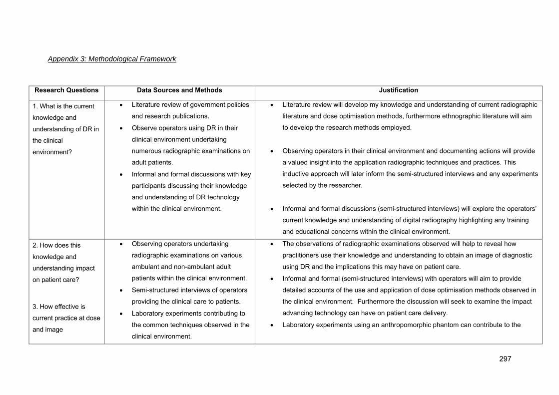
297
Appendix 3: Methodological Framework
Research Questions Data Sources and Methods Justification
1. What is the current
knowledge and
understanding of DR in
the clinical
environment?
Literature review of government policies
and research publications.
Observe operators using DR in their
clinical environment undertaking
numerous radiographic examinations on
adult patients.
Informal and formal discussions with key
participants discussing their knowledge
and understanding of DR technology
within the clinical environment.
Literature review will develop my knowledge and understanding of current radiographic
literature and dose optimisation methods, furthermore ethnographic literature will aim
to develop the research methods employed.
Observing operators in their clinical environment and documenting actions will provide
a valued insight into the application radiographic techniques and practices. This
inductive approach will later inform the semi-structured interviews and any experiments
selected by the researcher.
Informal and formal discussions (semi-structured interviews) will explore the operators’
current knowledge and understanding of digital radiography highlighting any training
and educational concerns within the clinical environment.
2. How does this
knowledge and
understanding impact
on patient care?
3. How effective is
current practice at dose
and image
Observing operators undertaking
radiographic examinations on various
ambulant and non-ambulant adult
patients within the clinical environment.
Semi-structured interviews of operators
providing the clinical care to patients.
Laboratory experiments contributing to
the common techniques observed in the
clinical environment.
The observations of radiographic examinations observed will help to reveal how
practitioners use their knowledge and understanding to obtain an image of diagnostic
using DR and the implications this may have on patient care.
Informal and formal (semi-structured interviews) with operators will aim to provide
detailed accounts of the use and application of dose optimisation methods observed in
the clinical environment. Furthermore the discussion will seek to examine the impact
advancing technology can have on patient care delivery.
Laboratory experiments using an anthropomorphic phantom can contribute to the

298
optimisation?
common variable techniques observed in the clinical environment.
4. What does this tell
us about DR in
contemporary
radiographic practice?
Examination of empirical evidence
discussing the implications of advancing
technology in contemporary
radiographic practices.
The author will use the empirical data collected to answer the following question.
Through examining the cultural underpinnings of radiographic practice and digital
radiography the author will provide arguments concerning the application of advancing
technology.

299
Appendix 4: Patient Information Sheet
Patient Information Sheet
Researcher: Mr Christopher Hayre BSc (Hons) || Supervisors: Dr Alison Eyden, Dr Shane
Blackman, Dr Kevin Carlton.
Study title: To explore radiographers’ knowledge and understanding of dose and image
optimisation in digital radiography (DR) with the aim of developing new knowledge for practice.
Invitation: PhD research is being performed in this X-ray department. Before you decide to take
part it is important for you to understand why the research is being done and what it will involve.
Please take time to read the following information carefully and please raise any concerns that
you may have.
What is the purpose of this project? As healthcare professionals our aim is to deliver the best
possible care to you. The purpose of this project is to improve how we take X-rays. When you
have an X-ray you receive a small dose of radiation, it is the purpose of this research to try and
reduce this dose with new equipment available to us. In doing this we strive to deliver the best
possible care to you and conform to current legislation ensuring that radiation doses should be
kept ‘as low as reasonable practicable’ (ALARP).
What information is recorded about my X-ray? No personal details about you will be recorded.
The only information recorded will be the technical information healthcare professionals use to
create an X-ray image of you.
Why have you been chosen? You have been chosen because before I attempt to reduce X-ray
doses I need to begin by observing the X-ray doses we currently use. The X-ray information
obtained will then be used in a research laboratory where I will perform experiments on an X-
ray model where I will attempt to reduce the X-ray doses without compromising the X-ray
quality.
What will happen to me if I take part? If you agree to take part the researcher will observe your
X-ray examination in the X-ray room behind the X-ray screen. As a result of this research no
changes to your service or to your patient care will be affected as a result of this observation.
Do I have to take part? It is up to you to decide whether or not to take part. If you do decide to
take part I will observe your X-ray examination behind the screen. You may also ask me to
leave the X-ray room at any time during the examination, without giving a reason and it will not
affect your care in anyway.

300
What are the possible benefits of taking part? Taking part will not benefit you individually but
you will be improving our current knowledge and understanding of X-ray use. This improvement
in knowledge and understanding will enable the researcher to potentially reduce radiation
doses using technology, ultimately developing our profession and improving our care to
patients.
What will happen to the results of the research study? The results of the research study are
part of a PhD project. The results will contribute to future academic studies and may inform
radiographic departments nationally through publications in scientific peer reviewed journals. It
is hoped that the results will be presented at the United Kingdom Radiology Conference and to
other appropriate audiences.
Who is organising and funding the research? The researcher and his academic supervisors are
all employees of Canterbury Christ Church University (CCCU). The research supervisors are
senior academics at CCCU and the research is funded through CCCU PhD Graduate School
Scholarship program from October 2010 – August 2013.
Thank you for taking the time to read this information leaflet, if you do have questions
regarding this study then please ask a member of staff.

A
Appendix 5: Deppartmental Poste
er
3001

302
Appendix 6: Participant Information Sheet
Participant Information Sheet
Name of Researcher: Mr Chris Hayre
Supervisors: Dr Alison Eyden, Dr Shane Blackman and Dr Kevin Carlton
Chair: Professor Kate Springett
To explore radiographers’ knowledge and understanding of dose and image optimisation in
digital radiography (DR) with the aim of developing new knowledge for practice.
You are being invited to take part in a PhD research study. Before you decide to take part it is
important for you to understand why the research is being done and what it will involve. Please take
time to read the following information carefully and discuss it with others if you wish. Please ask (the
researcher) if there is anything that is not clear or if you would like further information (contact Chris
Hayre on 07967280876 or email [email protected]). Take time to decide whether or not
you wish to take part and thank you for taking the time to read this.
1. What is the purpose of the project?
The purpose of this study is to develop dose reduction techniques using DR in order keep doses ‘as
low as reasonable practicable’ (ALARP) to all patient groups whilst undergoing a medical radiation
exposure. Medical exposures are the largest man made source ionising radiation to human beings
and although it is identified that individual X-ray doses are reducing in practice, the Health Protection
Agency recently identified that the average annual radiation dose to each member of the public has
increased. The purpose of this study is to study your experiences with DR to date to find out whether
it has facilitated or hindered certain areas of your radiographic practice; secondly, laboratory
experiments using an anthropomorphic phantom (to mimic a patient) with suitable radiation detectors
will be performed by the researcher, building on previous observations and knowledge to potentially
develop new methods of dose reduction for practice. Finally it will be determined whether the
technique used to produce the digital radiograph in the laboratory setting can practically and safely be
incorporated into a clinical environment.
2. Why have I been chosen?
You have been chosen because your job role allows you to undertake radiographic examinations
using digital radiography.
3. Do I have to take part?

303
It is up to you to decide whether or not to take part. If you do decide to take part you will be given this
information sheet to keep (and be asked to sign a consent form). If you decide to take part you are still
free to withdraw at any time, without penalty or loss of benefits, and without giving a reason.
4. What will happen to me if I take part?
If you agree to take part we will arrange a time and a place that is mutually convenient to conduct a
short interview. The interview will be recorded and later transcribed, we anticipate that the interview
could take between 30 and 40 minutes, but this is a rough guide. Secondly the researcher may
observe you as an operator performing digital radiographic examinations on an adult patient.
5. What are the possible benefits or disadvantages of taking part?
Taking part will not benefit you individually but you will be contributing to our existing current
knowledge and understanding of DR and its uses in practice which will enable the researcher to
potentially reduce radiation doses using DR technology. There are evidently implications for the use of
your time and we appreciate the help you will be giving us.
6. Will what I say in this study be kept confidential?
If you agree to take part in this study all information which is collected about you will be kept strictly
confidential. All research members of the team and the person transcribing the audio recordings have
signed a confidentiality agreement before commencing the study. The interview data will be
anonymised and kept in accordance with the Data Protection Act 1998.
7. What will happen to the results of the research study?
The results of the research study will be used to build on our existing knowledge of DR. It is intended
that the results could be further extended nationally within other radiographic departments and thus
contributing to future academic studies through publications in peer reviewed journals. It is hoped that
the results will presented at the United Kingdom Radiology Conference and to other appropriate
audiences.
8. Who is organising and funding the research?
The research team are all employees of Canterbury Christ Church University (CCCU). The research
supervisors are senior academics at CCCU and the research is funded through the CCCU PhD
Graduate School Scholarship program over three years from October 2010 – Jan 2014.

304
9. Who has reviewed the study?
This study has been reviewed and approved by the CCCU Graduate School Research Degrees Sub-
Committee. It has also been reviewed and approved by the Trusts Research and Development
Department.
10. Contact for further information
If you require further information you can contact a member of the research team.
Research Team
Thank you for taking the time to read this information leaflet, if you do have any questions then
please do not hesitate to contact me.

305
Appendix 7: Participant Consent Form
Participant Consent Form
Title of the Project: To explore radiographers’ knowledge and understanding of dose and
image optimisation in digital radiography (DR) with the aim of developing new knowledge for
practice.
Name of Researcher: Christopher Hayre / Alison Eyden
Please initial box
I confirm that I have read or been informed of and understand the information
sheet dated ……….……….. for the above study and have had an opportunity to
ask questions.
I have been informed that the confidentiality of the information I provide will be
safeguarded and understand that I could not be identified in any report, and the
audio recording of the interviews would be destroyed when the study is
completed.
I understand that my participation is voluntary and that I am free to withdraw at
any time without giving any reason, without my medical care or legal rights
being affected.
Data Protection: I agree to the researcher processing data that I have supplied.
I agree to the processing of such data for any purposes connected with the
Research Project as outlined to me.
I agree to take part in the above study.
_________________________
Name of Person
______________
Date
Signature
_________________________
Researcher
______________
Date
Signature
1 copy for participant; 1 copy for researcher

306
Appendix 8: Layout of general imaging sites A, B, C & D
Site A

307
Site B

308
Site C

309
Site D

310
Appendix 9: Initial Observation Data Sheet
Site: DR room:
Date:
Operator: Student | AP | Rad Exp: | 1 2 3 |± 5 yrs |6-10 yrs| 10≥
Patient ID: ________________
Exam: _______________________
__________________________________________________________________________________________
__________________________________________________________________________________________
__________________________________________________________________________________________
__________________________________________________________________________________________
__________________________________________________________________________________________
__________________________________________________________________________________________
__________________________________________________________________________________________
__________________________________________________________________________________________
Projection: AP | DP | PA | LAT | OBL Focus: Fine | Broad Manual Exposure: Y/N AEC: Y/N Chamber: __________________ Pre-set exposure: __________ kVp _____________ mAs X-ray exposure: __________ kVp ________ mA(s) / ________s FFD: ____________ EI: __________ DAP: __________ cGycm2 Field Size: __________
Projection: AP | DP | PA | LAT | OBL Focus: Fine | Broad Manual Exposure: Y/N AEC: Y/N Chamber: __________________ Pre-set exposure: __________ kVp _____________ mAs X-ray exposure: __________ kVp ________ mA(s) / ________s FFD: ____________ EI: __________ DAP: __________ cGycm2 Field Size: __________
View One View Two

Cha
Door
Cupboarand ClinBins
Chan
anging cubicl
G E N E
Compute
rds ical
nging cubicle
es
R A L R O O
er
es
O M
Wall d
X‐ra(ceilsusp)
etector
ay tube ling pension
X‐r
ray Table
Sink
DR detectobelow table
311
r e

Append
dix 10: Scannned copy aaltered data
a collection
312

313
Appendix 11: Observational transcription
Date: 12/07/12
Method: Observation
Time: 09:00-17:00
Site: Site B
Their attire was strictly clinical with their white tunics black trousers. I began by
observing just the one DR room, I began by taking notes on A4 noted paper, I am
finding extremely difficult to stand up and ensure that I took down all of the
information required for each radiological examination observed in practice. The
complexity behind this lays with the workload the radiographers have to content, thus
following the speed in which examinations need to be done. For example in a
department which had three radiographic rooms available for X-ray examinations,
only two were operational. A chest examination observed took over one minute to
complete from the patient entering the X-ray room to the patient exiting the X-ray
room. Although my intention was to participate as only an observer in the X-ray
room, this proved extremely difficult within the time observed. There were occasions
whereby I have to chaperone patients, assist with moving trolleys and chairs as the
practitioners were on occasions finding it difficult to move. One occasion occurred
whereby my presence as a research not only aided the practitioner as I had to
intervene. Upon observation a patient fell off a chair whilst sitting and in reaction the
radiographer rushed to the patients aid and I simply followed behind her, together we
were both able to safely and appropriately move the patient back in the position
necessary for the operator to take the radiograph. The operator ensured that the
appropriate paperwork was carried out during this matter. As time went on
throughout the observations the staff would ask me send their images to the PACS
system once they had been checked by the operator. This occurred only a few times
as the operators on occasion would forget to send their images upon accepting them
and would ask me to send them. I felt that it was ok for me to do this, as the
operator they had accepted the image and I was just conforming to their wishes. I
found that during my time on this first observation that I had to make the right
impression. I ensured that I helped out when I could as the operators did work on

314
their own the majority of the time. And with regards to the patient falling the operator
did say that ‘I’m glad you were here’… I felt that without aiding the staff I would have
built up unnecessary tension between the staff and myself which may have hindered
follow-up relationships in the future in such observational and interview work. Again
within this role I was not just sending images to PACS by I would also be asked if I
would bring up the ‘work list’, check a patients attendance to the department, as I
would be near the computer where this was normally done. As I was familiar with
the set up I was able to correct wrongly booked patients, which I felt allowed me to
become part of the team, culture and ultimately and insider.
As I observed I place myself behind the protective lead screen, on occasions I would
come out of this position to aid the operator with a trolley or in the matter whereby a
patient fell from his chair. As I aimed blend in with the staff I ensured that joined the
staff on their coffee breaks, this allowed me to talk about the general issues with
various members of staff, if also helped me being not being stereotyped as just a
researcher. I was also thanked on occasions with help I had shown and given during
the observations from various members of staff. There are other instances whereby
I would suggest/aid the operators, for example the digital equipment would have to
be on the correct setting for the operator in order for it to work effectively for the
patient. If these settings are missed then either poor image quality may be
produced. Therefore as I was familiar with this technology I knew when certain
settings were set up correct, for example the erect detector being selected for a
patient who is on the table couch. Or where there was a grid in a DR equipment it
was not supposed to be, I would therefore highlight this error to the operator as they
would be standing in the room with the patient and would not be able to see such
information. I felt that my assistance in this matter allowed the examination and
relationship between the informant and the research to run smoothly. This also
recognised that this happen on several occasions whereby the incorrect detector
was selected and or where the grid would need removing or placing in.
315
The environment
Radiographers would use the ‘viewing area’ to process their patients data ensuring it
had arrived to the PACS system. In the ‘viewing area’ the cassette radiography (CR)
processing took place and was often used when DR was either unavailable or not
preferred for a specific examination. One radiographer did mention that they
preferred the CR system and would use this over their DR systems. Outside the X-
ray rooms displayed the radiation notices on entrance to the room ‘X-ray controlled
area’, ‘no entry’ and no unauthorized access, with the local rules adjacent to this.
Similarly outside the DR room light boxes stating ‘controlled area’ in a constant
yellow light and ‘do not enter’ in a red light (when an X-ray exposure is occurring in
the room). However on another X-ray room (the CR room) the yellow light box
outside of the room did not stated ‘controlled area’ but stated ‘room in use’ and again
‘do not enter’ in red.
The DR room in question was large, approximately 6 meters by 9 meters the walls
were painted yellow and it was a dark environment, dark in order for the
radiographers to see the collimation light. Within the room it processes all of the
clinical facilities to undertake all radiographer examinations, patient types and patient
history, including hand washing/cleaning facilities, appropriate disposal of clinical
and/or general waste bins. Within the room there were no windows and the floor
was grey. Outside of the room illustrated three changing rooms connected to the
room which would allow them as members of the public enter the changing room
through one door and then through another door (within the X-ray room) the ‘patient’
would emerge. This environment provided the patient with a little more dignity as the
patient once changed into the hospital gown did not have to wait back in the waiting
room with the remaining patients. Similarly one the patients X-ray examination was
finished they would enter into their personal changing room and redress into their
normal attire and then leave the changing cubicle once appropriately dressed. On
entrance to the DR room signs illustrated the dangers and highlighted the authority
as ionising radiation was in use.
To the patient the X-ray room would be unlike any other hospital ward or clinical, with
alien looking technology situated in a cold and dark room. It appeared that maybe

316
there was an element of ‘performance’ associated with this environment. Whereby
the operator controls the performance, patient must do what they say, whereby the
patient must remain still in that position for several seconds before the X-ray is
taken. During this time the patient is left in the room and the performer (the
operator) leaves the patient behind a protective lead screen whereby the X-ray is
performed. At the speed of light this process is now complete and may or may not
require further imaging.
Within the DR room, there is an array of technical information, suggesting the
optimum exposures for particular examinations aiding to provide the operator with an
exposure adequate for the radiological examination being undertake. Following this
there is a document illustrating the diagnostic reference levels for particular
examinations, thus highlighting the need to keep within these levels. Notices such as
‘Are you using the correct lead protection?’ were clearly demonstrated behind the
lead screen. All of these notices aim to aid the operators and thus ultimately the
patients. Not only were there clinical devices in the DR but also a set of ladders in
the corner. And although this was a fully operational DR room it also allowed for
cassette radiography as a computing device was mounted on the protective screen.
Note that the DR system was fixed, and it did not have removable detectors, the
detectors were fixed in the table and in the wall stand. The environment presents
other sensory stimuli, such as a smell of burning, it was clarified that this was not
from the equipment or room but from the neighbouring corridors and was present in
the viewing room and other corridors alike. The temperature in the room was aimed
to remain at a set temperature through air conditioning was fully active, similarly this
present an acceptable level of noise in the room.
As a male I was required on occasions to leave the X-ray room, the operators would
on occasion suggest that I leave if the patient required to remove items such as a
bra, I was however invited back into the room once the bra had been removed to
further observe the examination taking place.
I got the impression that the clinical X-ray room was the performance area and yet
the viewing room was the place to discuss other matters, it was apparent for
example that the staff would happily discuss matters regarding the equipment

317
outside the X-ray room and in the clinical area. In this environment the ‘processing
room’ was several meters away from any clinical area. There appeared to be a clear
divide between the performance in the clinical room and the performance in the
viewing room. Within the ‘processing room’ the coordinating radiographer would
delegate work load and deal with the day-to-day requests or concerns via telephone
or face to face with other members of staff. The radiographer would performed X-
rays during the day therefore there role was demanding. They would be mainly
resident in the viewing room and appeared to be there during the observations.
Within the viewing room it was clear to see the notices and safety equipment such as
the COSHH, emergency eye washing bottles, first aid and good practice for hand
hygiene, and good practices for maintaining a good working clinical environment.
White boards would highlight where other radiographers were (either on a portable
or in a theatre). It would highlight current faults logged with specific radiography
equipment or general issues within the department. Within the processing room
computers were readily available for processing patient data, creating compact discs
for others. There was the ability to still print X-ray films in the likelihood of a PACS
system failure. The processing area provided educational facilities for the student
radiographers to perform paperwork associated with their academic portfolios. The
processing room itself has no secure access; accessible from the corridor where
patients and the general public alike can observe. The area itself is considerably
large when compared to other viewing rooms. Above the coordinator the CCTV was
illustrated and this allowed staff members to view all aspects of the waiting room and
corridors in the surrounding areas.
The waiting room was big and provides large cushioned chairs for the patients
waiting for their X-rays. Within this waiting room other patients on other modes of
transport were apparent including those in wheelchairs, A&E trolleys and hospital
beds. The patient types varied from children, adolescents, adults and the elderly
each with their clinical concern. A child’s play area provided stimulus for infants.
Within this area the visible aspects were the danger signs behind cupboard doors
warning of electrocution, signs warning individuals not to use their mobile phones
within the department.

318
The equipment
Throughout the three-hour observations of the Philips DR equipment a technical fault
occurred with different operators of the equipment, it is a known fault with one of the
sensors because although the machine was ‘locked’ into position for a radiographic
examination to take place, the computer system did not recoginse it was ‘locked in’
and therefore it would not expose. When it recognised that the device was locked in
a green circular light appeared on the exposure console, which deemed it necessary
for it to expose. The issue highlighted by the members of staff was that this was
completely random, on occasions they would have patients on the X-ray table,
standing for Chest X-rays and then the equipment would not allow them to expose
even though the equipment was set up and would allow them to exposure 10
seconds earlier. This was not the first time with this fault and was logged first thing
in the morning and the radiographers had organsied for an engineer to come in
immediately to fix the issue. This ‘green light issue’ which it became to be known
would occur during most of the radiological examinations examined throughout the 3
hour observational block.
It was observed that on the whole, radiographers would not alter the preset kVp and
mAs settings. It was mentioned several times by the radiographers, including a
senior radiographer that ‘someone preferred the images in the other room, although
the doses are much higher’… This comparison was made with another DR room, a
piece of Carestream DR equipment. However it was mentioned in the viewing area
that the does appeared much higher in the other DR room when compared to the
Philips room. This was mentioned informally by several radiographers to whom have
worked with the equipment.
It was commonly recognised that operators would not place markers on the
detectors and would use the markers on the DR system, thus placing them on post
radiation exposure. Similarly it was noted that operators do ‘crop the image’ post
exposure,
One obstacle the staff recognised with the DR system was that on certain
examinations the equipment was not as flexible as they would have liked. For

319
example the equipment would only expose when ‘locked in’ a certain position.
Therefore if you move the equipment out of this position it would not let the operator
expose, therefore the operators had to move the patient into position on the upright
detector, which took longer to do.
An aspect of the DR equipment can allow it to be controlled by a remote control if
required, however I did not observe any operators doing this. Once the equipment is
‘locked’ into position the operator may stay with the patient on the erect detector and
move the detector, this will in turn move the X-ray tube, this practice allows the
operator to be with the patient at all times and not have to move both wall detector
and X-ray tube into position. We talked about the performance from the staff
however what about a performance from the equipment, this equipment is essential
to patients care, however it is was often talked to as it if had a personality, whereby it
would not comply with what the operator wanted it to do and would therefore not be
‘playing ball’. This is not only a performance for the patient but for the radiographers
and others within the department.
The staff
Throughout the observations I observed several operators, both assistant
practitioners and practitioners. One AP had over 5 years’ experience and some of
the radiographers had between 2 and 10 years’ experience. What was highlighted
was that because I also practice as a radiographer on occasion’s radiographers
would ask my opinion regarding a certain technique, for example an ‘AP skull’ as
these are regarded as uncommon techniques to perform, which proved problematic
for the junior radiographer. Throughout the observations it was recognised that all
operators generate sound rapor with the patients and many were thanked as
patients left the X-ray rooms. Patients often asked questions regarding their care
and/or results and the operators would ensure that the correct information was given
at all times answering the patients concerns.
One staff did ask questions regarding the X-ray form she had and whether the
patient should be given it back for them to take to A&E, she was unaware of the

320
processed because she normally works at another site, therefore unfamiliar with the
process. She asked for my opinion however I was unable to help her on this
occasion as I was also unaware of the process, as I had never worked at the site.
After one X-ray of a patient an operator had come up to me and confirmed that a
patient had seemed quite ‘offish’ with her. She said that she had identified as soon
as the examination has taken place. She asked whether I felt the same and I did
agree that I felt the patient was ‘offish’ with the radiographer. This patient had
attended for a thumb and follow-up scaphoid X-rays and the examination took no
longer than 5 minutes to complete. What was observed was that we healthcare
professionals the operators had an immediate opinion of that patient.
During one examination I was asked by the radiographer ‘are we passing for you’, I
simply replied, ‘of course’ and smiled. This was the first glimpse into where there
may be hostility within the observational aspect of this study.
Techniques
Interestingly the AP shoulder projection was set up to use the AEC central chamber,
however when the operator (a practitioner of more than 10 years’ experience) did not
center over the central chamber and exposed the patient. While observing the
radiographers in the working environment, I had witnessed good practice, in the fast
paced environment radiographers produced images of diagnostic quality which were
acceptable for submission to the PACS system, ready to be interested by the refer
and/or reported on also. On a whole the exposures used were generally from the
preset exposures on the machines, one example for a DP foot projection was 52 kVp
and 3.2 mAs, thus producing an image of diagnostic quality. There was one
occasion whereby an operator reduced their kVp as the patient was much smaller
than your average patient and again produced an image of diagnostic quality.
Collimation was however an aspect where on occasions it would be cropped on the
post processing images, therefore although initially they had collimated, they had in
fact in their opinion from their image to have actually over collimated and therefore
operators would ‘crop’ the image before it was sent to PACS. The observation of DR
had identified that there was a difference in radiographic technique when compared
to CR systems, for example on this observation the use of the AEC was used for an
AP shoulder projection, they are able to move both X-ray tube and detector

321
simultaneously yet only moving the detector in order to be nearer the patient.
Secondly after the X-ray exposure has occurred the image is displayed on the
computer system, radiographers when use tools to crop the radiographic image and
place annotations onto it.
Challenges to observing DR practice
The challenges presented with observing DR for the first time was the time frame,
through the radiological examinations observed a chest X-ray would have been
completed in just over one minute, therefore in order to collect as much information
as possible I have needed to adapt ones observing techniques and only document
the vital information required. In its own sense this is the culture presented to
operators in that they are able to perform radiological examinations this fast,
therefore is this the norm? This intern highlights the ethnographic issues in observing
radiographic practice, to this is not gradual, whereby an research may observe a
doctor or nurse with a patient over a period during the course of a consultation or
treatment, during a radiographic examination one patient may enter and leave the X-
ray room in just over one minute, therefore capturing the essence of the digital
requires ‘quick fire ethnography’ and on occasions retaining a lot of information from
memory and making note when there is an opportunity to do so. Other challenges
experienced included the issues in that I was required at times to assist with aspects
in the room, due to my status operators would ask for me to have a look at the
equipment or ask to chaperone family members, on these occasions I was difficult to
take note and therefore I had to remember the situation and note much later. The
other challenged experience was that because I was waiting in a DR room I had to
rely on the examinations brought in by the radiographers and in a three hour
observational block I did not receive a hand or wrist examination, which are of
importance and what I will be using to produce laboratory data.

A
S
Appendix 12: Ra
Site A – Hand ex
adiographic extre
xaminations obs
emities documen
erved
nted
3222

S
Site A – Wrist ex
xaminations obse
erved
32
23

S
Site C – Wrist exxaminations obs
erved
32
24
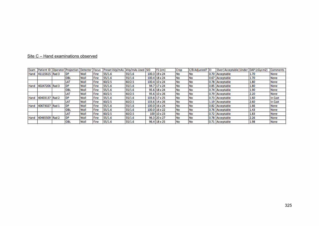
S
Site C – Hand exxaminations obs
served
32
25

326
Appendix 13: Interview Schedule (final version)
1. Can you tell me when you first started using DR?
a. As a Student? Probe was this a benefit and how would it compare if they did
not receive training in a clinical environment?
b. Since a Radiographer? Probe whether they think they were at a disadvantage
of having to learn the DR technology ‘on the job’.
2. Can you tell me about any education that you have received in relation to DR?
a. Probe at University?
b. Was this appropriate for clinical use?
c. Did the University have DR equipment?
3. Can you describe any DR training or education received within the imaging
department?
a. Probe was it appropriate?
b. Would you have preferred more / or to been have trained in a different way, if
so how?
4. What other training or education do you think would have been useful either at
University or in practice?
a. Probe such as an understanding of the detectors?
b. An understanding regarding the algorithms?
5. How long did it take to feel confident using the DR system?
a. Probe are there any areas which still concern you as a radiographer?
6. Can you identify areas where DR aids your clinical practice?
7. Can you identify areas where DR hinders your clinical practice?
8. How you change your radiographic technique when using DR, compared to CR?
9. How often do you rely on the exposure index during DR examinations and for you is it
an accurate assessment of the image quality produced?
a. Probe Are there occasions where you may use your own clinical judgment,
regardless of the EI?
10. Do you think technology such as DR is having an affect on our radiographic profession?
If so or not can you provide some examples of this?
a. Probe for the radiography students?
b. For healthcare professionals?
11. In our profession we have a particular set of skills to obtain an image of diagnostic
quality, can you tell me what you think these skills are?
a. Probe: How do you think others perceive our skills within our profession?
b. How do you think this skill base affects our radiographic profession?
12. Do you think the DR technology affects the patients’ care and experiences during their
X-ray examination?

327
a. Probe: Does it affect the healthcare professionals relationship with their
patients?
13. Do you think there are aspects of the patient, which alter our decisions in how we as
radiographers obtain an image of diagnostic quality from our patient in choosing either
DR or CR technology, such as a patients:
a. Illness?
b. Mental state - non compos mentis?
c. Mobility?
d. Hygiene and smell?
14. During my observations in a number of different hospitals over a number of days
observing lots of different staff I have notice that:
a. No Pb used – Question: The use of lead was rarely used when irradiating adult
patients – can you tell me why you think this may be?
b. Adjustment in brightness and contrast on X-ray images – Can you tell me what
you think are the advantages and disadvantages are of being able to
manipulate image contrast and brightness post exposure?
c. Post image cropping – Question: Although the use of cropping images varies at
the different sites observed, why do you think there is a tendency to crop X-ray
images in practice?
d. Altering exposure factors – Can you tell me how accurate the pre-set exposures
are on the DR console? Can you give some examples when you may adjust
them?
e. Limitations of equipment and environment– How do you feel about the
environment, setting and arrangement of the DR equipment and its use on
specific patients compare with CR? And how do you think this could be
improved for the operator?
f. During the extremely busy periods within the imaging department do you feel
that there may be a tendencies to alter ones practice in order to reduce waiting
times for other patients?
i. Probe such as using CR, whereby DR may be used?
ii. Being able to use concentrate on using protective measures such as
lead protection?
15. And Finally….Is there anything about the DR system that I haven’t asked you that you
think is important and would like to discuss?
Thank you for your time!!!

328
Appendix 14: Example of transcribed semi-structured interview
Respondent Mick
Researcher: Can you tell me when you first started using digital radiography?
Mick: October… Well, technically I started using it in 2010-ish, early 2010, because I used it
when I was on my electives.
Researcher: So you used it as a student.
Mick: Not as a student.
Researcher: Only on the electives.
Mick: Yes, I used it for about what, two weeks?
Researcher: So mainly as a radiographer then.
Mick: Yes. Well, since I’ve been qualified I’ve been using it in Medway.
Researcher: So do you think that learning it on the job as a radiographer, you were at more of a
disadvantage in that way - having to learn on the job? Would you have preferred maybe
education as a student?
Mick: I think that ideally as a student, it’s best to use different sorts - to use both and mix it - to
use both CR and DR, so you get a more appropriate skillset when you qualify. But it’s not a big
thing. I wouldn’t say that it’s an essential thing, but it would help you if you are going into a
different environment. Because I can see how, if you went into a hospital which is all DR, and
you’ve not used DR before, how you would be at somewhat of a disadvantage compared to
other people who have used it before.
Researcher: Fair enough - excellent. Can you tell me about any education that you’ve received
in relation to DR? For example, at university?
Mick: I can’t remember that much.
Researcher: How much of it was there, do you think?

329
Mick: There were a couple of lectures. with Christopher Geoffreys.
Researcher: Was it enough, do you think?
Mick: I think there was enough - because it was mainly surrounding the DR physics, and how
they produce the images. From that respect. Because most of your technique, you learn on
placement anyway. Well, when I was at university. “When I was at university…” [laughter] In
regards to technique, it wasn’t taught.
Researcher: OK. So do you think then this education at university was appropriate for clinical
use?
Mick: Yes. Because it gives you the background information that you need to be able to
understand how to produce images. And image optimisation to a certain extent.
Researcher: OK. And what was that - can you go into that? What did they teach you - can you
remember? Was it detectors?
Mick: It was to do with detectors and how the image is formed. The physics-y bit.
Researcher: Was there a lot of technique in optimisation [like] “if you did this, you’d get a better
image”?
Mick: No, there wasn’t that. It was the basics of…
Researcher: The physical part of it, the image acquisition sort of thing.
Mick: It wasn’t full-on. When I was originally trained in the DR room, the applications specialist
was here, so the applications specialist and the engineer were talking more about image
optimisation in that respect.
Researcher: OK, good. Did the uni have DR equipment?
Mick: No. The Medway campus at the time - I don’t know if it’s changed, but at the time, it had
CR.
Researcher: Can you describe any DR training or education received within the imaging
department?
Mick: I had CNST? Which was done by the trainer in the room.

330
Researcher: OK. What’s the CNST?
Mick: I’m not sure what CNST stands for, but it’s essentially to say you’re competent in every
aspect to use that room, and to know where all the buttons are and what all of the programs do.
And how to fix things if they go wrong.
Researcher: Oh right. Is that something from the applications specialist?
Mick: No, that is… I don’t know if other hospitals have it, but at Medway it’s your competency to
say that you are competent to use a piece of equipment in regards to either producing a
radiographic image, or... Even CR units have them.
Researcher: And the IIs I assume, and things like that.
Mick: IIs, yes. QI equipment’s got one - all that stuff.
Researcher: OK, good. Excellent. So do you think that was appropriate?
Mick: I think if it’s done properly, yes.
Researcher: So there are instances where it might be not? Some people have not even
mentioned it.
Mick: No, this is the thing. People don’t always get trained by people who should be training. It
becomes something where “someone told me to do this”, but it’s not the right way. So therefore
you’re not necessarily getting optimal images and you’re not reducing the dose to the extent
that you could do, because it’s just hearsay. And it’s not necessarily right, because I’ve been in
there with newly qualified members of staff and no offence to them, but they don’t understand
things as well. Because they’ve said “Oh well, Thingamajig did it this way” and you go “Well
actually, that’s not how you do it.”
Researcher: But because they’ve been shown that way -
Mick: Because they’ve been.
Researcher: And they get an image. It might not be optimal or the right way of doing it, but they
sort of get into that way of -

331
Mick: Yes. It’s the way that they’ve been taught, so therefore that’s the way that they follow,
because they don’t know -
Researcher: Any better. So that’s quite a thing, isn’t it really? OK.
Mick: It’s to do with staffing issues and things like that.
Researcher: Yes, there’s things like that, isn’t there? Would you have preferred more or to have
been trained in a different way within the department? And if so, how?
Mick: I think the way that I got trained was when the applications specialists were present, as
well as the room trainer. I think that was quite a good level of training, really.
Researcher: It is really, isn’t it?
Mick: I think I caught quite a lucky break.
Researcher: Because a lot of people obviously don’t get that.
Mick: No. A lot of people don’t get the applications specialist. That would tend to be the first
couple of months we were using the room. And obviously a lot of people that were trained by
the applications specialist have moved into specialities and things like that.
Researcher: Yes, so you sort of lose that core training don’t you as such? OK. What other
training or education do you think would have been useful either at university or in practice?
Mick: Well… [pause]
Researcher: Do you think maybe such as the understanding of… or you said you were sort of
educated in the understanding of detectors - maybe sort of with that, and the algorithms and
things? Or things that are happening behind the scenes that we don’t really see?
Mick: Yes, I think that would be good to a certain extent. I mean obviously, it’s a background
information sort of thing. Knowing how you get to that stage, I think it is. I think it would be
useful. Obviously there are a lot of other things that you have to cover in your time at university,
so obviously it’s not...
Researcher: That’s right, yes. So is there anything you think that would be useful, or were you
quite happy with the…?

332
Mick: Personally, I was quite happy with the way that it was put across to me.
Researcher: Good. How long did it take to feel confident using the DR system here?
Mick: Er [pause]. Confident using it? I would say just a couple of sessions, probably. Two, three
sessions? A day, a day and a half?
Researcher: So quite quick then.
Mick: Well yes, I’m quite into that sort of technological frame of mind.
Researcher: It helps.
Mick: It helps if you get on alright with technology, essentially. If you’re alright with computers
and things like that, you’re fine. Because then you understand the filters and the way that it
works. Because just the way that the GE system here was set up - it’s not the most user-
friendly. So you have to kind of know how to work it to get -
Researcher: Round things.
Mick: To get round certain aspects of the system which aren’t user-friendly, or patient-friendly.
Researcher: OK. Can you elaborate on any of that?
Mick: Yes - it’s just because the way the system works, it’s not always… DR generally isn’t.
Because CR, you adapt the plate a lot more than… the plates aren’t as adaptable in the DR
environment. The way that the system works in there… when I’ve used other DR systems,
they’re very adaptable in terms of angles, and it can work quite well. Very similar to the way that
I would use CR. If the patient can’t do that, you adapt the environment to what the patient can
do. Whereas the DR room seems to find it more difficult in that adaptation sort of angles [sic].
So you have to do free cassette more, and things like that. Which aren’t ideal really. It’s a heavy
cassette, it’s got a long lead - which isn’t always best.
Researcher: So do you think there’s a confidence - not just for yourself, but generally - issue
with that then?
Mick: With that room?
Researcher: Yes.

333
Mick: I think with that system, there is. Unfortunately so. I think compared to some other units
that I’ve used previously, the GE system doesn’t fill you with confidence when it gets a bit
finicky.
Researcher: Yes, fair enough. Good. Are there any areas which still concern you as a
radiographer? In regards to the confidence, you mentioned the moving of the stuff and the
detector and things - is there anything in particular in the entire system?
Mick: I would say probably the override concerns me. The use of the override.
Researcher: The turn of the key.
Mick: The turning of the key. Because obviously where people haven’t… as I referred to earlier,
people not getting trained appropriately has led to an increased use of the override in the
system. Which probably isn’t the best way to go about producing [an image].
Researcher: It was observed quite a bit that people would do that - that people would just -
Mick: Yes, that’s what I’m saying. Because if you turn the key, the collimators come out. So you
have to watch your collimation, which then affects dose and things like that. And also, if
you’re… when you put the override on, you don’t have to… you can be anywhere. You can be
pointing it at a totally different place. Because the plate doesn’t always follow the tube when you
put the override key on. So you can expose, and it won’t come out. Because someone’s
pressed the override key and forgotten to turn it off.
Researcher: OK. I noticed that it affects -
Mick: I suppose essentially that’s a radiation issue.
Researcher: I suppose if you look at it though - if you look at it from a CR perspective, we don’t
have that sort of feature, do we?
Mick: No.
Researcher: We can potentially have errors of over-collimation and… so I don’t know, is there a
tendency to sort of protect what we’re doing - trying to restrict us, do you think? Or is it a safety
[issue]? Do you think it’s a good intention, sort of thing?
Mick: What, why people turn the key?

334
Researcher: Why the key’s actually there in the first place, I suppose. Because we haven’t
needed that, have we?
Mick: Well, it’s because the system that is installed will only let you expose if you are in the 38
by 38 detector. And if all of the light beam is within that detector area. And you have to be… I
think it’s about 8 degrees it will let you do. So you have to have 8 degrees or less, and all the
light inside the detector area for it to expose. And when people perhaps lose patience with it,
they find it a lot easier to turn the key than fiddle around with the free cassette etc. So I think it’s
more of a patience and a training issue. Because if you were trained to bring the free cassette
out essentially straight away, then you wouldn’t have that issue. And it would work.
Researcher: True. OK, good. Can you identify areas where DR aids your clinical practice?
Where does it help it?
Mick: [pause] say that it aids clinical practice. As in skills?
Researcher: As in anything, really.
Mick: I think the throughput of the DR room essentially, because of the positioning of it and the
use of the DR room in this department, where it’s outpatients only. Well, not only, but the
majority of the throughput is outpatients and it’s in the outpatients area. It does get a lot of
throughput in a short space of time, if it’s used correctly. You can easily do more patients than a
CR room.
Researcher: OK, good. Can you identify any areas where DR hinders your clinical practice?
Mick: Er, in regards to this department, it can be harder to man it in a way. Because where it’s
out of the department, and…
Researcher: So its location, more than…?
Mick: Its location more than… DR as a whole: my experiences previous to this system, DR was
a very good tool. And I think if it had the right system and it had been thought through properly
in the planning stage, then it would be a very useful system to have. And I think the problem
with that is now that a lot of people I believe in this hospital or in this department, where they’ve
been at Medway for a while, and they’ve only had exposure to CR, that isn’t the best way to go
about having your first experience of DR. Because now people are like “Well, the DR room’s
rubbish”. But DR as a whole: that’s the worst DR room I’ve used. My [other] experiences with
DR are a lot better. I know that pretty much anything we get - if we got something different -
would be better.
335
Researcher: Yes, OK. So it hinders mainly because of your previous experiences.
Mick: It’s just the way -
Researcher: The actual whole thing itself - the way it’s set up -
Mick: That room I think can be a hindrance, in the way that it’s set up. They way that it’s been
thought through and the way that it’s been planned. But DR as a system and as a way of
working I think is a very useful way to work, and I think it’s a very efficient way to work.
Researcher: Sure, OK. How do you change your radiographic technique when using DR
compared to CR?
Mick: A lot of the time in DR, like I mentioned earlier, you have to work around the system rather
than work around the patient. Which tends to be more the way with CR. So in a way, you adapt
- in a way, it’s quite good that you get outpatients, because you get them to move rather than
moving the plate and the equipment and stuff like that.
Researcher: The patient’s moving a lot more for our benefit, really.
Mick: Yes. Rather than us moving the stuff. To get the same image - well, a slightly better
image.
Researcher: Do you think that’s a hindrance in itself? Do you think for not so much us, but -
Mick: I think for the patients… if you have a patient who’s not particularly ambulatory, I think that
could be quite a hindrance, really. Recently I had a lady, and she was quite old and we had a lot
of stuff to do. And she couldn’t turn, and things like that. In which case, with the way the system
works in that room, it was -
Researcher: You’re restricted, aren’t you?
Mick: It was restricted for her. If she’d gone to the CR room, maybe perhaps she would have
had a better patient experience.
Researcher: OK. Do you think she would have done?

336
Mick: I think perhaps yes, because… Probably. I think so. Obviously, I treated her the same way
and that, but perhaps in terms of time spent in the room, then I think perhaps she would have
spent less time unfortunately, in the CR room.
Researcher: Because of the DR equipment. OK. How often do you rely on the exposure index
during DR examinations and for you, is it an accurate assessment of the image quality
produced?
Mick: I think usually it’s quite reliable in that DR room. It’s hard in a way, because obviously it
goes back to how I was taught and that. I’m much more accustomed to the Kodak and the Fuji
exposure indexes for the CR units. So I’m used to saying between -
Researcher: 1900 and unclear
Mick: Yes, around 1900. And whatever it is on the Fuji system - I can’t remember. Whereas I’m
not used to that exposure index to kind of place the equality. But it seems to be quite consistent
in terms of body parts, when I’ve obviously observed it.
Researcher: And so is it a good aid for you in a sense?
Mick: Yes. You can look at it and say “That compares to what I’ve seen in the past”. Because
when I had my training, I don’t remember the applications specialist saying “This is what it
should be”. In terms of consistency, it seems fairly consistent.
Researcher: So are there occasions where you may use your own clinical judgement,
regardless of the exposure index - if it’s, say, under-exposed? Or are you more on the edge of
well actually, it should be within that range of exposure index for it to be diagnostic?
Mick: I think you can tell, really, when an image is of diagnostic quality.
Researcher: Irrespective of the exposure index?
Mick: I think in some cases, yes. Because I know this is off topic, but when I’ve used CR, and
you say that it should roughly be on the Kodak system - around 1900. You look at it, and you
say “Well, that’s not really diagnostic for what they’re looking for.” And sometimes you’ll get
something which is by definition over the exposure index - so like, 2300 or 2400, something like
that. And it will give you a much better, more diagnostic image for what they’re looking for. So I
think sometimes it does come down to clinical judgement, in a way.

337
Researcher: Because you mentioned CR exposure indexes and you know their values and
things: with DR, are you quite familiar with them or is it a little bit of a grey area, sort of thing?
Mick: It is more of a grey area. Because obviously like I say, I didn’t get trained using it. So I’m
not 100% used to looking at it and going “<snaps fingers>, that’s right!”, or “That’s not right”. But
when I’m in there, if I do a couple of body parts, then I’ll be back saying “Oh, that’s roughly…”
Researcher: Because it adds three boxes, doesn’t it? It adds like… They’re both amber if
they’re over or under and green if it’s within the range. It’s quite a nice indicator actually, isn’t it?
Mick: It’s quite easy.
Researcher: You don’t have to know your numbers to unclear
Mick: Yes, if you’re not hot on the numbers but you’ve got the green/amber system, so it’s good
in that unclear DR’s got a lot more sort of recovery in it. The system can create a better image
from when it’s under-exposed or whatever. So sometimes it can be amber, but it’s still a nice
image which isn’t worth repeating.
Researcher: But you do refer to the exposure index quite regularly.
Mick: Oh yes. It’s there, isn’t it?
Researcher: You can see it.
Mick: It’s a handy tool.
Researcher: Yes, absolutely. Good stuff. So do you think then that technology such as DR is
having an effect on our radiographic profession? If so (or not), can you provide some examples
of this? As a whole.
Mick: As a whole, I think DR is good for the profession.
Researcher: Is it having an effect though?
Mick: I think it is. It’s a different way of working. Because it’s kind of moving with the times, in a
way. It’s a more technological area… era, even, and you have to move with the times.
Everything’s more computer-based now, and as a result, it’s a more computer-based system.
It’s all linked together, it’s a lot faster and that’s what people tend to… These days, a lot of
people want things that are fast, they want things that are efficient. And it’s a fast, efficient

338
system which is technologically relevant to the era that we live in, really. And I think there’s only
so much development that you can do within a CR, whereas with DR it seems to me that people
are struggling. You’ve got DR mobile units - they’re making them lighter, they’re making them
wireless, they’re doing things like that. And it’s having that sort of effect to drive the profession
forward in that respect.
Researcher: OK. What do you think its effects are for DR? What do you think its effects are for
radiography students, for example? I suppose it’s different here in a sense because there’s a lot
of CR, and not so much DR.
Mick: I think in this department…
Researcher: But for DR-related - what do you think the effects may be for radiography students
coming into the profession?
Mick: [pause] I think it depends, really. I think a lot of students now realise they’re going into
quite a technologically-based profession. I think it’s quite a hard one to answer, really. In
regards to the effects on them.
Researcher: Do you think it affects differently their education throughout their training?
Mick: I think so, because obviously you get trained differently because there’s the wet film kind
of people, who know how to use film and how to get it spot on in terms of when they develop.
And then you’ve got the CR, and you’ve got the DR. I think there are three distinct ways in
which people have learned. Because I learned using CR really, and the people before me were
wet film. I think it will bring another kind of…
Researcher: Culture.
Mick: Yes. By learning that way - because essentially I’ve adapted my understanding about…
But I took to it quite well. But you will have people that have essentially been brought up - not
brought up, but that’s how they’ve learned, so they will be -
Researcher: Because it’s watch… how you, as a student - you watch don’t you, and then you
repeat.
Mick: They will be a lot more familiar with that system, so in a way, they could perhaps get
better use out of it. Because they will be used to using perhaps DR only. I mean, at this
department it’s not quite as relevant, because you have a lot of CR. But in the future, or in DR-
only or mainly-DR hospitals/departments, it will have a greater effect.

339
Researcher: OK, good. Do you think it’s having an effect on us as healthcare professionals, the
actual DR technology?
Mick: I think so, yes.
Researcher: What sort of effect have you seen, from yourself or…?
Mick: [pause] I don’t really know, to be honest. I think it is having an effect in terms of… It’s a
different way of working, in that kind of sense. I mean, it’s very technologically-based. But
people these days use a lot of technology for a lot of things. So it’s not really affecting that sort
of… But it affects your practice and the way that you work, the way that you’re able to adapt.
Because perhaps in an A&E situation - because obviously our DR unit’s here - we aren’t in an
A&E situation, but perhaps you’d use it differently so it would affect your practice differently in
that way. But I wouldn’t be able to comment 100%.
Researcher: No that’s fine - no problem.
Mick: I think you need a flexi-room, kind of thing.
Researcher: Mmmm. One that you use for various patients on different pathways.
Mick: Yes, because you have… I know Philips do a good flex-room. The whole room is a lot
more patient-friendly. So in a DR situation, when I went to the spinal centre for unclear the way
that they used DR… their DR room was lovely. It doesn’t affect the way that they work - they
just produce nicer images. For imaging the spine, it’s perfect.
Researcher: OK, good. In our profession, we use a particular set of skills to obtain an image of
diagnostic quality. Can you tell me what you think these skills are?
Mick: To obtain the image, it’s your technique, your exposure and patient interaction. Because if
your communication’s good with them, then they understand what you want from them and they
understand what they’ve got to do to get the best image to help them get better. And if you
know your exposure and your equipment to produce the best image in terms of diagnostic
quality… yes, and your technique - if you’ve got technique that’s good, then you will produce the
images that need to be produced for the patient to go down the right pathway with the… ALOR?
ALARA? The least dose for efficient treatment.
Researcher: OK. How do you think others perceive our skills within our profession?

340
Mick: Within the profession?
Researcher: Yes.
Mick: What, as in more specialist radiography or are you looking at general radiographers? Or
CR radiographers?
Researcher: Maybe a bit of everything, really. Different modalities looking on general, or what
they may perceive -
Mick: I think everyone is quite on the same page, really. Because everyone generally speaking
kind of starts in a general environment. So even if you go to a specialist modality or you become
a manager or whatever, you’ve got that background where you have worked in general for
some period of time, no matter how short or long it’s been. So they have an understanding, and
I know that for instance people that have never used DR would be quite daunted by the use or
however [sic]. But I think generally, there’s a good perception. Is that OK?
Researcher: Yes, sure. How do you think that the skill base that you mentioned a bit earlier
affects our radiographic profession?
Mick: What was I saying about the skill base earlier?
Researcher: You said about communication, exposures and unclear
Mick: Oh right, yes. Sorry.
Researcher: That’s alright. So how do you think that affects our profession - does it really affect
our profession, in a way?
Mick: I think it does. This is going to sound mean, but you have good radiographers and you
have bad radiographers. You have radiographers who are very good with patients, and you
have people that tend to have their areas where they excel, and you have people that are very
good with other people - they’re very… they’re a people person. They’re very good at
communicating and getting the best of what they need from patients, perhaps in the situation of
a paediatric patient. They’re the people that can get the best from paediatric patients - they get
them to keep still when required and do things like that. And you have other people that are
perhaps better at kind of actually looking at exposures and looking at images, and things like
that. You have areas which people excel in.
Researcher: Yes, there are different areas, aren’t there? OK.

341
Mick: And some people are very good
Researcher: Yes [laughs]. Do you think that DR technology affects the patient’s care and
experiences during their X-ray examinations?
Mick: I think in this department it does, yes. I think a lot of outpatients now, especially with the
way the department currently works, can be… say for instance if the DR room is down for
whatever reason, if it’s broken or if there’s not enough staffing levels… whatever the reason
may be, the knock-on effect in terms of… or even if you sent the DR patients to the main waiting
area to have their X-rays done in the CR rooms, I think the experience… because from my
experiences, when I’ve X-rayed patients these days, they are like “that’s fast compared to…”
because they come from outpatients, they sit there, you go talk to them and you get them in as
soon as you’re able. And it’s not often you have a long wait. And they’re still in the outpatients
area to get seen by the consultant. So the actual transition through the patient pathway in terms
of outpatients in general - maybe not perhaps physio or whatever but further down the line - but
in terms of just X-raying outpatients, the interaction in that time there in hospital on that
occasion is very good, I think.
Researcher: OK, good. Do you think it actually affects the healthcare professional’s
relationships with their patients, this DR technology? Your relationship with the patient.
Mick: I think in that split second, it probably does. Because obviously, if people wait longer, they
get more irritable. So if someone’s got a long wait, then perhaps not. I know that obviously if
you’re in hospital, you’re probably not very well. So therefore you’re not going to be in the best
of moods most of the time. Fair enough. But having the experience where it’s essentially
streamlined, so you’re getting your treatment as fast as it can go, nearly… that gives them a lot
better relationship. Because they see you as facilitating them getting better as soon as possible,
rather than being the person that they’re kind of irritated at because they’ve been waiting for
however long.
Researcher: So technology is helping that relationship between healthcare professionals and
patients.
Mick: I think technology is, yes. I mean obviously in some respects, where you’re getting them
to move around more, they might associate you with perhaps being more mean, in a way. I
know you do get them to move normally, but sometimes you can adapt the equipment for that
little bit of angle to produce the same image. Whereas in the DR room, you’re very much
focused on getting that image by in a way, getting them to help you rather than you help them.

342
Researcher: Yes, I see what you mean.
Mick: So in a way, some people might think you’re more mean or subconsciously they’re
associating you with more pain, perhaps. Because of the fact that you’re getting them to move,
when they don’t -
Researcher: Necessarily need to, if it -
Mick: If it was in a different environment. If it was in a CR environment, you would perhaps do it
slightly differently. Your practice.
Researcher: So it’s sort of a bit of both, really. It’s quite hard to answer.
Mick: I think consciously, yes. But subconsciously, maybe not.
Researcher: Yes. OK. Do you think there are aspects of the patient which alter our decisions in
how we as radiographers obtain an image of diagnostic quality from our patient, in choosing
either DR or CR technology? Such as a patient’s illness, for example. Do you want me to repeat
that?
Mick: Yes [laughs]
Researcher: Do you think there are aspects of the patient which alter our decisions in how we
as radiographers obtain an image of diagnostic quality from our patient, in choosing either DR
or CR technology? Such as a patient’s… do you know what I mean by that? A patient comes in
the room, and depending on whether they have a certain illness, you might go “Oh actually, we
might need to do…”
Mick: Actually, I think there probably is. Because well, I dunno. Because you would say that
some people, like I was saying earlier… some illnesses make people less ambulant, kind of
thing. So they’re less able to help you in a way, which we’ve already kind of touched on. In the
DR room, you need people to help you, rather than you help them more. So you would say
probably that less ambulant people would be better off with CR. But then again, you’d say
things like… I know you can have CR units for it, but we don’t have a CR long leg unit. So
therefore, if people are going to have things like osteotomies and things like that, the doctor will
refer them for something where they need to take a leg length measurement. That currently
here is only available in the DR room. So therefore that unclear if you want to say, or that
problem that they’re going to get fixed they need to have done in the DR room. So I think… yes.
Researcher: So it does.

343
Mick: Yes. [laughter] That’s it, yes.
Researcher: What about a patient’s mental state? Like if a patient’s non compos mentis for
example and they come there on a trolley - do you think there may…? It’s different here,
because the DR room is out in the outpatients. But imagining your other experiences that you’ve
witnessed, do you find that there is a tendency to use a certain technology rather than the other,
depending on a person’s mental state?
Mick: I think… obviously, when I’ve been out and about, I haven’t seen an A&E unit where
you’ve had a choice. You haven’t had a choice between CR and DR.
Researcher: Oh really?
Mick: Yes. When I’ve been to places, they’ve either had all DR in the A&E, or all CR in the A&E.
There hasn’t been a mixture in the actual… for A&E patients, or perhaps in-patients. So I
dunno, I think in a way, where I’ve previously said about the adaptability of CR, but then again it
might be my learning experiences, where I haven’t learned properly how to adapt with DR units.
But I think that CR will probably give you that level of adaptability to not produce perhaps an
optimal image in a difficult situation, but produce the best obtainable image as well.
Researcher: OK. So do you think it even comes down to the patient themselves in their hygiene
and things like that? Does it come down to their personal state and things? Is that something
where we make a decision based on that, do you think?
Mick: I think you could make a decision based on that - I can see how you would look at
someone and say “well, I think that CR or DR would be more suited to what we’re looking at on
your person.”
Researcher: Yes. OK. Excellent, thanks. Er… sort of lost it [laughter]
Mick: Sort of lost it?
Researcher: 1, 2, 3, 4… 6 questions. unclear So, during my observations in a number of
different hospitals over a number of different days observing lots of different staff, I’ve noticed -
and I’ve done it not only here, but at three sites in East Kent Trusts - I’ve noticed that lead is
hardly or very rarely used. So the use of lead is very rare… just when irradiating adult patients
unclear paediatric patients. Can you tell me why you think this may be?

344
Mick: It depends how people train. If you’re trained in a way where people always put lead on
people’s laps… because I’ve experienced in my learning different radiographers working in
different ways. And some radiographers, they will always cover… like if you’re doing a finger,
they will always put a lap belt on the person’s lap when they’re having their X-ray done. And that
is just how they work. It’s how they’ve always worked and that is it. Whereas there are other
people who do not do that. Because they believe - and I can see the point from both ways - but
they believe that if they’ve got collimation, fair enough there’s scatter, but that scatter is not
going to go far, in a way. You’ve got your inverse square rule and all that, which is preventing
that from being what they consider an issue for the patient.
Researcher: Do you think it’s an individual thing?
Mick: I think it is an individual thing - I think it’s how you’ve been trained and then perhaps your
research after you’ve qualified. Because I’m not sure about generally, but there are people
that…on pregnant ladies, they will say that you do not cone because it can produce an internal
scatter effect. What am I talking about - do not cone - do not put lead on, because of the internal
scatter effect. So therefore it’s causing more harm to the foetus in terms of your trapping X-rays
coming back out, if they’re high energy. To shoot back out the other way, rather than… so it’s
the kind of argument they use in nuclear Medway : “it’ll go straight through you - don’t worry
about it”. Whereas other people are saying “Well, you’re going to stick it on because it will stop it
going through in the first place.” But then again, you could say that by putting that lead on,
you’re just slowing it down so it’s more likely to stop on the way through. Or interact.
Researcher: Yes. OK. So it’s quite individual, isn’t it?
Mick: It is very individual.
Researcher: Er, there was a question I was going to ask unclear actually. It’s escaped [laughter]
Mick: Lead? Babies?
Researcher: No, it was more…
Mick: Individual training? Learning? Research?
Researcher: [laughs] It was something like that. Talking about the lead: you were saying about
the protection with lead, and how it’s individual, and then the risk… It was more to do with the
risk perception. I’m going to have to forget about it. No worries. Thank you for that, anyway. Er,
adjustment of brightness and contrast on X-ray images: So this is still on the observations that

345
I’ve done: can you tell me what you think about the advantages and disadvantages of being
able to manipulate image contrast and brightness post-exposure?
Mick: Pros and cons?
Researcher: Advantages and disadvantages, yes.
Mick: Well, I think there are pros and cons because… I’m not sure if this is perhaps correct, but
this is what I have been informed by the pacs person: that they say when you alter an image’s
contrast density; you’re altering essentially the algorithm and all that. So therefore you are
altering the window in the pacs - how much they can play with it once it’s onto the pax system.
So therefore that would be a con, because if you’ve messed around with it already, to produce
what you believe is the optimal quality, what you believe is the optimal quality and what a
referrer or someone perhaps who’s reporting it wants to look at isn’t necessarily the same thing.
So in that respect, it could be a con because you’re windowing it and then they can’t window to
the same extent. Whereas if you’d just left it, then they can put it to how you thought it was best,
and also look at whatever they wanted to look at in the future, if they’re looking at… perhaps on
a chest, if they’re looking and comparing them together. They might want to look at something
that they might have seen there, but they can’t brighten it or darken it to the same extent as that
one, and perhaps it could hinder prognosis. In an advantageous way, you are obviously doing
your best to produce the optimal image.
Researcher: Yes. Advantages of it?
Mick: Producing the optimum image. So what you see is the unclear So it’s both, really.
Researcher: In a sense it’s difficult, because you’re producing something that you think is good -
that’s the advantage. But then actually, it could be a disadvantage later on, which we never
actually see.
Mick: Yes. That’s further down the line, so in your head that doesn’t… but then again, it could
affect care further [down] the patient pathway.
Researcher: Good, OK. Post image cropping, shuttering and all that: although the use of
cropping images varies at different sites observed, why do you think there is a tendency to crop
X-ray images in practice?
Mick: I think people want to make them look… this is going to sound really… but people want to
make them look pretty. They want to produce a nice picture, and if they’ve got perhaps
information they don’t need, or what they see as they don’t need, they will crop it off. In this

346
department, because of where we’ve worked with CR, the Kodak system doesn’t allow
cropping. So it’s not generally the done thing. But you will see some of the newer members of
staff cropping their images in the DR. I can see that people would perhaps… if they didn’t want
everything on there, because they’ve done something by mistake, they might cone it off
perhaps. But perhaps that might be me being slightly cynical.
Researcher: No, that’s fine [laughs]. Altering exposure factors: can you tell me how accurate the
pre-set exposures are on the DR console, and can you give some examples of when you may
adjust them?
Mick: Er, yes [laughter]. In my use of the system, they are quite accurate. I mean a lot of the
time, it does work off like an OEC unclear. And once it feels that it’s got enough to produce a
nice image, then it will do that. However, obviously you alter it in terms of patient size and
patient unclear and stuff like that. You would alter it to an appropriate level. Or if you’re using
not a free cassette, but a tethered cassette. If you’ve got it out of the wall or table bucky, then
you put it on there, and you just go by your eye essentially. Pre-sets may not perhaps be…
although usually they are best for producing the diagnostic quality that is required, they might
not necessarily be -
Researcher: For the individual patient.
Mick: unclear
Researcher: Of course, yes. Limitations of equipment and environment - how do you feel about
the environment, setting and arrangement of the DR equipment and its use on specific patients
compared with CR? You’ve sort of mentioned it.
Mick: Yes, where do you want me to start? [laughs]
Researcher: How do you think this could be improved for the operator?
Mick: For the operator?
Researcher: Yes. Or for the patient.
Mick: I think generally speaking, the room isn’t in the best place for its use. I would say that it
would be better off in the general department perhaps - in the main department. Putting in a
room by itself opens lots of cans of worms in terms of lone working. And if someone was
perhaps unwell - in terms of that, and the way that the system is designed here isn’t perhaps the
most adaptable. Because other rooms here, you can just tilt it to however much angle you want.

347
That doesn’t work as well in there. The placing of the couch: it’s not perhaps the best place they
could have put it in there - it’s right in the corner. It’s quite hard to -
Researcher: To get to.
Mick: If you perhaps wanted to do anything unclear, then that makes it quite difficult in that
respect. Lots of little things really, that make it harder to use for the operator and in turn,
perhaps more time consuming for patients. And not necessarily the best patient experience they
could have.
Researcher: Sure, OK. Good. During the busy periods within the imaging department (DR
related), do you feel that there may be tendencies to alter one’s practice in order to reduce
waiting times for other patients? We spoke a little bit about the relationship between that and
trying to get them through and stuff.
Mick: I think it all relates back to that kind of override, where people don’ have patience, so
they’re more likely to turn the override key. Which isn’t best practice. Because they’ve got five or
so patients or whatever, and they want to just get it done rather than -
Researcher: They don’t want to lock it in in the right way.
Mick: No.
Researcher: You think that’s an issue?
Mick: I think that would be an issue, yes.
Researcher: Do you think that also that in a sense, that rushed-ness, that sort of busy period
that you get: do you think that sort of prevents the concentration of using protective measures?
Mick: I think yes - it does that as well, yes. I think because there is a lap belt in there for use. So
therefore if it’s not being used, it’s not because it’s not there. It’s because people… the way
they’re thinking, it’s not on their list of priorities when perhaps it should be. They’re thinking “get
the patient done” rather than “give the patient the best”.
Researcher: Yes, I know what you mean. There is that sort of -
Mick: Do it, rather than…
Researcher: Thinking maybe. Or maybe not thinking, but -

348
Mick: Not thinking.
Researcher: Exactly. Excellent. And finally: is there anything about the DR system that I haven’t
asked you that you think is important and would like to discuss?
Mick: Not really. No, I think it’s OK.
Researcher: Yes? Excellent. Thank you for your time.
349
Appendix 15: Quality Assurance on Digital Equipment
X-ray Beam Alignment
Site A & C
The X-ray beam alignment was tested using the Wisconsin RMI 161A and RMI
162A’s (test tool). Its suitability is sound, calibrated to test misalignment to
within 0.5 cm (IPEM 2010), constructed of brass; its centimeter etchings give
ruled dimensions on the radiograph (Gammex RMI 2003). The X-ray has been
scanned and can be found on pages 357 and 358. Collimators tested
successfully; the x-ray field fell within 1cm of the rectangular frame.
Furthermore the beam alignment is optimum as the top ball (larger shadow)
intercepts the first circle, approximately 1.5º away from the perpendicular and
within the second circle (IPEM, 2010).
kVp2 and Output Test
Site A
Radiation output tests were carried out using a ‘Unfors ThinX RAD’ to measure
the radiation output. It has a dose range between 20 µGy – 999 mGy and
contained a precision ion chamber with an energy response of ± 3% from 45 to
150 kVp giving it the approval of action necessary to provide adequate
confidence that radiation output will perform satisfactory and safely complying
with agreed standards (IRR 1999). Filtration was added to the tube in order to
evaluate the kVp; a thickness of absorbing material (Aluminium 2.5mm, Z=13)
was used reducing the transmission of the beam by one half, thus comparing
penetrating power of X-ray beams (IPEM 2010).

350
Table Ten: kVp² Test - Site A
kVp mAs
Output
µGy
(without
Al)
Output
µGy
(with Al
2.5 mm)
Measured
kVp
Measured
kVp² (DAP) Pass / Fail
50 20 331 137 50 2500 () Pass
60 20 522 246 59.5 3540.25 () Pass
70 20 735 378 70.5 4970.25 () Pass
81 20 999 559 82.5 6806.25 () Pass
90 20 1240 720 92.5 8559.25 () Pass
Table Eleven: Tube Output (mAs) – Site A
kVp mAs Output µGy
50 12.5 226
50 25 454
50 32 580
50 50 907
50 100 1800

Graph Six: kVp² Test -
- Site A
3551

Graph Seven: Tube Output
Test – Site A
3552

353
Site C
For the output test at this research site C the ‘06-526 Rad Check™’ tool was used to
measure the radiation output. It ranges from 0.01 to 20 mGy; 0.1 to 200 mGy/min, contains
a measurement area of 20.5 cm ², a precision ion chamber, with an energy response of ±
5% from 30 to 150 kVp giving it the approval of action necessary to provide adequate
confidence that radiation output will perform satisfactory and safely complying with agreed
standards (IRR 1999). Both devices were suitable for QA testing complying with and IRR
(1999) and IR(ME)R (2000) ensuring that the equipment is working and maintained at
optimum levels and within safe limits (IPEM 2010).
Table Fourteen: kVp² Test - Site C
kVp mAs
Output
mGy
(without
Al)
Output
µGy
(with
Al 2.5
mm)
Measured
kVp
Measured
kVp²
(DAP
(dGycm2)
Pass /
Fail
50 20 0.49 0.12 48 2304 (0.61) Pass
60 20 0.80 0.24 59 3481 (0.96) Pass
70 20 1.12 0.41 69 4761 (1.37) Pass
80 20 1.45 0.62 80 6400 (1.81) Pass
90 20 1.85 0.87 89 7921 (2.31) Pass
Table Thirteen: Tube Output (mAs) – Site C
kVp mAs Output µGy
50 12.5 230
50 25 470
50 32 600
50 50 950
50 100 1940

Graph EEight: kVp² Test
t - Site C
3554

Graph Ninee: Tube Output T
Test - Site C
35
55

356
The results for tube output with kVp² and tube output at sites A and C demonstrate linear
relationships in radiation dose output. These results met the local quality assurance
specifications ensuring no change in radiation output due to ageing, drift, component failure
or miscalibration (IPEM 2010). This demonstrates that the DR equipment was working within
the optimum and safe parameters according to local QA protocols at the time of this
research.

Site A – X-ray alig
gnment an
nd collimatiion test
357

Site C – X-ray alig
gnment an
nd collimatiion test
358

359
Appendix 16: Qualitative categories and themes
Overarching categories with themes
Learning Radiographer challenges
Students (learned behaviour)
Lack of clinical training (radiographers)
Lack of theoretical understanding (radiographers &
students)
Hidden curriculum (students)
Exposure index
Little confidence using DR
‘Is the imaged flipped’?
Performing the diagnosis - problematic
Operator error / near misses
Workload / time pressures
Deskilling
Laziness
Patient satisfaction with DR
Ionising radiation Patient care delivery
Hidden radiography
Lack of understanding to optimize doses
Cropping / shuttering
Little lead protection
Exposure factors
Depersonalization
Technology-focused
Increased pain/discomfort with DR
Quick repeats

360
Linking themes between categories
Learning Radiographer Challenges
Students (learned behaviour)
Clinical and cascaded training
Lack of theoretical understanding (radiographers &
students)
Hidden curriculum (students)
Exposure index
Little confidence using DR
Performing the diagnosis - problematic
Operator error / near misses
Workload / time pressures
Deskilling
Laziness
Ionising Radiation Patient care delivery
Hidden radiography
Lack of understanding to optimize doses
Cropping / shuttering
Little lead protection
Exposure factors
Radiation incidents
Dose creep
Depersonalization
Technology-focused
Increased pain/discomfort with DR
Quick repeats
Patient satisfaction with DR

361
Appendix 17: X-ray experiment results Research Site Body Part X‐ray Label Projection kVp mAs SID (cm) Field Size (cm) EI Under|Over|Acceptable DAP dGycm2
A Hand 1a DP 50 2 80 Small 19 x 17 125 Over 0.204
A Hand b DP 50 2 80 Small 19 x 17 125 Over 0.205
A Hand c DP 50 2 80 Small 19 x 17 125 Over 0.204
A Hand 2a DP 50 2 80 Medium 26 x 23 100 Over 0.376
A Hand b DP 50 2 80 Medium 26 x 23 100 Over 0.376
A Hand c DP 50 2 80 Medium 26 x 23 100 Over 0.374
A Hand 3a DP 50 2 80 Large 33 x 29 100 Over 0.611
A Hand b DP 50 2 80 Large 33 x 29 100 Over 0.611
A Hand c DP 50 2 80 Large 33 x 29 100 Over 0.614
A Hand 4a DP 50 2 85 Small 19 x 17 125 Over 0.179
A Hand b DP 50 2 85 Small 19 x 17 125 Over 0.179
A Hand c DP 50 2 85 Small 19 x 17 125 Over 0.180
A Hand 5a DP 50 2 85 Medium 26 x 23 125 Over 0.342
A Hand b DP 50 2 85 Medium 26 x 23 125 Over 0.342
A Hand c DP 50 2 85 Medium 26 x 23 125 Over 0.342
A Hand 6a DP 50 2 85 Large 33 x 29 125 Over 0.546
A Hand b DP 50 2 85 Large 33 x 29 125 Over 0.546
A Hand c DP 50 2 85 Large 33 x 29 125 Over 0.546
A Hand 7a DP 50 2 90 Small 19 x 17 160 Acceptable 0.165
A Hand b DP 50 2 90 Small 19 x 17 160 Acceptable 0.164
A Hand c DP 50 2 90 Small 19 x 17 160 Acceptable 0.165
A Hand 8a DP 50 2 90 Medium 26 x 23 125 Over 0.305
A Hand b DP 50 2 90 Medium 26 x 23 125 Over 0.299

362
A Hand c DP 50 2 90 Medium 26 x 23 125 Over 0.299
A Hand 9a DP 50 2 90 Large 33 x 29 125 Over 0.484
A Hand b DP 50 2 90 Large 33 x 29 125 Over 0.484
A Hand c DP 50 2 90 Large 33 x 29 125 Over 0.484
A Hand 10a DP 50 2 95 Small 19 x 17 160 Acceptable 0.142
A Hand b DP 50 2 95 Small 19 x 17 160 Acceptable 0.142
A Hand c DP 50 2 95 Small 19 x 17 160 Acceptable 0.142
A Hand 11a DP 50 2 95 Medium 26 x 23 160 Acceptable 0.272
A Hand b DP 50 2 95 Medium 26 x 23 160 Acceptable 0.271
A Hand c DP 50 2 95 Medium 26 x 23 160 Acceptable 0.272
A Hand 12a DP 50 2 95 Large 33 x 29 160 Acceptable 0.429
A Hand b DP 50 2 95 Large 33 x 29 160 Acceptable 0.429
A Hand c DP 50 2 95 Large 33 x 29 160 Acceptable 0.429
A Hand 13a DP 50 2 100 Small 19 x 17 200 Acceptable 0.131
A Hand b DP 50 2 100 Small 19 x 17 200 Acceptable 0.131
A Hand c DP 50 2 100 Small 19 x 17 200 Acceptable 0.131
A Hand 14a DP 50 2 100 Medium 26 x 23 160 Acceptable 0.242
A Hand b DP 50 2 100 Medium 26 x 23 160 Acceptable 0.241
A Hand c DP 50 2 100 Medium 26 x 23 160 Acceptable 0.243
A Hand 15a DP 50 2 100 Large 33 x 29 160 Acceptable 0.388
A Hand b DP 50 2 100 Large 33 x 29 160 Acceptable 0.390
A Hand c DP 50 2 100 Large 33 x 29 160 Acceptable 0.388
A Hand 16a DP 50 2 105 Small 19 x 17 200 Acceptable 0.121
A Hand b DP 50 2 105 Small 19 x 17 200 Acceptable 0.120

363
A Hand c DP 50 2 105 Small 19 x 17 200 Acceptable 0.121
A Hand 17a DP 50 2 105 Medium 26 x 23 200 Acceptable 0.222
A Hand b DP 50 2 105 Medium 26 x 23 200 Acceptable 0.222
A Hand c DP 50 2 105 Medium 26 x 23 200 Acceptable 0.221
A Hand 18a DP 50 2 105 Large 33 x 29 160 Acceptable 0.356
A Hand b DP 50 2 105 Large 33 x 29 160 Acceptable 0.356
A Hand c DP 50 2 105 Large 33 x 29 160 Acceptable 0.363
A Hand 19a DP 50 2 110 Small 19 x 17 250 Acceptable 0.107
A Hand b DP 50 2 110 Small 19 x 17 250 Acceptable 0.108
A Hand c DP 50 2 110 Small 19 x 17 250 Acceptable 0.108
A Hand 20a DP 50 2 110 Medium 26 x 23 200 Acceptable 0.202
A Hand b DP 50 2 110 Medium 26 x 23 200 Acceptable 0.202
A Hand c DP 50 2 110 Medium 26 x 23 200 Acceptable 0.202
A Hand 21a DP 50 2 110 Large 33 x 29 200 Acceptable 0.322
A Hand b DP 50 2 110 Large 33 x 29 200 Acceptable 0.323
A Hand c DP 50 2 110 Large 33 x 29 200 Acceptable 0.322
A Hand 1a OBL 50 2 80 Small 17 x 13 200 Acceptable 0.144
A Hand b OBL 50 2 80 Small 17 x 13 200 Acceptable 0.144
A Hand c OBL 50 2 80 Small 17 x 13 200 Acceptable 0.143
A Hand 2a OBL 50 2 80 Medium 22 x 20 160 Acceptable 0.281
A Hand b OBL 50 2 80 Medium 22 x 20 160 Acceptable 0.281
A Hand c OBL 50 2 80 Medium 22 x 20 160 Acceptable 0.281
A Hand 3a OBL 50 2 80 Large 28 x 27 125 Over 0.487
A Hand b OBL 50 2 80 Large 28 x 27 125 Over 0.485

364
A Hand c OBL 50 2 80 Large 28 x 27 125 Over 0.485
A Hand 4a OBL 50 2 85 Small 17 x 13 200 Acceptable 0.125
A Hand b OBL 50 2 85 Small 17 x 13 200 Acceptable 0.126
A Hand c OBL 50 2 85 Small 17 x 13 200 Acceptable 0.126
A Hand 5a OBL 50 2 85 Medium 22 x 20 160 Acceptable 0.254
A Hand b OBL 50 2 85 Medium 22 x 20 160 Acceptable 0.254
A Hand c OBL 50 2 85 Medium 22 x 20 160 Acceptable 0.255
A Hand 6a OBL 50 2 85 Large 28 x 27 160 Acceptable 0.430
A Hand b OBL 50 2 85 Large 28 x 27 160 Acceptable 0.430
A Hand c OBL 50 2 85 Large 28 x 27 160 Acceptable 0.430
A Hand 7a OBL 50 2 90 Small 17 x 13 250 Acceptable 0.112
A Hand b OBL 50 2 90 Small 17 x 13 250 Acceptable 0.113
A Hand c OBL 50 2 90 Small 17 x 13 250 Acceptable 0.112
A Hand 8a OBL 50 2 90 Medium 22 x 20 200 Acceptable 0.222
A Hand b OBL 50 2 90 Medium 22 x 20 200 Acceptable 0.221
A Hand c OBL 50 2 90 Medium 22 x 20 200 Acceptable 0.221
A Hand 9a OBL 50 2 90 Large 28 x 27 160 Acceptable 0.391
A Hand b OBL 50 2 90 Large 28 x 27 160 Acceptable 0.391
A Hand c OBL 50 2 90 Large 28 x 27 160 Acceptable 0.389
A Hand 10a OBL 50 2 95 Small 17 x 13 250 Acceptable 0.100
A Hand b OBL 50 2 95 Small 17 x 13 250 Acceptable 0.100
A Hand c OBL 50 2 95 Small 17 x 13 250 Acceptable 0.100
A Hand 11a OBL 50 2 95 Medium 22 x 20 200 Acceptable 0.200
A Hand b OBL 50 2 95 Medium 22 x 20 200 Acceptable 0.199

365
A Hand c OBL 50 2 95 Medium 22 x 20 200 Acceptable 0.199
A Hand 12a OBL 50 2 95 Large 28 x 27 200 Acceptable 0.351
A Hand b OBL 50 2 95 Large 28 x 27 200 Acceptable 0.352
A Hand c OBL 50 2 95 Large 28 x 27 200 Acceptable 0.351
A Hand 13a OBL 50 2 100 Small 17 x 13 250 Acceptable 0.093
A Hand b OBL 50 2 100 Small 17 x 13 250 Acceptable 0.094
A Hand c OBL 50 2 100 Small 17 x 13 250 Acceptable 0.094
A Hand 14a OBL 50 2 100 Medium 22 x 20 250 Acceptable 0.182
A Hand b OBL 50 2 100 Medium 22 x 20 250 Acceptable 0.182
A Hand c OBL 50 2 100 Medium 22 x 20 250 Acceptable 0.183
A Hand 15a OBL 50 2 100 Large 28 x 27 200 Acceptable 0.309
A Hand b OBL 50 2 100 Large 28 x 27 200 Acceptable 0.310
A Hand c OBL 50 2 100 Large 28 x 27 200 Acceptable 0.309
A Hand 16a OBL 50 2 105 Small 17 x 13 320 Under 0.081
A Hand b OBL 50 2 105 Small 17 x 13 320 Under 0.081
A Hand c OBL 50 2 105 Small 17 x 13 320 Under 0.081
A Hand 17a OBL 50 2 105 Medium 22 x 20 250 Acceptable 0.163
A Hand b OBL 50 2 105 Medium 22 x 20 250 Acceptable 0.162
A Hand c OBL 50 2 105 Medium 22 x 20 250 Acceptable 0.163
A Hand 18a OBL 50 2 105 Large 28 x 27 200 Acceptable 0.284
A Hand b OBL 50 2 105 Large 28 x 27 200 Acceptable 0.284
A Hand c OBL 50 2 105 Large 28 x 27 200 Acceptable 0.285
A Hand 19a OBL 50 2 110 Small 17 x 13 320 Under 0.077
A Hand b OBL 50 2 110 Small 17 x 13 320 Under 0.077
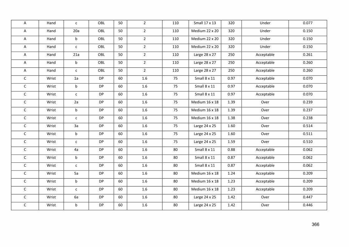
366
A Hand c OBL 50 2 110 Small 17 x 13 320 Under 0.077
A Hand 20a OBL 50 2 110 Medium 22 x 20 320 Under 0.150
A Hand b OBL 50 2 110 Medium 22 x 20 320 Under 0.150
A Hand c OBL 50 2 110 Medium 22 x 20 320 Under 0.150
A Hand 21a OBL 50 2 110 Large 28 x 27 250 Acceptable 0.261
A Hand b OBL 50 2 110 Large 28 x 27 250 Acceptable 0.260
A Hand c OBL 50 2 110 Large 28 x 27 250 Acceptable 0.260
C Wrist 1a DP 60 1.6 75 Small 8 x 11 0.97 Acceptable 0.070
C Wrist b DP 60 1.6 75 Small 8 x 11 0.97 Acceptable 0.070
C Wrist c DP 60 1.6 75 Small 8 x 11 0.97 Acceptable 0.070
C Wrist 2a DP 60 1.6 75 Medium 16 x 18 1.39 Over 0.239
C Wrist b DP 60 1.6 75 Medium 16 x 18 1.39 Over 0.237
C Wrist c DP 60 1.6 75 Medium 16 x 18 1.38 Over 0.238
C Wrist 3a DP 60 1.6 75 Large 24 x 25 1.60 Over 0.514
C Wrist b DP 60 1.6 75 Large 24 x 25 1.60 Over 0.511
C Wrist c DP 60 1.6 75 Large 24 x 25 1.59 Over 0.510
C Wrist 4a DP 60 1.6 80 Small 8 x 11 0.88 Acceptable 0.062
C Wrist b DP 60 1.6 80 Small 8 x 11 0.87 Acceptable 0.062
C Wrist c DP 60 1.6 80 Small 8 x 11 0.87 Acceptable 0.062
C Wrist 5a DP 60 1.6 80 Medium 16 x 18 1.24 Acceptable 0.209
C Wrist b DP 60 1.6 80 Medium 16 x 18 1.23 Acceptable 0.209
C Wrist c DP 60 1.6 80 Medium 16 x 18 1.23 Acceptable 0.209
C Wrist 6a DP 60 1.6 80 Large 24 x 25 1.42 Over 0.447
C Wrist b DP 60 1.6 80 Large 24 x 25 1.42 Over 0.446

367
C Wrist c DP 60 1.6 80 Large 24 x 25 1.41 Over 0.443
C Wrist 7a DP 60 1.6 85.2 Small 8 x 11 0.77 Acceptable 0.054
C Wrist b DP 60 1.6 85.2 Small 8 x 11 0.77 Acceptable 0.054
C Wrist c DP 60 1.6 85.2 Small 8 x 11 0.77 Acceptable 0.054
C Wrist 8a DP 60 1.6 85.2 Medium 16 x 18 1.09 Acceptable 0.183
C Wrist b DP 60 1.6 85.2 Medium 16 x 18 1.09 Acceptable 0.183
C Wrist c DP 60 1.6 85.2 Medium 16 x 18 1.09 Acceptable 0.183
C Wrist 9a DP 60 1.6 85.2 Large 24 x 25 1.25 Over 0.390
C Wrist b DP 60 1.6 85.2 Large 24 x 25 1.26 Over 0.392
C Wrist c DP 60 1.6 85.2 Large 24 x 25 1.26 Over 0.392
C Wrist 10a DP 60 1.6 90 Small 8 x 11 0.69 Acceptable 0.049
C Wrist b DP 60 1.6 90 Small 8 x 11 0.69 Acceptable 0.049
C Wrist c DP 60 1.6 90 Small 8 x 11 0.69 Acceptable 0.049
C Wrist 11a DP 60 1.6 90 Medium 16 x 18 0.97 Acceptable 0.163
C Wrist b DP 60 1.6 90 Medium 16 x 18 0.98 Acceptable 0.164
C Wrist c DP 60 1.6 90 Medium 16 x 18 0.99 Acceptable 0.166
C Wrist 12a DP 60 1.6 90 Large 24 x 25 1.13 Acceptable 0.348
C Wrist b DP 60 1.6 90 Large 24 x 25 1.14 Acceptable 0.352
C Wrist c DP 60 1.6 90 Large 24 x 25 1.14 Acceptable 0.352
C Wrist 13a DP 60 1.6 94.9 Small 8 x 11 0.62 Acceptable 0.043
C Wrist b DP 60 1.6 94.9 Small 8 x 11 0.61 Acceptable 0.043
C Wrist c DP 60 1.6 94.9 Small 8 x 11 0.61 Acceptable 0.043
C Wrist 14a DP 60 1.6 94.9 Medium 16 x 18 0.88 Acceptable 0.146
C Wrist b DP 60 1.6 94.9 Medium 16 x 18 0.88 Acceptable 0.147

368
C Wrist c DP 60 1.6 94.9 Medium 16 x 18 0.89 Acceptable 0.147
C Wrist 15a DP 60 1.6 94.9 Large 24 x 25 1.02 Acceptable 0.310
C Wrist b DP 60 1.6 94.9 Large 24 x 25 1.02 Acceptable 0.311
C Wrist c DP 60 1.6 94.9 Large 24 x 25 1.03 Acceptable 0.314
C Wrist 16a DP 60 1.6 100.1 Small 8 x 11 0.56 Acceptable 0.039
C Wrist b DP 60 1.6 100.1 Small 8 x 11 0.56 Acceptable 0.039
C Wrist c DP 60 1.6 100.1 Small 8 x 11 0.56 Acceptable 0.039
C Wrist 17a DP 60 1.6 100.1 Medium 16 x 18 0.81 Acceptable 0.132
C Wrist b DP 60 1.6 100.1 Medium 16 x 18 0.80 Acceptable 0.132
C Wrist c DP 60 1.6 100.1 Medium 16 x 18 0.81 Acceptable 0.133
C Wrist 18a DP 60 1.6 100.1 Large 24 x 25 0.93 Acceptable 0.284
C Wrist b DP 60 1.6 100.1 Large 24 x 25 0.94 Acceptable 0.286
C Wrist c DP 60 1.6 100.1 Large 24 x 25 0.93 Acceptable 0.282
C Wrist 19a DP 60 1.6 105.1 Small 8 x 11 0.51 Acceptable 0.035
C Wrist b DP 60 1.6 105.1 Small 8 x 11 0.51 Acceptable 0.035
C Wrist c DP 60 1.6 105.1 Small 8 x 11 0.51 Acceptable 0.035
C Wrist 20a DP 60 1.6 105.1 Medium 16 x 18 0.74 Acceptable 0.121
C Wrist b DP 60 1.6 105.1 Medium 16 x 18 0.73 Acceptable 0.119
C Wrist c DP 60 1.6 105.1 Medium 16 x 18 0.74 Acceptable 0.120
C Wrist 21a DP 60 1.6 105.1 Large 24 x 25 0.84 Acceptable 0.254
C Wrist b DP 60 1.6 105.1 Large 24 x 25 0.84 Acceptable 0.254
C Wrist c DP 60 1.6 105.1 Large 24 x 25 0.85 Acceptable 0.257
C Wrist 1a LAT 60 2 74.9 Small 8 x 11 1.26 Over 0.088
C Wrist b LAT 60 2 74.9 Small 8 x 11 1.27 Over 0.088

369
C Wrist c LAT 60 2 74.9 Small 8 x 11 1.27 Over 0.088
C Wrist 2a LAT 60 2 74.9 Medium 13 x 19 1.47 Over 0.255
C Wrist b LAT 60 2 74.9 Medium 13 x 19 1.46 Over 0.255
C Wrist c LAT 60 2 74.9 Medium 13 x 19 1.45 Over 0.255
C Wrist 3a LAT 60 2 74.9 Large 20 x 26 1.44 Over 0.550
C Wrist b LAT 60 2 74.9 Large 20 x 26 1.45 Over 0.550
C Wrist c LAT 60 2 74.9 Large 20 x 26 1.45 Over 0.551
C Wrist 4a LAT 60 2 80 Small 8 x 11 1.10 Acceptable 0.077
C Wrist b LAT 60 2 80 Small 8 x 11 1.12 Acceptable 0.078
C Wrist c LAT 60 2 80 Small 8 x 11 1.11 Acceptable 0.078
C Wrist 5a LAT 60 2 80 Medium 13 x 19 1.28 Over 0.224
C Wrist b LAT 60 2 80 Medium 13 x 19 1.29 Over 0.224
C Wrist c LAT 60 2 80 Medium 13 x 19 1.30 Over 0.225
C Wrist 6a LAT 60 2 80 Large 20 x 26 1.29 Over 0.484
C Wrist b LAT 60 2 80 Large 20 x 26 1.28 Over 0.483
C Wrist c LAT 60 2 80 Large 20 x 26 1.30 Over 0.486
C Wrist 7a LAT 60 2 84.9 Small 8 x 11 1.00 Acceptable 0.069
C Wrist b LAT 60 2 84.9 Small 8 x 11 1.00 Acceptable 0.069
C Wrist c LAT 60 2 84.9 Small 8 x 11 1.00 Acceptable 0.165
C Wrist 8a LAT 60 2 84.9 Medium 13 x 19 1.15 Acceptable 0.199
C Wrist b LAT 60 2 84.9 Medium 13 x 19 1.14 Acceptable 0.197
C Wrist c LAT 60 2 84.9 Medium 13 x 19 1.14 Acceptable 0.198
C Wrist 9a LAT 60 2 84.9 Large 20 x 26 1.15 Acceptable 0.432
C Wrist b LAT 60 2 84.9 Large 20 x 26 1.15 Acceptable 0.427
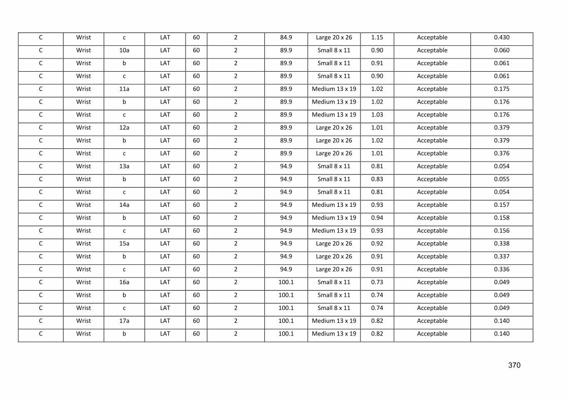
370
C Wrist c LAT 60 2 84.9 Large 20 x 26 1.15 Acceptable 0.430
C Wrist 10a LAT 60 2 89.9 Small 8 x 11 0.90 Acceptable 0.060
C Wrist b LAT 60 2 89.9 Small 8 x 11 0.91 Acceptable 0.061
C Wrist c LAT 60 2 89.9 Small 8 x 11 0.90 Acceptable 0.061
C Wrist 11a LAT 60 2 89.9 Medium 13 x 19 1.02 Acceptable 0.175
C Wrist b LAT 60 2 89.9 Medium 13 x 19 1.02 Acceptable 0.176
C Wrist c LAT 60 2 89.9 Medium 13 x 19 1.03 Acceptable 0.176
C Wrist 12a LAT 60 2 89.9 Large 20 x 26 1.01 Acceptable 0.379
C Wrist b LAT 60 2 89.9 Large 20 x 26 1.02 Acceptable 0.379
C Wrist c LAT 60 2 89.9 Large 20 x 26 1.01 Acceptable 0.376
C Wrist 13a LAT 60 2 94.9 Small 8 x 11 0.81 Acceptable 0.054
C Wrist b LAT 60 2 94.9 Small 8 x 11 0.83 Acceptable 0.055
C Wrist c LAT 60 2 94.9 Small 8 x 11 0.81 Acceptable 0.054
C Wrist 14a LAT 60 2 94.9 Medium 13 x 19 0.93 Acceptable 0.157
C Wrist b LAT 60 2 94.9 Medium 13 x 19 0.94 Acceptable 0.158
C Wrist c LAT 60 2 94.9 Medium 13 x 19 0.93 Acceptable 0.156
C Wrist 15a LAT 60 2 94.9 Large 20 x 26 0.92 Acceptable 0.338
C Wrist b LAT 60 2 94.9 Large 20 x 26 0.91 Acceptable 0.337
C Wrist c LAT 60 2 94.9 Large 20 x 26 0.91 Acceptable 0.336
C Wrist 16a LAT 60 2 100.1 Small 8 x 11 0.73 Acceptable 0.049
C Wrist b LAT 60 2 100.1 Small 8 x 11 0.74 Acceptable 0.049
C Wrist c LAT 60 2 100.1 Small 8 x 11 0.74 Acceptable 0.049
C Wrist 17a LAT 60 2 100.1 Medium 13 x 19 0.82 Acceptable 0.140
C Wrist b LAT 60 2 100.1 Medium 13 x 19 0.82 Acceptable 0.140
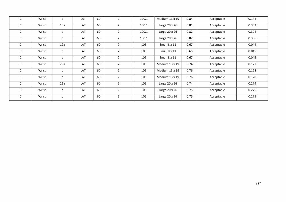
371
C Wrist c LAT 60 2 100.1 Medium 13 x 19 0.84 Acceptable 0.144
C Wrist 18a LAT 60 2 100.1 Large 20 x 26 0.81 Acceptable 0.302
C Wrist b LAT 60 2 100.1 Large 20 x 26 0.82 Acceptable 0.304
C Wrist c LAT 60 2 100.1 Large 20 x 26 0.82 Acceptable 0.306
C Wrist 19a LAT 60 2 105 Small 8 x 11 0.67 Acceptable 0.044
C Wrist b LAT 60 2 105 Small 8 x 11 0.65 Acceptable 0.045
C Wrist c LAT 60 2 105 Small 8 x 11 0.67 Acceptable 0.045
C Wrist 20a LAT 60 2 105 Medium 13 x 19 0.74 Acceptable 0.127
C Wrist b LAT 60 2 105 Medium 13 x 19 0.76 Acceptable 0.128
C Wrist c LAT 60 2 105 Medium 13 x 19 0.76 Acceptable 0.128
C Wrist 21a LAT 60 2 105 Large 20 x 26 0.74 Acceptable 0.274
C Wrist b LAT 60 2 105 Large 20 x 26 0.75 Acceptable 0.275
C Wrist c LAT 60 2 105 Large 20 x 26 0.75 Acceptable 0.275

Append
Hand S
dix 18: SPS
Site A
SS output daata
372

373

Wrist -
Site C
374

DAP an
nd EI Correelation – Haand - Site A
A
375

DAP an
nd EI Correelation – WWrist – Site C
C
376

377
Appendix 19: Variation in Wrist Examinations at Site A
Exam Operator Projection Focus Preset
kVp/mAs
kVp/mAs
Used SID
Collimation
(cm) EI Under|Over|Acceptable
DAP
(cGycm2)
Wrist Rad 1 DP Fine 57/2 52/2.5 107.0 20 x 9 200 Acceptable 0.70
LAT Fine 57/2.5 52/2.5 107.0 22 x 7 250 Acceptable 0.58
Wrist Rad 5 ± DP Fine 57/2.5 57/2.5 106.0 11 x 21 160 Acceptable 1.06
LAT Fine 57/2.5 57/2.5 106.0 9 x 20 200 Acceptable 0.89
Wrist Rad 10+ DP Fine 55/2 55/2 94.0 20 x 11 200 Acceptable 1.31
LAT Fine 55/2 55/2 93.0 8 x 17 400 Under 0.82
Wrist Rad 6-10 DP Fine 55/2 55/2 112.0 26 x 13 250 Acceptable 1.52
LAT Fine 55/2 55/2 113.0 29 x 11 320 Under 1.41
Wrist Rad 6-10 DP Fine 57/2.5 57/2.5 104.0 24 x 10 125 Over 1.21
LAT Fine 57/2.5 57/2.5 104.0 24 x 8 200 Acceptable 0.93
Wrist Rad 6-10 DP Fine 55/2 55/2 96.0 24 x 13 250 Acceptable 1.78
LAT Fine 55/2 55/2 104.0 24 x 10 400 Under 1.23
Wrist AP 2 DP Fine 55/2 55/2 109.0 29 x 11 250 Acceptable 1.49
LAT Fine 55/2 55/2 109.0 17 x 13 200 Acceptable 0.97
Wrist Rad 1 DP Fine 57/2.5 60/3.2 100.0 29 x 17 100 Over 3.66
LAT Fine 57/2.5 60/3.2 100.0 30 x 13 125 Over 3.05
Wrist Rad 10 DP Fine 55/2 57/2.5 102.0 24 x 8 320 Under 1.44
LAT Fine 55/2 60/2 110.0 18 x 9 320 Under 1.43
Wrist Rad 10 DP Fine 55/2 55/2 109.0 23 x 11 200 Under 1.19
LAT Fine 55/2 55/2 108.0 9 x 23 200 Acceptable 0.98