Embed Size (px)
Citation preview

Downloaded from www.microbiologyresearch.org by
IP: 54.237.57.119
On: Tue, 03 May 2016 08:08:26
Rapid necrotic killing of polymorphonuclearleukocytes is caused by quorum-sensing-controlled production of rhamnolipid byPseudomonas aeruginosa
Peter Ø. Jensen,1 Thomas Bjarnsholt,2 Richard Phipps,2
Thomas B. Rasmussen,2 Henrik Calum,1 Lars Christoffersen,1
Claus Moser,1 Paul Williams,4 Tacjana Pressler,3 Michael Givskov2
and Niels Høiby1
Correspondence
Niels Høiby
1Department of Clinical Microbiology, Rigshospitalet, DK-2100 Copenhagen Ø, Denmark
2Centre for Biomedical Microbiology, BioCentrum, Technical University of Denmark
3Copenhagen CF Center, Rigshospitalet, DK-2100 Copenhagen Ø, Denmark
4Centre for Biomolecular Sciences, University of Nottingham, UK
Received 30 October 2006
Revised 15 December 2006
Accepted 23 December 2006
Quorum sensing (QS) denotes a density-dependent mode of inter-bacterial communication based
on signal transmitter molecules. Active QS is present during chronic infections with the
opportunistic pathogen Pseudomonas aeruginosa in immunocompromised patients. The authors
have previously demonstrated a QS-regulated tolerance of biofilm bacteria to the antimicrobial
properties of polymorphonuclear leukocytes (PMNs). The precise QS-regulated effect on the
PMNs is, however, unknown. Incubation of human PMNs with supernatants from dense P.
aeruginosa cultures showed that the QS-competent P. aeruginosa induced rapid necrosis of the
PMNs. This mechanism was also observed in mouse lungs infected with P. aeruginosa, and in
sputum obtained from P.-aeruginosa-infected patients with cystic fibrosis. Evidence is presented
that the necrotic effect was caused by rhamnolipids, production of which is QS controlled. The
results demonstrate the potential of the QS system to facilitate infections with P. aeruginosa by
disabling the PMNs, which are a major first line of defence of the host. Furthermore, the study
emphasizes the inhibition of QS as a target for the treatment of infections with P. aeruginosa.
INTRODUCTION
Pseudomonas aeruginosa is an opportunistic human patho-gen, uncommon as a natural flora, but infecting hospitalizedand immunocompromised patients, such as burn victimsand AIDS patients. In particular, in patients with theautosomal recessive disease cystic fibrosis (CF), chronic P.aeruginosa lung infections represent the major morbidcomplication (Koch & Høiby, 1993). The lung infectionexperienced by CF patients can be divided into two stages:the colonizing non-mucoid state, and the chronic mucoid
state. The chronic infection is preceded by intermittentcolonizations and infections by non-mucoid P. aeruginosafor a mean period of 12 months (Høiby, 1974; Johansen &Høiby, 1992). During this stage, P. aeruginosa is apparentlycleared. The gradual transition from the colonizing state tothe chronic infection is characterized by a phenotypicalswitch from the non-mucoid to the mucoid state. This maybe caused primarily by H2O2 liberated by the polymorpho-nuclear leukocyte (PMNs) (Mathee et al., 1999), and, asrecently demonstrated, by anaerobiosis of P. aeruginosa inthe mucopurulent masses of the bronchioles (Worlitzschet al., 2002). The presence of P. aeruginosa growing inbiofilms, i.e. microcolonies surrounded by a self-madepolysaccharide matrix, is a hallmark of the chronic lunginfection (Høiby, 1974). Within the biofilm, the bacteria areprotected against the numerous surrounding PMNs, andthey exhibit a remarkable tolerance to antibiotic treatments(Donlan & Costerton, 2002; Drenkard, 2003). The abilityof bacteria to invade CF lungs in the first place is enabledby the high content of mucus and the concomitant cilia
Abbreviations: AHL, N-acylhomoserine lactone; BAL, broncheoalveolarlavage; CF, cystic fibrosis; C4-HSL, N-butanoyl-L-homoserine lactone;fMLP, N-formyl-L-methionyl-L-leucyl-L-phenylalanine; 3-oxo-C12-HSL,N-3-oxododecanoyl-L-homoserine lactone; PI, propidium iodide; PMN,polymorphonuclear leukocyte; PQS, Pseudomonas quinolone signal; QS,quorum sensing; 4Q, 4-quinolone.
A time-lapse movie showing the rapid death and disintegration ofPMNs is available as supplementary data with the online version of thispaper.
2006/003863 G 2007 SGM Printed in Great Britain 1329
Microbiology (2007), 153, 1329–1338 DOI 10.1099/mic.0.2006/003863-0

Downloaded from www.microbiologyresearch.org by
IP: 54.237.57.119
On: Tue, 03 May 2016 08:08:26
dysfunctionality, which result in a poor self-cleaning capa-city of the CF lung (Knowles & Boucher, 2002; Gibson et al.,2003). How the bacteria survive the encounter with thesummoned PMNs prior to the formation of the protectivebiofilm is only partly understood, as no primary defects ofPMNs from CF patients have been reported. The outcomeof P. aeruginosa lung infections has recently been demon-strated to be at least partly dependent on quorum-sensing(QS)-regulated mechanisms (Bjarnsholt et al., 2005a; Wuet al., 2001), and the presence of QS activity during chronicP. aeruginosa lung infections in CF patients has beendemonstrated (Storey et al., 1998; Middleton et al., 2002).QS, or cell-to-cell communication, is a regulatory mechan-ism by which bacteria respond to the population density(Fuqua et al., 1996). The QS systems of P. aeruginosa haverecently been intensively investigated, and they are respon-sive to chemically different signal molecules: one based onN-acylhomoserine lactone (AHL) signal molecules, and onebased on 4-quinolones (4Qs). The AHL-based circuits areencoded by the Las and Rhl systems, each of which is basedon LuxR and LuxI homologues. The two systems operatewith specific signal molecules: N-3-oxododecanoyl-L-homoserine lactone (3-oxo-C12-HSL) for the lasR-encodedreceptor, and N-butanoyl-L-homoserine lactone (C4-HSL)for the rhlR-encoded receptor. The 4Q-based system, alsoknown as the Pseudomonas quinolone signal (PQS) system,is somewhat interspaced between the Las system and the Rhlsystem (McKnight et al., 2000; Pesci et al., 1999; Diggle et al.,2006). The organization and interaction of the QS system inP. aeruginosa has been thoroughly investigated; however, it isnot yet completely understood. The Las and Rhl systems havebeen identified as being hierarchically ordered, with the Lassystem in control of the Rhl system (Pesci et al., 1997). It hasbeen suggested, however, that the Rhl system can be switchedon independently of the Las system. Diggle et al. (2003)proposed that this induction is governed by the PQS system.Intriguingly, LasR is required for the optimal production ofthe 4Q signal, whereas exogenously added PQS restores theexpression of lasB in a lasR mutant background.
METHODS
Bacterial strains. The wild-type P. aeruginosa PAO1 used for theplanktonic and biofilm in vitro experiments was obtained from thePseudomonas Genetic Stock Center (www.pseudomonas.med.ecu.edu; strain PAO0001). This isolate has served as the DNA source forthe Pseudomonas Genome Project (www.pseudomonas.com), and,subsequently, as a template for the design of the P. aeruginosaGeneChip (Affymetrix). The DlasR rhlR and DlasI rhlI mutants wereconstructed using the knockout systems described by Beatson et al.(2002). The knockout mutants were verified by Southern blot analy-sis, and by screening for AHL production (quorum signals). Strainsfor verification of genotypes were obtained from the University ofWashington, Seattle, WA, USA (see Table 1).
Production of P. aeruginosa supernatants. Planktonic cultureswere grown in shake flasks (180 r.p.m.) with Luria–Bertani (LB)medium at 37 uC for 24 h. For complementation, C4-HSL and3-oxo-C12-HSL were added to DlasI rhlI mutants. Inhibition of QSwas achieved by adding 12.5 mg furanone C-30 ml21 (Hentzer et al.,
2003) to the medium upon inoculation. Biofilm cultures were grownfor 2 days at 37 uC in six-well Nunc multidishes (Nunclon), eachwell containing 20 ml LB medium. Supernatants from planktonicand biofilm cultures were sterile-filtered through Minisart filters(16543; Sartorius), pore size 0.20 mm, and they were stored at220 uC until use.
Biofilms for direct interaction with PMNs. Biofilms were culti-vated in continuous-culture once-through flow chambers, and thesewere perfused with sterile AB trace minimal medium containing0.3 mM glucose, as described previously (Christensen et al., 1999;Bjarnsholt et al., 2005a).
Preparation of PMNs. Human blood samples were obtained byvenous puncture from normal healthy volunteers, and collected inBD Vacutainers containing 0.129 M sodium citrate (367704; BDDiagnostics). The PMNs were isolated by erythrocyte sedimentationand density-gradient centrifugation, as previously described (Bjarnsholtet al., 2005a).
PMN migration assay. Estimation of PMN migration against P.aeruginosa supernatants was carried out using Transwell trays (3415;Costar). Samples (350 ml) tested were: LB medium with 10 nM N-formyl-L-methionyl-L-leucyl-L-phenylalanine (fMLP) (F3506; Sigma),LB medium, and sterile filtered supernatants from PAO1, DlasR rhlR,DlasI rhlI, and DlasI rhlI complemented with C4-HSL and 3-oxo-C12-HSL. A Transwell filter (pore size, 3 mm) was inserted in the well, and100 ml isolated PMNs (2.56106 cells ml21 in RPMI 1640 with 5 %normal human AB+ serum) was added on top of the filter. Follow-ing incubation for 30 min at 37 uC, the Transwell filter was removed.A 100 ml volume was aspirated from the well, and added to aTruCount tube (340334; BD Biosciences) with 300 ml Facslysis(349202; BD Biosciences) containing 100 mg propidium iodide (PI)ml21 (P-4170; Sigma). After incubation in the dark for a minimumof 10 min, the samples were analysed by flow cytometry, and thenumber of migrated PMNs was calculated according to: migratedPMNs = (cells counted/beads counted)(beads added/volume of cellsadded)610360.35. Data from each set-up were normalized by settingmigration against fMLP to 100 %.
PMN killing by supernatants. The isolated PMNs (2.56106 cellsml21), and all tested sterile filtered supernatants, were equilibratedwith 2.5 mg PI ml21, and incubated at 37 uC for 15 min, beforemixing 50 ml isolated PMNs with 350 ml sterile filtered supernatant,followed by immediate analysis of PI staining with flow cytometry.
PMN killing by biofilms. In order to inoculate PMNs into thebiofilm chambers, the flow was stopped, and the flow cells wereclamped off. Isolated PMNs (100 ml, 2.56106 cells ml21, stainedwith 2.5 mg PI ml21) were inoculated into each flow channel. Theflow cells were incubated top down in a 37 uC water bath, with shak-ing, until microscopic inspection.
Haemolysis. Normal human venous blood collected in BDVacutainers (100 ml) was mixed with 3.5 ml sterile filtered superna-tant from batch cultures of P. aeruginosa. After 10 min, lysis wasevaluated by visual inspection.
Experimental animals. Female BALB/cj mice were purchased fromM&B Laboratory Animals at 10–11 weeks of age. The mice were ofequal size, and were maintained on standard mouse chow and waterad libitum for 1 week prior to challenge. All animal experimentswere authorized by the National Animal Ethics Committee,Denmark. The mouse experiments were performed as described byPedersen et al. (1990).
Isolation and staining for endobronchial PMNs
Broncheoalveolar lavage (BAL). Exposed trachea of anaesthetizedmice were canulated with a size 22 gauge catheter (OPTIVA* 2;
1330 Microbiology 153
P. Ø. Jensen and others
Downloaded from www.microbiologyresearch.org by
IP: 54.237.57.119
On: Tue, 03 May 2016 08:08:26
Johnson & Johnson Medical). BAL was performed by flushing sixtimes with 1.5 ml ice-cold PBS without Ca2+ and Mg2+. The BALfluid was stored on ice until staining for necrotic PMNs. The meanrecovery of BAL fluid was 1.1 ml (CV 13 %).
Staining for necrotic PMNs in the BAL fluid. Necrotic and apop-totic PMNs were stained with Annexin V-FITC Apoptosis DetectionKit I (556747; BD Biosciences), according to a modification of thepreparation supplied by the manufacturer. BAL fluid (200 ml) wasequilibrated by centrifugation with 2.5 ml cold 16 binding buffer(BD Biosciences) at 350 g for 7 min at 5 uC. To discriminate betweennecrotic and apoptotic PMNs, 100 ml 16 binding buffer containing2.5 mg PI ml21, annexin V-FITC component (1 : 40), and the PMNphenotypic surface marker monoclonal allophycocyanin-conjugatedrat anti-Ly 6G antibody (clone RB6–8C5; BD Biosciences) (1 : 50),was added to the pellet, and incubated for 15 min at room tempera-ture in the dark. The incubation was terminated by addition of400 ml 16 binding buffer, and the samples were analysed by flowcytometry.
Staining for the concentration of PMNs in the BAL fluid. A200 ml volume of BAL fluid was added to a TrueCount tube. PMNsand total leukocytes were stained by adding 20 ml cold PBS contain-ing phycoerythrin-conjugated monoclonal rat anti-mouse Ly 6Gantibody (clone RB6–8C5; BD Biosciences; 1 : 20) and peridininchlorophyll A protein-conjugated monoclonal rat anti-mouse CD45antibody (clone RB6–8C5; BD Biosciences; 1 : 10). After incubationfor 30 min on ice in the dark, 300 ml Facslysis solution was added,and the samples were incubated for at least 10 min prior to flow
cytometry. PMN concentration was calculated according to:cells ml21=(cells counted/beads counted)(beads added/BAL fluidadded)6103.
Flow cytometry. The samples were analysed using a FACSort(Becton Dickinson) equipped with a 15 mW argon-ion laser tunedat 488 nm, and a red diode laser emitting at 635 nm for excitation.Light scatter, time, and exponentially amplified fluorescence para-meters from at least 10 000 events, were recorded in list mode.Necrotic PMNs were identified according to their increased PI fluor-escence intensity, and their morphology was determined by lightscatter. The instrument was calibrated using Calibrite beads (BectonDickinson).
Quantitative lung bacteriology. For colony counting, the exposedlungs were isolated in 5 ml PBS, and homogenized on ice. A serialdilution of the lung homogenate was performed, and dilutions wereplated on blue agar plates (States Serum Institute), which are selec-tive for Gram-negative bacilli.
Proteinase K assay. Protein degradation by proteinase K wasperformed as described by the manufacturer (Promega).
Pyocyanin assay. The pyocyanin concentration was measured asdescribed by Essar et al. (1990).
Statistics. Data are presented as means±SEM; P values are fromStudent’s two-tailed unpaired t tests, except for comparison offrequencies, which was done using a x2 test.
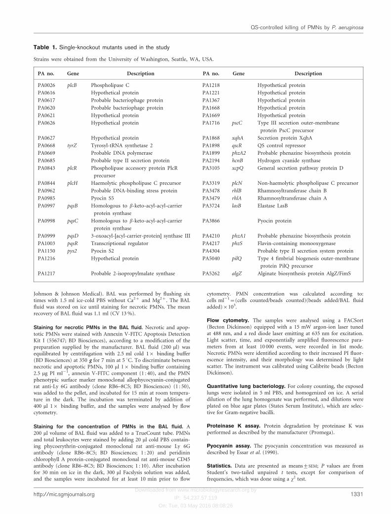
Table 1. Single-knockout mutants used in the study
Strains were obtained from the University of Washington, Seattle, WA, USA.
PA no. Gene Description PA no. Gene Description
PA0026 plcB Phospholipase C PA1218 Hypothetical protein
PA0616 Hypothetical protein PA1221 Hypothetical protein
PA0617 Probable bacteriophage protein PA1367 Hypothetical protein
PA0620 Probable bacteriophage protein PA1668 Hypothetical protein
PA0621 Hypothetical protein PA1669 Hypothetical protein
PA0626 Hypothetical protein PA1716 pscC Type III secretion outer-membrane
protein PscC precursor
PA0627 Hypothetical protein PA1868 xqhA Secretion protein XqhA
PA0668 tyrZ Tyrosyl-tRNA synthetase 2 PA1898 qscR QS control repressor
PA0669 Probable DNA polymerase PA1899 phzA2 Probable phenazine biosynthesis protein
PA0685 Probable type II secretion protein PA2194 hcnB Hydrogen cyanide synthase
PA0843 plcR Phospholipase accessory protein PlcR
precursor
PA3105 xcpQ General secretion pathway protein D
PA0844 plcH Haemolytic phospholipase C precursor PA3319 plcN Non-haemolytic phospholipase C precursor
PA0962 Probable DNA-binding stress protein PA3478 rhlB Rhamnosyltransferase chain B
PA0985 Pyocin S5 PA3479 rhlA Rhamnosyltransferase chain A
PA0997 pqsB Homologous to b-keto-acyl-acyl-carrier
protein synthase
PA3724 lasB Elastase LasB
PA0998 pqsC Homologous to b-keto-acyl-acyl-carrier
protein synthase
PA3866 Pyocin protein
PA0999 pqsD 3-oxoacyl-[acyl-carrier-protein] synthase III PA4210 phzA1 Probable phenazine biosynthesis protein
PA1003 pqsR Transcriptional regulator PA4217 phzS Flavin-containing monooxygenase
PA1150 pys2 Pyocin S2 PA4304 Probable type II secretion system protein
PA1216 Hypothetical protein PA5040 pilQ Type 4 fimbrial biogenesis outer-membrane
protein PilQ precursor
PA1217 Probable 2-isopropylmalate synthase PA5262 algZ Alginate biosynthesis protein AlgZ/FimS
http://mic.sgmjournals.org 1331
QS-controlled killing of PMNs by P. aeruginosa

Downloaded from www.microbiologyresearch.org by
IP: 54.237.57.119
On: Tue, 03 May 2016 08:08:26
RESULTS AND DISCUSSION
PMNs are unable to migrate towards thesupernatant of wild-type strain P. aeruginosaPAO1
We have previously described the presence of a QS-regulated phenotype that operates to paralyse PMNs invitro (Bjarnsholt et al., 2005a). This inspired us to furtherinvestigate the encounter between P. aeruginosa and thePMNs. The PMNs are the first major phagocytes to arriveduring P. aeruginosa lung infection, and their activity isrelated to the early outcome of the infection (Jensen et al.,2004). Based on our previous results, we speculated thatQS-induced paralysis of the PMNs also affected migration.When freshly isolated PMNs were allowed to migratetowards sterile-filtered P. aeruginosa supernatants [obtainedfrom the wild-type and the QS mutants (DlasR rhlR andDlasI rhlI), and from DlasI rhlI complemented with C4-HSLand 3-oxo-C12-HSL], no migration to the wild-type andcomplemented supernatant was observed (Fig. 1). Thiseffect, however, was not caused by the presence of AHLmolecules, as additional experiments showed that PMNsmigrated freely towards concentrations of pure AHL signalmolecules 3-oxo-C12-HSL and C4-HSL increasing from 0.1to 50 mM (data not shown).
PMNs are lysed by a P. aeruginosa wild-typesupernatant
Elaboration of this phenomenon found that sterile-filteredsupernatant from wild-type and DlasI rhlI mutant strains,grown in the presence of both C4-HSL and 3-oxo-C12-HSL(10 mM), caused rapid damage to PMN plasma membranes,as demonstrated by increased fluorescence from supple-mented PI during real-time flow cytometry (Fig. 2a).
Within the first minute after the increase in PI fluorescencewas recorded, the light scatter of the PMNs decreased belowthe threshold of detection, suggesting a rapid disintegrationof the PMNs (data not shown). The rapid death anddisintegration was confirmed by time-lapse recording usinga combination of fluorescence and light-transmissionmicroscopy (Fig. 2b, and supplementary movie availablewith the online version of this paper). Furthermore, thisanalysis revealed that the PMN DNA was released duringdisintegration. In contrast, the PMN plasma membraneremained intact when mixed with supernatants derivedfrom DlasI rhlI and DlasR rhlR mutants. This necrotic effectcontrasts with the previously reported acceleration of PMNapoptosis caused by P. aeruginosa QS signal transmittermolecules (Tateda et al., 2003). Pure C4-HSL and 3-oxo-C12-HSL at concentrations up to 50 mM failed to inducerapid necrosis (data not shown).
PMNs are lysed when in contact with in vitrobiofilms of wild-type P. aeruginosa
To determine if the cytotoxic effect accounted for paralysisof PMNs (Bjarnsholt et al., 2005a), PMNs were incubated onan in vitro biofilm. Necrosis was observed when PMNs wereincubated with QS-proficient P. aeruginosa biofilms grownfor 4 days in flow cells (Fig. 3), but it was not observed whenthe PMNs were incubated on a QS-deficient biofilm grownunder identical conditions. The cytotoxic effect was notspecific for the PMNs, as demonstrated by the haemolyticactivity of sterile-filtered supernatant from QS-competent P.aeruginosa cultures (Fig. 4).
PMNs disappear in mice infected with wild-typeP. aeruginosa
To determine if the necrotic effect was restricted to in vitrosettings only, a pulmonary infectious mouse model wasused for verification. Previously, we established a correlationbetween QS deficiency and faster clearing of infectingbacteria in the mouse model (Bjarnsholt et al., 2005a).BALB/cj mice were infected with either wild-type or QS-deficient mutant, both of which were alginate embedded. Atthe time points 3, 6, 18 and 24 h post-infection, BAL fluidwas obtained, after which the lungs were homogenized.Analysis of the BAL fluid by means of flow cytometry frommice infected with the wild-type P. aeruginosa showed a highproportion of dead PMNs in the endobronchial space, asdetected by their strong PI fluorescence. In accordance withour in vitro data, significantly fewer intact PMNs (estimatedby weak PI fluorescence and low annexin V staining) werefound in the BAL fluids from mice infected with the wild-type P. aeruginosa. After 18 and 24 h, an increased numberof bacteria were observed in the lungs of mice infected withthe wild-type P. aeruginosa (Fig. 5). This increase in thenumber of bacteria, however, is transient, as previousexperiments have shown that the number of wild-typebacteria decreases on days 3 and 5 (Bjarnsholt et al., 2005a).Previously, we reported an elevated concentration of thePMN-recruiting interleukin MIP-2 (a murine IL-8 analogue)
Fig. 1. Effects of QS on PMN migration. The mean PMNmigration towards supernatants from P. aeruginosa with compe-tent QS (PAO1) and deficient QS (DlasR rhlR and DlasI rhlI),and a QS-deficient mutant (DlasI rhlI) complemented with C4-HSL (C4) and 3-oxo-C12-HSL (C12) (10 mM), was calculatedas the percentage of human PMNs migrated towards superna-tants from P. aeruginosa, as compared with migration towardsfMLP (10 nM). Standard error bars are indicated (n=6).
1332 Microbiology 153
P. Ø. Jensen and others

Downloaded from www.microbiologyresearch.org by
IP: 54.237.57.119
On: Tue, 03 May 2016 08:08:26
when mice were infected with the wild-type (Bjarnsholt et al.,2005b). We therefore suggest that the results obtained ateach time point represent snapshots of a continuous pro-cess, i.e. the PMNs are constantly being recruited to the sitesof infections, but, in the wild-type situation, a substantialfraction of the incoming PMNs disintegrate due to the
cytotoxic bacterial activity, which, in turn, allows thebacterial population to expand. The recruited PMNs in miceinfected with the QS-deficient mutant manage to prevent anincrease in the number of bacteria (Fig. 5). We suggest thatthis difference is caused by a QS-regulated reduction of thePMN functionality. To estimate the clinical relevance of
Fig. 2. QS-regulated killing of PMNs. (a–d)Real-time flow cytometry of PI-stainedhuman PMNs mixed with sterile filteredsupernatants from P. aeruginosa. Dashedlines represent the lower PI fluorescenceintensity used for discriminating necrosis. (a)Supernatant from P. aeruginosa with compe-tent QS (PAO1). (b) Supernatant from P.
aeruginosa with deficient QS (DlasI rhlI)complemented with C4-HSL and 3-oxo-C12-HSL. (c) Supernatant from P. aerugi-
nosa with deficient QS (DlasR rhlR). (d)Supernatant from P. aeruginosa with defi-cient QS (DlasI rhlI). (e) Micrographs ofPMN disintegration and DNA releaseinduced by QS, and recorded by combinedfluorescence and light microscopy. HumanPMNs were mixed with sterile filtered super-natants from QS-competent (PAO1) andQS-deficient (DlasR rhlR) P. aeruginosa,and stained with PI. Bars, 50 mm.
Fig. 3. QS-regulated killing of PMNs by P.
aeruginosa biofilms visualized by combinedfluorescence and light microscopy. Four-day-old biofilms, grown in continuous-culture once-through flow chambers, were inoculated withhuman PMNs, and stained with the DNA stainPI. A greater number of PMNs became necro-tic on the QS-competent PAO1 biofilm (A)compared with the QS-mutant (DlasR rhlR)biofilm (B). Bars, 30 mm.
http://mic.sgmjournals.org 1333
QS-controlled killing of PMNs by P. aeruginosa

Downloaded from www.microbiologyresearch.org by
IP: 54.237.57.119
On: Tue, 03 May 2016 08:08:26
cytotoxic metabolites from P. aeruginosa, sputum from CFpatients chronically infected with and without P. aeruginosawas compared. Of the sputum samples from patients with P.aeruginosa infections, 9 of 13 induced rapid PMN necrosis inour in vitro assay, whereas 0 of 3 samples from patientswithout P. aeruginosa induced PMN necrosis (P¡0.03).
The QS inhibitor furanone C-30 blockscytotoxicity in vitro
Since the effect of QS blockers, such as furanone C-30,results in accelerated clearance of P. aeruginosa in thepulmonary mouse model (Hentzer et al., 2003), it was ofinterest to determine if the induction of PMN necrosis bytreating a growing batch culture with furanone C-30 couldbe blocked. We observed that furanone-C-30-treated cul-tures of the wild-type, and a DlasI rhlI mutant grown in thepresence of exogenously added AHL signal molecules,completely blocked development of both the PMN necrotic(Fig. 6) and the haemolytic effect (not shown).
Identification of the QS-regulated cytotoxin
Based upon these results, we conclude that the QS-regulatednecrotic PMN factor is an extracellular product that causesPMN malfunction both in vivo and in vitro. As for P.
aeruginosa, a minimum of 172 genes are QS regulated, andmany of these are only annotated as hypothetical proteins(Hentzer et al., 2003). Several of the QS-regulated genesencode known virulence factors, including proteins, lipidsand secondary metabolites (Hentzer et al., 2003; Wagneret al., 2003; Schuster et al., 2003). In addition, severalvirulence factors with potentially necrotic capacity are notQS regulated, and these include exotoxin A and pepA (exoUis not present in PAO1). Furthermore, the type III secretionapparatus is dependent on cell–cell contact for delivery oftoxins; in contrast, the effect we describe in the presentreport is not dependent on this process. AHLs have beendescribed to induce apoptosis in macrophages and PMNs(Tateda et al., 2003); however, we were unable to replicatethe fast killing of the PMNs by treatment with pure AHLsignal molecules (data not shown). To identify the com-pound responsible for the rapid necrotic killing, the super-natant of the wild-type was examined as described below.The active component was identified as rhamnolipid B,which belongs to a class of well-known biosurfactants fromP. aeruginosa. To verify this, single-knockout mutants wereinvestigated, including rhamnolipid, hydrogen cyanide,elastase and phospholipase C; for a complete list of strainstested refer to Table 1. Supernatants of these single-knockout mutants suggested that rhamnolipids wereresponsible for the necrotic activity identified by flowcytometry of PI-stained PMNs. Induction of PMN necrosiswas also found to be independent of the pyocyanin content(data not shown). P. aeruginosa produces quinolones (PQS)as another part of its QS apparatus, and these molecules havebeen shown to induce apoptosis and reduce T-cell pro-liferation (Calfee et al., 2005; Pritchard, 2006). No necroticeffect was observed in the PMNs upon direct addition of upto 100 mM PQS (data not shown). We did, however, findthat DpqsB, DpqsC, DpqsD and DpqsR mutants did notproduce the necrotic effect, indicating that the PQS system isinvolved in the regulation of the production of the com-pound(s) causing PMN necrosis.
The principal necrotic metabolite was identified fromsupernatants as 2-O-a-L-rhamnopyranosyl-a-L-rhamnopy-ranosyl-b-hydroxydecanoyl-b-hydroxydecanoic acid, whichis also known as rhamnolipid B. Supernatants of outgrownP. aeruginosa PAO1 batch cultures were found to contain100–200 mg rhamnolipid B ml21, whereas no rhamnolipids
Fig. 4. QS-regulated haemolysis of whole blood. Human per-ipheral blood was mixed with sterile filtered supernatants fromP. aeruginosa. Haemolysis was only induced with supernatantfrom QS-competent P. aeruginosa (PAO1).
Fig. 5. Significance of QS during P. aerugi-
nosa lung infection. BALB/cj mice wereinoculated with alginate-embedded QS-competent (PAO1, %) or QS-deficient(DlasR rhlR, #) P. aeruginosa in the leftlung, and sampled at 3, 6, 18 and 24 h. (a)Mean number of intact PMNs in the BALfluid. (b) Mean percentage of necrotic PMNsin the BAL fluid. (c) Mean number of c.f.u.in the lungs. Standard error bars are indi-cated. *P<0.01, n=10; unpaired t test.
1334 Microbiology 153
P. Ø. Jensen and others

Downloaded from www.microbiologyresearch.org by
IP: 54.237.57.119
On: Tue, 03 May 2016 08:08:26
were detected in DrhlA mutants and a DlasR rhlR mutant. Inaddition, PQS mutants were found to produce far lessrhamnolipid B than the wild-type. Incubating PMNs withwild-type supernatant containing approximately 100 mg
rhamnolipid B ml21 induced necrosis as fast as incubationwith 100 mg purified rhamnolipid B ml21. Earlier investiga-tions support our finding, as rhamnolipids are known to lysePMNs (Shryock et al., 1984), erythrocytes (Johnson &Boese-Marrazzo, 1980), and monocyte-derived macro-phages (McClure & Schiller, 1992).
Rhamnolipid isolation and identification
Sterile filtered culture supernatant from PAO1 (3 l) wasextracted three times in ethyl acetate (362 l), and the ethylacetate was removed under vacuum to yield 1.07 g yellowsolid material. This was adsorbed to celite, and applied to a10 g isolute DIOL column pre-equilibrated with 100 %heptane. Fractions were eluted as follows: two fractions of50 % dichloromethane (DCM) in heptane; two fractions of100 % DCM; 20, 30, 40, 50, 60 and 80 % ethyl acetate inDCM; 100 % ethyl acetate; 10 % methanol in ethyl acetate;and, finally, two 100 % methanol washes. All fractions were12 ml, except for the last methanol wash, which was 50 ml.PMN necrotic activity was seen in the last three fractions;these fractions were combined, and then further fractio-nated on a 20 g StrataX C18 column. Elution was with astepped acetonitrile/water gradient starting at 100 % water,increasing to 100 % acetonitrile in 10 % steps, skipping 10and 90 % acetonitrile fractions, and collecting 50 ml perfraction. The primary PMN necrotic activity was detected inthe seventh fraction (80 % acetonitrile). LC-MS showed amolecular mass of 651.3881 g mol21 (Fig. 7), correspond-ing to a molecular formula of C32H58O13 (4 dbe). Other ionswere observed at 359.3, 505.3, 668.4 and 673.4 g mol21,
Fig. 6. Inhibition and complementation of the QS-regulated fastkilling of PMNs. The contents of dead PMNs were analysed byflow cytometry of human PMNs stained with PI, and mixed withsterile filtered supernatants from cultures of P. aeruginosa for 5(black bars) and 10 min (white bars). Inhibition of QS wasdone by adding synthetic furanone C-30 to PAO1 during cul-ture, and with double-knockout (DlasR rhlR and DlasI rhlI) andsingle-knockout (DlasR) mutations. Complementation was per-formed by growing DlasI rhlI in the presence of C4-HSL (C4)and/or 3-oxo-C12-HSL (C12). The bars show representativevalues from several experiments.
Fig. 7. Mass spectrum of rhamnolipid Bpurified from supernatant of P. aeruginosa
PAO1.
http://mic.sgmjournals.org 1335
QS-controlled killing of PMNs by P. aeruginosa

Downloaded from www.microbiologyresearch.org by
IP: 54.237.57.119
On: Tue, 03 May 2016 08:08:26
corresponding to [M+H-26rhamnose]+, [M+H-rham-nose]+, [M+NH3]
+ and [M+Na]+, respectively.
The 1H NMR spectrum (Fig. 8) showed two anomericsignals, several glycosidic proton signals, and a large numberof aliphatic signals. The 13C NMR spectrum indicated thepresence of two carbonyl ester carbons, two anomeric
carbons, several oxygenated carbons, with the remainingsignals corresponding to aliphatic and methyl carbons.Analysis of the 2D NMR spectra (Table 2) showed tworhamnose and two fatty acid units, suggesting that themolecule was a rhamnolipid, which is a class of compoundswell known from P. aeruginosa. Extremely good agreementwas seen between the experimental NMR data and thatpublished by Sim et al. (1997) for the compound 2-O-a-L-rhamnopyranosyl-a-L-rhamnopyranosyl-b-hydroxydecanoyl-b-hydroxydecanoic acid, or rhamnolipid B.
Conclusion
Functional QS systems are present in the vast majority ofP. aeruginosa isolates from the urinary tract, the lowerrespiratory tract and wound infections (Schaber et al., 2004),and the presence of functional QS systems significantlydelays clearing of the bacteria in experimental studies of P.aeruginosa pulmonary infections and thermal wounds (Wuet al., 2001; Bjarnsholt et al., 2005a; Rumbaugh et al., 1999).Interestingly, P. aeruginosa frequently loses the ability toproduce the long-chain signal molecule over time (Heurlieret al., 2006), suggesting a pivotal role of QS during the initialstages of infection. It is now widely accepted that P. aerugi-nosa adapts to the lung environment, to the point ofevolving several subpopulations throughout the lung. Oneof the main adaptations involves production of alginate,
Fig. 8. 1H NMR spectrum of rhamnolipid B purified fromsupernatant of P. aeruginosa PAO1. The spectrum was refer-enced to CDCl3 at dH 7.26, and obtained on a Varian Inovaoperating at 500 MHz.
Table 2. NMR data of rhamnolipid B
Data were acquired on a Varian Inova machine at 500 MHz for 1H, and 300 MHz for 13C. Spectra
were referenced to CDCl3 at dH 7.26. The shifts for the atoms numbered from 2 to 7, and from 12 to
17, were not included in the table as they all overlapped in the region of dH 1.2–1.5 for proton, and dC
25–30 for carbon. COSY, correlated spectroscopy; HMBC, heteronuclear multiple-bond correlation.
Number dH, mult., (JHH Hz) COSY 13C HMBC
1 0.87, d 1.29 13.9 –
8 4.23, sept 2.41, 1.45 70.9 –
9 2.41, m 4.23 39.4 29.1, 24.6
10 – – 171.0 –
11 0.86, d 1.29 13.9 –
18 5.43, p, (6.2) 1.56, 2.53 70.2 24.9, 171.3, 173.6
19 2.53, d, (5.9) 5.43 39.1 171.3, 71.2
20 – – 173.0 –
19 4.89, br s 4.13 94.2 68.0, 70.1, 71.5, 79.7
29 4.13, br s 4.89, 3.77 70.3 71.0, 72.3
39 3.77, dd, (2.4, 10.3) 4.13, 3.48 71.1 –
49 3.48, t, (9.1) 3.77, 3.73 72.2 70.5, 68.0, 17.7
59 3.73, br q, (7.0) 3.48, 1.26 68.5 71.0
69 1.27, d, (6) 3.73, 1.45 17.4 –
79 4.89, br s 3.73 102.1 68.0, 70.1, 71.5, 79.7
89 3.73, m 4.89, 3.80 79.4 102.5, 73.4
99 3.80, dd, (1.8, 9.6) 3.36, 3.73 70.3 –
109 3.36, t, (9.4) 3.80, 3.65 73.2 173.6, 70.5
119 3.65, dt, (5.6, 14) 3.36, 1.27 67.5 71.0, 68.7, 17.7
129 1.26, d, (6) 3.65, 1.56 24.9 –
1336 Microbiology 153
P. Ø. Jensen and others

Downloaded from www.microbiologyresearch.org by
IP: 54.237.57.119
On: Tue, 03 May 2016 08:08:26
which is believed to represent a long-term defence mechan-ism. In contrast, the killing of the PMNs may be one of themechanisms that facilitates establishment of early P. aerugi-nosa lung infections. In support of this model, a highernumber of necrotic PMNs has been detected in CF lungsinfected with P. aeruginosa, as compared with other bacterialinfections (Watt et al., 2005). In addition, CF lungs chroni-cally infected with P. aeruginosa contain high amounts ofextracellular DNA, which is probably derived from necroticPMNs (Lethem et al., 1990; Shah et al., 1996). The ability ofP. aeruginosa to defend itself aggressively against PMNsby production of rhamnolipids may be a prerequisite forthe development of the chronic infection with P. aerugi-nosa. As rhamnolipids have been detected in sputum fromCF patients with chronic P. aeruginosa lung infections(Kownatzki et al., 1987), and nonmucoid clinical isolateshave been shown to produce more haemolytically activerhamnolipids than mucoid isolates (McClure & Schiller,1992), we believe that our observations add significantly tounderstanding the basis of how QS regulates the phenotypeof P. aeruginosa for successful establishment of chronicinfections, as seen in the CF lung. Several characteristics ofthe infected CF lungs are currently considered as being ableto promote the persistence of P. aeruginosa. In this context,our demonstration of QS-regulated secretion of cytotoxicamounts of rhamnolipids by P. aeruginosa may add signifi-cantly to understanding the establishment and persistenceof the infection: impairment of neutrophil migrationcaused by the viscous mucus has been proposed as amechanism for decreased bactericidal activity (Matsui et al.,2005). Baltimore et al. (1989), however, have clearly shownthat the neutrophils are able to accumulate very close to themicrocolonies of P. aeruginosa in the CF lung. Despite thismassive PMN accumulation, the microcolonies persist,suggesting another mechanism by which the bacteria impairthe nearby neutrophils. Impairment of the neutrophils hasalso been proposed to be caused by QS-regulated proteasescleaving surface receptors on the neutrophils (Kharazmiet al., 1984a, b); however, neutralizing antibodies againstthese proteases are present in the lungs of chronicallyinfected CF patients (Doring et al., 1985). We found thatQS-induced PMN necrosis was not caused by proteins. Inaddition, the formation of biofilm has been recognized as amechanism for protection against phagocytosis (Donlan &Costerton, 2002); however, we previously demonstrated thatbiofilm-induced protection against phagocytosis by neu-trophils is dependent on QS (Bjarnsholt et al., 2005a).Further work is necessary to elucidate the role andconsequences of the presence of QS-regulated rhamnolipdsduring the colonization and infection of CF lungs.
ACKNOWLEDGEMENTS
We would like to thank the nurses at the Danish Cystic Fibrosis Centerfor collecting samples. M. G. received financial support from theDanish Research Council FTP-supported ‘A new approach to thecontrol of microbial activity’, the Biomedical Consortium ‘Biomed’,and the German Mukoviszidose e.v. N. H. received financial support
from the Danish Research Agency (22-02-0203) and the ResearchCouncil of Rigshospitalet (410-005). C. M. received financial supportfrom the Toyota Foundation.
REFERENCES
Baltimore, R. S., Christie, C. D. & Smith, G. J. (1989). Immuno-histopathologic localization of Pseudomonas aeruginosa in lungs frompatients with cystic fibrosis. Implications for the pathogenesis ofprogressive lung deterioration. Am Rev Respir Dis 140, 1650–1661.
Beatson, S. A., Whitchurch, C. B., Semmler, A. B. & Mattick, J. S.(2002). Quorum sensing is not required for twitching motility inPseudomonas aeruginosa. J Bacteriol 184, 3598–3604.
Bjarnsholt, T., Jensen, P. O., Burmolle, M., Hentzer, M., Haagensen,J. A. J., Hougen, H. P., Calum, H., Madsen, K. G., Moser, C. & otherauthors (2005a). Pseudomonas aeruginosa tolerance to tobramycin,hydrogen peroxide and polymorphonuclear leukocytes is quorum-sensing dependent. Microbiology 151, 373–383.
Bjarnsholt, T., Jensen, P. O., Rasmussen, T. B., Christophersen, L.,Calum, H., Hentzer, M., Hougen, H. P., Rygaard, J., Moser, C. &other authors (2005b). Garlic blocks quorum sensing and promotesrapid clearing of pulmonary Pseudomonas aeruginosa infections.Microbiology 151, 3873–3880.
Calfee, M. W., Shelton, J. G., McCubrey, J. A. & Pesci, E. C. (2005).Solubility and bioactivity of the Pseudomonas quinolone signal areincreased by a Pseudomonas aeruginosa-produced surfactant. InfectImmun 73, 878–882.
Christensen, B. B., Sternberg, C., Andersen, J. B., Palmer, R. J., Jr,Nielsen, A. T., Givskov, M. & Molin, S. (1999). Molecular tools forstudy of biofilm physiology. Methods Enzymol 310, 20–42.
Diggle, S. P., Winzer, K., Chhabra, S. R., Worrall, K. E., Camara, M. &Williams, P. (2003). The Pseudomonas aeruginosa quinolone signalmolecule overcomes the cell density-dependency of the quorumsensing hierarchy, regulates rhl-dependent genes at the onset ofstationary phase and can be produced in the absence of LasR. MolMicrobiol 50, 29–43.
Diggle, S. P., Cornelis, P., Williams, P. & Camara, M. (2006).4-Quinolone signalling in Pseudomonas aeruginosa: old molecules,new perspectives. Int J Med Microbiol 296, 83–91.
Donlan, R. M. & Costerton, J. W. (2002). Biofilms: survivalmechanisms of clinically relevant microorganisms. Clin MicrobiolRev 15, 167–193.
Doring, G., Goldstein, W., Roll, A., Schiotz, P. O., Høiby, N. &Botzenhart, K. (1985). Role of Pseudomonas aeruginosa exoenzymesin lung infections of patients with cystic fibrosis. Infect Immun 49,557–562.
Drenkard, E. (2003). Antimicrobial resistance of Pseudomonasaeruginosa biofilms. Microbes Infect 5, 1213–1219.
Essar, D. W., Eberly, L., Hadero, A. & Crawford, I. P. (1990).Identification and characterization of genes for a second anthranilatesynthase in Pseudomonas aeruginosa: interchangeability of the twoanthranilate synthases and evolutionary implications. J Bacteriol 172,884–900.
Fuqua, C., Winans, S. C. & Greenberg, E. P. (1996). Census andconsensus in bacterial ecosystems: the LuxR–LuxI family of quorum-sensing transcriptional regulators. Annu Rev Microbiol 50, 727–751.
Gibson, R. L., Burns, J. L. & Ramsey, B. W. (2003). Pathophysiologyand management of pulmonary infections in cystic fibrosis. AmJ Respir Crit Care Med 168, 918–951.
Hentzer, M., Wu, H., Andersen, J. B., Riedel, K., Rasmussen, T. B.,Bagge, N., Kumar, N., Schembri, M. A., Song, Z. & other authors
http://mic.sgmjournals.org 1337
QS-controlled killing of PMNs by P. aeruginosa

Downloaded from www.microbiologyresearch.org by
IP: 54.237.57.119
On: Tue, 03 May 2016 08:08:26
(2003). Attenuation of Pseudomonas aeruginosa virulence by quorum
sensing inhibitors. EMBO J 22, 3803–3815.
Heurlier, K., Denervaud, V. & Haas, D. (2006). Impact of quorum
sensing on fitness of Pseudomonas aeruginosa. Int J Med Microbiol
296, 93–102.
Høiby, N. (1974). Pseudomonas aeruginosa infection in cystic fibrosis.Relationship between mucoid strains of Pseudomonas aeruginosa and
the humoral immune response. Acta Pathol Microbiol Scand [B]Microbiol Immunol 82, 551–558.
Jensen, P. Ø., Moser, C., Kobayashi, O., Hougen, H. P., Kharazmi, A.& Høiby, N. (2004). Faster activation of polymorphonuclear
neutrophils in resistant mice during early innate response to
Pseudomonas aeruginosa lung infection. Clin Exp Immunol 137,478–485.
Johansen, H. K. & Høiby, N. (1992). Seasonal onset of initial
colonisation and chronic infection with Pseudomonas aeruginosa in
patients with cystic fibrosis in Denmark. Thorax 47, 109–111.
Johnson, M. K. & Boese-Marrazzo, D. (1980). Production and
properties of heat-stable extracellular hemolysin from Pseudomonas
aeruginosa. Infect Immun 29, 1028–1033.
Kharazmi, A., Doring, G., Høiby, N. & Valerius, N. H. (1984a).Interaction of Pseudomonas aeruginosa alkaline protease and elastase
with human polymorphonuclear leukocytes in vitro. Infect Immun
43, 161–165.
Kharazmi, A., Høiby, N., Doring, G. & Valerius, N. H. (1984b).Pseudomonas aeruginosa exoproteases inhibit human neutrophilchemiluminescence. Infect Immun 44, 587–591.
Knowles, M. R. & Boucher, R. C. (2002). Mucus clearance as a
primary innate defense mechanism for mammalian airways. J Clin
Invest 109, 571–577.
Koch, C. & Høiby, N. (1993). Pathogenesis of cystic fibrosis. Lancet
341, 1065–1069.
Kownatzki, R., Tummler, B. & Doring, G. (1987). Rhamnolipid of
Pseudomonas aeruginosa in sputum of cystic fibrosis patients. Lancet
1, 1026–1027.
Lethem, M. I., James, S. L., Marriott, C. & Burke, J. F. (1990). The
origin of DNA associated with mucus glycoproteins in cystic fibrosis
sputum. Eur Respir J 3, 19–23.
Mathee, K., Ciofu, O., Sternberg, C., Lindum, P. W., Campbell, J. I.,Jensen, P., Johnsen, A. H., Givskov, M. M., Ohman, D. E. & otherauthors (1999). Mucoid conversion of Pseudomonas aeruginosa by
hydrogen peroxide: a mechanism for virulence activation in the
cystic fibrosis lung. Microbiology 145, 1349–1357.
Matsui, H., Verghese, M. W., Kesimer, M., Schwab, U. E., Randell,S. H., Sheehan, J. K., Grubb, B. R. & Boucher, R. C. (2005). Reduced
three-dimensional motility in dehydrated airway mucus prevents
neutrophil capture and killing bacteria on airway epithelial surfaces.
J Immunol 175, 1090–1099.
McClure, C. D. & Schiller, N. L. (1992). Effects of Pseudomonas
aeruginosa rhamnolipids on human monocyte-derived macrophages.
J Leukoc Biol 51, 97–102.
McKnight, S. L., Iglewski, B. H. & Pesci, E. C. (2000). The Pseudo-
monas quinolone signal regulates rhl quorum sensing in
Pseudomonas aeruginosa. J Bacteriol 182, 2702–2708.
Middleton, B., Rodgers, H. C., Camara, M., Knox, A. J., Williams, P. &Hardman, A. (2002). Direct detection of N-acylhomoserine lactones
in cystic fibrosis sputum. FEMS Microbiol Lett 207, 1–7.
Pedersen, S. S., Shand, G. H., Hansen, B. L. & Hansen, G. N. (1990).Induction of experimental chronic Pseudomonas aeruginosa lung
infection with P. aeruginosa entrapped in alginate microspheres.APMIS 98, 203–211.
Pesci, E. C., Pearson, J. P., Seed, P. C. & Iglewski, B. H. (1997).Regulation of las and rhl quorum sensing in Pseudomonas aeruginosa.J Bacteriol 179, 3127–3132.
Pesci, E. C., Milbank, J. B., Pearson, J. P., McKnight, S., Kende, A. S.,Greenberg, E. P. & Iglewski, B. H. (1999). Quinolone signaling inthe cell-to-cell communication system of Pseudomonas aeruginosa.Proc Natl Acad Sci U S A 96, 11229–11234.
Pritchard, D. I. (2006). Immune modulation by Pseudomonas aerugi-nosa quorum-sensing signal molecules. Int J Med Microbiol 296,111–116.
Rumbaugh, K. P., Griswold, J. A., Iglewski, B. H. & Hamood, A. N.(1999). Contribution of quorum sensing to the virulence of Pseudo-monas aeruginosa in burn wound infections. Infect Immun 67, 5854–5862.
Schaber, J. A., Carty, N. L., McDonald, N. A., Graham, E. D.,Cheluvappa, R., Griswold, J. A. & Hamood, A. N. (2004). Analysis ofquorum sensing-deficient clinical isolates of Pseudomonas aeruginosa.J Med Microbiol 53, 841–853.
Schuster, M., Lostroh, C. P., Ogi, T. & Greenberg, E. P. (2003).Identification, timing, and signal specificity of Pseudomonas aerugi-nosa quorum-controlled genes: a transcriptome analysis. J Bacteriol185, 2066–2079.
Shah, P. L., Scott, S. F., Knight, R. A. & Hodson, M. E. (1996). Theeffects of recombinant human DNase on neutrophil elastase activityand interleukin-8 levels in the sputum of patients with cystic fibrosis.Eur Respir J 9, 531–534.
Shryock, T., Silver, A. S., Baschbach, M. W. & Kramer, J. C. (1984).Effect of Pseudomonas aeruginosa rhamnolipid on human neutrophilmigration. Curr Microbiol 10, 323–328.
Sim, L., Ward, O. P. & Li, Z. Y. (1997). Production and characteri-sation of a biosurfactant isolated from Pseudomonas aeruginosaUW-1. J Ind Microbiol Biotechnol 19, 232–238.
Storey, D. G., Ujack, E. E., Rabin, H. R. & Mitchell, I. (1998).Pseudomonas aeruginosa lasR transcription correlates with thetranscription of lasA, lasB, and toxA in chronic lung infectionsassociated with cystic fibrosis. Infect Immun 66, 2521–2528.
Tateda, K., Ishii, Y., Horikawa, M., Matsumoto, T., Miyairi, S.,Pechere, J. C., Standiford, T. J., Ishiguro, M. & Yamaguchi, K.(2003). The Pseudomonas aeruginosa autoinducer N-3-oxododecanoylhomoserine lactone accelerates apoptosis in macrophages and neutro-phils. Infect Immun 71, 5785–5793.
Wagner, V. E., Bushnell, D., Passador, L., Brooks, A. I. & Iglewski,B. H. (2003). Microarray analysis of Pseudomonas aeruginosa quorum-sensing regulons: effects of growth phase and environment. J Bacteriol185, 2080–2095.
Watt, A. P., Courtney, J., Moore, J., Ennis, M. & Elborn, J. S. (2005).Neutrophil cell death, activation and bacterial infection in cysticfibrosis. Thorax 60, 659–664.
Worlitzsch, D., Tarran, R., Ulrich, M., Schwab, U., Cekiki, A., Meyer,K. C., Birrer, P., Bellon, G., Berger, J. & other authors (2002). Effectsof reduced mucus oxygen concentration in airway Pseudomonasinfections of cystic fibrosis patients. J Clin Invest 109, 317–325.
Wu, H., Song, Z., Givskov, M., Doring, G., Worlitzsch, D., Mathee, K.,Rygaard, J. & Høiby, N. (2001). Pseudomonas aeruginosa mutations inlasI and rhlI quorum sensing systems result in milder chronic lunginfection. Microbiology 147, 1105–1113.
Edited by: P. Cornelis
1338 Microbiology 153
P. Ø. Jensen and others


