Embed Size (px)
Citation preview

V O L U M E 6 5 . N . 2 . A P R I L E 2 0 1 4

Vol. 65 - No. 2 MINERVA ORTOPEDICA E TRAUMATOLOGICA 125
The purpose of this review is to highlight recent advances regarding the diagnosis
and treatment of fibular (lateral) collateral ligament (FCL) injuries in the knee.
Anatomy and biomechanics
The fibular collateral ligament (FCL), also known as the lateral collateral ligament, is the posterolateral corner (PLC) structure re-sponsible for primary varus and secondary external rotation stability. This ligament is on average 69.6-mm-long and courses from the lateral aspect of the femur to the fibular head, deep to the iliotibial band and biceps femoris muscle.1 The femoral attachment of the FCL is located in a small depression 1.4 mm proximal and 3.1 mm posterior to the lateral epicondyle of the femur. This loca-tion is directly 18.5 mm posterosuperior to the center of the popliteus tendon attach-ment (Figure 1). The average cross-section-al area of the femoral footprint is 48 mm2, with some fibers extending anteriorly over
1Center for Outcomes‑based Orthopedic Research Steadman Philippon Research Institute
Vail, CO, USA2Department of BioMedical Engineering
Steadman Philippon Research Institute Vail, CO, USA
3The Steadman Clinic, Vail, CO, USA
MINERVA ORTOP TRAUMATOL 2014;65:125-39
E. W. JAMES 1, A. M. JOHANNSEN 2, C. M. LAPRADE 2, R. F. LAPRADE 2, 3
Recent advances in the treatment of fibular (lateral) collateral ligament injuries
The purpose of this review is to highlight recent advances regarding the diagnosis and treatment of fibular (lateral) collateral ligament (FCL) injuries in the knee. The FCL originates just proximal and posterior to the lateral epicondyle on the femur and inserts distally in a bony depression on the lateral aspect of the fibular head. The FCL functions as the primary restraint to varus laxity at all knee flexion angles and as the primary re-straint to external rotation when the knee is in extension. Depending on the grade of FCL injury, treatment ranges from non-operative management to surgical reconstruction. For patients with grade I or II (partial) FCL tears, non-operative management consisting of an accelerated physical therapy program is of-ten the first line treatment. Indications for FCL surgical reconstruction include patients with grade III (complete) FCL tears or pa-tients who fail to improve with conservative management. Reconstruction is favored over repair for midsubstance tears due to a re-ported increased failure rate associated with repair. In addition, reconstruction functions to minimize the risk of complications asso-ciated with chronic lateral knee instability such as medial compartment osteoarthritis, medial meniscus tears, and failure of cruci-ate ligament reconstruction grafts. Outcomes after FCL reconstruction have demonstrated improvement in both subjective and objective measures.Key words: Collateral ligaments, injuries - Diag-nosis - Surgical procedures, operative.
Corresponding author: R. F. LaPrade, MD, PhD, Complex Knee and Sports Medicine Surgeon, The Steadman Clinic, 181 W. Meadow Dr., Suite 400, Vail, CO 81657, USA. E-mail: [email protected]
Anno: 2014Mese: AprilVolume: 65No: 2Rivista: MINERVA ORTOPEDICA E TRAUMATOLOGICACod Rivista: MINERVA ORTOP TRAUMATOL
Lavoro: 3618-MOTtitolo breve: RECENT ADVANCES IN THE TREATMENT OF FCL INJURIESprimo autore: JAMESpagine: 125-39

126 MINERVA ORTOPEDICA E TRAUMATOLOGICA April 2014
JAMES RECENT ADVANCES IN THE TREATMENT OF FCL INJURIES
ies.2-6 This ligament acts as the primary restraint to varus laxity at all knee flexion an-gles and is the primary restraint to external rotation when the knee is near extension.4, 5 As the knee increases its flexion angle, the popliteus tendon and popliteofibular liga-ment become the primary restraint to ex-ternal rotation, and the forces on the FCL diminish. On radiographic evaluation, varus gapping of 2.7-4.0 mm following the appli-cation of a varus directed force is indicative of an isolated FCL injury.3
Fibular collateral ligament deficiency can profoundly influence gait pattern and over-all knee mechanics. Complete FCL disrup-tion leads to increased forces on ACL and PCL grafts, putting them at higher risk of failure.5, 6 In addition, varus instability may lead to a varus thrust gait pattern, develop-ment of medial meniscal tears, and medial compartment arthritis if left untreated.7-9 Therefore, restoration of normal knee me-chanics and correction of varus instability is crucial to preventing further injury and disability.
Etiology of FCL and other lateral knee injuries
Injuries to the fibular collateral ligament and posterolateral corner of the knee often occur as a result of a blow to the medial or anteromedial corner of the knee causing varus stress, a contact or non-contact hy-perextension injury, or a varus noncontact injury.9, 10 Young men are the most likely to sustain injuries to the FCL and postero-lateral corner.11 Isolated injuries to the FCL are extremely rare and are almost always associated with meniscal tears, anterior cruciate ligament, posterior cruciate liga-ment, popliteus tendon, or popliteofibular ligament injury. One study reported that isolated posterolateral corner injury occurs in only 28% of all posterolateral corner knee injuries.12 Thorough neurovascular evaluation must occur, as up to 15% of patients with posterolateral corner injury also present with common peroneal nerve injury.9
the lateral epicondyle. The fibular attach-ment of the FCL is located on the lateral aspect of the fibular head, on average 8.2 mm posterior to the anterior margin of the fibular head and 28.4 mm distal to the apex of the fibular styloid process. This footprint is located in a bony depression with a 43 mm2 cross sectional area.
The biomechanics of the FCL have been thoroughly evaluated through both sequen-tial sectioning and force measurement stud-
Figure 1.—An illustration demonstrating the anatomic at-tachment sites of the fibular collateral ligament (lateral view, right knee). FCL, fibular collateral ligament; PLT, popliteus tendon. Reproduced with permission from: LaPrade et al.1

Vol. 65 - No. 2 MINERVA ORTOPEDICA E TRAUMATOLOGICA 127
RECENT ADVANCES IN THE TREATMENT OF FCL INJURIES JAMES
lized on the edge of the examination table (Figure 2).14 The leg is then allowed to hang freely from the examination table while the clinician stabilizes the knee by placing his or her hand over the tibiofemoral joint line. A varus stress force is then applied to the lower leg by grasping the patient’s foot. The test is performed at both 0° and 30° of flex-ion, while the amount of the lateral com-partment gapping and presence or absence of a solid endpoint is assessed. An increase in lateral compartment gapping is indicative of a positive varus stress test. A grade I FCL sprain is defined as a positive varus stress test with mild lateral compartment gapping and a firm endpoint. A grade II sprain is defined as moderate gapping with an ap-preciable endpoint. Finally, a grade III FCL sprain is defined as markedly increased lateral compartment gapping without the presence of a solid endpoint. A positive va-rus stress test at 30° of flexion usually indi-cates a complete FCL tear, while gapping at both 0° and 30° of flexion may indicate a combined ligament injury to lateral and posterior knee structures.9, 14
Hughston et al. described two tests to de-tect posterolateral knee instability: the pos-terolateral drawer test and the external ro-tation recurvatum test.17 The posterolateral drawer test is performed with the patient supine, the hip flexed at 45°, and the knee flexed to approximately 80° to 90°. The foot is then externally rotated approximately 15°
Diagnosis
The diagnosis of FCL injuries can some-times be difficult. A correct diagnosis is es-sential to determine an appropriate treat-ment and to prevent long-term sequelae associated with chronic lateral knee insta-bility. Approximately 73.4% and 31.6% of FCL injuries are combined ligament injuries with the ACL and PCL, respectively.13 In ad-dition, 56% of PLC injuries involve two or more of the major PLC structures (FCL, pop-liteofibular ligament, and popliteus tendon), while only 13.3% of FCL tears are isolated injuries. Numerous diagnostic techniques have been developed to diagnose complex injury patterns involving the FCL and other knee structures in order to mitigate the risk of long-term complications associated with non-treatment.
Diagnosis with physical examination
A thorough history and physical exam is an essential first step to diagnosing FCL injury. On inspection of an acutely injured knee, there may be significant edema, red-ness, and bruising. Palpation may reveal tenderness over the lateral aspect of the joint line and pain at the fibular head.14 Ac-tive range of motion is often intact, though may be limited depending on the amount of swelling in the injured knee. In addi-tion, rotational instability is often present in both acute and chronic injury.15, 16 The patient may report instability associated with feelings of knee hyperextension while ascending or descending stairs, twisting, or pivoting.16 Specific physical examination maneuvers, including the posterolateral drawer test, external rotation recurvatum test, dial test, varus stress test, and reverse pivot-shift test, may be especially helpful in distinguishing between an isolated FCL tear versus a complete grade III posterolateral corner injury. Finally, signs of common per-oneal nerve injury must also be evaluated.
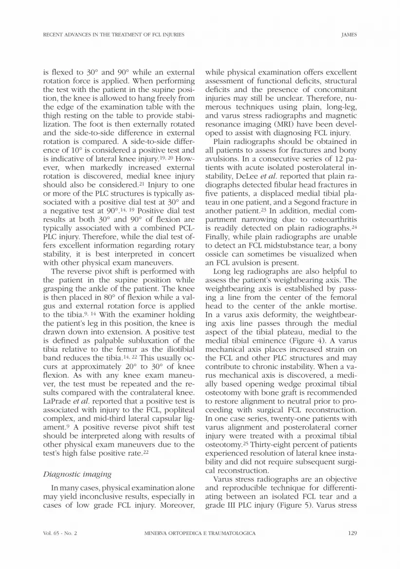
The varus stress test is the most impor-tant physical examination maneuver for di-agnosing FCL injury. In this test, the patient is positioned supine, with the femur stabi-
Figure 2.—The varus stress test is performed at 0° and 30° of knee flexion and is the best physical exam ma-neuver to assess for lateral knee instability secondary to FCL injury.

128 MINERVA ORTOPEDICA E TRAUMATOLOGICA April 2014
JAMES RECENT ADVANCES IN THE TREATMENT OF FCL INJURIES
test, the patient is positioned supine on the examination table.17 The patient is told to relax their quadriceps muscle while the ex-aminer grasps both great toes, lifting the patient’s lower legs off the examination table (Figure 3). A positive test is defined as a side-to-side difference in heel heights in the injured knee compared to the un-injured knee.14 While the external rotation recurvatum test is an essential test in the diagnostic armamentarium, it is less sensi-tive than other physical exam maneuvers. In one series, LaPrade et al. reported that the test detected just 7.5% of posterolateral corner injuries compared to 30% of com-bined ACL-PLC injuries.18 No positive test results were documented for patients with combined PCL-PLC injuries. Therefore, cau-tion is advised with interpreting the results of this test.
The dial test is performed with the patient in a supine or prone position.14, 19 The knee
and stabilized while the clinician applies a posterolateral drawer force to the anterior tibia.14, 17 A positive test is defined as in-creased rotational instability compared to the contralateral uninjured knee. From an anterior perspective, a positive test appears as posterior displacement of the lateral tibial plateau relative to the medial tibial plateau. From a medial or lateral perspective, the lat-eral tibial plateau moves posterior while the medial side does not displace. By contrast, testing the knee in neutral or internal tibial rotation is utilized to assess PCL integrity and a positive test should not be confused with a posterolateral corner injury. Previous studies have reported that a positive poste-rolateral drawer test was found in approxi-mately 71-80% of PLC injuries and the test is therefore an excellent indicator of lateral knee instability.15, 16
The external rotation recurvatum test is also used to evaluate FCL integrity. In this
Figure 3.—An illustration demonstrating the sagittal plane relationship of the tibia with respect to the femur in a posi-tive external rotation recurvatum test (lateral view, right knee). A positive test is defined as increased heel height in the injured knee compared to the uninjured knee. Reprinted with permission from: LaPrade et al.18
Vol. 65 - No. 2 MINERVA ORTOPEDICA E TRAUMATOLOGICA 129
RECENT ADVANCES IN THE TREATMENT OF FCL INJURIES JAMES
while physical examination offers excellent assessment of functional deficits, structural deficits and the presence of concomitant injuries may still be unclear. Therefore, nu-merous techniques using plain, long-leg, and varus stress radiographs and magnetic resonance imaging (MRI) have been devel-oped to assist with diagnosing FCL injury.
Plain radiographs should be obtained in all patients to assess for fractures and bony avulsions. In a consecutive series of 12 pa-tients with acute isolated posterolateral in-stability, DeLee et al. reported that plain ra-diographs detected fibular head fractures in five patients, a displaced medial tibial pla-teau in one patient, and a Segond fracture in another patient.23 In addition, medial com-partment narrowing due to osteoarthritis is readily detected on plain radiographs.24 Finally, while plain radiographs are unable to detect an FCL midstubstance tear, a bony ossicle can sometimes be visualized when an FCL avulsion is present.
Long leg radiographs are also helpful to assess the patient’s weightbearing axis. The weightbearing axis is established by pass-ing a line from the center of the femoral head to the center of the ankle mortise. In a varus axis deformity, the weightbear-ing axis line passes through the medial aspect of the tibial plateau, medial to the medial tibial eminence (Figure 4). A varus mechanical axis places increased strain on the FCL and other PLC structures and may contribute to chronic instability. When a va-rus mechanical axis is discovered, a medi-ally based opening wedge proximal tibial osteotomy with bone graft is recommended to restore alignment to neutral prior to pro-ceeding with surgical FCL reconstruction. In one case series, twenty-one patients with varus alignment and posterolateral corner injury were treated with a proximal tibial osteotomy.25 Thirty-eight percent of patients experienced resolution of lateral knee insta-bility and did not require subsequent surgi-cal reconstruction.
Varus stress radiographs are an objective and reproducible technique for differenti-ating between an isolated FCL tear and a grade III PLC injury (Figure 5). Varus stress
is flexed to 30° and 90° while an external rotation force is applied. When performing the test with the patient in the supine posi-tion, the knee is allowed to hang freely from the edge of the examination table with the thigh resting on the table to provide stabi-lization. The foot is then externally rotated and the side-to-side difference in external rotation is compared. A side-to-side differ-ence of 10° is considered a positive test and is indicative of lateral knee injury.19, 20 How-ever, when markedly increased external rotation is discovered, medial knee injury should also be considered.21 Injury to one or more of the PLC structures is typically as-sociated with a positive dial test at 30° and a negative test at 90°.14, 19 Positive dial test results at both 30° and 90° of flexion are typically associated with a combined PCL-PLC injury. Therefore, while the dial test of-fers excellent information regarding rotary stability, it is best interpreted in concert with other physical exam maneuvers.
The reverse pivot shift is performed with the patient in the supine position while grasping the ankle of the patient. The knee is then placed in 80° of flexion while a val-gus and external rotation force is applied to the tibia.9, 14 With the examiner holding the patient’s leg in this position, the knee is drawn down into extension. A positive test is defined as palpable subluxation of the tibia relative to the femur as the iliotibial band reduces the tibia.14, 22 This usually oc-curs at approximately 20° to 30° of knee flexion. As with any knee exam maneu-ver, the test must be repeated and the re-sults compared with the contralateral knee. LaPrade et al. reported that a positive test is associated with injury to the FCL, popliteal complex, and mid-third lateral capsular lig-ament.9 A positive reverse pivot shift test should be interpreted along with results of other physical exam maneuvers due to the test’s high false positive rate.22
Diagnostic imaging
In many cases, physical examination alone may yield inconclusive results, especially in cases of low grade FCL injury. Moreover,

130 MINERVA ORTOPEDICA E TRAUMATOLOGICA April 2014
JAMES RECENT ADVANCES IN THE TREATMENT OF FCL INJURIES
2.7 mm after a simulated isolated FCL in-jury and 4.0 mm after a grade III PLC injury compared to the intact state.3 In addition to assisting with diagnosis, stress radiographs can be utilized to document lateral knee stability at various time points following surgical treatment. In a consecutive series of 20 patients with FCL injury, preoperative stress radiographs demonstrated an average side-to-side difference of 3.9 mm in lateral compartment gapping, while postoperative stress radiographs found -0.4 mm of gap-ping following an anatomic FCL reconstruc-tion.26
Magnetic resonance imaging (MRI) is an-other diagnostic imaging tool that can be used to distinguish between isolated FCL injury and grade III PLC injury. LaPrade et al. examined the MRI appearance of indi-vidual PLC structures.27 The authors report-ed that the FCL was best visualized on MRI in the axial, coronal, and coronal-oblique views (Figure 6). The coronal-oblique view is usefully because the FCL traverses ob-liquely from its femoral attachment on the lateral epicondyle to its fibular attachment adjacent to the long head of the biceps femoris muscle. In addition, an avulsion of the femoral FCL insertion and an arcuate fracture of the fibular head are best visual-ized in the axial and coronal planes. The authors also reported diagnostic accuracy parameters for the ability of MRI to detect posterolateral corner injuries. For detection of FCL tears, MRI showed a sensitivity of 94.4%, specificity of 100%, positive predic-tive value of 100%, and a negative predic-tive value of 66.7%.
Additionally, MRI can be used to assess the presence of bone bruising on the tibial plateau and femoral condyles. Geeslin and LaPrade reported that 60.7% of patients with isolated injury to the PLC ligaments also had bone bruising to the anteromedial femoral condyle, while 21.4% of patients had a frac-ture of the anteromedial tibial plateau.11 In patients with a combined PLC injury plus injury to the ACL, PCL and/or MCL, 87.8% of patients had bone bruising. Of these, 60% were located in the anteromedial femoral condyle. Therefore, bone bruising on the
radiographs are performed with the knee in 20° of flexion while a clinician applies a varus directed force to the medial aspect of the knee. In a cadaveric study using manual varus stress, lateral gapping increased by
Figure 4.—A long leg radiograph demonstrating a varus mechanical axis deformity in the setting of a chronic fibular collateral ligament tear.

Vol. 65 - No. 2 MINERVA ORTOPEDICA E TRAUMATOLOGICA 131
RECENT ADVANCES IN THE TREATMENT OF FCL INJURIES JAMES
by intra-articular pathology including ACL, PCL, and meniscus tears.13 For this reason, arthroscopic surgery is often required in pa-tients with an FCL tear. The most important sign to look for on arthroscopic evaluation is the presence of the “drive through sign,” which may indicate injury to the FCL or other posterolateral corner structures.28 In a series of 30 consecutive knees with pos-terolateral knee instability, lateral compart-ment gapping of >1 cm was present in all knees with the application of varus stress at 30° of knee flexion. Therefore, the presence of a “drive-through” should increase suspi-cion of an FCL or associated posterolateral corner injury.
The natural history of FCL and plc injury: factors affecting healing
Several in vivo animal model studies have been performed to determine the natural history of posterolateral corner injuries in the knee.29-31 One study looked at the effect of chronic posterolateral knee instability in a rabbit model.31 After sectioning the FCL and popliteus tendon, none of the popli-
anteromedial femoral condyle may indicate PLC injury, including the injury to the FCL.
Arthroscopic diagnosis
While the FCL is not an intra-articular structure, FCL tears are often accompanied
Figure 5.—Varus stress radiographs demonstrating increased lateral compartment gapping indicative of an isolated FCL tear.
Figure 6.—A PD TSE FS coronal MRI demonstrating an acute FCL tear (arrow).

132 MINERVA ORTOPEDICA E TRAUMATOLOGICA April 2014
JAMES RECENT ADVANCES IN THE TREATMENT OF FCL INJURIES
and increased radiographic indications of osteoarthritis when compared to grade II sprains.8 Strobel et al. reported significantly higher rates of osteoarthritis on the medial femoral condyle in combined PLC-PCL inju-ries verus isolated PCL injuries.32
In addition, the bony geometry of the lat-eral tibial plateau and lateral femoral condyle creates “inherent… instability” in the lateral aspect of the knee, which contributes to the low healing response observed after FCL in-jury.31 Both the lateral tibial plateau and lat-eral femoral condyle have convex contours, which lead to instability that is not condu-cive for healing of the PLC structures (Fig-ure 7).29, 31 By comparison, the medial tibial plateau and medial femoral condyle have a concave on convex geometry, which con-fers additional stability to the medial knee. The geometry of the medial compartment may explain the excellent healing response often found following medial collateral liga-ment injury.33-35 Furthermore, the lateral meniscus is reportedly approximately twice as mobile as the medial meniscus, which further contributes to instability.36 Together, these structural and morphological factors contribute to lack of healing response ob-served following lateral knee injuries.
teus tendons injuries and only one FCL inju-ry healed after a 12-week follow-up. In ad-dition, biomechanical testing revealed that significantly less varus force was needed to produce 10 mm of varus displacement in the injured knees at 30°, 60°, and 90° of knee flexion compared to the contralateral control knee. In a second study with longer follow-up in 10 rabbits, none of simulated the FCL or popliteus tendon injuries healed after 6 months.30 Biomechanical testing demonstrated a significant difference in varus displacement at 30°, 60°, and 90° of knee flexion and a significant increase in external rotation at 30° and 60° of flexion. In addition, osteoarthritis was found on the medial tibial plateau, but not on the lateral tibial plateau. In a third study using an in vivo canine model with sectioning of the FCL, PFL, and popliteus tendon, increased varus angulation at 0°, 60°, and 90° of knee flexion, external rotation at 0°, 60°, and 90°, and internal rotation at 0° and 60° were re-ported at six months follow-up.31
Similar results of poor healing have been observed in humans. Kannus reported that chronic grade III PLC injuries resulted in significantly worse clinical outcome scores, lower thigh strength, lower knee stability,
Figure 7.—A, B) The convex on convex configuration of the lateral compartment articulating surfaces (A) compared to the convex on concave configuration of the medial compartment articulating surfaces (B) contributes to chronic instability following FCL injuries.

Vol. 65 - No. 2 MINERVA ORTOPEDICA E TRAUMATOLOGICA 133
RECENT ADVANCES IN THE TREATMENT OF FCL INJURIES JAMES
ping and establish a baseline against which future evaluations can be measured. If non-operative management fails to resolve lat-eral compartment instability, surgical inter-vention should be considered.
Surgical treatment
Repair
Primary repair of FCL injuries is recom-mended in only very select cases. Repair should only be attempted during the first two to three weeks following an FCL avul-sion and very rarely in patients with a mid-substance tear.14 In effect, most repairs oc-cur in skeletally immature patients. After several weeks pass, the FCL becomes re-tracted and entrapped by scar tissue, mak-ing reapposition and repair of the torn ends difficult. Moreover, tissue quality deterio-rates as the FCL becomes necrotic leading to suture pull-out failure. Therefore, in the acute setting, the authors recommend re-construction for all FCL midsubstance tears and for FCL avulsions that present greater than three weeks after injury.
Reconstruction
Indications for surgical reconstruction include patients with grade III (complete) FCL tears, acute midsubstance tears, avul-sions presenting greater than three weeks after injury, or those who fail to improve with conservative treatment measures. Dur-ing the preoperative workup, bilateral varus stress radiographs should be obtained to establish an objective baseline assessment of lateral compartment laxity. After surgery, these values can be compared to assess for resolution of lateral compartment laxity and to detect graft attenuation or failure. In ad-dition, long leg radiographs are necessary to determine the patient’s weightbearing axis. If a varus axis deformity is discovered in a patient with a chronic FCL or PLC in-jury, reconstruction should proceed in a staged fashion beginning with an opening wedge proximal tibial osteotomy followed
Finally, O’Brien et al. reported that undi-agnosed PLC injuries are a leading cause of ACL reconstruction failure.37 This has since been validated in biomechanical studies which demonstrated that sectioning of the FCL, PFL, and popliteus tendon resulted in significantly higher forces on both ACL and PCL reconstruction grafts.5, 6 In one study, the FCL was sectioned and forces on the anterior cruciate ligament (ACL) were meas-ured. Results demonstrated significantly in-creased forces on the reconstruction graft during varus loading at 0° and 30° of knee flexion.5 In addition, simulated grade III PLC injuries, including injuries to the FCL, resulted in significantly increased forces on PCL reconstruction grafts when a varus force at 30°, 60° and 90° of knee flexion was applied.6 These findings demonstrate that posterolateral corner injuries often do not heal and may lead to failure of other reconstruction grafts.
Non-operative treatment
Non-operative treatment is an important first step in management of grade I and II FCL injuries using an accelerated rehabilita-tion program to promote healing.14 In the early phases, the rehabilitation program typically consists of edema management, range of motion exercises, and quadriceps activation. Once the patient achieves full extension, flexion to 120°, and is able to perform a supine straight leg raise with out lag, the focus shifts to developing muscu-lar strength and restoring normal gait. Neu-romuscular coordination and functional exercises are added to continue building muscular strength. A medial unloader brace can also be used during higher-level activi-ties, especially in athletes who may require protection of the healing FCL while return-ing back to activities. Finally, exercises fo-cused on sport- or activity-specific move-ments are initiated to facilitate the transition from rehabilitation to return to sport. In all cases of lateral knee injury, it is essential to obtain varus stress radiographs to objec-tively document lateral compartment gap-

134 MINERVA ORTOPEDICA E TRAUMATOLOGICA April 2014
JAMES RECENT ADVANCES IN THE TREATMENT OF FCL INJURIES
anatomic technique using a semitendinosus tendon autograft and an arthroscopically assisted, mini-open surgical approach.46
The authors’ preferred technique is an open anatomic FCL reconstruction using a semitendinosus autograft, which has been validated biomechanically to improve ob-jective knee stability (Figure 8).7, 26 The pa-tient is positioned with the operative leg hanging freely at 70° to 80° of flexion with the foot of the operating table folded down. The non-operative leg is abducted and se-cured in a leg holder for the duration of the procedure. A tourniquet is placed around the distal thigh and inflated once proper draping is achieved and the surgical site is prepared.
A standard laterally based hockey stick
by surgical reconstruction if lateral knee in-stability fails to improve.25, 38
Numerous surgical techniques for FCL treatment have been reported including ad-vancement of the proximal FCL attachment,39 augmentation with the biceps femoris ten-don,40 biceps femoris tendon tenodesis,41 a doubled over semitendinosus autograft re-construction,42 quadriceps tendon-patellar bone autograft reconstruction,43 and iso-metric bone-patellar tendon-bone recon-struction.44, 45 More recently, there has been an increased emphasis on anatomic-based reconstruction techniques.7, 26, 46 LaPrade et al. has described an anatomic reconstruc-tion technique utilizing an open surgical approach and a semitendinosus tendon autograft.7, 26 Liu et al. described a similar
Figure 8.—A, B) An illustration demonstrating posterior (A) and lateral (B) views of an anatomic fibular collateral ligament reconstruction using a semitendinosus graft. FCL, fibular collateral ligament; PFL, popliteofibular ligament; PLT, popliteus tendon. Reproduced with permission from: LaPrade et al.26

Vol. 65 - No. 2 MINERVA ORTOPEDICA E TRAUMATOLOGICA 135
RECENT ADVANCES IN THE TREATMENT OF FCL INJURIES JAMES
positioning, a 6-mm closed socket tunnel is reamed to a depth of 30 mm and finished with a 7 mm tap to complete the femoral reconstruction tunnel.
In autograft reconstruction cases, an inci-sion is made over the pes anserine bursa on the medial aspect of the proximal tib-ia to locate the semitendinosus tendon. A standard hamstring harvesting instrument is used to harvest the semitendinosus tendon graft. Once harvested, the graft is cleaned of all residual muscle and tubularized us-ing number two nonabsorbable sutures to allow easy graft passage through the recon-struction tunnels.
Next, attention is turned to perform any required intra-articular arthroscopic proce-dures. Since the majority of posterolateral corner injuries are combined ligament in-juries, it is likely that many patients may require a cruciate ligament reconstruction. In addition, chronic lateral knee injuries are associated with medial meniscus tears and medial compartment degenerative cartilage lesions. These should be addressed at this time. Cruciate ligament grafts should be se-cured in their respective femoral tunnels, but tibial fixation should not be performed until after the FCL reconstruction graft is se-cured.
Once the arthroscopic procedures are es-sentially completed, the FCL graft is passed into the femoral tunnel with the assistance of passing sutures and secured using a 7x23 mm bioabsorbable screw (Figure 9). Secure fixation is confirmed by applying a lateral traction force to the graft fixed in the femoral tunnel. The FCL graft is guided deep to the iliotibial band and aponeurosis of the long head of the biceps femoris be-fore emerging adjacent to the lateral aspect of the fibular head (Figure 10). As with the femoral tunnel, passing sutures help pull the graft into the fibular reconstruction tunnel. The FCL graft is tensioned with the knee in 20º of flexion and the leg in neutral tibial rotation while applying a valgus force to eliminate any lateral compartment gapping. A 7x23 cannulated bioabsorbable interfer-ence screw is used to fix the graft in place in the fibular tunnel. Once securely fixed, a
incision is made over the posterolateral knee extending proximally along the iliotib-ial band to distally at the level of Gerdy’s tu-bercle.47 The incision is then carried down to develop a posteriorly based skin flap over the long and short heads of the biceps femoris muscle. A peroneal neurolysis is performed by carefully dissecting through the subcutaneous tissue to release the nerve from its connective tissue entrapments, minimizing the risk of foot drop postopera-tively due to swelling. The peroneal nerve can be located either by palpating two to three centimeters posterior to the long head of the biceps femoris or carefully dissecting along the lateral aspect of the fibular head. Extreme care should be followed in cases where the biceps femoris is avulsed off the fibular head because the peroneal nerve may be displaced over the fibular head. Next, the biceps bursa is identified and a 1 cm incision is created to access the fibular head. Through this interval, the distal FCL attachment site is readily identified.1 A trac-tion stitch can be placed in the FCL remnant to assist with later identification of the FCL femoral attachment.
Once the position of the distal FCL at-tachment is confirmed, a guide pin is drilled from the center of the native FCL attach-ment on the fibular head to the posterome-dial aspect of the fibular styloid near the popliteofibular ligament. To prevent guide pin over penetration, a retractor is placed along the posteromedial aspect of the fibu-lar head. Finally, a 6-mm reamer is used to create the fibular reconstruction tunnel over the guide pin.
Attention is next turned to the femoral reconstruction tunnel. A longitudinal inci-sion is made through the iliotibial band by splitting its fibers lengthwise. Through this interval, the femoral attachment of the FCL is identified proximal and posterior to the lateral epicondyle. The distally placed trac-tion stitch can also be used to isolate the proximal FCL footprint if it is not readily visualized. Once correct positioning is con-firmed, an eyelet tipped guide pin is aimed anteromedially to avoid breaching the in-tercondylar notch. After confirming correct

136 MINERVA ORTOPEDICA E TRAUMATOLOGICA April 2014
JAMES RECENT ADVANCES IN THE TREATMENT OF FCL INJURIES
es as pain allows. Once range of motion reaches 105° to 110° of flexion, stationary bike and low resistance muscular endur-ance base building exercises are initiated. Low impact exercises such as walking and swimming are added at twelve weeks. Once an adequate muscular endurance base is achieved, an increased emphasis is placed on restoring muscular strength.
Varus stress radiographs are obtained at between five to six months postsurgically to assess for lateral compartment stability. A side-to-side difference of less than two millimeters is considered successful resolu-tion of lateral instability, while gapping of greater than two millimeters suggests graft attenuation or failure. The goal is return to
varus stress exam is performed to verify res-olution of lateral knee stability before trim-ming excess graft near the posteromedial aperture of the fibular reconstruction tunnel while the common peroneal nerve is gently retracted (Figure 11). At this time, tibial fixa-tion can be completed for any concomitant cruciate ligament reconstructions. Once all grafts are secured, the incisions are closed and the patient is placed in an immobilizer brace.
Rehabilitation
Postoperative restrictions include non-weightbearing and avoidance of external rotation and varus stress for the first six weeks.26 An immobilizer brace is used to protect the grafted FCL. Straight leg raises and quadriceps sets should be performed with the knee in the immobilizer brace. Early range-of-motion exercises are empha-sized during the first phases of rehabilitation to minimize the risk of arthrofibrosis. Pas-sive range of motion is allowed from 0° to 90° of flexion for the first two weeks, stating on postoperative day one, and increased as tolerated thereafter, with the goal of achiev-ing full range of motion by the end of week six. After two weeks, straight leg raises and quadriceps sets can be completed without the immobilizer brace in the absence of an extensor lag. After six weeks, patients are allowed to progressively wean off crutch-
Figure 11.—The FCL graft is secured in the fibula and trimmed prior to closing.
Figure 10.—The FCL graft is passed distally and deep to the iliotibial band before being pulled through the fibular reconstruction tunnel.
Figure 9.—The FCL reconstruction graft is secured in a closed socket femoral tunnel using a 7 mm bioabsorb-able screw.
Vol. 65 - No. 2 MINERVA ORTOPEDICA E TRAUMATOLOGICA 137
RECENT ADVANCES IN THE TREATMENT OF FCL INJURIES JAMES
Outcomes following anatomic FCL re-construction have demonstrated a signifi-cant improvement in subjective and ob-jective outcome measures. LaPrade et al. performed anatomic FCL reconstructions in 20 patients with an average age of 24 years (range, 16-45 years).26 After a mean of two years, the average modified Cincin-nati score improved significantly from 28.2 preoperatively to 88.5 postoperatively and the average International Knee Documen-tation Committee (IKDC) subjective score increased significantly from 34.7 preopera-tively to 88.1 postoperatively. The Cincin-nati symptom and functional subscores also improved significantly. Objective out-comes were evaluated using varus stress radiographs, which improved from a mean side-to-side difference in lateral compart-ment gapping of 3.9 mm preoperatively to -0.4 mm postoperatively. Together, these outcomes demonstrate that an anatomic FCL reconstruction was able to significant improve patient function and lateral knee stability.
Conclusions
Numerous advances have been made recently in the understanding of postero-lateral corner anatomy and biomechanics, which in turn have led to the development and validation of FCL reconstruction tech-niques. Lateral knee injuries frequently do not heal due to the inherent bony instability of the lateral compartment. Patients present-ing with an acute knee injury should always be evaluated for signs and symptoms of va-rus instability, especially in cases of cruci-ate ligament injury, which may indicate an injury to the FCL. For patients with chronic lateral knee instability, it is essential to eval-uate the patient’s weightbearing axis prior to performing an FCL reconstruction. While several FCL reconstruction techniques have been described in the literature, the authors recommend an anatomic FCL reconstruc-tion utilizing a semitendinosus allograft or autograft because it has been demonstrat-ed to restore objective knee stability and
full activity within six to nine months. Re-turn to high-level activity should only occur after passing functional testing such as the Vail sports test and being cleared by a phy-sician.48
Complications
Standard surgical complications includ-ing infection, blood clot, or delayed healing should be discussed prior to surgery. Iatro-genic peroneal nerve palsy is a rare but se-rious complication. Risk of peroneal nerve palsy is minimized by performing a pero-neal neurolysis prior to FCL reconstruction, which diminishes the effect of compressive forces on the nerve due to post-operative swelling and allows the nerve to be safely retracted. In addition, the tourniquet should be let down prior to closing to cauterize bleeding vessels since hematoma formation at the fibular head may lead to peroneal nerve palsy.49 As with any ligament recon-struction, graft attenuation or failure is also possible.
Outcomes
While direct FCL repair has been de-scribed by several authors,14, 15 it is only rec-ommended in select patients with acute FCL avulsions. Outcomes following posterolater-al corner repair demonstrate poor outcomes compared to reconstruction.50, 51 Stannard et al. followed 57 patients following poster-olateral corner reconstructions for a mini-mum of two years and reported that 37% of repairs versus 9% of reconstructions result-ed in failure.51 Levy et al. also studied pos-terolateral repair versus reconstruction.50 In a series of 28 knees followed for a mean of 34 months, significantly more repairs (40%) failed compared to reconstruction (6%). While these results document outcomes after total posterolateral corner repair and reconstruction, results indicate that recon-struction of posterolateral corner structures following injury, including reconstruction of the FCL, yield superior results.

138 MINERVA ORTOPEDICA E TRAUMATOLOGICA April 2014
JAMES RECENT ADVANCES IN THE TREATMENT OF FCL INJURIES
rolateral rotatory instability of the knee. J Bone Joint Surg Am 1983;65:614-8.
16. Hughston JC, Jacobson KE. Chronic posterolateral rotatory instability of the knee. J Bone Joint Surg 1985;67:351-9.
17. Hughston JC, Norwood LA Jr. The posterolateral drawer test and the external rotation recurvatum test for posterolateral rotatory instability of the knee. Clin Orthop Relat Res 1980;82-7.
18. LaPrade RF, Ly TV, Griffith C. The external rotation recurvatum test revisited: reevaluation of the sagit-tal plane tibiofemoral relationship. Am J Sports Med 2008;36:709-12.
19. Bae JH, Choi IC, Suh SW, Lim HC, Bae TS, Nha KW, Wang JH. Evaluation of the reliability of the dial test for posterolateral rotatory instability: a cadaveric study using an isotonic rotation machine. Arthros-copy 2008;24:593-8.
20. Jung YB, Lee YS, Jung HJ, Nam CH. Evaluation of posterolateral rotatory knee instability using the dial test according to tibial positioning. Arthrscopy 2009;25:257-61.
21. Coobs BR, Wijdicks CA, Armitage BM, Spiridonov SI, Westerhaus BD, Johansen S et al. An in vitro analysis of an anatomical medial knee reconstruction. Am J Sports Med 2010;38:339-47.
22. Cooper DE. Tests for posterolateral instability of the knee in normal subjects. Results of examination un-der anesthesia. J Bone Joint Surg Am 1991;73:30-6.
23. DeLee JC, Riley MB, Rockwood CA Jr. Acute poste-rolateral rotatory instability of the knee. Am J Sports Med 1983;11:199-207.
24. Hughston JC, Andrews JR, Cross MJ, Moschi A. Classi-fication of knee ligament instabilities. Part II. The lat-eral compartment. J Bone Joint Surg Am 1976;58:173-9.
25. Arthur A, LaPrade RF, Agel J. Proximal tibial open-ing wedge osteotomy as the initial treatment for chronic posterolateral corner deficiency in the varus knee: a prospective clinical study. Am J Sports Med 2007;35:1844-50.
26. LaPrade RF, Spiridonov SI, Coobs BR, Ruckert PR, Griffith CJ. Fibular collateral ligament anatomical re-constructions: a prospective outcomes study. Am J Sports Med 2010;38:2005-11.
27. LaPrade RF, Gilbert TJ, Bollom TS, Wentorf F, Chaljub G. The magnetic resonance imaging appearance of individual structures of the posterolateral knee. A prospective study of normal knees and knees with surgically verified grade III injuries. Am J Sports Med 2000;28:191-9.
28. LaPrade RF. Arthroscopic evaluation of the lateral compartment of knees with grade 3 posterolateral knee complex injuries. Am J Sports Med 1997;25:596-602.
29. LaPrade RF, Wentorf FA, Crum JA. Assessment of healing of grade III posterolateral corner injuries: an in vivo model. J Orthop Res 2004;22:970-5.
30. LaPrade RF, Wentorf FA, Olson EJ, Carlson CS. An in vivo injury model of posterolateral knee instability. Am J Sports Med 2006;34:1313-21.
31. Griffith CJ, Wijdicks CA, Goerke U, Michaeli S, Eller-mann J, LaPrade RF. Outcomes of untreated postero-lateral knee injuries: an in vivo canine model. Knee Surg Sports Traumatol Arthrosc 2011;19:1192-7.
32. Strobel MJ, Weiler A, Schulz MS, Russe K, Eichhorn HJ. Arthroscopic evaluation of articular cartilage le-sions in posterior-cruciate-ligament-deficient knees. Arthroscopy 2003;19:262-8.
33. Inoue M, McGurk-Burleson E, Hollis JM, Woo SL. Treatment of the medial collateral ligament injury. I:
improve clinical outcomes. In the future, additional long-term outcome studies are needed to assess clinical and structural out-comes following FCL reconstruction.
References
1. LaPrade RF, Ly TV, Wentorf FA, Engebretsen L. The posterolateral attachments of the knee: a qualitative and quantitative morphologic analysis of the fibular collateral ligament, popliteus tendon, popliteofibu-lar ligament, and lateral gastrocnemius tendon. Am J Sports Med 2003;31:854-60.
2. LaPrade RF, Bollom TS, Wentorf FA, Wills NJ, Meister K. Mechanical properties of the posterolateral struc-tures of the knee. Am J Sports Med 2005;33:1386-91.
3. LaPrade RF, Heikes C, Bakker AJ, Jakobsen RB. The reproducibility and repeatability of varus stress radio-graphs in the assessment of isolated fibular collateral ligament and grade-III posterolateral knee injuries. An in vitro biomechanical study. J Bone Joint Surg Am 2008;90:2069-76.
4. LaPrade RF, Tso A, Wentorf FA. Force measurements on the fibular collateral ligament, popliteofibular liga-ment, and popliteus tendon to applied loads. Am J Sports Med 2004;32:1695-701.
5. LaPrade RF, Resig S, Wentorf F, Lewis JL. The effects of grade III posterolateral knee complex injuries on anterior cruciate ligament graft force. A biomechani-cal analysis. Am J Sports Med 1999;27:469-75.
6. LaPrade RF, Muench C, Wentorf F, Lewis JL. The ef-fect of injury to the posterolateral structures of the knee on force in a posterior cruciate ligament graft: a biomechanical study. Am J Sports Med 2002;30:233-8.
7. Coobs BR, LaPrade RF, Griffith CJ, Nelson BJ. Biome-chanical analysis of an isolated fibular (lateral) col-lateral ligament reconstruction using an autogenous semitendinosus graft. Am J Sports Med 2007;35:1521-7.
8. Kannus P. Nonoperative treatment of grade II and III sprains of the lateral ligament compartment of the knee. Am J Sports Med 1989;17:83-8.
9. LaPrade RF, Terry GC. Injuries to the posterolateral aspect of the knee. Association of anatomic injury patterns with clinical instability. Am J Sports Med 1997;25:433-8.
10. LaPrade RF, Wentorf F. Diagnosis and treatment of posterolateral knee injuries. Clin Orthop Relat Res 2002; 10-21.
11. Geeslin AG, LaPrade RF. Location of bone bruises and other osseous injuries associated with acute grade III isolated and combined posterolateral knee injuries. Am J Sports Med 2010;38:2502-8.
12. Geeslin AG, LaPrade RF. Outcomes of treatment of acute grade-III isolated and combined posterolateral knee injuries: a prospective case series and surgical technique. J Bone Joint Surg Am 2011;93:1672-83.
13. LaPrade RF, Wentorf FA, Fritts H, Gundry C, Hight-ower CD. A prospective magnetic resonance imaging study of the incidence of posterolateral and multiple ligament injuries in acute knee injuries presenting with a hemarthrosis. Arthroscopy 2007;23:1341-7.
14. Lunden JB, Bzdusek PJ, Monson JK, Malcomson KW, LaPrade RF. Current concepts in the recognition and treatment of posterolateral corner injuries of the knee. J Orthop Sports Phys Ther 2010;40:502-16.
15. Baker CL Jr, Norwood LA, Hughston JC. Acute poste-

Vol. 65 - No. 2 MINERVA ORTOPEDICA E TRAUMATOLOGICA 139
RECENT ADVANCES IN THE TREATMENT OF FCL INJURIES JAMES
ment reconstruction using quadriceps tendon-patel-lar bone autograft with bioscrew fixation. Arthros-copy 2001;17:551-4.
44. Latimer HA, Tibone JE, ElAttrache NS, McMahon PJ. Reconstruction of the lateral collateral ligament of the knee with patellar tendon allograft: report of a new technique in combined ligament injuries. Am J Sports Med 1998;26:656-62.
45. Noyes FR, Barber-Westin SD. Posterolateral knee reconstruction with an anatomical bone-patellar tendon-bone reconstruction of the fibular collateral ligament. Am J Sports Med 2007;35:259-73.
46. Liu P, Wang J, Zhao F, Xu Y, Ao Y. Anatomic, arthro-scopically assisted, mini-open fibular collateral liga-ment reconstruction: an in vitro biomechanical study. Am J Sports Med 2014;42:373-81.
47. Terry GC, LaPrade RF. The posterolateral aspect of the knee. Anatomy and surgical approach. Am J Sports Med 1996;24:732-9.
48. Garrison JC, Shanley E, Thigpen C, Geary R, Osler M, Delgiorno J. The reliability of the Vail sport test™ as a measure of physical performance following anterior cruciate ligament reconstruction. Int J Sports Phys Ther 2012;7:20-30.
49. Girolami M, Galletti S, Montanari G, Mignani G, Schuh R, Ellis S et al. Common peroneal nerve palsy due to hematoma at the fibular neck. J Knee Surg 2013;26(Suppl 1):S132-5.
50. Levy BA, Dajani KA, Morgan JA, Shah JP, Dahm DL, Stuart MJ. Repair versus reconstruction of the fibu-lar collateral ligament and posterolateral corner in the multiligament-injured knee. Am J Sports Med 2010;38:804-9.
51. Stannard JP, Brown SL, Farris RC, McGwin G Jr, Vol-gas DA. The posterolateral corner of the knee: repair versus reconstruction. Am J Sports Med 2005;33:881-8.
Conflicts of interest.—Dr. LaPrade is a paid consultant for Arthrex and Smith and Nephew. However, no funding was received directly related to this work.
The importance of anterior cruciate ligament on the varus-valgus knee laxity. Am J Sports Med 1987;15:15-21.
34. Weiss JA, Woo SL, Ohland KJ, Horibe S, Newton PO. Evaluation of a new injury model to study medial collateral ligament healing: primary healing versus nonoperative treatment. J Orthop Res 1991;9:516-28.
35. Woo SL, Young EP, Ohland KJ, Marcin JP, Horibe S, Lin HC. The effects of transection of the anterior cru-ciate ligament on healing of the medial collateral liga-ment. A biomechanical study of the knee in dogs. J Bone Joint Surg Am 1990;72:382-92.
36. Thompson WO, Thaete FL, Fu FH, Dye SF. Tibial me-niscal dynamics using three-dimensional reconstruc-tion of magnetic resonance images. Am J Sports Med 1991;19:210-215; discussion 215-216.
37. O’Brien SJ, Warren RF, Pavlov H, Panariello R, Wickie-wicz TL. Reconstruction of the chronically insufficient anterior cruciate ligament with the central third of the patellar ligament. J Bone Joint Surg Am 1981;73:278-86.
38. LaPrade RF, Engebretsen L, Johansen S, Wentorf FA, Kurtenbach C. The effect of a proximal tibial medial opening wedge osteotomy on posterolateral knee instability: a biomechanical study. Am J Sports Med 2008;36:956-60.
39. Noyes FR, Barber-Westin SD. Surgical restoration to treat chronic deficiency of the posterolateral complex and cruciate ligaments of the knee joint. Am J Sports Med 1996;24:415-26.
40. Veltri DM, Warren RF. Operative treatment of pos-terolateral instability of the knee. Clin Sports Med 1994;13:615-27.
41. Fanelli GC, Giannotti BF, Edson CJ. Arthroscopically assisted combined posterior cruciate ligament/pos-terior lateral complex reconstruction. Arthroscopy 1996;12:521-30.
42. Buzzi R, Aglietti P, Vena LM, Giron F. Lateral collateral ligament reconstruction using a semitendinosus graft. Knee Surg Sports Traumatol Arthrosc 2004;12:36-42.
43. Chen CH, Chen WJ, Shih CH. Lateral collateral liga-
Thi
s do
cum
ent
is p
rote
cted
by
inte
rnat
iona
l cop
yrig
ht la
ws.
No
addi
tiona
l rep
rodu
ctio
n is
aut
horiz
ed.I
t is
per
mitt
ed fo
r pe
rson
al u
se t
o do
wnl
oad
and
save
onl
y on
e fil
e an
d pr
int
only
one
cop
y of
thi
s A
rtic
le.I
t is
not
per
mitt
ed t
o m
ake
addi
tiona
l cop
ies
(eith
er s
pora
dica
lly o
r sy
stem
atic
ally
, ei
ther
prin
ted
or e
lect
roni
c) o
f th
e A
rtic
le fo
r an
y pu
rpos
e.It
is n
ot p
erm
itted
to
dist
ribut
e th
e el
ectr
onic
cop
y of
the
art
icle
thr
ough
onl
ine
inte
rnet
and
/or
intr
anet
file
sha
ring
syst
ems,
ele
ctro
nic
mai
ling
or a
ny o
ther
mea
ns w
hich
may
allo
w a
cces
s to
the
Art
icle
.The
use
of
all o
r an
y pa
rt o
f th
e A
rtic
le fo
r an
y C
omm
erci
al U
se is
not
per
mitt
ed.T
he c
reat
ion
of d
eriv
ativ
e w
orks
fro
m t
he A
rtic
le is
not
per
mitt
ed.T
he p
rodu
ctio
n of
rep
rints
for
pers
onal
or
com
mer
cial
use
isno
t pe
rmitt
ed.I
t is
not
per
mitt
ed t
o re
mov
e, c
over
, ov
erla
y, o
bscu
re,
bloc
k, o
r ch
ange
any
cop
yrig
ht n
otic
es o
r te
rms
of u
se w
hich
the
Pub
lishe
r m
ay p
ost
on t
he A
rtic
le.I
t is
not
per
mitt
ed t
o fr
ame
or u
se f
ram
ing
tech
niqu
es t
o en
clos
e an
y tr
adem
ark,
logo
,or
oth
er p
ropr
ieta
ry in
form
atio
n of
the
Pub
lishe
r.


