Embed Size (px)
Citation preview
Redetermination of cis-diaqua-diglycolatozinc(II)
Paul Kennedy, Neferterneken Francis, David Rovnyak and
Margaret E. Kastner*
Department of Chemistry, Bucknell University, Lewisburg, PA 17837, USA
Correspondence e-mail: [email protected]
Received 5 November 2008; accepted 24 November 2008
Key indicators: single-crystal X-ray study; T = 273 K; mean �(C–C) = 0.003 A;
R factor = 0.030; wR factor = 0.072; data-to-parameter ratio = 17.7.
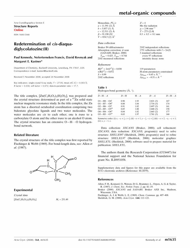
The title complex, [Zn(C2H3O3)2(H2O)2], was prepared and
the crystal structure determined as part of a 67Zn solid state
nuclear magnetic resonance study. In the title complex, the Zn
atom has a disorted octahedral coordination comprising two
bidentate glycolate ligands and two water molecules. The
water molecules are cis to each other; one is trans to a
carboxylate O atom and the other trans to an alcohol O atom.
The crystal structure has an extensive O—H� � �O hydrogen-
bond network.
Related literature
The crystal structure of the title complex was first reported by
Fischinger & Webb (1969). For bond-length data, see: Allen et
al. (1987).
Experimental
Crystal data
[Zn(C2H3O3)2(H2O)2] Mr = 251.49
Monoclinic, P21=ca = 11.391 (2) Ab = 5.857 (1) Ac = 12.511 (2) A� = 91.198 (9)�
V = 834.5 (2) A3
Z = 4Mo K� radiation� = 2.96 mm�1
T = 273 (2) K0.3 � 0.3 � 0.1 mm
Data collection
Bruker P4 diffractometerAbsorption correction: scan
(SADABS; Bruker, 2000)Tmin = 0.425, Tmax = 0.744
2102 measured reflections
2102 independent reflections1751 reflections with I > 2�(I)3 standard reflections
every 97 reflectionsintensity decay: none
Refinement
R[F 2 > 2�(F 2)] = 0.030wR(F 2) = 0.072S = 0.992102 reflections
119 parametersH-atom parameters constrained��max = 0.45 e A�3
��min = �0.51 e A�3
Table 1Hydrogen-bond geometry (A, �).
D—H� � �A D—H H� � �A D� � �A D—H� � �A
O1—H8� � �O4i 0.90 1.93 2.821 (3) 167O1—H7� � �O8ii 0.88 1.84 2.716 (3) 174O2—H6� � �O5i 0.91 1.82 2.697 (3) 161O2—H5� � �O5iii 0.85 1.88 2.688 (3) 158O6—H4� � �O8iv 0.84 1.82 2.665 (2) 177O3—H3� � �O7iv 0.83 1.97 2.761 (3) 160
Symmetry codes: (i) x;�yþ 12; zþ 1
2; (ii) �xþ 1; y� 12;�zþ 3
2; (iii) �xþ 2;�y;�zþ 1;(iv) x; y� 1; z.
Data collection: XSCANS (Bruker, 2000); cell refinement:
XSCANS; data reduction: XSCANS; program(s) used to solve
structure: SHELXS97 (Sheldrick, 2008); program(s) used to refine
structure: SHELXL97 (Sheldrick, 2008); molecular graphics:
SHELXTL (Sheldrick, 2008); software used to prepare material for
publication: SHELXTL.
The authors thank the Research Corporation (CC6447) for
financial support and the National Science Foundation for
grant No. ILI8951058.
Supplementary data and figures for this paper are available from theIUCr electronic archives (Reference: SU2079).
References
Allen, F. H., Kennard, O., Watson, D. G., Brammer, L., Orpen, A. G. & Taylor,R. (1987). J. Chem. Soc. Perkin Trans. 2, pp. S1–19.
Bruker (2000). XSCANS and SADABS. Bruker AXS Inc., Madison,Wisconsin, USA.
Fischinger, A. J. & Webb, L. E. (1969). Chem. Commun. pp. 407–408.Sheldrick, G. M. (2008). Acta Cryst. A64, 112–122.
metal-organic compounds
Acta Cryst. (2008). E64, m1635 doi:10.1107/S1600536808039585 Kennedy et al. m1635
Acta Crystallographica Section E
Structure ReportsOnline
ISSN 1600-5368

supplementary materials

supplementary materials
sup-1
Acta Cryst. (2008). E64, m1635 [ doi:10.1107/S1600536808039585 ]
Redetermination of cis-diaquadiglycolatozinc(II)
P. Kennedy, N. Francis, D. Rovnyak and M. E. Kastner
Comment
As part of a Zinc-67 solid state nuclear magnetic resonance study the title complex, (I), was prepared and the crystal structuredetermined. The structure of this complex was first report by Fischinger & Webb (1969) but no fractional crystal coordinateswere reported.
The molecular structure of (I) is illustrated in Fig. 1. The zinc atom has a distorted octahedral coordination sphere com-posed of two bidentate gylcolato ligands and two water molecules. The water molecules are cis to each other; one (O1) istrans to a carboxylate O-atom (O4), and the other, (O2), is trans to an alcohol O-atom (O6). The bond distances and anglesare normal for zinc(II) complexes (Allen et al., 1987)
In the crystal structure of (I) there in an extensive O—H···O hydrogen bonding network (Table 1). The two water mo-lecules (O1 and O2) bond to the two oxygens (O4 and O5) of a carboxylate group related by the c-glide. The two alcoholgroups (O3 and O6) form hydrogen bonds with the other carboxylate group (atoms O7 and O8) translated by one unit cellalong the b axis. The carbonyl oxygen O8 of this ligand also makes a two dimensional hydrogen bonded network with oneof the waters (O1) around the inversion center. The other water molecule, (O2), forms an H-bond with a carbonyl oxygen(O5) related by the 2-fold screw axis.
Experimental
Glycolic acid (100 mg, 3 mmol), purchased from Sigma-Aldrich (99%), was dissolved in 5 ml of deionized water. Basic zinccarbonate (80 mg, 2 mmol) was added and the mixture stirred for 10 minutes while heating to ca. 60°C. The resulting mixturewas filtered, and the filtrate left to stand at room temperature until large needle-like crystals grew by slow evaporation ofthe water.
Refinement
The alcohol and water H-atoms were placed at the locations identified in a difference Fourier map and were held fixed, with
Uiso(H) set to 0.05 A2: O-H = 0.8293 - 0.9135 Å. The C-bound H-atoms were included in calculated positions and treated
as riding atoms: C-H = 0.97 Å with Uiso(H) = 1.2Ueq(parent C-atom).
Figures
Fig. 1. A view of the molecular structure of compound (I), showing the atom numberingscheme and dispacement ellipsods drawn at the 50% probability level.

supplementary materials
sup-2
cis-diaquadiglycolatozinc(II)
Crystal data
[Zn(C2H3O3)2(H2O)2] F000 = 512
Mr = 251.49 Dx = 2.002 Mg m−3
Monoclinic, P21/c Mo Kα radiationλ = 0.71073 Å
Hall symbol: -P 2ybc Cell parameters from 20 reflectionsa = 11.391 (2) Å θ = 9.6–17.4ºb = 5.8570 (10) Å µ = 2.96 mm−1
c = 12.511 (2) Å T = 273 (2) Kβ = 91.198 (9)º Needle, colorless
V = 834.5 (2) Å3 0.3 × 0.3 × 0.1 mmZ = 4
Data collection
Bruker P4diffractometer
Rint = 0.0000
Radiation source: fine-focus sealed tube θmax = 28.5º
Monochromator: graphite θmin = 3.3ºT = 273(2) K h = −15→152θ/ω scans k = 0→7Absorption correction: ψ scan(SADABS; Bruker, 2000) l = 0→16
Tmin = 0.425, Tmax = 0.744 3 standard reflections2102 measured reflections every 97 reflections2102 independent reflections intensity decay: none1751 reflections with I > 2σ(I)
Refinement
Refinement on F2 Hydrogen site location: inferred from neighbouringsites
Least-squares matrix: full H-atom parameters constrained
R[F2 > 2σ(F2)] = 0.030 w = 1/[σ2(Fo
2) + (0.0337P)2 + 0.5879P]where P = (Fo
2 + 2Fc2)/3
wR(F2) = 0.072 (Δ/σ)max = 0.030
S = 0.99 Δρmax = 0.45 e Å−3
2102 reflections Δρmin = −0.50 e Å−3
119 parametersExtinction correction: SHELXL97 (Sheldrick, 2008),Fc*=kFc[1+0.001xFc2λ3/sin(2θ)]-1/4
Primary atom site location: structure-invariant directmethods Extinction coefficient: 0.0094 (8)
Secondary atom site location: difference Fourier map

supplementary materials
sup-3
Special details
Geometry. All e.s.d.'s (except the e.s.d. in the dihedral angle between two l.s. planes) are estimated using the full covariance mat-rix. The cell e.s.d.'s are taken into account individually in the estimation of e.s.d.'s in distances, angles and torsion angles; correlationsbetween e.s.d.'s in cell parameters are only used when they are defined by crystal symmetry. An approximate (isotropic) treatment ofcell e.s.d.'s is used for estimating e.s.d.'s involving l.s. planes.
Refinement. Refinement of F2 against ALL reflections. The weighted R-factor wR and
goodness of fit S are based on F2, conventional R-factors R are based
on F, with F set to zero for negative F2. The threshold expression of
F2 > σ(F2) is used only for calculating R-factors(gt) etc. and is
not relevant to the choice of reflections for refinement. R-factors based
on F2 are statistically about twice as large as those based on F, and R-
factors based on ALL data will be even larger.
Fractional atomic coordinates and isotropic or equivalent isotropic displacement parameters (Å2)
x y z Uiso*/Ueq
Zn 0.76227 (2) 0.14144 (5) 0.65837 (2) 0.02431 (11)O1 0.73409 (15) 0.0926 (3) 0.82160 (13) 0.0323 (4)O2 0.91409 (16) 0.3091 (3) 0.69490 (15) 0.0349 (4)O3 0.82248 (17) −0.1945 (3) 0.64048 (13) 0.0307 (4)O4 0.80776 (17) 0.1349 (3) 0.49645 (13) 0.0310 (4)O5 0.90455 (17) −0.0608 (4) 0.37503 (13) 0.0367 (4)O6 0.58388 (16) 0.0302 (3) 0.63177 (15) 0.0339 (4)O7 0.66840 (15) 0.4419 (3) 0.64627 (14) 0.0299 (4)O8 0.49499 (15) 0.6096 (3) 0.62300 (13) 0.0275 (4)C1 0.8665 (2) −0.2479 (4) 0.53732 (19) 0.0297 (5)H1A 0.8215 −0.3727 0.5062 0.036*H1B 0.9477 −0.2968 0.5444 0.036*C2 0.8588 (2) −0.0423 (4) 0.46432 (18) 0.0242 (5)C3 0.4989 (2) 0.2052 (4) 0.62244 (18) 0.0242 (5)H2A 0.4422 0.1896 0.6787 0.029*H2B 0.4575 0.1927 0.5542 0.029*C4 0.5587 (2) 0.4364 (4) 0.63092 (16) 0.0218 (4)H3 0.7697 −0.2865 0.6547 0.050*H4 0.5587 −0.1048 0.6293 0.050*H5 0.9803 0.2654 0.6719 0.050*H6 0.9284 0.3898 0.7563 0.050*H7 0.6612 0.0892 0.8427 0.050*H8 0.7688 0.1759 0.8742 0.050*

supplementary materials
sup-4
Atomic displacement parameters (Å2)
U11 U22 U33 U12 U13 U23
Zn 0.02730 (16) 0.01890 (15) 0.02686 (15) 0.00221 (12) 0.00378 (10) −0.00151 (11)O1 0.0314 (9) 0.0381 (10) 0.0275 (8) −0.0050 (8) 0.0044 (7) −0.0032 (7)O2 0.0280 (9) 0.0360 (10) 0.0408 (10) −0.0002 (8) 0.0038 (7) −0.0114 (8)O3 0.0452 (10) 0.0204 (8) 0.0269 (8) 0.0026 (8) 0.0121 (7) 0.0011 (7)O4 0.0404 (10) 0.0270 (9) 0.0260 (8) 0.0094 (8) 0.0063 (7) 0.0037 (7)O5 0.0435 (11) 0.0390 (11) 0.0280 (9) 0.0100 (9) 0.0116 (8) 0.0036 (8)O6 0.0327 (10) 0.0161 (8) 0.0527 (11) 0.0002 (8) −0.0013 (8) −0.0041 (8)O7 0.0268 (9) 0.0174 (8) 0.0457 (10) 0.0012 (7) 0.0026 (7) −0.0014 (8)O8 0.0294 (9) 0.0198 (8) 0.0333 (9) 0.0036 (7) 0.0046 (7) 0.0029 (7)C1 0.0414 (14) 0.0203 (11) 0.0277 (12) 0.0026 (11) 0.0100 (10) −0.0006 (10)C2 0.0216 (11) 0.0255 (12) 0.0257 (11) −0.0017 (10) 0.0017 (8) 0.0014 (9)C3 0.0294 (12) 0.0196 (10) 0.0238 (10) −0.0008 (10) −0.0008 (9) −0.0002 (9)C4 0.0284 (11) 0.0185 (10) 0.0188 (9) −0.0011 (9) 0.0049 (8) 0.0010 (8)
Geometric parameters (Å, °)
Zn—O2 2.0325 (19) O4—C2 1.259 (3)Zn—O7 2.0630 (17) O5—C2 1.247 (3)Zn—O1 2.0935 (17) O6—C3 1.412 (3)Zn—O3 2.0974 (18) O6—H4 0.8417Zn—O4 2.1019 (17) O7—C4 1.261 (3)Zn—O6 2.1531 (19) O8—C4 1.250 (3)O1—H7 0.8768 C1—C2 1.513 (3)O1—H8 0.9037 C1—H1A 0.9700O2—H5 0.8522 C1—H1B 0.9700O2—H6 0.9135 C3—C4 1.518 (3)O3—C1 1.429 (3) C3—H2A 0.9700O3—H3 0.8293 C3—H2B 0.9700
O2—Zn—O7 92.38 (7) C2—O4—Zn 116.58 (15)O2—Zn—O1 89.65 (7) C3—O6—Zn 115.85 (14)O7—Zn—O1 95.65 (7) C3—O6—H4 116.5O2—Zn—O3 101.47 (8) Zn—O6—H4 127.6O7—Zn—O3 164.27 (7) C4—O7—Zn 120.00 (16)O1—Zn—O3 91.89 (7) O3—C1—C2 110.72 (19)O2—Zn—O4 89.99 (7) O3—C1—H1A 109.5O7—Zn—O4 94.74 (7) C2—C1—H1A 109.5O1—Zn—O4 169.61 (7) O3—C1—H1B 109.5O3—Zn—O4 78.01 (7) C2—C1—H1B 109.5O2—Zn—O6 167.62 (7) H1A—C1—H1B 108.1O7—Zn—O6 76.15 (7) O5—C2—O4 124.2 (2)O1—Zn—O6 86.90 (7) O5—C2—C1 116.8 (2)O3—Zn—O6 90.53 (7) O4—C2—C1 119.00 (19)O4—Zn—O6 95.53 (7) O6—C3—C4 109.64 (19)Zn—O1—H7 117.5 O6—C3—H2A 109.7

supplementary materials
sup-5
Zn—O1—H8 124.3 C4—C3—H2A 109.7H7—O1—H8 101.3 O6—C3—H2B 109.7Zn—O2—H5 122.2 C4—C3—H2B 109.7Zn—O2—H6 125.1 H2A—C3—H2B 108.2H5—O2—H6 107.2 O8—C4—O7 124.3 (2)C1—O3—Zn 115.05 (14) O8—C4—C3 117.4 (2)C1—O3—H3 108.8 O7—C4—C3 118.3 (2)Zn—O3—H3 110.3
O2—Zn—O3—C1 −84.29 (19) O2—Zn—O7—C4 −174.79 (18)O7—Zn—O3—C1 67.0 (3) O1—Zn—O7—C4 −84.91 (18)O1—Zn—O3—C1 −174.31 (18) O3—Zn—O7—C4 33.4 (4)O4—Zn—O3—C1 3.24 (18) O4—Zn—O7—C4 95.01 (18)O6—Zn—O3—C1 98.77 (18) O6—Zn—O7—C4 0.47 (17)O2—Zn—O4—C2 94.74 (19) Zn—O3—C1—C2 0.1 (3)O7—Zn—O4—C2 −172.86 (19) Zn—O4—C2—O5 −170.4 (2)O1—Zn—O4—C2 6.7 (5) Zn—O4—C2—C1 9.4 (3)O3—Zn—O4—C2 −6.98 (18) O3—C1—C2—O5 173.5 (2)O6—Zn—O4—C2 −96.36 (19) O3—C1—C2—O4 −6.3 (3)O2—Zn—O6—C3 21.4 (5) Zn—O6—C3—C4 1.6 (2)O7—Zn—O6—C3 −1.22 (16) Zn—O7—C4—O8 179.73 (17)O1—Zn—O6—C3 95.41 (17) Zn—O7—C4—C3 0.3 (3)O3—Zn—O6—C3 −172.75 (16) O6—C3—C4—O8 179.2 (2)O4—Zn—O6—C3 −94.73 (17) O6—C3—C4—O7 −1.3 (3)
Hydrogen-bond geometry (Å, °)
D—H···A D—H H···A D···A D—H···A
O1—H8···O4i 0.90 1.93 2.821 (3) 167
O1—H7···O8ii 0.88 1.84 2.716 (3) 174
O2—H6···O5i 0.91 1.82 2.697 (3) 161
O2—H5···O5iii 0.85 1.88 2.688 (3) 158
O6—H4···O8iv 0.84 1.82 2.665 (2) 177
O3—H3···O7iv 0.83 1.97 2.761 (3) 160Symmetry codes: (i) x, −y+1/2, z+1/2; (ii) −x+1, y−1/2, −z+3/2; (iii) −x+2, −y, −z+1; (iv) x, y−1, z.

supplementary materials
sup-6
Fig. 1
![Octahedral ruthenium(II) complexes cis,cis-[RuX2(CNR)(CO)(P∧P)] and cis,cis,cis-[RuX2(CO)2(P∧P)] (X=Cl, Br; P∧P=1,1′-bis(diphenylphosphino)ferrocene, 1,1′-bis(diisopropylphosphino)ferrocene):](https://img.pdfslide.net/doc/110x75/6350510356d5ddd7dc052b00/octahedral-rutheniumii-complexes-ciscis-rux2cnrcopp-and-cisciscis-rux2co2pp.jpg)


![SYNTHESIS AND CRYSTAL STRUCTURE OF [BIS (DL-ALANINATO) DIAQUA] NICKEL (II) DIHYDRATE](https://img.pdfslide.net/doc/110x75/6349ebf8b029d9fbf200f07f/synthesis-and-crystal-structure-of-bis-dl-alaninato-diaqua-nickel-ii-dihydrate.jpg)


