Embed Size (px)
Citation preview
BioMed CentralC
TIONALINTERNACANCER CELLCancer Cell International
ss
Open AccePrimary researchReduced paxillin expression contributes to the antimetastatic effect of 4-hydroxycoumarin on B16-F10 melanoma cellsMarco A Velasco-Velázquez*, Nohemí Salinas-Jazmín, Nicandro Mendoza-Patiño and Juan J MandokiAddress: Departamento de Farmacología, Facultad de Medicina, Universidad Nacional Autónoma de México. Apdo. Postal 70-297, Ciudad Universitaria, México D.F. 04510, México
Email: Marco A Velasco-Velázquez* - [email protected]; Nohemí Salinas-Jazmín - [email protected]; Nicandro Mendoza-Patiño - [email protected]; Juan J Mandoki - [email protected]
* Corresponding author
AbstractBackground: 4-Hydroxycoumarin (4-HC) is a coumarin that lacks anticoagulant activity. 4-HCaffects the cytoskeletal stability and decreases cell adhesion and motility of the melanoma cell lineB16-F10. Together with integrins and other cytoskeletal proteins, paxillin participates in theregulation of cell adhesion and motility, acting as an adapter protein at focal adhesions. The presentstudy determined the participation of paxillin in the reported effects of 4-HC and analyzed the roleof paxillin in the formation of melanoma metastases.
Results: 4-HC decreased protein and mRNA levels of α- and β-paxillin isoforms in B16-F10 cells.Paxillin downregulation correlated with an inadequate translocation of paxillin to focal adhesionsand a reduced phosphotyr118-paxillin pool. Consequently, 4-HC altered paxillin-mediated signaling,decreasing the phosphorylation of FAK and the level of GTP-bound Rac-1. These results partiallyexplain the mechanism of the previously reported effects of 4-HC. Additionally, we studied theeffect of 4-HC on metastatic potential of B16-F10 cells through experimental metastasis assays. Invitro treatment of cells with 4-HC inhibited their capability to originate pulmonary metastases. 4-HC did not affect cell proliferation or survival, demonstrating that its antimetastatic effect isunrelated to changes on cell viability. We also studied the importance of paxillin in metastasis bytransfecting melanoma cells with paxillin-siRNA. Transfection produced a modest reduction onmetastatic potential, indicating that: i) paxillin plays a role as inducer of melanoma metastasis; andii) paxillin downregulation is not sufficient to explain the antimetastatic effect of 4-HC. Therefore,we evaluated other changes in gene expression by differential display RT-PCR analysis. Treatmentwith 4-HC produced a downregulation of Adhesion Regulating Molecule-1 (ARM-1), whichcorrelated with a decreased adhesion of melanoma cells to lung slides.
Conclusion: This study shows that reduced paxillin expression is associated with the impaired celladhesion and motility seen in 4-HC-treated cells and partially contributes to the antimetastaticeffect of 4-HC. In contrast, the role of ARM-1 reduced expression in the effects of 4-HC is still tobe clarified. The antimetastatic effect of 4-HC suggests that this compound, or others with similarmode of action, might be useful for the development of adjuvant therapies for melanoma.
Published: 20 May 2008
Cancer Cell International 2008, 8:8 doi:10.1186/1475-2867-8-8
Received: 25 March 2008Accepted: 20 May 2008
This article is available from: http://www.cancerci.com/content/8/1/8
© 2008 Velasco-Velázquez et al; licensee BioMed Central Ltd. This is an Open Access article distributed under the terms of the Creative Commons Attribution License (http://creativecommons.org/licenses/by/2.0), which permits unrestricted use, distribution, and reproduction in any medium, provided the original work is properly cited.
Page 1 of 12(page number not for citation purposes)
Cancer Cell International 2008, 8:8 http://www.cancerci.com/content/8/1/8
BackgroundFormation of metastases is still the leading cause of deathin advanced melanoma patients, regardless the numerousinnovations on the treatment of the disease. Hence, thesearch of therapeutic agents that can inhibit metastasis iscrucial for improving the management of melanoma.
The production of metastases is a highly complex processby which some cancer cells move away from the primarytumor and colonize other organs. This process requiresphenotypical changes that allow cancer cells to migrate,survive in the blood circulation, extravasate, and prolifer-ate in a tissue with a different microenvironment [1].Along the metastatic cascade, cell adhesion is an essentialprocess [2]. Cell adhesion mediated by integrin receptorsdrives the formation of focal adhesions, which are multi-molecular structures that enable cells to firmly adhere.Additionally, focal adhesions constitute important signal-ing centers that regulate the reorganization of thecytoskeleton needed for spreading and motility [3,4].These functions are critical in the acquisition of the abilityof cancer cells to invade distant tissues [2]; consequently,the focal adhesion molecules have been proposed as phar-macological targets for decreasing invasiveness of cancercells [5,6].
Paxillin is a multidomain adapter protein that participatesin linking scaffolding and in signaling at focal adhesions[7]. The structural features of paxillin (reviewed by Brownand Turner [7]) allow it to interact with different signalingproteins, such as FAK, Src, Crk, Csk, p120 RasGAP, andPTP-PEST [7,8]. Thus, paxillin has been implicated in theregulation of diverse cellular events, including adhesion[9,10], spreading [11], and motility [11,12].
In mouse, two isoforms of paxillin are generated by alter-native splicing, with molecular weights of 68 (α) and 70KDa (β) [13,14]. Even when both isoforms may share thesame functions [14], the β isoform has been implicated intransformation and malignancy [13]. Both paxillin iso-forms contain critical phosphorylation sites at tyrosines31 and 118 [7,12]. The phosphorylation of these two tyro-sines by FAK and Src regulates the paxillin turnover infocal adhesions [15-17] and generate docking sites forother molecules that participate in the rearrangement ofthe actin cytoskeleton [7,16-19]. Then, tyrosine phospho-rylation of paxillin and its localization into focal adhe-sions are necessary for the adequate control of adhesionand motility [12,17-19].
In cancer cells, overexpression and increased tyrosinephosphorylation of paxillin have been reported. Forexample, paxillin overexpression stimulates the adhesionof squamous carcinoma cells to collagen [20] as well astheir migration [21]. Paxillin is also overexpressed in
highly metastatic human osteosarcoma [22] and renal car-cinoma cell lines [23]. Similarly, levels of phospho-paxil-lin are much higher in melanoma cell lines than inmelanocytes [24]. These data suggest that paxillin plays arole in the acquisition and maintenance of a malignantphenotype.
4-Hydroxycoumarin (4-HC) is a simple coumarin used asprecursor for the synthesis of anticoagulant drugs androdenticides that are 3-substituted-4-hydroxicoumarins;however, 4-HC lacks of anticoagulant activity [25]. Previ-ously, we have provided evidence that 4-HC affects thestability of the actin cytoskeleton on the melanoma cellline B16-F10, impairing the formation of stress fibers andlamellipodia [26]. These effects correlate with reductionsin cell adhesion to extracellular matrix proteins and inhi-bition of motility [26]. The key role of paxillin in the reg-ulation of cytoskeletal rearrangements, adhesion, andmotility led us to the proposal that paxillin may beinvolved in the reported effects produced by 4-HC. There-fore, we analyzed the effects of 4-HC on paxillin expres-sion and paxillin-mediated signaling. Additionally, weevaluated the metastatic potential of B16-F10 cells treatedwith 4-HC, and studied the role of paxillin in metastasisby blocking its expression with siRNA. Finally, we per-formed differential display RT-PCR assays in order toidentify other proteins that participate in the effects of 4-HC.
MethodsMaterialsThe murine melanoma cell line B16-F10 was purchasedfrom the American Type Culture Collection (Manassas,VA, USA). C57BL/6 mice (Harlan, Mexico City, Mexico)were used in this study. The experiments with mice wereconducted in accordance with the Guide for the Care andUse of Laboratory Animals as adopted and promulgatedby the Declaration of Helsinki.
4-Hydroxycoumarin (4-Hydroxy-2H-1-benzopyran-2-one [cat. no. H23805]) and its vehicle, ethanol, were pur-chased from Sigma (St. Louis, MO, USA). Antibodiesagainst paxillin (sc-5574 rabbit polyclonal), β-tubulin (D-10 mouse monoclonal), FAK (H-1 mouse monoclonal),and phosphotyrosine (PY20 mouse monoclonal) as wellas siRNAs were obtained from Santa Cruz Biotechnology(Santa Cruz, CA, USA). Control siRNA (sc-37007) is anon-targeting 20–25 nt siRNA that will not lead to thespecific degradation of any known cellular mRNA, whilepaxillin siRNA (sc-36197) is a pool of 3 target-specific 20–25 nt siRNAs.
Cell culture and treatmentsB16-F10 cells were routinely cultured at 37°C in a humid5% CO2 atmosphere, using RPMI-1640 containing 10%
Page 2 of 12(page number not for citation purposes)
Cancer Cell International 2008, 8:8 http://www.cancerci.com/content/8/1/8
fetal bovine serum (FBS). For all experiments cells wereseeded at a density of 3 × 10⁴ cells/cm2. In experimentswith 4-HC, cells were incubated overnight and thenexposed for 24 h to 500 μM 4-HC (dissolved in ethanol)or 0.75% ethanol (control) in serum-free medium. Theconcentration and exposure time used here were previ-ously reported [26], and are those that induce changes inactin cytoskeleton, impairing cell adhesion and motility.
Transfection with siRNA (60 pmols) was carried out withLipofectamine 2000 (Invitrogen, Rockville, MD, USA)according to the procedure recommended by the manu-facturer.
Analysis of paxillin expression and phosphorylationTreated cells were washed twice with ice-cold PBS andlysed in cold lysis buffer [50 mM Tris (pH 8.0), 150 mMNaCl, 1% NP-40, 0.5% deoxycholate, 0.1% SDS] contain-ing a protease inhibitor cocktail (Roche, Indianapolis, IN,USA). Samples containing 60 μg of total protein were sep-arated by SDS-PAGE and transblotted onto PVDF mem-branes. The membranes were blocked and then probedwith anti-paxillin or with anti-phosphotyr118-paxillinantibodies. On both cases the membranes were strippedand reprobed with anti-β-tubulin antibody. After incuba-tion with the corresponding secondary antibody, theimmunoreactive bands were visualized by chemiluminis-cence and a densitometric analysis was carried out usingImageJ NIH software [27].
ImmunoflurescenceCellular localization of paxillin was evaluated on cellsseeded on Labtek chambers (Nunc, Rochester, NY, USA).After treatment, cells were fixed with 4% formaldehyde inPBS and permeated during 4 min at room temperaturewith 0.1% Triton X-100 diluted in PBS. Then, paxillin waslabeled using anti-paxillin antibody followed by a second-ary antibody conjugated with Alexa-546 (MolecularProbes, Eugene, OR, USA). After extensive washing, theslides were mounted and analyzed with a Nikon epifluo-rescence microscope (Melville, NY, USA).
Reverse transcription-polymerase chain reaction (RT-PCR)Total RNA was extracted using Trizol reagent (Invitrogen)according to the manufacturer's instructions and quanti-fied spectrophotometrically at 260 nm. Synthesis ofcDNA was carried out using Super-Script reverse tran-scriptase (Invitrogen) and oligo-dT as primer. Semiquan-titative PCR was performed in 50 μl of a reaction mixturecontaining 1× PCR buffer, 2.5 mM MgCl2, 0.2 mM dNTPs,1·U AmpliTaq DNA polymerase (Invitrogen), 0.2 μM ofeach primer, and cDNA obtained from 175 ng of totalRNA. Reactions were cycled 30 times through 30 s at94°C, 60 s at 55°C, and 60 s at 72°C. The paxillin primers
used were previously reported by Mazaki et. al. [14] andare as follows: pan-paxillin: sense 5'-aacaagcagaagtcagca-gagcc-3', antisense 5'-ctagcttgttcaggtcggac-3' (ampliconlength: 582 bp for α isoform and 684 bp for β isoform);β-paxillin: same sense primer than for pan-paxillin, anti-sense 5'-ctctccatccactctctgtt-3' (503 bp). The GAPDHprimers [28] were: sense 5'-accacagtccatgccatcac-3', anti-sense 5'-tccaccaccctgttgctgta-3' (452 bp). PCR productswere resolved by 2% agarose gel electrophoresis andstained with ethidium bromide. Negative controls lackingreverse transcriptase were run in parallel to confirm thatsamples were not contaminated with genomic DNA.
Real time PCR for ARM-1 was performed in a GeneAmp5700 Sequence Detection System (Applied Biosystems,Foster City, CA, USA) as described previously [29 javas-cript:popRef('b12')]. Briefly, 0.5× of SYBR Green I (Molec-ular Probes, Eugene, OR, USA) was added to the reactionmixture described above. The primers for amplification ofARM-1 were: sense 5'-ggacagcttggccctctcat-3'; antisense 5'-gggcaaatcacaatcaccactac-3'. Reactions for GAPDH wereperformed with the same primers used in semiquantita-tive PCR. Real time-PCR reactions were cycled 35 timesthrough 30 s at 95°C, 30 s at 60°C, 60 s at 72°C, and 5 sat temperature of fluorescence acquisition (FA). Data wereanalyzed with the GeneAmp 5700 SDS software version1.3 (Applied Biosystems).
Analysis of FAK phosphorylationLysates from adhered cells were obtained with cold lysisbuffer containing phosphatase inhibitors [1 mM sodiumpyrophosphate; 1 mM sodium orthovanadate; 50 mMsodium fluoride]. Lysates were immunoprecipitated withanti-FAK antibody bound to protein A- agarose beads. Theimmunoprecipitated proteins were recovered adding SDSsample buffer, separated by SDS-PAGE, transblotted ontoPVDF membranes, and probed with anti-phosphotyro-sine and anti-paxillin antibodies. The same membraneswere reprobed with anti-FAK antibody.
Affinity precipitation of activated Rac-1Pull-down assays for Rac-1 were performed using EZ-Detect activation kit from Pierce (Rockford, IL, USA),according to the manufacturer's instructions. Briefly, 106
cells were lysed in 400 μl of cold lysis buffer as describedabove. The samples were mixed with GST-PAK1-PBD,loaded on SwellGel immobilized glutathione disks, andincubated with constant agitation at 4°C for 2 h. Then,the disks were washed and the glutathione-bound Rac-1(active form) was recovered adding SDS sample buffer [60mM Tris-HCl (pH 6.8); 2% SDS; 0.05% 2-mercaptoetha-nol; 1% glycerol; and 0.05% bromophenol blue]. The iso-lated proteins as well as aliquots of cell lysates wereseparated by electrophoresis and Rac-1 was detected byimmunoblotting using anti-Rac-1 monoclonal antibody.
Page 3 of 12(page number not for citation purposes)
Cancer Cell International 2008, 8:8 http://www.cancerci.com/content/8/1/8
Neutral red assayCell viability was determined using the neutral red accu-mulation assay [30]. The cells were plated on 96-wellplates and then treated as described above. After 12, 24,36 or 48 h of exposure to 4-HC, the medium was changedand the cells were incubated for 90 min under cell cultureconditions with 50 μg/ml neutral red. After this incuba-tion the cells were fixed on the plate with an aqueous solu-tion containing 1% formaldehyde and 1% calciumchloride and then lysed with 50% ethanol/49% water/1%acetic acid. The concentration of accumulated neutral redas a marker for cell viability was measured spectrophoto-metrically at 560 nm.
Clonogenic assayCell survival was evaluated accordingly to the methodol-ogy reported by Franken et. al. [31]. Briefly, cells were har-vested and counted after treatment. Cell suspensions fromeach treatment were diluted in RPMI-1640 with 5% FBS,and immediately re-plated in 6-well plates at a density of20 cells/cm2. The plates were incubated until cells in con-trol wells have formed sufficiently large colonies. Afterthat, the colonies were fixed with 6% glutaraldehyde andstained with 0.5% crystal violet. The plates were photo-graphed and their digital images were analyzed withImageJ NIH software [27] to known the colony number.
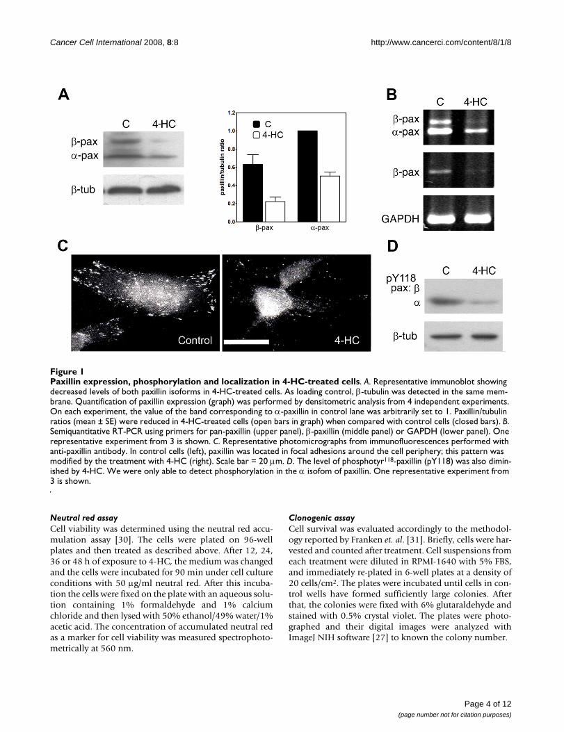
Paxillin expression, phosphorylation and localization in 4-HC-treated cellsFigure 1Paxillin expression, phosphorylation and localization in 4-HC-treated cells. A. Representative immunoblot showing decreased levels of both paxillin isoforms in 4-HC-treated cells. As loading control, β-tubulin was detected in the same mem-brane. Quantification of paxillin expression (graph) was performed by densitometric analysis from 4 independent experiments. On each experiment, the value of the band corresponding to α-paxillin in control lane was arbitrarily set to 1. Paxillin/tubulin ratios (mean ± SE) were reduced in 4-HC-treated cells (open bars in graph) when compared with control cells (closed bars). B. Semiquantitative RT-PCR using primers for pan-paxillin (upper panel), β-paxillin (middle panel) or GAPDH (lower panel). One representative experiment from 3 is shown. C. Representative photomicrographs from immunofluorescences performed with anti-paxillin antibody. In control cells (left), paxillin was located in focal adhesions around the cell periphery; this pattern was modified by the treatment with 4-HC (right). Scale bar = 20 μm. D. The level of phosphotyr118-paxillin (pY118) was also dimin-ished by 4-HC. We were only able to detect phosphorylation in the α isofom of paxillin. One representative experiment from 3 is shown.
Page 4 of 12(page number not for citation purposes)
Cancer Cell International 2008, 8:8 http://www.cancerci.com/content/8/1/8
Experimental metastasis assayB16-F10 cells were treated in vitro, as mentioned under"cell culture and treatments". After treatment, cells weredetached with a non-enzymatic cell dissociation buffer [4mM EDTA in Ca2+ and Mg2+-free PBS], resuspended inHank's balanced salt solution and immediately injectedinto the tail vein of 8-week old, male C57BL/6 mice. Eachmouse received 106 cells. At 2 weeks after i.v. injectionmice were euthanized, lungs were excised and the meta-static tumors counted in a blind manner under a dissect-ing microscopy.
Differential display reverse transcription polymerase chain reaction (DD-RT-PCR)Total RNA, free of DNA contamination was used for theDD-RT-PCR as described by Liang and Pardee [32].Briefly, cDNA was synthesized with 200 ng of RNA and 1μM of primer of sequence 5'-t12ca-3' to anneal. A controlreaction lacking reverse transcriptase was included to con-firm absence of non-specific amplification from genomicDNA. The cDNAs corresponding to 20 ng of RNA were
PCR amplified in the presence of 0.4 μM of [α-35S] dATP(37 GBq/pmol) using primers of sequence 5'-t12ca-3' (1μM) and 5'-gttgcgatcc-3' (0.2 μM). PCR reactions werecycled 35 times through 50 s at 95°C, 90 s at 40°C, and60 s at 72°C. The heat denatured PCR products were elec-trophoresed on a urea-PAGE gel (48% urea and 6% acry-lamide). The gels were dried and exposed to X-ray films at-70°C. After 10–12 h the X-ray film was developed untilthe DNA bands were clearly seen on the film. The DNAbands which were differentially displayed in the autoradi-ograph were visually selected, marked on the gel and theband was cut with a sterile razor. The DNA extracted fromthe gel was PCR amplified using the same set of primersused in the DD-RT-PCR analysis, cloned, and sequenced.
Adhesion of melanoma cells to lung sectionsTo evaluate the organ-specific adhesion of tumor cells invitro, the method of Vink et. al. [33] was used with modi-fications in incubation times. Briefly, fresh lungs obtainedfrom healthy C57BL/6 mice were embedded in TissueTeKO.C.T compound (Poly-Labo, Strasbourg, France) and
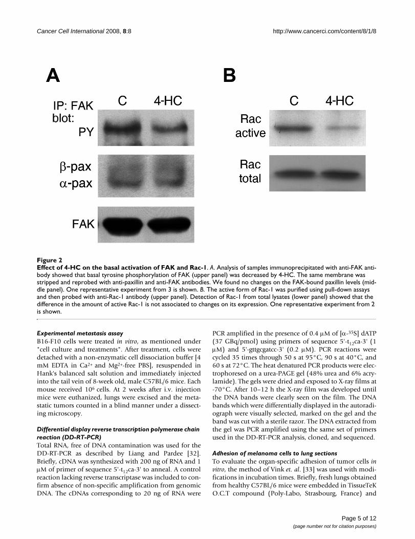
Effect of 4-HC on the basal activation of FAK and Rac-1Figure 2Effect of 4-HC on the basal activation of FAK and Rac-1. A. Analysis of samples immunoprecipitated with anti-FAK anti-body showed that basal tyrosine phosphorylation of FAK (upper panel) was decreased by 4-HC. The same membrane was stripped and reprobed with anti-paxillin and anti-FAK antibodies. We found no changes on the FAK-bound paxillin levels (mid-dle panel). One representative experiment from 3 is shown. B. The active form of Rac-1 was purified using pull-down assays and then probed with anti-Rac-1 antibody (upper panel). Detection of Rac-1 from total lysates (lower panel) showed that the difference in the amount of active Rac-1 is not associated to changes on its expression. One representative experiment from 2 is shown.
Page 5 of 12(page number not for citation purposes)
Cancer Cell International 2008, 8:8 http://www.cancerci.com/content/8/1/8
frozen at -196°C. Fresh cryostat sections (8–10 μm thick)mounted on glass slides were first incubated with PBS/BSA 3% for 60 min in a humid chamber. After treatment,melanoma cells were detached with a non-enzymatic celldissociation buffer, resuspended in medium, and placedon tissue sections for 30 min at 37°C with gentle agita-tion. The slides were washed 3 times with medium toremove nonattached cells and then fixed in paraformalde-hyde 3% in PBS. Slides were stained with hematoxiline-eosine and the numbers of cells attached to the cryostatsections were counted in 10 microscopic fields.
Results4-HC decreased the expression of paxillin, altering its cellular localization and the amount of phosphotyr118-paxillinAnalysis of paxillin expression by immunoblottingshowed that B16-F10 melanoma cells normally express αand β isoforms of paxillin (Figure 1A, control lane). Treat-ment of cells with 4-HC decreased the expression of bothpaxillin isoforms. Densitometric analyses of the immuno-blots revealed that in 4-HC-treated cells, the β-paxillin/tubulin and α-paxillin/tubulin ratios decreased to 35 and
50% of the control values, respectively (Figure 1A). Addi-tionally, we performed semiquantitative RT-PCRs in orderto evaluate whether the decrements of paxillin elicited by4-HC were produced through changes in mRNA levels.Using pan-paxillin primers as well as specific β-paxillinprimers we found decreased mRNA expression in cellsthat were treated with 4-HC (Figure 1B). Cellular localiza-tion of paxillin can regulate its activation; thus, we studiedthe paxillin localization through immunofluorescenceassays. In control cells paxillin is distributed in a punctatepattern, presumably forming part of focal adhesions. Incontrast, in B16-F10 cells treated with 4-HC paxillin local-ization was predominantly perinuclear (Figure 1C). Aspreviously reported [26], 4-HC impaired the formation oflamellipodia and induced shrinkage of the outer enve-lope, forming a round cell with radial filopodia attachedto adhesion points. Paxillin can be phosphorylated onseveral tyrosines [34]; among them, tyrosines 31 and 118have shown importance in the generation of signals nec-essary for adhesion and migration [7,12]. Thus, we stud-ied if the reduction in paxillin expression produced by 4-HC correlated with changes in the level of phosphotyr118-paxillin. Even when both isoforms can be phosphor-
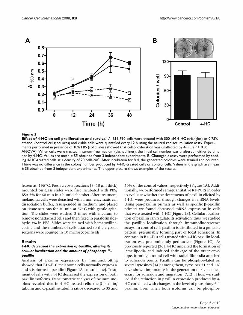
Effect of 4-HC on cell proliferation and survivalFigure 3Effect of 4-HC on cell proliferation and survival. A. B16-F10 cells were treated with 500 μM 4-HC (triangles) or 0.75% ethanol (control cells; squares) and viable cells were quantified every 12 h using the neutral red accumulation assay. Experi-ments performed in presence of 10% FBS (solid lines) showed that cell proliferation was unaffected by 4-HC (P > 0.05, ANOVA). When cells were treated in serum-free medium (dashed lines), the initial cell number was unaltered neither by time nor by 4-HC. Values are mean ± SE obtained from 3 independent experiments. B. Clonogenic assay were performed by seed-ing 4-HC-treated cells at a density of 20 cells/cm2. After incubation for 8 d, the generated colonies were stained and counted. There was no difference in the colony number produced by 4-HC-treated cells or control cells. Values in the graph are mean ± SE obtained from 3 independent experiments. The upper picture shows examples of the results.
Page 6 of 12(page number not for citation purposes)
Cancer Cell International 2008, 8:8 http://www.cancerci.com/content/8/1/8
ylated, we were able to detect phosphotyr118 only in the αisofom of paxillin (Figure 1D). For this isoform, thephosphotyr118-paxillin/paxillin ratio is 85% of the controlvalue, indicating that the reduction in phospho-paxillin ismainly caused by paxillin downregulation.
Paxillin-mediated signaling was impaired in 4-HC-treated cellsPaxillin participates in the regulation of different signal-ing pathways. We studied the activation of FAK, a key part-ner in paxillin-meditated signaling. Basal tyrosinephosphorylation of FAK was found in serum starved B16-F10, as previously reported [35] (Figure 2A, control lane).In 4-HC-treated cells, the phospho-FAK/FAK ratiodecreased to 46% of the control value (Figure 2A). How-ever, levels of FAK-bound paxillin did not change signifi-cantly (Figure 2A) suggesting that even when paxillin isdownregulated, the remaining paxillin supports FAKbinding.
The activation of Rac-1, which promotes the formation oflamellipodia, can be triggered by tyrosine-phosphorylatedpaxillin; therefore, we studied the activation of Rac-1 per-forming pull-down assays. 4-HC decreased the amount ofGTP-bound Rac-1 without changing Rac-1 expression
(Figure 2B), reducing the active Rac-1/total Rac-1 ratio to35% of the control value.
4-HC did not alter proliferation or survival of B16-F10 cellsReduction of paxillin expression as well as decreased FAKactivation might affect cell proliferation, survival or apop-tosis/anoikis [36]. For that reason, we studied the viabilityof cell cultures exposed to 4-HC during different times. Inpresence of FBS, 4-HC did not modify cell proliferation incomparison with control cells (Figure 3A, solid lines).When cultures were treated in serum-free medium, wefound that 4-HC caused a slight, non significant reductionon the number of viable cells at times longer than 24 h(Figure 3A, dashed lines). Congruously, 4-HC did notaffect the ability of single cells to grow into a colonies (Fig-ure 3B), suggesting that cell survival is unaffected by 4-HC.
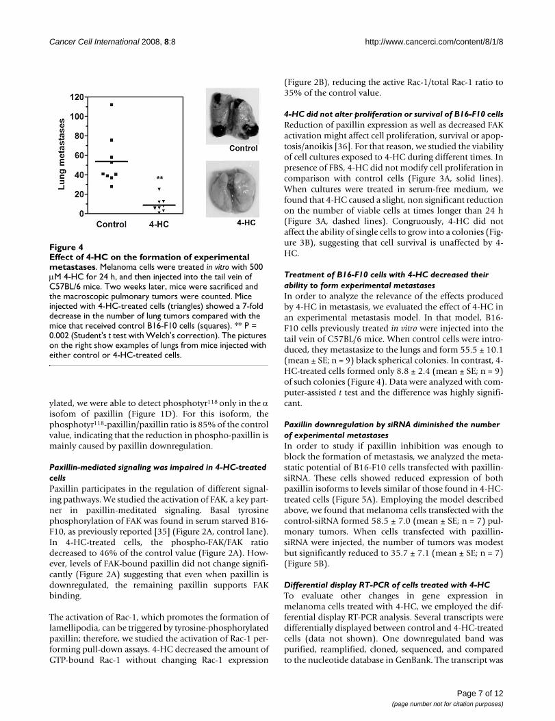
Treatment of B16-F10 cells with 4-HC decreased their ability to form experimental metastasesIn order to analyze the relevance of the effects producedby 4-HC in metastasis, we evaluated the effect of 4-HC inan experimental metastasis model. In that model, B16-F10 cells previously treated in vitro were injected into thetail vein of C57BL/6 mice. When control cells were intro-duced, they metastasize to the lungs and form 55.5 ± 10.1(mean ± SE; n = 9) black spherical colonies. In contrast, 4-HC-treated cells formed only 8.8 ± 2.4 (mean ± SE; n = 9)of such colonies (Figure 4). Data were analyzed with com-puter-assisted t test and the difference was highly signifi-cant.
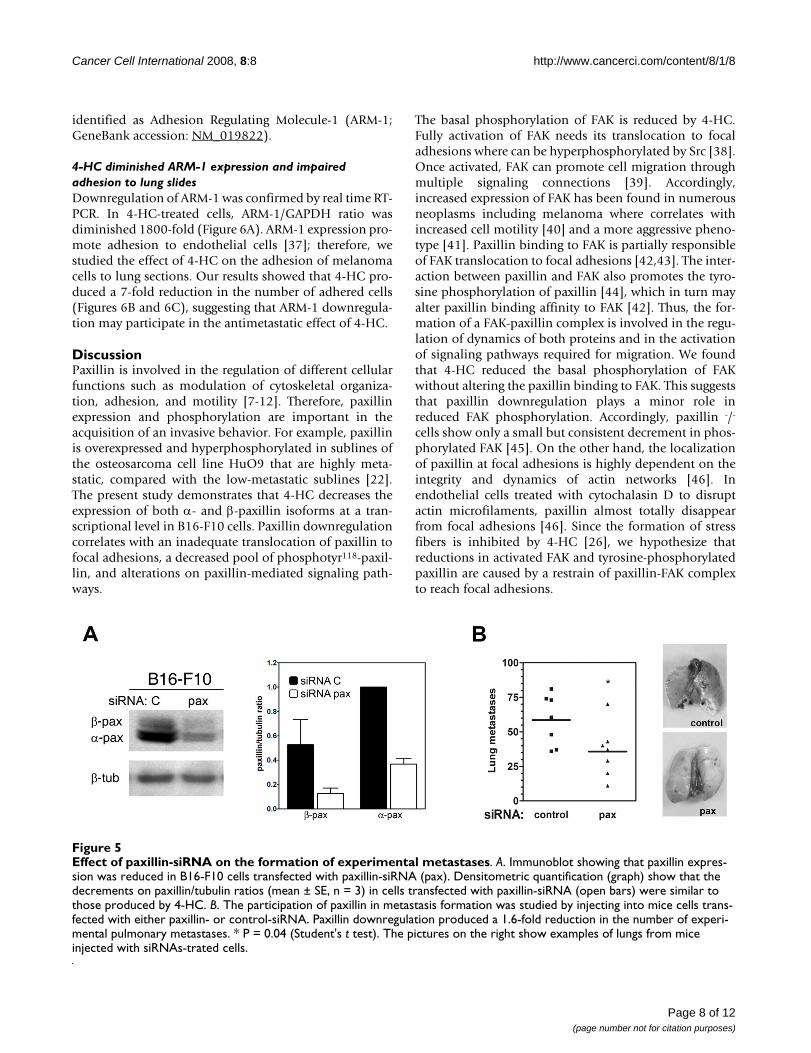
Paxillin downregulation by siRNA diminished the number of experimental metastasesIn order to study if paxillin inhibition was enough toblock the formation of metastasis, we analyzed the meta-static potential of B16-F10 cells transfected with paxillin-siRNA. These cells showed reduced expression of bothpaxillin isoforms to levels similar of those found in 4-HC-treated cells (Figure 5A). Employing the model describedabove, we found that melanoma cells transfected with thecontrol-siRNA formed 58.5 ± 7.0 (mean ± SE; n = 7) pul-monary tumors. When cells transfected with paxillin-siRNA were injected, the number of tumors was modestbut significantly reduced to 35.7 ± 7.1 (mean ± SE; n = 7)(Figure 5B).
Differential display RT-PCR of cells treated with 4-HCTo evaluate other changes in gene expression inmelanoma cells treated with 4-HC, we employed the dif-ferential display RT-PCR analysis. Several transcripts weredifferentially displayed between control and 4-HC-treatedcells (data not shown). One downregulated band waspurified, reamplified, cloned, sequenced, and comparedto the nucleotide database in GenBank. The transcript was
Effect of 4-HC on the formation of experimental metastasesFigure 4Effect of 4-HC on the formation of experimental metastases. Melanoma cells were treated in vitro with 500 μM 4-HC for 24 h, and then injected into the tail vein of C57BL/6 mice. Two weeks later, mice were sacrificed and the macroscopic pulmonary tumors were counted. Mice injected with 4-HC-treated cells (triangles) showed a 7-fold decrease in the number of lung tumors compared with the mice that received control B16-F10 cells (squares). ** P = 0.002 (Student's t test with Welch's correction). The pictures on the right show examples of lungs from mice injected with either control or 4-HC-treated cells.
Page 7 of 12(page number not for citation purposes)
Cancer Cell International 2008, 8:8 http://www.cancerci.com/content/8/1/8
identified as Adhesion Regulating Molecule-1 (ARM-1;GeneBank accession: NM_019822).
4-HC diminished ARM-1 expression and impaired adhesion to lung slidesDownregulation of ARM-1 was confirmed by real time RT-PCR. In 4-HC-treated cells, ARM-1/GAPDH ratio wasdiminished 1800-fold (Figure 6A). ARM-1 expression pro-mote adhesion to endothelial cells [37]; therefore, westudied the effect of 4-HC on the adhesion of melanomacells to lung sections. Our results showed that 4-HC pro-duced a 7-fold reduction in the number of adhered cells(Figures 6B and 6C), suggesting that ARM-1 downregula-tion may participate in the antimetastatic effect of 4-HC.
DiscussionPaxillin is involved in the regulation of different cellularfunctions such as modulation of cytoskeletal organiza-tion, adhesion, and motility [7-12]. Therefore, paxillinexpression and phosphorylation are important in theacquisition of an invasive behavior. For example, paxillinis overexpressed and hyperphosphorylated in sublines ofthe osteosarcoma cell line HuO9 that are highly meta-static, compared with the low-metastatic sublines [22].The present study demonstrates that 4-HC decreases theexpression of both α- and β-paxillin isoforms at a tran-scriptional level in B16-F10 cells. Paxillin downregulationcorrelates with an inadequate translocation of paxillin tofocal adhesions, a decreased pool of phosphotyr118-paxil-lin, and alterations on paxillin-mediated signaling path-ways.
The basal phosphorylation of FAK is reduced by 4-HC.Fully activation of FAK needs its translocation to focaladhesions where can be hyperphosphorylated by Src [38].Once activated, FAK can promote cell migration throughmultiple signaling connections [39]. Accordingly,increased expression of FAK has been found in numerousneoplasms including melanoma where correlates withincreased cell motility [40] and a more aggressive pheno-type [41]. Paxillin binding to FAK is partially responsibleof FAK translocation to focal adhesions [42,43]. The inter-action between paxillin and FAK also promotes the tyro-sine phosphorylation of paxillin [44], which in turn mayalter paxillin binding affinity to FAK [42]. Thus, the for-mation of a FAK-paxillin complex is involved in the regu-lation of dynamics of both proteins and in the activationof signaling pathways required for migration. We foundthat 4-HC reduced the basal phosphorylation of FAKwithout altering the paxillin binding to FAK. This suggeststhat paxillin downregulation plays a minor role inreduced FAK phosphorylation. Accordingly, paxillin -/-
cells show only a small but consistent decrement in phos-phorylated FAK [45]. On the other hand, the localizationof paxillin at focal adhesions is highly dependent on theintegrity and dynamics of actin networks [46]. Inendothelial cells treated with cytochalasin D to disruptactin microfilaments, paxillin almost totally disappearfrom focal adhesions [46]. Since the formation of stressfibers is inhibited by 4-HC [26], we hypothesize thatreductions in activated FAK and tyrosine-phosphorylatedpaxillin are caused by a restrain of paxillin-FAK complexto reach focal adhesions.
Effect of paxillin-siRNA on the formation of experimental metastasesFigure 5Effect of paxillin-siRNA on the formation of experimental metastases. A. Immunoblot showing that paxillin expres-sion was reduced in B16-F10 cells transfected with paxillin-siRNA (pax). Densitometric quantification (graph) show that the decrements on paxillin/tubulin ratios (mean ± SE, n = 3) in cells transfected with paxillin-siRNA (open bars) were similar to those produced by 4-HC. B. The participation of paxillin in metastasis formation was studied by injecting into mice cells trans-fected with either paxillin- or control-siRNA. Paxillin downregulation produced a 1.6-fold reduction in the number of experi-mental pulmonary metastases. * P = 0.04 (Student's t test). The pictures on the right show examples of lungs from mice injected with siRNAs-trated cells.
Page 8 of 12(page number not for citation purposes)
Cancer Cell International 2008, 8:8 http://www.cancerci.com/content/8/1/8
4-HC also decreased the basal activation of Rac-1, a down-stream effector of paxillin, indicating that reduced paxillinexpression affects this pathway involved in the regulationof motility. The participation of tyrosine-phosphorylatedpaxillin in Rac-1 activation is well documented [47,48].The phosphorylation of tyrosines 31 and 118 on paxillingenerate binding sites for the adaptor protein Crk [47].Crk-paxillin interaction promotes the binding ofDOCK180 to Crk, which in turn locally activates Rac-1[48]. Once activated, Rac-1 has a key role in cell motilitythrough its ability to stimulate lamellipodium protrusionat the leading edge [4,49]. Then, the reduced Rac-1 activa-tion in 4-HC-treated cells, which correlates with the lackof lamellipodia previously reported [26], seems to be sub-sequent to the reduced phospho-paxillin level. Notori-ously, basal activation of Rac-1 is increased in B16-F10cells relative to the poorly metastatic B16-F0 cells [50];therefore, impaired Rac-1 activation by 4-HC may becooperating to decrease the metastatic behavior of B16-F10 cells.
Changes in paxillin expression or FAK activation can pro-mote alterations on cell proliferation and survival [36,39].In this study we found that exposition to 4-HC during 48h did not modify cell proliferation, indicating that in B16-F10 cells this process is unaffected by paxillin downregu-lation. Cell survival was also unaltered by 4-HC, demon-strating that the antimetastatic effect of 4-HC is not relatedto changes in long term survival of melanoma cells. Thelack of cytotoxic effect of 4-HC on cancer cells reportedhere is consistent with previous studies [26,51]. Further-more, 4-HC (500 μM) does not affect the viability or thecytoskeleton stability of B82 fibroblasts [26] and is non-
toxic to cultured hepatocytes [52], supporting the hypoth-esis that 4-HC has low toxicity.
Additionally, our results show that paxillin downregula-tion, either by 4-HC or paxillin-siRNA, reduce the meta-static potential of B16-F10 cells. Contrary to othermolecules involved in the regulation of adhesion, such asintegrins or FAK, the role of paxillin in the biology ofmetastasis is still controversial. Several reports identifypaxillin as an inducer of metastasis; for instance, in headand neck cancers that have metastasized to lymph nodespaxillin expression is increased [53]. Similarly, paxillinup-regulation correlates with the presence of extrahepaticmetastasis in hepatocellular carcinoma [54] and lymphnode metastasis in breast tumors [55]. On the contrary,other reports state that paxillin overexpression is a markerof a less invasive tumor phenotype in breast [56] and lung[57] carcinomas. The fact that metastatic potential of B16-F10 cells is reduced by decreasing paxillin expression sup-port the hypothesis that paxillin facilitates metastasis andemphasize the importance of paxillin as an inducer ofmelanoma metastasis. Nevertheless, the number of pul-monary metastases produced by paxillin-silenced cells is4-fold greater than the quantity produced by 4-HC-treatedcells, indicating that paxillin downregulation is only par-tially responsible for the antimetastatic effect of 4-HC.
To further understand the mechanism involved in the 4-HC antimetastatic effect, we carried out a differential dis-play RT-PCR assay and discovered that 4-HC may be alter-ing the expression of several genes other than paxillin. Inthis work ARM-1 has been identified as one of the genesdifferentially expressed in 4-HC-treated cells. ARM-1 is a
ARM-1 expression and adhesion to lung sections by cells treated with 4-HCFigure 6ARM-1 expression and adhesion to lung sections by cells treated with 4-HC. A. The effect of 4-HC on ARM-1 expression was evaluated by real time-PCR. 4-HC inhibited ARM-1 mRNA expression. Values are represented as ratios of ARM-1/GAPDH ± SD. B. B16-F10 cells were treated as described previously, and then incubated with lung sections during 30 min to allow their adhesion. The preparations were stained and photographed. Photomicrographs (400X) show that control cells (left) were able to adhere to lung sections; in contrast, 4-HC-treated cells (right) displayed impaired adhesion. C. Quanti-fication of adhered cells (mean ± SE) from 5 independent experiments. On each experiment, 10 fields were counted. ***P < 0.0001 (Student's t test).
Page 9 of 12(page number not for citation purposes)
Cancer Cell International 2008, 8:8 http://www.cancerci.com/content/8/1/8
protein associated with the plasma membrane [58,59]whose precise functions have not been totally solved.ARM-1 was first discovered as an adhesion promotingmolecule [37,59], but recently it was identified as a novelcomponent of the 26 S proteasome [60]. The role of ARM-1 in the regulation of cell adhesion is supported by studiesin which changes in ARM-1 expression alter cell-cell adhe-sion. For example, overexpression of ARM-1 in endothe-lial cells increases lymphocyte adhesion [58]. Similarly,transfection of ARM-1 into kidney embryonic cells pro-motes their adhesion to endothelial cells [37]. A role ofARM-1 in metastasis is suggested by the fact that ARM-1expression is increased in metastatic human breast cancercells as compared with their non metastatic counterparts[37]. Besides, ARM-1 expression is constitutive in cell linesfrom gastric carcinoma [59], breast carcinoma, and T lym-phoma [37]. Together, these data suggest that ARM-1 maybe a metastasis-associated gene. We show that ARM-1dowregulation produced by 4-HC correlates with adecreased adhesion of B16-F10 cells to lung sections. Thiseffect suggests that 4-HC may be inhibiting ARM-1-medi-ated tumor cell adhesion and/or invasion during the for-mation of pulmonary metastases. Nevertheless, the role ofARM-1 reduced expression in the antimetastatic effect of4-HC needs further investigation. Furthermore, since sev-eral transcripts were differentially displayed in 4-HC-treated cells, it cannot be ruled out that 4-HC may beaffecting the expression of other regulatory proteins thatparticipate in metastasis. In particular, it will be necessaryto clarify the effect of 4-HC on integrin expression sinceseveral of these receptors participate in different stages ofthe metastatic cascade.
ConclusionThis study shows that 4-HC inhibits the formation ofexperimental metastases. Although the molecular mecha-nism of the antimetastatic action of 4-HC needs to be fur-ther defined, our present results indicate that 4-HC-induced decreased paxillin availability and inappropriatesubcellular localization impair the adequate generation ofsignals that promote malignancy. In addition, 4-HC mayaffect important steps of metastasis by changing theexpression of other molecules, such as ARM-1. However,further studies are required to clarify the effects of 4-HC invivo since there are not toxicological or pharmacokineticdata available. Together, our results suggests that 4-HC, oragents with similar mode of action, might be useful in theprevention of metastasis and could be used as an adjuvantin the therapy of melanoma.
Competing interestsThe authors declare that they have no competing interests.
Authors' contributionsMAVV performed cell transfections and clonogenic assays,helped to carry out FAK and Rac1 activation assays,designed and supervised the study, and prepared the man-uscript.
NSJ performed cell culture and carried out immunoblots,RT-PCRs, immunofluorescences, and experimental metas-tases assays.
NMP participated in the design of the study.
JJM participated in the design of the study and in the prep-aration of the manuscript.
All authors read and approved the manuscript.
AcknowledgementsWe thank Mayra Pérez-Tapia, Nestor Ocón, Abraham Landa, and Armando Pérez for technical assistance and Vanessa González and Marisol de la Fuente for critical reading of the manuscript. This research was supported by PAPIIT-UNAM (IN230202 and IN223806) and Conacyt (203595).
References1. Bogenrieder T, Herlyn M: Axis of evil: molecular mechanisms of
cancer metastasis. Oncogene 2003, 22:6524-6536.2. Hood JD, Cheresh DA: Role of integrins in cell invasion and
migration. Nat Rev Cancer 2002, 2:91-100.3. Geiger B, Bershadsky A, Pankov R, Yamada K: Transmembrane
extracellular matrix-cytoskeleton crosstalk. Nat Rev Mol CellBiol 2001, 2:793-805.
4. Ridley AJ, Schwartz MA, Burridge K, Firtel RA, Ginsberg MH, BorisyG, Parsons JT, Horwitz AR: Cell migration: integrating signalsfrom front to back. Science 2003, 302:1704-1709.
5. Hehlgans S, Haase M, Cordes N: Signaling via integrins: implica-tions for cell survival and anticancer strategies. Biochim BiophysActa 2007, 1775:163-180.
6. Velasco-Velázquez MA, Molina-Guarneros JA, Mendoza-Patiño N,López-González JS, Mandoki JJ: Integrins and integrin-associatedmolecules: targets for the development of antimetastatictherapies. Rev Invest Clin 1999, 51:183-93.
7. Brown MC, Turner CE: Paxillin: adapting to change. Physiol Rev2004, 84:1315-1339.
8. Wang Y, Gilmore TD: Zyxin and paxillin proteins: focal adhe-sion plaque LIM domain proteins go nuclear. Biochim BiophysActa 2003, 1593:115-120.
9. Burridge K, Turner CE, Romer LH: Tyrosine phosphorylation ofpaxillin and pp125FAK accompanies cell adhesion to extra-cellular matrix: A role in cytoskeletal assembly. J Cell Biol1992, 119:893-903.
10. Sanders MA, Basson MD: Collagen IV-dependent ERK activa-tion in human Caco-2 intestinal epithelial cells requires focaladhesion kinase. J Biol Chem 2000, 275:38040-38047.
11. Jamieson JS, Tumbarello DA, Halle M, Brown MC, Tremblay ML,Turner CE: Paxillin is essential for PTP-PEST-dependent reg-ulation of cell spreading and motility: a role for paxillinkinase linker. J Cell Sci 2005, 118:5835-5847.
12. Petit V, Boyer B, Lentz D, Turner CE, Thiery JP, Valles AM: Phospho-rylation of tyrosine residues 31 and 118 on paxillin regulatescell migration through an association with CRK in NBT-IIcells. J Cell Biol 2000, 148:957-970.
13. Mazaki Y, Hashimoto S, Sabe H: Monocyte cells and cancer cellsexpress novel paxillin isoforms with different binding proper-ties to focal adhesion proteins. J Biol Chem 1997, 272:7437-7444.
14. Mazaki Y, Hiroshi U, Okio H, Shigeru H: Paxillin Isoform inMouse. Lack of the gamma isoform and developmentallyspecific beta isoform expression. J Biol Chem 1998,273:22435-22441.
Page 10 of 12(page number not for citation purposes)
Cancer Cell International 2008, 8:8 http://www.cancerci.com/content/8/1/8
15. Richardson AR, Malik K, Hildebrand JD, Parsons JT: Inhibition ofcell spreading by expression of the C-terminal domain offocal adhesion kinase (FAK) is rescued by coexpression ofSrc or catalytically inactive FAK: a role for paxillin tyrosinephosphorylation. Mol Cell Biol 1997, 17:6906-6914.
16. Turner CE: Paxillin and focal adhesion signaling. Nat Cell Biol2000, 2:E231-E236.
17. Nakamura K, Yano H, Uchida H, Hashimoto S, Schaefer E, Sabe H:Tyrosine phosphorylation of paxillin alpha is involved in tem-porospatial regulation of paxillin-containing focal adhesionformation and F-actin organization in motile cells. J Biol Chem2000, 275:27155-27164.
18. Tsubouchi A, Sakakura J, Yagi R, Mazaki Y, Schaefer E, Yano H, SabeH: Localized suppression of RhoA activity by Tyr31/118-phosphorylated paxillin in cell adhesion and migration. J CellBiol 2002, 159:673-683.
19. Iwasaki T, Nakata A, Mukai M, Shinkai K, Yano H, Sabe H, Schaefer E,Tatsuta M, Tsujimura T, Terada N, Kakishita E, Akedo H: Involve-ment of phosphorylation of Tyr-31 and Tyr-118 of paxillin inMM1 cancer cell migration. Int J Cancer 2002, 97:330-335.
20. Conway WC, van Zyp J Van der Voort, Thamilselvan V, Walsh MF,Crowe DL, Basson MD: Paxillin modulates squamous cancercell adhesion and is important in pressure-augmented adhe-sion. J Cell Biochem 2006, 98:1507-1516.
21. Crowe DL, Ohannessian A: Recruitment of focal adhesionkinase and paxillin to beta1 integrin promotes cancer cellmigration via mitogen activated protein kinase activation.BMC Cancer 2004, 4:18.
22. Azuma K, Tanaka M, Uekita T, Inoue S, Yokota J, Ouchi Y, Sakai R:Tyrosine phosphorylation of paxillin affects the metastaticpotential of human osteosarcoma. Oncogene 2005,24:4754-4764.
23. Jenq W, Cooper D, Ramirez G: Integrin expression on cell adhe-sion function and up-regulation of P125FAK and paxillin inmetastatic renal carcinoma cells. Connect Tissue Res 1996,34:161-174.
24. Hamamura K, Furukawa K, Hayashi T, Hattori T, Nakano J,Nakashima H, Okuda T, Mizutani H, Hattori H, Ueda M, Urano T,Lloyd KO, Furukawa K: Ganglioside GD3 promotes cell growthand invasion through p130Cas and paxillin in malignantmelanoma cells. Proc Natl Acad Sci USA 2005, 102:11041-11046.
25. Mueller RL, Scheidt S: History of drugs for thrombotic disease.Discovery, development, and directions for the future. Circu-lation 1994, 89:432-449.
26. Velasco-Velazquez MA, Agramonte-Hevia J, Barrera D, Jimenez-Oro-zco A, Garcia-Mondragon MJ, Mendoza-Patino N, Landa A, MandokiJ: 4-Hydroxycoumarin disorganizes the actin cytoskeleton inB16-F10 melanoma cells but not in B82 fibroblasts, decreas-ing their adhesion to extracellular matrix proteins andmotility. Cancer Lett 2003, 198:179-186.
27. Rasband WS: ImageJ. [http://rsb.info.nih.gov/ij/].28. Chacón-Salinas R, Serafín-López J, Ramos-Payán R, Méndez-Aragón P,
Hernández-Pando R, Van Soolingen D, Flores-Romo L, Estrada-ParraS, Estrada-García I: Differential pattern of cytokine expressionby macrophages infected in vitro with different Mycobacte-rium tuberculosis genotypes. Clin Exp Immunol 2005, 140:443-449.
29. Ramos-Payán R, Aguilar-Medina M, Estrada-Parra S, González-Y-Mer-chand JA, Favila-Castillo L, Monroy-Ostria A, Estrada-García IC:Quantification of cytokine gene expression using an econom-ical real-time polymerase chain reaction method based onSYBR Green I. Scand J Immunol 2003, 57:439-445.
30. Babich H, Shopsis C, Borenfreund E: In vitro cytotoxicity testingof aquatic pollutants (cadmium, copper, zinc, nickel) usingestablished fish cell lines. Ecotoxicol Environ Saf 1986, 11:91-99.
31. Franken NA, Rodermond HM, Stap J, Haveman J, van Bree C: Clono-genic assay of cells in vitro. Nat Protoc 2006, 1:2315-2319.
32. Liang P, Pardee AB: Differential display of eukaryotic messen-ger RNA by means of the polymerase chain reaction. Science1992, 257:967-971.
33. Vink J, Thomas L, Etoh T, Bruijn JA, Mihm MC Jr, Gattoni-Celli S,Byers HR: Role of beta-1 integrins in organ specific adhesionof melanoma cells in vitro. Lab Invest 1993, 68:192-203.
34. Schroeder MJ, Webb DJ, Shabanowitz J, Horwitz AF, Hunt DF: Meth-ods for the detection of paxillin post-translational modifica-tions and interacting proteins by mass spectrometry. JProteome Res 2005, 4:1832-1841.
35. Abdel-Ghany M, Cheng HC, Elble RC, Pauli BU: Focal adhesionkinase activated by beta(4) integrin ligation to mCLCA1mediates early metastatic growth. J Biol Chem 2002,277:34391-34400.
36. Gabarra-Niecko V, Schaller MD, Dunty JM: FAK regulates biolog-ical processes important for the pathogenesis of cancer. Can-cer Metastasis Rev 2003, 22:359-374.
37. Simins AB, Weighardt H, Weidner KM, Weidle UH, Holzmann B:Functional cloning of ARM-1, an adhesion-regulating mole-cule upregulated in metastatic tumor cells. Clin Exp Metastasis1999, 17:641-648.
38. Cohen LA, Guan JL: Mechanisms of focal adhesion kinase regu-lation. Curr Cancer Drug Targets 2005, 5:629-643.
39. Mitra SK, Hanson DA, Schlaepfer DD: Focal adhesion kinase: incommand and control of cell motility. Nat Rev Mol Cell Biol2005, 6:56-68.
40. Akasaka T, van Leeuwen RL, Yoshinaga IG Jr, Mihm MC, Byers HR:Focal adhesion kinase (p125FAK) expression correlates withmotility of human melanoma cell lines. J Invest Dermatol 1995,105:104-108.
41. Hess AR, Postovit LM, Margaryan NV, Seftor EA, Schneider GB,Seftor RE, Nickoloff BJ, Hendrix MJ: Focal adhesion kinase pro-motes the aggressive melanoma phenotype. Cancer Res 2005,65:9851-9860.
42. Bertoloucci CM, Guibao CD, Zheng J: Structural features of thefocal adhesion kinase-paxillin complex give insight into thedynamics of focal adhesion assembly. Protein Sci 2005,14:644-652.
43. Cooley MA, Broome JM, Ohngemach C, Romer LH, Schaller MD:Paxillin binding is not the sole determinant of focal adhesionlocalization or dominant-negative activity of Focal AdhesionKinase/Focal Adhesion Kinase-related Nonkinase. Mol Biol Cell2000, 11:3247-3263.
44. Shen Y, Schaller MD: Focal Adhesion Targeting: the criticaldeterminant of FAK regulation and substrate phosphoryla-tion. Mol Biol Cell 1999, 10:2507-2518.
45. Hagel M, George EL, Kim A, Tamimi R, Opitz SL, Turner CE, ImamotoA, Thomas SM: The adaptor protein paxillin is essential fornormal development in the mouse and is a critical trans-ducer of fibronectin signaling. Mol Cell Biol 2002, 22:901-915.
46. Hu YL, Haga JH, Miao H, Wang Y, Li YS, Chien S: Roles of microfil-aments and microtubules in paxillin dynamics. Biochem BiophysRes Commun 2006, 348:1463-1471.
47. Schaller MD, Parsons JT: pp125FAK-dependent tyrosine phos-phorylation of paxillin creates a high-affinity binding site forCrk. Mol Cell Biol 1995, 15:2635-2345.
48. Kiyokawa E, Hashimoto Y, Kobayashi S, Sugimura H, Kurata T, Mat-suda M: Activation of Rac1 by a Crk SH3-binding protein,DOCK180. Genes Dev 1998, 12:3331-3336.
49. Webb DJ, Horwitz AF: New dimensions in cell migration. NatCell Biol 2003, 5:690-692.
50. Ferraro D, Corso S, Fasano E, Panieri E, Santangelo R, Borrello S,Giordano S, Pani G, Galeotti T: Pro-metastatic signaling by c-Met through RAC-1 and reactive oxygen species (ROS).Oncogene 2006, 25:3689-3698.
51. Kawaii S, Tomono Y, Ogawa K, Sugiura M, Yano M, Yoshizawa Y: Theantiproliferative effect of coumarins on several cancer celllines. Anticancer Res 2001, 21:917-923.
52. Ratanasavanh D, Lamiable D, Biour M, Guédès Y, Gersberg M, Leu-tenegger E, Riché C: Metabolism and toxicity of coumarin oncultured human, rat, mouse and rabbit hepatocytes. FundamClin Pharmacol 1996, 10:504-510.
53. Nagata M, Fujita H, Ida H, Hoshina H, Inoue T, Seki Y, Ohnishi M,Ohyama T, Shingaki S, Kaji M, Saku T, Takagi R: Identification ofpotential biomarkers of lymph node metastasis in oral squa-mous cell carcinoma by cDNA microarray analysis. Int J Can-cer 2003, 106:683-689.
54. Li HG, Xie DR, Shen XM, Li HH, Zeng H, Zeng YJ: Clinicopatholog-ical significance of expression of paxillin, syndecan-1 andEMMPRIN in hepatocellular carcinoma. World J Gastroenterol2005, 11:1445-14451.
55. Vadlamudi R, Adam L, Tseng B, Costa L, Kumar R: Transcriptionalup-regulation of paxillin expression by heregulin in humanbreast cancer cells. Cancer Res 1999, 59:2843-2846.
56. Madan R, Smolkin MB, Cocker R, Fayyad R, Oktay MH: Focal adhe-sion proteins as markers of malignant transformation and
Page 11 of 12(page number not for citation purposes)
Cancer Cell International 2008, 8:8 http://www.cancerci.com/content/8/1/8
Publish with BioMed Central and every scientist can read your work free of charge
"BioMed Central will be the most significant development for disseminating the results of biomedical research in our lifetime."
Sir Paul Nurse, Cancer Research UK
Your research papers will be:
available free of charge to the entire biomedical community
peer reviewed and published immediately upon acceptance
cited in PubMed and archived on PubMed Central
yours — you keep the copyright
Submit your manuscript here:http://www.biomedcentral.com/info/publishing_adv.asp
BioMedcentral
prognostic indicators in breast carcinoma. Hum Pathol 2006,37:9-15.
57. Salgia R, Li JL, Ewaniuk DS, Wang YB, Sattler M, Chen WC, RichardsW, Pisick E, Shapiro GI, Rollins BJ, Chen LB, Griffin JD, Sugarbaker DJ:Expression of the focal adhesion protein paxillin in lung can-cer and its relation to cell motility. Oncogene 1999, 18:67-77.
58. Lamerant N, Kieda C: Adhesion properties of adhesion-regulat-ing molecule 1 protein on endothelial cells. FEBS J 2005,272:1833-1844.
59. Shimada S, Ogawa M, Takahashi M, Schlom J, Greiner W: Molecularcloning and characterization of the complementary DNA ofan Mr 110,000 antigen expressed by human gastric carci-noma cells and upregulated by γ-Interferon. Cancer Res 1994,54:3831-3836.
60. Jørgensen JP, Lauridsen AM, Kristensen P, Dissing K, Johnsen AH,Hendil KB, Hartmann-Petersen R: Adrm1, a putative cell adhe-sion regulating protein, is a novel proteasome-associatedfactor. J Mol Biol 2006, 360:1043-1052.
Page 12 of 12(page number not for citation purposes)


