Embed Size (px)
Citation preview
Brain (1996), 119, 1617-1625
Regional brain activity during working memorytasksE. Salmon,12 M. Van der Linden,3 F. Collette,3 G. Delfiore,1 P. Maquet,12 C. Degueldre,1 A. Luxen1
and G. Franck2
1 Cyclotron Research Centre, the ^Department of Neurology Correspondence to: E. Salmon, MD, Cyclotron Researchand the ^Department of Neuropsychology, University of Centre, B30 Sart Tilman, 4000 Liege, BelgiumLiege, Belgium
SummaryThe first aim of our PET study was to replicate previousfindings concerning the brain areas activated by a verbalworking memory task. The second aim was to specify theneural basis of the central executive, using a task of workingmemory updating. Our data confirm that the lower leftsupramarginal gyrus and premotor area are the key regionssubserving short-term verbal memory processes. They alsosuggest that the updating memory task is related to mid-dorsolateral prefrontal activation, most probably responsible
Keywords: working memory; central executive; PET
for the updating function of the central executive. Anunexpected, predominantly right activation occurred in theinferior parietal region during the verbal memory updatingtask, which we related to a visuospatial strategy used tomaintain the information in short-term memory. A thirdpurpose was to explore the brain regions activated by anonverbal, visual memory task, and our results confirm theimportance of the superior occipital gyrus in the visual short-term memory.
Abbreviations: BA = Brodmann area; rCBF = regional cerebral blood flow; SPM = statistical parametric map
IntroductionWorking memory refers to a limited capacity system that isresponsible for the temporary storage and processing ofinformation while cognitive tasks are performed. Baddeley'smodel represents the most extensively investigated theoreticalconstruct of working memory (Baddeley and Hitch, 1974;Baddeley, 1986, 1992). This model comprises a modality-free controlling central executive that is aided by a numberof subsidiary slave systems which ensure temporarymaintenance of information. Two such systems have beenmore deeply explored: the phonological loop and thevisuospatial sketchpad. The visuospatial sketchpad system isassumed to be involved in setting up and maintainingvisuospatial images. The phonological loop system isspecialized for processing verbal material and is composedof two subsystems: a phonological store and an articulatoryrehearsal process.
The core of the working memory model is the centralexecutive. The central executive is assumed to be anattentional control system responsible for strategy selectionand for control and co-ordination of the various processesinvolved in short-term storage and more general processing
© Oxford University Press 1996
tasks. An important characteristic of this system is that itsresources are limited and divided into different processingand storage functions. The theoretical accounts of the centralexecutive are less clearly detailed than those of the slavesystems. Baddeley (1986) has suggested that the supervisoryattentional system component of the attentional control ofaction model proposed by Norman and Shallice (1986) mightbe an adequate approximation of the Central Executivesystem. According to Norman and Shallice, some actionschemata (routine actions) can run automatically, whereasother actions require deliberate attentional resources. Theseauthors assume the existence of two attentional controlprocesses: the contention scheduling mechanism, whichwould be involved in making selections between conflictingroutine actions, and the supervisory attentional system, whichwould intervene when the selection of routine actions is notsufficient (for instance, in tasks that require planning ordecision making, or when coping with novel or dangeroussituations). According to Shallice (1988), dysfunction of thesupervisory attentional system could plausibly account forthe cognitive deficits following frontal-lobe lesions. Recently,
by guest on July 13, 2011brain.oxfordjournals.org
Dow
nloaded from
1618 E. Salmon et al.
Shallice (1994) has also suggested that the function of thesupervisory attentional system could be fractionated intoseveral components.
Convincing evidence for the existence of the differentcomponents comprising the working memory model comesfrom the study of brain-damaged patients with specific short-term memory impairments (for reviews, see Gathercole,1994; Van der Linden, 1994). Some patients have deficits ofauditory short-term memory that can be attributed either toa selective impairment of the phonological store (e.g. Vallarand Baddeley, 1984) or to a disturbance of the articulatoryrehearsal process (e.g. Belleville et al, 1992). Other patientshave specific impairment of the visuospatial sketchpad(Hanley et al., 1991) or of the central executive (Van derLinden et al., 1992). Other data show that the centralexecutive is particularly sensitive to the effects of Alzheimer'sdisease (for review, see Morris, 1994). More specifically, itappears that patients with Alzheimer's disease manifest adeficit affecting one important component of the centralexecutive, i.e. the capacity to co-ordinate two or more sub-processes (Baddeley et al., 1986, 1991). A similar dual-taskdeficit has also recently been observed in patients withParkinson's disease (Dalrymple et al., 1994). Finally, anumber of studies have suggested that normal ageing ischaracterized by a decline in the capacity of the centralexecutive (e.g. Van der Linden et al., 1994a, b)
Concerning the physiological substrate of working memory,neuroimaging studies of patients with selective deficits ofthe phonological loop suggest that this component is localizedin the left hemisphere. More specifically, in a review of allpatients for whom sufficient details were available, DeliaSala and Logie (1993) (see also Shallice and Vallar, 1990)found that the common denominator seemed to be the inferiorpart of the parietal lobe, close to the junction with thesuperior, posterior temporal lobe. In the few cases where thelesion was described with more detail, it involved the supra-marginal gyrus of the inferior parietal lobe. A recent PETactivation study (Paulesu et al., 1993a) confirmed thesefindings, localizing the phonological store in the leftsupramarginal gyrus while the articulatory rehearsal processwas localized in Broca's area.
There exists less evidence for the neural basis of thevisuospatial sketchpad. The only studied case, described ashaving a specific visuospatial sketchpad deficit, sufferedfrom an extensive lesion in the frontal region of the righthemisphere (Hanley et al., 1991). When group studies areconsidered, it appears that the most frequent lesion associatedwith a visuospatial short-term memory deficit involves theposterior parietal lobe near its junction with the occipitallobe (see Warrington and James, 1967). However, certainother data also suggest that patients with frontal corticallesions can present with deficits of spatial working memory(e.g. Pigott and Milner, 1994). PET studies (Jonides et al.,1993; Frackowiak, 1994; Smith and Jonides, 1995) showedthat spatial and visual working memory tasks led to differentpatterns of activation. The spatial working memory task
activated the right occipital gyrus (Brodmann area or BA 19;see also Dupont et al., 1993), the right posterior parietalgyrus (area 40; see also Belger et al., 1995), the rightpremotor area (BA 6), and the right inferior dorsolateralprefrontal cortex (area 45/47). In another study usingfunctional MRI, however, a spatial working memory taskactivated the prefrontal area 46 (McCarthy et al., 1994). Thevisual short-term memory task studied by Smith and Jonides(1995) led to activation of the left inferior temporal cortexat the temporo-occipital junction (area 37; see also Belgeret al., 1995), the left parietal gyrus (area 40), the left anteriorcingulate gyrus (area 32), and the prefrontal gyrus (area 6).In another study, comparing shape judgement task and visualshort-term memory task with Korean letters, the workingmemory task caused a greater activation in right occipito-parietal area 19/40 and in left prefrontal area 47 (Paulesu etai,1993b). Bilateral activation of the prefrontal dorsolateral areawas also reported for a shape working memory task, using[18F]fluorodeoxyglucose (Swartz et al., 1995).
Finally, the localization of the central executive remainsto be considered. Some authors have proposed that executiveprocesses involve the frontal lobes (e.g. Shallice, 1988). Inrecent PET studies, Petrides et al. (1993a, b) observedbilateral activation of mid-dorsolateral prefrontal cortex (areas46 and 9) during the performance of verbal and nonverbalrandom generation tasks, in which subjects were asked togenerate random sequences. This task is generally consideredto place significant demands on the central executive (seeBaddeley, 1986; Van der Linden et al, 1994). A similarlocalization was observed when the subjects performed atask in which they had to monitor random sequences ofnumbers from 1 to 10 read by the experimenter and toidentify the number that had been omitted in the sequence(Petrides et al., 1993a). More recently, in a study usingfunctional MRI, D'Esposito et al. (1995) observed a bilateralactivation in the dorsolateral prefrontal cortex (areas 46 and9) as well as in the anterior cingulate during the concurrentperformance of two tasks, which is expected to engage thecentral executive.
However, the frontal localization of certain centralexecutive functions has been questioned (see Morris, 1994).Indeed, frontal lobe lesions do not necessarily impair the co-ordination of dual tasks involving working memory (e.g.Frisk and Milner, 1990). Similarly, we recently described aclosed-head injury patient in whom a specific dual-taskimpairment was interpreted as the consequence of a reduc-tion of central executive resources, with no concomitantsigns of frontal dysfunction (Van der Linden et al, 1992).Furthermore, Dalrymple et al. (1994) found no associationin patients with Parkinson's disease between dual taskperformance and various indices of frontal performance.These data suggest either that the co-ordination of dualtasks does not implicate the frontal lobes or that there aredissociations within central executive functions. Finally, someresearchers consider that executive functioning relies on a
by guest on July 13, 2011brain.oxfordjournals.org
Dow
nloaded from
Working memory 1619
network distributed between anterior and posterior areas ofthe brain (e.g. Fuster, 1993).
A major difficulty in the exploration of the neural basisof the central executive is that a wide range of cognitivefunctions has been ascribed to this system, i.e. control,processing and even storage activities. In this context, itseems important to explore the neural substrate that underliesdifferent central executive functions (other than dual-task co-ordination or random generation). Another problem is findinga working memory task in which the role of the centralexecutive can be clearly distinguished from that of the slavesystems. In a recent study, Morris and Jones (1990) showedthat the running memory paradigm initially used by Pollacket al. (1959) meets this requirement. The task requiressubjects to watch strings of consonants of unknown length,and then to recall serially a specific number of recent items.The running span task requires considerable flexibility ofinformation processing and a progressive shift of attention,i.e. discarding some items while new ones are registered.Morris and Jones (1990) showed that the running memorytask requires two independent mechanisms: the phonologicalloop (phonological store and articulatory rehearsal process)and the central executive. The updating process requirescentral executive resources but not the phonological loop.Conversely, the serial recall component of the task requiresthe phonological loop but not the central executive. Recently,we successfully used a running memory task similar to thatemployed by Morris and Jones (1990) to show that oldersubjects have decreased central executive resources (Van derLinden et al., 1994).
The main aim of the present study was to explore theneural basis of what appears to be an important activity ofthe central executive, i.e. the updating of working memory.We expected mainly dorsolateral prefrontal activation.Another aim was to replicate the findings reported byPaulesu et al. (1993a, b) concerning the localization of thephonological store and the articulatory rehearsal process, onthe one hand, and the functional anatomy of the visualsketchpad, on the other hand.
Material and methodsSubjectsTen male, European, right-handed volunteers (age range 19-30 years) gave written informed consent to take part in thisstudy, which was approved by the University of Liege EthicsCommittee. None had past medical history nor used anymedication.
PET scanningScans of regional cerebral blood flow (rCBF) were obtainedfor each subject using a CTI model 951/31 R PET scanner(CTI, Knoxville, Tenn., USA) with collimating septaextended. The physical characteristics of the tomograph have
been described previously (Degueldre and Quaglia, 1992).Subjects had an individual thermoplastic face mask for headstabilization. A transmission scan was acquired for attenuationcorrection using three rotating sources of 68Ge. Emissionscans were reconstructed using a Hanning filter at a cut-offfrequency of 0.5 cycles per pixel giving a transaxial resolutionof 8.7 mm full width at half maximum and an axial resolutionof 5 mm for each of 31 planes with a total field of view of10.8 cm in this direction.
Volunteers received a 60 s intravenous infusion of H2'5O(total activity 35 mCi) through a left forearm cannula. Adynamic PET scan consisting of two frames was collectedover a period of 3 min (background frame duration 1 min,second frame duration 2 min). The infusion of l5O-labelledwater began 45 s after acquisition start time (Lammertsmaet al., 1990). Cognitive activation started upon H2
15O infusion,15 s before the second scan. The integrated counts per pixelrecorded during the second scan were used as an index ofrCBF (Mazziotta et al., 1985; Fox and Mintun, 1989).
All subjects underwent six consecutive rCBF measure-ments (two for each experimental and control condition).Fifteen minutes elapsed between scans. The order of memorytasks was randomly distributed between subjects and it wasdetermined by a Latin square design.
Working memory tasksThe control and the first experimental tasks have previouslybeen described by Paulesu et al. (1993a). In the controlvisual short-term memory task, six Korean letters (whichcould not be transcoded into a phonological code) wererandomly presented on a computer screen at a rate of oneper second. Subjects were asked to remember the stimuliusing a visual code and to judge if a probe Korean letterdisplayed 2 s after each sequence was present in this particularsequence. Subjects responded by pressing one of two yes-no response buttons. In the phonological short-term memory(first experimental) task, randomized sequences of sixphonologically dissimilar consonants were displayed on themonitor. Subjects were instructed to rehearse the stimulisilently and to remember them serially to detect whether atarget consonant presented 2 s after this string was presentin the list. The task was otherwise identical to the control one.
In the updating working memory (second experimental)task (adapted from Morris and Jones, 1990) (see also Van derLinden et al., 1994), lists of eight, nine and 10 phonologicallydissimilar consonants were presented at a rate of one persecond. Subjects were not informed of the length of each listbefore presentation. They were asked to rehearse silently andto remember serially only the last six items. They had tojudge whether a consonant displayed 2 s after each list waspresent in the six last consonants for this particular list.Sequences sounding like words and abbreviations wereavoided. The various lists were presented in a randomizedorder, with the restriction that no more than two lists of thesame length were presented successively.
by guest on July 13, 2011brain.oxfordjournals.org
Dow
nloaded from
1620 E. Salmon et al.
Patients were trained 5 or 6 days before the PET session.Five minutes before each acquisition, instructions wererehearsed, followed by a short training period (foursequences). The control and first experimental tasks consistedof nine sequences (six sequences during blood flowmeasurement). The second experimental task comprised eightsequences (five sequences during blood flow measurement).
Data analysisImage analysis was performed on a SPARC workstation(Sun Microsystems Inc., Surrey, UK) using statisticalparametric mapping (SPM) software 94 (MRC CyclotronUnit, London, UK; Frackowiak and Friston, 1994)implemented in MATLAB (Mathworks Inc., Sherborn, Mass.,USA). Each reconstructed rCBF scan consisting of 31 primarytransverse planes was interpolated to 43 planes to render thevoxels isotropic. The six acquisitions from each subject wererealigned using the first as reference (Woods et al, 1992).The data were then transformed into a standard stereotacticspace (Talairach and Tournoux, 1988). A Gaussian filter(16 mm full width at half maximum) was applied to smootheach image to accommodate inter-subject differences in gyraland functional anatomy and to suppress high frequency noisein the images. Such transformation of the data allows forpixel-by-pixel averaging of data across subjects and for directcross-reference to the anatomical features in the standardstereotactic atlas. Due to variations of initial head position-ing, resultant images obtained in all subjects extended from28 mm below to 44 mm above the intercommissural plane.
Differences in global activity within and between subjectswere removed by analysis of covariance on a pixel-by-pixelbasis with global counts as covariate and regional activityacross subjects for each task as treatment (Friston etal, 1990).
The across-task comparisons were performed by averagingbetween paired measurements. For each pixel in stereotacticspace, the analysis of covariance generated a condition-specific adjusted mean rCBF value (normalized to 50 ml 100ml"' min"1) and an associated adjusted error variance. Theanalysis of covariance allowed comparison of the meansacross conditions on a pixel-by-pixel basis using the t statistic.The resulting sets of / values constituted statistical parametricmaps [SPM(0] (Friston et al, 1991). The SPM(f) weretransformed to the unit normal distribution [SPM(Z)]. Thedesign of our study was (i) a replication of a verbal workingmemory task (Paulesu et al, 1993a) and (ii) a task involvingthe central executive, with an a priori hypothesis, i.e.midfrontal activation, based on previous reports (Petrideset al, 1993; D'Esposito et al, 1995). Since the study wasreplicative or hypothesis driven, a statistical threshold of0.01, not corrected for multiple comparisons, was applied toidentify significant changes of regional blood flow inpreviously reported brain regions. Activation foci, not directlypredicted by previous studies, were only tentatively reportedin a second part of the tables. Moreover, a visual to verbalworking memory comparison was planned to obtain
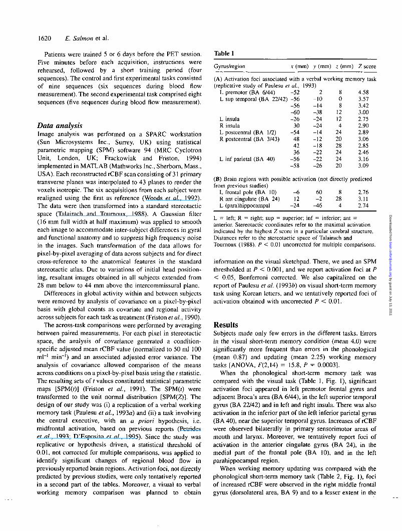
Table 1
Gyrus/region x (mm) y (mm) z (mm) Z score
(A) Activation foci associated with a verbal working memory task(replicative study of Paulesu et
L premotor (BA 6/44)L sup temporal (BA 22/42)
L insulaR insulaL postcentral (BA 1/2)R postcentral (BA 3/43)
L inf parietal (BA 40)
al,-52-56-56-60-26
30-54
484236
-56-58
1993)2
-10-14-38-24-24-14-12-18-22-22-26
808
12124
242028242420
4.583.573.423.002.752.902.893.062.852.463.163.09
(B) Brain regions with possible activation (not directly predictedfrom previous studies)
L frontal pole (BA 10) -6 60 8 2.76R ant cingulate (BA 24) 12 -2 28 3.11L (para)hippocampal -24 -46 4 2.74
L = left; R = right; sup = superior; inf = inferior; ant =anterior. Stereotactic coordinates refer to the maximal activationindicated by the highest Z score in a particular cerebral structure.Distances refer to the stereotactic space of Talairach andTournoux (1988). P < 0.01 unconnected for multiple comparisons.
information on the visual sketchpad. There, we used an SPMthresholded at P < 0.001, and we report activation foci at P< 0.05, Bonferroni corrected. We also capitalized on thereport of Paulesu et al. (1993ft) on visual short-term memorytask using Korean letters, and we tentatively reported foci ofactivation obtained with uncorrected P < 0.01.
ResultsSubjects made only few errors in the different tasks. Errorsin the visual short-term memory condition (mean 4.0) weresignificantly more frequent than errors in the phonological(mean 0.87) and updating (mean 2.25) working memorytasks [ANOVA, F(2,14) = 15.8, P = 0.0003].
When the phonological short-term memory task wascompared with the visual task (Table 1, Fig. 1), significantactivation foci appeared in left premotor frontal gyrus andadjacent Broca's area (BA 6/44), in the left superior temporalgyrus (BA 22/42) and in left and right insula. There was alsoactivation in the inferior part of the left inferior parietal gyrus(BA 40), near the superior temporal gyrus. Increases of rCBFwere observed bilaterally in primary sensorimotor areas ofmouth and larynx. Moreover, we tentatively report foci ofactivation in the anterior cingulate gyrus (BA 24), in themedial part of the frontal pole (BA 10), and in the leftparahippocampal region.
When working memory updating was compared with thephonological short-term memory task (Table 2, Fig. 1), fociof increased rCBF were observed in the right middle frontalgyrus (dorsolateral area, BA 9) and to a lesser extent in the
by guest on July 13, 2011brain.oxfordjournals.org
Dow
nloaded from
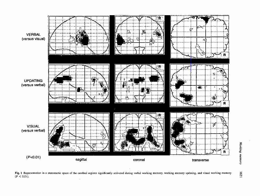
VERBAL(versus visual)
UPDATING(versus verbal)
VISUAL(versus verbal)
(P<0.01)sagittal coronal transverse
Fig. 1 Representation in a stereotactic space of the cerebral regions significantly activated during verbal working memory, working memory updating, and visual working memory(P < 0.01).
by guest on July 13, 2011brain.oxfordjournals.org
Dow
nloaded from
1622 E. Salmon et al.
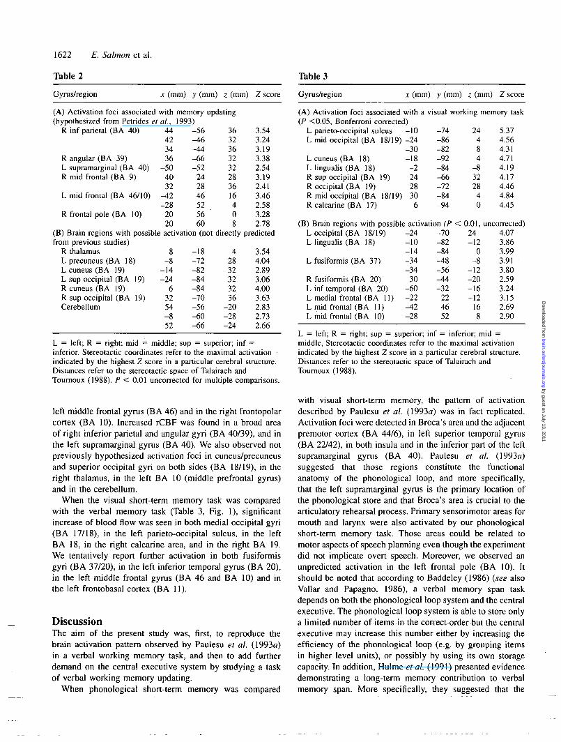
Table 2 Table 3
Gyms/region x (mm) y (mm) z (mm) Z score Gyrus/region x (mm) y (mm) z (mm) Z score
(A) Activation foci associated with memory updating(hypothesized from Petrides et al., 1993)
R inf parietal (BA 40)
R angular (BA 39)L supramarginal (BA 40)R mid frontal (BA 9)
L mid frontal (BA 46/10)
R frontal pole (BA 10)
(B) Brain regions with possiblefrom previous studies)
R thalamusL precuneus (BA 18)L cuneus (BA 19)L sup occipital (BA 19)R cuneus (BA 19)R sup occipital (BA 19)Cerebellum
44423436
-504032
-42-28
2020
-56-46-44-66-52
242846525660
activation (not
8-8
-14-24
63254-852
-18-72-82-84-84-70-56-60-66
3632363232283616408
directly
42832323236
-20-28-24
3.543.243.193.382.543.192.413.462.583.282.78
predicted
3.544.042.893.064.003.632.832.732.66
L = left; R = right; mid = middle; sup = superior; inf =inferior. Stereotactic coordinates refer to the maximal activationindicated by the highest Z score in a particular cerebral structure.Distances refer to the stereotactic space of Talairach andTournoux (1988). P < 0.01 uncorrected for multiple comparisons.
left middle frontal gyrus (BA 46) and in the right frontopolarcortex (BA 10). Increased rCBF was found in a broad areaof right inferior parietal and angular gyri (BA 40/39), and inthe left supramarginal gyrus (BA 40). We also observed notpreviously hypothesized activation foci in cuneus/precuneusand superior occipital gyri on both sides (BA 18/19), in theright thalamus, in the left BA 10 (middle prefrontal gyrus)and in the cerebellum.
When the visual short-term memory task was comparedwith the verbal memory task (Table 3, Fig. 1), significantincrease of blood flow was seen in both medial occipital gyri(BA 17/18), in the left parieto-occipital sulcus, in the leftBA 18, in the right calcarine area, and in the right BA 19.We tentatively report further activation in both fusiformisgyri (BA 37/20), in the left inferior temporal gyrus (BA 20),in the left middle frontal gyrus (BA 46 and BA 10) and inthe left frontobasal cortex (BA 11).
DiscussionThe aim of the present study was, first, to reproduce thebrain activation pattern observed by Paulesu et al. (1993a)in a verbal working memory task, and then to add furtherdemand on the central executive system by studying a taskof verbal working memory updating.
When phonological short-term memory was compared
(A) Activation foci associated with a visual working memory task(P <0.05, Bonferroni corrected)
L parieto-occipital sulcus -10 -74 24 5.37L mid occipital (BA 18/19) -24 -86 4 4.56
-30 -82 8 4.31L cuneus (BA 18) -18 -92 4 4.71L lingualis (BA 18) -2 -84 -8 4.19R sup occipital (BA 19) 24 -66 32 4.17R occipital (BA 19) 28 -72 28 4.46R mid occipital (BA 18/19) 30 -84 4 4.84R calcarine (BA 17) 6 -94 0 4.45
(B) Brain regions with possible activation (P < 0.01, uncorrected)I nrrinital CRA 18/101 -?4 -70 94 4 07L occipital (BA 18/19)L lingualis (BA 18)
L fusiformis (BA 37)
R fusiformis (BA 20)L inf temporal (BA 20)L medial frontal (BA 11)L mid frontal (BA 11)L mid frontal (BA 10)
-24-10-14-34-34
30-60-22^ 2-28
-70-82-84-48-56-4A-32
224652
24-12
0-8
-12-20-16-12
16
4.073.863.993.913.802.593.243.152.692.90
L = left; R = right; sup = superior; inf = inferior; mid =middle, Stereotactic coordinates refer to the maximal activationindicated by the highest Z score in a particular cerebral structure.Distances refer to the stereotactic space of Talairach andTournoux (1988).
with visual short-term memory, the pattern of activationdescribed by Paulesu et al. (1993a) was in fact replicated.Activation foci were detected in Broca's area and the adjacentpremotor cortex (BA 44/6), in left superior temporal gyrus(BA 22/42), in both insula and in the inferior part of the leftsupramarginal gyrus (BA 40). Paulesu et al. (1993a)suggested that those regions constitute the functionalanatomy of the phonological loop, and more specifically,that the left supramarginal gyrus is the primary location ofthe phonological store and that Broca's area is crucial to thearticulatory rehearsal process. Primary sensorimotor areas formouth and larynx were also activated by our phonologicalshort-term memory task. Those areas could be related tomotor aspects of speech planning even though the experimentdid not implicate overt speech. Moreover, we observed anunpredicted activation in the left frontal pole (BA 10). Itshould be noted that according to Baddeley (1986) (see alsoVallar and Papagno, 1986), a verbal memory span taskdepends on both the phonological loop system and the centralexecutive. The phonological loop system is able to store onlya limited number of items in the correct order but the centralexecutive may increase this number either by increasing theefficiency of the phonological loop (e.g. by grouping itemsin higher level units), or possibly by using its own storagecapacity. In addition, Hulme et al. (1991) presented evidencedemonstrating a long-term memory contribution to verbalmemory span. More specifically, they suggested that the
by guest on July 13, 2011brain.oxfordjournals.org
Dow
nloaded from
Working memory 1623
long-term memory of the phonological form of words isimportant in supporting the retrieval of partially decayedwords held in a rehearsal loop. From that perspective, oneof the central executive functions is to coordinate the transferof information between short- and long-term memory. In thistheoretical framework, activation of BA 10 could be relatedto the specific storage function of the central executive, orto its co-ordination function between short- and long-termmemory. The left parahippocampal activation that weobserved during the task would possibly reflect thecontribution of long-term memory. Anterior cingulate cortex(BA 24) was another possible focus of activation. As a matterof fact, the functions of the anterior cingulate cortex appearto be heterogeneous. This region has been implicated indifferent tasks that require selective attention, significantprocessing capacity, attention for action, or even preparationand initiation of movement (e.g. Petersen et al, 1988; Pardoet al, 1990; Frith et al, 1991; Paus et al, 1993). The specificcontribution of the cingulate gyrus to our phonological short-term memory task remains to be determined.
When working memory updating was compared withphonological short-term memory, we observed activation ofa set of frontal and parietal regions. An increase of rCBFoccurred in the right mid-dorsolateral prefrontal cortex (BA9), in left middle frontal regions (BA 46 and possibly BA10) and in the right frontal pole. Activation of mid-dorsolateralfrontal cortex (BA 9/46) was previously observed bilaterallyduring random generation tasks (Petrides et al, 1993a, ft),and it was associated with activation in the right trontopolarcortex during an externally ordered verbal working memorytask (Petrides etai, 1993ft). Since both random generation andupdating tasks require considerable flexibility of informationprocessing and a progressive shift of attention, they areconsidered to place significant demands on the centralexecutive (Baddeley, 1986; Morris and Jones, 1990; Van derLinden et al, 1994). From that perspective, our data confirmthat the prefrontal cortex, which was also activated in dual-task condition (D'Esposito et al, 1995), is a key structurefor the central executive.
Significant increases of rCBF were also observed in bothinferior parietal regions (BA 40/39, predominant on the rightside), and possibly in BA 19, when working memory updatingwas compared with the phonological short-term memory task.Activation of inferior parietal regions has been implicated invisual mental imagery in humans (Kosslyn et al, 1993;Mellet et al., 1995), suggesting that image generation couldbe an important process in our updating task. A similarbilateral posterior parietal (BA 40/7) activation was alsofound during verbal and nonverbal random generation tasks,using numbers or abstract figures (Petrides et al, 1993a, ft).An increase of rCBF was previously reported in left andright posterior parietal cortex during visual and spatial short-term memory tasks, with right activation preferentially relatedto processing of spatial information (Jonides et al, 1993;Smith and Jonides, 1995). In fact, post hoc questioning of oursubjects indicated that half of them had used a phonological
strategy in the updating memory experiment, while the othershad used a visual imagery strategy or a combination of both.The original updating task that requires serial recall of thelast six consonants of longer series (Morris and Jones, 1990)was converted, in our experiment, into a recognition task tocomply with Paulesu's procedure. We recently obtainedpreliminary data confirming that the recognition and therecall procedures promote the preferential utilization ofvisuospatial and phonological strategy, respectively. Now,if visuospatial strategy predominates in our experimentalupdating condition, parietal activation might be related togeneration and short-term storage of visuospatial images.Finally, activation was observed in the right thalamus.Increases in rCBF were also found in both thalami in asubspan task compared with the rest condition (Grasby et al.,1993). On the other hand, the dorsolateral prefrontal cortexand the mediodorsal nucleus of the thalamus were included inGoldman-Rakic's animal model of working memory (1990).
In the visual short-term memory task using Korean letters,we observed an activation in the superior occipital gyrus(BA 19) which appears to be crucial for temporary storageof visual information. Effectively, the right BA 19 andthe right occipitoparietal junction 19/40 were activated indifferent studies of visuospatial working memory (Dupontet al, 1993; Jonides et al, 1993; Paulesu et al, 1993ft). BA19 was also possibly activated during our task of workingmemory updating (see above). However, the superior occipitalregion is not specifically dedicated to short-term memoryprocesses, for it is activated, along with the inferior parietalregion, during visual mental imagery (Kosslyn et al, 1993;Mellet et al, 1995). It should be noted that according toBaddeley (1986), the visuospatial sketchpad is also assumedto play a role in holding and manipulating mental images.Significant rCBF increases were observed in the rightcalcarine area, and bilaterally in BA 18/19. This patternof activation is probably related to elementary perceptualoperations and visual coding taking place within the occipitallobe (Sergent et al, 1992). The possible bilateral activationthat we observed in the fusiform gyrus was reported in shapeworking memory tasks (Corbetta et al, 1991; Belger et al,1995). However, left fusiform and left inferior temporal gyriwere also activated during simple object recognition (Sergentet al, 1992). These relative increases in rCBF observed inthe 'occipito-temporal pathway' (Mishkin et al, 1983) duringthe visual short-term memory task compared with thephonological task most probably reflect a higher demand forvisual processing of the different attributes of Korean lettersthan for standard letters. Finally, the left middle frontal gyrus(BA 46 and BA 10) was amongst the brain regions possiblyactivated by our visual short-term memory task. Paulesuet al. (1993ft) observed an activation in the left BA 47 whencomparing the visual short-term memory task with shapejudgement on Korean letters. Those activations probablyreflect central executive contribution to the visual workingmemory task.
by guest on July 13, 2011brain.oxfordjournals.org
Dow
nloaded from
1624 E. Salmon et al.
AcknowledgementsThe authors wish to thank Dr K. Friston for his valuablecomments on SPM, and Dr G. Hartstein for reviewing themanuscript. The study was supported by a grant (3454293)from the Belgian National Fund for Scientific Research(FNRS) and by the Fondation Medicale Reine Elisabeth. E.Salmon is Senior Research Assistant, F. Collette is Aspirantand P. Maquet is Research Associate at the FNRS.
ReferencesBaddeley AD. Working memory. Oxford: Clarendon Press, 1986.
Baddeley A. Is working memory working? The fifteenth BartlettLecture. Q J Exp Psychol [A] 1992; 44: 1-31.
Baddeley AD, Hitch G. Working memory. In: Bower GH, editor.The psychology of learning and motivation, Vol. VIII. New York:Academic Press, 1974: 47-90.
Baddeley A, Logie RH, Bressi S, Delia Sala S, Spinnler H. Dementiaand working memory. Q J Exp Psychol [A] 1986; 38: 603-18.
Baddeley AD, Bressi S, Delia Sala S, Logie R, Spinnler H. Thedecline of working memory in Alzheimer's disease. A longitudinalstudy. Brain 1991; 114: 2521-42.
Belger A, McCarthy G, Gore J, Goldman-Rakic P, Krystal J.Dissociation of parietal and temporal lobe activation during workingmemory tasks: a functional MRI study [abstract]. Hum Brain Map1995; Suppl 1: 416.
Belleville S, Peretz I, Arguin H. Contribution of articulatoryrehearsal to short-term memory: evidence from a case of selectivedisruption. Brain Lang 1992; 43: 713-46.
Corbetta M, Miezin FM, Dobmeyer S, Shulman GL, Petersen SE.Selective and divided attention during visual discriminations ofshape, color, and speed: functional anatomy by positron emissiontomography. J Neurosci 1991; 11: 2383^102.
Dalrymple-Alford JC, Kalders AS, Jones RD, Watson RW. A centralexecutive deficit in patients with Parkinson's disease. J NeurolNeurosurg Psychiatry 1994; 57: 360-7.
Degueldre C, Quaglia L. Performance evaluation of a new wholebody positron tomograph: the ECAT 951/31 R. Proceedings of the14th Annual International Conference of the IEEE. EMBS 1992:1831-3.
Delia Sala S, Logie RH. When working memory does not work:the role of working memory in neuropsychology. In: Boiler F,Grafman J, editors. Handbook of neuropsychology, Vol. 8.Amsterdam: Elsevier, 1993: 1-62.
D'Esposito M, Detre JA, Alsop DC, Shin RK, Atlas S, GrossmanM. The neural basis of the central executive system of workingmemory. Nature 1995; 378: 279-81.
Dupont P, Orban GA, Vogels R, Bormans G, Nuyts J, Schiepers C,et al. Different perceptual tasks performed with the same visualstimulus attribute activate different regions of the human brain: apositron emission tomography study [see comments]. Proc NatlAcad Sci USA 1993; 90: 10927-31. Comment in: Proc Natl AcadSci USA 1993; 90: 10901-3.
Fox PT, Mintun MA. Noninvasive functional brain mapping bychange-distribution analysis of averaged PET images of H2
15Otissue activity. J Nucl Med 1989; 30: 141-9.
Frackowiak RSJ. Functional mapping of verbal memory andlanguage. [Review]. Trends Neurosci 1994; 17: 109-15.
Frackowiak RSJ, Friston KJ. Functional neuroanatomy of the humanbrain: positron emission tomography—a new neuroanatomicaltechnique. [Review]. J Anat 1994; 184: 211-25.
Frisk V, Milner B. The relationship of working memory to theimmediate recall of stories following unilateral temporal or frontallobectomy. Neuropsychologia 1990; 28: 121-35.
Friston KJ, Frith CD, Liddle PF, Dolan RJ, Lammertsma AA,Frackowiak RSJ. The relationship between global and local changesin PET scans. J Cereb Blood Flow Metab 1990; 10: 458-66.
Friston KJ, Frith CD, Liddle PF, Frackowiak RSJ. Comparingfunctional (PET) images: the assessment of significant change. JCereb Blood Flow Metab 1991; 11: 690-9.
Frith CD, Friston K, Liddle PF, Frackowiak RSJ. Willed action andthe prefrontal cortex in man: a study with PET. Proc R Soc LondB BiolSci 1991; 244: 241-6.
Fuster JM. Frontal lobes. [Review]. Curr Opin Neurobiol 1993; 3:160-5.
Gathercole SE. Neuropsychology and working memory.Neuropsychology 1994; 8: 494-505.
Goldman-Rakic PS. Cellular and circuit basis of working memoryin prefrontal cortex of nonhuman primates. [Review]. Progr BrainRes 1990; 85: 325-36.
Grasby PM, Frith CD, Friston KJ, Bench C, Frackowiak RSJ, DolanRJ. Functional mapping of brain areas implicated in auditory-verbalmemory function. Brain 1993; 116: 1-20.
Hanley JR, Young AW, Pearson NA. Impairment of the visuo-spatial sketch pad. Q J Exp Psychol [A] 1991; 43: 101-25.
Hulme C, Maughan S, Brown GDA. Memory for familiar andunfamiliar words: evidence for a long-term memory contribution toshort-term memory span. J Mem Lang 1991; 30: 685-701.
Jonides J, Smith EE, Koeppe RA, Awh E, Minoshima S, MintunMA. Spatial working memory in humans as revealed by PET [seecomments]. Nature 1993; 363: 623-5. Comment in: Nature 1993;363:
Kosslyn SM, Alpert NM, Thompson WL, Maljkovic V, Weise SB,Chabris CF, et al. Visual mental imagery activates topographicallyorganized cortex: PET investigations. J Cogn Neurosci 1993; 5:263-87.
Lammertsma AA, Cunningham VJ, Deiber MP, Heather JD,Bloomfield PM, Nutt J, et al. Combination of dynamic and integralmethods for generating reproducible functional CBF images. JCereb Blood Flow Metab 1990; 10: 675-86.
Mazziotta JC, Huang SC, Phelps ME, Carson RE, MacDonaldNS, Mahoney K. A noninvasive positron computed tomographytechnique using oxygen-15 labeled water for the evaluation ofneurobehavioral task batteries. J Cereb Blood Flow Metab 1985; 5:70-8.
by guest on July 13, 2011brain.oxfordjournals.org
Dow
nloaded from
Working memory 1625
McCarthy G, Blamire AM, Puce A, Nobre AC, Bloch G, Hyder F,et al. Functional magnetic resonance imaging of human prefrontalcortex activation during a spatial working memory task. Proc NatlAcad Sci USA 1994; 91: 8690-4.
Mellet E, Crivello F, Tzourio N, Joliot M, Petit L, Laurier L,et al. Construction of mental images based on verbal description:functional neuroanatomy with PET [abstract]. Hum Brain Map1995; Suppl 1: 273.
Mishkin M, Ungerleider LG, Macko KA. Object vision and spatialvision: two cortical pathways. Trends Neurosci 1983; 6: 414-17.
Morris RG. Working memory in Alzheimer-type dementia.Neuropsychology 1994; 8: 544-54.
Morris N, Jones DM. Memory updating in working memory: therole of the central executive. Br J Psychol 1990; 81: 111-21.
Norman DA, Shallice T. Attention to action: willed and automaticcontrol of behavior. In: Davidson RJ, Schwarts GE, Shapiro D,editors. Consciousness and self-regulation. Advances in researchand theory, Vol. 4. New York: Plenum Press, 1986: 1-18.
Pardo JV, Pardo PJ, Janer KW, Raichle ME. The anterior cingulatecortex mediates processing selection in the Stroop attentional conflictparadigm. Proc Natl Acad Sci USA 1990; 87: 256-9.
Paulesu E, Frith CD, Frackowiak RSJ. The neural correlates of theverbal component of working memory [see comments]. Nature1993a; 362: 342-5. Comment in: Nature 1993; 363: 583-4.
Paulesu E, Frith CD, Bench CJ, Bottini G, Grasby PG, FrackowiakRSJ. Functional anatomy of working memory: the visuospatial'sketch pad' [abstract]. J Cereb Blood Flow Metab 1993b; 13 Suppl1: S552.
Paus T, Petrides M, Evans AC, Meyer E. Role of the human anteriorcingulate cortex in the control of oculomotor, manual, and speechresponses: a positron emission tomography study. J Neurophysiol1993; 70: 453-69.
Petersen SE, Fox PT, Posner MI, Mintun M, Raichle ME. Positronemission tomographic studies of the cortical anatomy of single-word processing. Nature 1988; 331: 585-9.
Petrides M, Alivisatos B, Evans AC, Meyer E. Dissociation ofhuman mid-dorsolateral from posterior dorsolateral frontal cortexin memory processing. Proc Natl Acad Sci USA 1993a; 90: 873-7.
Petrides M, Alivisatos B, Meyer E, Evans AC. Functional activationof the human frontal cortex during the performance of verbalworking memory tasks. Proc Natl Acad Sci USA 1993b; 90: 878-82.
Pigott S, Milner B. Capacity of visual short-term memory afterunilateral frontal or anterior temporal-lobe resection.Neuropsychologia 1994; 32: 969-81.
Pollack I, Johnson LB, Knaff PR. Running memory span. J ExpPsychol 1959; 57: 137-46.
Sergent J, Ohta S, MacDonald B. Functional neuroanatomy of faceand object processing. A positron emission tomography study. Brain1992, 115: 15-36.
Shallice T. From neuropsychology to mental structure. Cambridge:Cambridge University Press, 1988.
Shallice T. Multiple levels of control processes. In: Umilta C,Moscovitch M, editors. Attention and performance XV. Consciousand nonconscious information processing. Cambridge (MA): MITPress, 1994: 395^20.
Shallice T, Vallar G. The impairment of auditory-verbal short-term storage. In: Vallar G, Shallice T, editors. Neuropsychologicalimpairments of short-term memory. Cambridge: CambridgeUniversity Press, 1990: 11-53.
Smith EE, Jonides J. Working memory in humans:neuropsychological evidence. In: Gazzaniga MS, editors. Thecognitive neurosciences. Cambridge (MA): MIT Press, 1995:1009-20.
Swartz BE, Halgren E, Fuster JM, Simpkins E, Gee M, MandelkernM. Cortical metabolic activation in humans during a visual memorytask. Cereb Cortex 1995; 5: 205-14.
Talairach J, Tournoux P. Co-planar stereotaxic atlas of the humanbrain: 3-dimensional proportional system: an approach to cerebralimaging. Stuttgart: Thieme, 1988.
Vallar G, Baddeley AD. Fractionation of working memory:neuropsychological evidence for a phonological short-term store. JVerb Learn Verb Behav 1984; 23: 151-61.
Vallar G, Papagano C. Phonological short-term store and the natureof the recency effect: evidence from neuropsychology. Brain Cogn1986; 5: 428-42.
Van der Linden M. Neuropsychologie de la memoire. In: SeronX, Jeannerod M, editors. Traite de neuropsychologie humaine.Bruxelles: Mardaga, 1994: 282-316.
Van der Linden M, Coyette F, Seron X. Selective impairment ofthe 'central executive' component of working memory: a singlecase study. Cogn Neuropsychol 1992; 9: 301-26.
Van der Linden M, Beerten A, Pesenti M. Age-related differencesin random generation [poster]. International Conference on WorkingMemory. Cambridge, 1994a.
Van der Linden M, Bredart S, Beerten A. Age-related differencesin updating working memory. Br J Psychol 1994b; 85: 145-52.
Warrington EK, James M. Disorders of visual perception in patientswith localised cerebral lesions. Neuropsychologia 1967; 5: 253-66.
Woods RP, Cherry SR, Mazziotta JC. Rapid automated algorithmfor aligning and reslicing PET images. J Comput Assist Tomogr1992; 16: 620-33.
Received March 18, 1996. Accepted May 10, 1996
by guest on July 13, 2011brain.oxfordjournals.org
Dow
nloaded from


