Embed Size (px)
Citation preview

The
Journ
al o
f Exp
erim
enta
l M
edic
ine
JEM © The Rockefeller University Press $8.00Vol. 201, No. 6, March 21, 2005 859–870 www.jem.org/cgi/doi/10.1084/jem.20041891
ARTICLE
859
Regulation of anaphylactic responses by phosphatidylinositol phosphate kinase type I
�
Junko Sasaki,
1,4,5
Takehiko Sasaki,
1,5,6,7
Masakazu Yamazaki,
1
Kunie Matsuoka,
2
Choji Taya,
2
Hiroshi Shitara,
2
Shunsuke Takasuga,
5,6
Miki Nishio,
4,5,6,7
Katsunori Mizuno,
1,5,6
Teiji Wada,
8
Hideyuki Miyazaki,
1
Hiroshi Watanabe,
1
Ryota Iizuka,
1,4,5
Shuichi Kubo,
2
Shigeo Murata,
3,7
Tomoki Chiba,
3
Tomohiko Maehama,
1
Koichi Hamada,
4
Hiroyuki Kishimoto,
4
Michael A. Frohman,
9
Keiji Tanaka,
3
Josef M. Penninger,
8
Hiromichi Yonekawa,
2
Akira Suzuki,
4
and Yasunori Kanaho
1
1
Department of Pharmacology,
2
Department of Laboratory Animal Science, and
3
Department of Molecular Oncology, Tokyo Metropolitan Institute of Medical Science, Tokyo 113-8613, Japan
4
Department of Molecular Biology and
5
Department of Pathology and Immunology and
6
The 21st Century Center of Excellence Program, Akita University School of Medicine, Akita 010-8543, Japan
7
Precursory Research for Embryonic Science and Technology, Japan Science and Technology Agency, Kawaguchi, Saitama 332-0012, Japan
8
Institute of Molecular and Biotechnology of the Austrian Academy of Sciences, A-1030 Vienna, Austria
9
Center for Developmental Genetics and Department of Pharmacology, State University of New York, Stony Brook, NY 11794
The membrane phospholipid phosphatidylinositol 4, 5-bisphosphate [PI(4,5)P
2
] is a critical signal transducer in eukaryotic cells. However, the physiological roles of the type I phosphatidylinositol phosphate kinases (PIPKIs) that synthesize PI(4,5)P
2
are largely unknown. Here, we show that the
�
isozyme of PIPKI (PIPKI
�
) negatively regulates mast cell functions and anaphylactic responses. In vitro, PIPKI
�
-deficient mast cells exhibited increased degranulation and cytokine production after Fc
�
receptor-I cross-linking. In vivo,
PIPKI
�
���
mice displayed enhanced passive cutaneous and systemic anaphylaxis. Filamentous actin was diminished in
PIPKI
�
���
mast cells, and enhanced degranulation observed in the absence of PIPKI
�
was also seen in wild-type mast cells treated with latrunculin, a pharmacological inhibitor of actin polymerization. Moreover, the association of Fc
�
RI with lipid rafts and Fc
�
RI-mediated activation of signaling proteins was augmented in
PIPKI
�
���
mast cells. Thus, PIPKI
�
is a negative regulator of Fc
�
RI-mediated cellular responses and anaphylaxis, which functions by controlling the actin cytoskeleton and dynamics of Fc
�
RI signaling. Our results indicate that the different PIPKI isoforms might be functionally specialized.
Engagement of mast cell Fc
�
RI by IgE, fol-lowed by the aggregation of multiple IgE-bearing Fc
�
RI molecules by polyvalent antigen,leads to cellular activation and the initiation ofallergic reactions (1–3). Activated mast cellsrelease preformed granule-associated chemicalmediators, transcribe multiple cytokine genes,and secrete newly synthesized arachidonic acidmetabolites and various proteins that triggerallergic inflammation. At the molecular level,the cross-linking of mast cell Fc
�
RI moleculesinitiates various signaling cascades that leadto the generation of second messengers, pro-
tein phosphorylation, and protein–protein andlipid–protein interactions (4). Among the mostimportant intracellular signaling molecules acti-vated after Fc
�
RI cross-linking are the phos-pholipid-metabolizing enzymes, which includephospholipase C (PLC; reference 5), src ho-mology 2-containing inositol 5-phosphatase 1(6), and the phosphoinositide 3-kinases (PI3Ks;references 7, 8).
Phosphorylated derivatives of phosphati-dylinositol, collectively referred to as phospho-inositides, make up a minor proportion ofmembrane phospholipids, but are importantintermediates in eukaryotic cellular responses(9, 10). Two major second messengers, phos-phatidylinositol 3, 4, 5-trisphosphate (PIP
3
)
J. Sasaki and T. Sasaki contributed equally to this work.The online version of this article contains supplemental material.
CORRESPONDENCETakehiko Sasaki: [email protected] OR Yasunori Kanaho: [email protected]
Abbreviations used: BMMC, bone marrow–derived mast cell; IP
3
, inositol 1,4,5-trisphosphate; LAT, linker for activation of T cells; PI3K, phosphoinositide3-kinase; PIP
3
, phosphatidylino-sitol 3, 4, 5-trisphosphate; PIPKI, type I phosphatidylinositol phos-phate kinase; PIPKI
�
,
�
isozyme of PIPKI; PLC, phospholipase C; RBL, rat basophilic leukemia
.
on August 2, 2015
jem.rupress.org
Dow
nloaded from
Published March 14, 2005
http://jem.rupress.org/content/suppl/2005/03/14/jem.20041891.DC1.html Supplemental Material can be found at:
PIPKI
�
REGULATES ANAPHYLAXIS | Sasaki et al.
860
and inositol 1,4,5-trisphosphate (IP
3
), are produced by PI3Ksand PLC, respectively. PIP
3
and IP
3
are required for intracel-lular signal transduction pathways activated by most cell sur-face receptors, including cytokine/growth factor receptors,the T cell receptor, the B cell receptor, and receptors for Ig.There is increasing evidence that the metabolism of PIP
3
andIP
3
has physiological and pathophysiological significance forimmune system regulation (for reviews see references 11–15).
Phosphatidylinositol 4, 5-bisphosphate [PI(4,5)P
2
] is asubstrate shared by PI3K and PLC. Phosphorylation ofPI(4,5)P
2
by PI3K generates PIP
3
, which controls the intra-cellular localization and activity of several proteins containingpleckstrin homology domains (16). Conversely, hydrolysis ofPI(4,5)P
2
by PLC produces IP
3
and DAG, second messen-gers necessary for the entry of calcium into cells and proteinkinase C activation, respectively (13). In addition to its well-established role as a precursor of second messengers, a moredirect function for PI(4,5)P
2
in the control of diverse cellularprocesses has been demonstrated recently (17–20). Based onbiochemical assays and studies of permeable cell preparations,PI(4,5)P
2
appears to be particularly important for the regula-tion of cytoskeletal reorganization and intracellular mem-brane transport (9, 17, 18, 21–23).
The phosphatidylinositol phosphate kinases (PIPKs) areenzymes important for PI(4,5)P
2
synthesis. There are twotypes of PIPKs: type I (PIPKI) and type II (PIPKII). Three
genes encode the
�
,
�
and
�
PIPKI isoforms, and the
�
genefurther generates three splice variants (24–28); in addition,three genes encode PIPKII (29). PI(4,5)P
2
can be synthesizedeither by phosphorylation of PI(4)P by PIPKI, or by phos-phorylation of PI(5)P by PIPKII. In mammalian cells, PI(4)P isat least fifty times more abundant than PI(5)P. Moreover, PIPKI(but not PIPKII) can induce dramatic changes in the actin cy-toskeleton. Thus, it is generally accepted that the majority ofPI(4,5)P
2
molecules in a mammalian cell are derived fromPIPKI-mediated phosphorylation of PI(4)P. Given the broadrange of potential functions of PI(4,5)P
2
, and the fact that thesteady-state level of PI(4,5)P
2
is much higher than that ofother lipid second messengers, it is likely that some kind offunctional compartmentalization of PI(4,5)P
2
production ex-ists inside the cell (17, 30). Such compartmentalization couldbe achieved by the interplay of multiple PIPKI molecules.
In this work, we genetically inactivated the
�
isozyme ofPIPKI (PIPKI
�
) in mice. PIPKI
�
deficiency leads to en-hanced degranulation and cytokine production in responseto Fc
�
RI stimulation in cultured mast cells. Moreover, ana-phylactic responses are enhanced in mice deficient for PIP-KI
�
. From a mechanistic standpoint, our results indicate thatPIPKI
�
is crucial for the integrity of the actin cytoskeletonand Fc
�
RI translocation to lipid rafts. Thus, our data are thefirst genetic evidence for a nonredundant role of PIPKI
�
invivo: the restraint of allergic reactions.
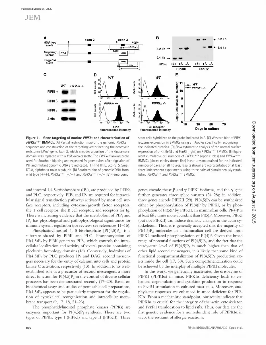
Figure 1. Gene targeting of murine PIPKI� and characterization of PIPKI��/� BMMCs. (A) Partial restriction map of the genomic PIPKI� sequence and construction of the targeting vector bearing the neomycin resistance (Neor) gene. Exon 3, which encodes a portion of the kinase core domain, was replaced with a PGK-Neo cassette. The PIPKI� flanking probe used for Southern blotting and expected fragment sizes after digestion of WT and mutant genomic DNA are indicated. H, Hind III; E, EcoRV; S, SmaI; DT-A, diphtheria toxin A subunit. (B) Southern blot of genomic DNA from wild type (���), PIPKI���� (���), and PIPKI���� (���) E14 embryonic
stem cells hybridized to the probe indicated in A. (C) Western blot of PIPKI isozyme expression in BMMCs using antibodies specifically recognizing the indicated proteins. (D) Flow cytometric analysis of the normal surface expression of c-Kit (left) and Fc�RI (right) on PIPKI���� BMMCs. (E) Equiv-alent cumulative cell numbers of PIPKI���� (open circles) and PIPKI���� BMMCs (closed circles, dotted line) in cultures maintained for the indicated number of days. For all figures, results shown are representative of at least three independent experiments using three pairs of simultaneously estab-lished PIPKI���� and PIPKI���� BMMCs.
on August 2, 2015
jem.rupress.org
Dow
nloaded from
Published March 14, 2005

JEM VOL. 201, March 21, 2005 861
ARTICLE
RESULTSGeneration of PIPKI� knockout mice and bone marrow–derived mast cellsMurine embryonic stem cells heterozygous for a deletionmutation of the PIPKI� gene were generated by replacing1.7 kb of the PIPKI� gene (including the region encodingthe NH2-terminal amino acids 68–106, indispensable for ki-nase activity) with a PGK-Neo cassette (Fig. 1 A). Southernblot analysis using a short arm flanking probe confirmed dis-ruption of the gene (Fig. 1 B). No random integrations ofthe PGK-Neo cassette were detected (unpublished data).These cells were used to derive homozygous PIPKI����
mice, which were born at the expected Mendelian ratio,were healthy and fertile, and displayed no histological abnor-malities up to 12 mo of age. There were no overt differencesfrom the WT in lymphocyte numbers or in subpopulationspresent in the thymus, lymph node, spleen, and bone mar-row (unpublished data).
It has been demonstrated that the PI3Ks and PLC�,which utilize PI(4,5)P2 as a substrate to generate either PIP3
or IP3, respectively, play critical roles in the developmentand effector functions of mast cells (31–34). In addition,PI(4,5)P2 modulates the organization of the actin cytoskele-ton, another cellular process that has been implicated in the
Figure 2. Enhanced Fc�RI-mediated degranulation and calcium mobilization in PIPKI��/� mast cells. (A) Increased �-hexosaminidase release. PIPKI���� (WT; open circles) and PIPKI���� (closed circles) BMMCs were preloaded with mouse anti-DNP IgE and stimulated with 50 ng/ml�1 DNP-HSA for the indicated times (left) or stimulated for 10 min with the indicated concentrations of DNP-HSA (right). The percentage of total cellular �-hexosaminidase that was released was taken as degranulation. Data shown are the mean � SD of triplicate samples. **, P 0.01 for
PIPKI���� cells compared with PIPKI���� cells, as determined byStudent’s t test. (B) Increased calcium mobilization. IgE-sensitized, Fura-2-loaded BMMCs were stimulated with 50 ng/ml�1 DNP-HSA and Ca2� flux was monitored by spectrofluorimetry. (C) Restoration of normal degranu-lation and Ca2� flux after introduction of PIPKI� cDNA. Degranulation (left) and Ca2� flux (right) were measured as in A and B, respectively, in WT BMMCs or PIPKI���� BMMCs reconstituted with either empty vector (red) or PIPKI� cDNA (blue).
on August 2, 2015
jem.rupress.org
Dow
nloaded from
Published March 14, 2005

PIPKI� REGULATES ANAPHYLAXIS | Sasaki et al.862
degranulation response (35, 36). To investigate the physio-logical role of PIPKI� in mast cells, we established IL-3–dependent bone marrow–derived mast cell (BMMC) linesfrom PIPKI���� and PIPKI���� littermates. BMMCs fromPIPKI���� mice lacked PIPKI� protein, but showed normalexpression of two other PIPKIs, PIPKI� and PIPKI� (Fig.1 C). Both PIPKI���� and PIPKI���� BMMCs expressedsimilar levels of c-Kit and Fc�RI, markers that are character-istic of mature BMMCs (Fig. 1 D), and PIPKI���� BMMCsproliferated at the WT rate (Fig. 1 E). Thus, PIPKI� expres-sion appears to be dispensable for the cytokine-dependentemergence and differentiation of BMMCs.
Enhanced degranulation, cytokine gene expression, and Fc�RI signaling in PIPKI��/� BMMCsTo determine whether PIPKI� was required for mast cellfunctions, we investigated mast cell granule release by mea-
suring the extracellular activity of �-hexosaminidase, amarker enzyme for histamine-containing granules. The totalactivity of �-hexosaminidase per cell did not differ betweenPIPKI���� and PIPKI���� BMMCs, and treatment withIgE alone did not induce granule release from BMMCs ofeither genotype. However, Fc�RI-evoked degranulationwas increased in PIPKI�-deficient mast cells compared withWT controls (Fig. 2 A). From a total of 15 experimentsusing five pairs of BMMC lines from littermate mice, wecalculated that the degranulation at 5 min after 10 ng/mlDNP stimulation was 56.7% � 6.2 for PIPKI���� BMMCscompared with 23.2% � 3.6 for WT BMMCs. This differ-ence was statistically significant as determined by the Mann-Whitney’s U test (P � 0.00078) and suggested that PIPKI�is involved in the control of Fc�RI signaling.
It is well known that a transient increase in intracellularcalcium is essential for degranulation (34, 37). We found that
Figure 3. Augmented cytokine gene expression and Fc�RI signaling in PIPKI��/� BMMCs. (A) Increased cytokine mRNA expression. PIPKI���� and PIPKI���� BMMCs were sensitized with IgE and stimulated with DNP (50 ng/ml�1) for the indicated times. Induction of mRNA expression for the indicated cytokines was detected by RT-PCR. (B) Enhanced signaling mole-cule phosphorylation after Fc�RI cross-linking. BMMCs were sensitized with IgE and stimulated with DNP for the indicated times. For both panels, 30 �g of cell lysates were subjected to successive rounds of immunoblotting using phospho-specific and total antibodies recognizing the indicated proteins. Syk phosphorylation was monitored by immunoprecipitation of Syk followed by immunoblotting using antiphosphotyrosine antibody.
One trial representative of a minimum of three experiments is shown in each case. White lines indicate that intervening lanes have been spliced out. (C) Band intensities in B were quantified using Dolphin-1 software, and relative phosphorylation levels were normalized to protein levels as described in Materials and methods. Values shown are the fold increase in the phos-phorylated form of each molecule in PIPKI���� BMMCs compared with the value in WT cells activated for 2 min (except for SAPK, which was 10 min). (D) Normal signaling molecule phosphorylation after IL-3 or SCF stimula-tion. WT and PIPKI���� BMMCs were stimulated with either 30 ng/ml�1
IL-3 or 30 ng/ml�1 SCF and the phosphorylation of ERK and p38 was assessed as in B.
on August 2, 2015
jem.rupress.org
Dow
nloaded from
Published March 14, 2005

JEM VOL. 201, March 21, 2005 863
ARTICLE
the amplitude of Ca2� elevation was increased in PIPKI����
BMMCs compared with that in WT cells (Fig. 2 B), consis-tent with the enhanced degranulation observed in the mu-tant cells. To confirm that the enhanced degranulation trulyresulted from the loss of PIPKI� expression, we reestablishedPIPKI� expression in PIPKI���� BMMCs using retroviralinfection. Degranulation and Ca2� mobilization were re-stored to normal by infection of PIPKI���� BMMCs with aretrovirus containing PIPKI�, but not by infection with acontrol virus (Fig. 2 C). Together, these results demonstratethat a lack of PIPKI� expression engenders increased anti-gen-induced degranulation mediated by high affinity Fc�RI.
Cytokines produced by mast cells are critical mediators inallergy and inflammation. Therefore, we investigated whetherPIPKI� played a role in mast cell cytokine production. Quan-titative RT-PCR was performed to determine the inductionof various cytokine mRNA transcripts in WT and PIPKI����
BMMCs. Fc�RI engagement induced higher levels of all cy-tokine mRNAs examined in PIPKI���� BMMCs comparedwith WT cells (Fig. 3 A). IL-2 and IL-3 were the moleculesmost markedly affected; relative quantitation revealed 50-fold increases in these mRNAs in PIPKI���� cells that hadbeen stimulated for 1 h (Fig. 3 A and not depicted).
Cytokine gene expression is regulated by several sig-naling cascades, including those governed by mitogen-acti-vated protein kinases (38). We stimulated PIPKI���� andPIPKI���� BMMCs with IgE plus antigen and monitoredthe phosphorylation (activation) of various signaling pro-teins. Fc�RI-triggered phosphorylation of ERK1/ERK2(T202/Y204), SAPK (T183/Y185), p38 (T180/Y182),PLC�-1 (Y783), PKB (Ser473), and Syk was increased inPIPKI���� BMMCs compared with WT BMMCs (Fig. 3, Band C), indicating that multiple signaling cascades are acti-vated in the absence of PIPKI�. Of interest, normal phos-phorylation of ERK and p38 occurred in PIPKI���� BM-MCs stimulated with either IL-3 or SCF (Fig. 3 D), and thebasal phosphorylation of mitogen-activated protein kinaseswas not increased in PIPKI���� BMMCs (Fig. 3, B and D).Therefore, an absence of PIPKI� does not lead to hy-perphosphorylation of these signaling molecules per se.Rather, the hyperphosphorylation of signaling molecules inPIPKI���� BMMCs depends on Fc�RI stimulation.
Increased severity of local and systemic anaphylactic reactions in PIPKI�-deficient miceAnaphylaxis is an extreme form of allergic reaction triggeredby allergen-induced cross-linking of allergen-specific IgEpresent on the surface of mast cells. Mast cell degranulationis induced, which leads to the release of copious amounts ofvasoactive amines and inflammatory mediators. To deter-mine whether PIPKI� functions as a negative regulator ofmast cell activation in vivo, we examined IgE-dependentanaphylactic reactions in PIPKI���� mice. Antigen chal-lenge of mice that had been sensitized previously withmonoclonal DNP-specific IgE antibody demonstrated that
PIPKI���� mice exhibited a greater degree of systemic ana-phylaxis than WT mice, as assessed by core temperaturechanges (Fig. 4 A). An increase in passive cutaneous anaphy-laxis was also observed in the mutants (Fig. 4 B). Mast cellnumbers were identical in the ear skin of WT (12 � 0.9 perfield; n � 8) and PIPKI���� (12 � 1.0 per field; n � 8)mice, so that the augmented anaphylaxis in PIPKI���� micewas most likely due to mast cell hyperreactivity to antigens.These data imply that a major physiological role of PIPKI�is to prevent inappropriate mast cell degranulation and cyto-kine production and, thus, anaphylaxis.
Decreased PI(4,5)P2 levels and atypical actin cytoskeleton in PIPKI�-deficient mast cellsIn view of the in vitro evidence that PIPKI� producesPI(4,5)P2, the precursor of IP3 and PIP3, the finding of in-
Figure 4. Enhanced anaphylactic responses in PIPKI��/� mice. (A) Systemic anaphylaxis. WT and PIPKI���� mice (n � 7 mice per genotype) received 5 �g anti-DNP IgE i.v., followed by stimulation with 1 mg DNP-HSA per mouse. The systemic anaphylactic response was monitored by measuring rectal temperature at the indicated times after antigen injection. (B) Passive cutaneous anaphylaxis. WT and PIPKI���� mice (n � 9 per genotype) received 100 �g anti-TNP IgE i.v. After 24 h, mice were epicuta-neously challenged with 10 �l 1% picryl chloride on the right ears, and with 10 �l 1% oxazolone on the left ears. Net ear swelling (thickness of the right ear minus that of the left ear) was measured with a caliper at the indicated times. Data are expressed as mean � SD. *, P 0.05 and **, P 0.01 for PIPKI���� mice compared with PIPKI���� mice as determined byStudent’s unpaired t test.
on August 2, 2015
jem.rupress.org
Dow
nloaded from
Published March 14, 2005

PIPKI� REGULATES ANAPHYLAXIS | Sasaki et al.864
Figure 5. PI(4,5)P2 levels and actin cytoskeleton in PIPKI��/� BMMCs. (A) Altered phospholipids. HPLC analysis of phospholipids prepared from WT and PIPKI���� BMMCs that were metabolically labeled with [3H]inositol for 48 h. The chromatographic tracings shown are one result representative of five independent trials. The decrease in PI(4,5)P2 (0.84-fold) and increase in PI(4)P (1.13-fold) in PIPKI���� BMMCs were statistically significant (insets). *, P 0.05 for PIPKI���� cells compared with untreated WT cells. (B) Decreased F-actin content as determined by flow cytometry. IgE-sensitized PIPKI���� BMMCs and IgE-sensitized WT BMMCs, which were either left untreated or pretreated with 0.5 �M latrunculin (Latr) for 15 min, were
stimulated with 50 ng/ml�1 DNP for the indicated times. F-actin was stained with Alexa 488–labeled phalloidin and analyzed by flow cytometry. The mean channel fluorescence (MCF) in untreated WT BMMCs was arbi-trarily assigned a value of 100. Data shown are the mean percentage of the control value � SD of triplicate samples. *, P 0.05 for PIPKI���� cells or latrunculin-treated WT cells compared with untreated WT cells at the indicated times. (C) Decreased F-actin content as determined by con-focal fluorescence microscopy. F-actin in the cells examined in B was visu-alized using Alexa 488–labeled phalloidin. Confocal images were collected every 1 �m, and summation images (top and middle rows) or single
on August 2, 2015
jem.rupress.org
Dow
nloaded from
Published March 14, 2005

JEM VOL. 201, March 21, 2005 865
ARTICLE
creased mast cell activation and allergic reactions in the ab-sence of PIPKI� was an unexpected result. Therefore, wedetermined the relative contribution of PIPKI� to overallPI(4,5)P2 production. The intracellular PI(4,5)P2 contentwas assessed in BMMCs that had been metabolically labeledwith [3H]inositol for 48 h to achieve a stable equilibrium.Lipid extraction followed by HPLC analysis revealed a statis-tically significant decrease in PI(4,5)P2 in BMMCs lackingPIPKI� (Fig. 5 A). A concomitant increase in PI(4)P, thesubstrate of PIPKI�, was observed in PIPKI���� BMMCs.These results provide the first genetic evidence that PIPKI�acts as a functional PI(4)P 5-kinase in vivo, and are in agree-ment with a recent paper describing a partial decrease inPI(4,5)P2 levels in HeLa cells treated with small interferenceRNA for PIPKI� (39). We conclude that the loss of PIPKI�can be compensated for by other PIPKs in the context ofbulk PI(4,5)P2 synthesis, but that a specific PI(4,5)P2 poolmay exist that is exclusively maintained by PIPKI�.
To gain insights into the functional characteristics of thePI(4,5)P2 compartment controlled by PIPKI�, we analyzedthe production of the PI(4,5)P2–derived second messengerIP3. At 1 min after Fc�RI stimulation, IP3 production wasenhanced in PIPKI���� BMMCs (2.8 � 0.37 pmol/106)compared with WT cells (2.1 � 0.17 pmol/106; P � 0.13;n � 8). In addition, the observation that PKB activation wasincreased in PIPKI���� BMMCs (Fig. 3 B) suggests thatPIP3 formation is also enhanced in the absence of PIPKI�.Thus, somewhat surprisingly, the production of the secondmessengers IP3 and PIP3 is increased, rather than decreased,in the absence of PIPKI�.
In sharp contrast, the absence of PIPKI� had a clearlysuppressive effect on the filamentous actin cytoskeleton(F-actin) in BMMCs. F-actin in PIPKI���� BMMCs wasconsistently decreased to 70–80% of the WT level (Fig. 5 B).We used confocal fluorescence microscopy to examine themorphology and distribution of F-actin in PIPKI���� BM-MCs. A dramatic reduction in F-actin staining was found inPIPKI���� BMMCs in comparison with WT cells (Fig. 5 C).Notably, the pattern of F-actin distribution and the cellshape of PIPKI���� BMMCs were similar to those of WTBMMCs treated with latrunculin, a potent inhibitor of actinpolymerization (40). Cortical actin filaments were decreasedin both cases. Together, these results indicate that PIPKI� isdispensable for IP3 and PIP3 production, but is a critical reg-ulator of actin cytoskeletal reorganization.
A role for F-actin in the down-regulation of mast cell degranulation, but not cytokine expressionThe peripheral actin cytoskeleton is essential for many biologi-cal processes involving changes to the plasma membrane archi-tecture (41, 42). In the rat basophilic leukemia (RBL) cell line,it has been demonstrated that Fc�RI-triggered actin polymer-ization plays a negative regulatory role in degranulation, cyto-kine production, and Ca2� elevation as well as in Fc�RI signal-ing (35, 43). To test whether F-actin was involved in BMMCresponses, we first examined the effect of latrunculin on de-granulation. WT BMMCs pretreated with latrunculin showedenhanced degranulation in response to Fc�RI engagement(Fig. 5 D). Moreover, jasplakinolide, a cell-permeable stabilizerof actin filaments, suppressed degranulation in BMMCs uponFc�RI cross-linking (Fig. 5 E). Latrunculin and jasplakinolidealso had parallel effects on BMMC degranulation induced byionomycin (Fig. S1, available at http://www.jem.org/cgi/content/full/jem.20041891/DC1). These results suggest thatpolymerized actin can restrain degranulation, and that the aug-mented degranulation observed in PIPKI���� BMMCs can beattributed to their decreased polymerized actin content.
Next, we examined the possible involvement of the actincytoskeleton in Fc�RI-mediated cytokine production and pro-tein phosphorylation. Contrary to its striking effect on degran-ulation, treatment of WT BMMCs with latrunculin beforeFc�RI cross-linking did not enhance cytokine induction orphosphorylation of signaling proteins, and did not lead to ele-vated intracellular calcium (Fig. 5 F). These observations mayaccount for the fact that latrunculin only partially mimics theeffect of PIPKI� disruption on BMMC degranulation. Ourfindings concur with a previous paper that demonstrated nor-mal activation of ERK and p38 despite increased actin poly-merization in BMMCs lacking Wiskott-Aldrich syndromeprotein-interacting protein (44). Thus, contrary to the situationin RBL cells, alterations to the actin cytoskeleton in BMMCsdo not necessarily result in anomalies to all Fc�RI-evoked cel-lular responses. Rather, our data show that PIPKI�-mediatedsuppression of Fc�RI-mediated cellular responses in mast cellsappears to operate via at least two different mechanisms, onlyone of which depends on regulation of actin polymerization.
Increased localization of Fc�RI in lipid rafts in PIPKI��/� BMMCsAlthough most Fc�RI-mediated responses were potentiatedin PIPKI�-deficient BMMCs, those induced by IL-3 and
images from the center of representative cells (bottom row) are presented. Bar, 10 �m. The WT cells exhibited a more jagged circumferential F-actin structure compared with PIPKI���� cells (arrowheads). (D) Increased degranulation induced by latrunculin. IgE-sensitized PIPKI���� BMMCs, and IgE-sensitized WT BMMCs that were either left untreated or pretreated with latrunculin (Latr) for 15 min were stimulated with 50 ng/ml�1 DNP for the indicated times. Degranulation was measured as in Fig. 2 A. **, P 0.01 for latrunculin-treated WT cells or PIPKI���� cells compared with un-treated WT cells at 5 min after cross-linking (n � 4). (E) Suppression of degranulation induced by jasplakinolide. IgE-sensitized PIPKI���� BMMCs
that were either left untreated or pretreated with 1�M jasplakinolide (Jasp) for 15 min were stimulated with DNP for 5 min. Degranulation was assessed as for in D. (F) Normal cytokine mRNA expression, signaling molecule phosphorylation, and Ca2� mobilization in the presence of latrunculin. IgE-sensitized WT BMMCs that were either left untreated or pretreated with latrunculin for 15 min were stimulated with DNP for the indicated times. Cytokine gene expression, protein phosphorylation, and Ca2� mobilization were determined as in Fig. 3, A and B, and Fig. 2 B, respectively.
on August 2, 2015
jem.rupress.org
Dow
nloaded from
Published March 14, 2005

PIPKI� REGULATES ANAPHYLAXIS | Sasaki et al.866
SCF treatment were similar in magnitude to those of WTBMMCs. Therefore, we investigated whether an absence ofPIPKI� could lead to a change in the dynamics of Fc�RIdistribution on the plasma membrane. Lipid rafts are definedas plasma membrane microdomains enriched with glycosphin-golipids and cholesterol. These structures are now generallyrecognized as “signaling platforms” (45, 46). It has beenshown that, after cross-linking, Fc�RI molecules are recruitedto the rafts where they undergo tyrosine-phosphorylation asthe initiation step of several downstream signaling cascades.Indeed, disruption of lipid rafts with methyl-�-cyclodextrininhibited Fc�RI signaling in BMMCs, as exemplified by a de-crease in ERK and p38 phosphorylation (Fig. S2, available athttp://www.jem.org/cgi/content/full/jem.20041891/DC1).
Due to their low density, lipid rafts can be separatedfrom nonraft plasma membrane components by ultracentri-fugation of detergent-lysed cells in sucrose density gradients.As shown in Fig. 6 A, engagement of Fc�RI induced trans-location of at least the Fc�RI� chain component of Fc�RIto the lipid raft fractions (fractions 3 and 4) of both WT andPIPKI���� BMMCs. Importantly, the presence of Fc�RI�in the raft fractions was significantly enhanced in PIPKI����
BMMCs even before aggregation. Immunoblotting for thelipid raft marker linker for activation of T cells (LAT) dem-onstrated that the partitioning of LAT between the lipid rafts
and nonlipid raft regions of the membrane was not altered inthe absence of PIPKI� (Fig. 6 B). It should also be notedthat the amounts of Fc�RI� in the lipid rafts were not affectedby latrunculin, suggesting that the actin cytoskeleton doesnot play a major role in regulating the redistribution ofFc�RI. These results suggest that PIPKI� suppresses theinteraction between Fc�RI and the lipid rafts independentlyof actin cytoskeletal organization. Such a role for PIPKI�could explain its apparent ability to impede the propagationof Fc�RI-mediated signaling cascades leading to BMMCcytokine production and degranulation. The precise mecha-nism by which PIPKI� regulates Fc�RI localization is underinvestigation.
DISCUSSIONMast cells play a key role in allergic reactions due to theirability to synthesize and release proinflammatory mediatorsand cytokines (1–3). Upon exposure to allergens, specificIgE bound to Fc�RI on mast cells becomes cross-linked andintracellular signals are transduced that lead to cellular activa-tion. These intracellular signals are tightly regulated, as spurioussignals could result in unwanted, and possibly deleterious, re-sponses. Although recent work has identified many of theproteins that positively regulate Fc�RI signaling, little isknown about the negative regulators of these signaling cas-
Figure 6. Regulation of Fc�RI localization to lipid rafts by PIPKI�. (A) Enhanced localization of Fc�RI� to lipid rafts. BMMCs sensitized with anti-DNP IgE were either left untreated or treated with latrunculin for 15 min. Cells were incubated with or without DNP (stimulation) for 2 min, lysed in 0.5% Triton X-100 buffer, and subjected to sucrose gradient ul-tracentrifugation to purify lipid rafts. (left) Fractions were separated by SDS-PAGE, transferred to PVDF membranes, and immunoblotted (IB)
with anti-Fc�RI �-chain antibody. Fractions 3 and 4 contain the lipid rafts. (right) The distribution of Fc�RI� in fractions 3 and 4 was quanti-tated by densitometric analysis of the immunoblot. (B) Normal distribu-tion of LAT. Fractions from A were immunoblotted with anti-LAT anti-body as for in A (right) and LAT distribution was densitometrically quantitated as in A (left).
on August 2, 2015
jem.rupress.org
Dow
nloaded from
Published March 14, 2005
JEM VOL. 201, March 21, 2005 867
ARTICLE
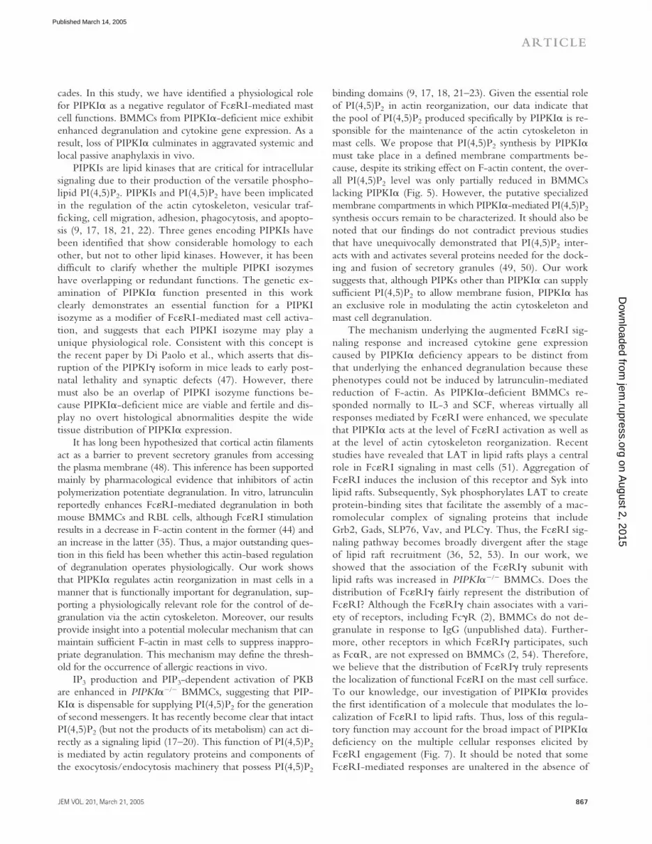
cades. In this study, we have identified a physiological rolefor PIPKI� as a negative regulator of Fc�RI-mediated mastcell functions. BMMCs from PIPKI�-deficient mice exhibitenhanced degranulation and cytokine gene expression. As aresult, loss of PIPKI� culminates in aggravated systemic andlocal passive anaphylaxis in vivo.
PIPKIs are lipid kinases that are critical for intracellularsignaling due to their production of the versatile phospho-lipid PI(4,5)P2. PIPKIs and PI(4,5)P2 have been implicatedin the regulation of the actin cytoskeleton, vesicular traf-ficking, cell migration, adhesion, phagocytosis, and apopto-sis (9, 17, 18, 21, 22). Three genes encoding PIPKIs havebeen identified that show considerable homology to eachother, but not to other lipid kinases. However, it has beendifficult to clarify whether the multiple PIPKI isozymeshave overlapping or redundant functions. The genetic ex-amination of PIPKI� function presented in this workclearly demonstrates an essential function for a PIPKIisozyme as a modifier of Fc�RI-mediated mast cell activa-tion, and suggests that each PIPKI isozyme may play aunique physiological role. Consistent with this concept isthe recent paper by Di Paolo et al., which asserts that dis-ruption of the PIPKI� isoform in mice leads to early post-natal lethality and synaptic defects (47). However, theremust also be an overlap of PIPKI isozyme functions be-cause PIPKI�-deficient mice are viable and fertile and dis-play no overt histological abnormalities despite the widetissue distribution of PIPKI� expression.
It has long been hypothesized that cortical actin filamentsact as a barrier to prevent secretory granules from accessingthe plasma membrane (48). This inference has been supportedmainly by pharmacological evidence that inhibitors of actinpolymerization potentiate degranulation. In vitro, latrunculinreportedly enhances Fc�RI-mediated degranulation in bothmouse BMMCs and RBL cells, although Fc�RI stimulationresults in a decrease in F-actin content in the former (44) andan increase in the latter (35). Thus, a major outstanding ques-tion in this field has been whether this actin-based regulationof degranulation operates physiologically. Our work showsthat PIPKI� regulates actin reorganization in mast cells in amanner that is functionally important for degranulation, sup-porting a physiologically relevant role for the control of de-granulation via the actin cytoskeleton. Moreover, our resultsprovide insight into a potential molecular mechanism that canmaintain sufficient F-actin in mast cells to suppress inappro-priate degranulation. This mechanism may define the thresh-old for the occurrence of allergic reactions in vivo.
IP3 production and PIP3-dependent activation of PKBare enhanced in PIPKI���� BMMCs, suggesting that PIP-KI� is dispensable for supplying PI(4,5)P2 for the generationof second messengers. It has recently become clear that intactPI(4,5)P2 (but not the products of its metabolism) can act di-rectly as a signaling lipid (17–20). This function of PI(4,5)P2
is mediated by actin regulatory proteins and components ofthe exocytosis/endocytosis machinery that possess PI(4,5)P2
binding domains (9, 17, 18, 21–23). Given the essential roleof PI(4,5)P2 in actin reorganization, our data indicate thatthe pool of PI(4,5)P2 produced specifically by PIPKI� is re-sponsible for the maintenance of the actin cytoskeleton inmast cells. We propose that PI(4,5)P2 synthesis by PIPKI�must take place in a defined membrane compartments be-cause, despite its striking effect on F-actin content, the over-all PI(4,5)P2 level was only partially reduced in BMMCslacking PIPKI� (Fig. 5). However, the putative specializedmembrane compartments in which PIPKI�-mediated PI(4,5)P2
synthesis occurs remain to be characterized. It should also benoted that our findings do not contradict previous studiesthat have unequivocally demonstrated that PI(4,5)P2 inter-acts with and activates several proteins needed for the dock-ing and fusion of secretory granules (49, 50). Our worksuggests that, although PIPKs other than PIPKI� can supplysufficient PI(4,5)P2 to allow membrane fusion, PIPKI� hasan exclusive role in modulating the actin cytoskeleton andmast cell degranulation.
The mechanism underlying the augmented Fc�RI sig-naling response and increased cytokine gene expressioncaused by PIPKI� deficiency appears to be distinct fromthat underlying the enhanced degranulation because thesephenotypes could not be induced by latrunculin-mediatedreduction of F-actin. As PIPKI�-deficient BMMCs re-sponded normally to IL-3 and SCF, whereas virtually allresponses mediated by Fc�RI were enhanced, we speculatethat PIPKI� acts at the level of Fc�RI activation as well asat the level of actin cytoskeleton reorganization. Recentstudies have revealed that LAT in lipid rafts plays a centralrole in Fc�RI signaling in mast cells (51). Aggregation ofFc�RI induces the inclusion of this receptor and Syk intolipid rafts. Subsequently, Syk phosphorylates LAT to createprotein-binding sites that facilitate the assembly of a mac-romolecular complex of signaling proteins that includeGrb2, Gads, SLP76, Vav, and PLC�. Thus, the Fc�RI sig-naling pathway becomes broadly divergent after the stageof lipid raft recruitment (36, 52, 53). In our work, weshowed that the association of the Fc�RI� subunit withlipid rafts was increased in PIPKI���� BMMCs. Does thedistribution of Fc�RI� fairly represent the distribution ofFc�RI? Although the Fc�RI� chain associates with a vari-ety of receptors, including Fc�R (2), BMMCs do not de-granulate in response to IgG (unpublished data). Further-more, other receptors in which Fc�RI� participates, suchas Fc�R, are not expressed on BMMCs (2, 54). Therefore,we believe that the distribution of Fc�RI� truly representsthe localization of functional Fc�RI on the mast cell surface.To our knowledge, our investigation of PIPKI� providesthe first identification of a molecule that modulates the lo-calization of Fc�RI to lipid rafts. Thus, loss of this regula-tory function may account for the broad impact of PIPKI�deficiency on the multiple cellular responses elicited byFc�RI engagement (Fig. 7). It should be noted that someFc�RI-mediated responses are unaltered in the absence of
on August 2, 2015
jem.rupress.org
Dow
nloaded from
Published March 14, 2005
PIPKI� REGULATES ANAPHYLAXIS | Sasaki et al.868
LAT (51), indicating that signaling pathways independentof lipid rafts exist. The role of PIPKI� in such pathways re-mains to be defined.
Our work provides clues to the mechanisms involved inFc�RI dynamics in the plasma membrane. Because phos-phoinositides (including PI(4,5)P2) are reportedly enrichedin lipid rafts (55), it will be interesting to analyze Fc�RI dis-tribution in mutant cells deficient for other phosphoinosi-tide-metabolizing enzymes such as PIPKI� and PIPKI�.These types of studies may further establish that thePI(4,5)P2 synthesized by each PIPK isozyme is functionallycompartmentalized. More importantly, such investigationsmay clarify the precise mechanisms regulating Fc�RI dy-namics in the plasma membrane that influence the outcomeof allergen challenge in vivo.
Our findings may have applications in the clinical arena.Fc�RI engagement is known to trigger allergic reactions,and an association between human Fc�RI� chain polymor-phisms and atopic phenotypes has been reported previously(56). Fc�RI engagement has also been linked to the patho-genesis of parasitic diseases and autoimmune disorders (2).These associations arise because Fc�RI is found on mono-cytes, eosinophils, platelets, Langerhans cells, and dendriticcells as well as on mast cells. Thus, the functional specializa-tion reported here for a PIPKI isoform in the molecular at-tenuation of Fc�RI signaling may represent a distinct mech-anism underlying a subset of allergic hypersensitivities,parasite susceptibilities or autoimmune diseases. Such spe-cialization among PIPKs could provide clinical researcherswith novel therapeutic targets for these disorders.
MATERIALS AND METHODSGeneration of PIPKI�-deficient mice. Mice deficient for PIPKI�were generated using homologous recombination in embryonic stem cells asdescribed previously (57). For further experimental details, see SupplementalMaterials and methods (available at http://www.jem.org/cgi/content/full/jem.20041891/DC1). All experimental protocols were reviewed and ap-proved by the Akita University Institutional Committee for Animal Studies.
Retroviral gene transfer. BMMC reconstitution assays using the retroviralvector pBabe-puro were performed as described previously (58). In brief, Mycepitope-tagged full-length PIPKI� cDNA (59) was cloned into the SalI site ofpBabe-puro. This vector was transfected into a phoenix-E packaging cell line.PIPKI���� BMMCs were cultured in the supernatant for 1 d and selected in2.5 mg/ml�1 puromycin for 3 wk before being used for experiments.
Degranulation and calcium mobilization. 2 106 cells ml�1 BMMCswere sensitized with 0.2 �g/ml�1 anti-DNP IgE (SPE-7) for 15 h. The cellswere washed, resuspended in OPTI-MEM (GIBCO BRL) containing 0.1%BSA, and challenged with DNP-HSA (Sigma-Aldrich). The percentage oftotal cellular �-hexosaminidase that was released was taken as degranulationas described previously (60). For calcium mobilization, the sensitized BM-MCs were incubated at room temperature for 45 min with 2 �M Fura-2-AM (Molecular Probes) in Tyrode’s buffer (10 mM Hepes, pH 7.4, 112mM NaCl, 2.7 mM KCl, 0.4 mM NaH2PO4, 1.6 mM CaCl2, 1 mMMgCl2, 2 mM glucose, and 1% BSA). Fc�RI-mediated calcium mobiliza-tion was measured every 0.5 s using a spectrophotometer (Shimadzu) set fordual excitation at 340 and 380 nm and emission at 510 nm.
Immunoblotting, immunoprecipitation, and in vitro PI3K assay.BMMCs were sensitized as described before. After washing and resuspensionin OPTI-MEM containing 0.1% BSA, the cells (5–10 106) were stimu-lated with 50 ng ml�1 DNP-HSA. Reactions were terminated by the addi-tion of ice-cold PBS and cell lysates were prepared in RIPA buffer (1% TritonX-100, 10 mM Tris/HCl, pH 7.4, 150 mM NaCl, 30 mM Na4P2O7, 5 mMEDTA, 50 mM NaF, 1 mM Na3VO4, and a protease inhibitor cocktail fromRoche Molecular Chemicals). Immunoprecipitation and immunoblottingwere performed as described previously (61). The relative phosphorylation ofeach signaling protein was normalized to its protein level in each sample (fordetails, see Supplemental Materials and methods). Immune complex lipidkinase assays were performed as described previously (57).
Passive systemic and cutaneous anaphylaxis. For passive systemic ana-phylaxis, mice (10–15 wk old) were sensitized for 24 h by intravenous injec-tion of 5 �g anti-DNP IgE (SPE-7). The mice were subsequently challengedwith an intravenous injection of 100 �g DNP-HSA. Body temperature wasmonitored using a rectal probe (Shibaura) starting from the time of antigeninjection. For passive cutaneous anaphylaxis, mice (10–15 wk old) were in-jected intravenously with 100 �g anti-TNP IgE (62). After 24 h, ear-swellingresponses were elicited by painting 10 �l 1% picryl chloride (Nakalai Tesque)in acetone on the right ears of each animal, and 10 �l 1% 4-ethoxymethylene-2-phenyl-2-oxazoline-5-one (oxazolone; Sigma-Aldrich) in acetone on theleft ear. Ear thickness was measured as described previously (62).
Cellular PI(4,5)P2 measurement and actin cytoskeleton assess-ments. BMMCs were labeled for 48 h with 10 �Ci ml�1 [3H]-myo-inosi-tol (Amersham Biosciences) in inositol-free DMEM containing dialyzed10% heat-inactivated FCS. Labeling was quenched and lipids were ex-tracted as described (57). Dried lipids were deacylated and analyzed byHPLC according to Serunian et al. using a Partisphere SAX column (What-man; reference 63). Radioactivity was assayed in 0.5-ml fractions using aliquid scintillation counter. To compare data among experiments, the rawradioactive counts determined for PI(4)P and PI(4,5)P2 were normalized tothe raw radioactive counts for total phosphoinositides. The normalizedamounts of PI(4)P or PI(4,5)P2 present in PIPKI���� BMMCs in each ex-periment were assigned a value of 1, and the relative amounts of PI(4)P or
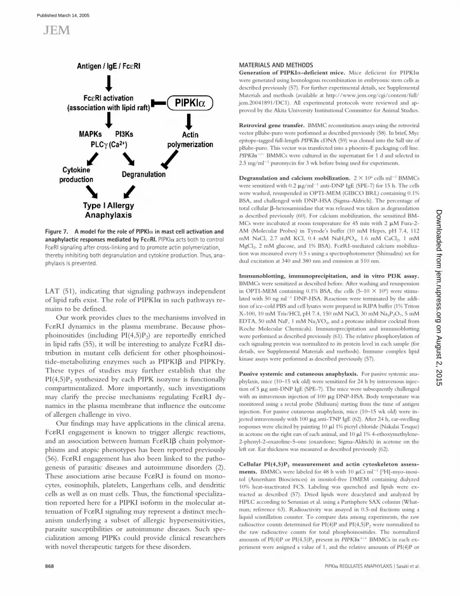
Figure 7. A model for the role of PIPKI� in mast cell activation and anaphylactic responses mediated by Fc�RI. PIPKI� acts both to control Fc�RI signaling after cross-linking and to promote actin polymerization, thereby inhibiting both degranulation and cytokine production. Thus, ana-phylaxis is prevented.
on August 2, 2015
jem.rupress.org
Dow
nloaded from
Published March 14, 2005

JEM VOL. 201, March 21, 2005 869
ARTICLE
PI(4,5)P2 in PIPKI���� BMMCs were calculated. Significance was assessedwith Student’s t test. p-values 0.05 were considered significant. Flow cy-tometry and fluorescence microscopy were used to analyze the status of theactin cytoskeleton (for details, see Supplemental Materials and methods).
RT-PCR. 107 BMMCs sensitized with anti-DNP IgE were stimulatedwith 50 ng ml�1 DNP-HSA. Total RNA was prepared using TRIzol re-agent (GIBCO BRL), and first-strand cDNA was synthesized using 5 �gtotal RNA with M-MLV Reverse Transcriptase (Toyobo). Specific PCRprimers and amplification conditions for cytokine gene expression are de-scribed in Supplemental Materials and methods.
Lipid raft preparation. Preparation of lipid rafts was performed as de-scribed previously with some modifications (64). In brief, 2 107 BMMCssensitized with anti-DNP IgE were incubated for 2 min at 37�C with orwithout DNP. Cells were washed and pellets were lysed in 1 ml MBSbuffer (0.5% Triton X-100, 25 mM MES, pH 6.5, 150 mM NaCl, 1 mMNa3VO4, and protease inhibitor cocktail) and incubated for 30 min on ice.Subsequent steps were performed at 4�C. Lysates were mixed with 0.5 ml85% sucrose in MBS, transferred to 5PA centrifuge tubes (Hitachi), andoverlaid with 2.4 ml 35% sucrose followed by 1.5 ml 5% sucrose. After cen-trifugation for 18 h at 200,000 g in a Hitachi P55ST2 rotor at 4�C, 10 0.5-ml fractions were collected starting at the top of the gradient. For analysis ofprotein composition, aliquots were mixed directly with 4 SDS-PAGEsample buffer and analyzed by Western blotting. Distribution of Fc�RI andLAT in lipid rafts was determined by densitometric analysis of immunoblotsusing Dolphin-1D software (Kurabo).
Online supplemental material. Primers used for cytokine gene expres-sion and procedures for the generation of PIPKI�-deficient mice and theanalysis of Fc�RI surface expression, actin cytoskeleton assessments, and im-munoblotting are available in Supplemental Materials and methods. Fig. S1shows the effects of latrunculin and jasplakinolide on ionomycin-induceddegranulation in WT and PIPKI���� BMMCs. Fig. S2 shows methyl-�-cyclodextrin inhibition of Fc�RI-mediated activation of p38 and ERK.Online supplemental material is available at http://www.jem.org/cgi/content/full/jem.20041891/DC1.
We thank Dr. U. Blank for providing anti-Syk antibody, Dr. J. Rivera for providing anti-Fc�RI� antibody, and Dr. T. Kitamura for the retroviral vectors. We thank Drs. H. Nishina, T. Katada, M. Ui, M. Mori, Y. Hachiya, K. Yamashita, K. Nagata, O. Kaminuma, S. Miyatake, N. Yajima, Y. Horie, K. Hamada, M. Yamada, Y. Sato, Y. Tsuya, and T. Nakano for valuable comments and advice.
Part of this work was supported by research grants from the Ministry of Education, Science, Sports and Culture; the Japan Society for the Promotion of Science (to J. Sasaki, T. Sasaki, A. Suzuki, and Y. Kanaho); Japan Science and Technology; a Grant-in-Aid for Cancer Research from the Japanese Ministry of Health, Labor and Welfare (to T. Sasaki); and Yamanouchi Foundation for Research on Metabolic Disorders and Intelligent Cosmos Academic Foundation (to J. Sasaki).
The authors have no conflicting financial interests.
Submitted: 13 September 2004Accepted: 24 January 2005
REFERENCES1. Metcalfe, D.D., D. Baram, and Y.A. Mekori. 1997. Mast cells. Physiol.
Rev. 77:1033–1079.2. Kinet, J.P. 1999. The high-affinity IgE receptor (Fc�RI): from physiol-
ogy to pathology. Annu. Rev. Immunol. 17:931–972.3. Galli, S.J. 2000. Mast cells and basophils. Curr. Opin. Hematol. 7:32–39.4. Turner, H., and J.P. Kinet. 1999. Signalling through the high-affinity
IgE receptor Fc�RI. Nature. 402:B24–B30.5. Zhang, J., E.H. Berenstein, R.L. Evans, and R.P. Siraganian. 1996.
Transfection of Syk protein tyrosine kinase reconstitutes high affinityIgE receptor-mediated degranulation in a Syk-negative variant of ratbasophilic leukemia RBL-2H3 cells. J. Exp. Med. 184:71–79.
6. Osborne, M.A., G. Zenner, M. Lubinus, X. Zhang, Z. Songyang,L.C. Cantley, P. Majerus, P. Burn, and J.P. Kochan. 1996. The inosi-tol 5�-phosphatase SHIP binds to immunoreceptor signaling motifsand responds to high affinity IgE receptor aggregation. J. Biol. Chem.271:29271–29278.
7. Barker, S.A., K.K. Caldwell, A. Hall, A.M. Martinez, J.R. Pfeiffer, J.M.Oliver, and B.S. Wilson. 1995. Wortmannin blocks lipid and protein ki-nase activities associated with PI 3-kinase and inhibits a subset of re-sponses induced by FceR1 cross-linking. Mol. Biol. Cell. 6:1145–1158.
8. Gu, H., K. Saito, L.D. Klaman, J. Shen, T. Fleming, Y. Wang, J.C.Pratt, G. Lin, B. Lim, J.P. Kinet, and B.G. Neel. 2001. Essential rolefor Gab2 in the allergic response. Nature. 412:186–190.
9. Takenawa, T., and T. Itoh. 2001. Phosphoinositides, key molecules forregulation of actin cytoskeletal organization and membrane traffic fromthe plasma membrane. Biochim. Biophys. Acta. 1533:190–206.
10. Cantley, L.C. 2002. The phosphoinositide 3-kinase pathway. Science.296:1655–1657.
11. Katso, R., K. Okkenhaug, K. Ahmadi, S. White, J. Timms, and M.D.Waterfield. 2001. Cellular function of phosphoinositide 3-kinases: im-plications for development, homeostasis, and cancer. Annu. Rev. CellDev. Biol. 17:615–675.
12. Vanhaesebroeck, B., S.J. Leevers, K. Ahmadi, J. Timms, R. Katso,P.C. Driscoll, R. Woscholski, P.J. Parker, and M.D. Waterfield. 2001.Synthesis and function of 3-phosphorylated inositol lipids. Annu. Rev.Biochem. 70:535–602.
13. Rebecchi, M.J., and S.N. Pentyala. 2000. Structure, function, andcontrol of phosphoinositide-specific phospholipase C. Physiol. Rev.80:1291–1335.
14. Lewis, R.S. 2001. Calcium signaling mechanisms in T lymphocytes.Annu. Rev. Immunol. 19:497–521.
15. Sasaki, T., A. Suzuki, J. Sasaki, and J.M. Penninger. 2002. Phospho-inositide 3-kinases in immunity: lessons from knockout mice. J. Bio-chem. (Tokyo). 131:495–501.
16. Lemmon, M.A., and K.M. Ferguson. 2000. Signal-dependent mem-brane targeting by pleckstrin homology (PH) domains. Biochem. J.350:1–18.
17. Itoh, T., and T. Takenawa. 2002. Phosphoinositide-binding domains:functional units for temporal and spatial regulation of intracellularsignalling. Cell. Signal. 14:733–743.
18. Czeck, M.P. 2000. PIP2 and PIP3: complex roles at the cell surface.Cell. 100:603–606.
19. Cremona, O., G. Di Paolo, M. Wenk, A. Luthi, W. Kim, K. Takei,L. Daniell, Y. Nemoto, S. Shears, R. Flavell, et al. 1999. Essentialrole of phosphoinositide metabolism in synaptic vesicle recycling.Cell. 15:179–188.
20. Caroni, P. 2001. New EMBO members’ review: actin cytoskeleton reg-ulation through modulation of PI(4,5)P(2) rafts. EMBO J. 20:4332–4336.
21. Botelho, R.J., H. Tapper, W. Furuya, D. Mojdami, and S. Grinstein.2002. Fc gamma R-mediated phagocytosis stimulates localized pinocy-tosis in human neutrophils. J. Immunol. 169:4423–4429.
22. Mejillano, M., M. Yamamoto, R. AL, H. Sun, X. Wang, and H. Yin.2001. Regulation of apoptosis by phosphatidylinositol 4,5-bisphos-phate inhibition of caspases, and caspase inactivation of phosphatidyl-inositol phosphate 5-kinases. J. Biol. Chem. 276:1865-1872.
23. Janmey, P., W. Xian, and L. Lanagan. 1999. Controlling cytoskeletonstructure by phosphoinositide-protein interactions: phosphoinositidebinding protein domains and effects of lipid packing. Chem. Physiol.Lipids. 101:93–107.
24. Loijens, J.C., and R.A. Anderson. 1996. Type I phosphatidylinositol-4-phosphate 5-kinases are distinct members of this novel lipid kinasefamily. J. Biol. Chem. 271:32937–32943.
25. Ishihara, H., Y. Shibasaki, N. Kizuki, H. Katagiri, Y. Yazaki, T. Asano,and Y. Oka. 1996. Cloning of cDNAs encoding two isoforms of 68-kDa type I phosphatidylinositol-4-phosphate 5-kinase. J. Biol. Chem.271:23611–23614.
26. Di Paolo, G., L. Pellegrini, K. Letinic, G. Cestra, R. Zoncu, S.Voronov, S. Chang, J. Guo, M.R. Wenk, and P. De Camilli. 2002.Recruitment and regulation of phosphatidylinositol phosphate kinase
on August 2, 2015
jem.rupress.org
Dow
nloaded from
Published March 14, 2005

PIPKI� REGULATES ANAPHYLAXIS | Sasaki et al.870
type 1 � by the FERM domain of talin. Nature. 420:85–89.27. Ling, K., R.L. Doughman, A.J. Firestone, M.W. Bunce, and R.A.
Anderson. 2002. Type I � phosphatidylinositol phosphate kinase tar-gets and regulates focal adhesions. Nature. 420:89–93.
28. Giudici, M.L., P.C. Emson, and R.F. Irvine. 2004. A novel neuronal-specific splice variant of Type I phosphatidylinositol 4-phosphate5-kinase isoform �. Biochem. J. 379:489–496.
29. Rameh, L.E., K.F. Tolias, B.C. Duckworth, and L.C. Cantley. 1997.A new pathway for synthesis of phosphatidylinositol-4,5-bis-phosphate.Nature. 390:192–196.
30. Hinchlife, K. 2000. Intracellular signalling: is PIP2 a messenger too?Curr. Biol. 10:104–105.
31. Liu, Q., T. Sasaki, I. Kozieradzki, A. Wakeham, A. Itie, D.J. Dumont,and J.M. Penninger. 1999. SHIP is a negative regulator of growth fac-tor receptor-mediated PKB/Akt activation and myeloid cell survival.Genes Dev. 13:786–791.
32. Fukao, T., T. Yamada, M. Tanabe, Y. Terauchi, T. Ota, T. Takayama,T. Asano, T. Takeuchi, T. Kadowaki, J. Hata Ji, and S. Koyasu. 2002.Selective loss of gastrointestinal mast cells and impaired immunity inPI3K-deficient mice. Nat. Immunol. 3:295–304.
33. Laffargue, M., R. Calvez, P. Finan, A. Trifilieff, M. Barbier, F. Al-truda, E. Hirsch, and M.P. Wymann. 2002. Phosphoinositide 3-kinase� is an essential amplifier of mast cell function. Immunity. 16:441–451.
34. Wen, R., S.T. Jou, Y. Chen, A. Hoffmeyer, and D. Wang. 2002.Phospholipase C � 2 is essential for specific functions of FceR and Fcgamma R. J. Immunol. 169:6743–6752.
35. Frigeri, L., and J. Apgar. 1999. The role of actin microfilaments in thedown-regulation of the degranulation response in RBL-2H3 mast cells.J. Immunol. 162:2243–2250.
36. Holowka, D., E.D. Sheets, and B. Baird. 2000. Interactions betweenFc(epsilon)RI and lipid raft components are regulated by the actincytoskeleton. J. Cell Sci. 113:1009–1119.
37. Baram, D., Y. Mekori, and R. Sagi-Eisenberg. 2001. Synaptotagminregulates mast cell functions. Immunol. Rev. 179:25–34.
38. Hata, D., Y. Kawakami, N. Inagaki, C.S. Lantz, T. Kitamura, W.N.Khan, M. Maeda-Yamamoto, T. Miura, W. Han, S.E. Hartman, etal. 1998. Involvement of Bruton’s tyrosine kinase in Fc�RI-depen-dent mast cell degranulation and cytokine production. J. Exp. Med.187:1235–1247.
39. Pardon, D., Y.J. Wang, M. Yamamoto, H. Yin, and G. Roth. 2003.Phosphatidylinositol phosphate 5-kinase I� recruits AP-2 to the plasmamembrane and regulates rates of constitutive endocytosis. J. Cell Biol.162:693–701.
40. Morton, W., K. Ayscough, and P. McLaughlin. 2000. Latrunculin al-ters the actin-monomer subunit interface to prevent polymerization.Nat. Cell Biol. 2:376–378.
41. Oliver, J.M. 1978. Cell biology of leukocyte abnormalities–membraneand cytoskeletal function in normal and defective cells. Am. J. Pathol.93:221–270.
42. Pollard, T.D., and G.G. Borisy. 2003. Cellular motility driven by as-sembly and disassembly of actin filaments. Cell. 112:453–465.
43. Narasimhan, V., D. Holowka, and B. Baird. 1990. Microfilaments reg-ulate the rate of exocytosis in rat basophilic leukemia cells. Biochem.Biophys. Res. Commun. 171:222–229.
44. Kettner, A., L. Kumar, I.M. Anton, Y. Sasahara, M. de la Fuente, V.I.Pivniouk, H. Falet, J.H. Hartwig, and R.S. Geha. 2004. WIP regulatessignaling via the high affinity receptor for immunoglobulin E in mastcells. J. Exp. Med. 199:357–368.
45. Draber, P., and L. Draberova. 2002. Lipid rafts in mast cell signaling.Mol. Immunol. 38:1247–1252.
46. Pike, L.J. 2004. Lipid rafts: heterogeneity on the high seas. Biochem. J.378:281–292.
47. Di Paolo, G., H.S. Moskowitz, K. Gipson, M.R. Wenk, S. Voronov,M. Obayashi, R. Flavell, R.M. Fitzsimonds, T.A. Ryan, and P. DeCamilli. 2004. Impaired PtdIns(4,5)P2 synthesis in nerve terminals pro-
duces defects in synaptic vesicle trafficking. Nature. 431:415–422.48. Burgoyne, R.D., and T.R. Cheek. 1985. Reorganisation of peripheral
actin filaments as a preclude to expcytosis. Biosci. Rep. 7:281–288.49. Hay, J.C., P.L. Fisette, G.H. Jenkins, K. Fukami, T. Takenawa, R.A.
Anderson, and T.F. Martin. 1995. ATP-dependent inositide phosphor-ylation required for Ca(2�)-activated secretion. Nature. 374:173–177.
50. Tucker, W.C., J.M. Edwardson, J. Bai, H.J. Kim, T.F. Martin, andE.R. Chapman. 2003. Identification of synaptotagmin effectors viaacute inhibition of secretion from cracked PC12 cells. J. Cell Biol.162:199–209.
51. Saitoh, S., R. Arudchandran, T.S. Manetz, W. Zhang, C.L. Sommers,P.E. Love, J. Rivera, and L.E. Samelson. 2000. LAT is essential forFc�RI-mediated mast cell activation. Immunity. 12:525–535.
52. Rivera, J., J. Cordero, Y. Furumoto, C. Luciano-Montalvo, C.Gonzalez-Espinosa, M. Kovarova, S. Odom, and V. Parravicini. 2002.Macromolecular protein signaling complexes and mast cell responses:a view of the organization of IgE-dependent mast cell signaling. Mol.Immunol. 38:1253–1258.
53. Wilson, B.S., J.R. Pfeiffer, and J.M. Oliver. 2002. Fc�RI signalingobserved from the inside of the mast cell membrane. Mol. Immunol.38:1259–1268.
54. Lobell, R.B., K.F. Austen, and H.R. Katz. 1994. FcgR-mediated en-docytosis and expression of cell surface Fc�RIIb1 and Fc�RIIb2 bymouse bone marrow culture-derived progenitor mast cells. J. Immunol.152:811–818.
55. Hope, H.R., and L.J. Pike. 1996. Phosphoinositides and phosphoino-sitide-utilizing enzymes in detergent-insoluble lipid domains. Mol. Biol.Cell. 7:843–851.
56. Shirakawa, T., A. Li, M. Dubowitz, J.W. Dekker, A.E. Shaw, J.A.Faux, C. Ra, W.O. Cookson, and J.M. Hopkin. 1994. Association be-tween atopy and variants of the beta subunit of the high-affinity immu-noglobulin E receptor. Nat. Genet. 7:125–129.
57. Sasaki, T., J. Irie-Sasaki, R.G. Jones, A.J. Oliveira-dos-Santos, W.L.Stanford, B. Bolon, A. Wakeham, A. Itie, D. Bouchard, I. Ko-zieradzki, et al. 2000. Function of PI3K� in thymocyte development,T cell activation, and neutrophil migration. Science. 287:1040–1046.
58. Sasaki, T., T. Wada, H. Kishimoto, J. Irie-Sasaki, G. Matsumoto, T.Goto, Z. Yao, A. Wakeham, T.W. Mak, A. Suzuki, et al. 2001. Thestress kinase mitogen-activated protein kinase kinase (MKK)7 is a neg-ative regulator of antigen receptor and growth factor receptor-inducedproliferation in hematopoietic cells. J. Exp. Med. 194:757–768.
59. Honda, A., M. Nogami, T. Yokozeki, M. Yamazaki, H. Nakamura,H. Watanabe, K. Kawamoto, K. Nakayama, A.J. Morris, M.A. Froh-man, and Y. Kanaho. 1999. Phosphatidylinositol 4-phosphate 5-kinasealpha is a downstream effector of the small G protein ARF6 in mem-brane ruffle formation. Cell. 99:521–532.
60. Manetz, T.S., C. Gonzalez-Espinosa, R. Arudchandran, S. Xirasagar, V.Tybulewicz, and J. Rivera. 2001. Vav1 regulates phospholipase C � acti-vation and calcium responses in mast cells. Mol. Cell. Biol. 21:3763–3774.
61. Irie-Sasaki, J., T. Sasaki, W. Matsumoto, A. Opavsky, M. Cheng, G.Welstead, E. Griffiths, C. Krawczyk, C.D. Richardson, K. Aitken, etal. 2001. CD45 is a JAK phosphatase and negatively regulates cytokinereceptor signalling. Nature. 409:349–354.
62. Naito, K., M. Hirama, C. Okumura, and C. Ra. 1995. Soluble formof the human high-affinity receptor for IgE inhibits recurrent allergicreaction in a novel mouse model of type I allergy. Eur. J. Immunol.25:1631–1637.
63. Serunian, L.A., K.R. Auger, and L.C. Cantley. 1991. Identification andquantification of polyphosphoinositides produced in response to plate-let-derived growth factor stimulation. Methods Enzymol. 198:78–87.
64. Saito, K., K.F. Tolias, A. Saci, H.B. Koon, L.A. Humphries, A.Scharenberg, D.J. Rawlings, J.P. Kinet, and C.L. Carpenter. 2003.BTK regulates PtdIns-4,5-P2 synthesis: importance for calcium signalingand PI3K activity. Immunity. 19:669–678.
on August 2, 2015
jem.rupress.org
Dow
nloaded from
Published March 14, 2005


