Embed Size (px)
Citation preview
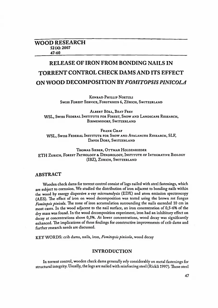
WOOD RESEARCH 52(4): 2007 47-60
RELEASE OF IRON FROM BONDING NAILS IN
TORRENT CONTROL CHECK DAMS AND ITS EFFECT
ON WOOD DECOMPOSITION BY FOMITOPSIS PINICOLA
KoNRAD PHILLIP NoETZLI Swiss FoREST SERVICE, FoRSTKREIS 6, ZüRICH, SwiTZERLAND
ALBERT BöLL, BEAT FREY WSL, Swiss FEDERAL INSTITUTE FOR FoREST, SNow AND LANDSCAPE RESEARCH,
BIRMENSDORF, SwiTZERLAND
FRANK GRAF WSL, Swiss FEDERAL INSTITUTE FOR SNow AND AvALANCHE RESEARCH, SLF,
DAvos DoRF, SwiTZERLAND
TuoMAS SIEBER, ÜTTMAR HoLDENRIEDER ETH ZuRICH, FoREST PATHOLOGY & DENDROLOGY, INsTITUTE OF INTECRATIVE BioLocv
(IBZ), ZuRICH, SwiTZERLAND
ABSTRACT
Wooden check dams for torrent control consist oflogs nailed with steel fastenings, which are subject to corrosion. We studied the distribution of iron adjacent to bonding nails within the wood by energy dispersive x-ray microanalysis (EDX) and atom emission spectroscopy {AES). The effect of iron on wood decomposition was tested using the brown rot fungus Fomitopsis pinicola. The zone of iron accumulation surrounding the nails exceeded 10 cm in most cases. In the wood adjacent to the nail surface, an iron concentration of 0,5-6% of the dry mass was found. In the wood decomposition experiment, iron had an inhibitory effect on decay at concentrations above 0,5%. At lower concentrations, wood decay was significantly enhanced. The implications of these findings for constructive improvements of crib dams and further research needs are discussed.
KEY WORDS: crib dams, nails, iron, Fomitopsis pinicola, wood decay
INTRODUCTION
In torrent control, wooden check dams generally rely considerably on metal fastenings for structural integrity. Usually, the Iogs are nailed with reinforcing steel {Rickli 1997}. Those steel
47

fastenings are subject to a particular type of corrosion, called crevice corrosion (Baker 1974). The corrosion products of the steel nails may considerably alter the chemical characteristics of the surrounding wood as well as its strength. In an experiment the tensile strength of wood samples in contact with rusting iron during nine months decreased about 50%, which is caused by the decomposition ofboth hemicellulose and cellulose (Marian and Wissing 1960).
Moreover, wood inhabiting fungi use metals and, in particular, iron present in the wood to degrade it and for maintaining their metabolism (Assmann et al. 2003, Goodell 2003). The availability of ferrous iron in an oxidizing environment is limited by the extreme insolubility of Fe (OHh (Winkelmann and Winge 1994). In general, decay fungi produce high affinity chelating compounds to scavenge meta! ions for their metabolic needs (Qian et al. 2002, Rodriguez et al. 2003).
The limited size of pores in the intact cell walls does not allow enzymes, including cellulase, to penetrate. Therefore, brown rot fungi rely on non-enzymatic oxidative systems (Henry 2003) and use Fenton type mechanisms in the early stages of wood cell wall depolymerization involving the participation of iron chelators (Goodell et al. 1997, Goodell and Jellison 1998, Rodriguez et al. 2003). To carry out the Fenton reaction, the fungi have developed different systems to reduce Fe (III) (Goodell2003, Wang and Gao 2003, Shimokawa et al. 2004).
Based on these facts it is hypothesized that an increasing iron concentration in wood enhances decomposition of wood by brown rotting fungi. Investigations on wood decay by fungi related to iron are rare and the findings are inconsistent. A positive effect on the growth of several decay fungi in cultures was reported by Negrutzkiy et al. (1998). However, Ruddick and Kundzewicz (1991) did not observe an increase in decay at higher amounts of iron. No information on iron concentrations in the vicinity of steel nails of inservice wood could be found in literature .
. The aim of this study was to analyse the distribution of iron adjacent to bonding steel na1!s. of wooden check dams. In addition, the influence of iron on the wood degrading actiVIty of the brown rot fungus Fomitopsis pinicola was tested experimentally. This species was found to be the most common brown-rot fungus on wooden check dams made of Iogs of Picea ahies and Ahies alha in Switzerland (Nötzli 2002).
MATERIALAND METHODS
Pteparation of the source material
The iron content was analyzed around bonding steel nails of fifteen-year-old wooden check dams of a catchment in the canton of Schwyz (Langenrainbach, Vorderthal) situated on the northern slopes of the Swiss Alps. The check dams were heavily damaged due to severe sliding processes in the area and, therefore, the destructive investigation was admissible. Ten check dams - all made of norway spruce (Picea ahies L. Karst) - were selected for detailed analysis. One log section of 50 cm around a steel nail conjunction was taken from each of the 10 different check dams. The diameter of the Iogs varied between 25 and 40 cm. They were either part of the lateral parts of the check dams (wings) or of its longitudinal protection structure, which were built in order to stabilise the lateral slopes of the channel. All logs were split longitudinally near the plane of the bonding steel nail and the latter was removed (Fig. 1). With ~ band saw, a series of wood samples of the dimension 10x10x50 mm was taken along the gram of alllO Iogs keeping a minimum space of 5 cm from the surface of the log and from its core (Fig. 2).
48

Six samples were taken each at intervals of 5, 15, 25, 35, 45, and 95 mm, respectively, measured between the original nail surface and the centres of the samples (Fig. 2). From seven randomly selected Iogs analoguous sample series were prepared perpendicular to the grain direction. Consequently, 17 sample series consisting of six samples each were produced.
Fig. 1: Logaftersplitting along the steel fostening
Log, cut longitudinally I radially
a)
b)
c)
5
sample series along the grain
~ ··-.. _
c D
15 25 35 45 95
Distance from nail surface (mmJ
Fig. 2: Above: scheme of a nail in a log and sample series longitudinal to the grain
Below: Detail of sample series: a) Sampie for EDX-analysis (white}. Scaled up:
Cutfing oJ samples and arrangemenf of spot measuremenfs
b} Sampies for AES-analysis (grey}: Analysed samples (specimens in a distance oJ 45 mm were not analysed}
c} Disfances oJ which the iron confents (AESanalysis) were used for fhe decomposition experiment. Distance D represents an unknown iron concentration in a greater distance than 95mm
49

Detection of iron in wood
Energy Dispersive X-ray Microanalysis (EDX} The samples of four series along and of two series perpendicular to the grain direction were dried
at 7o·c during 72 h. Of each sample, a small specimen (3x10x10 mm) with a smooth surface tangential to the grain direction was prepared using a rawr blade (Fig. 2 below, a). Subsequently, all specimens were cleaned with compressed air. The EDX analysis was conducted with a low-temperature scanning electron microscope (Philips SEM 515). Preparation and cooling of the specimens followed Frey et al ( 2000). The specimens were sputter-coated with platinum (coating thickness 8 nm). On the tangential cut surface spot measurements were carried out at intervals of 2 mm at a magnification of x 300. All spectra were acquired for 60 s (live time) and a dead time of about 30%. Therefore, at the distance of 0 to 50 mm from the nail surface, series of measurements at intervals of2 mm were performed (Fig. 2 below, a). The investigation of the area between 90 and 100 mm off the nail surface was restricted to two sample series along and one sample series perpendicular to the grain direction. Foreach measurement, a specimen surface of about 290 x 290 flm was analysed. Thus, an average measurement included 5 to 10 xylem cells. The spectra were processed using the Voyagersoftware package (Noran Instruments Inc., Middleton, WI) for automatic background subtraction which calculates net counts of elements of interest (Brunner and Frey 2000, Frey et al. 2000, Cosio et al. 2005). Data generared by the X-ray micro analyzer are at best semi-quantitative (Van Steveninck and Van Steveninck 1991). The values were not converted to concentrations due to the difficulties associated with obtaining quantitative results from specimens ofundefined thickness. Furthermore, the counts of non-standardized specimens may be subject of variance, depending on the angle of the specimens surface and the distance of the detector. Therefore, in place of the net counts ofiron, the ratio of the net counts ofiron and carbon (Fe/C) was taken. The detection limit for iron was set at 50 counts within 60 s. In order to check for possible iron contamination due to the use of a sted rawr blade, 25 measurements on specimens of freshly cut norway spruce were analogously performed.
Atomic Emission Spectroscopy {AES) Basedon the results of the EDX analysis the samples at the intervals 5, 15, 25, 35, and 95 mm of the
nail surface of all series (10 along and 7 perpendicular to the grain direction) were subjected to quantitative spectroscopic iron analysis (Fig. 2, below, b). The dimension of the samples was 10x10x40 mm. After drying at 70"C during 72 h and cleaningwith compressed air, they were grinded in a vibration-grinding mill (Retsch MM 2000) during 240 s at a frequency of 30 Hz. The grinding vessel and -balls were of zircon-oxide (vessel volume: 25 rnl, ball diameter: 20 mm). Subsequently, the samples were dried again at 7o·c during 24 h. The iron content of the samples was measured with an inductively coupled plasma atomic-emission spectrometer (IPC-AES Optima 3000, Perkin Eimer). Five control samples of freshly cut norway spruce were analogously prepared in the same way and checked for possible iron contamination due to sample preparation techniques.
Wood decomposition depending on the iron concentration
Fungal cultures Heterokaryotic cultures of Fomitopsis pinicola (Fr.) Karst. (strain 011002.1) were produced
in Petri dishes (diameter: 145 mm, height: 20 mm) on maltagar (20 g/1 malt extract, 15 g/1 agar, 1000 ml demin. H 20) and incubated at room temperature during 10 days. After this period the cultures had a diameter of about 80 mm.
50

Preparation oJ FeC/3 solutions The concentration c of a FeC13 solution that results in a distinct iron concentration k in the
wood was calculated according to equation [1].
c: Required concentration of the FeCh·solution [moVI)
c = k · PL [mol!l] u·M·(W<Fe> -k)
k: Iron-content of wood [gfg], from AES-Analyse
(i...: Density of the solution [gfl] == I 000 gfl
u: Solution-content in% of dry mass (wood) ==190%
w(Fe):Proportion ofFein FeCh (water-free) [gfg)==0.34
M: Mol mass of FeCI3 [gfmol)
The maximum error that is made by considering the density PL of the FeC13 solution as 1000 g/1 is about 2.5% (in series A with cA = 0.2 mol/1) and was ignored.
The iron concentrations of the samples after soaking were calculated after transformation of equation [1] and under consideration of the absorbed FeCI3 solution. lt was further arithmetically checked if the iron concentration within the wood blocks of the series A, B, and C corresponded to those measured with AES at the intervals 5, 15, and 95 mm of the nail surface, respectively. The pH of each solution was determined with an universal colour indicator (Merck, Darmstadt, D).
Wood sample preparation and soaking Following the concept of Jellison et al. (1997a), the decomposition experimentwas
conducted with wood samples soaked in FeCirsolution of different concentrations. The samples were prepared from the same scantling (80x80 mm; 10 m off the base cut) of an 80-year old Norway spruce. The length of the samples along the grain direction was fixed to 10 mm to guarantee good soaking. All samples (80x80x10 mm) were dried at 103"C until weight equilibrium (DIN 52183 1977) and the dryweight was determined with an accuracy of 0.001 g. Subsequently, the samples were soaked during 88 h under vacuum conditions and continuous stirring. The vacuum was interrupted for 30 min every 24 h to improve the soaking process.
Five different FeC13 concentrations (A: 0.2, B: 0.05, C: 0.01, D: 0.001, E: 0.000 molll) were applied to 23 samples each. The solutions were prepared in distilled water with dehydrated FeC13. The concentrations of the series A, B, and C approximately corresponded to the average total amount of iron resulting from the AES analysis at the intervals 5, 15, and 95 mm of the nail surface along the grain direction, respectively (Fig. 2, below, c). Series D represented an unknown !arger distance of the nail surface, and series E was the zero control.
After the soaking process, the samples were drained, weighted and the amount ofFeCI3 absorbed was calculated. The samples were then dried at 70"C until the water content was set on about 80%. Subsequently, each sample was put directly on the mycelium of a Petri dish and sealed with Parafitm•. After an incubation time of 6 months at room temperature, the fungal mycelium was carefully removed from the surface of the samples that were then kiln dried and weighted again.
51

The loss of mass of the wood was calculated after equation [2].
MV= Po-P'·lOO [%] MV: Mass loss of sample [%I
Po Po: Weight of sample before the experiment [g)
whereas P,: Weight of sample after the experiment [g)
mo: Dry mass of sample before the experiment [g)
Po = mo + mL[g] 111[.: Absorbed mass of FeCI3 [g)
Statistical analysis The data were statistically analysed with non-parametric tests (Hollander and Wolfe 1973)
of the software package R 2.0.0 (R Development Core Team 2004). In a first step a KruskalWallis rank sum testwas performed with the data ofthe AES analysis to test the null hypothesis that the iron concentrations are the same in each group (distances off the nail). The alternative hypothesiswas that they differ in at least one location. Likewise, the data of the decomposition experiment were tested with the null hypothesis that the proportional mass loss through fungal decay was the same in each group (iron concentrations).
In a second step the pairwise Wilcoxon rank sum test was applied to calculate pairwise comparisons between group Ievels of the distances off the nail and the iron concentrations, respectively, considering the correction ofHolm (1979) for multiple testing. Furthermore, this latter test design was applied to compare the iron concentrations of the AES analyses with those of the samples artificially soaked in corresponding FeCI3 solutions.
RESULTS
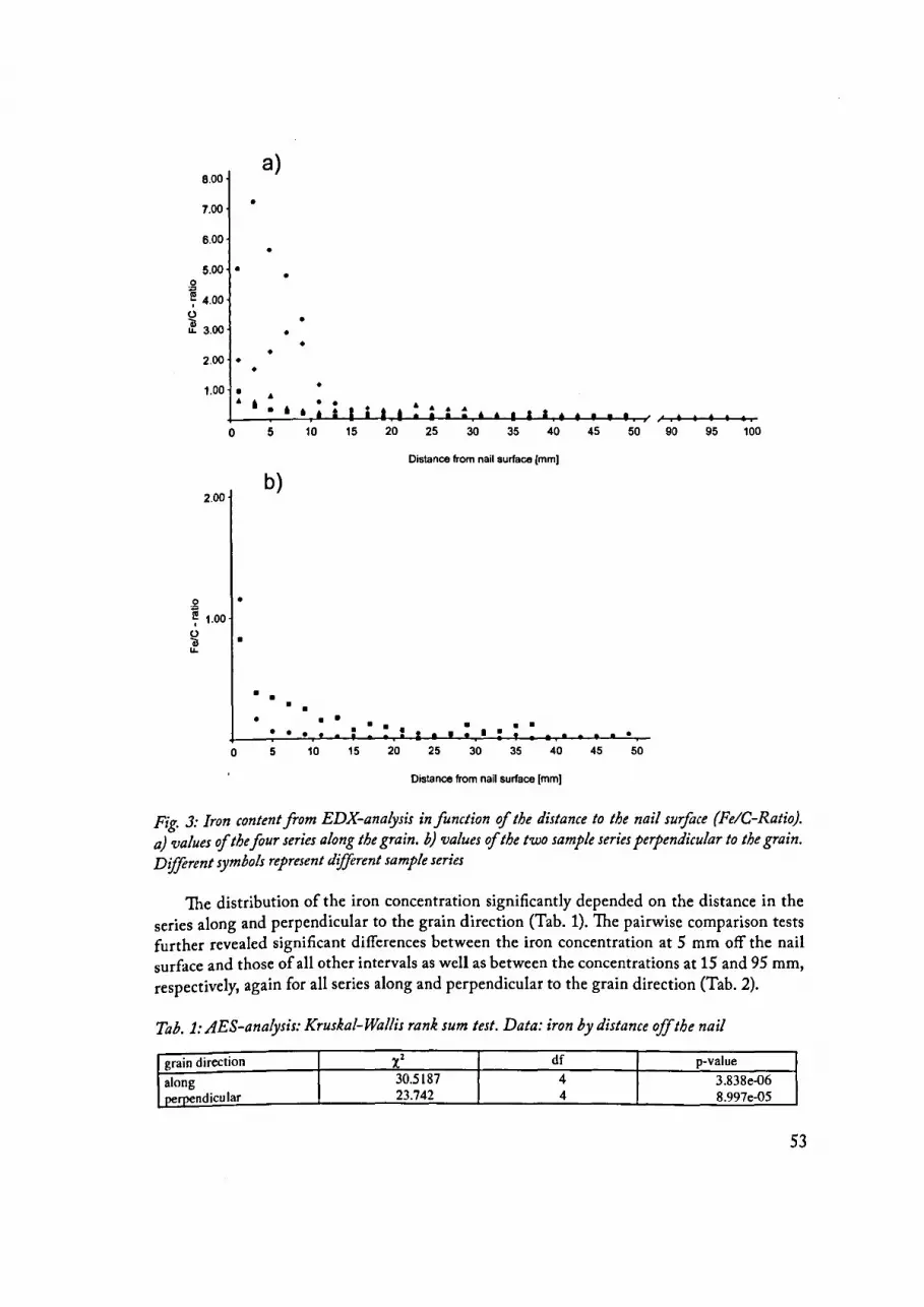
The EDX measurements indicated a strong decrease of the iron concentration in the wood (net count ratio of Fe/C) within only a few millimetres off the nail surface (Fig. 3). Along the grain direction high variation between the series was observed within the interval of 0 and 20 mm. From about 20 mm onwards, the measurements of the series were closely together. At the largest measurement distance of about 100 mm off the nail surface the iron concentration was still above the detection limit in one series. The distribution of the net counts (ratio Fe/C) of the series perpendicular to the grain direction was similar to the longitudinal one, however, with a considerably lower frequency (Fig. 3). The control measurements of the freshly cut Norway spruce only twice exceeded the detection limit with a net count ratio (Fe/C) of0.01 each.
The iron concentrations measured by AES analyses confirmed the assumed distribution based on the EDX measurements. In all series, the highest iron values were detected in the samples 5 mm off the nail surface. Within this interval, the iron concentration varied from 0.5 to 6% of the dried mass with a median of 1.9% (Fig. 4a). From 5 mm onwards, a remarkable decrease of the iron concentration was observed. Nevertheless, along the grain direction still substantial iron contents were measured several centimetres off the nail surface. Comparable to the EDX analyses, the distribution of the iron concentrations perpendicular to the grain direction was similar to the longitudinal one, however, on a noticeably lower Ievel (Fig. 4b). The control samples of the freshly cut Norway spruce had concentrations of about 10 ppm and correspond to values found in Iiterature for this tree species very well (Young and Guinn 1966).
52
8.00 a)
7.00 •
6.00 •
5.00 • • g 7 4.00
~ u. 3.00
• •
• • 2.00 • • 1.00 • •
& &
' • • • 0 5 10 15 20 25 30 35 40 45
Distance from nail surface (mm)
2.00 b)
~ • 7 1.00
~ • u.
• • • • • • • • • • • • • • 0 5 10 15 20 25 30 35 40 45 50
Distance from nail surface [mm)
Fig. 3: Iron content from EDX-analysis in function of the distance to the nail sur:foce {FeiG-Ratio}. a) values of the Jour series along the grain. b) values of the two sample series perpendicular to the grain. Dijftrent symbols represent different sample series
The distribution of the iron concentration significantly depended on the distance in the series along and perpendicular to the grain direction (Tab. 1). The pairwise comparison tests further revealed significant differences between the iron concentration at 5 mm off the nail surface and those of all other intervals as well as between the concentrations at 15 and 95 mm, respectively, again for all series along and perpendicular to the grain direction (Tab. 2).
Tab. 1: AES-analysis: Kruskal-Wallis rank sum fest. Data: iron by distance offthe nail
grain direction x2 df p-value
along 30.5187 4 3.838e-06 perpendicu lar 23.742 4 8.997e-05
53
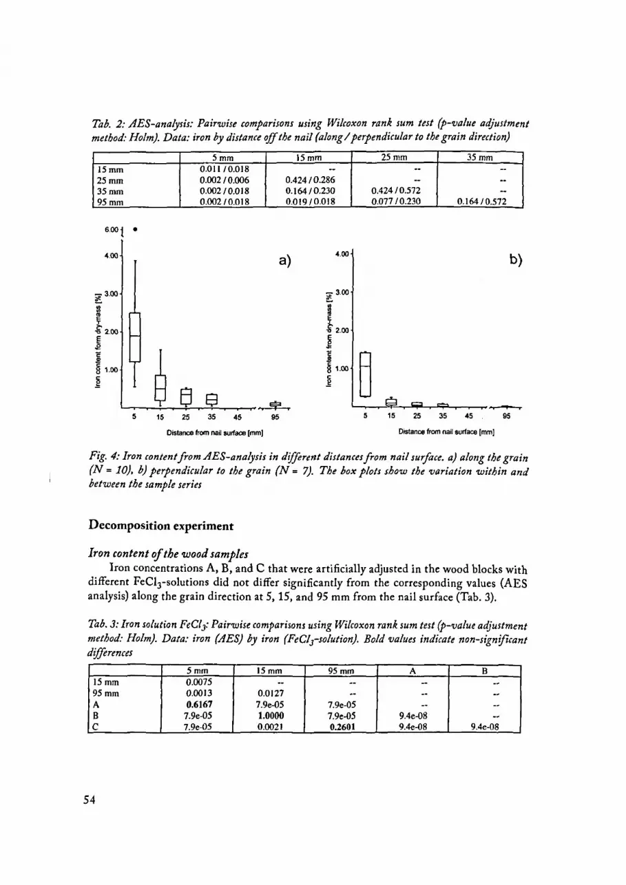
Tab. 2: AES-analysis: Pairwise comparisons using Wilcoxon rank sum test (p-value adjustment method: Holm). Data: iron by distance off the nail (along I perpendicular to the grain direction)
15mm 25mm 35mm 95mm
4.00
;e 3.00 t..
! ." 2.00
~ i 8 1.00
:g 8 5 15
5mm 15mm 0.011/0.018 --0.002 I 0.006 0.42410.286 0.00210.018 0.1641 0.230 0.002/0.018 0.01910.018
a) 4.00
-3.00 ~ :g .. b '0 2.00
~ c ~ 8 1.00 c _g s 8 ~
25 35 45 95
Oistance from nail surface [mm]
25mm 35mm -- ---- --
0.42410.572 --0.077 I 0.230 0.164/ 0.572
b)
~ g 5 15 25 35 45 95
Distance from nail surface [mm)
Fig. 4: Iron content from AES-analysis in different distances from nail surjace. a) along the grain (N = 10), b) perpendicular to the grain (N = 7). The box plots show the variation within and between the sample series
Decomposition experiment
Iron content oJ the wood samples Iron concentrations A, B, and C that were artificially adjusted in the wood blocks with
different FeClrsolutions did not differ significantly from the corresponding values (AES analysis) along the grain direction at 5, 15, and 95 mm from the nail surface {Tab. 3).
Tab. J: Iron solution FeCl3: Pairwise comparisons using Wilcoxon rank sum test (p-value adjustment method: Holm). Data: iron (AES) by iron (FeCls-solution). Bold values indicate non-significant diflerences
5mm 15mm 95mm A B 15mm 0.0075 -- -- -- --95mm 0.0013 0.0127 -- -- --A 0.6167 7.9e-05 7.9e-05 -- --B 7.9e-05 1.0000 7.9e-05 9.4e-08 --c 7.9e-05 0.0021 0.2601 9.4e-08 9.4e-08
54

Fungal wood decomposition After six months of incubation with Fomitopsis pinicola, a considerable variation of the
loss of dry mass was observed between the different series (Tab. 4). The highest decomposition rate with a median of 20.5% was measured at an iron concentration of 0.11% (series C) corresponding to the AES measurements at 95 mm off the nail surface (Fig. 2). At iron concentrations above 0.5% of wood dry mass (series A and B, according to iron concentration at 5 mm and 15 mm off the nail surface) virtually no mass loss was observed except for one sample of series B. In these series, virtually no decay was observed, and the fungus could not be reisolated after the incubation period.
The influence of the iron concentration on the loss of dried mass was highly significant (Kruskal-Wallis: x2 = 87.5491, df = 4, p-value < 2.2e-16). Furthermore, all pairwise comparisons were significantly different, except the one between the iron concentration D with 0.01% and the control E (0%) (Tab. 5). The decomposition rate of the latter was lower (median= 13.2%) than the one of the former (median = 15.6%). However, this difference was not significant.
Tab. 4: Results oJ decomposition experiment: All values except those oJ the concentrations c are medians oJ the sample series. c: concentrations oJ the FeClrsolutions {mol/1}; u: absorbed solution in % oJ dry mass oJ the sample; k: experimentally provided iron content oJ the samples {% oJ dry mass}; MV: mass loss ofthe samples in% ofsample weight (dry mass + absorbed FeClrsolution)
Serie c u k MV
A 0.20 188.09 1.99 3.33
B 0.05 172.40 0.48 1.79
c 0.01 202.34 0.11 20.48
D 0.001 196.08 0.01 15.60
E 0.00 - - 13.20
Tab. 5: Fungal wood decomposition: Pairwise comparisons using Wilcoxon rank sum test (pvalue adjustment method: Holm). Data: decay by iron concentration. Bold values indicate non
significant differences
A B c D
B 4.7e-07 -- -- --c 6.6e-08 6.6e-08 -- --D 1.4e-07 8.9e-08 0.029 --E I.Oe-07 1.2e-07 0.029 0.428
DISCUSSION
Iron content of the wood surrounding steel fastenings
The wood in the vicinity of steel nails showed a distinctly raised iron content, although a remarkable variationwas observed. In a distance of 10 cm along the grain from the nail surface the concentration ofiron was stillabout 100x higher compared to the control samples. Considering the normal distance of steel fastenings in crib dams of about 1.5 m, it can be supposed that the iron content would be raised in the bigger part of the log during the life-time of the dam. The great variation of the iron concentration between the samples and also along one nail may be caused by irregular crevice geometry along the nail. Unequal pH, electrolyte-concentration and
55

oxygen transport finaily Iead to an irregular corrosion and to an unequal distribution of corrosion products (Baker 1974, Wrangleu 1985, Tostmann 2001). Another reason for the considerable variation may be the different humidity regimes of the analysed Iogs.
Other important reasons for an unequal distribution of the iron content are probably growth characteristics of the wood as weil as darnage caused during the construction of the dams. These factors influence each other as weil as the activity of fungi and other microorganisms, which again influence porosity of the wood and the pH of the electrolyte.
lnfluence of iron on Fomitopsis pinicola
In general, the decomposition rate of the wood samples was rather small. This could have been caused by a reduced gas exchange in the Petri dishes. Furthermore, the malt agar was an additional nutrient-source for the test fungi during the initial phase of the experiment. In previous studies, decomposition rates of 30 (Schwarze 1995) up to 70% (Srebotnik and Messner 1991) were reported from experiments with Fomitopsis pinicola after an incubation time of three months. Other authors found a decomposition rate of only 2% after 8 months (Fomitopsis pinicola on Picea rubens, Ostrofsky et al. 1997).
The Iack of any decomposition in the test-series A and B (2 and 0.5% iron content) is most likely due to the fact, that the test fungi could not colonise the whole woodblock and died during the experiment.
In series C (0.11% iron content) the decomposition rate was significantly higher than in all other test series, indicating that this iron content enhances the activity of F. pinicola. This is in accordance with previous findings (Danninger 1980, Ritschkoff 1996). Most likely, the iron increased the non-enzymatic cracking of the ligno-cellulose complex (Fenton's reaction). However, the detailed mechanisms that are involved in the reduction of iron from its trivalent to its bivalent form are not understood. Nevertheless, Fomitopsis pinicola is able to reduce Fe3+ from a FeCI3 solution to Fe2+ (Lundborg 1988) and we assume that this fungus can use different corrosion products of iron.
Even when the iron concentrations in the decomposition experiment did not differ significantly from those measured in the wood around steel nails in Iogs, a direct comparison is difficult, because in the case of the Iogs, the overall-concentration of iron was measured. In the decomposition experiment, at least at the beginning, iron was dissolved in its trivalent form. Even though the high concentration ofFe3•-chloride applied in the sample series A and B seems to be toxic to the test fungi, it cannot be assumed that the same total concentration of iron (including other corrosion products such as rust) has the same effect to fungi in Iogs.
The pH in the wood samples varied from 2.5 in series A up to 4 in series D respectively. In crevice corrosion processes pH values of the electrolyte solution are reported to decrease to values between 2 and 3 (Fontana and Greene 1978). So the provided pH values in the experiment do not differ much from those along the shaft of a nail in wood. Nevertheless, the Iack of decomposition in series A and B is probably not only due to the low pH: Brown rot fungiareweil known to reduce pH values of their substrates to 2.5-3.8 (Hintikka 1969, Hyde and Wood 1997, Jellison et al. 1997b).
In the present study we investigated wood samples from the wings and longitudinal protection structures of the crib dam. Decomposition of permanently water saturated parts of crib dams seems to be of less practical importance, as water saturated conditions are
56

inhibitory for most decay fungi. However, some white rot fungi are able to decompose also water saturated wood (Metzler and Hecht 2004).
The use offungicides to seal the surroundingwood ofthe nails proposed by Yang (2001) can not be recommended here for reasons of water pollution. Our results indicate that it is most important to prevent the diffusion of the corrosion products into the longitudinal direction of the wood. Pre-drilling of the holes and pointing of the nails are the simplest methods to prevent additional damages to the wood which might act as diffusion channels for corrosion products(Rickli 1997, Böll et al. 1999). Butthis method cannot reduce crevice corrosion along the shaft of the nail.
CONCLUSIONS
Our data show, that the activity of Fomitopsis pinicola in the wood of norway spruce is influenced by the iron content of the wood. Steel nails raise the iron content of the adjacent wood up to a distance of several centimetres. Close to the nail, mycelial growth is probably suppressed, whereas low concentrations at larger distances might enhance decay. However, the actual significance of this process in crib dams remains to be studied under field conditions.
ACKNOWLEDGEMENTS
The authors wish to thank the local forestry staff of the Canton Schwyz, in particular Mr. P. Cuny for providing the study objects. Furthermore we wish to thank Dr. M. Bariska from the Swiss Federal Institute of Technology (ETH) in Zurich, for support in preliminary studies and Prof. Dr. Hans Böhni as well as Dr. Thomas Suter from the Institute of Building Materials (ETH), for scientific support and many discussions about the subject. This research was conducted as part of the project K Ursachen und Dynamik von Fäulen an Holzkonstruktionen im Bachverbau", funded by the Swiss Federal Office for the Environment (BAFU)
REFERENCES
1. Assmann, E. M., Ottoboni, L. M. M., Ferraz, A., Rodriguez, J., de Mello, M. P., 2003: Iron-responsive genes of Phanerochaete chrysosporium isolated by differential display reverse transcription polymerase chain reaction. Environmental Microbiology 5{9): 777-
786 2. Baker, A., 1974: Degradation of wood by products of metal corrosion. U.S.D.A Forest
Service Research Paper FPL 229: 6 pp. 3. Böll, A., Gerber, W., Graf, F., Rickli, C., 1999: Holzkonstruktionen im Wildbach-,
Hang- und Runsenverbau. Birmensdorf, Eidgenössische Forschungsanstalt für Wald, Schnee und Landschaft, 60 pp.
4. Brunner, I., Frey, B., 2000: Detection and localization of aluminium and heavy metals in ectomycorrhizal Norway spruce seedlings. Environmental Pollution 108: 121-128
5. Cosio, C., DeSantis, L., Frey, B., Diallo, S., Keller, C., 2005: Cadmium distribution in leaves of Thlaspi caerulescens. Journal of Experimental Botany 56: 765-775
57

6. Danninger, E., 1980: Untersuchungen über Abbaumechanismen holzzerstörender Pilze. Dissertation der Technischen Universität Wien, 110 pp.
7. DIN 52183, 1977: Prüfung von Holz, Bestimmung des Feuchtigkeitsgehaltes. In: DIN Taschenbuch 31, 2000. Eds., Beuth, Berlin
8. Fontana, M.G., Greene, N.D., 1978: Corrosion Engineering. McGraw-Hill Book Company, New York, 465 pp.
9. Frey, B., Keller, C., Zierold, K., Schulin, R., 2000: Distribution of Zn in functionally different leaf epidermal cells of the hyperaccumulator Thlaspi caerulescens. Plant, Cell and Environment 23: 675-687
10. Goodell, B.,Jellison,J., Liu,J., Daniel, G., Paszczynski, A., Fekete, F., Krishnamurthy, S., Jun, L., Xu, G., 1997: Low molecular weight chelators and phenolic compounds isolated from wood decay fungi and their role in the fungal biodegradation of wood. Journal ofBiotechnology 53: 133-162
11. Goodell, B.,Jellison,J., 1998: Role ofbiological metal chelators in wood biodeterioration. In: Forest Products Biotechnology. Eds. Palfreyman,J. W., Taylor and Francis Publishers, London, Pp. 235-250
12. Goodell, B., 2003: Brown-rot fungal degradation of wood: our evolving view. In: Wood Deterioration and Preservation. ACS Symposium series 845. Eds. Goodell, B., Nicholas, D.D., Schultz, T. P., American Chemical Society, Washington DC, Pp. 97-118
13. Henry, W. P., 2003: Non-enzymatic iron, manganese, and copper chemistry of potential importance in wood decay. In: Wood Deterioration and Preservation. ACS Symposium series 845. Eds. Schultz, T. P. Washington DC, Pp. 175-195
14. Hintikka, V., 1969: Acetic tolerance in wood and litter-decomposing Hymenomycetes. Karstenia X: 177-183
15. Hollander, M., Wolfe, D. A., 1973: Nonparametrie statistical methods. John Wiley and Sons, New York, 503 pp.
16. Holm, S., 1979: A simple sequentially rejective multiple test procedure. Scandinavian Journal of Statistics 6: 65-70
17. Hyde, S. M., Wood, P. M., 1997: A mechanism for production of hydroxyl radicals by the brown-rot fungus Coniophora puteana: Fe(III) reduction by cellobiose dehydrogenase and Fe(II) oxidation at a distance from the hyphae. Microbiology 143: 259-266
18. Jellison, J., Chen, Y., Fekete, F., 1997a: Hyphal sheath and iron-binding compound formation in liquid cultures of wood decay fungi Gloeophyllum trabeum and Postia placenta. Holzforschung 51(6): 503-510
19. Jellison, J., Connolly, J., Goodell, B., Doyle, B., Illman, B., Fekete, F., Ostrofsky, A., 1997b: The role of cations in the biodegradation of wood by the brown rot fungi. International Biodeterioration and Biodegradation 39(2/3): 165-179
20. Lundborg, A., 1988: Deformation of agar bywood decaying fungi- a possible indication of the ocurrence of radicals. Material und Organismen 23(4): 259-269
21. Marian, J. E., Wissing, A., 1960: The chemical and mechanical deterioration ofwood in contact with iron: part 1. Mechanical deterioration. Svensk Papperstidning 63(3): 47-57
22. Metzler, B., Hecht, U., 2004: Three-dimensional structure of tubular air channels formed by Armillaria spp. In water-saturated Iogs of silver fir and Norway spruce. Can. J. Bot. 82: 1388-1345.
23. Negrutskiy, M. N., Soukhomlin, M. M., Zaporozchenko, Y. V., Demechenko, S. I., 1998: The influence of Fe++ and Fe+++ upon physiology and development of wood-
58

attacking fungi. In: Delatour, C., Guillaumin, J.J., Lung-Escarmant, B., Mar~ais, B. (Eds): Root and butt Rot of Forest Trees. 9th lnt. Conference on Root and Butt Rots, Carcans, France, August 31-September 7, 1997, INRA Editions (Paris), Les Colloques No 89:. Eds. Carcans-Maubuisson (F), Pp. 341-347
24. Nötzli, K. P., 2002: Ursachen und Dynamik von Fäulen an Holzkonstruktionen im Wildbachverbau. Dissertation der Eidgenössischen Technischen Hochschule Zürich, Nr. 14974, 169 pp.
25. Ostrofsky, A.,Jellison,J., Smith, K., Shortle, W., 1997: Changes in cation concentrations in red spruce wood decayed by brown rot fungi and white rot fungi. Canadian Journal of Forest Research 27: 567-571
26. Qian, Y., Goodell, B., Felix, C. C., 2002: The effect oflow molecularweight chelators on iron chelation and free radical generation as studied by ESR measurement. Chemosphere 48:21-28
27. R Development Core Team R, 2004: A language and environment for statistical computing. R Foundation for Statistical Computing, Vienna, Austria
28. Rickli, C., 1997: Holzbauwerke im forstlichen Bachverbau. In:, Kursunterlagen FANKurs Forstlicher Bachverbau 15.-17. Oktober 1997 in Stalden, Obwalden, Schweiz (ed. Forstliche Arbeitsgruppe Naturgefahren und Eidg. Forschungsanstalt für Wald, Schnee und Landschaft, Birmensdorf
29. Ritschkoff, A., 1996: Decay mechanisms ofbrown-rot fungi. Technical Research Center afFinland VTT Publications 268, Espoo
30. Rodriguez, J., Ferraz, A., de Mello, M.P., 2003: Role of metals in wood biodegradation. In: Wood Deterioration and Preservation. ACS Symposium series 845. Eds. Schultz, T. P., American chemical Society, Washington DC, Pp. 154-174
31. Ruddick J.N.R, Kundzewicz, A.W., 1991: Bacterial movement of iron in waterlogged soil an its effect on decay in untreated wood. Material und Organismen 26: 169-181
32. Schwarze, F., 1995: Entwicklung und biomechanische Auswirkungen von holzzersetzenden Pilzen in lebenden Bäumen und in vitro. svk-Verlag, Erndtebrück,
163 PP· 33. Shimokawa, T., Nakamura, M., Noriko, H., Ishihara, M., 2004: Production of 2,5-
dimethoxyhydroquinone by the brown-rot fungus Serpula lacrymans to drive extracellular Fenton reaction. Holzforschung 58: 305-310
34. Srebotnik, E., Messner, K., 1991: Immunoelectron microscopial study of the porosity of brown-rot degraded pine wood. Holzforschung 45(2): 95-101
35. Tostmann, K. H. Korrosion: Ursachen und Vermeidung. Wiley-VCH, Weinheim, 2001 36. Van Stevenwinck, R.F.M., Van Steveninck, M.E., 1991: Microanalysis. In: Electron
Microscopy ofPlant Cells. Eds. Hawes, C., Academic Press, London, Pp. 415-455 37. Wang, W., Gao, P., 2003: Function and mechanism of a low-molecular-weight
peptide produced by Gloeophyllum traheum in biodegradation of cellulose. Journal of Biotechnology 101: 119-130
38. Winkelmann, G., Winge, D.R.,1994: Metal Ions in Fungi. Marcell Dekker, New York, Basel, Hongkong
39. Wranglen, G., 1985: Korrosion und Korrosionsschutz. Springer, Berlin, 301 pp. 40. Yang, D. Q, 2001: A method for the prevention of wood decay from nail penetration.
Forest Products Journal51(4): 67-70 40. Young, H. E., Guinn, V. P., 1966: Chemical elements in complete mature trees of seven
species in maine. Tappi 49(5): 190-197
59

60
KoNRAD PHILLIP NöTzu
Sw1ss FoREST SERVICE
FoRSTKREIS 6
ZwEIERSTRASSE 129
CH-8003 ZURICH
SwiTZERLAND
ALBERT BöLL
WSL Sw1ss FEDERAL INsTITUTE FOR FoREST, SNow AND LANDSCAPE RESEARCH
RESEARCH GRouP: MouNTAIN ToRRENTS, ERoSION AND LANDSLIDES
ZücHERSTRASSE 111
CH-8903 BIRMENSDORF
Sw1TZERLAND
BEAT FREY
WSL Sw1ss FEDERAL INSTITUTE FOR FoREST, SNow AND LANDSCAPE RESEARCH
RESEARCH GRoup: SolL SciENCE
ZüCHERSTRASSE 111
CH-8903 BIRMENSDORF
SWITZERLAND
FRANK GRAF
WSL Sw1ss FEDERAL INSTITUTE FOR FoREST, SNow AND LANDSCAPE RESEARCH
RESEARCH GROUP: ALPINE EcosYSTEMS
ZüCHERSTRASSE 111
CH-8903 BIRMENSDORF
SWITZERLAND
ÜTTMAR HOLDENRIEDER
FoREST PATHOLOGY AND DENDROLOGY
INSTITUTE OF lNTEGRATIVE BIOLOGY
UNIVERSITÄSTR. 16
CH-8092 ZURICH
SWITZERLAND
E-mail: [email protected]


