Embed Size (px)
Citation preview
(Archives of Environmental Health, 56: 175-180, 2001)
Respiratory Sinus Arrhythmia in Persons with Organic Solvent Exposure:
Comparisons with Anxiety Patients and Controls
Stuart R. Steinhauer, Ph.D.1,2, Lisa A. Morrow, Ph.D.1
Ruth Condray, Ph.D.1,2, Allene J. Scott, M.D., M.P.H.3
1Department of Psychiatry, University of Pittsburgh School of Medicine, Pittsburgh, PA
2Biometrics Research, VA Pittsburgh Healthcare System, Pittsburgh, PA, and
3Armstrong Occupational Health Care, Kittaning, PA
Address correspondence to: Stuart R. Steinhauer, Ph.D. Biometrics Research, 151R VA Pittsburgh Healthcare System 7180 Highland Drive Pittsburgh, PA 15206
Tel. (412) 365-5251
Fax (412) 365-5259
email: [email protected]
ACKNOWLEDGEMENTS
This research was supported by Grants MH-43846 and MH-55762 from the National Institute of
Mental Health. The authors wish to thank Annette Holcomb and Dan Massaro for assistance
with various aspects of data collection, George G. Dougherty, Jr., M.D., for participation in the
diagnostic evaluation conferences, and J. Richard Jennings, Ph.D., for valuable consultation in
the analysis of cardiac data.
ABSTRACT
Persons with exposure to organic solvents have been shown to have psychiatric
symptomatology as well as cognitive impairments. Several studies have suggested that some of
the psychiatric symptoms in these patients reflect similar reactions observed in patients with
anxiety disorders. Respiratory sinus arrhythmia (RSA) provides an indication of impaired
autonomic functioning. Whereas decreased RSA has been reported in anxiety patients, it has
also been noted that higher respiratory rates in these patients obscures differentiation of RSA
from normal values. We recorded multiple parameters of RSA under a paced-breathing
condition in 28 patients with solvent exposure, 18 patients having anxiety disorder, and 31
controls. High one-year retest reliability was observed for a subset of the subjects. Significantly
reduced RSA was observed for both anxiety and solvent-exposed patients as compared to
controls. Maximum mean heart rates/cycle did not differ among groups, but minimum heart
rates were significantly lower for controls than for the two patient groups. The findings suggest
that the reduced RSA among patients is not related to higher maximum rates, but rather to a
decrease in vagally-mediated alteration associated with respiratory changes that is observed in
both types of patient groups.
RUNNING TITLE: RSA in Solvent Exposure and Anxiety
KEYWORDS: Respiratory sinus arrhythmia, solvent exposure, anxiety, heart rate
variability
INTRODUCTION
Individuals with toxic exposures to organic solvents exhibit both cognitive deficits and
emotional lability (1,2). It has been suggested that some of the personality variation in these
patients reflects similar reactions observed in patients with anxiety disorders, including
posttraumatic stress syndrome (3). Alterations in emotional expressivity are strongly associated
with the autonomic nervous system (ANS). Previously, cardiac and pupillary measures obtained
during cognitive tasks indicated impairment of ANS functioning in persons with a history of
exposure to organic solvents (4). These findings suggest that further measures of ANS function
also could indicate impairment in such individuals.
Respiratory sinus arrhythmia (RSA) is a well studied measure of ANS activity (5). RSA
refers to changes in heart rate produced by inhalation and exhalation during the respiratory cycle.
Various terms and measures are used to represent RSA, such as differences in maximum and
minimum rates during respiratory cycles, absolute change between successive beats, and
estimates of variance based on these measures, although these measures are highly
intercorrelated (6). Assessment of RSA depends upon accurate determination of time between
successive heart beats, obtained from the time of the R-wave of the electrocardiogram (ECG).
The time between beats is termed interbeat interval (IBI) or R-R interval.
Changes in heart rate linked to respiratory activity are produced by activation of the
parasympathetically-mediated vagal nerve. Greater vagal activation leads to greater increases in
interbeat interval, which is therefore reflected as decreased heart rate (5). Thus, decreased RSA
represents decreased modulation of vagal activity, and provides a clinical indication of impaired
parasympathetic function. Murata and colleagues have conducted several studies of workers
exposed to such solvents as xylene, toluene, n-hexane, and styrene, reporting reductions in
measures of RSA among these workers (7-10).
Changes in RSA are often elicited in the clinical setting by having the subject perform
the Valsalva maneuver, which elicits high respiratory volume through deep breathing (11).
Paced breathing has previously been employed to study RSA in a number of experimental
paradigms (12-14). Respiration is often assessed during such procedures. However, recording
of additional psychophysiological measures (not reported here) precluded direct assessment of
respiration. One of the objectives of the present study was to examine the use of normal levels
of respiration when pacing was indicated to the subject by an external cue.
Patients with anxiety disorders exhibit decreased RSA (15, 16). Anxiety is also a clinical
feature presented by persons with organic exposure (17). The primary objective of the current
study was to determine the extent to which similar alterations of RSA were evidenced in both
types of disorders. A finding of similar patterns of impairment in both groups would suggest a
communality contributing to the final autonomic pathway related to these measures.
METHOD
All subjects recruited for this study participated in a comprehensive series of
neuropsychological, psychophysiological, and psychiatric diagnostic evaluations, which were
almost always conducted on a single day. In addition, all subjects completed the SCL-90-R, a
self report inventory assessing symptoms of psychopathology, including anxiety (18).
Assessment included a comprehensive history of chemical exposure in the workplace and home,
and psychiatric evaluation of DSM-IV Axis I and Axis II symptomatology utilizing well
established semi-structured interviews (SCID and PDE). This paper examines the cardiac
modulation associated with respiratory cycles cued by an external stimulus.
Individuals with a history of exposure to organic solvents meeting criteria for Type
2A/2B solvent encephalopathy (cognitive and mood changes) were referred for participation in
the study after evaluation by the Dept. of Occupational and Environmental Medicine, University
of Pittsburgh School of Medicine. Twenty-eight consecutive referrals (mean age = 44. 3 years,
s.d. = 9.9) were evaluated. Non-exposed controls (n = 31, mean age = 40.0, s.d. = 11.3) were
recruited as a primary comparison group. These individuals had no history of chemical exposure
and were free of psychiatric disorder or medical complications such as diabetes, or neurological
disorder (mean age = 44. 3 years, s.d. = 9.9). A second comparison group consisted of
individuals who were similar to the non-exposed group but who met DSM-IV criteria for
General Anxiety Disorder (n = 18, mean age = 41.0, s.d. = 9.2) and were referred by clinicians of
the Department of Psychiatry. All diagnoses were independently confirmed based on the
research interviews and case conferences conducted by our research group. The groups did not
differ significantly with respect to age. All subjects were evaluated only after informed consent
was obtained; subjects were paid for their participation in the protocol. The study was approved
by the IRBs of the University of Pittsburgh School of Medicine and the VA Pittsburgh
Healthcare System.
Within the exposed group, the average exposure duration was 6.3 years (range 1 day to
30 years). All subjects had been exposed to mixtures of organic solvents (e.g., toluene, benzene,
xylene) and the primary route of exposure was inhalation, although some had direct dermal
contact with the solvents as well. The average exposure-to-test interval was 18 months (range 1
week to 84 months). One-third of the subjects reported at least one peak exposure -- an episode
in which they had been exposed to an excessive amount of solvent that resulted in a visit to a
doctor, or hospitalization. Given the wide range of exposure histories, subjects were ranked on a
scale of 1 to 3. Those rated as 1 had the mildest exposure (i.e., shortest duration of exposure, no
history of peak exposure with acute treatment). Those rated 3 had longest durations of exposure
and/or a history of peak exposure with treatment. Persons who fell between these limits were
rated 2.
Prescribed medications being taken by the patients were grouped as antidepressants and
anxiolytics ( 12 solvent and 12 anxiety patients), blood pressure medications (5 solvent patients),
or no medications other than antacids (11 solvent and 6 anxiety patients).
As part of the psychophysiological battery, respiratory sinus arrhythmia was evaluated
from the electrocardiogram (ECG). Subjects sat in a dimly illuminated room, separated from the
experimenter's control room, and looked at a small red light-emitting diode (LED). They were
told that the LED would be turned on for three sec, and then off for three sec, repeating for
several minutes. The subject was instructed to inhale when the light came on, and to exhale
when the light was turned off. The subject was given a warning by intercom when recording
was ready to begin, and the first light on/off cycle was then initiated and continued for a total of
40 cycles (240 sec).
Heart rate was obtained from silver/silver chloride electrodes placed on the right shoulder
and lower left rib cage, with a forehead ground. The electrocardiogram was amplified by a
Grass Model 12A5 amplifier. Using VPM software (19) the analog signal was constantly
displayed, and the R-wave triggered a digital interrupt. The time of the R-wave was stored to the
nearest msec beginning with the beat prior to light onset.
Off-line, spurious artifacts were corrected and the data were transformed to interbeat
intervals (IBI, i.e., time in msec between successive R-waves). Several complementary
approaches were used to evaluate changes in cardiac activity.
Two evaluations of respiratory sinus arrhythmia were conducted in which data for the
first minute of recording was used for stabilization, and effects were computed over the final 30
cycles (180 sec). First, the absolute change in IBI from one beat to the next was calculated, and
the mean change in successive beats across the 180 msec epoch was determined.
Second, maximum and minimum IBIs within each cycle were determined, and the
difference in msec, equivalent to the range of the cycle, was then averaged across the 30 cycles.
Note that for this set of computations, a one sec phase delay was introduced, since the change in
rate due to a previous cycle often occurs at the beginning of the next cycle. For this measure as
well as the previous measure of mean absolute change in IBI, all individual subjects' values were
also converted to heart rate in beats/min.
A third series of analyses, focusing on spectral analysis of the data, was accomplished
utilizing the PSPAT program (20). This program performs appropriate evaluation for
stationarity of the data set. Because a minimum number of beats (200) is required for this
computation, all of the IBIs were included in this analysis. Power in the spectral band of .15-.20
Hz was extracted from the PSPAT output (the respiratory rate employed in this procedure of 6
breaths/minute = .175 Hz). The natural logarithm of power in this band was used in subsequent
analyses.
All measures were initially compared among groups using ANOVA, with post-hoc
Newman-Keuls comparisons between paired groups reported for significance at the p = .05 level.
RESULTS
All of the assessments of RSA indicated significant differences among the three groups,
with the solvent-exposed group consistently showing significantly reduced RSA compared to
non-exposed controls. Means and standard deviations for all comparisons are presented in Table
1.
Across the final 30 cycles (180 msec), there were significant differences in the mean
absolute change in IBI between successive beats (F2,74 = 4.30, p = 0.017). On this measure, the
solvent-exposed subjects and anxiety patients showed nearly identical values, and both showed
significant reductions in change in IBI compared to non-exposed controls. Data transformed to
heart rate gave the same results (F2,74 = 4.48, p < 0.015).
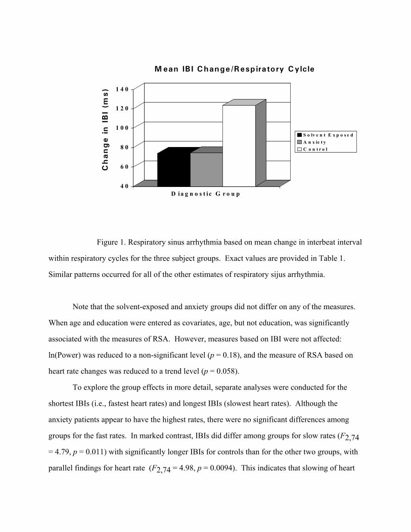
Respiratory cycle range changes showed a similar pattern of effects (F2,74 = 4.89, p =
0.010). That is, the differences between slowest and fastest beats in response to respiratory
activity were significantly reduced for both the solvent-exposed and anxiety groups compared to
non-exposed controls (Figure 1). When recomputed on the basis of heart rates, the effects were
similar (F2,74 = 3.67, p = 0.030).
Analysis of ln(Power) in the .15 to .20 Hz bandwidth also showed a significant difference
among groups (F2,74 = 3.56, p = 0.033). In this case, however, only the solvent-exposed and
non-exposed groups differed in post-hoc comparisons, while the difference for anxiety patients
from controls did not reach significance; this may have represented a reduction of statistical
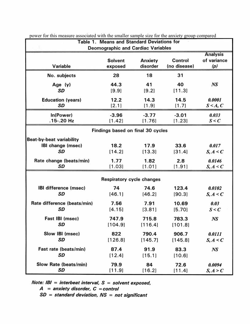
power for this measure associated with the smaller sample size for the anxiety group compared
to the other groups.
4 0
6 0
8 0
1 0 0
1 2 0
1 4 0
D ia g n o s t i c G r o u p
S o lv e n t E x p o s e dA n x ie t yC o n t r o l
M ean IB I Change/Resp iratory Cylcle
Ch
an
ge i
n I
BI
(ms)
Figure 1. Respiratory sinus arrhythmia based on mean change in interbeat interval
within respiratory cycles for the three subject groups. Exact values are provided in Table 1.
Similar patterns occurred for all of the other estimates of respiratory sijus arrhythmia.
Note that the solvent-exposed and anxiety groups did not differ on any of the measures.
When age and education were entered as covariates, age, but not education, was significantly
associated with the measures of RSA. However, measures based on IBI were not affected:
ln(Power) was reduced to a non-significant level (p = 0.18), and the measure of RSA based on
heart rate changes was reduced to a trend level (p = 0.058).
To explore the group effects in more detail, separate analyses were conducted for the
shortest IBIs (i.e., fastest heart rates) and longest IBIs (slowest heart rates). Although the
anxiety patients appear to have the highest rates, there were no significant differences among
groups for the fast rates. In marked contrast, IBIs did differ among groups for slow rates (F2,74
= 4.79, p = 0.011) with significantly longer IBIs for controls than for the other two groups, with
parallel findings for heart rate (F2,74 = 4.98, p = 0.0094). This indicates that slowing of heart
rate during the respiratory cycle was significantly greater for the controls than for the patient
groups, even though the groups did not differ in initial high rates.
Intercorrelations among the different measures of RSA all were high. Correlations
between mean change in successive beats and mean range within respiratory cycles were high
using either IBI or heart rate measures (r = 0.98, and r = 0.94, respectively, both p < 0.001).
Correlations among the same measures and ln(Power) were somewhat lower, but still highly
significant (r ranging from 0.61 to 0.74, all p < 0.001).
Associations between RSA and the SCL-90-R Anxiety subscale were not significant for
the entire group. Within the group of anxiety patients, there was a weak trend for increased
reported anxiety to be associated with the fastest heart rates based on interbeat interval (r =
0.369, p = .15 ; this degree of association would only reach significance with at least 29
subjects). Similar trends were not present for the controls or solvent exposed groups.
For the solvent patients, rank order correlations between the Exposure Ratings were not
significantly associated with any of the RSA indices.
Findings for Drug-Free Subjects: To determine the extent to which findings were
independent of drug effects, we compared controls and a subset of 17 solvent and anxiety
patients who were either medication free or who were receiving only drugs with no central
anxiolytic, antidepressant, or blood pressure effects (e.g., antacids). With this reduced sample,
RSA continued to be significantly reduced when based on beats per minute (F1,46 = 4.46, p =
0.04) and marginal for RSA based on interbeat interval (F1,46 = 3.96, p = 0.053), though
differences in ln(Power) were not significant ( p = 0.15). When the same group of 17 patients
was compared with the remaining 29 patients who were receiving antidepressant, anxiolytic, or
blood pressure medications, there were no significant differences on any of the measures of
RSA. Similarly, no significant differences were found within either patient group examined
separately for presence or absence of medication.
Long-Term Reliability: A follow-up evaluation was conducted after one year for 25 of
the subjects (7 solvent-exposed, 11 anxiety, and 7 non-exposed control subjects). Table 2 shows
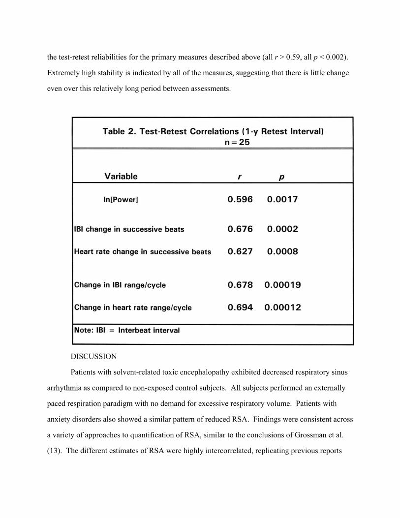
the test-retest reliabilities for the primary measures described above (all r > 0.59, all p < 0.002).
Extremely high stability is indicated by all of the measures, suggesting that there is little change
even over this relatively long period between assessments.
DISCUSSION
Patients with solvent-related toxic encephalopathy exhibited decreased respiratory sinus
arrhythmia as compared to non-exposed control subjects. All subjects performed an externally
paced respiration paradigm with no demand for excessive respiratory volume. Patients with
anxiety disorders also showed a similar pattern of reduced RSA. Findings were consistent across
a variety of approaches to quantification of RSA, similar to the conclusions of Grossman et al.
(13). The different estimates of RSA were highly intercorrelated, replicating previous reports
(6), although ln(Power) was less closely associated with the other measures, and was not as
powerful a measure in discriminating among groups.
The differences in RSA in the patient groups appear related to a lack of slowing of heart
rate during the respiratory cycle, rather than to an excessively higher rate overall. Since
modulation of heart rate during the respiratory cycle has been associated with vagal control (5,
10, 14,21), the current finding is entirely consistent with prevalent notions that vagal activation
is responsible for cardiac changes during respiration. In particular, the present data suggest that
vagal activation is deficient in these patient groups. This is an assumption normally made in
clinical studies in which decreased RSA is indicated even using deep breathing instructions (11).
Our findings in patients with clinically significant exposures to organic solvents are entirely
consistent with reduced RSA function which has been reported among workers exposed to a
variety of organic solvents under workplace conditions (7-10). Moreover, these effects are not
likely to be attributable to medication effects: the same patterns were observed even when only
patients who were free of medication or were receiving only inert antacids were compared with
controls.
Typical studies employing a paced-breathing technique involve teaching a rate of
breathing to the subjects, and then recording RSA during self-paced breathing during changing
cognitive demands (12, 13). These procedures have an advantage in the ability to observe the
effects of different tasks on RSA. The present approach, relying on an external stimulus to cue
breathing, is probably not optimal for comparing within subject differences during varying
processing conditions. However, the current procedure seems appropriate as a clinical
evaluation technique when no separate cognitive or attentional demands are required from the
subject. Moreover, this measure was highly reliable across a one-year retest period. In typical
clinical settings, RSA during the Valsalva maneuver is typically employed with such other
measures as the autonomic reflex screen (including quantitative sudomotor axon reflex test
(QSART), orthostatic blood pressure, Valsalva ratio, and heart rate response to tile), and reflex
sympathetic dystrophy (including skin temperature, resting seat output, and bilateral QSART
measures) (22). Clinical testing for autonomic dysfunction or autonomic neuropathy involving
RSA (23) is used for a variety of disorders, including diabetic autonomic neuropathy (24),
ulcerative colitis (25), and cluster headache (26).
Anxiety is present both behaviorally and physiologically in toxic encephalopathy. The
complex of symptomatology is similar to that seen for primary anxiety disorders. The
mechanisms associated with the anxiety in both types of patient cohorts may be similar.
However, data from other physiological studies (e.g., event-related brain potentials) suggests that
the disorders differ in the occurrence of greater cognitive and electrophysiological impairment
among the solvent patients as compared to anxiety patients (27). Thus, anxiety appears to
accompany a clinically significant history of exposure to organic solvents. Given the high test-
retest reliability observed over one year in this sample, the continued evaluation of respiratory
sinus arrhythmia as an indicator of impaired parasympathetic function in association with solvent
exposure seems highly justified.
REFERENCES
1. Morrow L, Ryan C, Goldstein G, Hodgson M. A distinct pattern of personality
disturbance following exposure to mixtures of organic solvents. J Occ Med 1989; 31: 743-746.
2. Morrow L, Ryan C, Hodgson, M, Robin, N. Alterations in cognitive and psychological
functioning after organic solvent exposure. J Occ Med 1990; 32: 444-450.
3. Schottenfeld RS, Cullen MR. Recognition of occupation-induced posttraumatic stress
disorders. J Occ Med 1986; 28: 365-369.
4. Morrow LA, Steinhauer SR. Alterations in heart rate and pupillary response in persons
with organic solvent exposure. Biol Psychiatry 1995; 37: 721-730.
5. Grossman P, Wientjes K. Respiratory sinus arrhythmia and parasympathetic cardiac
control: Some basic issues concerning quantification, application and implications. In Grossman
P, Jansenn KH, Vaitl D, eds. Cardiorespiratory and cardiosomatic psychophysiology. NY:
Plenum Press 1986; 117-138.
6. Zahn TP. Effects of motor restlessness on respiratory sinus arrhythmia in children and
adolescents. Soc. for Psychophysiological Research, Oct., 1992.
7. Murata K, Araki S, Yokoyama K. Assessment of the peripheral, central, and
autonomic nervous system function in styrene workers. Am J Ind Med 1991; 20: 775-784.
8. Murata K, Araki S, Yokoyama K, et al. Cardiac autonomic dysfunction in rotogravure
printers exposed to toluene in relation to peripheral nerve conduction. Ind Health 1993; 31: 79-
90.
9. Murata K, Araki S, Yokoyama K, et al. Changes in autonomic function as determined
by ECG R-R interval variability in sandal, shoe and leather workers exposed to n-hexane, xylene
and toluene. Neurotoxicology 1994; 15: 867-875.
10. Murata K, Araki S. Assessment of autonomic neurotoxicity in occupational and
environmental health as determined by ECG R-R interval variability: A review. Am J Ind Med
1996; 30: 155-163.
11. Low PA, Denq JC, Opfer-Gehrking TL, et al. Effect of age and gender on sudomotor
and cardiovagal function and blood pressure response to tilt in normal subjects. Muscle Nerve
1997; 20: 1561-1568.
12. Allen MT, Crowell MD. Patterns of autonomic response during laboratory stressors.
Psychophysiology 1989; 26: 603-614.
13. Grossman P, van Beek J, Wientes C. A comparison of three quantification methods
for estimation of respiratory sinus arrhythmia. Psychophysiology 1990; 27: 702-714.
14. Grossman, P., Stemmler, G., & Meinhardt, E. Paced respiratory sinus arrhythmia as
an index of cardiac parasympathetic tone during varying behavioral tasks. Psychophysiology
1990; 27: 404-416.
15. Kollai, M., & Kollai, B. Cardiac vagal tone in generalised anxiety disorder. Br J
Psychiatry 1992; 161: 831-835.
16. Roth WT, Margraf J, Ehlers A, et al. Stress test reactivity in panic disorder. Arch Gen
Psychiatry 1992; 49: 301-310.
17. Morrow L, Kamis H, Hodgson M. Psychiatric symptomatology in persons with
organic solvent exposure. J Cons Clin Psychol 1993; 61: 171-174.
18. Derogatis LR. SCL-90-R Manual-II. Towson MD: Clinical Psychometric Research
1983.
19. Cook, E.W., III. VPM reference manual. Author, 1995.
20. Weber EJM, Molenaar PC, van der Molen MW. PSPAT: A program for spectral
analysis of point events inclduing a test for stationarity. In: Mulder LJM et al., eds. Computers
in Psychology. Amsterdam: Swets & Zeitlinger 1991; 132-139.
21. Porges SW, Bohrer RE. The analysis of periodic processes in psychophysiological
research. In Cacioppo JT, Tassinary L.G, eds. Principles of Psychophysiology. NY: Cambridge
1990; 708-753.
22. Low, P.A. Autonomic nervous system function. J Clin Neurophysiology 1993; 10: 1-
27.23. Wieling W, Karemaker JF, Borst C, Dunning AJ. Testing for autonomic neuropathy: heart
rate response to forced breathing. Clin Physiol 1985; 5 Suppl 5:28-33.
24. Smith SA. Reduced sinus arrhythmia in diabetic autonomic neuropathy: diagnostic
value of an age-related normal range. Br Med J:1982 Dec 4; 285:1599-601.
25. Straub RH, Antoniou E, Zeuner M, Gross V, Scholmerich J, Andus T. Association of
autonomic nervous hyperreflexia and systemic inflammation in patients with Crohn's disease and
ulcerative colitis. Neuroimmunol 199; 80:149-57.
26. Kruszewski P. Respiratory sinus arrhythmia in cluster headache syndrome. Headache
1993; 33:98-104.
27. Steinhauer SR, Morrow LA, Condray R, Scott A. Event-related potential and cardiac
measures differentiate patients with anxiety disorders from patients with toxic solvent exposure.
J Int Neuropsychological Soc 1999; 5: 130.


