Embed Size (px)
Citation preview

E 441
Original Article
Irina Bancos, MD1; Clive S. Grant, MD2; Sarah Nadeem, MBBS1;Marius N. Stan, MD1; Carl C. Reading, MD3; Thomas J. Sebo, MD, PhD4;
Alicia Algeciras-Schimnich, PhD4; Ravinder J. Singh, PhD4;Diana S. Dean, MD, FACE1
Submitted for publication May 23, 2011Accepted for publication November 18, 2011From the 1Department of Endocrinology, 2Department of Surgery, 3Department of Radiology, and 4Department of Laboratory Medicine and Pathology, Mayo Clinic Rochester, Rochester, Minnesota.Address correspondence to Dr. Irina Bancos, Department of Endocrinology, Mayo Clinic Rochester, Mayo Building 18W, 200 First Street Southwest, Rochester, MN 55905. E-mail: [email protected] as a Rapid Electronic Article in Press at http://www.endocrinepractice.org. DOI:10.4158/EP11148.ORTo purchase reprints of this article, please visit: www.aace.com/reprints.Copyright © 2012 AACE.
ABSTRACT
Objective: To describe the experience with parathy-roid fine-needle aspiration (FNA) and parathyroid hor-mone (PTH) washout at Mayo Clinic Rochester, Rochester, Minnesota. Methods: We retrospectively reviewed all parathy-roid FNA procedures performed at Mayo Clinic Rochester between January 2000 and December 2007. Clinical, bio-chemical, and imaging information, parathyroid FNA pro-cedure, and cytology, surgical, and pathology reports were reviewed, and descriptive statistics, sensitivity, specificity, and positive predictive values are presented. Results: During the study period, 75 parathyroid FNAs were performed on 74 patients. Cytology results were available for 74 of 75 procedures, with only 31% in-terpreted as parathyroid cells. PTH washout was performed in 67 patients (91%). Parathyroid FNA with PTH washout had a sensitivity of 84%, specificity of 100%, positive pre-
dictive value of 100%, and accuracy of 84%. At the time of surgical treatment, 2 patients were noted to have an inflam-matory response from the parathyroid FNA biopsy, 1 had a parathyroid abscess, and 2 had a hematoma. In 3 of these 5 patients, the necessary conversion of a minimally invasive surgical procedure to the standard surgical approach pro-longed the surgical time. Conclusion: Parathyroid FNA with PTH washout had a superior performance in comparison with parathyroid scanning or ultrasonography alone. The main limitations of parathyroid FNA with PTH washout are (1) the need for initial identification of a potential parathyroid adenoma by ultrasonography and (2) the number of false-negative re-sults. Parathyroid FNA resulted in complications affecting the surgical procedure in 3 patients. (Endocr Pract. 2012; 18:441-449)
Abbreviations:FNA = fine-needle aspiration; PHPT = primary hyper-parathyroidism; PPV = positive predictive value; PTH = parathyroid hormone
INTRODUCTION
Despite the shift from standard, bilateral, 4-gland parathyroid exploration to image-directed minimal access parathyroidectomy, persistent or recurrent hyperparathy-roidism still occurs in 5% to 10% of patients. Parathyroid localization is crucial to the success of reoperation, in contrast to merely a convenience before an initial surgi-cal exploration. In a reoperative setting when scarring, distortion of anatomic landmarks, and a higher number of ectopic parathyroid glands in this subgroup make another intervention more difficult, correct localization is pivotal. Sequentially used are noninvasive studies including cervi-cal ultrasonography, sestamibi scintigraphy, computed to-mography, and magnetic resonance imaging. If these stud-ies are unsuccessful or unconvincing, invasive testing such as arteriography or venous sampling may be undertaken.

442
There have been interest and debate regarding the utility and safety of ultrasound-guided parathyroid fine-needle aspiration (FNA). First described by Doppman et al (1) in 1983, parathyroid FNA with parathyroid hormone (PTH) washout became more frequently used during the late 1990s and thereafter, when several institutions report-ed their experience (2-9). Theoretically, parathyroid FNA with PTH washout provides a more certain localization of the culprit parathyroid lesion in the preoperative setting. In the hands of various operators and a setting of inho-mogeneous groups of patients, ultrasonography-guided parathyroid FNA with PTH washout was reported to have a specificity of 91% to 100% and a sensitivity of 91% to 100% (2,3,5,10). Although this localizing technique pro-vides valuable information, several authors have expressed a concern that, if widely used, complications would be un-avoidable, with repercussions of prolonged and more labo-rious surgical procedures and even possibly introduction of confusion histologically with malignant lesions (11,12). Our objectives were to describe our institutional experi-ence with parathyroid FNA, specifically as associated with PTH washout, and to report its performance statistics, safe-ty, and limitations.
PATIENTS AND METHODS
This study was conducted with the approval of the Mayo Clinic Rochester (Rochester, Minnesota) institution-al review board and ethics committee.
Study Subjects We identified retrospectively all patients who under-went a parathyroid FNA at Mayo Clinic Rochester between January 2000 and December 2007. Their medical records were reviewed for clinical presentation, biochemical mea-surements, cervical ultrasonography, parathyroid sestamibi scan, parathyroid FNA procedure, cytology, surgical treat-ment, pathology reports, and procedure-related complica-tions.
Technique for Parathyroid FNA and PTH Washout FNA of the parathyroid gland was performed under continuous, real-time, ultrasound visualization with use of a 10-MHz linear transducer (Acuson-Siemens Sequoia, Mountain View, California). Before the FNA, the skin was cleansed with a povidone-iodine solution. Sterile gel was used as a coupling agent. A 25-gauge standard, noncut-ting, bevel-edge needle (Becton-Dickinson, Rutherford, New Jersey) was inserted by using a freehand technique. The needle was moved in a back-and-forth motion within the mass, and material from the target lesion was obtained through capillary action, without the use of attached suc-tion. A total of 6 passes were usually made, although the technique varied slightly, depending on the physician and
the patient’s anatomic variation. Aspirated material was then expelled onto a slide for cytologic analysis. For the washout procedure, isotonic saline was drawn into the nee-dle to fill the needle hub, after which it was then expelled through the needle into a plastic tube. After the 6 needle passes were made, a total of 0.5 to 1.5 mL of blood-tinged fluid was submitted for PTH assay. At our institution, the ordering party is advised that no established reference ranges for PTH in needle wash specimens currently exist at Mayo Clinic Rochester.
Data Collection
Patient Characteristics Patients were classified in one of the following cat-egories: (1) initial presentation of primary hyperparathy-roidism (PHPT); (2) recurrent PHPT; (3) persistent PHPT; (4) tertiary hyperparathyroidism; or (5) parathyroid carci-noma. Demographic data, pertinent clinical information (bone disease, kidney stones, or any subjective symptoms related to hyperparathyroidism), neck surgical history (number of parathyroid or thyroid explorations), and his-tory of radiation therapy were gathered.
Biochemical Data Preoperative serum PTH and total calcium levels were recorded.
Imaging From the cervical ultrasonography, we recorded the diameter of the suspected parathyroid lesion, its localiza-tion, and the presence of multinodular goiter if identified. Localization details were also recorded from the parathy-roid sestamibi scan, including the description of multifo-cality of a lesion.
Parathyroid FNA We reviewed parathyroid FNA procedure notes for number of passes, needle gauge, whether PTH washout was performed, and the level of PTH in the aspirate. All FNAs were performed because of a clinical concern about hyperparathyroidism; thus, the pathologists knew that the cytologic material originated from a potential parathyroid adenoma. Cytology reports were recorded as one of the following: (1) parathyroid cells; (2) parathyroid versus thyroid cells; (3) thyroid cells; (4) lymphoid cells; or (5) nondiagnostic. We recorded complications noted at the time of the parathyroid FNA procedure from the radiology reports (“immediate”). For patients who subsequently underwent surgical intervention, the interval between parathyroid FNA and surgical intervention was recorded as well as any complications related to the parathyroid FNA (“de-layed”).

443
Therapy For each patient, the mode of treatment (surgical in-tervention, alcohol ablation, medical treatment, or obser-vation) was recorded. For patients who underwent either surgical exploration or parathyroid FNA alcohol ablation, follow-up biochemical data were obtained for the subse-quent 12 months. For patient’s undergoing surgical treat-ment, operative and pathology reports were reviewed spe-cifically for the anatomic position and the weight of the lesion. Results of treatment were characterized as cure (eu-calcemia for 12 months), temporary palliation (recurrence of hyperparathyroidism 6 months after treatment), or fail-ure (recurrence of hyperparathyroidism within 6 months after treatment).
Statistical Analysis The Center for Translational Science Activities con-sultative services were used to provide biostatistical sup-port for this project. All statistical analyses were performed with use of JMP statistical software (version 8.0, SAS Institute Inc., Cary, North Carolina). Continuous variables are expressed as means ± standard deviation or medians (ranges) on the basis of distribution of the data. Univariate logistic regression analysis was used to identify variables associated with positive parathyroid FNA results. Variables having a statistically meaningful effect were included in multivariate regression analysis. Significance was estab-lished at P<.05. Sensitivity, specificity, positive predictive value (PPV), and accuracy were calculated for parathyroid FNA with PTH washout, cytology alone, and scintigraphy when appropriate. True-positive, false-positive, and false-negative results were identified for various preoperative localization modalities on the basis of comparison with operative and histology reports.
RESULTS
Patient Characteristics Between January 1, 2000, and December 31, 2007, 75 ultrasound-guided parathyroid FNA procedures were
performed on 74 patients (45 women and 29 men), with a mean age of 57.8 ± 17 years. At the time of parathyroid FNA, 27 patients (36%) had persistent PHPT, 15 (20%) had recurrent hyperparathyroidism, 28 (38%) had initial presentation of PHPT, 2 (3%) had tertiary hyperparathy-roidism, and 2 (3%) had parathyroid carcinoma. Familial hypocalciuric hypercalcemia was excluded in all patients. The majority of patients (68%) had previous neck interven-tion, and 15 patients (20%) were suspected of having only a single remaining parathyroid gland. Complications from the hyperparathyroidism included bone disease (27% with osteopenia and 36% with osteoporosis) and nephrolithiasis (35%). The median serum PTH level was 82 pg/mL (range, 20 to 1,450; reference range, 15 to 65), and the mean serum calcium level was 11.1 mg/dL (range, 9.7 to 14.7; reference range, 8.9 to 10.1). 25-Hydroxyvitamin D was measured in 29 patients, with increasing frequency during later years of the study (25 measurements after 2004). The mean 25-hy-droxyvitamin D level was 26 ng/dL (range, 7.9 to 51). On neck ultrasonography, multinodular goiter was de-scribed in 18 patients (24%). The median largest diameter of the target parathyroid nodules identified by ultrasonog-raphy was 10 mm (range, 2 to 37). We did not demand precision to the level of predicting superior versus inferior location, although this could often be identified. Parathyroid sestamibi scanning was performed at our institution in 70 patients (95%). We compared the results of parathyroid scanning with the ultrasound findings (Table 1). In only 28 of 70 patients (40%), both tests were concor-dant, showing the same lesion. In 12 patients (17%), the sestamibi parathyroid scan findings were multifocal or am-biguous. In 30 patients (43%), the results were discordant. In patients with an initial diagnosis of PHPT, 14 of 28 had discordant imaging and another 4 of the 28 had planned observation (in 2) or alcohol ablation (in 2) rather than a surgical procedure.
Parathyroid FNA Cytology results were available in 74 of 75 procedures, and PTH washout was performed in 67 patients (91%).
Table 1Imaging and Surgical Results in Patients
With and Without Previous Cervical Surgery
Previous Ultrasoundfindingsincomparisonwith cervical Parathyroid scan results Surgical results surgery Concordant Discordant Total True-positive False-positive Total
Yes 15 17 32 26 1 27 No 13 25 38 20 2 22 Total 28 42a 70 46 3 49
a The parathyroid scan findings were multifocal or ambiguous in 12 patients; results were discordant in 30.

444
Detectable levels of PTH were found in 50 of 67 patients (75%), and 45 of 67 (67%) had values above serum levels of PTH. When results were positive, the median washout PTH was 3,963 pg/mL (range, 45 to 5,000,000). The most frequent cytology result was “nondiagnostic” (34%), and only 31% of all parathyroid aspirates were true positive, with interpretation as parathyroid cells (Table 2). Although our institutional cutoff for aspirate PTH to be considered unequivocally diagnostic is >1,000 pg/mL, aspirate levels above the serum level of PTH were considered positive. PTH washout added to the diagnostic certitude in 56% of cases of nondiagnostic cytology, 67% of cases of parathy-roid FNA interpreted as lymphoid cells, 83% of cases of thyroid cytology, and 60% of cases of parathyroid versus thyroid cytology. Four patients with lymphoid, parathyroid versus thyroid, or nondiagnostic cytology results (n = 1, 2, and 1, respectively) had detectable PTH levels in the aspi-rate (45, 54, 81, and 279 pg/mL); however, they were lower than their serum PTH levels (Table 2). Nondiagnostic cytology had the least likelihood of providing a positive PTH aspirate value in comparison with parathyroid cytology (odds ratio = 0.2 [0.05 to 0.9]; P = .04). The likelihood of obtaining a diagnostic PTH washout in lymphoid, thyroid, or parathyroid versus thy-roid cytology did not differ significantly from that for para-thyroid cytology. The size of the visualized adenoma was positively correlated with the diagnostic PTH aspirate level (P = .0001). No other factors had any correlation with para-thyroid FNA results.
Therapy A surgical procedure was performed in 50 patients (68%), 9 (12%) were treated with alcohol ablation, 12 (16%) were monitored nonsurgically, and 2 (3%) opted for medical treatment (cinacalcet). Information for 1 patient was not available. The median time to surgical treatment was 13 days (range, 1 to 210). Surgical intervention was curative in 42 of the 50 patients (84%) and palliative in 2 of the 50 (4%); 6 patients (12%) developed recurrent hyperparathyroidism within the first 6 months after surgery. Alcohol ablation was less effective, providing cure in 2 of 9 patients (22%) and temporary palliation in 4 of 9 (44%); 3 patients (33%) failed to achieve eucalcemia.
PreoperativeLocalizationVersusTreatmentResults Correlation of operative findings, parathyroid FNA with PTH washout, cytology alone, and parathyroid ses-tamibi scintigraphy are detailed in Table 3. PTH washout was performed in 90% of patients (45 of 50) who under-went surgical treatment, and 96% (48 of 50) had a parathy-roid scan performed. The best results were from parathy-roid FNA in conjunction with PTH washout, which had a sensitivity of 84% and a specificity and PPV of 100%.
Biopsy-RelatedComplications Only 4 of 74 patients (5%) had parathyroid FNA procedure-related “immediate” complications—minor he-matomas. All 4 of these patients underwent surgical inter-vention within 1 to 210 days. The hematomas noted at the time of parathyroid FNA did not result in any problems described at the time of the surgical procedure in any of these patients. In all 4 patients, the lesion identified on ul-trasonography proved to be the culprit lesion, and surgical excision cured all 4 patients. In 1 patient, the parathyroid FNA procedure could not be completed because of theencountered hematoma (Table 4). Review of operative reports identified 5 of 49 patients with a “delayed” complication ascribed to the parathyroid FNA. In these patients, the time between the parathyroid FNA and the surgical intervention was relatively short (1 to 20 days). None of the 5 patients had a complication identi-fied at the time of parathyroid FNA. In 2 patients, the sur-gical reports described an inflammatory response from the parathyroid FNA, an abscess was discovered in 1 patient, and a hematoma was noted in the other 2. In 3 patients with FNA-related events, a minimally invasive surgical proce-dure had to be converted to a standard surgical approach, an adjustment that prolonged the surgical time (Table 4).
DISCUSSION
During the period from 2000 to 2007, 2,265 parathy-roid operations were performed at Mayo Clinic Rochester. Only 3% of the parathyroid operations (75 of 2,265) in-cluded parathyroid FNA procedures during the same time span, an indication that it is rarely part of our preopera-tive localization algorithm. Indeed, parathyroid FNA was used in only selected patients; 57% of patients presented with recurrent or persistent hyperparathyroidism, 68% of patients had undergone at least 1 prior neck surgical pro-cedure, 60% of patients had parathyroid sestamibi scan re-sults and parathyroid ultrasonographic findings that were discordant or ambiguous, 20% of patients were suspected of having only a single remaining parathyroid gland, and 24% of patients had multinodular goiter that complicated the parathyroid imaging. The obvious limitation of parathyroid FNA is its de-pendence on identification of a suspicious lesion by ultra-sonography. Multinodular goiter, lymph nodes, fibrosis, and other tumors in the neck are the usual other factors that can cause misinterpretation of an enlarged parathy-roid gland by ultrasonography (13-15). Ultrasound-guided parathyroid FNA with determination of PTH in the aspi-rated material has proved to be a valuable procedure for distinguishing a parathyroid lesion especially from lym-phatic or thyroid tissue (2,4-6,8,9). Although we found no difference between the proportion of positive PTH aspi-

445
Tabl
e 2
ResultsofP
arathyroidFine-NeedleA
spirationWith
ParathyroidHormoneWashout
PT
H
washout
Cytology
Cytologyresultsof
PTHwashout
Surgery
versus
+PT
H
parathyroidfine-needle
Done
Range
performed
surgical
washout
aspiration,no.(%
)(no.)
Distribution
No.(%
)(pg/mL)
(no.)
result
result
Pa
rath
yroi
d ce
lls, 2
3 (3
1%)
20
PTH
>1,
000
pg/m
L 15
(75)
1,
050-
5,00
0,00
0 15
15
TP
PPV
= 1
00%
PT
H >
seru
m le
vel o
f PTH
but
<1,
000
pg/m
L 2
(10)
76
0, 7
66
2 2
TP
Sens
itivi
ty =
100
%
PTH
<se
rum
leve
l of P
TH o
r und
etec
tabl
e 3
(15)
2,
2, 2
1
1 FN
Pa
rath
yroi
d ve
rsus
thyr
oid
10
PTH
>1,
000
pg/m
L 5
(50)
1,
547-
300,
000
5 5
TP
PPV
= 1
00%
ce
lls, 1
0 (1
3.5%
) PT
H >
seru
m le
vel o
f PTH
but
<1,
000
pg/m
L 1
(10)
66
0
Sens
itivi
ty =
83%
PT
H <
seru
m le
vel o
f PTH
or u
ndet
ecta
ble
4 (4
0)
2, 2
, 45,
279
1
1 FN
Th
yroi
d ce
lls, 6
(8%
) 6
PTH
>1,
000
pg/m
L 4
(67)
8,
930-
540,
000
4 4
TP
PPV
= 1
00%
PT
H >
seru
m le
vel o
f PTH
but
<1,
000
pg/m
L 1
(17)
62
0 1
1 TP
Se
nsiti
vity
= 8
3%
PTH
<se
rum
leve
l of P
TH o
r und
etec
tabl
e 1
(17)
2
1 1
FN
Ly
mph
oid
cells
, 10
(13.
5%)
6 PT
H >
1,00
0 pg
/mL
3 (5
0)
4,07
3 1
1 TP
PP
V =
100
%
6,68
0 Se
nsiti
vity
= 5
0%
50,0
00
PTH
>se
rum
leve
l of P
TH b
ut <
1,00
0 pg
/mL
1 (1
7)
990
0
PTH
<se
rum
leve
l of P
TH o
r und
etec
tabl
e 2
(33)
2
2 1
TN
54
1 FN
N
ondi
agno
stic
, 25
(34%
) 25
PT
H >
1,00
0 pg
/mL
10 (4
0)
1,01
2-99
,640
7
7 TP
PP
V =
100
%
PTH
>se
rum
leve
l of P
TH b
ut <
1,00
0 pg
/mL
4 (1
6)
530,
587
, 700
, 750
1
1 TP
Se
nsiti
vity
= 6
7%
PTH
<se
rum
leve
l of P
TH o
r und
etec
tabl
e 11
(44)
81
(1 p
atie
nt)
5 1
TN
2 (1
0 pa
tient
s)
4 FN
A
bbre
viat
ions
: FN
= fa
lse-
nega
tive
(neg
ativ
e pr
esur
gica
l loc
aliz
atio
n w
ith a
par
athy
roid
ade
nom
a fo
und
intra
oper
ativ
ely)
; PPV
= p
ositi
ve p
redi
ctiv
e va
lue;
PTH
= p
arat
hyro
id h
orm
one;
TN
= tr
ue-
ne
gativ
e (n
egat
ive
preo
pera
tive
loca
lizat
ion
corr
espo
ndin
g to
no
dise
ase
in th
e si
mila
r loc
atio
n in
traop
erat
ivel
y); T
P =
true-
posi
tive
(cor
resp
onde
nce
betw
een
preo
pera
tive
resu
lt an
d po
stsu
rgic
al
hi
stol
ogic
find
ings
in lo
catin
g pa
rath
yroi
d ad
enom
a).

446
rates in our patients with and without multinodular goiter, the number of false-negative parathyroid FNA procedures with PTH washout emphasizes the importance of a clearly identified ultrasound target for accurate localization. In agreement with Abraham et al (2), we observed a positive correlation between the size of the visualized adenoma and the diagnostic PTH aspirate level (P = .0001); however, we found no relationship with the serum calcium level or the serum PTH value. In our experience, only 31% of patients had cytologic results of FNA indicative of parathyroid tissue, which pro-vided a definite diagnosis. Therefore, severe limitations of ambiguous or frankly inaccurate cytologic findings minimize the value of this method. Although a few au-thors maintain that parathyroid tissue can be distinguished from a thyroid lesion on cytomorphologic grounds alone (16,17), others state that cytomorphology is not reliable (18,19). Our experience prompts us to agree with the latter opinion. Therefore, we rely heavily on the PTH value in the aspirate washout rather than on the cytologic results. Historically, our institution’s cutoff for a PTH positive value in the needle-washing fluid has been >1,000 pg/mL. This cutoff value was arbitrary, selected to avoid false-posi-tive results that might lead to inappropriate operations. Our current study indicates, however, that a lower value might
be preferred to decrease the number of false-negative re-sults. Our institution changed to a new PTH immunoassay in 2008. Because PTH values from different assays are not interchangeable, efforts to establish a more robust clinical cutoff value that provides high sensitivity and specificity are currently under way. Even with a clinical cutoff, some caveats need to be taken into consideration for interpreta-tion of a positive or negative result: (1) contamination of the needle washing with blood might lead to a positive re-sult if the serum PTH concentration is highly elevated; (2) inadequate sampling during the FNA procedure might lead to false-negative results; and (3) inadequate handling of the sample might result in false-negative results attributable to instability of PTH at room temperature. In light of the retrospective nature of the current study, the patients were preselected on the basis of at least a le-sion suspicious for a parathyroid adenoma identified by ul-trasonography. They also underwent parathyroid scintigra-phy, with a sensitivity of 78% and PPV of 62% in patients with available postsurgical histology reports (n = 48). In contrast, parathyroid FNA with PTH washout yielded a sensitivity of 84% and a PPV of 100%. It is important to emphasize that, although an ultrasonography-disclosed finding suggestive of a possible parathyroid lesion was identified, sometimes contradictory parathyroid imaging or
Table 3True-Positive,True-Negative,False-Positive,andFalse-NegativeResults,
Sensitivity,Specificity,PositivePredictiveValue,andAccuracyforParathyroidFine-NeedleAspirationWithParathyroidHormoneWashout,
CytologyAlone,andParathyroidSestamibiScintigraphyAloneinComparisonWithFinalOperativeFindings
Preoperativelocalization TP TN FP FN Result
Parathyroid fine-needle aspiration Sensitivity = 84% + parathyroid hormone washout Specificity = 100% (N = 45) 36 2 0 7 Positive predictive value = 100% Accuracy = 84%
Cytology alone (N = 26) 17 1 0 8 Sensitivity = 68% Specificity = 100% Positive predictive value = 100%
Parathyroid scan (N = 48) 25 1 15 7 Sensitivity = 78% Specificity = 6% Positive predictive value = 62% Accuracy = 53%
Abbreviations: FN = false-negative (negative presurgical localization with a parathyroid adenoma found intraoperatively); FP = false-positive (positive presurgical localization but a parathyroid adenoma in a different location at operation); TN = true-negative (negative preoperative localization corresponding to no disease in the similar location during operation); TP = true-positive (correspondence between preoperative result and postsurgical histologic findings in locating parathyroid adenoma).

447
Tabl
e 4
OverviewofE
ventsR
elatedtoParathyroidFine-NeedleA
spiration
In
terv
al
Parathyroid
between
Com
plication
horm
one
FNAand
attimeof
Com
plication
Age(y)
No.of
Needle
assay
Cytology
surgery
parathyroid
attimeof
Case
andsex
passes
gauge
Diagnosis
(pg/mL)
report
(days)
FNA
surgery
1
50
M
6 25
N
ew D
x of
prim
ary
3,87
9,00
0 Pa
rath
yroi
d ce
lls
2 N
one
Hem
atom
a (6
0 to
90
cc);
hy
perp
arat
hyro
idis
m
min
imal
ly in
vasi
ve su
rger
y ha
d
to b
e co
nver
ted
to a
stan
dard
su
rgic
al a
ppro
ach
2
59
F 7
25
New
Dx
of p
rimar
y 31
,000
Pa
rath
yroi
d ce
lls
1 N
one
Hem
atom
a fo
und
durin
g
hype
rpar
athy
roid
ism
su
rgic
al p
roce
dure
3
85
F 4
25
New
Dx
of p
rimar
y N
ot o
btai
ned
NA
21
0 M
inor
hem
atom
a N
one
hy
perp
arat
hyro
idis
m
4
69
M
4 25
Pe
rsis
tent
prim
ary
3,71
7 N
ondi
agno
stic
1
Min
or h
emat
oma
Non
e
hype
rpar
athy
roid
ism
5
77
F 4
25
New
Dx
of p
rimar
y <2
N
ondi
agno
stic
40
M
inor
hem
atom
a N
one
hy
perp
arat
hyro
idis
m
6
73
F 5
25
New
Dx
of p
rimar
y 3,
852
Non
diag
nost
ic
30
Min
or h
emat
oma
Non
e
hype
rpar
athy
roid
ism
7
76
M
5 25
Pe
rsis
tent
prim
ary
540,
000
Thyr
oid
cells
6
Non
e In
flam
mat
ory
resp
onse
from
1
22
hype
rpar
athy
roid
ism
bi
opsy
8
53
M
6 25
N
ew D
x of
prim
ary
12,1
90
Para
thyr
oid/
20
N
one
Infla
mm
ator
y re
actio
n
hype
rpar
athy
roid
ism
th
yroi
d ce
lls
lead
ing
to a
long
er in
cisi
on;
m
inim
ally
inva
sive
surg
ery
had
to
be
conv
erte
d to
a st
anda
rd
surg
ical
app
roac
h
9
71
M
6 25
N
ew D
x of
prim
ary
89,6
50
Para
thyr
oid
cells
13
N
one
Abs
cess
, for
min
g a
firm
mas
s
hype
rpar
athy
roid
ism
(2
.3 b
y 1.
7 by
1.4
cm
);
min
imal
ly in
vasi
ve su
rger
y ha
d
to b
e co
nver
ted
to a
stan
dard
su
rgic
al a
ppro
ach
A
bbre
viat
ions
: Dx
= di
agno
sis;
FN
A =
fine
-nee
dle
aspi
ratio
n; N
A =
not
app
licab
le.
448
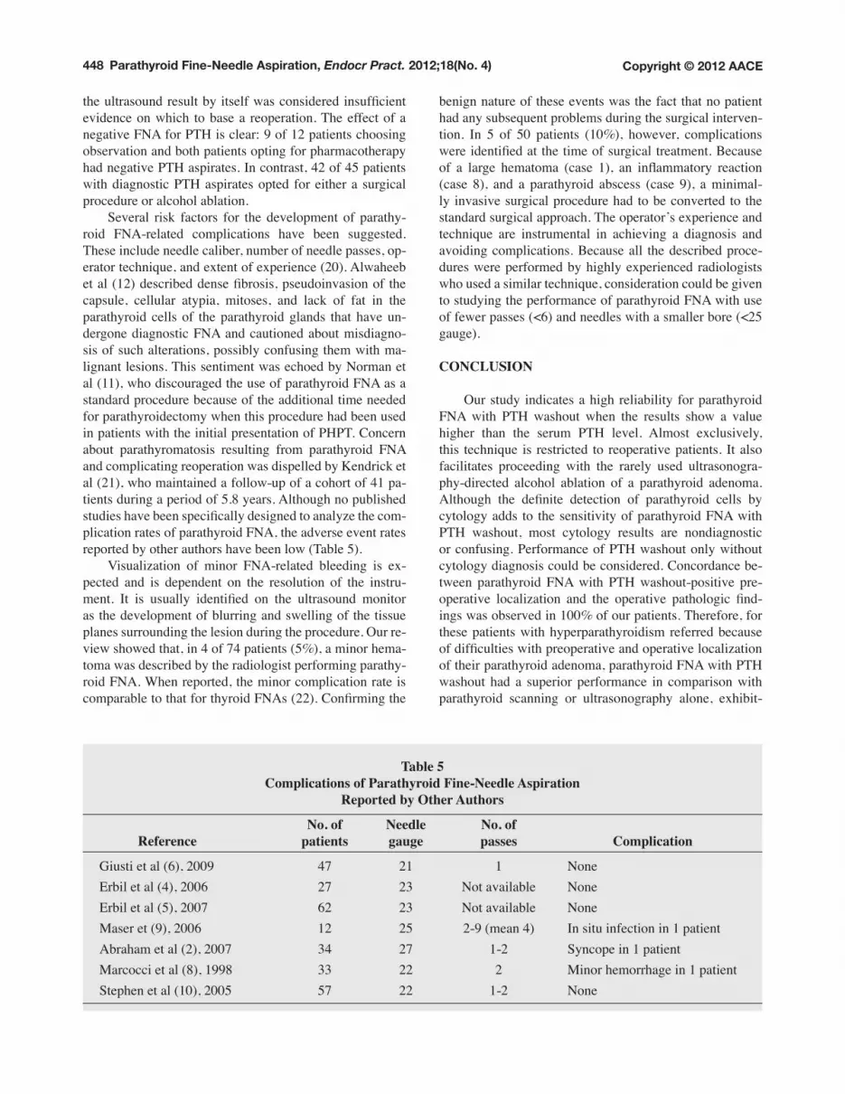
the ultrasound result by itself was considered insufficient evidence on which to base a reoperation. The effect of a negative FNA for PTH is clear: 9 of 12 patients choosing observation and both patients opting for pharmacotherapy had negative PTH aspirates. In contrast, 42 of 45 patients with diagnostic PTH aspirates opted for either a surgical procedure or alcohol ablation. Several risk factors for the development of parathy-roid FNA-related complications have been suggested. These include needle caliber, number of needle passes, op-erator technique, and extent of experience (20). Alwaheeb et al (12) described dense fibrosis, pseudoinvasion of the capsule, cellular atypia, mitoses, and lack of fat in the parathyroid cells of the parathyroid glands that have un-dergone diagnostic FNA and cautioned about misdiagno-sis of such alterations, possibly confusing them with ma-lignant lesions. This sentiment was echoed by Norman et al (11), who discouraged the use of parathyroid FNA as a standard procedure because of the additional time needed for parathyroidectomy when this procedure had been used in patients with the initial presentation of PHPT. Concern about parathyromatosis resulting from parathyroid FNA and complicating reoperation was dispelled by Kendrick et al (21), who maintained a follow-up of a cohort of 41 pa-tients during a period of 5.8 years. Although no published studies have been specifically designed to analyze the com-plication rates of parathyroid FNA, the adverse event rates reported by other authors have been low (Table 5). Visualization of minor FNA-related bleeding is ex-pected and is dependent on the resolution of the instru-ment. It is usually identified on the ultrasound monitor as the development of blurring and swelling of the tissue planes surrounding the lesion during the procedure. Our re-view showed that, in 4 of 74 patients (5%), a minor hema-toma was described by the radiologist performing parathy-roid FNA. When reported, the minor complication rate is comparable to that for thyroid FNAs (22). Confirming the
benign nature of these events was the fact that no patient had any subsequent problems during the surgical interven-tion. In 5 of 50 patients (10%), however, complications were identified at the time of surgical treatment. Because of a large hematoma (case 1), an inflammatory reaction (case 8), and a parathyroid abscess (case 9), a minimal-ly invasive surgical procedure had to be converted to the standard surgical approach. The operator’s experience and technique are instrumental in achieving a diagnosis and avoiding complications. Because all the described proce-dures were performed by highly experienced radiologists who used a similar technique, consideration could be given to studying the performance of parathyroid FNA with use of fewer passes (<6) and needles with a smaller bore (<25 gauge).
CONCLUSION
Our study indicates a high reliability for parathyroid FNA with PTH washout when the results show a value higher than the serum PTH level. Almost exclusively, this technique is restricted to reoperative patients. It also facilitates proceeding with the rarely used ultrasonogra-phy-directed alcohol ablation of a parathyroid adenoma. Although the definite detection of parathyroid cells by cytology adds to the sensitivity of parathyroid FNA with PTH washout, most cytology results are nondiagnostic or confusing. Performance of PTH washout only without cytology diagnosis could be considered. Concordance be-tween parathyroid FNA with PTH washout-positive pre-operative localization and the operative pathologic find-ings was observed in 100% of our patients. Therefore, for these patients with hyperparathyroidism referred because of difficulties with preoperative and operative localization of their parathyroid adenoma, parathyroid FNA with PTH washout had a superior performance in comparison with parathyroid scanning or ultrasonography alone, exhibit-
Table 5ComplicationsofParathyroidFine-NeedleAspiration
ReportedbyOtherAuthors
No. of Needle No. of Reference patients gauge passes Complication
Giusti et al (6), 2009 47 21 1 None Erbil et al (4), 2006 27 23 Not available None Erbil et al (5), 2007 62 23 Not available None Maser et (9), 2006 12 25 2-9 (mean 4) In situ infection in 1 patient Abraham et al (2), 2007 34 27 1-2 Syncope in 1 patient Marcocci et al (8), 1998 33 22 2 Minor hemorrhage in 1 patient Stephen et al (10), 2005 57 22 1-2 None

449
ing a PPV of 100%, a sensitivity of 84%, a specificity of 100%, and an accuracy of 84%. In contrast, cytology alone should not be used for localization. The main limitations of parathyroid FNA with PTH washout are (1) the need for initial identification of a potential parathyroid adenoma by ultrasonography and (2) the number of false-negative results. In our experience, the rate of parathyroid FNA-related complications affecting surgical treatment was only 6%. The added cost and invasiveness of parathyroid FNA, especially in view of the high success of usual preoperative localization tests, generally preclude its use before initial cervical exploration. In the reoperative setting, however, when localization is crucial, the 100% PPV of parathyroid FNA with PTH washout makes it a highly attractive, safe, and well-tolerated procedure.
DISCLOSURE
The authors have no multiplicity of interest to disclose.
REFERENCES
1. DoppmanJL,KrudyAG,MarxSJ,etal. Aspiration of enlarged parathyroid glands for parathyroid hormone assay. Radiology. 1983;148:31-35.
2. AbrahamD,SharmaPK,BentzJ,GaultPM,NeumayerL,McClainDA. Utility of ultrasound-guided fine-needle aspiration of parathyroid adenomas for localization be-fore minimally invasive parathyroidectomy. Endocr Pract. 2007;13:333-337.
3. Barczynski M, Golkowski F, Konturek A, et al. Technetium-99m-sestamibi subtraction scintigraphy vs. ultrasonography combined with a rapid parathyroid hor-mone assay in parathyroid aspirates in preoperative local-ization of parathyroid adenomas and in directing surgical approach. Clin Endocrinol (Oxf). 2006;65:106-113.
4. ErbilY,BarbarosU,SalmasliogluA,etal. Value of para-thyroid hormone assay for preoperative sonographically guided parathyroid aspirates for minimally invasive para-thyroidectomy. J Clin Ultrasound. 2006;34:425-429.
5. ErbilY,SalmaslioğluA,KabulE,etal. Use of preopera-tive parathyroid fine-needle aspiration and parathormone assay in the primary hyperparathyroidism with concomi-tant thyroid nodules. Am J Surg. 2007;193:665-671.
6. GiustiM,DolcinoM,VeraL,etal. Institutional experi-ence of PTH evaluation on fine-needle washing after aspi-ration biopsy to locate hyperfunctioning parathyroid tissue. J Zhejiang University Sci B. 2009;10:323-330.
7. KiblutNK,CussacJF,SoudanB,etal. Fine needle as-piration and intraparathyroid intact parathyroid hormone measurement for reoperative parathyroid surgery. World J Surg. 2004;28:1143-1147.
8. Marcocci C, Mazzeo S, Bruno-Bossio G, et al. Preoperative localization of suspicious parathyroid adeno-mas by assay of parathyroid hormone in needle aspirates. Eur J Endocrinol. 1998;139:72-77.
9. Maser C, Donovan P, Santos F, et al. Sonographically guided fine needle aspiration with rapid parathyroid hor-mone assay. Ann Surg Oncol. 2006;13:1690-1695.
10. Stephen AE, Milas M, Garner CN, Wagner KE,Siperstein AE. Use of surgeon-performed office ultra-sound and parathyroid fine needle aspiration for complex parathyroid localization. Surgery. 2005;138:1143-1150.
11. NormanJ,PolitzD,BrowarskyI. Diagnostic aspiration of parathyroid adenomas causes severe fibrosis complicat-ing surgery and final histologic diagnosis. Thyroid. 2007; 17:1251-1255.
12. AlwaheebS,RambaldiniG,BoernerS,CoiréC,FiserJ,AsaSL. Worrisome histologic alterations following fine-needle aspiration of the parathyroid. J Clin Pathol. 2006; 59:1094-1096.
13. Mihai R,Gleeson F, Buley I, Roskell DE, SadlerGP. Negative imaging studies for primary hyperparathyroidism are unavoidable: correlation of sestamibi and high-resolu-tion ultrasound scanning with histological analysis in 150 patients. World J Surg. 2006;30:697-704.
14. AllendorfJ,KimL,ChabotJ,DiGiorgiM,SpanknebelK,LoGerfoP. The impact of sestamibi scanning on the outcome of parathyroid surgery. J Clin Endocrinol Metab. 2003;88:3015-3018.
15. Solbiati L,OstiV,CovaL,TonoliniM. Ultrasound of thyroid, parathyroid glands and neck lymph nodes. Eur Radiol. 2001;11:2411-2424.
16. SolbiatiL,MontaliG,CroceF,BellottiE,GiangrandeA,RavettoC. Parathyroid tumors detected by fine-needle aspiration biopsy under ultrasonic guidance. Radiology. 1983;148:793-797.
17. KiniU,ShariffS,ThomasJA. Ultrasonically guided fine needle aspiration of the parathyroid: a report of two cases. Acta Cytol. 1993;37:747-751.
18. Tseleni-Balafouta S, Gakiopoulou H, Kavantzas N,AgrogiannisG,GivalosN,PatsourisE. Parathyroid pro-liferations: a source of diagnostic pitfalls in FNA of thy-roid. Cancer. 2007;111:130-136.
19. Bondeson L, Bondeson AG, Nissborg A, ThompsonNW. Cytopathological variables in parathyroid lesions: a study based on 1,600 cases of hyperparathyroidism. Diagn Cytopathol. 1997;16:476-482.
20. AbrahamD,DuickDS,BaskinHJ. Appropriate adminis-tration of fine-needle aspiration (FNA) biopsy on selective parathyroid adenomas is safe. Thyroid. 2008;18:581-582.
21. Kendrick ML, Charboneau JW, Curlee KJ, vanHeerdenJA,FarleyDR. Risk of parathyromatosis after fine-needle aspiration. Am Surg. 2001;67:290-293.
22. NewkirkKA,RingelMD,JelinekJ, et al. Ultrasound-guided fine-needle aspiration and thyroid disease. Otolaryngol Head Neck Surg. 2000;123:700-705.


